EP0619370A1 - Keratinocyte growth factor (KGF) for its use in method of therapeutic treatment for the human or animal body - Google Patents
Keratinocyte growth factor (KGF) for its use in method of therapeutic treatment for the human or animal body Download PDFInfo
- Publication number
- EP0619370A1 EP0619370A1 EP94104804A EP94104804A EP0619370A1 EP 0619370 A1 EP0619370 A1 EP 0619370A1 EP 94104804 A EP94104804 A EP 94104804A EP 94104804 A EP94104804 A EP 94104804A EP 0619370 A1 EP0619370 A1 EP 0619370A1
- Authority
- EP
- European Patent Office
- Prior art keywords
- kgf
- treatment
- cells
- chemical compound
- compound according
- Prior art date
- Legal status (The legal status is an assumption and is not a legal conclusion. Google has not performed a legal analysis and makes no representation as to the accuracy of the status listed.)
- Granted
Links
Images


Classifications
-
- A—HUMAN NECESSITIES
- A61—MEDICAL OR VETERINARY SCIENCE; HYGIENE
- A61K—PREPARATIONS FOR MEDICAL, DENTAL OR TOILETRY PURPOSES
- A61K38/00—Medicinal preparations containing peptides
- A61K38/16—Peptides having more than 20 amino acids; Gastrins; Somatostatins; Melanotropins; Derivatives thereof
- A61K38/17—Peptides having more than 20 amino acids; Gastrins; Somatostatins; Melanotropins; Derivatives thereof from animals; from humans
- A61K38/18—Growth factors; Growth regulators
-
- C—CHEMISTRY; METALLURGY
- C07—ORGANIC CHEMISTRY
- C07K—PEPTIDES
- C07K14/00—Peptides having more than 20 amino acids; Gastrins; Somatostatins; Melanotropins; Derivatives thereof
- C07K14/435—Peptides having more than 20 amino acids; Gastrins; Somatostatins; Melanotropins; Derivatives thereof from animals; from humans
- C07K14/475—Growth factors; Growth regulators
- C07K14/50—Fibroblast growth factors [FGF]
-
- A—HUMAN NECESSITIES
- A61—MEDICAL OR VETERINARY SCIENCE; HYGIENE
- A61K—PREPARATIONS FOR MEDICAL, DENTAL OR TOILETRY PURPOSES
- A61K39/00—Medicinal preparations containing antigens or antibodies
- A61K39/12—Viral antigens
-
- A—HUMAN NECESSITIES
- A61—MEDICAL OR VETERINARY SCIENCE; HYGIENE
- A61P—SPECIFIC THERAPEUTIC ACTIVITY OF CHEMICAL COMPOUNDS OR MEDICINAL PREPARATIONS
- A61P1/00—Drugs for disorders of the alimentary tract or the digestive system
-
- A—HUMAN NECESSITIES
- A61—MEDICAL OR VETERINARY SCIENCE; HYGIENE
- A61P—SPECIFIC THERAPEUTIC ACTIVITY OF CHEMICAL COMPOUNDS OR MEDICINAL PREPARATIONS
- A61P1/00—Drugs for disorders of the alimentary tract or the digestive system
- A61P1/04—Drugs for disorders of the alimentary tract or the digestive system for ulcers, gastritis or reflux esophagitis, e.g. antacids, inhibitors of acid secretion, mucosal protectants
-
- A—HUMAN NECESSITIES
- A61—MEDICAL OR VETERINARY SCIENCE; HYGIENE
- A61P—SPECIFIC THERAPEUTIC ACTIVITY OF CHEMICAL COMPOUNDS OR MEDICINAL PREPARATIONS
- A61P1/00—Drugs for disorders of the alimentary tract or the digestive system
- A61P1/16—Drugs for disorders of the alimentary tract or the digestive system for liver or gallbladder disorders, e.g. hepatoprotective agents, cholagogues, litholytics
-
- A—HUMAN NECESSITIES
- A61—MEDICAL OR VETERINARY SCIENCE; HYGIENE
- A61P—SPECIFIC THERAPEUTIC ACTIVITY OF CHEMICAL COMPOUNDS OR MEDICINAL PREPARATIONS
- A61P11/00—Drugs for disorders of the respiratory system
-
- A—HUMAN NECESSITIES
- A61—MEDICAL OR VETERINARY SCIENCE; HYGIENE
- A61P—SPECIFIC THERAPEUTIC ACTIVITY OF CHEMICAL COMPOUNDS OR MEDICINAL PREPARATIONS
- A61P17/00—Drugs for dermatological disorders
-
- A—HUMAN NECESSITIES
- A61—MEDICAL OR VETERINARY SCIENCE; HYGIENE
- A61P—SPECIFIC THERAPEUTIC ACTIVITY OF CHEMICAL COMPOUNDS OR MEDICINAL PREPARATIONS
- A61P17/00—Drugs for dermatological disorders
- A61P17/02—Drugs for dermatological disorders for treating wounds, ulcers, burns, scars, keloids, or the like
-
- A—HUMAN NECESSITIES
- A61—MEDICAL OR VETERINARY SCIENCE; HYGIENE
- A61P—SPECIFIC THERAPEUTIC ACTIVITY OF CHEMICAL COMPOUNDS OR MEDICINAL PREPARATIONS
- A61P17/00—Drugs for dermatological disorders
- A61P17/14—Drugs for dermatological disorders for baldness or alopecia
-
- A—HUMAN NECESSITIES
- A61—MEDICAL OR VETERINARY SCIENCE; HYGIENE
- A61P—SPECIFIC THERAPEUTIC ACTIVITY OF CHEMICAL COMPOUNDS OR MEDICINAL PREPARATIONS
- A61P43/00—Drugs for specific purposes, not provided for in groups A61P1/00-A61P41/00
-
- A—HUMAN NECESSITIES
- A61—MEDICAL OR VETERINARY SCIENCE; HYGIENE
- A61K—PREPARATIONS FOR MEDICAL, DENTAL OR TOILETRY PURPOSES
- A61K38/00—Medicinal preparations containing peptides
Definitions
- KGF keratinocyte growth factor
- Gastric ulcers although treatable by H2 antagonists, cause significant morbidity and a recurrence rate, and heal by scar formation of the mucosal lining.
- Gut toxicity is a major limiting factor in radiation and chemotherapy treatment regimes.
- Pretreatment with KGF may have a cytoprotective effect on the small intestinal mucosa, allowing increased dosages of such therapies while reducing potential fatal side effects of gut toxicity.
- Emphysema results from the progressive loss of alveoli.
- a growth factor such as KGF which could stimulate regrowth or is cytoprotective for remaining alveoli would be of therapeutic benefit. At present, no effective treatment is available.
- Acute viral hepatitis is frequently subclinical and self-limiting. However, in a minority of patients, severe liver damage can result over several weeks.
- a cytoprotective agent such as KGF would be of use in preventing hepatocellular degeneration.
- Figure 13A shows hyperplastic effects of KGF treatment in the bronchial epithelium after intratracheal administration to healthy rats.
- Figure 20 depicts the decrease in blood serum levels of serum glutamate-oxaloacetate transaminase (SGOT) in carbon tetrachloride poisoned rats, following treatment with KGF.
- SGOT serum glutamate-oxaloacetate transaminase
- each wound was bisected, with one section being frozen in an optimum cooling temperature medium (OCT, Miles Inc., Elkhart, IN), and the second section being fixed in Omnifix (Al-Con, Genetics, Inc., Melville, NY) and processed according to routine histological methods. Masson Trichrome, oil red o, and immunohistochemical (IHC) stains were performed on 3 ⁇ m thick sections for each wound.
- OCT optimum cooling temperature medium
- Omnifix Al-Con, Genetics, Inc., Melville, NY
- the rats were sacrificed, the lungs were inflated with Bouin's fixative via an intratracheal catheter, saggital sections of the lung were paraffin-embedded, and histologic sections were stained with hematoxylin and eoxin.
- This Example evaluates the biological effects of KGF on epithelial cells of the intestinal mucosa of healthy rats.
- mice Male rats received water and food ad libitum . All animals received either KGF or PBS by intraperitoneal injection daily for one, four or seven days. In all experiments, at least five animals were analyzed per group.
- the animals treated with KGF for four and seven days showed a mild increase in the thickness of the mucosa (Fig. 21).
- the mucus neck cell layer was widened and displaced toward the serosal surface.
- PAS staining at four and seven days demonstrated striking increases in the number and size of PAS positive (mucin producing) goblet cells.
- BrdUstained sections further identified this layer as the dividing cell layer in the glandular stomach and showed an increase in proliferating cells with treatment.
- the thickness of the PAS positive layer of the luminal surface of the gastric mucosa was also markedly increased at four and seven days KGF treatment (Fig. 21).
- KGF KGF to be dosed
- the amount of KGF to be dosed will vary depending on the disease being treated and the route of administration, but may be effective in the range, for example, from 0.01 mg per kg of body weight to 500 mg per kg of body weight. It may be used once or administered repeatedly, depending on the disease and condition of the patient. It may be used in conjunction with other treatments, including, but not limited to, other cytokines and growth factors and other pharmaceutical preparations used to treat diseases of the skin, lungs, liver and gastrointestinal tract.
Abstract
Description
- The present invention relates to the application of keratinocyte growth factor (KGF) to stimulate the proliferation, growth and differentiation of cells other than keratinocytes, and to the use of KGF to regenerate damaged or diseased cells and tissues.
- In recent years advances in biotechnology have lead to the development and therapeutic use of novel recombinant polypeptides, including erythropoietin and granulocyte colony stimulating factor, to the benefit of many human patients. Increasing numbers of other polypeptides have since been discovered which exhibit biological activity in vitro. However, the characterization of target cells of these factors and the biological effects of the factors in vivo are essential for the identification of potential therapeutic applications in humans.
- KGF is a known mitogen which has been previously identified as specific for epithelial cells, particularly keratinocytes. Rubin et al., Proc. Natl. Acad. Sci. USA, 86:802-806 (1989); Finch et al., Science, 245:752-755 (1989); Marchese et al., J. Cell. Phys. 144:326-332 (1990). Expression of messenger RNA for KGF has been detected in several stromal fibroblast cell lines derived from epithelial tissues of embryonic, neonatal and adult human sources, and in RNA extracted from normal adult kidneys and gastrointestinal tracts. Such RNA has not been detected in glial cells, lung, brain, or in a variety of epithelial cell lines that included squamous cell carcinomas, mammary epithelial cells, immortalized bronchial epithelial cells, immortalized keratinocytes, and primary keratinocyte cultures. Finch et al., Science, above. However, KGF is mitogenically active for the continuous mouse cell line, BALB/MK cells. Weissman and Aaronson, Cell, 32:599 (1983); also, see PNAS and Science, above. This observation supports evidence indicating a paracrine action of KGF in the skin, with production of KGF in the dermis (but not in the epidermis) and action on keratinocytes.
- In addition, using the expression of keratins and filagrin genes it has been demonstrated that KGF influences the differentiation and maturation of keratinocytes. See J. Cell Phys., above.
- Much of the aforementioned work investigating the biological activity of KGF has been carried out with recombinant KGF, which has permitted more widespread study of this factor. Published PCT patent application WO 90/08771 describes the purification of KGF from the conditioned medium of a human embryonic fibroblast cell line, partial amino acid sequencing of the isolated polypeptide, cloning of the gene, and expression in bacterial cells (E. coli) to achieve recombinant, biologically active KGF.
- While the role of KGF as a stimulatory agent for keratinocytes in vitro has been clearly identified, much remains to be known regarding KGF as a growth factor, including additional types of cells for which it may be targeted, and the effect of KGF on such cell in vivo. This information is vital for an understanding of the full potential of KGF as a therapeutic agent.
- Briefly, the present invention concerns discoveries made with regard to the stimulatory effect of KGF on non-keratinocyte epithelial cells. More specifically, it has now been found that KGF used in vivo induces proliferation and differentiation in normal adnexal structures such as sebocytes and hair follicle cells, in liver cells such as hepatocytes, and in cells of the respiratory mucosal epithelium such as type II pneumocytes. There is also compelling evidence that KGF stimulates proliferation and growth of cells of the intestinal mucosa, e.g., mucin-producing goblet cells in the glandular stomach and small intestine, crypt cells in the colon, and other epithelial cells within the gut.
- These discoveries, which form the basis of this invention, have significant implications in terms of enabling the application of KGF to tissues specifically characterized by damage to or deficiencies in these particular types of cells. The following is a description of diseases and medical conditions which can be treated with KGF in accordance with the invention.
- Stimulation of proliferation and differentiation of adnexal structures such as hair follicles, sweat glands, and sebaceous glands is of critical importance in regenerating epidermis and dermis in patients with burns and other partial and full thickness injuries. At present, surface defects heal by scar formation and keratinocyte resurfacing; full regeneration of skin is not yet possible. Repopulation of hair follicles, sweat glands, and sebaceous glands does not occur presently in full thickness skin defects, including burns. The use of KGF can enable such repopulation.
- Epidermolysis bullosa is a defect in adherence of the epidermis to the underlying dermis, resulting in frequent open, painful blisters which can cause severe morbidity. Accelerated reepithelialization of these lesions, such as by treatment with KGF, would result in less risk of infection, diminished pain, and less wound care.
- Chemotherapy-induced alopecia results when patients are treated with courses of chemotherapy for malignancy. At present no therapeutics are effective at preventing the hair follicle cells from death, which cause the transient loss of hair. KGF provides such a means.
- Male-pattern baldness is prevalent and essentially untreatable. The progressive loss of hair in men and women is a serious cosmetic problem. This condition could be treated using KGF either systemically, or topically if the drug could be applied and absorbed through the scalp, or by spray injection into the scalp using an air gun or similar technologies.
- Gastric ulcers, although treatable by H2 antagonists, cause significant morbidity and a recurrence rate, and heal by scar formation of the mucosal lining. The ability to regenerate glandular mucosa more rapidly, e.g., by treatment with KGF, would offer a significant therapeutic improvement in the treatment of gastric ulcers.
- Duodenal ulcers, like gastric ulcers, are treatable, but the development of a therapeutic agent to more fully and more rapidly regenerate the mucosal lining of the duodenum would be an important advance. In addition, a therapeutic agent to regeneratively heal these ulcers and decrease their recurrence would be of benefit. KGF offers such potential.
- Inflammatory bowel diseases, such as Crohn's disease (affecting primarily the small intestine) and ulcerative colitis (affecting primarily the large bowel), are chronic diseases of unknown etiology which result in the destruction of the mucosal surface, inflammation, scar and adhesion formation during repair, and significant morbidity to the affected individuals. Therapy at present is designed to control the inflammation. A therapeutic such as KGF to stimulate resurfacing of the mucosal surface, resulting in faster healing, may be of benefit in controlling progression of disease.
- Gut toxicity is a major limiting factor in radiation and chemotherapy treatment regimes. Pretreatment with KGF may have a cytoprotective effect on the small intestinal mucosa, allowing increased dosages of such therapies while reducing potential fatal side effects of gut toxicity.
- KGF treatment has a striking effect on the production of mucus throughout the gastrointestinal tract. This property may be useful in protecting the gut mucosa from injurious substances that are ingested, or in limiting the spread of injury in conditions such as inflammatory bowel diseases.
- Hyaline membrane disease of premature infants results in the absence of surfactant production by type II pneumocytes within the lung, resulting in the collapse of the alveoli. Although corticosteroids can accelerate maturation and secretion in fetuses twenty-eight weeks old and beyond to a large extent, there is presently no treatment for younger fetuses, resulting in significant morbidity and mortality in this population. A therapeutic agent such as KGF which would induce proliferation and differentiation of type II pneumocytes would be of considerable benefit in the treatment of this disease.
- Smoke inhalation is a significant cause of morbidity and mortality in the week following a burn injury, due to necrosis of the bronchiolar epithelium and the alveoli. A growth factor such as KGF which could stimulate proliferation and differentiation of these structures, and induce their repair and regeneration, would be of benefit in treating inhalation injuries.
- Emphysema results from the progressive loss of alveoli. A growth factor such as KGF which could stimulate regrowth or is cytoprotective for remaining alveoli would be of therapeutic benefit. At present, no effective treatment is available.
- Hepatic cirrhosis, secondary to viral hepatitis and chronic alcohol ingestion, is a significant cause of morbidity and mortality. Cytoprotection, proliferation, and differentiation of hepatocytes such as by KGF to increase liver function would be of benefit to slow or prevent the development of cirrhosis.
- Fulminant liver failure is a life-threatening condition which occurs with endstage cirrhosis. An agent such as KGF which could induce proliferation of remaining hepatocytes would be of direct benefit to this disease, which is presently treatable only with liver transplantation.
- Acute viral hepatitis is frequently subclinical and self-limiting. However, in a minority of patients, severe liver damage can result over several weeks. A cytoprotective agent such as KGF would be of use in preventing hepatocellular degeneration.
- Toxic insults to the liver caused by acetaminophen, halothane, carbon tetrachloride, and other toxins could be ameliorated by a growth factor (KGF) which is cytoprotective for hepatocytes.
- Accordingly, the present invention encompasses the use of KGF therapeutically (or where appropriate, prophylactically) to treat the above mentioned conditions, as well as pharmaceutical preparations containing KGF in suitable, therapeutically effective amounts.
- Figure 1 shows the nucleotide sequence of a synthetic oligonucleotide DNA (SEQ ID NO:6 and SEQ ID NO:7) which was introduced into the DNA sequence of the KGF gene (SEQ ID NO:5) for the construction of a recombinant expression system for the KGF employed in the in vivo experiments described herein.
- Figure 2 depicts the rabbit ear partial thickness dermal wound model, as modified to produce a wound through the cartilage for better quantitation of new tissue growth.
- Figure 3 depicts the reepithelialization of KGF-treated and control wounds from the modified rabbit ear partial thickness dermal wound model. The total area of regenerating epithelium after KGF treatment is shown on the left, and the percentage of reepithelialization of wounds is shown on the right.
- Figure 4 shows the proliferation of basal and suprabasal keratinocytes at wound margins up to five days after KGF treatment, using the rabbit wound model.
- Figure 5 depicts a histologic analysis of Bromodeoxyuridine (BrdU)-treated hair follicles from KGF-treated and control wounds to show cellular proliferation in the modified rabbit model.
- Figure 6 depicts oil red o staining of sebaceous glands from KGF-treated and control wounds of the same rabbit model.
- Figures 7 and 8 show micropapillary overgrowth of alveolar septal epithelial cells within the lungs of healthy rats which have been treated intratracheally with KGF.
- Figures 9 and 10 show cuboidal growth of epithelial cells in the alveoli of large segments of the lung after intratracheal administration of KGF to the same healthy rats.
- Figures 11 and 12 show that the hyperplastic alveolar epithelial cells lining the alveolar septae of such treated rats contain lamellar inclusions of varying size and shape.
- Figure 13A shows hyperplastic effects of KGF treatment in the bronchial epithelium after intratracheal administration to healthy rats.
- Figure 13B shows the bronchial epithelium from a control (excipient only) group of healthy rats corresponding to the test group of Figure 13A.
- Figure 14 depicts the results of an RNase protection assay for KGF receptor (KGFR) in the adult rat lung.
- Figure 15 shows the increased weight of the gastrointestinal tract and liver in a dose-dependent fashion following treatment with KGF for four days.
- Figure 16 shows the increase in protein synthesis by the liver in rats treated with KGF for four days in dose-dependent manner.
- Figure 17 demonstrates the increased proliferation of hepatocytes within the liver of animals treated for one to seven days with KGF, assessed using BrdU labeling (A) or mitotic counts (B).
- Figures 18 and 19 depict the recovery in liver mass, following treatment with KGF, of partially hepatectomized rats.
- Figure 20 depicts the decrease in blood serum levels of serum glutamate-oxaloacetate transaminase (SGOT) in carbon tetrachloride poisoned rats, following treatment with KGF.
- Figure 21 demonstrates the increased length of gastric glands and amount of mucus formation within the glandular stomach of rats treated with KGF from one to seven days.
- Figure 22 shows the increased length of duodenal crypts and mucus production with the small intestine of rats treated from one to seven days with KGF.
- Figure 23 shows an increase in hyperplastic crypts in the colon of rats treated from one to seven days with KGF.
- Figure 24 shows an increase in colonic crypt depth in rats treated from one to seven days with KGF.
- To identify specific types of epithelial cells in which KGF produces a significant biological effect indicative of potential therapeutic value, an extensive study was undertaken using live animals and KGF administration in vivo, with observation and analysis of the results. A recombinant KGF was used in each of the following Examples, which was prepared as follows:
- The recombinant KGF gene was isolated from human foreskin fibroblast (AG1523) RNA by PCR and the resulting DNA ligated into a pCFM1156 expression vector between the NdeI and BamHI restriction sites. The plasmid pCFM1156 can be derived from the pCFM836 plasmid described in U.S. Patent No. 4,710,473, incorporated herein by reference, by destroying the two endogenous NdeI restriction sites by end filling with T4 polymerase enzyme, followed by blunt end ligation and substituting the small DNA sequence between the unique ClaI and KpnI restriction sites with the following oligonucleotide duplex comprised of SEQ. ID NO.: 1 and SEQ. ID NO.: 2:

The pCFM1156KGF plasmid was then cloned into FM5 host cells (ATCC #53911) using a standard electroporation (BioRad Gene Pulser) transformation procedure. To reduce an observed internal translational initiation, the DNA sequence in the KGF gene between KpnI and EcoRI was replaced with synthetic oligonucleotide DNA designed to reduce the use of an internal ribosomal binding site (Fig. 1). Following ligation and electroporation into FM5 cells (ATCC #53911), a clone containing pCFM1156KGF-dsd was selected. The DNA encoding the KGF gene was sequence confirmed. The cells were then cultured at 30°C in 10-liter fermentors with a standard fed media that allows the cells to grow at an exponential rate under glucose limitation to prevent toxic byproduct accumulation. At mid-exponential cell density, the temperature was raised briefly to 42°C to induce trancription of the KGF gene and then maintained at approximately 37°C for the remainder of the KGF induction phase of the fermentation. Cell paste was collected and stored frozen. The biologically active KGF was purified from mechanically lysed cell paste by standard chromatography procedures that take advantage of the protein's very high isoelectric point (S-Sepharose Fast Flow by Pharmacia) and size (Superdex 75 by Pharmacia). The purified recombinant KGF protein was measured for endotoxin and biological activity in the mitogenic assay as described by Rubin et al. in PNAS, above. - In this Example, a modified rabbit ear partial thickness dermal wound model was used. The rabbit ear dermal ulcer model described by Mustoe et al. in J. Clin. Invest., 87:694-703 (1991) was modified to produce a wound through the cartilage, to the dermis on the back side of the ear, using a 6mm trephine. As a result, the wound heals by sprouting of epithelial elements beneath the cartilage, and contraction is not a variable during healing, permitting accurate quantitation of new tissues (See Fig. 2). Recombinant KGF, prepared as described above, was utilized as the therapeutic wound healing agent.
- KGF or phosphate buffered saline vehicle alone was applied once on the day of surgery and the wounds (0.25cm²) were covered with Tegaderm occlusive dressing (manufactured by 3M Company, St. Paul, MN). Prior to sacrifice, each animal received intravenous injections of BrdU (Aldrich Chemical Company, Milwaukee, WI), in an amount of 50 mg per kg of body weight. Administration of BrdU was employed to better quantify the degree of basal keratinocyte proliferation and the kinetics of basal cell migration toward the stratum spinosum and stratum corneum. After sacrifice, each wound was bisected, with one section being frozen in an optimum cooling temperature medium (OCT, Miles Inc., Elkhart, IN), and the second section being fixed in Omnifix (Al-Con, Genetics, Inc., Melville, NY) and processed according to routine histological methods. Masson Trichrome, oil red o, and immunohistochemical (IHC) stains were performed on 3 µm thick sections for each wound.
- Measurements of the total wound gap and epithelial gap were made for each wound using a calibrated stage micrometer. The percentage of reepithelialization for each wound was calculated by taking the difference of the total gap and the epithelial gap and dividing it by the total gap for each wound. Data from two sections per wound were averaged. Differences in the epithelial gap measurements and the percent of reepthelialization were analyzed by a one-tailed, unpaired Student's t-test.
- The area of epithelium produced in treated and untreated wounds was calculated for each dose group. A one-way ANOVA and Dunnett's t-test was run for each dose against the control group.
- Paraffin-embedded 3 µm sections of tissue were stained using anti-BrdU (Dako Corp. Carpinteria, CA), Avidin-Biotin Complex (Vector Laboratories, Inc., Burlingame, CA), and diaminobenzidine substrate (DAB, Sigma Chemical Co., St. Louis, MO). Sections were digested with 0.1% protease solution, followed by treatment with 2N HCI. Endogenous peroxidase was quenched by exposure to 3% hydrogen peroxide solution. Slides were blocked with a 10% solution of normal horse serum in phosphate buffered saline (PBS), then incubated with anti-BrdU diluted 1:400 in 1% bovine serum albumin. Following washing, sections were incubated for twenty minutes with peroxidase-linked Avidin-Biotin Complex diluted 1:100 in 1% BSA. Slides were then exposed to DAB substrate (10 mg of DAB, 20 ml of PBS, 20 µl of 30% hydrogen peroxide) for ten minutes. Sections were counterstained with hematoxylin.
- Following BrdU incorporation and IHC staining, the total number of follicles per wound bed were counted and analyzed using the Kruskall-Wallis multiple comparison test. Further analysis included a Chi-square test on the percentage of wounds having follicles with ten or more proliferating cells in each treatment group. Additionally, the number of BrdU positive-staining cells was counted in all follicles having greater than five proliferating cells.
- Oil red o, a stain specific for neutral lipids, was used for the identification of sebocytes and sebaceous glands, and was performed on frozen sections using the procedure described by F.L. Carson in Histotechnology: A Self-Instructional Text; ASCP Press, Chicago (1990). Briefly, 7 µm frozen sections were air dried and fixed in zinc-formalin for ten minutes. Slides were immersed on 0.3% w/v oil red o (Sigma Chemical Co., St. Louis, MO), then in 60% isopropanol for thirty minutes at room temperature, decolorized in 60% isopropanol, and counterstained with Learner's hematoxylin. The size of sebaceous glands and number of cells per gland were assessed.
- Accelerated reepithelialization and increased thickness of epithelium was observed for wounds treated with 4-40 µg/cm² of KGF. Wounds on the fifth day post treatment showed significantly enhanced reepithelialization compared with the control (76.7% vs 52.5%, 1 µg of KGF). The thickness of new epithelium was increased in a dose-dependent manner by the fifth and seventh days of post treatment to nearly twice the area of the control wound epithelium at a dose of 10 µg of KGF per wound (Fig. 3). Histologic analysis suggested epithelial regeneration occurred largely by sprouting from adnexal elements within the dermis, i.e., sebaceous glands, sweat glands and hair follicles.
- Analysis of basal keratinocyte proliferation and basal cell migration revealed increased numbers of proliferating basal keratinocytes at the wound margins after the first day of KGF treatment (Fig. 4). By the second day of KGF treatment, increased numbers of proliferating keratinocytes in the suprabasal layer of the epidermis suggested accelerated transit time of proliferating cells toward the stratum spinosum. By the fifth day of treatment, wounds showed less proliferation within the basal layer, indicating self-limited acceleration of epithelial repair and differentiation (Fig. 4). Keratinocytes appeared to undergo normal maturation as they migrated upward to form the stratum corneum.
- Unexpectedly, increased sebocyte proliferation and differentiation as well as increased hair follicle proliferation were also observed. Both hair follicles and sebaceous glands appeared larger and more numerous in KGF-treated wounds compared with control wounds (Figs. 5 and 6, respectively). Histologic sections which had been stained with oil red o to selectively identify sebaceous glands showed significantly increased numbers of such glands which were also markedly enlarged, indicating increased proliferation and differentiation of sebocytes into sebum-producing cells. Also, a dose dependent increase in the number of proliferating cells per hair follicle was observed in KGF-treated wounds.
- Previously, epidermal growth factor (EGF) and basic fibroblast growth factor (FGF) have been examined in the rabbit ear model. J. Clin. Invest., above, and Pierce et al., Amer. J. Pathol. 140:1375-1388 (1992). Although both of these growth factors are known to stimulate reepithelialization, neither EGF nor basic FGF influence proliferation or differentiation of adnexal structures such as sebaceous glands and hair follicles. The ability of KGF to stimulate proliferation and subsequent differentiation of multiple epithelial cell types within the skin, together with its original isolation from fibroblasts, suggests that KGF is a potent paracrine stimulator of the skin regenerative process.
- This example was carried out to evaluate the effect of KGF administration on epithelial cells of the respiratory tract of healthy rats.
- Male Lewis rats, weighing 200-250 grams each, received a single intratracheal injection of varying doses of KGF diluted in saline or phosphate buffered saline, using the protocol described by Ulich et al. in Amer. J. Pathol., 138:1485-1496 (1991). KGF was injected at doses of 0.1, 1.0, 5.0 and 10.0 mg/kg. Control rats received an intratracheal injection of excipient. At time points of six hours and one, two, three, four, five and six days after KGF injection the rats were sacrificed, the lungs were inflated with Bouin's fixative via an intratracheal catheter, saggital sections of the lung were paraffin-embedded, and histologic sections were stained with hematoxylin and eoxin.
- KGF at 0.1 mg/kg did not cause histologically discernible alveolar epithelial cell hyperplasia. KGF at 1.0 mg/kg caused a mild but definite increase in alveolar epithelial cells. Increasing alveolar epithelial cell hyperplasia was noted at 5.0 and 10.0 mg/kg, respectively. KGF did not cause a histologically discernible increase in alveolar epithelial cells at six hours or one day after intratracheal injection. At two days, a knobby micropapillary overgrowth of alveolar septal epithelial cells was noted within the lungs of KGF-treated rats (Figs. 7, 8). At three days, a diffuse low-cuboidal to cuboidal growth of alveolar epithelial cells is noted lining entire alveoli in large segments of the lung (Figs. 9, 10). Some areas of the lung, however, retained a normal histology. The histologic appearance of the hyperplastic alveolar epithelium in the rat on day three was essentially the same as the appearance of reactive type II pneumocyte hyperplasia in human lungs. Hyperplastic alveolar epithelium was recognizable in decreasing amount in the lungs of KGF-treated rats on days four and five after intratracheal injection. On day six, the lungs of KGF-treated and control rats were indistinguishable.
- The lung of a KGF-treated rat was ultrastructurally examined three days after intratracheal injection. The hyperplastic alveolar epithelial cells lining the alveolar septae almost invariably contained one or more lamellar inclusions of varying size and shape (Figs. 11, 12). The bronchial epithelium of KGF-treated rats was hyperplastic on day three, but the hyperplasia was not as immediately striking as the alveolar cell hyperplasia. Hyperplasia was appreciated as pseudostratification of the epithelial lining of smaller distal bronchi that are normally lined by a single layer of epithelium, by tufting or micropapillary growth of bronchial epithelium, and by an increase in mitotic figures within the bronchial epithelium (Figs. 13A, 13B).
- KGF administered intratracheally causes a striking hyperplasia of alveolar epithelial cells. The presence of lamellar cytoplasmic inclusions within the cells at the ultrastructural level is consistent with the proposal that the hyperplastic cells are type II pneumocytes. The lamellar inclusions did not appear as numerous or as osmiphilic as those illustrated in some cases of experimental type II pneumocyte hyperplasia in the rat, but are very similar to the lamellar inclusions illustrated in normal rat lung. In addition to causing alveolar cell hyperplasia, KGF causes bronchial epithelial cell hyperplasia. It is possible that the hyperplastic alveolar epithelium represents a downgrowth of bronchial epithelium from terminal bronchioles into the alveolar parenchyma. However, it appears more likely that KGF acts directly on type II pneumocytes because of the multifocal micropapillary budding growth pattern of pneumocytes on alveolar septae two days after injection of KGF, a growth pattern that precedes the confluent lining of the alveoli by cuboidal cells on the third day. In addition, type II pneumocytes are thought to be the mitotically responsive alveolar epithelial cell population and would be expected to be the alveolar cell type to respond to a potential endogenous mediator of alveolar cell growth such as KGF. Finally, the identification of lamellar inclusions within the hyperplastic cells suggests not only their differentiation into type II pneumocytes but also their origin from type II pneumocytes.
- The stimulatory effect of KGF on type II pneumocytes is consistent with the observation that the amount of KGF receptor messenger RNA in the lungs per equivalent sample of total RNA is two to three times the level found for various other organs from young adult rats. In this experiment, KGF receptor messenger RNA levels were detected using a modified Ambion RNAse Protection Assay (RPA II, No. 1410).
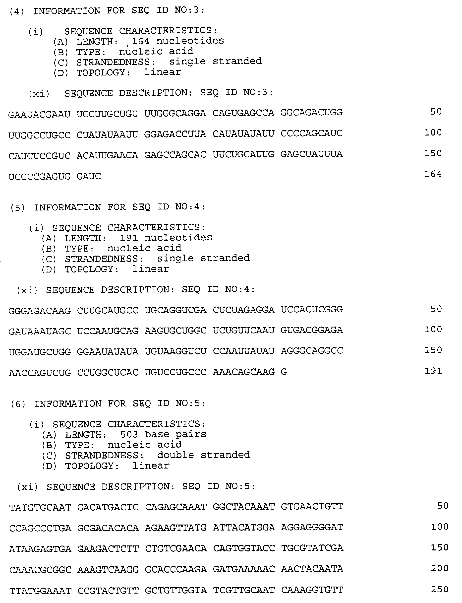
- The "antisense" RNA probe used in this assay had the following sequence (SEQ. ID NO.: 3):

The antisense probe was prepared using a Promega Riboprobe Gemini II kit (#P2020) from a linearized template DNA encoding the corresponding DNA sequence described above. The RNA probe was purified from a urea-polyacrylamide gel by cutting out the correct size band and eluting the RNA probe in Ambion Solution F. - The "sense" RNA standard coding for the complement RNA sequence to the "antisense" probe is as follows (SEQ. ID NO.: 4):

The sense standard was prepared cold (without radioactive label) using the Promega Riboprobe Gemini II kit (#P2020) and purified over a G50 Sephadex Quick spin column. - Following the standard Ambion (RPA II #1410) protocol, 50 µg of total RNA were incubated with 10⁵ cpm of probe, first at 95°C for four minutes, then twelve to eighteen hours at 45°C. RNAse (1:500 Ambion Solution R) was incubated with the hybridization solution at 37°C for forty minutes, followed by addition of the inactivating/precipitation reagent (Solution Dx).
- The RNAse protected fragments were then separated by size using urea-polyacrylamide gel electrophoresis and the whole gel was exposed to a phosphorimager screen. The bands were then quantified on a Molecular Dynamics Phosphorimager. The results are shown in Fig. 14.
- The identification of KGF as a growth factor for type II pneumocytes raises the possibility that KGF will show therapeutic potential similar to glucocorticoids as a stimulant of fetal lung maturation. KGF might also be clinically useful in stimulating bronchoalveolar repair after lung injury in the adult. The proliferative effect of KGF on type II pneumocytes suggests that KGF may play an important role in regulating the synthesis and secretion of surfactant.
- The effects of systemically administered KGF on the epithelial tissues of the liver in adult rats were evaluated.
- Male rats received water and food ad libitum. All animals received either KGF or PBS by intraperitoneal injection daily, for one, four, or seven days. In all experiments, at least five animals were analyzed per group.
- All rats were euthanized by CO₂ asphyxiation and blood was obtained for serum chemistry by cardiac puncture immediately postmortem and stored frozen until assayed. Serum was analyzed for standard serum chemistries and electrolytes. The liver was weighed in a blinded fashion and weights were expressed as grams per 100 grams body weight (percent body weight). Tissue samples were placed in 10% buffered formalin for routine histological processing.
- Sections from the liver were stained with hematoxylin and eosin (H and E). One hour prior to sacrifice, rats received 50 mg/kg of BrdU via intraperitioneal infection. Anti-BrdU immunohistochemistry was performed to visualize BrdU incorporation into DNA of replicating cells.
- The labeling index (LI) of the liver was determined using a calibrated eye objective grid. Ten fields per animal were counted and averaged. Total nuclei and labeled nuclei per unit area were counted. To standardize the fields counted as representing similar areas of hepatic parenchyma, all fields counted were adjacent to a portal triad.
- All data was analyzed using an unpaired two-tailed Student's t-test or one-way ANOVA for multiple group comparison.
- A dose response study was performed to identify an effective dose of KGF. KGF was injected daily for four days, and organs were harvested. Liver, expressed as percent body weight, was significantly different from controls only at the highest dose of 5 mg/kg per day when compared by ANOVA (Fig. 15). Of note, serum proteins, cholesterol and triglycerides, compounds synthesized by the liver were significantly higher compared with controls at both 1 and 5 mg/kg of KGF (Fig. 16).
- Animals were analyzed after one, four, or seven days daily treatment with KGF at 5 mg/kg. Upon gross necropsy exam, the liver was notably enlarged at the seven-day time point. Liver weight was significantly increased at all study time points in KGF-treated rats (day seven, KGF 4.08 ± 0.08% vs. control 3.49 ± 0.06%, p=0.0003).
- Quantitation of BrdU-positive nuclei demonstrated three- to four-fold increases in KGF-treated rats compared with controls at one day (Fig. 17). Mitotic Index expressed as mitotic figures per high powered field was significantly elevated over controls at one-day treatment, consistent with the BrdU counts.
- These results demonstrate potent mitogenic and differentiation effects of KGF on the liver. The rapid increase in liver weight after one day of KGF treatment coupled with an elevated mitotic index and BrdU-positive fraction demonstrates the rapid and potent effects of KGF on this predominantly epithelial tissue. Though the mitotic effects of KGF drop off rapidly, the liver weight expressed as percent body weight continued to increase throughout the studies. The increased weight was primarily due to an increase in cells that was histologically observable as early as one day and persisted through four and seven days of treatment. Since liver mass can increase by greater than two-fold in less than two weeks during liver regeneration, it was not surprising to find marked differences beginning after one day treatment.
- Elevated levels of albumin are not associated with any known disease state other than dehydration. KGF was likely inducing greater serum levels of albumin (and other proteins) due to the increased number of hepatocytes which were present. Increased protein synthesis induced by KGF would be of tremendous lifesaving benefit to individuals with end stage liver disease or cirrhosis.
- In this experiment, male Sprague-Dawley rats, each weighing from 300 to 340 grams, were used as the test subjects.
- Each of the rats was subjected to two-thirds partial hepatectomy using the procedure of Higgins and Anderson, Archives of Pathology, Volume 12, page 186 (1931). Commencing eight hours following surgery, and continuing once a day thereafter, the rats were injected subcutaneously with either PBS (control group) or KGF at a dose of 1 mg/kg. The control and KGF-treated rats were weighed and sacrificed four, seven, ten, or fourteen days after surgery. Liver remnants were then removed and weighed.
- It was found that treatment of partially hepatectomized rats with KGF resulted in significant acceleration in the recovery of liver mass, both on an absolute and relative basis, as shown in Figs. 18 and 19, respectively. In the KGF-treated group liver mass returned to pre-surgical values within seven days. By contrast, for the control (non-KGF treated) group neither absolute nor relative liver mass was completely restored even after fourteen days.
- Male Sprague-Dawley rats, weighing 300 to 340 grams each, were tested to evaluate the therapeutic effect of KGF on the liver after introduction of a chemical toxin.
- Groups of twelve male Sprague-Dawley rats were given either 1 ml/kg or 2 ml/kg of carbon tetrachloride (CCl₄) in a corn oil vehicle on day zero. Each dosage group was subdivided into two subgroups of six rats each. Beginning at three hours after CCl₄ administration, and continuing daily thereafter, each subgroup was injected subcutaneously with either PBS or 1 mg/kg/day of KGF. At twenty-two, seventy-two and one hundred and forty-four hours after CCl₄ administration, a blood sample was taken from each rat and the serum glutamate-oxaloacetate transaminase (SGOT) level was determined.
- As shown in Fig. 20, subcutaneous injection of KGF after oral administration of CCl₄ significantly decreased blood serum levels of an enzyme (SGOT) which is a known indicator of liver damage. The effect is especially pronounced at 2 ml/kg, on the first day after administration.
- This Example evaluates the biological effects of KGF on epithelial cells of the intestinal mucosa of healthy rats.
- Male rats received water and food ad libitum. All animals received either KGF or PBS by intraperitoneal injection daily for one, four or seven days. In all experiments, at least five animals were analyzed per group.
- All rats were euthanized by CO₂ asphyxiation. Major organs were weighed in a blinded fashion and weights were expressed as grams per 100 grams body weight (percent body weight). The gut was divided into the non-glandular foregut, glandular stomach, small intestine, and large intestine. Tissue samples were placed in 10% buffered formalin for routine histological processing.
- Sections from all organs harvested were stained with hematoxylin and eosin (H and E). One hour prior to sacrifice, rats received 50 mg/kg BrdU via intraperitoneal injection. Anti-BrdU immunohistochemistry was performed using standard methods to visualize BrdU incorporation into DNA of replicating cells. Selected tissues were stained with periodic-acid-schiff (PAS), Alcian blue at pH 2.5, and Masson-Trichrome.
- A calibrated ocular was used to measure the heights of the gastric mucosa and the depth of PAS staining with the gastric mucosa. Similarly, the length of villi and depth of intestinal crypts in the duodenum and depth of the colonic crypts in the colon were measured. To quantify the changes in goblet cell production, the number of PAS positive goblet cells was counted along 100 µm lengths beginning at the base of the duodenal villi. In all areas of the gut, measurements were made only on glands or villi that were perpendicular to the underlying muscularis. Results are recorded in microns or in the case of small intestinal goblet cells average number per unit area. In addition, the number of colonic crypts and the number of hyperplastic (bifurcated or trifurcated) crypts occupying a defined length of colon were determined. Five to ten replicate measurements were made per animal and averaged. All data was analyzed using an unpaired two-tailed Student's t-test or one-way ANOVA for multiple group comparisons (Statview II, Abacus Concepts, Berkeley, CA).
- A dose response study was performed initially to identify an effective dose of KGF. KGF was injected daily for four days, and organs were harvested. A pronounced dose response was observed in the glandular stomach, small intestine, and colon (Fig. 15).
- Animals were analyzed after one, four and seven days daily treatment with KGF at 5 mg/kg. Upon gross necropsy exam, the foregut was grossly thickened with folding of the mucosa at four- and seven-day treatment. The foregut, glandular stomach, duodenum and colon showed significant weight increases at four and seven days, but not at one-day treatment. At seven days, the glandular stomach was 0.65 ± 0.01g in KGF-treated animals, and 0.48 ± 0.02g in controls (p = 0. 0001).
- The non-glandular foregut was significantly increased in weight at four and seven days of treatment. Histologically, the squamous epithelium of the non-glandular foregut was mildly (day four) to markedly (day seven) hyperplastic in KGF-treated animals. The proliferative epithelium changed abruptly at the cardia from the normal squamous epithelium of the esophagus to the proliferative epithelium of the treated foregut. The glandular gastric mucosa appeared normal histologically after one-day treatment. At four and seven days, an increased number of goblet cells was apparent histologically as cells with clear open cytoplasm. The animals treated with KGF for four and seven days showed a mild increase in the thickness of the mucosa (Fig. 21). At seven days, the mucus neck cell layer was widened and displaced toward the serosal surface. PAS staining at four and seven days demonstrated striking increases in the number and size of PAS positive (mucin producing) goblet cells. BrdUstained sections further identified this layer as the dividing cell layer in the glandular stomach and showed an increase in proliferating cells with treatment. The thickness of the PAS positive layer of the luminal surface of the gastric mucosa was also markedly increased at four and seven days KGF treatment (Fig. 21).
- The small intestine was grossly normal at all study points. PAS staining demonstrated an increased number of mucus producing goblet cells. This difference was significant at four and seven days (Fig. 22). The villus height was not different at any time point but the crypt depth was significantly greater at one and seven days (Fig. 22).
- In seven-day treatment groups the colonic mucosa was thrown into rugal-like folds that greatly increased the overall surface area. Indicators of hyperplasia, bifurcated and trifurcated colonic crypts, were observed in all groups, including controls, but were most numerous and prominent in the short term treatment groups, e.g., days one and four, with the number of hyperplastic crypts decreasing with length of treatment (Fig. 23). Correspondingly, the depth of colonic crypts increased with treatment (Fig. 24).
- PAS and Alcian blue staining indicted an increase in mucus producing goblet cells throughout the colon following KGF treatment. Additionally, the number of Alcian blue positive goblet cells at the luminal surface was increased at seven days in all treatment animals. Treatment animals as early as four days showed marked increases in PAS positive cells in the upper one-half and one-third of the colonic crypts.
- These results demonstrate potent mitogenic and differentiation effects of KGF on epithelial tissues within the GI tract. It is apparent from BrdU labeling that KGF initiates proliferation of cells in the mucus neck layer of the gastric mucosa. These cells constitute the progenitor cells of the glandular stomach. Following division, cells from this layer migrate upwards, towards the gut lumen, to form surface cells or downward, towards the serosal surface, to become cells that occupy the gastric glands. (Toner et al., 1989, in Gastrointestinal and Esophageal Pathology, Churchill Livingstone, New York, NY, at pages 13-28). Additionally, KGF promotes selective differentiation of mucus neck cells to cells that move upward and that become goblet cells. The overall thickness of the gastric mucosa was increased at four and seven days of treatment. Similar effects were seen in the small and large intestines. KGF promoted increased cell division, resulting in elongated crypts, via stimulation of progenitor cells. KGF also induced differentiation of dividing crypt cells, as evidenced by increased mucus production and increased number of goblet cells in the villi or crypts.
- While this invention has been described in relation to specific embodiments and examples, it should be understood that there is no intention for this description to be limiting. Thus, for instance, any form of KGF which substantially duplicates the biological properties of naturally occurring KGF can be employed to carry out the described uses. Such forms include purified naturally occurring KGF itself, KGF synthesized by chemical procedures, and KGF derived recombinantly using other expression systems besides the one illustrated above, as well as analogs, variants, chimeras, etc., based on the naturally occurring amino acid sequence. All such forms are intended for use in the practice of the present invention.
- As those skilled in the art will also appreciate, a variety of host-vector systems may be utilized to express the KGF protein-coding sequence. These include but are not limited to mammalian cell systems infected with virus (e.g., vaccinia virus, adenovirus, etc.); insect cell systems infected with virus (e.g., baculovirus); microorganisms such as yeast containing yeast vectors, or other bacteria besides E.coli transformed with bacteriophage DNA, plasmid DNA, or cosmid DNA. The expression elements of these vectors vary in their strengths and specificities. Depending on the host-vector system utilized, any one of a number of suitable transcription and translation elements may be used.
- Once the protein product of KGF cDNA expression has been isolated, purified and assayed for KGF activity, using methods such as described in this text or other procedures known to those skilled in the art, it may be formulated in a variety of pharmaceutical compositions. Typically, such compositions include a suitable, usually chemically defined, carrier or excipient for the therapeutic agent (KGF), and, depending on the intended form of administration, other ingredients as well. The composition can include aqueous carriers or consist of solid phase formulations in which KGF is incorporated into non-aqueous carriers such as collagens, hyaluronic acid, and various polymers. The composition can be suitably formulated to be administered in a variety of ways, including by injection, orally, topically, intranasally, and by pulmonary delivery.
- As the skilled practitioner will understand, the amount of KGF to be dosed will vary depending on the disease being treated and the route of administration, but may be effective in the range, for example, from 0.01 mg per kg of body weight to 500 mg per kg of body weight. It may be used once or administered repeatedly, depending on the disease and condition of the patient. It may be used in conjunction with other treatments, including, but not limited to, other cytokines and growth factors and other pharmaceutical preparations used to treat diseases of the skin, lungs, liver and gastrointestinal tract.


Claims (13)
- Chemical compound composed of keratinocyte growth factor (KGF) for its use in a method of therapeutic treatment for the human or animal body.
- Chemical compound according to claim 1 for stimulating the production of non-keratinocyte epithelial cells.
- Chemical compound according to claim 1 or 2 for stimulating sebocytes, hair follicle cells, hepatocytes, type II pneumocytes, mucin-producing goblet cells, and other epithelial cells and their progenitors contained within the skin, lung, liver and gastrointestinal tract.
- Chemical compound according to claim 1 or 2 for the treatment of dermal wounds to stimulate the production of cells of the sebaceous glands and hair follicles.
- Chemical compound according to claim 4, in which the dermal wound is a burn wound.
- Chemical compound according to claim 1 for the treatment of epidermolysis bullosa, for the treatment of chemotherapy-induced alopecia, for the treatment of malepattern baldness, for the treatment of gastric ulcers, for the treatment of duodenal ulcer, for the treatment of erosions of the stomach and esophagus, erosive gastritis, esophagitis, or esophageal reflux, for the treatment of inflammatory bowel diseases, for the treatment or prevention of radiation or chemotherapy-induced gut toxicity, for the treatment of hyaline membrane disease, for the treatment of necrosis of the respiratory epithelium caused by smoke inhalation, for the treatment of emphysema, for the treatment of pulmonary inflammation and fibrosis, for the treatment of hepatic cirrhosis, for the treatment of fulminant liver failure, for the treatment of acute viral hepatitis and for the treatment of toxic insult to the liver.
- Chemical compound according to anyone of claims 1 to 6, in which the KGF is a recombinant KGF.
- Chemical compound according to claim 7, in which the recombinant KGF is produced in bacterial cells.
- Chemical compound according to claim 8, in which the bacterial cells are E.coli.
- A pharmaceutical composition comprising a therapeutically effective amount of KGF and a pharmaceutically acceptable carrier.
- The composition of claim 10 in which the KGF is a recombinant KGF.
- The composition of claim 11 in which the recombinant KGF is produced in bacterial cells.
- The composition of claim 12 in which the bacterial cells are E.coli.
Priority Applications (1)
| Application Number | Priority Date | Filing Date | Title |
|---|---|---|---|
| SI9430265T SI0619370T1 (en) | 1993-03-26 | 1994-03-25 | The use of keratinocyte growth factor (KGF) for the preparation of pharmaceutical compositions |
Applications Claiming Priority (2)
| Application Number | Priority Date | Filing Date | Title |
|---|---|---|---|
| US4074293A | 1993-03-26 | 1993-03-26 | |
| US40742 | 1993-03-26 |
Publications (2)
| Publication Number | Publication Date |
|---|---|
| EP0619370A1 true EP0619370A1 (en) | 1994-10-12 |
| EP0619370B1 EP0619370B1 (en) | 1999-08-11 |
Family
ID=21912689
Family Applications (1)
| Application Number | Title | Priority Date | Filing Date |
|---|---|---|---|
| EP94104804A Expired - Lifetime EP0619370B1 (en) | 1993-03-26 | 1994-03-25 | The use of keratinocyte growth factor (KGF) for the preparation of pharmaceutical compositions |
Country Status (23)
| Country | Link |
|---|---|
| EP (1) | EP0619370B1 (en) |
| JP (1) | JP3578761B2 (en) |
| KR (1) | KR100390340B1 (en) |
| CN (1) | CN1064710C (en) |
| AT (1) | ATE183233T1 (en) |
| AU (1) | AU707340B2 (en) |
| CA (1) | CA2159109C (en) |
| CZ (1) | CZ285996B6 (en) |
| DE (1) | DE69419958T2 (en) |
| DK (1) | DK0619370T3 (en) |
| ES (1) | ES2136673T3 (en) |
| FI (1) | FI954541A (en) |
| GR (1) | GR3031509T3 (en) |
| HU (1) | HU220641B1 (en) |
| IL (4) | IL137581A (en) |
| NO (1) | NO953781L (en) |
| NZ (1) | NZ263967A (en) |
| RU (1) | RU2146148C1 (en) |
| SG (1) | SG49841A1 (en) |
| SI (1) | SI0619370T1 (en) |
| SK (1) | SK281674B6 (en) |
| UA (1) | UA46706C2 (en) |
| WO (1) | WO1994023032A1 (en) |
Cited By (19)
| Publication number | Priority date | Publication date | Assignee | Title |
|---|---|---|---|---|
| WO1996011950A1 (en) * | 1994-10-13 | 1996-04-25 | Amgen Inc. | Method of treating diabetes mellitus using kgf |
| WO1997013857A1 (en) * | 1995-10-11 | 1997-04-17 | Chiron Corporation | Combination pdgf, kgf, igf and igfbp for wound healing |
| EP0815115A1 (en) * | 1995-02-14 | 1998-01-07 | Human Genome Sciences, Inc. | Keratinocyte growth factor-2 |
| WO1998011910A1 (en) * | 1996-09-20 | 1998-03-26 | Scientific Hospital Supplies International Limited | Prevention of gastrointestinal damage |
| WO1998016243A1 (en) * | 1996-10-15 | 1998-04-23 | Amgen Inc. | Uses of keratinocyte growth factor-2 |
| WO1999018128A1 (en) * | 1997-10-03 | 1999-04-15 | Amgen Inc. | Fibroblast growth factor with hepatocyte proliferation activity |
| US6228839B1 (en) * | 1997-12-19 | 2001-05-08 | Emory University | Use of keratinocyte growth factor to improve oxidative status |
| US6335317B1 (en) | 1998-04-10 | 2002-01-01 | Emory University | Use of gut-trophic growth factors to improve oxidative status |
| WO2002062842A1 (en) * | 2001-02-06 | 2002-08-15 | Merck Patent Gmbh | Modified keratinocyte growth factor (kgf) with reduced immunogenicity |
| US6599879B1 (en) | 1998-02-13 | 2003-07-29 | Human Genome Sciences, Inc. | Therapeutic uses of keratinocyte growth factor-2 |
| US6653284B2 (en) | 1997-12-22 | 2003-11-25 | Human Genome Sciences, Inc. | Keratinocyte growth factor-2 formulations |
| US6693077B1 (en) | 1995-02-14 | 2004-02-17 | Human Genome Sciences, Inc. | Keratinocyte growth factor-2 |
| US6743422B1 (en) | 1996-10-15 | 2004-06-01 | Amgen, Inc. | Keratinocyte growth factor-2 products |
| EP1452205A1 (en) * | 1996-10-15 | 2004-09-01 | Amgen Inc. | Uses of keratinocyte growth factor-2 |
| US6869927B1 (en) | 1997-12-22 | 2005-03-22 | Human Genome Sciences, Inc. | Keratinocyte growth factor-2 formulations |
| US6903072B2 (en) | 1995-02-14 | 2005-06-07 | Human Genome Sciences, Inc. | Keratinocyte growth factor-2 |
| WO2006030241A2 (en) * | 2004-09-13 | 2006-03-23 | University Of Southampton | Growth factor treatment for asthma |
| US7202066B2 (en) | 2002-01-29 | 2007-04-10 | Carrington Laboratories, Inc. | Combination of a growth factor and a protease enzyme |
| US7232667B2 (en) | 1995-02-14 | 2007-06-19 | Human Genome Sciences, Inc. | Keratinocyte growth factor-2 polynucleotides |
Families Citing this family (8)
| Publication number | Priority date | Publication date | Assignee | Title |
|---|---|---|---|---|
| AUPN271295A0 (en) | 1995-05-02 | 1995-05-25 | Gropep Pty Ltd | Method of treatment |
| ATE297751T1 (en) * | 2000-03-20 | 2005-07-15 | Pfizer Prod Inc | COMBINATION THERAPY WITH KERATINOCYTE GROWTH FACTOR AND AN EPIDERMIS GROWTH FACTOR INHIBITOR |
| CN101822821B (en) * | 2010-04-27 | 2013-01-23 | 复旦大学附属中山医院 | Application of keratinocyte growth factor-2 in preparation of medicines for preventing and curing lung injury |
| CN104707164B (en) * | 2015-03-31 | 2017-01-18 | 中国人民解放军军事医学科学院基础医学研究所 | Composite chitosan hydrogel dressing as well as preparation method and applications thereof |
| CN106226511B (en) * | 2016-07-28 | 2018-04-06 | 苏州金盟生物技术有限公司 | A kind of recombinant human horny cell growth factor-2 biological activity detection method |
| CN107753932A (en) * | 2016-08-22 | 2018-03-06 | 中国辐射防护研究院 | KGF is preparing the purposes in treating or preventing radiation enteritis medicine |
| EP3888625A4 (en) | 2018-11-27 | 2022-10-05 | Jellyfish Research Laboratories, Inc. | Composition for scalp and hair |
| KR20210114646A (en) * | 2020-03-11 | 2021-09-24 | (주)메디코스바이오텍 | Composition for Treating Alopecia or Stimulating Hair Growth Containing Growth Factor |
Citations (3)
| Publication number | Priority date | Publication date | Assignee | Title |
|---|---|---|---|---|
| WO1990008771A1 (en) * | 1989-01-31 | 1990-08-09 | Rubin Jeffrey S | Dna encoding a growth factor specific for epithelial cells |
| EP0455422A2 (en) * | 1990-04-27 | 1991-11-06 | Merck & Co. Inc. | Method of treating or preventing baldness with compositions containing fibroblast growth factor |
| WO1992014480A1 (en) * | 1991-02-22 | 1992-09-03 | Amgen Inc. | Use of gm-csf and g-csf to promote accelerated wound healing |
-
1994
- 1994-03-24 NZ NZ263967A patent/NZ263967A/en not_active IP Right Cessation
- 1994-03-24 IL IL137581A patent/IL137581A/en not_active IP Right Cessation
- 1994-03-24 KR KR1019950704154A patent/KR100390340B1/en not_active IP Right Cessation
- 1994-03-24 IL IL10912294A patent/IL109122A/en not_active IP Right Cessation
- 1994-03-24 SK SK1185-95A patent/SK281674B6/en not_active IP Right Cessation
- 1994-03-24 CA CA002159109A patent/CA2159109C/en not_active Expired - Lifetime
- 1994-03-24 AU AU65243/94A patent/AU707340B2/en not_active Expired
- 1994-03-24 RU RU95117959A patent/RU2146148C1/en active
- 1994-03-24 UA UA95104698A patent/UA46706C2/en unknown
- 1994-03-24 JP JP52218894A patent/JP3578761B2/en not_active Expired - Lifetime
- 1994-03-24 WO PCT/US1994/003208 patent/WO1994023032A1/en active IP Right Grant
- 1994-03-24 CN CN94192037A patent/CN1064710C/en not_active Expired - Lifetime
- 1994-03-24 HU HU9502800A patent/HU220641B1/en unknown
- 1994-03-24 CZ CZ952482A patent/CZ285996B6/en not_active IP Right Cessation
- 1994-03-25 SG SG1996007376A patent/SG49841A1/en unknown
- 1994-03-25 AT AT94104804T patent/ATE183233T1/en active
- 1994-03-25 ES ES94104804T patent/ES2136673T3/en not_active Expired - Lifetime
- 1994-03-25 EP EP94104804A patent/EP0619370B1/en not_active Expired - Lifetime
- 1994-03-25 SI SI9430265T patent/SI0619370T1/en unknown
- 1994-03-25 DK DK94104804T patent/DK0619370T3/en active
- 1994-03-25 DE DE69419958T patent/DE69419958T2/en not_active Expired - Lifetime
-
1995
- 1995-09-25 FI FI954541A patent/FI954541A/en unknown
- 1995-09-25 NO NO953781A patent/NO953781L/en not_active Application Discontinuation
-
1999
- 1999-10-13 GR GR990402604T patent/GR3031509T3/en unknown
-
2000
- 2000-07-30 IL IL13758100A patent/IL137581A0/en unknown
-
2004
- 2004-04-22 IL IL16156804A patent/IL161568A0/en unknown
Patent Citations (3)
| Publication number | Priority date | Publication date | Assignee | Title |
|---|---|---|---|---|
| WO1990008771A1 (en) * | 1989-01-31 | 1990-08-09 | Rubin Jeffrey S | Dna encoding a growth factor specific for epithelial cells |
| EP0455422A2 (en) * | 1990-04-27 | 1991-11-06 | Merck & Co. Inc. | Method of treating or preventing baldness with compositions containing fibroblast growth factor |
| WO1992014480A1 (en) * | 1991-02-22 | 1992-09-03 | Amgen Inc. | Use of gm-csf and g-csf to promote accelerated wound healing |
Non-Patent Citations (14)
| Title |
|---|
| CHEMICAL ABSTRACTS, vol. 118, no. 15, 12 April 1993, Columbus, Ohio, US; abstract no. 140028v, YAN ET AL: "SEQUENCE OF RAT KERATINOCYTE GROWTH FACTOR (HEPARIN-BINDING GROWTH FACTOR TYPE 7)" page 103; column 1; * |
| CHEMICAL ABSTRACTS, vol. 120, no. 3, 17 January 1994, Columbus, Ohio, US; abstract no. 24276s, PANOS ET AL: "KERATINOCYTE GROWTH FACTOR AND HEPATOCYTE GROWTH FACTOR/SCATTER FACTOR ARE HEPARIN-BINDING GROWTH FACTORS FOR ALVEOLAR TYPE II CELLS IN FIBROBLAST-CONDITIONED MEDIUM" page 163; column 2; * |
| FILE SERVER STN KARLSRUHE,FILE MEDLINE ABSTRACT NO.92394861 & IN VITRO CELL DEV BIOL, (1992 JUL-AUG) 28A (7-8) 515-20 * |
| FILE SERVER STN KARLSRUHE,FILE MEDLINE ABSTRACT NO.94101630 & CANCER RES, (1994 JAN 15) 54 (2) 518-22 * |
| FILE SERVER STN KARLSRUHE,FILE MEDLINE ABSTRACT NO.94157403 & J EXP MED, (1994 MAR 1) 179 (3) 831-40 * |
| FINCH ET AL: "HUMAN KGF IS FGF-RELATED WITH PROPERTIES OF A PARACRINE EFFECTOR OF EPITHELIAL CELL GROWTH", SCIENCE, vol. 245, 1989, WASHINGTON D.C.,USA, pages 752 - 755, XP002937855, DOI: doi:10.1126/science.2475908 * |
| HAVILL ET AL: "KERATINOCYTE GROWTH FACTOR (RHKGF) HAS HEPATIC STIMULATORY EFFECTS IN VIVO", THE FASEB JOURNAL, vol. 8, no. 5, 18 March 1994 (1994-03-18), BETHESDA,MD,USA, pages A930 * |
| IN VITRO CELL.DEV.BIO.:ANIM., vol. 27A, no. 6, 1991, pages 437 - 438 * |
| J.CLIN.INVEST., vol. 92, no. 2, August 1993 (1993-08-01), pages 968 - 977 * |
| RUBIN ET AL: "PURIFICATION AND CHARACTERIZATION OF A NEWLY IDENTIFIED GROWTH FACTOR SPECIFIC FOR EPITHELIAL CELLS", PROCEEDINGS OF THE NATIONAL ACADEMY OF SCIENCES, vol. 86, 1989, WASHINGTON D.C.,USA, pages 802 - 806, XP002138002, DOI: doi:10.1073/pnas.86.3.802 * |
| SURGICAL FORUM, vol. 44, no. 0, 1993, pages 704 - 706 * |
| WILSON ET AL: "HEPATOCYTE GROWTH FACTOR (HGF),KERATINOCYTE GROWTH FACTOR (KGF),THEIR RECEPTORS,AND THE CELLS OF THE CORNEA", THE FASEB JOURNAL, vol. 7, no. 3, 19 February 1993 (1993-02-19), BETHESDA,MD,USA, pages A493 * |
| WU ET AL: "KGF ACCELERATES ISCHEMIC DERMAL ULCER HEALING IN THE RABBIT EAR", PROCEEDINGS FOR THE 49TH ANNUAL SESSIONS OF THE FORUM ON FUNDAMENTAL SURGICAL PROBLEMS, October 1993 (1993-10-01), SAN FRANSISCO,CA,USA * |
| YI ET AL: "KERATINOCYTE GROWTH FACTOR IS A GROWTH FACTOR FOR TYPE II PNEUMOCYTE IN VIVO", MODERN PATHOLOGY, vol. 7, no. 1, January 1994 (1994-01-01), BALTIMORE,MD,USA, pages 156A * |
Cited By (25)
| Publication number | Priority date | Publication date | Assignee | Title |
|---|---|---|---|---|
| WO1996011950A1 (en) * | 1994-10-13 | 1996-04-25 | Amgen Inc. | Method of treating diabetes mellitus using kgf |
| EP1323824A2 (en) * | 1995-02-14 | 2003-07-02 | Human Genome Sciences, Inc. | Use of human keratinocyte growth factor-2 for the preparation of pharmaceutical compositions |
| EP0815115A1 (en) * | 1995-02-14 | 1998-01-07 | Human Genome Sciences, Inc. | Keratinocyte growth factor-2 |
| US6693077B1 (en) | 1995-02-14 | 2004-02-17 | Human Genome Sciences, Inc. | Keratinocyte growth factor-2 |
| EP1323824A3 (en) * | 1995-02-14 | 2004-01-21 | Human Genome Sciences, Inc. | Use of human keratinocyte growth factor-2 for the preparation of pharmaceutical compositions |
| EP0815115A4 (en) * | 1995-02-14 | 1999-09-22 | Human Genome Sciences Inc | Keratinocyte growth factor-2 |
| US6903072B2 (en) | 1995-02-14 | 2005-06-07 | Human Genome Sciences, Inc. | Keratinocyte growth factor-2 |
| US7232667B2 (en) | 1995-02-14 | 2007-06-19 | Human Genome Sciences, Inc. | Keratinocyte growth factor-2 polynucleotides |
| US6916786B2 (en) | 1995-02-14 | 2005-07-12 | Human Genome Sciences, Inc. | Keratinocyte growth factor-2 |
| WO1997013857A1 (en) * | 1995-10-11 | 1997-04-17 | Chiron Corporation | Combination pdgf, kgf, igf and igfbp for wound healing |
| WO1998011910A1 (en) * | 1996-09-20 | 1998-03-26 | Scientific Hospital Supplies International Limited | Prevention of gastrointestinal damage |
| WO1998016243A1 (en) * | 1996-10-15 | 1998-04-23 | Amgen Inc. | Uses of keratinocyte growth factor-2 |
| AU725509B2 (en) * | 1996-10-15 | 2000-10-12 | Biovitrum Ab (Publ) | Uses of keratinocyte growth factor-2 |
| EP1452205A1 (en) * | 1996-10-15 | 2004-09-01 | Amgen Inc. | Uses of keratinocyte growth factor-2 |
| US6743422B1 (en) | 1996-10-15 | 2004-06-01 | Amgen, Inc. | Keratinocyte growth factor-2 products |
| WO1999018128A1 (en) * | 1997-10-03 | 1999-04-15 | Amgen Inc. | Fibroblast growth factor with hepatocyte proliferation activity |
| US6228839B1 (en) * | 1997-12-19 | 2001-05-08 | Emory University | Use of keratinocyte growth factor to improve oxidative status |
| US6869927B1 (en) | 1997-12-22 | 2005-03-22 | Human Genome Sciences, Inc. | Keratinocyte growth factor-2 formulations |
| US6653284B2 (en) | 1997-12-22 | 2003-11-25 | Human Genome Sciences, Inc. | Keratinocyte growth factor-2 formulations |
| US6599879B1 (en) | 1998-02-13 | 2003-07-29 | Human Genome Sciences, Inc. | Therapeutic uses of keratinocyte growth factor-2 |
| US6335317B1 (en) | 1998-04-10 | 2002-01-01 | Emory University | Use of gut-trophic growth factors to improve oxidative status |
| WO2002062842A1 (en) * | 2001-02-06 | 2002-08-15 | Merck Patent Gmbh | Modified keratinocyte growth factor (kgf) with reduced immunogenicity |
| US7202066B2 (en) | 2002-01-29 | 2007-04-10 | Carrington Laboratories, Inc. | Combination of a growth factor and a protease enzyme |
| WO2006030241A2 (en) * | 2004-09-13 | 2006-03-23 | University Of Southampton | Growth factor treatment for asthma |
| WO2006030241A3 (en) * | 2004-09-13 | 2006-06-15 | Univ Southampton | Growth factor treatment for asthma |
Also Published As
Similar Documents
| Publication | Publication Date | Title |
|---|---|---|
| US5814605A (en) | Therapeutic uses of keratinocyte growth factor | |
| EP0619370B1 (en) | The use of keratinocyte growth factor (KGF) for the preparation of pharmaceutical compositions | |
| US4959353A (en) | Promotion of corneal stroma wound healing with human epidermal growth factor prepared from recombinant DNA | |
| Marchbank et al. | Dimerization of human pS2 (TFF1) plays a key role in its protective/healing effects | |
| EP0134385A2 (en) | A protein having cell growth stimulating action, composition thereof and method for producing the same | |
| EP0492614B1 (en) | Epitheliocyte growth accelerator | |
| US7011965B2 (en) | Compositions and methods for stimulating wound healing and fibroblast proliferation | |
| Kaneko et al. | Changes in serum human hepatocyte growth factor levels after transcatheter arterial embolization and partial hepatectomy. | |
| WO1994016723A2 (en) | Wound healing composition | |
| Tchorzewski et al. | Characterization of an aFGF gene expression vector with therapeutic potential | |
| US20230220026A1 (en) | Nucleic acid carrier for producing the single-chain vegf fusion protein with high physiological stability and dimeric efficiency, preparation method thereof, and use thereof | |
| EP0895478A1 (en) | Component b as cicatrizant | |
| JP4583763B2 (en) | Cleaved 24 kDa basic fibroblast growth factor | |
| EP1013280A1 (en) | Skin preparations for external use | |
| CN108434441A (en) | Application of novel fibroblast growth factor -21 analogs of son in treating the chromic fibrous pneumonopathy of diffusivity | |
| WO1996016669A1 (en) | Use of human recombinant epidermal growth factor in the manufacture of a medicament for treating acne | |
| Steidler et al. | Histological Effects of Epidermal Growth Factor on Eyelids and Cornea in Neonatal Mice |
Legal Events
| Date | Code | Title | Description |
|---|---|---|---|
| PUAI | Public reference made under article 153(3) epc to a published international application that has entered the european phase |
Free format text: ORIGINAL CODE: 0009012 |
|
| AK | Designated contracting states |
Kind code of ref document: A1 Designated state(s): AT BE CH DE DK ES FR GB GR IE IT LI LU MC NL PT SE |
|
| RAX | Requested extension states of the european patent have changed |
Free format text: SI PAYMENT 940325 |
|
| 17P | Request for examination filed |
Effective date: 19950405 |
|
| 17Q | First examination report despatched |
Effective date: 19951024 |
|
| GRAG | Despatch of communication of intention to grant |
Free format text: ORIGINAL CODE: EPIDOS AGRA |
|
| GRAG | Despatch of communication of intention to grant |
Free format text: ORIGINAL CODE: EPIDOS AGRA |
|
| GRAH | Despatch of communication of intention to grant a patent |
Free format text: ORIGINAL CODE: EPIDOS IGRA |
|
| GRAH | Despatch of communication of intention to grant a patent |
Free format text: ORIGINAL CODE: EPIDOS IGRA |
|
| GRAA | (expected) grant |
Free format text: ORIGINAL CODE: 0009210 |
|
| AK | Designated contracting states |
Kind code of ref document: B1 Designated state(s): AT BE CH DE DK ES FR GB GR IE IT LI LU MC NL PT SE |
|
| AX | Request for extension of the european patent |
Free format text: SI PAYMENT 19940325 |
|
| REF | Corresponds to: |
Ref document number: 183233 Country of ref document: AT Date of ref document: 19990815 Kind code of ref document: T |
|
| REG | Reference to a national code |
Ref country code: CH Ref legal event code: NV Representative=s name: KIRKER & CIE SA Ref country code: CH Ref legal event code: EP |
|
| REF | Corresponds to: |
Ref document number: 69419958 Country of ref document: DE Date of ref document: 19990916 |
|
| ITF | It: translation for a ep patent filed |
Owner name: JACOBACCI & PERANI S.P.A. |
|
| ET | Fr: translation filed | ||
| REG | Reference to a national code |
Ref country code: IE Ref legal event code: FG4D |
|
| REG | Reference to a national code |
Ref country code: PT Ref legal event code: SC4A Free format text: AVAILABILITY OF NATIONAL TRANSLATION Effective date: 19990830 |
|
| REG | Reference to a national code |
Ref country code: ES Ref legal event code: FG2A Ref document number: 2136673 Country of ref document: ES Kind code of ref document: T3 |
|
| REG | Reference to a national code |
Ref country code: DK Ref legal event code: T3 |
|
| PLBQ | Unpublished change to opponent data |
Free format text: ORIGINAL CODE: EPIDOS OPPO |
|
| PLBI | Opposition filed |
Free format text: ORIGINAL CODE: 0009260 |
|
| PLBF | Reply of patent proprietor to notice(s) of opposition |
Free format text: ORIGINAL CODE: EPIDOS OBSO |
|
| 26 | Opposition filed |
Opponent name: CHIRON CORPORATION Effective date: 20000511 |
|
| NLR1 | Nl: opposition has been filed with the epo |
Opponent name: CHIRON CORPORATION |
|
| PLBF | Reply of patent proprietor to notice(s) of opposition |
Free format text: ORIGINAL CODE: EPIDOS OBSO |
|
| RAP2 | Party data changed (patent owner data changed or rights of a patent transferred) |
Owner name: AMGEN INC. |
|
| PLBF | Reply of patent proprietor to notice(s) of opposition |
Free format text: ORIGINAL CODE: EPIDOS OBSO |
|
| NLT2 | Nl: modifications (of names), taken from the european patent patent bulletin |
Owner name: AMGEN INC. |
|
| PLBF | Reply of patent proprietor to notice(s) of opposition |
Free format text: ORIGINAL CODE: EPIDOS OBSO |
|
| PLBF | Reply of patent proprietor to notice(s) of opposition |
Free format text: ORIGINAL CODE: EPIDOS OBSO |
|
| REG | Reference to a national code |
Ref country code: GB Ref legal event code: IF02 |
|
| PLBP | Opposition withdrawn |
Free format text: ORIGINAL CODE: 0009264 |
|
| PLBD | Termination of opposition procedure: decision despatched |
Free format text: ORIGINAL CODE: EPIDOSNOPC1 |
|
| REG | Reference to a national code |
Ref country code: CH Ref legal event code: PFA Owner name: AMGEN INC, US Free format text: AMGEN INC.#AMGEN CENTER, 1840 DEHAVILLAND DRIVE#THOUSAND OAKS, CA 91320-1789 (US) -TRANSFER %%- AMGEN INC.#ONE AMGEN CENTER DRIVE#THOUSAND OAKS, CA 91320-1799 (US) |
|
| REG | Reference to a national code |
Ref country code: PT Ref legal event code: TE4A Free format text: AMGEN, INC. US Effective date: 20030616 |
|
| PLBM | Termination of opposition procedure: date of legal effect published |
Free format text: ORIGINAL CODE: 0009276 |
|
| STAA | Information on the status of an ep patent application or granted ep patent |
Free format text: STATUS: OPPOSITION PROCEDURE CLOSED |
|
| 27C | Opposition proceedings terminated |
Effective date: 20030725 |
|
| NLR2 | Nl: decision of opposition |
Effective date: 20030725 |
|
| REG | Reference to a national code |
Ref country code: SI Ref legal event code: IF |
|
| REG | Reference to a national code |
Ref country code: HK Ref legal event code: WD Ref document number: 1014031 Country of ref document: HK |
|
| PLAB | Opposition data, opponent's data or that of the opponent's representative modified |
Free format text: ORIGINAL CODE: 0009299OPPO |
|
| REG | Reference to a national code |
Ref country code: PT Ref legal event code: PC4A Owner name: BIOVITRUM AB (PUBL), SE Effective date: 20090528 |
|
| REG | Reference to a national code |
Ref country code: CH Ref legal event code: PUE Owner name: BIOVITRUM AB (PUBL) Free format text: AMGEN INC.#ONE AMGEN CENTER DRIVE#THOUSAND OAKS, CA 91320-1799 (US) -TRANSFER TO- BIOVITRUM AB (PUBL)# #112 76 STOCKHOLM (SE) Ref country code: CH Ref legal event code: NV Representative=s name: BUGNION S.A. |
|
| REG | Reference to a national code |
Ref country code: GB Ref legal event code: 732E Free format text: REGISTERED BETWEEN 20090709 AND 20090715 |
|
| REG | Reference to a national code |
Ref country code: FR Ref legal event code: TP |
|
| REG | Reference to a national code |
Ref country code: NL Ref legal event code: SD Effective date: 20110120 |
|
| REG | Reference to a national code |
Ref country code: ES Ref legal event code: PC2A Owner name: BIOVITRUM AB (PUBL) Effective date: 20111018 |
|
| PGFP | Annual fee paid to national office [announced via postgrant information from national office to epo] |
Ref country code: IT Payment date: 20120320 Year of fee payment: 19 |
|
| PGFP | Annual fee paid to national office [announced via postgrant information from national office to epo] |
Ref country code: ES Payment date: 20130313 Year of fee payment: 20 Ref country code: MC Payment date: 20130128 Year of fee payment: 20 Ref country code: SE Payment date: 20130312 Year of fee payment: 20 Ref country code: FR Payment date: 20130325 Year of fee payment: 20 Ref country code: DE Payment date: 20130320 Year of fee payment: 20 Ref country code: CH Payment date: 20130312 Year of fee payment: 20 Ref country code: GB Payment date: 20130320 Year of fee payment: 20 Ref country code: IE Payment date: 20130312 Year of fee payment: 20 Ref country code: LU Payment date: 20130328 Year of fee payment: 20 Ref country code: DK Payment date: 20130312 Year of fee payment: 20 |
|
| PGFP | Annual fee paid to national office [announced via postgrant information from national office to epo] |
Ref country code: GR Payment date: 20130215 Year of fee payment: 20 Ref country code: NL Payment date: 20130309 Year of fee payment: 20 |
|
| PGFP | Annual fee paid to national office [announced via postgrant information from national office to epo] |
Ref country code: AT Payment date: 20130226 Year of fee payment: 20 Ref country code: PT Payment date: 20130325 Year of fee payment: 20 |
|
| PGFP | Annual fee paid to national office [announced via postgrant information from national office to epo] |
Ref country code: BE Payment date: 20130312 Year of fee payment: 20 |
|
| REG | Reference to a national code |
Ref country code: DE Ref legal event code: R071 Ref document number: 69419958 Country of ref document: DE |
|
| REG | Reference to a national code |
Ref country code: NL Ref legal event code: V4 Effective date: 20140325 Ref country code: DE Ref legal event code: R071 Ref document number: 69419958 Country of ref document: DE |
|
| BE20 | Be: patent expired |
Owner name: BIOVITRUM A.B. PUBL Effective date: 20140325 |
|
| REG | Reference to a national code |
Ref country code: CH Ref legal event code: PL Ref country code: DK Ref legal event code: EUP Effective date: 20140325 |
|
| REG | Reference to a national code |
Ref country code: PT Ref legal event code: MM4A Free format text: MAXIMUM VALIDITY LIMIT REACHED Effective date: 20140325 |
|
| REG | Reference to a national code |
Ref country code: GB Ref legal event code: PE20 Expiry date: 20140324 |
|
| REG | Reference to a national code |
Ref country code: IE Ref legal event code: MK9A |
|
| REG | Reference to a national code |
Ref country code: SE Ref legal event code: EUG |
|
| PG25 | Lapsed in a contracting state [announced via postgrant information from national office to epo] |
Ref country code: GB Free format text: LAPSE BECAUSE OF EXPIRATION OF PROTECTION Effective date: 20140324 Ref country code: DE Free format text: LAPSE BECAUSE OF EXPIRATION OF PROTECTION Effective date: 20140326 |
|
| REG | Reference to a national code |
Ref country code: AT Ref legal event code: MK07 Ref document number: 183233 Country of ref document: AT Kind code of ref document: T Effective date: 20140325 |
|
| REG | Reference to a national code |
Ref country code: GR Ref legal event code: MA Ref document number: 990402604 Country of ref document: GR Effective date: 20140326 |
|
| REG | Reference to a national code |
Ref country code: ES Ref legal event code: FD2A Effective date: 20140606 |
|
| PG25 | Lapsed in a contracting state [announced via postgrant information from national office to epo] |
Ref country code: IE Free format text: LAPSE BECAUSE OF EXPIRATION OF PROTECTION Effective date: 20140325 |
|
| PG25 | Lapsed in a contracting state [announced via postgrant information from national office to epo] |
Ref country code: PT Free format text: LAPSE BECAUSE OF EXPIRATION OF PROTECTION Effective date: 20140402 Ref country code: ES Free format text: LAPSE BECAUSE OF EXPIRATION OF PROTECTION Effective date: 20140326 |