EP0596524B1 - Hepatocyte growth factor HGF converting protease and gene encoding said protease - Google Patents
Hepatocyte growth factor HGF converting protease and gene encoding said protease Download PDFInfo
- Publication number
- EP0596524B1 EP0596524B1 EP93117988A EP93117988A EP0596524B1 EP 0596524 B1 EP0596524 B1 EP 0596524B1 EP 93117988 A EP93117988 A EP 93117988A EP 93117988 A EP93117988 A EP 93117988A EP 0596524 B1 EP0596524 B1 EP 0596524B1
- Authority
- EP
- European Patent Office
- Prior art keywords
- protein
- gene
- seq
- amino acid
- dna
- Prior art date
- Legal status (The legal status is an assumption and is not a legal conclusion. Google has not performed a legal analysis and makes no representation as to the accuracy of the status listed.)
- Expired - Lifetime
Links
- 108090000623 proteins and genes Proteins 0.000 title claims abstract description 275
- 108091005804 Peptidases Proteins 0.000 title claims abstract description 33
- 239000004365 Protease Substances 0.000 title claims abstract description 33
- 102100037486 Reverse transcriptase/ribonuclease H Human genes 0.000 title claims abstract description 33
- 102100021866 Hepatocyte growth factor Human genes 0.000 title 1
- 102000004169 proteins and genes Human genes 0.000 claims abstract description 191
- 230000000694 effects Effects 0.000 claims abstract description 33
- 238000000034 method Methods 0.000 claims abstract description 31
- 102000003745 Hepatocyte Growth Factor Human genes 0.000 claims abstract description 29
- 108090000100 Hepatocyte Growth Factor Proteins 0.000 claims abstract description 29
- 210000004027 cell Anatomy 0.000 claims description 53
- 239000013604 expression vector Substances 0.000 claims description 26
- 238000004519 manufacturing process Methods 0.000 claims description 13
- 108090000765 processed proteins & peptides Proteins 0.000 claims description 12
- 239000013598 vector Substances 0.000 claims description 11
- 102000004196 processed proteins & peptides Human genes 0.000 claims description 10
- 108010022999 Serine Proteases Proteins 0.000 claims description 8
- 102000012479 Serine Proteases Human genes 0.000 claims description 8
- 229920001184 polypeptide Polymers 0.000 claims description 8
- 101000898034 Homo sapiens Hepatocyte growth factor Proteins 0.000 claims description 7
- 102000057308 human HGF Human genes 0.000 claims description 7
- FWMNVWWHGCHHJJ-SKKKGAJSSA-N 4-amino-1-[(2r)-6-amino-2-[[(2r)-2-[[(2r)-2-[[(2r)-2-amino-3-phenylpropanoyl]amino]-3-phenylpropanoyl]amino]-4-methylpentanoyl]amino]hexanoyl]piperidine-4-carboxylic acid Chemical compound C([C@H](C(=O)N[C@H](CC(C)C)C(=O)N[C@H](CCCCN)C(=O)N1CCC(N)(CC1)C(O)=O)NC(=O)[C@H](N)CC=1C=CC=CC=1)C1=CC=CC=C1 FWMNVWWHGCHHJJ-SKKKGAJSSA-N 0.000 claims description 6
- 238000012258 culturing Methods 0.000 claims description 3
- 125000003275 alpha amino acid group Chemical group 0.000 claims 6
- 230000009466 transformation Effects 0.000 claims 2
- 239000002243 precursor Substances 0.000 abstract description 43
- 210000002966 serum Anatomy 0.000 abstract description 9
- 235000018102 proteins Nutrition 0.000 description 174
- 108020004414 DNA Proteins 0.000 description 76
- 239000000872 buffer Substances 0.000 description 57
- 239000012634 fragment Substances 0.000 description 49
- FAPWRFPIFSIZLT-UHFFFAOYSA-M Sodium chloride Chemical compound [Na+].[Cl-] FAPWRFPIFSIZLT-UHFFFAOYSA-M 0.000 description 42
- 239000000243 solution Substances 0.000 description 39
- 150000001413 amino acids Chemical group 0.000 description 32
- WEVYAHXRMPXWCK-UHFFFAOYSA-N Acetonitrile Chemical compound CC#N WEVYAHXRMPXWCK-UHFFFAOYSA-N 0.000 description 27
- LFQSCWFLJHTTHZ-UHFFFAOYSA-N Ethanol Chemical compound CCO LFQSCWFLJHTTHZ-UHFFFAOYSA-N 0.000 description 24
- 239000000203 mixture Substances 0.000 description 22
- 239000011780 sodium chloride Substances 0.000 description 21
- 238000010561 standard procedure Methods 0.000 description 20
- QKNYBSVHEMOAJP-UHFFFAOYSA-N 2-amino-2-(hydroxymethyl)propane-1,3-diol;hydron;chloride Chemical compound Cl.OCC(N)(CO)CO QKNYBSVHEMOAJP-UHFFFAOYSA-N 0.000 description 19
- 239000002609 medium Substances 0.000 description 19
- 239000002299 complementary DNA Substances 0.000 description 17
- 239000012528 membrane Substances 0.000 description 16
- 239000013612 plasmid Substances 0.000 description 16
- 241000588724 Escherichia coli Species 0.000 description 15
- 238000003752 polymerase chain reaction Methods 0.000 description 15
- DTQVDTLACAAQTR-UHFFFAOYSA-N Trifluoroacetic acid Chemical compound OC(=O)C(F)(F)F DTQVDTLACAAQTR-UHFFFAOYSA-N 0.000 description 14
- 239000000499 gel Substances 0.000 description 14
- 150000007523 nucleic acids Chemical group 0.000 description 14
- 238000002415 sodium dodecyl sulfate polyacrylamide gel electrophoresis Methods 0.000 description 13
- TWRXJAOTZQYOKJ-UHFFFAOYSA-L Magnesium chloride Chemical compound [Mg+2].[Cl-].[Cl-] TWRXJAOTZQYOKJ-UHFFFAOYSA-L 0.000 description 12
- 108091028043 Nucleic acid sequence Proteins 0.000 description 11
- 210000004185 liver Anatomy 0.000 description 11
- 239000011541 reaction mixture Substances 0.000 description 11
- 239000000523 sample Substances 0.000 description 11
- HEDRZPFGACZZDS-UHFFFAOYSA-N Chloroform Chemical compound ClC(Cl)Cl HEDRZPFGACZZDS-UHFFFAOYSA-N 0.000 description 10
- 108020001019 DNA Primers Proteins 0.000 description 10
- 239000003155 DNA primer Substances 0.000 description 10
- KFZMGEQAYNKOFK-UHFFFAOYSA-N isopropyl alcohol Natural products CC(C)O KFZMGEQAYNKOFK-UHFFFAOYSA-N 0.000 description 10
- XLYOFNOQVPJJNP-UHFFFAOYSA-N water Substances O XLYOFNOQVPJJNP-UHFFFAOYSA-N 0.000 description 10
- KCXVZYZYPLLWCC-UHFFFAOYSA-N EDTA Chemical compound OC(=O)CN(CC(O)=O)CCN(CC(O)=O)CC(O)=O KCXVZYZYPLLWCC-UHFFFAOYSA-N 0.000 description 9
- 238000002965 ELISA Methods 0.000 description 9
- 210000004102 animal cell Anatomy 0.000 description 9
- ISWSIDIOOBJBQZ-UHFFFAOYSA-N Phenol Chemical compound OC1=CC=CC=C1 ISWSIDIOOBJBQZ-UHFFFAOYSA-N 0.000 description 8
- 229920002684 Sepharose Polymers 0.000 description 8
- 239000008351 acetate buffer Substances 0.000 description 8
- 239000006228 supernatant Substances 0.000 description 8
- DGVVWUTYPXICAM-UHFFFAOYSA-N β‐Mercaptoethanol Chemical compound OCCS DGVVWUTYPXICAM-UHFFFAOYSA-N 0.000 description 8
- 108090000190 Thrombin Proteins 0.000 description 7
- BFNBIHQBYMNNAN-UHFFFAOYSA-N ammonium sulfate Chemical compound N.N.OS(O)(=O)=O BFNBIHQBYMNNAN-UHFFFAOYSA-N 0.000 description 7
- 229910052921 ammonium sulfate Inorganic materials 0.000 description 7
- 235000011130 ammonium sulphate Nutrition 0.000 description 7
- 238000006243 chemical reaction Methods 0.000 description 7
- 239000008367 deionised water Substances 0.000 description 7
- 229910021641 deionized water Inorganic materials 0.000 description 7
- 210000004408 hybridoma Anatomy 0.000 description 7
- 108091008146 restriction endonucleases Proteins 0.000 description 7
- 229960004072 thrombin Drugs 0.000 description 7
- 102000004190 Enzymes Human genes 0.000 description 6
- 108090000790 Enzymes Proteins 0.000 description 6
- 108010033276 Peptide Fragments Proteins 0.000 description 6
- 102000007079 Peptide Fragments Human genes 0.000 description 6
- HEMHJVSKTPXQMS-UHFFFAOYSA-M Sodium hydroxide Chemical compound [OH-].[Na+] HEMHJVSKTPXQMS-UHFFFAOYSA-M 0.000 description 6
- 238000005119 centrifugation Methods 0.000 description 6
- 229940088598 enzyme Drugs 0.000 description 6
- 229910001629 magnesium chloride Inorganic materials 0.000 description 6
- 239000003550 marker Substances 0.000 description 6
- 238000010369 molecular cloning Methods 0.000 description 6
- 239000013615 primer Substances 0.000 description 6
- 108091003079 Bovine Serum Albumin Proteins 0.000 description 5
- 241000701959 Escherichia virus Lambda Species 0.000 description 5
- 108010010803 Gelatin Proteins 0.000 description 5
- 235000001014 amino acid Nutrition 0.000 description 5
- 229960000723 ampicillin Drugs 0.000 description 5
- AVKUERGKIZMTKX-NJBDSQKTSA-N ampicillin Chemical compound C1([C@@H](N)C(=O)N[C@H]2[C@H]3SC([C@@H](N3C2=O)C(O)=O)(C)C)=CC=CC=C1 AVKUERGKIZMTKX-NJBDSQKTSA-N 0.000 description 5
- 210000004369 blood Anatomy 0.000 description 5
- 239000008280 blood Substances 0.000 description 5
- 210000004978 chinese hamster ovary cell Anatomy 0.000 description 5
- 239000012091 fetal bovine serum Substances 0.000 description 5
- 239000008273 gelatin Substances 0.000 description 5
- 229920000159 gelatin Polymers 0.000 description 5
- 235000019322 gelatine Nutrition 0.000 description 5
- 235000011852 gelatine desserts Nutrition 0.000 description 5
- 125000001449 isopropyl group Chemical group [H]C([H])([H])C([H])(*)C([H])([H])[H] 0.000 description 5
- 108020004707 nucleic acids Proteins 0.000 description 5
- 102000039446 nucleic acids Human genes 0.000 description 5
- 230000008488 polyadenylation Effects 0.000 description 5
- 238000000746 purification Methods 0.000 description 5
- 238000011160 research Methods 0.000 description 5
- 239000000126 substance Substances 0.000 description 5
- PQMRRAQXKWFYQN-UHFFFAOYSA-N 1-phenyl-2-sulfanylideneimidazolidin-4-one Chemical compound S=C1NC(=O)CN1C1=CC=CC=C1 PQMRRAQXKWFYQN-UHFFFAOYSA-N 0.000 description 4
- 108010039627 Aprotinin Proteins 0.000 description 4
- IJGRMHOSHXDMSA-UHFFFAOYSA-N Atomic nitrogen Chemical compound N#N IJGRMHOSHXDMSA-UHFFFAOYSA-N 0.000 description 4
- 244000063299 Bacillus subtilis Species 0.000 description 4
- 235000014469 Bacillus subtilis Nutrition 0.000 description 4
- DHMQDGOQFOQNFH-UHFFFAOYSA-N Glycine Chemical compound NCC(O)=O DHMQDGOQFOQNFH-UHFFFAOYSA-N 0.000 description 4
- 229920000209 Hexadimethrine bromide Polymers 0.000 description 4
- 108090000144 Human Proteins Proteins 0.000 description 4
- 102000003839 Human Proteins Human genes 0.000 description 4
- 206010035226 Plasma cell myeloma Diseases 0.000 description 4
- 239000007983 Tris buffer Substances 0.000 description 4
- XSQUKJJJFZCRTK-UHFFFAOYSA-N Urea Chemical compound NC(N)=O XSQUKJJJFZCRTK-UHFFFAOYSA-N 0.000 description 4
- 238000002835 absorbance Methods 0.000 description 4
- 229960004405 aprotinin Drugs 0.000 description 4
- PXXJHWLDUBFPOL-UHFFFAOYSA-N benzamidine Chemical compound NC(=N)C1=CC=CC=C1 PXXJHWLDUBFPOL-UHFFFAOYSA-N 0.000 description 4
- 230000015556 catabolic process Effects 0.000 description 4
- 238000004587 chromatography analysis Methods 0.000 description 4
- 238000006731 degradation reaction Methods 0.000 description 4
- 238000012217 deletion Methods 0.000 description 4
- 230000037430 deletion Effects 0.000 description 4
- 229910001873 dinitrogen Inorganic materials 0.000 description 4
- 229910000397 disodium phosphate Inorganic materials 0.000 description 4
- BRZYSWJRSDMWLG-CAXSIQPQSA-N geneticin Natural products O1C[C@@](O)(C)[C@H](NC)[C@@H](O)[C@H]1O[C@@H]1[C@@H](O)[C@H](O[C@@H]2[C@@H]([C@@H](O)[C@H](O)[C@@H](C(C)O)O2)N)[C@@H](N)C[C@H]1N BRZYSWJRSDMWLG-CAXSIQPQSA-N 0.000 description 4
- 239000011521 glass Substances 0.000 description 4
- 229960000789 guanidine hydrochloride Drugs 0.000 description 4
- PJJJBBJSCAKJQF-UHFFFAOYSA-N guanidinium chloride Chemical compound [Cl-].NC(N)=[NH2+] PJJJBBJSCAKJQF-UHFFFAOYSA-N 0.000 description 4
- RVALTWBPTNLNAV-UHFFFAOYSA-N hexyl 6-aminohexanoate;4-methylbenzenesulfonic acid Chemical compound CC1=CC=C(S(O)(=O)=O)C=C1.CCCCCCOC(=O)CCCCCN RVALTWBPTNLNAV-UHFFFAOYSA-N 0.000 description 4
- 238000011534 incubation Methods 0.000 description 4
- ZPNFWUPYTFPOJU-LPYSRVMUSA-N iniprol Chemical compound C([C@H]1C(=O)NCC(=O)NCC(=O)N[C@H]2CSSC[C@H]3C(=O)N[C@@H](CCCCN)C(=O)N[C@@H](C)C(=O)N[C@@H](CCCNC(N)=N)C(=O)N[C@H](C(N[C@H](C(=O)N[C@@H](CCCNC(N)=N)C(=O)N[C@@H](CC=4C=CC(O)=CC=4)C(=O)N[C@@H](CC=4C=CC=CC=4)C(=O)N[C@@H](CC=4C=CC(O)=CC=4)C(=O)N[C@@H](CC(N)=O)C(=O)N[C@@H](C)C(=O)N[C@@H](CCCCN)C(=O)N[C@@H](C)C(=O)NCC(=O)N[C@@H](CC(C)C)C(=O)N[C@@H](CSSC[C@H](NC(=O)[C@H](CC(O)=O)NC(=O)[C@H](CCC(O)=O)NC(=O)[C@H](C)NC(=O)[C@H](CO)NC(=O)[C@H](CCCCN)NC(=O)[C@H](CC=4C=CC=CC=4)NC(=O)[C@H](CC(N)=O)NC(=O)[C@H](CC(N)=O)NC(=O)[C@H](CCCNC(N)=N)NC(=O)[C@H](CCCCN)NC(=O)[C@H](C)NC(=O)[C@H](CCCNC(N)=N)NC2=O)C(=O)N[C@@H](CCSC)C(=O)N[C@@H](CCCNC(N)=N)C(=O)N[C@@H]([C@@H](C)O)C(=O)N[C@@H](CSSC[C@H](NC(=O)[C@H](CC=2C=CC=CC=2)NC(=O)[C@H](CC(O)=O)NC(=O)[C@H]2N(CCC2)C(=O)[C@@H](N)CCCNC(N)=N)C(=O)N[C@@H](CC(C)C)C(=O)N[C@@H](CCC(O)=O)C(=O)N2[C@@H](CCC2)C(=O)N2[C@@H](CCC2)C(=O)N[C@@H](CC=2C=CC(O)=CC=2)C(=O)N[C@@H]([C@@H](C)O)C(=O)NCC(=O)N2[C@@H](CCC2)C(=O)N3)C(=O)NCC(=O)NCC(=O)N[C@@H](C)C(O)=O)C(=O)N[C@@H](CCC(N)=O)C(=O)N[C@H](C(=O)N[C@@H](CC=2C=CC=CC=2)C(=O)N[C@H](C(=O)N1)C(C)C)[C@@H](C)O)[C@@H](C)CC)=O)[C@@H](C)CC)C1=CC=C(O)C=C1 ZPNFWUPYTFPOJU-LPYSRVMUSA-N 0.000 description 4
- JDNTWHVOXJZDSN-UHFFFAOYSA-N iodoacetic acid Chemical compound OC(=O)CI JDNTWHVOXJZDSN-UHFFFAOYSA-N 0.000 description 4
- 230000004048 modification Effects 0.000 description 4
- 238000012986 modification Methods 0.000 description 4
- 201000000050 myeloid neoplasm Diseases 0.000 description 4
- 229920002401 polyacrylamide Polymers 0.000 description 4
- 239000002244 precipitate Substances 0.000 description 4
- 238000002360 preparation method Methods 0.000 description 4
- 238000012216 screening Methods 0.000 description 4
- 210000004989 spleen cell Anatomy 0.000 description 4
- 230000002103 transcriptional effect Effects 0.000 description 4
- LENZDBCJOHFCAS-UHFFFAOYSA-N tris Chemical compound OCC(N)(CO)CO LENZDBCJOHFCAS-UHFFFAOYSA-N 0.000 description 4
- 230000004544 DNA amplification Effects 0.000 description 3
- PEDCQBHIVMGVHV-UHFFFAOYSA-N Glycerine Chemical compound OCC(O)CO PEDCQBHIVMGVHV-UHFFFAOYSA-N 0.000 description 3
- 125000001429 N-terminal alpha-amino-acid group Chemical group 0.000 description 3
- 240000004808 Saccharomyces cerevisiae Species 0.000 description 3
- 108010006785 Taq Polymerase Proteins 0.000 description 3
- 230000004913 activation Effects 0.000 description 3
- 238000001042 affinity chromatography Methods 0.000 description 3
- 238000000137 annealing Methods 0.000 description 3
- 230000027455 binding Effects 0.000 description 3
- 238000009739 binding Methods 0.000 description 3
- 239000003153 chemical reaction reagent Substances 0.000 description 3
- 238000010276 construction Methods 0.000 description 3
- NKLPQNGYXWVELD-UHFFFAOYSA-M coomassie brilliant blue Chemical compound [Na+].C1=CC(OCC)=CC=C1NC1=CC=C(C(=C2C=CC(C=C2)=[N+](CC)CC=2C=C(C=CC=2)S([O-])(=O)=O)C=2C=CC(=CC=2)N(CC)CC=2C=C(C=CC=2)S([O-])(=O)=O)C=C1 NKLPQNGYXWVELD-UHFFFAOYSA-M 0.000 description 3
- SUYVUBYJARFZHO-RRKCRQDMSA-N dATP Chemical compound C1=NC=2C(N)=NC=NC=2N1[C@H]1C[C@H](O)[C@@H](COP(O)(=O)OP(O)(=O)OP(O)(O)=O)O1 SUYVUBYJARFZHO-RRKCRQDMSA-N 0.000 description 3
- SUYVUBYJARFZHO-UHFFFAOYSA-N dATP Natural products C1=NC=2C(N)=NC=NC=2N1C1CC(O)C(COP(O)(=O)OP(O)(=O)OP(O)(O)=O)O1 SUYVUBYJARFZHO-UHFFFAOYSA-N 0.000 description 3
- RGWHQCVHVJXOKC-SHYZEUOFSA-J dCTP(4-) Chemical compound O=C1N=C(N)C=CN1[C@@H]1O[C@H](COP([O-])(=O)OP([O-])(=O)OP([O-])([O-])=O)[C@@H](O)C1 RGWHQCVHVJXOKC-SHYZEUOFSA-J 0.000 description 3
- HAAZLUGHYHWQIW-KVQBGUIXSA-N dGTP Chemical compound C1=NC=2C(=O)NC(N)=NC=2N1[C@H]1C[C@H](O)[C@@H](COP(O)(=O)OP(O)(=O)OP(O)(O)=O)O1 HAAZLUGHYHWQIW-KVQBGUIXSA-N 0.000 description 3
- NHVNXKFIZYSCEB-XLPZGREQSA-N dTTP Chemical compound O=C1NC(=O)C(C)=CN1[C@@H]1O[C@H](COP(O)(=O)OP(O)(=O)OP(O)(O)=O)[C@@H](O)C1 NHVNXKFIZYSCEB-XLPZGREQSA-N 0.000 description 3
- 238000004925 denaturation Methods 0.000 description 3
- 230000036425 denaturation Effects 0.000 description 3
- 238000009396 hybridization Methods 0.000 description 3
- 230000002209 hydrophobic effect Effects 0.000 description 3
- 230000003053 immunization Effects 0.000 description 3
- BPHPUYQFMNQIOC-NXRLNHOXSA-N isopropyl beta-D-thiogalactopyranoside Chemical compound CC(C)S[C@@H]1O[C@H](CO)[C@H](O)[C@H](O)[C@H]1O BPHPUYQFMNQIOC-NXRLNHOXSA-N 0.000 description 3
- 239000011535 reaction buffer Substances 0.000 description 3
- 239000011347 resin Substances 0.000 description 3
- 229920005989 resin Polymers 0.000 description 3
- 230000005030 transcription termination Effects 0.000 description 3
- 239000012588 trypsin Substances 0.000 description 3
- 238000000108 ultra-filtration Methods 0.000 description 3
- 238000011144 upstream manufacturing Methods 0.000 description 3
- 108700026220 vif Genes Proteins 0.000 description 3
- 229920000936 Agarose Polymers 0.000 description 2
- ATRRKUHOCOJYRX-UHFFFAOYSA-N Ammonium bicarbonate Chemical compound [NH4+].OC([O-])=O ATRRKUHOCOJYRX-UHFFFAOYSA-N 0.000 description 2
- 229910000013 Ammonium bicarbonate Inorganic materials 0.000 description 2
- 241000972773 Aulopiformes Species 0.000 description 2
- UXVMQQNJUSDDNG-UHFFFAOYSA-L Calcium chloride Chemical compound [Cl-].[Cl-].[Ca+2] UXVMQQNJUSDDNG-UHFFFAOYSA-L 0.000 description 2
- 241000701022 Cytomegalovirus Species 0.000 description 2
- 239000004471 Glycine Substances 0.000 description 2
- 244000068988 Glycine max Species 0.000 description 2
- 235000010469 Glycine max Nutrition 0.000 description 2
- 108091005904 Hemoglobin subunit beta Proteins 0.000 description 2
- CSNNHWWHGAXBCP-UHFFFAOYSA-L Magnesium sulfate Chemical compound [Mg+2].[O-][S+2]([O-])([O-])[O-] CSNNHWWHGAXBCP-UHFFFAOYSA-L 0.000 description 2
- 108090000157 Metallothionein Proteins 0.000 description 2
- AFVFQIVMOAPDHO-UHFFFAOYSA-M Methanesulfonate Chemical compound CS([O-])(=O)=O AFVFQIVMOAPDHO-UHFFFAOYSA-M 0.000 description 2
- 229930193140 Neomycin Natural products 0.000 description 2
- 108091034117 Oligonucleotide Proteins 0.000 description 2
- 238000012300 Sequence Analysis Methods 0.000 description 2
- 101710162629 Trypsin inhibitor Proteins 0.000 description 2
- 229940122618 Trypsin inhibitor Drugs 0.000 description 2
- JLCPHMBAVCMARE-UHFFFAOYSA-N [3-[[3-[[3-[[3-[[3-[[3-[[3-[[3-[[3-[[3-[[3-[[5-(2-amino-6-oxo-1H-purin-9-yl)-3-[[3-[[3-[[3-[[3-[[3-[[5-(2-amino-6-oxo-1H-purin-9-yl)-3-[[5-(2-amino-6-oxo-1H-purin-9-yl)-3-hydroxyoxolan-2-yl]methoxy-hydroxyphosphoryl]oxyoxolan-2-yl]methoxy-hydroxyphosphoryl]oxy-5-(5-methyl-2,4-dioxopyrimidin-1-yl)oxolan-2-yl]methoxy-hydroxyphosphoryl]oxy-5-(6-aminopurin-9-yl)oxolan-2-yl]methoxy-hydroxyphosphoryl]oxy-5-(6-aminopurin-9-yl)oxolan-2-yl]methoxy-hydroxyphosphoryl]oxy-5-(6-aminopurin-9-yl)oxolan-2-yl]methoxy-hydroxyphosphoryl]oxy-5-(6-aminopurin-9-yl)oxolan-2-yl]methoxy-hydroxyphosphoryl]oxyoxolan-2-yl]methoxy-hydroxyphosphoryl]oxy-5-(5-methyl-2,4-dioxopyrimidin-1-yl)oxolan-2-yl]methoxy-hydroxyphosphoryl]oxy-5-(4-amino-2-oxopyrimidin-1-yl)oxolan-2-yl]methoxy-hydroxyphosphoryl]oxy-5-(5-methyl-2,4-dioxopyrimidin-1-yl)oxolan-2-yl]methoxy-hydroxyphosphoryl]oxy-5-(5-methyl-2,4-dioxopyrimidin-1-yl)oxolan-2-yl]methoxy-hydroxyphosphoryl]oxy-5-(6-aminopurin-9-yl)oxolan-2-yl]methoxy-hydroxyphosphoryl]oxy-5-(6-aminopurin-9-yl)oxolan-2-yl]methoxy-hydroxyphosphoryl]oxy-5-(4-amino-2-oxopyrimidin-1-yl)oxolan-2-yl]methoxy-hydroxyphosphoryl]oxy-5-(4-amino-2-oxopyrimidin-1-yl)oxolan-2-yl]methoxy-hydroxyphosphoryl]oxy-5-(4-amino-2-oxopyrimidin-1-yl)oxolan-2-yl]methoxy-hydroxyphosphoryl]oxy-5-(6-aminopurin-9-yl)oxolan-2-yl]methoxy-hydroxyphosphoryl]oxy-5-(4-amino-2-oxopyrimidin-1-yl)oxolan-2-yl]methyl [5-(6-aminopurin-9-yl)-2-(hydroxymethyl)oxolan-3-yl] hydrogen phosphate Polymers Cc1cn(C2CC(OP(O)(=O)OCC3OC(CC3OP(O)(=O)OCC3OC(CC3O)n3cnc4c3nc(N)[nH]c4=O)n3cnc4c3nc(N)[nH]c4=O)C(COP(O)(=O)OC3CC(OC3COP(O)(=O)OC3CC(OC3COP(O)(=O)OC3CC(OC3COP(O)(=O)OC3CC(OC3COP(O)(=O)OC3CC(OC3COP(O)(=O)OC3CC(OC3COP(O)(=O)OC3CC(OC3COP(O)(=O)OC3CC(OC3COP(O)(=O)OC3CC(OC3COP(O)(=O)OC3CC(OC3COP(O)(=O)OC3CC(OC3COP(O)(=O)OC3CC(OC3COP(O)(=O)OC3CC(OC3COP(O)(=O)OC3CC(OC3COP(O)(=O)OC3CC(OC3COP(O)(=O)OC3CC(OC3COP(O)(=O)OC3CC(OC3CO)n3cnc4c(N)ncnc34)n3ccc(N)nc3=O)n3cnc4c(N)ncnc34)n3ccc(N)nc3=O)n3ccc(N)nc3=O)n3ccc(N)nc3=O)n3cnc4c(N)ncnc34)n3cnc4c(N)ncnc34)n3cc(C)c(=O)[nH]c3=O)n3cc(C)c(=O)[nH]c3=O)n3ccc(N)nc3=O)n3cc(C)c(=O)[nH]c3=O)n3cnc4c3nc(N)[nH]c4=O)n3cnc4c(N)ncnc34)n3cnc4c(N)ncnc34)n3cnc4c(N)ncnc34)n3cnc4c(N)ncnc34)O2)c(=O)[nH]c1=O JLCPHMBAVCMARE-UHFFFAOYSA-N 0.000 description 2
- 239000002671 adjuvant Substances 0.000 description 2
- 238000003277 amino acid sequence analysis Methods 0.000 description 2
- 235000012538 ammonium bicarbonate Nutrition 0.000 description 2
- 239000001099 ammonium carbonate Substances 0.000 description 2
- 239000000427 antigen Substances 0.000 description 2
- 108091007433 antigens Proteins 0.000 description 2
- 102000036639 antigens Human genes 0.000 description 2
- 239000001110 calcium chloride Substances 0.000 description 2
- 229910001628 calcium chloride Inorganic materials 0.000 description 2
- 239000004202 carbamide Substances 0.000 description 2
- 239000012539 chromatography resin Substances 0.000 description 2
- 238000004440 column chromatography Methods 0.000 description 2
- BNIILDVGGAEEIG-UHFFFAOYSA-L disodium hydrogen phosphate Chemical compound [Na+].[Na+].OP([O-])([O-])=O BNIILDVGGAEEIG-UHFFFAOYSA-L 0.000 description 2
- 238000001962 electrophoresis Methods 0.000 description 2
- 238000004128 high performance liquid chromatography Methods 0.000 description 2
- 229950002475 mesilate Drugs 0.000 description 2
- 244000005700 microbiome Species 0.000 description 2
- 229960004927 neomycin Drugs 0.000 description 2
- 239000000047 product Substances 0.000 description 2
- 235000019515 salmon Nutrition 0.000 description 2
- 239000000758 substrate Substances 0.000 description 2
- 210000001519 tissue Anatomy 0.000 description 2
- 239000002753 trypsin inhibitor Substances 0.000 description 2
- 238000001262 western blot Methods 0.000 description 2
- BRZYSWJRSDMWLG-DJWUNRQOSA-N (2r,3r,4r,5r)-2-[(1s,2s,3r,4s,6r)-4,6-diamino-3-[(2s,3r,4r,5s,6r)-3-amino-4,5-dihydroxy-6-[(1r)-1-hydroxyethyl]oxan-2-yl]oxy-2-hydroxycyclohexyl]oxy-5-methyl-4-(methylamino)oxane-3,5-diol Chemical compound O1C[C@@](O)(C)[C@H](NC)[C@@H](O)[C@H]1O[C@@H]1[C@@H](O)[C@H](O[C@@H]2[C@@H]([C@@H](O)[C@H](O)[C@@H]([C@@H](C)O)O2)N)[C@@H](N)C[C@H]1N BRZYSWJRSDMWLG-DJWUNRQOSA-N 0.000 description 1
- UHEPSJJJMTWUCP-DHDYTCSHSA-N (2r,3r,4r,5r)-2-[(1s,2s,3r,4s,6r)-4,6-diamino-3-[(2s,3r,4r,5s,6r)-3-amino-4,5-dihydroxy-6-[(1r)-1-hydroxyethyl]oxan-2-yl]oxy-2-hydroxycyclohexyl]oxy-5-methyl-4-(methylamino)oxane-3,5-diol;sulfuric acid Chemical compound OS(O)(=O)=O.OS(O)(=O)=O.O1C[C@@](O)(C)[C@H](NC)[C@@H](O)[C@H]1O[C@@H]1[C@@H](O)[C@H](O[C@@H]2[C@@H]([C@@H](O)[C@H](O)[C@@H]([C@@H](C)O)O2)N)[C@@H](N)C[C@H]1N UHEPSJJJMTWUCP-DHDYTCSHSA-N 0.000 description 1
- JKMHFZQWWAIEOD-UHFFFAOYSA-N 2-[4-(2-hydroxyethyl)piperazin-1-yl]ethanesulfonic acid Chemical compound OCC[NH+]1CCN(CCS([O-])(=O)=O)CC1 JKMHFZQWWAIEOD-UHFFFAOYSA-N 0.000 description 1
- HRPVXLWXLXDGHG-UHFFFAOYSA-N Acrylamide Chemical compound NC(=O)C=C HRPVXLWXLXDGHG-UHFFFAOYSA-N 0.000 description 1
- 102000002260 Alkaline Phosphatase Human genes 0.000 description 1
- 108020004774 Alkaline Phosphatase Proteins 0.000 description 1
- 239000004475 Arginine Substances 0.000 description 1
- 108010039209 Blood Coagulation Factors Proteins 0.000 description 1
- 102000015081 Blood Coagulation Factors Human genes 0.000 description 1
- 108091035707 Consensus sequence Proteins 0.000 description 1
- 101150074155 DHFR gene Proteins 0.000 description 1
- 108010014303 DNA-directed DNA polymerase Proteins 0.000 description 1
- 102000016928 DNA-directed DNA polymerase Human genes 0.000 description 1
- YQYJSBFKSSDGFO-UHFFFAOYSA-N Epihygromycin Natural products OC1C(O)C(C(=O)C)OC1OC(C(=C1)O)=CC=C1C=C(C)C(=O)NC1C(O)C(O)C2OCOC2C1O YQYJSBFKSSDGFO-UHFFFAOYSA-N 0.000 description 1
- WQZGKKKJIJFFOK-GASJEMHNSA-N Glucose Natural products OC[C@H]1OC(O)[C@H](O)[C@@H](O)[C@@H]1O WQZGKKKJIJFFOK-GASJEMHNSA-N 0.000 description 1
- 239000007995 HEPES buffer Substances 0.000 description 1
- HTTJABKRGRZYRN-UHFFFAOYSA-N Heparin Chemical compound OC1C(NC(=O)C)C(O)OC(COS(O)(=O)=O)C1OC1C(OS(O)(=O)=O)C(O)C(OC2C(C(OS(O)(=O)=O)C(OC3C(C(O)C(O)C(O3)C(O)=O)OS(O)(=O)=O)C(CO)O2)NS(O)(=O)=O)C(C(O)=O)O1 HTTJABKRGRZYRN-UHFFFAOYSA-N 0.000 description 1
- 238000012404 In vitro experiment Methods 0.000 description 1
- FBOZXECLQNJBKD-ZDUSSCGKSA-N L-methotrexate Chemical compound C=1N=C2N=C(N)N=C(N)C2=NC=1CN(C)C1=CC=C(C(=O)N[C@@H](CCC(O)=O)C(O)=O)C=C1 FBOZXECLQNJBKD-ZDUSSCGKSA-N 0.000 description 1
- KZSNJWFQEVHDMF-BYPYZUCNSA-N L-valine Chemical compound CC(C)[C@H](N)C(O)=O KZSNJWFQEVHDMF-BYPYZUCNSA-N 0.000 description 1
- 108090001030 Lipoproteins Proteins 0.000 description 1
- 102000004895 Lipoproteins Human genes 0.000 description 1
- 241001465754 Metazoa Species 0.000 description 1
- 241001045988 Neogene Species 0.000 description 1
- 241000283973 Oryctolagus cuniculus Species 0.000 description 1
- 102000003992 Peroxidases Human genes 0.000 description 1
- 239000002202 Polyethylene glycol Substances 0.000 description 1
- 108010076504 Protein Sorting Signals Proteins 0.000 description 1
- 239000012980 RPMI-1640 medium Substances 0.000 description 1
- 108090000631 Trypsin Proteins 0.000 description 1
- 102000004142 Trypsin Human genes 0.000 description 1
- 108010075344 Tryptophan synthase Proteins 0.000 description 1
- KZSNJWFQEVHDMF-UHFFFAOYSA-N Valine Natural products CC(C)C(N)C(O)=O KZSNJWFQEVHDMF-UHFFFAOYSA-N 0.000 description 1
- SRXKIZXIRHMPFW-UHFFFAOYSA-N [4-[6-[amino(azaniumylidene)methyl]naphthalen-2-yl]oxycarbonylphenyl]-(diaminomethylidene)azanium;methanesulfonate Chemical compound CS([O-])(=O)=O.CS([O-])(=O)=O.C1=CC(N=C([NH3+])N)=CC=C1C(=O)OC1=CC=C(C=C(C=C2)C([NH3+])=N)C2=C1 SRXKIZXIRHMPFW-UHFFFAOYSA-N 0.000 description 1
- FPWNQPQTICPCOM-UHFFFAOYSA-N acetonitrile;propan-2-ol Chemical compound CC#N.CC(C)O FPWNQPQTICPCOM-UHFFFAOYSA-N 0.000 description 1
- 125000000539 amino acid group Chemical group 0.000 description 1
- 238000004458 analytical method Methods 0.000 description 1
- 239000003957 anion exchange resin Substances 0.000 description 1
- 239000003242 anti bacterial agent Substances 0.000 description 1
- 230000000890 antigenic effect Effects 0.000 description 1
- ODKSFYDXXFIFQN-UHFFFAOYSA-N arginine Natural products OC(=O)C(N)CCCNC(N)=N ODKSFYDXXFIFQN-UHFFFAOYSA-N 0.000 description 1
- 238000003556 assay Methods 0.000 description 1
- 230000003115 biocidal effect Effects 0.000 description 1
- 239000003114 blood coagulation factor Substances 0.000 description 1
- 239000001506 calcium phosphate Substances 0.000 description 1
- 229910000389 calcium phosphate Inorganic materials 0.000 description 1
- 235000011010 calcium phosphates Nutrition 0.000 description 1
- 239000003795 chemical substances by application Substances 0.000 description 1
- 229960002376 chymotrypsin Drugs 0.000 description 1
- 238000003776 cleavage reaction Methods 0.000 description 1
- 125000000151 cysteine group Chemical group N[C@@H](CS)C(=O)* 0.000 description 1
- 239000008121 dextrose Substances 0.000 description 1
- 238000004520 electroporation Methods 0.000 description 1
- 108020001507 fusion proteins Proteins 0.000 description 1
- 102000037865 fusion proteins Human genes 0.000 description 1
- 238000001502 gel electrophoresis Methods 0.000 description 1
- 238000001641 gel filtration chromatography Methods 0.000 description 1
- 239000003102 growth factor Substances 0.000 description 1
- 229960002897 heparin Drugs 0.000 description 1
- 229920000669 heparin Polymers 0.000 description 1
- 101150029559 hph gene Proteins 0.000 description 1
- 229910052588 hydroxylapatite Inorganic materials 0.000 description 1
- 230000003100 immobilizing effect Effects 0.000 description 1
- 239000000411 inducer Substances 0.000 description 1
- 238000005342 ion exchange Methods 0.000 description 1
- 229910052943 magnesium sulfate Inorganic materials 0.000 description 1
- 229960000485 methotrexate Drugs 0.000 description 1
- HPNSFSBZBAHARI-UHFFFAOYSA-N micophenolic acid Natural products OC1=C(CC=C(C)CCC(O)=O)C(OC)=C(C)C2=C1C(=O)OC2 HPNSFSBZBAHARI-UHFFFAOYSA-N 0.000 description 1
- HPNSFSBZBAHARI-RUDMXATFSA-N mycophenolic acid Chemical compound OC1=C(C\C=C(/C)CCC(O)=O)C(OC)=C(C)C2=C1C(=O)OC2 HPNSFSBZBAHARI-RUDMXATFSA-N 0.000 description 1
- 229960000951 mycophenolic acid Drugs 0.000 description 1
- 101150091879 neo gene Proteins 0.000 description 1
- XYJRXVWERLGGKC-UHFFFAOYSA-D pentacalcium;hydroxide;triphosphate Chemical compound [OH-].[Ca+2].[Ca+2].[Ca+2].[Ca+2].[Ca+2].[O-]P([O-])([O-])=O.[O-]P([O-])([O-])=O.[O-]P([O-])([O-])=O XYJRXVWERLGGKC-UHFFFAOYSA-D 0.000 description 1
- 108040007629 peroxidase activity proteins Proteins 0.000 description 1
- 230000001766 physiological effect Effects 0.000 description 1
- 239000013600 plasmid vector Substances 0.000 description 1
- 238000002264 polyacrylamide gel electrophoresis Methods 0.000 description 1
- 229920001223 polyethylene glycol Polymers 0.000 description 1
- 238000001556 precipitation Methods 0.000 description 1
- 125000002924 primary amino group Chemical group [H]N([H])* 0.000 description 1
- 239000012264 purified product Substances 0.000 description 1
- 238000010188 recombinant method Methods 0.000 description 1
- 230000001105 regulatory effect Effects 0.000 description 1
- 230000007017 scission Effects 0.000 description 1
- 230000028327 secretion Effects 0.000 description 1
- 239000012679 serum free medium Substances 0.000 description 1
- 210000000952 spleen Anatomy 0.000 description 1
- 238000004114 suspension culture Methods 0.000 description 1
- 150000003573 thiols Chemical class 0.000 description 1
- 230000001131 transforming effect Effects 0.000 description 1
- QORWJWZARLRLPR-UHFFFAOYSA-H tricalcium bis(phosphate) Chemical compound [Ca+2].[Ca+2].[Ca+2].[O-]P([O-])([O-])=O.[O-]P([O-])([O-])=O QORWJWZARLRLPR-UHFFFAOYSA-H 0.000 description 1
- 239000004474 valine Substances 0.000 description 1
- 210000003462 vein Anatomy 0.000 description 1
Images
Classifications
-
- C—CHEMISTRY; METALLURGY
- C12—BIOCHEMISTRY; BEER; SPIRITS; WINE; VINEGAR; MICROBIOLOGY; ENZYMOLOGY; MUTATION OR GENETIC ENGINEERING
- C12N—MICROORGANISMS OR ENZYMES; COMPOSITIONS THEREOF; PROPAGATING, PRESERVING, OR MAINTAINING MICROORGANISMS; MUTATION OR GENETIC ENGINEERING; CULTURE MEDIA
- C12N9/00—Enzymes; Proenzymes; Compositions thereof; Processes for preparing, activating, inhibiting, separating or purifying enzymes
- C12N9/14—Hydrolases (3)
- C12N9/48—Hydrolases (3) acting on peptide bonds (3.4)
- C12N9/50—Proteinases, e.g. Endopeptidases (3.4.21-3.4.25)
- C12N9/64—Proteinases, e.g. Endopeptidases (3.4.21-3.4.25) derived from animal tissue
-
- C—CHEMISTRY; METALLURGY
- C07—ORGANIC CHEMISTRY
- C07K—PEPTIDES
- C07K14/00—Peptides having more than 20 amino acids; Gastrins; Somatostatins; Melanotropins; Derivatives thereof
- C07K14/435—Peptides having more than 20 amino acids; Gastrins; Somatostatins; Melanotropins; Derivatives thereof from animals; from humans
- C07K14/475—Growth factors; Growth regulators
- C07K14/4753—Hepatocyte growth factor; Scatter factor; Tumor cytotoxic factor II
-
- C—CHEMISTRY; METALLURGY
- C12—BIOCHEMISTRY; BEER; SPIRITS; WINE; VINEGAR; MICROBIOLOGY; ENZYMOLOGY; MUTATION OR GENETIC ENGINEERING
- C12N—MICROORGANISMS OR ENZYMES; COMPOSITIONS THEREOF; PROPAGATING, PRESERVING, OR MAINTAINING MICROORGANISMS; MUTATION OR GENETIC ENGINEERING; CULTURE MEDIA
- C12N9/00—Enzymes; Proenzymes; Compositions thereof; Processes for preparing, activating, inhibiting, separating or purifying enzymes
- C12N9/14—Hydrolases (3)
- C12N9/48—Hydrolases (3) acting on peptide bonds (3.4)
- C12N9/50—Proteinases, e.g. Endopeptidases (3.4.21-3.4.25)
- C12N9/64—Proteinases, e.g. Endopeptidases (3.4.21-3.4.25) derived from animal tissue
- C12N9/6421—Proteinases, e.g. Endopeptidases (3.4.21-3.4.25) derived from animal tissue from mammals
Definitions
- the present invention relates to a novel protein, a gene encoding the protein, a transformant comprising the gene, and a method for producing the protein with the use of the transformant. More specifically, the present invention relates to a novel protein having a protease activity which converts inactive single-chain hepatocyte growth factor (HGF) into active two-chain HGF by cleaving the inactive HGF at a specific site, a gene encoding the protein, a transformant comprising the gene, and a method for producing the protein with the use of the transformant. The present invention also relates to a precursor of the protein, a gene encoding the precursor, a transformant comprising the gene, and a method for producing the precursor with the use of the transformant.
- HGF hepatocyte growth factor
- Some of the inventors of the present invention previously discovered a protein in mammalian serum that converts single-chain HGF into two-chain HGF. They also found that the protein had a molecular weight of about 34,000 dalton as determined by SDS polyacrylamide gel electrophoresis (Japanese Patent Publication (Kokai) No. 5-103670). However, it requires considerable labor to obtain a purified product of the protein, because the protein exists only in a very small amount in serum. Further, the protein in an active form is unstable in serum.
- the inventors of the present invention eventually obtained a gene encoding the protein, and found that the protein can be produced on a large scale by the use of a gene engineering technique.
- the inventors attempted to purify, from human plasma, a precursor of the protein having protease activity, which converts single-chain HGF into active two-chain HGF.
- the inventors have discovered two novel precursor proteins having molecular weights of about 34,000 dalton and about 96,000 dalton as determined by SDS polyacrylamide gel electrophoresis.
- a mouse antibody against the active protease existing in human serum reacts with the precursor proteins.
- the two precursor proteins are processed to obtain the active protein of this invention having the above-mentioned protease activity.
- the inventors eventually obtained a gene encoding the precursor protein, and found that the proteins can be produced on a large scale by the use of a gene engineering technique.
- the present invention provides a protein characterized by the amino acid sequence of SEQ ID NO: 1, a gene encoding the protein, a vector that can be used to express a polypeptide encoded by the gene, a transformant including the vector, and a method for producing the protein with the use of the transformant.
- the present invention provides a novel protein characterized by the following physical and chemical properties:
- the present invention also provides a protein characterized by the amino acid sequence of SEQ ID NO: 12, a gene encoding the protein, a vector that can be used to express a polypeptide encoded by the gene, a transformant including the vector, and a method for producing the protein with the use of the transformant.
- the present invention is further illustrated below.
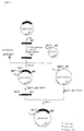
- Figure 1 shows the construction scheme for an expression vector for the production of the protein having the protease activity of the present invention, wherein P 1ac represents a E. coli lac promoter, P CMV represents a cytomegalovirus (CMV) promoter, Ap r represents an ampicillin resistance gene, and Nm r represents a neomycin resistance gene.
- P 1ac represents a E. coli lac promoter
- P CMV represents a cytomegalovirus (CMV) promoter
- Ap r represents an ampicillin resistance gene
- Nm r represents a neomycin resistance gene.
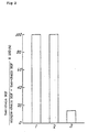
- Figure 2 presents a bar graph showing the rate of the production of two-chain HGF and single-chain HGF in the presence or absence of the precursor proteins activated by thrombin of the present invention, wherein 1 represents the experimental result in the presence of about 100 ng/ml of the precursor protein having a molecular weight of about 96,000 dalton, 2 represents the result in the presence of about 100 ng/ml of the precursor protein having a molecular wight of about 34,000 dalton, and 3 represents the result in the absence of the precursor proteins.
- a novel protein having a protease activity of the present invention includes a protein having the amino acid sequence of SEQ ID NO: 1 and variants thereof having the same protease activity obtained by a deletion, replacement, modification, or addition of a part of amino acids of the protein having the sequence of SEQ ID NO: 1.
- Examples of a gene encoding the above-mentioned protein having the protease activity include a gene comprising the base sequence of SEQ ID NO: 2 as a part of the gene and a gene represented by the base sequence of SEQ ID NO: 3.
- a DNA fragment including such gene can be obtained as described below.
- a cDNA library including DNA encoding the protein having the protease activity of the present invention a commercially available cDNA library prepared from human liver can be used. Phagemids from the library are used to infect cells as described by Saito et al., (Proc. Natl. Acad. Sci. USA, 83, 8664-8668 (1986)) and the infected cells are cultured.
- Colonies obtained after culturing are screened by the colony hybridization method ("Molecular Cloning", Cold Spring Harbor Laboratory, 320-328 (1982)) with the use of partial DNA fragments or DNA fragments having base sequences that correspond to partial amino acid sequences of the protein of the present invention as probes and desired DNA fragments can be obtained.
- DNA fragments which include parts of the gene encoding the protein having the protease activity of the present invention and which are prepared by the polymerase chain reaction (PCR) method can be used.
- PCR is carried out with the use of the DNA fragment of SEQ ID NO: 4 (corresponding to a part of the amino acid sequence of SEQ ID NO: 6) as a plus (+) strand DNA primer and the DNA fragment of SEQ ID NO: 5 (corresponding to a part of the amino acid sequence of SEQ ID NO: 7) as a minus (-) strand DNA primer and the resulting DNA fragment of SEQ ID NO: 2 is used as a probe.
- Synthetic oligonucleotides based on DNA sequences deduced from the amino acid sequence of the protein of the present invention can also be used as probes.
- DNAs are then prepared from positive colonies by the method of T. Maniatis et al. ("Molecular Cloning", Cold Spring Harbor Laboratory, 85 (1982)).
- the resulting DNAs are digested with an appropriate restriction enzyme, e.g., BamHI, cloned in a plasmid, e.g., pUC18 or the like, and sequenced by the dideoxy method of Sanger et al. (Proc. Natl. Acad. Sci. USA, 74 , 5463 (1977)), thereby the base sequences of the desired DNA fragments can be determined.
- an appropriate restriction enzyme e.g., BamHI
- the base sequences of the DNA fragments thus determined (for example, the sequences comprising the base sequence of SEQ ID NO: 2 as a part and the base sequence of SEQ ID NO: 3) encode the protein of the present invention of SEQ ID NO: 1.
- the DNA fragments of the present invention also include variants of the DNA fragments encoding the protein as long as variant-encoded polypeptides have a protease activity to convert single-chain HGF into two-chain HGF. Such variants are prepared by a deletion, replacement, modification, and addition of a part of the bases.
- Each of the DNA fragments so obtained is modified at their 5' terminal, inserted downstream of a promoter into an expression vector, and introduced into a host cell, for example, E. coli, Bacillus subtilis, yeast, or an animal cell.
- a host cell for example, E. coli, Bacillus subtilis, yeast, or an animal cell.
- an expression vector including a promoter at an appropriate site to transcribe the DNA fragment encoding the protein of the present invention is used.
- a host cell is derived from microorganisms, such as E. coli , Bacillus subtilis
- an expression vector comprising a promoter, ribosome binding (SD) sequence, the gene encoding the protein, transcription termination sequence, and a gene regulating the promoter is preferable.
- promoters used in the present invention include promoters derived from E. coli, phages or the like, such as promoter of tryptophan synthase (trp); lambda phage P L and P R ; and P 25 and P 26 promoters, which are promoters of early gene of T 5 phage. Promoters which are modified or designed artificially, such as pac promoter (Agric. Biol. Chem., 52 , 983-988 (1988)) are also useful.
- pac promoter Agric. Biol. Chem., 52 , 983-988 (1988)
- ribosome binding sequences sequences derived from E. coli, phage or the like as well as synthetic sequences which include a consensus sequence having contiguous four or more bases of a sequence complimentary to a sequence at a 3' terminal region of 16S ribosome RNA can be used.
- Transcription termination sequences are not critical, but preferably, an expression vector includes lipoprotein terminator, trp operon terminator, or the like.
- an expression vector includes the above-mentioned sequences, a promoter, a SD sequence, the gene encoding the protein, and a transcription termination sequence in this order from the 5' upstream position.
- expression vector including multiple units of SD sequence and the gene in the same orientation can also be used for increasing the number of the copies of transcriptional units in a vector (Japanese Patent Publication (Kokai) No. 1-95798).
- the method comprises a step of inserting the gene encoding the protein into an expression vector.
- expression vectors examples include pUA12 (Japanese Patent Publication (Kokai) No.1-95798), commercially available pKK233-2 (Pharmacia), and the like.
- the expression vectors pGEX series which can express a desired protein as a fusion protein, are also useful. Standard methods for transforming host cells can be employed.
- Transformants can be cultured according to the method described in "Molecular Cloning” (Cold Spring Harbor Laboratory, 1982).
- host cells derived from microorganisms such as E. coli , Bacillus subtilis, yeast, and the like can be used, but animal cells, such as CHO cells, COS cells, mouse L cells, mouse C127 cells, mouse FM3A cells and the like are preferably used to express the gene encoding the protein, taking into consideration that the activity of the protein of the present invention may be affected by the protein conformation and the sites of thiol bindings between many cysteine residues included in the protein.
- SV40 promoter SV40 promoter
- promoter of the metallothionein gene and the like can be used. Under such promoter, a secretion signal and the gene encoding the protein are inserted according to the transcriptional orientation.
- a DNA fragment including two or three genes can also be inserted instead of a single gene. Two or three units of the gene and a promoter at its 5' side can be combined together and inserted into an expression vector according to the transcriptional orientation.
- the gene encoding the protein is followed by a polyadenylation site downstream of the gene.
- a polyadenylation site downstream of the gene For example, one of the polyadenylation sites derived from SV40 DNA, ⁇ -globin gene, metallothionein gene, and the like is inserted downstream of the gene encoding the protein.
- each of the genes in a unit can be followed by a polyadenylation site at its 3' side.
- genes derived from animal such as SV40 gene, rabbit ⁇ -globin gene or the like; or splicing signal sequences, of intron or exon, synthesized chemically, into upstream or downstream of the gene encoding the protein.
- selection markers when animal cells, e.g., CHO cells, are transformed with the above mentioned expression vectors.
- an expression vector including selection marker gene downstream of a polyadenylation site in the same or reverse orientation, it is not necessary to cotransform a cell with additional plasmid including a selection marker gene.
- selection marker genes include a DHFR gene which confers a cell a methotrexate resistance (J. Mol. Biol., 159 , 601 (1982)); a Neo gene which confers a cell an antibiotic G-418 resistance (J. Mol. Appl. Gene., 1 , 327 (1982)); the Ecogpt gene derived from E.
- Each of these resistance genes is linked to a promoter, e.g., a promoter derived from SV40, at its 5' upstream end and to a polyadenylation site at its 3' downstream end.
- a promoter e.g., a promoter derived from SV40
- a polyadenylation site at its 3' downstream end.
- Commercially available expression vectors comprising a selection marker can be used.
- pcDNA/Neo that comprises a neomycin resistance gene can be used.
- an additional vector comprising a selection marker, e.g., pSV2Neo (J. Mol. Appl. Genet., 1, 327 (1982)), pMBG (Nature, 294 , 228 (1981)), pSV2gpt (Proc. Natl. Acad. Sci. USA, 78 , 2072 (1981)), pAd-D26-1 (J. Mol. Biol., 159, 601 (1982)) can be used with the expression vector to cotransform a cell, thereby the resulting transformant can be selected readily by a phenotype.
- a selection marker e.g., pSV2Neo (J. Mol. Appl. Genet., 1, 327 (1982)), pMBG (Nature, 294 , 228 (1981)), pSV2gpt (Proc. Natl. Acad. Sci. USA, 78 , 2072 (1981)), pAd-D26-1 (J. Mol
- An expression vector is introduced into an animal cell by the calcium phosphate precipitation method (Virology, 52 , 456 (1973)), the electroporation method (J. Membr. Biol., 10 , 279 (1972)), or the like.
- Transformed animal cells can be cultured by the methods known to those of ordinary skilled in the art, including a suspension culture and an adhesion culture. MEM medium, RPMI 1640 medium, and the like can be used and the cells are cultured in the presence of 5 to 10 % serum or an appropriate growth factor, or in the absence of serum. The transformed animal cells producing the protein secrete the protein into a medium. Therefore, the protein can be separated and purified from the supernatant of the culture.
- the supernatant of the culture including the produced protein can be purified by various chromatography methods, such as a chromatography method including a combination of two or more resins selected from anion exchange resin, heparin immobilized resin, hydrophobic chromatography resin, affinity chromatography resin, and the like to obtain the isolated and purified protein.
- a chromatography method including a combination of two or more resins selected from anion exchange resin, heparin immobilized resin, hydrophobic chromatography resin, affinity chromatography resin, and the like to obtain the isolated and purified protein.
- the precursor proteins of the present invention are described below.
- the precursor proteins of the present invention can be obtained by the following purification steps.
- the precursor protein with the molecular weight of about 96,000 dalton can be derived from the plasma prepared by the steps of drawing blood from a normal individual (human), adding ethylenediaminetetraacetic acid (EDTA), benzamidine, 6-amino-n-caproic acid n-hexyl ester p-toluensulfonate, soybean trypsin inhibitor, 6-amidino-2-naphthyl p-guanidinobenzoate dimethanesulfonate (naphamostat mesilate), aprotinin or the like to inhibit the action of coagulation factors in the blood, and then removing the cells by centrifugation.
- EDTA ethylenediaminetetraacetic acid
- benzamidine 6-amino-n-caproic acid n-hexyl ester p-tol
- the diluted plasma is chromatographed by a Heparin-sepharose column (Pharmacia) or the like.
- the resulting fractions including the 96 kDa precursor protein are further chromatographed by a hydrophobic chromatography column (for example, with the use of Phenyl-sepharose column manufactured by Pharmacia).
- the fractions including the 96 kDa precursor protein are chromatographed by an antibody affinity chromatography column, thereby the 96 kDa precursor protein of the present invention can be obtained.
- This antibody affinity chromatography column is prepared by the steps of immunizing a mouse with the novel human protein described in Japanese Patent Publication (Kokai) No. 5-103670, fusing cells from the mouse spleen and myeloma cells to obtain a hybridoma producing an antibody reactive with the immunized protein, and immobilizing the antibody onto appropriate resins according to a standard method.
- the precursor protein with the molecular weight of about 34,000 dalton can be derived from the plasma prepared by the steps of drawing blood from a normal individual (human), and adding EDTA, 6-amino-n-caproic acid n-hexyl ester p-toluensulfonate, or the like.
- the plasma is chromatographed by a Heparin-sepharose column or the like in the same manner as described above.
- the resulting fractions including the precursor protein are chromatographed by a hydrophobic chromatography column or the like in the same manner as described above.
- a step of purifying the proteins of the present invention includes an ion exchange column chromatography, a gel filtration chromatography, a hydroxyapatite column chromatography, or the like.
- the molecular weights of the purified precursor proteins of the present invention are about 96,000 dalton and about 34,000 dalton determined by SDS polyacrylamide gel electrophoresis. They are processed to have the similar activity as that of the human protein described in Japanese Patent Publication (Kokai) No.5-103670 by the treatment with serine proteases such as thrombin.
- One of the precursor proteins of the present invention includes the amino acid sequence of SEQ ID NO: 11 as a part of the protein.
- One of the precursor proteins of the present invention include the amino acid sequence of SEQ ID NO: 12 as a part of the protein.
- variants of the precursor proteins prepared by a deletion, replacement, modification, or addition of a part of the amino acid sequences of the precursor proteins are also included in the scope of the present invention.
- genes encoding the precursor proteins include a gene comprising the base sequence of SEQ ID NO: 13 as a part of the gene, a gene-represented by the base sequence of SEQ ID NO: 14, and the like.
- a DNA fragment including such gene can be obtained by the same method for obtaining the protein having a protease activity as described above.
- DNA fragments that include parts of the gene encoding the precursor proteins and that prepared by the PCR method can be used.
- PCR is carried out with the use of the DNA fragment of SEQ ID NO: 4 as a plus (+) strand DNA primer and the DNA fragment of SEQ ID NO: 5 as a minus (-) strand DNA primer and the resulting DNA fragment of SEQ ID NO: 13 is used as a probe.
- Synthetic oligonucleotides based on DNA sequences deduced from the amino acid sequence of the precursor proteins of the present invention can also be used as probes.
- DNAs are prepared from positive colonies and the base sequences of the desired DNA fragments can be determined by the method described above.
- the base sequences of the DNA fragments thus determined encode the precursor proteins of the present invention.
- the DNA fragments of the present invention also include variants of the DNA fragments encoding the precursor proteins as long as variant-encoded polypeptides acquire a protease activity to convert single-chain HGF into two-chain HGF, after the polypeptides are processed. Such variants are prepared by a deletion, replacement, modification, and addition of a part of the bases.
- Each of the DNA fragments so obtained is modified at their 5' terminal, inserted downstream of a promoter in an expression vector, and introduced into a host cell, for example, E. coli, Bacillus subtilis, yeast, or an animal cell by a standard method.
- a host cell for example, E. coli, Bacillus subtilis, yeast, or an animal cell by a standard method.
- Production of the precursor proteins of the present invention may be conducted in a similar manner to the method for producing the protein having a protease activity described above.
- a protein having a molecular weight of about 34,000 determined by SDS polyacrylamide gel electrophoresis was purified from human serum by the method described in Example 2 in Japanese Patent Publication (Kokai) No. 5-103670, said protein having a protease activity to convert single-chain HGF into two-chain HGF.
- the purified protein was allowed to react in Buffer A (6 M guanidine hydrochloride, 0.002 M EDTA, and 1 M Tris-HCl buffer, pH 8.5) with 2-mercaptoethanol at 4°C for 2 hours to reduce the protein.
- the resulting reaction mixture was applied to a high performance liquid chromatography (HPLC) column.
- HPLC high performance liquid chromatography
- the peptide fragments were dried under reduced pressure, dissolved in 60 ⁇ l of 50% trifluoroacetic acid, added to a glass filter treated with polybrene, and the amino acid sequence thereof was determined by 470A Sequencer (Applied Biosystems) employing Edman degradation. The identification of each phenylthiohydantoin (PTH) amino acid was performed with the use of MCI gel ODS IHU (0.46 x 15 cm) column (Mitsubishi Kasei Corp.).
- the column was eluted with acetate buffer (10 mM acetate buffer, pH 4.7, 0.01% SDS, and 38% acetonitrile) at a flow rate of 1.2 ml/min, at 43°C, and a PTH-amino acid was detected by 269 nm absorbance.
- acetate buffer (10 mM acetate buffer, pH 4.7, 0.01% SDS, and 38% acetonitrile) at a flow rate of 1.2 ml/min, at 43°C, and a PTH-amino acid was detected by 269 nm absorbance.
- PCR was performed by Perkin Elmer Cetus DNA Thermal Cycler with the use of Gene Amp DNA Amplification Reagent Kit (Takara Shuzo Co. Ltd.).
- reaction mixture including a template DNA (lng), 10 ⁇ l of x 10 reaction buffer (500 mM KCl, 100 mM Tris-HCl buffer, pH 8.3, 15 mM MgCl 2 , and 0.1% (w/v) gelatin), 2 ⁇ l each of 10 mM dGTP, dATP, dCTP, and dTTP, 0.1 ⁇ l of (+) strand DNA primer of SEQ ID NO: 4 as primer #1, 0.1 ⁇ l of (-) strand DNA primer of SEQ ID NO: 5 as primer #2 to make final concentration of 0.1 ⁇ M, 0.5 ⁇ l of Taq DNA polymerase, and sterilized deionized water.
- Template DNA was then amplified by 30 cycles of the PCR including pretreatment at 94°C for 10 minutes, denaturation at 94°C for 1 minute, annealing at 37°C for 2 minutes, and extension at 72°C for 3 minutes. The reaction was stopped by incubation at 72°C for 7 minutes.
- the precipitate was dissolved in 21.5 ⁇ l of sterilized deionized water.
- 2.5 ⁇ l of x 10 restriction enzyme buffer 50 mM Tris-HCl buffer, pH7.5, 10 mM MgCl 2 , 100 mM KCl, and 1 mM DTT
- 1 ⁇ l of restriction enzyme Bgl II 15 units
- the DNA fragment was inserted into pUC19 vector at Bam HI site and the base sequence thereof was identified according to a standard method.
- the base sequence of the identified DNA fragment prepared by PCR is shown in SEQ ID NO: 2.
- Example 3 Screening for a clone including a complete gene encoding the protein having the protease activity
- the fragment with 323 bp prepared as described in Example 2 was labeled with 32 P according to the method described in "Molecular Cloning" (Cold Spring Harbor Laboratory, 1982) and used as a probe for screening.
- the library to be screened was Premade Lambda Phage Library (Stratagene), which was a human liver cDNA library derived from 49 years old male.
- E.coli XL1-Blue (Stratagene) was infected with the phage to make about 500 million plaques. The infected cells were cultured on NZY medium overnight and then transfered on Gene Screen Plus membranes (Dupont).
- the membranes were allowed to stand on paper filters soaked with 0.1 M NaOH - 0.5 M NaCl for 2 minutes, and then on paper filters soaked with 1.5 M NaCl-0.5 M Tris-HCl buffer (pH 7.5) for 5 minutes. The membranes were further treated two more times, washed with 2 x SSC, and dried on a dried paper filter in the air. DNAs on the membranes were fixed with UV light at 120 mJ/cm 2 .
- membranes thus treated were soaked in 50 ml of solution including 50 mM Tris-HCl buffer, pH 7.5, 1 M NaCl, and 1 % SDS and allowed to stand at 65°C for 2 hours.
- the membranes were hybridized in 40 ml of solution including 5 ng/ml probe labeled with P, 100 ⁇ g/ml salmon sperm DNA, 50 mM Tris-HCl buffer, pH 7.5, 1 M NaCl, and 1% SDS at 65°C for 16 hours.
- the membranes were then washed in 2 x SSC at room temperature for 5 minutes, 0.1 x SSC at room temperature for 30 minutes for two times, and autoradiographed by a standard method, thereby 40 positive clones were obtained.
- Plasmids were prepared directly from positive phage clones obtained in Example 3 by the excision method. Phage was extracted from single plaque by treatment with 500 ⁇ l of SM buffer (50 mM Tris-HCl buffer, pH 7.5, 100 mM NaCl, 10 mM MgSO 4 , and 0.01 % gelatin) and 20 ⁇ l of chloroform. The mixture including 200 ⁇ l of the above-mentioned phage extracted solution, 200 ⁇ l of XL1-Blue, and 1 ⁇ l of R408 helper phage was allowed to stand 37°C for 15 minutes. Then, 5 ml of 2 x YT medium was added to the mixture and the resulting solution was shake cultured at 37°C for 3 hours.
- SM buffer 50 mM Tris-HCl buffer, pH 7.5, 100 mM NaCl, 10 mM MgSO 4 , and 0.01 % gelatin
- the culture was then heated to 70°C for 20 minutes followed by centrifugation with 4000 g for 5 minutes to obtain a supernatant.
- the supernatant was diluted to make 100-fold diluted solution.
- 20 ⁇ l of the diluted solution was mixed with 200 ⁇ l of XL1-Blue and the mixture was allowed to react 37°C for 15 minutes.
- 2 ⁇ l of the resulting mixture was plated onto a LB agarose medium including 40 ⁇ l/ml ampicillin. Twenty-four plasmids from the colonies appeared on the plate were obtained, and analyzed.
- the clone having the longest insert (pBHGFAP) was analyzed and the base sequence of the insert was identified.
- the base sequence thus determined is shown in SEQ ID NO: 3. Based on this base sequence, the amino acid sequence of the protein having a protease activity of the present invention was deduced (SEQ ID NO: 1).
- Figure 1 shows a construction of an expression vector including the gene encoding the protein having a protease activity of the present invention.
- the plasmid pBHGFAP prepared in Example 3 was used as a template DNA and PCR was performed by Perkin Elmer Cetus DNA Thermal Cycler with the use-of the Gene Amp DNA Amplification Reagent Kit (Takara Shuzo Co., Ltd.).
- reaction mixture including a template DNA (0.5 ⁇ g), 10 ⁇ l of x 10 reaction buffer (500 mM KCl, 100 mM Tris-HCl buffer, pH 8.3, 15 mM MgCl 2 , and 0.1% (w/v) gelatin), 16 ⁇ l each of 1.25 mM dGTP, dATP, dCTP, and dTTP, 5 ⁇ l of (+) strand DNA primer of SEQ ID NO: 8 as primer #3, 5 ⁇ l of (-) strand DNA primer of SEQ ID NO: 9 as primer #4, 0.5 ⁇ l of Taq DNA polymerase, and sterilized deionized water.
- a template DNA 0.5 ⁇ g
- 10 ⁇ l of x 10 reaction buffer 500 mM KCl, 100 mM Tris-HCl buffer, pH 8.3, 15 mM MgCl 2 , and 0.1% (w/v) gelatin
- the DNA fragment thus obtained was blunt-ended by a standard method with the use of T4 DNA polymerase and then cleaved with Xba I.
- the cleaved DNA fragments were recovered again, and 15 ng of the DNA fragments was mixed with 5 ng of synthetic DNA of SEQ ID NO: 10 and 20 ng of a plasmid vector pUC18 cleaved with Eco RI and Xba I, and the mixture was allowed to ligate by the use of Ligation Kit (Takara Shuzo Co. Ltd.).
- the resulting plasmid was transformed into competent cells, E.coli JM105 (Competent High, Toyobo) according to the manufacturer's instructions.
- the transformants thus obtained were screened by a standard method and the plasmid pSHGFAP including the desired 926 bp insert was obtained.
- the plasmid pSHGFAP (8 ⁇ g) was cleaved with restriction enzymes Bam HI and Xba I, treated with phenol/chloroform, and ethanol precipitated. The precipitate was dissolved in sterilized deionized water and electrophoresed using 5 % polyacrylamide gel. The 920 bp band was extracted from the gel by a standard technique to recover a DNA fragment, which was then ethanol precipitated.
- E. coli comprising the plasmid pSHGFAP prepared in Example 5 was inoculated in 10 ml of LB medium containing 50 ⁇ g/ml ampicillin and cultured at 37°C overnight (12 to 16 hours). Aliquot (0.1 ml) of the culture was added to 10 ml of LB medium including 50 ⁇ g/ml ampicillin and the medium was cultured at 37°C for 2 hours. Isopropyl ⁇ -D thio galactoside (IPTG), which is a transcriptional inducer of the lac promoter in the vector, was added to the culture at a final concentration of 1 mM and the culture was incubated at 37°C for 6 hours. Cells were collected from 1 ml of the culture by centrifugation.
- IPTG Isopropyl ⁇ -D thio galactoside
- Example 7 Establishment of an animal cell strain expressing the protein having the protease activity of the present invention
- Plasmid pNHGFAP which was prepared in Example 5 and includes the cDNA encoding the protein of the present invention at a restriction enzyme cleavage site of the expression vector pcDNAI/Neo, was recovered from E.coli transformants and purified according to the method of Maniatis et al. ("Molecular Cloning", Cold Spring Harbor Laboratory, 86-96 (1982)) to obtain an expression vector for the protein of the present invention in a large amount.
- CHO cells were transformed with the expression vector so obtained according to the method of Ausubel et al. (Current Protocols in Molecular Biology, Green Publishing Associates and Wiley-Inter Science, 9.1.1-9.1.4 (1987)).
- CHO cells were cultured in a tissue culture dish with a diameter of 9 cm containing ERDF medium (Kyokuto Pharmaceutical) supplemented by 10% fetal bovine serum (FBS) until cells were grown to semiconfluence. The medium was then removed from the dish and a DNA solution, which was prepared as described below, was added dropwise.
- the DNA solution for a single dish with a diameter of 9 cm, was prepared by the steps of making 570 ⁇ l mixture in an eppendorf tube including 300 ⁇ l of 2 x HEBS solution (1.6 % NaCl, 0.074 % KCl, 0.05 % disodium hydrogenphosphate 12H 2 O, 0.2 % dextrose, and 1 % HEPES, pH 7.05), 10 ⁇ g plasmid DNA, and sterilized water; dropwisely adding 30 ⁇ l of 2.5 M CaCl 2 solution to the mixture while the mixture was vigorously vortexed for a few seconds; and allowing the mixture to stand for 30 minutes at room temperature.
- 2 x HEBS solution 1.6 % NaCl, 0.074 % KCl, 0.05 % disodium hydrogenphosphate 12H 2 O, 0.2 % dextrose, and 1 % HEPES, pH 7.05
- 10 ⁇ g plasmid DNA and sterilized water
- the DNA solution thus obtained was applied to the CHO cells and the cells were allowed to stand for 30 minutes at room temperature. Then 9 ml of ERDF medium containing 10 % FBS was added to the dish and the cells were cultured for 4 to 5 hours at 37°C in the presence of 5 % CO 2 . The medium was then removed from the dish. The cells were washed with 5 ml of 1 x TBS ++ solution (25 mM Tris-HCl buffer, pH 7.5, 140 mM NaCl, 5 mM KCl, 0.6 mM disodium hydrogenphosphate, 0.08 mM CaCl 2 , and 0.08 mM MgCl 2 ). The 1 x TBS ++ solution was then removed.
- 1 x TBS ++ solution 25 mM Tris-HCl buffer, pH 7.5, 140 mM NaCl, 5 mM KCl, 0.6 mM disodium hydrogenphosphate, 0.08 mM CaCl 2 , and 0.08 m
- G418 G418 sulfate (GENETICIN, GIBCO) was added to each dish at a concentration of 200 ⁇ g/ml and the cells were cultured. After 10 days, vital cells, which resisted G418, were isolated. The cells were then cultured for 7 days in a 24 well plate including 1 ml of ERDF medium supplemented by 10 % FBS. Then the cells were cultured in serum-free ERDF medium for 72 hours and the resulting culture in each well was recovered. The culture was concentrated by ultrafiltration and analyzed by SDS acrylamide gel electrophoresis. The expression of the protein of the present invention was detected by western blotting.
- G418 sulfate GENETICIN, GIBCO
- Example 8 Purification and amino acid sequence analysis of the precursor protein with a molecular weight of about 96,000 dalton
- Blood from a normal individual was mixed with 10 mM EDTA, 10 mM benzamidine (Aldrich Chemical), 5 mM 6-amino-n-caproic acid n-hexyl ester p-toluensulfonate (Tokyo Chemical), 2 mg/ml soybean trypsin inhibitor (Sigma), 50 mM naphamostat mesilate (Torii Pharmaceutical), and 500 KIU/ml aprotinin (Pentapharm). Cells were immediately removed by centrifugation to obtain plasma.
- the diluted plasma was applied on a Heparin-sepharose column (pharmacia), which had been pre-equilibrated with buffer A (including 10 mM NaH 2 PO 4 -Na 2 HPO 4 buffer, pH 7, 50 mM NaCl), and the column was washed with buffer A. The column was then eluted with a linear gradient of 50 mM to 700 mM NaCl in buffer A and buffer B (including 10 mM NaH 2 PO 4 -Na 2 HPO 4 buffer, pH 7, 700 mM NaCl).
- buffer A including 10 mM NaH 2 PO 4 -Na 2 HPO 4 buffer, pH 7, 50 mM NaCl
- fractions including the protein of the present invention were recovered with a sandwich enzyme-linked immunosorbent assay (ELISA) with the use of an antibody that was prepared by immunizing a mouse with the protein derived from human disclosed in Japanese Patent Publication (Kokai) No: 5-103670, fusing spleen cells from the mouse and myeloma cells by a standard method to obtain hybridomas, and obtaining the antibody from the hybridoma.
- the fractions were mixed with equivalent 2 M ammonium sulfate.
- Example 10 The solution was applied to the antibody affinity column described in Example 10 equilibrated with buffer C. The column was washed with buffer C and eluted with buffer D (50 mM Glycine/HCl buffer, pH 3). Fractions were collected and neutralized with buffer E (1 M Tris/HCl, pH 8). The resulting protein product was activated with thrombin, analyzed to determine its amino acid sequence, and analyzed by SDS polyacrylamide gel electrophoresis as described below.
- the column was then eluted with a linear gradient of 10 % to 70 % acetonitrile-isopropyl alcohol (3/7) for 20 minutes and two major peaks were obtained. These peaks were dried under a reduced pressure, dissolved in 60 ⁇ l of 50 % trifluoroacetic acid (TFA). The solution was added to a glass filter treated with polybrene, and the amino acid sequence of the N terminal of the protein was determined by 470A Sequencer (Applied Biosystems) employing Edman degradation. The identification of each phenylthiohydantoin (PTH) amino acid was performed with the use of MCI gel ODS IHU (0.46 x 15 cm) column (Mitsubishi Kasei Crop.).
- the column was eluted with acetate buffer (10 mM acetate buffer, pH 4.7, 0.01% SDS, and 38% acetonitrile) at a flow rate of 1.2 ml/min, at 43°C, and a PTH-amino acid was detected through 269 nm absorbance.
- acetate buffer (10 mM acetate buffer, pH 4.7, 0.01% SDS, and 38% acetonitrile) at a flow rate of 1.2 ml/min, at 43°C, and a PTH-amino acid was detected through 269 nm absorbance.
- Example 9 Purification and amino acid sequence analysis of a protein with a molecular weight of about 34,000 dalton
- Plasma samples Blood from a normal individual (human) were mixed with 10 mM EDTA, 10 mM benzamidine (Aldrich Chemical) and 5 mM 6-amino-n-caproic acid n-hexyl ester p-toluensulfonate (Tokyo Chemical). Cells were immediately removed by centrifugation to obtain plasma. The plasma was applied on a Heparin-sepharose column (Pharmacia), which had been pre-equilibrated with buffer G (including 10 mM NaH 2 PO 4 -Na 2 HPO 4 buffer, pH 7, 150 mM NaCl), and the column was washed with buffer G. The column was then eluted with a linear gradient of 150 mM to 700 mM NaCl in buffer G and buffer B (including 10 mM NaH 2 PO 4 -Na 2 HPO 4 buffer, pH 7, 700 mM NaCl).
- buffer G including 10 mM NaH 2 PO 4 -Na 2 HPO
- fractions including the protein of the present invention were recovered by a sandwich enzyme-linked immunosorbent assay (ELISA) by the use of an antibody that was prepared by immunizing a mouse with the protein derived from human protein disclosed in Japanese Patent Publication (Kokai) No: 5-103670, fusing spleen cells from the mouse and myeloma cells by a standard method to obtain hybridomas, and obtaining the antibody from the hybridoma.
- ELISA sandwich enzyme-linked immunosorbent assay
- Fractions including the protein of the present invention were recovered by ELISA.
- the fractions were concentrated with the use of an amicon ultrafiltration membrane YM-30 and dialyzed against buffer C (including 10 mM Tris/HCl buffer, pH 8, 150 mM NaCl).
- the solution was applied to an aprotinin immobilized affinity column (Pentapharm) equilibrated with buffer C.
- the unbound fraction was applied to an antibody affinity column described in Example 10 equilibrated with buffer C.
- the column was washed with buffer C.
- the column was eluted with buffer D (50 mM Glycine/HCl buffer, pH 3). Fractions were collected and neutralized with buffer E (1 M Tris/HCl, pH 8). The resulting protein product was activated with thrombin, analyzed to determine its amino acid sequence, and analyzed by SDS polyacrylamide gel electrophoresis as described below.
- the column was then eluted with a linear gradient of 10 % to 70 % acetonitrile/isopropyl alcohol (3/7) for 30 minutes and the peak was obtained.
- the peak was dried under a reduced pressure, dissolved in 60 ⁇ l of 50 % trifluoroacetic acid (TFA).
- TFA trifluoroacetic acid
- the solution was added to a glass filter treated with polybrene, and the amino acid sequence of the N-terminal of the proteins was determined by 470A Sequencer (Applied Biosystems) employing Edman degradation.
- the identification of each phenylthiohydantoin (PTH) amino acid was performed with the use of MCI gel ODS IHU (0.46 x 15 cm) column (Mitsubishi Kasei Corp.).
- the column was eluted with acetate buffer (10 mM acetate buffer, pH 4.7, 0.01% SDS, and 38% acetonitrile) at a flow rate of 1.2 ml/min, at 43°C, and a PTH-amino acid was detected through 269 nm absorbance.
- acetate buffer (10 mM acetate buffer, pH 4.7, 0.01% SDS, and 38% acetonitrile) at a flow rate of 1.2 ml/min, at 43°C, and a PTH-amino acid was detected through 269 nm absorbance.
- the sequence analysis of the reduced carboxymethylated protein showed that the N-terminal amino acid sequence has the sequence of SEQ ID NO: 11.
- a Balb/c mouse was immunized with a novel human protein disclosed in Japanese Patent Publication (Kokai) No. 5-103670 by a standard method.
- the mouse was first immunized with about 10 ⁇ g antigen with Freund's complete adjuvant and then boosted with about 10 ⁇ g antigen with Freund's incomplete adjuvant intraperitoneally.
- the mouse was given a final boost from a tail vein.
- Three days after the final boost, spleen cells of the mouse were taken.
- the spleen cells were fused with mouse myeloma P3U1 cells in the presence of polyethylene glycol by a standard method. Fused cells were screened by ELISA employing enzyme-labeled anti-mouse antibodies to select hybridomas producing antibodies against the protein.
- Each of the obtained hybridomas was cultured in a serum-free medium. The supernatant of the culture was applied to a Protein A affinity column to obtain antibodies. Two antibodies recognizing discrete antigenic determinants were used to perform ELISA by a standard method. The second antibody was labeled with peroxidase by a standard method. One of the obtained antibodies was used to couple to CNBr-sepharose (Pharmacia) to obtain an antibody affinity column.
- SDS polyacrylamide gel electrophoresis was conducted.
- the purified proteins were analyzed by SDS polyacrylamide gel electrophoresis involving 12.5 % polyacrylamide slab gel under a non-reduced condition. Molecular makers were available from Pharmacia. After electrophoresis, the gel was stained with Coomassie Brilliant Blue (CBB) R250.
- CBB Coomassie Brilliant Blue
- the protein prepared in Example 8 was 96,000 dalton and the protein prepared in Example 9 was 34,000 dalton as determined by SDS polyacrylamide gel electrophoresis. The sizes of these proteins were estimated by comparing the location of the bands of the proteins to the location of the bands of the marker.
- the activity of the protein was determined by the ratio of single-chain HGF to two-chain HGF.
- the result is shown in Fig. 2, wherein 1 represents the result in the presence of about 100 ng/ml protein obtained by the activation of the protein having a molecular weight of about 96,000 dalton, 2 represents the result in the presence of about 100 ng/ml protein obtained by the activation of the protein having a molecular weight of about 34,000 dalton, and 3 represents the result in the absence of the proteins.
- the protein which has a molecular weight of about 96,000 dalton determined by SDS polyacrylamide gel electrophoresis and acquired a protease activity to convert single-chain HGF into two-chain HGF by the treatment with serine proteases, was obtained from human plasma according to the method described in Example 8.
- This purified protein was reduced with 2-mercaptoethanol in buffer A (6 M guanidine hydrochloride, 0.002 M EDTA, and 1 M Tris-HCl buffer, pH 8.5) for 2 hours at 40°C.
- Monoiodoacetic acid at the same concentration as the protein was added to the solution, and the mixture was allowed to react in the presence of nitrogen gas for 1 hour at room temperature in darkness to carboxymethylate the protein.
- the fraction corresponding to the peak was dissolved in 0.1 % ammonium bicarbonate including 2 M urea.
- TPCK-trypsin Miles Laboratory
- TLCK-chymotrypsin Miles Laboratory
- the reaction mixture was applied to an HPLC column and the column was eluted with a linear gradient of 0 % to 80 % acetonitrile/isopropyl alcohol (3/7) for 1 hour to obtain multiple peptide fragments.
- peptide fragments were dried in the air and dissolved in 60 ⁇ l of 50 % trifluoroacetic acid (TFA).
- TFA trifluoroacetic acid
- the solution was added to a glass filter treated with polybrene, and the amino acid sequence of the protein was determined by 470A Sequencer (Applied Biosystems) involving Edman degradation.
- the identification of phenylthiohydantoin (PTH) amino acid was performed with the use of MCI gel ODS IHU (0.46 x 15 cm) column (Mitsubishi Kasei Corp.).
- the column was eluted with acetate buffer (10 mM acetate buffer, pH 4.7, 0.01% SDS, and 38% acetonitrile) at a flow rate of 1.2 ml/min, at 43 °C, and a PTH-amino acid was detected through 269 nm absorbance.
- acetate buffer (10 mM acetate buffer, pH 4.7, 0.01% SDS, and 38% acetonitrile) at a flow rate of 1.2 ml/min, at 43 °C, and a PTH-amino acid was detected through 269 nm absorbance.
- PCR was performed by Perkin Elmer Cetus DNA Thermal Cycler with the use of Gene Amp DNA Amplification Reagent Kit (Takara Shuzo Co. Ltd.).
- reaction mixture including a template DNA (1 ng), 10 ⁇ l of x 10 reaction buffer (500 mM KCl, 100 mM Tris-HCl buffer, pH 8.3, 15 mM MgCl 2 , and 0.1% (w/v) gelatin), 2 ⁇ l each of 10 mM dGTP, dATP, dCTP, and dTTP, 2 ⁇ l of (+) strand DNA primer of SEQ ID NO: 4 as primer #1, 0.1 ⁇ M (-) strand DNA primer of SEQ ID NO: 5 as primer #2 to make final concentration to 0.1 ⁇ M, 0.5 ⁇ l of Taq DNA polymerase, and sterilized deionized water.
- x 10 reaction buffer 500 mM KCl, 100 mM Tris-HCl buffer, pH 8.3, 15 mM MgCl 2 , and 0.1% (w/v) gelatin
- restriction enzyme buffer 50 mM Tris-HCl buffer, pH 7.5, 10 mM MgCl 2 , 100 mM KCl, and 1 mM DTT
- restriction enzyme Bgl II 15 units
- the DNA fragment was inserted into pUC19 vector at Bam HI site and the base sequence thereof was identified according to a standard method.
- the base sequence of the identified DNA fragment prepared by PCR is shown in SEQ ID NO: 13.
- Example 15 Screening for a DNA fragment including the complete gene encoding the precursor protein of the present invention
- the 323 bp fragment prepared as described in Example 14 was labeled with 32 P according to the method described in "Molecular Cloning" (Cold Spring Harbor Laboratory, 1982) and used as a probe for screening.
- the library to be screened was Premade Lambda Phage Library (Stratagene), which was a human liver cDNA library derived from a 49 year old male.
- E.coli XL1-Blue (Stratagene) was infected with the phage to make about 500 million plaques. The infected cells were cultured on NZY medium and then transferred on Gene Screen Plus membranes (Dupont).
- the membranes were allowed to stand on paper filters soaked with 0.1 M NaOH - 0.5 M NaCl for 2 minutes, and then on paper filters soaked with 1.5 M NaCl-0.5 M Tris-HCl buffer (pH 7.5) for 5 minutes. The membranes were further treated two more times, washed with 2 x SSC, and dried on a dried paper filter in the air. DNAs on the membranes were fixed with UV light at 120 mJ/cm 2 .
- membranes thus treated were soaked in 50 ml of solution including 50 mM Tris-HCl buffer, pH 7.5, 1 M NaCl, and 1 % SDS and allowed to stand at 65°C for 2 hours.
- the membranes were hybridized in 40 ml of solution including 5 ng/ml P labeled probe, 100 ⁇ g/ml salmon sperm DNA, 50 mM Tris-HCl buffer, pH 7.5, 1 M NaCl, and 1% SDS at 65°C for 16 hours.
- the membranes were then washed in 2 x SSC at room temperature for 5 minutes, 0.1 x SSC at room temperature for 30 minutes for two times, and autoradiographed by a standard method, thereby positive clones were obtained.
- Plasmids were prepared directly from positive phage clones obtained in Example 15 by the excision method. Phage was extracted from single plaque by treatment with 500 ⁇ l of SM buffer (50 mM Tris-HCl buffer, pH 7.5, 100 mM NaCl, 10 mM Mg 2 SO 4 , and 0.01 % gelatin) and 20 ⁇ l of chroloform. A mixture including 200 ⁇ l of the above-mentioned phage extract, 200 ⁇ l of XL1-Blue, and 1 ⁇ l of R408 helper phage was allowed to stand 37°C for 15 minutes. Then 5 ml of 2 x YT medium was added to the mixture and the resulting solution was shake cultured at 37°C for 3 hours.
- SM buffer 50 mM Tris-HCl buffer, pH 7.5, 100 mM NaCl, 10 mM Mg 2 SO 4 , and 0.01 % gelatin
- chroloform 20 ⁇ l of chroloform
- the culture was then heated to 70°C for 20 minutes followed by centrifugation with 4000 g for 5 minutes to obtain a supernatant.
- the supernatant was diluted to make a 100-fold diluted solution.
- 20 ⁇ l of the diluted solution was mixed with 200 ⁇ l of XL1-Blue and the mixture was allowed to react at 37°C for 15 minutes.
- 2 ⁇ l of the resulting mixture was plated onto a LB agarose medium including 40 ⁇ l/ml ampicillin. Twenty-four plasmids from the colonies which appeared on the plate were obtained and analyzed.
- the base sequence thus determined is shown in SEQ ID NO: 14. Based on this base sequence, the amino acid sequence of the precursor protein of the present invention was deduced (SEQ ID NO: 12).
- the present invention enables a stable and simple production of a protein having the protease activity to convert single-chain HGF to active two-chain HGF and the precursor protein thereof in a large amount. These proteins can be used as a regulator in producing active HGF.
Landscapes
- Health & Medical Sciences (AREA)
- Chemical & Material Sciences (AREA)
- Life Sciences & Earth Sciences (AREA)
- Organic Chemistry (AREA)
- Genetics & Genomics (AREA)
- Engineering & Computer Science (AREA)
- Zoology (AREA)
- Molecular Biology (AREA)
- Biomedical Technology (AREA)
- Bioinformatics & Cheminformatics (AREA)
- General Health & Medical Sciences (AREA)
- Wood Science & Technology (AREA)
- Medicinal Chemistry (AREA)
- Biochemistry (AREA)
- Gastroenterology & Hepatology (AREA)
- General Engineering & Computer Science (AREA)
- Microbiology (AREA)
- Biotechnology (AREA)
- Toxicology (AREA)
- Biophysics (AREA)
- Proteomics, Peptides & Aminoacids (AREA)
- Preparation Of Compounds By Using Micro-Organisms (AREA)
- Micro-Organisms Or Cultivation Processes Thereof (AREA)
- Enzymes And Modification Thereof (AREA)
- Peptides Or Proteins (AREA)
Abstract
Description
SEQUENCE TYPE: amino acid
SEQUENCE LENGTH: 300
STRANDNESS: single
TOPOLOGY: linear
MOLECULE TYPE: protein
ORIGINAL SOURCE
ORGANISM: human


SEQUENCE TYPE: nucleic acid
SEQUENCE LENGTH: 329
STRANDNESS: double
TOPOLOGY: linear
MOLECULE TYPE: cDNA
ORIGINAL SOURCE
ORGANISM: human
IMMEDIATE EXPERIMENTAL SOURCE: Quick-cloneTM human liver cDNA (Clonetech)

SEQUENCE TYPE: nucleic acid
SEQUENCE LENGTH: 970
STRANDNESS: double
TOPOLOGY: linear
MOLECULE TYPE: cDNA
ORIGINAL SOURCE
ORGANISM: human
IMMEDIATE EXPERIMENTAL SOURCE: Pre-made Lambda phage Library, human liver (49, male) cDNA Library (Stratagene)

SEQUENCE TYPE: nucleic acid
SEQUENCE LENGTH: 26
TOPOLOGY: linear
MOLECULE TYPE: Other nucleic acid (synthetic DNA)
ORIGINAL SOURCE
ORGANISM: human

SEQUENCE TYPE: nucleic acid
SEQUENCE LENGTH: 26
TOPOLOGY: linear
MOLECULE TYPE: Other nucleic acid (synthetic DNA)
ORIGINAL SOURCE
ORGANISM: human

SEQUENCE TYPE: amino acid
SEQUENCE LENGTH: 7
TOPOLOGY: linear
MOLECULE TYPE: peptide
FRAGMENT TYPE: internal fragment
ORIGINAL SOURCE
ORGANISM: human

SEQUENCE TYPE: amino acid
SEQUENCE LENGTH: 8
TOPOLOGY: linear
MOLECULE TYPE: peptide
FRAGMENT TYPE: internal fragment
ORIGINAL SOURCE
ORGANISM: human

SEQUENCE TYPE: nucleic acid
SEQUENCE LENGTH: 19
TOPOLOGY: linear
MOLECULE TYPE: Other nucleic acid (synthetic DNA)
ORIGINAL SOURCE
ORGANISM: human
SEQUENCE TYPE: nucleic acid
SEQUENCE LENGTH: 19
TOPOLOGY: linear
MOLECULE TYPE: Other nucleic acid (synthetic DNA)
ORIGINAL SOURCE
ORGANISM: human
SEQUENCE TYPE: nucleic acid
SEQUENCE LENGTH: 71
STRANDNESS: double
TOPOLOGY: linear:
MOLECULE TYPE: Other nucleic acid (synthetic DNA)
ORIGINAL SOURCE
ORGANISM: human

SEQUENCE TYPE: amino acid
SEQUENCE LENGTH: 8
TOPOLOGY: linear
MOLECULE TYPE: protein
FRAGMENT TYPE: partial peptide
ORIGINAL SOURCE
ORGANISM: human
TISSUE TYPE: plasuma

SEQUENCE TYPE: amino acid
SEQUENCE LENGTH: 655
STRANDEDNESS: single
TOPOLOGY: linear
MOLECULE TYPE: protein
ORIGINAL SOURCE
ORGANISM: human


SEQUENCE TYPE: nucleic acid
SEQUENCE LENGTH: 329
STRANDEDNESS: double
TOPOLOGY: linear
MOLECULE TYPE: cDNA
ORIGINAL SOURCE
ORGANISM: human
IMMEDIATE EXPERIMENTAL SOURCE: Quick-cloneTM human liver cDNA (Clonetech)

SEQUENCE TYPE: nucleic acid
SEQUENCE LENGTH: 2033
STRANDEDNESS: double
TOPOLOGY: linear
MOLECULE TYPE: cDNA
ORIGINAL SOURCE
ORGANISM: human
IMMEDIATE EXPERIMENTAL SOURCE: Pre-made Lambda phage Library, human liver (49, male) cDNA Library (Stratagene)


Claims (20)
- A protein having the amino acid sequence of SEQ ID NO: 1.
- A gene encoding the protein according to claim 1.
- The gene according to claim 2, wherein said gene comprises the base sequence of SEQ ID NO: 2 as a part of the gene.
- The gene according to claim 2, wherein said gene has the base sequence of SEQ ID NO: 3.
- An expression vector comprising the gene according to claim 2, wherein said gene is positioned downstream of a promoter so as to express a polypeptide encoded by the gene.
- A transformant obtained by transformation of a host cell with the vector according to claim 5.
- A method for producing a protein having the amino acid sequence of SEQ ID NO: 1 comprising a step of culturing the transformant according to claim 6.
- Amethod for producing a hepatocyte growth factor with the use of the protein according to claim 1 as a regulator.
- The protein according to claim 1, wherein said protein acquires a protease activity to specifically cleave human hepatocyte growth factor by treatment with a serine protease.
- An activated protein which is obtained by treating a protein having the amino acid sequence of SEQ ID NO: 1 with a serine protease.
- A protein having the amino acid sequence of SEQ ID NO: 12.
- A gene encoding the protein according to claim 11.
- The gene according to claim 12, wherein said gene comprises the base sequence of SEQ ID NO: 13 as a part of the gene.
- The gene according to claim 12, wherein said gene has the base sequence of SEQ ID NO: 14.
- An expression vector comprising the gene according to claim 12, wherein said gene is positioned downstream of a promoter so as to express a polypeptide encoded by the gene.
- A transformant obtained by transformation of a host cell with the vector according to claim 15.
- A method of producing a protein having the amino acid sequence of SEQ ID NO: 12 comprising a step of culturing the transformant according to claim 16.
- A method of producing a hepatocyte growth factor with the use of the protein according to claim 11 as a regulator.
- The protein according to claim 11, wherein said protein acquires a protease activity to specifically cleave human hepatocyte growth factor by treatment with a serine protease.
- An activated protein which is obtained by treating a protein having the amino acid sequence of SEQ ID NO: 12 with a serine protease.
Applications Claiming Priority (6)
| Application Number | Priority Date | Filing Date | Title |
|---|---|---|---|
| JP296133/92 | 1992-11-05 | ||
| JP29613392A JP3214106B2 (en) | 1992-11-05 | 1992-11-05 | Novel protein |
| JP312234/92 | 1992-11-20 | ||
| JP31224292A JP3232714B2 (en) | 1992-11-20 | 1992-11-20 | Novel proteins and genes encoding them |
| JP04312234A JP3144097B2 (en) | 1992-11-20 | 1992-11-20 | Novel proteins and genes encoding them |
| JP312242/92 | 1992-11-20 |
Publications (3)
| Publication Number | Publication Date |
|---|---|
| EP0596524A2 EP0596524A2 (en) | 1994-05-11 |
| EP0596524A3 EP0596524A3 (en) | 1995-04-05 |
| EP0596524B1 true EP0596524B1 (en) | 1999-04-21 |
Family
ID=27338033
Family Applications (1)
| Application Number | Title | Priority Date | Filing Date |
|---|---|---|---|
| EP93117988A Expired - Lifetime EP0596524B1 (en) | 1992-11-05 | 1993-11-05 | Hepatocyte growth factor HGF converting protease and gene encoding said protease |
Country Status (5)
| Country | Link |
|---|---|
| US (2) | US5466593A (en) |
| EP (1) | EP0596524B1 (en) |
| AT (1) | ATE179213T1 (en) |
| CA (1) | CA2102463A1 (en) |
| DE (1) | DE69324547T2 (en) |
Families Citing this family (9)
| Publication number | Priority date | Publication date | Assignee | Title |
|---|---|---|---|---|
| DE69632435T2 (en) * | 1995-07-24 | 2005-05-12 | Mitsubishi Chemical Corp. | Inhibitor of hepatocyte growth factor activator |
| AU5902398A (en) | 1996-12-31 | 1998-07-31 | Human Genome Sciences, Inc. | Cortistatin polypeptides |
| US5910430A (en) * | 1997-01-24 | 1999-06-08 | Smithkline Beecham Corporation | Isolated nucleic acid encoding G-protein coupled receptor (HTADX50) |
| US7045602B2 (en) * | 2000-12-05 | 2006-05-16 | Mitsubishi Chemical Corporation | Specific antibody directed to active hepatocyte growth factor activator and method for using the same |
| AU2011254032B2 (en) * | 2004-10-04 | 2012-08-23 | Genentech, Inc. | Modulators of Hepatocyte Growth Factor Activator |
| ZA200701789B (en) * | 2004-10-04 | 2008-11-26 | Genentech Inc | Modulators of hepatocyte growth factor activator |
| JP6190813B2 (en) * | 2011-10-14 | 2017-08-30 | ジェネンテック, インコーポレイテッド | Zymogen activator |
| TWI728409B (en) | 2014-04-28 | 2021-05-21 | 日商衛材R&D企管股份有限公司 | HGF freeze-dried preparation |
| HUE055414T2 (en) * | 2016-03-17 | 2021-11-29 | Eisai R&D Man Co Ltd | Method for producing activated hepatocyte growth factor (hgf) |
Family Cites Families (3)
| Publication number | Priority date | Publication date | Assignee | Title |
|---|---|---|---|---|
| US4740461A (en) * | 1983-12-27 | 1988-04-26 | Genetics Institute, Inc. | Vectors and methods for transformation of eucaryotic cells |
| CA2022752C (en) * | 1989-08-11 | 1998-07-07 | Naomi Kitamura | Hepatic parenchymal cell growth factor, gene encoding the same, process for producing the factor, and transformants producing the factor |
| JP3213985B2 (en) * | 1991-10-18 | 2001-10-02 | 三菱化学株式会社 | Novel protein |
-
1993
- 1993-11-04 CA CA002102463A patent/CA2102463A1/en not_active Abandoned
- 1993-11-05 DE DE69324547T patent/DE69324547T2/en not_active Expired - Lifetime
- 1993-11-05 EP EP93117988A patent/EP0596524B1/en not_active Expired - Lifetime
- 1993-11-05 US US08/148,910 patent/US5466593A/en not_active Expired - Lifetime
- 1993-11-05 AT AT93117988T patent/ATE179213T1/en not_active IP Right Cessation
-
1995
- 1995-05-24 US US08/448,937 patent/US5677164A/en not_active Expired - Lifetime
Also Published As
| Publication number | Publication date |
|---|---|
| US5466593A (en) | 1995-11-14 |
| CA2102463A1 (en) | 1994-05-06 |
| DE69324547D1 (en) | 1999-05-27 |
| DE69324547T2 (en) | 1999-11-18 |
| EP0596524A3 (en) | 1995-04-05 |
| EP0596524A2 (en) | 1994-05-11 |
| US5677164A (en) | 1997-10-14 |
| ATE179213T1 (en) | 1999-05-15 |
Similar Documents
| Publication | Publication Date | Title |
|---|---|---|
| US5424205A (en) | Amyloidin protease and uses thereof | |
| EP0679189B1 (en) | Cloning of enterokinase and method of use | |
| JP3136551B2 (en) | DNA encoding interleukin-1β precursor converting enzyme | |
| Ferlinz et al. | Occurrence of two molecular forms of human acid sphingomyelinase | |
| CA2018592C (en) | Polypeptides and polypeptide analogues with inhibitory activity against human elastase | |
| Grinnell et al. | Trans–Activated Expression of Fully Gamma–Carboxylated Recombinant Human Protein C, an Antithrombotic Factor | |
| CZ20031718A3 (en) | Von Willebrand factor (vWF) splitting polypeptide protease, nucleic acid encoding the polypeptide and use of such polypeptide | |
| Ohara et al. | Cloning, nucleotide sequence, and expression of Achromobacter protease I gene | |
| EP0596524B1 (en) | Hepatocyte growth factor HGF converting protease and gene encoding said protease | |
| CA2136981A1 (en) | Dna encoding precursor interleukin 1.beta. converting enzyme | |
| CA1339859C (en) | Dna clone of human skin fibroblast collagenase enzyme | |
| EP0758682B1 (en) | Hepatocyte growth factor activator inhibitor | |
| Skuce et al. | Cloning and characterization of glutamate dehydrogenase (GDH) from the gut of Haemonchus contortus | |
| EP0264118B1 (en) | Novel method for preparing proteins | |
| Nakagawa et al. | The His-Pro-Phe motif of angiotensinogen is a crucial determinant of the substrate specificity of renin | |
| US5567602A (en) | Recombinant production of chymase | |
| US5500344A (en) | Serine protease and uses thereof | |
| JP3558364B2 (en) | Novel polypeptide | |
| KR20080021682A (en) | A process comprising codon optimization for the production of recombinant activated human protein c for the treatment of sepsis | |
| JPH06153966A (en) | New protein and gene coding the protein | |
| WO2005103264A1 (en) | Process for the preparation of recombinant amediplase |
Legal Events
| Date | Code | Title | Description |
|---|---|---|---|
| PUAI | Public reference made under article 153(3) epc to a published international application that has entered the european phase |
Free format text: ORIGINAL CODE: 0009012 |
|
| AK | Designated contracting states |
Kind code of ref document: A2 Designated state(s): AT BE CH DE DK ES FR GB GR IE IT LI LU NL PT SE |
|
| PUAL | Search report despatched |
Free format text: ORIGINAL CODE: 0009013 |
|
| AK | Designated contracting states |
Kind code of ref document: A3 Designated state(s): AT BE CH DE DK ES FR GB GR IE IT LI LU NL PT SE |
|
| RAP1 | Party data changed (applicant data changed or rights of an application transferred) |
Owner name: MITSUBISHI CHEMICAL CORPORATION |
|
| 17P | Request for examination filed |
Effective date: 19950608 |
|
| 17Q | First examination report despatched |
Effective date: 19961203 |
|
| GRAG | Despatch of communication of intention to grant |
Free format text: ORIGINAL CODE: EPIDOS AGRA |
|
| GRAG | Despatch of communication of intention to grant |
Free format text: ORIGINAL CODE: EPIDOS AGRA |
|
| GRAG | Despatch of communication of intention to grant |
Free format text: ORIGINAL CODE: EPIDOS AGRA |
|
| GRAH | Despatch of communication of intention to grant a patent |
Free format text: ORIGINAL CODE: EPIDOS IGRA |
|
| GRAG | Despatch of communication of intention to grant |
Free format text: ORIGINAL CODE: EPIDOS AGRA |
|
| GRAH | Despatch of communication of intention to grant a patent |
Free format text: ORIGINAL CODE: EPIDOS IGRA |
|
| GRAG | Despatch of communication of intention to grant |
Free format text: ORIGINAL CODE: EPIDOS AGRA |
|
| GRAH | Despatch of communication of intention to grant a patent |
Free format text: ORIGINAL CODE: EPIDOS IGRA |
|
| GRAH | Despatch of communication of intention to grant a patent |
Free format text: ORIGINAL CODE: EPIDOS IGRA |
|
| GRAA | (expected) grant |
Free format text: ORIGINAL CODE: 0009210 |
|
| AK | Designated contracting states |
Kind code of ref document: B1 Designated state(s): AT BE CH DE DK ES FR GB GR IE IT LI LU NL PT SE |
|
| PG25 | Lapsed in a contracting state [announced via postgrant information from national office to epo] |
Ref country code: NL Free format text: LAPSE BECAUSE OF FAILURE TO SUBMIT A TRANSLATION OF THE DESCRIPTION OR TO PAY THE FEE WITHIN THE PRESCRIBED TIME-LIMIT Effective date: 19990421 Ref country code: LI Free format text: LAPSE BECAUSE OF FAILURE TO SUBMIT A TRANSLATION OF THE DESCRIPTION OR TO PAY THE FEE WITHIN THE PRESCRIBED TIME-LIMIT Effective date: 19990421 Ref country code: GR Free format text: LAPSE BECAUSE OF NON-PAYMENT OF DUE FEES Effective date: 19990421 Ref country code: ES Free format text: THE PATENT HAS BEEN ANNULLED BY A DECISION OF A NATIONAL AUTHORITY Effective date: 19990421 Ref country code: CH Free format text: LAPSE BECAUSE OF FAILURE TO SUBMIT A TRANSLATION OF THE DESCRIPTION OR TO PAY THE FEE WITHIN THE PRESCRIBED TIME-LIMIT Effective date: 19990421 Ref country code: BE Free format text: LAPSE BECAUSE OF FAILURE TO SUBMIT A TRANSLATION OF THE DESCRIPTION OR TO PAY THE FEE WITHIN THE PRESCRIBED TIME-LIMIT Effective date: 19990421 Ref country code: AT Free format text: LAPSE BECAUSE OF FAILURE TO SUBMIT A TRANSLATION OF THE DESCRIPTION OR TO PAY THE FEE WITHIN THE PRESCRIBED TIME-LIMIT Effective date: 19990421 |
|
| REF | Corresponds to: |
Ref document number: 179213 Country of ref document: AT Date of ref document: 19990515 Kind code of ref document: T |
|
| REG | Reference to a national code |
Ref country code: CH Ref legal event code: EP |
|
| REG | Reference to a national code |
Ref country code: IE Ref legal event code: FG4D |
|
| REF | Corresponds to: |
Ref document number: 69324547 Country of ref document: DE Date of ref document: 19990527 |
|
| ET | Fr: translation filed | ||
| PG25 | Lapsed in a contracting state [announced via postgrant information from national office to epo] |
Ref country code: PT Free format text: LAPSE BECAUSE OF FAILURE TO SUBMIT A TRANSLATION OF THE DESCRIPTION OR TO PAY THE FEE WITHIN THE PRESCRIBED TIME-LIMIT Effective date: 19990721 Ref country code: DK Free format text: LAPSE BECAUSE OF FAILURE TO SUBMIT A TRANSLATION OF THE DESCRIPTION OR TO PAY THE FEE WITHIN THE PRESCRIBED TIME-LIMIT Effective date: 19990721 |
|
| REG | Reference to a national code |
Ref country code: CH Ref legal event code: PL |
|
| PG25 | Lapsed in a contracting state [announced via postgrant information from national office to epo] |
Ref country code: LU Free format text: LAPSE BECAUSE OF NON-PAYMENT OF DUE FEES Effective date: 19991105 Ref country code: IE Free format text: LAPSE BECAUSE OF NON-PAYMENT OF DUE FEES Effective date: 19991105 |
|
| PLBE | No opposition filed within time limit |
Free format text: ORIGINAL CODE: 0009261 |
|
| STAA | Information on the status of an ep patent application or granted ep patent |
Free format text: STATUS: NO OPPOSITION FILED WITHIN TIME LIMIT |
|
| 26N | No opposition filed | ||
| REG | Reference to a national code |
Ref country code: IE Ref legal event code: MM4A |
|
| REG | Reference to a national code |
Ref country code: GB Ref legal event code: IF02 |
|
| PGFP | Annual fee paid to national office [announced via postgrant information from national office to epo] |
Ref country code: DE Payment date: 20121031 Year of fee payment: 20 Ref country code: FR Payment date: 20121130 Year of fee payment: 20 |
|
| PGFP | Annual fee paid to national office [announced via postgrant information from national office to epo] |
Ref country code: GB Payment date: 20121031 Year of fee payment: 20 Ref country code: IT Payment date: 20121110 Year of fee payment: 20 Ref country code: SE Payment date: 20121113 Year of fee payment: 20 |
|
| REG | Reference to a national code |
Ref country code: DE Ref legal event code: R071 Ref document number: 69324547 Country of ref document: DE |
|
| REG | Reference to a national code |
Ref country code: DE Ref legal event code: R071 Ref document number: 69324547 Country of ref document: DE |
|
| REG | Reference to a national code |
Ref country code: GB Ref legal event code: PE20 Expiry date: 20131104 |
|
| REG | Reference to a national code |
Ref country code: SE Ref legal event code: EUG |
|
| PG25 | Lapsed in a contracting state [announced via postgrant information from national office to epo] |
Ref country code: DE Free format text: LAPSE BECAUSE OF EXPIRATION OF PROTECTION Effective date: 20131106 Ref country code: GB Free format text: LAPSE BECAUSE OF EXPIRATION OF PROTECTION Effective date: 20131104 |