EP0576623B2 - Binding domains in notch proteins - Google Patents
Binding domains in notch proteins Download PDFInfo
- Publication number
- EP0576623B2 EP0576623B2 EP92911557A EP92911557A EP0576623B2 EP 0576623 B2 EP0576623 B2 EP 0576623B2 EP 92911557 A EP92911557 A EP 92911557A EP 92911557 A EP92911557 A EP 92911557A EP 0576623 B2 EP0576623 B2 EP 0576623B2
- Authority
- EP
- European Patent Office
- Prior art keywords
- notch
- protein
- delta
- sequence
- fragment
- Prior art date
- Legal status (The legal status is an assumption and is not a legal conclusion. Google has not performed a legal analysis and makes no representation as to the accuracy of the status listed.)
- Expired - Lifetime
Links
Images


Classifications
-
- C—CHEMISTRY; METALLURGY
- C07—ORGANIC CHEMISTRY
- C07K—PEPTIDES
- C07K16/00—Immunoglobulins [IGs], e.g. monoclonal or polyclonal antibodies
- C07K16/18—Immunoglobulins [IGs], e.g. monoclonal or polyclonal antibodies against material from animals or humans
-
- A—HUMAN NECESSITIES
- A61—MEDICAL OR VETERINARY SCIENCE; HYGIENE
- A61P—SPECIFIC THERAPEUTIC ACTIVITY OF CHEMICAL COMPOUNDS OR MEDICINAL PREPARATIONS
- A61P37/00—Drugs for immunological or allergic disorders
- A61P37/02—Immunomodulators
- A61P37/04—Immunostimulants
-
- C—CHEMISTRY; METALLURGY
- C07—ORGANIC CHEMISTRY
- C07K—PEPTIDES
- C07K14/00—Peptides having more than 20 amino acids; Gastrins; Somatostatins; Melanotropins; Derivatives thereof
- C07K14/435—Peptides having more than 20 amino acids; Gastrins; Somatostatins; Melanotropins; Derivatives thereof from animals; from humans
- C07K14/705—Receptors; Cell surface antigens; Cell surface determinants
-
- A—HUMAN NECESSITIES
- A61—MEDICAL OR VETERINARY SCIENCE; HYGIENE
- A61K—PREPARATIONS FOR MEDICAL, DENTAL OR TOILETRY PURPOSES
- A61K38/00—Medicinal preparations containing peptides
-
- C—CHEMISTRY; METALLURGY
- C07—ORGANIC CHEMISTRY
- C07K—PEPTIDES
- C07K2319/00—Fusion polypeptide
-
- Y—GENERAL TAGGING OF NEW TECHNOLOGICAL DEVELOPMENTS; GENERAL TAGGING OF CROSS-SECTIONAL TECHNOLOGIES SPANNING OVER SEVERAL SECTIONS OF THE IPC; TECHNICAL SUBJECTS COVERED BY FORMER USPC CROSS-REFERENCE ART COLLECTIONS [XRACs] AND DIGESTS
- Y10—TECHNICAL SUBJECTS COVERED BY FORMER USPC
- Y10S—TECHNICAL SUBJECTS COVERED BY FORMER USPC CROSS-REFERENCE ART COLLECTIONS [XRACs] AND DIGESTS
- Y10S530/00—Chemistry: natural resins or derivatives; peptides or proteins; lignins or reaction products thereof
- Y10S530/827—Proteins from mammals or birds
- Y10S530/839—Nerves; brain
Definitions
- the present invention relates to the human Notch genes and their encoded products.
- the invention also relates to sequences (termed herein "adhesive sequences") within the proteins encoded by the human Notch genes which mediate heterotypic binding to sequences within proteins encoded by other toporythmic genes.
- Adhesive sequences within the proteins encoded by the human Notch genes which mediate heterotypic binding to sequences within proteins encoded by other toporythmic genes.
- Such genes include but are not limited to Delta, and Serrate.
- This effect is due to the misrouting of epidermal precursor cells into a neuronal pathway, and implies that neurogenic gene function is necessary to divert cells within the neurogenic region from a neuronal fate to an epithelial fate.
- Studies that assessed the effects of laser ablation of specific embryonic neuroblasts in grasshoppers Doe and Goodman 1985, Dev. Biol.
- the Notch gene encodes a ⁇ 300 kd protein (we use "Notch” to denote this protein) with a large N-terminal extracellular domain that includes 36 epidermal growth factor (EGF)-like tandem repeats followed by three other cysteine-rich repeats, designated Notch / lin -12 repeats ( Wharton et al., 1985, Cell 43, 567-581 ; Kidd et al., 1986, Mol. Cell Biol. 6, 3094-3108 ; Yochem et al., 1988, Nature 335, 547-550 ).
- EGF epidermal growth factor
- Delta encodes a ⁇ 100 kd protein (we use "Delta” to denote DLZM, the protein product of the predominant zygotic and maternal transcripts; Kopczynski et al., 1988, Genes Dev. 2, 1723-1735 ) that has nine EGF-like repeats within its extracellular domain ( Vassin et al., 1987, EMBO J. 6, 3431-3440 ; Kopczynski et al., 1988, Genes Dev. 2, 1723-1735 ). Although little is known about the functional significance of these repeats, the EGF-like motif has been found in a variety of proteins, including those involved in the blood clotting cascade ( Furie and Furie, 1988, Cell 53, 505-518 ).
- this motif has been found in extracellular proteins such as the blood clotting factors IX and X ( Rees et al., 1988, EMBO J. 7, 2053-2061 ; Furie and Furie, 1988, Cell 53, 505-518 ), in other Drosophila genes ( Knust et al., 1987, EMBO J. 761-766 ; Rothberg et al., 1988, Cell 55, 1047-1059 ), and in some cell-surface receptor proteins, such as thrombomodulin ( Suzuki et al., 1987, EMBO J. 6, 1891-1897 ) and LDL receptor ( Sudhof et al., 1985, Science 228, 815-822 ).
- a protein binding site has been mapped to the EGF repeat domain in thrombomodulin and urokinase ( Kurosawa et al., 1988, J. Biol. Chem 263, 5993-5996 ; Appella et al., 1987, J. Biol. Chem. 262, 4437-4440 ).
- a 50% reduction in the dose of Delta in a wild-type Notch background causes a broadening of the wing veins creating a "delta" at the base (Lindsley and Grell, 1968, Publication Number 627, Washington, D.C., Carnegie Institute of Washington).
- a similar phenotype is caused by a 50% increase in the dose of Notch in a wild-type Delta background (a "Confluens” phenotype; Welshons, 1965, Science 150, 1122-1129 ). This Delta phenotype is partially suppressed by a reduction in the Notch dosage.
- Ax alleles are associated with missense mutations within the EGF-like repeats of the Notch extracellular domain ( Kelley et al., 1987, Cell 51, 539-548 ; Hartley et al., 1987, EMBO J. 6, 3407-3417 ).
- Notch is expressed on axonal processes during the outgrowth of embryonic neurons ( Johansen et al., 1989, J. Cell Biol. 109, 2427-2440 ; Kidd et al., 1989, Genes Dev. 3, 1113-1129 ).
- the adhesive fragment of Notch is that fragment comprising the Notch sequence most homologous to Drosophila Notch EGF-like repeats 11 and 12.
- the present specification describes the nucleotide sequences of the human Notch and Delta genes, and amino acid sequences of their encoded proteins, as well as fragments thereof containing an antigenic determinant or which are functionally active.
- the specification also describes fragments (termed herein "adhesive fragments"), and the sequences thereof, of the proteins ("toporythmic proteins”) encoded by toporythmic genes which mediate homotypic or heterotypic binding to toporythmic proteins.
- Toporythmic genes refers to the genes Notch, Delta, and Serrate, as well as other members of the Delta / Serrate family which may be identified, e.g., by the methods described in Section 5.3, infra.
- Antibodies to human Notch and to adhesive fragments are additionally described.
- the present invention relates only to Notch-related proteins, fragments, chimeric proteins, nucleic acids, vectors, host cells and methods as defined in the claims.
- the other toporyhtmic genes are described to assist in understanding the present invention.
- the adhesive fragment of Delta mediating heterotypic binding is that fragment comprising the sequence most homologous to Drosophila Delta amino acids 1-230; the adhesive fragment of Delta mediating homotypic binding is that fragment comprising the sequence most homologous to Drosophila Delta amino acids 32-230; and the adhesive fragment of Serrate is that fragment comprising the sequence most homologous to Drosophila Serrate amino acids 85-283 or 79-282.
- underscoring the name of a gene shall indicate the gene, in contrast to its encoded protein product which is indicated by the name of the gene in the absence of any underscoring.
- “Notch” shall mean the Notch gene, whereas “Notch” shall indicate the protein product of the Notch gene.
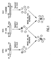
- FIG. 1 Expression Constructs and Experimental Design for Examining Notch-Delta Interactions. S2 cells at log phase growth were transiently transfected with one of the three constructs shown. Notch encoded by the MGlla minigene (a cDNA/genomic chimeric construct: cDNA-derived sequences are represented by stippling, genomically derived sequences by diagonal-hatching ( Ramos et al., 1989, Genetics 123, 337-348 )) was expressed following insertion into the metallothionein promoter vector pRmHa-3 ( Bunch et al., 1988, Nucl. Acids Res. 16, 1043-1061 ).
- Notch encoded by the MGlla minigene a cDNA/genomic chimeric construct: cDNA-derived sequences are represented by stippling, genomically derived sequences by diagonal-hatching ( Ramos et al., 1989, Genetics 123, 337-348 )
- the extracellular Notch (ECN1) variant was derived from a genomic cosmid containing the complete Notch locus ( Ramos et al., 1989, Genetics 123, 337-348 ) by deleting the coding sequence for amino acids 1790-2625 from the intracellular domain (denoted by ⁇ ; Wharton et al., 1985, Cell 43, 567-581 ), leaving 25 membrane-proximal residues from the wild-type sequence fused to a novel 59 amino acid tail (see Experimental Procedures, Section 6.1, infra ).
- This construct was expressed under control of the Notch promoter region.
- expression was induced with CuSO 4 following transfection. Cells were then mixed, incubated under aggregation conditions, and scored for their ability to aggregate using specific antisera and immunofluorescence microscopy to visualize expressing cells.
- MT metallothionein promoter
- ATG translation start site
- TM transmembrane domain
- 3' N Notch gene polyadenylation signal
- 3' Adh polyadenylation signal from Adh gene
- 5' N Notch gene promoter region.
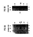
- FIG. 1 Expression of Notch and Delta in Cultured Cells.
- S2 nontransfected (S2) and Notch -transfected (N) cells induced with 0.7 mM CuSO 4 for 12-16 hr were prepared for sodium dodecyl sulfate polyacrylamide gel electrophoresis (SDS-PAGE), run on 3%-15% gradient gels, and blotted to nitrocellulose.
- Notch was visualized using a monoclonal antibody (MAb C17.9C6) against the intracellular domain of Notch. Multiple bands below the major band at 300 kd may represent degradation products of Notch.
- MAb C17.9C6 monoclonal antibody
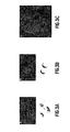
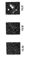
- FIG. 1 S2 Cells That Express Notch and Delta Form Aggregates. In all panels, Notch is shown in green and Delta in red.
- Notch and Delta are Associated in Cotransfected Cells. Staining for Notch is shown in the left column (A, C, and E) and that for Delta is shown in the right column (B, D, and F).
- FIG. 1 Coimmunoprecipitation Shows that Delta and Notch are Associated in Lysates from Transfected S2 and Drosophila Embryonic Cells.
- Delta was precipitated from NP-40/deoxycholate lysates using a polyclonal anti-Delta rat antiserum precipitated with fixed Staph A cells, and proteins in the precipitated fraction were visualized on Western blots (for details, see Experimental Procedures).
- Lanes 1, 2, 3, and 5 Notch visualized with MAb C17.9C6; Lanes 4 and 6: Delta visualized using MAb 201.
- Lane 1 shows a polyclonal anti-Delta immunoprecipitation from cells that express Notch alone visualized for Notch. No Notch was detectable in this sample, indicating that the polyclonal anti-Delta does not cross-react with Notch.
- Lane 2 shows Notch - Delta cotransfected cells immunoprecipitated with Staph A without initial treatment with anti-Delta antiserum and visualized for Notch, demonstrating that Notch is not precipitated nonspecifically by the Staph A or secondary antibody.
- Lane 3 shows protein precipitated with anti-Delta antiserum visualized for Delta (Dl), and lane 4 shows the same sample visualized for Notch (N). Lane 4 shows that Notch coprecipitates with immunoprecipitated Delta. Note that Notch appears as a doublet as is typical for Notch in immunoprecipitates.
- (B) shows the same experiment using embryonic lysates rather than transfected cell lysates.
- Lane 5 shows protein precipitated with anti-Delta antiserum visualized for Delta (Dl), and lane 6 shows the same sample visualized for Notch (N).
- Dl protein precipitated with anti-Delta antiserum visualized for Delta
- N Notch
- SA Staph A
- H anti-Delta antiserum heavy
- L light chains
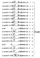
- FIG. 6 Notch Expression Constructs and the Deletion Mapping of the Delta/Serrate Binding Domain. S2 cells in log phase growth were transiently transfected with the series of expression constructs shown; the drawings represent the predicted protein products of the various Notch deletion mutants created. All expression constructs were derived from construct #1 pMtNMg. Transiently transfected cells were mixed with Delta expressing cells from the stably transformed line L49-6-7 or with transiently transfected Serrate expressing cells, induced with CuSO 4 , incubated under aggregation conditions and then scored for their ability to aggregate using specific antisera and immunofluorescence microscopy. Aggregates were defined as clusters of four or more cells containing both Notch and Delta/Serrate expressing cells.
- % Aggregation refers to the percentage of all Notch expressing cells found in such clusters either with Delta (Dl) (left column) or with Serrate (Ser) (right column).
- the various Notch deletion constructs are represented diagrammatically with splice lines indicating the ligation junctions. Each EGF repeat is denoted as a stippled rectangular box and numbers of the EGF repeats on either side of a ligation junction are noted.
- partial EGF repeats produced by the various deletions are denoted by open boxes and closed brackets (for example see #23 ⁇ Cla+EGF(10-12)).
- Constructs #3-13 represent the ClaI deletion series.
- EGF repeat 14 which carries the split point mutation, is drawn as a striped box.
- construct #33 ⁇ Cla+XEGF(10-13)
- the Xenopus Notch derived EGF repeats are distinguished from Drosophila repeats by a different pattern of shading.
- SP signal peptide
- EGF epidermal growth factor repeat
- N Notch / lin -12 repeat
- TM transmembrane domain
- cdc10 cdc 10/ankyrin repeats
- PA putative nucleotide binding consensus sequence
- opa polyglutamine stretch termed opa
- Dl Delta
- Ser Serrate.
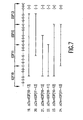
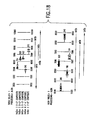
- FIG. 7 Detailed Structure of Notch Deletion Constructs #19-24: Both EGF Repeats 11 and 12 are Required for Notch-Delta Aggregation. EGF repeats 10-13 are diagrammed at the top showing the regular spacing of the six cysteine residues (C). PCR products generated for these constructs (names and numbers as given in Figure 6) are represented by the heavy black lines and the exact endpoints are noted relative to the various EGF repeats. Ability to aggregate with Delta is recorded as (+) or (-) for each construct. The PCR fragments either break the EGF repeats in the middle, just after the third cysteine in the same place as four out of the five ClaI sites, or exactly in between two repeats in the same place as the most C-terminal ClaI site.
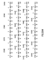
- FIG. 8 Comparison of Amino Acid Sequence of EGF Repeats 11 and 12 from Drosophila and Xenopus Notch.
- the amino acid sequence of EGF repeats 11 and 12 of Drosophila Notch ( Wharton et al., 1985, Cell 43:567-581 ; Kidd et al., 1986, Mol. Cell Biol. 3:194-3108 ) is aligned with that of the same two EGF repeats from Xenopus Notch ( Coffman et al., 1990, Science 249:1438-1441 ). Identical amino acids are boxed.
- the six conserved cysteine residues of each EGF repeat and the Ca ++ binding consensus residues ( Rees et al., 1988, EMBO J. 7:2053-2061 ) are marked with an asterisk (*).
- the leucine to proline change found in the Xenopus PCR clone that failed to aggregate is noted underneath.
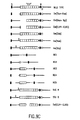
- FIG. 9 Constructs Employed in this Study. Schematic diagrams of the Delta variants defined in Table IV are shown. Extracellular, amino-proximal terminus is to the left in each case. S, signal peptide; "EGF”, EGF-like motifs; M, membrane-spanning helix; H, stop-transfer sequence; solid lines, other Delta sequences; hatched lines, neuroglian sequences. Arrowheads indicate sites of translatable linker insertions.
- Figure 9A Dependence of Aggregation on Input DNA Amounts.
- A Heterotypic aggregation observed using S2 cell populations transiently transfected, respectively, with varied amounts of pMTDl1 DNA (2, 4, 10 or 20 ⁇ g/plate) that were subsequently incubated under aggregation conditions with S2 cell populations transiently transfected with a constant amount of pMtNMg DNA (20 ⁇ g/plate).
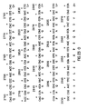
- FIG. 10 Delta-Serrate Amino-Terminal Sequence Alignment. Residues are numbered on the basis of conceptual translation of Delta (Dl, upper sequence (SEQ ID NO:3); beginning at amino acid 24, ending at amino acid 226) and Serrate (Ser, lower sequence (SEQ ID NO:4); beginning at amino acid 85, ending at amino acid 283) coding sequences. Vertical lines between the two sequences indicates residues that are identical within the Delta and Serrate sequences, as aligned. Dots represent gaps in the alignment. Boxes enclose cysteine residues within the aligned regions. N1, amino-proximal domain 1; N2, amino-proximal domain 2; N3, amino-proximal domain 3.
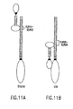
- FIG. 11 Potential Geometries of Delta-Notch Interactions.
- A Potential register of Delta (left) and Notch (right) molecules interacting between opposing plasma membranes.
- B Potential register of Delta (left) and Notch (right) molecules interacting within the same plasma membranes.
- ELR EGF-like repeat; open boxes, EGF-like repeats; dotted boxes, LNR repeats; solid boxes, membrane-spanning helices. Delta amino-terminal domain and Delta and Notch intracellular domains represented by ovals.
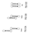
- Figure 12 Potential Geometries of Delta-Delta Interactions.
- a and B Potential register of Delta molecules interacting between opposing plasma membranes.
- B Potential register of Delta molecules interacting within the same plasma membranes.
- Open boxes EGF-like repeats; solid boxes, membrane-spanning helices. Delta amino-terminal extracellular and intracellular domains represented by ovals.
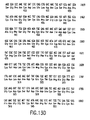
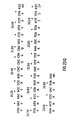
- FIG. 13 Primary Nucleotide Sequence of the Delta cDNA Dl1 (SEQ ID NO:5) and Delta amino acid sequence (SEQ ID NO:6) The DNA sequence of the 5'-3' strand of the Dl1 cDNA is shown, which contains a number of corrections in comparison to that presented in Kopczynksi et al. (1988, Genes Dev. 2, 1723-1735 ).
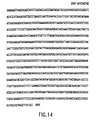
- Figure 14 Primary Nucleotide Sequence of the Neuroglian cDNA 1B7A-250 (SEQ ID NO:7). This is the DNA sequence of a portion of the 5'-3' strand of the 1B7A-250 cDNA (A.J. Bieber, pers. comm.; Hortsch et al., 1990, Neuron 4, 697-709 ). Nucleotide 2890 corresponds to the first nucleotide of an isoleucine codon that encodes amino acid 952 of the conceptually translated neuroglian-long form protein.
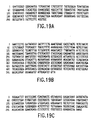
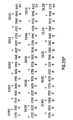
- FIG. 15 Nucleic Acid Sequence Homologies Between Serrate and Delta .
- a portion of the Drosophila Serrate nucleotide sequence (SEQ ID NO:8), with the encoded Serrate protein sequence (SEQ ID NO:9) written below, ( Fleming et al., 1990, Genes & Dev. 4, 2188-2201 at 2193-94 ) is shown.
- the four regions showing high sequence homology with the Drosophila Delta sequence are numbered above the line and indicated by brackets.
- the total region of homology spans nucleotide numbers 627 through 1290. of the Serrate nucleotide sequence (numbering as in Figure 4 of Fleming et al., 1990, Genes & Dev. 4, 2188-2201 ).
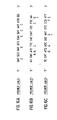
- FIG. 16 Primers used for PCR in the Cloning of Human Notch.
- the sequence of three primers used for PCR to amplify DNA in a human fetal brain cDNA library are shown.
- the three primers, cdc1 (SEQ ID NO:10), cdc2 (SEQ ID NO:11), and cdc3 (SEQ ID NO:12) were designed to amplify either a 200 bp or a 400 bp fragment as primer pairs cdc1/cdc2 or cdc1/cdc3, respectively.
- I inosine.
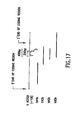
- FIG. 17 Schematic Diagram of Human Notch Clones. A schematic diagram of human Notch is shown. Heavy bold-face lines below the diagram show that portion of the Notch sequence contained in each of the four cDNA clones. The location of the primers used in PCR, and their orientation, are indicated by arrows.
- Figure 18 Human Notch Sequences Aligned with Drosophila Notch Sequence. Numbered vertical lines correspond to Drosophila Notch coordinates. Horizontal lines below each map show where clones lie relative to stretches of sequence (thick horizontal lines).
- Figure 19 Nucleotide Sequences of Human Notch contained in Plasmid cDNA Clone hN2k.
- Figure 19A The DNA sequence (SEQ ID NO:13) of a portion of the human Notch insert is shown, starting at the EcoRI site at the 3' end, and proceeding in the 3' to 5' direction.
- Figure 19B The DNA sequence (SEQ ID NO:14) of a portion of the human Notch insert is shown, starting at the EcoRI site at the 5' end, and proceeding in the 5' to 3' direction.
- Figure 19C The DNA sequence (SEQ ID NO:15) of a portion of the human Notch insert is shown, starting 3' of the sequence shown in Figure 19B, and proceeding in the 5' to 3' direction. The sequences shown are tentative, subject to confirmation by determination of overlapping sequences.
- Figure 20 Nucleotide Sequences of Human Notch Contained in Plasmid cDNA clone hN3k.
- Figure 20A The DNA sequence (SEQ ID NO:16) of a portion of the human Notch insert is shown, starting at the EcoRI site at the 3' end, and proceeding in the 3' to 5' direction.
- Figure 20B The DNA sequence (SEQ ID NO:17) of a portion of the human Notch insert is shown, starting at the EcoRI site at the 5' end, and proceeding in the 5' to 3' direction.
- Figure 20C The DNA sequence (SEQ ID NO:18) of a portion of the human Notch insert is shown, starting 3' of the sequence shown in Figure 20B, and proceeding in the 5' to 3' direction.
- Figure 20D The DNA sequence (SEQ ID NO:19) of a portion of the human Notch insert is shown, starting 5' of the sequence shown in Figure 20A, and proceeding in the 3' to 5' direction.
- the sequences shown are tentative, subject to confirmation by determination of overlapping sequences.
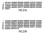
- Figure 21 Nucleotide Sequences of Human Notch Contained in Plasmid cDNA clone hN4k.
- Figure 21A The DNA sequence (SEQ ID NO:20) of a portion of the human Notch insert is shown, starting at the EcoRI site at the 5' end, and proceeding in the 5' to 3' direction.
- Figure 21B The DNA sequence (SEQ ID NO:21) of a portion of the human Notch insert is shown, starting near the 3' end, and proceeding in the 3' to 5' direction. The sequences shown are tentative, subject to confirmation by determination of overlapping sequences.
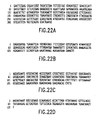
- Figure 22 Nucleotide Sequences of Human Notch Contained in Plasmid cDNA Clone hN5k.
- Figure 22A The DNA sequence (SEQ ID NO:22) of a portion of the human Notch insert is shown, starting at the EcoRI site at the 5' end, and proceeding in the 5' to 3' direction.
- Figure 22B The DNA sequence (SEQ ID NO:23) of a portion of the human Notch insert is shown, starting near the 3' end, and proceeding in the 3' to 5' direction.
- Figure 22C The DNA sequence (SEQ ID NO:24) of a portion of the human Notch insert is shown, starting 3' of the sequence shown in Figure 22A, and proceeding in the 5' to 3' direction.
- Figure 22D The DNA sequence (SEQ ID NO:25) of a portion of the human Notch insert is shown, starting 5' of the sequence shown in Figure 22B, and proceeding in the 3' to 5' direction.
- the sequences shown are tentative, subject to confirmation by determination of overlapping sequences.
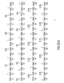
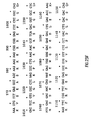
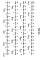
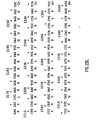
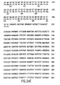
- FIG. 24 DNA (SEQ ID NO:33) and Amino Acid (SEQ ID NO:34) Sequences of Human Notch Contained in Plasmid cDNA Clone hN5k.
- Figure 25 Comparison of hN5k With Other Notch Homologs.
- Figure 25A Schematic representation of Drosophila Notch. Indicated are the signal sequence (signal), the 36 EGF-like repeats, the three Notch / lin -12 repeats, the transmembrane domain (TM), the six CDC10 repeats, the OPA repeat, and the PEST (proline, glutamic acid, serine, threonine)-rich region.
- Figure 25B Alignment of the deduced amino acid sequence of hN5k with sequences of other Notch homologs. Amino acids are numbered on the left side. The cdc10 and PEST-rich regions are both boxed, and individual cdc10 repeats are marked.
- NLS nuclear localization sequence
- CKII casein kinase II
- cdc2 cdc2 kinase
- the present invention provides nucleotide sequences of the human Notch genes, and amino acid sequences of their encoded proteins.
- the specification also describes nucleotides sequences of Delta genes, and amino acid sequences of their encoded proteins.
- the specification further describes fragments (termed herein "adhesive fragments") of the proteins encoded by toporythmic genes which mediate homotypic or heterotypic binding to toporythmic proteins or adhesive fragments thereof.
- Toporythmic genes shall mean the genes Notch , Delta, and Serrate , as well as other members of the Delta / Serrate family which may be identified, e.g. by the methods described in Section 5.3, infra.
- nucleic acid and amino acid sequences and antibodies thereto of the invention can be used for the detection and quantitation of mRNA for human Notch molecules, to study expression thereof, to produce human Notch sequences, in the study and manipulation of differentiation processes.
- Toporythmic protein fragments which mediate homotypic or heterotypic binding (and thus are termed herein "adhesive"), and nucleic acid sequences relating to the foregoing are described below.
- the adhesive fragment of Notch which is part of the present invention, is that comprising the portion of Notch most homologous to ELR 11 and 12, i.e., amino acid numbers 447 through 527 (SEQ ID NO:1) of the Drosophila Notch sequence (see Figure 8).
- the adhesive fragment of Delta mediating homotypic binding is that comprising the portion of Delta most homologous to about amino acid numbers 32-230 of the Drosophila Delta sequence (SEQ ID NO:6).
- the adhesive fragment of Delta mediating binding to Notch is that comprising the portion of Delta most homologous to about amino acid numbers 1-230 of the Drosophila Delta sequence (SEQ ID NO:6).
- such fragment is that comprising the portion of Serrate most homologous to about amino acid numbers 85-283 or 79-282 of the Drosophila Serrate sequence (see Figure 10 (SEQ ID NO:4), and Figure 15 (SEQ ID NO:9)).
- toporythmic adhesive domains can be isolated from porcine, bovine, feline, avian, equine, or canine, as well as primate sources and any other species in which homologs of known toporythmic genes [including but not limited to the following genes (with the publication of sequences in parentheses): Notch ( Wharton et al., 1985, Cell 43, 567-581 ), Delta ( Vassin et al., 1987, EMBO J. 6, 3431-3440 ; Kopczynski et al., 1988, Genes Dev. 2, 1723-1735 ; note corrections to the Kopczynski et al.
- sequence found in Figure 13 hereof SEQ ID No:5 and SEQ ID NO:6
- Serrate Fleming et al., 1990, Genes & Dev. 4, 2188-2201
- sequences can be altered by substitutions, additions or deletions that provide for functionally equivalent (adhesive) molecules.
- nucleotide coding sequences other DNA sequences which encode substantially the same amino acid sequence as the adhesive sequences may be used in the practice of the present invention. These include but are not limited to nucleotide sequences comprising all or portions of the Notch genes which are altered by the substitution of different codons that encode a functionally equivalent amino acid residue within the sequence, thus producing a silent change.
- the adhesive protein fragments or derivatives thereof include, but are not limited to, those containing, as a primary amino acid sequence, all or part of the amino acid sequence of the adhesive domains including altered sequences in which functionally equivalent amino acid residues are substituted for residues within the sequence resulting in a silent change.
- one or more amino acid residues within the sequence can be substituted by another amino acid of a similar polarity which acts as a functional equivalent, resulting in a silent alteration.
- Substitutes for an amino acid within the sequence may be selected from other members of the class to which the amino acid belongs.
- the nonpolar (hydrophobic) amino acids include alanine, leucine, isoleucine, valine, proline, phenylalanine, tryptophan and methionine.
- the polar neutral amino acids include glycine, serine, threonine, cysteine, tyrosine, asparagine, and glutamine.
- the positively charged (basic) amino acids include arginine, lysine and histidine.
- the negatively charged (acidic) amino acids include aspartic acid and glutamic acid.
- Adhesive fragments of toporythmic proteins and peptides related to adhesive toporythmic protein sequences can be tested for the desired binding activity e.g., by the in vitro aggregation assays described in the examples herein.
- Adhesive fragments of toporythmic proteins include but are not limited to those peptides which are substantially homologous to the adhesive fragments, or whose encoding nucleic acid is capable of hybridizing to the nucleic acid sequence encoding the adhesive fragments, and which peptides have positive binding activity e.g., as tested in vitro by an aggregation assay such as described in the examples sections infra .
- the adhesive-protein related peptides can be produced by various methods known in the art. The manipulations which result in their production can occur at the gene or protein level.
- the cloned adhesive protein-encoding gene sequence can be modified by any of numerous strategies known in the art (Maniatis, T., 1990, Molecular Cloning, A Laboratory Manual, 2d ed., cold Spring Harbor Laboratory, Cold Spring Harbor, New York ). The sequence can be cleaved at appropriate sites with restriction endonuclease(s), followed by further enzymatic modification if desired, isolated, and ligated in vitro .
- the adhesive-encoding nucleic acid sequence can be mutated in vitro or in vivo , to create and/or destroy translation, initiation, and/or termination sequences, or to create variations in coding regions and/or form new restriction endonuclease sites or destroy preexisting ones, to facilitate further in vitro modification.
- Any technique for mutagenesis known in the art can be used, including but not limited to, in vitro site-directed mutagenesis (Hutchinson, C., et al., 1978, J. Biol. Chem 253, 6551), use of TAB® linkers (Pharmacia), etc.
- Toporythmic protein fragments may be differentially modified during or after translation, e.g. , by glycosylation, acetylation, phosphorylation, proteolytic cleavage, linkage to an antibody molecule or other cellular ligand, etc. Any of numerous chemical modifications may be carried out by known techniques, including but not limited to specific chemical cleavage by cyanogen bromide, trypsin, chymotrypsin, papain, V8 protease, NaBH 4 ; acetylation, formylation, oxidation, reduction; metabolic synthesis in the presence of tunicamycin; etc.
- peptides related to adhesive fragments can be chemically synthesized.
- a peptide corresponding to a portion of a toporythmic protein which mediates the desired aggregation activity in vitro can be synthesized by use of a peptide synthesizer.
- a Delta protein which have the ability to bind to a second Delta protein or fragment or derivative thereof, but do not bind to Notch. Such binding or lack thereof can be assayed in vitro as described in Section 8.
- a Delta derivative is that containing an insertion of the tetrapeptide Arg-Lys-Ile-Phe between Delta residues 198 and 199 of the Drosophila protein.
- the invention further relates to the amino acid sequences of human Notch and fragments thereof which comprise an antigenic determinant (i.e., can be recognized by an antibody) or which are functionally active, as well as nucleic acid sequences encoding the foregoing. Also described herein are the amino acid and nucleic acid sequences of Delta and corresponding fragments and derivatives thereof.
- "Functionally active" material as used herein refers to that material displaying one or more known functional activities associated with the full-length (wild-type) protein product, e.g., in the case of Notch, binding to Delta, binding to Serrate, antigenicity (binding to an anti-Notch antibody), etc.
- the invention provides fragments of a human Notch protein consisting of at least 40 amino acids, or of at least 77 amino acids.
- the proteins of the invention comprise or consist essentially of the intracellular domain, transmembrane region, extracellular domain, cdc10 region, Notch / lin -12 repeats, or the EGF-homologous repeats, or any combination of the foregoing, of a human Notch protein. Fragments, or proteins comprising fragments, lacking some or all of the EGF-homologous repeats of human Notch are also provided.
- the invention is further directed to the nucleotide sequences and subsequences of human Notch consisting of at least 25 nucleotides, at least 50 nucleotides, or at least 121 nucleotides.
- Nucleic acids encoding the proteins and protein fragments described above are also provided, as well as nucleic acids complementary to and capable of hybridizing to such nucleic acids.
- such a complementary sequence may be complementary to a human Notch cDNA sequence of at least 25 nucleotides, or of at least 121 nucleotides.
- the invention relates to cDNA sequences encoding human Notch or a portion thereof.
- the invention relates to the nucleotide sequence of the human Notch gene or cDNA, in particular, comprising those sequences depicted in Figures 19, 20, 21 and/or 22 (SEQ ID NO:13 through NO:25), or contained in plasmids hN3k, hN4k, or hN5k (see Section 9, infra), and the encoded Notch protein sequences.
- a "nucleic acid encoding a fragment or portion of a Notch protein” shall be construed as referring to a nucleic acid encoding only the recited fragment or portion of the Notch protein and not other portions of the Notch protein.
- a human Notch DNA sequence can be cloned and sequenced by the method described in Section 9, infra .
- PCR is used to amplify the desired sequence in the library, prior to selection.
- oligonucleotide primers representing part of the adhesive domains encoded by a homologue of the desired gene can be used as primers in PCR.
- Any human cell can potentially serve as the nucleic acid source for the molecular cloning of the Notch and Delta genes.
- the DNA may be obtained by standard procedures known in the art from cloned DNA (e.g. , a DNA "library”), by chemical synthesis, by cDNA cloning, or by the cloning of genomic DNA, or fragments thereof, purified from the desired human cell. (See, for example Maniatis et al., 1982, Molecular Cloning, A Laboratory Manual, Cold Spring Harbor Laboratory, Cold Spring Harbor, New York ; Glover, D.M. (ed.), 1985, DNA Cloning: A Practical Approach, MRL Press, Ltd., Oxford, U.K. Vol.
- Clones derived from genomic DNA may contain regulatory and intron DNA regions in addition to coding regions; clones derived from cDNA will contain only exon sequences. Whatever the source, the gene should be molecularly cloned into a suitable vector for propagation of the gene.
- DNA fragments are generated, some of which will encode the desired gene.
- the DNA may be cleaved at specific sites using various restriction enzymes.
- DNAse in the presence of manganese to fragment the DNA, or the DNA can be physically sheared, as for example, by sonication.
- the linear DNA fragments can then be separated according to size by standard techniques, including but not limited to, agarose and polyacrylamide gel electrophoresis and column chromatography.
- identification of the specific DNA fragment containing the desired gene may be accomplished in a number of ways. For example, if an amount of a portion of a Notch or Delta (of any species) gene or its specific RNA, or a fragment thereof e.g., the adhesive domain, is available and can be purified and labeled, the generated DNA fragments may be screened by nucleic acid hybridization to the labeled probe ( Benton, W. and Davis, R., 1977, Science 196, 180 ; Grunstein, M. And Hogness, D., 1975, Proc. Natl. Acad. Sci. U.S.A. 72, 3961 ). Those DNA fragments with substantial homology to the probe will hybridize.
- cDNA clones or DNA clones which hybrid-select the proper mRNAs, can be selected which produce a protein that, e.g. , has similar or identical electrophoretic migration, isolectric focusing behavior, proteolytic digestion maps, in vitro aggregation activity ("adhesiveness") or antigenic properties as known for Notch or Delta.
- the Notch or Delta protein may be identified by binding of labeled antibody to the putatively Notch or Delta synthesizing clones, in an ELISA (enzyme-linked immunosorbent assay)-type procedure.
- the Notch or Delta gene can also be identified by mRNA selection by nucleic acid hybridization followed by in vitro translation. In this procedure, fragments are used to isolate complementary mRNAs by hybridization. Such DNA fragments may represent available, purified Notch or Delta DNA of another species (e.g., Drosophila ). Immunoprecipitation analysis or functional assays (e.g. , aggregation ability in vitro ; see examples infra) of the in vitro translation products of the isolated products of the isolated mRNAs identifies the mRNA and, therefore, the complementary DNA fragments that contain the desired sequences.
- Immunoprecipitation analysis or functional assays e.g. , aggregation ability in vitro ; see examples infra
- specific mRNAs may be selected by adsorption of polysomes isolated from cells to immobilized antibodies specifically directed against Notch or Delta protein.
- a radiolabelled Notch or Delta cDNA can be synthesized using the selected mRNA (from the adsorbed polysomes) as a template. The radiolabelled mRNA or cDNA may then be used as a probe to identify the Notch or Delta DNA fragments from among other genomic DNA fragments.
- RNA for cDNA cloning of the Notch or Delta gene can be isolated from cells which express Notch or Delta. Other methods are possible.
- the identified and isolated gene can then be inserted into an appropriate cloning vector.
- vector-host systems known in the art may be used. Possible vectors include, but are not limited to, plasmids or modified viruses, but the vector system must be compatible with the host cell used. Such vectors include, but are not limited to, bacteriophages such as lambda derivatives, or plasmids such as PBR322 or pUC plasmid derivatives.
- the insertion into a cloning vector can, for example, be accomplished by ligating the DNA fragment into a cloning vector which has complementary cohesive termini.
- the ends of the DNA molecules may be enzymatically modified.
- any site desired may be produced by ligating nucleotide sequences (linkers) onto the DNA termini; these ligated linkers may comprise specific chemically synthesized oligonucleotides encoding restriction endonuclease recognition sequences.
- the cleaved vector and Notch or Delta gene may be modified by homopolymeric tailing. Recombinant molecules can be introduced into host cells via transformation, transfection, infection, electroporation, etc., so that many copies of the gene sequence are generated.
- the desired gene may be identified and isolated after insertion into a suitable cloning vector in a "shot gun" approach. Enrichment for the desired gene, for example, by size fractionization, can be done before insertion into the cloning vector.
- transformation of host cells with recombinant DNA molecules that incorporate the isolated Notch or Delta gene, cDNA, or synthesized DNA sequence enables generation of multiple copies of the gene.
- the gene may be obtained in large quantities by growing transformants, isolating the recombinant DNA molecules from the transformants and, when necessary, retrieving the inserted gene from the isolated recombinant DNA.
- the human Notch sequences provided by the instant invention include those nucleotide sequences encoding substantially the same amino acid sequences as found in human Notch, and those encoded amino acid sequences with functionally equivalent amino acids, all as described supra in section 5.1 for adhesive portions of toporythmic proteins.
- a rational search for additional members of the Delta / Serrate gene family may be carried out using an approach that takes advantage of the existence of the conserved segments of strong homology between Serrate and Delta (see Figure 10, SEQ ID NO:3 and NO:4).
- additional members of this gene family may be identified by selecting, from among a diversity of nucleic acid sequences, those sequences that are homologous to both Serrate and Delta (see Figure 13 (SEQ ID NO:5), and Figure 15 (SEQ ID NO:8)), and further identifying, from among the selected sequences, those that also contain nucleic acid sequences which are non-homologous to Serrate and Delta .
- the term "non-homologous" may be construed to mean a region which contains at least about 6 contiguous nucleotides in which at least about two nucleotides differ from Serrate and Delta sequence.
- a preferred method is as follows: Corresponding to two conserved segments between Delta and Serrate , Delta AA 63-73 and Delta AA 195-206 (see Figure 13, SEQ ID NO:6), sets of degenerate. oligonucleotide probes of about 10-20 nucleotides may be synthesized, representing all of the possible coding sequences for the amino acids found in either Delta and Serrate for about three to seven contiguous codons.
- oligonucleotides may be obtained corresponding to parts of the four highly conserved regions between Delta and Serrate shown in Figure 15 (SEQ ID NO:8 and NO:9), i.e., that represented by Serrate AA 124-134, 149-158, 214-219, and 250-259.
- the synthetic oligonucleotides may be utilized as primers to amplify by PCR sequences from a source (RNA or DNA) of potential interest. (PCR can be carried out, e.g., by use of a Perkin-Elmer Cetus thermal cycler and Taq polymerase (Gene AmpTM)).
- PCR reactions By carrying out the PCR reactions, it may be possible to detect a gene or gene product sharing the above-noted segments of conserved sequence between Serrate and Delta. If one chooses to synthesize several different degenerate primers, it may still be possible to carry out a complete search with a reasonably small number of PCR reactions. It is also possible to vary the stringency of hybridization conditions used in priming the PCR reactions, to allow for greater or lesser degrees of nucleotide sequence similarity between the unknown gene and Serrate or Delta .
- a segment of a previously unknown member of the Serrate / Delta gene family is amplified successfully, that segment may be molecularly cloned and sequenced, and utilized as a probe to isolate a complete cDNA or genomic clone. This, in turn, will permit the determination of the unknown gene's complete nucleotide sequence, the analysis of its expression, and the production of its protein product for functional analysis. In this fashion, additional genes encoding "adhesive" proteins may be identified.
- Serrate / Delta sequence homologies may be used in the design of novel recombinant molecules which are members of the Serrate / Delta gene family but which may not occur in nature.
- a recombinant molecule can be constructed comprising portions of both Serrate and Delta genes. Such a molecule could exhibit properties associated with both Serrate and Delta and portray a novel profile of biological activities, including agonists as well as antagonists.
- the primary sequence of Serrate and Delta may also be used to predict tertiary structure of the molecules using computer simulation ( Hopp and Woods, 1981, Proc. Natl. Acad. Sci. U.S.A.
- Serrate / Delta chimeric recombinant genes could be designed in light of correlations between tertiary structure and biological function.
- chimeric genes comprising portions of any one or more members of the toporythmic gene family (e.g., Notch) may be constructed.
- the nucleotide sequence coding for an adhesive fragment of a toporythmic protein (preferably, Notch , Serrate, or Delta ), or human Notch or Delta or a functionally active fragment thereof, can be inserted into an appropriate expression vector, i.e. , a vector which contains the necessary elements for the transcription and translation of the inserted protein-coding sequence.
- an appropriate expression vector i.e. , a vector which contains the necessary elements for the transcription and translation of the inserted protein-coding sequence.
- the necessary transcriptional and translational signals can also be supplied by the native toporythmic gene and/or its flanking regions.
- host-vector systems may be utilized to express the protein-coding sequence. These include but are not limited to mammalian cell systems infected with virus (e.g.
- the expression elements of vectors vary in their strengths and specificities. Depending on the host-vector system utilized, any one of a number of suitable transcription and translation elements may be used.
- the adhesive portion of the Notch gene e.g., that encoding EGF-like repeats 11 and 12, is expressed.
- the adhesive portion of the Delta gene e.g., that encoding amino acids 1-230, is expressed.
- the human Notch or human Delta gene is expressed, or a sequence encoding a functionally active portion of human Notch or Delta.
- the adhesive portion of the Serrate gene is expressed.
- any of the methods previously described for the insertion of DNA fragments into a vector may be used to construct expression vectors containing a chimeric gene consisting of appropriate transcriptional/translational control signals and the protein coding sequences. These methods may include in vitro recombinant DNA and synthetic techniques and in vivo recombinants (genetic recombination). Expression of nucleic acid sequence encoding a toporythmic protein or peptide fragment may be regulated by a second nucleic acid sequence so that the toporythmic protein or peptide is expressed in a host transformed with the recombinant DNA molecule. For example, expression of a toporythmic protein may be controlled by any promoter/enhancer element known in the art.
- Promoters which may be used to control toporythmic gene expression include, but are not limited to, the SV40 early promoter region ( Bernoist and Chambon, 1981, Nature 290, 304-310 ), the promoter contained in the 3' long terminal repeat of Rous sarcoma virus ( Yamamoto, et al., 1980, Cell 22, 787-797 ), the herpes thymidine kinase promoter ( Wagner et al., 1981, Proc. Natl. Acad. Sci. U.S.A.
- the regulatory sequences of the metallothionein gene ( Brinster et al., 1982, Nature 296, 39-42 ); prokaryotic expression vectors such as the ⁇ -lactamase promoter ( Villa-Kamaroff, et al., 1978, Proc. Natl. Acad. Sci. U.S.A. 75, 3727-3731 ), or the tac promoter ( DeBoer, et al., 1983, Proc. Natl. Acad. Sci. U.S.A.
- promoter of the photosynthetic enzyme ribulose biphosphate carboxylase Herrera-Estrella et al., 1984, Nature 310, 115-120
- promoter elements from yeast or other fungi such as the Gal 4 promoter, the ADC (alcohol dehydrogenase) promoter, PGK (phosphoglycerol kinase) promoter, alkaline phosphatase promoter, and the following animal transcriptional control regions, which exhibit tissue specificity and have been utilized in transgenic animals: elastase I gene control region which is active in pancreatic acinar cells ( Swift et al., 1984, Cell 38, 639-646 ; Ornitz et al., 1986, Cold Spring Harbor Symp.
- mouse mammary tumor virus control region which is active in testicular, breast, lymphoid and mast cells ( Leder et al., 1986, Cell 45, 485-495 ), albumin gene control region which is active in liver ( Pinkert et al., 1987, Genes and Devel. 1, 268-276 ), alpha-fetoprotein gene control region which is active in liver ( Krumlauf et al., 1985, Mol. Cell. Biol. 5, 1639-1648 ; Hammer et al., 1987, Science 235, 53-58 ; alpha 1-antitrypsin gene control region which is active in the liver ( Kelsey et al., 1987, Genes and Devel.
- beta-globin gene control region which is active in myeloid cells ( Mogram et al., 1985, Nature 315, 338-340 ; Kollias et al., 1986, Cell 46, 89-94 ; myelin basic protein gene control region which is active in oligodendrocyte cells in the brain ( Readhead et al., 1987, Cell 48, 703-712 ); myosin light chain-2 gene control region which is active in skeletal muscle ( Sani, 1985, Nature 314, 283-286 ), and gonadotropic releasing hormone gene control region which is active in the hypothalamus ( Mason et al., 1986, Science 234, 1372-1378 ).
- Expression vectors containing toporythmic gene inserts can be identified by three general approaches: (a) nucleic acid hybridization, (b) presence or absence of "marker" gene functions, and (c) expression of inserted sequences.
- first approach the presence of a foreign gene inserted in an expression vector can be detected by nucleic acid hybridization using probes comprising sequences that are homologous to an inserted toporythmic gene.
- second approach the recombinant vector/host system can be identified and selected based upon the presence or absence of certain "marker" gene functions (e.g.
- telomere kinase activity thymidine kinase activity, resistance to antibiotics, transformation phenotype, occlusion body formation in baculovirus, etc.
- recombinants containing the toporythmic insert can be identified by the absence of the marker gene function.
- recombinant expression vectors can be identified by assaying the foreign gene product expressed by the recombinant.
- Such assays can be based, for example, on the physical or functional properties of the toporythmic gene product in vitro assay systems, e.g., aggregation (adhesive) ability (see Sections 6-8, infra ).
- the expression vectors which can be used include, but are not limited to, the following vectors or their derivatives: human or animal viruses such as vaccinia virus or adenovirus; insect viruses such as baculovirus; yeast vectors; bacteriophage vectors ( e.g. , lambda), and plasmid and cosmid DNA vectors, to name but a few.
- a host cell strain may be chosen which modulates the expression of the inserted sequences, or modifies and processes the gene product in the specific fashion desired. Expression from certain promoters can be elevated in the presence of certain inducers; thus, expression of the genetically engineered toporythmic protein may be controlled.
- different host cells have characteristic and specific mechanisms for the translational and post-translational processing and modification (e.g. , glycosylation, cleavage) of proteins. Appropriate cell lines or host systems can be chosen to ensure the desired modification and processing of the foreign protein expressed. For example, expression in a bacterial system can be used to produce an unglycosylated core protein product. Expression in yeast will produce a glycosylated product.
- Expression in mammalian cells can be used to ensure "native" glycosylation of a heterologous mammalian toporythmic protein.
- different vector/host expression systems may effect processing reactions such as proteolytic cleavages to different extents.
- the adhesive toporythmic protein or fragment may be expressed as a fusion, or chimeric protein product (comprising the protein or fragment joined to a heterologous protein sequence).
- a chimeric product can be made by ligating the appropriate nucleic acid sequences encoding the desired amino acid sequences to each other by methods known in the art, in the proper coding frame, and expressing the chimeric product by methods commonly known in the art.
- a chimeric product may be made by protein synthetic techniques, e.g., by use of a peptide synthesizer.
- cDNA and genomic sequences can be cloned and expressed.
- a human Notch cDNA sequence may be chromosomally integrated and expressed. Homologous recombination procedures known in the art may be used.
- the gene product may be analyzed. This can be achieved by assays based on the physical or functional properties of the product, including radioactive labelling of the product followed by analysis by gel electrophoresis.
- toporythmic protein Once the toporythmic protein is identified, it may be isolated and purified by standard methods including chromatography (e.g. , ion exchange, affinity, and sizing column chromatography), centrifugation, differential solubility, or by any other standard technique for the purification of proteins.
- the functional properties may be evaluated using any suitable assay, including, but not limited to, aggregation assays (see Sections 6-8).
- Toporythmic protein fragments which mediate homotypic or heterotypic binding, or human Notch or human Delta proteins or fragments thereof, may be used as an immunogen to generate anti-toporythmic protein antibodies.
- Such antibodies can be polyclonal or monoclonal.
- antibodies specific to EGF-like repeats 11 and 12 of Notch may be prepared.
- antibodies reactive with the "adhesive portion" of Delta can be generated.
- One example of such antibodies may prevent aggregation in an in vitro assay.
- antibodies specific to human Notch are produced.
- polyclonal antibodies to a toporythmic protein or peptide may be obtained.
- rabbit polyclonal antibodies to an epitope of the human Notch protein encoded by a sequence depicted in Figure 19, 20, 21 or 22 (SEQ ID NO:13 through NO:25), or a subsequence thereof can be obtained.
- various host animals can be immunized by injection with the native toporythmic protein, or a synthetic version, or fragment thereof, including but not limited to rabbits, mice, rats, etc.
- adjuvants may be used to increase the immunological response, depending on the host species, and including but not limited to Freund's (complete and incomplete), mineral gels such as aluminum hydroxide, surface active substances such as lysolecithin, pluronic polyols, polyanions, peptides, oil emulsions, keyhold limpet hemocyanins, dinitrophenol, and potentially useful human adjuvants such as BCG (bacille Calmette-Guerin) and corynebacterium parvum.
- BCG Bacille Calmette-Guerin
- any technique which provides for the production of antibody molecules by continuous cell lines in culture may be used.
- the hybridoma technique originally developed by Kohler and Milstein (1975, Nature 256, 495-497 ), as well as the trioma technique, the human B-cell hybridoma technique (Kozbor et al., 1983, Immunology Today 4, 72), and the EBV-hybridoma technique to produce human monoclonal antibodies Cole et al., 1985, in Monoclonal Antibodies and Cancer Therapy, Alan R. Liss, Inc., pp. 77-96 ).
- Antibody fragments which contain the idiotype of the molecule can be generated by known techniques.
- such fragments include but are not limited to: the F(ab') 2 fragment which can be produced by pepsin digestion of the antibody molecule; the Fab' fragments which can be generated by reducing the disulfide bridges of the F(ab') 2 fragment, and the Fab fragments which can be generated by treating the antibody molecule with papain and a reducing agent.
- screening for the desired antibody can be accomplished by techniques known in the art, e.g. ELISA (enzyme-linked immunosorbent assay).
- ELISA enzyme-linked immunosorbent assay
- to select antibodies which recognize the adhesive domain of a toporythmic protein one may assay generated hybridomas for a product which binds to a protein fragment containing such domain.
- For selection of an antibody specific to human Notch one can select on the basis of positive binding to human Notch and a lack of binding to Drosophila Notch.
- antibodies can be used in methods known in the art relating to the localization and activity of the protein sequences of the invention.
- various immunoassays known in the art can be used, including but not limited to competitive and non-competitive assay systems using techniques such as radioimmunoassays, ELISA (enzyme linked immunosorbent assay), "sandwich” immunoassays, precipitin reactions, gel diffusion precipitin reactions, immunodiffusion assays, agglutination assays, fluorescent immunoassays, protein A immunoassays, and immunoelectrophoresis assays, to name but a few.
- the invention also provides methods for delivery of agents into Notch-expressing cells.
- Delivery of agents into a Notch-expressing cell can be achieved by conjugation of an agent to a Delta protein or an adhesive fragment thereof capable of binding to Notch, and exposing a Notch-expressing cell to the conjugate, such that the conjugate is taken up by the cell.
- the conjugated agent can be, but is not limited to, a label or a biologically active agent.
- the biologically active agent can be a therapeutic agent, a toxin, a chemotherapeutic, a growth factor, an enzyme, a hormone, a drug, a nucleic acid, (e.g., antisense DNA or RNA), etc.
- the label can be an imaging agent, including but not limited to heavy metal contrast agents for x-ray imaging, magnetic resonance imaging agents, and radioactive nuclides (i.e., isotopes) for radio-imaging.
- the agent is conjugated to a site in the amino terminal half of the Delta molecule.
- the Delta-agent conjugate can be delivered to the Notch-expressing cell by exposing the Notch-expressing cell to cells expressing the Delta-agent conjugate or exposing the Notch-expressing cell to the Delta-agent conjugate in a solution, suspension, or other carrier. Where delivery is in vivo, the Delta-agent conjugate can be formulated in a pharmaceutically acceptable carrier or excipient, to comprise a pharmaceutical composition.
- the pharmaceutically acceptable carrier can comprise saline, phosphate buffered saline, etc.
- the Delta-agent conjugate can be formulated as a liquid, tablet, pill, powder, in a slow-release form, in a liposome, etc., and can be administered orally, intravenously, intramuscularly, subcutaneously, intraperitoneally, to name but a few routes, with the preferred choice readily made based on the knowledge of one skilled in the art.
- Notch and Delta form detergent-soluble complexes both in cultured cells and embryonic cells, suggesting that Notch and Delta interact directly at the molecular level in vitro and in vivo.
- Our analyses suggest that Notch and Delta proteins interact at the cell surface via their extracellular domains.
- the 6 kb HpaI fragment from the 5' end of the Notch coding sequence in MgIIa was blunt-end ligated into the metallothionein promoter vector pRmHa-3 ( Bunch, et al., 1988, Nucl. Acids Res. 16, 1043-1061 ) after the vector had been cut with EcoRI and the ends were filled with the Klenow fragment of DNA polymerase I ( Maniatis et al., 1982, Molecular Cloning: A Laboratory Manual (Cold Spring Harbor, New York: Cold spring Harbor Laboratory )). A single transformant, incorrectly oriented, was isolated.
- DNA from this transformant was then digested with SacI, and a resulting 3 kb fragment was isolated that contained the 5' end of the Notch coding sequence fused to the polylinker from pRmHa-3. This fragment was then ligated into the SacI site of pRmHa-3 in the correct orientation.
- DNA from this construct was digested with KpnI and XbaI to remove must of the Notch sequence and all of the Adh polyadenylation signal in pRmHa-3 and ligated to an 11 kb KpnI-XbaI fragment from MgIIa containing the rest of the Notch coding sequence and 3' sequences necessary for polyadenylation.
- the metallothionein promoter in pRmHa-3 is fused to Notch sequences starting 20 nucleotides upstream of the translation start site.
- the CosP479BE Notch cosmid ( Ramos et al., 1989, Genetics 123, 337-348 ), which contains all Notch genomic sequences necessary for normal Notch function in vivo , was partially digested with AatII. Fragment ends were made blunt using the exonuclease activity of T4 DNA polymerase (Maniatis et al., 1982, Molecular Cloning: A Laboratory Manual (Cold Spring Harbor, New York: Cold Spring Harbor Laboratory)), and the fragments were then redigested completely with StuI.
- the Dl1 cDNA ( Kopczynski et al., 1988, Genes Dev. 2, 1723-1735 ), which includes the complete coding capacity for Delta, was inserted into the EcoRI site of pRmHa-3. This construct was called pMTDl1
- Hybridoma cell line C17.9C6 was obtained from a mouse immunized with a fusion protein based on a 2.1 kb SalI-HindIII fragment that includes coding sequences for most of the intracellular domain of Notch (amino acids 1791-2504; Wharton et al., 1985, Cell 43, 567-581 ). The fragment was subcloned into pUR289 ( Ruther and Muller-Hill, 1983, EMBO J. 2, 1791-1794 ), and then transferred into the pATH 1 expression vector ( Dieckmann and Tzagoloff, 1985, J. Biol. Chem. 260, 1513-1520 ) as a BglII-HindIII fragment.
- Soluble fusion protein was expressed, precipitated by 25% (NH 4 ) 2 SO 4 , resuspended in 6 M urea, and purified by preparative isoelectric focusing using a Rotofor (Bio-Rad) (for details, see Fehon, 1989, Rotofor Review No. 7, Bulletin 1518, Richmond, California: Bio-Rad Laboratories).
- Mouse and rat antisera were precipitated with 50% (NH 4 ) 2 SO 4 and resuspended in PBS (150 mM NaCl, 14 mM Na 2 HPO 4 , 6 mM NaH 2 PO 4 ) with 0.02% NaN 3 .
- Hybridoma cell line 201 was obtained from a mouse immunized with a fusion protein based on a 0.54 kb ClaI fragment that includes coding sequences from the extracellular domain of Delta ( Kopczynski et al., 1988, Genes Dev. 2, 1723-1735 ) subcloned into the ClaI site within the lacZ gene of pUR 288 ( Ruther and Muller-Hill, 1983, EMBO J. 2, 1791-1794 ). This fragment includes sequences extending from the fourth through the ninth EGF-like repeats in Delta (amino acids 350-529). Fusion protein was prepared by isolation of inclusion bodies ( Gilmer et al., 1982, Proc. Natl. Acad. Sci.
- Rat polyclonal antisera were obtained following immunization with antigen derived from the same fusion protein construct.
- fusion protein was prepared by lysis of IPTG-induced cells in SDS-Laemmli buffer ( Carroll and Laughon, 1987, in DNA Cloning, Volume III, D.M. Glover, ed. (Oxford: IRL Press), pp. 89-111 ), separation of proteins by SDS-PAGE, excision of the appropriate band from the gel, and electroelution of antigen from the gel slice for use in immunization ( Harlow and Lane, 1988, Antibodies: A Laboratory Manual (cold Spring Harbor, New York: Cold Spring Harbor Laboratory )).
- the S2 cell line ( Schneider, 1972, J. Embryol. Exp. Morph. 27, 353-365 ) was grown in M3 medium (prepared by Hazleton Co.) supplemented with 2.5 mg/ml Bacto-Peptone (Difco), 1 mg/ml TC Yeastolate (Difco), 11% heat-inactivated fetal calf serum (FCS) (Hyclone), and 100 U/ml penicillin-100 ⁇ g/ml streptomycin-0.25 ⁇ g/ml fungizone (Hazleton).
- M3 medium prepared by Hazleton Co.
- FCS heat-inactivated fetal calf serum
- Hyclone 100 U/ml penicillin-100 ⁇ g/ml streptomycin-0.25 ⁇ g/ml fungizone
- cells were transferred to conical centrifuge tubes, pelleted in a clinical centrifuge at full speed for 30 seconds, rinsed once with 1/4 volume of fresh complete medium, resuspended in their original volume of complete medium, and returned to the original flask. Transfected cells were then allowed to recover for 24 hr before induction.
- Notch -transfected cells were collected and rinsed in M3 medium and then treated before aggregation in M3 medium for 1 hr at 4°C with a 1:250 dilution of immune or preimmune sera from each of the four mice immunized with fusion proteins containing segments from the extracellular domain of Notch (see Antibody Preparation above).
- Cells were then rinsed once in PBS and stained for 1 hr in specific secondary antibodies (double-labeling grade goat anti-mouse and goat anti-rat, Jackson Immunoresearch) in PBS-saponin-normal goat serum. After this incubation, cells were rinsed twice in PBS and mounted on slides in 90% glycerol, 10% 1 M Tris (pH 8.0), and 0.5% n-propyl gallate. Cells were viewed under epifluorescence on a Leitz Orthoplan 2 microscope.
- Nondenaturing detergent lysates of tissue culture and wild-type Canton-S embryos were prepared on ice in ⁇ 10 cell vol of lysis buffer (300 mM NaCl, 50 mM Tris [pH 8.0], 0.5% NP-40, 0.5% deoxycholate, 1 mM CaCl 2 , 1 mM MgCl 2 ) with 1 mM phenylmethysulfonyl fluoride (PMSF) and diisopropyl fluorophosphate diluted 1:2500 as protease inhibitors.
- lysis buffer 300 mM NaCl, 50 mM Tris [pH 8.0], 0.5% NP-40, 0.5% deoxycholate, 1 mM CaCl 2 , 1 mM MgCl 2
- PMSF phenylmethysulfonyl fluoride
- diisopropyl fluorophosphate diluted 1:2500 as protease inhibitors.
- Lysates were sequentially triturated using 18G, 21G, and 25G needles attached to 1 cc tuberculin syringes and then centrifuged at full speed in a microfuge 10 min at 4°C to remove insoluble material.
- Immunoprecipitation was performed by adding ⁇ 1 ⁇ g of antibody (1-2 ⁇ l of polyclonal antiserum) to 250-500 ⁇ l of cell lysate and incubating for 1 hr at 4°C with agitation. To this mixture, 15 ⁇ g of goat anti-mouse antibodies (Jackson Immunoresearch; these antibodies recognize both mouse. and rat IgG) were added and allowed to incubate for 1 hr at 4°C with agitation.
- Staph A fixed Staphylococcus aureus bacteria
- Zysorbin Zymed; resuspended according to manufacturer's instructions
- Staph A-antibody complexes were then pelleted by centrifugation and washed three times in lysis buffer followed by two 15 min washes in lysis buffer.
- ECN1 extracellular Notch 1
- ECN1 contains the 5' Notch promoter region and 3' Notch polyadenylation signal together with coding capacity for the extracellular and transmembrane regions of the Notch gene from genomic sequences, but lacks coding sequences for 835 amino acids of the ⁇ 1000 amino acid intracellular domain.
- the remaining 78 carboxy-terminal amino acid residues are replaced by a novel 59 amino acid carboxyterminal tail (see Experimental Procedures).
- S2 cells in log phase growth were separately transfected with either the Notch or Delta metallothionein promoter construct. After induction with CuSO 4 , transfected cells were mixed in equal numbers and allowed to aggregate overnight at room temperature (for details, see Experimental Procedures, Section 6.1). Alternatively, in some experiments intended to reduce metabolic activity, cells were mixed gently at 4°C for 1-2 hr. To determine whether aggregates had formed, cells were processed for immunofluorescence microscopy using antibodies specific for each gene product and differently labeled fluorescent secondary antibodies. As previously mentioned, expressing cells usually constituted less than 5% of the total cell population because we used transient rather than stable transformants. The remaining cells either did not express a given protein or expressed at levels too low for detection by immunofluorescence microscopy. As controls, we performed aggregations with only a single type of transfected cell.
- Figure 3 shows representative photomicrographs from aggregation experiments, and Table I presents the results in numerical form.
- Figure 3C and Table I shows representative photomicrographs from aggregation experiments, and Table I presents the results in numerical form.
- Notch-expressing (Notch + ) cells alone do not form aggregates in our assay, Delta-expressing (Delta + ) cells do.
- Notch + Notch-expressing
- Delta + Delta expressing
- b control cells taken directly from transfection flasks without incubation in the aggregation assay.
- c Control cells after incubation in the aggregation assay.
- d Combined aggregation data for both Notch + and Delta + cells in Notch + -Delta + aggregates.
- e Aggregation data for Notch + cells in Notch + -Delta + aggregates.
- f Aggregation data for Delta + cells in Notch + -Delta + aggregates.
- Notch + cells pretreated with preimmune sera aggregated with Delta + cells in one of three experiments, 23% of the Notch + cells were in Notch + -Delta + cell aggregates
- those treated with immune sera did not (only 2% of Notch + cells were in aggregates).
- This result suggests that the extracellular domain of Notch is required for Notch + -Delta + cell aggregation, although we cannot rule out the possibility that the reduced aggregation was due to inhibitory steric or membrane structure effects resulting from exposure of Notch + cells to the antiserum.
- EGF-like repeats that contain a particular consensus sequence may serve as calcium (Ca 2+ ) binding domains ( Morita et al., 1984, J. Biol. Chem. 259, 5698-5704 ; Sugo et al., 1984, J. Biol. Chem. 259, 5705-5710 ; Rees et al., 1988, EMBO J. 7, 2053-2061 ; Handford et al., 1990, EMBO J. 9, 475-480 ).
- Ca 2+ binding has further been shown to be a necessary component of their interactions with other proteins ( V Amsterdam et al., 1980, FEBS Lett.
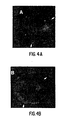
- Notch and Delta are associated within the membrane of one cell that expresses both proteins by examining the distributions of Notch and Delta in cotransfected cells. As shown in Figures 4A and 4B, these two proteins often show very similar distributions at the surface of cotransfected cells. To test whether the observed colocalization was coincidental or represented a stable interaction between Notch and Delta, we treated live cells with an excess of polyclonal anti-Notch antiserum. This treatment resulted in "patching" of Notch on the surface of expressing cells into discrete patches as detected by immunofluorescence. There was a distinct correlation between the distributions of Notch and Delta on the surfaces of these cells after this treatment ( Figures 4C and 4D), indicating that these proteins are associated within the membrane.
- Notch has a sizeable intracellular (IC) domain of ⁇ 940 amino acids.
- the IC domain includes a phosphorylation site ( Kidd et al., 1989, Genes Dev. 3, 1113-1129 ), a putative nucleotide binding domain, a polyglutamine stretch ( Wharton et al., 1985, Cell 43, 567-581 ; Kidd, et al., 1986, Mol. Cell. Biol. 6, 3094-3108 ), and sequences homologous to the yeast cdc10 gene, which is involved in cell cycle control in yeast ( Breeden and Nasmyth, 1987, Nature 329, 651-654 ).
- Notch-Delta interactions Given the size and structural complexity of this domain, we investigated whether it is required for Notch-Delta interactions. We therefore used a variant Notch construct from which coding sequences for 835 amino acids of the IC domain, including all of the structural features noted above, had been deleted (leaving 25 membrane-proximal amino acids and a novel 59 amino acid carboxyl terminus; see Experimental Procedures and Figure 1 for details).
- This construct designated ECN1
- ECN1 was expressed constitutively under control of the normal Notch promoter in transfected cells at a level lower than that observed for the metallothionein promoter constructs, but still readily detectable by immunofluorescence.
- Notch and Delta interact at the cell surface is further supported by three lines of evidence.
- Notch + cells do not aggregate homotypically suggests that Notch-Notch associations are likely to occur within a single cell and not between cells.
- homotypic Notch interactions require gene products that are not expressed in S2 cells.
- EGF domains can serve as Ca 2+ binding domains when they contain a consensus sequence consisting of Asp, Asp/Asn, Asp/Asn, and Tyr/Phe residues at conserved positions within EGF-like repeats ( Rees et al., 1988, EMBO J. 7, 2053-2061 ; Handford et al., 1990, EMBO J. 9, 475-480 ). Comparisons with a proposed consensus sequence for Ca 2+ binding have revealed that similar sequences are found within many of the EGF-like repeats of Notch ( Rees et al., 1988, EMBO J.
- Notch may have an adhesive function in concert with Delta, axonal expression of Notch may play some role in axon guidance.
- the constructs described are all derivatives of the full length Notch expression construct #1 pMtNMg (see Section 6, supra). All ligations were performed using DNA fragments cut from low melting temperature agarose gels (Sea Plaque, FMC BioProducts). The 6 kb EcoRI-XhoI fragment from pMtNMg containing the entire extracellular domain of Notch was ligated into the EcoRI-XhoI sites of the Bluescript vector (Stratagene), and named RI/XBS. All subsequent deletions and insertions of EGF repeats were performed in this subclone.
- the Notch sequence containing the EcoRI-XhoI fragment of these RI/XBS derivatives was then mixed with the 5.5 kb XhoI-XbaI fragment from pMtNMg containing the intracellular domain and 3' sequences needed for polyadenylation, and then inserted into the EcoRI-XbaI site of pRMHa-3 ( Bunch et al., 1988, Nucl. Acids Res. 16, 1043-1061 ) in a three piece ligation. All subsequent numbers refer to nucleotide coordinates of the Notch sequence according to Wharton et al. (1985, Cell 43, 567-581 ).
- RI/XBS was digested to completion with SphI and then recircularized, resulting in a 3.5 kb in-frame deletion from SphI(996) to SphI(4545).
- RI/XBS was digested to completion with ClaI and then religated, producing a 2.7 kb in-frame deletion from ClaI(1668) to ClaI(4407).
- the ligation junction was checked by double strand sequencing (as described by Xu et al., 1990, Genes Dev. 4, 464-475 ) using the Sequenase Kit (U.S. Biochemical Corp., Cleveland). We found that although the ClaI site at position 4566 exists according to the sequence, it was not recognized under our conditions by the ClaI restriction enzyme.
- construct #4-12 RI/XBS was partially digested with ClaI and then religated to produce all possible combinations of in-frame deletions: construct #4 ⁇ EGF7-17 removed the sequence between ClaI(1668) and ClaI(2820); Construct #5 ⁇ EGF9-26 removed the sequence between ClaI(1905) and ClaI(3855); construct #6 ⁇ EGF17-31 removed the sequence between ClaI(2820) and ClaI(4407); construct #7 ⁇ EGF7-9 removed the sequence between ClaI(1668) and ClaI(1905); construct #8 ⁇ EGF9-17 removed the sequence between ClaI(1905) and ClaI(2820); construct #9 ⁇ EGF17-26 removed the sequence between ClaI(2820) and ClaI(3855); construct #10 ⁇ EGF 26-30 removed the sequence between ClaI(3855) and ClaI(4407); construct #11 ⁇ EGF9-30 removed the sequence between ClaI(1905) and ClaI(4407); construct #12 ⁇ EGF 7-26 removed the sequence
- constructs #13 ⁇ Cla+EGF9-17 and #14 ⁇ Cla+EGF17-26 the ⁇ 0.9 kb fragment between ClaI(1905) and ClaI(2820), and the ⁇ 1.0 kb fragment between ClaI(2820) and ClaI(3855), respectively, were inserted into the unique ClaI site of construct #3 ⁇ Cla.
- All primers were 20-mers, and were named with the number of the nucleotide at their 5' end, according to the nucleotide coordinates of the Notch sequence in Wharton et al. (1985, Cell 43, 567-581 ), and S refers to a sense strand primer while A refers to an antisense strand primer.
- Construct #16 ⁇ Cla+EGF(9-13) used primers S1917 and A2367.
- Construct #17 ⁇ Cla+EGF(11-15) used primers S2141 and A2591.
- Construct #18 ⁇ Cla+EGF(13-17) used primers S2375 and A2819.
- Construct #19 ⁇ Cla+EGF(10-13) used primers S2018 and A2367.
- construct #25 ⁇ EGF construct R1/XBS was digested to completion with SphI(996) and partially digested with BamHI(5135). The resulting incompatible ends were joined using a synthetic linker designed to create a unique ClaI site. This produced an in frame deletion which removed all 36 EGF repeats with the exception of the first half of repeat 1. For constructs #26-29, the EGF fragments were inserted into this ClaI site as previously described for the corresponding constructs #13, 16, 19, and 23.
- construct R1/XBS was digested to completion with BglI, EcoRI and XhoI.
- the ⁇ 0.2 kb EcoRI-BglI fragment (722-948) and the ⁇ 0.7 kb BglI-XhoI (5873-6627) fragments were ligated with EcoRI-XhoI cut Bluescript vector and a synthetic linker designed to create a unique ClaI site, resulting in an in-frame deletion from BglI(941) to BglI(5873) that removed all 36 EGF repeats except for the first third of repeat 1 as well as the 3 Notch / lin -12 repeats.
- the EGF fragments were inserted into the unique ClaI site as previously described for constructs #19 and 23.
- PCR primers S1508 and A1859 based on the Xenopus Notch sequence were used to amplify EGF repeats 11 and 12 out of a Xenopus stage 17 cDNA library (library was made by D. Melton and kindly provided by M. Danilchek). The fragment was ligated into construct #3 DCla and sequenced.
- the Drosphila S2 cell line was grown and transfected as described in Section 6, supra.
- the Delta-expressing stably transformed S2 cell line L-49-6-7 (kindly established by L. Cherbas) was grown in M3 medium (prepared by Hazleton Co.) supplemented with 11% heat inactivated fetal calf serum (FCS) (Hyclone), 100 U/ml penicillin-100 ⁇ g/ml streptomycin-0.25 ⁇ g/ml fungizone (Hazleton), 2 x 10 -7 M methotrexate, 0.1 mM hypoxanthine, and 0.016 mM thymidine.
- FCS heat inactivated fetal calf serum
- Notch deletion constructs were transiently transfected into Drosophila S2 cells, induced with CuSO 4 , and then aggregated overnight at room temperature with a small amount of cells from the stably transformed Delta expressing cell line L49-6-7(Cherbas), yielding a population typically composed of ⁇ 1% Notch expressing cells and ⁇ 5% Delta expressing cells, with the remaining cells expressing neither protein.
- Constructs #20-24 represented attempts to break this domain down even further using the same PCR strategy (see Figure 7). We asked first whether both EGF repeats 11 and 12 were necessary, and second, whether the flanking sequences from EGF repeats 10 and 13 were directly involved in binding to Delta. Constructs #20 ⁇ Cla+EGF(11-13), in which EGF repeat 12 is the only entire repeat added, and #21 ⁇ Cla+EGF(10-12), in which EGF repeat 11 is the only entire repeat added, failed to mediate aggregation, suggesting that the presence of either EGF repeat 11 or 12 alone was not sufficient for Notch-Delta interactions.
- EGF repeat 12 might not be directly involved in binding to Delta but instead might contribute the minimum amount of buffer sequence needed to protect the structure of EGF repeat 11, thereby allowing interactions with Delta.
- Constructs #22-24 addressed this issue. We designed PCR primers that broke at the end of the EGF repeat and therefore were less likely to disrupt the EGF disulfide formation at the 3' ligation juncture.
- EGF repeats 11 and 12 appear to function as independent modular units which are sufficient to mediate Notch-Delta interactions in S2 cells, even in the absence of most of the extracellular domain of Notch.
- EGF domains containing a specific consensus sequence can bind Ca ++ ions ( Morita et al., 1984, J. Biol. Chem. 259, 5698-5704 ; Sugo et al., 1984, J. Biol. Chem. 259, 5705-5710 ; Rees et al., 1988, EMBO J. 7, 2053-2061 ; Handford et al., 1990, EMBO J. 9, 475-480 ). In fact, about one half of the EGF repeats in Notch, including repeats 11 and 12, conform to this consensus.
- Notch and Delta may reflect a dose sensitive interaction between their protein products. Genetic studies have indicated that the relative gene dosages of Notch and Delta are crucial for normal development. For example, Xu et al. (1990, Genes Dev. 4, 464-475 ) found that null mutations at Delta could suppress lethal interactions between heterozygous combinations of Abruptex ( Ax ) alleles, a class of Notch mutations that correlate with missense mutations within the EGF repeats ( Hartley et al., 1987, EMBO J. 6, 3407-3417 ; Kelley et al., 1987, Mol. Cell Biol. 6, 3094-3108 ).
- Notch protein expression during development have recently revealed Notch expression to be more restricted at the tissue and subcellular levels than previous studies had indicated ( Johansen et al., 1989, J. Cell Biol. 109, 2427-2440 ; Kidd et al., 1989, Genes Dev. 3, 1113-1129 ).
- Notch encodes a structurally complex transmembrane protein that has been proposed to play a pleotropic role throughout Drosophila development.
- EGF repeats 11 and 12 appear to function as an independent modular unit that is sufficient, at least in cell culture, for interactions with Delta, immediately presents the question of the role of the hypothesis is that these may also form modular binding domains for other proteins interacting with Notch at various times during development.
- EGF homologous transmembrane proteins which are structurally quite similar to Drosophila and Xenopus Notch. All four proteins contain EGF homologous repeats followed by three other cysteine rich repeats (Notch/ lin -12 repeats) in the extracellular domain, a single transmembrane domain, and six cdc10/ankyrin repeats in the intracellular region.
- lin -12 and glp -1 probably encode distinct members of the same gene family. Comparison of the predicted protein products of lin -12 and glp -1 with Notch reveal specific differences despite an overall similar organization of structural motifs. The most obvious difference is that lin -12 and glp -1 proteins contain only 13 and 10 EGF repeats, respectively, as compared to the 36 for both Xenopus and Drosophila Notch.
- EGF repeats 11 and 12 of Notch there are no two contiguous EGF repeats in the lin -12 or glp -1 proteins exhibiting the Ca ++ binding consensus sequence, nor any two contiguous repeats exhibiting striking similarity to EGF repeats 11 and 12 of Notch, again suggesting that the lin -12 and glp -1 gene products are probably functionally distinct from Notch.
- EGF repeats 11 and 12 of Notch form a discrete Delta binding unit represents the first concrete evidence supporting the idea that each EGF repeat or small subset of repeats may play a unique role during development, possibly through direct interactions with other proteins.
- the homologies seen between the adhesive domain of Delta and Serrate suggest that the homologous portion of Serrate is "adhesive" in that it mediates binding to other toporythmic proteins.
- the gene scabrous which encodes a secreted protein with similarity to fibrinogen, may interact with Notch.
- EGF repeat multiple copies of other structural motifs commonly occur in a variety of proteins.
- One relevant example is the cdc10/ankyrin motif, six copies of which are found in the intracellular domain of Notch.
- Ankyrin contains 22 of these repeats.
- repeated arrays of structural motifs may in general represent a linear assembly of a series of modular protein binding units.
- Notch may interact with a number of different ligands in a precisely regulated temporal and spacial pattern throughout development.
- Such context specific interactions with extracellular proteins could be mediated by the EGF and Notch / lin -12 repeats, while interactions with cytoskeletal and cytoplasmic proteins could be mediated by the intracellular cdc 10/ankyrin motifs.
- the S2 Drosophila cell line ( Schneider, 1972, J. Embryol. Exp. Morph. 27, 353-365 )) used in these experiments was grown as described in Section 6.
- Antisera and antibodies employed included mouse polyclonal anti-Delta sera raised against a Delta ELR array segment that extends from the fourth through ninth ELRs (see Section 6); rat polyclonal anti-Delta sera raised against the same Delta segment (see Section 6); rat polyclonal anti-Notch sera raised against a Notch ELR array segment that extends from the fifth through thirteenth ELRs; mouse monoclonal antibody C17.9C6 (see Section 6), which recognizes the Notch intracellular domain; and mouse monoclonal antibody BP-104 ( Hortsch et al., 1990, Neuron 4, 697-709 ), which recognizes the long form of Drosophila neuroglian.
- Constructs employed to program expression of wild type Delta (pMTDl1) and wild type Notch (pMTNMg) are described in Section 6, supra .
- Constructs that direct expression of variant Delta proteins were generated using pMTDl1, the Dl1 cDNA cloned into Bluescript+ (pBSDl1; Kopczynski et al., 1988, Genes Dev. 2, 1723-1735 ), and pRmHa3-104 (A.J. Bieber, pers. comm.), which consists of the insertion of the 1B7A-250 cDNA into the metallothionein promoter vector pRmHa-3 ( Bunch et al., 1988, Nucl. Acids Res. 16, 1043-1061 ) and supports inducible expression of the long form of Drosophila neuroglian ( Hortsch et al., 1990, Neuron 4, 697-709 ).
- constructs were made as follows: Del(Sca-Nae) - Cut pBSDl1 with SalI (complete digest) and Scal (partial), isolate vector-containing fragment. Cut pBSDl1 with NaeI (partial) and SalI (complete), isolate Delta carboxyl-terminal coding fragment. Ligate fragments, transform, and isolate clones. Transfer EcoRI insert into pRmHa-3. Del(Bam-Bg1) - Cut pBSDl1 with BglII (complete) and BamHI (partial), fill ends with Klenow DNA polymerase, ligate, transform, and isolate clones. Transfer EcoRI insert into pRmHa-3.
- Del(ELR4-ELR5) - pBSDl1 was digested to completion with BglII and partially with PstI.
- the 5.6 kb vector-containing fragment was isolated, circularized using T4 DNA ligase in the presence of a 100X molar excess of the oligonucleotide 5'-GATCTGCA-3', and transformed and clones were isolated.
- the resulting EcoRI insert was then transferred into pRmHa-3.
- NAE A clone that contained multiple inserts of the linker was isolated.
- NAE A was digested to completion with BglII, and the resulting 0.4 kb and 5.4 kb fragments were isolated, ligated and transformed, and clones were isolated.
- STU B was digested to completion with BglII, and the resulting 0.6 kb and 5.2 kb fragments were isolated, ligated and transformed, and clones were isolated.
- B Cells that express neuroglian-based constructs were detected using a monoclonal antibody that recognizes the intracellular domain of neuroglian (see Materials and Methods).
- B indicates that cells were aggregated in a 12-well microtiter plate (see Materials and Methods).
- D Data for individual cell types i.e., Delta + and Notch + ) in aggregates and unaggregated were not recorded.
- Delta amino acids (AA) 1-230 is the current minimum sequence interval defined as being sufficient for interaction with Notch. This is based on the success of NG2-Notch aggregation.
- Delta AA198-230 are critical because their deletion in the NG1 construct inactivated the Notch-binding activity observed for the NG2 construct. Also within this interval, Delta AA32-198 are critical because their deletion in the NG4 construct also inactivated the Notch-binding activity observed for the NG3 construct.
- the importance of Delta AA192-230 is also supported by the observation that the Del(ELR1-ELR3) variant, which contains all Delta amino acids except AA231-331, possessed Notch-binding activity, while the Del(Bam-Bg1) variant, which contains all Delta amino acids except AA192-331, was apparently inactivated for Notch-binding activity.
- Conformation and/or primary sequence in the vicinity of Delta AA197/198 is apparently critical because a multimeric insertion of the tetrapeptide-Arg-Lys-Ile-Phe [in one letter code (see e.g. Lehninger et al., 1975, Biochemistry, 2d ed., p. 72), RKIF] (SEQ ID NO:30) - between these two residues, as in the Ins(Nae)A construct, inactivated the Notch-binding activity observed with wild type Delta.
- the observation that the Del(ELR1-ELR3) construct supported aggregation implies that ELR1-ELR3 are not required for Delta-Notch interaction; the observation that the Del(ELR4-ELR5) construct supported aggregation implies that ELR4 and ELR5 are not required for Delta-Notch interaction, and the observation that the Ter(Dde) construct supported aggregation implies that the Delta intracellular domain is not required for Delta-Notch interaction.
- B Transfected cells were incubated under aggregation conditions overnight, then diluted into the appropriate volume of log-phase S2 cells in the presence of inducer and incubated under aggregation conditions for an additional four to six hours.
- C Transfected cells to which inducer had been added were diluted into the appropriate volume of logphase S2 cells to which inducer had been added, and the cell mixture was incubated under aggregation conditions overnight.
- Deletion of Delta AA32-198 [Del(Sca-Nae)] or Delta AA192-331 [Del(Bam-Bgl)] from the full-length Delta protein eliminated the Delta-Delta interaction.
- Deletion of Delta AA231-331 [Del(ELR1-ELR3)] did not eliminate the Delta-Delta interaction. Therefore, sequences within the Delta AA32-230 are required for the Delta-Delta interaction.
- Conformation and/or primary sequence in the vicinity of Delta AA197/198 is apparently critical for the Delta-Delta interaction because a multimeric insertion of the tetrapeptide -Arg-Lys-Ile-Phe- (SEQ ID NO:30) between these two residues, as in the Ins(Nae)A construct, inactivated Delta-Delta interaction.
- the observation that the Del(ELR1-ELR3) construct could support aggregation implies that ELR1-ELR3 are not required for Delta-Delta interaction; the observation that the Del(ELR-ELR5) construct supported aggregation implies that ELR4 and ELR5 are not required for Delta-Delta interaction, and the observation that the Ter(Dde) construct supported aggregation implies that the Delta intracellular domain is not required for Delta-Delta interaction.
- Delta variants in which short, in-frame, translatable linker insertions were introduced into the Delta amino terminus (i.e., NAE B and STU B; Figure 9, Table VI A).
- STU B variant pentapeptide GKIFP
- NAE B variant tetrapeptide RKIF between Delta residues 198 and 199
- Delta protein can sometimes be found within cells that have been programmed to express Notch, but not Delta.
- We conduct heterotypic aggregation assays by mixing initially separate populations of S2 cells that have been independently transfected with expression constructs that program expression of either Delta or Notch. Yet, we often detect punctate staining of Delta within Notch-expressing cells found in heterotypic aggregates using Delta-specific antisera. Our observations are consistent with Delta binding directly to Notch at the cell surface and subsequent clearance of this Delta-Notch complex from the cell surface via endocytosis.
- deletion of AA32-198 also inactivates Notch-binding activity suggests that sequences amino-proximal to AA198 are also required, although the deleterious impact of this deletion could result from the removal of additional amino acids in the immediate vicinity of AA198.
- Sequences within Delta sufficient for interaction with Notch can be grouped into three subdomains - N1, N2, and N3 - that differ in their respective contents of cysteine residues (Figure 10, SEQ ID NO:3).
- the N1 and N3 domains each contain six cysteine residues, while the N2 domain contains none.
- the even number of cysteines present in N1 and N3, respectively allows for the possibility that the respective structures of these subdomains are dictated, in part, by the formation of particular disulfide bonds.
- the broad organizational pattern of the Delta amino-terminus is also generally analogous to that of the extracellular domain of the vertebrate EGF receptor ( Lax et al., 1988, Mol. Cell. Biol. 8, 1970-1978 ), in which sequences believed to interact with EGF are bounded by two cysteine-rich subdomains.
- Delta-Notch interaction could involve contacts between the Delta EGF-binding domain with either of two regions within Notch, depending on whether the interaction were between molecules that reside on opposing membranes or within the same membrane ( Figure 11).
- the cell aggregation assays which presumably detect the interaction of molecules in opposing membranes, imply that the Delta EGF-binding domain interacts with Notch EGF-like repeats 11 and 12 (see examples supra ). If tandem arrays of EGF-like motifs do form rod-like structures ( Engel, 1989, FEBS Lett.
- the estimated displacement of the Delta EGF-binding domain from the cell surface would presumably be sufficient to accommodate the rigid array of Notch EGF-like repeats 1-10. It is also interesting to note that the displacement of the Delta EGF-binding domain from the cell surface could place this domain in the vicinity of the Notch EGF-like repeats (25-29) that are affected by Abruptex mutations ( Hartley et al., 1987, EMBO J. 6, 3407-3417 ; Kelley et al., 1987, Mol. Cell. Biol. 6, 3094-3108 ) and could allow for interaction of Delta and Notch proteins present within the same membrane.
- S2 cells were transfected with a Serrate expression construct and mixed with Notch expressing cells in our aggregation assay.
- Serrate expression construct a synthetic primer containing an artificial BamHI site immediately 5' to the initiator AUG at position 442 (all sequence numbers are according to Fleming et al., 1990, Genes & Dev. 4:2188-2201 ) and homologous through position 464, was used in conjunction with a second primer from position 681-698 to generate a DNA fragment of ⁇ 260 base pairs. This fragment was cut with BamHI and KpnI (position 571) and ligated into Bluescript KS+ (Stratagene).
- BTSer5'PCR was checked by sequencing, then cut with KpnI.
- the Serrate KpnI fragment (571-2981) was inserted and the proper orientation selected, to generate BTSer5'PCR-Kpn.
- the 5' SacII fragment of BTSer5'PCR-Kpn (SacII sites in Bluescript polylinker and in Serrate (1199)) was isolated and used to replace the 5' SacII fragment of cDNA C1 ( Fleming et al., 1990, Genes & Dev. 4:2188-2201 ), thus regenerating the full length Serrate cDNA minus the 5' untranslated regions.
- This insert was isolated by a SalI and partial BamHI digestion and shuttled into the BamHI and SalI sites of pRmHa-3 to generate the final expression construct, Ser-mtn.
- Notch and Serrate appear to aggregate less efficiently than Notch and Delta, perhaps because the Notch-Serrate interaction is weaker. For example, when scoring Notch-Delta aggregates, we detect ⁇ 40% of all Notch expressing cells in clusters with Delta expressing cells ( Figure 6, #1 pMtNMg) and ⁇ 40% of all Delta expressing cells in contact with Notch expressing cells. For Notch-Serrate, we find only ⁇ 20% of all Notch expressing cells ( Figure 6; pMtNMg) and -15% of all Serrate expressing cells in aggregates.
- the 400 bp fragment obtained in this manner was then used as a probe with which to screen the same library for human Notch clones.
- the original screen yielded three unique clones, hN3k, hN2K, and hN5k, all of which were shown by subsequent sequence analysis to fall in the 3' end of human Notch ( Figure 17).
- a second screen using the 5' end of hN3k as probe was undertaken to search for clones encompassing the 5' end of human Notch.
- One unique clone, hN4k was obtained from this screen, and preliminary sequencing data indicate that it contains most of the 5' end of the gene (Figure 17).
- clones hN4k, hN3k and hN5k encompass about 10 kb of the human Notch homolog, beginning early in the EGF-repeats and extending into the 3' untranslated region of the gene. All three clones are cDNA inserts in the EcoRI site of pBluescript SK - (Stratagene).
- the host E . coli strain is XL1-Blue (see Maniatis, T., 1990, Molecular Cloning, A Laboratory Manual, 2d ed., Cold Spring Harbor Laboratory, Cold Spring Harbor, New York, p. A12).
- the complete nucleotide sequences of the human Notch cDNA contained in hN3k and hN5k was determined by the dideoxy chain termination method using the Sequenase® kit (U.S. Biochemical Corp.). Those nucleotide sequences encoding human Notch, in the appropriate reading frame, were readily identified since translation in only one out of the three possible reading frames yields a sequence which, upon comparison with the published Drosophila Notch deduced amino acid sequence, yields a sequence with a substantial degree of homology to the Drosophila Notch sequence. Since there are no introns, translation of all three possible reading frames and comparison with Drosophila Notch was easily accomplished, leading to the ready identification of the coding region.
- Clone hN3k encodes a portion of a Notch polypeptide starting at approximately the third Notch / lin -12 repeat to several amino acids short of the carboxy-terminal amino acid.
- Clone hN5k encodes a portion of a Notch polypeptide starting approximately before the cdc10 region through the end of the polypeptide, and also contains a 3' untranslated region.
- the amino acid sequence shown in Figure 24 was compared with the predicted sequence of the TAN-1 polypeptide shown in Figure 2 of Ellisen et al., August 1991, Cell 66:649-661 . Differences were found between the deduced amino acid sequences.
- the deduced Notch polypeptide of hN5k is 79% identical to the TAN-1 polypeptide (64% identical to Drosophila Notch) in the cdc10 region that encompasses both the cc10 motif (TAN-1 amino acids 1860 to 2217) and the well-conserved flanking regions (Fig. 25).
- the cdc10 region covers amino acids 1860 through 2217 of the TAN-1 sequence.
- the hN5k encoded polypeptide is 65% identical to the TAN-1 polypeptide (44% identical to Drosophila Notch) at the carboxy-terminal end of the molecule containing a PEST (proline, glutamic acid, serine, threonine)-rich region (TAN-1 amino acids 2482 to 2551) (Fig. 25B).
- PEST proline, glutamic acid, serine, threonine
- TAN-1 amino acids 2482 to 2551 Fig. 25B.
- the stretch of 215 amino acids lying between the aforementioned regions is not well conserved among any of the Notch -homologous clones represented by hN3k, hN5k, and TAN-1.
- Xenopus Notch Xotch
- rat Notch SEQ ID NO:36
- TAN-1 SEQ ID NO:37
- the pGEX vector system was used to obtain expression of human Notch fusion (chimeric) proteins from the constructs described below.
- the cloned Notch DNA in each case was inserted, in phase, into the appropriate pGEX vector.
- Each construct was then electroporated into bacteria ( E . coli ), and was expressed as a fusion protein containing the Notch protein sequences fused to the carboxyl terminus of glutathione S-transferase protein. Expression of the fusion proteins was confirmed by analysis of bacterial protein extracts by polyacrylamide gel electrophoresis, comparing protein extracts obtained from bacteria containing the pGEX plasmids with and without the inserted • Notch DNA.
- the fusion proteins were soluble in aqueous solution, and were purified from bacterial lysates by affinity chromatography using glutathione-coated agarose (since the carboxyl terminus of glutathione S-transferase binds to glutathionine).
- the expressed fusion proteins were bound by an antibody to Drosophila Notch, as assayed by Western blotting.
- these fusion proteins are used to make either polyclonal and/or monoclonal antibodies to human Notch.
Abstract
Description
- This invention was made in part with government support under Grant numbers GM 19093 and NS 26084 awarded by the Department of Health and Human Services. The government has certain rights in the invention.
- The present invention relates to the human Notch genes and their encoded products. The invention also relates to sequences (termed herein "adhesive sequences") within the proteins encoded by the human Notch genes which mediate heterotypic binding to sequences within proteins encoded by other toporythmic genes. Such genes include but are not limited to Delta, and Serrate.
- Genetic analyses in Drosophila have been extremely useful in dissecting the complexity of developmental pathways and identifying interacting loci. However, understanding the precise nature of the processes that underlie genetic interactions requires a knowledge of the biochemical properties of the protein products of the genes in question.
- Null mutations in any one of the zygotic neurogenic loci -- Notch (N), Delta (Dl), mastermind (mam), Enhancer of Split (E(spl), neuralized (neu), and big brain (bib) --result in hypertrophy of the nervous system at the expense of ventral and lateral epidermal structures. This effect is due to the misrouting of epidermal precursor cells into a neuronal pathway, and implies that neurogenic gene function is necessary to divert cells within the neurogenic region from a neuronal fate to an epithelial fate. Studies that assessed the effects of laser ablation of specific embryonic neuroblasts in grasshoppers (Doe and Goodman 1985, Dev. Biol. 111, 206-219) have shown that cellular interactions between neuroblasts and the surrounding accessory cells serve to inhibit these accessory cells from adopting a neuroblast fate. Together, these genetic and developmental observations have led to the hypothesis that the protein products of the neurogenic loci function as components of a cellular interaction mechanism necessary for proper epidermal development (Artavanis-Tsakonas, 1988, Trends Genet. 4, 95-100).
- Sequence analyses (Wharton et al., 1985, Cell 43, 567-581; Kidd et al., 1986, Mol. Cell. Biol. 6, 3094-3108; Vassin et al., 1987, EMBO J. 6, 3431-3440; Kopczynski et al., 1988, Genes Dev. 2, 1723-1735) have shown that two of the neurogenic loci, Notch and Delta, appear to encode transmembrane proteins that span the membrane a single time. The Notch gene encodes a ∼300 kd protein (we use "Notch" to denote this protein) with a large N-terminal extracellular domain that includes 36 epidermal growth factor (EGF)-like tandem repeats followed by three other cysteine-rich repeats, designated Notch/lin-12 repeats (Wharton et al., 1985, Cell 43, 567-581; Kidd et al., 1986, Mol. Cell Biol. 6, 3094-3108; Yochem et al., 1988, Nature 335, 547-550). Delta encodes a ∼100 kd protein (we use "Delta" to denote DLZM, the protein product of the predominant zygotic and maternal transcripts; Kopczynski et al., 1988, Genes Dev. 2, 1723-1735) that has nine EGF-like repeats within its extracellular domain (Vassin et al., 1987, EMBO J. 6, 3431-3440; Kopczynski et al., 1988, Genes Dev. 2, 1723-1735). Although little is known about the functional significance of these repeats, the EGF-like motif has been found in a variety of proteins, including those involved in the blood clotting cascade (Furie and Furie, 1988, Cell 53, 505-518). In particular, this motif has been found in extracellular proteins such as the blood clotting factors IX and X (Rees et al., 1988, EMBO J. 7, 2053-2061; Furie and Furie, 1988, Cell 53, 505-518), in other Drosophila genes (Knust et al., 1987, EMBO J. 761-766; Rothberg et al., 1988, Cell 55, 1047-1059), and in some cell-surface receptor proteins, such as thrombomodulin (Suzuki et al., 1987, EMBO J. 6, 1891-1897) and LDL receptor (Sudhof et al., 1985, Science 228, 815-822). A protein binding site has been mapped to the EGF repeat domain in thrombomodulin and urokinase (Kurosawa et al., 1988, J. Biol. Chem 263, 5993-5996; Appella et al., 1987, J. Biol. Chem. 262, 4437-4440).
- An intriguing array of interactions between Notch and Delta mutations has been described (Vassin, et al., 1985, J. Neurogenet. 2, 291-308; Shepard et al., 1989, Genetics 122, 429-438; Xu et al., 1990, Genes Dev., 4, 464-475). A number of genetic studies (summarized in Alton et al., 1989, Dev. Genet. 10, 261-272) has indicated that the gene dosages of Notch and Delta in relation to one another are crucial for normal development. A 50% reduction in the dose of Delta in a wild-type Notch background causes a broadening of the wing veins creating a "delta" at the base (Lindsley and Grell, 1968, Publication Number 627, Washington, D.C., Carnegie Institute of Washington). A similar phenotype is caused by a 50% increase in the dose of Notch in a wild-type Delta background (a "Confluens" phenotype; Welshons, 1965, Science 150, 1122-1129). This Delta phenotype is partially suppressed by a reduction in the Notch dosage. Recent work in our laboratories has shown that lethal interactions between alleles that correlate with alterations in the EGF-like repeats in Notch can be rescued by reducing the dose of Delta (Xu et al., 1990, Genes Dev. 4, 464-475). Xu et al. (1990, Genes Dev. 4, 464-475) found that null mutations at either Delta or mam suppress lethal interactions between heterozygous combinations of certain Notch alleles, known as the Abruptex (Ax) mutations. Ax alleles are associated with missense mutations within the EGF-like repeats of the Notch extracellular domain (Kelley et al., 1987, Cell 51, 539-548; Hartley et al., 1987, EMBO J. 6, 3407-3417).
- Notch is expressed on axonal processes during the outgrowth of embryonic neurons (Johansen et al., 1989, J. Cell Biol. 109, 2427-2440; Kidd et al., 1989, Genes Dev. 3, 1113-1129).
- A study has shown that certain Ax alleles of Notch can severely alter axon pathfinding during sensory neural outgrowth in the imaginal discs, although it is not yet known whether aberrant Notch expression in the axon itself or the epithelium along which it grows is responsible for this defect (Palka et al., 1990, Development 109, 167-175).
- The present invention is defined in the claims. In a specific embodiment of the invention, the adhesive fragment of Notch is that fragment comprising the Notch sequence most homologous to Drosophila Notch EGF-
like repeats - The present specification describes the nucleotide sequences of the human Notch and Delta genes, and amino acid sequences of their encoded proteins, as well as fragments thereof containing an antigenic determinant or which are functionally active. The specification also describes fragments (termed herein "adhesive fragments"), and the sequences thereof, of the proteins ("toporythmic proteins") encoded by toporythmic genes which mediate homotypic or heterotypic binding to toporythmic proteins. Toporythmic genes, as used herein, refers to the genes Notch, Delta, and Serrate, as well as other members of the Delta/Serrate family which may be identified, e.g., by the methods described in Section 5.3, infra. Antibodies to human Notch and to adhesive fragments are additionally described. In the following description, reference is made to a number of toporythmic genes, including Notch, Delta and Serrate. However, the present invention relates only to Notch-related proteins, fragments, chimeric proteins, nucleic acids, vectors, host cells and methods as defined in the claims. The other toporyhtmic genes are described to assist in understanding the present invention.
- In specific examples, the adhesive fragment of Delta mediating heterotypic binding is that fragment comprising the sequence most homologous to Drosophila Delta amino acids 1-230; the adhesive fragment of Delta mediating homotypic binding is that fragment comprising the sequence most homologous to Drosophila Delta amino acids 32-230; and the adhesive fragment of Serrate is that fragment comprising the sequence most homologous to Drosophila Serrate amino acids 85-283 or 79-282.
- As used herein, the following terms shall have the meanings indicated:
- AA = amino acid
- EGF = epidermal growth factor
- ELR = EGF-like (homologous) repeat
- IC = intracellular
- PCR = polymerase chain reaction
- As used herein, underscoring the name of a gene shall indicate the gene, in contrast to its encoded protein product which is indicated by the name of the gene in the absence of any underscoring. For example, "Notch" shall mean the Notch gene, whereas "Notch" shall indicate the protein product of the Notch gene.
- Figure 1. Expression Constructs and Experimental Design for Examining Notch-Delta Interactions. S2 cells at log phase growth were transiently transfected with one of the three constructs shown. Notch encoded by the MGlla minigene (a cDNA/genomic chimeric construct: cDNA-derived sequences are represented by stippling, genomically derived sequences by diagonal-hatching (Ramos et al., 1989, Genetics 123, 337-348)) was expressed following insertion into the metallothionein promoter vector pRmHa-3 (Bunch et al., 1988, Nucl. Acids Res. 16, 1043-1061). Delta encoded by the Dl1 cDNA (Kopczynski et al., 1988, Genes Dev. 2, 1723-1735) was expressed after insertion into the same vector. The extracellular Notch (ECN1) variant was derived from a genomic cosmid containing the complete Notch locus (Ramos et al., 1989, Genetics 123, 337-348) by deleting the coding sequence for amino acids 1790-2625 from the intracellular domain (denoted by δ; Wharton et al., 1985, Cell 43, 567-581), leaving 25 membrane-proximal residues from the wild-type sequence fused to a novel 59 amino acid tail (see Experimental Procedures, Section 6.1, infra). This construct was expressed under control of the Notch promoter region. For constructs involving the metallothionein vector, expression was induced with CuSO4 following transfection. Cells were then mixed, incubated under aggregation conditions, and scored for their ability to aggregate using specific antisera and immunofluorescence microscopy to visualize expressing cells. MT, metallothionein promoter; ATG, translation start site; TM, transmembrane domain; 3' N, Notch gene polyadenylation signal; 3' Adh, polyadenylation signal from Adh gene; 5' N, Notch gene promoter region.
- Figure 2. Expression of Notch and Delta in Cultured Cells. (A) Lysates of nontransfected (S2) and Notch-transfected (N) cells induced with 0.7 mM CuSO4 for 12-16 hr were prepared for sodium dodecyl sulfate polyacrylamide gel electrophoresis (SDS-PAGE), run on 3%-15% gradient gels, and blotted to nitrocellulose. Notch was visualized using a monoclonal antibody (MAb C17.9C6) against the intracellular domain of Notch. Multiple bands below the major band at 300 kd may represent degradation products of Notch. (B) Lysates of nontransfected (S2) and Delta-transfected (Dl) cells visualized with a monoclonal antibody (MAb 201) against Delta. A single band of ~105 kd is detected. In both cases, there is no detectable endogenous Notch or Delta in the S2 cell line nor are there cross-reactive species. In each lane, 10 µl of sample (prepared as described in Experimental Procedures) was loaded.
- Figure 3. S2 Cells That Express Notch and Delta Form Aggregates. In all panels, Notch is shown in green and Delta in red.
- (A) A single Notch+ cell. Note the prominent intracellular stain, including vesicular structures as well as an obviously unstained nucleus.
- (A) Bright-field micrograph of same field, showing specificity of antibody staining.
- (B) A single Delta+ cell. Staining is primarily at the cell surface.
- (B) Bright-field micrograph of same field.
- (C) Aggregate of Delta+ cells from a 24 hr aggregation experiment. Note against that staining is primarily at the cell surface.
- (D)-(F) An aggregate of Notch+ and Delta+ cells formed from a 1:1 mixture of singly transfected cell populations that was allowed to aggregate overnight at room temperature. (D) shows Notch+ cells in this aggregate; (E) shows Delta+ cells; and (F) is a double exposure showing both cell types. Bands of Notch and Delta are prominent at points of contact between Notch+ and Delta+ cells (arrows). In (F), these bands appear yellow because of the coincidence of green and red at these points. The apparently doubly stained single cell (*) is actually two cells (one on top of the other), one expressing Notch and the other Delta.
- (G) and (H) Pseudocolor confocal micrographs of Notch+-Delta+ cell aggregates. Note that in (G) extensions (arrows) formed by at least two Delta+ cells completely encircle the Notch+ cell in the center of the aggregate. (H) shows an aggregate formed from a 2 hr aggregation experiment performed at 4°C. Intense bands of Notch are apparent within regions of contact with Delta+ cells.
- (I) An aggregate composed of Delta+ cells and cells that express only the extracellular domain of Notch (ECN1 construct). Scale bar = 10 µm.
- Figure 4. Notch and Delta are Associated in Cotransfected Cells. Staining for Notch is shown in the left column (A, C, and E) and that for Delta is shown in the right column (B, D, and F).
- (A) and (B) S2 cell cotransfected with both Notch and Delta constructs. In general, there was a good correlation between Notch and Delta localization at the cell surface (arrows).
- (C) and (D) Cotransfected cells were exposed to polyclonal anti-Notch antiserum (a 1:250 dilution of each anti-extracellular domain antiserum) for 1 hr at room temperature before fixation and staining with specific antisera. Note punctate staining of Notch and Delta and the correlation of their respective staining (arrows).
- (E) and (F) Cells cotransfected with the extracellular Notch (ECN1) and Delta constructs, induced, and then patched using anti-Notch polyclonal antisera. There was a close correlation between ECN1 and Delta staining at the surface as observed for full-length Notch. Scale bar = 10 µm.
- Figure 5. Coimmunoprecipitation Shows that Delta and Notch are Associated in Lysates from Transfected S2 and Drosophila Embryonic Cells. In all experiments, Delta was precipitated from NP-40/deoxycholate lysates using a polyclonal anti-Delta rat antiserum precipitated with fixed Staph A cells, and proteins in the precipitated fraction were visualized on Western blots (for details, see Experimental Procedures).
Lanes Lanes 4 and 6: Delta visualized using MAb 201. - In (A),
lanes Lane 1 shows a polyclonal anti-Delta immunoprecipitation from cells that express Notch alone visualized for Notch. No Notch was detectable in this sample, indicating that the polyclonal anti-Delta does not cross-react with Notch.Lane 2 shows Notch-Delta cotransfected cells immunoprecipitated with Staph A without initial treatment with anti-Delta antiserum and visualized for Notch, demonstrating that Notch is not precipitated nonspecifically by the Staph A or secondary antibody.Lane 3 shows protein precipitated with anti-Delta antiserum visualized for Delta (Dl), andlane 4 shows the same sample visualized for Notch (N).Lane 4 shows that Notch coprecipitates with immunoprecipitated Delta. Note that Notch appears as a doublet as is typical for Notch in immunoprecipitates. - (B) shows the same experiment using embryonic lysates rather than transfected cell lysates.
Lane 5 shows protein precipitated with anti-Delta antiserum visualized for Delta (Dl), andlane 6 shows the same sample visualized for Notch (N). These lanes demonstrate that Notch and Delta are stably associated in embryo lysates. Bands (in all lanes) below the Delta band are from Staph A (SA) and the anti-Delta antiserum heavy (H) and light (L) chains. - Figure 6. Notch Expression Constructs and the Deletion Mapping of the Delta/Serrate Binding Domain. S2 cells in log phase growth were transiently transfected with the series of expression constructs shown; the drawings represent the predicted protein products of the various Notch deletion mutants created. All expression constructs were derived from
construct # 1 pMtNMg. Transiently transfected cells were mixed with Delta expressing cells from the stably transformed line L49-6-7 or with transiently transfected Serrate expressing cells, induced with CuSO4, incubated under aggregation conditions and then scored for their ability to aggregate using specific antisera and immunofluorescence microscopy. Aggregates were defined as clusters of four or more cells containing both Notch and Delta/Serrate expressing cells. The values given for % Aggregation refer to the percentage of all Notch expressing cells found in such clusters either with Delta (Dl) (left column) or with Serrate (Ser) (right column). The various Notch deletion constructs are represented diagrammatically with splice lines indicating the ligation junctions. Each EGF repeat is denoted as a stippled rectangular box and numbers of the EGF repeats on either side of a ligation junction are noted. At the ligation junctions, partial EGF repeats produced by the various deletions are denoted by open boxes and closed brackets (for example see #23 ΔCla+EGF(10-12)). Constructs #3-13 represent the ClaI deletion series. As diagrammed, four of the ClaI sites, inrepeats closed box repeat 31; again see Figure 7). Inconstruct # 15 split,EGF repeat 14 which carries the split point mutation, is drawn as a striped box. Inconstruct # 33 ΔCla+XEGF(10-13), the Xenopus Notch derived EGF repeats are distinguished from Drosophila repeats by a different pattern of shading. SP, signal peptide; EGF, epidermal growth factor repeat; N, Notch/lin-12 repeat; TM, transmembrane domain; cdc10, cdc10/ankyrin repeats; PA, putative nucleotide binding consensus sequence; opa, polyglutamine stretch termed opa; Dl, Delta; Ser, Serrate. - Figure 7. Detailed Structure of Notch Deletion Constructs #19-24: Both EGF Repeats 11 and 12 are Required for Notch-Delta Aggregation. EGF repeats 10-13 are diagrammed at the top showing the regular spacing of the six cysteine residues (C). PCR products generated for these constructs (names and numbers as given in Figure 6) are represented by the heavy black lines and the exact endpoints are noted relative to the various EGF repeats. Ability to aggregate with Delta is recorded as (+) or (-) for each construct. The PCR fragments either break the EGF repeats in the middle, just after the third cysteine in the same place as four out of the five ClaI sites, or exactly in between two repeats in the same place as the most C-terminal ClaI site.
- Figure 8. Comparison of Amino Acid Sequence of EGF Repeats 11 and 12 from Drosophila and Xenopus Notch. The amino acid sequence of EGF repeats 11 and 12 of Drosophila Notch (Wharton et al., 1985, Cell 43:567-581; Kidd et al., 1986, Mol. Cell Biol. 6:3094-3108) is aligned with that of the same two EGF repeats from Xenopus Notch (Coffman et al., 1990, Science 249:1438-1441). Identical amino acids are boxed. The six conserved cysteine residues of each EGF repeat and the Ca++ binding consensus residues (Rees et al., 1988, EMBO J. 7:2053-2061) are marked with an asterisk (*). The leucine to proline change found in the Xenopus PCR clone that failed to aggregate is noted underneath.
- Figure 9. Constructs Employed in this Study. Schematic diagrams of the Delta variants defined in Table IV are shown. Extracellular, amino-proximal terminus is to the left in each case. S, signal peptide; "EGF", EGF-like motifs; M, membrane-spanning helix; H, stop-transfer sequence; solid lines, other Delta sequences; hatched lines, neuroglian sequences. Arrowheads indicate sites of translatable linker insertions. Sca, ScaI; Nae, NaeI; Bam, BamHI; Bgl, BglII; ELR, EGF-like repeat; Bst, BstEII; Dde, DdeI; Stu, StuI; NG1-NG5, Delta-neuroglian chimeras.
- Figure 9A. Dependence of Aggregation on Input DNA Amounts. A, Heterotypic aggregation observed using S2 cell populations transiently transfected, respectively, with varied amounts of pMTDl1 DNA (2, 4, 10 or 20 µg/plate) that were subsequently incubated under aggregation conditions with S2 cell populations transiently transfected with a constant amount of pMtNMg DNA (20 µg/plate). Data presented are mean fraction (%) of Delta cells in aggregates of four or more cells ± standard error for each input DNA amount (N = 3 replicates, except 2 µg and 10 µg inputs for which N = 2). A minimum of 100 Delta-expressing cells were counted for each replicate. B, Homotypic aggregation observed using S2 cell populations transiently transfected, respectively, with varied amounts of pMTDl1 DNA (2, 4, 10 or 20 µg/plate) that were subsequently incubated under aggregation conditions. Data presented are mean fraction (%) of Delta cells in aggregates of four or more cells ± standard error for each input DNA amount (N = 3 replicates). A minimum of 500 Delta-expressing cells were counted for each replicate.
- Figure 10. Delta-Serrate Amino-Terminal Sequence Alignment. Residues are numbered on the basis of conceptual translation of Delta (Dl, upper sequence (SEQ ID NO:3); beginning at
amino acid 24, ending at amino acid 226) and Serrate (Ser, lower sequence (SEQ ID NO:4); beginning atamino acid 85, ending at amino acid 283) coding sequences. Vertical lines between the two sequences indicates residues that are identical within the Delta and Serrate sequences, as aligned. Dots represent gaps in the alignment. Boxes enclose cysteine residues within the aligned regions. N1, amino-proximal domain 1; N2, amino-proximal domain 2; N3, amino-proximal domain 3. Translatable insertions associated with STU B [replacement of Delta amino acid 132 (A) with GKIFP] and NAE B [insertion of RKIF between Delta amino acid 197 and amino acid 198] constructs, respectively, are depicted above the wild type Delta sequence. - Figure 11. Potential Geometries of Delta-Notch Interactions. A, Potential register of Delta (left) and Notch (right) molecules interacting between opposing plasma membranes. B, Potential register of Delta (left) and Notch (right) molecules interacting within the same plasma membranes. ELR, EGF-like repeat; open boxes, EGF-like repeats; dotted boxes, LNR repeats; solid boxes, membrane-spanning helices. Delta amino-terminal domain and Delta and Notch intracellular domains represented by ovals.
- Figure 12. Potential Geometries of Delta-Delta Interactions. A and B, Potential register of Delta molecules interacting between opposing plasma membranes. B, Potential register of Delta molecules interacting within the same plasma membranes. Open boxes, EGF-like repeats; solid boxes, membrane-spanning helices. Delta amino-terminal extracellular and intracellular domains represented by ovals.
- Figure 13. Primary Nucleotide Sequence of the Delta cDNA Dl1 (SEQ ID NO:5) and Delta amino acid sequence (SEQ ID NO:6) The DNA sequence of the 5'-3' strand of the Dl1 cDNA is shown, which contains a number of corrections in comparison to that presented in Kopczynksi et al. (1988, Genes Dev. 2, 1723-1735).
- Figure 14. Primary Nucleotide Sequence of the Neuroglian cDNA 1B7A-250 (SEQ ID NO:7). This is the DNA sequence of a portion of the 5'-3' strand of the 1B7A-250 cDNA (A.J. Bieber, pers. comm.; Hortsch et al., 1990, ).
Nucleotide 2890 corresponds to the first nucleotide of an isoleucine codon that encodes amino acid 952 of the conceptually translated neuroglian-long form protein. - Figure 15. Nucleic Acid Sequence Homologies Between Serrate and Delta. A portion of the Drosophila Serrate nucleotide sequence (SEQ ID NO:8), with the encoded Serrate protein sequence (SEQ ID NO:9) written below, (Fleming et al., 1990, Genes & Dev. 4, 2188-2201 at 2193-94) is shown. The four regions showing high sequence homology with the Drosophila Delta sequence are numbered above the line and indicated by brackets. The total region of homology spans nucleotide numbers 627 through 1290. of the Serrate nucleotide sequence (numbering as in Figure 4 of Fleming et al., 1990, Genes & Dev. 4, 2188-2201).
- Figure 16. Primers used for PCR in the Cloning of Human Notch. The sequence of three primers used for PCR to amplify DNA in a human fetal brain cDNA library are shown. The three primers, cdc1 (SEQ ID NO:10), cdc2 (SEQ ID NO:11), and cdc3 (SEQ ID NO:12), were designed to amplify either a 200 bp or a 400 bp fragment as primer pairs cdc1/cdc2 or cdc1/cdc3, respectively. I: inosine.
- Figure 17. Schematic Diagram of Human Notch Clones. A schematic diagram of human Notch is shown. Heavy bold-face lines below the diagram show that portion of the Notch sequence contained in each of the four cDNA clones. The location of the primers used in PCR, and their orientation, are indicated by arrows.
- Figure 18. Human Notch Sequences Aligned with Drosophila Notch Sequence. Numbered vertical lines correspond to Drosophila Notch coordinates. Horizontal lines below each map show where clones lie relative to stretches of sequence (thick horizontal lines).
- Figure 19. Nucleotide Sequences of Human Notch contained in Plasmid cDNA Clone hN2k. Figure 19A: The DNA sequence (SEQ ID NO:13) of a portion of the human Notch insert is shown, starting at the EcoRI site at the 3' end, and proceeding in the 3' to 5' direction. Figure 19B: The DNA sequence (SEQ ID NO:14) of a portion of the human Notch insert is shown, starting at the EcoRI site at the 5' end, and proceeding in the 5' to 3' direction. Figure 19C: The DNA sequence (SEQ ID NO:15) of a portion of the human Notch insert is shown, starting 3' of the sequence shown in Figure 19B, and proceeding in the 5' to 3' direction. The sequences shown are tentative, subject to confirmation by determination of overlapping sequences.
- Figure 20. Nucleotide Sequences of Human Notch Contained in Plasmid cDNA clone hN3k. Figure 20A: The DNA sequence (SEQ ID NO:16) of a portion of the human Notch insert is shown, starting at the EcoRI site at the 3' end, and proceeding in the 3' to 5' direction. Figure 20B: The DNA sequence (SEQ ID NO:17) of a portion of the human Notch insert is shown, starting at the EcoRI site at the 5' end, and proceeding in the 5' to 3' direction. Figure 20C: The DNA sequence (SEQ ID NO:18) of a portion of the human Notch insert is shown, starting 3' of the sequence shown in Figure 20B, and proceeding in the 5' to 3' direction. Figure 20D: The DNA sequence (SEQ ID NO:19) of a portion of the human Notch insert is shown, starting 5' of the sequence shown in Figure 20A, and proceeding in the 3' to 5' direction. The sequences shown are tentative, subject to confirmation by determination of overlapping sequences.
- Figure 21. Nucleotide Sequences of Human Notch Contained in Plasmid cDNA clone hN4k. Figure 21A: The DNA sequence (SEQ ID NO:20) of a portion of the human Notch insert is shown, starting at the EcoRI site at the 5' end, and proceeding in the 5' to 3' direction. Figure 21B: The DNA sequence (SEQ ID NO:21) of a portion of the human Notch insert is shown, starting near the 3' end, and proceeding in the 3' to 5' direction. The sequences shown are tentative, subject to confirmation by determination of overlapping sequences.
- Figure 22. Nucleotide Sequences of Human Notch Contained in Plasmid cDNA Clone hN5k. Figure 22A: The DNA sequence (SEQ ID NO:22) of a portion of the human Notch insert is shown, starting at the EcoRI site at the 5' end, and proceeding in the 5' to 3' direction. Figure 22B: The DNA sequence (SEQ ID NO:23) of a portion of the human Notch insert is shown, starting near the 3' end, and proceeding in the 3' to 5' direction. Figure 22C: The DNA sequence (SEQ ID NO:24) of a portion of the human Notch insert is shown, starting 3' of the sequence shown in Figure 22A, and proceeding in the 5' to 3' direction. Figure 22D: The DNA sequence (SEQ ID NO:25) of a portion of the human Notch insert is shown, starting 5' of the sequence shown in Figure 22B, and proceeding in the 3' to 5' direction. The sequences shown are tentative, subject to confirmation by determination of overlapping sequences.
- Figure 23. DNA (SEQ ID NO:31) and Amino Acid (SEQ ID NO:34) Sequences of Human Notch Contained in Plasmid cDNA Clone hN3k.
- Figure 24. DNA (SEQ ID NO:33) and Amino Acid (SEQ ID NO:34) Sequences of Human Notch Contained in Plasmid cDNA Clone hN5k.
- Figure 25. Comparison of hN5k With Other Notch Homologs. Figure 25A. Schematic representation of Drosophila Notch. Indicated are the signal sequence (signal), the 36 EGF-like repeats, the three Notch/lin-12 repeats, the transmembrane domain (TM), the six CDC10 repeats, the OPA repeat, and the PEST (proline, glutamic acid, serine, threonine)-rich region. Figure 25B. Alignment of the deduced amino acid sequence of hN5k with sequences of other Notch homologs. Amino acids are numbered on the left side. The cdc10 and PEST-rich regions are both boxed, and individual cdc10 repeats are marked. Amino acids which are identical in three or more sequences are highlighted. The primers used to clone hN5k are indicated below the sequences from which they were designed. The nuclear localization sequence (NLS), casein kinase II (CKII), and cdc2 kinase (cdc2) sites of the putative CcN motif of the vertebrate Notch homologs are boxed. The possible bipartite nuclear targeting sequence (BNTS) and proximal phosphorylation sites of Drosophila Notch are also boxed.
- The present invention provides nucleotide sequences of the human Notch genes, and amino acid sequences of their encoded proteins. The specification also describes nucleotides sequences of Delta genes, and amino acid sequences of their encoded proteins. The specification further describes fragments (termed herein "adhesive fragments") of the proteins encoded by toporythmic genes which mediate homotypic or heterotypic binding to toporythmic proteins or adhesive fragments thereof. Toporythmic genes, as used herein, shall mean the genes Notch, Delta, and Serrate, as well as other members of the Delta/Serrate family which may be identified, e.g. by the methods described in Section 5.3, infra.
- The nucleic acid and amino acid sequences and antibodies thereto of the invention can be used for the detection and quantitation of mRNA for human Notch molecules, to study expression thereof, to produce human Notch sequences, in the study and manipulation of differentiation processes.
- For clarity of disclosure, and not by way of limitation, the detailed description below will be divided into the following sub-sections:
- (i) Identification of and the sequences of toporythmic protein domains that mediate binding to toporythmic protein domains;
- (ii) The cloning and sequencing of human Notch and Delta;
- (iii) Identification of additional members of the Delta/Serrate family;
- (iv) The expression of toporythmic genes;
- (v) Identification and purification of the expressed gene product; and
- (vi) Generation of antibodies to toporythmic proteins and adhesive sequences thereof.
- Toporythmic protein fragments which mediate homotypic or heterotypic binding (and thus are termed herein "adhesive"), and nucleic acid sequences relating to the foregoing are described below.
- In a specific example, the adhesive fragment of Notch, which is part of the present invention, is that comprising the portion of Notch most homologous to
ELR - The nucleic acid sequences encoding toporythmic adhesive domains can be isolated from porcine, bovine, feline, avian, equine, or canine, as well as primate sources and any other species in which homologs of known toporythmic genes [including but not limited to the following genes (with the publication of sequences in parentheses): Notch (Wharton et al., 1985, Cell 43, 567-581), Delta (Vassin et al., 1987, EMBO J. 6, 3431-3440; Kopczynski et al., 1988, Genes Dev. 2, 1723-1735; note corrections to the Kopczynski et al. sequence found in Figure 13 hereof (SEQ ID No:5 and SEQ ID NO:6)) and Serrate (Fleming et al., 1990, Genes & Dev. 4, 2188-2201)] can be identified. Such sequences can be altered by substitutions, additions or deletions that provide for functionally equivalent (adhesive) molecules. Due to the degeneracy of nucleotide coding sequences, other DNA sequences which encode substantially the same amino acid sequence as the adhesive sequences may be used in the practice of the present invention. These include but are not limited to nucleotide sequences comprising all or portions of the Notch genes which are altered by the substitution of different codons that encode a functionally equivalent amino acid residue within the sequence, thus producing a silent change. Likewise, the adhesive protein fragments or derivatives thereof include, but are not limited to, those containing, as a primary amino acid sequence, all or part of the amino acid sequence of the adhesive domains including altered sequences in which functionally equivalent amino acid residues are substituted for residues within the sequence resulting in a silent change. For example, one or more amino acid residues within the sequence can be substituted by another amino acid of a similar polarity which acts as a functional equivalent, resulting in a silent alteration. Substitutes for an amino acid within the sequence may be selected from other members of the class to which the amino acid belongs. For example, the nonpolar (hydrophobic) amino acids include alanine, leucine, isoleucine, valine, proline, phenylalanine, tryptophan and methionine. The polar neutral amino acids include glycine, serine, threonine, cysteine, tyrosine, asparagine, and glutamine. The positively charged (basic) amino acids include arginine, lysine and histidine. The negatively charged (acidic) amino acids include aspartic acid and glutamic acid.
- Adhesive fragments of toporythmic proteins and peptides related to adhesive toporythmic protein sequences, can be tested for the desired binding activity e.g., by the in vitro aggregation assays described in the examples herein. Adhesive fragments of toporythmic proteins include but are not limited to those peptides which are substantially homologous to the adhesive fragments, or whose encoding nucleic acid is capable of hybridizing to the nucleic acid sequence encoding the adhesive fragments, and which peptides have positive binding activity e.g., as tested in vitro by an aggregation assay such as described in the examples sections infra.
- The adhesive-protein related peptides can be produced by various methods known in the art. The manipulations which result in their production can occur at the gene or protein level. For example, the cloned adhesive protein-encoding gene sequence can be modified by any of numerous strategies known in the art (Maniatis, T., 1990, Molecular Cloning, A Laboratory Manual, 2d ed., cold Spring Harbor Laboratory, Cold Spring Harbor, New York). The sequence can be cleaved at appropriate sites with restriction endonuclease(s), followed by further enzymatic modification if desired, isolated, and ligated in vitro. In the production of the gene encoding a peptide related to an adhesive domain, care should be taken to ensure that the modified gene remains within the same translational reading frame as the adhesive protein, uninterrupted by translational stop signals, in the gene region where the desired adhesive activity is encoded.
- Additionally, the adhesive-encoding nucleic acid sequence can be mutated in vitro or in vivo, to create and/or destroy translation, initiation, and/or termination sequences, or to create variations in coding regions and/or form new restriction endonuclease sites or destroy preexisting ones, to facilitate further in vitro modification. Any technique for mutagenesis known in the art can be used, including but not limited to, in vitro site-directed mutagenesis (Hutchinson, C., et al., 1978,
J. Biol. Chem 253, 6551), use of TAB® linkers (Pharmacia), etc. - Manipulations of the adhesive sequence may also be made at the protein level. Toporythmic protein fragments, may be differentially modified during or after translation, e.g., by glycosylation, acetylation, phosphorylation, proteolytic cleavage, linkage to an antibody molecule or other cellular ligand, etc. Any of numerous chemical modifications may be carried out by known techniques, including but not limited to specific chemical cleavage by cyanogen bromide, trypsin, chymotrypsin, papain, V8 protease, NaBH4; acetylation, formylation, oxidation, reduction; metabolic synthesis in the presence of tunicamycin; etc.
- In addition, peptides related to adhesive fragments can be chemically synthesized. For example, a peptide corresponding to a portion of a toporythmic protein which mediates the desired aggregation activity in vitro can be synthesized by use of a peptide synthesizer.
- Another specific example described herein relates to fragments of a Delta protein which have the ability to bind to a second Delta protein or fragment or derivative thereof, but do not bind to Notch. Such binding or lack thereof can be assayed in vitro as described in
Section 8. By way of example, but not limitation, such a Delta derivative is that containing an insertion of the tetrapeptide Arg-Lys-Ile-Phe between Delta residues 198 and 199 of the Drosophila protein. - The invention further relates to the amino acid sequences of human Notch and fragments thereof which comprise an antigenic determinant (i.e., can be recognized by an antibody) or which are functionally active, as well as nucleic acid sequences encoding the foregoing. Also described herein are the amino acid and nucleic acid sequences of Delta and corresponding fragments and derivatives thereof. "Functionally active" material as used herein refers to that material displaying one or more known functional activities associated with the full-length (wild-type) protein product, e.g., in the case of Notch, binding to Delta, binding to Serrate, antigenicity (binding to an anti-Notch antibody), etc.
- In specific embodiments, the invention provides fragments of a human Notch protein consisting of at least 40 amino acids, or of at least 77 amino acids. In other embodiments, the proteins of the invention comprise or consist essentially of the intracellular domain, transmembrane region, extracellular domain, cdc10 region, Notch/lin-12 repeats, or the EGF-homologous repeats, or any combination of the foregoing, of a human Notch protein. Fragments, or proteins comprising fragments, lacking some or all of the EGF-homologous repeats of human Notch are also provided.
- In other specific embodiments, the invention is further directed to the nucleotide sequences and subsequences of human Notch consisting of at least 25 nucleotides, at least 50 nucleotides, or at least 121 nucleotides. Nucleic acids encoding the proteins and protein fragments described above are also provided, as well as nucleic acids complementary to and capable of hybridizing to such nucleic acids. In one embodiment, such a complementary sequence may be complementary to a human Notch cDNA sequence of at least 25 nucleotides, or of at least 121 nucleotides. In a preferred aspect, the invention relates to cDNA sequences encoding human Notch or a portion thereof. In a specific embodiment, the invention relates to the nucleotide sequence of the human Notch gene or cDNA, in particular, comprising those sequences depicted in Figures 19, 20, 21 and/or 22 (SEQ ID NO:13 through NO:25), or contained in plasmids hN3k, hN4k, or hN5k (see
Section 9, infra), and the encoded Notch protein sequences. As is readily apparent, as used herein, a "nucleic acid encoding a fragment or portion of a Notch protein" shall be construed as referring to a nucleic acid encoding only the recited fragment or portion of the Notch protein and not other portions of the Notch protein. - In a preferred, but not limiting, aspect of the invention, a human Notch DNA sequence can be cloned and sequenced by the method described in
Section 9, infra. - A preferred method for the cloning of human Delta, is presented as a particular example but not by way of limitation as follows:
- A human expression library is constructed by methods known in the art. For example, human mRNA is isolated, cDNA is made and ligated into an expression vector (e.g., a bacteriophage derivative) such that it is capable of being expressed by the host cell into which it is then introduced. Various screening assays can then be used to select for the expressed human Delta product. In one embodiment, selection can be carried out on the basis of positive binding to the adhesive domain of human Notch, (i.e., that portion of human Notch most homologous to
Drosophila ELR 11 and 12 (SEQ ID NO:1)). In an alternative embodiment, anti-Delta antibodies can be used for selection. - In another preferred aspect, PCR is used to amplify the desired sequence in the library, prior to selection. For example, oligonucleotide primers representing part of the adhesive domains encoded by a homologue of the desired gene can be used as primers in PCR.
- The above-methods are not meant to limit the following general description of methods by which clones of human Notch and Delta may be obtained.
- Any human cell can potentially serve as the nucleic acid source for the molecular cloning of the Notch and Delta genes. The DNA may be obtained by standard procedures known in the art from cloned DNA (e.g., a DNA "library"), by chemical synthesis, by cDNA cloning, or by the cloning of genomic DNA, or fragments thereof, purified from the desired human cell. (See, for example Maniatis et al., 1982, Molecular Cloning, A Laboratory Manual, Cold Spring Harbor Laboratory, Cold Spring Harbor, New York; Glover, D.M. (ed.), 1985, DNA Cloning: A Practical Approach, MRL Press, Ltd., Oxford, U.K. Vol. I, II.) Clones derived from genomic DNA may contain regulatory and intron DNA regions in addition to coding regions; clones derived from cDNA will contain only exon sequences. Whatever the source, the gene should be molecularly cloned into a suitable vector for propagation of the gene.
- In the molecular cloning of the gene from genomic DNA, DNA fragments are generated, some of which will encode the desired gene. The DNA may be cleaved at specific sites using various restriction enzymes. Alternatively, one may use DNAse in the presence of manganese to fragment the DNA, or the DNA can be physically sheared, as for example, by sonication. The linear DNA fragments can then be separated according to size by standard techniques, including but not limited to, agarose and polyacrylamide gel electrophoresis and column chromatography.
- Once the DNA fragments are generated, identification of the specific DNA fragment containing the desired gene may be accomplished in a number of ways. For example, if an amount of a portion of a Notch or Delta (of any species) gene or its specific RNA, or a fragment thereof e.g., the adhesive domain, is available and can be purified and labeled, the generated DNA fragments may be screened by nucleic acid hybridization to the labeled probe (Benton, W. and Davis, R., 1977, ; Grunstein, M. And Hogness, D., 1975, Proc. Natl. Acad. Sci. U.S.A. 72, 3961). Those DNA fragments with substantial homology to the probe will hybridize. It is also possible to identify the appropriate fragment by restriction enzyme digestion(s) and comparison of fragment sizes with those expected according to a known restriction map if such is available. Further selection can be carried out on the basis of the properties of the gene. Alternatively, the presence of the gene may be detected by assays based on the physical, chemical, or immunological properties of its expressed product. For example, cDNA clones, or DNA clones which hybrid-select the proper mRNAs, can be selected which produce a protein that, e.g., has similar or identical electrophoretic migration, isolectric focusing behavior, proteolytic digestion maps, in vitro aggregation activity ("adhesiveness") or antigenic properties as known for Notch or Delta. If an antibody to Notch or Delta is available, the Notch or Delta protein may be identified by binding of labeled antibody to the putatively Notch or Delta synthesizing clones, in an ELISA (enzyme-linked immunosorbent assay)-type procedure.
- The Notch or Delta gene can also be identified by mRNA selection by nucleic acid hybridization followed by in vitro translation. In this procedure, fragments are used to isolate complementary mRNAs by hybridization. Such DNA fragments may represent available, purified Notch or Delta DNA of another species (e.g., Drosophila). Immunoprecipitation analysis or functional assays (e.g., aggregation ability in vitro; see examples infra) of the in vitro translation products of the isolated products of the isolated mRNAs identifies the mRNA and, therefore, the complementary DNA fragments that contain the desired sequences. In addition, specific mRNAs may be selected by adsorption of polysomes isolated from cells to immobilized antibodies specifically directed against Notch or Delta protein. A radiolabelled Notch or Delta cDNA can be synthesized using the selected mRNA (from the adsorbed polysomes) as a template. The radiolabelled mRNA or cDNA may then be used as a probe to identify the Notch or Delta DNA fragments from among other genomic DNA fragments.
- Alternatives to isolating the Notch or Delta genomic DNA include, but are not limited to, chemically synthesizing the gene sequence itself from a known sequence or making cDNA to the mRNA which encodes the Notch or Delta gene. For example, RNA for cDNA cloning of the Notch or Delta gene can be isolated from cells which express Notch or Delta. Other methods are possible.
- The identified and isolated gene can then be inserted into an appropriate cloning vector. A large number of vector-host systems known in the art may be used. Possible vectors include, but are not limited to, plasmids or modified viruses, but the vector system must be compatible with the host cell used. Such vectors include, but are not limited to, bacteriophages such as lambda derivatives, or plasmids such as PBR322 or pUC plasmid derivatives. The insertion into a cloning vector can, for example, be accomplished by ligating the DNA fragment into a cloning vector which has complementary cohesive termini. However, if the complementary restriction sites used to fragment the DNA are not present in the cloning vector, the ends of the DNA molecules may be enzymatically modified. Alternatively, any site desired may be produced by ligating nucleotide sequences (linkers) onto the DNA termini; these ligated linkers may comprise specific chemically synthesized oligonucleotides encoding restriction endonuclease recognition sequences. In an alternative method, the cleaved vector and Notch or Delta gene may be modified by homopolymeric tailing. Recombinant molecules can be introduced into host cells via transformation, transfection, infection, electroporation, etc., so that many copies of the gene sequence are generated.
- In an alternative method, the desired gene may be identified and isolated after insertion into a suitable cloning vector in a "shot gun" approach. Enrichment for the desired gene, for example, by size fractionization, can be done before insertion into the cloning vector.
- In specific examples, transformation of host cells with recombinant DNA molecules that incorporate the isolated Notch or Delta gene, cDNA, or synthesized DNA sequence enables generation of multiple copies of the gene. Thus, the gene may be obtained in large quantities by growing transformants, isolating the recombinant DNA molecules from the transformants and, when necessary, retrieving the inserted gene from the isolated recombinant DNA.
- The human Notch sequences provided by the instant invention include those nucleotide sequences encoding substantially the same amino acid sequences as found in human Notch, and those encoded amino acid sequences with functionally equivalent amino acids, all as described supra in section 5.1 for adhesive portions of toporythmic proteins.
- A rational search for additional members of the Delta/Serrate gene family may be carried out using an approach that takes advantage of the existence of the conserved segments of strong homology between Serrate and Delta (see Figure 10, SEQ ID NO:3 and NO:4). For example, additional members of this gene family may be identified by selecting, from among a diversity of nucleic acid sequences, those sequences that are homologous to both Serrate and Delta (see Figure 13 (SEQ ID NO:5), and Figure 15 (SEQ ID NO:8)), and further identifying, from among the selected sequences, those that also contain nucleic acid sequences which are non-homologous to Serrate and Delta. The term "non-homologous" may be construed to mean a region which contains at least about 6 contiguous nucleotides in which at least about two nucleotides differ from Serrate and Delta sequence.
- For example, a preferred method, is as follows: Corresponding to two conserved segments between Delta and Serrate, Delta AA 63-73 and Delta AA 195-206 (see Figure 13, SEQ ID NO:6), sets of degenerate. oligonucleotide probes of about 10-20 nucleotides may be synthesized, representing all of the possible coding sequences for the amino acids found in either Delta and Serrate for about three to seven contiguous codons. In another embodiment, oligonucleotides may be obtained corresponding to parts of the four highly conserved regions between Delta and Serrate shown in Figure 15 (SEQ ID NO:8 and NO:9), i.e., that represented by Serrate AA 124-134, 149-158, 214-219, and 250-259. The synthetic oligonucleotides may be utilized as primers to amplify by PCR sequences from a source (RNA or DNA) of potential interest. (PCR can be carried out, e.g., by use of a Perkin-Elmer Cetus thermal cycler and Taq polymerase (Gene Amp™)). This might include mRNA or cDNA or genomic DNA from any eukaryotic species that could express a polypeptide closely related to Serrate and Delta. By carrying out the PCR reactions, it may be possible to detect a gene or gene product sharing the above-noted segments of conserved sequence between Serrate and Delta. If one chooses to synthesize several different degenerate primers, it may still be possible to carry out a complete search with a reasonably small number of PCR reactions. It is also possible to vary the stringency of hybridization conditions used in priming the PCR reactions, to allow for greater or lesser degrees of nucleotide sequence similarity between the unknown gene and Serrate or Delta. If a segment of a previously unknown member of the Serrate/Delta gene family is amplified successfully, that segment may be molecularly cloned and sequenced, and utilized as a probe to isolate a complete cDNA or genomic clone. This, in turn, will permit the determination of the unknown gene's complete nucleotide sequence, the analysis of its expression, and the production of its protein product for functional analysis. In this fashion, additional genes encoding "adhesive" proteins may be identified.
- Serrate/Delta sequence homologies may be used in the design of novel recombinant molecules which are members of the Serrate/Delta gene family but which may not occur in nature. For example, and not by way of limitation, a recombinant molecule can be constructed comprising portions of both Serrate and Delta genes. Such a molecule could exhibit properties associated with both Serrate and Delta and portray a novel profile of biological activities, including agonists as well as antagonists. The primary sequence of Serrate and Delta may also be used to predict tertiary structure of the molecules using computer simulation (Hopp and Woods, 1981, Proc. Natl. Acad. Sci. U.S.A. 78, 3824-3828); Serrate/Delta chimeric recombinant genes could be designed in light of correlations between tertiary structure and biological function. Likewise, chimeric genes comprising portions of any one or more members of the toporythmic gene family (e.g., Notch) may be constructed.
- The nucleotide sequence coding for an adhesive fragment of a toporythmic protein (preferably, Notch, Serrate, or Delta), or human Notch or Delta or a functionally active fragment thereof, can be inserted into an appropriate expression vector, i.e., a vector which contains the necessary elements for the transcription and translation of the inserted protein-coding sequence. The necessary transcriptional and translational signals can also be supplied by the native toporythmic gene and/or its flanking regions. A variety of host-vector systems may be utilized to express the protein-coding sequence. These include but are not limited to mammalian cell systems infected with virus (e.g., vaccinia virus, adenovirus, etc.); insect cell systems infected with virus (e.g., baculovirus); microorganisms such as yeast containing yeast vectors, or bacteria transformed with bacteriophage, DNA, plasmid DNA, or cosmid DNA. The expression elements of vectors vary in their strengths and specificities. Depending on the host-vector system utilized, any one of a number of suitable transcription and translation elements may be used. In a specific example, the adhesive portion of the Notch gene, e.g., that encoding EGF-
like repeats - Any of the methods previously described for the insertion of DNA fragments into a vector may be used to construct expression vectors containing a chimeric gene consisting of appropriate transcriptional/translational control signals and the protein coding sequences. These methods may include in vitro recombinant DNA and synthetic techniques and in vivo recombinants (genetic recombination). Expression of nucleic acid sequence encoding a toporythmic protein or peptide fragment may be regulated by a second nucleic acid sequence so that the toporythmic protein or peptide is expressed in a host transformed with the recombinant DNA molecule. For example, expression of a toporythmic protein may be controlled by any promoter/enhancer element known in the art. Promoters which may be used to control toporythmic gene expression include, but are not limited to, the SV40 early promoter region (Bernoist and Chambon, 1981, ), the promoter contained in the 3' long terminal repeat of Rous sarcoma virus (Yamamoto, et al., 1980, ), the herpes thymidine kinase promoter (Wagner et al., 1981, Proc. Natl. Acad. Sci. U.S.A. 78, 1441-1445), the regulatory sequences of the metallothionein gene (Brinster et al., 1982, Nature 296, 39-42); prokaryotic expression vectors such as the β-lactamase promoter (Villa-Kamaroff, et al., 1978, Proc. Natl. Acad. Sci. U.S.A. 75, 3727-3731), or the tac promoter (DeBoer, et al., 1983, Proc. Natl. Acad. Sci. U.S.A. 80, 21-25); see also "Useful proteins from recombinant bacteria" in Scientific American, 1980, 242, 74-94; plant expression vectors comprising the nopaline synthetase promoter region (Herrera-Estrella et al., Nature 303, 209-213) or the cauliflower mosaic virus 35S RNA promoter (Gardner, et al., 1981, Nucl. Acids Res. 9, 2871), and the promoter of the photosynthetic enzyme ribulose biphosphate carboxylase (Herrera-Estrella et al., 1984, ); promoter elements from yeast or other fungi such as the
Gal 4 promoter, the ADC (alcohol dehydrogenase) promoter, PGK (phosphoglycerol kinase) promoter, alkaline phosphatase promoter, and the following animal transcriptional control regions, which exhibit tissue specificity and have been utilized in transgenic animals: elastase I gene control region which is active in pancreatic acinar cells (Swift et al., 1984, Cell 38, 639-646; Ornitz et al., 1986, Cold Spring Harbor Symp. Quant. Biol. 50, 399-409; MacDonald, 1987, ); insulin gene control region which is active in pancreatic beta cells (Hanahan, 1985, ), immunoglobulin gene control region which is active in lymphoid cells (Grosschedl et al., 1984, Cell 38, 647-658; Adames et al., 1985, Nature 318, 533-538; Alexander et al., 1987, Mol. Cell. Biol. 7, 1436-1444), mouse mammary tumor virus control region which is active in testicular, breast, lymphoid and mast cells (Leder et al., 1986, ), albumin gene control region which is active in liver (Pinkert et al., 1987, Genes and Devel. 1, 268-276), alpha-fetoprotein gene control region which is active in liver (Krumlauf et al., 1985, Mol. Cell. Biol. 5, 1639-1648; Hammer et al., 1987, ; alpha 1-antitrypsin gene control region which is active in the liver (Kelsey et al., 1987, Genes and Devel. 1, 161-171), beta-globin gene control region which is active in myeloid cells (Mogram et al., 1985, ; Kollias et al., 1986, ; myelin basic protein gene control region which is active in oligodendrocyte cells in the brain (Readhead et al., 1987, ); myosin light chain-2 gene control region which is active in skeletal muscle (Sani, 1985, Nature 314, 283-286), and gonadotropic releasing hormone gene control region which is active in the hypothalamus (Mason et al., 1986, Science 234, 1372-1378). - Expression vectors containing toporythmic gene inserts can be identified by three general approaches: (a) nucleic acid hybridization, (b) presence or absence of "marker" gene functions, and (c) expression of inserted sequences. In the first approach, the presence of a foreign gene inserted in an expression vector can be detected by nucleic acid hybridization using probes comprising sequences that are homologous to an inserted toporythmic gene. In the second approach, the recombinant vector/host system can be identified and selected based upon the presence or absence of certain "marker" gene functions (e.g., thymidine kinase activity, resistance to antibiotics, transformation phenotype, occlusion body formation in baculovirus, etc.) caused by the insertion of foreign genes in the vector. For example, if the toporythmic gene is inserted within the marker gene sequence of the vector, recombinants containing the toporythmic insert can be identified by the absence of the marker gene function. In the third approach, recombinant expression vectors can be identified by assaying the foreign gene product expressed by the recombinant. Such assays can be based, for example, on the physical or functional properties of the toporythmic gene product in vitro assay systems, e.g., aggregation (adhesive) ability (see Sections 6-8, infra).
- Once a particular recombinant DNA molecule is identified and isolated, several methods known in the art may be used to propagate it. Once a suitable host system and growth conditions are established, recombinant expression vectors can be propagated and prepared in quantity. As previously explained, the expression vectors which can be used include, but are not limited to, the following vectors or their derivatives: human or animal viruses such as vaccinia virus or adenovirus; insect viruses such as baculovirus; yeast vectors; bacteriophage vectors (e.g., lambda), and plasmid and cosmid DNA vectors, to name but a few.
- In addition, a host cell strain may be chosen which modulates the expression of the inserted sequences, or modifies and processes the gene product in the specific fashion desired. Expression from certain promoters can be elevated in the presence of certain inducers; thus, expression of the genetically engineered toporythmic protein may be controlled. Furthermore, different host cells have characteristic and specific mechanisms for the translational and post-translational processing and modification (e.g., glycosylation, cleavage) of proteins. Appropriate cell lines or host systems can be chosen to ensure the desired modification and processing of the foreign protein expressed. For example, expression in a bacterial system can be used to produce an unglycosylated core protein product. Expression in yeast will produce a glycosylated product. Expression in mammalian cells can be used to ensure "native" glycosylation of a heterologous mammalian toporythmic protein. Furthermore, different vector/host expression systems may effect processing reactions such as proteolytic cleavages to different extents.
- In other specific examples, the adhesive toporythmic protein or fragment may be expressed as a fusion, or chimeric protein product (comprising the protein or fragment joined to a heterologous protein sequence). Such a chimeric product can be made by ligating the appropriate nucleic acid sequences encoding the desired amino acid sequences to each other by methods known in the art, in the proper coding frame, and expressing the chimeric product by methods commonly known in the art. Alternatively, such a chimeric product may be made by protein synthetic techniques, e.g., by use of a peptide synthesizer.
- Both cDNA and genomic sequences can be cloned and expressed.
- In other examples, a human Notch cDNA sequence may be chromosomally integrated and expressed. Homologous recombination procedures known in the art may be used.
- Once a recombinant which expresses the toporythmic gene sequence is identified, the gene product may be analyzed. This can be achieved by assays based on the physical or functional properties of the product, including radioactive labelling of the product followed by analysis by gel electrophoresis.
- Once the toporythmic protein is identified, it may be isolated and purified by standard methods including chromatography (e.g., ion exchange, affinity, and sizing column chromatography), centrifugation, differential solubility, or by any other standard technique for the purification of proteins. The functional properties may be evaluated using any suitable assay, including, but not limited to, aggregation assays (see Sections 6-8).
- Toporythmic protein fragments which mediate homotypic or heterotypic binding, or human Notch or human Delta proteins or fragments thereof, may be used as an immunogen to generate anti-toporythmic protein antibodies. Such antibodies can be polyclonal or monoclonal. In a specific embodiment, antibodies specific to EGF-
like repeats - Various procedures known in the art may be used for the production of polyclonal antibodies to a toporythmic protein or peptide. In a particular embodiment, rabbit polyclonal antibodies to an epitope of the human Notch protein encoded by a sequence depicted in Figure 19, 20, 21 or 22 (SEQ ID NO:13 through NO:25), or a subsequence thereof, can be obtained. For the production of antibody, various host animals can be immunized by injection with the native toporythmic protein, or a synthetic version, or fragment thereof, including but not limited to rabbits, mice, rats, etc. Various adjuvants may be used to increase the immunological response, depending on the host species, and including but not limited to Freund's (complete and incomplete), mineral gels such as aluminum hydroxide, surface active substances such as lysolecithin, pluronic polyols, polyanions, peptides, oil emulsions, keyhold limpet hemocyanins, dinitrophenol, and potentially useful human adjuvants such as BCG (bacille Calmette-Guerin) and corynebacterium parvum.
- For preparation of monoclonal antibodies directed toward a toporythmic protein sequence, any technique which provides for the production of antibody molecules by continuous cell lines in culture may be used. For example, the hybridoma technique originally developed by Kohler and Milstein (1975, Nature 256, 495-497), as well as the trioma technique, the human B-cell hybridoma technique (Kozbor et al., 1983,
Immunology Today 4, 72), and the EBV-hybridoma technique to produce human monoclonal antibodies (Cole et al., 1985, in Monoclonal Antibodies and Cancer Therapy, Alan R. Liss, Inc., pp. 77-96). - Antibody fragments which contain the idiotype of the molecule can be generated by known techniques. For example, such fragments include but are not limited to: the F(ab')2 fragment which can be produced by pepsin digestion of the antibody molecule; the Fab' fragments which can be generated by reducing the disulfide bridges of the F(ab')2 fragment, and the Fab fragments which can be generated by treating the antibody molecule with papain and a reducing agent.
- In the production of antibodies, screening for the desired antibody can be accomplished by techniques known in the art, e.g. ELISA (enzyme-linked immunosorbent assay). For example, to select antibodies which recognize the adhesive domain of a toporythmic protein, one may assay generated hybridomas for a product which binds to a protein fragment containing such domain. For selection of an antibody specific to human Notch, one can select on the basis of positive binding to human Notch and a lack of binding to Drosophila Notch.
- The foregoing antibodies can be used in methods known in the art relating to the localization and activity of the protein sequences of the invention. For example, various immunoassays known in the art can be used, including but not limited to competitive and non-competitive assay systems using techniques such as radioimmunoassays, ELISA (enzyme linked immunosorbent assay), "sandwich" immunoassays, precipitin reactions, gel diffusion precipitin reactions, immunodiffusion assays, agglutination assays, fluorescent immunoassays, protein A immunoassays, and immunoelectrophoresis assays, to name but a few.
- The invention also provides methods for delivery of agents into Notch-expressing cells. As discussed in
Section 8 infra, upon binding to a Notch protein on the surface of a Notch-expressing cell, Delta protein appears to be taken up into the Notch-expressing cell. Delivery of agents into a Notch-expressing cell can be achieved by conjugation of an agent to a Delta protein or an adhesive fragment thereof capable of binding to Notch, and exposing a Notch-expressing cell to the conjugate, such that the conjugate is taken up by the cell. The conjugated agent can be, but is not limited to, a label or a biologically active agent. The biologically active agent can be a therapeutic agent, a toxin, a chemotherapeutic, a growth factor, an enzyme, a hormone, a drug, a nucleic acid, (e.g., antisense DNA or RNA), etc. In one embodiment, the label can be an imaging agent, including but not limited to heavy metal contrast agents for x-ray imaging, magnetic resonance imaging agents, and radioactive nuclides (i.e., isotopes) for radio-imaging. In a preferred aspect, the agent is conjugated to a site in the amino terminal half of the Delta molecule. - The Delta-agent conjugate can be delivered to the Notch-expressing cell by exposing the Notch-expressing cell to cells expressing the Delta-agent conjugate or exposing the Notch-expressing cell to the Delta-agent conjugate in a solution, suspension, or other carrier. Where delivery is in vivo, the Delta-agent conjugate can be formulated in a pharmaceutically acceptable carrier or excipient, to comprise a pharmaceutical composition. The pharmaceutically acceptable carrier can comprise saline, phosphate buffered saline, etc. The Delta-agent conjugate can be formulated as a liquid, tablet, pill, powder, in a slow-release form, in a liposome, etc., and can be administered orally, intravenously, intramuscularly, subcutaneously, intraperitoneally, to name but a few routes, with the preferred choice readily made based on the knowledge of one skilled in the art.
- To examine the possibility of intermolecular association between the products of the Notch and Delta genes, we studied the effects of their expression on aggregation in Drosophila Schneider's 2 (S2) cells (Fehon et al., 1990, ). We present herein direct evidence of intermolecular interactions between Notch and Delta, and describe an assay system that will be used in dissecting the components of this interaction. We show that normally nonadhesive Drosophila S2 cultured cells that express Notch bind specifically to cells that express Delta, and that this aggregation is calcium dependent. Furthermore, while cells that express Notch do not bind to one another, cells that express Delta do bind to one another, suggesting that Notch and Delta can compete for binding to Delta at the cell surface. We also present evidence indicating that Notch and Delta form detergent-soluble complexes both in cultured cells and embryonic cells, suggesting that Notch and Delta interact directly at the molecular level in vitro and in vivo. Our analyses suggest that Notch and Delta proteins interact at the cell surface via their extracellular domains.
- For the Notch expression construct, the 6 kb HpaI fragment from the 5' end of the Notch coding sequence in MgIIa (Ramos et al., 1989, Genetics 123, 337-348) was blunt-end ligated into the metallothionein promoter vector pRmHa-3 (Bunch, et al., 1988, Nucl. Acids Res. 16, 1043-1061) after the vector had been cut with EcoRI and the ends were filled with the Klenow fragment of DNA polymerase I (Maniatis et al., 1982, Molecular Cloning: A Laboratory Manual (Cold Spring Harbor, New York: Cold spring Harbor Laboratory)). A single transformant, incorrectly oriented, was isolated. DNA from this transformant was then digested with SacI, and a resulting 3 kb fragment was isolated that contained the 5' end of the Notch coding sequence fused to the polylinker from pRmHa-3. This fragment was then ligated into the SacI site of pRmHa-3 in the correct orientation. DNA from this construct was digested with KpnI and XbaI to remove must of the Notch sequence and all of the Adh polyadenylation signal in pRmHa-3 and ligated to an 11 kb KpnI-XbaI fragment from MgIIa containing the rest of the Notch coding sequence and 3' sequences necessary for polyadenylation. In the resulting construct, designated pMtNMg, the metallothionein promoter in pRmHa-3 is fused to Notch sequences starting 20 nucleotides upstream of the translation start site.
- For the extracellular Notch construct (ECN1), the CosP479BE Notch cosmid (Ramos et al., 1989, Genetics 123, 337-348), which contains all Notch genomic sequences necessary for normal Notch function in vivo, was partially digested with AatII. Fragment ends were made blunt using the exonuclease activity of T4 DNA polymerase (Maniatis et al., 1982, Molecular Cloning: A Laboratory Manual (Cold Spring Harbor, New York: Cold Spring Harbor Laboratory)), and the fragments were then redigested completely with StuI. The resulting fragments were separated in a low melting temperature agarose gel (SeaPlaque, FMC BioProducts), and the largest fragment was excised. This fragment was then blunt-end ligated to itself. This resulted in an internal deletion of the Notch coding sequences from
amino acid 1790 to 2625 inclusive (Wharton et al., 1985, Cell 43, 567-581), and a predicted frameshift that produces a novel 59 amino acid carboxyl terminus. (The ligated junction of this construct has not been checked by sequencing.) - For the Delta expression construct, the Dl1 cDNA (Kopczynski et al., 1988, Genes Dev. 2, 1723-1735), which includes the complete coding capacity for Delta, was inserted into the EcoRI site of pRmHa-3. This construct was called pMTDl1
- Hybridoma cell line C17.9C6 was obtained from a mouse immunized with a fusion protein based on a 2.1 kb SalI-HindIII fragment that includes coding sequences for most of the intracellular domain of Notch (amino acids 1791-2504; Wharton et al., 1985, Cell 43, 567-581). The fragment was subcloned into pUR289 (Ruther and Muller-Hill, 1983, EMBO J. 2, 1791-1794), and then transferred into the
pATH 1 expression vector (Dieckmann and Tzagoloff, 1985, J. Biol. Chem. 260, 1513-1520) as a BglII-HindIII fragment. Soluble fusion protein was expressed, precipitated by 25% (NH4)2SO4, resuspended in 6 M urea, and purified by preparative isoelectric focusing using a Rotofor (Bio-Rad) (for details, see Fehon, 1989, Rotofor Review No. 7, Bulletin 1518, Richmond, California: Bio-Rad Laboratories). - Mouse polyclonal antisera were raised against the extracellular domain of Notch using four BstYl fragments of 0.8 kb (amino acids 237-501: Wharton et al., 1985, Cell 43, 567-581), 1.1 kb (amino acids 501-868), 0.99 kb (amino acids 868-1200), and 1.4 kb (amino acids 1465-1935) length, which spanned from the fifth EGF-like repeat across the transmembrane domain, singly inserted in-frame into the appropriate pGEX expression vector (Smith and Johnson, 1988, Gene 67, 31-40). Fusion proteins were purified on glutathione-agarose beads (SIGMA). Mouse and rat antisera were precipitated with 50% (NH4)2SO4 and resuspended in PBS (150 mM NaCl, 14 mM Na2HPO4, 6 mM NaH2PO4) with 0.02% NaN3.
- Hybridoma cell line 201 was obtained from a mouse immunized with a fusion protein based on a 0.54 kb ClaI fragment that includes coding sequences from the extracellular domain of Delta (Kopczynski et al., 1988, Genes Dev. 2, 1723-1735) subcloned into the ClaI site within the lacZ gene of pUR 288 (Ruther and Muller-Hill, 1983, EMBO J. 2, 1791-1794). This fragment includes sequences extending from the fourth through the ninth EGF-like repeats in Delta (amino acids 350-529). Fusion protein was prepared by isolation of inclusion bodies (Gilmer et al., 1982, Proc. Natl. ); inclusion bodies were solubilized in urea (Carroll and Laughon, 1987, in DNA Cloning, Volume III, D.M. Glover, ed. (Oxford: IRL Press), pp. 89-111) before use in immunization.
- Rat polyclonal antisera were obtained following immunization with antigen derived from the same fusion protein construct. In this case, fusion protein was prepared by lysis of IPTG-induced cells in SDS-Laemmli buffer (Carroll and Laughon, 1987, in DNA Cloning, Volume III, D.M. Glover, ed. (Oxford: IRL Press), pp. 89-111), separation of proteins by SDS-PAGE, excision of the appropriate band from the gel, and electroelution of antigen from the gel slice for use in immunization (Harlow and Lane, 1988, Antibodies: A Laboratory Manual (cold Spring Harbor, New York: Cold Spring Harbor Laboratory)).
- The S2 cell line (Schneider, 1972, J. Embryol. Exp. Morph. 27, 353-365) was grown in M3 medium (prepared by Hazleton Co.) supplemented with 2.5 mg/ml Bacto-Peptone (Difco), 1 mg/ml TC Yeastolate (Difco), 11% heat-inactivated fetal calf serum (FCS) (Hyclone), and 100 U/ml penicillin-100 µg/ml streptomycin-0.25 µg/ml fungizone (Hazleton). Cells growing in log phase at -2
x 106 cells/ml were transfected with 20 µg of DNA-calcium phosphate coprecipitate in 1 ml per 5 ml of culture as previously described (Wigler et al., 1979, Proc. Natl. Acad. Sci. USA 78, 1373-1376), with the exception that BES buffer (SIGMA) was used in place of HEPES buffer (Chen and Okayama, 1987, Mol. Cell. Biol. 7, 2745-2752). After 16-18 hr, cells were transferred to conical centrifuge tubes, pelleted in a clinical centrifuge at full speed for 30 seconds, rinsed once with 1/4 volume of fresh complete medium, resuspended in their original volume of complete medium, and returned to the original flask. Transfected cells were then allowed to recover for 24 hr before induction. - Expression of the Notch and Delta metallothionein constructs was induced by the addition of CuSO4 to 0.7 mM. Cells transfected with the ECN1 construct were treated similarly. Two types of aggregation assays were used. In the first assay, a total of 3 ml of cells (5-10
x 106 cells/ml) was placed in a 25 ml Erlenmeyer flask and rotated at 40-50 rpm on a rotary shaker for 24-48 hr at room temperature. For these experiments, cells were mixed 1-4 hr after induction began and induction was continued throughout the aggregation period. In the second assay, ∼0.6 ml of cells were placed in a 0.6 ml Eppendorf tube (leaving a small bubble) after an overnight induction (12-16 hr) at room temperature and rocked gently for 1-2 hr at 4°C. The antibody inhibition and Ca2+ dependence experiments were performed using the latter assay. For Ca2+ dependence experiments, cells were first collected and rinsed in balanced saline solution (BSS) with 11% FCS (BSS-FCS; FCS was dialyzed against 0.9% NaCl, 5mM Tris [pH 7.5]) or in Ca2+ free BSS-FCS containing 10 mM EGTA (Snow et al., 1989, Cell 59, 313-323) and then resuspended in the same medium at the original volume. For the antibody inhibition experiments, Notch-transfected cells were collected and rinsed in M3 medium and then treated before aggregation in M3 medium for 1 hr at 4°C with a 1:250 dilution of immune or preimmune sera from each of the four mice immunized with fusion proteins containing segments from the extracellular domain of Notch (see Antibody Preparation above). - Cells were collected by centrifugation (3000 rpm for 20 seconds in an Eppendorf microcentrifuge) and fixed in 0.6 ml Eppendorf tubes with 0.5 ml of freshly made 2% paraformaldehyde in PBS for 10 min at room temperature. After fixing, cells were collected by centrifugation, rinsed twice in PBS, and stained for 1 hr in primary antibody in PBS with 0.1% saponin (SIGMA) and 1% normal goat serum (Pocono Rabbit Farm, Canadensis, PA). Monoclonal antibody supernatants were diluted 1:10 and mouse or rat sera were diluted 1:1000 for this step. Cells were then rinsed once in PBS and stained for 1 hr in specific secondary antibodies (double-labeling grade goat anti-mouse and goat anti-rat, Jackson Immunoresearch) in PBS-saponin-normal goat serum. After this incubation, cells were rinsed twice in PBS and mounted on slides in 90% glycerol, 10% 1 M Tris (pH 8.0), and 0.5% n-propyl gallate. Cells were viewed under epifluorescence on a
Leitz Orthoplan 2 microscope. - Confocal micrographs were taken using the
Bio-Rad MRC 500 system connected to a Zeiss Axiovert compound microscope. Images were collected using the BHS and GHS filter sets, aligned using the ALIGN program, and merged using MERGE. Fluorescent bleed-through from the green into the red channel was reduced using the BLEED program (all software provided by Bio-Rad). Photographs were obtained directly from the computer monitor usingKodak Ektar 125 film. - Nondenaturing detergent lysates of tissue culture and wild-type Canton-S embryos were prepared on ice in ∼10 cell vol of lysis buffer (300 mM NaCl, 50 mM Tris [pH 8.0], 0.5% NP-40, 0.5% deoxycholate, 1 mM CaCl2, 1 mM MgCl2) with 1 mM phenylmethysulfonyl fluoride (PMSF) and diisopropyl fluorophosphate diluted 1:2500 as protease inhibitors. Lysates were sequentially triturated using 18G, 21G, and 25G needles attached to 1 cc tuberculin syringes and then centrifuged at full speed in a microfuge 10 min at 4°C to remove insoluble material. Immunoprecipitation was performed by adding ∼1 µg of antibody (1-2 µl of polyclonal antiserum) to 250-500 µl of cell lysate and incubating for 1 hr at 4°C with agitation. To this mixture, 15 µg of goat anti-mouse antibodies (Jackson Immunoresearch; these antibodies recognize both mouse. and rat IgG) were added and allowed to incubate for 1 hr at 4°C with agitation. This was followed by the addition of 100 µl of fixed Staphylococcus aureus (Staph A) bacteria (Zysorbin, Zymed; resuspended according to manufacturer's instructions), which had been collected, washed five times in lysis buffer, and incubated for another hour. Staph A-antibody complexes were then pelleted by centrifugation and washed three times in lysis buffer followed by two 15 min washes in lysis buffer. After being transferred to a new tube, precipitated material was suspended in 50 µl of SDS-PAGE sample buffer, boiled immediately for 10 min, run on 3%-15% gradient gels, blotted to nitrocellulose, and detected using monoclonal antibodies and HRP-conjugated goat anti-mouse secondary antibodies as previously described (Johansen et al., 1989, J. Cell Biol. 109, 2427-2440). For total cellular protein samples used on Western blots (Figure 2), cells were collected by centrifugation, lysed in 10 cell vol of sample buffer that contained 1 mM PMSF, and boiled immediately.
- To detect interactions between Notch and Delta, we examined the behavior of cells expressing these proteins on their surfaces using an aggregation assay. We chose the S2 cell line (Schneider, 1972, J. Embryol. Exp. Morph. 27, 353-365) for these studies for several reasons. First, these cells are relatively nonadhesive, grow in suspension, and have been used previously in a similar assay to study fasciclin III function (Snow et al., 1989, Cell 59, 313-323). Second, they are readily transfectable, and an inducible metallothionein promoter vector that has been designed for expression of exogenous genes in Drosophila cultured cells is available (Bunch et al., 1988, Nucl. Acids Res. 16, 1043-1061). Third, S2 cells express an aberrant Notch message and no detectable Notch due to a rearrangement of the 5' end of the Notch coding sequence (see below). These cells also express no detectable Delta (see below).
- Schematic drawings of the constructs used are shown in Figure 1 (see Experimental Procedures, Section 6.1, for details). To express Notch in cultured cells, the Notch minigene MGlla, described in Ramos et al. (1989, Genetics 123, 337-348) was inserted into the metallothionein promoter vector pRmHa-3 (Bunch et al., 1988, Nucl. Acids Res. 16, 1043-1061). The Delta expression construct was made by inserting Dl1 cDNA, which contains the entire coding sequence for Delta from the major embryonic Delta transcript (5.4Z; Kopczynski et al., 1988, Genes Dev. 2, 1723-1735), into the same vector. A third construct, designated ECN1 for "
extracellular Notch 1", contains the 5' Notch promoter region and 3' Notch polyadenylation signal together with coding capacity for the extracellular and transmembrane regions of the Notch gene from genomic sequences, but lacks coding sequences for 835 amino acids of the ∼1000 amino acid intracellular domain. In addition, due to a predicted frameshift, the remaining 78 carboxy-terminal amino acid residues are replaced by a novel 59 amino acid carboxyterminal tail (see Experimental Procedures). - For all of the experiments described in this paper, expression constructs were transfected into S2 cells and expressed transiently rather than in stable transformants. Expressing cells typically composed 1%-5% of the total cell population, as judged by immunofluorescent staining (data not shown). A Western blot of proteins expressed after transfection is shown in Figure 2. Nontransfected cells do not express detectable levels of Notch or Delta. However, after transfection, proteins of the predicted apparent molecular weights are readily detectable using monoclonal antibodies specific for each of these proteins, respectively. In the case of Notch, multiple bands were apparent in transfected cells below the ∼300 kd full-length product. We do not yet know whether these bands represent degradation of Notch during sample preparation or perhaps synthesis or processing intermediates of Notch that are present within cells, but we consistently detect them in samples from transfected cells and from embryos. In addition, we performed immunofluorescent staining of live transfected cells with antibodies specific for the extracellular domains of each protein to test for cell surface expression of these proteins. In each case we found surface staining as expected for a surface antigen. Taken together, these results clearly show that the Notch and Delta constructs support expression of proteins of the expected sizes and subcellular localization.
- To test the prediction that Notch and Delta interact, we designed a simple aggregation assay to detect these interactions between proteins expressed on the surface of S2 cells. We reasoned that if Notch and Delta are able to form stable heterotypic complexes at the cell surface, then cells that express these proteins might bind to one another and form aggregates under appropriate conditions. A similar assay system has recently been described for the fasciclin III protein (Snow et al., 1989, Cell 59, 313-323).
- S2 cells in log phase growth were separately transfected with either the Notch or Delta metallothionein promoter construct. After induction with CuSO4, transfected cells were mixed in equal numbers and allowed to aggregate overnight at room temperature (for details, see Experimental Procedures, Section 6.1). Alternatively, in some experiments intended to reduce metabolic activity, cells were mixed gently at 4°C for 1-2 hr. To determine whether aggregates had formed, cells were processed for immunofluorescence microscopy using antibodies specific for each gene product and differently labeled fluorescent secondary antibodies. As previously mentioned, expressing cells usually constituted less than 5% of the total cell population because we used transient rather than stable transformants. The remaining cells either did not express a given protein or expressed at levels too low for detection by immunofluorescence microscopy. As controls, we performed aggregations with only a single type of transfected cell.
- Figure 3 shows representative photomicrographs from aggregation experiments, and Table I presents the results in numerical form. As is apparent from Figure 3C and Table I, while Notch-expressing (Notch+) cells alone do not form aggregates in our assay, Delta-expressing (Delta+) cells do.
TABLE I PERCENTAGES OF NOTCH+ AND DELTA+ CELLS IN AGGREGATESa Notch+ Delta+ Notch+-Delta Control Cellsb Aggregated Cellsc Control Cellsb Aggregated Cellsc Overalld N Cellse D1 Cellsf Experiment 1 0 0 19 37 32 26 42 Experiment 2 - 1 - 40 54 47 79 Experiment 30 - 12 - 43 42 44 Experiment 45 5 20 - 47 41 59 Experiment 5gh - 2 - 48 71 66 82 Experiment 6h0 0 13 61 63 60 73 a Aggregates defined as clusters of four or more expressing cells. For all values, at least 100 expressing cell units (single cells or cell clusters) were scored. Notch+, Notch-expressing; Delta+, Delta expressing.
b control cells taken directly from transfection flasks without incubation in the aggregation assay.
c Control cells after incubation in the aggregation assay.
d Combined aggregation data for both Notch+ and Delta+ cells in Notch+-Delta+ aggregates.
e Aggregation data for Notch+ cells in Notch+-Delta+ aggregates.
f Aggregation data for Delta+ cells in Notch+-Delta+ aggregates.
g Cells from this experiment from same transfection asExperiment 4.
h Data from 48 hr aggregation experiments. All other data are from 24 hr aggregation experiments.Experiment 1 in Table I), indicating that Delta+ cells are able to form stable contacts with one another in this assay. It is important to note that while nonstaining cells constituted over 90% of the cells in our transient transfections, we never found them within aggregates. On rare occasions, nonstaining cells were found at the edge of an aggregate. Due to the common occurrence of weakly staining cells at the edges of aggregates, it is likely that these apparently nonexpressing cells were transfected but expressed levels of Delta insufficient to be detected by immunofluorescence. - In remarkable contrast to control experiments with Notch+ cells alone, aggregation of mixtures of Notch+ and Delta+ cells resulted in the formation of clusters of up to 20 or more cells (Figures 3D-3H, Table I). As Table I shows, the fraction of expressing cells found in clusters of four or more stained cells after 24 hr of aggregation ranged from 32%-54% in mixtures of Notch+ and Delta+ cells. This range was similar to that seen for Delta+ cells alone (37%-40%) but very different from that for Notch+ cells alone (only 0%-5%). Although a few clusters that consisted only of Delta+ cells were found, Notch+ cells were never found in clusters of greater than four to five cells unless Delta+ cells were also present. Again, all cells within these clusters expressed either Notch or Delta, even though transfected cells composed only a small fraction of the total cell population. At 48 hr (Table I,
experiments 5 and 6), the degree of aggregation appeared higher (63%-71%), suggesting that aggregation had not yet reached a maximum after 24 hr under these conditions. Also, cells cotransfected with Notch and Delta constructs (so that all transfected cells express both proteins) aggregated in a similar fashion under the same experimental conditions. - These results indicate that the aggregation observed in these experiments requires the expression of Notch and Delta and is not due to the fortuitous expression of another interacting protein in nontransfected S2 cells. We further tested the specificity of this interaction by diluting Notch+ and Delta+ cells 10-fold with nontransfected S2 cells and allowing them to aggregate for 24 hr at room temperature. In this experiment, 39% of the expressing cells were found in aggregates with other expressing cells, although they composed less than 0.1% of the total cell population. Not surprisingly, however, these aggregates were smaller on average than those found in standard aggregation experiments. In addition, to control for the possibility that Notch+ cells are nonspecifically recruited into the Delta+ aggregates because they overexpress a single type of protein on the cell surface, we mixed Delta+ cells with cells that expressed neuroglian, a transmembrane cell-surface protein (Bieber et al., 1989, Cell 59, 447-460), under the control of the metallothionein promoter (this metallothionein-neuroglian construct was kindly provided by A. Bieber and C. Goodman). We observed no tendency for neuroglian+ cells to adhere to Delta+ aggregates, indicating that Notch-Delta aggregation is not merely the result of high levels of protein expression on the cell surface.
- We also tested directly for Notch involvement in the aggregation process by examining the effect of a mixture of polyclonal antisera directed against fusion proteins that spanned almost the entire extracellular domain of Notch on aggregation (see Experimental Procedures, Section 6.1). To minimize artifacts that might arise due to a metabolic response to patching of surface antigens, antibody treatment and the aggregation assay were performed at 4°C in these experiments. Notch+ cells were incubated with either preimmune or immune mouse sera for 1 hr, Delta+ cells were added, and aggregation was performed for 1-2 hr. While Notch+ cells pretreated with preimmune sera aggregated with Delta+ cells (in one of three experiments, 23% of the Notch+ cells were in Notch+-Delta+ cell aggregates), those treated with immune sera did not (only 2% of Notch+ cells were in aggregates). This result suggests that the extracellular domain of Notch is required for Notch+-Delta+ cell aggregation, although we cannot rule out the possibility that the reduced aggregation was due to inhibitory steric or membrane structure effects resulting from exposure of Notch+ cells to the antiserum.
- Three other observations worth noting are apparent in Figure 3. First, while Delta was almost always apparent only at the cell surface (Figures 3B and 3C), Notch staining was always apparent both at the cell surface and intracellularly, frequently associated with vesicular structures (Figure 3A). Second, we consistently noted a morphological difference between Delta+ and Notch+ cells in mixed aggregates that were incubated overnight. Delta+ cells often had long extensions that completely surrounded adjacent Notch+ cells, while Notch+ cells were almost always rounded in appearance without noticeable cytoplasmic extensions (Figure 3G). Third, Notch and Delta often appeared to gather within regions of contact between Notch+ and Delta+ cells, producing a sharp band of immunofluorescent staining (Figures 3D-3F). These bands were readily visible in optical sections viewed on the confocal microscope (Figure 3H), indicating that they were not merely due to a whole-mount artifact. We also observed that these bands formed rapidly (within 2 hr of mixing cells) and at 4°C, indicating that their formation probably did not depend upon cellular metabolism. These observations would be expected if, within regions of cell contact, Notch and Delta bind to one another and therefore become immobilized. This pattern of expression is also consistent with that observed for other proteins that mediate cell aggregation (Takeichi, 1988, Development 102, 639-655; Snow et al., 1989, Cell 59, 313-323).
- Previous studies have suggested that EGF-like repeats that contain a particular consensus sequence may serve as calcium (Ca2+) binding domains (Morita et al., 1984, J. Biol. Chem. 259, 5698-5704; Sugo et al., 1984, J. Biol. Chem. 259, 5705-5710; Rees et al., 1988, EMBO J. 7, 2053-2061; Handford et al., 1990, EMBO J. 9, 475-480). For at least two of these proteins, C and Cl, Ca2+ binding has further been shown to be a necessary component of their interactions with other proteins (Villiers et al., 1980, FEBS Lett. 117, 289-294; Esmon et al., 1983, J. Biol. Chem. 258, 5548-5553; Johnson, et al., 1983, J. Biol. Chem. 258, 5554-5560). Many of the EGF-homologous repeats within Notch and most of those within Delta contain the necessary consensus sequence for Ca2+ binding (Rees et al., 1988, EMBO J. 7, 2053-2061; Stenflo et al., 1987, Proc. Natl. Acad. Sci. USA 84, 368-372; Kopczynski et al., 1988, Genes Dev. 2, 1723-1735; Handford et al., 1990, EMBO J. 9, 475-480), although it has not yet been determined whether or not these proteins do bind calcium. We therefore tested the ability of expressing cells to aggregate in the presence or absence of Ca2+ ions to determine whether there is a Ca2+ ion requirement for Notch-Delta aggregation. To minimize possible nonspecific effects due to metabolic responses to the removal of Ca2+, these experiments were performed at 4°C. Control mixtures of Notch+ and Delta+ cells incubated under aggregation conditions in Ca2+ -containing medium at 4°C readily formed aggregates (an average of 34% ± 13%, mean ± SD, n = 3; Table II). In contrast, cells mixed in medium that lacked Ca2+ ions and contained EGTA formed few aggregates (5% ± 5%). These results clearly demonstrate a dependence of Notch-Delta-mediated aggregation on exogenous Ca2+ and are in marked contrast to those recently published for the Drosophila fasciclin III and fasciclin I proteins in S2 cells (Snow et al., 1989, Cell 59, 313-323; Elkins et al., 1990, J. Cell Biol. 110, 1825-1832), which detected no effect of Ca2+ ion removal on aggregation mediated by either protein.
TABLE II EFFECT OF EXOGENOUS Ca2+ ON NOTCH+-DELTA+ AGGREGATIONa Without Ca2+ Ions With Ca2+ Ions Overallb N Cellsc D1 Cellsd Overallb N Cellsc D1 Cellsd Experiment 1 4 2 5 28 28 27 Experiment 212 0 13 53 63 50 Experiment 30 0 0 22 28 17 a Data presented as percentage of expressing cells found in aggregates (as in Table I).
b Combined aggregation data for both Notch+ and Delta+ cells
c Aggregation data for Notch+ cells in Notch+-Delta+ aggregates.
d Aggregation data for Delta+ cells in Notch+-Delta+ aggregates. - We asked whether Notch and Delta are associated within the membrane of one cell that expresses both proteins by examining the distributions of Notch and Delta in cotransfected cells. As shown in Figures 4A and 4B, these two proteins often show very similar distributions at the surface of cotransfected cells. To test whether the observed colocalization was coincidental or represented a stable interaction between Notch and Delta, we treated live cells with an excess of polyclonal anti-Notch antiserum. This treatment resulted in "patching" of Notch on the surface of expressing cells into discrete patches as detected by immunofluorescence. There was a distinct correlation between the distributions of Notch and Delta on the surfaces of these cells after this treatment (Figures 4C and 4D), indicating that these proteins are associated within the membrane. It is important to note that these experiments do not address the question of whether this association is direct or mediated by other components, such as the cytoskeleton. To control for the possibility that Delta is nonspecifically patched in this experiment, we cotransfected cells with Notch and with the previously mentioned neuroglian construct (A. Bieber and C. Goodman, unpublished data) and patched with anti-Notch antisera. In this case there was no apparent correlation between Notch and neuroglian.
- In addition to a large extracellular domain that contains EGF-like repeats, Notch has a sizeable intracellular (IC) domain of ∼940 amino acids. The IC domain includes a phosphorylation site (Kidd et al., 1989, Genes Dev. 3, 1113-1129), a putative nucleotide binding domain, a polyglutamine stretch (Wharton et al., 1985, Cell 43, 567-581; Kidd, et al., 1986, Mol. Cell. Biol. 6, 3094-3108), and sequences homologous to the yeast cdc10 gene, which is involved in cell cycle control in yeast (Breeden and Nasmyth, 1987, Nature 329, 651-654). Given the size and structural complexity of this domain, we wondered whether it is required for Notch-Delta interactions. We therefore used a variant Notch construct from which coding sequences for 835 amino acids of the IC domain, including all of the structural features noted above, had been deleted (leaving 25 membrane-proximal amino acids and a novel 59 amino acid carboxyl terminus; see Experimental Procedures and Figure 1 for details). This construct, designated ECN1, was expressed constitutively under control of the normal Notch promoter in transfected cells at a level lower than that observed for the metallothionein promoter constructs, but still readily detectable by immunofluorescence.
- In aggregation assays, cells that expressed the ECN1 construct consistently formed aggregates with Delta+ cells (31% of ECN1-expressing cells were in aggregates in one of three experiments; see also Figure 3I), but not with themselves (only 4% in aggregates), just as we observed for cells that expressed intact Notch. We also observed sharp bands of ECN1 staining within regions of contact with Delta+ cells, again indicating a localization of ECN1 within regions of contact between cells. To test for interactions within the membrane, we repeated the surface antigen co-patching experiments using cells cotransfected with the ECN1 and Delta constructs. As observed for intact Notch, we found that when ECN1 was patched using polyclonal antisera against the extracellular domain of Notch, ECN1 and Delta colocalized at the cell surface (Figures 4E and 4F). These results demonstrate that the observed interactions between Notch and Delta within the membrane do not require the deleted portion of the IC domain of Notch and are therefore probably mediated by the extracellular domain. However, it is possible that the remaining transmembrane or IC domain sequences in ECN1 are sufficient to mediate interactions within a single cell.
- Together, we take the preceding results to indicate molecular interactions between Notch and Delta present within the same membrane and between these proteins expressed on different cells. As a further test for such interactions, we asked whether these proteins would coprecipitate from nondenaturing detergent extracts of cells that express Notch and Delta. If Notch and Delta form a stable intermolecular complex either between or within cells, then it should be possible to precipitate both proteins from cell extracts using specific antisera directed against one of these proteins. We performed this analysis by immunoprecipitating Delta with polyclonal antisera from NP-40/deoxycholate lysates (see Experimental Procedures) of cells cotransfected with the Notch and Delta constructs that had been allowed to aggregate overnight or of 0-24 hr wild-type embryos. We were unable to perform the converse immunoprecipitates because it was not possible to discern unambiguously a faint Delta band among background Staph A bands. It is important to note that we tested this polyclonal anti-Delta antiserum for cross-reactivity against Notch in cell lysates (Figure 5A, lane 1) and by immunofluorescence (e.g., compare Figures 3D and 3E) and found none. After repeated washing to remove nonspecifically adhering proteins, we assayed for coprecipitation of Notch using a monoclonal antibody (MAb C17.9C6) against Notch on Western blots.
- As Figure 5 shows, we did detect coprecipitation of Notch in Delta immunoprecipitates from cotransfected cells and embryos. However, coprecipitating Notch appeared to be present in much smaller quantities than Delta and was therefore difficult to detect. This disparity is most likely due to the disruption of Notch-Delta complexes during the lysis and washing steps of the procedure. However, it is also possible that this disparity reflects a nonequimolar interaction between Notch and Delta or greatly different affinities of the antisera used to detect these proteins. The fact that immunoprecipitation of Delta results in the coprecipitation of Notch constitutes direct evidence that these two proteins form stable intermolecular complexes in transfected S2 cells and in embryonic cells.
- We have studied interactions between the protein products of two of the neurogenic loci, Notch and Delta, in order to understand their cellular functions better. Using an in vitro aggregation assay that employs normally nonadhesive S2 cells, we showed that cells that express Notch and Delta adhere specifically to one another. The specificity of this interaction is apparent from the observation that Notch+-Delta+ cell aggregates rarely contained nonexpressing cells, even though nonexpressing cells composed the vast majority of the total cell population in these experiments. We propose that this aggregation is mediated by heterotypic binding between the extracellular domains of Notch and Delta present on the surfaces of expressing cells. Consistent with this proposal, we find that antisera directed against the extracellular domain of Notch inhibit Notch-Delta-mediated aggregation, and that the ECN1 Notch variant, which lacks almost all of the Notch intracellular domain, can mediate aggregation with cells that express Delta. We also found that cells that express only Delta aggregate with one another, while those that express only Notch do not. These findings suggest that Delta can participate in a homotypic interaction when present on apposed cell surfaces but that Notch cannot under our assay conditions.
- The proposal that Notch and Delta interact at the cell surface is further supported by three lines of evidence. First, we find an intense localization of both proteins within regions of contact which Notch+ and Delta+ cells, implying that Notch and Delta interact directly, even when expressed in different cells. Second, Notch and Delta colocalize on the surface of cells that express both proteins, suggesting that these proteins can interact within the cell membrane. Third, Notch and Delta can be coprecipitated from nondenaturing detergent extracts of cultured cells that express both proteins as well as from extracts of embryonic cells. Together, these results strongly support the hypothesis that Notch and Delta can interact heterotypically when expressed on the surfaces of either the same or different cells.
- The underlying basis for the observed genetic interactions between Notch and Delta and between Notch and mam (Xu et al., 1990, Genes Dev. 4, 464-475) may be a dose-sensitive interaction between the proteins encoded by these genes.
- Two lines of evidence suggest that the Notch and Delta proteins function similarly in vitro and in vivo. First, the genetic analyses have indicated that the stoichiometry of Notch and Delta is crucial for their function in development. Our observations that both Notch-Delta and Delta-Delta associations may occur in vitro imply that Notch and Delta may compete for binding to Delta. Thus, dose-sensitive genetic interactions between Notch and Delta may be the result of competitive binding interactions between their protein products. Second, we were able to detect Notch-Delta association in lysates of cultured cells and in lysates of Drosophila embryos using immunoprecipitation. Taken together, these genetic and biochemical analyses suggest that Notch and Delta do associate in vivo in a manner similar to that which we propose on the basis of our aggregation assays.
- Genetic and molecular analyses of Notch have also raised the possibility that there may be interactions between individual Notch proteins (Portin, 1975, Genetics 81, 121-133; Kelley et al., 1987, Cell 51, 539-548; Artavanis-Tsakonas, 1988, Trends Genet. 4, 95-100). Indeed, Kidd et al. (1989, Genes Dev. 3, 1113-1129) have proposed that this protein forms disulfide cross-linked dimers, although this point has not yet been rigorously proven. With or without the formation of covalent cross-links, such interactions could presumably occur either within a single cell or between cells. However, our find that Notch+ cells do not aggregate homotypically suggests that Notch-Notch associations are likely to occur within a single cell and not between cells. Alternatively, it is possible that homotypic Notch interactions require gene products that are not expressed in S2 cells.
- The Notch-Delta interactions indicated by our analysis are probably mediated by the extracellular domains of these proteins. Aggregation experiments using the ECN1 construct, from which almost the entire intracellular domain of Notch has been removed or altered by in vitro mutagenesis, confirmed this conclusion. Further experiments that demonstrate ECN1-Delta associations within the membrane on the basis of their ability to co-patch indicated that these interactions are also likely to be mediated by the extracellular domains of Notch and Delta, although in this case we cannot exclude possible involvement of the transmembrane domain or the remaining portion of the Notch intracellular domain. These results are especially interesting in light of the fact that both Notch and Delta have EGF-like repeats within their extracellular domains (Wharton et al., 1985, Cell 43, 567-581; Kidd et al., 1986, Mol. Cell Biol. 6, 3094-3108; Vassin et al., 1987, EMBO J. 6, 3431-3440; Kopczynski et al., 1988, Genes Dev. 2, 1723-1735).
- A second issue of interest regarding EGF domains is the proposal that they can serve as Ca2+ binding domains when they contain a consensus sequence consisting of Asp, Asp/Asn, Asp/Asn, and Tyr/Phe residues at conserved positions within EGF-like repeats (Rees et al., 1988, EMBO J. 7, 2053-2061; Handford et al., 1990, EMBO J. 9, 475-480). Comparisons with a proposed consensus sequence for Ca2+ binding have revealed that similar sequences are found within many of the EGF-like repeats of Notch (Rees et al., 1988, EMBO J. 7, 2053-2061) and within most of the EGF-like repeats of Delta (Kopczynski et al., 1988, Genes Dev. 2, 1723-1735). Furthermore, sequence analyses of Notch mutations have shown that certain Ax alleles are associated with changes in amino acids within this putative Ca2+ binding domain (Kelley et al., 1987, Cell 51, 539-548; Hartley et al., 1987, EMBO J. 6, 3407-3417; Rees et al., 1988, EMBO J. 7, 2053-2061). For example, the AxE2 mutation, which correlates with a His to Tyr change in the 29th EGF-like repeat, appears to change this repeat toward the consensus for Ca2+ binding. Conversely, the Ax9B2 mutation appears to change the 24th EGF-like repeat away from this consensus as a result of an Asp to Val change. Thus, the genetic interactions between Ax alleles and Delta mutations (Xu et al., 1990, Genes Dev., 4, 464-475) raise the possibility that Ca2+ ions play a role in Notch-Delta interactions. Our finding that exogenous Ca2+ is necessary for Notch-Delta-mediated aggregation of transfected S2 cells supports this contention.
- As we have argued (Johansen et al., 1989, J. Cell Biol. 109, 2427-2440; Alton et al., 1989, Dev. Genet. 10, 261-272), on the basis of previous molecular and genetic analyses one could not predict with any certainty the cellular function of either Notch or Delta beyond their involvement in cell-cell interactions. However, given the results presented here, it now seems reasonable to suggest that Notch and Delta may function in vivo to mediate adhesive interactions between cells. At the same time, it is quite possible that the observed Notch-Delta interactions may not reflect a solely adhesive function and may in addition reflect receptor-ligand binding interactions that occur in vivo. Indeed, the presence of a structurally complex 1000 amino acid intracellular domain within Notch may be more consistent with a role in signal transduction than with purely adhesive interactions. Given that Notch may have an adhesive function in concert with Delta, axonal expression of Notch may play some role in axon guidance.
- In this study, we use the same aggregation assay as described in
Section 6, together with deletion mutants of Notch to identify regions within the extracellular domain of Notch necessary for interactions with Delta. We present evidence that the EGF repeats of Notch are directly involved in this interaction and that only two of the 36 EGF repeats appear necessary. We demonstrate that these two EGF repeats are sufficient for binding to Delta and that the calcium dependence of Notch-Delta mediated aggregation also associates with these two repeats. Finally, the two corresponding EGF repeats from the Xenopus homolog of Notch also mediate aggregation with Delta, implying that not only has the structure of Notch been evolutionarily conserved, but also its function. These results suggest that the extracellular domain of Notch is surprisingly modular, and could potentially bind a variety of proteins in addition to Delta. - The constructs described are all derivatives of the full length Notch
expression construct # 1 pMtNMg (seeSection 6, supra). All ligations were performed using DNA fragments cut from low melting temperature agarose gels (Sea Plaque, FMC BioProducts). The 6 kb EcoRI-XhoI fragment from pMtNMg containing the entire extracellular domain of Notch was ligated into the EcoRI-XhoI sites of the Bluescript vector (Stratagene), and named RI/XBS. All subsequent deletions and insertions of EGF repeats were performed in this subclone. The Notch sequence containing the EcoRI-XhoI fragment of these RI/XBS derivatives was then mixed with the 5.5 kb XhoI-XbaI fragment from pMtNMg containing the intracellular domain and 3' sequences needed for polyadenylation, and then inserted into the EcoRI-XbaI site of pRMHa-3 (Bunch et al., 1988, Nucl. Acids Res. 16, 1043-1061) in a three piece ligation. All subsequent numbers refer to nucleotide coordinates of the Notch sequence according to Wharton et al. (1985, Cell 43, 567-581). - For
construct # 2 DSph, RI/XBS was digested to completion with SphI and then recircularized, resulting in a 3.5 kb in-frame deletion from SphI(996) to SphI(4545). - For
construct # 3 ΔCla, RI/XBS was digested to completion with ClaI and then religated, producing a 2.7 kb in-frame deletion from ClaI(1668) to ClaI(4407). The ligation junction was checked by double strand sequencing (as described by Xu et al., 1990, Genes Dev. 4, 464-475) using the Sequenase Kit (U.S. Biochemical Corp., Cleveland). We found that although the ClaI site at position 4566 exists according to the sequence, it was not recognized under our conditions by the ClaI restriction enzyme. - For constructs #4-12, RI/XBS was partially digested with ClaI and then religated to produce all possible combinations of in-frame deletions:
construct # 4 ΔEGF7-17 removed the sequence between ClaI(1668) and ClaI(2820);Construct # 5 ΔEGF9-26 removed the sequence between ClaI(1905) and ClaI(3855);construct # 6 ΔEGF17-31 removed the sequence between ClaI(2820) and ClaI(4407);construct # 7 ΔEGF7-9 removed the sequence between ClaI(1668) and ClaI(1905);construct # 8 ΔEGF9-17 removed the sequence between ClaI(1905) and ClaI(2820);construct # 9 ΔEGF17-26 removed the sequence between ClaI(2820) and ClaI(3855);construct # 10 ΔEGF 26-30 removed the sequence between ClaI(3855) and ClaI(4407);construct # 11 ΔEGF9-30 removed the sequence between ClaI(1905) and ClaI(4407);construct # 12 ΔEGF 7-26 removed the sequence between ClaI(1668) and ClaI(3855). - For constructs #13 ΔCla+EGF9-17 and #14 ΔCla+EGF17-26, the ∼0.9 kb fragment between ClaI(1905) and ClaI(2820), and the ∼1.0 kb fragment between ClaI(2820) and ClaI(3855), respectively, were inserted into the unique ClaI site of
construct # 3 ΔCla. - For
construct # 16 split, the 11 kb KpnI/XbaI fragment of pMtNMg was replaced with the corresponding KpnI/XbaI fragment from a Notch minigene construct containing the split mutation inEGF repeat 14. - For constructs #17-25, synthetic primers for polymerase chain reaction (PCR) were designed to amplify stretches of EGF repeats while breaking the EGF repeats at the ends of the amplified piece in the same place as the common ClaI sites just after the third cysteine of the repeat (see Figure 7). The PCR products were gel purified as usual and ligated into the ClaI site of
construct # 3 ΔCla which was made blunt by filling with the Klenow fragment of DNA Polymerase I (Maniatis et al., 1990, Molecular Cloning, A Laboratory Manual, Cold Spring Harbor Laboratory, Cold Spring Harbor, New York). The correct orientation of the inserts was determined by PCR using a sense strand primer within the insert together with an antisense strand primer inEGF repeat 35. All primers were 20-mers, and were named with the number of the nucleotide at their 5' end, according to the nucleotide coordinates of the Notch sequence in Wharton et al. (1985, Cell 43, 567-581), and S refers to a sense strand primer while A refers to an antisense strand primer.Construct # 16 ΔCla+EGF(9-13) used primers S1917 and A2367.Construct # 17 ΔCla+EGF(11-15) used primers S2141 and A2591.Construct # 18 ΔCla+EGF(13-17) used primers S2375 and A2819.Construct # 19 ΔCla+EGF(10-13) used primers S2018 and A2367.Construct # 20 ΔCla+EGF(11-13) used primers S2141 and A2367.Construct # 21 ΔCla+EGF(10-12) used primers S2018 and A2015.Construct # 22 ΔCla+EGF(10-11) used primers S2018 and A2322.Construct # 23 ΔCla+EGF(10-12) used primers S2018 and A2322.Construct # 24 ΔCla+EGF(11-12) used primers S2081 and A2322. - For
construct # 25 ΔEGF, construct R1/XBS was digested to completion with SphI(996) and partially digested with BamHI(5135). The resulting incompatible ends were joined using a synthetic linker designed to create a unique ClaI site. This produced an in frame deletion which removed all 36 EGF repeats with the exception of the first half ofrepeat 1. For constructs #26-29, the EGF fragments were inserted into this ClaI site as previously described for the correspondingconstructs # - For
construct # 30 ΔECN, construct R1/XBS was digested to completion with BglI, EcoRI and XhoI. The ∼0.2 kb EcoRI-BglI fragment (722-948) and the ∼0.7 kb BglI-XhoI (5873-6627) fragments were ligated with EcoRI-XhoI cut Bluescript vector and a synthetic linker designed to create a unique ClaI site, resulting in an in-frame deletion from BglI(941) to BglI(5873) that removed all 36 EGF repeats except for the first third ofrepeat 1 as well as the 3 Notch/lin-12 repeats. Forconstructs # constructs # - For
constructs # 33 and 34, PCR primers S1508 and A1859 based on the Xenopus Notch sequence (Coffman et al., 1990, Science 249, 1438-1441; numbers refer to nucleotide coordinates used in this paper), were used to amplify EGF repeats 11 and 12 out of aXenopus stage 17 cDNA library (library was made by D. Melton and kindly provided by M. Danilchek). The fragment was ligated intoconstruct # 3 DCla and sequenced. - The Drosphila S2 cell line was grown and transfected as described in
Section 6, supra. The Delta-expressing stably transformed S2 cell line L-49-6-7 (kindly established by L. Cherbas) was grown in M3 medium (prepared by Hazleton Co.) supplemented with 11% heat inactivated fetal calf serum (FCS) (Hyclone), 100 U/ml penicillin-100 µg/ml streptomycin-0.25 µg/ml fungizone (Hazleton), 2 x 10⁻⁷ M methotrexate, 0.1 mM hypoxanthine, and 0.016 mM thymidine. - Aggregation assays and Ca++ dependence experiments were as described supra,
Section 6. Cells were stained with the anti-Notch monoclonal antibody 9C6.C17 and anti-Delta rat polyclonal antisera (details described inSection 6, supra). Surface expression of Notch constructs in unpermeabilized cells was assayed using rat polyclonal antisera raised against the 0.8 kb (amino acids 237-501; Wharton et al., 1985, Cell 43, 567-581) BstYI fragment from the extracellular domain of Notch. Cells were viewed under epifluorescence on aLeitz Orthoplan 2 microscope. - We have undertaken an extensive deletion analysis of the extracellular domain of the Notch protein, which we have shown (supra, Section 6) to be involved in Notch-Delta interactions, to identify the precise domain of Notch mediating these interactions. We tested the ability of cells transfected with the various deletion constructs to interact with Delta using the aggregation assay described in
Section 6. Briefly, Notch deletion constructs were transiently transfected into Drosophila S2 cells, induced with CuSO4, and then aggregated overnight at room temperature with a small amount of cells from the stably transformed Delta expressing cell line L49-6-7(Cherbas), yielding a population typically composed of ∼1% Notch expressing cells and ∼5% Delta expressing cells, with the remaining cells expressing neither protein. To assay the degree of aggregation, cells were stained with antisera specific to each gene product and examined with immunofluorescent microscopy (see experimental procedures for details). Aggregates were defined as clusters of four or more cells containing both Notch and Delta expressing cells, and the values shown in Figure 6 represent the percentage of all Notch expressing cells found in such clusters. All numbers reflect the average result from at least two separate transfection experiments in which at least 100 Notch expressing cell units (either single cells or clusters) were scored. - Schematic drawings of the constructs tested and results of the aggregation experiments are shown in Figure 6 (see Experimental Procedures for details). All expression constructs were derivatives of the full length Notch
expression construct # 1 pMtNMg (described inSection 6, supra). - The initial constructs (#2 DSph and #3 ΔCla) deleted large portions of the EGF repeats. Their inability to promote Notch-Delta aggregation suggested that the EGF repeats of Notch were involved in the interaction with Delta. We took advantage of a series of six in-frame ClaI restriction sites to further dissect the region between EGF repeats 7 and 30. Due to sequence homology between repeats, five of the ClaI sites occur in the same relative place within the EGF repeat, just after the third cysteine, while the sixth site occurs just before the first cysteine of EGF repeat 31 (Figure 7). Thus, by performing a partial ClaI digestion and then religating, we obtained deletions that not only preserved the open reading frame of the Notch protein but in addition frequently maintained the structural integrity and conserved spacing, at least theoretically, of the three disulfide bonds in the chimeric EGF repeats produced by the religation (Figure 6, constructs #4-14). Unfortunately, the most 3' ClaI site was resistant to digestion while the next most 3' ClaI site broke between EGF repeats 30 and 31. Therefore, when various ClaI digestion fragments were reinserted into the framework of the complete ClaI digest (
construct # 3 ΔCla), the overall structure of the EGF repeats was apparently interrupted at the 3' junction. - Several points about this series of constructs are worth noting. First, removal of the ClaI restriction fragment breaking in EGF repeats 9 and 17 (
construct # 8 ΔEGF9-17) abolished aggregation with Delta, while reinsertion of this piece intoconstruct # 3 ΔCla, which lacks EGF repeats 7-30, restored aggregation to roughly wild type levels (construct # 13 ΔCla+EGF9-17), suggesting that EGF repeats 9 through 17 contain sequences important for binding to Delta. Second, all constructs in this series (#4-14) were consistent with the binding site mapping to EGF repeats 9 through 17. Expression constructs containing these repeats (#6, 7, 9, 10, 13) promoted Notch-Delta interactions while constructs lacking these repeats (#4, 5, 8, 11, 12, 14) did not. To confirm that inability to aggregate with Delta cells was not simply due to failure of the mutagenized Notch protein to reach the cell surface, but actually reflected the deletion of the necessary binding site, we tested for cell surface expression of all constructs by immunofluorescently staining live transfected cells with antibodies specific to the extracellular domain of Notch. All constructs failing to mediate Notch-Delta interactions produced a protein that appeared to be expressed normally at the cell surface. Third, although the aggregation assay is not quantitative, two constructs which contained EGF repeats 9-17, #9 ΔEGF17-26 or most noticeably #10 ΔEGF26-30, aggregated at a seemingly lower level. Cells transfected withconstructs # 9 ΔEGF17-26 and 10 ΔEGF26-30 showed considerably less surface staining than normal, although fixed and permeabilized cells reacted with the same antibody stained normally, indicating we had not simply deleted the epitopes recognized by the antisera. By comparing the percentage of transfected cells in either permeabilized or live cell populations, we found that roughly 50% of transfected cells forconstruct # 9 ΔEGF17-26 and 10% forconstruct # 10 ΔEGF26-30 produced detectable protein at the cell surface. Thus these two constructs produced proteins which often failed to reach the cell surface, perhaps because of misfolding, thereby reducing, but not abolishing, the ability of transfected cells to aggregate with Delta-expressing cells. - Having mapped the binding site to EGF repeats 9 through 17, we checked whether any Notch mutations whose molecular lesion has been determined mapped to this region. The only such mutation was split, a semidominant Notch allele that correlates with a point mutation in EGF repeat 14 (Hartley et al., 1987, EMBO J. 6, 3407-3417; Kelley et al., 1987, Mol. Cell. Biol. 6, 3094-3108). In fact, a genetic screen for second site modifiers of split revealed several alleles of Delta, suggesting a special relationship between the split allele of Notch, and Delta (Brand and Campus-Ortega, 1990, Roux's Arch. Dev. Biol. 198(5), 275-285). To test for possible effects of the split mutation on Notch-Delta mediated aggregation, an 11 kb fragment containing the missense mutation associated with split was cloned into the Notch expression construct (#15 split). However, aggregation with Delta-expressing cells was unaffected in this construct suggesting, as was confirmed by subsequent constructs, that
EGF repeat 14 of Notch was not involved in the interactions with Delta modelled by our tissue culture assay. - Thus, to further map the Delta binding domain within EGF repeats 9-17, we used specific oligonucleotide primers and the PCR technique to generate several subfragments of this region. To be consistent with constructs #4-14 which produced proteins that were able to interact with Delta, we designed the primers to splice the EGF repeats just after the third cysteine, in the same place as the common ClaI site (Figure 7). The resulting PCR products were ligated into the ClaI site of
construct # 3 ΔCla. Three overlapping constructs, #16, 17 and 18 were produced, only one of which, #16 ΔCla+EGF9-13, when transfected into S2 cells, allowed aggregation with Delta cells.Construct # 19 ΔCla+EGF(10-13), which lacksEGF repeat 9, further defined EGF repeats 10-13 as the region necessary for Notch-Delta interactions. - Constructs #20-24 represented attempts to break this domain down even further using the same PCR strategy (see Figure 7). We asked first whether both EGF repeats 11 and 12 were necessary, and second, whether the flanking sequences from EGF repeats 10 and 13 were directly involved in binding to Delta.
Constructs # 20 ΔCla+EGF(11-13), in whichEGF repeat 12 is the only entire repeat added, and #21 ΔCla+EGF(10-12), in whichEGF repeat 11 is the only entire repeat added, failed to mediate aggregation, suggesting that the presence of eitherEGF repeat construct # 19 ΔCla+EGF(10-13),EGF repeat 12 might not be directly involved in binding to Delta but instead might contribute the minimum amount of buffer sequence needed to protect the structure ofEGF repeat 11, thereby allowing interactions with Delta. Constructs #22-24 addressed this issue. We designed PCR primers that broke at the end of the EGF repeat and therefore were less likely to disrupt the EGF disulfide formation at the 3' ligation juncture.Constructs # 22 ΔCla+EGF(10-11), which did not mediate aggregation, and #23 ΔCla+EGF(10-12), which did, again suggested that both repeats 11 and 12 are required while the flanking sequence fromrepeat 13 clearly is not. Finally, construct #24 ΔCla+EGF(11-12), although now potentially structurally disrupted at the 5' junction, convincingly demonstrated that the sequences fromEGF repeat 10 are not crucial. Thus based on entirely consistent data from 24 constructs, we propose that EGF repeats 11 and 12 of Notch together define the smallest functional unit obtainable from this analysis that contains the necessary sites for binding to Delta in transfected S2 cells. - The large ClaI deletion into which PCR fragments were inserted (#3 ΔCla) retains roughly 1/3 of the original 36 EGF repeats as well as the three Notch/lin-12 repeats. While these are clearly not sufficient to promote aggregation, it is possible that they form a necessary framework within which specific EGF repeats can interact with Delta. To test whether only a few EGF repeats were in fact sufficient to promote aggregation, we designed two constructs, #25 ΔEGF which deleted all 36 EGF repeats except for the first two-thirds of
repeat 1, and #30 ΔECN which deleted the entire extracellular portion of Notch except for the first third ofEGF repeat construct # 3 ΔCla, when inserted intoconstruct # 25 ΔEGF, were again able to promote interactions with Delta (constructs #26-30). Analogous constructs (#31,32) in which the Notch/lin 12 repeats were also absent, again successfully mediated Notch-Delta aggregation. Thus EGF repeats 11 and 12 appear to function as independent modular units which are sufficient to mediate Notch-Delta interactions in S2 cells, even in the absence of most of the extracellular domain of Notch. - As described in
Section 6, supra (Fehon et al., 1990, ), we showed that Notch-Delta-mediated S2 cell aggregation is calcium dependent. We therefore examined the ability of cells expressing certain deletion constructs to aggregate with Delta expressing cells in the presence or absence of Ca++ ions. We testedconstructs # 1 pMtNMg as a control, and #13, 16, 19, 23, 24, 26, 27 and 28, and found that cells mixed in Ca++ containing medium at 4°C readily formed aggregates while cells mixed in Ca++ free medium containing EGTA failed to aggregate (Table III).TABLE III EFFECT OF EXOGENOUS Ca++ ON NOTCH - DELTA AGGREGATIONa Without Ca++ Ions With Ca++ Ions 1. pMtNMg 0 37 13. ΔCla+EGF(9-17) 0 31 16. ΔCla+EGF(9-13) 0 38 19. ΔCla+EGF(10-13) 0 42 23. ΔCla+EGF(10-12) 0 48 29. ΔEGF+EGF(10-12) 0 44 32. ΔECN+EGF(10-12) 0 39 33. ΔCla+XEGF(10-13 0 34 aData presented as percentage of Notch-expressing cells found in aggregates (as in Figure 6). - Clearly, the calcium dependence of the interaction has been preserved in even the smallest construct, consistent with the notion that the minimal constructs containing EGF repeats 11 and 12 bind to Delta in a manner similar to that of full length Notch. This result is also interesting in light of recent studies suggesting EGF-like repeats with a particular consensus sequence may act as Ca++ binding domains (Morita et al., 1984, J. Biol. Chem. 259, 5698-5704; Sugo et al., 1984, J. Biol. Chem. 259, 5705-5710; Rees et al., 1988, EMBO J. 7, 2053-2061; Handford et al., 1990, EMBO J. 9, 475-480). Over half of the EGF repeats in Notch, including repeats 11 and 12, conform to this consensus, further strengthening the argument that EGF repeats 11 and 12 are responsible for promoting Notch-Delta interactions.
- Having mapped the Delta binding site to EGF repeats 11 and 12 of Notch, we were interested in asking whether this function was conserved in the Notch homolog that has been identified in Xenopus (Coffman et al., 1990, Science 249, 1438-1441). This protein shows a striking similarity to Drosophila Notch in overall structure and organization. For example, within the EGF repeat region both the number and linear organization of the repeats has been preserved, suggesting a possible functional conservation as well. To test this, we made PCR primers based on the Xenopus Notch sequence (Coffman et al., 1990, Science 249, 1438-1441) and used these to obtain an -350 bp fragment from a
Xenopus Stage 17 cDNA library that includes EGF repeats 11 and 12 flanked by half ofrepeats construct # 3 ΔCla, and three independent clones were tested for ability to interact with Delta in the cell culture aggregation assay. Two of the clones, #33a&bΔCla+XEGF(10-13), when transfected into S2 cells were able to mediate Notch-Delta interactions at a level roughly equivalent to the analogous Drosophila Notch construct #19ΔCla+EGF(10-13), and again in a calcium dependent manner (Table III). However, the third clone #33cΔCla+XEGF(10-13) failed to mediate Notch-Delta interactions although the protein was expressed normally at the cell surface as judged by staining live unpermeabilized cells. Sequence comparison of the Xenopus PCR product in constructs #33a and 33c revealed a missense mutation resulting in a leucine to proline change (amino acid #453, Coffman, et al., 1990, Science 249, 1438-1441) inEGF repeat 11 of construct #33c. Although this residue is not conserved between Drosophila and Xenopus Notch (Figure 8), the introduction of a proline residue might easily disrupt the structure of the EGF repeat, and thus prevent it from interacting properly with Delta. - Comparison of the amino acid sequence of EGF repeats 11 and 12 of Drosophila and Xenopus Notch reveals a high degree of amino acid identity, including the calcium binding consensus sequence (Figure 8, SEQ ID NO:1 and NO:2). However the level of homology is not strikingly different from that shared between most of the other EGF repeats, which overall exhibit about 50% identity at the amino acid level. This one to one correspondence between individual EGF repeats suggests that perhaps they too may comprise conserved functional units. Delta interactions, again in a calcium ion-dependent manner.
- We have continued our study of interactions between the protein products of the genes Notch and Delta, using the in vitro S2 cell aggregation assay described in
Section 6, supra. Based on an extensive deletion analysis of the extracellular domain of Notch, we show that the regions of Notch containing EGF-homologous repeats 11 and 12 are both necessary and sufficient for Notch-Delta-mediated aggregation, and that this Delta binding capability has been conserved in the same two EGF repeats of Xenopus Notch. Our finding that the aggregation mapped to EGF repeats 11 and 12 of Notch demonstrates that the EGF repeats of Notch also function as specific protein binding domains. - Recent studies have demonstrated that EGF domains containing a specific consensus sequence can bind Ca++ ions (Morita et al., 1984, J. Biol. Chem. 259, 5698-5704; Sugo et al., 1984, J. Biol. Chem. 259, 5705-5710; Rees et al., 1988, EMBO J. 7, 2053-2061; Handford et al., 1990, EMBO J. 9, 475-480). In fact, about one half of the EGF repeats in Notch, including repeats 11 and 12, conform to this consensus. We have shown that exogenous Ca++ was necessary for Notch-Delta mediated aggregation of transfected S2 cells (see
Section 6; Fehon et al., 1990, ). We tested a subset of our deletion constructs and found that EGF repeats 11 and 12 alone (#32ΔECN+EGF(11-12)) were sufficient to maintain the Ca++ dependence of Notch-Delta interactions. - A number of studies have suggested that the genetic interactions between Notch and Delta may reflect a dose sensitive interaction between their protein products. Genetic studies have indicated that the relative gene dosages of Notch and Delta are crucial for normal development. For example, Xu et al. (1990, Genes Dev. 4, 464-475) found that null mutations at Delta could suppress lethal interactions between heterozygous combinations of Abruptex (Ax) alleles, a class of Notch mutations that correlate with missense mutations within the EGF repeats (Hartley et al., 1987, EMBO J. 6, 3407-3417; Kelley et al., 1987, Mol. Cell Biol. 6, 3094-3108). The in vitro interactions we have described in which we observe both Notch-Delta and Delta-Delta associations (see Section 6) imply that a competitive interaction between Notch and Delta for binding to Delta may reflect the underlying basis for the observed genetic interactions. Furthermore, we were able to coimmunoprecipitate Notch and Delta from both tissue culture and embryonic cell extracts (see Section 6), indicating a possible in vivo association of the two proteins. In addition, mRNA in situ analyses of Notch and Delta expression patterns in the embryo suggest that expression of the two is overlapping but not identical (Kopczynski and Muskavitch, 1989, Development 107, 623-636; Hartley et al., 1987, EMBO J. 6, 3407-3417). Detailed antibody analysis of Notch protein expression during development have recently revealed Notch expression to be more restricted at the tissue and subcellular levels than previous studies had indicated (Johansen et al., 1989, J. Cell Biol. 109, 2427-2440; Kidd et al., 1989, Genes Dev. 3, 1113-1129).
- Our finding that the same two EGF repeats from the Xenopus Notch homolog are also able to mediate interactions with Delta in tissue culture cells argues strongly that a similar function will have been conserved in vivo. Although these two EGF repeats are sufficient in vitro, it is of course possible that in vivo more of the Notch molecule may be necessary to facilitate Notch-Delta interactions. In fact, we were somewhat surprised for two reasons to find that the Delta binding site did not map to EGF repeats where several of the Ax mutations have been shown to fall, first, because of the genetic screen (Xu et al., 1990, Genes Dev. 4, 464-475) demonstrating interactions between Ax alleles and Delta mutations, and second, because of sequence analyses that have shown certain Ax alleles are associated with single amino acid changes within the putative Ca++ binding consensus of the EGF repeats. For example, the AXE2 mutation changes EGF repeat 29 toward the Ca++ binding consensus sequence while the AX9B2 mutation moves
EGF repeat 24 away from the consensus. It is possible that in vivo these regions of the Notch protein may be involved in interactions, either with Delta and/or other proteins, that may not be accurately modelled by our cell culture assay. - Our in vitro mapping of the Delta binding domain to EGF repeats 11 and 12 of Notch represents the first assignment of function to a structural domain of Notch. In fact, the various deletion constructs suggest that these two EGF repeats function as a modular unit, independent of the immediate context into which they are placed. Thus, neither the remaining 34 EGF repeats nor the three Notch/lin-12 repeats appear necessary to establish a structural framework required for EGF repeats 11 and 12 to function. Interestingly, almost the opposite effect was observed: although our aggregation assay does not measure the strength of the interaction, as we narrowed down the binding site to smaller and smaller fragments, we observed an increase in the ability of the transfected cells to aggregate with Delta expressing cells, suggesting that the normal flanking EGF sequences actually impede association between the proteins. For two separate series of constructs, either in the background of
construct # 3 ΔCla (compare #9, 16, 19, 23) or in the background ofconstruct # 25 ΔEGF (compare #26, 27, 28), we observed an increase in ability to aggregate such that the smallest constructs (#19, 23, 28, 29) consistently aggregated above wild type levels (#1 pMtNMg). These results imply that the surrounding EGF repeats may serve to limit the ability of EGF repeats 11 and 12 to access Delta, thereby perhaps modulating Notch-Delta interactions in vivo. - Notch encodes a structurally complex transmembrane protein that has been proposed to play a pleotropic role throughout Drosophila development. The fact that EGF repeats 11 and 12 appear to function as an independent modular unit that is sufficient, at least in cell culture, for interactions with Delta, immediately presents the question of the role of the hypothesis is that these may also form modular binding domains for other proteins interacting with Notch at various times during development.
- In addition to Xenopus Notch, lin-12 and glp-1, two genes thought to function in cell-cell interactions involved in the specification of certain cell fates during C. elegans development, encode EGF homologous transmembrane proteins which are structurally quite similar to Drosophila and Xenopus Notch. All four proteins contain EGF homologous repeats followed by three other cysteine rich repeats (Notch/lin-12 repeats) in the extracellular domain, a single transmembrane domain, and six cdc10/ankyrin repeats in the intracellular region. Unlike Xenopus Notch, which, based on both sequence comparison as well as the results of our Delta binding assay, seems likely to encode the direct functional counterpart of Drosophila Notch, lin-12 and glp-1 probably encode distinct members of the same gene family. Comparison of the predicted protein products of lin-12 and glp-1 with Notch reveal specific differences despite an overall similar organization of structural motifs. The most obvious difference is that lin-12 and glp-1 proteins contain only 13 and 10 EGF repeats, respectively, as compared to the 36 for both Xenopus and Drosophila Notch. In addition, in the nematode genes the array of EGF repeats is interrupted after the first EGF repeat by a distinct stretch of sequence absent from Notch. Furthermore, with respect to the Delta binding domain we have defined as EGF repeats 11 and 12 of Notch, there are no two contiguous EGF repeats in the lin-12 or glp-1 proteins exhibiting the Ca++ binding consensus sequence, nor any two contiguous repeats exhibiting striking similarity to EGF repeats 11 and 12 of Notch, again suggesting that the lin-12 and glp-1 gene products are probably functionally distinct from Notch.
- Our finding that EGF repeats 11 and 12 of Notch form a discrete Delta binding unit represents the first concrete evidence supporting the idea that each EGF repeat or small subset of repeats may play a unique role during development, possibly through direct interactions with other proteins. The homologies seen between the adhesive domain of Delta and Serrate (see Section 8.3.4, infra) suggest that the homologous portion of Serrate is "adhesive" in that it mediates binding to other toporythmic proteins. In addition, the gene scabrous, which encodes a secreted protein with similarity to fibrinogen, may interact with Notch.
- In addition to the EGF repeat, multiple copies of other structural motifs commonly occur in a variety of proteins. One relevant example is the cdc10/ankyrin motif, six copies of which are found in the intracellular domain of Notch. Ankyrin contains 22 of these repeats. Perhaps repeated arrays of structural motifs may in general represent a linear assembly of a series of modular protein binding units. Given these results together with the known structural, genetic and developmental complexity of Notch, Notch may interact with a number of different ligands in a precisely regulated temporal and spacial pattern throughout development. Such context specific interactions with extracellular proteins could be mediated by the EGF and Notch/lin-12 repeats, while interactions with cytoskeletal and cytoplasmic proteins could be mediated by the intracellular cdc10/ankyrin motifs.
- Aggregation of cultured cells programmed to express wild type and variant Delta proteins has been employed to delineate Delta sequences required for heterotypic interaction with Notch and homotypic Delta interaction. We have found that the amino terminus of the Delta extracellular domain is necessary and sufficient for the participation of Delta in heterotypic (Delta-Notch) and homotypic (Delta-Delta) interactions. We infer that the amino terminus of Delta is an EGF motif-binding domain (EBD), given that Notch EGF-like sequences are sufficient to mediate heterotypic interaction with Delta. The Delta EBD apparently possesses two activities: the ability to bind EGF-related sequences and the ability to self-associate. We also find that Delta is taken up by cultured cells that express Notch, which may be a reflection of a mechanism by which these proteins interact in vivo.
- The S2 Drosophila cell line (Schneider, 1972, J. Embryol. Exp. Morph. 27, 353-365)) used in these experiments was grown as described in
Section 6. - Immunohistochemistry was performed as described in
Section 6, supra, or sometimes with minor modifications of this procedure. Antisera and antibodies employed included mouse polyclonal anti-Delta sera raised against a Delta ELR array segment that extends from the fourth through ninth ELRs (see Section 6); rat polyclonal anti-Delta sera raised against the same Delta segment (see Section 6); rat polyclonal anti-Notch sera raised against a Notch ELR array segment that extends from the fifth through thirteenth ELRs; mouse monoclonal antibody C17.9C6 (see Section 6), which recognizes the Notch intracellular domain; and mouse monoclonal antibody BP-104 (Hortsch et al., 1990, ), which recognizes the long form of Drosophila neuroglian. - Constructs employed to program expression of wild type Delta (pMTDl1) and wild type Notch (pMTNMg) are described in
Section 6, supra. Constructs that direct expression of variant Delta proteins were generated using pMTDl1, the Dl1 cDNA cloned into Bluescript+ (pBSDl1; Kopczynski et al., 1988, Genes Dev. 2, 1723-1735), and pRmHa3-104 (A.J. Bieber, pers. comm.), which consists of the insertion of the 1B7A-250 cDNA into the metallothionein promoter vector pRmHa-3 (Bunch et al., 1988, Nucl. Acids Res. 16, 1043-1061) and supports inducible expression of the long form of Drosophila neuroglian (Hortsch et al., 1990, ). - Briefly, constructs were made as follows:
Del(Sca-Nae) - Cut pBSDl1 with SalI (complete digest) and Scal (partial), isolate vector-containing fragment. Cut pBSDl1 with NaeI (partial) and SalI (complete), isolate Delta carboxyl-terminal coding fragment. Ligate fragments, transform, and isolate clones. Transfer EcoRI insert into pRmHa-3.
Del(Bam-Bg1) - Cut pBSDl1 with BglII (complete) and BamHI (partial), fill ends with Klenow DNA polymerase, ligate, transform, and isolate clones. Transfer EcoRI insert into pRmHa-3.
Del(ELR1-ELR3) - PCR-amplify basepairs 236-830 of the Dl1 cDNA using 5-ACTTCAGCAACGATCACGGG-3' (SEQ ID NO:26) and 5'-TTGGGTATGTGACAGTAATCG-3'(SEQ ID NO:27), treat with T4 DNA polymerase, ligate into pBSDl1 cut with Scal (partial) and BglII (complete) and end-filled with Klenow DNA polymerase, transform, and isolate clones. Transfer BamHI-SalI Delta carboxyl-terminal coding fragment into pRmHa-3.
Del(ELR4-ELR5) - pBSDl1 was digested to completion with BglII and partially with PstI. The 5.6 kb vector-containing fragment was isolated, circularized using T4 DNA ligase in the presence of a 100X molar excess of the oligonucleotide 5'-GATCTGCA-3', and transformed and clones were isolated. The resulting EcoRI insert was then transferred into pRmHa-3.
Ter(Dde) - Cut pBSDl1 with DdeI (partial), end-fill with Klenow DNA polymerase, ligate with 100X molar excess of 5'-TTAAGTTAACTTAA-3' (SEQ ID NO:28), transform, and isolate clones. Transfer EcoRI insert into pRmHa-3.
Ins(Nae)A - Cut pMTDl1 with NaeI (partial), isolate vector-containing fragment, ligate with 100X molar excess of 5'-GGAAGATCTTCC-3' (SEQ ID NO:29), transform, and isolate clones.
NAE B - pMTDl1 was digested partially with NaeI, and the population of tentatively linearized circles approximately 5.8 kb in length was isolated. The fragments were recircularized using T4 DNA ligase in the presence of a 100X molar excess of the oligonucleotide 5'-GGAAGATCTTCC-3' (SEQ ID NO:29) and transformed, and a clone (NAE A) that contained multiple inserts of the linker was isolated. NAE A was digested to completion with BglII, and the resulting 0.4 kb and 5.4 kb fragments were isolated, ligated and transformed, and clones were isolated.
Ins(Stu) - Cut pMTDl1 with StuI (complete), isolate vector-containing fragment, ligate with 100X molar excess of 5'-GGAAGATCTTCC-3'(SEQ ID NO:29), transform and isolate clones.
STU B - pMTDl1 was digested completely with StuI, and the resulting 5.8 kb fragment was isolated. The fragment was recircularized using T4 DNA ligase in the presence of a 100X molar excess of the oligonucleotide 5'-GGAAGATCTTCC-3' (SEQ ID NO:29) and transformed, and a clone (STU A) that contained multiple inserts of the linker was isolated. STU B was digested to completion with BglII, and the resulting 0.6 kb and 5.2 kb fragments were isolated, ligated and transformed, and clones were isolated.
NG1 - Cut pRmHa3-104 with BglII (complete) and EcoRI (complete), isolate vector-containing fragment. Cut Ins(Nae)A with EcoRI (complete) and BglII (complete), isolate Delta amino-terminal coding fragment. Ligate fragments, transform and isolate clones.
NG2 - Cut pRmHa3-104 with BglII (complete) and EcoRI (complete), isolate vector-containing fragment. Cut Del(ELR1-ELR3) with EcoRI (complete) and BglII (complete), isolate Delta amino-terminal coding fragment. Ligate fragments, transform and isolate clones.
NG3 - Cut pRmHa3-104 with BglII (complete) and EcoRI (complete), isolate vector-containing fragment. Cut pMTDl1 with EcoRI (complete) and BglII (complete), isolate Delta amino-terminal coding fragment. Ligate fragments, transform and isolate clones.
NG4 - Cut pRmHa3-104 with BglII (complete) and EcoRI (complete), isolate vector containing fragment. Cut Del(Sca-Nae) with EcoRI (complete) and BglII (complete), isolate Delta amino-terminal coding fragment. Ligate fragments, transform and isolate clones.
NG5 - Generate Del(Sca-Stu) as follows: cut pMTDl1 with ScaI (complete) and StuI (complete), isolate Scal-ScaI amino-terminal coding fragment and StuI-ScaI carboxyl-terminal coding fragment, ligate, transform and isolate clones. Cut Del(Sca-Stu) with EcoRI (complete) and BglII (complete), isolate Delta amino terminal coding fragment. Cut pRmHa3-104 with BglII (complete) and EcoRI (complete), isolate vector-containing fragment. Ligate fragments, transform and isolate clones.
The sequence contents of the various Delta variants are shown in Table IV. Schematic diagrams of the Delta variants defined in Table IV are shown in Figure 9.TABLE IV SEQUENCE CONTENTS OF DELTA VARIANTS EMPLOYED IN THIS STUDY Nucleotides Amino Acids Wild type 1-2892A 1-833 Del(Sca-Nae) 1-235/734-2892 1-31/W/199-833 Del(Bam-Bgl) 1-713/1134-2892 1-191/332-833 Del(ELR1-ELR3) 1-830/1134-2892 1-230/332-833 Del(ELR4-ELR5) 1-1137/1405-2892 1-332/422-833 Ter(Dde) 1-2021/TTAAGTTAACTTAAE/ 2227-2892 1-626/H Ins(Nae)A 1-733/(GGAAGATCTTCC)n F/ 734-2892B 1-197/(RKIF)n 198-833 NAE B 1-733/GGAAGATCTTCCF/ 734-2892 1-197/RXIF 198-833 Ins(Stu) 1-535/(GGAAGATCTTCC)n F/ 536-2892B 1-131/ G(KIFR)n-1 KIFP/133-833 STU B 1-535/GGAAGATCTTCCF/ 536-2892 1-131/GKIFP 133-833 NG1 1-733/GGAA/2889-3955(NG)c 1-198/K/952-1302D NG2 1-830/2889-3955(NG) 1-230/952-1302 NG3 1-1133/2889-3955(NG) 1-331/952-1302 NG4 1-235/734-1133/ 2889-3955(NG) 1-31/199-331/ 952-1302 NG5 1-235/536-1133/ 2889-3955(NG) 1-31/S/133-952-1302 A Coordinates for Delta sequences correspond to the sequence of the D11 cDNA (Figure 12).
B The exact number of linkers inserted has not been determined for this construct.
C Coordinates for neuroglian (Bieber et al., 1989, Cell 59, 447-460; Hortsch et al., 1990, ) nucleotide sequences present in Delta-neuroglian chimeras correspond to the sequence of the 1B7A-250 cDNA (Figure 13, SEQ ID NO:5) and are indicated in bold face type.
D Neuroglian amino acid sequences are derived from conceptual translation of the 1B7A-250 cDNA nucleotide sequence (Figure 13, SEQ ID NO:5) and are indicated in bold face type.
E SEQ ID NO:28
F SEQ ID NO:29 - Cell transfection and aggregation were performed as described in
section 6, supra, or with minor modifications thereof. - Because we anticipated that some Delta variants might not be efficiently localized on the cell surface, we investigated the relationship between the level of expression of wild type Delta and the extent of aggregation with Notch-expressing cells by varying the input amount of Delta expression construct in different transfections. We found that the heterotypic Delta-Notch interaction exhibits only slight dependence on the Delta input level over a 10-fold range in this assay (Figure 9A). Given the robustness of the heterotypic interaction over the range tested and our observations that each of the Delta variants we employed exhibited substantial surface accumulation in transfected cells, we infer that the inability of a given Delta variant to support heterotypic aggregation most probably reflects a functional deficit exhibited by that variant, as opposed to the impact of reduced levels of surface expression on heterotypic aggregation.
- The results of the heterotypic aggregation experiments mediated by Delta variants and wild-type Notch are shown in Table V.
TABLE V HETEROTYPIC AGGREGATION MEDIATED BY DELTA VARIANTS AND WILD TYPE NOTCH Aggregated Unaggregated Aggregated Unaggregated Construct TotalA Total Expt.# Notch+ Delta+B Notch+ Delta+ Wild type 33(H)c 179 1 15 18 67 112 58(H) 247 2 37 21 218 29 38(H) 209 3 21 17 148 61 29(H) 174 4 18 11 95 79 175(B) 68 5 84 91 37 31 Del(Sca-Nae) 0(H) 207 1 0 0 125 82 0(H) 226 2 0 0 215 11 0(H) 287 3 0 0 215 72 0(H) 200 4 NDD ND ND ND Del(Bam-Bgl) 4(H) 245 1 3 1 171 74 0(H) 200 2 0 0 110 90 0(H) 200 3 ND ND ND ND Del(ELR1-ELR3) 28(B) 296 1 11 17 139 157 20(B) 90 2 9 11 53 37 22(B) 227 3 19 13 114 113 127(B) 97 4 19 78 66 61 Del(ELR4-ELR5) 38(H) 188 1 26 12 141 47 36(H) 204 2 20 16 90 114 Ter(Dde) 53(H) 236 1 24 29 144 92 51(H) 214 2 30 21 126 88 52(H) 190 3 30 22 110 80 Ins(Nae)A 0(B) 205 1 0 0 111 94 0(B) 254 2 0 0 161 93 0(B) 201 3 0 0 121 80 NG1 0(B) 208 1 0 0 140 68 0(B) 114 2 0 0 38 76 0(B) 218 3 0 0 76 142 NG2 14(B) 106 1 7 7 54 52 50(B) 216 2 35 15 94 122 36(B) 168 3 12 24 29 139 NG3 71(B) 175 1 43 28 84 91 NG4 0(B) 254 1 0 0 150 104 0(B) 215 2 0 0 35 180 0(B) 200 3 0 0 93 107 A Total number of expressing cells in aggregates that contain four or more cells.
B Cells that express neuroglian-based constructs (NGn) were detected using a monoclonal antibody that recognizes the intracellular domain of neuroglian (see Materials and Methods).
C (H) indicates that cells were aggregated in a 25 ml Erlenmeyer flask, (B) indicates that cells were aggregated in a 12-well microtiter plate (see Materials and Methods).
D Data for individual cell types (i.e., Delta+ and Notch+) in aggregates and unaggregated were not recorded. - Conformation and/or primary sequence in the vicinity of Delta AA197/198 is apparently critical because a multimeric insertion of the tetrapeptide-Arg-Lys-Ile-Phe [in one letter code (see e.g. Lehninger et al., 1975, Biochemistry, 2d ed., p. 72), RKIF] (SEQ ID NO:30) - between these two residues, as in the Ins(Nae)A construct, inactivated the Notch-binding activity observed with wild type Delta.
- In addition, the observation that the Del(ELR1-ELR3) construct supported aggregation implies that ELR1-ELR3 are not required for Delta-Notch interaction; the observation that the Del(ELR4-ELR5) construct supported aggregation implies that ELR4 and ELR5 are not required for Delta-Notch interaction, and the observation that the Ter(Dde) construct supported aggregation implies that the Delta intracellular domain is not required for Delta-Notch interaction.
- The results of the homotypic aggregation experiments mediated by Delta variants is shown in Table VI.
TABLE VI HOMOTYPIC AGGREGATION MEDIATED BY DELTA VARIANTS Construct Aggregated Unaggregated Expt. # Wild type 38(H)A 175 1 48(H) 171 2 13(H) 95 3 33(H) 173 4 134(B) 72 5 Del(Sca-Nae) 0(H) 200 1 0(H) 200 2 0(H) 200 3 Del(Bam-Bgl) 0(H) 200 1 0(H) 200 2 0(H) 200 3 Del(ELR1-ELR3) 160(B) 62 1 55(B) 80 2 0(B) 200 3 4(B) 203 4 41(B) 234 5 4(B) 366 6B 23(B) 325 (1:20) 0(B) 400 7B 5(B) 347 (1:5) 10(B) 228 (1:20) 0(B) 400 8B 16(B) 346 (1:5) 4(B) 268 (1:20) 4(B) 500 9C 18(B) 500 (1:5) 12(B) 271 (1:20) 7(B) 128 (1:50) 0(B) 500 10C 0(B) 500 (1:5) 0(B) 500 (1:20) 21(B) 246 (1:50) 0(B) 500 11C 5(B) 500 (1:5) 8(B) 177 (1:20) 4(B) 69 (1:50) Del(ELR4-ELR5) 21(H) 175 1 29(H) 243 2 35(H) 179 3 Ter(Dde) 53(H) 164 1 33(H) 178 2 36(H) 203 3 Ins(Nae)A 0(B) 200 1 0(B) 200 2 0(B) 200 3 A (H) indicates that cells were aggregated in a 25 ml Erlenmeyer flask; (B) indicates that cells were aggregated in a 12-well microtiter plate (see Materials and Methods).
B Transfected cells were incubated under aggregation conditions overnight, then diluted into the appropriate volume of log-phase S2 cells in the presence of inducer and incubated under aggregation conditions for an additional four to six hours.
C Transfected cells to which inducer had been added were diluted into the appropriate volume of logphase S2 cells to which inducer had been added, and the cell mixture was incubated under aggregation conditions overnight. - Conformation and/or primary sequence in the vicinity of Delta AA197/198 is apparently critical for the Delta-Delta interaction because a multimeric insertion of the tetrapeptide -Arg-Lys-Ile-Phe- (SEQ ID NO:30) between these two residues, as in the Ins(Nae)A construct, inactivated Delta-Delta interaction.
- In addition, the observation that the Del(ELR1-ELR3) construct could support aggregation implies that ELR1-ELR3 are not required for Delta-Delta interaction; the observation that the Del(ELR-ELR5) construct supported aggregation implies that ELR4 and ELR5 are not required for Delta-Delta interaction, and the observation that the Ter(Dde) construct supported aggregation implies that the Delta intracellular domain is not required for Delta-Delta interaction.
- A summary of the results of assays for heterotypic and homotypic aggregation with various constructs are shown in Table VI A.
TABLE VI A AGGREGATION MEDIATED BY WILD TYPE AND VARIANT DELTA PROTEINS HETEROTYPIC AGGREGATIONa HOMOTYPIC AGGREGATIONb CONSTRUCT DELTA NOTCH DELTA Wild Type 33 ± 12c 26 ± 11c 27 ± 10c Del(Sca-Nae) 0 0 0 Del(Bam-Bgl) 0.4 ± 0.4 0.6 ± 0.6 0 Del(ELR1-ELR3) 25 ± 11d 15 ± 3d 32 ±15d Del(ELR4-ELR5) 17 ± 2 18 ± 2 13 ± 2 Ter(Dde) 22 ± 1 18 ± 2 18 ± 3 NAE B 25 ± 5 0 27 ± 7 STU B 0 0 0 NG1 0 0 0 NG2 13 ± 1 23 ± 6 4 ± 1d NG3 16 ± 1 13 ± 1 27 ± 17 NG4 0 0 0.5 ± 0.3 a: Mean fraction (%) of Delta or Notch cells in aggregates of four or more cells (± standard error). N=3 replicates, unless otherwise noted.
b: Mean fraction (%) of Delta cells in aggregates of four or more cells (± standard error). N= 3 replicates, unless otherwise noted.
c: N = 5 replicates.
d: N = 4 replicates. - The respective characteristics of Delta sequences repaired for heterotypic and homotypic interaction were further defined using Delta variants in which short, in-frame, translatable linker insertions were introduced into the Delta amino terminus (i.e., NAE B and STU B; Figure 9, Table VI A). Replacement of Delta residue 132 (A) with the pentapeptide GKIFP (STU B variant) leads to the inactivation of heterotypic and homotypic interaction activities of the Delta amino terminus. This suggests that some Delta sequences required for these two distinct interactions are coincident and reside in proximity to residue 132. On the other hand, insertion of the tetrapeptide RKIF between Delta residues 198 and 199 (NAE B variant) eliminates the ability of the Delta amino terminus to mediate heterotypic interaction with Notch, but has no apparent effect on the ability of the altered amino terminus to mediate homotypic interaction. The finding that the NAE B insertion affects only one of the two activities of the Delta amino terminus implies that the Delta sequences that mediate heterotypic and homotypic interactions, while coincident, are qualitatively distinct.
- During the course of many heterotypic aggregation experiments, we have noted that Delta protein can sometimes be found within cells that have been programmed to express Notch, but not Delta. We conduct heterotypic aggregation assays by mixing initially separate populations of S2 cells that have been independently transfected with expression constructs that program expression of either Delta or Notch. Yet, we often detect punctate staining of Delta within Notch-expressing cells found in heterotypic aggregates using Delta-specific antisera. Our observations are consistent with Delta binding directly to Notch at the cell surface and subsequent clearance of this Delta-Notch complex from the cell surface via endocytosis.
- We have employed cell aggregation assays to define a region within the amino-proximal region of the Delta extracellular domain that is necessary and sufficient to mediate the Delta-Notch interaction. Functional analyses of a combination of deletion and sufficiency constructs revealed that this region extends, maximally, from AA1 through AA230. It is striking that this region does not include any of the EGF-like sequences that reside within the Delta extracellular domain. It is probable that the particular Delta sequences within the sufficient interval required for interaction with Notch include AA198-230 because deletion of these residues eliminates Notch-binding activity. The fact that deletion of AA32-198 also inactivates Notch-binding activity suggests that sequences amino-proximal to AA198 are also required, although the deleterious impact of this deletion could result from the removal of additional amino acids in the immediate vicinity of AA198.
- Sequences within Delta sufficient for interaction with Notch can be grouped into three subdomains - N1, N2, and N3 - that differ in their respective contents of cysteine residues (Figure 10, SEQ ID NO:3). The N1 and N3 domains each contain six cysteine residues, while the N2 domain contains none. The even number of cysteines present in N1 and N3, respectively, allows for the possibility that the respective structures of these subdomains are dictated, in part, by the formation of particular disulfide bonds. The broad organizational pattern of the Delta amino-terminus is also generally analogous to that of the extracellular domain of the vertebrate EGF receptor (Lax et al., 1988, Mol. Cell. Biol. 8, 1970-1978), in which sequences believed to interact with EGF are bounded by two cysteine-rich subdomains.
- Our results also indicate that sequences essential for homotypic Delta interaction reside within the interval AA32-230. Deletion of sequences or insertion of additional amino acids within this amino-proximal domain eliminate the ability of such Delta variants to singly promote cell aggregation. Thus, sequences required for Delta-Delta interaction map within the same domain of the protein as those required for Delta-Notch interaction.
- The work described in examples supra has revealed that Notch sequences required for Delta-Notch interaction in the cell aggregation assay map within the EGF-like repeat array of the Notch extracellular domain. This finding implies that Delta and Notch interact by virtue of the binding of the Delta amino-terminus to EGF-like sequences within Notch and, therefore, that the amino-terminus of the Delta extracellular domain constitutes an EGF-binding domain (Figure 11).
- These results also raise the possibility that homotypic Delta interaction involves the binding of the Delta amino-terminus to EGF-like sequences within the Delta extracellular domain (Figure 12). However, none of the EGF-like repeats within the Delta extracellular domain are identical to any of the EGF-like repeats within the Notch extracellular domain (Figure 13, SEQ ID NO:6; Wharton et al., 1985, Cell 43, 567-581). Given this fact, if Delta homotypic interactions are indeed mediated by interaction between the Delta amino-terminus and Delta EGF-like repeats, then the Delta EGF-binding domain has the capacity to interact with at least two distinct EGF-like sequences.
- Alignment of amino acid sequences from the amino termini of the Delta (Figure 13, SEQ ID NO:6, and Figure 15, SEQ ID NO:9) and Serrate (Fleming et al., 1990, Genes & Dev. 4, 2188-2201; Thomas et al., 1991, Devel. 111, 749-761) reveals a striking conservation of structural character and sequence composition. The general N1-N2-N3 subdomain structure of the Delta amino terminus is also observed within the Serrate amino terminus, as is the specific occurrence of six cysteine residues within the Delta N1- and Delta N3-homologous domains of the Serrate protein. Two notable blocks of conservation correspond to Delta AA63-73 (8/11 residues identical) and Delta AA195-206 (10/11 residues identical). The latter block is of particular interest because insertion of additional amino acids in this interval can eliminate the ability of Delta to bind to Notch or Delta.
- Inspection of the overall structures of Delta and Notch suggests that Delta-Notch interaction could involve contacts between the Delta EGF-binding domain with either of two regions within Notch, depending on whether the interaction were between molecules that reside on opposing membranes or within the same membrane (Figure 11). The cell aggregation assays, which presumably detect the interaction of molecules in opposing membranes, imply that the Delta EGF-binding domain interacts with Notch EGF-
like repeats 11 and 12 (see examples supra). If tandem arrays of EGF-like motifs do form rod-like structures (Engel, 1989, FEBS Lett. 251, 1-7) within the Delta and Notch proteins, then the estimated displacement of the Delta EGF-binding domain from the cell surface would presumably be sufficient to accommodate the rigid array of Notch EGF-like repeats 1-10. It is also intriguing to note that the displacement of the Delta EGF-binding domain from the cell surface could place this domain in the vicinity of the Notch EGF-like repeats (25-29) that are affected by Abruptex mutations (Hartley et al., 1987, EMBO J. 6, 3407-3417; Kelley et al., 1987, Mol. Cell. Biol. 6, 3094-3108) and could allow for interaction of Delta and Notch proteins present within the same membrane. - Given the interaction between Delta and Notch in Drosophila, it is quite probable that a Delta homologue (Helta?) exists in vertebrates and that the qualitative and molecular aspects of the Delta-Notch and Delta-Delta interactions that we have defined in Drosophila will be highly conserved in vertebrates, including humans. Such homologs can be cloned and sequenced as described supra, Section 5.2.
- We report a novel molecular interaction between Notch and Serrate, and show that the two EGF repeats of Notch which mediate interactions with Delta, namely EGF repeats 11 and 12, also constitute a Serrate binding domain.
- To test whether Notch and Serrate directly interact, S2 cells were transfected with a Serrate expression construct and mixed with Notch expressing cells in our aggregation assay. For the Serrate expression construct, a synthetic primer containing an artificial BamHI site immediately 5' to the initiator AUG at position 442 (all sequence numbers are according to Fleming et al., 1990, Genes & Dev. 4:2188-2201) and homologous through position 464, was used in conjunction with a second primer from position 681-698 to generate a DNA fragment of ∼260 base pairs. This fragment was cut with BamHI and KpnI (position 571) and ligated into Bluescript KS+ (Stratagene). This construct, BTSer5'PCR, was checked by sequencing, then cut with KpnI. The Serrate KpnI fragment (571-2981) was inserted and the proper orientation selected, to generate BTSer5'PCR-Kpn. The 5' SacII fragment of BTSer5'PCR-Kpn (SacII sites in Bluescript polylinker and in Serrate (1199)) was isolated and used to replace the 5' SacII fragment of cDNA C1 (Fleming et al., 1990, Genes & Dev. 4:2188-2201), thus regenerating the full length Serrate cDNA minus the 5' untranslated regions. This insert was isolated by a SalI and partial BamHI digestion and shuttled into the BamHI and SalI sites of pRmHa-3 to generate the final expression construct, Ser-mtn.
- We found that Serrate expressing cells adhere to Notch expressing cells in a calcium dependent manner (Figure 6 and Table VII). However, unlike Delta, under the experimental conditions tested, Serrate does not appear to interact homotypically. In addition, we detect no interactions between Serrate and Delta.
TABLE VII Effect of Exogenous Ca++ on Notch - Serrate Aggregationa Notch-Serrate Without Ca++ With Ca ++1. pMtNMg 0 15 32. ΔECN+EGF(10-12) 0 13 33. ΔCla+XEGF(10-13) 0 15 a Data presented as percentage of Notch expressing cells found in aggregates (as in Figure 6). All numbers are from single transfection experiments (rather than an average of values from several separate experiments as in Figure 6). - We have tested a subset of our Notch deletion constructs to map the Serrate-binding domain and have found that EGF repeats 11 and 12, in addition to binding to Delta, also mediate interactions with Serrate (Figure 6;
Constructs # 1, 7-10, 13, 16, 17, 19, 28, and 32). In addition, the Serrate-binding function of these repeats also appears to have been conserved in the corresponding two EGF repeats of Xenopus Notch (#33ΔCla+XEGF(10-13)). These results unambiguously show that Notch interacts with both Delta and Serrate, and that the same two EGF repeats of Notch mediate both interactions. We were also able to define the Serrate region which is essential for the Notch/Serrate aggregation. Deleting nucleotides 676-1287 (i.e. amino acids 79-282) (See Figure 15) eliminates the ability of the Serrate protein to aggregate with Notch. - Notch and Serrate appear to aggregate less efficiently than Notch and Delta, perhaps because the Notch-Serrate interaction is weaker. For example, when scoring Notch-Delta aggregates, we detect ∼40% of all Notch expressing cells in clusters with Delta expressing cells (Figure 6, #1 pMtNMg) and ∼40% of all Delta expressing cells in contact with Notch expressing cells. For Notch-Serrate, we find only ∼20% of all Notch expressing cells (Figure 6; pMtNMg) and -15% of all Serrate expressing cells in aggregates. For the various Notch deletion constructs tested, we consistently detect a reduction in the amount of aggregation between Notch and Serrate as compared to the corresponding Notch-Delta levels (Figure 6), with the possible exception of
constructs # - Clones for the human Notch sequence were originally obtained using the polymerase chain reaction (PCR) to amplify DNA from a 17-18 week human fetal brain cDNA library in the Lambda Zap II vector (Stratagene). Degenerate primers to be used in this reaction were designed by comparing the amino acid sequences of the Xenopus homolog of Notch with Drosophila Notch. Three primers (cdc1 (SEQ ID NO:10), cdc2 (SEQ ID NO:11), and cdc3 (SEQ ID NO:12); Figure 16) were designed to amplify either a 200 bp or a 400 bp fragment as primer pairs cdc1/cdc2 or cdc1/cdc3, respectively.
- The 400 bp fragment obtained in this manner was then used as a probe with which to screen the same library for human Notch clones. The original screen yielded three unique clones, hN3k, hN2K, and hN5k, all of which were shown by subsequent sequence analysis to fall in the 3' end of human Notch (Figure 17). A second screen using the 5' end of hN3k as probe was undertaken to search for clones encompassing the 5' end of human Notch. One unique clone, hN4k, was obtained from this screen, and preliminary sequencing data indicate that it contains most of the 5' end of the gene (Figure 17). Together, clones hN4k, hN3k and hN5k encompass about 10 kb of the human Notch homolog, beginning early in the EGF-repeats and extending into the 3' untranslated region of the gene. All three clones are cDNA inserts in the EcoRI site of pBluescript SK- (Stratagene). The host E. coli strain is XL1-Blue (see Maniatis, T., 1990, Molecular Cloning, A Laboratory Manual, 2d ed., Cold Spring Harbor Laboratory, Cold Spring Harbor, New York, p. A12).
- The sequence of various portions of Notch contained in the cDNA clones was determined (by use of Sequenase®, U.S. Biochemical Corp.) and is shown in Figures 19-22 (SEQ ID NO:13 through NO:25).
- The complete nucleotide sequences of the human Notch cDNA contained in hN3k and hN5k was determined by the dideoxy chain termination method using the Sequenase® kit (U.S. Biochemical Corp.). Those nucleotide sequences encoding human Notch, in the appropriate reading frame, were readily identified since translation in only one out of the three possible reading frames yields a sequence which, upon comparison with the published Drosophila Notch deduced amino acid sequence, yields a sequence with a substantial degree of homology to the Drosophila Notch sequence. Since there are no introns, translation of all three possible reading frames and comparison with Drosophila Notch was easily accomplished, leading to the ready identification of the coding region. The DNA and deduced protein sequences of the human Notch cDNA in hN3k and hN5k are presented in Figures 23 and 24, respectively. Clone hN3k encodes a portion of a Notch polypeptide starting at approximately the third Notch/lin-12 repeat to several amino acids short of the carboxy-terminal amino acid. Clone hN5k encodes a portion of a Notch polypeptide starting approximately before the cdc10 region through the end of the polypeptide, and also contains a 3' untranslated region.
- Comparing the DNA and protein sequences presented in Figure 23 (SEQ ID NO:31 and NO:32) with those in Figure 24 (SEQ ID NO:33 and NO:34) reveals significant differences between the sequences, suggesting that hN3k and hN5k represent part of two distinct Notch-homologous genes. Our data thus suggest that the human genome harbors more than one Notch-homologous gene. This is unlike Drosophila, where Notch appears to be a single-copy gene.
- Comparison of the DNA and amino acid sequences of the human Notch homologs contained in hN3k and hN5k with the corresponding Drosophila Notch sequences (as published in Wharton et al., 1985, Cell 43:567-581) and with the corresponding Xenopus Notch sequences (as published in Coffman et al., 1990, Science 249:1438-1441 or available from Genbank® (accession number M33874)) also revealed differences.
- The amino acid sequence shown in Figure 23 (hN3k) was compared with the predicted sequence of the TAN-1 polypeptide shown in Figure 2 of Ellisen et al., August 1991, Cell 66:649-661. Some differences were found between the deduced amino acid sequences; however, overall the hN3k Notch polypeptide sequence is 99% identical to the corresponding TAN-1 region (TAN-1 amino acids 1455 to 2506). Four differences were noted: in the region between the third Notch/lin-12 repeat and the first cdc10 motif, there is an arginine (hN3k) instead of an X (TAN-1 amino acid 1763); (2) there is a proline (hN3k) instead of an X (TAN-1, amino acid 1787); (3) there is a conservative change of an aspartic acid residue (hN3k) instead of a glutamic acid residue (TAN-1, amino acid 2495); and (4) the carboxyl-terminal region differs substantially between TAN-1 amino acids 2507 and 2535.
- The amino acid sequence shown in Figure 24 (hN5k) was compared with the predicted sequence of the TAN-1 polypeptide shown in Figure 2 of Ellisen et al., August 1991, Cell 66:649-661. Differences were found between the deduced amino acid sequences. The deduced Notch polypeptide of hN5k is 79% identical to the TAN-1 polypeptide (64% identical to Drosophila Notch) in the cdc10 region that encompasses both the cc10 motif (TAN-1
amino acids 1860 to 2217) and the well-conserved flanking regions (Fig. 25). The cdc10 region coversamino acids 1860 through 2217 of the TAN-1 sequence. In addition, the hN5k encoded polypeptide is 65% identical to the TAN-1 polypeptide (44% identical to Drosophila Notch) at the carboxy-terminal end of the molecule containing a PEST (proline, glutamic acid, serine, threonine)-rich region (TAN-1 amino acids 2482 to 2551) (Fig. 25B). The stretch of 215 amino acids lying between the aforementioned regions is not well conserved among any of the Notch-homologous clones represented by hN3k, hN5k, and TAN-1. Neither the hN5k polypeptide nor Drosophila Notch shows significant levels of amino acid identity to the other proteins in this region (e.g., hN5k/TAN-1 = 24% identity; hN5k/Drosophila Notch = 11% identity; TAN-1/Drosophila Notch = 17% identity). In contrast, Xenopus Notch (Xotch) (SEQ ID NO:35), rat Notch (SEQ ID NO:36), and TAN-1 (SEQ ID NO:37) continue to share significant levels of sequence identity with one another (e.g., TAN-1/rat Notch = 75% identity, TAN-1/Xenopus Notch = 45% identity, rat Notch/Xenopus Notch = 50% identity). - Finally, examination of the sequence of the intracellular domains of the vertebrate Notch homologs shown in Figure 25B revealed an unexpected finding: all of these proteins, including hN5k, contain a putative CcN motif, associated with nuclear targeting function, in the conserved region following the last of the six cdc10 repeats (Fig. 25B). Although Drosophila Notch lacks such a defined motif, closer inspection of its sequence revealed the presence of a possible bipartite nuclear localization sequence (Robbins et al., 1991, Cell 64:615-623), as well as of possible CK II and cdc2 phosphorylation sites, all in relative proximity to one another, thus possibly defining an alternative type of CcN motif (Fig. 25B).
- Expression constructs were made using the human Notch cDNA clones discussed in Section 10.1 above. In the cases of hN3k and hN2k, the entire clone was excised from its vector as an EcoRI restriction fragment and subcloned into the EcoRI restriction site of each of the three pGEX vectors (Glutathione S-Transferase expression vectors; Smith and Johnson, 1988, ). This allows for the expression of the Notch protein product from the subclone in the correct reading frame. In the case of hN5k, the clone contains two internal EcoRI restriction sites, producing 2.6, 1.5 and 0.6 kb fragments. Both the 2.6 and the 1.5 kb fragments have also been subcloned into each of the pGEX vectors.
- The pGEX vector system was used to obtain expression of human Notch fusion (chimeric) proteins from the constructs described below. The cloned Notch DNA in each case was inserted, in phase, into the appropriate pGEX vector. Each construct was then electroporated into bacteria (E. coli), and was expressed as a fusion protein containing the Notch protein sequences fused to the carboxyl terminus of glutathione S-transferase protein. Expression of the fusion proteins was confirmed by analysis of bacterial protein extracts by polyacrylamide gel electrophoresis, comparing protein extracts obtained from bacteria containing the pGEX plasmids with and without the inserted • Notch DNA. The fusion proteins were soluble in aqueous solution, and were purified from bacterial lysates by affinity chromatography using glutathione-coated agarose (since the carboxyl terminus of glutathione S-transferase binds to glutathionine). The expressed fusion proteins were bound by an antibody to Drosophila Notch, as assayed by Western blotting.
- The constructs used to make human Notch-glutathione S-transferase fusion proteins were as follows:
- hNFP#2 - PCR was used to obtain a fragment starting just before the cdc10 repeats at nucleotide 192 of the hN5k insert to just before the PEST-rich region at nucleotide 1694. The DNA was then digested with BamHI and SmaI and the resulting fragment was ligated into pGEX-3. After expression, the fusion protein was purified by binding to glutathione agarose. The purified polypeptide was quantitated on a 4-15% gradient polyacrylamide gel. The resulting fusion protein had an approximate molecular weight of 83 kD.
- hN3FP#1 - The entire hN3k DNA insert (
nucleotide 1 to 3235) was excised from the Bluescript (SK) vector by digesting with EcoRI. The DNA was ligated into pGEX-3. - hN3FP#2 - A 3' segment of hN3k DNA (nucleotide 1847 to 3235) plus some of the polylinker was cut out of the Bluescript (SK) vector by digesting with XmaI. The fragment was ligated into pGEX-1.
- Following purification, these fusion proteins are used to make either polyclonal and/or monoclonal antibodies to human Notch.
- The following recombinant bacteria, each carrying a plasmid encoding a portion of human Notch, were deposited on May 2, 1991 with the American Type Culture Collection, 1201 Parklawn Drive, Rockville, Maryland 20852, under the provisions of the Budapest Treaty on the International Recognition of the Deposit of Microorganisms for the Purposes of Patent Procedures.
Bacteria carrying Plasmid ATCC Accession No. E. coli XL1-Blue hN4k 68610 E. coli XL1-Blue hN3k 68609 E. coli XL1-Blue hN5k 68611 - The present invention is not to be limited in scope by the microorganisms deposited or the specific embodiments described herein. Indeed, various modifications of the invention in addition to those described herein will become apparent to those skilled in the art from the foregoing description and accompanying figures. Such modifications are intended to fall within the scope of the appended claims.
- Various publications are cited herein, the disclosures of which are incorporated by reference in their entireties.
-
- (2) GENERAL INFORMATION:
- (i) APPLICANT: Artavanis-Tsakonas, Spyridon et al.
- (ii) TITLE OF INVENTION: Human Notch And Delta, Binding Domains In Toporythmic Proteins, And Methods Based Thereon
- (iii) NUMBER OF SEQUENCES: 37
- (iv) CORRESPONDENCE ADDRESS:
- (A) ADDRESSEE: Pennie & Edmonds
- (B) STREET: 1155 Avenue of the Americas
- (C) CITY: New York
- (D) STATE: New York
- (E) COUNTRY: U.S.A.
- (F) ZIP: 10036
- (v) COMPUTER READABLE FORM:
- (A) MEDIUM TYPE: Floppy disk
- (B) COMPUTER: IBM PC compatible
- (C) OPERATING SYSTEM: PC-DOS/MS-DOS
- (D) SOFTWARE: PatentIn Release #1.0, Version #1.25
- (vi) CURRENT APPLICATION DATA:
- (A) APPLICATION NUMBER:
- (B) FILING DATE:
- (C) CLASSIFICATION:
- (viii) ATTORNEY/AGENT INFORMATION:
- (A) NAME: Misrock, S. Leslie
- (B) REGISTRATION NUMBER: 18,872
- (C) REFERENCE/DOCKET NUMBER: 7326-009
- (ix) TELECOMMUNICATION INFORMATION:
- (A) TELEPHONE: 212 790-9090
- (B) TELEFAX: 212 8698864/9741
- (C) TELEX: 66141 PENNIE
- (2) INFORMATION FOR SEQ ID NO:1:
- (i) SEQUENCE CHARACTERISTICS:
- (A) LENGTH: 77 amino acids
- (B) TYPE: amino acid
- (C) STRANDEDNESS: single
- (D) TOPOLOGY: unknown
- (ii) MOLECULE TYPE: peptide
- (xi) SEQUENCE DESCRIPTION: SEQ ID NO:1:

- (i) SEQUENCE CHARACTERISTICS:
- (2) INFORMATION FOR SEQ ID NO:2:
- (i) SEQUENCE CHARACTERISTICS:
- (A) LENGTH: 78 amino acids
- (B) TYPE: amino acid
- (C) STRANDEDNESS: single
- (D) TOPOLOGY: unknown
- (ii) MOLECULE TYPE: peptide
- (xi) SEQUENCE DESCRIPTION: SEQ ID NO:2:

- (i) SEQUENCE CHARACTERISTICS:
- (2) INFORMATION FOR SEQ ID NO:3:
- (i) SEQUENCE CHARACTERISTICS:
- (A) LENGTH: 203 amino acids
- (B) TYPE: amino acid
- (C) STRANDEDNESS: single
- (D) TOPOLOGY: unknown
- (ii) MOLECULE TYPE: peptide
- (xi) SEQUENCE DESCRIPTION: SEQ ID NO:3:


- (i) SEQUENCE CHARACTERISTICS:
- (2) INFORMATION FOR SEQ ID NO:4:
- (i) SEQUENCE CHARACTERISTICS:
- (A) LENGTH: 199 amino acids
- (B) TYPE: amino acid
- (C) STRANDEDNESS: single
- (D) TOPOLOGY: unknown
- (ii) MOLECULE TYPE: peptide
- (xi) SEQUENCE DESCRIPTION: SEQ ID NO:4:

- (i) SEQUENCE CHARACTERISTICS:
- (2) INFORMATION FOR SEQ ID NO:5:
- (i) SEQUENCE CHARACTERISTICS:
- (A) LENGTH: 2892 base pairs
- (B) TYPE: nucleic acid
- (C) STRANDEDNESS: double
- (D) TOPOLOGY: unknown
- (ii) MOLECULE TYPE: cDNA
- (ix) FEATURE:
- (A) NAME/KEY: CDS
- (B) LOCATION: 142..2640
- (xi) SEQUENCE DESCRIPTION: SEQ ID NO:5:


- (i) SEQUENCE CHARACTERISTICS:
- (2) INFORMATION FOR SEQ ID NO:6:
- (i) SEQUENCE CHARACTERISTICS:
- (A) LENGTH: 833 amino acids
- (B) TYPE: amino acid
- (D) TOPOLOGY: unknown
- (ii) MOLECULE TYPE: protein
- (xi) SEQUENCE DESCRIPTION: SEQ ID NO:6:


- (i) SEQUENCE CHARACTERISTICS:
- (2) INFORMATION FOR SEQ ID NO:7:
- (i) SEQUENCE CHARACTERISTICS:
- (A) LENGTH: 1067 base pairs
- (B) TYPE: nucleic acid
- (C) STRANDEDNESS: double
- (D) TOPOLOGY: unknown
- (ii) MOLECULE TYPE: CDNA
- (xi) SEQUENCE DESCRIPTION: SEQ ID NO:7:

- (i) SEQUENCE CHARACTERISTICS:
- (2) INFORMATION FOR SEQ ID NO:8:
- (i) SEQUENCE CHARACTERISTICS:
- (A) LENGTH: 1320 base pairs
- (B) TYPE: nucleic acid
- (C) STRANDEDNESS: double
- (D) TOPOLOGY: unknown
- (ii) MOLECULE TYPE: cDNA
- (ix) FEATURE:
- (A) NAME/KEY: CDS
- (B) LOCATION: 442..1320
- (xi) SEQUENCE DESCRIPTION: SEQ ID NO:8:


- (i) SEQUENCE CHARACTERISTICS:
- (2) INFORMATION FOR SEQ ID NO:9:
- (i) SEQUENCE CHARACTERISTICS:
- (A) LENGTH: 293 amino acids
- (B) TYPE: amino acid
- (D) TOPOLOGY: unknown
- (ii) MOLECULE TYPE: protein
- (xi) SEQUENCE DESCRIPTION: SEQ ID NO:9:


- (i) SEQUENCE CHARACTERISTICS:
- (2) INFORMATION FOR SEQ ID NO:10:
- (i) SEQUENCE CHARACTERISTICS:
- (A) LENGTH: 26 base pairs
- (B) TYPE: nucleic acid
- (C) STRANDEDNESS: single
- (D) TOPOLOGY: unknown
- (ii) MOLECULE TYPE: cDNA
- (ix) FEATURE:
- (A) NAME/KEY: modified_base
- (B) LOCATION: 6
- (D) OTHER INFORMATION: /mad_base= i /label= N
- (ix) FEATURE:
- (A) NAME/KEY: modified_base
- (B) LOCATION: 12
- (D) OTHER INFORMATION: /mod_base= i /label= N
- (xi) SEQUENCE DESCRIPTION: SEQ ID NO:10:
GAYGCNAAYG TNCARGAYAA YATGGC 26
- (i) SEQUENCE CHARACTERISTICS:
- (2) INFORMATION FOR SEQ ID NO:11:
- (i) SEQUENCE CHARACTERISTICS:
- (A) LENGTH: 25 base pairs
- (B) TYPE: nucleic acid
- (C) STRANDEDNESS: single
- (D) TOPOLOGY: unknown
- (ii) MOLECULE TYPE: cDNA
- (ix) FEATURE:
- (A) NAME/KEY: modified_base
- (B) LOCATION: 3
- (D) OTHER INFORMATION: /mod_base= i /label= N
- (ix) FEATURE:
- (A) NAME/KEY: modified_base
- (B) LOCATION: 12
- (D) OTHER INFORMATION: /mod_base= i /label= N
- (ix) FEATURE:
- (A) NAME/KEY: modified_base
- (B) LOCATION: 18
- (D) OTHER INFORMATION: /mod_base= i /label= N
- (xi) SEQUENCE DESCRIPTION: SEQ ID NO:11:
ATNARRTCYT CNACCATNCC YTCDA 25
- (i) SEQUENCE CHARACTERISTICS:
- (2) INFORMATION FOR SEQ ID NO:12:
- (i) SEQUENCE CHARACTERISTICS:
- (A) LENGTH: 26 base pairs
- (B) TYPE: nucleic acid
- (C) STRANDEDNESS: single
- (D) TOPOLOGY: unknown
- (ii) MOLECULE TYPE: cDNA
- (ix) FEATURE:
- (A) NAME/KEY: modified_base
- (B) LOCATION: 12
- (D) OTHER INFORMATION: /mod_base= i /label= N
- (ix) FEATURE:
- (A) NAME/KEY: modified_base
- (B) LOCATION: 18
- (D) OTHER INFORMATION: /mod_base= i /label= N
- (ix) FEATURE:
- (A) NAME/KEY: modified_base
- (B) LOCATION: 21
- (D) OTHER INFORMATION: /mod_base= i /label= N
- (xi) SEQUENCE DESCRIPTION: SEQ ID NO:12:
TCCATRTGRT CNGTDATNTC NCKRTT 26
- (i) SEQUENCE CHARACTERISTICS:
- (2) INFORMATION FOR SEQ ID NO:13:
- (i) SEQUENCE CHARACTERISTICS:
- (A) LENGTH: 267 base pairs
- (B) TYPE: nucleic acid
- (C) STRANDEDNESS: double
- (D) TOPOLOGY: unknown
- (ii) MOLECULE TYPE: cDNA
- (xi) SEQUENCE DESCRIPTION: SEQ ID NO:13:

- (i) SEQUENCE CHARACTERISTICS:
- (2) INFORMATION FOR SEQ ID NO:14:
- (i) SEQUENCE CHARACTERISTICS:
- (A) LENGTH: 574 base pairs
- (B) TYPE: nucleic acid
- (C) STRANDEDNESS: double
- (D) TOPOLOGY: unknown
- (ii) MOLECULE TYPE: cDNA
- (xi) SEQUENCE DESCRIPTION: SEQ ID NO:14:

- (i) SEQUENCE CHARACTERISTICS:
- (2) INFORMATION FOR SEQ ID NO:15:
- (i) SEQUENCE CHARACTERISTICS:
- (A) LENGTH: 295 base pairs
- (B) TYPE: nucleic acid
- (C) STRANDEDNESS: double
- (D) TOPOLOGY: unknown
- (ii) MOLECULE TYPE: cDNA
- (xi) SEQUENCE DESCRIPTION: SEQ ID NO:15:

- (i) SEQUENCE CHARACTERISTICS:
- (2) INFORMATION FOR SEQ ID NO:16:
- (i) SEQUENCE CHARACTERISTICS:
- (A) LENGTH: 333 base pairs
- (B) TYPE: nucleic acid
- (C) STRANDEDNESS: double
- (D) TOPOLOGY: unknown
- (ii) MOLECULE TYPE: cDNA
- (xi) SEQUENCE DESCRIPTION: SEQ ID NO:16:

- (i) SEQUENCE CHARACTERISTICS:
- (2) INFORMATION FOR SEQ ID NO:17:
- (i) SEQUENCE CHARACTERISTICS:
- (A) LENGTH: 582 base pairs
- (B) TYPE: nucleic acid
- (C) STRANDEDNESS: double
- (D) TOPOLOGY: unknown
- (ii) MOLECULE TYPE: cDNA
- (xi) SEQUENCE DESCRIPTION: SEQ ID NO:17:

- (i) SEQUENCE CHARACTERISTICS:
- (2) INFORMATION FOR SEQ ID NO:18:
- (i) SEQUENCE CHARACTERISTICS:
- (A) LENGTH: 150 base pairs
- (B) TYPE: nucleic acid
- (C) STRANOEDNESS: double
- (D) TOPOLOGY: unknown
- (ii) MOLECULE TYPE: CDNA
- (xi) SEQUENCE DESCRIPTION: SEQ ID NO:18:

- (i) SEQUENCE CHARACTERISTICS:
- (2) INFORMATION FOR SEQ ID NO:19:
- (i) SEQUENCE CHARACTERISTICS:
- (A) LENGTH: 247 base pairs
- (B) TYPE: nucleic acid
- (C) STRANDEDNESS: double
- (D) TOPOLOGY: unknown
- (ii) MOLECULE TYPE: cDNA
- (xi) SEQUENCE DESCRIPTION: SEQ ID NO:19:


- (i) SEQUENCE CHARACTERISTICS:
- (2) INFORMATION FOR SEQ ID NO:20:
- (i) SEQUENCE CHARACTERISTICS:
- (A) LENGTH: 248 base pairs
- (B) TYPE: nucleic acid
- (C) STRANDEDNESS: double
- (D) TOPOLOGY: unknown
- (ii) MOLECULE TYPE: cDNA
- (xi) SEQUENCE DESCRIPTION: SEQ ID NO:20:

- (i) SEQUENCE CHARACTERISTICS:
- (2) INFORMATION FOR SEQ ID NO:21:
- (i) SEQUENCE CHARACTERISTICS:
- (A) LENGTH: 323 base pairs
- (B) TYPE: nucleic acid
- (C) STRANDEDNESS: double
- (D) TOPOLOGY: unknown
- (ii) MOLECULE TYPE: cDNA
- (xi) SEQUENCE DESCRIPTION: SEQ ID NO:21:

- (i) SEQUENCE CHARACTERISTICS:
- (2) INFORMATION FOR SEQ ID NO:22:
- (i) SEQUENCE CHARACTERISTICS:
- (A) LENGTH: 330 base pairs
- (B) TYPE: nucleic acid
- (C) STRANDEDNESS: double
- (D) TOPOLOGY: unknown
- (ii) MOLECULE TYPE: cDNA
- (xi) SEQUENCE DESCRIPTION: SEQ ID NO:22:

- (i) SEQUENCE CHARACTERISTICS:
- (2) INFORMATION FOR SEQ ID NO:23:
- (i) SEQUENCE CHARACTERISTICS:
- (A) LENGTH: 167 base pairs
- (B) TYPE: nucleic acid
- (C) STRANDEDNESS: double
- (D) TOPOLOGY: unknown
- (ii) MOLECULE TYPE: cDNA
- (xi) SEQUENCE DESCRIPTION: SEQ ID NO:23:

- (i) SEQUENCE CHARACTERISTICS:
- (2) INFORMATION FOR SEQ ID NO:24:
- (i) SEQUENCE CHARACTERISTICS:
- (A) LENGTH: 225 base pairs
- (B) TYPE: nucleic acid
- (C) STRANDEDNESS: double
- (D) TOPOLOGY: unknown
- (ii) MOLECULE TYPE: cDNA
- (xi) SEQUENCE DESCRIPTION: SEQ ID NO:24:

- (i) SEQUENCE CHARACTERISTICS:
- (2) INFORMATION FOR SEQ ID NO:25:
- (i) SEQUENCE CHARACTERISTICS:
- (A) LENGTH: 121 base pairs
- (B) TYPE: nucleic acid
- (C) STRANDEDNESS: double
- (D) TOPOLOGY: unknown
- (ii) MOLECULE TYPE: cDNA
- (xi) SEQUENCE DESCRIPTION: SEQ ID NO:25:

- (i) SEQUENCE CHARACTERISTICS:
- (2) INFORMATION FOR SEQ ID NO:26:
- (i) SEQUENCE CHARACTERISTICS:
- (A) LENGTH: 20 base pairs
- (B) TYPE: nucleic acid
- (C) STRANDEDNESS: single
- (D) TOPOLOGY: unknown
- (ii) MOLECULE TYPE: cDNA
- (xi) SEQUENCE DESCRIPTION: SEQ ID NO:26:
ACTTCAGCAA CGATCACGGG 20
- (i) SEQUENCE CHARACTERISTICS:
- (2) INFORMATION FOR SEQ ID NO:27:
- (i) SEQUENCE CHARACTERISTICS:
- (A) LENGTH: 21 base pairs
- (B) TYPE: nucleic acid
- (C) STRANDEDNESS: single
- (D) TOPOLOGY: unknown
- (ii) MOLECULE TYPE: cDNA
- (xi) SEQUENCE DESCRIPTION: SEQ ID NO:27:
TTGGGTATGT GACAGTAATC G 21
- (i) SEQUENCE CHARACTERISTICS:
- (2) INFORMATION FOR SEQ ID NO:28:
- (i) SEQUENCE CHARACTERISTICS:
- (A) LENGTH: 14 base pairs
- (B) TYPE: nucleic acid
- (C) STRANDEDNESS: single
- (D) TOPOLOGY: unknown
- (ii) MOLECULE TYPE: cDNA
- (xi) SEQUENCE DESCRIPTION: SEQ ID NO:28:
TTAAGTTAAC TTAA 14
- (i) SEQUENCE CHARACTERISTICS:
- (2) INFORMATION FOR SEQ ID NO:29:
- (i) SEQUENCE CHARACTERISTICS:
- (A) LENGTH: 12 base pairs
- (B) TYPE: nucleic acid
- (C) STRANDEDNESS: single
- (D) TOPOLOGY: unknown
- (ii) MOLECULE TYPE: cDNA
- (xi) SEQUENCE DESCRIPTION: SEQ ID NO:29:
GGAAGATCTT CC 12
- (i) SEQUENCE CHARACTERISTICS:
- (2) INFORMATION FOR SEQ ID NO:30:
- (i) SEQUENCE CHARACTERISTICS:
- (A) LENGTH: 4 amino acids
- (B) TYPE: amino acid
- (C) STRANDEDNESS: single
- (D) TOPOLOGY: unknown
- (ii) MOLECULE TYPE: peptide
- (xi) SEQUENCE DESCRIPTION: SEQ ID NO:30:

- (i) SEQUENCE CHARACTERISTICS:
- (2) INFORMATION FOR SEQ ID NO:31:
- (i) SEQUENCE CHARACTERISTICS:
- (A) LENGTH: 3234 base pairs
- (B) TYPE: nucleic acid
- (C) STRANDEDNESS: double
- (D) TOPOLOGY: unknown
- (ii) MOLECULE TYPE: cDNA
- (ix) FEATURE:
- (A) NAME/KEY: CDS
- (B) LOCATION: 1..3234
- (xi) SEQUENCE DESCRIPTION: SEQ ID NO:31:


- (i) SEQUENCE CHARACTERISTICS:
- (2) INFORMATION FOR SEQ ID NO:32:
- (i) SEQUENCE CHARACTERTSTICS:
- (A) LENGTH: 1078 amino acids
- (B) TYPE: amino acid
- (D) TOPOLOGY: unknown
- (ii) MOLECULE TYPE: protein
- (xi) SEQUENCE DESCRIPTION: SEQ ID NO:32:


- (i) SEQUENCE CHARACTERTSTICS:
- (2) INFORMATION FOR SEQ ID NO:33:
- (i) SEQUENCE CHARACTERISTICS:
- (A) LENGTH: 4268 base pairs
- (B) TYPE: nucleic acid
- (C) STRANDEDNESS: double
- (D) TOPOLOGY: unknown
- (ii) MOLECULE TYPE: cDNA
- (ix) FEATURE:
- (A) NAME/KEY: CDS
- (B) LOCATION: 2..1972
- (xi) SEQUENCE DESCRIPTION: SEQ ID NO:33:


- (i) SEQUENCE CHARACTERISTICS:
- (2) INFORMATION FOR SEQ ID NO:34:
- (i) SEQUENCE CHARACTERISTICS:
- (A) LENGTHS 657 amino acids
- (B) TYPE: amino acid
- (D) TOPOLOGY: unknown
- (ii) MOLECULE TYPE: protein
- (xi) SEQUENCE DESCRIPTION: SEQ ID NO:34:


- (i) SEQUENCE CHARACTERISTICS:
- (2) INFORMATION FOR SEQ ID NO:35:
- (i) SEQUENCE CHARACTERISTICS:
- (A) LENGTH: 654 amino acids
- (B) TYPE: amino acid
- (C) STRANDEDNESS: single
- (D) TOPOLOGY: unknown
- (ii) MOLECULE TYPE: peptide
- (xi) SEQUENCE DESCRIPTION: SEQ ID NO:35:


- (i) SEQUENCE CHARACTERISTICS:
- (2) INFORMATION FOR SEQ ID NO:36:
- (i) SEQUENCE CHARACTERISTICS:
- (A) LENGTH: 666 amino acids
- (B) TYPE: amino acid
- (C) STRANDEDNESS: single
- (D) TOPOLOGY: unknown
- (ii) MOLECULE TYPE: peptide
- (xi) SEQUENCE DESCRIPTION: SEQ ID NO:36:


- (i) SEQUENCE CHARACTERISTICS:
- (2) INFORMATION FOR SEQ ID NO:37:
- (i) SEQUENCE CHARACTERISTICS:
- (A) LENGTH: 681 amino acids
- (B) TYPE: amino acid
- (C) STRANDEDNESS: single
- (D) TOPOLOGY: unknown
- (ii) MOLECULE TYPE: peptide
- (xi) SEQUENCE DESCRIPTION: SEQ ID NO:37:


- (i) SEQUENCE CHARACTERISTICS:
Claims (29)
- A purified human Notch protein encoded by a first nucleic acid that is obtainable from human DNA or cDNA and that is hybridisable to a second nucleic acid consisting of the human Notch sequence, or its complement, contained in (a) plasmid hN3k, as deposited with the ATCC and assigned accession number 68609, or (b) plasmid hN5k, as deposited with the ATCC and assigned accession number 68611, which protein is capable of binding to a Delta protein or is capable of binding to an antibody specific to human Notch protein.
- A purified protein comprising the amino acid sequence depicted in Figure 23 or the amino acid sequence depicted in Figure 24.
- A purified protein comprising the Notch amino acid sequence:encoded by the human Notch DNA sequence contained in plasmid hN3k as deposited with the ATCC and assigned accession number 68609; orencoded by the human Notch DNA sequence contained in plasmid hN5k as deposited with the ATCC and assigned accession number 68611.
- A purified protein which is a fragment of a human Notch protein of claim 1, which fragment is capable of binding to a Delta protein or is capable of binding to an antibody specific to human Notch protein.
- A purified protein fragment according to claim 4, wherein said fragment is a portion of the Notch protein sequence encoded by the human cDNA sequence contained in plasmid hN3k, as deposited with the ATCC and assigned accession number 68609, or in plasmid hN5k as deposited with the ATCC and assigned accession number 68611.
- A purified protein fragment according to claim 4, comprising:a fragment of a human Notch protein consisting of the extracellular domain of the protein;a fragment of a human Notch protein consisting of the intracellular domain of the protein;a fragment of a human Notch protein consisting of the extracellular and transmembrane domains of the protein;a fragment of a human Notch protein consisting of the region containing the cdc 10 repeats of the protein;a region of a human Notch protein consisting of the epidermal growth factor (EGF) homologous repeats of the protein; ora region of a human Notch protein consisting of the Notch/lin-12 repeats of the protein.
- A purified protein fragment according to claim 6 wherein the sequence of the intracellular domain of the protein and the sequence of the region containing the cdc 10 repeats are encoded by a portion of plasmid hN3k, as deposited with the ATCC and assigned accession number 68609, or by a portion of plasmid hN5k, as deposited with the ATCC and assigned accession number 68611.
- A purified protein fragment according to claim 4, comprising the portion of a human Notch protein of claim 1 with the greatest homology to the EGF-like repeats 11 and 12 of the Drosophila Notch sequence as shown in Figure 8 (SEQ ID NO:1).
- A purified protein fragment of claim 4:consisting of the intracellular domain of the protein;consisting of the extracellular domain of the protein; orconsisting of the extracellular and transmembrane domains of the protein.
- A purified protein of claim 2 or claim 3 or a purified protein fragment of claim 4, which is characterized by the ability in vitro, when expressed on the surface of a first cell, to bind to a Delta protein expressed on the surface of a second cell.
- A purified protein of claim 2 or claim 3 or a purified protein fragment of claim 4, which fragment is able to be bound by an antibody specific to human Notch.
- A chimeric protein comprising a protein according to any one of claims 1 to 3 or a protein fragment according to any one of claims 4 to 11 joined to a heterologous protein sequence.
- A purified nucleic acid encoding:a human Notch protein according to any one of claims 1 to 3;a protein fragment according to any one of claims 4 to 11; ora chimeric protein according to claim 12.
- A purified nucleic acid according to claim 13 comprising a sequence encoding the amino acid sequence depicted in Figure 23 or in Figure 24.
- A purified nucleic acid comprising the human Notch cDNA contained in plasmid hN3k, as deposited with the ATCC and assigned accession number 68609, or in plasmid hN5k, as deposited with the ATCC and assigned accession number 68611, or a nucleotide sequence complementary thereto.
- A purified nucleic acid according to claim 13 comprising a cDNA sequence encoding the protein fragment of claim 6.
- A purified nucleic acid according to claim 13 encoding the protein fragment of claim 8.
- A nucleic acid vector comprising a nucleic acid according to any one of claims 13 to 17.
- A recombinant cell containing the nucleic acid vector of claim 18.
- A method for producing:a human Notch protein according to any one of claims 1 to 3;a protein fragment according to any one of claims 4 to 11; ora chimeric protein according to claim 12,comprising growing a recombinant cell according to claim 19 such that the protein, protein fragment or chimeric protein is expressed by the cell; and isolating the protein, protein fragment or chimeric protein expressed by the cell.
- An antibody specific to human Notch and which binds to a human Notch protein according to claim 1 or a purified protein fragment according to claim 4.
- A method of producing an antibody according to claim 21, which comprises immunizing a host animal with a protein according to any one of claims 1 to 3, a protein fragment according to any one of claims 4 to 11 or a chimeric protein according to claim 12, and selecting an antibody that binds to said protein, protein fragment or chimeric protein.
- A method according to claim 22, in which said antibody is a polyclonal antibody.
- A method according to claim 22, in which said antibody is a monoclonal antibody.
- A fragment or derivative of the antibody according to claim 21, containing the idiotype of the antibody.
- An antibody specific to human Notch, which antibody binds to the Notch amino acid sequence of the protein according to claim 3.
- A method of isolating a human Notch nucleic acid comprising:(a) contacting a sample containing nucleic acids comprising human nucleotide sequences with a nucleic acid probe, said probe comprising the nucleic acid according to claim 13 or a portion thereof, under conditions such that hybridization can occur; and(b) isolating a nucleic acid in the sample that hybridizes to the probe.
- The method according to claim 27 in which the sample contains human Notch cDNA, in which the cDNA comprises the DNA sequence shown in Figure 23 or in Figure 24.
- A method of identifying an analog of an adhesive toporythmic protein comprising:(a) testing a molecule for binding to a human Notch protein according to claim 1 or portion thereof comprising EGF-like repeats 11 and 12; and(b) identifying a molecule which binds in step (a);in which binding to a human Notch protein indicates that the molecule is an analog of an adhesive toporythmic protein.
Priority Applications (4)
| Application Number | Priority Date | Filing Date | Title |
|---|---|---|---|
| EP99101561A EP0933082B1 (en) | 1991-05-03 | 1992-05-01 | Methods of manipulation of cell differentiation |
| EP99101563A EP0930366B1 (en) | 1991-05-03 | 1992-05-01 | Binding domains in delta proteins |
| EP99101562A EP0930365B1 (en) | 1991-05-03 | 1992-05-01 | Binding domains in serrate protein |
| DK99101561.1T DK0933082T3 (en) | 1991-05-03 | 1992-05-01 | Method for manipulating cell differentiation |
Applications Claiming Priority (5)
| Application Number | Priority Date | Filing Date | Title |
|---|---|---|---|
| US69518991A | 1991-05-03 | 1991-05-03 | |
| US695189 | 1991-05-03 | ||
| US79192391A | 1991-11-14 | 1991-11-14 | |
| US791923 | 1991-11-14 | ||
| PCT/US1992/003651 WO1992019734A1 (en) | 1991-05-03 | 1992-05-01 | Binding domains in notch and delta proteins |
Related Child Applications (3)
| Application Number | Title | Priority Date | Filing Date |
|---|---|---|---|
| EP99101562A Division EP0930365B1 (en) | 1991-05-03 | 1992-05-01 | Binding domains in serrate protein |
| EP99101561A Division EP0933082B1 (en) | 1991-05-03 | 1992-05-01 | Methods of manipulation of cell differentiation |
| EP99101563A Division EP0930366B1 (en) | 1991-05-03 | 1992-05-01 | Binding domains in delta proteins |
Publications (4)
| Publication Number | Publication Date |
|---|---|
| EP0576623A1 EP0576623A1 (en) | 1994-01-05 |
| EP0576623A4 EP0576623A4 (en) | 1995-01-18 |
| EP0576623B1 EP0576623B1 (en) | 2002-10-23 |
| EP0576623B2 true EP0576623B2 (en) | 2007-10-03 |
Family
ID=27105529
Family Applications (4)
| Application Number | Title | Priority Date | Filing Date |
|---|---|---|---|
| EP92911557A Expired - Lifetime EP0576623B2 (en) | 1991-05-03 | 1992-05-01 | Binding domains in notch proteins |
| EP99101561A Expired - Lifetime EP0933082B1 (en) | 1991-05-03 | 1992-05-01 | Methods of manipulation of cell differentiation |
| EP99101562A Expired - Lifetime EP0930365B1 (en) | 1991-05-03 | 1992-05-01 | Binding domains in serrate protein |
| EP99101563A Expired - Lifetime EP0930366B1 (en) | 1991-05-03 | 1992-05-01 | Binding domains in delta proteins |
Family Applications After (3)
| Application Number | Title | Priority Date | Filing Date |
|---|---|---|---|
| EP99101561A Expired - Lifetime EP0933082B1 (en) | 1991-05-03 | 1992-05-01 | Methods of manipulation of cell differentiation |
| EP99101562A Expired - Lifetime EP0930365B1 (en) | 1991-05-03 | 1992-05-01 | Binding domains in serrate protein |
| EP99101563A Expired - Lifetime EP0930366B1 (en) | 1991-05-03 | 1992-05-01 | Binding domains in delta proteins |
Country Status (12)
| Country | Link |
|---|---|
| US (1) | US5849869A (en) |
| EP (4) | EP0576623B2 (en) |
| JP (7) | JPH07503123A (en) |
| AT (4) | ATE386116T1 (en) |
| AU (1) | AU675203B2 (en) |
| CA (1) | CA2102208C (en) |
| DE (4) | DE69233735D1 (en) |
| DK (4) | DK0930366T3 (en) |
| ES (4) | ES2303363T3 (en) |
| IE (2) | IE85350B1 (en) |
| IL (1) | IL101728A (en) |
| WO (1) | WO1992019734A1 (en) |
Families Citing this family (75)
| Publication number | Priority date | Publication date | Assignee | Title |
|---|---|---|---|---|
| IL101728A (en) * | 1991-05-03 | 2007-08-19 | Univ Yale | Human notch and delta, binding domains in toporythmic proteins, and methods based thereon |
| US5869282A (en) * | 1991-12-11 | 1999-02-09 | Imperial Cancer Research Technology, Ltd. | Nucleotide and protein sequences of the serrate gene and methods based thereon |
| US6004924A (en) | 1991-12-11 | 1999-12-21 | Imperial Cancer Research Technology, Ltd. | Protein sequences of serrate gene products |
| US5786158A (en) * | 1992-04-30 | 1998-07-28 | Yale University | Therapeutic and diagnostic methods and compositions based on notch proteins and nucleic acids |
| US20050112121A1 (en) * | 1992-04-30 | 2005-05-26 | Yale University | Therapeutic and diagnostic methods and compositions based on notch proteins and nucleic acids |
| US5750652A (en) * | 1994-01-21 | 1998-05-12 | Yale University | Deltex proteins |
| DK0861261T3 (en) | 1995-06-28 | 2009-12-14 | Imp Cancer Res Tech | Nucleotide and protein sequences of vertebrate Delta genes and methods based thereon |
| US20050158859A1 (en) * | 1995-09-29 | 2005-07-21 | Yale University | Manipulation of non-terminally differentiated cells using the Notch pathway |
| US5780300A (en) * | 1995-09-29 | 1998-07-14 | Yale University | Manipulation of non-terminally differentiated cells using the notch pathway |
| AU716889B2 (en) | 1995-11-17 | 2000-03-09 | Asahi Kasei Kogyo Kabushiki Kaisha | Differentiation-suppressive polypeptide |
| EP0869802A4 (en) * | 1995-11-22 | 2001-11-14 | Univ Yale | Vertebrate deltex proteins, nucleic acids, and antibodies, and related methods and compositions |
| AU720930B2 (en) | 1996-07-16 | 2000-06-15 | Asahi Kasei Kabushiki Kaisha | Differentiation inhibitor |
| RO120850B1 (en) * | 1996-11-07 | 2006-08-30 | Imperial College Of Science Technology & Medic | Notch |
| US6887475B1 (en) | 1996-11-07 | 2005-05-03 | Lorantis Limited | Notch |
| CA2285020A1 (en) | 1997-04-04 | 1998-10-15 | Millennium Biotherapeutics, Inc. | Novel human delta3 compositions and therapeutic and diagnostic uses therefor |
| US20060122373A1 (en) | 1997-04-04 | 2006-06-08 | Millennium Pharmaceuticals, Inc. | Delta3, FTHMA-070, Tango85, Tango77, SPOIL,NEOKINE, Tango129 and integrin alpha subunit protein and nucleic acid molecules and uses thereof |
| US6121045A (en) * | 1997-04-04 | 2000-09-19 | Millennium Biotherapeutics, Inc. | Human Delta3 nucleic acid molecules |
| AU730782B2 (en) | 1997-05-14 | 2001-03-15 | Asahi Kasei Kabushiki Kaisha | Novel differentiation-inhibitor |
| US6692919B1 (en) | 1997-07-23 | 2004-02-17 | Yale University | Activated forms of notch and methods based thereon |
| US6436650B1 (en) * | 1997-07-23 | 2002-08-20 | Yale University | Activated forms of notch and methods based thereon |
| US6808890B2 (en) | 1999-07-28 | 2004-10-26 | Nuvelo, Inc. | Method of detecting a cancerous cell expressing EGFL6, and EGF mutif protein |
| US6392019B1 (en) | 1997-11-22 | 2002-05-21 | John Ford | Antibodies specific for EGF motif proteins |
| WO1999027096A1 (en) * | 1997-11-22 | 1999-06-03 | Hyseq, Inc. | A NOVEL EGF MOTIF PROTEIN OBTAINED FROM A cDNA LIBRARY OF FETAL LIVER-SPLEEN |
| AU4202699A (en) * | 1998-05-28 | 1999-12-13 | Incyte Pharmaceuticals, Inc. | Human socs proteins |
| US8084258B2 (en) | 1999-07-12 | 2011-12-27 | University Of Basel | Manipulation of tissue of organ type using the notch pathway |
| GB9927328D0 (en) | 1999-11-18 | 2000-01-12 | Lorantis Ltd | Immunotherapy |
| US6448197B1 (en) * | 2000-07-13 | 2002-09-10 | Exxonmobil Chemical Patents Inc. | Method for making a metal containing small pore molecular sieve catalyst |
| US8044259B2 (en) * | 2000-08-03 | 2011-10-25 | The Regents Of The University Of Michigan | Determining the capability of a test compound to affect solid tumor stem cells |
| US6984522B2 (en) | 2000-08-03 | 2006-01-10 | Regents Of The University Of Michigan | Isolation and use of solid tumor stem cells |
| AU2001288628A1 (en) * | 2000-08-31 | 2002-03-13 | Loyola University Chicago | Method and reagents for treatment of skin disorders by modulating the notch pathway |
| US7399633B2 (en) * | 2000-10-27 | 2008-07-15 | Fred Hutchinson Cancer Research Center | Methods for immortalizing cells |
| GB0118155D0 (en) | 2001-07-25 | 2001-09-19 | Lorantis Ltd | Superantigen |
| CA2469204A1 (en) * | 2001-12-07 | 2003-06-19 | Regents Of The University Of Michigan | Prospective identification and characterization of breast cancer stem cells |
| CA2528669A1 (en) * | 2003-06-09 | 2005-01-20 | The Regents Of The University Of Michigan | Compositions and methods for treating and diagnosing cancer |
| US20050232927A1 (en) * | 2004-02-03 | 2005-10-20 | The Regents Of The University Of Michigan | Compositions and methods for characterizing, regulating, diagnosing, and treating cancer |
| US20060252073A1 (en) * | 2005-04-18 | 2006-11-09 | Regents Of The University Of Michigan | Compositions and methods for the treatment of cancer |
| WO2006135886A2 (en) * | 2005-06-13 | 2006-12-21 | The Regents Of The University Of Michigan | Compositions and methods for treating and diagnosing cancer |
| AU2006259583A1 (en) * | 2005-06-13 | 2006-12-28 | The Regents Of The University Of Michigan | Compositions and methods for treating and diagnosing cancer |
| AU2006308847C1 (en) * | 2005-10-31 | 2012-05-10 | Oncomed Pharmaceuticals, Inc. | Compositions and methods for treating and diagnosing cancer |
| US7723477B2 (en) | 2005-10-31 | 2010-05-25 | Oncomed Pharmaceuticals, Inc. | Compositions and methods for inhibiting Wnt-dependent solid tumor cell growth |
| WO2007053577A2 (en) | 2005-10-31 | 2007-05-10 | Oncomed Pharmaceuticals, Inc. | Compositions and methods for diagnosing and treating cancer |
| JP2009539403A (en) | 2006-06-13 | 2009-11-19 | オンコメッド ファーマシューティカルズ インコーポレイテッド | Compositions and methods for diagnosing and treating cancer |
| ME02371B (en) | 2006-09-29 | 2016-06-20 | Oncomed Pharm Inc | Compositions and methods for diagnosing and treating cancer |
| NO347649B1 (en) | 2006-12-14 | 2024-02-12 | Regeneron Pharma | Human antibody or antibody fragment that specifically binds human delta-like ligand 4 (hDII4), nucleic acid molecule that codes for such and vector and host-vector systems, as well as method for production, composition and use. |
| TWI419904B (en) | 2006-12-18 | 2013-12-21 | Genentech Inc | Antagonist anti-notch3 antibodies and their use in the prevention and treatment of notch3-related diseases |
| CA2676008A1 (en) | 2007-01-24 | 2008-07-31 | Oncomed Pharmaceuticals, Inc. | Compositions and methods for diagnosing and treating cancer |
| US8148147B2 (en) * | 2007-01-24 | 2012-04-03 | The Regents Of The University Of Michigan | Compositions and methods for treating and diagnosing pancreatic cancer |
| PE20090321A1 (en) | 2007-06-04 | 2009-04-20 | Genentech Inc | ANTI-NOTCH1 NRR ANTIBODIES, METHOD OF PREPARATION AND PHARMACEUTICAL COMPOSITION |
| US8068607B2 (en) | 2007-07-31 | 2011-11-29 | Ricoh Company, Limited | Information processing apparatus and information processing method |
| CN102055023A (en) | 2007-11-12 | 2011-05-11 | 株式会社杰士汤浅国际 | Process for producing lithium rechargeable battery |
| US9132189B2 (en) | 2008-07-08 | 2015-09-15 | Oncomed Pharmaceuticals, Inc. | Notch1 binding agents and methods of use thereof |
| CN102112490B (en) | 2008-07-08 | 2014-10-22 | 昂考梅德药品有限公司 | Notch1 receptor binding agents and methods of use thereof |
| TWI506037B (en) | 2008-09-26 | 2015-11-01 | Oncomed Pharm Inc | Frizzled-binding agents and uses thereof |
| JP5965318B2 (en) | 2009-10-16 | 2016-08-03 | オンコメッド ファーマシューティカルズ インコーポレイテッド | Therapeutic combinations of DLL4 antagonists and antihypertensive agents and methods of treatment therewith |
| TWI535445B (en) | 2010-01-12 | 2016-06-01 | 安可美德藥物股份有限公司 | Wnt antagonists and methods of treatment and screening |
| CN102971337B (en) | 2010-04-01 | 2016-09-21 | 昂考梅德药品有限公司 | FZ combines medicament and application thereof |
| CA3090548A1 (en) | 2010-04-09 | 2011-10-13 | Fred Hutchinson Cancer Research Center | Compositions and methods for providing hematopoietic function |
| WO2011127470A1 (en) | 2010-04-09 | 2011-10-13 | Fred Hutchinson Cancer Research Center | Compositions and methods for providing hematopoietic function without hla matching |
| US8551479B2 (en) | 2010-09-10 | 2013-10-08 | Oncomed Pharmaceuticals, Inc. | Methods for treating melanoma |
| EP3485903B1 (en) | 2011-09-23 | 2022-11-16 | Mereo BioPharma 5, Inc. | Vegf/dll4 binding agents and uses thereof |
| WO2013086436A1 (en) | 2011-12-08 | 2013-06-13 | Fred Hutchinson Cancer Research Center | Compositions and methods for enhanced generation of hematopoietic stem/progenitor cells |
| CN104768579A (en) | 2012-10-23 | 2015-07-08 | 昂科梅德制药有限公司 | Methods of treating neuroendocrine tumors using Wnt pathway-binding agents |
| JP6371294B2 (en) | 2012-10-31 | 2018-08-08 | オンコメッド ファーマシューティカルズ インコーポレイテッド | Methods and monitoring of treatment with DLL4 antagonists |
| EP2943566A1 (en) | 2013-01-08 | 2015-11-18 | Fred Hutchinson Cancer Research Center | Compositions and methods for expansion of embryonic hematopoietic stem cells |
| EP2950885B1 (en) | 2013-02-04 | 2018-11-21 | Oncomed Pharmaceuticals, Inc. | Methods and monitoring of treatment with a wnt pathway inhibitor |
| US9168300B2 (en) | 2013-03-14 | 2015-10-27 | Oncomed Pharmaceuticals, Inc. | MET-binding agents and uses thereof |
| BR112016009898A2 (en) | 2013-10-31 | 2017-12-05 | Hutchinson Fred Cancer Res | unmodified hematopoietic and effector stem cells / progenitors and their uses |
| KR102382905B1 (en) | 2014-06-04 | 2022-04-04 | 프레드 헛친슨 켄서 리서치 센터 | Expansion and engraftment of stem cells using notch 1 and/or notch 2 agonists |
| ES2808153T3 (en) | 2014-10-31 | 2021-02-25 | Mereo Biopharma 5 Inc | Combination therapy for disease treatment |
| EP3288569A4 (en) | 2015-04-29 | 2018-12-19 | Fred Hutchinson Cancer Research Center | Modified hematopoietic stem/progenitor and non-t effector cells, and uses thereof |
| WO2016176652A2 (en) | 2015-04-29 | 2016-11-03 | Fred Hutchinson Cancer Research Center | Modified stem cells and uses thereof |
| WO2017053705A1 (en) | 2015-09-23 | 2017-03-30 | Oncomed Pharmaceuticals, Inc. | Methods and compositions for treatment of cancer |
| BR112018011327A2 (en) | 2015-12-04 | 2018-12-04 | Hutchinson Fred Cancer Res | uses of expanded hematopoietic stem cell / progenitor populations |
| US20220098613A1 (en) | 2018-09-12 | 2022-03-31 | Fred Hutchinson Cancer Research Center | Reducing cd33 expression to selectively protect therapeutic cells |
| WO2023150393A2 (en) | 2022-02-07 | 2023-08-10 | Ensoma, Inc. | Inhibitor-resistant mgmt modifications and modification of mgmt-encoding nucleic acids |
Family Cites Families (8)
| Publication number | Priority date | Publication date | Assignee | Title |
|---|---|---|---|---|
| US3884853A (en) * | 1973-11-14 | 1975-05-20 | Staley Mfg Co A E | Alkali-curable cationic/anionic starch for paper coating binders |
| US5115096A (en) * | 1988-04-15 | 1992-05-19 | Oncogen | Amphiregulin: a bifunctional growth modulating glycoprotein |
| US5098833A (en) * | 1989-02-23 | 1992-03-24 | Genentech, Inc. | DNA sequence encoding a functional domain of a lymphocyte homing receptor |
| US5368712A (en) * | 1989-11-02 | 1994-11-29 | Synporin Technologies, Inc. | Biologically mimetic synthetic ion channel transducers |
| WO1992010518A1 (en) * | 1990-12-07 | 1992-06-25 | Yale University | Purified slit protein and sequence elements thereof |
| IL101728A (en) * | 1991-05-03 | 2007-08-19 | Univ Yale | Human notch and delta, binding domains in toporythmic proteins, and methods based thereon |
| WO1993012141A1 (en) * | 1991-12-11 | 1993-06-24 | Yale University | Nucleotide and protein sequences of the serrate gene and methods based thereon |
| EP1013762A3 (en) * | 1992-12-11 | 2000-10-11 | THE GOVERNMENT OF THE UNITED STATES OF AMERICA, as represented by THE SECRETARY, DEPARTMENT OF HEALTH AND HUMAN SERVICES | Delta-like gene expressed in neuroendocrine tumors |
-
1992
- 1992-04-29 IL IL101728A patent/IL101728A/en not_active IP Right Cessation
- 1992-05-01 AT AT99101562T patent/ATE386116T1/en active
- 1992-05-01 JP JP4510668A patent/JPH07503123A/en not_active Withdrawn
- 1992-05-01 CA CA002102208A patent/CA2102208C/en not_active Expired - Lifetime
- 1992-05-01 AT AT99101563T patent/ATE397661T1/en active
- 1992-05-01 DE DE69233735T patent/DE69233735D1/en not_active Expired - Lifetime
- 1992-05-01 AT AT92911557T patent/ATE226633T1/en active
- 1992-05-01 ES ES99101562T patent/ES2303363T3/en not_active Expired - Lifetime
- 1992-05-01 AU AU19197/92A patent/AU675203B2/en not_active Expired
- 1992-05-01 AT AT99101561T patent/ATE473754T1/en active
- 1992-05-01 ES ES92911557T patent/ES2181674T5/en not_active Expired - Lifetime
- 1992-05-01 EP EP92911557A patent/EP0576623B2/en not_active Expired - Lifetime
- 1992-05-01 DK DK99101563T patent/DK0930366T3/en active
- 1992-05-01 DK DK99101562T patent/DK0930365T3/en active
- 1992-05-01 DE DE69233724T patent/DE69233724T2/en not_active Expired - Lifetime
- 1992-05-01 EP EP99101561A patent/EP0933082B1/en not_active Expired - Lifetime
- 1992-05-01 EP EP99101562A patent/EP0930365B1/en not_active Expired - Lifetime
- 1992-05-01 ES ES99101561T patent/ES2348919T3/en not_active Expired - Lifetime
- 1992-05-01 ES ES99101563T patent/ES2310933T3/en not_active Expired - Lifetime
- 1992-05-01 DK DK92911557T patent/DK0576623T4/en active
- 1992-05-01 DK DK99101561.1T patent/DK0933082T3/en active
- 1992-05-01 EP EP99101563A patent/EP0930366B1/en not_active Expired - Lifetime
- 1992-05-01 DE DE69233791T patent/DE69233791D1/en not_active Expired - Lifetime
- 1992-05-01 WO PCT/US1992/003651 patent/WO1992019734A1/en active Search and Examination
- 1992-05-01 DE DE69232832T patent/DE69232832T3/en not_active Expired - Lifetime
- 1992-07-01 IE IE20030774A patent/IE85350B1/en active IP Right Revival
- 1992-07-01 IE IE136792A patent/IE921367A1/en not_active IP Right Cessation
-
1994
- 1994-11-28 US US08/346,126 patent/US5849869A/en not_active Expired - Lifetime
-
2003
- 2003-05-26 JP JP2003148322A patent/JP4430891B2/en not_active Expired - Lifetime
-
2007
- 2007-07-11 JP JP2007182683A patent/JP2008005843A/en not_active Withdrawn
- 2007-07-11 JP JP2007182684A patent/JP4604227B2/en not_active Expired - Lifetime
- 2007-07-11 JP JP2007182688A patent/JP4604228B2/en not_active Expired - Lifetime
-
2011
- 2011-02-04 JP JP2011023396A patent/JP2011135886A/en not_active Withdrawn
- 2011-03-07 JP JP2011049708A patent/JP2011155979A/en not_active Withdrawn
Also Published As
Similar Documents
| Publication | Publication Date | Title |
|---|---|---|
| EP0576623B2 (en) | Binding domains in notch proteins | |
| US5856441A (en) | Serrate fragments and derivatives | |
| US5789195A (en) | Human notch and delta, binding domains in toporythmic proteins, and methods based thereon | |
| IE84505B1 (en) | Human notch and delta, binding domains in toporythmic proteins, and methods based thereon | |
| AU685067B2 (en) | Therapeutic and diagnostic methods and compositions based on notch proteins and nucleic acids | |
| US20050112121A1 (en) | Therapeutic and diagnostic methods and compositions based on notch proteins and nucleic acids | |
| IL183831A (en) | Fragments of a serrate protein which bind a notch protein | |
| IE85870B1 (en) | Human notch and delta, binding domains in toporythmic proteins, and methods based thereon |
Legal Events
| Date | Code | Title | Description |
|---|---|---|---|
| PUAI | Public reference made under article 153(3) epc to a published international application that has entered the european phase |
Free format text: ORIGINAL CODE: 0009012 |
|
| 17P | Request for examination filed |
Effective date: 19931029 |
|
| AK | Designated contracting states |
Kind code of ref document: A1 Designated state(s): AT BE CH DE DK ES FR GB GR IT LI LU MC NL SE |
|
| A4 | Supplementary search report drawn up and despatched |
Effective date: 19941128 |
|
| AK | Designated contracting states |
Kind code of ref document: A4 Designated state(s): AT BE CH DE DK ES FR GB GR IT LI LU MC NL SE |
|
| 17Q | First examination report despatched |
Effective date: 19971204 |
|
| GRAG | Despatch of communication of intention to grant |
Free format text: ORIGINAL CODE: EPIDOS AGRA |
|
| RIC1 | Information provided on ipc code assigned before grant |
Free format text: 7C 12N 15/12 A, 7C 12N 15/63 B, 7C 12P 21/02 B, 7C 07K 14/47 B, 7C 07K 14/705 B |
|
| GRAG | Despatch of communication of intention to grant |
Free format text: ORIGINAL CODE: EPIDOS AGRA |
|
| GRAG | Despatch of communication of intention to grant |
Free format text: ORIGINAL CODE: EPIDOS AGRA |
|
| GRAH | Despatch of communication of intention to grant a patent |
Free format text: ORIGINAL CODE: EPIDOS IGRA |
|
| GRAH | Despatch of communication of intention to grant a patent |
Free format text: ORIGINAL CODE: EPIDOS IGRA |
|
| GRAA | (expected) grant |
Free format text: ORIGINAL CODE: 0009210 |
|
| RAP1 | Party data changed (applicant data changed or rights of an application transferred) |
Owner name: ADVANCED RESEARCH & TECHNOLOGY INSTITUTE Owner name: YALE UNIVERSITY |
|
| AK | Designated contracting states |
Kind code of ref document: B1 Designated state(s): AT BE CH DE DK ES FR GB GR IT LI LU MC NL SE |
|
| REF | Corresponds to: |
Ref document number: 226633 Country of ref document: AT Date of ref document: 20021115 Kind code of ref document: T |
|
| REG | Reference to a national code |
Ref country code: GB Ref legal event code: FG4D |
|
| REG | Reference to a national code |
Ref country code: CH Ref legal event code: EP |
|
| REF | Corresponds to: |
Ref document number: 69232832 Country of ref document: DE Date of ref document: 20021128 |
|
| REG | Reference to a national code |
Ref country code: CH Ref legal event code: NV Representative=s name: KIRKER & CIE SA |
|
| REG | Reference to a national code |
Ref country code: DK Ref legal event code: T3 |
|
| REG | Reference to a national code |
Ref country code: GR Ref legal event code: EP Ref document number: 20030400257 Country of ref document: GR |
|
| REG | Reference to a national code |
Ref country code: ES Ref legal event code: FG2A Ref document number: 2181674 Country of ref document: ES Kind code of ref document: T3 |
|
| ET | Fr: translation filed | ||
| PLBQ | Unpublished change to opponent data |
Free format text: ORIGINAL CODE: EPIDOS OPPO |
|
| PLBI | Opposition filed |
Free format text: ORIGINAL CODE: 0009260 |
|
| PLAX | Notice of opposition and request to file observation + time limit sent |
Free format text: ORIGINAL CODE: EPIDOSNOBS2 |
|
| 26 | Opposition filed |
Opponent name: LORANTIS LIMITED Effective date: 20030718 |
|
| NLR1 | Nl: opposition has been filed with the epo |
Opponent name: LORANTIS LIMITED |
|
| PLAX | Notice of opposition and request to file observation + time limit sent |
Free format text: ORIGINAL CODE: EPIDOSNOBS2 |
|
| PLBB | Reply of patent proprietor to notice(s) of opposition received |
Free format text: ORIGINAL CODE: EPIDOSNOBS3 |
|
| PLAY | Examination report in opposition despatched + time limit |
Free format text: ORIGINAL CODE: EPIDOSNORE2 |
|
| PLBC | Reply to examination report in opposition received |
Free format text: ORIGINAL CODE: EPIDOSNORE3 |
|
| RTI2 | Title (correction) |
Free format text: BINDING DOMAINS IN NOTCH PROTEINS |
|
| PUAH | Patent maintained in amended form |
Free format text: ORIGINAL CODE: 0009272 |
|
| STAA | Information on the status of an ep patent application or granted ep patent |
Free format text: STATUS: PATENT MAINTAINED AS AMENDED |
|
| 27A | Patent maintained in amended form |
Effective date: 20071003 |
|
| AK | Designated contracting states |
Kind code of ref document: B2 Designated state(s): AT BE CH DE DK ES FR GB GR IT LI LU MC NL SE |
|
| REG | Reference to a national code |
Ref country code: CH Ref legal event code: AEN Free format text: MAINTIEN DU BREVET DONT L'ETENDUE A ETE MODIFIEE |
|
| NLR2 | Nl: decision of opposition |
Effective date: 20071003 |
|
| REG | Reference to a national code |
Ref country code: DK Ref legal event code: T4 |
|
| REG | Reference to a national code |
Ref country code: SE Ref legal event code: RPEO |
|
| REG | Reference to a national code |
Ref country code: GR Ref legal event code: EP Ref document number: 20070403666 Country of ref document: GR |
|
| REG | Reference to a national code |
Ref country code: ES Ref legal event code: DC2A Date of ref document: 20071030 Kind code of ref document: T5 |
|
| NLR3 | Nl: receipt of modified translations in the netherlands language after an opposition procedure | ||
| ET3 | Fr: translation filed ** decision concerning opposition | ||
| REG | Reference to a national code |
Ref country code: CH Ref legal event code: PFA Owner name: YALE UNIVERSITY Free format text: YALE UNIVERSITY#216 PROSPECT STREET#NEW HAVEN, CT 06511 (US) $ ADVANCED RESEARCH & TECHNOLOGY INSTITUTE#1100 WATERWAY BOULEVARD#INDIANAPOLIS, INDIANA 46202 (US) -TRANSFER TO- YALE UNIVERSITY#216 PROSPECT STREET#NEW HAVEN, CT 06511 (US) $ INDIANA UNIVERSITY RESEARCH AND TECHNOLOGY CORPORATION#1100 WATERWAY BOULEVARD#INDIANAPOLIS, IN 46202 (US) |
|
| NLT1 | Nl: modifications of names registered in virtue of documents presented to the patent office pursuant to art. 16 a, paragraph 1 |
Owner name: YALE UNIVERSITY Owner name: INDIANA UNIVERSITY RESEARCH AND TECHNOLOGY CORPORA |
|
| REG | Reference to a national code |
Ref country code: ES Ref legal event code: PC2A |
|
| PGFP | Annual fee paid to national office [announced via postgrant information from national office to epo] |
Ref country code: CH Payment date: 20110512 Year of fee payment: 20 Ref country code: MC Payment date: 20110427 Year of fee payment: 20 Ref country code: SE Payment date: 20110512 Year of fee payment: 20 Ref country code: LU Payment date: 20110531 Year of fee payment: 20 Ref country code: GR Payment date: 20110412 Year of fee payment: 20 Ref country code: FR Payment date: 20110523 Year of fee payment: 20 Ref country code: ES Payment date: 20110617 Year of fee payment: 20 |
|
| PGFP | Annual fee paid to national office [announced via postgrant information from national office to epo] |
Ref country code: AT Payment date: 20110427 Year of fee payment: 20 Ref country code: DK Payment date: 20110512 Year of fee payment: 20 Ref country code: BE Payment date: 20110511 Year of fee payment: 20 Ref country code: GB Payment date: 20110427 Year of fee payment: 20 Ref country code: NL Payment date: 20110526 Year of fee payment: 20 |
|
| PGFP | Annual fee paid to national office [announced via postgrant information from national office to epo] |
Ref country code: IT Payment date: 20110513 Year of fee payment: 20 Ref country code: DE Payment date: 20110427 Year of fee payment: 20 |
|
| REG | Reference to a national code |
Ref country code: FR Ref legal event code: CD Owner name: INDIANA UNIVERSITY RESEARCH AND TECHNOLOGY COR, US Effective date: 20111020 Ref country code: FR Ref legal event code: CD Owner name: YALE UNIVERSITY Effective date: 20111020 |
|
| REG | Reference to a national code |
Ref country code: DE Ref legal event code: R071 Ref document number: 69232832 Country of ref document: DE |
|
| REG | Reference to a national code |
Ref country code: DE Ref legal event code: R071 Ref document number: 69232832 Country of ref document: DE |
|
| REG | Reference to a national code |
Ref country code: NL Ref legal event code: V4 Effective date: 20120501 |
|
| REG | Reference to a national code |
Ref country code: CH Ref legal event code: PL |
|
| REG | Reference to a national code |
Ref country code: DK Ref legal event code: EUP |
|
| REG | Reference to a national code |
Ref country code: GB Ref legal event code: PE20 Expiry date: 20120430 |
|
| BE20 | Be: patent expired |
Owner name: *YALE UNIVERSITY Effective date: 20120501 Owner name: *ADVANCED RESEARCH & TECHNOLOGY INSTITUTE Effective date: 20120501 |
|
| REG | Reference to a national code |
Ref country code: GR Ref legal event code: MA Ref document number: 20070403666 Country of ref document: GR Effective date: 20120502 |
|
| REG | Reference to a national code |
Ref country code: SE Ref legal event code: EUG |
|
| REG | Reference to a national code |
Ref country code: ES Ref legal event code: FD2A Effective date: 20120717 |
|
| PG25 | Lapsed in a contracting state [announced via postgrant information from national office to epo] |
Ref country code: ES Free format text: LAPSE BECAUSE OF EXPIRATION OF PROTECTION Effective date: 20120502 |
|
| PG25 | Lapsed in a contracting state [announced via postgrant information from national office to epo] |
Ref country code: GB Free format text: LAPSE BECAUSE OF EXPIRATION OF PROTECTION Effective date: 20120430 |
|
| REG | Reference to a national code |
Ref country code: AT Ref legal event code: MK07 Ref document number: 226633 Country of ref document: AT Kind code of ref document: T Effective date: 20120501 |
|
| PG25 | Lapsed in a contracting state [announced via postgrant information from national office to epo] |
Ref country code: DE Free format text: LAPSE BECAUSE OF EXPIRATION OF PROTECTION Effective date: 20120502 |