EP0575497B1 - Dna sequences which code for virulence characteristics of -$(streptococcus suis) and parts thereof, polypeptides and antibodies derived therefrom and the use thereof for the diagnosis of and protection against infection by -$(s. suis) in mammals, including man - Google Patents
Dna sequences which code for virulence characteristics of -$(streptococcus suis) and parts thereof, polypeptides and antibodies derived therefrom and the use thereof for the diagnosis of and protection against infection by -$(s. suis) in mammals, including man Download PDFInfo
- Publication number
- EP0575497B1 EP0575497B1 EP92908242A EP92908242A EP0575497B1 EP 0575497 B1 EP0575497 B1 EP 0575497B1 EP 92908242 A EP92908242 A EP 92908242A EP 92908242 A EP92908242 A EP 92908242A EP 0575497 B1 EP0575497 B1 EP 0575497B1
- Authority
- EP
- European Patent Office
- Prior art keywords
- mrp
- strains
- suis
- sequence
- polypeptide
- Prior art date
- Legal status (The legal status is an assumption and is not a legal conclusion. Google has not performed a legal analysis and makes no representation as to the accuracy of the status listed.)
- Expired - Lifetime
Links
- 241000194021 Streptococcus suis Species 0.000 title claims abstract description 176
- 108090000765 processed proteins & peptides Proteins 0.000 title claims abstract description 53
- 102000004196 processed proteins & peptides Human genes 0.000 title claims abstract description 52
- 229920001184 polypeptide Polymers 0.000 title claims abstract description 50
- 208000015181 infectious disease Diseases 0.000 title claims abstract description 37
- 108091028043 Nucleic acid sequence Proteins 0.000 title claims abstract description 19
- 230000001018 virulence Effects 0.000 title claims abstract description 19
- 238000003745 diagnosis Methods 0.000 title claims abstract description 10
- 241000124008 Mammalia Species 0.000 title claims abstract description 8
- 101710084977 Muramidase-released protein Proteins 0.000 claims abstract description 210
- 108090000623 proteins and genes Proteins 0.000 claims description 217
- 239000000523 sample Substances 0.000 claims description 80
- 239000002773 nucleotide Substances 0.000 claims description 55
- 125000003729 nucleotide group Chemical group 0.000 claims description 55
- 238000000034 method Methods 0.000 claims description 42
- 230000001717 pathogenic effect Effects 0.000 claims description 23
- 230000014509 gene expression Effects 0.000 claims description 18
- 108091033319 polynucleotide Proteins 0.000 claims description 14
- 102000040430 polynucleotide Human genes 0.000 claims description 14
- 239000002157 polynucleotide Substances 0.000 claims description 14
- 229960005486 vaccine Drugs 0.000 claims description 11
- 238000001514 detection method Methods 0.000 claims description 7
- 230000036961 partial effect Effects 0.000 claims description 7
- 238000009007 Diagnostic Kit Methods 0.000 claims description 5
- 230000001105 regulatory effect Effects 0.000 claims description 2
- 244000052616 bacterial pathogen Species 0.000 abstract 1
- 102000004169 proteins and genes Human genes 0.000 description 117
- 241000282887 Suidae Species 0.000 description 80
- 239000012634 fragment Substances 0.000 description 64
- 108020004414 DNA Proteins 0.000 description 55
- 239000012228 culture supernatant Substances 0.000 description 52
- 238000002965 ELISA Methods 0.000 description 46
- 238000003752 polymerase chain reaction Methods 0.000 description 38
- 238000001262 western blot Methods 0.000 description 34
- 210000004027 cell Anatomy 0.000 description 27
- 210000002741 palatine tonsil Anatomy 0.000 description 27
- 150000001413 amino acids Chemical class 0.000 description 25
- 101150062876 mrp gene Proteins 0.000 description 25
- 108700026244 Open Reading Frames Proteins 0.000 description 23
- 125000003275 alpha amino acid group Chemical group 0.000 description 20
- 208000037265 diseases, disorders, signs and symptoms Diseases 0.000 description 20
- 241000588724 Escherichia coli Species 0.000 description 19
- 241000282898 Sus scrofa Species 0.000 description 19
- 241000283973 Oryctolagus cuniculus Species 0.000 description 18
- 201000010099 disease Diseases 0.000 description 18
- 238000002474 experimental method Methods 0.000 description 18
- 238000010790 dilution Methods 0.000 description 17
- 239000012895 dilution Substances 0.000 description 17
- 239000013612 plasmid Substances 0.000 description 17
- 239000012528 membrane Substances 0.000 description 16
- 239000013611 chromosomal DNA Substances 0.000 description 15
- 239000006228 supernatant Substances 0.000 description 15
- FAPWRFPIFSIZLT-UHFFFAOYSA-M Sodium chloride Chemical compound [Na+].[Cl-] FAPWRFPIFSIZLT-UHFFFAOYSA-M 0.000 description 14
- 238000002835 absorbance Methods 0.000 description 14
- 238000004458 analytical method Methods 0.000 description 14
- 210000004369 blood Anatomy 0.000 description 14
- 239000008280 blood Substances 0.000 description 14
- LOKCTEFSRHRXRJ-UHFFFAOYSA-I dipotassium trisodium dihydrogen phosphate hydrogen phosphate dichloride Chemical compound P(=O)(O)(O)[O-].[K+].P(=O)(O)([O-])[O-].[Na+].[Na+].[Cl-].[K+].[Cl-].[Na+] LOKCTEFSRHRXRJ-UHFFFAOYSA-I 0.000 description 14
- 239000002953 phosphate buffered saline Substances 0.000 description 14
- 241001465754 Metazoa Species 0.000 description 13
- 239000000427 antigen Substances 0.000 description 13
- 102000036639 antigens Human genes 0.000 description 13
- 108091007433 antigens Proteins 0.000 description 13
- 239000000969 carrier Substances 0.000 description 13
- 210000001938 protoplast Anatomy 0.000 description 13
- 239000000243 solution Substances 0.000 description 13
- 230000001580 bacterial effect Effects 0.000 description 12
- 238000009396 hybridization Methods 0.000 description 12
- 210000002966 serum Anatomy 0.000 description 12
- 210000004408 hybridoma Anatomy 0.000 description 11
- 238000002415 sodium dodecyl sulfate polyacrylamide gel electrophoresis Methods 0.000 description 11
- 241000894007 species Species 0.000 description 11
- 238000012360 testing method Methods 0.000 description 11
- 241001515965 unidentified phage Species 0.000 description 11
- 102000002260 Alkaline Phosphatase Human genes 0.000 description 10
- 108020004774 Alkaline Phosphatase Proteins 0.000 description 10
- 241000894006 Bacteria Species 0.000 description 10
- 108060003951 Immunoglobulin Proteins 0.000 description 10
- DBMJMQXJHONAFJ-UHFFFAOYSA-M Sodium laurylsulphate Chemical compound [Na+].CCCCCCCCCCCCOS([O-])(=O)=O DBMJMQXJHONAFJ-UHFFFAOYSA-M 0.000 description 10
- 102000018358 immunoglobulin Human genes 0.000 description 10
- 238000011534 incubation Methods 0.000 description 10
- 239000000463 material Substances 0.000 description 10
- 210000001519 tissue Anatomy 0.000 description 10
- 241000588779 Bordetella bronchiseptica Species 0.000 description 9
- 108010001336 Horseradish Peroxidase Proteins 0.000 description 9
- 201000009906 Meningitis Diseases 0.000 description 9
- 239000000872 buffer Substances 0.000 description 9
- 239000000499 gel Substances 0.000 description 9
- 229940072221 immunoglobulins Drugs 0.000 description 9
- 210000000056 organ Anatomy 0.000 description 9
- 238000012216 screening Methods 0.000 description 9
- 238000003307 slaughter Methods 0.000 description 9
- 241000192125 Firmicutes Species 0.000 description 8
- 101710159752 Poly(3-hydroxyalkanoate) polymerase subunit PhaE Proteins 0.000 description 8
- 101710130262 Probable Vpr-like protein Proteins 0.000 description 8
- 238000010367 cloning Methods 0.000 description 8
- 238000002405 diagnostic procedure Methods 0.000 description 8
- 244000144980 herd Species 0.000 description 8
- 238000011081 inoculation Methods 0.000 description 8
- HRNLUBSXIHFDHP-UHFFFAOYSA-N N-(2-aminophenyl)-4-[[[4-(3-pyridinyl)-2-pyrimidinyl]amino]methyl]benzamide Chemical compound NC1=CC=CC=C1NC(=O)C(C=C1)=CC=C1CNC1=NC=CC(C=2C=NC=CC=2)=N1 HRNLUBSXIHFDHP-UHFFFAOYSA-N 0.000 description 7
- 239000000020 Nitrocellulose Substances 0.000 description 7
- 108010076504 Protein Sorting Signals Proteins 0.000 description 7
- 229920001220 nitrocellulos Polymers 0.000 description 7
- 229920002401 polyacrylamide Polymers 0.000 description 7
- 108091008146 restriction endonucleases Proteins 0.000 description 7
- 239000011780 sodium chloride Substances 0.000 description 7
- 101000768957 Acholeplasma phage L2 Uncharacterized 37.2 kDa protein Proteins 0.000 description 6
- 101000823746 Acidianus ambivalens Uncharacterized 17.7 kDa protein in bps2 3'region Proteins 0.000 description 6
- 101000916369 Acidianus ambivalens Uncharacterized protein in sor 5'region Proteins 0.000 description 6
- 101000769342 Acinetobacter guillouiae Uncharacterized protein in rpoN-murA intergenic region Proteins 0.000 description 6
- 101000823696 Actinobacillus pleuropneumoniae Uncharacterized glycosyltransferase in aroQ 3'region Proteins 0.000 description 6
- 101000786513 Agrobacterium tumefaciens (strain 15955) Uncharacterized protein outside the virF region Proteins 0.000 description 6
- 101000618005 Alkalihalobacillus pseudofirmus (strain ATCC BAA-2126 / JCM 17055 / OF4) Uncharacterized protein BpOF4_00885 Proteins 0.000 description 6
- 102100020724 Ankyrin repeat, SAM and basic leucine zipper domain-containing protein 1 Human genes 0.000 description 6
- 101000967489 Azorhizobium caulinodans (strain ATCC 43989 / DSM 5975 / JCM 20966 / LMG 6465 / NBRC 14845 / NCIMB 13405 / ORS 571) Uncharacterized protein AZC_3924 Proteins 0.000 description 6
- 101000823761 Bacillus licheniformis Uncharacterized 9.4 kDa protein in flaL 3'region Proteins 0.000 description 6
- 101000819719 Bacillus methanolicus Uncharacterized N-acetyltransferase in lysA 3'region Proteins 0.000 description 6
- 101000789586 Bacillus subtilis (strain 168) UPF0702 transmembrane protein YkjA Proteins 0.000 description 6
- 101000792624 Bacillus subtilis (strain 168) Uncharacterized protein YbxH Proteins 0.000 description 6
- 101000790792 Bacillus subtilis (strain 168) Uncharacterized protein YckC Proteins 0.000 description 6
- 101000819705 Bacillus subtilis (strain 168) Uncharacterized protein YlxR Proteins 0.000 description 6
- 101000948218 Bacillus subtilis (strain 168) Uncharacterized protein YtxJ Proteins 0.000 description 6
- 101000718627 Bacillus thuringiensis subsp. kurstaki Putative RNA polymerase sigma-G factor Proteins 0.000 description 6
- 101000641200 Bombyx mori densovirus Putative non-structural protein Proteins 0.000 description 6
- 101000947633 Claviceps purpurea Uncharacterized 13.8 kDa protein Proteins 0.000 description 6
- 101000948901 Enterobacteria phage T4 Uncharacterized 16.0 kDa protein in segB-ipI intergenic region Proteins 0.000 description 6
- 101000805958 Equine herpesvirus 4 (strain 1942) Virion protein US10 homolog Proteins 0.000 description 6
- 101000790442 Escherichia coli Insertion element IS2 uncharacterized 11.1 kDa protein Proteins 0.000 description 6
- 101000788354 Escherichia phage P2 Uncharacterized 8.2 kDa protein in gpA 5'region Proteins 0.000 description 6
- LFQSCWFLJHTTHZ-UHFFFAOYSA-N Ethanol Chemical compound CCO LFQSCWFLJHTTHZ-UHFFFAOYSA-N 0.000 description 6
- 101000770304 Frankia alni UPF0460 protein in nifX-nifW intergenic region Proteins 0.000 description 6
- 101000797344 Geobacillus stearothermophilus Putative tRNA (cytidine(34)-2'-O)-methyltransferase Proteins 0.000 description 6
- 101000748410 Geobacillus stearothermophilus Uncharacterized protein in fumA 3'region Proteins 0.000 description 6
- PEDCQBHIVMGVHV-UHFFFAOYSA-N Glycerine Chemical compound OCC(O)CO PEDCQBHIVMGVHV-UHFFFAOYSA-N 0.000 description 6
- 101000772675 Haemophilus influenzae (strain ATCC 51907 / DSM 11121 / KW20 / Rd) UPF0438 protein HI_0847 Proteins 0.000 description 6
- 101000631019 Haemophilus influenzae (strain ATCC 51907 / DSM 11121 / KW20 / Rd) Uncharacterized protein HI_0350 Proteins 0.000 description 6
- 101000768938 Haemophilus phage HP1 (strain HP1c1) Uncharacterized 8.9 kDa protein in int-C1 intergenic region Proteins 0.000 description 6
- 101000785414 Homo sapiens Ankyrin repeat, SAM and basic leucine zipper domain-containing protein 1 Proteins 0.000 description 6
- 101000782488 Junonia coenia densovirus (isolate pBRJ/1990) Putative non-structural protein NS2 Proteins 0.000 description 6
- 101000811523 Klebsiella pneumoniae Uncharacterized 55.8 kDa protein in cps region Proteins 0.000 description 6
- 101000818409 Lactococcus lactis subsp. lactis Uncharacterized HTH-type transcriptional regulator in lacX 3'region Proteins 0.000 description 6
- 101000878851 Leptolyngbya boryana Putative Fe(2+) transport protein A Proteins 0.000 description 6
- 101000758828 Methanosarcina barkeri (strain Fusaro / DSM 804) Uncharacterized protein Mbar_A1602 Proteins 0.000 description 6
- 101001122401 Middle East respiratory syndrome-related coronavirus (isolate United Kingdom/H123990006/2012) Non-structural protein ORF3 Proteins 0.000 description 6
- 101001055788 Mycolicibacterium smegmatis (strain ATCC 700084 / mc(2)155) Pentapeptide repeat protein MfpA Proteins 0.000 description 6
- 108091034117 Oligonucleotide Proteins 0.000 description 6
- 101000740670 Orgyia pseudotsugata multicapsid polyhedrosis virus Protein C42 Proteins 0.000 description 6
- 101000769182 Photorhabdus luminescens Uncharacterized protein in pnp 3'region Proteins 0.000 description 6
- 101000961392 Pseudescherichia vulneris Uncharacterized 29.9 kDa protein in crtE 3'region Proteins 0.000 description 6
- 101000731030 Pseudomonas oleovorans Poly(3-hydroxyalkanoate) polymerase 2 Proteins 0.000 description 6
- 101001065485 Pseudomonas putida Probable fatty acid methyltransferase Proteins 0.000 description 6
- 101000711023 Rhizobium leguminosarum bv. trifolii Uncharacterized protein in tfuA 3'region Proteins 0.000 description 6
- 101000948156 Rhodococcus erythropolis Uncharacterized 47.3 kDa protein in thcA 5'region Proteins 0.000 description 6
- 101000917565 Rhodococcus fascians Uncharacterized 33.6 kDa protein in fasciation locus Proteins 0.000 description 6
- 101000790284 Saimiriine herpesvirus 2 (strain 488) Uncharacterized 9.5 kDa protein in DHFR 3'region Proteins 0.000 description 6
- 241000191967 Staphylococcus aureus Species 0.000 description 6
- 241000194017 Streptococcus Species 0.000 description 6
- 101000936719 Streptococcus gordonii Accessory Sec system protein Asp3 Proteins 0.000 description 6
- 241000193996 Streptococcus pyogenes Species 0.000 description 6
- 101000788499 Streptomyces coelicolor Uncharacterized oxidoreductase in mprA 5'region Proteins 0.000 description 6
- 101001102841 Streptomyces griseus Purine nucleoside phosphorylase ORF3 Proteins 0.000 description 6
- 101000708557 Streptomyces lincolnensis Uncharacterized 17.2 kDa protein in melC2-rnhH intergenic region Proteins 0.000 description 6
- 101000649826 Thermotoga neapolitana Putative anti-sigma factor antagonist TM1081 homolog Proteins 0.000 description 6
- 101000827562 Vibrio alginolyticus Uncharacterized protein in proC 3'region Proteins 0.000 description 6
- 101000778915 Vibrio parahaemolyticus serotype O3:K6 (strain RIMD 2210633) Uncharacterized membrane protein VP2115 Proteins 0.000 description 6
- JLCPHMBAVCMARE-UHFFFAOYSA-N [3-[[3-[[3-[[3-[[3-[[3-[[3-[[3-[[3-[[3-[[3-[[5-(2-amino-6-oxo-1H-purin-9-yl)-3-[[3-[[3-[[3-[[3-[[3-[[5-(2-amino-6-oxo-1H-purin-9-yl)-3-[[5-(2-amino-6-oxo-1H-purin-9-yl)-3-hydroxyoxolan-2-yl]methoxy-hydroxyphosphoryl]oxyoxolan-2-yl]methoxy-hydroxyphosphoryl]oxy-5-(5-methyl-2,4-dioxopyrimidin-1-yl)oxolan-2-yl]methoxy-hydroxyphosphoryl]oxy-5-(6-aminopurin-9-yl)oxolan-2-yl]methoxy-hydroxyphosphoryl]oxy-5-(6-aminopurin-9-yl)oxolan-2-yl]methoxy-hydroxyphosphoryl]oxy-5-(6-aminopurin-9-yl)oxolan-2-yl]methoxy-hydroxyphosphoryl]oxy-5-(6-aminopurin-9-yl)oxolan-2-yl]methoxy-hydroxyphosphoryl]oxyoxolan-2-yl]methoxy-hydroxyphosphoryl]oxy-5-(5-methyl-2,4-dioxopyrimidin-1-yl)oxolan-2-yl]methoxy-hydroxyphosphoryl]oxy-5-(4-amino-2-oxopyrimidin-1-yl)oxolan-2-yl]methoxy-hydroxyphosphoryl]oxy-5-(5-methyl-2,4-dioxopyrimidin-1-yl)oxolan-2-yl]methoxy-hydroxyphosphoryl]oxy-5-(5-methyl-2,4-dioxopyrimidin-1-yl)oxolan-2-yl]methoxy-hydroxyphosphoryl]oxy-5-(6-aminopurin-9-yl)oxolan-2-yl]methoxy-hydroxyphosphoryl]oxy-5-(6-aminopurin-9-yl)oxolan-2-yl]methoxy-hydroxyphosphoryl]oxy-5-(4-amino-2-oxopyrimidin-1-yl)oxolan-2-yl]methoxy-hydroxyphosphoryl]oxy-5-(4-amino-2-oxopyrimidin-1-yl)oxolan-2-yl]methoxy-hydroxyphosphoryl]oxy-5-(4-amino-2-oxopyrimidin-1-yl)oxolan-2-yl]methoxy-hydroxyphosphoryl]oxy-5-(6-aminopurin-9-yl)oxolan-2-yl]methoxy-hydroxyphosphoryl]oxy-5-(4-amino-2-oxopyrimidin-1-yl)oxolan-2-yl]methyl [5-(6-aminopurin-9-yl)-2-(hydroxymethyl)oxolan-3-yl] hydrogen phosphate Polymers Cc1cn(C2CC(OP(O)(=O)OCC3OC(CC3OP(O)(=O)OCC3OC(CC3O)n3cnc4c3nc(N)[nH]c4=O)n3cnc4c3nc(N)[nH]c4=O)C(COP(O)(=O)OC3CC(OC3COP(O)(=O)OC3CC(OC3COP(O)(=O)OC3CC(OC3COP(O)(=O)OC3CC(OC3COP(O)(=O)OC3CC(OC3COP(O)(=O)OC3CC(OC3COP(O)(=O)OC3CC(OC3COP(O)(=O)OC3CC(OC3COP(O)(=O)OC3CC(OC3COP(O)(=O)OC3CC(OC3COP(O)(=O)OC3CC(OC3COP(O)(=O)OC3CC(OC3COP(O)(=O)OC3CC(OC3COP(O)(=O)OC3CC(OC3COP(O)(=O)OC3CC(OC3COP(O)(=O)OC3CC(OC3COP(O)(=O)OC3CC(OC3CO)n3cnc4c(N)ncnc34)n3ccc(N)nc3=O)n3cnc4c(N)ncnc34)n3ccc(N)nc3=O)n3ccc(N)nc3=O)n3ccc(N)nc3=O)n3cnc4c(N)ncnc34)n3cnc4c(N)ncnc34)n3cc(C)c(=O)[nH]c3=O)n3cc(C)c(=O)[nH]c3=O)n3ccc(N)nc3=O)n3cc(C)c(=O)[nH]c3=O)n3cnc4c3nc(N)[nH]c4=O)n3cnc4c(N)ncnc34)n3cnc4c(N)ncnc34)n3cnc4c(N)ncnc34)n3cnc4c(N)ncnc34)O2)c(=O)[nH]c1=O JLCPHMBAVCMARE-UHFFFAOYSA-N 0.000 description 6
- 210000003169 central nervous system Anatomy 0.000 description 6
- 238000012217 deletion Methods 0.000 description 6
- 230000037430 deletion Effects 0.000 description 6
- 230000002209 hydrophobic effect Effects 0.000 description 6
- 238000002649 immunization Methods 0.000 description 6
- 238000002360 preparation method Methods 0.000 description 6
- 239000000047 product Substances 0.000 description 6
- 239000000758 substrate Substances 0.000 description 6
- 239000003298 DNA probe Substances 0.000 description 5
- 241000699666 Mus <mouse, genus> Species 0.000 description 5
- 206010037660 Pyrexia Diseases 0.000 description 5
- 238000012300 Sequence Analysis Methods 0.000 description 5
- 241000295644 Staphylococcaceae Species 0.000 description 5
- 108091081024 Start codon Proteins 0.000 description 5
- 239000002671 adjuvant Substances 0.000 description 5
- 239000006781 columbia blood agar Substances 0.000 description 5
- 238000001962 electrophoresis Methods 0.000 description 5
- 230000012010 growth Effects 0.000 description 5
- 230000002163 immunogen Effects 0.000 description 5
- 238000002955 isolation Methods 0.000 description 5
- KBOPZPXVLCULAV-UHFFFAOYSA-N mesalamine Chemical compound NC1=CC=C(O)C(C(O)=O)=C1 KBOPZPXVLCULAV-UHFFFAOYSA-N 0.000 description 5
- 229960004963 mesalazine Drugs 0.000 description 5
- 210000001331 nose Anatomy 0.000 description 5
- 238000011144 upstream manufacturing Methods 0.000 description 5
- 238000011725 BALB/c mouse Methods 0.000 description 4
- 108020004705 Codon Proteins 0.000 description 4
- KCXVZYZYPLLWCC-UHFFFAOYSA-N EDTA Chemical compound OC(=O)CN(CC(O)=O)CCN(CC(O)=O)CC(O)=O KCXVZYZYPLLWCC-UHFFFAOYSA-N 0.000 description 4
- 239000006137 Luria-Bertani broth Substances 0.000 description 4
- TWRXJAOTZQYOKJ-UHFFFAOYSA-L Magnesium chloride Chemical compound [Mg+2].[Cl-].[Cl-] TWRXJAOTZQYOKJ-UHFFFAOYSA-L 0.000 description 4
- 102000016943 Muramidase Human genes 0.000 description 4
- 108010014251 Muramidase Proteins 0.000 description 4
- 108010062010 N-Acetylmuramoyl-L-alanine Amidase Proteins 0.000 description 4
- 239000004793 Polystyrene Substances 0.000 description 4
- 238000002105 Southern blotting Methods 0.000 description 4
- 101000986624 Streptococcus pyogenes Fibrinogen- and Ig-binding protein Proteins 0.000 description 4
- 239000011543 agarose gel Substances 0.000 description 4
- AVKUERGKIZMTKX-NJBDSQKTSA-N ampicillin Chemical compound C1([C@@H](N)C(=O)N[C@H]2[C@H]3SC([C@@H](N3C2=O)C(O)=O)(C)C)=CC=CC=C1 AVKUERGKIZMTKX-NJBDSQKTSA-N 0.000 description 4
- 229960000723 ampicillin Drugs 0.000 description 4
- 230000003698 anagen phase Effects 0.000 description 4
- 210000002421 cell wall Anatomy 0.000 description 4
- 230000000295 complement effect Effects 0.000 description 4
- 238000010276 construction Methods 0.000 description 4
- 239000002054 inoculum Substances 0.000 description 4
- 208000030175 lameness Diseases 0.000 description 4
- 235000010335 lysozyme Nutrition 0.000 description 4
- 239000002609 medium Substances 0.000 description 4
- 239000013642 negative control Substances 0.000 description 4
- 239000013600 plasmid vector Substances 0.000 description 4
- 229920002223 polystyrene Polymers 0.000 description 4
- 208000012802 recumbency Diseases 0.000 description 4
- 230000035945 sensitivity Effects 0.000 description 4
- 238000013207 serial dilution Methods 0.000 description 4
- 239000007787 solid Substances 0.000 description 4
- 239000000725 suspension Substances 0.000 description 4
- 108010087967 type I signal peptidase Proteins 0.000 description 4
- 239000000304 virulence factor Substances 0.000 description 4
- 230000007923 virulence factor Effects 0.000 description 4
- 238000005406 washing Methods 0.000 description 4
- QKNYBSVHEMOAJP-UHFFFAOYSA-N 2-amino-2-(hydroxymethyl)propane-1,3-diol;hydron;chloride Chemical compound Cl.OCC(N)(CO)CO QKNYBSVHEMOAJP-UHFFFAOYSA-N 0.000 description 3
- 206010003445 Ascites Diseases 0.000 description 3
- 244000063299 Bacillus subtilis Species 0.000 description 3
- 235000014469 Bacillus subtilis Nutrition 0.000 description 3
- 238000007399 DNA isolation Methods 0.000 description 3
- 239000007836 KH2PO4 Substances 0.000 description 3
- 108010052285 Membrane Proteins Proteins 0.000 description 3
- 102000018697 Membrane Proteins Human genes 0.000 description 3
- 241000192041 Micrococcus Species 0.000 description 3
- 241000699670 Mus sp. Species 0.000 description 3
- 238000011887 Necropsy Methods 0.000 description 3
- 241000293869 Salmonella enterica subsp. enterica serovar Typhimurium Species 0.000 description 3
- 206010040047 Sepsis Diseases 0.000 description 3
- 241000194049 Streptococcus equinus Species 0.000 description 3
- 241000194005 Streptococcus sp. 'group G' Species 0.000 description 3
- IQFYYKKMVGJFEH-XLPZGREQSA-N Thymidine Chemical group O=C1NC(=O)C(C)=CN1[C@@H]1O[C@H](CO)[C@@H](O)C1 IQFYYKKMVGJFEH-XLPZGREQSA-N 0.000 description 3
- 238000007792 addition Methods 0.000 description 3
- 230000000721 bacterilogical effect Effects 0.000 description 3
- 230000015572 biosynthetic process Effects 0.000 description 3
- 210000004556 brain Anatomy 0.000 description 3
- 210000000170 cell membrane Anatomy 0.000 description 3
- 238000012512 characterization method Methods 0.000 description 3
- 238000003776 cleavage reaction Methods 0.000 description 3
- 230000001276 controlling effect Effects 0.000 description 3
- 230000000875 corresponding effect Effects 0.000 description 3
- 230000004069 differentiation Effects 0.000 description 3
- BNIILDVGGAEEIG-UHFFFAOYSA-L disodium hydrogen phosphate Chemical compound [Na+].[Na+].OP([O-])([O-])=O BNIILDVGGAEEIG-UHFFFAOYSA-L 0.000 description 3
- 229910000397 disodium phosphate Inorganic materials 0.000 description 3
- 239000012153 distilled water Substances 0.000 description 3
- 230000000694 effects Effects 0.000 description 3
- 239000012530 fluid Substances 0.000 description 3
- 230000002068 genetic effect Effects 0.000 description 3
- 230000001900 immune effect Effects 0.000 description 3
- 238000003119 immunoblot Methods 0.000 description 3
- 238000001802 infusion Methods 0.000 description 3
- 238000002372 labelling Methods 0.000 description 3
- 210000004072 lung Anatomy 0.000 description 3
- 229960000274 lysozyme Drugs 0.000 description 3
- 239000004325 lysozyme Substances 0.000 description 3
- 238000004519 manufacturing process Methods 0.000 description 3
- 210000002418 meninge Anatomy 0.000 description 3
- 244000005700 microbiome Species 0.000 description 3
- 229910000402 monopotassium phosphate Inorganic materials 0.000 description 3
- 244000052769 pathogen Species 0.000 description 3
- 230000007918 pathogenicity Effects 0.000 description 3
- 230000008807 pathological lesion Effects 0.000 description 3
- 239000008363 phosphate buffer Substances 0.000 description 3
- 239000013641 positive control Substances 0.000 description 3
- GNSKLFRGEWLPPA-UHFFFAOYSA-M potassium dihydrogen phosphate Chemical compound [K+].OP(O)([O-])=O GNSKLFRGEWLPPA-UHFFFAOYSA-M 0.000 description 3
- 230000007017 scission Effects 0.000 description 3
- 210000000952 spleen Anatomy 0.000 description 3
- 231100000331 toxic Toxicity 0.000 description 3
- 230000002588 toxic effect Effects 0.000 description 3
- 238000013518 transcription Methods 0.000 description 3
- 230000035897 transcription Effects 0.000 description 3
- 239000013598 vector Substances 0.000 description 3
- XLYOFNOQVPJJNP-UHFFFAOYSA-N water Chemical compound O XLYOFNOQVPJJNP-UHFFFAOYSA-N 0.000 description 3
- 206010003591 Ataxia Diseases 0.000 description 2
- 241001148111 Brucella suis Species 0.000 description 2
- BVKZGUZCCUSVTD-UHFFFAOYSA-L Carbonate Chemical compound [O-]C([O-])=O BVKZGUZCCUSVTD-UHFFFAOYSA-L 0.000 description 2
- HEDRZPFGACZZDS-UHFFFAOYSA-N Chloroform Chemical compound ClC(Cl)Cl HEDRZPFGACZZDS-UHFFFAOYSA-N 0.000 description 2
- 241001522882 Clostridium perfringens A Species 0.000 description 2
- 108091035707 Consensus sequence Proteins 0.000 description 2
- 241000194032 Enterococcus faecalis Species 0.000 description 2
- 241000194031 Enterococcus faecium Species 0.000 description 2
- 108050001049 Extracellular proteins Proteins 0.000 description 2
- ZRALSGWEFCBTJO-UHFFFAOYSA-N Guanidine Chemical compound NC(N)=N ZRALSGWEFCBTJO-UHFFFAOYSA-N 0.000 description 2
- 241000282412 Homo Species 0.000 description 2
- 101000687323 Homo sapiens Rabenosyn-5 Proteins 0.000 description 2
- 241000588749 Klebsiella oxytoca Species 0.000 description 2
- 241000588747 Klebsiella pneumoniae Species 0.000 description 2
- 101710125418 Major capsid protein Proteins 0.000 description 2
- 241000191938 Micrococcus luteus Species 0.000 description 2
- 239000004677 Nylon Substances 0.000 description 2
- 241000606856 Pasteurella multocida Species 0.000 description 2
- ISWSIDIOOBJBQZ-UHFFFAOYSA-N Phenol Chemical compound OC1=CC=CC=C1 ISWSIDIOOBJBQZ-UHFFFAOYSA-N 0.000 description 2
- 206010035664 Pneumonia Diseases 0.000 description 2
- 102100024910 Rabenosyn-5 Human genes 0.000 description 2
- 240000004808 Saccharomyces cerevisiae Species 0.000 description 2
- 241000607717 Serratia liquefaciens Species 0.000 description 2
- BQCADISMDOOEFD-UHFFFAOYSA-N Silver Chemical compound [Ag] BQCADISMDOOEFD-UHFFFAOYSA-N 0.000 description 2
- 241000191963 Staphylococcus epidermidis Species 0.000 description 2
- 241000191982 Staphylococcus hyicus Species 0.000 description 2
- 241000264435 Streptococcus dysgalactiae subsp. equisimilis Species 0.000 description 2
- 241000194048 Streptococcus equi Species 0.000 description 2
- 241000120569 Streptococcus equi subsp. zooepidemicus Species 0.000 description 2
- 241001464947 Streptococcus milleri Species 0.000 description 2
- 241000194023 Streptococcus sanguinis Species 0.000 description 2
- 241001505901 Streptococcus sp. 'group A' Species 0.000 description 2
- 241000186064 Trueperella pyogenes Species 0.000 description 2
- 230000003321 amplification Effects 0.000 description 2
- 230000000890 antigenic effect Effects 0.000 description 2
- 230000036528 appetite Effects 0.000 description 2
- 235000019789 appetite Nutrition 0.000 description 2
- 206010003246 arthritis Diseases 0.000 description 2
- 238000009395 breeding Methods 0.000 description 2
- 230000001488 breeding effect Effects 0.000 description 2
- 206010006451 bronchitis Diseases 0.000 description 2
- 238000004113 cell culture Methods 0.000 description 2
- 239000003795 chemical substances by application Substances 0.000 description 2
- 239000012568 clinical material Substances 0.000 description 2
- 230000002596 correlated effect Effects 0.000 description 2
- RGWHQCVHVJXOKC-SHYZEUOFSA-J dCTP(4-) Chemical compound O=C1N=C(N)C=CN1[C@@H]1O[C@H](COP([O-])(=O)OP([O-])(=O)OP([O-])([O-])=O)[C@@H](O)C1 RGWHQCVHVJXOKC-SHYZEUOFSA-J 0.000 description 2
- 230000034994 death Effects 0.000 description 2
- 239000000032 diagnostic agent Substances 0.000 description 2
- 229940039227 diagnostic agent Drugs 0.000 description 2
- 208000035475 disorder Diseases 0.000 description 2
- 239000003814 drug Substances 0.000 description 2
- 230000008030 elimination Effects 0.000 description 2
- 238000003379 elimination reaction Methods 0.000 description 2
- 229940032049 enterococcus faecalis Drugs 0.000 description 2
- 238000009585 enzyme analysis Methods 0.000 description 2
- 238000013401 experimental design Methods 0.000 description 2
- 238000000605 extraction Methods 0.000 description 2
- 230000002489 hematologic effect Effects 0.000 description 2
- 238000002169 hydrotherapy Methods 0.000 description 2
- 238000003018 immunoassay Methods 0.000 description 2
- 230000008676 import Effects 0.000 description 2
- 238000001990 intravenous administration Methods 0.000 description 2
- 230000000670 limiting effect Effects 0.000 description 2
- 210000004185 liver Anatomy 0.000 description 2
- 239000006166 lysate Substances 0.000 description 2
- 229910001629 magnesium chloride Inorganic materials 0.000 description 2
- 239000003550 marker Substances 0.000 description 2
- 239000000203 mixture Substances 0.000 description 2
- 238000012986 modification Methods 0.000 description 2
- 230000004048 modification Effects 0.000 description 2
- 238000012544 monitoring process Methods 0.000 description 2
- 210000001989 nasopharynx Anatomy 0.000 description 2
- 238000003199 nucleic acid amplification method Methods 0.000 description 2
- 229920001778 nylon Polymers 0.000 description 2
- 206010030899 opisthotonus Diseases 0.000 description 2
- 230000003287 optical effect Effects 0.000 description 2
- 230000036285 pathological change Effects 0.000 description 2
- 239000008188 pellet Substances 0.000 description 2
- 235000010482 polyoxyethylene sorbitan monooleate Nutrition 0.000 description 2
- 229920000053 polysorbate 80 Polymers 0.000 description 2
- 238000001556 precipitation Methods 0.000 description 2
- 230000003449 preventive effect Effects 0.000 description 2
- 125000001500 prolyl group Chemical group [H]N1C([H])(C(=O)[*])C([H])([H])C([H])([H])C1([H])[H] 0.000 description 2
- 230000002829 reductive effect Effects 0.000 description 2
- 230000003252 repetitive effect Effects 0.000 description 2
- 238000003118 sandwich ELISA Methods 0.000 description 2
- 229910052709 silver Inorganic materials 0.000 description 2
- 239000004332 silver Substances 0.000 description 2
- 239000011734 sodium Substances 0.000 description 2
- 239000001509 sodium citrate Substances 0.000 description 2
- 238000001179 sorption measurement Methods 0.000 description 2
- 239000000126 substance Substances 0.000 description 2
- 238000000954 titration curve Methods 0.000 description 2
- -1 /or Species 0.000 description 1
- NKDFYOWSKOHCCO-YPVLXUMRSA-N 20-hydroxyecdysone Chemical compound C1[C@@H](O)[C@@H](O)C[C@]2(C)[C@@H](CC[C@@]3([C@@H]([C@@](C)(O)[C@H](O)CCC(C)(O)C)CC[C@]33O)C)C3=CC(=O)[C@@H]21 NKDFYOWSKOHCCO-YPVLXUMRSA-N 0.000 description 1
- CSCPPACGZOOCGX-UHFFFAOYSA-N Acetone Chemical compound CC(C)=O CSCPPACGZOOCGX-UHFFFAOYSA-N 0.000 description 1
- 241000606750 Actinobacillus Species 0.000 description 1
- 241000193792 Aerococcus viridans Species 0.000 description 1
- 241000607528 Aeromonas hydrophila Species 0.000 description 1
- 229920001817 Agar Polymers 0.000 description 1
- 229920000936 Agarose Polymers 0.000 description 1
- 102100021253 Antileukoproteinase Human genes 0.000 description 1
- 201000008283 Atrophic Rhinitis Diseases 0.000 description 1
- 241000304886 Bacilli Species 0.000 description 1
- 241000193830 Bacillus <bacterium> Species 0.000 description 1
- 241000193755 Bacillus cereus Species 0.000 description 1
- 241000194108 Bacillus licheniformis Species 0.000 description 1
- 108700015774 Bacillus subtilis wapA Proteins 0.000 description 1
- DWRXFEITVBNRMK-UHFFFAOYSA-N Beta-D-1-Arabinofuranosylthymine Natural products O=C1NC(=O)C(C)=CN1C1C(O)C(O)C(CO)O1 DWRXFEITVBNRMK-UHFFFAOYSA-N 0.000 description 1
- 108091003079 Bovine Serum Albumin Proteins 0.000 description 1
- 201000004813 Bronchopneumonia Diseases 0.000 description 1
- 238000009631 Broth culture Methods 0.000 description 1
- 241000589877 Campylobacter coli Species 0.000 description 1
- 241000252852 Campylobacter faecalis Species 0.000 description 1
- 241000589875 Campylobacter jejuni Species 0.000 description 1
- 241000222122 Candida albicans Species 0.000 description 1
- 101710167800 Capsid assembly scaffolding protein Proteins 0.000 description 1
- 208000002691 Choroiditis Diseases 0.000 description 1
- 201000007336 Cryptococcosis Diseases 0.000 description 1
- 241000221204 Cryptococcus neoformans Species 0.000 description 1
- 102000000634 Cytochrome c oxidase subunit IV Human genes 0.000 description 1
- 108090000365 Cytochrome-c oxidases Proteins 0.000 description 1
- 230000004544 DNA amplification Effects 0.000 description 1
- 238000001712 DNA sequencing Methods 0.000 description 1
- 108010067770 Endopeptidase K Proteins 0.000 description 1
- 102000004190 Enzymes Human genes 0.000 description 1
- 108090000790 Enzymes Proteins 0.000 description 1
- 241000186810 Erysipelothrix rhusiopathiae Species 0.000 description 1
- 241000282326 Felis catus Species 0.000 description 1
- 108010010803 Gelatin Proteins 0.000 description 1
- 241000606807 Glaesserella parasuis Species 0.000 description 1
- WQZGKKKJIJFFOK-GASJEMHNSA-N Glucose Natural products OC[C@H]1OC(O)[C@H](O)[C@@H](O)[C@@H]1O WQZGKKKJIJFFOK-GASJEMHNSA-N 0.000 description 1
- 206010018690 Granulocytosis Diseases 0.000 description 1
- 101000615334 Homo sapiens Antileukoproteinase Proteins 0.000 description 1
- 108010042653 IgA receptor Proteins 0.000 description 1
- 206010061218 Inflammation Diseases 0.000 description 1
- 208000029523 Interstitial Lung disease Diseases 0.000 description 1
- ONIBWKKTOPOVIA-BYPYZUCNSA-N L-Proline Chemical compound OC(=O)[C@@H]1CCCN1 ONIBWKKTOPOVIA-BYPYZUCNSA-N 0.000 description 1
- 241000194041 Lactococcus lactis subsp. lactis Species 0.000 description 1
- 241000186779 Listeria monocytogenes Species 0.000 description 1
- 101710085938 Matrix protein Proteins 0.000 description 1
- 101710127721 Membrane protein Proteins 0.000 description 1
- 241000186367 Mycobacterium avium Species 0.000 description 1
- 241000204045 Mycoplasma hyopneumoniae Species 0.000 description 1
- 241000202938 Mycoplasma hyorhinis Species 0.000 description 1
- 241001148549 Mycoplasma hyosynoviae Species 0.000 description 1
- CHJJGSNFBQVOTG-UHFFFAOYSA-N N-methyl-guanidine Natural products CNC(N)=N CHJJGSNFBQVOTG-UHFFFAOYSA-N 0.000 description 1
- 101710113540 ORF2 protein Proteins 0.000 description 1
- 208000012868 Overgrowth Diseases 0.000 description 1
- 238000002944 PCR assay Methods 0.000 description 1
- 241000222051 Papiliotrema laurentii Species 0.000 description 1
- 241001494479 Pecora Species 0.000 description 1
- 102000004160 Phosphoric Monoester Hydrolases Human genes 0.000 description 1
- 108090000608 Phosphoric Monoester Hydrolases Proteins 0.000 description 1
- 206010036030 Polyarthritis Diseases 0.000 description 1
- 208000003971 Posterior uveitis Diseases 0.000 description 1
- ONIBWKKTOPOVIA-UHFFFAOYSA-N Proline Natural products OC(=O)C1CCCN1 ONIBWKKTOPOVIA-UHFFFAOYSA-N 0.000 description 1
- 241000588767 Proteus vulgaris Species 0.000 description 1
- 241000589517 Pseudomonas aeruginosa Species 0.000 description 1
- 101710090523 Putative movement protein Proteins 0.000 description 1
- 108091081062 Repeated sequence (DNA) Proteins 0.000 description 1
- 206010039088 Rhinitis atrophic Diseases 0.000 description 1
- 108010022999 Serine Proteases Proteins 0.000 description 1
- 102000012479 Serine Proteases Human genes 0.000 description 1
- 206010072170 Skin wound Diseases 0.000 description 1
- 108010088160 Staphylococcal Protein A Proteins 0.000 description 1
- 101001060868 Strawberry mild yellow edge-associated virus Helicase Proteins 0.000 description 1
- 206010061372 Streptococcal infection Diseases 0.000 description 1
- 241000193985 Streptococcus agalactiae Species 0.000 description 1
- 235000014969 Streptococcus diacetilactis Nutrition 0.000 description 1
- 241000194042 Streptococcus dysgalactiae Species 0.000 description 1
- 244000057717 Streptococcus lactis Species 0.000 description 1
- 235000014897 Streptococcus lactis Nutrition 0.000 description 1
- 241000194019 Streptococcus mutans Species 0.000 description 1
- 101000815632 Streptococcus suis (strain 05ZYH33) Rqc2 homolog RqcH Proteins 0.000 description 1
- 241000194054 Streptococcus uberis Species 0.000 description 1
- 210000001744 T-lymphocyte Anatomy 0.000 description 1
- ZMZDMBWJUHKJPS-UHFFFAOYSA-M Thiocyanate anion Chemical compound [S-]C#N ZMZDMBWJUHKJPS-UHFFFAOYSA-M 0.000 description 1
- 101710120037 Toxin CcdB Proteins 0.000 description 1
- 239000007983 Tris buffer Substances 0.000 description 1
- 229920004890 Triton X-100 Polymers 0.000 description 1
- 239000013504 Triton X-100 Substances 0.000 description 1
- 108090000631 Trypsin Proteins 0.000 description 1
- 102000004142 Trypsin Human genes 0.000 description 1
- 241000700605 Viruses Species 0.000 description 1
- 101710144099 Wall-associated protein Proteins 0.000 description 1
- 241000607447 Yersinia enterocolitica Species 0.000 description 1
- 230000001154 acute effect Effects 0.000 description 1
- 239000008272 agar Substances 0.000 description 1
- 238000000246 agarose gel electrophoresis Methods 0.000 description 1
- 230000004520 agglutination Effects 0.000 description 1
- 125000000539 amino acid group Chemical group 0.000 description 1
- BFNBIHQBYMNNAN-UHFFFAOYSA-N ammonium sulfate Chemical compound N.N.OS(O)(=O)=O BFNBIHQBYMNNAN-UHFFFAOYSA-N 0.000 description 1
- 229910052921 ammonium sulfate Inorganic materials 0.000 description 1
- 239000001166 ammonium sulphate Substances 0.000 description 1
- 235000011130 ammonium sulphate Nutrition 0.000 description 1
- 238000000540 analysis of variance Methods 0.000 description 1
- 238000010171 animal model Methods 0.000 description 1
- 238000003556 assay Methods 0.000 description 1
- 210000003719 b-lymphocyte Anatomy 0.000 description 1
- 239000011324 bead Substances 0.000 description 1
- 235000015278 beef Nutrition 0.000 description 1
- IQFYYKKMVGJFEH-UHFFFAOYSA-N beta-L-thymidine Natural products O=C1NC(=O)C(C)=CN1C1OC(CO)C(O)C1 IQFYYKKMVGJFEH-UHFFFAOYSA-N 0.000 description 1
- 102000023732 binding proteins Human genes 0.000 description 1
- 108091008324 binding proteins Proteins 0.000 description 1
- 238000001574 biopsy Methods 0.000 description 1
- 210000003969 blast cell Anatomy 0.000 description 1
- 239000006161 blood agar Substances 0.000 description 1
- XJMXIWNOKIEIMX-UHFFFAOYSA-N bromo chloro 1h-indol-2-yl phosphate Chemical compound C1=CC=C2NC(OP(=O)(OBr)OCl)=CC2=C1 XJMXIWNOKIEIMX-UHFFFAOYSA-N 0.000 description 1
- 239000007975 buffered saline Substances 0.000 description 1
- 210000004899 c-terminal region Anatomy 0.000 description 1
- 229940095731 candida albicans Drugs 0.000 description 1
- 238000005119 centrifugation Methods 0.000 description 1
- 210000001638 cerebellum Anatomy 0.000 description 1
- 210000004720 cerebrum Anatomy 0.000 description 1
- 230000008859 change Effects 0.000 description 1
- 238000003115 checkerboard titration Methods 0.000 description 1
- 238000006243 chemical reaction Methods 0.000 description 1
- 239000003153 chemical reaction reagent Substances 0.000 description 1
- 210000000349 chromosome Anatomy 0.000 description 1
- 239000013599 cloning vector Substances 0.000 description 1
- 239000011248 coating agent Substances 0.000 description 1
- 238000000576 coating method Methods 0.000 description 1
- 210000003022 colostrum Anatomy 0.000 description 1
- 235000021277 colostrum Nutrition 0.000 description 1
- 239000007382 columbia agar Substances 0.000 description 1
- 108091036078 conserved sequence Proteins 0.000 description 1
- 239000000356 contaminant Substances 0.000 description 1
- 238000011109 contamination Methods 0.000 description 1
- 238000012258 culturing Methods 0.000 description 1
- 230000009089 cytolysis Effects 0.000 description 1
- 230000006378 damage Effects 0.000 description 1
- 230000001419 dependent effect Effects 0.000 description 1
- 238000012631 diagnostic technique Methods 0.000 description 1
- ZBCBWPMODOFKDW-UHFFFAOYSA-N diethanolamine Chemical compound OCCNCCO ZBCBWPMODOFKDW-UHFFFAOYSA-N 0.000 description 1
- 238000007865 diluting Methods 0.000 description 1
- SWSQBOPZIKWTGO-UHFFFAOYSA-N dimethylaminoamidine Natural products CN(C)C(N)=N SWSQBOPZIKWTGO-UHFFFAOYSA-N 0.000 description 1
- 239000000975 dye Substances 0.000 description 1
- 230000004064 dysfunction Effects 0.000 description 1
- 210000002310 elbow joint Anatomy 0.000 description 1
- 206010014599 encephalitis Diseases 0.000 description 1
- 230000000688 enterotoxigenic effect Effects 0.000 description 1
- 229940088598 enzyme Drugs 0.000 description 1
- ZMMJGEGLRURXTF-UHFFFAOYSA-N ethidium bromide Chemical compound [Br-].C12=CC(N)=CC=C2C2=CC=C(N)C=C2[N+](CC)=C1C1=CC=CC=C1 ZMMJGEGLRURXTF-UHFFFAOYSA-N 0.000 description 1
- 229960005542 ethidium bromide Drugs 0.000 description 1
- 230000002550 fecal effect Effects 0.000 description 1
- 210000003608 fece Anatomy 0.000 description 1
- 239000012894 fetal calf serum Substances 0.000 description 1
- 102000036072 fibronectin binding proteins Human genes 0.000 description 1
- 238000001914 filtration Methods 0.000 description 1
- 238000005194 fractionation Methods 0.000 description 1
- 230000037433 frameshift Effects 0.000 description 1
- 238000001502 gel electrophoresis Methods 0.000 description 1
- 239000008273 gelatin Substances 0.000 description 1
- 229920000159 gelatin Polymers 0.000 description 1
- 235000019322 gelatine Nutrition 0.000 description 1
- 235000011852 gelatine desserts Nutrition 0.000 description 1
- 239000011521 glass Substances 0.000 description 1
- 239000008103 glucose Substances 0.000 description 1
- 239000001963 growth medium Substances 0.000 description 1
- 230000003394 haemopoietic effect Effects 0.000 description 1
- 230000036541 health Effects 0.000 description 1
- 210000000087 hemolymph Anatomy 0.000 description 1
- 210000004394 hip joint Anatomy 0.000 description 1
- ZMZDMBWJUHKJPS-UHFFFAOYSA-N hydrogen thiocyanate Natural products SC#N ZMZDMBWJUHKJPS-UHFFFAOYSA-N 0.000 description 1
- 230000000521 hyperimmunizing effect Effects 0.000 description 1
- 230000028993 immune response Effects 0.000 description 1
- 230000003053 immunization Effects 0.000 description 1
- 230000000951 immunodiffusion Effects 0.000 description 1
- 108091009323 immunoglobulin binding proteins Proteins 0.000 description 1
- 102000028557 immunoglobulin binding proteins Human genes 0.000 description 1
- 229940027941 immunoglobulin g Drugs 0.000 description 1
- 238000000338 in vitro Methods 0.000 description 1
- 238000001727 in vivo Methods 0.000 description 1
- 238000010348 incorporation Methods 0.000 description 1
- 230000004054 inflammatory process Effects 0.000 description 1
- 239000004615 ingredient Substances 0.000 description 1
- 239000003112 inhibitor Substances 0.000 description 1
- 230000000977 initiatory effect Effects 0.000 description 1
- 238000003780 insertion Methods 0.000 description 1
- 230000037431 insertion Effects 0.000 description 1
- 230000003434 inspiratory effect Effects 0.000 description 1
- 238000007918 intramuscular administration Methods 0.000 description 1
- 210000000629 knee joint Anatomy 0.000 description 1
- 101150109249 lacI gene Proteins 0.000 description 1
- 210000000265 leukocyte Anatomy 0.000 description 1
- 230000004807 localization Effects 0.000 description 1
- 239000012139 lysis buffer Substances 0.000 description 1
- 230000007246 mechanism Effects 0.000 description 1
- 210000001767 medulla oblongata Anatomy 0.000 description 1
- 210000001259 mesencephalon Anatomy 0.000 description 1
- 108020004999 messenger RNA Proteins 0.000 description 1
- 238000009343 monoculture Methods 0.000 description 1
- 210000004877 mucosa Anatomy 0.000 description 1
- 229940051921 muramidase Drugs 0.000 description 1
- 230000035772 mutation Effects 0.000 description 1
- 210000000440 neutrophil Anatomy 0.000 description 1
- 102000039446 nucleic acids Human genes 0.000 description 1
- 108020004707 nucleic acids Proteins 0.000 description 1
- 150000007523 nucleic acids Chemical class 0.000 description 1
- 235000015097 nutrients Nutrition 0.000 description 1
- 230000008520 organization Effects 0.000 description 1
- 229940051027 pasteurella multocida Drugs 0.000 description 1
- 230000008506 pathogenesis Effects 0.000 description 1
- 230000001575 pathological effect Effects 0.000 description 1
- KHIWWQKSHDUIBK-UHFFFAOYSA-N periodic acid Chemical compound OI(=O)(=O)=O KHIWWQKSHDUIBK-UHFFFAOYSA-N 0.000 description 1
- 206010034674 peritonitis Diseases 0.000 description 1
- 229920003023 plastic Polymers 0.000 description 1
- 239000004033 plastic Substances 0.000 description 1
- 208000008423 pleurisy Diseases 0.000 description 1
- 208000030428 polyarticular arthritis Diseases 0.000 description 1
- 210000002975 pon Anatomy 0.000 description 1
- 235000015277 pork Nutrition 0.000 description 1
- 238000012809 post-inoculation Methods 0.000 description 1
- 239000002243 precursor Substances 0.000 description 1
- 230000035935 pregnancy Effects 0.000 description 1
- 238000012545 processing Methods 0.000 description 1
- 230000000750 progressive effect Effects 0.000 description 1
- 229940007042 proteus vulgaris Drugs 0.000 description 1
- 238000000746 purification Methods 0.000 description 1
- 239000011541 reaction mixture Substances 0.000 description 1
- 238000011084 recovery Methods 0.000 description 1
- 230000014493 regulation of gene expression Effects 0.000 description 1
- 238000011160 research Methods 0.000 description 1
- 230000029058 respiratory gaseous exchange Effects 0.000 description 1
- 210000002345 respiratory system Anatomy 0.000 description 1
- 230000000717 retained effect Effects 0.000 description 1
- 238000005070 sampling Methods 0.000 description 1
- 238000012163 sequencing technique Methods 0.000 description 1
- 230000000405 serological effect Effects 0.000 description 1
- 210000000323 shoulder joint Anatomy 0.000 description 1
- 239000013605 shuttle vector Substances 0.000 description 1
- NLJMYIDDQXHKNR-UHFFFAOYSA-K sodium citrate Chemical compound O.O.[Na+].[Na+].[Na+].[O-]C(=O)CC(O)(CC([O-])=O)C([O-])=O NLJMYIDDQXHKNR-UHFFFAOYSA-K 0.000 description 1
- 229910001220 stainless steel Inorganic materials 0.000 description 1
- 239000010935 stainless steel Substances 0.000 description 1
- 229940115921 streptococcus equinus Drugs 0.000 description 1
- 229940115922 streptococcus uberis Drugs 0.000 description 1
- 238000007920 subcutaneous administration Methods 0.000 description 1
- 238000006467 substitution reaction Methods 0.000 description 1
- 208000024891 symptom Diseases 0.000 description 1
- 238000003786 synthesis reaction Methods 0.000 description 1
- 230000009885 systemic effect Effects 0.000 description 1
- 239000008399 tap water Substances 0.000 description 1
- 235000020679 tap water Nutrition 0.000 description 1
- UEUXEKPTXMALOB-UHFFFAOYSA-J tetrasodium;2-[2-[bis(carboxylatomethyl)amino]ethyl-(carboxylatomethyl)amino]acetate Chemical compound [Na+].[Na+].[Na+].[Na+].[O-]C(=O)CN(CC([O-])=O)CCN(CC([O-])=O)CC([O-])=O UEUXEKPTXMALOB-UHFFFAOYSA-J 0.000 description 1
- 230000001225 therapeutic effect Effects 0.000 description 1
- 229940104230 thymidine Drugs 0.000 description 1
- 230000001256 tonic effect Effects 0.000 description 1
- 230000005030 transcription termination Effects 0.000 description 1
- 238000012546 transfer Methods 0.000 description 1
- 238000013519 translation Methods 0.000 description 1
- 230000014621 translational initiation Effects 0.000 description 1
- 239000001226 triphosphate Substances 0.000 description 1
- 235000011178 triphosphate Nutrition 0.000 description 1
- 125000002264 triphosphate group Chemical class [H]OP(=O)(O[H])OP(=O)(O[H])OP(=O)(O[H])O* 0.000 description 1
- 239000003656 tris buffered saline Substances 0.000 description 1
- HRXKRNGNAMMEHJ-UHFFFAOYSA-K trisodium citrate Chemical compound [Na+].[Na+].[Na+].[O-]C(=O)CC(O)(CC([O-])=O)C([O-])=O HRXKRNGNAMMEHJ-UHFFFAOYSA-K 0.000 description 1
- 229940038773 trisodium citrate Drugs 0.000 description 1
- 239000012588 trypsin Substances 0.000 description 1
- 238000000108 ultra-filtration Methods 0.000 description 1
- 238000002255 vaccination Methods 0.000 description 1
- 230000003442 weekly effect Effects 0.000 description 1
- 229940098232 yersinia enterocolitica Drugs 0.000 description 1
- 206010048282 zoonosis Diseases 0.000 description 1
Images


Classifications
-
- C—CHEMISTRY; METALLURGY
- C07—ORGANIC CHEMISTRY
- C07K—PEPTIDES
- C07K14/00—Peptides having more than 20 amino acids; Gastrins; Somatostatins; Melanotropins; Derivatives thereof
- C07K14/195—Peptides having more than 20 amino acids; Gastrins; Somatostatins; Melanotropins; Derivatives thereof from bacteria
- C07K14/315—Peptides having more than 20 amino acids; Gastrins; Somatostatins; Melanotropins; Derivatives thereof from bacteria from Streptococcus (G), e.g. Enterococci
-
- A—HUMAN NECESSITIES
- A61—MEDICAL OR VETERINARY SCIENCE; HYGIENE
- A61P—SPECIFIC THERAPEUTIC ACTIVITY OF CHEMICAL COMPOUNDS OR MEDICINAL PREPARATIONS
- A61P31/00—Antiinfectives, i.e. antibiotics, antiseptics, chemotherapeutics
- A61P31/04—Antibacterial agents
-
- C—CHEMISTRY; METALLURGY
- C12—BIOCHEMISTRY; BEER; SPIRITS; WINE; VINEGAR; MICROBIOLOGY; ENZYMOLOGY; MUTATION OR GENETIC ENGINEERING
- C12Q—MEASURING OR TESTING PROCESSES INVOLVING ENZYMES, NUCLEIC ACIDS OR MICROORGANISMS; COMPOSITIONS OR TEST PAPERS THEREFOR; PROCESSES OF PREPARING SUCH COMPOSITIONS; CONDITION-RESPONSIVE CONTROL IN MICROBIOLOGICAL OR ENZYMOLOGICAL PROCESSES
- C12Q1/00—Measuring or testing processes involving enzymes, nucleic acids or microorganisms; Compositions therefor; Processes of preparing such compositions
- C12Q1/68—Measuring or testing processes involving enzymes, nucleic acids or microorganisms; Compositions therefor; Processes of preparing such compositions involving nucleic acids
- C12Q1/6876—Nucleic acid products used in the analysis of nucleic acids, e.g. primers or probes
- C12Q1/6888—Nucleic acid products used in the analysis of nucleic acids, e.g. primers or probes for detection or identification of organisms
- C12Q1/689—Nucleic acid products used in the analysis of nucleic acids, e.g. primers or probes for detection or identification of organisms for bacteria
-
- A—HUMAN NECESSITIES
- A61—MEDICAL OR VETERINARY SCIENCE; HYGIENE
- A61K—PREPARATIONS FOR MEDICAL, DENTAL OR TOILETRY PURPOSES
- A61K39/00—Medicinal preparations containing antigens or antibodies
-
- Y—GENERAL TAGGING OF NEW TECHNOLOGICAL DEVELOPMENTS; GENERAL TAGGING OF CROSS-SECTIONAL TECHNOLOGIES SPANNING OVER SEVERAL SECTIONS OF THE IPC; TECHNICAL SUBJECTS COVERED BY FORMER USPC CROSS-REFERENCE ART COLLECTIONS [XRACs] AND DIGESTS
- Y10—TECHNICAL SUBJECTS COVERED BY FORMER USPC
- Y10S—TECHNICAL SUBJECTS COVERED BY FORMER USPC CROSS-REFERENCE ART COLLECTIONS [XRACs] AND DIGESTS
- Y10S435/00—Chemistry: molecular biology and microbiology
- Y10S435/8215—Microorganisms
- Y10S435/822—Microorganisms using bacteria or actinomycetales
- Y10S435/885—Streptococcus
-
- Y—GENERAL TAGGING OF NEW TECHNOLOGICAL DEVELOPMENTS; GENERAL TAGGING OF CROSS-SECTIONAL TECHNOLOGIES SPANNING OVER SEVERAL SECTIONS OF THE IPC; TECHNICAL SUBJECTS COVERED BY FORMER USPC CROSS-REFERENCE ART COLLECTIONS [XRACs] AND DIGESTS
- Y10—TECHNICAL SUBJECTS COVERED BY FORMER USPC
- Y10S—TECHNICAL SUBJECTS COVERED BY FORMER USPC CROSS-REFERENCE ART COLLECTIONS [XRACs] AND DIGESTS
- Y10S435/00—Chemistry: molecular biology and microbiology
- Y10S435/975—Kit
Definitions
- the invention is in the field of veterinary and human preventive medicine, in particular that of the diagnosis of and protection against infection by pathogenic strains of the bacterium Streptococcus suis.
- the pig industry in the Netherlands and many other countries has a pyramid structure, with a small number of breeding herds at the top, from where animals are distributed to multiplication herds. These supply a large number of fattening herds, from where the (animal) product finally goes to the slaughterhouses.
- a control program based on diagnosis should primarily aim at creating herds which are free of S. suis type 2 high in this pyramid.
- a vaccine would primarily be useful in affecting herds lower in the pyramid.
- means and methods for diagnosing infections by Streptococcus suis in human medicine can be of value.
- the object of the invention is to provide methods and means which make it possible, in a more effective manner than hitherto, to detect infections by Streptococcus suis on the one hand and to prevent such infections by elimination of infected and carrier pigs on the other hand.
- a virulence characteristic is defined as a polypeptide whose presence is associated with the virulence of an organism, in this case the bacterium S. suis, in particular serotype 2.
- MRP and EF are high molecular weight proteins.
- MRP 136 kD
- EF 110 kD
- EF* higher molecular weight counterparts which are denoted herein as EF*.
- the invention provides new diagnostic methods which are able to differentiate between virulent and avirulent strains. These methods are based on the genes encoding MRP, EF and EF* and their expression products. On the basis of the expression of one or both proteins by said genes, three different phenotypes of S. suis type 2 have been found to date: i.e. the MRP+ EF+ phenotype. the XRP+ EF- phenotype and the MRP- EF- phenotype.
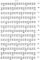
- the invention therefore relates to the DNA sequence of the gene which, apart from coding for specific high molecular weight polypeptides. codes for the 90-120 kDa polypeptide which is a characteristic of S. suis virulence, which gene, hereinafter designated the ef gene has the nucleotide sequence according to Fig. 1A for S. suis serotype 2, strain D-282, and to equivalent sequences and to parts of said sequences. The nucleotide sequence of the entire region coding for EF and the flanking sequences have been determined. Analysis of the sequence of the ef gene (Fig. 1A) provides an open reading frame of 2529 nucleotides which codes for a polypeptide of 843 amino acids (calculated molecular weight 90,014).
- the 110 kDa EF and the high molecular weight proteins are related, which implies that at least part of the ef gene, from strains with a MRP+ EF- phenotype, is identical to the ef gene of strains with the MRP+ EF+ phenotype.
- the higher molecular weight counterpart of the protein EF is designated herein as EF*, and the gene encoding it as the ef* gene.
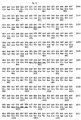
- the corresponding nucleotide and amino acid sequences are represented in Fig. 1B.
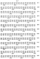
- the invention also relates to the DNA sequence of the gene which codes for the 135-136 kDa polypeptide which is also a virulence characteristic of S. suis, which gene, hereinafter designated the mrp gene, has the nucleotide sequence according to Figure 2 for S. suis serotype 2 strain D-282, and to equivalent sequences and to parts of said sequences.
- the nucleotide sequence of the entire region coding for MRP and the flanking sequences have been determined.
- Analysis of the sequence of the mrp gene (Fig. 2) shows an open reading frame of 3768 nucleotides which codes for a polypeptide of 1256 amino acids (calculated molecular weight 135.794).
- an equivalent sequence comprises a sequence which is essentially the same as the sequence shown but can display slight differences, such as point mutations. or other modifications which may be caused by substitution, deletion, insertion or addition; similarly, an equivalent sequence also comprises a sequence which, despite any differences in nucleotide sequence, hybridises with the sequence shown or with its complement, and also a related sequence which means that it codes for the same amino acid sequence despite differences in nucleotide sequence.
- the invention also relates to a recombinant polynucleotide which contains an ef/ef * gene and/or mrp gene sequence as described above, in the presence of a regulating sequence.
- a recombinant of this type such as a virus vector, a plasmid or a bacterium, can be used for expression of the gene or of relevant parts thereof in a desired environment, for example for the production of immunogenic peptides intended for the diagnosis of an infection, or for controlling infections with virulent strains of S. suis by vaccination.
- Polynucleotide probes which contain a sequence as described above, derived from a gene which codes for a virulence characteristic of S. suis, also form part of the invention.
- a probe of this type in particular corresponds with part of the nucleotide sequence of one of the two said genes.
- the probe can be used for direct detection of the presence of sequences of virulent strains of S. suis.
- the probe can also be used as a basis for a primer for the multiplication of polynucleotides (for example in a polymerase chain reaction) as part of a diagnostic method or a protection method.
- a suitable polynucleotide probe was found to be a partial sequence containing at least 10 nucleotides, preferably at least 15 nucleotides, up to 835 nucleotides from the sequence 1100-1934 of the mrp gene.
- Another suitable polynucleotide probe was found to be a partial sequence containing 10-417, in particular 15-417 nucleotides from the sequence 2890-3306 of the ef * gene. These probes differentiate effectively between pathogenic and non-pathogenic strains of S. suis.
- a combination of such an mrp based probe and an ef* based probe is an especially powerful diagnostic tool.
- the invention also relates to polypeptides which are derived from a polynucleotide sequence described above.
- a polypeptide of this type is either coded by said sequence or obtained by expression of said sequence and essentially corresponds to a S. suis protein characteristic of virulence, or to a part thereof.
- a polypeptide of this type can, for example, be used as an antigen in an immunoassay, as an immunogen in the immunisation of mammals or as an immunogen for the production of antibodies for diagnostic purposes.
- the antibodies generated in this way also form part of the invention.
- Such antibodies can be polyclonal or monoclonal and can be conjugated with a marker (enzyme, isotope, luminescent substance or complex-forming agent); the antibody can also be bound to solid carriers.
- the invention also relates to methods for the detection of an infection by a pathogenic strain or by a non-pathogenic strain of S. suis , in which one or more polynucleotide probes, polypeptides and/or antibodies as described above are used.
- "Infection” signifies here the presence of the pathogenic organism, both in the case where there are clinical signs of disease (infection in a narrow sense) and in the case where there are no clinical signs of disease (infection in a broad sense, or contamination).
- immunoassays such as a determination of the presence of antigens of and/or antibodies against S.
- a polypeptide (110 kDa) which is encoded by the ef/ef * gene or a part thereof, and/or an antibody which has been generated against such a polypeptide it is possible, for example, to use on a microtiter plate a polypeptide (110 kDa) which is encoded by the ef/ef * gene or a part thereof, and/or an antibody which has been generated against such a polypeptide.
- the diagnostic methods can be carried out using procedures known per se. Examples are Enzyme-Linked Immunosorbent Assays (ELISA) and Double Antibody Sandwich (DAS)-ELISA.
- a diagnostic kit according to the invention contains, respectively, at least one polynucleotide or a polypeptide which corresponds to or is derived from a sequence of the ef/ef* gene or mrp gene or a part thereof or contains an antibody which has been generated against the polypeptide derived from one of the said ef/ef* and mrp sequences. It is also possible to use combinations of probes and the like, in particular of ef * diagnostic agents and mrp diagnostic agents. or combinations of primers, for example for carrying out PCR.
- the kits can also contain the components required for carrying out diagnoses, such as reagents (labelling substances, dyes and the like), supports (filters, plates and the like), media and calibrating agents as well as a manual for carrying out the diagnosis.
- the invention also relates to a method for protecting mammals against infection with Streptococcus suis, in which method a polynucleotide, a polypeptide or an antibody as described above is used.
- the method is a passive immunisation, that is to say there is direct provision of antibodies against the pathogenic organism; since antibodies which are derived from EF, EF * and MRP are directed against the most virulent forms of S. suis , a procedure of this type can be an effective method for protecting against, or controlling, infection, especially if the animal to be protected is not itself able to produce sufficient antibodies. for example if infection has already taken place or in the case of young animals.
- Another form of passive immunisation in the case of pigs is the administration of antibodies to the piglets via the colostrum from the sow.
- the dam is actively immunised with one or both polypeptides during pregnancy, that is to say before the birth of the piglets.
- a polypeptide or a polynucleotide (optionally in the form of a recombinant organism) is used, the procedure is an active immunisation, the animal to be protected being stimulated, by means of the immunogenic polypeptide which is administered directly or in the form of a gene for expression, to produce antibodies.
- Another suitable method of immunisation is the administration of a polypeptide from which the activity responsible for virulence has been neutralised.
- a polypeptide should then no longer be pathogenic, while immunogenic characteristics are retained. It can be obtained. for example, by expression of a gene which has been modified with respect to the original ef/ef* or mrp gene. such as by means of deletion.
- Vaccines for protecting mammals against an infection by S. suis. which vaccines contain a polynucleotide, a polypeptide or an antibody as described above, also form part of the invention.
- a particular vaccine according to the invention is a vaccine which contains a S. suis material which does not or does not completely bring to expression at least one of the polypeptides corresponding to EF and MRP.
- This material can originate from or can be formed by a possible live strain which is not virulent or is less virulent.
- the role of virulence factors which are involved in the pathogenesis of S. suis type 2 has been studied in vivo by means of gnotobiotic/germ-free piglets with S. suis type 2 strains defined in respect of virulence factors (MRP and EF). The animal experiments were monitored by means of haematological, bacteriological and (histo)-pathological analytical techniques.
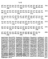
- the presumed ribosome binding site, the -35 and -10 regions of the presumed promoters, and the regions with complementary symmetry are marked.
- the possible cleaving site for signal peptidase is between nucleotides 498-499.
- the putative ribosome binding site, the -35 and -10 regions of the putative promoter sequences, the repetitive regions R1 - R11, and the putative termination signals are indicated.
- the region between the nucleotides 2859 and 5228 is absent in the gene encoding the 110 kDa EF protein.
- the region between the nucleotides 3423 and 4456 is absent in the genes encoding the class IV and class V EF* proteins.
- the probable ribosome binding site, the -35 and -10 regions of the presumed promoter sequences, the region of complementary symmetry beyond the mrp gene, the putative cleaving site for signal peptidase, the proline-rich region, the repeating amino acid sequences and the envelope anchor region are indicated.
- ORF1 open reading frame 1
- ORF2 and ORF3 are separated by a TAA stop codon. Restriction sites of interest are indicated.
- the uppermost and middle sequences represent regions flanking the left and right ends of the lacking fragments.
- the bottom sequences show the junctions as found in the class IV and V ef * genes (A) and in the ef gene (B). Directly repeated sequences are shown in boxes.
- the bold nucleotides indicate the first bases of the translational triplets.
- A Restriction maps of the DNA inserts of putative MRP-positive recombinant bacteriophages. The thick line indicates the DNA region which is present in all of these clones. Restriction sites: E: Eco RI; H: Hin dIII; X: Xba I; K: Kpn I; S: Sac I. B. Parts of the DNA inserts subcloned in the plasmid vector pKUN19 ( 24 ) .
- Lane 1 negative control; proteins extracted from the cell wall of a MRP-negative strain of S. suis.
- Lane 2 crude MRP preparation which contains proteins extracted from the cell wall of strain D282.
- Lane 3 pMR7-1.
- Lane 4 pMR7-2.
- Lane 5 pMR9-1.
- Lane 6 pMR9-2.
- Lane 7 pMR10-1.
- Lane 8 PMR10-2.
- Lane 9 lambda GEM11 with control insert.
- Lane 10 lambda clone 7.
- Lane 11 lambda clone 9.
- Lane 12 lambda clone 10.
- Lane 13 lambda clone 11.
- the amino acid sequence of S. suis MRP was compared with M6 protein of Streptococcus pyogenes (20), protein A of Staphylococcus aureus (16), protein G of group G streptococci (10), AP4 of S. pyogenes (13) , LP of Lactococcus lactis (46), WAP4 of S. mutans (11), T6 of S. pyogenes (38) , and Fn-BP of S. aureus (39).
- Fragments of the mrp and ef genes that were used as a probe On top of each figure is the localisation of restriction sites that were used to create the probes. The fragments which were used as probes are indicated with solid bars. Left of the solid bar is the abbreviation of the probe. The arrow indicates the open reading frame (ORF) of each gene.
- Fig. 14a Probes of the mrp gene. The Sac I and Hin dIII sites are not authentic but are generated by subcloning fragments of the mrp gene.
- Fig. 14b Probes of the ef gene.
- Fig. 14c Probe of the ef* gene. The open bar indicates the insert sequence of ef* that is not part of the ef gene.
- Lanes 1 to 4 contained amplified DNA of MRP + EF + strains (D282, 3, 10, and 22), lanes 5, 6, 7, and 9 of MRP+EF* strains (17, 24, 26, 28), lanes 10 to 14 of MRP - EF - strains (T15, 12, 16, 18, and 25), and lane 15 contained the negative control; all ingredients except DNA.
- Lanes 8 and 16 contained 300 ng size marker Lambda DNA digested with Hin dIII and Eco RI .
- row A contains 1 ⁇ g/spot DNA of four MRP+EF+ strains; D282, 3, 10 and 22, and one positive control.
- Row B contains four MRP+EF* strains: strain 17, 24, 26 and 28; and row C five MRP-EF- strains; T15, 12, 16, 18 and 25.
- Bacterial strains and growth conditions Bacterial strains and growth conditions .
- E. coli strains JM101 (29) and LE392 (33) were used as hosts for recombinant plasmids and bacteriophages.
- the pathogenic MRP+EF+ strain D282 of S. suis type 2 (43) was used for the isolation of chromosomal DNA.
- E. coli strains were grown in Luria broth (30) . Ampicillin was added as needed to a final concentration of 50 ⁇ g/ml.
- S. suis strains were grown in Todd-Hewitt broth (Oxoid, Ltd., London, England).
- a DNA library of S. suis type 2 strain D282 was constructed in LambdaGEM-11 as recommended by the manufacturer of the cloning vector (Promega, Madison, USA). Recombinant bacteriophages were plated on E. coli strain LE392 and incubated for 16 h at 37°C.
- Nitrocellulose filters (Schleicher and Schuell, Inc., Dassel, Germany) were placed on the plaques, and the plates were further incubated for 2 h at 37°C. Recombinants that produced EF were visualized with monoclonal antibodies (Mabs) directed against EF (Example 4). Bound antibodies were detected with anti-mouse serum conjugated with alkaline phosphatase (Zymed Laboratories, Inc., San Francisco, USA) as described by Maniatis et al. (28). Selected EF positive clones were purified by several rounds of single plaque isolation and immunological screening.
- SDS Sodium dodecyl sulfate
- PAGE polyacrylamide gel electroDhoresis
- Western blot analysis Proteins were separated by SDS gel electrophoresis in which 4% stacking and 6% separating gels were used (26) . The separated proteins were transferred to nitrocellulose in a Semi-Dry transfer cell (Bio-Rad Laboratories, Richmond, USA). Specific proteins were visualized by use of polyclonal antibodies (Pabs, Example 4) or Mabs directed against EF and anti-rabbit or anti-mouse sera conjugated with alkaline phosphatase (Zymed Laboratories).
- DNA manipulations and nucleotide sequence analysis Selected restriction fragments were (sub)-cloned in the plasmid vector pKUN19 (24) by standard molecular biological techniques ( 28 ). Progressive unidirectional deletions were made with the Erase-a-Base system from Promega (Madison, USA). DNA sequences were determined by the dideoxy chain termination method (37) . DNA and protein sequences were analysed by the software packages PCGENE (Intelli-genetics Corp., Mountain View, CA) and Wisconsin GCG (University of Wisconsin).
- a DNA library was constructed by isolating chromosomal DNA from strain D282 of S. suis type 2. This DNA was partially digested with the restriction enzyme Sau 3A and cloned into the bacteriophage LambdaGEM11 replacement vector. The library contained approximately 5 x 10 5 recombinants per ⁇ g of DNA. Two thousand plaques of recombinant phages were tested for the presence of antigenic determinants of EF by use of a Mab directed against EF. Two plaques were positive.
- the plasmid containing the 6.8 kb K pn I- Sal I fragment encoded a protein with a molecular weight identical to EF, that was recognized by Mabs directed against EF. Plasmids containing the 5.8 kb Eco RV- Sal I or the 5.3 kb Bgl II- Sal I fragment, however, did not express EF. These data indicate that the Eco RV and the Bgl II sites are within regions required for EF expression.
- Nucleotide sequence of the ef gene The nucleotide sequence of the fragment comprising the EF encoding region was determined. The sequence (Fig. 1A) showed the presence of 3 major open reading frames (ORFs).
- ORF1 (from nucleotide 361 to 2890), ORF2 (from nucleotide 2856 to 3459) and ORF3 (from nucleotide 3462 to 4053) encoded polypeptides of 843 amino acids, of 201 amino acids and of 197 amino acids respectively.
- ORF1 contained a putative ATG start codon that is preceded by a sequence that is similar to ribosome binding sites of several types of gram-positive bacteria (17) .
- EF is exclusively found in the supernatant of S. suis cultures, and thus the protein is expected to be preceded by a signal peptide.
- the first 46 amino acids of the deduced amino acid sequence of EF are characteristic of a typical signal peptide.
- An N-terminal part that contained six positively charged amino acids was followed by a hydro-phobic core of 21 amino acids and a putative signal peptidase cleavage site (45) .
- the hydropathy pattern (25) of the deduced amino acid sequence showed that, apart from the signal peptide, the EF protein was very hydrophilic and did not contain extended hydrophobic regions (cf. MRP, Example 3). No significant similarities were found between the deduced amino acid sequence of EF and the protein sequences in the EMBL Data Library.
- ORF2 and ORF3 Although appropriate translation initiation signals upstream of ORF2 and ORF3 could not be found, the deduced amino acid sequences of ORF2 and ORF3 showed some properties which raised doubt to the idea that those frames are not expressed.
- the N-terminus of the putative ORF2 protein showed two highly repetative units of 57 amino acids (identity 82%).
- the C-terminus of the putative ORF3 protein is functionally similar to C-terminal regions of several cell-envelope located proteins of gram-positive bacteria ( 10, 12, 13, 16, 41 ) . A hydrophobic region was preceded by the conserved sequence Leu-Pro-X-Thr-Gly-Glu and followed by a highly hydrophilic region. This similarity suggests that the putative ORF3 protein is associated with the cell-envelope.
- Escherichia coli strain JM101 (29) was used as host for recombinant plasmids. Seventeen MRP + EF* strains of S. suis type 2 were isolated from human patients, five strains from tonsils of slaughthered pigs, seven strains from organs of diseased pigs and from two strain the origin was unknown (Example 4). The E. coli strain was grown in Luria broth (30). Ampicillin was added as needed to a final concentration of 50 ⁇ g/ml. Streptococcus suis strains were grown in Todd-Hewitt broth (Oxoid, Ltd., London, England).
- Genomic DNA and oligonucleotides Genomic DNA and oligonucleotides .
- Genomic DNA was isolated by lysis in proteinase K/SDS solution, extraction with phenol/chloroform and precipitation with ethanol (28).
- the sequences of the oligonucleotides used in the polymerase chain reaction (PCR) were: 5'-ATGTAATT GAATTC TCTTTTTAAGT-3' and 5'-AAACGTCCGCAGACT TCTAGA TTAAAAGC-3'. These oligonucleotides correspond to the positions 35 to 59 and 4308 to 4279 in the S. suis type 2 ef gene.
- the underlined sequences indicate the recognition sites for the restriction enzymes Eco RI and Xba I .
- the membranes were washed twice with a solution of 2 x SSC (1 x SSC is 0.15M NaCl plus 0.015 M trisodium citrate, pH 7.0) for 5 min at room temperature and twice with a solution of 0.1 x SSC plus 0.5% SDS for 30 min at 65°C.
- PCR Polymerase Chain Reaction
- ef* genes Cloning of ef* genes .
- the genes encoding the different EF* proteins were obtained using PCR to amplify the ef * containing DNA fragments.
- Genomic DNA of 5 different MRP + EF* strains of S. suis type 2 (one representative of each class) was used as a template.
- the amplified fragments were digested with restriction enzymes Eco RI and Xba I and cloned into E. coli .
- Ef* gene of class I The nucleotide sequence of a 6.8 kb Eco RI -Xba I fragment containing the entire ef* gene of class I and the regions flanking it was determined. Analysis of the sequence revealed two open-reading frames (ORFs, Fig. 1B ). The first ORF (from nucleotide 361 to 5827) and the second ORF (from nucleotide 5830 to 6421) encoded polypeptides of 1822 amino acids and 197 amino acids respectively. Based on its size the first ORF is expected to encode the EF* protein (195 kDa). The ORFs were separated by a single TAA stop codon.
- the first ORF contained a putative ATG start codon that was preceded by a sequence similar to bacterial ribosome-binding sites (17).
- the second ORF was not preceded by an appropriate start codon, nor by a putative ribosome-binding site.
- the first 46 amino acids of the deduced amino acid sequence of the EF* protein had the characteristics of a typical signal peptide (45) .
- the C terminus of the mature part of the protein contained a number of imperfect repeats of 76 amino acids.
- the first four repeats were contiguous as were the last six and a half repeats.
- the fourth and the fifth repeated unit were separated by 113 amino acids and the fifth and the six unit by 22 amino acids (Fig. 5 ).
- the amino acid sequences of the last five and a half unit were highly conserved, whereas the sequences of the first five units were more variable.
- One particular amino acid sequence, Asn-Pro-Asn-Leu was conserved in all repeated units. No significant homology was found between the EF* sequence of class I and any protein sequence in the EMBL Data Library.
- Ef* genes of class II, III, IV and V are Ef* genes of class II, III, IV and V . Because the genes encoding the various EF* proteins differed mainly at their 3' ends, the nucleotide sequences of the small Pst I fragments from the genes of class II, III, IV and V were determined. Comparison of the nucleotide sequences showed that the various ef * genes were highly homologous in this region. The ef * genes differed, however, in the number and the arrangement of repeated units (Fig. 5 ).
- the ef* genes of class II and IV l acked the R9 and R10 regions; that of class III lacked the R6, R7 and R9 regions and that of class IV lacked the R7, R8 and R9 regions.
- the ef* genes of class IV and V l acked a fragment of 1,032 bp, which contained R4, R5 and parts of R3 and R6. The translational reading frame of the region located at the 3' end of the missing fragment remained the same. The nucleotide sequences at the regions of the left and right ends of this 1,032 bp fragment showed direct repeats of 9 bp (Fig. 6A ).
- the reading frame was altered and the region located at the 3'-end of the 2,368 bp fragment was translated in different frames in ef and ef * genes. Consequently, the 110 kDa EF protein will not contain the repeated amino acid units.
- Analysis of the nucleotide sequences at the regions of the left and right ends of the 2,368 bp fragment showed direct repeats of 10 bp (containing one mismatch) (Fig. 6B ).
- the gene encoding the 110 kDa EF protein could have been the result of a specific deletion of 2,368 bp within an ef * gene. This would implicate that a S. suis strain that is non-pathogenic can change into a strain that is pathogenic.
- Escherichia coli strain JM101 (sup E, thi,(lac-pro AB - )[F'tra D36, lac I q Z ⁇ M15], 29) was used as a host for recombinant plasmid DNA.
- E. coli strain LE392 [F - ⁇ hsd R574(rk - ⁇ mk + ), sup E44, sup F58, lac Y1, or ⁇ ( lac 1ZY)6, gal K2, gal T22, mel B1, trp R55] ( 33 ) was used as a host for recombinant bacteriophages.
- the pathogenic MRP + EF + strain D282 of S.suis type 2 (43) was used for isolating chromosomal DNA.
- E. coli strains were grown on LB broth (30) .
- Solid LB medium contained 1.5% agar. Ampicillin was added as needed to a final concentration of 50 ⁇ g/ml.
- Streptococcus suis strains were grown in Todd-Hewitt broth (Oxoid Ltd.)
- Chromosomal DNA isolated from strain D282 of S. suis type 2 was partially digested with the restriction enzyme Sau 3A .
- a DNA library was then constructed in the bacteriophage LambdaGEM11 replacement vector. Approximately 5 x 10 5 recombinants/ ⁇ g DNA were obtained.
- a MAb directed against MRP was used to screen 1,400 recombinant plaques for the presence of antigenic determinants of MRP. Five recombinant plaques reacted positive.
- the complete gene for MRP was obtained by hybridization of the 32 P labeled Kpn I -Sac I fragment of pMR7-2 (Fig. 7B ) with Eco RI or Kpn I digested chromosomal DNA of strain D282 of S. suis type 2. An Eco RI fragment of 7 kb and a Kpn I fragment of 7 kb hybridized with the probe. Because of its size, the Eco RI fragment was expected to contain the complete mrp gene and because the expression of mrp is initiated from the 0.7 kb Eco RI -Kpn I fragment, the Kpn I fragment was expected to contain only the 3' end of the gene.
- Fragments ranging from 6 to 8 kb from Eco RI and Kpn I digested chromosomal DNA were isolated, and ligated into the Eco RI or Kpn I site of pKUN19, whereafter the ligation mixtures were transformed into E. coli JM101.
- Thirteen out of 50 selected recombinant clones obtained with the KpnI fragments hybridized with a MRP probe. All of these recombinant clones contained a plasmid (pMR-C) with a 7 kb Kpn I insert. In contrast, of 2,500 selected recombinant clones obtained with Eco RI fragments, none hybridized with the probe.
- Nucleotide sequence of the mrp gene The nucleotide sequence of a 4.6 kb Eco RI -Hind III fragment, containing the entire mrp gene and the regions flanking it was determined. Analysis of the sequence, Fig. 2, revealed an open reading frame of 3.768 nucleotides coding for a polypeptide of 1,256 amino acids (with a calculated MW of 135,794). The putative ATG start codon is preceded by a sequence that is similar to ribosome-binding sites in several types of gram-positive bacteria (17). The nucleotide sequence upstream of mrp resembles the -35 and -10 consensus sequences of promoters commonly found in gram-positive bacteria.
- a region showing extended dyad symmetry can be detected downstream of the mrp gene.
- MRP Amino acid sequence of MRP .
- MRP is a cell-envelope associated protein and must be translocated across the cytoplasmic membrane.
- the mature protein must therefore contain a signal peptide. Indeed, the first 47 amino acids of the MRP have the characteristics of a typical signal peptide.
- An N-terminal part that contains seven positively charged residues is followed by a hydrophobic core of 21 amino acids and a putative signal peptidase cleavage site ( 45 , vertical arrow in Fig. 2 ). Cleavage of the signal peptide would result in a mature protein with an MW of 131,094, which is close to the MW (136 kDa) of MRP, estimated from SDS-polyacrylamide gels (Example 4).
- a second hydrophobic region of 20 amino acids was identified at the C terminus of the protein (Fig. 11 ). If this region is analogous to other envelope associated proteins of gram-positive bacteria (10, 11, 12, 13, 16, 20, 38, 39, 46), it is probably a cell membrane anchor. A short highly charged region and a region with the Leu-Pro-X-Thr-Gly-Glu amino acid sequence, two regions that flank the presumed cellmembrane anchor, are also highly conserved among surface proteins of gram-positive bacteria (Fig. 12 ). The amino acid sequence Leu-Pro-X-Thr-Gly-Glu is putatively involved in cell-wall binding.
- MRP sequence The mature form of MRP starts with a unique N-terminal sequence of 824 amino acids. This region is followed by a stretch of amino acids that is rich in proline residues: of 86 amino acids, 26 are proline residues. This region is followed by three repeated units of 54 amino acids (Fig. 13 ). The first unit is separated from the second by 77 amino acids, but the second and third unit are contiguous. The sequences of the first and the second unit are highly conserved, whereas the third varies. The third repeated unit is followed by the envelope anchor sequence. There was little homology between the MRP sequence and the protein sequences of the EMBL Data Library.
- strains 1 (MRP + ) and 2 (MRP - ) were used as reference strains.
- Culture conditions A 1-day-old colony of each bacterial strain was grown on Columbia blood agar base (code CM 331; Oxoid, Ltd.) containing 6% horse blood and was incubated overnight at 37°C in Todd-Hewitt broth (code CM 189; Oxoid). Early stationary growth phase cultures were obtained from the overnight cultures, diluted 10 times in Todd-Hewitt broth, and incubated for 4 h at 37°C.
- the rabbits were given intravenous booster inoculations of the same antigen dose, but without the adjuvant. After 6 weeks, the rabbits were exsanguinated.
- the serum of one rabbit (rabbit K191) was used as a probe in the Western blot analysis.
- mice were immunized intraperitoneally with 0.5 ml portions of antigen containing 25 ⁇ g of protein emulsified in equal volumes of Freund imcomplete adjuvant; 3 weeks later this procedure was repeated. After 5 weeks, the mice were given intravenous booster inoculations of the same antigen dose, but without the adjuvant.
- Hybridoma cell lines were prepared as described by Van Zijderveld et al. (51). After 10 to 14 days, hybridomas were tested for antibodies against EF by using an enzyme-linked immunosorbent assay. Hybridoma culture supernatants (diluted 1:2) were then tested for anti-EF MAb on Western blots of culture supernatants from strain D-282.
- Binding of MAb to the 110 kDa protein on the nitrocellulose filters was visualized with anti-mouse immunoglobulins conjugated with alkaline phosphatase.
- the positive cells were cloned twice by limiting dilution in microtiter plates.
- the resulting monoclonal cell lines were used to produce ascites fluid in pristane-primed male BALB/c mice, as described previously (51).
- Electrophoresis and Western blotting The various cell fractions were analysed by SDS-PAGE as described by Laemmli (26) on 6 or 12% polyacrylamide. After electrophoresis, the proteins were stained with silver (32). For Western blot analysis, the proteins were electroblotted onto nitrocellulose by using a Multiphor II Nova Blot system (Pharmacia LKB, Uppsala, Sweden). The blots were probed with a 1:500 dilution of rabbit K191 PAb or with a 1:300 dilution of mouse MAb. Bound PAb were visualized with anti-rabbit immunoglobulins conjugated with alkaline phosphatase. Bound MAb were visualized with a 1:1,000 dilution of anti-mouse immunoglobulins conjugated with alkaline phosphatase (Zymed).
- Protein profiles of four cell fractions of 23 selected strains were almost identical. In contrast, the protein profiles of the culture and protoplast supernatants differed distinctly.
- the protein profiles of isolates obtained from diseased pigs contained two protein bands that were absent in the protein profiles of most isolates obtained from healthy pigs. One band represented a 136 kDa protein, which was identified as MRP (43) .
- MRP and EF were clearly detected in the cell fractions of S. suis type 2 strains.
- Protoplast supernatants, culture supernatants, and membrane vesicles of strains 1, 5, 24, and 26 contained the 136-kDa MRP (Fig. 9 ). Because MRP is a major component of protoplast supernatants, this protein must be localized in the cell envelope of the bacteria.
- the culture supernatants of strains 1 and 5 also contained the 110 kDa EF.
- Strains 24 and 26 contained MRP but not EF; strains 2 and 13 contained neither of the proteins.
- MRP+EF+ Proteins bands at various molecular masses higher than 150 kDa reacted with rabbit K191 serum and were visualized in Western blots of culture supernatants of strains 17, 24, 25, 26, and 28. As such proteins were also recognized by the anti-EF MAb, except in the culture supernatant of strain 25, the 110 kDa EF was probably related to these proteins.
- Pigs Fifty-two germ-free pigs, cross-breeds of Great England and Dutch Landrace, were obtained from four sows by caesarian sections. Sows in both experiments were full sisters. Pigs were allotted to 12 groups each consisting of 4 or 5 pigs. Each group was housed in a sterile stainless steel incubator. Housing and feeding were as described before (43) .
- PBS phosphate-buffered saline
- Bordetella bronchiseptica strain 92932 isolated from the nose of a pig with atrophic rhinitis, was used to predispose pigs to S. suis infection (23, 43) .
- the strain was kept on Dorset egg medium.
- the inoculum was prepared by culturing a 48 hour old colony from sheep blood agar in brain heart infusion broth. After 18 h of incubation at 37°C, this medium contained approximately 10 9 CFU/ml.
- the brain heart infusion broth was diluted (1:100) in PBS to prepare the inoculum.
- Electrophoresis and Western blotting The MRP/EF phenotypes of the S. suis strains used as inocula and of the isolates recovered at the end of the experiments were determined. SDS-PAGE as described by Laemmli (26) (6% polyacrylamide) and Western blotting were used to analyse cell culture supernatants of isolates recovered from nasopharynx of all pigs, and from inflamed tissues such as meninges or joints of affected pigs. After electrophoresis the proteins were stained with silver (32) . For Western blot analysis, the proteins were electroblotted onto nitrocellulose by the Multiphor II Nova Blot system, according to the recommendations of the manufacturer (Pharmacia LKB).
- Nitrocellulose filters were incubated either with a 1:1 mixture of mouse anti-MRP monoclonal antibodies (MAb) (11.3 mg/ml) and anti-EF MAb (8.4 mg/ml) each in a 1:200 dilution, or with a 1:500 dilution of polyclonal anti-MRP/EF rabbit serum (K191) (8.2 mg/ml) (Examples 4, 6).
- Filters were incubated with a 1:1000 dilution of anti-mouse immunoglobulins conjugated with alkaline phosphatase (AP) or a 1:3000 dilution of AP conjugated anti-rabbit immunoglobulin G ( ⁇ + ⁇ ) (Zymed).
- Bound antibodies were visualized by adding the substrate bromochloroindolyl phosphate (Sigma, St. Louis, Mo) - nitro blue tetrazolium (Merck, Darmstad, Germany) in phosphatase buffer (100mM NaCl, 5 mM MgCl 2 , 100 mM diethanolamine; pH 9.5).
- mice were inoculated intranasally with a plastic disposable syringe filled with a suspension of B. bronchiseptica strain 92932 in brain heart infusion broth.
- the inocula contained 0.84 x 10 7 CFU in experiment I and 1.0 x 10 7 CFU in experiment II.
- Two days post inoculation (pi) the pigs were similarly inoculated inside the sterile incubator with one of the ten S. suis type 2 strains (Table 2).
- Strains 10, 12, 16, and 18 were isolated at slaughter from the tonsils of healthy pigs. Strains 22 (no. 830544), 24 (no. 740113), 25 (no. 821021) and 28 (no 760366) were isolated from human patients with S. suis type 2 meningitis. (Numbers between parentheses refer to those by J.P. Arends and H.C. Zanen (2) ). 2 x 10 6 CFU.
- Pigs were monitored daily for clinical signs of disease, such as fever, dysfunction of the CNS and lameness. Blood samples from each pig were collected three times weekly by venipuncture of the cranial vena cava. White blood cells were counted with a conducting counter (Contraves A.G., Switzerland) (18) . The number of neutrophils was calculated after differential count of Giemsa-stained blood smears. Swabs specimens of nasopharynx and feces were collected daily and plated directly onto Columbia agar containing 6% horse blood. The presence of S. suis type 2 and of B.
- bronchiseptica was confirmed by slide agglutination test in which a suspension of the monocultures was mixed with the appropiate hyperimmune rabbit serum (DLO-Central Veterinary Institute, Lelystad, NL). After pigs were killed, they were examined for pathologic changes. Tissue specimens of the CNS, serosae, liver, spleen, and tonsils were bacteriologically and histologically examined as described before (43) .
- Electrophoresis and Western blotting When immunoblots were used to analyse culture supernatants of the S. suis strains before inoculation, three phenotypes were distinguished. Strains 3, 10, and 22 belonged to the MRP+EF+ phenotype, strains 17, 24, and 28 were of the MRP+EF-phenotype, and strains 12, 16, 18, and 25 belonged to the MRP-EF-phenotype.
- the rabbit polyclonal antibodies (PAb) recognized proteins that were greater than 150 kDa in the culture supernatants of the MRP+EF-strains. These high molecular weight proteins were also detected by the anti-EF MAb, indicating that the 110 kDa EF and the > 150 kDa proteins share epitopes.
- the phenotypes of the S. suis strains used as inocula were identical to the phenotypes of the isolates collected at the end of both experiments from tonsils and inflamed tissues of infected pigs.
- Morbidity in pigs inoculated with strains of the MRP+EF+ phenotype was 100%. From day 2 onwards, non-specific signs of systemic disease, such as depression, recumbency, lack of appetite, and fever were observed. During the following days, pigs showed more specific signs of disease, such as ataxia, circular movements, opisthotonus, recumbency with paddling, and lameness. The frequency of specific signs of disease in the MRP+EF+ groups was 57% (Table 3). Nine pigs died in the course of the experiment, and three were killed in the terminal stages of disease. The mortality rate in these groups was thus 12/18 (67%).
- broncho-pneumonia 1 1 1 Fibrinous pneumonia 3 0 0 Interstitial pneumonia 7 5 5 Bronchitis/Peribronchiolitis 2 2 3 Liver Periportal and/or intralobular foci 11 8 3 Spleen Active white pulp 2 6 5 Active red pulp 4 0 2 Tonsil Active follicles 3 9 12 Exudation in crypts 1 5 6 1 Affecting cerebrum, cerebellum, pons, mesencephalon and medulla oblongata in various combinations. 2 Affecting carpal, metacarpal, tarsal, metarsal, knee, elbow, shoulder and hip joints in various combinations.
- S. suis type 2 was mostly isolated from organs and tissues (CNS, serosae, and joints) that also showed pathologic changes (Table 5).
- B. bronchiseptica was only isolated from lungs and tonsils. Both S. suis and B. bronchiseptica were also isolated from the tonsils of all pigs. TABLE 5. Isolation of streptococci from various tissues of pigs inoculated with S. suis type 2 (10 strains of three phenotypes). No. of pigs from which S. suis was isolated at necropsy Tissue phenotype MRP+EF+ (no.
- Culture supernatant of S. suis type 2 strain D 282 was concentrated and partially purified by ultrafiltration (type PM30 filters, Amicon Cooperation). It was diluted in phosphate-buffered saline (PBS) (136.89 mM NaCl, 2.68 mM KCl, 8.1 mM Na 2 HPO 4 , 2.79mM KH 2 PO 4 , pH 7.2), to a final protein concentration of 75 ⁇ g/ml. This product was used as coating antigen for the selection of different monoclonals in the direct competition ELISA and for screening hybridoma culture supernatants in the indirect ELISA.
- PBS phosphate-buffered saline
- Hybridoma culture supernatants (diluted 1:2) were then tested on Western blots of culture supernatants of strain D-282 for antibodies directed against MRP. Bound MAb to the 136 kDa protein were visualized by using anti-mouse immunoglobulins conjugated to alkaline phosphatase and the substrate decribed below. Five supernatants were found positive, and the cells from these wells were cloned twice by limiting dilution in microtiter plates.
- the five cell lines that were positive for anti-MRP antibodies and the three cell lines that were positive for anti-EF antibodies were used to produce ascites fluid in pristane-primed male BALB/c mice.
- MAbs directed against MRP and EF were purified from ascites fluid by ammonium sulphate precipitation (50% saturation) and dialysed against PBS.
- the five anti-MRP MAbs were designated: MRP 1 to MRP 5
- the three anti-EF MABs were designated: EF 1 to EF 3 .
- the immunoglobulin isotype of all MAbs was IgG 1 and was determined by double immunodiffusion with mouse isotype-specific antisera (Nordic) in gels of 1% agarose in PBS.
- the PAbs and MAbs were stored at -20°C.
- Direct competition ELISA MAbs were selected with the direct competition ELISA and were used to develop the MRP and EF double antibody sandwich (DAS) ELISAs. Purified anti-MRP and anti-EF MAbs and rabbit PAbs were conjugated to HRPO (Boehringer Mannheim, Germany) with the periodate method of Wilson and Nakane (49) . Conjugated immunoglobulins were stored at -20°C in 50% glycerol. Conjugate solutions were made in PBS-Tw containing 5% fetal calf serum and 0.5% sodium chloride.
- 50 ⁇ l of nonconjugated anti-MRP MAbs in serial twofold dilutions were added to the wells of polystyrene microtiter ELISA plates (Greiner) that had been coated with the culture supernatant of strain D 282 that had been partially purified in PBS (75 ⁇ g/ml protein). The plates were then incubated for 30 min at 37°C.
- 50 ml of the optimal dilution of each of the five anti-MRP MAbs conjugated to HRPO were added.
- Direct competition ELISA The five anti-MRP clones and the three anti-EF clones were tested for competition. Some anti-MRP clones competed with each other. The five anti-MRP MAbs were directed against at least three different epitopes: the first was recognized by MRP 1 and MRP 2 , the second by MRP 3 , and the third by MRP 4 and MRP 5 . Because all three anti-EF clones competed, they are probably directed against the same epitope.
- MRP double antibody sandwich ELISA In an MRP DAS-ELISA using MRP 3 as catching antibody and HRPO-MRP 1 as conjugate, each well of the polystyrene microtiter ELISA plates was coated with 100 ⁇ l containing 2.3 ⁇ g MRP 3 per well in 0.05 M carbonate buffer, pH 9.6. After adsorption for 16 h at 37°C, coated plates were used immediately or stored at -20°C.
- the MRP DAS-ELISA was used to test 179 strains of S. suis type 2 belonging to the three phenotypes MRP+EF+, MRP+EF-, and MRP-EF-, as was previously determined by SDS-PAGE and Western blot. Most strains scored in the ELISA the same as they did in the Western blot (Table 7). All MRP+EF+ strains were MRP-positive in the ELISA. One MRP+EF- strain scored false negative. Three of the MRP-EF- strains (6%) scored false positive.
- the MRP DAS-ELISA discriminated well between the MRP-positive and MRP-negative strains of S. suis type 2. TABLE 7 Results of 179 strains of S. suis type 2 (three phenotypes) tested in the MRP and EF DAS-ELISAs. MRP DAS ELISA EF DAS ELISA phenotype No. strains + No.
- Culture supernatants of 18 of the 21 reference S. suis strains of other serotypes had absorbances lower than 0.2.
- These serotypes were also positive in the Western blot; MRP 3 apparently recognized proteins of higher molecular weight than 150 kDa in the culture supernatants of these serotypes. Absorbances of all other microorganisms listed in Table 6 were ⁇ 0.2.
- EF Double Antibody Sandwich ELISA In a DAS ELISA that recognizes a specific antigen in the test sample, two different MAbs were used, one as catching antibody and the other as conjugate, and each recognizing different epitopes on the antigen, as was done for the MRP DAS-ELISA. In the Western blot the EF MAbs recognize a high molecular form of EF (> 150 kDa) in the culture supernatants of all strains belonging to the MAP+EF- phenotype (Example 4). Therefore it is unlikely that an ELISA with EF 2 as catching antibody can discriminate between MRP+EF+ and MRP+EF- strains.
- the procedure of the EF DAS-ELISA was essentially as that described for the MRP DAS-ELISA.
- Each well of the microtiter ELISA plates was coated with 100 ml containing 3.3 ⁇ g of EF 2 in 0.05M carbonate buffer, pH 9.6. After adsorption, coated plates were used immediately or stored at -20°C. Twofold serial dilutions of 100 ⁇ l culture supernatants ranging from 1:1 to 1:128 were used. After incubation and washings, 100 ⁇ l containing 2.7 ⁇ g polyclonal Ra K 191 HRPO conjugate in PBS, pH 7.2, was added to each well. After 1 h incubation at 37°C, the plates were developed with substrate 5-AS H 2 O 2 as described above. Wells with an A 450 ⁇ 0.4 were scored positive. The same controls as mentioned above were used on each plate.
- the 179 S. suis type 2 strains with a predetermined protein profile were tested in the EF DAS-ELISA. Surprisingly, none of the 39 MRP+EF- strains scored positive in this ELISA, whereas all 92 MRP+EF+ strains did (Table 7). All 48 MRP-EF- strains were negative in the EF DAS-ELISA. Since no other false positive or false negative results were detected, the EF DAS-ELISA apparently discriminated reliably between the high and the low molecular form of EF, and hence between S. suis type 2 strains belonging to the MRP+EF+ and MRP+EF- phenotypes.
- Mab EF 2 captured the 110-kDa EF in the culture supernatant of all MRP+EF+ strains but apparently not the higher-molecular weight form in the MRP+EF- strains (Table 7). Some MRP-EF- strains gave signals between 0.2 and 0.4, which were still lower than 50% of the maximal absorbance values and thus not high enough to be interpreted as positive. Treating the culture supernatants with SDS before blotting may uncover epitopes of the higher-molecular weight form of EF that are not accessible to the EF 2 MAb in its undenaturated form. Because all MRP-EF- strains and other S. suis serotypes showed no false negative or false positive reactions in this ELISA, the sensitivity and specificity of the tests were considered to be 100%.
- PCR polymerase chain reaction
- S. suis type 2 Bacteria and growth conditions . Thirteen strains of S. suis type 2 were selected to examine whether the Polymerase Chain Reaction (PCR) method (36) could be useful to differentiate between the three phenotypes of S. suis type 2. Pathogenicity and the expression of the MRP and EF proteins of these strains were determined in Examples 4 and 5. Strains were grown overnight at 37°C on Columbia blood agar base (code CM 331, Oxoid) containing 6% horse blood. S. suis type 2 colonies were inoculated in 10 ml Todd-Hewitt broth (code CM 189, Oxoid), and grown overnight at 37°C.
- PCR Polymerase Chain Reaction
- DNA Isolations DNA of overnight grown cultures was isolated as described by Maniatis et. al (28). DNA was diluted to 10 ng/ ⁇ l in distilled water before use in the PCR.
- the filters were not allowed to run dry between the wash steps.
- the bottom of the microtiter plate was dried on a tissue and the samples were dried completely for 15 minutes at 56°C.
- 75 ⁇ l PCR buffer (see below) was added to the individual wells. The plate was incubated for 15 minutes at 56°C.
- the microtiter plate was again put on the vacuum manifold, with a standard microtiter plate (Micronic) beneath the Durapore plate. Vacuum was applied, and the PCR buffer, containing the DNA was collected in the lower microtiter plate, whereas the diatom earth remained on the Durapore filters.
- the PCR contained 10 ng purified DNA or 25 ⁇ l clinical specimen in a total volume of 50 ⁇ l.
- the reaction mixtures contained 10 mM Tris-HCl (pH 9.0), 2 mM MgCl 2 , 50 mM KCl, 0.01% gelatin, 0.2 mM of each of the four deoxynucleotide triphosphates, 1 ⁇ M of each of the four primers and 0.5 U of Amplitaq polymerase (Perkin Elmer Cetus, Norwalk, Conn.), and was overlaid with 2 drops of paraffine oil.
- DNA amplification was carried out in a Perkin Elmer Thermal Cycler for 25 or 40 cycles: 1 minute 94°C, 1 minute 55°C, and 2 minutes 72°C. Ten to 20 ⁇ l of the amplified DNA was analysed on a 1.5% agarose gel, that contained ethidium bromide.
- PCR primers The sequences of the oligonucleotides used in the PCR were: p15: 1403-1425: 5'- GGT ATA CCT TGC TGG TAC CGT TC -3', p16: 1914-1934: 5'- AGT CTC TAC AGC TGT AGC TGG -3', p-34: 2890-2908: 5'- GTT GAA AAC AAA GCA TTC G -3', and p-35: 3229-3249: 5'- CTT CGA CAA AAT GTC AGA TTC -3'.
- the oligonucleotides p-15 and p-16 correspond to the indicated positions in the S. suis type 2 mrp gene (Example 3, Fig.
- the oligonucleotides p-34 and p-35 correspond to the indicated positions and in the S. suis type 2 ef * gene (Example 2, Fig. 1B ). Primers were synthesized on an Applied Biosystem synthesizer type 381A following the manufacturers protocol.
- PCR Specificity of PCR .
- two regions (designated as m-VI and e-V) were determined that could be used to differentiate between the three phenotypes of S. suis type 2 strains (see also Example 8).
- the primers p-15 and p-16 amplified a 532 bp fragment in the m-VI region.
- the primers p-34 and p-35 amplified a 360 bp fragment in the e-V region.
- MRP + EF* cells were diluted in phosfate buffered saline (PBS (pH 7.2); 137 mM NaCl, 2.7 mM KCl, 8.1 mM Na 2 HPO 4 , 2.8 mM KH 2 PO 4 ) and prepared for PCR as described above. Amplified fragments could still be detected in samples that contained about 50 cells prior to the PCR (40 cycli).
- PBS phosfate buffered saline
- the PCR can be used directly on clinical material. Serial dilutions of S. suis type 2 cells were added to nose swabs. It was found that amplified fragments can still be detected in samples that contain about 50 cells prior to the PCR.
- S. suis type 2 170 strains of S. suis type 2 were obtained from three sources: From organs of diseased pigs (103 strains), from tonsils of healthy pigs at slaughter (40 strains) and from human patients (27 strains). Reference strains of S. suis serotypes 1 to 22 (15), 21 other Streptococci species and 45 other bacterial strains (38 different species, DLO Central Veterinary Institute, Table 8) were used to test the specificity of the mrp and ef probes.
- E. coli JM101 strains were grown in LB broth (30) . Ampicillin was added as needed to a final concentration of 50 ⁇ g/ml. All other bacterial strains were grown overnight at 37°C on Columbia blood agar base (code CM 331, Oxoid) containing 6% horse blood. Overnight grown colonies were incubated in 10 ml Todd-Hewitt broth (code CM 189, Oxoid). and grown overnight at 37°C.
- Probes The plasmids pMR11, pEF2-19 and pEF17-7 (cf. Examples 1, 2, 3) were used to generate subclones into pKUN19 (24). Fragments of appropriate subclones were isolated from preparative agarose gels with the gene-clean kit (Bio 101 Inc., La Jolla, USA). Purified fragments were subsequently labeled with ⁇ - 32 P dCTP (3000 Ci/mMol, Amersham) with the random primed labeling kit (Boehringer GmbH) following the manufacturers protocol and used as probes.
- ⁇ - 32 P dCTP 3000 Ci/mMol, Amersham
- the membranes were incubated with the 32 P-labeled mrp and ef probes as recommended by the manufacturer. After overnight hybridization, the membranes were washed twice in 40 mM Na phosphate buffer, pH 7.2 plus 5% SDS plus 1 mM EDTA for 30 minutes at 65°C and twice in 40 mM Na phosphate buffer pH 7.2 plus 1% SDS plus 1 mM EDTA for 30 minutes at 65°C. All (pre)hybridizations were carried out in a hybridization oven (Hybaid).
- Mrp probes Chromosomal DNA of the 3 phenotypes of S. suis type 2 was hybridized to different regions of the mrp gene. Six different mrp probes were used (schematically shown in Fig. 14a ). The EcoRI-SnaBI fragment, m-I. contained the entire mrp encoding region. The m-II, m-III, m-IV and m-V probes contained different regions of the mrp gene (see Fig. 16 ). The MRP + EF + and the MRP + EF* strains strongly hybridized with all mrp probes.
- probe m-VI was constructed by removing 385 bp at the 5', and 325 bp at the 3' ends of probe m-III.
- the 5 MRP - EF - strains did not hybridize at all with probe m-VI, indicating that these strains lacked the region homologous to the m-VI probe. Therefore, probe m-VI can be used to differentiate between MRP + and MRP - strains.
- probe e-V hybridized with all MRP + EF* strains.
- MRP + EF + and MRP - EF - strains hybridized with the e-V probe.
- S. suis type 2 strains hybridize with probe m-VI and e-V, these strains belong to the MRP + EF * phenotype. If S. suis type 2 strains hybridize with m-VI but not with e-V, these strains belong to the MRP + EF + phenotype, and finally if strains do not hybridize with m-VI and e-V, these strains belong to the MRP - EF - phenotype.
- the mrp , ef and ef * probes were tested on 170 other strains of S. suis type 2. 88 strains had a MRP + EF + phenotype, 37 strains a MRP + EF* phenotype and 45 strains had a MRP - EF - phenotype. In accord with the data presented above, all MRP + EF + strains hybridized with the probes m-I to m-VI and e-I to e-IV, but none hybridized with probe e-V. Moreover, all the 37 MRP + EF* strains hybridized with all the probes.
- probes m-I, m-VI, e-III and e-V Chromosomal DNA from swine pathogens and several common bacteria was tested with the probes m-I, m-VI, e-III and e-V.
- the species tested are listed in Table 8. Although some species hybridized with probe m-I (Escherichia coli, Klebsiella oxytoca, K. pneumoniae and Salmonella typhimurium), none hybridized with the probes m-VI, e-III and e-V. These data show that although in some species parts of the mrp gene are found, the probes m-VI and e-V are specific for S. suis. Hence, the probes m-VI and e-V have potential diagnostic value.
Abstract
Description
- The invention is in the field of veterinary and human preventive medicine, in particular that of the diagnosis of and protection against infection by pathogenic strains of the bacterium Streptococcus suis.
- Infections with Streptococcus suis
serotype 2 in young pigs at about the time of weaning have been a growing problem in the Netherlands since 1983. The disease is characterised by meningitis, arthritis, sepsis and death (Clifton-Hadley 1983. ref. 6; Vecht et al. 1985, ref. 44; Windsor 1977, ref. 50). It is estimated that 5-10 per cent of farms have problems of this type. The mortality is estimated at 2.5% and the morbidity in affected farms is on average 2-5%. Therapeutic and preventive measures have only a limited effect. The economic damage is accordingly appreciable. The disease is a zoonosis. Humans are also susceptible to this infection, with the risk of sepsis and meningitis with possibly permanent side-effects; rare cases of death have been reported (Arends and Zanen 1988, ref. 2). This related mostly to cases of people with a skin wound coming into contact with infected pork. In particular, pig farmers and slaughterhouse staff belong to the risk group. - There are indications that the increased rate of illness on pig farms in the Netherlands since 1983 is to be ascribed to the import of breeding animals which are carriers of S.
suis type 2. Carriers are often healthy adult pigs which harbour the streptococci in the tonsils and mucosa of the upper respiratory tract. The infection is transmitted via these carriers to susceptible animals, frequently piglets at weaning age. Diagnosis of animals which are already sick or have died is based on isolation and determination ofS. suis type 2 from clinical samples or organs after necropsy. Detection of carriers is based on bacteriological examination of nose or throat swabs or tonsil biopsies using a selective/elective medium (Van Leengoed et al. 1987, ref. 27). On the basis of diagnostic testing to detect carriers, it should be possible to set up a control programme. However, testing for carriers using the conventional becteriological techniques is time-consuming, which complicates the processing of large numbers of samples; there is also a risk of false negative results due to overgrowth with contaminants. Finally, interpretation of the test demands a great deal of experience. Moreover, diagnosis and possible control on the basis of diagnosis are further complicated by the occurrence of differences in pathogenicity within theS. suis type 2 species. Regular testing for carriers within a control programme is sensible only if truly virulent strains ofS. suis type 2 can be differentiated from avirulent strains. Current diagnostic techniques do not make such discrimination. Consequently, control based on the detection of carriers of virulentS. suis type 2 strains is not yet possible. - Differences in virulence are ascribed, inter alia, to the presence or absence of virulence factors. In 1984, Arends and Zanen (ref. 1) already described "lysozyme-positive proteins" in human strains. In a study with experimental animals it was found that a "lysozyme-positive" strain (D-282) was pathogenic for gnotobiotic pigs, in contrast to a "lysozyme-negative" strain (T-15) (Vecht et al. 1989, ref. 43). The "lysozyme-positive protein" is probably identical to the muramidase-released protein (MRP) of strain D-282.
- The pig industry in the Netherlands and many other countries has a pyramid structure, with a small number of breeding herds at the top, from where animals are distributed to multiplication herds. These supply a large number of fattening herds, from where the (animal) product finally goes to the slaughterhouses. A control program based on diagnosis (certification of farms, elimination of positive carriers, import requirements) should primarily aim at creating herds which are free of
S. suis type 2 high in this pyramid. A vaccine would primarily be useful in affecting herds lower in the pyramid. Furthermore, means and methods for diagnosing infections by Streptococcus suis in human medicine can be of value. - The object of the invention is to provide methods and means which make it possible, in a more effective manner than hitherto, to detect infections by Streptococcus suis on the one hand and to prevent such infections by elimination of infected and carrier pigs on the other hand.
- This object is achieved by using a DNA sequence from the gene which codes for a virulence characteristic of S. suis. In this context, a virulence characteristic is defined as a polypeptide whose presence is associated with the virulence of an organism, in this case the bacterium S. suis, in
particular serotype 2. - Two genes of virulent strains of
S. suis type 2 have been found which code for two proteins, which are designated MRP (muramidase released protein) and EF (extracellular factor) and which appear to be characteristic for virulence (virulence factors). MRP and EF are high molecular weight proteins. MRP (136 kD) is a protein associated with the cell envelope and can be released from the cell wall by muramidase. EF (110 kD) is an extracellular product which is secreted by the bacterium into the growth medium. EF has higher molecular weight counterparts which are denoted herein as EF*. - The invention provides new diagnostic methods which are able to differentiate between virulent and avirulent strains. These methods are based on the genes encoding MRP, EF and EF* and their expression products. On the basis of the expression of one or both proteins by said genes, three different phenotypes of
S. suis type 2 have been found to date: i.e. the MRP+ EF+ phenotype. the XRP+ EF- phenotype and the MRP- EF- phenotype. 77% (n = 111) of strains isolated from organs of pigs showing clinical symptoms of disease were found to possess the MRP+ EF+ phenotype, while 86% (n = 42) of isolates from tonsils of non-suspect normal slaughter pigs were found to possess the MRP- EF- phenotype. The MRP+ EF- phenotype was most frequently found (74%) (n = 27) in isolates from human patients with infections of S. suis type 2 (see Figure 10). Hence infected animals and carriers of virulent strains can be detected, and a vaccine based on MRP, EF and/or EF* can be developed. It is thus possible to detect carriers of virulent strains of S. suis and a vaccine can be developed. Using the diagnostic methods for detecting carriers and infected pig herds and/or using vaccines based on MRP, EF and/or EF*, a program for controlling infections byS. suis type 2 in pig herds can be developed. - The invention therefore relates to the DNA sequence of the gene which, apart from coding for specific high molecular weight polypeptides. codes for the 90-120 kDa polypeptide which is a characteristic of S. suis virulence, which gene, hereinafter designated the ef gene has the nucleotide sequence according to Fig. 1A for
S. suis serotype 2, strain D-282, and to equivalent sequences and to parts of said sequences. The nucleotide sequence of the entire region coding for EF and the flanking sequences have been determined. Analysis of the sequence of the ef gene (Fig. 1A) provides an open reading frame of 2529 nucleotides which codes for a polypeptide of 843 amino acids (calculated molecular weight 90,014). Monoclonal antibodies generated against the 110 kDa EF protein recognised proteins with a higher molecular weight in culture supernatants of all 38 strains with a MRP+ EF- phenotype. This indicates that certain epitopes of the 110 kDa EF and the high molecular weight proteins are identical. None of the 91 strains with a MRP+ EF+ phenotype was found to produce these high molecular weight proteins. At the same time, DNA probes based on the gene which codes for the 110 kDa EF were found to hybridise with genes which code for the high molecular weight proteins of MRP+ EF- strains. This indicates that the 110 kDa EF and the high molecular weight proteins are related, which implies that at least part of the ef gene, from strains with a MRP+ EF- phenotype, is identical to the ef gene of strains with the MRP+ EF+ phenotype. The higher molecular weight counterpart of the protein EF is designated herein as EF*, and the gene encoding it as the ef* gene. The corresponding nucleotide and amino acid sequences are represented in Fig. 1B. - The invention also relates to the DNA sequence of the gene which codes for the 135-136 kDa polypeptide which is also a virulence characteristic of S. suis, which gene, hereinafter designated the mrp gene, has the nucleotide sequence according to Figure 2 for
S. suis serotype 2 strain D-282, and to equivalent sequences and to parts of said sequences. The nucleotide sequence of the entire region coding for MRP and the flanking sequences have been determined. Analysis of the sequence of the mrp gene (Fig. 2) shows an open reading frame of 3768 nucleotides which codes for a polypeptide of 1256 amino acids (calculated molecular weight 135.794). - In this context, an equivalent sequence comprises a sequence which is essentially the same as the sequence shown but can display slight differences, such as point mutations. or other modifications which may be caused by substitution, deletion, insertion or addition; similarly, an equivalent sequence also comprises a sequence which, despite any differences in nucleotide sequence, hybridises with the sequence shown or with its complement, and also a related sequence which means that it codes for the same amino acid sequence despite differences in nucleotide sequence.
- The invention also relates to a recombinant polynucleotide which contains an ef/ef* gene and/or mrp gene sequence as described above, in the presence of a regulating sequence. A recombinant of this type, such as a virus vector, a plasmid or a bacterium, can be used for expression of the gene or of relevant parts thereof in a desired environment, for example for the production of immunogenic peptides intended for the diagnosis of an infection, or for controlling infections with virulent strains of S. suis by vaccination.
- Polynucleotide probes which contain a sequence as described above, derived from a gene which codes for a virulence characteristic of S. suis, also form part of the invention. A probe of this type in particular corresponds with part of the nucleotide sequence of one of the two said genes. The probe can be used for direct detection of the presence of sequences of virulent strains of S. suis. The probe can also be used as a basis for a primer for the multiplication of polynucleotides (for example in a polymerase chain reaction) as part of a diagnostic method or a protection method.
- A suitable polynucleotide probe was found to be a partial sequence containing at least 10 nucleotides, preferably at least 15 nucleotides, up to 835 nucleotides from the sequence 1100-1934 of the mrp gene. Another suitable polynucleotide probe was found to be a partial sequence containing 10-417, in particular 15-417 nucleotides from the sequence 2890-3306 of the ef* gene. These probes differentiate effectively between pathogenic and non-pathogenic strains of S. suis. A combination of such an mrp based probe and an ef* based probe is an especially powerful diagnostic tool.
- The invention also relates to polypeptides which are derived from a polynucleotide sequence described above. A polypeptide of this type is either coded by said sequence or obtained by expression of said sequence and essentially corresponds to a S. suis protein characteristic of virulence, or to a part thereof. A polypeptide of this type can, for example, be used as an antigen in an immunoassay, as an immunogen in the immunisation of mammals or as an immunogen for the production of antibodies for diagnostic purposes. The antibodies generated in this way also form part of the invention. Such antibodies can be polyclonal or monoclonal and can be conjugated with a marker (enzyme, isotope, luminescent substance or complex-forming agent); the antibody can also be bound to solid carriers.
- The invention also relates to methods for the detection of an infection by a pathogenic strain or by a non-pathogenic strain of S. suis, in which one or more polynucleotide probes, polypeptides and/or antibodies as described above are used. "Infection" signifies here the presence of the pathogenic organism, both in the case where there are clinical signs of disease (infection in a narrow sense) and in the case where there are no clinical signs of disease (infection in a broad sense, or contamination). For immunoassays, such as a determination of the presence of antigens of and/or antibodies against S. suis in a sample or in clinical material, it is possible, for example, to use on a microtiter plate a polypeptide (110 kDa) which is encoded by the ef/ef* gene or a part thereof, and/or an antibody which has been generated against such a polypeptide. In addition, it is also possible to use a polypeptide (136 kDa) encoded by the mrp gene or a part thereof, and/or an antibody which has been generated against such a polypeptide. The diagnostic methods can be carried out using procedures known per se. Examples are Enzyme-Linked Immunosorbent Assays (ELISA) and Double Antibody Sandwich (DAS)-ELISA.
- The methods described above can be carried out with the aid of diagnostic kits. A diagnostic kit according to the invention contains, respectively, at least one polynucleotide or a polypeptide which corresponds to or is derived from a sequence of the ef/ef* gene or mrp gene or a part thereof or contains an antibody which has been generated against the polypeptide derived from one of the said ef/ef* and mrp sequences. It is also possible to use combinations of probes and the like, in particular of ef* diagnostic agents and mrp diagnostic agents. or combinations of primers, for example for carrying out PCR. The kits can also contain the components required for carrying out diagnoses, such as reagents (labelling substances, dyes and the like), supports (filters, plates and the like), media and calibrating agents as well as a manual for carrying out the diagnosis.
- The invention also relates to a method for protecting mammals against infection with Streptococcus suis, in which method a polynucleotide, a polypeptide or an antibody as described above is used. When an antibody is used, the method is a passive immunisation, that is to say there is direct provision of antibodies against the pathogenic organism; since antibodies which are derived from EF, EF* and MRP are directed against the most virulent forms of S. suis, a procedure of this type can be an effective method for protecting against, or controlling, infection, especially if the animal to be protected is not itself able to produce sufficient antibodies. for example if infection has already taken place or in the case of young animals.
- Another form of passive immunisation in the case of pigs is the administration of antibodies to the piglets via the colostrum from the sow. In this case the dam is actively immunised with one or both polypeptides during pregnancy, that is to say before the birth of the piglets. When a polypeptide or a polynucleotide (optionally in the form of a recombinant organism) is used, the procedure is an active immunisation, the animal to be protected being stimulated, by means of the immunogenic polypeptide which is administered directly or in the form of a gene for expression, to produce antibodies.
- Another suitable method of immunisation is the administration of a polypeptide from which the activity responsible for virulence has been neutralised. Such a polypeptide should then no longer be pathogenic, while immunogenic characteristics are retained. It can be obtained. for example, by expression of a gene which has been modified with respect to the original ef/ef* or mrp gene. such as by means of deletion.
- Vaccines for protecting mammals against an infection by S. suis. which vaccines contain a polynucleotide, a polypeptide or an antibody as described above, also form part of the invention.
- A particular vaccine according to the invention is a vaccine which contains a S. suis material which does not or does not completely bring to expression at least one of the polypeptides corresponding to EF and MRP. This material can originate from or can be formed by a possible live strain which is not virulent or is less virulent.
- The role of virulence factors which are involved in the pathogenesis of S. suis
type 2 has been studied in vivo by means of gnotobiotic/germ-free piglets with S. suistype 2 strains defined in respect of virulence factors (MRP and EF). The animal experiments were monitored by means of haematological, bacteriological and (histo)-pathological analytical techniques. - Nucleotide sequence of the ef gene and the adjacent sequences and the EF amino acid sequence derived therefrom. The presumed ribosome binding site, the -35 and -10 regions of the presumed promoters, and the regions with complementary symmetry are marked. The possible cleaving site for signal peptidase is between nucleotides 498-499.
- Nucleotide sequence of the fragment encoding the S. suis
type 2 ef* gene of strain 1890 and the deduced amino acid sequence of the EF* protein of class I. The putative ribosome binding site, the -35 and -10 regions of the putative promoter sequences, the repetitive regions R1 - R11, and the putative termination signals are indicated. The region between the nucleotides 2859 and 5228 is absent in the gene encoding the 110 kDa EF protein. The region between the nucleotides 3423 and 4456 is absent in the genes encoding the class IV and class V EF* proteins. - Nucleotide sequence of the 4.6 kb EcoRI-HindIII fragment with the mrp gene of S. suis
type 2 and the MRP amino acid sequence derived therefrom. The probable ribosome binding site, the -35 and -10 regions of the presumed promoter sequences, the region of complementary symmetry beyond the mrp gene, the putative cleaving site for signal peptidase, the proline-rich region, the repeating amino acid sequences and the envelope anchor region are indicated. - Restriction maps of ef containing fragments, subcloned into the plasmid vector pKUN19 (24). The open reading frames are boxed. Restriction sites: B:BamHI; Bg:BglII; E:EcoRI; K:KpnI; N;NarI; P;PstI; S;SnaBI; Sa:SalI; Sp:SpeI.
- Schematic representation of the gene encoding the 110 kDa EF protein and the flanking regions. EF is encoded by the open reading frame 1 (ORF1). The 3' end of ORF1 is overlapping with the 5' end of ORF2. ORF2 and ORF3 are separated by a TAA stop codon. Restriction sites of interest are indicated.
- Schematic representation of the PstI-SnaBI fragment of the ef* genes of 5 different classes of the ef gene. The arrows indicate the repeated amino acid units. The lines indicate regions present in the different strains. The gaps indicate the regions lacking in the different strains.
- Nucleotide sequences near the ends of the fragments lacking in the ef* genes of class IV and V (A) and in the ef gene (B). The uppermost and middle sequences represent regions flanking the left and right ends of the lacking fragments. The bottom sequences show the junctions as found in the class IV and V ef* genes (A) and in the ef gene (B). Directly repeated sequences are shown in boxes. The bold nucleotides indicate the first bases of the translational triplets. The numbers refer to the nucleotide positions in the ef* gene of class I (Fig. 1B).
- A. Restriction maps of the DNA inserts of putative MRP-positive recombinant bacteriophages. The thick line indicates the DNA region which is present in all of these clones. Restriction sites: E:EcoRI; H:HindIII; X:XbaI; K:KpnI; S:SacI. B. Parts of the DNA inserts subcloned in the plasmid vector pKUN19 (24).
- Western blot analysis of proteins, encoded by recombinant plasmids and recombinant bacteriophages, which have been selected with monoclonal antibodies against MRP. Lane 1: negative control; proteins extracted from the cell wall of a MRP-negative strain of S. suis. Lane 2: crude MRP preparation which contains proteins extracted from the cell wall of strain D282. Lane 3: pMR7-1. Lane 4: pMR7-2. Lane 5: pMR9-1. Lane 6: pMR9-2. Lane 7: pMR10-1. Lane 8: PMR10-2. Lane 9: lambda GEM11 with control insert. Lane 10:
lambda clone 7. Lane 11:lambda clone 9. Lane 12:lambda clone 10. Lane 13:lambda clone 11. - Western blot of the protoplast supernatant (PPS), culture supernatant (Cult. Sup.), and membrane vesicle (Membr.) fractions probed with anti-MRP/EF rabbit K191 serum (diluted 1:500). The lane designations are numbered strain designations.
- Western blot of cell culture supernatants of selected S. suis
type 2 strains probed with rabbit anti-MRP/EF serum (K191), anti-MRP serum, and anti-EF serum (1:500 diluted). The PAbs revealed three S. suistype 2 phenotypes: MRP+EF+, MRP+EF- and MRP- EF-. The lane designations are strain designations. Reference strain 1 (D-282) and strains 3 to 9 (MRP+EF+) were isolated from pigs with S. suis meningitis. Reference strain 2 (T-15) and strains 10, 12, 16, and 17 were isolated from the tonsils of healthy pigs. Strains 22, 23, 24, 25, 26, 28, and 29 were isolated from human patients. - Hydropathy profile (25) of MRP. Sequences above and below the line represent hydrophobic and hydrophilic regions respectively.
- Homology between the amino acid sequences at the C terminus of MRP and several cell-envelope associated proteins of gram-positive bacteria. The amino acid sequence of S. suis MRP was compared with M6 protein of Streptococcus pyogenes (20), protein A of Staphylococcus aureus (16), protein G of group G streptococci (10), AP4 of S. pyogenes (13), LP of Lactococcus lactis (46), WAP4 of S. mutans (11), T6 of S. pyogenes (38), and Fn-BP of S. aureus (39).
- Comparison of the amino acid sequence of the repeat units in MRP. Homologous regions are enclosed in boxes.
- Fragments of the mrp and ef genes that were used as a probe. On top of each figure is the localisation of restriction sites that were used to create the probes. The fragments which were used as probes are indicated with solid bars. Left of the solid bar is the abbreviation of the probe. The arrow indicates the open reading frame (ORF) of each gene.
Fig. 14a: Probes of the mrp gene. The SacI and HindIII sites are not authentic but are generated by subcloning fragments of the mrp gene.
Fig. 14b: Probes of the ef gene.
Fig. 14c: Probe of the ef* gene. The open bar indicates the insert sequence of ef* that is not part of the ef gene. - Specificity of PCR, 10 ng of chromosomal DNA of S. suis
type 2 strains was used in the PCR with the primers p-15, p-16, p-34, and p-35.Lanes 1 to 4 contained amplified DNA of MRP+EF+ strains (D282, 3, 10, and 22),lanes lanes 10 to 14 of MRP-EF- strains (T15, 12, 16, 18, and 25), andlane 15 contained the negative control; all ingredients except DNA.Lanes - Dot spot hybridization of 13 S. suis
type 2 strains with the mrp and ef probes. In each experiment, row A contains 1 µg/spot DNA of four MRP+EF+ strains; D282, 3, 10 and 22, and one positive control. Row B contains four MRP+EF* strains:strain - Bacterial strains and growth conditions.
- E. coli strains JM101 (29) and LE392 (33) were used as hosts for recombinant plasmids and bacteriophages. The pathogenic MRP+EF+ strain D282 of S. suis type 2 (43) was used for the isolation of chromosomal DNA. E. coli strains were grown in Luria broth (30). Ampicillin was added as needed to a final concentration of 50 µg/ml. S. suis strains were grown in Todd-Hewitt broth (Oxoid, Ltd., London, England).
- Construction and immunological screening of the DNA library. A DNA library of S. suis
type 2 strain D282 was constructed in LambdaGEM-11 as recommended by the manufacturer of the cloning vector (Promega, Madison, USA). Recombinant bacteriophages were plated on E. coli strain LE392 and incubated for 16 h at 37°C. - Nitrocellulose filters (Schleicher and Schuell, Inc., Dassel, Germany) were placed on the plaques, and the plates were further incubated for 2 h at 37°C. Recombinants that produced EF were visualized with monoclonal antibodies (Mabs) directed against EF (Example 4). Bound antibodies were detected with anti-mouse serum conjugated with alkaline phosphatase (Zymed Laboratories, Inc., San Francisco, USA) as described by Maniatis et al. (28). Selected EF positive clones were purified by several rounds of single plaque isolation and immunological screening.
- Sodium dodecyl sulfate (SDS) polyacrylamide gel electroDhoresis (PAGE) and Western blot analysis. Proteins were separated by SDS gel electrophoresis in which 4% stacking and 6% separating gels were used (26). The separated proteins were transferred to nitrocellulose in a Semi-Dry transfer cell (Bio-Rad Laboratories, Richmond, USA). Specific proteins were visualized by use of polyclonal antibodies (Pabs, Example 4) or Mabs directed against EF and anti-rabbit or anti-mouse sera conjugated with alkaline phosphatase (Zymed Laboratories).
- DNA manipulations and nucleotide sequence analysis. Selected restriction fragments were (sub)-cloned in the plasmid vector pKUN19 (24) by standard molecular biological techniques (28). Progressive unidirectional deletions were made with the Erase-a-Base system from Promega (Madison, USA). DNA sequences were determined by the dideoxy chain termination method (37). DNA and protein sequences were analysed by the software packages PCGENE (Intelli-genetics Corp., Mountain View, CA) and Wisconsin GCG (University of Wisconsin).
- Cloning of the ef gene. A DNA library was constructed by isolating chromosomal DNA from strain D282 of S. suis
type 2. This DNA was partially digested with the restriction enzyme Sau3A and cloned into the bacteriophage LambdaGEM11 replacement vector. The library contained approximately 5 x 10⁵ recombinants per µg of DNA. Two thousand plaques of recombinant phages were tested for the presence of antigenic determinants of EF by use of a Mab directed against EF. Two plaques were positive. - The expression of EF by the two selected recombinant bacteriophages was studied by Western blotting to analyse the proteins eluted from plaques. Both recombinants encoded a protein that comigrated with EF secreted by S. suis and that was recognized by Mabs directed against EF. Thus both recombinant bacteriophages contained the complete genetic information for EF. The genetic information for EF on the recombinant bacteriophages was localized using restriction enzyme analysis. The two clones shared a DNA region of about 13 kb. Parts of the common DNA region were subcloned into plasmid pKUN19 (Fig. 3) and the proteins expressed by the recombinant plasmids were analyzed by Western blotting. The plasmid containing the 6.8 kb KpnI-SalI fragment (pEF2-19, Fig. 3) encoded a protein with a molecular weight identical to EF, that was recognized by Mabs directed against EF. Plasmids containing the 5.8 kb EcoRV-SalI or the 5.3 kb BglII-SalI fragment, however, did not express EF. These data indicate that the EcoRV and the BglII sites are within regions required for EF expression.
- Nucleotide sequence of the ef gene. The nucleotide sequence of the fragment comprising the EF encoding region was determined. The sequence (Fig. 1A) showed the presence of 3 major open reading frames (ORFs). ORF1 (from
nucleotide 361 to 2890), ORF2 (fromnucleotide 2856 to 3459) and ORF3 (from nucleotide 3462 to 4053) encoded polypeptides of 843 amino acids, of 201 amino acids and of 197 amino acids respectively. ORF1 contained a putative ATG start codon that is preceded by a sequence that is similar to ribosome binding sites of several types of gram-positive bacteria (17). In contrast, neither a start codon, nor a ribosome binding site upstream of theORFs ORFs region 2856 to 2892 and an error in the stop codon at position 3459. This possibility was excluded by sequencing the ef gene from three additional, independently selected clones. Fragments of the initial clones were used as hybridization probes in order to isolate these clones from the chromosome. The nucleotide sequences of these fragments were identical to those presented in Fig. 1A. - Amino acid sequence of EF. Because only ORF1 was preceded by appropriate expression/initiation signals, this ORF probably encodes EF. This was confirmed by subcloning two fragments into plasmid pKUN19: a SpeI-SnaBI fragment, that contained the
entire ORFs - EF is exclusively found in the supernatant of S. suis cultures, and thus the protein is expected to be preceded by a signal peptide. Indeed, the first 46 amino acids of the deduced amino acid sequence of EF are characteristic of a typical signal peptide. An N-terminal part that contained six positively charged amino acids was followed by a hydro-phobic core of 21 amino acids and a putative signal peptidase cleavage site (45). The hydropathy pattern (25) of the deduced amino acid sequence showed that, apart from the signal peptide, the EF protein was very hydrophilic and did not contain extended hydrophobic regions (cf. MRP, Example 3). No significant similarities were found between the deduced amino acid sequence of EF and the protein sequences in the EMBL Data Library.
- Although appropriate translation initiation signals upstream of ORF2 and ORF3 could not be found, the deduced amino acid sequences of ORF2 and ORF3 showed some properties which raised doubt to the idea that those frames are not expressed. The N-terminus of the putative ORF2 protein showed two highly repetative units of 57 amino acids (identity 82%). The C-terminus of the putative ORF3 protein is functionally similar to C-terminal regions of several cell-envelope located proteins of gram-positive bacteria (10, 12, 13, 16, 41). A hydrophobic region was preceded by the conserved sequence Leu-Pro-X-Thr-Gly-Glu and followed by a highly hydrophilic region. This similarity suggests that the putative ORF3 protein is associated with the cell-envelope.
- Bacterial strains and growth conditions. Escherichia coli strain JM101 (29) was used as host for recombinant plasmids. Seventeen MRP+EF* strains of S. suis
type 2 were isolated from human patients, five strains from tonsils of slaughthered pigs, seven strains from organs of diseased pigs and from two strain the origin was unknown (Example 4). The E. coli strain was grown in Luria broth (30). Ampicillin was added as needed to a final concentration of 50 µg/ml. Streptococcus suis strains were grown in Todd-Hewitt broth (Oxoid, Ltd., London, England). - Genomic DNA and oligonucleotides. Genomic DNA was isolated by lysis in proteinase K/SDS solution, extraction with phenol/chloroform and precipitation with ethanol (28). The sequences of the oligonucleotides used in the polymerase chain reaction (PCR) were: 5'-ATGTAATTGAATTCTCTTTTTAAGT-3' and 5'-AAACGTCCGCAGACTTCTAGATTAAAAGC-3'. These oligonucleotides correspond to the
positions 35 to 59 and 4308 to 4279 in the S. suistype 2 ef gene. The underlined sequences indicate the recognition sites for the restriction enzymes EcoRI and XbaI. - DNA manipulations and nucleotide sequence analyses were carried out as described in Example 1.
- SDS - PAGE and Western blot analysis were carried out as described in Example 1.
- Southern hybridization. DNA was transferred to Gene-Screen Plus membranes (New England Nuclear Corp., Dreieich, Germany) as described by Maniatis et al. (28). DNA probes were labeled with (32P)dCTP (3000Ci/mMol, Amersham Corp., Arlington Heights, USA) by the use of a random primed labeling kit (Boehringer GmbH, Mannheim, Germany). The blots were hybridized with DNA probes as recommended by the supplier of the Gene-Screen Plus membranes. After hybridization the membranes were washed twice with a solution of 2 x SSC (1 x SSC is 0.15M NaCl plus 0.015 M trisodium citrate, pH 7.0) for 5 min at room temperature and twice with a solution of 0.1 x SSC plus 0.5% SDS for 30 min at 65°C.
- Amplification of genomic DNA fragments bv Polymerase Chain Reaction (PCR). PCR was used to amplify ef* sequences. Genomic DNA from different MRP+EF* strains of S. suis
type 2 was used as a template. Amplified DNA fragments were isolated by agarose gelelectrophoresis and extraction from the gel with Gene Clean (BiolOl, La Jolla, USA). The purified fragments were digested with EcoRI and XbaI and cloned into the plasmid pKUN19 (24). To exclude mistakes in the DNA sequences as a result of the PCR, six independently choosen clones were mixed prior to the nucleotide sequence analyses. - Western blot of EF* proteins. Culture supernatants of strains of S. suis
type 2 belonging to the MRP+EF* phenotype contained proteins that were recognized by Mabs directed against EF (Examples 4, 6). The molecular weights (MW) of these proteins varied and were higher than that of EF. The proteins secreted by thirty-one strains of the MRP+EF* phenotype were compared with those secreted by a strain of the MRP+EF+ phenotype. EF* proteins of five different molecular weight classes were found. Three strains synthesized an EF* protein of approximately 195 kDa (class I); eighteen an EF* of approximately 180 kDa (class II); one an EF* of approximately 175 kDa (class III); five an EF* of approximately 160 kDa (class IV) and four an EF* of approximately 155 kDa (class V). - Southern hybridization of ef * genes. The relationship between the genes encoding the 110 kD EF and the EF* proteins was studied. Chromosomal DNA of different MRP+EF* strains (two representatives of each class were taken) and of the MRP+EF+ strain D282 (43) was digested with the restriction enzyme PstI. The various DNAs were hybridized with a 32P labeled EcoRV-SnaBI fragment containing the entire ef gene (Fig. 4, see Example 1). The results showed that the DNA digests of the MRP+EF* as well as the MRP+EF+ strains contained two PstI fragments that strongly hybridized with the probe. These data indicated that the genes encoding the 110 kDa EF and the EF* proteins are strongly related. The length of the largest hybridizing fragment was the same in all strains. In contrast, the length of the smallest hybridizing fragment differed between the strains. Moreover, the variation in length of the smallest hybridizing fragment correlated well with the variation in the molecular weight of the EF* proteins secreted by the different strains. Since the smallest hybridizing fragment is located at the 3' end of the ef gene (Fig. 4, Example 1), these data suggest that the ef and ef* genes differed mainly at their 3' ends.
- Cloning of ef* genes. The genes encoding the different EF* proteins were obtained using PCR to amplify the ef* containing DNA fragments. Genomic DNA of 5 different MRP+EF* strains of S. suis type 2 (one representative of each class) was used as a template. The amplified fragments were digested with restriction enzymes EcoRI and XbaI and cloned into E. coli.
- Ef* gene of class I. The nucleotide sequence of a 6.8 kb EcoRI-XbaI fragment containing the entire ef* gene of class I and the regions flanking it was determined. Analysis of the sequence revealed two open-reading frames (ORFs, Fig. 1B). The first ORF (from
nucleotide 361 to 5827) and the second ORF (from nucleotide 5830 to 6421) encoded polypeptides of 1822 amino acids and 197 amino acids respectively. Based on its size the first ORF is expected to encode the EF* protein (195 kDa). The ORFs were separated by a single TAA stop codon. The first ORF contained a putative ATG start codon that was preceded by a sequence similar to bacterial ribosome-binding sites (17). In contrast, the second ORF was not preceded by an appropriate start codon, nor by a putative ribosome-binding site. - The first 46 amino acids of the deduced amino acid sequence of the EF* protein had the characteristics of a typical signal peptide (45). The C terminus of the mature part of the protein contained a number of imperfect repeats of 76 amino acids. In the EF* protein of class I ten and a half repeats were present (denoted as R1 to R11, Fig. 1B). The first four repeats were contiguous as were the last six and a half repeats. The fourth and the fifth repeated unit, however, were separated by 113 amino acids and the fifth and the six unit by 22 amino acids (Fig. 5). The amino acid sequences of the last five and a half unit were highly conserved, whereas the sequences of the first five units were more variable. One particular amino acid sequence, Asn-Pro-Asn-Leu, was conserved in all repeated units. No significant homology was found between the EF* sequence of class I and any protein sequence in the EMBL Data Library.
- Ef* genes of class II, III, IV and V. Because the genes encoding the various EF* proteins differed mainly at their 3' ends, the nucleotide sequences of the small PstI fragments from the genes of class II, III, IV and V were determined. Comparison of the nucleotide sequences showed that the various ef* genes were highly homologous in this region. The ef* genes differed, however, in the number and the arrangement of repeated units (Fig. 5). Unlike the ef* gene of class I, the ef* genes of class II and IV lacked the R9 and R10 regions; that of class III lacked the R6, R7 and R9 regions and that of class IV lacked the R7, R8 and R9 regions. In addition, the ef* genes of class IV and V lacked a fragment of 1,032 bp, which contained R4, R5 and parts of R3 and R6. The translational reading frame of the region located at the 3' end of the missing fragment remained the same. The nucleotide sequences at the regions of the left and right ends of this 1,032 bp fragment showed direct repeats of 9 bp (Fig. 6A).
- Homology between ef* and ef genes. Because EF* proteins were recognized by Mabs directed against the 110 kDa EF protein and because the ef* genes strongly hybridized with an ef-probe, the ef (Example 1) and ef* genes are assumed to be partly identical. Comparison of the nucleotide sequences of the ef and the ef* gene of class I showed that the 2,499 nucleotides located at the 5' end of the ef and ef* encoding regions were identical. Unlike the gene encoding the EF* protein of class I, the gene encoding the 110 kDa EF protein lacked a 2,368 bp fragment. As a result of this deletion the reading frame was altered and the region located at the 3'-end of the 2,368 bp fragment was translated in different frames in ef and ef* genes. Consequently, the 110 kDa EF protein will not contain the repeated amino acid units. Analysis of the nucleotide sequences at the regions of the left and right ends of the 2,368 bp fragment showed direct repeats of 10 bp (containing one mismatch) (Fig. 6B). Thus, the gene encoding the 110 kDa EF protein could have been the result of a specific deletion of 2,368 bp within an ef* gene. This would implicate that a S. suis strain that is non-pathogenic can change into a strain that is pathogenic.
- Bacterial strains and growth conditions. Escherichia coli strain JM101 (supE,thi,(lac-proAB- )[F'traD36, lacIqZΔM15], 29) was used as a host for recombinant plasmid DNA. E. coli strain LE392 [F-·hsdR574(rk-·mk+), supE44, supF58, lacY1, or Δ(lac1ZY)6, galK2, galT22, melB1, trpR55] (33) was used as a host for recombinant bacteriophages. The pathogenic MRP+EF+ strain D282 of S.suis type 2 (43) was used for isolating chromosomal DNA. E. coli strains were grown on LB broth (30). Solid LB medium contained 1.5% agar. Ampicillin was added as needed to a final concentration of 50 µg/ml. Streptococcus suis strains were grown in Todd-Hewitt broth (Oxoid Ltd.)
- Southern hybridization was carried out as described in Example 2.
- Construction and immunological screening of the DNA library were carried out as described in Example 1 substituting MRP for EF.
- SDS - PAGE and Western blot analysis were carried out as described in Example 1 substituting MRP for EF.
- Nucleotide sequence analysis was carried out as described in Example 1.
- Construction and screening of the library. Chromosomal DNA isolated from strain D282 of S. suis
type 2 was partially digested with the restriction enzyme Sau3A. A DNA library was then constructed in the bacteriophage LambdaGEM11 replacement vector. Approximately 5 x 10⁵ recombinants/µg DNA were obtained. A MAb directed against MRP was used to screen 1,400 recombinant plaques for the presence of antigenic determinants of MRP. Five recombinant plaques reacted positive. - Characterization of the immunoreactive recombinants. The expression of MRP by the five selected recombinant bacteriophages was studied by Western blotting to analyse the proteins eluted from the plaques. All five recombinants encoded proteins that were recognized by MAbs directed against MRP. These proteins, however, had lower molecular weights (MW) than the MRP. Two clones encoded a protein of approximately 70 kDa (
clones 10 and 11); two clones encoded a protein of approximately 80 kDa (clones 9 and 12), and one clone encoded a protein of approximately 90 kDa (clone 7). Therefore, it was concluded that the five recombinants did not contain the complete genetic information for MRP. Restriction enzyme analysis was used to compare the DNA inserts of the five recombinants. All clones shared a DNA region of about 17 kb (Fig. 7A). The DNA inserts differed, however, at the 3' and 5' ends. The variation in length at the 3' ends of the inserts correlated well with the variation in MW of the truncated MRP proteins (cf. Fig. 7A). This correlation indicates that MRP encoding sequences were located at the 3' end of the DNA inserts. This was confirmed by subcloning fragments derived from the 3' end of the DNA inserts ofclones - Cloning of the complete mrp gene. The complete gene for MRP was obtained by hybridization of the 32P labeled KpnI-SacI fragment of pMR7-2 (Fig. 7B) with EcoRI or KpnI digested chromosomal DNA of strain D282 of S. suis
type 2. An EcoRI fragment of 7 kb and a KpnI fragment of 7 kb hybridized with the probe. Because of its size, the EcoRI fragment was expected to contain the complete mrp gene and because the expression of mrp is initiated from the 0.7 kb EcoRI-KpnI fragment, the KpnI fragment was expected to contain only the 3' end of the gene. Fragments ranging from 6 to 8 kb from EcoRI and KpnI digested chromosomal DNA were isolated, and ligated into the EcoRI or KpnI site of pKUN19, whereafter the ligation mixtures were transformed into E. coli JM101. Thirteen out of 50 selected recombinant clones obtained with the KpnI fragments hybridized with a MRP probe. All of these recombinant clones contained a plasmid (pMR-C) with a 7 kb KpnI insert. In contrast, of 2,500 selected recombinant clones obtained with EcoRI fragments, none hybridized with the probe. Since the 7 kb EcoRI fragment is expected to contain the complete mrp gene, this finding indicates that expression of MRP is toxic in E. coli. Nevertheless, a plasmid (pMR11) with the entire mrp gene could be constructed by combining the 5' end of the mrp gene (isolated from pMR7-2) and the 3' end of the gene (isolated from pMR-C) by forced cloning. The copy number of this plasmid appeared to be strongly reduced, about 20 times, compared to the copy number of pKUN19. The low copy number presumably reduced the toxic effects of high-level expression of MRP in E. coli to tolerable levels. The proteins produced by E. coli cells containing pMR11, were analysed by Western blotting. As expected, these cells produced a 136 kDa protein that comigrated with MRP and that was recognized by PAbs directed against MRP. - Nucleotide sequence of the mrp gene. The nucleotide sequence of a 4.6 kb EcoRI-HindIII fragment, containing the entire mrp gene and the regions flanking it was determined. Analysis of the sequence, Fig. 2, revealed an open reading frame of 3.768 nucleotides coding for a polypeptide of 1,256 amino acids (with a calculated MW of 135,794). The putative ATG start codon is preceded by a sequence that is similar to ribosome-binding sites in several types of gram-positive bacteria (17). The nucleotide sequence upstream of mrp resembles the -35 and -10 consensus sequences of promoters commonly found in gram-positive bacteria. Downstream of the mrp gene, a region showing extended dyad symmetry can be detected. The potential hairpin structure in the corresponding mRNA has a 12 bp stem separated by a 6 bp loop (ΔG = -15.9 kcal/mol, calculated according to the rules of Tinoco et al., 40). Since the region of dyad symmetry is not followed by a thymidine-rich region, this potential transcription terminator signal appears to be rho-dependent (34).
- Amino acid sequence of MRP. MRP is a cell-envelope associated protein and must be translocated across the cytoplasmic membrane. The mature protein must therefore contain a signal peptide. Indeed, the first 47 amino acids of the MRP have the characteristics of a typical signal peptide. An N-terminal part that contains seven positively charged residues is followed by a hydrophobic core of 21 amino acids and a putative signal peptidase cleavage site (45, vertical arrow in Fig. 2). Cleavage of the signal peptide would result in a mature protein with an MW of 131,094, which is close to the MW (136 kDa) of MRP, estimated from SDS-polyacrylamide gels (Example 4). A second hydrophobic region of 20 amino acids was identified at the C terminus of the protein (Fig. 11). If this region is analogous to other envelope associated proteins of gram-positive bacteria (10, 11, 12, 13, 16, 20, 38, 39, 46), it is probably a cell membrane anchor. A short highly charged region and a region with the Leu-Pro-X-Thr-Gly-Glu amino acid sequence, two regions that flank the presumed cellmembrane anchor, are also highly conserved among surface proteins of gram-positive bacteria (Fig. 12). The amino acid sequence Leu-Pro-X-Thr-Gly-Glu is putatively involved in cell-wall binding.
- Several other regions were identified in the MRP sequence. The mature form of MRP starts with a unique N-terminal sequence of 824 amino acids. This region is followed by a stretch of amino acids that is rich in proline residues: of 86 amino acids, 26 are proline residues. This region is followed by three repeated units of 54 amino acids (Fig. 13). The first unit is separated from the second by 77 amino acids, but the second and third unit are contiguous. The sequences of the first and the second unit are highly conserved, whereas the third varies. The third repeated unit is followed by the envelope anchor sequence. There was little homology between the MRP sequence and the protein sequences of the EMBL Data Library. One subsequence of MRP, amino acid residues 619 - 985, however, shared some similarity (17.2% identity in a 377 amino acids sequence) with a sequence of the fibronectin-binding protein of Staphylococcus aureus (39).
- Streptococcal isolates. 180 strains of S. suis
type 2 were obtained from three different sources. A total of 111 of these strains were obtained from four Animal Health Services in the Netherlands. These strains were isolated from organs of diseased pigs in the course of routine diagnostic procedures. Another 42 strains were isolated from tonsils of healthy pigs when they were slaughtered. 27 strains were isolated from human patients with S. suistype 2 infections. Tonsillar and human strains were kindly provided by J.P. Arends, Streeklaboratorium voor de Volksgezondheid voor Groningen en Drente, Groningen, the Netherlands. All strains were typed as S. suistype 2 by using biochemical and serological methods, as described previously (44). Strain 1 (= D282) had been determined previously to be virulent for newborn germfree pigs and produced MRP, whereas strain 2 (= T-15) was nonvirulent and did not produce MRP (43). - Therefore, strains 1 (MRP+) and 2 (MRP-) were used as reference strains. Culture conditions. A 1-day-old colony of each bacterial strain was grown on Columbia blood agar base (code CM 331; Oxoid, Ltd.) containing 6% horse blood and was incubated overnight at 37°C in Todd-Hewitt broth (code CM 189; Oxoid). Early stationary growth phase cultures were obtained from the overnight cultures, diluted 10 times in Todd-Hewitt broth, and incubated for 4 h at 37°C.
- Cell fractionation. Two cell fractions (protoplast supernatant and culture supernatant) were prepared from each of the 180 strains. Two more cell fractions (protoplasts and membrane vesicles) were prepared from 23 strains selected randomly from the 180 strains. The 23 strains were isolated from both diseased and healthy pigs, as well as from human patients. The four cell fractions were isolated from early stationary growth phase cultures in Todd-Hewitt broth. Protoplasts were isolated as described by Van der Vossen et al. (47). After centrifugation in an Eppendorf centrifuge, the protoplasts and the remaining supernatants (protoplast supernatant) were collected. Membrane vesicles were isolated as decribed by Driessen et al. (9). The broth cultures were centrifuged at 4,000 x g for 15 min, and the culture supernatants were collected.
- Preparation of antigens and antisera. After a stationary growth phase culture of strain D-282 was centrifuged, the supernatant was harvested. concentrated by filtration (type PM30 filters; Amicon Corp., Danvers, Mass.) to a concentration of 3 mg/ml, and dialysed once against Tris-buffered saline (50 mM, pH 7.5). This product was used as an antigen for raising polyclonal antibodies (PAb) in rabbits and monoclonal antibodies (MAb) in mice. Rabbits were immunized by intramuscular and subcutaneous inoculation of 2 mg portions of protein emulsified in equal volumes of Freund imcomplete adjuvant. Inoculations were repeated the following day without the adjuvant. After 5 weeks the rabbits were given intravenous booster inoculations of the same antigen dose, but without the adjuvant. After 6 weeks, the rabbits were exsanguinated. The serum of one rabbit (rabbit K191) was used as a probe in the Western blot analysis.
- MAbs against the protein EF were raised in BALB/c mice. The mice were immunized intraperitoneally with 0.5 ml portions of antigen containing 25 µg of protein emulsified in equal volumes of Freund imcomplete adjuvant; 3 weeks later this procedure was repeated. After 5 weeks, the mice were given intravenous booster inoculations of the same antigen dose, but without the adjuvant. Hybridoma cell lines were prepared as described by Van Zijderveld et al. (51). After 10 to 14 days, hybridomas were tested for antibodies against EF by using an enzyme-linked immunosorbent assay. Hybridoma culture supernatants (diluted 1:2) were then tested for anti-EF MAb on Western blots of culture supernatants from strain D-282. Binding of MAb to the 110 kDa protein on the nitrocellulose filters was visualized with anti-mouse immunoglobulins conjugated with alkaline phosphatase. The positive cells were cloned twice by limiting dilution in microtiter plates. The resulting monoclonal cell lines were used to produce ascites fluid in pristane-primed male BALB/c mice, as described previously (51).
- Indirect enzyme-linked immunosorbent assay for screening hybridoma culture supernatants. Polystyrene microtiter plates (Greiner, Nürtingen, Germany) were coated for 16 h at 37°C with a solution containing the concentrated, dialysed culture supernatant from strain D-282 (see above) diluted in phosphate-buffered saline (pH 7.2; 0.075 mg of protein per ml), and these preparations were incubated for 16 h at 37°C. Twofold dilutions of hybridoma culture supernatants were applied and tested as described previously (51). Bound antibodies were incubated with anti-mouse immunoglobulins (diluted 1:500) that were conjugated with horseradish peroxidase (HRPO, Nordic, Tilburg, The Netherlands).
- Electrophoresis and Western blotting. The various cell fractions were analysed by SDS-PAGE as described by Laemmli (26) on 6 or 12% polyacrylamide. After electrophoresis, the proteins were stained with silver (32). For Western blot analysis, the proteins were electroblotted onto nitrocellulose by using a Multiphor II Nova Blot system (Pharmacia LKB, Uppsala, Sweden). The blots were probed with a 1:500 dilution of rabbit K191 PAb or with a 1:300 dilution of mouse MAb. Bound PAb were visualized with anti-rabbit immunoglobulins conjugated with alkaline phosphatase. Bound MAb were visualized with a 1:1,000 dilution of anti-mouse immunoglobulins conjugated with alkaline phosphatase (Zymed).
- Protein profiles of four cell fractions of 23 selected strains. The protein profiles of the protoplast supernatants and membrane vesicle cell fractions from two S. suis isolates belonging to each group studied (diseased pigs, healthy pigs, and human patients), prepared from the 23 strains examined were almost identical. In contrast, the protein profiles of the culture and protoplast supernatants differed distinctly. The protein profiles of isolates obtained from diseased pigs contained two protein bands that were absent in the protein profiles of most isolates obtained from healthy pigs. One band represented a 136 kDa protein, which was identified as MRP (43). In the SDS-PAGE analysis, separating gels containing 6% polyacrylamide revealed the presence of MRP in both culture and protoplast supernatants (strains 1, 5, 24, and 26). The second band represented a 110 kDa protein; because this protein was detected only in culture supernatants, it was designated EF. Both MRP and EF were present in the culture supernatant of virulent reference strain 1 (= D-282), but were absent in all cell fractions of nonvirulent reference strain 2 (= T-15). The eight strains isolated from diseased pigs contained both MRP and EF. Six of the eight strains isolated from healthy pigs lacked these proteins. Six of the seven strains isolated from human patients contained MRP, but only three of the six also contained EF.
- When rabbit K191 PAb directed against culture supernatants were used as probes in the immunoblotting analysis, MRP and EF were clearly detected in the cell fractions of S. suis
type 2 strains. Protoplast supernatants, culture supernatants, and membrane vesicles ofstrains strains - On the basis of the presence of MRP and EF in culture supernatants, the following three phenotypes of S. suis
type 2 strains were distinguished: MRP+EF+, MRP+EF-, and MRP-EF- (Fig. 10). Proteins bands at various molecular masses higher than 150 kDa reacted with rabbit K191 serum and were visualized in Western blots of culture supernatants ofstrains strain 25, the 110 kDa EF was probably related to these proteins. Western blots probed with the mouse anti-EF MAb showed that all of the strains with the MRP+EF- phenotype contained higher molecular weight proteins in their culture supernatants. However, none of the strains with the MRP+EF+ phenotype contained such proteins. Probing with rabbit K191 serum revealed high molecular weight proteins in culture supernatants of 12 MRP-EF- strains, includingstrain 25. Immunoblotting with anti-EF MAb showed that these proteins were not related to EF. When the four cell fractions were analysed by SDS-PAGE on 12% slab gels, no low molecular weight proteins associated with virulence were detected. - Protein profiles of culture and protoplast supernatants of 180 strains. All 180 S. suis
type 2 strains were analysed for the occurrence of the three phenotypes in culture and protoplast supernatants by using 6% slab gels. Eighty percent of the strains isolated from the organs of diseased pigs had the MRP+EF+ phenotype (Table 1).TABLE 1. Prevalence of MRP and EF phenotypes in 180 streptococcal strains isolated from diseased pigs, from healthy pigs when they were slaughtered, and from human patients. S. suis type 2 phenotypeNo. (%) of strains isolated from: Organs of diseased pigs Tonsils of healthy pigs Human patients MRP+EF+ 86 (77) 1 (2) 4 (15) MRP+EF- 13 (12) 5 (12) 20 (74) MRP-EF- 12 (11) 36 (86) 3 (11) - In contrast, only 2% of the strains isolated from tonsils of healthy pigs had this phenotype; 86% of these strains were MRP-EF-. Only 15% of the strains isolated from human patients had the MRP+EF+ phenotype. Among the S. suis
type 2 strains tested, far more human strains (74%) than porcine strains (12%) had the MRP+EF- phenotype; 89% of the human strains were MRP+. The MRP-EF+ phenotype was not detected. - Pigs. Fifty-two germ-free pigs, cross-breeds of Great Yorkshire and Dutch Landrace, were obtained from four sows by caesarian sections. Sows in both experiments were full sisters. Pigs were allotted to 12 groups each consisting of 4 or 5 pigs. Each group was housed in a sterile stainless steel incubator. Housing and feeding were as described before (43).
- Inocula. Ten S. suis
type 2 strains belonging to either phenotype MRP+EF+, MRP+EF-, or MRP-EF- were obtained from three sources: from a pig with meningitis, from healthy pigs at slaughter, and from human patients (Table 2). The strains were biochemically and serologically typed as described earlier (44). Strains were stored as stock suspensions on glass beads in Nutrient Broth with 15% glycerol at -70°C. A one-day-old colony of each strain, grown on Columbia blood agar base (Code CM 331, Oxoid) containing 6% horse blood, was incubated overnight at 37°C in Todd-Hewitt broth (Code CM 189, Oxoid). Early stationary growth phase cultures were obtained by diluting the overnight cultures in Todd-Hewitt broth (1:10) and incubated them at 37°C. Incubation was stopped after approximately 4 h, when the optical density at 600 nm was 0.5. Cultures containing approximately 1 to 3 x 10⁹ CFU/ml were then centrifuged at 4000 x g for 15 min. The supernatant was analysed for MRP and EF. Then the pellets were washed and suspended at an A600 = 1 in a solution of phosphate-buffered saline (PBS), 136.89 mM NaCl, 2.68 mM KCl, 8.1 mM Na2HPO4, 2.79mM KH2PO4, pH 7.2, and then used as inoculum. Bordetella bronchiseptica strain 92932, isolated from the nose of a pig with atrophic rhinitis, was used to predispose pigs to S. suis infection (23, 43). The strain was kept on Dorset egg medium. The inoculum was prepared by culturing a 48 hour old colony from sheep blood agar in brain heart infusion broth. After 18 h of incubation at 37°C, this medium contained approximately 109 CFU/ml. The brain heart infusion broth was diluted (1:100) in PBS to prepare the inoculum. - Electrophoresis and Western blotting. The MRP/EF phenotypes of the S. suis strains used as inocula and of the isolates recovered at the end of the experiments were determined. SDS-PAGE as described by Laemmli (26) (6% polyacrylamide) and Western blotting were used to analyse cell culture supernatants of isolates recovered from nasopharynx of all pigs, and from inflamed tissues such as meninges or joints of affected pigs. After electrophoresis the proteins were stained with silver (32). For Western blot analysis, the proteins were electroblotted onto nitrocellulose by the Multiphor II Nova Blot system, according to the recommendations of the manufacturer (Pharmacia LKB). Nitrocellulose filters were incubated either with a 1:1 mixture of mouse anti-MRP monoclonal antibodies (MAb) (11.3 mg/ml) and anti-EF MAb (8.4 mg/ml) each in a 1:200 dilution, or with a 1:500 dilution of polyclonal anti-MRP/EF rabbit serum (K191) (8.2 mg/ml) (Examples 4, 6). Filters were incubated with a 1:1000 dilution of anti-mouse immunoglobulins conjugated with alkaline phosphatase (AP) or a 1:3000 dilution of AP conjugated anti-rabbit immunoglobulin G (γ + κ) (Zymed). Bound antibodies were visualized by adding the substrate bromochloroindolyl phosphate (Sigma, St. Louis, Mo) - nitro blue tetrazolium (Merck, Darmstad, Germany) in phosphatase buffer (100mM NaCl, 5 mM MgCl2, 100 mM diethanolamine; pH 9.5).
- Experimental design. The study consisted of two experiments with an interval of five months. Five day old germ-free pigs were inoculated intranasally with a plastic disposable syringe filled with a suspension of B. bronchiseptica strain 92932 in brain heart infusion broth. The inocula contained 0.84 x 10⁷ CFU in experiment I and 1.0 x 10⁷ CFU in experiment II. Two days post inoculation (pi) the pigs were similarly inoculated inside the sterile incubator with one of the ten S. suis
type 2 strains (Table 2). - The mean (± SD) inoculum size of these strains was 1.4 (+ 0.60) x 106 CFU. All inoculations consisted of a 0.5 ml bacterial suspension in each nostril during the inspiratory phase of breathing. In both experiments strain 3 (MRP+EF+) was used as positive control and strain 12 (MRP-EF-) was used as negative control (see Results section). Pigs were killed either when they became mortally ill or at the end of the experiment (3 to 4 weeks pi), and they were subsequently necropsied.
TABLE 2. Experimental design. S. suis strain no. S. suis phenotype Source1 of S. suis isolation Dosage2 of S. suis inoculation No. of pigs inoculated 3 MRP+EF+ meninges pig 1.84 5 3 MRP+EF+ meninges pig 1.96 4 10 MRP+EF+ tonsil pig 1.52 5 22 MRP+EF+ human 2.93 4 17 MRP+EF- tonsil pig 1.26 4 24 MRP+EF- human 1.22 4 28 MRP+EF- human 1.23 4 12 MRP-EF- tonsil pig 1.05 5 12 MRP-EF- tonsil pig 0.98 4 16 MRP-EF- tonsil pig 0.70 4 18 MRP-EF- tonsil pig 1.10 4 25 MRP-EF- human 0.97 4 1 Strain 3 was isolated during routine diagnostic procedures from a pig with meningitis. Strains 10, 12, 16, and 18 were isolated at slaughter from the tonsils of healthy pigs. Strains 22 (no. 830544), 24 (no. 740113), 25 (no. 821021) and 28 (no 760366) were isolated from human patients with S. suistype 2 meningitis. (Numbers between parentheses refer to those by J.P. Arends and H.C. Zanen (2)).2 x 106 CFU. - Disease monitoring. Pigs were monitored daily for clinical signs of disease, such as fever, dysfunction of the CNS and lameness. Blood samples from each pig were collected three times weekly by venipuncture of the cranial vena cava. White blood cells were counted with a conducting counter (Contraves A.G., Zürich, Switzerland) (18). The number of neutrophils was calculated after differential count of Giemsa-stained blood smears. Swabs specimens of nasopharynx and feces were collected daily and plated directly onto Columbia agar containing 6% horse blood. The presence of S. suis
type 2 and of B. bronchiseptica was confirmed by slide agglutination test in which a suspension of the monocultures was mixed with the appropiate hyperimmune rabbit serum (DLO-Central Veterinary Institute, Lelystad, NL). After pigs were killed, they were examined for pathologic changes. Tissue specimens of the CNS, serosae, liver, spleen, and tonsils were bacteriologically and histologically examined as described before (43). - Electrophoresis and Western blotting. When immunoblots were used to analyse culture supernatants of the S. suis strains before inoculation, three phenotypes were distinguished.
Strains - Clinical signs of disease. In both experiments, rectal temperatures of all pigs inoculated with strains of the MRP+EF+ phenotype increased from
day 2 pi onwards, with peaks at 41.8°C betweendays days day 20 pi, the means in the MRP+EF+ and MRP+EF- groups did not differ significantly from each other, but those means differed significantly (p<0.01) from the means in the MRP-EF- groups. - Morbidity in pigs inoculated with strains of the MRP+EF+ phenotype was 100%. From
day 2 onwards, non-specific signs of systemic disease, such as depression, recumbency, lack of appetite, and fever were observed. During the following days, pigs showed more specific signs of disease, such as ataxia, circular movements, opisthotonus, recumbency with paddling, and lameness. The frequency of specific signs of disease in the MRP+EF+ groups was 57% (Table 3). Nine pigs died in the course of the experiment, and three were killed in the terminal stages of disease. The mortality rate in these groups was thus 12/18 (67%). Nine pigs inoculated with strains of the MRP+EF- phenotype developed fever or granulocytosis or showed other nonspecific signs of disease, but did not show specific clinical signs, such as nervous disorders or lameness. Pigs in the MRP-EF- groups did not develop clinical signs of disease (Table 3).TABLE 3. Frequency of three parameters of disease observed in pigs inoculated with S. suis type 2 (10 strains belonging to three phenotypes) Frequency1 (%) of 3 parameters of disease S. suis phenotype Fever > 40°C PML in blood > 1010/L Clinical signs of disease specific2 non-specific3 MRP+ EF+ 40 78 57 21 MRP+EF- 5 16 0 5 MRP-EF- 0 3 0 0 1 Number of positive records / total number of records 2 Lameness and nervous disorders such as ataxia, circular movements, opisthotonus, and recumbency with paddling. 3 Depression, lack of appetite, and recumbency. - Pathologic findings are summarized in Table 4. Severe and frequent inflammations of the CNS, serosa, and joints were only detected in pigs inoculated with strains of the MRP+EF+ phenotype. Pneumonia and bronchitis were observed in various forms. Follicle formation in B cell areas and blast cell formation in T cell areas of the white pulp of the spleen - signs of active immune response - were more frequently observed in pigs inoculated with strains of the MRP+EF- phenotype (50%) than in pigs inoculated with strains of the MRP-EF- phenotype (22%) or strains of the MRP+EF+ phenotype (11%) (Table 4). Some pigs inoculated with MRP+EF+ showed lymphocytolysis in the germinal centres, while the marginal zone surrounding the white pulp was inflamed, signs of acute septichaemia in young animals (42). Active follicles in tonsils were also more often seen in pigs inoculated with strains of the MRP+EF- or MRP-EF- phenotype.
TABLE 4. Pathologic lesions detected in various tissues of pigs inoculated with S. suis type 2 (10 strains of three phenotypes Tissue and pathologic lesions No. of pigs with pathologic lesions phenotype MRP+EF+ (no. tested = 18) phenotype MRP+EF- (no. tested = 12) phenotype MRP-EF- (no. tested = 22) CNS Meningitis 1 12 0 0 Encephalitis 110 1 0 Choroiditis 7 0 0 Malacia 5 0 0 Seroase/joints Peri-/ epicarditis 11 1 1 Pleuritis 5 1 0 Peritonitis 14 6 0 Polyarthritis2 15 0 0 Lungs Cath. broncho- pneumonia 1 1 1 Fibrinous pneumonia 3 0 0 Interstitial pneumonia 7 5 5 Bronchitis/ Peribronchiolitis 2 2 3 Liver Periportal and/or intralobular foci 11 8 3 Spleen Active white pulp 2 6 5 Active red pulp 4 0 2 Tonsil Active follicles 3 9 12 Exudation in crypts 1 5 6 1 Affecting cerebrum, cerebellum, pons, mesencephalon and medulla oblongata in various combinations. 2 Affecting carpal, metacarpal, tarsal, metarsal, knee, elbow, shoulder and hip joints in various combinations. - Bacteriologic findings. From
day 1 one pi to the end of the experiment, the streptococcal strains and B. bronchiseptica were isolated daily from naso-pharyngeal and fecal swab specimens of all pigs. A Bacillus species was also isolated from day six pi onwards from pigs inoculated with strain 16 (experiment I) and from day 19 pi onwards from pigs inoculated with strain 24 (experiment II). Pigs in the other groups remained free from contaminating bacteria. - At necropsy, S. suis
type 2 was mostly isolated from organs and tissues (CNS, serosae, and joints) that also showed pathologic changes (Table 5). B. bronchiseptica was only isolated from lungs and tonsils. Both S. suis and B. bronchiseptica were also isolated from the tonsils of all pigs.TABLE 5. Isolation of streptococci from various tissues of pigs inoculated with S. suis type 2 (10 strains of three phenotypes). No. of pigs from which S. suis was isolated at necropsy Tissue phenotype MRP+EF+ (no. tested = 18) phenotye MRP+EF- (no tested = 12) phenotype MRP-EF- (no tested = 22) CNS 14 0 0 Serosae 9 2 0 Joints 13 2 0 Lungs 6 (9) 0 (2) 2 (8) 1Numbers in parentheses indicate number of pigs from which B. bronchiseptica was also isolated. - Bacteria. 179 strains of S. suis
type 2 obtained from three sources were examined: from organs of diseased pigs in the course of routine diagnostic procedures, from tonsils of healthy pigs at slaughter, and from human patients suffering from S. suistype 2 infection. SDS-PAGE and Western blotting techniques were used in an earlier study to detect MRP and EF in culture supernatants, and on the basis of these results strains were categorized into three phenotypes: MRP+EF+, MRP+EF-, and MRP-EF- (Example 4). Also tested were 22 strains of S. suisserotypes 1 to 22 (15), 22 other streptococci, 20 bacterial strains of 15 different species, and one yeast (DLO-Central Veterinary Institute, Lelystad) (Table 6).TABLE 6 List of microorganisms Group Microorganisms Microorganisms A Streptococcus pyogenes humanis Other bacterial species: B Streptococcus agalactiae Staphylococcus aureus C Streptococcus equi Staphylococcus epidermidis Streptococcus equisimilis porcine Staphylococcus hyicus Sreptococcus dysgalactiae Aerococcus viridans Streptococcus zooepidemicus Actinomyces pyogenes D Enterococcus faecalis Escherichia coli (3x) Enterococcus faecium Klebsiella oxytoca Enterococcus liquefaciens Klebsiella pneumoniae Streptococcus bovis (2x) Micrococcus strain 3551 Streptococcus zymogenes Micrococcus luteus E Streptococcus group E Pasteurella multocida (4x) G Streptococcus group G (2X) Proteus vulgaris L Streptococcus group L (2X) Salmonella typhimurium P Streptococcus group P Serratia liquefaciens Q Streptococcus group Q Streptococcus milleri III Yeast: Streptococcus sanguis Cryptococcus laurentii Streptococcus uberis - Culture conditions and antigen preparation. A 1 day old colony of the bacteria grown overnight on Columbia blood agar base (code CM 331, Oxoid Ltd.) containing 6% horse blood was inoculated into Todd-Hewitt broth (code CM 189, Oxoid). After overnight growth at 37°C, cultures were centrifuged at 4000 x g for 15 min. At 600 nm the optical densities of the 20 hour cultures were found to vary from 0.60 to 1.04. Some species had lower densities, these were Bordetella bronchiseptica (0.23), Micrococcus species (0.08 to 0.15), Streptococcus equinus (0.36), Cryptococcus neoformans (0.05). Twofold serial dilutions of untreated culture supernatants were used as test samples in the two DAS-ELISAs.
- Culture supernatant of S. suis
type 2 strain D 282(MRP+EF+) was concentrated and partially purified by ultrafiltration (type PM30 filters, Amicon Cooperation). It was diluted in phosphate-buffered saline (PBS) (136.89 mM NaCl, 2.68 mM KCl, 8.1 mM Na2HPO4, 2.79mM KH2PO4, pH 7.2), to a final protein concentration of 75 µg/ml. This product was used as coating antigen for the selection of different monoclonals in the direct competition ELISA and for screening hybridoma culture supernatants in the indirect ELISA. - Preparation of polyclonal and monoclonal antibodies. Rabbit (Ra) polyclonal antibodies (PAb) directed against MRP and EF (Ra K191) and three different MAbs directed against EF were prepared as described in Example 4. MAbs that specifically recognize MRP were prepared essentially the same as MAbs that recognize EF. Antigen production and immunization procedures in female BALB/c mice have been described (Example 4). Hybridoma cell lines were prepared as described (52). After 10 to 14 days, hybridoma culture supernatants were tested for antibodies against MRP in an indirect ELISA (see below). Hybridoma culture supernatants (diluted 1:2) were then tested on Western blots of culture supernatants of strain D-282 for antibodies directed against MRP. Bound MAb to the 136 kDa protein were visualized by using anti-mouse immunoglobulins conjugated to alkaline phosphatase and the substrate decribed below. Five supernatants were found positive, and the cells from these wells were cloned twice by limiting dilution in microtiter plates.
- The five cell lines that were positive for anti-MRP antibodies and the three cell lines that were positive for anti-EF antibodies were used to produce ascites fluid in pristane-primed male BALB/c mice. MAbs directed against MRP and EF were purified from ascites fluid by ammonium sulphate precipitation (50% saturation) and dialysed against PBS. The five anti-MRP MAbs were designated: MRP1 to MRP5, the three anti-EF MABs were designated: EF1 to EF3. The immunoglobulin isotype of all MAbs was IgG1 and was determined by double immunodiffusion with mouse isotype-specific antisera (Nordic) in gels of 1% agarose in PBS. The PAbs and MAbs were stored at -20°C.
- Indirect ELISA for screening hybridoma culture supernatants. Polystyrene microtiter plates (Greiner, Nürtingen, Germany) were coated for 16 h at 37°C with the solution of concentrated and dialysed culture supernatant of strain D-282 (see above). They were then diluted in PBS, pH 7.2 (75 µg/ml protein). Twofold dilutions of hybridoma culture supernatants were added to the wells according to the procedure described by Van Zijderveld et al. (51). After the plates were washed, antimouse immunoglobulins (diluted 1:500) conjugated with horse radish peroxidase (HRPO, Nordic) were added. After incubation for 1 h at 37°C and five washings, the bound HRPO-antibody was then detected by the addition of substrate, 0.1% (w/v) solution of recrystallized 5-aminosalicylic acid (5-AS) (Merck) in 0.01 M phosphate buffer, pH 5.95. containing O.01M sodium EDTA to which H2O2 had been added, immediately before use to an end concentration of 0.005% (wt/vol). After 2 h incubation at room temperature, the absorbance was measured at 450 nm with a Titertek Multiskan photometer (Flow Labs).
- Direct competition ELISA. MAbs were selected with the direct competition ELISA and were used to develop the MRP and EF double antibody sandwich (DAS) ELISAs. Purified anti-MRP and anti-EF MAbs and rabbit PAbs were conjugated to HRPO (Boehringer Mannheim, Germany) with the periodate method of Wilson and Nakane (49). Conjugated immunoglobulins were stored at -20°C in 50% glycerol. Conjugate solutions were made in PBS-Tw containing 5% fetal calf serum and 0.5% sodium chloride. 50 µl of nonconjugated anti-MRP MAbs in serial twofold dilutions (range 1:20 to 1:10,240) were added to the wells of polystyrene microtiter ELISA plates (Greiner) that had been coated with the culture supernatant of strain D282 that had been partially purified in PBS (75 µg/ml protein). The plates were then incubated for 30 min at 37°C. To allow the nonconjugated MAb to compete with the MAb conjugates, 50 ml of the optimal dilution of each of the five anti-MRP MAbs conjugated to HRPO were added. After incubation for 1 h at 37°C, plates were washed and the bound HRPO antibody was then detected by the addition of the substrate 5-AS H2O2 as described above. After 2 h incubation at room temperature, the absorbance was read. The titers of competition were expressed as the highest dilution showing an A450 of 50% of the mean absorbance of wells to which only conjugate was added. The epitope specificity of the three anti-EF MAbs was determined with a competition ELISA similar to the one described for the anti-MRP MAbs.
- SDS-PAGE and Western blotting techniques. Culture supernatants of the 22 S. suis serotypes and the other microrganisms (Table 6) were separated by SDS-PAGE on 6% polyacrylamide. For Western blot analysis, the proteins were electroblotted onto nitrocellulose by the Multiphor II Nova Blot system according to the recommendations of the manufacturer (Pharmacia LKB). The blots were probed with a 1:300 dilution of mouse MAb. Bound MAbs were visualized with a 1:1000 dilution of anti-mouse immunoglobulins conjugated with alkaline phosphatase (Zymed).
- Direct competition ELISA. The five anti-MRP clones and the three anti-EF clones were tested for competition. Some anti-MRP clones competed with each other. The five anti-MRP MAbs were directed against at least three different epitopes: the first was recognized by MRP1 and MRP2, the second by MRP3, and the third by MRP4 and MRP5. Because all three anti-EF clones competed, they are probably directed against the same epitope.
- MRP double antibody sandwich ELISA. In an MRP DAS-ELISA using MRP3 as catching antibody and HRPO-MRP1 as conjugate, each well of the polystyrene microtiter ELISA plates was coated with 100 µl containing 2.3 µg MRP3 per well in 0.05 M carbonate buffer, pH 9.6. After adsorption for 16 h at 37°C, coated plates were used immediately or stored at -20°C.
- Twofold serial dilutions of 100 µl culture supernatants, ranging from 1:1 to 1:128 in PBS containing 0.05% (wt/vol)
Tween 80, of strains to be tested, were added to the wells. After 1 h incubation at 37°C, plates were washed five times with 0.05% Tween 80 in tap water, and 100 µl solution containing 2.2 µg of the HRPO-conjugated MRP1 in PBS pH 7.2. was added to each well. Using checker-board titrations, the optimal dilution of catching antibody and conjugate was determined. After 1 h incubation at 37°C, the substrate 5-AS H2O2 was added as described above. Wells with an A450 ≥ 0.2 were scored positive. To each plate a positive control was added, consisting of 100 µl of undiluted culture supernatant of the virulent S. suistype 2 strain 4005 (MRP+EF+). A negative control was also added, consisting of 100 µl of undiluted culture supernatant of the non-virulent strain T-15 (MRP-EF-) (43). - The MRP DAS-ELISA was used to test 179 strains of S. suis
type 2 belonging to the three phenotypes MRP+EF+, MRP+EF-, and MRP-EF-, as was previously determined by SDS-PAGE and Western blot. Most strains scored in the ELISA the same as they did in the Western blot (Table 7). All MRP+EF+ strains were MRP-positive in the ELISA. One MRP+EF- strain scored false negative. Three of the MRP-EF- strains (6%) scored false positive. The sensitivity (TP/TP+FN) (TP = true positive, FN = false negative) of the MRP DAS-ELISA was 99% (130 out of 131 strains), the specificity (TN/TN+FP) (TN = true negative, FP = false positive) was 94% (45 out of 48 strains), and the predictive value (TP/TP+FP) was 98% (130 out of 133 strains). The MRP DAS-ELISA discriminated well between the MRP-positive and MRP-negative strains of S. suistype 2.TABLE 7 Results of 179 strains of S. suis type 2 (three phenotypes) tested in the MRP and EF DAS-ELISAs. MRP DAS ELISA EF DAS ELISA phenotype No. strains + No. strains - No. strains + No. strains - MRP+EF+ 92 (100%) 0 92 (100%) 0 MRP+EF- 38 (97%) 1 (3%) 0 39 (100%) MRP-EF- 3 (6%) 45 (94%) 0 48 (100%) - Titration curves of culture supernatants of strains belonging to three phenotypes of S. suis
type 2, after testing in the MRP DAS-ELISA, were recorded. The mean (± standard deviation) of the absorbances obtained from the undiluted culture supernatants of the 92 MRP+EF+ isolates was 1.2259 (± 0.1165), the mean absorbance of the 39 MRP+EF- isolates was 1.2129 (± 0.2076), and the mean absorbance of the 48 MRP-EF- isolates was 0.1180 (± 0.2546). Therefore plates can be read visually instead of having to be measured photometrically to discriminate MRP-positive strains (phenotypes MRP+EF+ or MRP+EF-) from MRP-negative strains (phenotype MRP-EF-). - Culture supernatants of 18 of the 21 reference S. suis strains of other serotypes had absorbances lower than 0.2. Three serotypes were positive and had the following absorbance values: undiluted culture supernatant of
serotype 3 had A450 = 0.731; culture supernatant ofserotype 5 had A450 = 0.587, and culture supernatant of serotype 15 (former Lancefield group T) had A450 = 0.516. These serotypes were also positive in the Western blot; MRP3 apparently recognized proteins of higher molecular weight than 150 kDa in the culture supernatants of these serotypes. Absorbances of all other microorganisms listed in Table 6 were < 0.2. - EF Double Antibody Sandwich ELISA. In a DAS ELISA that recognizes a specific antigen in the test sample, two different MAbs were used, one as catching antibody and the other as conjugate, and each recognizing different epitopes on the antigen, as was done for the MRP DAS-ELISA. In the Western blot the EF MAbs recognize a high molecular form of EF (> 150 kDa) in the culture supernatants of all strains belonging to the MAP+EF- phenotype (Example 4). Therefore it is unlikely that an ELISA with EF2 as catching antibody can discriminate between MRP+EF+ and MRP+EF- strains. Moreover, because the three EF MABs blocked each other, we had to use EF2 as catching antibody and the polyclonal rabbit serum (K191) as conjugate. Some ELISAs were tested using EF1 as catching antibody and EF2 or EF3 as conjugates, and indeed these MAbs blocked each other completely.
- The procedure of the EF DAS-ELISA was essentially as that described for the MRP DAS-ELISA. Each well of the microtiter ELISA plates was coated with 100 ml containing 3.3 µg of EF2 in 0.05M carbonate buffer, pH 9.6. After adsorption, coated plates were used immediately or stored at -20°C. Twofold serial dilutions of 100 µl culture supernatants ranging from 1:1 to 1:128 were used. After incubation and washings, 100 µl containing 2.7 µg polyclonal Ra K191 HRPO conjugate in PBS, pH 7.2, was added to each well. After 1 h incubation at 37°C, the plates were developed with substrate 5-AS H2O2 as described above. Wells with an A450 ≥ 0.4 were scored positive. The same controls as mentioned above were used on each plate.
- The 179 S. suis
type 2 strains with a predetermined protein profile were tested in the EF DAS-ELISA. Surprisingly, none of the 39 MRP+EF- strains scored positive in this ELISA, whereas all 92 MRP+EF+ strains did (Table 7). All 48 MRP-EF- strains were negative in the EF DAS-ELISA. Since no other false positive or false negative results were detected, the EF DAS-ELISA apparently discriminated reliably between the high and the low molecular form of EF, and hence between S. suistype 2 strains belonging to the MRP+EF+ and MRP+EF- phenotypes. - Since the direct competition ELISA had shown that the three anti-EF MAbs blocked each other, MAb EF2 was used as catching antibody and the polyclonal Ra K191 serum as conjugate. Streptococcus suis
type 2 strains belonging to the MRP+EF- phenotype, however, produce a high-molecular weight (>150 kDa) form of EF (example 4). Because MAb EF2 does not discriminate between the 110-kDa EF and this high-molecular weight form in the Western blot, it was unlikely to do so in the EF DAS-ELISA. Surprisingly Mab EF2 captured the 110-kDa EF in the culture supernatant of all MRP+EF+ strains but apparently not the higher-molecular weight form in the MRP+EF- strains (Table 7). Some MRP-EF- strains gave signals between 0.2 and 0.4, which were still lower than 50% of the maximal absorbance values and thus not high enough to be interpreted as positive. Treating the culture supernatants with SDS before blotting may uncover epitopes of the higher-molecular weight form of EF that are not accessible to the EF2 MAb in its undenaturated form. Because all MRP-EF- strains and other S. suis serotypes showed no false negative or false positive reactions in this ELISA, the sensitivity and specificity of the tests were considered to be 100%. - Titration curves of culture supernatants of strains belonging to three phenotypes of S. suis
type 2 were recorded after testing in the EF DAS-ELISA. The mean (± standard deviation) of the absorbances obtained from the undiluted culture supernatants of the 93 MRP+EF+ strains was 0.8204 (± 0.149), the mean absorbance of the 39 MRP+EF- strains was 0.1551 (± 0.046), and the mean absorbance of the 48 MRP-EF- strains was 0.1061 (± 0.0371). Thus, as for the MRP DAS-ELISA, plates can be read visually to discriminate between EF-positive strains (phenotype MRP+EF+) and EF- negative strains (phenotypes MRP+EF- or MRP-EF-). - None of the 21 reference S. suis strains with a serotype other than
type 2 were EF-positive in the ELISA. Some other bacterial species had positive absorbance values: Streptococcus Lancefield group G (A450 = 0.445), group L (A450 = 0.348), Streptococcus equi (A450 = 0.671), and Staphylococcus aureus (A450 = 0.718). - Bacteria and growth conditions. Thirteen strains of S. suis
type 2 were selected to examine whether the Polymerase Chain Reaction (PCR) method (36) could be useful to differentiate between the three phenotypes of S. suistype 2. Pathogenicity and the expression of the MRP and EF proteins of these strains were determined in Examples 4 and 5. Strains were grown overnight at 37°C on Columbia blood agar base (code CM 331, Oxoid) containing 6% horse blood. S. suistype 2 colonies were inoculated in 10 ml Todd-Hewitt broth (code CM 189, Oxoid), and grown overnight at 37°C. - DNA Isolations. DNA of overnight grown cultures was isolated as described by Maniatis et. al (28). DNA was diluted to 10 ng/µl in distilled water before use in the PCR.
- Clinical specimens. Nose swabs and tonsillar tissues were obtained post mortem from sows at slaughter. Nose swabs were inoculated on blood plates. S. suis
type 2 strains were isolated from tonsils as described before (27). - Sample preparation. Clinical specimens for the PCR were prepared by the method described by Boom et. al (4), with some minor modifications: The specimens were added to 900 µl L6 lysis buffer plus 40 µl diatom earth solution in an Eppendorf tube [L6 buffer is 100 ml 0.1 M TRIS HCl (pH 6.4) plus 120 g guanidine (iso)thiocyanate (GuSCN, Fluka cat nr. 50990) plus 22 ml 0.2 M EDTA (pH 8.0) plus 2.6 g Triton X-100. Diatom earth solution is 10 g Diatom earth (Janssen Chimica Cat. nr. 17.346.80) in 50 ml distilled water plus 500 µl 32% (w/v) HCl]. The clinical specimens were incubated overnight in L6 buffer in the dark at room temperature. 150 µl of the solution was pipetted in wells of microtiter plates containing Durapore membranes (Multiscreen MAHV N45, Millipore). The microtiter plate was put on the vacuum manifold (MAVM 09600, Millipore), and the samples were washed 5 times with 200 µl L2 washing solution (L2 buffer is 100 ml 0.1 M Tris-HCl (pH 6.4) plus 120 g GuSCN), 5 times with 200
µl 70% ethanol, and once with 200 µl aceton. The filters were not allowed to run dry between the wash steps. The bottom of the microtiter plate was dried on a tissue and the samples were dried completely for 15 minutes at 56°C. 75 µl PCR buffer (see below) was added to the individual wells. The plate was incubated for 15 minutes at 56°C. The microtiter plate was again put on the vacuum manifold, with a standard microtiter plate (Micronic) beneath the Durapore plate. Vacuum was applied, and the PCR buffer, containing the DNA was collected in the lower microtiter plate, whereas the diatom earth remained on the Durapore filters. - PCR assay. The PCR contained 10 ng purified DNA or 25 µl clinical specimen in a total volume of 50 µl. The reaction mixtures contained 10 mM Tris-HCl (pH 9.0), 2 mM MgCl2, 50 mM KCl, 0.01% gelatin, 0.2 mM of each of the four deoxynucleotide triphosphates, 1 µM of each of the four primers and 0.5 U of Amplitaq polymerase (Perkin Elmer Cetus, Norwalk, Conn.), and was overlaid with 2 drops of paraffine oil. DNA amplification was carried out in a Perkin Elmer Thermal Cycler for 25 or 40 cycles: 1
minute 94°C, 1minute 55°C, and 2 minutes 72°C. Ten to 20 µl of the amplified DNA was analysed on a 1.5% agarose gel, that contained ethidium bromide. - PCR primers. The sequences of the oligonucleotides used in the PCR were: p15: 1403-1425: 5'- GGT ATA CCT TGC TGG TAC CGT TC -3', p16: 1914-1934: 5'- AGT CTC TAC AGC TGT AGC TGG -3', p-34: 2890-2908: 5'- GTT GAA AAC AAA GCA TTC G -3', and p-35: 3229-3249: 5'- CTT CGA CAA AAT GTC AGA TTC -3'. The oligonucleotides p-15 and p-16 correspond to the indicated positions in the S. suis
type 2 mrp gene (Example 3, Fig. 2). The oligonucleotides p-34 and p-35 correspond to the indicated positions and in the S. suistype 2 ef* gene (Example 2, Fig. 1B). Primers were synthesized on an Applied Biosystem synthesizer type 381A following the manufacturers protocol. - Specificity of PCR. Within the mrp and ef* genes (cf. Examples 3 and 2), two regions (designated as m-VI and e-V) were determined that could be used to differentiate between the three phenotypes of S. suis
type 2 strains (see also Example 8). Primers based on the m-VI region (p-15 and p-16), and the e-V region (p-34 and p-35) were used in a PCR. The primers p-15 and p-16 amplified a 532 bp fragment in the m-VI region. The primers p-34 and p-35 amplified a 360 bp fragment in the e-V region. Chromosomal DNA of 4 MRP+EF+, 4 MRP+EF* and 5 MRP-EF- strains was used in a PCR with these primers (see Fig. 15). After 25 cycli the amplified fragments were analysed on an agarose gel. A 532 bp fragment was amplified from DNA of MRP+EF+ strains. A 532 bp fragment as well as a 360 bp fragment were amplified from DNA of MRP+EF* strains. In contrast, neither the 532 bp nor the 360 bp fragment was amplified from DNA of MRP- EF- strains. These data show that this PCR can be used to differentiate between the three phenotypes of S. suistype 2. - The phenotypes of 82 strains of S. suis
type 2, isolated from the tonsils of 37 healthy sows at slaughter, were determined by Western blotting (Example 4), ELISA (Example 6), hybridization experiments with DNA probes m-VI and e-V (Example 8), and by PCR. 79 strains, isolated from 36 of the 37 sows were classified identical by the four methods (96.3%). 3 strains, isolated from one sow, were classified as MRP+EF* by the PCR and DNA hybridization experiments and as XRP-EF- by Western blotting and ELISA. These results indicate that the PCR is a useful alternative to determine the phenotype of a S. suistype 2 strain. - Sensitivity of PCR. Purified chromosomal DNA of a MRP+EF* S. suis
type 2 strain was diluted in distilled water and used directly in the PCR. After 40 cycli of PCR, 25 fg DNA was detected. This indicates that DNA of 14 cells, after amplification by PCR, could be detected on an agarose gel, based on data that a Streptococcal cel contains about 1.75 fg DNA (35). The sensitivity of the PCR on whole cells was determined. Therefore, MRP+EF* cells were diluted in phosfate buffered saline (PBS (pH 7.2); 137 mM NaCl, 2.7 mM KCl, 8.1 mM Na2HPO4, 2.8 mM KH2PO4) and prepared for PCR as described above. Amplified fragments could still be detected in samples that contained about 50 cells prior to the PCR (40 cycli). - The PCR can be used directly on clinical material. Serial dilutions of S. suis
type 2 cells were added to nose swabs. It was found that amplified fragments can still be detected in samples that contain about 50 cells prior to the PCR. - Bacteria. Thirteen strains of S. suis type 2 (4 MRP+EF+ strains, 4 MRP+E* strains and 5 MRP-EF- strains) were selected to examine whether regions of the mrp, ef, and ef* genes could be useful to differentiate between the three phenotypes of S. suis
type 2. Except forstrain 16, pathogenicity of these strains was tested in an infection experiment of piglets (Example 5). - 170 strains of S. suis
type 2 were obtained from three sources: From organs of diseased pigs (103 strains), from tonsils of healthy pigs at slaughter (40 strains) and from human patients (27 strains). Reference strains of S. suisserotypes 1 to 22 (15), 21 other Streptococci species and 45 other bacterial strains (38 different species, DLO Central Veterinary Institute, Table 8) were used to test the specificity of the mrp and ef probes. - Media. E. coli JM101 strains were grown in LB broth (30). Ampicillin was added as needed to a final concentration of 50 µg/ml. All other bacterial strains were grown overnight at 37°C on Columbia blood agar base (code CM 331, Oxoid) containing 6% horse blood. Overnight grown colonies were incubated in 10 ml Todd-Hewitt broth (code CM 189, Oxoid). and grown overnight at 37°C.
- DNA isolations and matriculations. Chromosomal DNA isolations and routine DNA techniques were performed as described by Maniatis et al (28). Crude lysates were made as follows: overnight grown cultures were centrifuged at 4000 x g for 10 minutes, and the pellet fraction was resuspended in 500 to 1000 µl TEG-lysozym buffer (25 mM TRIS.Cl pH 8.0, 10 mM EDTA, 50 mM glucose and 1 mg/ml lysozym). After 30 minutes at 25°C, the samples were used in the dot-blot assay.
- Probes. The plasmids pMR11, pEF2-19 and pEF17-7 (cf. Examples 1, 2, 3) were used to generate subclones into pKUN19 (24). Fragments of appropriate subclones were isolated from preparative agarose gels with the gene-clean kit (Bio 101 Inc., La Jolla, USA). Purified fragments were subsequently labeled with α-32P dCTP (3000 Ci/mMol, Amersham) with the random primed labeling kit (Boehringer GmbH) following the manufacturers protocol and used as probes.
- Southern hybridizations. Chromosomal DNA of the 13 selected
S. suis 2 strains (1 µg DNA) was spotted on Gene-screen nylon membrane (New-England Nuclear Corp., Boston, USA). The membranes were incubated with the 32P-labeled mrp and ef probes as recommended by the manufacturer. After overnight hybridization, the filters were washed twice with 2 x SSC for 5 minutes at room temperature, and twice with 0.1 x SSC plus 0.5% (SDS) for 30 minutes at 65°C (1 x SSC = 0.15 M NaCl plus 0.015 M Sodium Citrate). For the group of 170S. suis 2 strains, the 22 reference strains of S. suistype 1 to 22, and the group of other Streptococci and other bacteria, 20 µl of a DNA or crude lysate sample was dotted on Zeta probe nylon membrane (Biorad) with a dot blot apparatus (Bethesda Research Laboratories). - The membranes were incubated with the 32P-labeled mrp and ef probes as recommended by the manufacturer. After overnight hybridization, the membranes were washed twice in 40 mM Na phosphate buffer, pH 7.2 plus 5% SDS plus 1 mM EDTA for 30 minutes at 65°C and twice in 40 mM Na phosphate buffer pH 7.2 plus 1% SDS plus 1 mM EDTA for 30 minutes at 65°C. All (pre)hybridizations were carried out in a hybridization oven (Hybaid).
- Mrp probes . Chromosomal DNA of the 3 phenotypes of S. suis
type 2 was hybridized to different regions of the mrp gene. Six different mrp probes were used (schematically shown in Fig. 14a). The EcoRI-SnaBI fragment, m-I. contained the entire mrp encoding region. The m-II, m-III, m-IV and m-V probes contained different regions of the mrp gene (see Fig. 16). The MRP+EF+ and the MRP+EF* strains strongly hybridized with all mrp probes. In addition, the m-I, m-II, m-IV and m-V probes strongly hybridized with 4 of the 5 MRP-EF- strains. One MRP-EF- strain (strain 25) did not hybridize with any of the mrp probes. These data indicate that 4 MRP-EF- strains contained large regions homologous to the mrp gene of strain D282, whereasstrain 25 lacked the entire mrp gene. These 4 MRP-EF- strains, however, hybridized only weakly with probe m-III, indicating that only a small part of probe m-III was homologous to their DNA. A probe m-VI was constructed by removing 385 bp at the 5', and 325 bp at the 3' ends of probe m-III. The 5 MRP-EF- strains did not hybridize at all with probe m-VI, indicating that these strains lacked the region homologous to the m-VI probe. Therefore, probe m-VI can be used to differentiate between MRP+ and MRP- strains. - Ef and ef* probes. Chromosomal DNA of the 3 phenotypes S. suis
type 2 was hybridized to different regions of the ef gene. Four different ef probes (schematically shown in Fig. 14b) were used. All MRP+EF+ and MRP+EF* strains and 1 MRP-EF- strain hybridized with all ef probes. In contrast, 4 MRP-EF- strains did not hybridize with any of the ef probes. These data indicate that most of these MRP-EF- strains lacked the entire region homologous to the ef gene, whereas 1 MRP-EF- strain seemed to contain the entire region homologous to the ef gene. Therefore, the probes e-I to e-IV could not been used to differentiate between the 3 phenotypes. - Since the gene encoding the EF* proteins contain a DNA fragment which is absent in the gene encoding the EF protein, part of this extra DNA was selected as a probe (Fig. 14c, probe e-V). Probe e-V hybridized with all MRP+EF* strains. On the contrary, none of the MRP+EF+ and MRP-EF- strains hybridized with the e-V probe. These data suggest that the MRP+EF+ and MRP-EF- strains lacked the region homologous to e-V. Probe e-V is thus specific for MRP+EF* strains.
- Therefore, if m-VI and e-V are used in complementary hybridization studies, a differentiation between the three phenotypes of S. suis
type 2 will be possible. If S. suistype 2 strains hybridize with probe m-VI and e-V, these strains belong to the MRP+EF* phenotype. If S. suistype 2 strains hybridize with m-VI but not with e-V, these strains belong to the MRP+EF+ phenotype, and finally if strains do not hybridize with m-VI and e-V, these strains belong to the MRP-EF- phenotype. - The mrp, ef and ef* probes were tested on 170 other strains of S. suis
type 2. 88 strains had a MRP+EF+ phenotype, 37 strains a MRP+EF* phenotype and 45 strains had a MRP-EF- phenotype. In accord with the data presented above, all MRP+EF+ strains hybridized with the probes m-I to m-VI and e-I to e-IV, but none hybridized with probe e-V. Moreover, all the 37 MRP+EF* strains hybridized with all the probes. Only two of the 45 MRP- EF- strains, however, hybridized with probe m-VI and e-V and would therefore wrongly be classified as MRP+EF* strains. Therefore, by using m-VI and e-V, the phenotype of a S. suistype 2 strain can be predicted with a very high probability (168/170; 98.8%). - Specificity of the m-VI and e-V probes. DNA of the reference strains of S. suis
serotype 1 to serotype 22 was tested for hybridization with probes m-VI and e-V. It was found that S. suis type 2 (strain 735), 4, 5 and 14 hybridized with the m-VI probe and thattype 1/2, 2, 4, 5, 6, 14 and 15 hybridized with the e-V probe. These data suggest that the mrp and ef genes are not specific for S. suistype 2, but that homologous sequences are present in several serotypes. Based on these data, serotypes 2, 4, 5 and 14 would be classified as MRP+EF* strains, whereasserotypes 1/2, 6 and 15 would be classified as MRP-EF- strains. - Chromosomal DNA from swine pathogens and several common bacteria was tested with the probes m-I, m-VI, e-III and e-V. The species tested are listed in Table 8. Although some species hybridized with probe m-I (Escherichia coli, Klebsiella oxytoca, K. pneumoniae and Salmonella typhimurium), none hybridized with the probes m-VI, e-III and e-V. These data show that although in some species parts of the mrp gene are found, the probes m-VI and e-V are specific for S. suis. Hence, the probes m-VI and e-V have potential diagnostic value.
TABLE 8 List of other species on which the probes were tested for specificity. Streptococcus species S. agalactiae S. equi S. equisimilis porcine S. zooepidemicus S. dysgalactiae Enterococcus faecalis E. liquefaciens E. zymogenes E. faecium S. group E S. milleri III S. bovis S. pyogenes humanis S. uburis S. animale G S. group G S. group L biotype I S. group L biotype II S. group P S. group Q S. sanguis Other Bacteria Actinobacillus pleuropneumoniae Actinobacillus suis Actinobacillus viridans Actinomyces pyogenes Aeromonas hydrophila Bacillus cereus Bacillus licheniformis Bacillus subtilis Bordetella bronchiseptica Brucella suis biotype I Brucella suis biotype II Campylobacter coli Campylobacter faecalis Campylobacter jejuni Candida albicans Clostridium perfringens A non-tonic Clostridium perfringens A toxic Escherichia coli Erysipelothrix rhusiopathiae Haemophilus parasuis Klebsiella oxytoca Klebsiella pneumoniae Listeria monocytogenes Micrococcus strain 3551 Micrococcus luteus Mycobacterium avium serovar2 Mycoplasma hyopneumoniae Mycoplasma hyorhinis Mycoplasma hyosynoviae Pseudomonas aeruginosa Pasteurella multocida Pasteurella vulgaris Salmonella typhimurium Serratia liquefaciens Staphylococcus aureus Staphylococcus epidermidis Staphylococcus hyicus hyicus Yersinia enterocolitica -
- 1. Arends, J.P., and H.C. Zanen. 1984. Proc. 9th Lancefield Int. Symp. Streptococci Streptococcal Dis., p. 343-344.
- 2. Arends, J.P., and H.C. Zanen. 1988. Meningitis caused by Streptoccus suis in humans. Rev. Infect. Dis., 10: 131-137.
- 3. Arends, J.P., N. Hartwig, M. Rudolphy, and H.C. Zanen. 1984. Carrier rate of Streptococcus suis
capsular type 2 in palatine tonsils of slaughtered pigs. J. Clin. Microbiol. 20: 945-947. - 4. Boom, R., C.J.A. Sol, M.M.M. Salimans, C.L. Jansen, P.M.W. Wertheim-van Dillen, and J. van der Noordaa. 1990. Rapid and simple method for purification of nucleic acids. J. Clin. Microbiol. 28: 495-503.
- 5. Breton, J., W.R. Mitchell, and S. Rosendal. 1986. Streptococcus suis in slaughter pigs and abbatoir workers. Can. J. Vet. Res., 50: 338-341.
- 6. Clifton-Hadley, F.A. 1983. Streptococcus suis
type 2 infections. Br. Vet. J. 139: 1-5. - 7. Clifton-Hadley, F.A., T.J.L. Alexander, M.R. Enright, and J. Guise. 1984. Monitoring herds for Streptococcus suis
type 2 by sampling tonsils of slaughter pigs. Vet. Rec. 115: 562-564. - 8. Clifton-Hadley, F.A., T.J.L. Alexander, I. Upton, and W.P.H. Duffus. 1984. Further studies on the subclinical carrier state of Streptococcus suis
type 2 in pigs. Vet. Rec., 114: 513-518. - 9. Driessen, A.M.J., W. de Vrij, and W.N. Konings. 1985. Incorporation of beef heart cytochrome c oxidase as a proton-motive-force generating mechanism in bacterial membrane vesicles. Proc. Natl. Acad. Sci. USA 82: 7555-7559.
- 10. Fahnestock, S.R., P. Alexander, J. Nagle and D. Filpula. 1987. Gene for an immunoglobulin-binding protein from a group G Streptococcus. J. Bacteriol. 167: 870-880.
- 11. Ferretti, J.J., R.R.B. Russell, and M.L. Dao. 1989. Sequence analysis of the wall-associated protein precursor of Streptococcus mutans antigen A. Mol. Microbiol. 3: 469-478.
- 12. Fischetti, V. A., V. Pancholi, and 0. Schneewind. 1990. Conservation of a hexapeptide sequence in the anchor region of surface proteins from Gram-positive cocci. Mol. Microbiol. 4: 1603-1605.
- 13. Frithz, E., L-O. Heden and G. Lindahl. 1989. Extended sequence homology between IgA receptor and M proteins in Streptococcus pyogenes. Mol. Microbiol. 3: 1111-1119.
- 14. Gogolewski, R.P., Cook, R.W. and O'Connel, C.J., 1990. Streptococcus suis serotypes associated with disease in weaned pigs. Austr. Vet. J., 67: 202-204.
- 15. Gottschalk, M., R. Higgins, M. Jaques, K.R. Mittal, and J. Henrichsen. 1989. Description of 14 new capsular types Streptococcus suis. J. Clin. Microbiol., 27: 2633-2636.
- 16. Guss, B.. M. Uhlen, B. Nilsson, M. Lindberg, J. Sjöquist, and J. Sjödahl. 1984. Region X, the cell-wall-attachment part of staphylococcal protein A. Eur. J. Biochem. 138: 413-420.
- 17. Hager, P.W., and J.C. Rabinowitz. 1985. Translational specificity in Bacillus subtilis. In: The Molecular Biology of the Bacilli. Dubnau, D. A. (ed.). New York, Academic Press, pp 1-32.
- 18. Ham-Hoffies, A.M., L.A.M.G. van Leengoed, and A. Hoogendoorn. 1986. Calibration of a conducting counter in determining haematological values in blood samples of various animals. Proc. IVth Int. Symp. of Vet. Lab. Diagn. Amsterdam. p. 692-693.
- 19. Higgins, R., M. Gottschalk, K.R. Mittal, and M. Beaudoin. 1989. Streptococcus suis infections in swine. A sixteen month study. Can. J. Vet. Res., 54: 170-173.
- 20. Hollingshead, S.K., V.A. Fischetti, and J.R. Scott. 1987. The complete nucleotide sequence of type 6M protein of the group A Streptococcus: repetitive structure and membrane anchor. J. Biol. Chem. 261: 1677-1686.
- 21. Hommez, J., L.A. Devriese, J. Henrichsen, and F. Castryck. 1986. Identification and characterisation of Streptococcus suis. Vet. Microb., 16: 349-355.
- 22. Jaarsveld, B.C., E. van Kregten, R.G. van Kesteren, M. Rozenberg-Arska, and A.K.M. Bartelink. 1990. Fulminante sepsis door Streptococcus suis. Ned. Tijdschr. Geneesk., 134: 1462-1464.
- 23. Kamp, E.M., and T.G. Kimman. 1988. Induction of nasal turbinate atrophy in germ-free pigs, using Pasteurella multocida as well as bacterium-free crude and purified dermonecrotic toxin of P. multocida. Am. J. Vet. Res. 49: 1844-1849.
- 24. Konings, R.N.H., E.J.M. Verhoeven, and B.P.H. Peeters. 1987. pKUN vectors for the separate production of both DNA strands of recombinant plasmids. Methods Enzymol. 153: 12-34.
- 25. Kyte, J., and R.F. Doolittle. 1982. A simple method for displaying the hydrophatic character of a protein. J. Mol. Biol. 157: 105-132.
- 26. Laemmli, U.K. 1970. Cleavage of structural proteins during the assembly of the head of T4. Nature 227: 680-685.
- 27. Leengoed, L.A.M.G. van, U. Vecht, and E.R.M. Verheyen. 1987. Streptococcus suis
type 2 infections in pigs in the Netherlands (part two). Vet. Quart. 9: 111-117. - 28. Maniatis, T., E.F. Fritsch, and J. Sambrook. 1982. Molecular cloning: A laboratory manual. Cold Spring Harbor Laboratory, Cold Spring Harbor, New York.
Sambrook, J., E.F. Fritsch, and T. Maniatis. 1989. Molecular cloning. A laboratory manual. Second edition. Cold Spring Harbor Laboratory Press. Cold Spring Harbor. New York. - 29. Messing, J. 1979. A multipurpose cloning system based on the single-stranded DNA bacteriophage M13. Recombinant DNA Technical Bulletin. NIH Publication no 79-99, 2, no 2, 43-48.
- 30. Miller. J. 1972. Experiments in Molecular Genetics. Cold Spring Harbor Laboratory. Cold Spring Harbor NY.
- 31. Moreau, A., R. Higgins, M. Bigras-Poulin, and M. Nadeau. 1989. Rapid detection of Streptococcus suis
serotype 2 in weaned pigs. Am. J. Vet. Res., 50: 1667-1671. - 32. Morissey, J.H. 1981. Silver stains for proteins in polyacrylamide gels: A modified procedure with enhanced uniform sensitivity. Anal. Biochem. 117: 307-310.
- 33. Murray, N.E., W.J. Brammer, and K. Murray. 1977. Lamboid phages that simplify the recovery of in vitro recombinants. Mol. Gen. Genet. 150: 53-58.
- 34. Platt, T. 1986. Transcription termination and the regulation of gene expression. Annu. Rev. Biochem. 55: 339-372.
- 35. Pozzi, G., M.R. Oggioni, and A. Tomasz. 1989. DNA probe for identification of Streptococcus pneumoniae. J. Clin. Microbiol. 27: 370-372.
- 36. Saiki, R.K., D.H. Gelfand, S. Stoffel, S.J. Scharf, R. Higuchi, G.T. Horn, D.B. Mullis, and H.A. Ehrlich. 1988. Primer-directed enzymatic amplification of DNA with thermostable DNA polymerase. Science 239: 487-491.
- 37. Sanger, F., S. Nicklen, and A.R. Coulson. 1977. DNA sequencing with chain-terminating inhibitors. Proc. Natl. Acad. Sci. USA. 74: 5463-5467.
- 38. Schneewind, O., K. F. Jones, and V. A. Fischetti. 1990. Sequence and structural characteristics of the trypsin-resistant T6 surface protein of group A Streptococci. J. Bacteriol. 172: 3310-3317.
- 39. Signas, C., G. Raucci, K. Jonsson, P-.E. Lindgren, G. M. Anantharamaiah, M. Hook, and M. Lindberg. 1989. Nucleotide sequence of the gene for a fibronectine-binding protein from Staphylococcus aureus: Use of this peptide sequence in the synthesis of biologically active peptides. Proc. Natl. Acad. Sci. 16: 699-703.
- 40. Tinoco, Jr. I., P.N. Borer, B. Dengler, M.D. Devine, O.C. Uhlenbeck, D.M. Crothers, and J. Gralla. 1973. Improved estimation of secondary structure in ribonucleic acids. Nature 246: 40-41.
- 41. Uhlen, M., B. Guss, B. Nilsson, S. Gatenbeck, L. Philipson and M. Lindberg. 1984. Complete sequence of the Staphylococcal gene encoding protein A. J. Biol. Chem.259: 1695-1702.
- 42. Valli, V.E.O. 1985. The Hematopoietic System-Spleen and Hemolymph Nodes, p. 194-200. In: K.V.F. Jubb, P.C. Kennedy and N. Palmer, Pathology of Domestic Animals.
Volume 3. 3rd ed. Academic Press, Inc. Orlando Florida. - 43. Vecht, U., J.P. Arends, E.J. van der Molen, and L.A.M.G. van Leengoed. 1989. Differences in virulence between two strains of Streptococcus suis
type 2 after experimentally induced infection of newborn germ-free pigs. Am. J. Vet. Res., 50: 1037-1043. - 44. Vecht, U., L.A.M.G. van Leengoed, and E.R.M. Verheyen. 1985. Streptococcus suis infections in pigs in The Netherlands (part one). Vet. Quart. 7: 315-321.
- 45. Von Heijne, G. 1986. A new method for predicting signal sequence cleavage sites. Nucleic Acids Res. 14: 4683-4690.
- 46. Vos, P., G. Simons, R.J. Siezen, and W. M. de Vos. 1989. Primary structure and organization of the gene for a procaryotic cell envelope-located serine proteinase. J. Biol. Chem. 264: 13579-13585.
- 47. Vossen, J.M.B.M. van der, J. Kok, and G. Venema. 1985. Construction of cloning, promoter-screening, and terminator-screening shuttle vectors for Bacillus subtilis and Lactococcus lactis subsp. lactis. Appl. Environ. Microbiol. 50: 540-542.
- 48. Williams, A.E. and W.F. Blakemore. 1990. Pathogenesis of Meningitis Caused by Streptococcus suis
type 2. J. Infect. Dis. 162: 474-481. - 49. Wilson, B. and Nakane, P.K., 1978. Recent developments in the periodate method of conjugating horse-radish peroxidase (HRPO) to antibodies. In: W. Knapp, K. Holubar, and G. Wicks (Editors), Immunofluorescence and related staining techniques. Biomedical Press, Elsevier, Amsterdam, pp 215-224.
- 50. Windsor, R.S. 1977. Meningitis in pigs caused by Streptococcus suis
type 2. Vet. Rec. 101: 378-379. - 51. Zijderveld, F.G. van, Westenbrink, F., Anakotta, J., Brouwers. R.A.M. and van Zijderveld, A.M., 1989. Characterization of the F41 Fimbrial Antigen of Enterotoxigenic Escherichia coli by Using Monoclonal Antibodies. Inf. and Imm. 57: 1192-1199.
Claims (18)
- A DNA sequence of a gene encoding a virulence characteristic of Streptococcus suis, said gene being selected from (a) the gene encoding a 90,000 - 120,000 dalton polypeptide in virulent strains of Streptococcus suis and encoding a polypeptide having a higher molecular weight than the former polypeptide and substantially comprising the former polypeptide in less virulent strains of Streptococcus suis, which sequence or which gene has the nucleotide sequences according to Figures 1A and 1B for S. suis serotype 2. strain D-282, or an equivalent sequence or a partial sequence thereof, containing at least 10 nucleotides, preferably at least 15 nucleotides; and (b) the gene encoding a 135,000 - 136,000 dalton polypeptide (Muramidase Released Protein) which is a virulence characteristic of Streptococcus suis, which sequence or which gene has the nucleotide sequence according to Figure 2 for S. suis serotype 2, strain D-282, or an equivalent sequence or a partial sequence thereof, containing at least 10 nucleotides, preferably at least 15 nucleotides.
- A DNA sequence according to claim 1, encoding a part of the higher molecular weight polypeptide encoded by (a), which part does not correspond to the 90,000 - 120,000 dalton polypeptide, or a partial sequence thereof, containing at least 10 nucleotides, preferably at least 15 nucleotides.
- A DNA sequence according to claim 1, which is a partial sequence containing at least 10 nucleotides, preferably at least 15 nucleotides, from the sequence 2890-3306 of Figure 1B.
- A DNA sequence according to claim 1, which a partial sequence containing at least 10 nucleotides, preferably at least 15 nucleotides, from the sequence 1100-1934 of Figure 2.
- A recombinant polynucleotide, comprising a sequence according to any one of claims 1-4 in the presence of a regulatory sequence.
- A polynucleotide probe for the diagnosis of an infection by Streptococcus suis, comprising a sequence according to any one of claims 1-4.
- A polypeptide encoded by, or obtained by expression of a sequence according to any one of claims 1-5.
- An antibody raised against a polypeptide according to claim 7.
- A method for detecting an infection by a pathogenic strain of Streptococcus suis, characterised by using at least one probe according to claim 6.
- A method according to claim 9, wherein at least one probe having a sequence according to claim 2 or 3 is used.
- A method for detecting an infection by a pathogenic strain of Streptococcus suis, characterised by using at least one polypeptide according to claim 7.
- A method for detecting an infection by a pathogenic strain of Streptococcus suis, characterised by using at least one antibody according to claim 8.
- A diagnostic kit for the detection of an infection by a pathogenic strain of Streptococcus suis, characterised by containing at least one probe according to claim 6.
- A diagnostic kit for the detection of an infection by a pathogenic strain of Streptococcus suis, characterised by containing at least one polypeptide according to claim 7.
- A diagnostic kit for the detection of an infection by a pathogenic strain of Streptococcus suis, characterised by containing at least one antibody according to claim 8.
- Vaccine which protects mammals against an infection by Streptococcus suis, containing a polynucleotide according to claim 5, a polypeptide according to claim 7, or an antibody according to claim 8.
- Vaccine which protects mammals against an infection by Streptococcus suis, containing a polypeptide according to claim 7 lacking the parts which are responsible for virulence.
- Vaccine which protects mammals against an infection by Streptococcus suis, containing a Streptococcus suis strain wherein the gene encoding the 90,000 - 120,000 dalton polypeptide or the gene encoding the 135,000 - 136,000 dalton polypeptide according to claim 1 has been defunctionalised.
Applications Claiming Priority (3)
| Application Number | Priority Date | Filing Date | Title |
|---|---|---|---|
| NL9100510 | 1991-03-21 | ||
| NL9100510A NL9100510A (en) | 1991-03-21 | 1991-03-21 | DNA SEQUENCES CODING FOR CHARACTERISTICS OF VIRULENCE OF STREPTOCOCCUS SUIS AND PARTS THEREOF, DERIVATIVES OF POLYPEPTIDES AND ANTIBODIES, AND USE THEREOF IN THE DIAGNOSIS AND PROTECTION AGAINST INFECTION WITHOUT SUCH ANIMALS. |
| PCT/NL1992/000054 WO1992016630A1 (en) | 1991-03-21 | 1992-03-19 | Dna sequences which code for virulence characteristics of streptococcus suis and parts thereof, polypeptides and antibodies derived therefrom and the use thereof for the diagnosis of and protection against infection by s. suis in mammals, including man |
Publications (2)
| Publication Number | Publication Date |
|---|---|
| EP0575497A1 EP0575497A1 (en) | 1993-12-29 |
| EP0575497B1 true EP0575497B1 (en) | 1996-10-02 |
Family
ID=19859048
Family Applications (1)
| Application Number | Title | Priority Date | Filing Date |
|---|---|---|---|
| EP92908242A Expired - Lifetime EP0575497B1 (en) | 1991-03-21 | 1992-03-19 | Dna sequences which code for virulence characteristics of -$(streptococcus suis) and parts thereof, polypeptides and antibodies derived therefrom and the use thereof for the diagnosis of and protection against infection by -$(s. suis) in mammals, including man |
Country Status (14)
| Country | Link |
|---|---|
| US (1) | US5610011A (en) |
| EP (1) | EP0575497B1 (en) |
| JP (1) | JP2776633B2 (en) |
| CN (1) | CN1055500C (en) |
| AT (1) | ATE143693T1 (en) |
| AU (1) | AU659637B2 (en) |
| CA (1) | CA2105057C (en) |
| DE (1) | DE69214277T2 (en) |
| DK (1) | DK0575497T3 (en) |
| ES (1) | ES2094907T3 (en) |
| GR (1) | GR3022052T3 (en) |
| NL (1) | NL9100510A (en) |
| NZ (1) | NZ242037A (en) |
| WO (1) | WO1992016630A1 (en) |
Families Citing this family (19)
| Publication number | Priority date | Publication date | Assignee | Title |
|---|---|---|---|---|
| US6015889A (en) * | 1993-03-19 | 2000-01-18 | Gunnar Lindahl | Protein rib, a cell surface protein that confers immunity to many strains of the group B streptococcus: process for purification of the protein, reagent kit and pharmaceutical composition |
| US5726016A (en) * | 1995-01-18 | 1998-03-10 | The Trustees Of The University Of Pennsylvania | Compositions and methods for diagnosis of diseases associated with actinobacillus actinomycetemcomitans infection |
| DK0807185T3 (en) * | 1995-01-30 | 2002-10-07 | Lunamed Ag | Anti-tumor preparations containing a streptococcus lipoteichoic acid |
| WO2000005378A2 (en) * | 1998-07-22 | 2000-02-03 | Id-Lelystad Instituut Voor Dierhouderij En Diergezondheid B.V. | Streptococcus suis vaccines and diagnostic tests |
| EP1205552A1 (en) * | 2000-11-09 | 2002-05-15 | ID-Lelystad, Instituut voor Dierhouderij en Diergezondheid B.V. | Virulence of streptococci, involving ORFs from Streptococcus suis |
| US6905816B2 (en) | 2000-11-27 | 2005-06-14 | Intelligent Medical Devices, Inc. | Clinically intelligent diagnostic devices and methods |
| JP4275413B2 (en) * | 2001-02-02 | 2009-06-10 | イーデー−レーリスタット,インスティチュート フォール ディールハウデレイ エン ディールゲゾントヘイト ベー.フェー. | Streptococcus swiss environmentally regulated genes |
| DK1943342T3 (en) * | 2005-09-02 | 2016-07-25 | Valorisation-Recherche Ltd Partnership | STREPTOCOCCUS SUIS POLYPEPTIDES AND POLYNUCLEOTIDES ENCODING SAME AND THEIR USE IN DIAGNOSTIC USES vaccination and |
| CN100388952C (en) * | 2006-04-25 | 2008-05-21 | 范红结 | Chain coccus recombination subunit vaccine and prepn. thereof |
| CN100449005C (en) * | 2006-06-29 | 2009-01-07 | 中国检验检疫科学研究院动植物检疫研究所 | Multiple fluorescence PCR detection reagent for detecting pathogenicity of streptococcus suis serotype 2 and method |
| CN100449006C (en) * | 2006-08-10 | 2009-01-07 | 中国检验检疫科学研究院动植物检疫研究所 | Primer and probe sequence for pig streptococcus 2 type ectofactor (EF) fluorescence PCR detection |
| EP2023143A1 (en) * | 2007-08-06 | 2009-02-11 | Boehringer Ingelheim Vetmedica Gmbh | Immunogenic streptococcus proteins |
| CN101613402B (en) * | 2008-06-25 | 2012-11-28 | 中国人民解放军军事医学科学院微生物流行病研究所 | Surface protein of streptococcus suis 2-type and preparation method and application thereof |
| CN101613399B (en) * | 2008-06-25 | 2012-11-28 | 中国人民解放军军事医学科学院微生物流行病研究所 | Surface protein of streptococcus suis type-2, preparation method thereof and application |
| CN101613400B (en) * | 2008-06-25 | 2012-11-28 | 中国人民解放军军事医学科学院微生物流行病研究所 | Surface cytoderm protein of streptococcus suis 2-type and preparation method and application thereof |
| WO2014143781A1 (en) * | 2013-03-15 | 2014-09-18 | Camas Incorporated | Methods and compositions for reducing microorganisms in the oropharynx, nasopharynx and oral cavities |
| CN104762244B (en) * | 2015-02-04 | 2017-09-15 | 中国农业科学院哈尔滨兽医研究所 | Streptococcus suis SBP_bac_5 gene-deleted strains and its construction method and application |
| CN107805660B (en) * | 2017-12-08 | 2021-11-26 | 江汉大学 | Streptococcus suis microspecies separation and identification method |
| GB2587228B (en) * | 2019-09-20 | 2021-10-27 | Protein Ark Ltd | Biological sample purification apparatus, use of the same, and systems comprising the same |
Family Cites Families (2)
| Publication number | Priority date | Publication date | Assignee | Title |
|---|---|---|---|---|
| US4784948A (en) * | 1983-08-10 | 1988-11-15 | The Rockefeller University | Production of streptococcal m protein immunogens and molecular probes |
| CA2009708A1 (en) * | 1989-02-13 | 1990-08-13 | Jane D. Madonna | Nucleic acid probe for the detection of salmonella human pathogens |
-
1991
- 1991-03-21 NL NL9100510A patent/NL9100510A/en not_active Application Discontinuation
-
1992
- 1992-03-18 NZ NZ242037A patent/NZ242037A/en not_active IP Right Cessation
- 1992-03-19 EP EP92908242A patent/EP0575497B1/en not_active Expired - Lifetime
- 1992-03-19 DE DE69214277T patent/DE69214277T2/en not_active Expired - Fee Related
- 1992-03-19 CA CA002105057A patent/CA2105057C/en not_active Expired - Fee Related
- 1992-03-19 WO PCT/NL1992/000054 patent/WO1992016630A1/en active IP Right Grant
- 1992-03-19 ES ES92908242T patent/ES2094907T3/en not_active Expired - Lifetime
- 1992-03-19 JP JP4507787A patent/JP2776633B2/en not_active Expired - Fee Related
- 1992-03-19 DK DK92908242.8T patent/DK0575497T3/en active
- 1992-03-19 AT AT92908242T patent/ATE143693T1/en not_active IP Right Cessation
- 1992-03-19 US US08/119,125 patent/US5610011A/en not_active Expired - Lifetime
- 1992-03-19 AU AU15464/92A patent/AU659637B2/en not_active Ceased
- 1992-03-21 CN CN92101913A patent/CN1055500C/en not_active Expired - Fee Related
-
1996
- 1996-12-17 GR GR960403484T patent/GR3022052T3/en unknown
Also Published As
| Publication number | Publication date |
|---|---|
| CA2105057C (en) | 2000-05-02 |
| EP0575497A1 (en) | 1993-12-29 |
| NZ242037A (en) | 1997-03-24 |
| DE69214277T2 (en) | 1997-03-20 |
| DE69214277D1 (en) | 1996-11-07 |
| JPH06505390A (en) | 1994-06-23 |
| JP2776633B2 (en) | 1998-07-16 |
| ES2094907T3 (en) | 1997-02-01 |
| DK0575497T3 (en) | 1996-11-18 |
| NL9100510A (en) | 1992-10-16 |
| WO1992016630A1 (en) | 1992-10-01 |
| GR3022052T3 (en) | 1997-03-31 |
| CN1065490A (en) | 1992-10-21 |
| US5610011A (en) | 1997-03-11 |
| CA2105057A1 (en) | 1992-09-22 |
| AU1546492A (en) | 1992-10-21 |
| AU659637B2 (en) | 1995-05-25 |
| CN1055500C (en) | 2000-08-16 |
| ATE143693T1 (en) | 1996-10-15 |
Similar Documents
| Publication | Publication Date | Title |
|---|---|---|
| EP0575497B1 (en) | Dna sequences which code for virulence characteristics of -$(streptococcus suis) and parts thereof, polypeptides and antibodies derived therefrom and the use thereof for the diagnosis of and protection against infection by -$(s. suis) in mammals, including man | |
| Madoff et al. | Group B streptococci escape host immunity by deletion of tandem repeat elements of the alpha C protein. | |
| Benz et al. | AIDA‐I, the adhesin involved in diffuse adherence of the diarrhoeagenic Escherichia coli strain 2787 (O126: H27), is synthesized via a precursor molecule | |
| US8101187B2 (en) | Secreted Streptococcus pneumoniae proteins | |
| US7670835B2 (en) | Virulence of streptococci | |
| US20060078565A1 (en) | Nucleic acids and proteins from Group B Streptococcus | |
| US8324354B2 (en) | Environmentally regulated genes of Streptococcus suis | |
| EP1100920A2 (en) | Nucleic acids and proteins from group b streptococcus | |
| Swihart et al. | The HpmA hemolysin is more common than HlyA among Proteus isolates | |
| JP2003527100A (en) | protein | |
| Cruz et al. | Molecular characterization of a common 48-kilodalton outer membrane protein of Actinobacillus pleuropneumoniae | |
| US6296855B1 (en) | 17-KDA Brucella abortus antigen, recombinant polypeptides, nucleic acids coding for the same and use thereof in diagnostic and prophylactic methods and kits | |
| JP2001504335A (en) | Lactoferrin-binding protein of Streptococcus uberis | |
| US20030165527A1 (en) | Novel fibronectin-binding protein |
Legal Events
| Date | Code | Title | Description |
|---|---|---|---|
| PUAI | Public reference made under article 153(3) epc to a published international application that has entered the european phase |
Free format text: ORIGINAL CODE: 0009012 |
|
| 17P | Request for examination filed |
Effective date: 19930921 |
|
| AK | Designated contracting states |
Kind code of ref document: A1 Designated state(s): AT BE CH DE DK ES FR GB GR IT LI LU NL SE |
|
| 17Q | First examination report despatched |
Effective date: 19940531 |
|
| GRAH | Despatch of communication of intention to grant a patent |
Free format text: ORIGINAL CODE: EPIDOS IGRA |
|
| GRAH | Despatch of communication of intention to grant a patent |
Free format text: ORIGINAL CODE: EPIDOS IGRA |
|
| GRAA | (expected) grant |
Free format text: ORIGINAL CODE: 0009210 |
|
| AK | Designated contracting states |
Kind code of ref document: B1 Designated state(s): AT BE CH DE DK ES FR GB GR IT LI LU NL SE |
|
| REF | Corresponds to: |
Ref document number: 143693 Country of ref document: AT Date of ref document: 19961015 Kind code of ref document: T |
|
| REF | Corresponds to: |
Ref document number: 69214277 Country of ref document: DE Date of ref document: 19961107 |
|
| ET | Fr: translation filed | ||
| REG | Reference to a national code |
Ref country code: CH Ref legal event code: NV Representative=s name: DIPL.-ING. ETH H. R. WERFFELI PATENTANWALT |
|
| REG | Reference to a national code |
Ref country code: DK Ref legal event code: T3 |
|
| ITF | It: translation for a ep patent filed |
Owner name: STUDIO TORTA SOCIETA' SEMPLICE |
|
| REG | Reference to a national code |
Ref country code: ES Ref legal event code: FG2A Ref document number: 2094907 Country of ref document: ES Kind code of ref document: T3 |
|
| REG | Reference to a national code |
Ref country code: GR Ref legal event code: FG4A Free format text: 3022052 |
|
| PLBE | No opposition filed within time limit |
Free format text: ORIGINAL CODE: 0009261 |
|
| STAA | Information on the status of an ep patent application or granted ep patent |
Free format text: STATUS: NO OPPOSITION FILED WITHIN TIME LIMIT |
|
| 26N | No opposition filed | ||
| REG | Reference to a national code |
Ref country code: GB Ref legal event code: IF02 |
|
| REG | Reference to a national code |
Ref country code: FR Ref legal event code: TP |
|
| REG | Reference to a national code |
Ref country code: FR Ref legal event code: TP |
|
| BECA | Be: change of holder's address |
Owner name: *VETOQUINOLMAGNY VERNOIS, F-70204 LEURE Effective date: 20051110 |
|
| BECH | Be: change of holder |
Owner name: *VETOQUINOLMAGNY VERNOIS, F-70204 LEURE Effective date: 20051110 Owner name: *VETOQUINOL Effective date: 20051110 |
|
| BECN | Be: change of holder's name |
Owner name: *VETOQUINOL Effective date: 20051110 |
|
| REG | Reference to a national code |
Ref country code: CH Ref legal event code: PUE Owner name: VETOQUINOL Free format text: STICHTING CENTRAAL DIERGENEESKUNDIG INSTITUUT#EDELHERTWEG 15#NL-8219 PH LELYSTAD (NL) -TRANSFER TO- VETOQUINOL#MAGNY VERNOIS#70204 LURE (FR) Ref country code: CH Ref legal event code: NV Representative=s name: TROESCH SCHEIDEGGER WERNER AG Ref country code: GB Ref legal event code: 732E |
|
| REG | Reference to a national code |
Ref country code: ES Ref legal event code: PC2A |
|
| NLS | Nl: assignments of ep-patents |
Owner name: VETOQUINOL Effective date: 20060202 Owner name: STICHTING DIENST LANDBOUWKUNDIG ONDERZOEK Effective date: 20060202 Owner name: ID-LELYSTAD, INSTITUUT VOOR DIERHOUDERIJ EN DIERGE Effective date: 20060202 |
|
| NLT1 | Nl: modifications of names registered in virtue of documents presented to the patent office pursuant to art. 16 a, paragraph 1 |
Owner name: STICHTING INSTITUUT VOOR VEEHOUDERIJ EN DIERGEZOND Owner name: STICHTING INSTITUUT VOOR DIERHOUDERIJ EN DIERGEZON |
|
| BECA | Be: change of holder's address |
Owner name: *VETOQUINOLMAGNY VERNOIS, F-70204 LEURE Effective date: 20051110 |
|
| BECH | Be: change of holder |
Owner name: *VETOQUINOLMAGNY VERNOIS, F-70204 LEURE Effective date: 20051110 Owner name: *VETOQUINOL Effective date: 20051110 |
|
| BECN | Be: change of holder's name |
Owner name: *VETOQUINOL Effective date: 20051110 |
|
| PGFP | Annual fee paid to national office [announced via postgrant information from national office to epo] |
Ref country code: ES Payment date: 20080319 Year of fee payment: 17 Ref country code: DK Payment date: 20080222 Year of fee payment: 17 Ref country code: CH Payment date: 20080313 Year of fee payment: 17 |
|
| PGFP | Annual fee paid to national office [announced via postgrant information from national office to epo] |
Ref country code: SE Payment date: 20080219 Year of fee payment: 17 Ref country code: NL Payment date: 20080221 Year of fee payment: 17 Ref country code: LU Payment date: 20080313 Year of fee payment: 17 Ref country code: IT Payment date: 20080326 Year of fee payment: 17 Ref country code: GB Payment date: 20080307 Year of fee payment: 17 |
|
| PGFP | Annual fee paid to national office [announced via postgrant information from national office to epo] |
Ref country code: AT Payment date: 20080220 Year of fee payment: 17 |
|
| PGFP | Annual fee paid to national office [announced via postgrant information from national office to epo] |
Ref country code: DE Payment date: 20080311 Year of fee payment: 17 |
|
| PGFP | Annual fee paid to national office [announced via postgrant information from national office to epo] |
Ref country code: BE Payment date: 20080411 Year of fee payment: 17 |
|
| PGFP | Annual fee paid to national office [announced via postgrant information from national office to epo] |
Ref country code: FR Payment date: 20080327 Year of fee payment: 17 |
|
| PGFP | Annual fee paid to national office [announced via postgrant information from national office to epo] |
Ref country code: GR Payment date: 20080222 Year of fee payment: 17 |
|
| BERE | Be: lapsed |
Owner name: *VETOQUINOL Effective date: 20090331 |
|
| PG25 | Lapsed in a contracting state [announced via postgrant information from national office to epo] |
Ref country code: AT Free format text: LAPSE BECAUSE OF NON-PAYMENT OF DUE FEES Effective date: 20090319 |
|
| REG | Reference to a national code |
Ref country code: CH Ref legal event code: PL |
|
| REG | Reference to a national code |
Ref country code: DK Ref legal event code: EBP |
|
| EUG | Se: european patent has lapsed | ||
| GBPC | Gb: european patent ceased through non-payment of renewal fee |
Effective date: 20090319 |
|
| NLV4 | Nl: lapsed or anulled due to non-payment of the annual fee |
Effective date: 20091001 |
|
| REG | Reference to a national code |
Ref country code: FR Ref legal event code: ST Effective date: 20091130 |
|
| PG25 | Lapsed in a contracting state [announced via postgrant information from national office to epo] |
Ref country code: LI Free format text: LAPSE BECAUSE OF NON-PAYMENT OF DUE FEES Effective date: 20090331 Ref country code: DE Free format text: LAPSE BECAUSE OF NON-PAYMENT OF DUE FEES Effective date: 20091001 Ref country code: CH Free format text: LAPSE BECAUSE OF NON-PAYMENT OF DUE FEES Effective date: 20090331 |
|
| PG25 | Lapsed in a contracting state [announced via postgrant information from national office to epo] |
Ref country code: BE Free format text: LAPSE BECAUSE OF NON-PAYMENT OF DUE FEES Effective date: 20090331 Ref country code: NL Free format text: LAPSE BECAUSE OF NON-PAYMENT OF DUE FEES Effective date: 20091001 |
|
| PG25 | Lapsed in a contracting state [announced via postgrant information from national office to epo] |
Ref country code: GB Free format text: LAPSE BECAUSE OF NON-PAYMENT OF DUE FEES Effective date: 20090319 Ref country code: FR Free format text: LAPSE BECAUSE OF NON-PAYMENT OF DUE FEES Effective date: 20091123 Ref country code: DK Free format text: LAPSE BECAUSE OF NON-PAYMENT OF DUE FEES Effective date: 20090331 |
|
| REG | Reference to a national code |
Ref country code: ES Ref legal event code: FD2A Effective date: 20090320 |
|
| PG25 | Lapsed in a contracting state [announced via postgrant information from national office to epo] |
Ref country code: GR Free format text: LAPSE BECAUSE OF NON-PAYMENT OF DUE FEES Effective date: 20091002 |
|
| PG25 | Lapsed in a contracting state [announced via postgrant information from national office to epo] |
Ref country code: ES Free format text: LAPSE BECAUSE OF NON-PAYMENT OF DUE FEES Effective date: 20090320 |
|
| PG25 | Lapsed in a contracting state [announced via postgrant information from national office to epo] |
Ref country code: IT Free format text: LAPSE BECAUSE OF NON-PAYMENT OF DUE FEES Effective date: 20090319 |
|
| PG25 | Lapsed in a contracting state [announced via postgrant information from national office to epo] |
Ref country code: LU Free format text: LAPSE BECAUSE OF NON-PAYMENT OF DUE FEES Effective date: 20090319 |
|
| PG25 | Lapsed in a contracting state [announced via postgrant information from national office to epo] |
Ref country code: SE Free format text: LAPSE BECAUSE OF NON-PAYMENT OF DUE FEES Effective date: 20090320 |