EP0542414B1 - Method of measuring ventricular volumes - Google Patents
Method of measuring ventricular volumes Download PDFInfo
- Publication number
- EP0542414B1 EP0542414B1 EP92308551A EP92308551A EP0542414B1 EP 0542414 B1 EP0542414 B1 EP 0542414B1 EP 92308551 A EP92308551 A EP 92308551A EP 92308551 A EP92308551 A EP 92308551A EP 0542414 B1 EP0542414 B1 EP 0542414B1
- Authority
- EP
- European Patent Office
- Prior art keywords
- voxels
- voxel
- blood
- image
- image representation
- Prior art date
- Legal status (The legal status is an assumption and is not a legal conclusion. Google has not performed a legal analysis and makes no representation as to the accuracy of the status listed.)
- Expired - Lifetime
Links
Images
Classifications
-
- A—HUMAN NECESSITIES
- A61—MEDICAL OR VETERINARY SCIENCE; HYGIENE
- A61B—DIAGNOSIS; SURGERY; IDENTIFICATION
- A61B5/00—Measuring for diagnostic purposes; Identification of persons
- A61B5/103—Measuring devices for testing the shape, pattern, colour, size or movement of the body or parts thereof, for diagnostic purposes
- A61B5/107—Measuring physical dimensions, e.g. size of the entire body or parts thereof
- A61B5/1076—Measuring physical dimensions, e.g. size of the entire body or parts thereof for measuring dimensions inside body cavities, e.g. using catheters
-
- A—HUMAN NECESSITIES
- A61—MEDICAL OR VETERINARY SCIENCE; HYGIENE
- A61B—DIAGNOSIS; SURGERY; IDENTIFICATION
- A61B5/00—Measuring for diagnostic purposes; Identification of persons
- A61B5/02—Detecting, measuring or recording for evaluating the cardiovascular system, e.g. pulse, heart rate, blood pressure or blood flow
- A61B5/026—Measuring blood flow
- A61B5/0263—Measuring blood flow using NMR
-
- G—PHYSICS
- G01—MEASURING; TESTING
- G01R—MEASURING ELECTRIC VARIABLES; MEASURING MAGNETIC VARIABLES
- G01R33/00—Arrangements or instruments for measuring magnetic variables
- G01R33/20—Arrangements or instruments for measuring magnetic variables involving magnetic resonance
- G01R33/44—Arrangements or instruments for measuring magnetic variables involving magnetic resonance using nuclear magnetic resonance [NMR]
- G01R33/48—NMR imaging systems
- G01R33/54—Signal processing systems, e.g. using pulse sequences ; Generation or control of pulse sequences; Operator console
- G01R33/56—Image enhancement or correction, e.g. subtraction or averaging techniques, e.g. improvement of signal-to-noise ratio and resolution
- G01R33/563—Image enhancement or correction, e.g. subtraction or averaging techniques, e.g. improvement of signal-to-noise ratio and resolution of moving material, e.g. flow contrast angiography
-
- G—PHYSICS
- G06—COMPUTING OR CALCULATING; COUNTING
- G06T—IMAGE DATA PROCESSING OR GENERATION, IN GENERAL
- G06T7/00—Image analysis
- G06T7/60—Analysis of geometric attributes
- G06T7/62—Analysis of geometric attributes of area, perimeter, diameter or volume
-
- G—PHYSICS
- G06—COMPUTING OR CALCULATING; COUNTING
- G06T—IMAGE DATA PROCESSING OR GENERATION, IN GENERAL
- G06T7/00—Image analysis
- G06T7/60—Analysis of geometric attributes
- G06T7/66—Analysis of geometric attributes of image moments or centre of gravity
-
- G—PHYSICS
- G01—MEASURING; TESTING
- G01R—MEASURING ELECTRIC VARIABLES; MEASURING MAGNETIC VARIABLES
- G01R33/00—Arrangements or instruments for measuring magnetic variables
- G01R33/20—Arrangements or instruments for measuring magnetic variables involving magnetic resonance
- G01R33/44—Arrangements or instruments for measuring magnetic variables involving magnetic resonance using nuclear magnetic resonance [NMR]
- G01R33/48—NMR imaging systems
- G01R33/54—Signal processing systems, e.g. using pulse sequences ; Generation or control of pulse sequences; Operator console
- G01R33/56—Image enhancement or correction, e.g. subtraction or averaging techniques, e.g. improvement of signal-to-noise ratio and resolution
-
- G—PHYSICS
- G01—MEASURING; TESTING
- G01R—MEASURING ELECTRIC VARIABLES; MEASURING MAGNETIC VARIABLES
- G01R33/00—Arrangements or instruments for measuring magnetic variables
- G01R33/20—Arrangements or instruments for measuring magnetic variables involving magnetic resonance
- G01R33/44—Arrangements or instruments for measuring magnetic variables involving magnetic resonance using nuclear magnetic resonance [NMR]
- G01R33/48—NMR imaging systems
- G01R33/54—Signal processing systems, e.g. using pulse sequences ; Generation or control of pulse sequences; Operator console
- G01R33/56—Image enhancement or correction, e.g. subtraction or averaging techniques, e.g. improvement of signal-to-noise ratio and resolution
- G01R33/5608—Data processing and visualization specially adapted for MR, e.g. for feature analysis and pattern recognition on the basis of measured MR data, segmentation of measured MR data, edge contour detection on the basis of measured MR data, for enhancing measured MR data in terms of signal-to-noise ratio by means of noise filtering or apodization, for enhancing measured MR data in terms of resolution by means for deblurring, windowing, zero filling, or generation of gray-scaled images, colour-coded images or images displaying vectors instead of pixels
-
- G—PHYSICS
- G01—MEASURING; TESTING
- G01R—MEASURING ELECTRIC VARIABLES; MEASURING MAGNETIC VARIABLES
- G01R33/00—Arrangements or instruments for measuring magnetic variables
- G01R33/20—Arrangements or instruments for measuring magnetic variables involving magnetic resonance
- G01R33/44—Arrangements or instruments for measuring magnetic variables involving magnetic resonance using nuclear magnetic resonance [NMR]
- G01R33/48—NMR imaging systems
- G01R33/54—Signal processing systems, e.g. using pulse sequences ; Generation or control of pulse sequences; Operator console
- G01R33/56—Image enhancement or correction, e.g. subtraction or averaging techniques, e.g. improvement of signal-to-noise ratio and resolution
- G01R33/561—Image enhancement or correction, e.g. subtraction or averaging techniques, e.g. improvement of signal-to-noise ratio and resolution by reduction of the scanning time, i.e. fast acquiring systems, e.g. using echo-planar pulse sequences
- G01R33/5615—Echo train techniques involving acquiring plural, differently encoded, echo signals after one RF excitation, e.g. using gradient refocusing in echo planar imaging [EPI], RF refocusing in rapid acquisition with relaxation enhancement [RARE] or using both RF and gradient refocusing in gradient and spin echo imaging [GRASE]
- G01R33/5617—Echo train techniques involving acquiring plural, differently encoded, echo signals after one RF excitation, e.g. using gradient refocusing in echo planar imaging [EPI], RF refocusing in rapid acquisition with relaxation enhancement [RARE] or using both RF and gradient refocusing in gradient and spin echo imaging [GRASE] using RF refocusing, e.g. RARE
-
- G—PHYSICS
- G01—MEASURING; TESTING
- G01R—MEASURING ELECTRIC VARIABLES; MEASURING MAGNETIC VARIABLES
- G01R33/00—Arrangements or instruments for measuring magnetic variables
- G01R33/20—Arrangements or instruments for measuring magnetic variables involving magnetic resonance
- G01R33/44—Arrangements or instruments for measuring magnetic variables involving magnetic resonance using nuclear magnetic resonance [NMR]
- G01R33/48—NMR imaging systems
- G01R33/54—Signal processing systems, e.g. using pulse sequences ; Generation or control of pulse sequences; Operator console
- G01R33/56—Image enhancement or correction, e.g. subtraction or averaging techniques, e.g. improvement of signal-to-noise ratio and resolution
- G01R33/561—Image enhancement or correction, e.g. subtraction or averaging techniques, e.g. improvement of signal-to-noise ratio and resolution by reduction of the scanning time, i.e. fast acquiring systems, e.g. using echo-planar pulse sequences
- G01R33/5619—Image enhancement or correction, e.g. subtraction or averaging techniques, e.g. improvement of signal-to-noise ratio and resolution by reduction of the scanning time, i.e. fast acquiring systems, e.g. using echo-planar pulse sequences by temporal sharing of data, e.g. keyhole, block regional interpolation scheme for k-Space [BRISK]
-
- G—PHYSICS
- G06—COMPUTING OR CALCULATING; COUNTING
- G06T—IMAGE DATA PROCESSING OR GENERATION, IN GENERAL
- G06T2207/00—Indexing scheme for image analysis or image enhancement
- G06T2207/20—Special algorithmic details
- G06T2207/20212—Image combination
- G06T2207/20224—Image subtraction
Definitions
- the present invention relates to medical measurement techniques. It finds particular application in conjunction with measuring the ventricular volumes in a patient's heart from magnetic resonance image data and will be described with particular reference thereto.
- a method of determining cardiac output on the basis of ventricular volumes is known from US-A-4 716 904.
- ventricular volumes have been determined by first generating a volumetric image representation of a rectangular portion of the patient's body that includes the heart.
- the volume image representation included a plurality of parallel planar or slice images which were coordinated with the cardiac cycle such that each slice or plane was taken at or near the same cardiac phase.
- a trained radiologist or technician viewed each slice and marked the edges of the ventricles. Once the boundary of the ventricle regions was marked, the volume could be readily calculated. For example, the number of voxels in the volume within the boundary or the number of voxels of each slice within the boundary could be counted. From the dimensions of the voxel or the dimensions of the voxels and the interslice spacing, the volume was readily calculated. Not only did this technique require a large amount of time, but the results were not reproduceable. Each radiologist or other trained expert tended to define the edges of the ventricles differently.
- Another drawback to this method is that it assumed that the intensity profile passing through the heart/blood interface was sigmoidal, with the optimal edge position existing at voxels having the largest gradient. This assumption is unsupported. When the intensity profile is not sigmoidal, there are errors in the estimate of the cardiac volume.
- a threshold was used to create a binary image from the volume image. That is, the intensity or gray scale of each voxel was examined and compared with a threshold. Based on this comparison, each pixel or voxel was classified as blood, hence an interior region of the ventricle, or non-blood. Identifying the threshold value commonly required an initial human guess. The guess was iteratively adjusted in repetitions of the binary image forming process until appropriate results were achieved. This technique was again slow and computationally burdensome.
- Another technique used a priori probabilities for the expected tissue classes within the volume to produce an initial segmentation of the image, i.e. define the ventricle boundaries. These probabilities were updated in accordance with the initial image and the segmentation process repeated with the updated probabilities. This process was iteratively repeated until the segmentation reached a steady state. This technique required some manual segmentation to create a training set and to determine the initial probabilities. Moreover, modifying the decision criteria, i.e. the probabilities, based on results tended to perpetuate inaccuracies.
- each voxel was classified based on its gray scale level, gradient, and location.
- the voxel classifications were iteratively updated in order to make the relative clarifications more consistent using the Dempster-Schafer theory of knowledge. This technique again required a priori information and was computationally excessive. Moreover, because the iterations were based on prior iterations, errors were compounded.
- the present invention provides a new and improved volume measurement technique which alleviates the above-referenced problems and others.
- a method of measuring ventricular volumes comprising: non-invasively examining a region of a patient which includes a heart and generating a first image representation representing the region and a second image representation of the region, the first and second image representations being displaced in time; subtracting the first and second image representations to create a difference image representation in which voxel values of voxels of the rust and second image representation representing substantially stationary tissue cancel and voxel values of like voxels representing tissues which moved in the time between generating the first and second image representations do not cancel, whereby a surface of non-cancelling voxel values is defined along a surface around ventricles of the heart, and counting a number of voxels within the ventricle defining surface, the number of counted voxels being proportional to ventricular volume.
- an apparatus for analysing ventricular volumes comprising: non-invasive examination means for non-invasively examining a region of a patient which includes a heart and generating a first image representation representing the region and a second image representation of the region, the first and second image representations being displaced in time; subtracting means for subtracting the first and second image representations to create a difference image representation in which voxel values of voxels of the first and second image representations representing substantially stationary tissue cancel and voxel values of like voxels representing tissues which moved between the first and second images do not cancel, whereby the difference image representation represents a surface of non-cancelling voxel values along a surface around ventricles of the heart, counting means for counting a number of voxels within the ventricle defining surface, the number of counted voxels being proportional to ventricular volume; and, display means for displaying the ventricular volume.
- One advantage of the present invention resides in its fast and simple, yet accurate segmentation.
- Another advantage of the present invention resides in the improved accuracy. Eliminating lung and uncertain edge values before determining the criteria for classifying a voxel as blood and non-blood eliminates a major source of error in the resultant images.
- Another advantage of the present invention is that it produces accurate results even with noisy images. Making voxel classification decisions based on a histogram as opposed to individual voxel properties improves overall reliability.
- a magnetic resonance imaging apparatus A examines a selected region of a subject and generates a volume image representation thereof.
- the volume image representation of the heart and surrounding areas is a product of a black blood cine sequence in which the volume is defined by a plurality of parallel slices.
- a motion detection means B locates the region of the ventricles.
- a segmentation means C parametrically determines intensity ranges of voxels of the image
- Another advantage of the present invention resides in the improved accuracy. Eliminating lung and uncertain edge values before determining the criiteria for classifying a voxel as blood and non-blood eliminates a major source of error in the resultant images.
- Another advantage of the present invention is that iot produces accurate results even with noisy images. Making voxel classification decisions based on a histogram as opposed to individual voxel properties improves overall reliability.
- a magnetic resonance imaging apparatus A examines a selected region of a subject and generates a volume image representation thereof.
- the volume image representation of the heart and surrounding areas is a product of a black blood cine sequence in which the volume is defined by a plurality of parallel slices.
- a motion detection means B locates the region of the ventricles.
- a segmentation means C parametrically determines intensity ranges of voxels of the image representation in a blood intensity range and voxels in a non-blood tissue intensity range.
- a region growing means D collects or counts contiguous image voxels which have the intensity of blood volume of the image representation.
- the magnetic resonance data acquisition means A includes a conventional magnetic resonance scanner 10 .
- the magnetic resonance scanner includes the appropriate coils and supporting electronics for generating a substantially uniform main magnetic field through an imaging field.
- Radio frequency coils and supporting electronics selectively introduce radio frequency pulses into the examination region to induce resonance of selected bipoles and to manipulate the induced resonance.
- Gradient field coils and supporting electronics apply gradient field pulses across the examination region to provide phase and frequency encoding in the excited resonance field and for defining slices or volumes.
- Magnetic resonance echoes or other diagnostic data is collected and reconstructed by a reconstruction means 12 into the volume image representation which is stored in an appropriate volume image memory 14 .
- the reconstruction means 12 reconstructs a series of two dimensional slices which taken together define the volume. Alternately, a three dimensional reconstruction algorithm may be used.
- a three dimensional reconstruction algorithm may be used.
- blood tissue appears at one intensity extreme, the intensity extreme which is commonly displayed as black on a black and white monitor. Non-blood tissue contributions to the image tend to be near the other intensity extreme, the intensity extreme which is generally displayed as white.
- the black blood cine technique is used to generate a plurality of planes collected at the same point of the cardiac cycle. Obtaining high temporal resolution is attained by using cardiac gating.
- the patient's cardiac cycle is monitored and the MRI scan commences a preselected duration after the R-wave or other characteristic portion of the cardiac cycle.
- Multiple measurements (up to 64) are acquired at a fixed interval (typically 10-25 msec).
- the preferred black blood cine sequence results in temporal resolution of 20 msec. which allows for 40 images covering 80% of a typical 1000 msec. R-R interval.
- CT or other data acquisition means may be utilized.
- the motion detecting means B causes two temporally adjacent images to be generated. That is, the black blood cine sequence is used to generate the same spatial volume, but offset a very short time interval.
- the second MRI scan is triggered a duration after the R-wave which is slightly longer (or shorter) than the duration after the R-wave at which the first image was generated.
- the motion means B includes a first memory means 20 for storing a first temporal image and a second temporal memory means 22 for storing a second temporal image.
- the first and second memory means are preferably portions of a large RAM or disc memory.
- a subtracting means 24 subtracts corresponding voxels of the two images generating a temporal difference image.
- first and second temporally displaced images are taken very close together in time, they are essentially identical, except in areas of movement.
- the movement is primarily in the ventricles.
- the difference image is essentially blank, except for a dark line around the ventricles, which dark line has a width which substantially corresponds to the amount of cardiac movement in the time between the first and second images.
- An absolute value means 26 compares the absolute value of each voxel of the difference image with a preselected threshold value selected to differentiate between the differential ventricle movement surface or band line and stray differences. Small values are set to zero and other values are set to one.
- an additional algorithm may be incorporated for discarding voxels which are remote from the contiguous surface of voxels surrounding the ventricles.
- the absolute value of the difference image is stored in a difference image memory means 28 .
- the center of mass of the temporal difference image falls at or near the boundary between the left and right ventricles in a short axis view of a normal human heart.
- the second central moments of the difference Image correlate directly to how large a subimage is necessary in order to contain just the heart.
- a center of mass means 30 determines the center of mass or geometric center from the temporal difference image.
- a second moment means 32 calculates the second moments of the difference image.
- An editing means 34 edits the original volume image, slice by slice, from the first temporal image memory means 20 to produce a square cross section subimage which contains substantially just the heart. That is, the subimage is centered at the center of mass of the difference image and has side lengths equal to the root of the second central moments of the difference image at the largest cross section of the heart.
- the center and image size information can be used to reformat the MRI sequence to limit the examination region to the subimage region.
- the segmentation means C in the preferred embodiment, serpents or identifies three tissues of tissue - blood, lung, and other tissues of non-blood tissue.
- a lung editing means 40 examines the voxels of the original volume image in memory 20 to locate the lung region. Prior to computation of the histogram, all voxels in the lung region are excluded from the computation. Lung location is determined by region growing to collect background voxels over the entire images. The largest regions are along the left and right sides of the patient and the next largest region is the cavity containing the lung.
- An edge detection means 42 detects edge voxels corresponding to edge regions in the selected subimage in the subimage memory means 36 .
- An edge image is computed by passing a sobel operator over the subimage and thresholding the results.
- the resulting histogram now contains primarily information related to blood and tissue only. This given a strong binomial flavor and assists in clustering the histogram to determine probabilities for blood and tissue.
- a subimage histogram means 44 generates a histogram, i.e. intensity versus number of voxels, for voxels in the blood region and in the non-blood, non-lung, non-edge region. More specifically, a subimage histograms means 46 determines the intensity corresponding to each voxel of the subimage stored the subimage memory 36 . An edge voxel subtracting means 48 subtracts or deletes the contributions from the edge voxels as determined by the edge detection means 42 . In this manner, voxels which contain part blood and part non-blood tissue are excluded fron being part of the basis of the projected probabilities.
- a lung voxel subtracting means 50 subtracts or deletes the contribution of the voxels or voxels identified by the lung region identifying means 40 as being lung tissue.
- a bimodal histogram memory means 52 stores the histogram of voxel intensity versus number of occurrences of the blood and of the non-blood, non-lung, non-edge voxels.
- the histogram defines a curve with two peaks, one peak 60 corresponding to the average blood tissue intensity and the other peak 62 centered around the average non-blood (non-lung, non-edge) tissue intensity.
- the low or black intensity cluster of voxels represents blood and the high or light intensity cluster represents non-blood tissue.
- a first peak detector 64 detects one of the peaks, e.g. the blood cluster.
- a first curve fitting means 66 fits a smooth curve to the blood peak of the histogram. In the preferred embodiment, the curve fitting means 66 fits the blood peak to a first Gaussian curve 68 . The intensity level occurring most frequently in the histogram is used as the initial estimate of the mean Gaussian distribution.
- the frequency of occurrence of the initial estimate of the amplitude and the initial estimate of the variance is estimated by tracking amplitude in the vicinity of the peak in both directions.
- These initial estimates of the mean and variance are entered into a non-linear least squares fit routine to estimate the Gaussian curve best representing the blood peak.
- a subtracting means 70 subtracts the blood tissue Gaussian curve from the bimodal histogram in the vicinity of the blood peak.
- a second peak detector means 72 detects the non-blood cluster peak.
- a second curve fitting means 74 fits a second Gaussian curve 76 to the non-blood tissue peak.
- a classification means 84 compares each available voxel intensity with the probability curves 68 , 76 and classifies the voxel as either blood or non-blood tissue and the probability or certainty of the classification. That is, the apex of each of Gaussian curves 68 and 76 is set equal to one, i.e. 100% probability. For each intensity, the probability that it represents blood or non-blood tissue is indicated by the probability curve. If a given intensity has a higher probability on the blood curve than on the non-blood curve, it is classified as blood tissue. If a given intensity has a higher probability on the non-blood curve than the blood curve, it is classified as non-blood tissue.
- the region growing means D includes a high contrast or blood image forming means 90 which determines the classification of each voxel of the subimage. More specifically, it consults the classification means 84 with the intensity of each voxel to determine whether the voxel represents blood or non-blood tissue. If the intensity represents non-blood tissue, a zero value is assigned to that voxel. If the intensity is classified as blood tissue, then the blood image forming means 90 consults the probability distribution curve memory means 80 to determine the confidence with which the corresponding intensity was classified as blood. Again, the apex of the best fit curve is classified as one or a 100% confidence value. Values further does the curve from the peak have a correspondingly lower confidence value.
- each voxel which is determined to represent blood is assigned the corresponding confidence value or probability.
- each voxel which is determined to represent blood tissue may be given the binary value one and voxels which are determined to represent non-blood tissue may be given the value zero.
- those voxels which are determined to represent non-blood tissue may be given the inverse of their confidence level, i.e. the probability that the voxel does not represent non-blood tissue, i.e. represents blood tissue.
- the high contrast or blood image representation is stored in a high contrast image memory means 92 .
- the growing means D starts at a voxel which is known to be in either the left or right ventricle from the center of mass and second moment determinations made by means 30 and 32 .
- a stepping means 94 reads out the voxel value at the voxel which is known to be in one of the left and right ventricles.
- the stepping means than moves outward from this voxel, generally in concentric circles, reading the confidence value of each non-zero, contiguous voxel.
- the stepping means continues to step outward, but only in directions along which blood voxel was found, until all contiguous voxels which have intensities which represent blood are read out or counted.
- Non-contiguous voxels that are classified as blood tissue may represent other blood vessels rather than the ventricle interior and are not read out.
- slices of the volume image are processed serially.
- the stepping means determines the initial point of the first slice based on the center of mass. Once the volume of the ventricle in that slice has been determined, the center of mass in that slice can be determined precisely.
- the stepping means preferably includes a means for calculating the center of mass of each slice and using the calculated center of mass as the starting point for the next contiguous slice.
- a summing means 96 sums the contiguous blood voxel values which the stepping means 94 reads out of the high contrast memory 92 . This sum of the confidences is multiplied by the volume of each voxel by multiplying means 98 . This product is indicative of the volume of the ventricle, which volume is stored in a ventricle volume memory means 100 . The stepping means 94 then steps to a voxel which is predicted from the center mass to be generally centered in the other ventricle and the process repeated.
- the ventricle volume memory means 100 includes a left ventricle volume memory means 100 l and a right ventricle volume memory means 100r .
- a volume display means 102 converts the stored ventricle volumes to an appropriate format for display. In the illustrated embodiment, the display means 102 converts the number into an appropriate format to be displayed on a video monitor 104 .
- a slice selecting means 106 enables an operator to select one or more slices of the image representation stored in the high contrast memory means 92 or the subimage memory means 36 to be displayed on the video monitor 104 .
- the left and right ventricles may not appear as distinct subregions in every slice and in every cardiac phase. Thus, in slices in which the ventricles appear connected, the stepping means will tend to step through both ventricles as it moves from contiguous blood voxel to contiguous blood voxel.
- An edge operator routine 110 examines the initial image from memory means 20 or the subimage from memory means 36 . The high frequency components of the data which was reconstructed by the reconstructing means 12 identifies the interfaces indicative of the edge or boundary between each ventricle and the cardiac tissue. In the preferred embodiment, the edge operator routine 110 uses a sobel edge detector to determine those points lying along the edges. The edge operator based on this analysis defines the edges of each ventricle.
- the stepping means compares the address of each voxel which it is about to access in the high contrast memory means 92 to determine whether or not it is within or across the edge parameters determined by the edge operator routine 110 . Voxels outside of the ventricle volume indicated by the edge operator routine 110 are not accessed or read out to the summing means 96 .
- a comparing means 112 compares the volumes of the left and right ventricles after the voxels of each slice are summed. By comparing the relative volumes of the left and right ventricles in each slice and by comparing the volumes of each ventricle with the preceding slice or adjacent slices, the comparing means 112 readily determines whether the stepping means has stepped between the two ventricles. When the comparing means determines that the ventricles have been combined, the comparing means enables the stepping means 94 to access the edge operator routine 110 to determine whether or not it is crossing the ventricle boundaries.
Landscapes
- Health & Medical Sciences (AREA)
- Physics & Mathematics (AREA)
- Engineering & Computer Science (AREA)
- Life Sciences & Earth Sciences (AREA)
- General Health & Medical Sciences (AREA)
- General Physics & Mathematics (AREA)
- Veterinary Medicine (AREA)
- Surgery (AREA)
- Theoretical Computer Science (AREA)
- Computer Vision & Pattern Recognition (AREA)
- Biomedical Technology (AREA)
- Public Health (AREA)
- Animal Behavior & Ethology (AREA)
- Nuclear Medicine, Radiotherapy & Molecular Imaging (AREA)
- Molecular Biology (AREA)
- Medical Informatics (AREA)
- Heart & Thoracic Surgery (AREA)
- Geometry (AREA)
- Biophysics (AREA)
- Pathology (AREA)
- Vascular Medicine (AREA)
- Physiology (AREA)
- Cardiology (AREA)
- Hematology (AREA)
- Condensed Matter Physics & Semiconductors (AREA)
- High Energy & Nuclear Physics (AREA)
- Signal Processing (AREA)
- Radiology & Medical Imaging (AREA)
- Dentistry (AREA)
- Oral & Maxillofacial Surgery (AREA)
- Magnetic Resonance Imaging Apparatus (AREA)
- Image Analysis (AREA)
- Apparatus For Radiation Diagnosis (AREA)
- Image Processing (AREA)
Description
- The present invention relates to medical measurement techniques. It finds particular application in conjunction with measuring the ventricular volumes in a patient's heart from magnetic resonance image data and will be described with particular reference thereto.
- A method of determining cardiac output on the basis of ventricular volumes is known from US-A-4 716 904.
- Heretofore, ventricular volumes have been determined by first generating a volumetric image representation of a rectangular portion of the patient's body that includes the heart. Commonly, the volume image representation included a plurality of parallel planar or slice images which were coordinated with the cardiac cycle such that each slice or plane was taken at or near the same cardiac phase. A trained radiologist or technician viewed each slice and marked the edges of the ventricles. Once the boundary of the ventricle regions was marked, the volume could be readily calculated. For example, the number of voxels in the volume within the boundary or the number of voxels of each slice within the boundary could be counted. From the dimensions of the voxel or the dimensions of the voxels and the interslice spacing, the volume was readily calculated. Not only did this technique require a large amount of time, but the results were not reproduceable. Each radiologist or other trained expert tended to define the edges of the ventricles differently.
- Various automatic methods have been proposed for determining the ventricular volumes without human assistance. Because the blood and the tissue have different gray scale or intensity, one could use this difference to determine the boundary. Boundaries or edges of the magnetic resonance image are characterized by high frequency components. Noise, motion, and other artifacts are present at all frequencies but most significant in the high frequency components. This noise created uncertainty in the location of the interface. In one approach, a contour following algorithm was applied to the image to make a first approximation of the boundary of the ventricle in each slice. Density profiles on either side of the proposed border were examined and the contour following algorithm was reapplied. This procedure was repeated iteratively until a stable threshold was obtained. This iterative approach was not only time consuming and computationally expensive, but tended to have inaccuracies arising from the inclusion of surrounding tissue other than ventricles in the process.
- Another drawback to this method is that it assumed that the intensity profile passing through the heart/blood interface was sigmoidal, with the optimal edge position existing at voxels having the largest gradient. This assumption is unsupported. When the intensity profile is not sigmoidal, there are errors in the estimate of the cardiac volume.
- In a second technique, a threshold was used to create a binary image from the volume image. That is, the intensity or gray scale of each voxel was examined and compared with a threshold. Based on this comparison, each pixel or voxel was classified as blood, hence an interior region of the ventricle, or non-blood. Identifying the threshold value commonly required an initial human guess. The guess was iteratively adjusted in repetitions of the binary image forming process until appropriate results were achieved. This technique was again slow and computationally burdensome.
- Another technique used a priori probabilities for the expected tissue classes within the volume to produce an initial segmentation of the image, i.e. define the ventricle boundaries. These probabilities were updated in accordance with the initial image and the segmentation process repeated with the updated probabilities. This process was iteratively repeated until the segmentation reached a steady state. This technique required some manual segmentation to create a training set and to determine the initial probabilities. Moreover, modifying the decision criteria, i.e. the probabilities, based on results tended to perpetuate inaccuracies.
- In another approach, each voxel was classified based on its gray scale level, gradient, and location. The voxel classifications were iteratively updated in order to make the relative clarifications more consistent using the Dempster-Schafer theory of knowledge. This technique again required a priori information and was computationally excessive. Moreover, because the iterations were based on prior iterations, errors were compounded.
- The present invention provides a new and improved volume measurement technique which alleviates the above-referenced problems and others.
- According to a first aspect of the invention, there is provided a method of measuring ventricular volumes comprising: non-invasively examining a region of a patient which includes a heart and generating a first image representation representing the region and a second image representation of the region, the first and second image representations being displaced in time; subtracting the first and second image representations to create a difference image representation in which voxel values of voxels of the rust and second image representation representing substantially stationary tissue cancel and voxel values of like voxels representing tissues which moved in the time between generating the first and second image representations do not cancel, whereby a surface of non-cancelling voxel values is defined along a surface around ventricles of the heart, and counting a number of voxels within the ventricle defining surface, the number of counted voxels being proportional to ventricular volume.
- According to a second aspect of the invention, there is provided an apparatus for analysing ventricular volumes, the apparatus comprising: non-invasive examination means for non-invasively examining a region of a patient which includes a heart and generating a first image representation representing the region and a second image representation of the region, the first and second image representations being displaced in time; subtracting means for subtracting the first and second image representations to create a difference image representation in which voxel values of voxels of the first and second image representations representing substantially stationary tissue cancel and voxel values of like voxels representing tissues which moved between the first and second images do not cancel, whereby the difference image representation represents a surface of non-cancelling voxel values along a surface around ventricles of the heart, counting means for counting a number of voxels within the ventricle defining surface, the number of counted voxels being proportional to ventricular volume; and, display means for displaying the ventricular volume.
- One advantage of the present invention resides in its fast and simple, yet accurate segmentation.
- Another advantage of the present invention resides in the improved accuracy. Eliminating lung and uncertain edge values before determining the criteria for classifying a voxel as blood and non-blood eliminates a major source of error in the resultant images.
- Another advantage of the present invention is that it produces accurate results even with noisy images. Making voxel classification decisions based on a histogram as opposed to individual voxel properties improves overall reliability.
- Other advantages of the present invention reside in its improved processing speed and reduced complexity. Iterative data analysis is eliminated, as are training sets and the inputting of a priori information or a knowledge base.
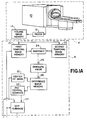
- One embodiment of a method and apparatus according to the invention will now be described with reference to the accompanying drawings in which:
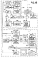
- Figures 1A and 1B illustrate an embodiment of the present invention;
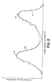
- Figure 2 illustrates exemplary blood and non-blood tissue probability curves.
- With reference to Figure 1, a magnetic resonance imaging apparatus A examines a selected region of a subject and generates a volume image representation thereof. The volume image representation of the heart and surrounding areas, is a product of a black blood cine sequence in which the volume is defined by a plurality of parallel slices. A motion detection means B locates the region of the ventricles. A segmentation means C parametrically determines intensity ranges of voxels of the image
- Another advantage of the present invention resides in the improved accuracy. Eliminating lung and uncertain edge values before determining the criiteria for classifying a voxel as blood and non-blood eliminates a major source of error in the resultant images.
- Another advantage of the present invention is that iot produces accurate results even with noisy images. Making voxel classification decisions based on a histogram as opposed to individual voxel properties improves overall reliability.
- Other advantages of the present invention reside in its improved processing speed and reduced complexity. Iterative data analysis is eliminated, as are training sets and the inputting of a priori information or a knowledge base.
- One embodiment of a method and apparatus according to the invention will now be described with reference to the accompanying drawings in which:
- Figure 1 is a diagrammatic illustration of the present invention;
- Figure 2 illustrates exemplary blood and non-blood tissue probability curves.
- With reference to Figure 1, a magnetic resonance imaging apparatus A examines a selected region of a subject and generates a volume image representation thereof. The volume image representation of the heart and surrounding areas, is a product of a black blood cine sequence in which the volume is defined by a plurality of parallel slices. A motion detection means B locates the region of the ventricles. A segmentation means C parametrically determines intensity ranges of voxels of the image representation in a blood intensity range and voxels in a non-blood tissue intensity range. A region growing means D collects or counts contiguous image voxels which have the intensity of blood volume of the image representation.
- The magnetic resonance data acquisition means A includes a conventional
magnetic resonance scanner 10. The magnetic resonance scanner includes the appropriate coils and supporting electronics for generating a substantially uniform main magnetic field through an imaging field. Radio frequency coils and supporting electronics selectively introduce radio frequency pulses into the examination region to induce resonance of selected bipoles and to manipulate the induced resonance. Gradient field coils and supporting electronics apply gradient field pulses across the examination region to provide phase and frequency encoding in the excited resonance field and for defining slices or volumes. - Magnetic resonance echoes or other diagnostic data is collected and reconstructed by a reconstruction means 12 into the volume image representation which is stored in an appropriate
volume image memory 14. The reconstruction means 12 reconstructs a series of two dimensional slices which taken together define the volume. Alternately, a three dimensional reconstruction algorithm may be used. In the preferred black blood cine sequence, blood tissue appears at one intensity extreme, the intensity extreme which is commonly displayed as black on a black and white monitor. Non-blood tissue contributions to the image tend to be near the other intensity extreme, the intensity extreme which is generally displayed as white. Preferably, the black blood cine technique is used to generate a plurality of planes collected at the same point of the cardiac cycle. Obtaining high temporal resolution is attained by using cardiac gating. That is, the patient's cardiac cycle is monitored and the MRI scan commences a preselected duration after the R-wave or other characteristic portion of the cardiac cycle. Multiple measurements (up to 64) are acquired at a fixed interval (typically 10-25 msec). The preferred black blood cine sequence results in temporal resolution of 20 msec. which allows for 40 images covering 80% of a typical 1000 msec. R-R interval. Optionally, CT or other data acquisition means may be utilized. - The motion detecting means B causes two temporally adjacent images to be generated. That is, the black blood cine sequence is used to generate the same spatial volume, but offset a very short time interval. The second MRI scan is triggered a duration after the R-wave which is slightly longer (or shorter) than the duration after the R-wave at which the first image was generated. The motion means B includes a first memory means 20 for storing a first temporal image and a second temporal memory means 22 for storing a second temporal image. The first and second memory means are preferably portions of a large RAM or disc memory. A subtracting means 24 subtracts corresponding voxels of the two images generating a temporal difference image. Because the first and second temporally displaced images are taken very close together in time, they are essentially identical, except in areas of movement. The movement, of course, is primarily in the ventricles. Thus, the difference image is essentially blank, except for a dark line around the ventricles, which dark line has a width which substantially corresponds to the amount of cardiac movement in the time between the first and second images. An absolute value means 26 compares the absolute value of each voxel of the difference image with a preselected threshold value selected to differentiate between the differential ventricle movement surface or band line and stray differences. Small values are set to zero and other values are set to one. Optionally, an additional algorithm may be incorporated for discarding voxels which are remote from the contiguous surface of voxels surrounding the ventricles. The absolute value of the difference image is stored in a difference image memory means 28.
- The center of mass of the temporal difference image falls at or near the boundary between the left and right ventricles in a short axis view of a normal human heart. The second central moments of the difference Image correlate directly to how large a subimage is necessary in order to contain just the heart. A center of mass means 30 determines the center of mass or geometric center from the temporal difference image. A second moment means 32 calculates the second moments of the difference image. An editing means 34 edits the original volume image, slice by slice, from the first temporal image memory means 20 to produce a square cross section subimage which contains substantially just the heart. That is, the subimage is centered at the center of mass of the difference image and has side lengths equal to the root of the second central moments of the difference image at the largest cross section of the heart. Optionally, the center and image size information can be used to reformat the MRI sequence to limit the examination region to the subimage region.
- The segmentation means C, in the preferred embodiment, serpents or identifies three tissues of tissue - blood, lung, and other tissues of non-blood tissue. Prior to the determination of probabilities, a lung editing means 40 examines the voxels of the original volume image in
memory 20 to locate the lung region. Prior to computation of the histogram, all voxels in the lung region are excluded from the computation. Lung location is determined by region growing to collect background voxels over the entire images. The largest regions are along the left and right sides of the patient and the next largest region is the cavity containing the lung. An edge detection means 42 detects edge voxels corresponding to edge regions in the selected subimage in the subimage memory means 36. An edge image is computed by passing a sobel operator over the subimage and thresholding the results. The resulting histogram now contains primarily information related to blood and tissue only. This given a strong binomial flavor and assists in clustering the histogram to determine probabilities for blood and tissue. - A subimage histogram means 44 generates a histogram, i.e. intensity versus number of voxels, for voxels in the blood region and in the non-blood, non-lung, non-edge region. More specifically, a subimage histograms means 46 determines the intensity corresponding to each voxel of the subimage stored the
subimage memory 36. An edge voxel subtracting means 48 subtracts or deletes the contributions from the edge voxels as determined by the edge detection means 42. In this manner, voxels which contain part blood and part non-blood tissue are excluded fron being part of the basis of the projected probabilities. Analogously, a lung voxel subtracting means 50 subtracts or deletes the contribution of the voxels or voxels identified by the lung region identifying means 40 as being lung tissue. A bimodal histogram memory means 52 stores the histogram of voxel intensity versus number of occurrences of the blood and of the non-blood, non-lung, non-edge voxels. - With continuing reference to FIGURE 1 and further reference to FIGURE 2, the histogram defines a curve with two peaks, one
peak 60 corresponding to the average blood tissue intensity and theother peak 62 centered around the average non-blood (non-lung, non-edge) tissue intensity. The low or black intensity cluster of voxels represents blood and the high or light intensity cluster represents non-blood tissue. Afirst peak detector 64 detects one of the peaks, e.g. the blood cluster. A first curve fitting means 66 fits a smooth curve to the blood peak of the histogram. In the preferred embodiment, the curve fitting means 66 fits the blood peak to a firstGaussian curve 68. The intensity level occurring most frequently in the histogram is used as the initial estimate of the mean Gaussian distribution. The frequency of occurrence of the initial estimate of the amplitude and the initial estimate of the variance is estimated by tracking amplitude in the vicinity of the peak in both directions. An amplitude decrease by a factor of e, the base of naperian logarithms, marks the variance. These initial estimates of the mean and variance are entered into a non-linear least squares fit routine to estimate the Gaussian curve best representing the blood peak. A subtracting means 70 subtracts the blood tissue Gaussian curve from the bimodal histogram in the vicinity of the blood peak. A second peak detector means 72 detects the non-blood cluster peak. A second curve fitting means 74 fits a secondGaussian curve 76 to the non-blood tissue peak. - Of course, other curve fitting routines may also be used. Other appropriate curves for fitting to the histogram include:
- (1)

- (2)

- (3) Erlang x r e -x
- (4) Cauchy 1/(1+x 2)
- (5) Normal e -x/2
- (6) Beta x b (1-x) c
- A classification means 84 compares each available voxel intensity with the probability curves 68, 76 and classifies the voxel as either blood or non-blood tissue and the probability or certainty of the classification. That is, the apex of each of
Gaussian curves - The region growing means D includes a high contrast or blood image forming means 90 which determines the classification of each voxel of the subimage. More specifically, it consults the classification means 84 with the intensity of each voxel to determine whether the voxel represents blood or non-blood tissue. If the intensity represents non-blood tissue, a zero value is assigned to that voxel. If the intensity is classified as blood tissue, then the blood
image forming means 90 consults the probability distribution curve memory means 80 to determine the confidence with which the corresponding intensity was classified as blood. Again, the apex of the best fit curve is classified as one or a 100% confidence value. Values further does the curve from the peak have a correspondingly lower confidence value. In the preferred embodiment, each voxel which is determined to represent blood, is assigned the corresponding confidence value or probability. Alternately, each voxel which is determined to represent blood tissue, may be given the binary value one and voxels which are determined to represent non-blood tissue may be given the value zero. As yet another alternative, those voxels which are determined to represent non-blood tissue may be given the inverse of their confidence level, i.e. the probability that the voxel does not represent non-blood tissue, i.e. represents blood tissue. - The high contrast or blood image representation is stored in a high contrast image memory means 92.
- The growing means D starts at a voxel which is known to be in either the left or right ventricle from the center of mass and second moment determinations made by
means - A summing
means 96 sums the contiguous blood voxel values which the stepping means 94 reads out of thehigh contrast memory 92. This sum of the confidences is multiplied by the volume of each voxel by multiplyingmeans 98. This product is indicative of the volume of the ventricle, which volume is stored in a ventricle volume memory means 100. The stepping means 94 then steps to a voxel which is predicted from the center mass to be generally centered in the other ventricle and the process repeated. - More specifically to the preferred embodiment, the ventricle volume memory means 100 includes a left ventricle volume memory means 100ℓ and a right ventricle volume memory means 100r. A volume display means 102 converts the stored ventricle volumes to an appropriate format for display. In the illustrated embodiment, the display means 102 converts the number into an appropriate format to be displayed on a
video monitor 104. A slice selecting means 106 enables an operator to select one or more slices of the image representation stored in the high contrast memory means 92 or the subimage memory means 36 to be displayed on thevideo monitor 104. - The left and right ventricles may not appear as distinct subregions in every slice and in every cardiac phase. Thus, in slices in which the ventricles appear connected, the stepping means will tend to step through both ventricles as it moves from contiguous blood voxel to contiguous blood voxel. An
edge operator routine 110 examines the initial image from memory means 20 or the subimage from memory means 36. The high frequency components of the data which was reconstructed by the reconstructingmeans 12 identifies the interfaces indicative of the edge or boundary between each ventricle and the cardiac tissue. In the preferred embodiment, theedge operator routine 110 uses a sobel edge detector to determine those points lying along the edges. The edge operator based on this analysis defines the edges of each ventricle. The stepping means compares the address of each voxel which it is about to access in the high contrast memory means 92 to determine whether or not it is within or across the edge parameters determined by theedge operator routine 110. Voxels outside of the ventricle volume indicated by theedge operator routine 110 are not accessed or read out to the summingmeans 96. - A comparing
means 112 compares the volumes of the left and right ventricles after the voxels of each slice are summed. By comparing the relative volumes of the left and right ventricles in each slice and by comparing the volumes of each ventricle with the preceding slice or adjacent slices, the comparing means 112 readily determines whether the stepping means has stepped between the two ventricles. When the comparing means determines that the ventricles have been combined, the comparing means enables the stepping means 94 to access theedge operator routine 110 to determine whether or not it is crossing the ventricle boundaries.
Claims (8)
- A method of measuring ventricular volumes comprising: non-invasively examining a region of a patient which includes a heart and generating a first image representation representing the region and a second image representation of the region, the first and second image representations being displaced in time; subtracting the first and second image representations to create a difference image representation in which voxel values of voxels of the first and second image representation representing substantially stationary tissue cancel and voxel values of like voxels representing tissues which moved in the time between generating the first and second image representations do not cancel, whereby a surface of non-cancelling voxel values is defined along a surface around ventricles of the heart, and counting a number of voxels within the ventricle defining surface, the number of counted voxels being proportional to ventricular volume.
- A method according to Claim 1 wherein the non-invasive examination step includes using a black blood cine technique such that the first and second image representations each depict blood tissue in black and non-blood tissue in lighter shades, whereby the difference image representation depicts the ventricle defining surface in black against a substantially white background.
- A method according to Claim 1 further including determining a confidence value indicative of a probability that each voxel value within the ventricular surface defines blood, and weighing each counted voxel in accordance with the confidence value.
- A method according to Claim 3, wherein the step of determining the confidence value includes; determining a frequency with which each voxel value occurs in voxels of the first image representation within the ventricle defining surfaces; determining a deviation between a most commonly occurring voxel value and each other occurring voxel value, the confidence value being proportional to the deviation.
- A method according to Claim 1, wherein the counting step includes: independently counting contiguous voxels of left and right ventricles within a common generally horizontal plane, whereby in planes in which voxels within the left and right ventricle are contiguous, there is a tendency to count voxels attributable to both the left and the right ventricle as representing the volume of a single one of the left and right ventricles; comparing a number of voxels counted in each plane with a number of voxels counted in an adjacent plane to determine whether voxels in both the left and right ventricles have been counted as voxels of the single one of the ventricles.
- A method according to Claim 5 further including: analysing the first image representation with an edge operator routine to determine an interface between the left and right ventricles; and limiting the counting step to voxels on each side of the determined interface.
- An apparatus for analysing ventricular volumes, the apparatus comprising: non invasive examination means (A,B) for non-invasively examining a region of a patient which includes a heart and generating (1) a first image representation representing the region and (2) a second image representation of the region, the first and second image representations being displaced in time; subtracting means (24) for subtracting the first and second image representations to create a difference image representation in which voxel values of voxels of the first and second image representations representing substantially stationary tissue cancel and voxel values of like voxels representing tissues which moved between the first and second images do not cancel, whereby the difference image representation represents a surface of non-cancelling voxel values along a surface around ventricles of the heart, counting means (96) for counting a number of voxels within the ventricle defining surface, the number of counted voxels being proportional to ventricular volume; and, display means (104) for displaying the ventricular volume.
- An apparatus according to Claim 7 further including; probability determining means (90) for determining a confidence value indicative of a probability that each voxel value within the ventricular surface defines blood; and weighting means (90) for weighting each voxel in accordance with the confidence value.
Priority Applications (1)
| Application Number | Priority Date | Filing Date | Title |
|---|---|---|---|
| EP96202027A EP0747004B1 (en) | 1991-11-14 | 1992-09-18 | Method of measuring ventricular volumes |
Applications Claiming Priority (2)
| Application Number | Priority Date | Filing Date | Title |
|---|---|---|---|
| US791855 | 1991-11-14 | ||
| US07/791,855 US5273040A (en) | 1991-11-14 | 1991-11-14 | Measurement of vetricle volumes with cardiac MRI |
Related Child Applications (2)
| Application Number | Title | Priority Date | Filing Date |
|---|---|---|---|
| EP96202027A Division EP0747004B1 (en) | 1991-11-14 | 1992-09-18 | Method of measuring ventricular volumes |
| EP96202027.7 Division-Into | 1996-07-16 |
Publications (2)
| Publication Number | Publication Date |
|---|---|
| EP0542414A1 EP0542414A1 (en) | 1993-05-19 |
| EP0542414B1 true EP0542414B1 (en) | 1997-11-26 |
Family
ID=25154982
Family Applications (2)
| Application Number | Title | Priority Date | Filing Date |
|---|---|---|---|
| EP96202027A Expired - Lifetime EP0747004B1 (en) | 1991-11-14 | 1992-09-18 | Method of measuring ventricular volumes |
| EP92308551A Expired - Lifetime EP0542414B1 (en) | 1991-11-14 | 1992-09-18 | Method of measuring ventricular volumes |
Family Applications Before (1)
| Application Number | Title | Priority Date | Filing Date |
|---|---|---|---|
| EP96202027A Expired - Lifetime EP0747004B1 (en) | 1991-11-14 | 1992-09-18 | Method of measuring ventricular volumes |
Country Status (4)
| Country | Link |
|---|---|
| US (3) | US5273040A (en) |
| EP (2) | EP0747004B1 (en) |
| JP (1) | JP3593616B2 (en) |
| DE (2) | DE69231102T2 (en) |
Cited By (1)
| Publication number | Priority date | Publication date | Assignee | Title |
|---|---|---|---|---|
| US8320652B2 (en) | 2005-04-01 | 2012-11-27 | Koninklijke Philips Electronics N.V. | Method, a system and a computer program for segmenting a structure in a Dataset |
Families Citing this family (116)
| Publication number | Priority date | Publication date | Assignee | Title |
|---|---|---|---|---|
| US5329925A (en) * | 1991-11-14 | 1994-07-19 | Picker International, Inc. | Reduced scan time cardiac gated magnetic resonance cine and flow imaging |
| US5348011A (en) * | 1991-11-14 | 1994-09-20 | Picker International, Inc. | Shared excitation phase encode grouping for improved throughput cardiac gated MRI cine imaging |
| US5421331A (en) * | 1992-03-10 | 1995-06-06 | Siemens Medical Systems, Inc. | Automatic identification of the long axis of the left ventricle from nuclear medicine spect data for use in myocardial perufsion studies |
| US5425368A (en) * | 1993-03-17 | 1995-06-20 | Board Of Regents, The University Of Texas System | Method and apparatus for estimating tissue volumes in magnetic resonance images |
| US5368033A (en) * | 1993-04-20 | 1994-11-29 | North American Philips Corporation | Magnetic resonance angiography method and apparatus employing an integration projection |
| US5435310A (en) * | 1993-06-23 | 1995-07-25 | University Of Washington | Determining cardiac wall thickness and motion by imaging and three-dimensional modeling |
| US5601084A (en) * | 1993-06-23 | 1997-02-11 | University Of Washington | Determining cardiac wall thickness and motion by imaging and three-dimensional modeling |
| FR2708166A1 (en) * | 1993-07-22 | 1995-01-27 | Philips Laboratoire Electroniq | A method of processing digitized images for the automatic detection of stenoses. |
| US6415048B1 (en) * | 1993-10-12 | 2002-07-02 | Schneider Medical Technologies, Inc. | Compositional analysis system |
| EP0687373A1 (en) * | 1993-12-30 | 1995-12-20 | Koninklijke Philips Electronics N.V. | Automatic segmentation and skinline detection in digital mammograms |
| US5550933A (en) * | 1994-05-27 | 1996-08-27 | Duke University | Quadrature shape detection using the flow integration transform |
| US5644646A (en) * | 1994-08-05 | 1997-07-01 | University Of Utah Research Foundation | Vessel enhancement filtering in magnetic resonance angiography |
| US5825936A (en) * | 1994-09-22 | 1998-10-20 | University Of South Florida | Image analyzing device using adaptive criteria |
| US6510240B1 (en) * | 1995-05-09 | 2003-01-21 | Texas Instruments Incorporated | Automatic detection of die absence on the wire bonding machine |
| US5875267A (en) * | 1995-08-21 | 1999-02-23 | Integrated Device Technology, Inc. | Fast digital image sharpening method and structure |
| US5845007A (en) * | 1996-01-02 | 1998-12-01 | Cognex Corporation | Machine vision method and apparatus for edge-based image histogram analysis |
| US5802212A (en) * | 1996-02-09 | 1998-09-01 | Seiko Epson Corporation | Clustered-dot dither with white-fleck suppression |
| JPH09261481A (en) * | 1996-03-22 | 1997-10-03 | Toshiba Corp | Image forming device |
| US5946454A (en) * | 1996-08-15 | 1999-08-31 | Seiko Epson Corporation | Image enhancement during half-toning using low-pass and high-pass filtering |
| US5754524A (en) * | 1996-08-30 | 1998-05-19 | Wark; Barry J. | Computerized method and system for analysis of an electrophoresis gel test |
| US5903664A (en) * | 1996-11-01 | 1999-05-11 | General Electric Company | Fast segmentation of cardiac images |
| JP3702054B2 (en) * | 1996-11-14 | 2005-10-05 | 株式会社東芝 | Magnetic resonance imaging system |
| US5966475A (en) * | 1997-02-20 | 1999-10-12 | Hewlett-Packard Company | System for enlarging and smoothing textual characters |
| JP4040742B2 (en) * | 1997-03-28 | 2008-01-30 | 株式会社東芝 | MRI equipment |
| US5867606A (en) * | 1997-08-12 | 1999-02-02 | Hewlett-Packard Company | Apparatus and method for determining the appropriate amount of sharpening for an image |
| US6246784B1 (en) * | 1997-08-19 | 2001-06-12 | The United States Of America As Represented By The Department Of Health And Human Services | Method for segmenting medical images and detecting surface anomalies in anatomical structures |
| US6246783B1 (en) | 1997-09-17 | 2001-06-12 | General Electric Company | Iterative filter framework for medical images |
| US6016057A (en) * | 1998-04-17 | 2000-01-18 | General Electric Company | System and method for improved water and fat separation using a set of low resolution MR images |
| US6516092B1 (en) | 1998-05-29 | 2003-02-04 | Cognex Corporation | Robust sub-model shape-finder |
| US6240201B1 (en) * | 1998-07-24 | 2001-05-29 | Arch Development Corporation | Computerized detection of lung nodules using energy-subtracted soft-tissue and standard chest images |
| US6492809B1 (en) * | 1998-12-04 | 2002-12-10 | Schlumberger Technology Corporation | Preconditioning spins near a nuclear magnetic resonance region |
| US6614474B1 (en) * | 1998-08-27 | 2003-09-02 | Polycom, Inc. | Electronic pan tilt zoom video camera with adaptive edge sharpening filter |
| RU2150146C1 (en) | 1998-09-03 | 2000-05-27 | Семенченко Михаил Григорьевич | Method for image processing |
| US6295384B1 (en) * | 1998-11-04 | 2001-09-25 | Schlumberger Technologies, Inc. | Removing noise caused by artifacts from a digital image signal |
| AU1726900A (en) * | 1998-11-24 | 2000-06-13 | Acuson Corporation | Processing ultrasound data during or after compression |
| US6042545A (en) * | 1998-11-25 | 2000-03-28 | Acuson Corporation | Medical diagnostic ultrasound system and method for transform ultrasound processing |
| EP1171033A4 (en) | 1999-04-21 | 2005-09-21 | Auckland Uniservices Ltd | METHOD AND SYSTEM FOR EVALUATION OF THE CHARACTERISTICS OF AN ORGAN |
| US6845342B1 (en) * | 1999-05-21 | 2005-01-18 | The United States Of America As Represented By The Department Of Health And Human Services | Determination of an empirical statistical distribution of the diffusion tensor in MRI |
| US6434412B1 (en) * | 1999-05-21 | 2002-08-13 | Siemens Medical Systems, Inc. | Cardiac cine imaging with a short repetition time and high contrast between the blood and the myocardium |
| US6721457B1 (en) | 1999-08-27 | 2004-04-13 | Hewlett-Packard Development Company, L.P. | Method for enhancing digital images |
| US6397096B1 (en) | 2000-03-31 | 2002-05-28 | Philips Medical Systems (Cleveland) Inc. | Methods of rendering vascular morphology in MRI with multiple contrast acquisition for black-blood angiography |
| US6340887B1 (en) | 1999-09-21 | 2002-01-22 | Picker International Inc. | Multiple contrast FSE approach to black blood angiography with redundant and supplementary vascular information |
| US6249693B1 (en) * | 1999-11-01 | 2001-06-19 | General Electric Company | Method and apparatus for cardiac analysis using four-dimensional connectivity and image dilation |
| US6438403B1 (en) * | 1999-11-01 | 2002-08-20 | General Electric Company | Method and apparatus for cardiac analysis using four-dimensional connectivity |
| US6563941B1 (en) * | 1999-12-14 | 2003-05-13 | Siemens Corporate Research, Inc. | Model-based registration of cardiac CTA and MR acquisitions |
| WO2002005544A1 (en) * | 2000-07-06 | 2002-01-17 | Seiko Epson Corporation | Image processing method, recording medium, and image processing device |
| EP1223553A3 (en) * | 2000-10-17 | 2003-09-24 | Fuji Photo Film Co., Ltd. | Apparatus for suppressing noise by adapting filter characteristics to input image signal based on characteristics of input image signal |
| WO2002056240A1 (en) * | 2000-11-22 | 2002-07-18 | R2 Technology, Inc. | Graphical user interface for display of anatomical information |
| US6556009B2 (en) | 2000-12-11 | 2003-04-29 | The United States Of America As Represented By The Department Of Health And Human Services | Accelerated magnetic resonance imaging using a parallel spatial filter |
| US6725077B1 (en) * | 2000-12-29 | 2004-04-20 | Ge Medical Systems Global Technology Company, Llc | Apparatus and method for just-in-time localization image acquisition |
| EP1362329B1 (en) | 2001-02-13 | 2019-03-27 | Koninklijke Philips N.V. | Processing of images in a direction of succession |
| WO2002065395A2 (en) * | 2001-02-13 | 2002-08-22 | Koninklijke Philips Electronics N.V. | Analysis of successive data sets |
| DE10107765A1 (en) * | 2001-02-17 | 2002-08-29 | Siemens Ag | Process for image processing based on a computed tomography (CT) image of a lung taken using a contrast medium and CT device for carrying out such a process |
| US6618609B2 (en) * | 2001-03-30 | 2003-09-09 | Koninklifke Philips Electronics, N.V. | Flow adaptive guided determination of imaging parameters for 3D magnetic resonance angiography |
| US6771067B2 (en) | 2001-04-03 | 2004-08-03 | The United States Of America As Represented By The Department Of Health And Human Services | Ghost artifact cancellation using phased array processing |
| US7020314B1 (en) | 2001-11-13 | 2006-03-28 | Koninklijke Philips Electronics N.V. | Black blood angiography method and apparatus |
| US7024027B1 (en) | 2001-11-13 | 2006-04-04 | Koninklijke Philips Electronics N.V. | Method and apparatus for three-dimensional filtering of angiographic volume data |
| US7397937B2 (en) * | 2001-11-23 | 2008-07-08 | R2 Technology, Inc. | Region growing in anatomical images |
| US7123008B1 (en) | 2002-04-19 | 2006-10-17 | Fonar Corporation | Positional magnetic resonance imaging |
| US8036730B1 (en) | 2002-04-19 | 2011-10-11 | Fonar Corporation | Temporal magnetic resonance imaging |
| US7149369B2 (en) * | 2002-04-23 | 2006-12-12 | Hewlett-Packard Development Company, L.P. | Method and system for image scaling |
| US6658280B1 (en) | 2002-05-10 | 2003-12-02 | E. Mark Haacke | Susceptibility weighted imaging |
| KR100639139B1 (en) * | 2002-06-28 | 2006-10-30 | 후지쯔 가부시끼가이샤 | Computer-readable recording media, three-dimensional image comparison methods, and three-dimensional image comparison apparatuses recording three-dimensional image comparison programs |
| US7260250B2 (en) * | 2002-09-30 | 2007-08-21 | The United States Of America As Represented By The Secretary Of The Department Of Health And Human Services | Computer-aided classification of anomalies in anatomical structures |
| US7289841B2 (en) | 2002-10-25 | 2007-10-30 | Koninklijke Philips Electronics N.V. | Method and apparatus for volumetric cardiac computed tomography imaging |
| US20040101184A1 (en) * | 2002-11-26 | 2004-05-27 | Radhika Sivaramakrishna | Automatic contouring of tissues in CT images |
| US8805073B2 (en) * | 2003-02-18 | 2014-08-12 | Koninklijke Philips N.V. | Image segmentation by assigning classes to adaptive mesh primitives |
| EP1620827B1 (en) | 2003-04-24 | 2010-06-02 | Koninklijke Philips Electronics N.V. | Non-invasive left ventricular volume determination |
| SG115540A1 (en) * | 2003-05-17 | 2005-10-28 | St Microelectronics Asia | An edge enhancement process and system |
| US7283655B2 (en) * | 2003-06-26 | 2007-10-16 | General Electric Company | System and method for segmenting tissue types in dual-echo magnetic resonance images |
| US7634120B2 (en) * | 2003-08-13 | 2009-12-15 | Siemens Medical Solutions Usa, Inc. | Incorporating spatial knowledge for classification |
| US7310435B2 (en) * | 2003-11-25 | 2007-12-18 | General Electric Company | Method and apparatus for extracting multi-dimensional structures using dynamic constraints |
| US7596255B2 (en) * | 2003-11-26 | 2009-09-29 | Ge Medical Systems Information Technologies, Inc. | Image navigation system and method |
| DE102004008979B4 (en) * | 2004-02-24 | 2006-12-28 | Siemens Ag | Method for filtering tomographic 3D representations after reconstruction of volume data |
| US7555171B2 (en) * | 2004-03-15 | 2009-06-30 | Sharp Laboratories Of America, Inc. | Systems and methods for down scaling a bitmap |
| US7664326B2 (en) * | 2004-07-09 | 2010-02-16 | Aloka Co., Ltd | Method and apparatus of image processing to detect and enhance edges |
| EP1794715A2 (en) * | 2004-09-24 | 2007-06-13 | Koninklijke Philips Electronics N.V. | Image interpolation |
| US7936941B2 (en) * | 2004-10-08 | 2011-05-03 | Panasonic Corporation | Apparatus for clearing an image and method thereof |
| US7477771B2 (en) * | 2004-12-03 | 2009-01-13 | Genral Electric Company | Method and system for extracting information about the cardiac cycle from CT projection data |
| US7599542B2 (en) * | 2005-04-08 | 2009-10-06 | John Philip Brockway | System and method for detection and display of diseases and abnormalities using confidence imaging |
| US8611632B2 (en) * | 2005-04-08 | 2013-12-17 | 361° Systems, Inc. | Method of selecting and visualizing findings within medical images |
| DE102005038940B4 (en) | 2005-08-17 | 2007-08-30 | Siemens Ag | Method for filtering tomographic 3D representations after reconstruction of volume data |
| DE102005043024A1 (en) * | 2005-09-09 | 2007-03-22 | Siemens Ag | Method for creating a preparation of the image requiring a preparation of the heart |
| TW200733720A (en) * | 2006-02-24 | 2007-09-01 | Pixart Imaging Inc | Digital image processing method and the device thereof |
| DE102006034132A1 (en) * | 2006-07-20 | 2008-01-24 | BAM Bundesanstalt für Materialforschung und -prüfung | Method for parameter-free image processing |
| JP5288745B2 (en) * | 2006-09-13 | 2013-09-11 | 株式会社東芝 | Magnetic resonance imaging system |
| EP2137694B1 (en) * | 2007-04-23 | 2013-08-28 | Philips Intellectual Property & Standards GmbH | Spatial-temporal warping of different pre-captured medical images |
| US20080281182A1 (en) * | 2007-05-07 | 2008-11-13 | General Electric Company | Method and apparatus for improving and/or validating 3D segmentations |
| FR2918486B1 (en) * | 2007-07-03 | 2009-09-18 | Gen Electric | METHOD FOR PROCESSING INTERVENTIONAL RADIOSCOPY IMAGES FOR DETECTION OF GUIDANCE INSTRUMENTATION MATERIALS |
| US8107758B2 (en) * | 2008-04-16 | 2012-01-31 | Microsoft Corporation | Block based image processing |
| US8798340B2 (en) * | 2009-11-10 | 2014-08-05 | Regents Of The University Of Minnesota | Dipole matched filter for MRI |
| WO2011093921A1 (en) * | 2010-01-29 | 2011-08-04 | Mayo Foundation For Medical Education And Research | Automated vascular region separation in medical imaging |
| US8326142B2 (en) * | 2010-02-12 | 2012-12-04 | Sri International | Optical image systems |
| DE102010013361B4 (en) * | 2010-03-30 | 2016-12-08 | Siemens Healthcare Gmbh | Improved time resolution for cardiac CT recordings |
| US8422756B2 (en) | 2010-04-27 | 2013-04-16 | Magnetic Resonance Innovations, Inc. | Method of generating nuclear magnetic resonance images using susceptibility weighted imaging and susceptibility mapping (SWIM) |
| US8897519B2 (en) * | 2010-09-28 | 2014-11-25 | Siemens Aktiengesellschaft | System and method for background phase correction for phase contrast flow images |
| US8738113B2 (en) | 2010-09-30 | 2014-05-27 | University Of Utah Research Foundation | Retrospectively correlated turbo spin echo imaging |
| GB2485209B (en) * | 2010-11-05 | 2015-11-04 | Inst Medical Informatics | Microscopy analysis technique |
| CN103596489A (en) * | 2011-06-10 | 2014-02-19 | 株式会社日立医疗器械 | Image diagnosis assisting apparatus, and method |
| KR101805623B1 (en) * | 2011-06-15 | 2017-12-07 | 삼성전자주식회사 | Method and apparatus for generating superresolution image using prediction and stabilization of high-frequency information of image |
| GB2495150B (en) * | 2011-09-30 | 2015-07-01 | Mirada Medical | Method and system of defining a region of interest on medical scan images |
| US8995782B2 (en) | 2011-11-21 | 2015-03-31 | Georgetown University | System and method for enhancing the legibility of degraded images |
| US9081097B2 (en) | 2012-05-01 | 2015-07-14 | Siemens Medical Solutions Usa, Inc. | Component frame enhancement for spatial compounding in ultrasound imaging |
| CN103513204B (en) * | 2012-06-29 | 2016-03-30 | 西门子(深圳)磁共振有限公司 | The track correcting method of K space data and device in a kind of magnetic resonance imaging |
| CN104412296B (en) * | 2012-06-29 | 2017-12-01 | 株式会社岛津制作所 | Image processing apparatus |
| TWI500412B (en) * | 2012-11-09 | 2015-09-21 | Iner Aec Executive Yuan | A method for improving image quality and imaging system using the same |
| CN102961161A (en) * | 2012-11-27 | 2013-03-13 | 华南理工大学 | Method for automatically obtaining heart function parameters of four-dimensional heart |
| JP5722984B2 (en) * | 2013-12-06 | 2015-05-27 | ザイオソフト株式会社 | Medical image processing apparatus and medical image processing program |
| CN105426927B (en) * | 2014-08-26 | 2019-05-10 | 东芝医疗系统株式会社 | Medical image processing apparatus, medical image processing method and medical image equipment |
| US10430688B2 (en) | 2015-05-27 | 2019-10-01 | Siemens Medical Solutions Usa, Inc. | Knowledge-based ultrasound image enhancement |
| WO2018002221A1 (en) * | 2016-06-29 | 2018-01-04 | Koninklijke Philips N.V. | Change detection in medical images |
| US10545211B2 (en) * | 2017-06-28 | 2020-01-28 | Synaptive Medical (Barbados) Inc. | Method of correcting gradient nonuniformity in gradient motion sensitive imaging applications |
| JP6996930B2 (en) * | 2017-10-13 | 2022-01-17 | キヤノンメディカルシステムズ株式会社 | Magnetic resonance imaging device |
| US10726588B2 (en) * | 2018-10-31 | 2020-07-28 | Biosense Webster (Israel) Ltd. | Magnetic resonance imaging (MRI) image filtration according to different cardiac rhythms |
| DE102020206515A1 (en) | 2020-05-26 | 2021-12-02 | Siemens Healthcare Gmbh | Saturation-prepared recording of MR image data |
| CN114764757A (en) * | 2022-03-17 | 2022-07-19 | 重庆师范大学 | Soil image brightness controllable enhancement method based on weighted Gaussian subtraction fitting |
Family Cites Families (43)
| Publication number | Priority date | Publication date | Assignee | Title |
|---|---|---|---|---|
| US4295473A (en) * | 1979-05-24 | 1981-10-20 | George Diamond | Apparatus and method for analysis of motion of a dynamic structure |
| US4499493A (en) * | 1983-02-22 | 1985-02-12 | The Board Of Trustees Of The Leland Stanford Junior University | Multiple measurement noise reducing system using artifact edge identification and selective signal processing |
| US4503461A (en) * | 1983-02-22 | 1985-03-05 | The Board Of Trustees Of The Leland, Stanford Junior University | Multiple measurement noise reducing system using space-variant filters |
| IL70213A (en) * | 1983-11-13 | 1988-02-29 | Paul Fenster | Digital fluorographic image enhancement system |
| US4851678A (en) * | 1984-07-31 | 1989-07-25 | Fuji Photo Film Co., Ltd. | Method of adjusting radiation image read-out conditions |
| US4637400A (en) * | 1985-02-15 | 1987-01-20 | Marcus Edward N | For characterizing a heart condition |
| US4777957A (en) * | 1985-06-14 | 1988-10-18 | General Electric Company | Method for measuring and imaging fluid flow |
| DE3605990A1 (en) * | 1986-02-25 | 1987-09-03 | Spectrospin Ag | METHOD FOR DETERMINING FLOWING MATERIALS BY MEANS OF NMR TOMOGRAPHY |
| US4834964A (en) * | 1986-03-07 | 1989-05-30 | M.R.I., Inc. | Use of charged nitroxides as NMR image enhancing agents for CSF |
| US4716904A (en) * | 1986-06-23 | 1988-01-05 | University Of Pittsburgh Of The Commonwealth System Of Higher Education | Heart output and circulatory impedance measuring method and apparatus |
| US4836209A (en) * | 1986-08-07 | 1989-06-06 | Stanford University | NMR imaging of moving material using variable spatially selected excitation |
| US4715383B1 (en) * | 1986-11-10 | 1995-10-31 | Mayo Medical Resources | Method for reducing artifacts in NMR images |
| US4761819A (en) * | 1987-02-27 | 1988-08-02 | Picker International, Inc. | Adaptive noise reduction filter for reconstructed images |
| WO1988007349A1 (en) * | 1987-03-25 | 1988-10-06 | Fonar Corporation | Nuclear magnetic resonance imaging |
| DE3816780A1 (en) * | 1987-05-18 | 1988-12-01 | Canon Kk | Method and device for image processing |
| US4961425A (en) * | 1987-08-14 | 1990-10-09 | Massachusetts Institute Of Technology | Morphometric analysis of anatomical tomographic data |
| FR2621693A1 (en) * | 1987-10-13 | 1989-04-14 | Thomson Cgr | METHOD FOR IMAGING INTRAVOXAL MOVEMENTS BY NMR IN A BODY |
| EP0327107B1 (en) * | 1988-02-05 | 1994-07-13 | Dainippon Screen Mfg. Co., Ltd. | Method and processor for enhancing image sharpness for image reproduction with scanner |
| US5078141A (en) * | 1988-04-29 | 1992-01-07 | Kabushiki Kaisha Toshiba | Method and system for acquiring image representing brain surface anatomy |
| US4849697A (en) * | 1988-06-27 | 1989-07-18 | General Electric Company | Three-dimensional magnetic resonance flow-contrast angiography with suppression of stationary material |
| US4991092A (en) * | 1988-08-12 | 1991-02-05 | The Regents Of The University Of California | Image processor for enhancing contrast between subregions of a region of interest |
| US5063607A (en) * | 1988-10-24 | 1991-11-05 | Hughes Aircraft Company | Image contrast enhancement technique |
| US5072384A (en) * | 1988-11-23 | 1991-12-10 | Arch Development Corp. | Method and system for automated computerized analysis of sizes of hearts and lungs in digital chest radiographs |
| JPH02162475A (en) * | 1988-12-15 | 1990-06-22 | Dainippon Screen Mfg Co Ltd | Picture contour correcting method |
| JPH02255126A (en) * | 1989-03-29 | 1990-10-15 | Toshiba Corp | Magnetic resonance imaging method |
| US5159550A (en) * | 1989-04-20 | 1992-10-27 | Mitsubishi Denki Kabushiki Kaisha | Artifact suppression system for magnetic resonnance imaging apparatus |
| US5199435A (en) * | 1989-06-13 | 1993-04-06 | Kabushiki Kaisha Toshiba | Magnetic resonance imaging system |
| DE4018683A1 (en) * | 1989-06-23 | 1991-01-10 | Siemens Ag | LAYERING PROFILE OPTIMIZATION FOR A NUCLEAR SPIN TOMOGRAPHER USED WITH A GRADIENT ECHOSE SEQUENCE |
| IL90862A (en) * | 1989-07-04 | 1992-09-06 | Elscint Ltd | Localized multiregion magnetic resonance data acquisition |
| US5001429A (en) * | 1989-11-21 | 1991-03-19 | General Electric Company | Removal of truncation artifacts in NMR imaging |
| US5034694A (en) * | 1989-11-22 | 1991-07-23 | Picker International, Inc. | Minimum basis set MR angiography |
| JP2867055B2 (en) * | 1990-01-29 | 1999-03-08 | 富士写真フイルム株式会社 | Edge determination method and apparatus |
| JP2853232B2 (en) * | 1990-02-02 | 1999-02-03 | 株式会社日立製作所 | Fluid imaging device using nuclear magnetic resonance |
| JPH0820725B2 (en) * | 1990-02-06 | 1996-03-04 | 大日本スクリーン製造株式会社 | How to create image contour data |
| US5072314A (en) * | 1990-04-04 | 1991-12-10 | Rockwell International Corporation | Image enhancement techniques using selective amplification of spatial frequency components |
| US5218649A (en) * | 1990-05-04 | 1993-06-08 | U S West Advanced Technologies, Inc. | Image enhancement system |
| US5222500A (en) * | 1990-06-22 | 1993-06-29 | Kabushiki Kaisha Toshiba | Magnetic resonance imaging method and apparatus for the same |
| US5031624A (en) * | 1990-08-17 | 1991-07-16 | Wisconsin Alumni Research Foundation | Phase contrast, line-scanned method for NMR angiography |
| US5031227A (en) * | 1990-09-27 | 1991-07-09 | Loral Aerospace Corp. | Edge enhancement and smoothing in digital images |
| US5170440A (en) * | 1991-01-30 | 1992-12-08 | Nec Research Institute, Inc. | Perceptual grouping by multiple hypothesis probabilistic data association |
| US5081692A (en) * | 1991-04-04 | 1992-01-14 | Eastman Kodak Company | Unsharp masking using center weighted local variance for image sharpening and noise suppression |
| US5307014A (en) * | 1991-10-18 | 1994-04-26 | Siemens Medical Systems, Inc. | Inflow MR angiography with spatially variable flip angles |
| US5271399A (en) * | 1991-11-27 | 1993-12-21 | Trustees Of The University Of Pennsylvania | Three dimensional Fourier transform, fast spin echo, black blood magnetic resonance angtography |
-
1991
- 1991-11-14 US US07/791,855 patent/US5273040A/en not_active Expired - Lifetime
-
1992
- 1992-03-27 US US07/859,153 patent/US5447155A/en not_active Expired - Lifetime
- 1992-06-05 US US07/893,785 patent/US5351305A/en not_active Expired - Lifetime
- 1992-09-18 EP EP96202027A patent/EP0747004B1/en not_active Expired - Lifetime
- 1992-09-18 DE DE69231102T patent/DE69231102T2/en not_active Expired - Fee Related
- 1992-09-18 DE DE69223307T patent/DE69223307T2/en not_active Expired - Fee Related
- 1992-09-18 EP EP92308551A patent/EP0542414B1/en not_active Expired - Lifetime
- 1992-11-13 JP JP32762692A patent/JP3593616B2/en not_active Expired - Fee Related
Cited By (1)
| Publication number | Priority date | Publication date | Assignee | Title |
|---|---|---|---|---|
| US8320652B2 (en) | 2005-04-01 | 2012-11-27 | Koninklijke Philips Electronics N.V. | Method, a system and a computer program for segmenting a structure in a Dataset |
Also Published As
| Publication number | Publication date |
|---|---|
| US5447155A (en) | 1995-09-05 |
| DE69223307T2 (en) | 1998-03-19 |
| EP0747004B1 (en) | 2000-05-24 |
| JPH05269115A (en) | 1993-10-19 |
| DE69231102T2 (en) | 2000-09-21 |
| EP0542414A1 (en) | 1993-05-19 |
| EP0747004A3 (en) | 1998-02-25 |
| EP0747004A2 (en) | 1996-12-11 |
| DE69231102D1 (en) | 2000-06-29 |
| US5273040A (en) | 1993-12-28 |
| US5351305A (en) | 1994-09-27 |
| JP3593616B2 (en) | 2004-11-24 |
| DE69223307D1 (en) | 1998-01-08 |
Similar Documents
| Publication | Publication Date | Title |
|---|---|---|
| US5273040A (en) | Measurement of vetricle volumes with cardiac MRI | |
| US7974473B2 (en) | Image processing method and image processing apparatus | |
| EP1446051B1 (en) | Calibration of tissue densities in computerized tomography | |
| EP1302163B1 (en) | Apparatus for calculating an index of local blood flows | |
| US7620227B2 (en) | Computer-aided detection system utilizing temporal analysis as a precursor to spatial analysis | |
| US20210361254A1 (en) | Methods and apparatus for extended low contrast detectability for radiographic imaging systems | |
| US7995825B2 (en) | Histogram segmentation of FLAIR images | |
| US6745066B1 (en) | Measurements with CT perfusion | |
| US7567696B2 (en) | System and method for detecting the aortic valve using a model-based segmentation technique | |
| CN101626726B (en) | Identification and analysis of lesions in medical imaging | |
| US5433199A (en) | Cardiac functional analysis method using gradient image segmentation | |
| US20030053669A1 (en) | Magnetic resonance angiography method and apparatus | |
| Adams et al. | Breast lesion discrimination using statistical analysis and shape measures on magnetic resonance imagery | |
| US5530789A (en) | Image processing apparatus and method for constructing a functional image using feature amounts of functions approximating changes in image data on a time axis | |
| Bland et al. | Robust three‐dimensional object definition in CT and MRI | |
| Koechner et al. | Segmentation of small structures in MR images: semiautomated tissue hydration measurement | |
| Duda et al. | Method for quantitative assessment of atherosclerotic lesion burden on the basis of high-resolution black-blood MRI | |
| Soltanian-Zadeh et al. | Semiautomatic brain morphometry from CT images |
Legal Events
| Date | Code | Title | Description |
|---|---|---|---|
| PUAI | Public reference made under article 153(3) epc to a published international application that has entered the european phase |
Free format text: ORIGINAL CODE: 0009012 |
|
| AK | Designated contracting states |
Kind code of ref document: A1 Designated state(s): DE FR NL |
|
| 17P | Request for examination filed |
Effective date: 19931018 |
|
| 17Q | First examination report despatched |
Effective date: 19960325 |
|
| GRAG | Despatch of communication of intention to grant |
Free format text: ORIGINAL CODE: EPIDOS AGRA |
|
| GRAH | Despatch of communication of intention to grant a patent |
Free format text: ORIGINAL CODE: EPIDOS IGRA |
|
| GRAH | Despatch of communication of intention to grant a patent |
Free format text: ORIGINAL CODE: EPIDOS IGRA |
|
| GRAA | (expected) grant |
Free format text: ORIGINAL CODE: 0009210 |
|
| AK | Designated contracting states |
Kind code of ref document: B1 Designated state(s): DE FR NL |
|
| XX | Miscellaneous (additional remarks) |
Free format text: TEILANMELDUNG 96202027.7 EINGEREICHT AM 16/07/96. |
|
| ET | Fr: translation filed | ||
| REF | Corresponds to: |
Ref document number: 69223307 Country of ref document: DE Date of ref document: 19980108 |
|
| PLBE | No opposition filed within time limit |
Free format text: ORIGINAL CODE: 0009261 |
|
| 26N | No opposition filed | ||
| PGFP | Annual fee paid to national office [announced via postgrant information from national office to epo] |
Ref country code: FR Payment date: 20010911 Year of fee payment: 10 |
|
| PGFP | Annual fee paid to national office [announced via postgrant information from national office to epo] |
Ref country code: NL Payment date: 20010927 Year of fee payment: 10 |
|
| PG25 | Lapsed in a contracting state [announced via postgrant information from national office to epo] |
Ref country code: NL Free format text: LAPSE BECAUSE OF NON-PAYMENT OF DUE FEES Effective date: 20030401 |
|
| PG25 | Lapsed in a contracting state [announced via postgrant information from national office to epo] |
Ref country code: FR Free format text: LAPSE BECAUSE OF NON-PAYMENT OF DUE FEES Effective date: 20030603 |
|
| REG | Reference to a national code |
Ref country code: FR Ref legal event code: ST |
|
| PGFP | Annual fee paid to national office [announced via postgrant information from national office to epo] |
Ref country code: DE Payment date: 20081112 Year of fee payment: 17 |
|
| PG25 | Lapsed in a contracting state [announced via postgrant information from national office to epo] |
Ref country code: DE Free format text: LAPSE BECAUSE OF NON-PAYMENT OF DUE FEES Effective date: 20100401 |