EP0536520B1 - Use of Human Interferon-Beta 2A for the preparation of medicaments - Google Patents
Use of Human Interferon-Beta 2A for the preparation of medicaments Download PDFInfo
- Publication number
- EP0536520B1 EP0536520B1 EP92114478A EP92114478A EP0536520B1 EP 0536520 B1 EP0536520 B1 EP 0536520B1 EP 92114478 A EP92114478 A EP 92114478A EP 92114478 A EP92114478 A EP 92114478A EP 0536520 B1 EP0536520 B1 EP 0536520B1
- Authority
- EP
- European Patent Office
- Prior art keywords
- ifn
- cells
- human
- gene
- cdna
- Prior art date
- Legal status (The legal status is an assumption and is not a legal conclusion. Google has not performed a legal analysis and makes no representation as to the accuracy of the status listed.)
- Expired - Lifetime
Links
Images


Classifications
-
- C—CHEMISTRY; METALLURGY
- C07—ORGANIC CHEMISTRY
- C07K—PEPTIDES
- C07K14/00—Peptides having more than 20 amino acids; Gastrins; Somatostatins; Melanotropins; Derivatives thereof
- C07K14/435—Peptides having more than 20 amino acids; Gastrins; Somatostatins; Melanotropins; Derivatives thereof from animals; from humans
- C07K14/52—Cytokines; Lymphokines; Interferons
- C07K14/555—Interferons [IFN]
- C07K14/565—IFN-beta
-
- A—HUMAN NECESSITIES
- A61—MEDICAL OR VETERINARY SCIENCE; HYGIENE
- A61K—PREPARATIONS FOR MEDICAL, DENTAL OR TOILETRY PURPOSES
- A61K38/00—Medicinal preparations containing peptides
- A61K38/16—Peptides having more than 20 amino acids; Gastrins; Somatostatins; Melanotropins; Derivatives thereof
- A61K38/17—Peptides having more than 20 amino acids; Gastrins; Somatostatins; Melanotropins; Derivatives thereof from animals; from humans
- A61K38/19—Cytokines; Lymphokines; Interferons
- A61K38/21—Interferons [IFN]
- A61K38/215—IFN-beta
-
- C—CHEMISTRY; METALLURGY
- C07—ORGANIC CHEMISTRY
- C07K—PEPTIDES
- C07K14/00—Peptides having more than 20 amino acids; Gastrins; Somatostatins; Melanotropins; Derivatives thereof
- C07K14/435—Peptides having more than 20 amino acids; Gastrins; Somatostatins; Melanotropins; Derivatives thereof from animals; from humans
- C07K14/52—Cytokines; Lymphokines; Interferons
- C07K14/54—Interleukins [IL]
- C07K14/5412—IL-6
-
- A—HUMAN NECESSITIES
- A61—MEDICAL OR VETERINARY SCIENCE; HYGIENE
- A61K—PREPARATIONS FOR MEDICAL, DENTAL OR TOILETRY PURPOSES
- A61K38/00—Medicinal preparations containing peptides
Definitions
- Interferon is an important antiviral and antitumor protein produced by the human body. Because of its species specificity, clinical use of interferon requires human interferon. Based on their neutralization by antibodies three types of interferon were defined which include interferon-alpha, produced mainly by leucocytes, interferon-beta, produced mainly by fibroblasts, and interferon-gamma, produced mainly by T-lymphocytes.
- the major species of interferon produced by human fibroblasts in response to ds RNA is the 20 Kd glycoprotein IFN- ⁇ 1 , encoded by a 0.9 kb RNA which is transcribed from an intron-less gene on chromosome 9.
- additional mRNAs which yield IFN activity when microinjected into frog oocytes have been observed in human fibroblasts induced by poly (rI)(rC) and by a sequential cycloheximide (CHX)-actinomycin D treatment.
- IFN- ⁇ 2 Since this activity was inhibited by anti-IFN- ⁇ 1 antibodies, it was designated IFN- ⁇ 2 .
- the protein product of IFN- ⁇ 2 RNA is immunologically distinct from IFN- ⁇ 1 , although the biological activities of the two proteins are cross-neutralized by the same antibodies.
- the IFN- ⁇ 2 1.3 kb RNA clearly originates from another gene than IFN- ⁇ 1 , since IFN- ⁇ 2 RNA is formed in mouse-human hybrids lacking chromosome 9 (U. Nir (1984) Ph.D. Thesis, Weizmann Institute of Science, Rehovot, Israel).
- interferon- ⁇ 2 is produced by isolating DNA containing the nucleotide sequence coding for interferon- ⁇ 2 in human cells after cultivating those cells which are able to produce interferon- ⁇ 2 when exposed to an inducer of interferon.
- mRNA from said induced cells is extracted and purified. Afterwards, the mRNA is transcribed into DNA and the DNA is cloned in a suitable vector.
- IFN- ⁇ 2 there is a second gene encoding IFN- ⁇ 2 , which, when isolated and transfected into hamster or mouse cells produces human-specific IFN activity after induction by ds RNA and cycloheximide.
- the IFN produced has the properties ascribed to IFN- ⁇ 2 but does not have identical amino acid sequences, the proteins produced by the two genes are denominated IFN- ⁇ 2A and IFN- ⁇ 2B respectively.
- IFN- ⁇ 2A and its use in pharmaceutical compositions is also disclosed in WO 88/00206 (not pre-published). Further, JP-A-60169424 discloses the administration of BCDF, which is another designation for IFN- ⁇ 2A , to patients suffering from immune incompetence.
- human interferon- ⁇ 2A is used for preparing a medicament useful in the field of inflammation and/or acute phase response.
- the IFN- ⁇ 2A is used for the preparation of a medicament inhibiting fibroblast proliferation or preventing sclerosis after infection.
- IFN- ⁇ 2 biologically active human interferon-beta 2
- IFN- ⁇ 2 is a glycoprotein secreted by human fibroblasts which are induced by double-stranded (ds) RNA or viruses to produce interferon.
- IFN- ⁇ 2 amounts to about 5% of the total IFN activity produced by fibroblasts.
- the amino acid sequence of IFN- ⁇ 2 which was determined by cDNA sequencing, indicates only about 20% overall homology with that of the other human IFNs produced by such human cells (IFN- ⁇ 1 and IFN- ⁇ s).
- a human cDNA or gene encoding IFN- ⁇ 2A may be cloned in accordance with the following description and fused to a strong viral transcriptional promoter.
- Hamster cells transformed by the fused cDNA or gene produce constitutively human-specific interferon activity which inhibits viral replication and cytopathic effect and induces the proteins typical of the biological response of human cells to interferons.
- a second gene encoding IFN- ⁇ 2 is also disclosed which, when isolated and transfected into hamster or mouse cells, produces human-specific IFN activity after induction by ds RNA and cycloheximide.
- the proteins produced by the two genes are denominated IFN- ⁇ 2A and IFN- ⁇ 2B respectively.
- interferon-beta 2 The existence of interferon-beta 2 was discovered in the course of attempts to isolate genetic material containing the nucleotide sequence coding for interferon in human fibroblast cells.
- fibroblast cells were induced for the production of inducible interferon mRNA with a suitable exogenous factor and then the mRNAs were extracted.
- the mRNAs from a non-induced culture of the same host cells were extracted.
- cDNA probes were synthesized from the mRNAs of both the induced culture and the non-induced control culture using the corresponding mRNAs as templates (induced cDNAs and non-induced cDNAs).
- Double-stranded cDNAs derived from the mRNA extracted from the induced culture were then synthesized and such cDNAs were inserted in appropriate vectors and transfected into suitable microorganisms.
- the microorganisms were cultivated under conditions suitable to cause selective development of microorganism colonies of the modified vectors (initial colonies). Duplicate colonies of the initial colonies were formed and the DNAs of both the initial and duplicate colonies were freed in situ .
- the DNAs of one of the two sets of duplicate colonies were hybridized with the cDNA probes synthesized from the mRNAs extracted from the induced culture and the other of the two sets of duplicate colonies were hybridized with the cDNA probes synthesized from the mRNAs extracted from the non-induced culture.
- Clones which selectively hybridized with the induced cDNA probes but not with the non-induced cDNA probes were selected and their DNAs recovered. These cloned DNAs were further tested to determine those DNAs capable of hybridizing with mRNA translatable in frog oocytes or reticulocyte-lysates into interferon, which DNAs are essentially those which code for interferon, or contain enough of the sequence coding for interferon to be used to obtain a complete interferon DNA.
- cDNA clones hybridizable to mRNA which translates to interferon-beta 2 were prepared in such a manner and an IFN- ⁇ 2 cDNA probe was prepared therefrom.
- IFN- ⁇ 2 clones A341 and E474 were found to represent overlapping sequencing and were fused to reconstitute the IFN- ⁇ 2A AE20 cDNA ( Figure 1).
- the nucleotide sequence of this IFN- ⁇ 2A cDNA was determined and revealed an open reading frame of 212 amino acids predicting a protein of 23,500 daltons. Transcription-translation experiments confirmed that this cDNA encodes such a protein.
- IFN- ⁇ 2A RNAs are induced in human fibroblasts by poly (rI)(rC) and induction is stimulated by IFN priming as in the case of IFN- ⁇ 1 RNA in these cells (U. Nir et al. (1984) Nucl. Acids Res. 12: 6979-6993).
- a prolonged (6.5 h) treatement with CHX also induces IFN- ⁇ 2A RNA, but shorter (3.5 h) CHX treatment is effective only with poly (rI)(rC) ( Figure 3).
- the low induction of IFN- ⁇ 2A RNA by CHX without ds RNA distinguishes this gene from IFN- ⁇ 1 .
- DNA constructs were made to express the IFA-2 gene under the control of the SV40 early gene promoter.
- a 4.8 kb segment of IFA-2 DNA was fused through a synthetic oligonucleotide to pSVE3 DNA (Y. Chernajovsky et al. (1984). supra) so that the ATG codon is 150 bp downstream from the SV40 early early RNA start (pSVIFA2-II, see legend, Figure 5).
- pSVIFA2-II see legend, Figure 5
- RNAs of 1.35 and 2.2 kb were detected in Northern blots by IFN- ⁇ 2 cDNA; these two RNAs would correspond to transcripts ending at the IFN- ⁇ 2 ad SV40 polyadenylation sites respectively ( Figure 5).
- Medium from the Cos7 cells during transient expression of the pSVIFA2-II DNA was assayed for IFN activity by inhibition of the cytopathic effect of VSV on human FS11 and Wish cells as described in D. Novick et al. (1983) J.Cen.Virol. 64: 905-910. Antiviral activity could be clearly detected two days after transfection (Table 1).
- the specific antiviral activity of IFN- ⁇ 2A per unit protein has been estimated to be 30 to 100 times lower than that of IFN- ⁇ 1 and closer to that of IFN- ⁇ 1 (5-10 x 10 6 units/mg).
- the IFN titers observed by transient expression of the IFA-2 gene were as expected, about 3% of those seen with the IFN- ⁇ 1 gene under the same SV40 promoter control (Table 1).
- Production of IFN- ⁇ 2A activity by pSVIFA2-II DNA cells was further demonstrated by induction of (2'-5') oligo A synthetase (M. Revel et al. (1981), supra) in FS11 cells exposed to the medium of the transfected Cos7 cells ( Figure 6). The induction was dose-dependent and corresponded quantitatively to the antiviral activity.
- the IFA-2 gene was also fused to the SV40 early promoter directly at the Xho1 site near S-2 (pSVIFA2-I DNA, legend of Figure 5).
- Hamster CHO-K1 DHFR - cells were cotransformed with pSVIFA2-I and pSVDHFR (Y. Chernajovsky et al. (1984), supra) DNAs and a stably transformed line SI-15 was isolated.
- This cell line produces constitutively 10-50 U/ml of IFN antiviral activity measured on human FS11 and Wish cells, while CHO cells transformed only by pSVDHFR produce no human IFN (Table 1).
- rIFN- ⁇ 2A solutions titrating about 3000 U/ml could regularly be obtained.
- This rIFN- ⁇ 2A has the properties expected for a human IFN and induces the (2'-5') oligo A synthetase in FS11 cells even at low concentrations of 1 U/ml ( Figure 6).
- the rIFN- ⁇ 2A constitutively produced by CHO SI-15 cells demonstrated human-specific induction of the mRNAs for (2'-5') oligo A synthetase, C56 and HLA in the presence of cycloheximide, its antiviral activity was neutralized by anti-IFN- ⁇ 1 (but not anti-IFN- ⁇ or anti-IFN - ⁇ ) polyclonal and monoclonal antibodies and it acted on mouse-human hybrids only if they contain human chromosome 21 which carries the gene for the type 1 IFN receptor.
- IFN- ⁇ 1 and ⁇ 2A Although the sequence homology between IFN- ⁇ 1 and ⁇ 2A is low, the cross-neutralization of the antiviral activities of IFN- ⁇ 1 and IFN- ⁇ 2A by the same antibodies, including a monoclonal anti-IFN- ⁇ 1 (D. Novick et al. (1983), supra), could result from some conserved structure in the active site of IFN- ⁇ . IFN- ⁇ 2A activity is not neutralized by anti-IFN- ⁇ or ⁇ antibodies, justifying its designation as a ⁇ -type IFN.
- the genes encoding IFN- ⁇ 2 are not on chromosome 9 (U. Nir (1984), supra) and differ from the type I IFN genes in the chromosome 9 cluster by the presence of introns.
- the IFA-2 gene was determined to be on chromosome 7 (P-B. Sehgal et al. (1986) Proc.Natl.Acad.Sci. USA , in press). The amino acid sequence homology detected is compatible with the notion that the IFN- ⁇ 2 genes are related to the ancestral gene which gave rise to the type I IFN gene cluster.
- the second genomic DNA clone IFA-11 hybridizing with the IFN- ⁇ 2A cDNA has a restriction map which differs from that of IFA-2 ( Figure 8). Transcript mapping and sequencing showed that the 3' exon of IFA-2 and IFA-11 (Xbal-HindIII segment) are identical (99% sequence homology), while there was no cross-hybridization under stringent conditions between more 5' exons.
- the IFA-11 genomic clone (19 Kb) was transfected into L-TK - cells together with a HSV-TK gene (F. Colbere-Garapin et al. (1981) J.Mol.Biol. 150: 1-14) and stable transformants were isolated.
- Table 1 shows that L cell clone LI-39 containing the human IFA-11 gene, produces human IFN activity when induced by poly (rI)(rC) and cycloheximide, while L-TK + cells produced no such activity.
- a CHO clone 1C40 containing the IFA-11 gene also produced human IFN activity (Table 1). No activity was seen in non-induced LI-39 or 1C40 cells.
- the IFN activity obtained with IFA- 11 transformants after induction was significantly higher than with the IFA-2 gene.
- the rIFN- ⁇ 2B produced by expression of the IFA-11 gene was neutralized with anti-IFN- ⁇ 1 similarly to the IFA-2 gene product.
- IFA-11 specific DNA probes hybridized to RNA from induced human fibroblasts similarly to IFN-2 DNA probes, indicating that both genes are active in human cells.
- IFN- ⁇ 2 genes The function of the IFN- ⁇ 2 genes seems to result from their expression under conditions in which other IFN genes are not expressed.
- TNF Tumor Necrosis Factor
- lymphokine IL-1 fibroblasts exposed to Tumor Necrosis Factor (TNF) or to lymphokine IL-1 produce the IFN- ⁇ 2 protein.
- TNF Tumor Necrosis Factor
- RNA hybridizing to IFN- ⁇ 2 cDNA was recently detected in peripheral blood mononuclear cells induced by mitogens such as PHA along with IFN- ⁇ mRNA. It has also been reported that an IL-1 like lymphokine strongly induces RNA hybridizing to IFN- ⁇ 2 cDNA in diploid fibroblasts and other human cell lines (J. Content et al. (1985) Eur.J.Biochem. 152: 253-257). The promoter region of the IFA-2 genes seems to contain two TATA boxes and two RNA starts ( Figures 1 and 3). This IFN- ⁇ 2 promoter was shown to respond to either poly (rI)(rC) or to cycloheximide (Y.
- IFN- ⁇ 2 gene promoters may be activated by different inducers (ds RNA, protein synthesis inhibitors, IL-1, PHA).
- inducers ds RNA, protein synthesis inhibitors, IL-1, PHA.
- IFN- ⁇ 2 may belong to the same group of minor IFN species produced by cells to auto-regulate their growth, while the classical type I IFNs are probably more specifically involved in theresponse to viral infections.
- IFN- ⁇ 2 The biological significance of IFN- ⁇ 2 lies most probably in the fact that it is induced under conditions where IFN- ⁇ 1 is not induced, as in metabolically stressed cells (A. Zilberstein et al., (1985) in The Interferon System, Serono Symposia Raven Press, pp.73-83). Most important is the observation that TNF induces IFN- ⁇ 2 in fibroblasts and that the proliferation of these cells which is stimulated by TNF is further enhanced if anti-IFN- ⁇ antibodies are added. A number of human cell lines have been shown to produce autocrine IFN- ⁇ species when undergoing differentiation, and these IFNs are responsible for the induction of HLA antigens on these cells (A. Yarden et al. (1984) Embo.J.
- IFN- ⁇ 2 appears to be one of these autocrine autoregulators of cell growth produced in response to growth factors. This would be in line with the low specific activity of IFN- ⁇ 2 (estimated by comparison of immunoassay and antiviral activity to be 50-100 times lower than for IFN- ⁇ 1 ), since only very small antiviral activity is seen in cells producing the autocrine IFN- ⁇ species (M. Revel (1983) in Interferon 5 , Academic Press, pp. 205-239). This could also explain why the hamster cells expressing IFN- ⁇ 2 cDNA produce less IFN activity than similar CHO cells harbouring the human IFN- ⁇ 1 gene (Y. Chernajovsky et al.
- IFN- ⁇ 1 (D) species has also a 100 times lower specific activity on human cells than other IFN- ⁇ species (T. Goren et al. (1983), Virology 130: 273-280), although it represents a large proportion of the total mass of leukocyte IFN (C. Weissmann (1981) in Interferon 3 , Academic Press, N.Y. pp.101-134). It is not excluded that some of the multiple IFN functions will be expressed more efficiently by IFN- ⁇ 2 than the antiviral effect.
- IFN- ⁇ 2 IL-1 and TNF suggests that it may play a role as an autocrine mediator of some effects of these cytokines in inflammation and acute-phase responses, as well as regulating cell proliferation.
- IFN- ⁇ 2 -specific immunocompetition assay with R-antibodies was used to examine if human fibroblasts FS11 cells indeed secrete the IFN- ⁇ 2 protein in response to these two cytokines ( Figure 9).
- rIL-1 ⁇ and TNF- ⁇ induced the synthesis and secretion of the IFN- ⁇ 2 protein, to levels estimated by the immunoassay to be 10-20 U/ml IFN- ⁇ 2 .
- Optimal IFN- ⁇ 2 induction was seen with concentrations of rIL-1 ⁇ of 4 U/ml (0.13 ng/ml), similar to those required for IL-1 induction of prostaglandin E 2 , collagenase and hyaluronic acid in human fibroblasts (J.H. Korn (1985), Arthritis Rheum. 28: 315-322).
- TNF- ⁇ 2 induction 400 U/ml; 40 ng/ml was higher than that required for the cytolytic effect on sensitive cells (0.1 ng/ml; A.M. Wang et al. (1985) Science 228: 149-154) and even higher than that giving optimal growth stimulation of diploid fibroblasts (2 ng/ml).
- the onset of IFN- ⁇ 2 secretion was somewhat faster with TNF- ⁇ reaching half maximal levels at about 6 hours, versus 8-12 hours with IL-1.
Landscapes
- Health & Medical Sciences (AREA)
- Life Sciences & Earth Sciences (AREA)
- Chemical & Material Sciences (AREA)
- Organic Chemistry (AREA)
- Medicinal Chemistry (AREA)
- Zoology (AREA)
- Gastroenterology & Hepatology (AREA)
- Proteomics, Peptides & Aminoacids (AREA)
- General Health & Medical Sciences (AREA)
- Biochemistry (AREA)
- Genetics & Genomics (AREA)
- Molecular Biology (AREA)
- Biophysics (AREA)
- Toxicology (AREA)
- Epidemiology (AREA)
- Bioinformatics & Cheminformatics (AREA)
- Immunology (AREA)
- Pharmacology & Pharmacy (AREA)
- Engineering & Computer Science (AREA)
- Animal Behavior & Ethology (AREA)
- Public Health (AREA)
- Veterinary Medicine (AREA)
- Preparation Of Compounds By Using Micro-Organisms (AREA)
- Medicines That Contain Protein Lipid Enzymes And Other Medicines (AREA)
- Peptides Or Proteins (AREA)
- Micro-Organisms Or Cultivation Processes Thereof (AREA)
Description
- Interferon is an important antiviral and antitumor protein produced by the human body. Because of its species specificity, clinical use of interferon requires human interferon. Based on their neutralization by antibodies three types of interferon were defined which include interferon-alpha, produced mainly by leucocytes, interferon-beta, produced mainly by fibroblasts, and interferon-gamma, produced mainly by T-lymphocytes.
- The major species of interferon produced by human fibroblasts in response to ds RNA is the 20 Kd glycoprotein IFN-β1, encoded by a 0.9 kb RNA which is transcribed from an intron-less gene on
chromosome 9. However, additional mRNAs which yield IFN activity when microinjected into frog oocytes have been observed in human fibroblasts induced by poly (rI)(rC) and by a sequential cycloheximide (CHX)-actinomycin D treatment. While cloning the IFN-β1 cDNA, cDNA clones of another co-induced 1.3 kb RNA have been isolated which in reticulocyte lysates code for a 23-26 Kd polypeptide and in oocytes produce human IFN antiviral activity (J. Weissenbach et al. 1980 Proc. Natl. Acad. Sci. USA 77:7152-7156;British patent 2 063 882.) - Since this activity was inhibited by anti-IFN-β1 antibodies, it was designated IFN-β2. The protein product of IFN-β2 RNA is immunologically distinct from IFN-β1, although the biological activities of the two proteins are cross-neutralized by the same antibodies. The IFN-β2 1.3 kb RNA clearly originates from another gene than IFN-β1, since IFN-β2 RNA is formed in mouse-human hybrids lacking chromosome 9 (U. Nir (1984) Ph.D. Thesis, Weizmann Institute of Science, Rehovot, Israel).
- In the German
patent application P 30 43 981 a process is disclosed for the preparation of interferon-β2 in purified form. Said interferon-β2 is produced by isolating DNA containing the nucleotide sequence coding for interferon-β2 in human cells after cultivating those cells which are able to produce interferon-β2 when exposed to an inducer of interferon. mRNA from said induced cells is extracted and purified. Afterwards, the mRNA is transcribed into DNA and the DNA is cloned in a suitable vector. - It could be demonstrated that there is a second gene encoding IFN-β2, which, when isolated and transfected into hamster or mouse cells produces human-specific IFN activity after induction by ds RNA and cycloheximide. The IFN produced has the properties ascribed to IFN-β2 but does not have identical amino acid sequences, the proteins produced by the two genes are denominated IFN-β2A and IFN-β2B respectively.
- IFN-β2A and its use in pharmaceutical compositions is also disclosed in WO 88/00206 (not pre-published). Further, JP-A-60169424 discloses the administration of BCDF, which is another designation for IFN-β2A, to patients suffering from immune incompetence.
- Therefore, it is an object of the present invention to provide further uses of IFN-β2A in pharmaceuticals.
- According to the present invention, human interferon-β2A is used for preparing a medicament useful in the field of inflammation and/or acute phase response. According to preferred embodiments, the IFN-β2A is used for the preparation of a medicament inhibiting fibroblast proliferation or preventing sclerosis after infection.
- The inventors were able to confirm the existence of biologically active human interferon-beta2 (IFN-β2) molecules and these molecules are produced by recombinant DNA technology. IFN-β2 is a glycoprotein secreted by human fibroblasts which are induced by double-stranded (ds) RNA or viruses to produce interferon. IFN-β2 amounts to about 5% of the total IFN activity produced by fibroblasts. The amino acid sequence of IFN-β2, which was determined by cDNA sequencing, indicates only about 20% overall homology with that of the other human IFNs produced by such human cells (IFN-β1 and IFN-αs).
- A human cDNA or gene encoding IFN-β2Amay be cloned in accordance with the following description and fused to a strong viral transcriptional promoter. Hamster cells transformed by the fused cDNA or gene produce constitutively human-specific interferon activity which inhibits viral replication and cytopathic effect and induces the proteins typical of the biological response of human cells to interferons. A second gene encoding IFN-β2 is also disclosed which, when isolated and transfected into hamster or mouse cells, produces human-specific IFN activity after induction by ds RNA and cycloheximide. As the IFN produced by both genes have the properties ascribed to IFN-β2, but do not have identical amino acid sequences, the proteins produced by the two genes are denominated IFN-β2Aand IFN-β2B respectively.
-
- Figure 1 shows the restriction map, sequencing strategy and nucleotide sequence of IFN-β2A cDNA determined from clone AE20, formed by fusing E474 cDNA and A341 cDNA clones (Weissenbach et al. (1980), supra) by their Xba-1 site. The 5' end sequence was completed from the genomic clone IFA-2 shown in Fig. 2. Numbering begins from the S-1 start of Fig. 3 and 4. The amino acid sequence of the IFN-β2A protein is deduced.
- Figure 2 shows the structure of genomic clone IFA-2 containing the
IFN-β2A gene. A132 is a subclone of an EcoRI segment of IFA-2 in
pBR322. Shaded areas are gene regions hybridizing to cDNA clones
A341 and E474 (Weissenbach et al. (1980), supra). The positions of
the two RNA starts (
cap 1 and 2) and the TATA boxes are shown. In the RNA the longest open reading frame (ORF) of 212 amino acids (Fig. 1) is shown in black. M.C. indicates a second internal ATG in the same frame. No other long ORF are found in the entire IFN-β2A cDNA (Fig. 1). - Figure 3 shows the S1 nuclease analysis of 5' starts in IFN-β2 RNA. The DNA probe, a fragment from subclone A132 of IFA-2 gene (Figure 2) labelled at BstN1 site was hybridized to total RNA from diploid fibroblasts treated (from left to right) by: priming with IFN-β1 200 U/ml, 16 hrs; priming and poly (rI)(rC)(pIC) 50 µg/ml, 3.5 hours; priming and cycloheximide (CHX) 50 µg/ml, 3.5 hours; priming and pIC plus CHX 3.5 hours; priming and CHX for 6.5 hours; no priming and pIC 3.5 hours; same CHX 3.5 hours; same pIC plus CHX 3.5 hours. S-1 and S-2 are the signals generated by the 2 RNA starts of Figure 2.
- Figure 4 shows the sequence of the promoter region of the IFA-2 gene
encoding IFN-β2A. The sequence of part of subclone A132 (Figure 2)
is shown compared to the IFN-β2 cDNA. S1 and S2 are the 2 RNA starts
determined in Figure 3. The initiator ATG is marked by X. The first
intron starts at
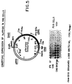
intr 1. - Figure 5 illustrates the fusion of IFN-β2A cDNA to the SV40 early promoter. A plasmid pSVIFA2-II was first constructed by subcloning the 5' Xho1-BamH1 (Xh-B) segment of the IFA-2 gene (from 5.5 kb to 7.5 kb in Figure 2) fused with a Cla1-Xho1 synthetic adaptor of 26 bp (restoring the 5' cDNA sequence), into a Cla1 and Bam H1-cut pBR plasmid. The 3' BamH1-Hindlll (B-H) gene segment (from 7.5 kb to 10.5 kb in Figure 2), subcloned in a a Hindlll, BamHI-cut pBR plasmid, was excised using the Cla1 site adjacent to Hindlll in pBR322 and ligated to the above 5' segment to obtain the complete IFA-2 gene. A pSVE3 vector cut by Hindlll as described in Y. Chernajovsky et al. (1984) DNA 3: 294-308, was reclosed with Cla1 linkers(forming pSVC1a) and the above IFA-2 gene introduced in the Cla1 site of pSVC1a in the same orientation as the SV40 early promoter forming pSVIFA2-II. A plasmid pSVIFA2-I was similarly constructed but the Xho1 site of IFA-2 was fused directly to the Cla1 site of pBR before introduction in pSVE3. To obtain the pSVCIFβ2 plasmid containing the IFN-β2A cDNA fused to the SV40 promoter, the above pSVIFA2-II DNA, cut by Xb1, was partially cut by Xmn1 to open the Xmn1 site located 60 bp downstream from the Xho1 site in the IFN-β2 cDNA sequence (Figure 1), and not an Xmn1 site located in the amp gene of the vector. To this construct, the Xmn1-Xba1 segment of IFN-β2 cDNA clone AE20 (coordinates 92-566 in Fig. 1) was ligated, restoring the complete IFN-β2A cDNA sequence as shown in the Figure. EES is the early-early RNA start of SV40 T-ag gene; ATG is the IFN-β2 initiation codon; pA are IFN-β2 and SV40 polyadenylation sites, respectively. pSVCIFβ2 DNA was cotransfected with a pDHFR plasmid into CHO-K1 DHFR- cells as in Y. Chernajovsky et al. (1984), (supra) and clones were tested for human IFN antiviral activity in FS11 cells using Vesicular Stomatitis Virus (VSV). Gene amplification was obtained by selecting CHO clones for methotrexate (MTX)-resistance. Assay of the culture medium of CHO-SVCIFβ2 B132-5M cells, resistant to 250 nM MTX, is shown in the lower part of the Figure. After VSV cytopathic effect developed, cells in the microplate were stained by methylene blue. Serial two-fold dilutions of the medium are from left to right. By comparison to IFN standard (ST) and non-transfected CHO cells, a titer of 300 U/ml rIFN-β2 is calculated.
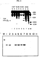
- Figure 6 shows the results of an assay of (2'-5') oligo A synthetase with poly (rI)(rC)-agarose bound NP40 extracts (M. Revel et al. (1981) Meth. Enzymol. 79:149-161) of human FS11 fibroblasts previously exposed for 16 hours to medium from a) Cos7 cells two days post-transfection with the pSVIFA2-II DNA (described in Figure 5)(leftmost 4 lanes), b) hamster CHO SI-15 cells stably transformed by the pSVIFA2-I DNA of Fig. 5 (next 4 lanes), c) CHO MEIF-5 cells expressing IFN-β1 gene (Y. Chernajovsky et al. (1984), supra) (next 2 lanes); d) CHO cells with DHFR gene only (next lane); e) fresh medium (Non Ind.). The IFN antiviral activity measured per ml of the media added to FS11 cells is indicated. An electrophoresis of phosphatased, 32P-α-ATP labelled (2'-5') oligo A product is shown.
- Figure 7 is a comparison of amino acid sequences between type I human IFNs. Amino acids that are conserved in IFN-β1 and the various IFN-αs (shown are αA, αC, replacements in other αs excluding αE, and IFN-α class II), are marked by stars over the IFN-β1 sequence. Amino acids conserved in IFN-β2 are shown above its sequence by stars if conserved in all type I IFNs, or by squares if conserved in some IFNs. IFN-β2A was aligned by comparing hydropathy plots (see text) and numbered from the presumed processing site.
- Figure 8 shows restriction maps of the two different human IFN-β2 genes which are expressed in rodent cells. The IFA-11 DNA (IFN-β2B gene) was transfected into either CHO or L cells and expressed IFN-β activity after induction (Table 1).
- Figure 9 shows an IFN-β2 specific immunocompetition assay with R-antibodies to show that human fibroblasts FS11 cells secrete the IFN-β2 protein in response to the two cytokines interleukin-I (Il-I) and Tumor Necrosis Factor (TNF-α). Both cytokines induce the synthesis and secretion of the IFN-β2 protein in two levels which have been estimated by the immunoassay to be 10 to 20 U/ml IFN-β2. Optimal IFN-β2 induction was seen with the concentrations of rIL-1α of 4 U/ml (0.13 ng/ml). The optimal concentration of TNF-α for IFN-β2 induction (400 U/ml; 40 ng/ml) was higher than that required for cytolytic effect on sensitive cells (0.1 ng/ml) and even higher than that giving optimal growth stimulation of diploid fibroblasts (2 ng/ml).
-
- The existence of interferon-beta2 was discovered in the course of attempts to isolate genetic material containing the nucleotide sequence coding for interferon in human fibroblast cells. In this process, fibroblast cells were induced for the production of inducible interferon mRNA with a suitable exogenous factor and then the mRNAs were extracted. At the same time the mRNAs from a non-induced culture of the same host cells were extracted. cDNA probes were synthesized from the mRNAs of both the induced culture and the non-induced control culture using the corresponding mRNAs as templates (induced cDNAs and non-induced cDNAs). Double-stranded cDNAs derived from the mRNA extracted from the induced culture were then synthesized and such cDNAs were inserted in appropriate vectors and transfected into suitable microorganisms. The microorganisms were cultivated under conditions suitable to cause selective development of microorganism colonies of the modified vectors (initial colonies). Duplicate colonies of the initial colonies were formed and the DNAs of both the initial and duplicate colonies were freed in situ. The DNAs of one of the two sets of duplicate colonies were hybridized with the cDNA probes synthesized from the mRNAs extracted from the induced culture and the other of the two sets of duplicate colonies were hybridized with the cDNA probes synthesized from the mRNAs extracted from the non-induced culture. Clones which selectively hybridized with the induced cDNA probes but not with the non-induced cDNA probes were selected and their DNAs recovered. These cloned DNAs were further tested to determine those DNAs capable of hybridizing with mRNA translatable in frog oocytes or reticulocyte-lysates into interferon, which DNAs are essentially those which code for interferon, or contain enough of the sequence coding for interferon to be used to obtain a complete interferon DNA.
- During this investigation, it was discovered that two different mRNAs coding for interferon could be isolated from human fibroblast cells after being appropriately induced. Upon glycerol gradient centrifugation, the smallest mRNA sediments at 11S and yields by translation in a cell-free system a protein of molecular weight 20,000 which is selectively precipitated by antibodies prepared against one of the interferons that can be purified from these cells. This protein is human interferon-beta1 of which the amino acid sequence was partially determined by E. Knight, et al. (1980) Science, 207, 525-526. The largest mRNA was found to sediment at 14S and yield a protein of molecular weight 23,000 which is precipitated by antibodies against a less purified preparation of fibroblast interferon. This protein is designated human interferon-beta2. cDNA clones hybridizable to mRNA which translates to interferon-beta2 were prepared in such a manner and an IFN-β2 cDNA probe was prepared therefrom.
- Two such IFN-β2 clones A341 and E474 were found to represent overlapping sequencing and were fused to reconstitute the IFN-β2A AE20 cDNA (Figure 1). The nucleotide sequence of this IFN-β2A cDNA was determined and revealed an open reading frame of 212 amino acids predicting a protein of 23,500 daltons. Transcription-translation experiments confirmed that this cDNA encodes such a protein.
- From a human gene library of human adult blood cell DNA cloned after partial EcoRI digestion in λ charon 4A (Y. Mory et al. (1981) Eur. J. Biochem. 120: 197-202), two genomic clones IFA-2 and IFA-11 hybridizing to such an IFN-β2 cDNA probe were isolated. Clone IFA-2 contains a gene with at least 4 exons hybridizing to various segments of the cloned cDNA (Figure 1). An Xho1 site present near the 5' of the AE20 cDNA allowed to map the genomic segment A132 (Figure 2) which contains the first IFA-2 exon ending 70 bp downstream from this Xho1 site. By S1 nuclease analysis using DNA probes labelled at a
BstN1 site 40 bp after the Xho1 site, it was determined that induced FS11 human fibroblasts contain IFN-β2ARNA with two distinct 5' ends S-1 and S-2 separated by 20 nucleotides (Figure 3). Each of these two starts is preceded in the IFA-2 gene by a potential TATA box at -30 (Figure 4). RNA ending at S-2 seems to be more abundant than the longer RNA under a variety of induction conditions (Figure 3). Both RNA 5' ends were also seen with cytoplasmic RNA instead of total cell RNA. Therefore, it is apparent that both RNAs are formed and active in human cells. Both IFN-β2ARNAs are induced in human fibroblasts by poly (rI)(rC) and induction is stimulated by IFN priming as in the case of IFN-β1 RNA in these cells (U. Nir et al. (1984) Nucl. Acids Res. 12: 6979-6993). A prolonged (6.5 h) treatement with CHX also induces IFN-β2ARNA, but shorter (3.5 h) CHX treatment is effective only with poly (rI)(rC) (Figure 3). The low induction of IFN-β2ARNA by CHX without ds RNA distinguishes this gene from IFN-β1. - DNA constructs were made to express the IFA-2 gene under the control of the SV40 early gene promoter. A 4.8 kb segment of IFA-2 DNA was fused through a synthetic oligonucleotide to pSVE3 DNA (Y. Chernajovsky et al. (1984). supra) so that the ATG codon is 150 bp downstream from the SV40 early early RNA start (pSVIFA2-II, see legend, Figure 5). After transfection of pSVIFA2-II DNA into Cos7 monkey cells (Y. Gluzman (1981) Cell 23: 175-182), two RNAs of 1.35 and 2.2 kb were detected in Northern blots by IFN-β2 cDNA; these two RNAs would correspond to transcripts ending at the IFN-β2 ad SV40 polyadenylation sites respectively (Figure 5). Medium from the Cos7 cells during transient expression of the pSVIFA2-II DNA was assayed for IFN activity by inhibition of the cytopathic effect of VSV on human FS11 and Wish cells as described in D. Novick et al. (1983) J.Cen.Virol. 64: 905-910. Antiviral activity could be clearly detected two days after transfection (Table 1). The specific antiviral activity of IFN-β2Aper unit protein has been estimated to be 30 to 100 times lower than that of IFN-β1 and closer to that of IFN-α1 (5-10
x 106 units/mg). The IFN titers observed by transient expression of the IFA-2 gene were as expected, about 3% of those seen with the IFN-β1 gene under the same SV40 promoter control (Table 1). Production of IFN-β2Aactivity by pSVIFA2-II DNA cells was further demonstrated by induction of (2'-5') oligo A synthetase (M. Revel et al. (1981), supra) in FS11 cells exposed to the medium of the transfected Cos7 cells (Figure 6). The induction was dose-dependent and corresponded quantitatively to the antiviral activity. - The IFA-2 gene was also fused to the SV40 early promoter directly at the Xho1 site near S-2 (pSVIFA2-I DNA, legend of Figure 5). Hamster CHO-K1 DHFR- cells were cotransformed with pSVIFA2-I and pSVDHFR (Y. Chernajovsky et al. (1984), supra) DNAs and a stably transformed line SI-15 was isolated. This cell line produces constitutively 10-50 U/ml of IFN antiviral activity measured on human FS11 and Wish cells, while CHO cells transformed only by pSVDHFR produce no human IFN (Table 1). By concentration of the medium from SI-15 cultures, rIFN-β2Asolutions titrating about 3000 U/ml could regularly be obtained. This rIFN-β2Ahas the properties expected for a human IFN and induces the (2'-5') oligo A synthetase in FS11 cells even at low concentrations of 1 U/ml (Figure 6). The rIFN-β2Aconstitutively produced by CHO SI-15 cells demonstrated human-specific induction of the mRNAs for (2'-5') oligo A synthetase, C56 and HLA in the presence of cycloheximide, its antiviral activity was neutralized by anti-IFN-β1 (but not anti-IFN-α or anti-IFN -γ ) polyclonal and monoclonal antibodies and it acted on mouse-human hybrids only if they contain
human chromosome 21 which carries the gene for thetype 1 IFN receptor. These further characterizations of the biological activity of rIFN-β2A convincingly demonstrate that it acts directly as an IFN and is not an inducer of IFN-β1. - Improved yields of rIFN-β2Awere obtained with DNA constructs containing the IFN-β2A cDNA sequence fused to the SV40 early promotor (pSVCIFβ2 shown in Figure 5), when this DNA cotransfected with pSVDHFR into CHO cells as above, was amplified by selecting cells resistant to increasing concentrations of methotrexate. Without amplification, the pSVCIFβ2
A transformed CHO cells (e.g. clone B-132) produced levels of IFN-β2Aactivity similar to those produced by the SI-15 cells (Table 2). However, after selection for resistance to methotrexate, higher yields were obtained with up to 800 U/ml (Table 2). - From such clones it was possible to immunoprecipitate the IFN-β2A protein secreted into the culture media and if the cells had been labelled by 35S-methionine to analyse the IFN-β2Aprotein by dodecyl sulphate polyacrylamide gel electrophoresis. The size of the secreted rIFN-β2Awas found to be 21,000 daltons. Indeed, experiments have shown that the 23-26 Kd primary translation product of IFN-β2ARNA in reticulocyte lysates is processed in vitro by dog pancreatic membranes into a shortened 21 Kd protein, and that induced human fibroblasts produce a 21 Kd protein which is immunologically similar to the IFN-β2ARNA 26 Kd product, but distinct from IFN-B1. The N-terminus of the mature IFN-
β 2A21 Kd protein has not been determined and two potential glycosylation sites are present in the IFN-β2Asequence (Figure 1) making it difficult to calculate the size of the region removed by processing. However, the decrease in size indicates that this processed region is longer than that in IFN-β1, the size of which increases by maturation (W. Degrave et al. (1981) Gene 10: 11-15). A hydropathy plot of the IFN-β2A26 Kd polypeptide has shown that there is a marked similarity between hydrophobic and hydrophilic regions of IFN-β2Aand of mature IFN-β1, when the two proteins are aligned by their C-termini. The same alignment also reveals conservation in the amino acid sequence of IFN-β2Awith those of the other type I human IFNs (Figure 7). Between all the known IFN- αs and β1 sequences, there are 38 amino acids which are conserved (marked by stars in Figure 7). When IFN-β2Ais aligned is above, 18 of these 38 amino acids common to all type I human IFNs can be seen to be conserved (Figure 7). There is an overall homology of about 20% between IFN-β2Aand the other type I IFNs. - Although the sequence homology between IFN-β1 and β2A is low, the cross-neutralization of the antiviral activities of IFN-β1 and IFN-β2A by the same antibodies, including a monoclonal anti-IFN-β1 (D. Novick et al. (1983), supra), could result from some conserved structure in the active site of IFN-β. IFN-β2Aactivity is not neutralized by anti-IFN-α or γ antibodies, justifying its designation as a β-type IFN. The genes encoding IFN-β2 are not on chromosome 9 (U. Nir (1984), supra) and differ from the type I IFN genes in the
chromosome 9 cluster by the presence of introns. The IFA-2 gene was determined to be on chromosome 7 (P-B. Sehgal et al. (1986) Proc.Natl.Acad.Sci. USA, in press). The amino acid sequence homology detected is compatible with the notion that the IFN-β2 genes are related to the ancestral gene which gave rise to the type I IFN gene cluster. - The second genomic DNA clone IFA-11 hybridizing with the IFN-β2AcDNA has a restriction map which differs from that of IFA-2 (Figure 8). Transcript mapping and sequencing showed that the 3' exon of IFA-2 and IFA-11 (Xbal-HindIII segment) are identical (99% sequence homology), while there was no cross-hybridization under stringent conditions between more 5' exons. The IFA-11 genomic clone (19 Kb) was transfected into L-TK- cells together with a HSV-TK gene (F. Colbere-Garapin et al. (1981) J.Mol.Biol. 150: 1-14) and stable transformants were isolated. Table 1 shows that L cell clone LI-39 containing the human IFA-11 gene, produces human IFN activity when induced by poly (rI)(rC) and cycloheximide, while L-TK+ cells produced no such activity. A CHO clone 1C40 containing the IFA-11 gene also produced human IFN activity (Table 1). No activity was seen in non-induced LI-39 or 1C40 cells. The IFN activity obtained with IFA- 11 transformants after induction was significantly higher than with the IFA-2 gene. The rIFN-β2B produced by expression of the IFA-11 gene was neutralized with anti-IFN-β1 similarly to the IFA-2 gene product. IFA-11 specific DNA probes hybridized to RNA from induced human fibroblasts similarly to IFN-2 DNA probes, indicating that both genes are active in human cells.
- The function of the IFN-β2 genes seems to result from their expression under conditions in which other IFN genes are not expressed. We find that fibroblasts exposed to Tumor Necrosis Factor (TNF) or to lymphokine IL-1 produce the IFN-β2 protein. Others found that neutralization of IFN-β by antibodies results in a stimulation of cell proliferation (M. Kohase et al. (1986) Cell, in press; D. Resnitzky et al. (1986) Cell, in press), suggesting that the IFN-β2 acts to limit cell growth in response to growth factors. Supporting this conclusion is the fact that RNA hybridizing to IFN-β2 cDNA was recently detected in peripheral blood mononuclear cells induced by mitogens such as PHA along with IFN-γ mRNA. It has also been reported that an IL-1 like lymphokine strongly induces RNA hybridizing to IFN-β2 cDNA in diploid fibroblasts and other human cell lines (J. Content et al. (1985) Eur.J.Biochem. 152: 253-257). The promoter region of the IFA-2 genes seems to contain two TATA boxes and two RNA starts (Figures 1 and 3). This IFN-β2 promoter was shown to respond to either poly (rI)(rC) or to cycloheximide (Y. Chernajovsky et al. (1984), supra). In vivo, the two IFN-β2 gene promoters may be activated by different inducers (ds RNA, protein synthesis inhibitors, IL-1, PHA). A variety of cells have been found to produce autocrine IFN-β in low amounts during growth arrest and differentiation (M. Revel (1983)
Interferon 5 pp.205-239, Acad. Press, London). IFN-β2 may belong to the same group of minor IFN species produced by cells to auto-regulate their growth, while the classical type I IFNs are probably more specifically involved in theresponse to viral infections. - The biological significance of IFN-β2 lies most probably in the fact that it is induced under conditions where IFN-β1 is not induced, as in metabolically stressed cells (A. Zilberstein et al., (1985) in The Interferon System, Serono Symposia Raven Press, pp.73-83). Most important is the observation that TNF induces IFN-β2 in fibroblasts and that the proliferation of these cells which is stimulated by TNF is further enhanced if anti-IFN-β antibodies are added. A number of human cell lines have been shown to produce autocrine IFN-β species when undergoing differentiation, and these IFNs are responsible for the induction of HLA antigens on these cells (A. Yarden et al. (1984) Embo.J. 3: 969-973). In a mouse myeloleukemic cell line induced to differentiate by CSF-1, the growth arrest of the cells which characterizes terminal differentiation, is abolished by anti-IFN-β antibodies. IL-1 stimulates growth of several cell types and the classical growth factor PDGF was found to induce IFN-β RNA in mouse cells (J.N. Zullo et al. (1985) Cell 43: 793-800). Although multiple IFN-β species may be involved (P.B. Sehgal, (1982) in
Interferon 4, Academic Press, pp.1-22, and M. Revel (1983) inInterferon 5, Academic Press, pp. 205 - 239), IFN-β2 appears to be one of these autocrine autoregulators of cell growth produced in response to growth factors. This would be in line with the low specific activity of IFN-β2 (estimated by comparison of immunoassay and antiviral activity to be 50-100 times lower than for IFN-β1), since only very small antiviral activity is seen in cells producing the autocrine IFN-β species (M. Revel (1983) inInterferon 5, Academic Press, pp. 205-239). This could also explain why the hamster cells expressing IFN-β2 cDNA produce less IFN activity than similar CHO cells harbouring the human IFN-β1 gene (Y. Chernajovsky et al. (1984), DNA 3: 297-308). Interestingly, the IFN-α 1 (D) species has also a 100 times lower specific activity on human cells than other IFN-α species (T. Goren et al. (1983), Virology 130: 273-280), although it represents a large proportion of the total mass of leukocyte IFN (C. Weissmann (1981) inInterferon 3, Academic Press, N.Y. pp.101-134). It is not excluded that some of the multiple IFN functions will be expressed more efficiently by IFN-β2 than the antiviral effect. Despite their low concentration, growth-regulatory IFN-β induce HLA and (2'-5') oligo A synthetase more strongly than expected, have more prolonged effects and markedly inhibit the growth of the cells which produce them. The induction of IFN-β2 by IL-1 and TNF suggests that it may play a role as an autocrine mediator of some effects of these cytokines in inflammation and acute-phase responses, as well as regulating cell proliferation. - Induction of mRNA which hybridizes with IFN-β2 cDNA probes but not to IFN-β1 cDNA has been observed in human cell lines after exposure to Interleukin-1 (IL-1) (J. Content et al. (1985), Eur.J.Biochem. 152: 253-257) and Tumor Necrosis Factor (TNF- α ). The IFN-β2-specific immunocompetition assay with R-antibodies was used to examine if human fibroblasts FS11 cells indeed secrete the IFN-β2 protein in response to these two cytokines (Figure 9). Both rIL-1α and TNF-α induced the synthesis and secretion of the IFN-β2 protein, to levels estimated by the immunoassay to be 10-20 U/ml IFN-β2. Optimal IFN-β2 induction was seen with concentrations of rIL-1α of 4 U/ml (0.13 ng/ml), similar to those required for IL-1 induction of prostaglandin E2, collagenase and hyaluronic acid in human fibroblasts (J.H. Korn (1985), Arthritis Rheum. 28: 315-322). The optimal concentration of TNF-α for IFN-β2 induction (400 U/ml; 40 ng/ml) was higher than that required for the cytolytic effect on sensitive cells (0.1 ng/ml; A.M. Wang et al. (1985) Science 228: 149-154) and even higher than that giving optimal growth stimulation of diploid fibroblasts (2 ng/ml). The onset of IFN-β2 secretion was somewhat faster with TNF-α reaching half maximal levels at about 6 hours, versus 8-12 hours with IL-1.
- To the extent that it may be demed necessary in order to complete the disclosure in this case; all of the subject matter of each of the publications referred to and cited hereinabove are hereby incorporated herein by reference.
- It will be obvious to those skilled in the art that various other changes and modifications may be made without departing from the scope of the invention and the invention is not to be considered limited to what is shown in the drawing and described in the specification.
Antiviral Activity of rIFN-β2 Produced in Transfected Mammalian Cells I. IFA-2 GENE
A. Constitutive expression in CHO cellspSVIFA2-I S1-15 cells 10-50 U/ml (control CHO cells 0 ) B. Transient expression in Cos7 cells pSVIFA2-II DNA 15 U/ml (control IFN-β1 pSVEIF DNA 500 U/ml) II. IFA-11 GENE
A. Induced expression in CHO cellsClone 1C40 400-500 U/ml (control CHO cells 100 U/ml) B. Induced expression in L cells Clone LI-39 250 U/ml (control L-TK+ cells <4 U/ml) Antiviral Activity of Human rIFN-β2A Produced in Hamster Cells Transfected and Amplified with IFN-β2A cDNA CHO-SVCIFβ2 B132-10M CLONE: Human IFN activity U/ml B-132 (0 MTX) 64 B-132-5M (250 nM MTX)* 300 B-132-10M (500 nM MTX)* 800
Claims (3)
- Use of human interferon-β2A for preparing a medicament for treatment of diseases in the fields of inflammation and/or acute phase response.
- Use according to claim 1, wherein IFN-β2A inhibits fibroblast proliferation.
- Use according to claim 1, wherein IFN-β2A prevents sclerosis after infection.
Priority Applications (2)
| Application Number | Priority Date | Filing Date | Title |
|---|---|---|---|
| EP97119173A EP0835661B1 (en) | 1985-10-14 | 1986-10-10 | Human interferon-Beta2A for use as a medicament |
| EP94113232A EP0645452A1 (en) | 1985-10-14 | 1986-10-10 | Human interferon-beta 2A and interfeon-beta 2B, vectors containing genes coding for said interferons, cell lines producing same and use of said interferons as pharmaceuticals |
Applications Claiming Priority (5)
| Application Number | Priority Date | Filing Date | Title |
|---|---|---|---|
| IL7671485 | 1985-10-14 | ||
| IL76714A IL76714A0 (en) | 1985-10-14 | 1985-10-14 | Recombined human interferon beta-2 and a method of producing same |
| US86088386A | 1986-05-08 | 1986-05-08 | |
| US860883 | 1986-05-08 | ||
| EP86114049A EP0220574B2 (en) | 1985-10-14 | 1986-10-10 | Human interferon beta2A, vectors containing genes coding for said interferon, cell lines producing same |
Related Parent Applications (2)
| Application Number | Title | Priority Date | Filing Date |
|---|---|---|---|
| EP86114049.9 Division | 1986-10-10 | ||
| EP86114049A Division EP0220574B2 (en) | 1985-10-14 | 1986-10-10 | Human interferon beta2A, vectors containing genes coding for said interferon, cell lines producing same |
Related Child Applications (3)
| Application Number | Title | Priority Date | Filing Date |
|---|---|---|---|
| EP97119173A Division EP0835661B1 (en) | 1985-10-14 | 1986-10-10 | Human interferon-Beta2A for use as a medicament |
| EP94113232A Division EP0645452A1 (en) | 1985-10-14 | 1986-10-10 | Human interferon-beta 2A and interfeon-beta 2B, vectors containing genes coding for said interferons, cell lines producing same and use of said interferons as pharmaceuticals |
| EP94113232.6 Division-Into | 1986-10-10 |
Publications (2)
| Publication Number | Publication Date |
|---|---|
| EP0536520A1 EP0536520A1 (en) | 1993-04-14 |
| EP0536520B1 true EP0536520B1 (en) | 1999-03-03 |
Family
ID=26321482
Family Applications (4)
| Application Number | Title | Priority Date | Filing Date |
|---|---|---|---|
| EP97119173A Expired - Lifetime EP0835661B1 (en) | 1985-10-14 | 1986-10-10 | Human interferon-Beta2A for use as a medicament |
| EP86114049A Expired - Lifetime EP0220574B2 (en) | 1985-10-14 | 1986-10-10 | Human interferon beta2A, vectors containing genes coding for said interferon, cell lines producing same |
| EP94113232A Ceased EP0645452A1 (en) | 1985-10-14 | 1986-10-10 | Human interferon-beta 2A and interfeon-beta 2B, vectors containing genes coding for said interferons, cell lines producing same and use of said interferons as pharmaceuticals |
| EP92114478A Expired - Lifetime EP0536520B1 (en) | 1985-10-14 | 1986-10-10 | Use of Human Interferon-Beta 2A for the preparation of medicaments |
Family Applications Before (3)
| Application Number | Title | Priority Date | Filing Date |
|---|---|---|---|
| EP97119173A Expired - Lifetime EP0835661B1 (en) | 1985-10-14 | 1986-10-10 | Human interferon-Beta2A for use as a medicament |
| EP86114049A Expired - Lifetime EP0220574B2 (en) | 1985-10-14 | 1986-10-10 | Human interferon beta2A, vectors containing genes coding for said interferon, cell lines producing same |
| EP94113232A Ceased EP0645452A1 (en) | 1985-10-14 | 1986-10-10 | Human interferon-beta 2A and interfeon-beta 2B, vectors containing genes coding for said interferons, cell lines producing same and use of said interferons as pharmaceuticals |
Country Status (6)
| Country | Link |
|---|---|
| EP (4) | EP0835661B1 (en) |
| AU (1) | AU602872B2 (en) |
| CA (1) | CA1341369C (en) |
| DE (4) | DE220574T1 (en) |
| ES (2) | ES2131513T3 (en) |
| GR (2) | GR3030150T3 (en) |
Families Citing this family (16)
| Publication number | Priority date | Publication date | Assignee | Title |
|---|---|---|---|---|
| IE67035B1 (en) * | 1986-07-08 | 1996-02-21 | Genetics Inst | Production and use of non-glycosylated IL-6 |
| US6284237B1 (en) | 1986-07-08 | 2001-09-04 | Steven C. Clark | Methods of treatment using IL-6 |
| EP0257406B2 (en) * | 1986-08-06 | 1998-08-12 | Ajinomoto Co., Inc. | Recombinant B-cell differentiation factor |
| US4973478A (en) * | 1987-07-20 | 1990-11-27 | Allelix Biopharmaceuticals, Inc. | Treating inflammation with hepatocyte stimulating factor interferon β2 |
| NZ226799A (en) * | 1987-11-06 | 1991-08-27 | Oncogen | Breast cancer inhibitory factor and method for inhibiting proliferation of neoplastic cells and compositions therefor |
| IL85204A0 (en) * | 1988-01-26 | 1988-07-31 | Yeda Res & Dev | Recombinant human interferon beta2,its preparation and pharmaceutical compositions comprising it |
| JPH0372429A (en) * | 1988-10-07 | 1991-03-27 | Chugai Pharmaceut Co Ltd | Medicine for thrombocytopenia |
| US5641868A (en) * | 1991-04-18 | 1997-06-24 | Toray Industries, Inc. | Interlekin-6 compositions, and a production process thereof |
| IT1254359B (en) * | 1992-05-11 | 1995-09-14 | Serono Cesare Ist Ricerca | PHARMACEUTICAL COMPOSITIONS CONTAINING IL-6 |
| US5902576A (en) * | 1992-10-22 | 1999-05-11 | Yeda Research And Development Co. Ltd. At Weizmann Institute Of Science | Antitumor pharmaceutical composition comprising IL-6 transfected cells |
| US5338834A (en) * | 1993-01-26 | 1994-08-16 | Allelix Biopharmaceuticals Inc. | Ultrapure human interleukin-6 |
| IL147414A0 (en) | 2001-12-31 | 2002-08-14 | Yeda Res & Dev | Ifnar2 mutants, their production and use |
| WO2004019886A2 (en) | 2002-08-29 | 2004-03-11 | Cytocure Llc | Methods for up-regulating antigen expression in tumors |
| US7534423B2 (en) | 2003-03-28 | 2009-05-19 | Faron Pharmaceuticals Oy | Method for inducing an elevated level of adenosine in an individual |
| CN108004270B (en) * | 2017-11-24 | 2021-06-18 | 上海交通大学 | Method for detecting activity of duck IFN-beta promoter by using dual-luciferase reporter gene |
| CN112442504B (en) * | 2020-12-10 | 2023-01-24 | 电子科技大学 | Preparation method of recombinant grass carp interleukin-12 active protein |
Citations (1)
| Publication number | Priority date | Publication date | Assignee | Title |
|---|---|---|---|---|
| WO1988000206A1 (en) * | 1986-07-08 | 1988-01-14 | Genetics Institute, Inc. | Production and use of il-6 |
Family Cites Families (4)
| Publication number | Priority date | Publication date | Assignee | Title |
|---|---|---|---|---|
| IL58765A (en) * | 1979-11-21 | 1986-09-30 | Yeda Res & Dev | Process for the production of essentially pure messenger rna of human fibroblast interferon and process for the production of interferon beta |
| JPS60169424A (en) * | 1984-02-15 | 1985-09-02 | Chuzo Kishimoto | Novel human b cell differentiation factor, its preparation and its use |
| JPS61246197A (en) * | 1985-04-24 | 1986-11-01 | Ajinomoto Co Inc | Dna |
| EP0257406B2 (en) * | 1986-08-06 | 1998-08-12 | Ajinomoto Co., Inc. | Recombinant B-cell differentiation factor |
-
1986
- 1986-10-10 EP EP97119173A patent/EP0835661B1/en not_active Expired - Lifetime
- 1986-10-10 DE DE198686114049T patent/DE220574T1/en active Pending
- 1986-10-10 CA CA000520275A patent/CA1341369C/en not_active Expired - Lifetime
- 1986-10-10 ES ES92114478T patent/ES2131513T3/en not_active Expired - Lifetime
- 1986-10-10 DE DE3650790T patent/DE3650790T2/en not_active Expired - Lifetime
- 1986-10-10 EP EP86114049A patent/EP0220574B2/en not_active Expired - Lifetime
- 1986-10-10 DE DE3650715T patent/DE3650715T2/en not_active Expired - Lifetime
- 1986-10-10 ES ES86114049T patent/ES2058053T5/en not_active Expired - Lifetime
- 1986-10-10 EP EP94113232A patent/EP0645452A1/en not_active Ceased
- 1986-10-10 DE DE3688671T patent/DE3688671T3/en not_active Expired - Lifetime
- 1986-10-10 EP EP92114478A patent/EP0536520B1/en not_active Expired - Lifetime
- 1986-10-14 AU AU63867/86A patent/AU602872B2/en not_active Expired
-
1999
- 1999-05-06 GR GR990401238T patent/GR3030150T3/en unknown
-
2000
- 2000-06-23 GR GR20000401465T patent/GR3033769T3/en not_active IP Right Cessation
Patent Citations (1)
| Publication number | Priority date | Publication date | Assignee | Title |
|---|---|---|---|---|
| WO1988000206A1 (en) * | 1986-07-08 | 1988-01-14 | Genetics Institute, Inc. | Production and use of il-6 |
Also Published As
| Publication number | Publication date |
|---|---|
| GR3030150T3 (en) | 1999-08-31 |
| DE3650715D1 (en) | 1999-04-08 |
| DE3650790D1 (en) | 2007-10-11 |
| DE3688671D1 (en) | 1993-08-12 |
| DE220574T1 (en) | 1987-11-26 |
| DE3650790T2 (en) | 2008-01-24 |
| EP0536520A1 (en) | 1993-04-14 |
| EP0645452A1 (en) | 1995-03-29 |
| CA1341369C (en) | 2002-06-18 |
| EP0835661B1 (en) | 2007-08-29 |
| ES2131513T3 (en) | 1999-08-01 |
| EP0220574B2 (en) | 2000-04-12 |
| EP0835661A2 (en) | 1998-04-15 |
| AU602872B2 (en) | 1990-11-01 |
| DE3688671T3 (en) | 2001-05-03 |
| DE3650715T2 (en) | 1999-06-24 |
| AU6386786A (en) | 1987-04-16 |
| GR3033769T3 (en) | 2000-10-31 |
| EP0220574A1 (en) | 1987-05-06 |
| ES2058053T5 (en) | 2000-11-01 |
| EP0835661A3 (en) | 1998-05-20 |
| ES2058053T3 (en) | 1994-11-01 |
| EP0220574B1 (en) | 1993-07-07 |
| DE3688671T2 (en) | 1993-10-14 |
Similar Documents
| Publication | Publication Date | Title |
|---|---|---|
| US5554513A (en) | Production of recombinant human interferon-beta2 | |
| EP0536520B1 (en) | Use of Human Interferon-Beta 2A for the preparation of medicaments | |
| Zilberstein et al. | Structure and expression of cDNA and genes for human interferon‐beta‐2, a distinct species inducible by growth‐stimulatory cytokines. | |
| Charleston et al. | An interferon-induced Mx protein: cDNA sequence and high-level expression in the endometrium of pregnant sheep | |
| Pestka et al. | Interferons and their actions | |
| Capon et al. | Two distinct families of human and bovine interferon-α genes are coordinately expressed and encode functional polypeptides | |
| Simon et al. | Interferon-regulated Mx genes are not responsive to interleukin-1, tumor necrosis factor, and other cytokines | |
| KR100223255B1 (en) | Improved alpha interferon composition and method for its production from human peripheral blood leukocytes | |
| US7446172B2 (en) | Cytokine protein family | |
| US5004689A (en) | DNA sequences, recombinant DNA molecules and processes for producing human gamma interferon-like polypeptides in high yields | |
| US20020013452A1 (en) | Interferon tau compositions and methods of use | |
| EP0750668A1 (en) | NOVEL MUTEINS OF IFN-$g(b) | |
| EP1942187B1 (en) | Cytokine protein family | |
| JP2648860B2 (en) | Human interferon-β2A and human interferon-β2B, vector containing the gene encoding the interferon, cell line producing the interferon, and use of the interferon as a pharmaceutical | |
| US5510472A (en) | Production of recombinant human interferon-beta2 | |
| Revel et al. | Autocrine interferons and interferon-β2 | |
| CA1341252C (en) | Human interferon-.beta.2a and interferon-.beta.2b; vectors containing genes coding for said interferons: cell lines producing same and use of said interferons as pharmaceuticals | |
| Abramovich et al. | The human interferon α-receptor protein confers differential responses to human interferon-β versus interferon-α subtypes in mouse and hamster cell transfectants | |
| McCULLAGH et al. | Biological properties of human interferon beta 1 synthesized in recombinant bacteria | |
| Revel et al. | Production of recombinant human interferon beta 2< 2unull> s | |
| Pestka et al. | Molecular activities of recombinant and hybrid leukocyte interferons | |
| US8231866B2 (en) | Cytokine protein family | |
| ES2353150T3 (en) | FAMILY OF CYTOKIN PROTEINS. | |
| Mahmoudi et al. | Induction of interferon messenger RNA in human lymphoblastoid cells (Namalva) | |
| Ruggieri | IFN-β2: A multifunctional cytokine |
Legal Events
| Date | Code | Title | Description |
|---|---|---|---|
| PUAI | Public reference made under article 153(3) epc to a published international application that has entered the european phase |
Free format text: ORIGINAL CODE: 0009012 |
|
| 17P | Request for examination filed |
Effective date: 19920825 |
|
| AC | Divisional application: reference to earlier application |
Ref document number: 220574 Country of ref document: EP |
|
| AK | Designated contracting states |
Kind code of ref document: A1 Designated state(s): BE CH DE ES FR GB GR IT LI NL SE |
|
| RIN1 | Information on inventor provided before grant (corrected) |
Inventor name: REVEL, MICHAEL Inventor name: ZILBERSTEIN, ASHER |
|
| 17Q | First examination report despatched |
Effective date: 19970127 |
|
| GRAG | Despatch of communication of intention to grant |
Free format text: ORIGINAL CODE: EPIDOS AGRA |
|
| GRAG | Despatch of communication of intention to grant |
Free format text: ORIGINAL CODE: EPIDOS AGRA |
|
| GRAH | Despatch of communication of intention to grant a patent |
Free format text: ORIGINAL CODE: EPIDOS IGRA |
|
| GRAH | Despatch of communication of intention to grant a patent |
Free format text: ORIGINAL CODE: EPIDOS IGRA |
|
| GRAA | (expected) grant |
Free format text: ORIGINAL CODE: 0009210 |
|
| AC | Divisional application: reference to earlier application |
Ref document number: 220574 Country of ref document: EP |
|
| AK | Designated contracting states |
Kind code of ref document: B1 Designated state(s): BE CH DE ES FR GB GR IT LI NL SE |
|
| XX | Miscellaneous (additional remarks) |
Free format text: TEILANMELDUNG 94113232.6 EINGEREICHT AM 10/10/86. |
|
| REG | Reference to a national code |
Ref country code: CH Ref legal event code: NV Representative=s name: ISLER & PEDRAZZINI AG Ref country code: CH Ref legal event code: EP |
|
| REF | Corresponds to: |
Ref document number: 3650715 Country of ref document: DE Date of ref document: 19990408 |
|
| ITF | It: translation for a ep patent filed |
Owner name: SOCIETA' ITALIANA BREVETTI S.P.A. |
|
| ET | Fr: translation filed | ||
| REG | Reference to a national code |
Ref country code: ES Ref legal event code: FG2A Ref document number: 2131513 Country of ref document: ES Kind code of ref document: T3 |
|
| PLBE | No opposition filed within time limit |
Free format text: ORIGINAL CODE: 0009261 |
|
| STAA | Information on the status of an ep patent application or granted ep patent |
Free format text: STATUS: NO OPPOSITION FILED WITHIN TIME LIMIT |
|
| 26N | No opposition filed | ||
| REG | Reference to a national code |
Ref country code: GB Ref legal event code: IF02 |
|
| PGFP | Annual fee paid to national office [announced via postgrant information from national office to epo] |
Ref country code: IT Payment date: 20050728 Year of fee payment: 20 |
|
| PGFP | Annual fee paid to national office [announced via postgrant information from national office to epo] |
Ref country code: GR Payment date: 20050913 Year of fee payment: 20 |
|
| PGFP | Annual fee paid to national office [announced via postgrant information from national office to epo] |
Ref country code: NL Payment date: 20051003 Year of fee payment: 20 |
|
| PGFP | Annual fee paid to national office [announced via postgrant information from national office to epo] |
Ref country code: SE Payment date: 20051005 Year of fee payment: 20 Ref country code: GB Payment date: 20051005 Year of fee payment: 20 |
|
| PGFP | Annual fee paid to national office [announced via postgrant information from national office to epo] |
Ref country code: DE Payment date: 20051006 Year of fee payment: 20 |
|
| PGFP | Annual fee paid to national office [announced via postgrant information from national office to epo] |
Ref country code: FR Payment date: 20051010 Year of fee payment: 20 |
|
| PGFP | Annual fee paid to national office [announced via postgrant information from national office to epo] |
Ref country code: CH Payment date: 20051014 Year of fee payment: 20 |
|
| PGFP | Annual fee paid to national office [announced via postgrant information from national office to epo] |
Ref country code: ES Payment date: 20051129 Year of fee payment: 20 |
|
| PGFP | Annual fee paid to national office [announced via postgrant information from national office to epo] |
Ref country code: BE Payment date: 20051215 Year of fee payment: 20 |
|
| PG25 | Lapsed in a contracting state [announced via postgrant information from national office to epo] |
Ref country code: GB Free format text: LAPSE BECAUSE OF EXPIRATION OF PROTECTION Effective date: 20061009 |
|
| PG25 | Lapsed in a contracting state [announced via postgrant information from national office to epo] |
Ref country code: NL Free format text: LAPSE BECAUSE OF EXPIRATION OF PROTECTION Effective date: 20061010 |
|
| PG25 | Lapsed in a contracting state [announced via postgrant information from national office to epo] |
Ref country code: ES Free format text: LAPSE BECAUSE OF EXPIRATION OF PROTECTION Effective date: 20061011 |
|
| REG | Reference to a national code |
Ref country code: GB Ref legal event code: PE20 |
|
| REG | Reference to a national code |
Ref country code: CH Ref legal event code: PL |
|
| NLV7 | Nl: ceased due to reaching the maximum lifetime of a patent |
Effective date: 20061010 |
|
| EUG | Se: european patent has lapsed | ||
| REG | Reference to a national code |
Ref country code: ES Ref legal event code: FD2A Effective date: 20061011 |
|
| BE20 | Be: patent expired |
Owner name: *YEDA RESEARCH AND DEVELOPMENT CO. LTD Effective date: 20061010 |