EP0497883B1 - Compositions and their use for promoting immunopotentiation - Google Patents
Compositions and their use for promoting immunopotentiation Download PDFInfo
- Publication number
- EP0497883B1 EP0497883B1 EP90916853A EP90916853A EP0497883B1 EP 0497883 B1 EP0497883 B1 EP 0497883B1 EP 90916853 A EP90916853 A EP 90916853A EP 90916853 A EP90916853 A EP 90916853A EP 0497883 B1 EP0497883 B1 EP 0497883B1
- Authority
- EP
- European Patent Office
- Prior art keywords
- cells
- composition
- cell
- protein
- specific
- Prior art date
- Legal status (The legal status is an assumption and is not a legal conclusion. Google has not performed a legal analysis and makes no representation as to the accuracy of the status listed.)
- Expired - Lifetime
Links
Images


Classifications
-
- C—CHEMISTRY; METALLURGY
- C07—ORGANIC CHEMISTRY
- C07K—PEPTIDES
- C07K14/00—Peptides having more than 20 amino acids; Gastrins; Somatostatins; Melanotropins; Derivatives thereof
- C07K14/005—Peptides having more than 20 amino acids; Gastrins; Somatostatins; Melanotropins; Derivatives thereof from viruses
-
- A—HUMAN NECESSITIES
- A61—MEDICAL OR VETERINARY SCIENCE; HYGIENE
- A61K—PREPARATIONS FOR MEDICAL, DENTAL OR TOILETRY PURPOSES
- A61K39/00—Medicinal preparations containing antigens or antibodies
- A61K39/02—Bacterial antigens
- A61K39/085—Staphylococcus
-
- A—HUMAN NECESSITIES
- A61—MEDICAL OR VETERINARY SCIENCE; HYGIENE
- A61K—PREPARATIONS FOR MEDICAL, DENTAL OR TOILETRY PURPOSES
- A61K39/00—Medicinal preparations containing antigens or antibodies
- A61K39/39—Medicinal preparations containing antigens or antibodies characterised by the immunostimulating additives, e.g. chemical adjuvants
-
- A—HUMAN NECESSITIES
- A61—MEDICAL OR VETERINARY SCIENCE; HYGIENE
- A61P—SPECIFIC THERAPEUTIC ACTIVITY OF CHEMICAL COMPOUNDS OR MEDICINAL PREPARATIONS
- A61P37/00—Drugs for immunological or allergic disorders
- A61P37/02—Immunomodulators
- A61P37/04—Immunostimulants
-
- C—CHEMISTRY; METALLURGY
- C07—ORGANIC CHEMISTRY
- C07K—PEPTIDES
- C07K14/00—Peptides having more than 20 amino acids; Gastrins; Somatostatins; Melanotropins; Derivatives thereof
- C07K14/435—Peptides having more than 20 amino acids; Gastrins; Somatostatins; Melanotropins; Derivatives thereof from animals; from humans
- C07K14/705—Receptors; Cell surface antigens; Cell surface determinants
- C07K14/70503—Immunoglobulin superfamily
- C07K14/7051—T-cell receptor (TcR)-CD3 complex
-
- C—CHEMISTRY; METALLURGY
- C07—ORGANIC CHEMISTRY
- C07K—PEPTIDES
- C07K16/00—Immunoglobulins [IG], e.g. monoclonal or polyclonal antibodies
-
- C—CHEMISTRY; METALLURGY
- C07—ORGANIC CHEMISTRY
- C07K—PEPTIDES
- C07K16/00—Immunoglobulins [IG], e.g. monoclonal or polyclonal antibodies
- C07K16/18—Immunoglobulins [IG], e.g. monoclonal or polyclonal antibodies against material from animals or humans
- C07K16/28—Immunoglobulins [IG], e.g. monoclonal or polyclonal antibodies against material from animals or humans against receptors, cell surface antigens or cell surface determinants
- C07K16/2803—Immunoglobulins [IG], e.g. monoclonal or polyclonal antibodies against material from animals or humans against receptors, cell surface antigens or cell surface determinants against the immunoglobulin superfamily
- C07K16/2809—Immunoglobulins [IG], e.g. monoclonal or polyclonal antibodies against material from animals or humans against receptors, cell surface antigens or cell surface determinants against the immunoglobulin superfamily against the T-cell receptor (TcR)-CD3 complex
-
- A—HUMAN NECESSITIES
- A61—MEDICAL OR VETERINARY SCIENCE; HYGIENE
- A61K—PREPARATIONS FOR MEDICAL, DENTAL OR TOILETRY PURPOSES
- A61K39/00—Medicinal preparations containing antigens or antibodies
- A61K2039/555—Medicinal preparations containing antigens or antibodies characterised by a specific combination antigen/adjuvant
- A61K2039/55511—Organic adjuvants
- A61K2039/55516—Proteins; Peptides
-
- A—HUMAN NECESSITIES
- A61—MEDICAL OR VETERINARY SCIENCE; HYGIENE
- A61K—PREPARATIONS FOR MEDICAL, DENTAL OR TOILETRY PURPOSES
- A61K39/00—Medicinal preparations containing antigens or antibodies
- A61K2039/555—Medicinal preparations containing antigens or antibodies characterised by a specific combination antigen/adjuvant
- A61K2039/55511—Organic adjuvants
- A61K2039/55544—Bacterial toxins
-
- A—HUMAN NECESSITIES
- A61—MEDICAL OR VETERINARY SCIENCE; HYGIENE
- A61K—PREPARATIONS FOR MEDICAL, DENTAL OR TOILETRY PURPOSES
- A61K39/00—Medicinal preparations containing antigens or antibodies
- A61K2039/60—Medicinal preparations containing antigens or antibodies characteristics by the carrier linked to the antigen
- A61K2039/6031—Proteins
- A61K2039/6037—Bacterial toxins, e.g. diphteria toxoid [DT], tetanus toxoid [TT]
-
- A—HUMAN NECESSITIES
- A61—MEDICAL OR VETERINARY SCIENCE; HYGIENE
- A61K—PREPARATIONS FOR MEDICAL, DENTAL OR TOILETRY PURPOSES
- A61K39/00—Medicinal preparations containing antigens or antibodies
- A61K2039/60—Medicinal preparations containing antigens or antibodies characteristics by the carrier linked to the antigen
- A61K2039/6031—Proteins
- A61K2039/6056—Antibodies
-
- C—CHEMISTRY; METALLURGY
- C07—ORGANIC CHEMISTRY
- C07K—PEPTIDES
- C07K2317/00—Immunoglobulins specific features
- C07K2317/70—Immunoglobulins specific features characterized by effect upon binding to a cell or to an antigen
- C07K2317/73—Inducing cell death, e.g. apoptosis, necrosis or inhibition of cell proliferation
-
- C—CHEMISTRY; METALLURGY
- C12—BIOCHEMISTRY; BEER; SPIRITS; WINE; VINEGAR; MICROBIOLOGY; ENZYMOLOGY; MUTATION OR GENETIC ENGINEERING
- C12N—MICROORGANISMS OR ENZYMES; COMPOSITIONS THEREOF; PROPAGATING, PRESERVING, OR MAINTAINING MICROORGANISMS; MUTATION OR GENETIC ENGINEERING; CULTURE MEDIA
- C12N2740/00—Reverse transcribing RNA viruses
- C12N2740/00011—Details
- C12N2740/10011—Retroviridae
- C12N2740/16011—Human Immunodeficiency Virus, HIV
- C12N2740/16111—Human Immunodeficiency Virus, HIV concerning HIV env
- C12N2740/16122—New viral proteins or individual genes, new structural or functional aspects of known viral proteins or genes
Definitions
- This invention relates to immunology, and, more specifically, to the preparation and use of immunopotentiating agents which are capable of eliciting, enhancing and/or otherwise modifying immune responses. These agents, through their ability to elicit or enhance cellular or humoral responses, have potential utility in a variety of disease conditions wherein immunotherapy might be expected to provide a benefit.
- the body's immune system serves as a defense against a variety of conditions, including, e.g., injury, infection and neoplasia, and is mediated by two separate but interrelated systems, the cellular and humoral immune systems.
- the humoral system is mediated by soluble products, termed antibodies or immunoglobulins, which have the ability to combine with and neutralize products recognized by the system as being foreign to the body.
- the cellular immune system involves the mobilization of certain cells, termed T-cells, that serve a variety of therapeutic roles.
- T cells thymus derived cells
- B cells bone marrow derived cells
- Mature T cells emerge from the thymus and circulate between the tissues, lymphatics, and the bloodstream. T cells exhibit immunological specificity and are directly involved in cell-mediated immune responses (such as graft rejection). T cells act against or in response to a variety of foreign structures (antigens). In many instances these foreign antigens are expressed on host cells as a result of infection. However, foreign antigens can also come from the host having been altered by neoplasia or infection. Although T cells do not themselves secrete antibodies, they are usually required for antibody secretion by the second class of lymphocytes, B cells.
- T cells There are various subsets of T cells, which are generally defined by antigenic determinants found on their cell surfaces, as well as functional activity and foreign antigen recognition. Some subsets of T cells, such as CD8 + cells, are killer/suppressor cells that play a regulating function in the immune system, while others, such as CD4 + cells, serve to promote inflammatory and humoral responses.
- CD refers to cell differentiation cluster; the accompanying numbers are provided in accordance with terminology set forth by the International Workshops on Leukocyte Differentiation (5). A general reference for all aspects of the immune system may be found in (1)).
- Human peripheral T lymphocytes can be stimulated to undergo mitosis by a variety of agents including foreign antigens, monoclonal antibodies and lectins such as phytohemaggluttinin and concanavalin A. Although activation presumably occurs by binding of the mitogens to specific sites on cell membranes, the nature of these receptors, and their mechanism of activation, is not completely elucidated. Induction of proliferation is only one indication of T cell activation. Other indications of activation, defined as alterations in the basal or resting state of the cell, include increased lymphokine production and cytotoxic cell activity.
- T cell activation is an unexpectedly complex phenomenon that depends on the participation of a variety of cell surface molecules expressed on the responding T cell population (2,3).
- the antigen-specific T cell receptor (TcR) is composed of a disulfide-linked heterodimer, containing two clonally distributed, integral membrane glycoprotein chains, ⁇ and ⁇ , or ⁇ and ⁇ , non-covalently associated with a complex of low molecular weight invariant proteins, commonly designated as CD3 (the older terminology is T3) (2,4).
- the TcR ⁇ and ⁇ chains determine antigen specificities (6).
- the CD3 structures are thought to represent accessory molecules that may be the transducing elements of activation signals initiated upon binding of the TcR ⁇ to its ligand.
- Polymorphic TcR variable regions define subsets of T cells, with distinct specificities.
- the TcR complex interacts with small peptidic antigen presented in the context of major histocompatability complex (MHC) proteins.
- MHC proteins represent another highly polymorphic set of molecules randomly dispersed throughout the species. Thus, activation usually requires the tripartite interaction of the TcR and foreign peptidic antigen bound to the major MHC proteins.
- Monoclonal antibodies were developed by Kohler and Milstein in 1975.
- the methods generally used to produce mAb consist of fusing (hybridizing) two types of somatic cells: (1) a neoplastic myeloma cell line; and (2) a normal B lymphocyte obtained from an immunized animal.
- the result is called a hybridoma which is characterized by immortal growth and the ability to secrete antibodies specific for the immunization antigen.
- One of the clinically successful uses of monoclonal antibodies is to suppress the immune system, thus enhancing the efficacy of organ or tissue transplantation.
- U.S. Patent 4,658,019 describes a novel hybridoma (designated OKT3) which is capable of producing a monoclonal antibody against an antigen found on essentially all normal human peripheral T cells. This antibody is said to be monospecific for a single determinant on these T cells, and does not react with other normal peripheral blood lymphoid cells.
- the OKT3 mAb described in this patent is currently employed to prevent renal transplant rejection (10).
- OKT3 has not been previously used to stimulate the immune system in vivo .
- T cell surface molecules have been identified that can activate T cell function, but are not necessarily part of the T cell surface receptor complex.
- Monoclonal antibodies against Thy-1, TAP, Ly-6, CD2, or CD28 molecules can activate T cells in the absence of foreign antigen in vitro (12, 13,14,15,16).
- certain bacterial proteins although differing in structure from mAbs, also have been shown to bind to subsets of T cells and activate them in vitro (17). Although some of these agents, in vitro effects have previously been demonstrated, in vitro activity is often not a reliable predictor of in vitro effects.
- malignant tumor growth is believed to be the inability of the immune system to respond appropriately to tumor antigen.
- malignant progressor tumors are only weakly immunogenic and can evade host recognition and rejection. Both specific and non-specific effector pathways have been implicated in tumor immunity.
- Treatment by immunotherapy is aimed at remedying defects in the immune weaponry.
- the aim of immunotherapy has been the enhancement of one or both of these pathways.
- One potential approach to therapy is to activate host antitumor cellular effector mechanisms.
- non-specific adjuvants such as BCG or pertussis have been used to augment immune responses.
- these adjuvants amplify immune responses by providing non-specific stimuli that enhance overall immunity.
- these adjuvants do not selectively act on T cells, or subsets of T cells, and have not been shown capable of overcoming immunodeficiency states.
- current modes of immunotherapy which induce non-specific effector cells are not effective enough in potentiating anti-tumor responses (18).
- immunotherapy regimens which utilize the ability of the immune system to recognize tumor antigens in a specific manner, for instance utilizing specific tumor-infiltrating lymphocytes, for immunotherapy, have been suggested to result in superior anti-tumor immunity (19).
- HIV Human immunodeficiency virus
- HIV Human immunodeficiency virus
- humoral and cell mediated effector mechanisms Although current efforts in treatment and vaccine design have fallen short of success either due to the immunodeficiency associated with the viral infection, or to the low immunogenicity of the vaccine (20).
- the development of a safe and effective vaccine against infection with human immunodeficiency virus (HIV) is complicated by a lack of understanding of protective immunity to HIV and disease development, and the absence of an adequate and convenient animal model for studying HIV infection.
- HIV can be transmitted as either a cell-free or cell-associated virus
- a protective immune response against HIV infection will likely require both humoral and cell-mediated immunity, including neutralizing antibody against HIV, antibodies involved in antibody-dependent cellular cytotoxicity and cytotoxic lymphocytes. All of these activities involve virus-specific T cells. T cell activation requires potent in vivo immune responses to foreign antigens such as viruses.
- CD4 is a membrane protein that acts as a binding site and entry port into CD4 + lymphocytes for the human immunodeficiency virus-type 1 (HIV-1) (21).
- MHC major histocompatibility antigens
- Any vaccine approach which utilizes HIV peptides or inactivated virus antigen must depend on the ability of antigenic peptides to bind the appropriate MHC antigens necessary to initiate an immune response. Given the tremendous polymorphism of the MHC antigens expressed in the population, and the variation of the HIV virus, developing a successful HIV vaccine for general use is difficult and has not yet been successful.
- peptides, proteins or other potential or desired immunogens in vaccines can be limited by several critical factors.
- low immunogenicity of the peptide or other structure which one desires to employ can be a difficult problem to overcome, particularly with smaller peptides and those peptides which do not contain appropriately strong B- and/or T-cell potentiating sequences.
- Such peptides are typically only weakly immunogenic at best.
- the peptides chosen must be capable of inducing an immune response in a majority of the population.
- compositions and/or methodology for eliciting or enhancing cellular or humoral responses in mammals are needed both to provide animal models for investigation of therapeutic regimes, to provide novel means of preparing improved immune system-directed products such as improved immunotherapeutic antibodies, and to advance treatment and possible immunization, e.g., for conditions such as HIV, cancer and infections.
- the present invention is concerned with an immunopotentiating composition and its use as defined in the claims.
- activation is generally defined to refer to any change induced in the basal or resting state of T or B cells. This includes, but is not limited to, increased cell proliferation and DNA synthesis, lymphokine and cytotoxic cell production, a rapid rise in intracellular calcium, release of water soluble inositol phosphates, increased IL-2 receptor expression, enhanced proliferative response to IL-2, and enhanced responses to foreign antigens or MHC (23).
- immunopotentiating is classically defined as the ability to produce an effect on the immune system which enhances the system's ability to respond to foreign antigens. Thus, immunopotentiation may affect the cellular response, humoral response, or both.
- Exemplary indices of immunopotentiation include, but are not limited to, cell proliferation, increased DNA synthesis, increased production of lymphokines, increased production of cytotoxic cells, calcium efflux, or any other change that raises the cell above the basal or resting state.
- EP-A-0 336 379 discloses the use of antibody heteroconjugates, binding to different cell-surface antigens, to regulate T cells or B cells.
- WO89/12458 discloses heterofunctional reagents in which a binding ligand that binds to a T cell is linked to a disease-associated antigen.
- WO90/03985 relates to an antibody against a differentiation antigen NDAS.
- the immunopotentiation achieved by the methods and agents of this invention may affect all T cells, certain subsets of T cells, or B cells, depending on the nature of the agent(s) and their dose levels.
- One of the objects of this invention is to provide means for fine tuning immunopotentiation, allowing one to target T and/or B cell response depending on the nature of the clinical condition to be treated.
- useful immunopotentiation agents will generally involve antibodies against a cell surface epitope of T-cells wherein binding of the antibody to the surface epitope of the T-cell will result in immunopotentiation.
- An exemplary antibody is anti-CD3 (e.g., OKT3), previously known only to be immunosuppressive and not previously known to be immunopotentiating.
- anti-CD3 e.g., OKT3
- the present inventor has surprisingly discovered that, in fact, when anti-CD3 is administered at relatively low doses (e.g., on the order of 100 to 200 ⁇ g/kgm body weight), rather than being immunosuppressive it exhibits a very profound immunopotentiation effect. The reason for this appears to include but may not be limited to the induction of lymphokines, the proliferation of T cells, or even the progression of T cells from a naive to memory state.
- anti-CD3 is a useful immunopotentiation agent
- numerous other immunopotentiation antibodies are contemplated to be within the scope of the present invention.
- Such antibodies are defined generally as antibodies which recognize and activate a T cell activation molecule or epitope on the cell surface of T cells.
- monoclonal antibodies which are particularly useful in the practice of the present invention will comprise those directed against the T cell variable or constant epitopes on the cell surface of T cells.
- the T cell activation molecules which are expressed on the cell surface may be either those associated with the T cell receptor complex or those with the antigens distinct from, that is not physically associated with, TcR on the cell surface.
- Specific embodiments of T cell activation molecules comprise either the variable or the constant region epitopes as expressed on the antigen specific T cell receptor polymorphic chains, e.g., ⁇ , ⁇ , ⁇ , and ⁇ chains.
- Embodiments of the non-polymorphic TcR associated CD3 chains against which monoclonal antibodies may be directed for use as immunopotentiating agents are the ⁇ , ⁇ , ⁇ or ⁇ chains. Monoclonal antibodies have been developed against some of these chains, as exemplified by OKT3, SP34, UCHTI or 64.1 (68-70).
- T cell surface antigens which are distinct from, and not physically associated with, TcR, are CD2, CD28, Thy-1, and the activation epitopes expressed on members of the Ly-6 protein family.
- a pharmacologically suitable vehicle such as combining with an appropriate diluent or buffer
- an appropriate diluent or buffer in an amount and concentration that is appropriate to effectuate potentiation of the immune system when administered.
- parenteral compositions having from about 0.1 mg/ml to about 1 mg/ml, in order to allow the parenteral administration of appropriate amounts of the antibody.
- the present invention contemplates the use of these immunopotentiating agents in immunogen containing compositions such as vaccines, where the agents serve as "adjuvants" to improve the immunogenicity of other components of the composition.
- the agents serve as "adjuvants" to improve the immunogenicity of other components of the composition.
- the immunopotentiating protein is admixed or otherwise coadministered with the molecule against which an immune response is desired, with the immunopotentiating agent being present in amounts effective to promote potentiation upon administration to the particular subject.
- an immunopotentiating heteroconjugate is defined as an immunopotentiating agent conjugated to a second protein or other molecule against which an immune response is desired, the conjugation being in the nature of a chemical or molecular crosslink.
- Exemplary embodiments of the second molecule include proteins which comprise amino acid sequences or other potential determinants (e.g., a non-amino acid determinant such as a sialo group) against which a cellular or humoral immune response is desired.
- a non-amino acid determinant such as a sialo group
- the present invention also contemplates the use of non-protein molecules, such as glycolipids, carbohydrates or even lectins, as the second molecule against which an immune reaction is sought. All that is required for use in the context of heterconjugates is that one be capable of conjugating the molecule to the immunopotentiation agent.
- the epitopes used as second proteins may either be derived from the cell surface or extracted from cells which are infected or diseased.
- amino acid sequences can be made synthetically by standard chemical synthetic or molecular biology methods.
- amino acid sequences are known for various peptides isolated from the HIV virus gp120 cell membrane which can be employed as embodiments of the second compound simply admixed in an appropriate pharmaceutical vehicle.
- Preferred embodiments of peptides which may be used as second proteins in a heteroconjugate used to augment immunity against HIV comprise peptides 18, T1, T2, or peptides derived from the CD4 binding site.
- epitopes specific for amino acid sequences homologous with those expressed on the surface of human hepatitis virus, or extracted from cells infected with the virus may be used as the second protein.
- Viral specific epitopes may also comprise amino sequences homologous with those expressed on the surface of infected cells.
- Immunogenic compositions may be prepared for administration to subjects by including an unconjugated monoclonal antibody that is directed to a T cell activation molecule or epitope on the cell surface of a human T cell, wherein the antibody is not directed to NDA5 or CD28.
- the materials that are formulated are preferably rendered pharmacologically acceptable by manufacturing in accordance with good manufacturing practices, extensively dialyzed to remove undesirable small molecular weight molecules and/or lyophilized for more ready formulation into a desired vehicle.
- compositions are intended for human administration, one will typically desire to include an amount of immunopotentiation agent that will result in T cell activation in the absence of concommitant immunosuppression.
- the determination of exact amounts will depend on the particular circumstances, such as the particular condition to be treated, the physical condition of the patient, the type of immunogenic composition that is to be administered, and the like.
- suitable formulations should typically include from about 10 ug to about 1000 ug bolus/patient every 14 days or so, and more preferably 100 ug to about 400 ug of the antibody per patient.
- enterotoxins that one should typically desire to employ from about 100 ug to about 10 mg of the enterotoxin per dosage, and more preferably about 1 to about 5 mg/dose.
- the immunogens of the invention may be formulated in oils such as sesame or peanut oil, aqueous propylene glycol, in liposomes or in sterile aqueous solutions. Such solutions are typically suitably buffered if necessary and the liquid diluent first rendered isotonic with sufficient saline or glucose. Additionally, stabilizers in the form of, for example, sorbitol or stabilized gelatin may be included. These particular aqueous solutions may be particularly well suited for intramuscular and subcutaneous injections, as may be preferred for vaccination using antigenic preparations.
- the proteins may be formulated into the composition as neutral or salt forms.
- Pharmaceutically acceptable salts include the acid addition salts (formed with the free amino groups of the peptide) and which are formed with inorganic acids such as, for example, hydrochloric or phosphoric acids, or such organic acids as acetic, oxalic, tartaric, mandelic, and the like. Salts formed with the free carboxyl groups may also be derived from inorganic bases such as, for example, sodium, potassium, ammonium, calcium, or ferric hydroxides, and such organic bases as isoprophylamine, trimethylamine, 2-ethylamino ethanol, histidine, procaine, and the like.
- compositions are administered in a manner compatible with the dosage formulation, and in such amount as will be therapeutically effective and immunogenic.
- quantity to be administered depends on the subject to be treated, including, e.g. , the capacity of the individual's immune system to synthesize antibodies, and the degree of protection desired.
- Precise amounts of active ingredient required to be administered depend on the judgment of the practitioner. Suitable regimens for initial administration and booster shots are also variable, but are typified by an initial administration followed by subsequent inoculations or other administrations.
- Various methods of achieving additional adjuvant effect for the vaccine includes use of agents such as aluminum hydroxide or phosphate (alum), commonly used as 0.05 to 0.1 percent solution in phosphate buffered saline, admixture with synthetic polymers of sugars (Carbopol) used as 0.25 percent solution, aggregation of the protein in the vaccine by heat treatment with temperatures ranging between 70° to 101°C for 30 second to 2 minute periods respectively. Aggregation by reactivating with pepsin treated (Fab) antibodies to albumin, mixture with bacterial cells such as C.
- agents such as aluminum hydroxide or phosphate (alum), commonly used as 0.05 to 0.1 percent solution in phosphate buffered saline, admixture with synthetic polymers of sugars (Carbopol) used as 0.25 percent solution, aggregation of the protein in the vaccine by heat treatment with temperatures ranging between 70° to 101°C for 30 second to 2 minute periods respectively. Aggregation by reactivating with pepsin treated (Fab
- parvum or endotoxins or lipopolysacchraide components of gram-negative bacterial, emulsion in physiologically acceptable oil vehicles such as mannide mono-oleate (Aracel A) or emulsion with 20 percent solution of a perfluorocarbon (Fluosol-DA) used as a block substitute may also be employed.
- the invention is by no means limited to human application, and is intended to apply to any mammal having an immune system, including, e.g., rodents such as mice, hamsters and rats, primates, rabbits, even farm animals such as cows, pigs, etc.
- rodents such as mice, hamsters and rats, primates, rabbits, even farm animals such as cows, pigs, etc.
- the invention contemplates, e.g., that certain of the foregoing embodiments will have general applicability wherever one desires to obtain an enhanced immune response against a desired molecule, such as in the initial immunization of animals for hybridoma or even polyclonal antibody development.
- the nature of the second molecule is not crucial to the successful practice of the invention, it is recognized that the invention will find its greatest utility where the second molecule is a peptide, in that peptides are often notoriously difficult to obtain an immune response against. Thus, it is believed that particular benefits will be realized through the use of peptides having from about 8 to about 100 amino acids in length, and even more preferably, about 8 to about 50 amino acids in length.
- the present invention contemplates that various useful biological products may be derived through the application of the foregoing immunopotentiating compositions.
- immunopotentiating antibodies such as anti-CD3 can serve to promote the recruitment of hematopoetic progenitor cells, presumably by stimulating the release of cytokines and lymphokines from activated T-cells.
- This lends the possibility that such embodiments may be employed to prepare highly active bone marrow for transplantation, or even for administration to bone marrow transplant recipients or those with depleted bone marrow cells to provide a metabolic boost to the marrow.
- activated T cells themselves will find some utility, such as in anti-tumor therapy that employs tumor-infiltrating lymphocytes.
- the body's immune system is not invincible. It succumbs to attack when it is unable to recognize the need to repel invaders, for example, when the foreign or abnormal stimulus is too weakly immunogenic to produce a response. Genetic immunodeficiency states as well as acquired immunodeficiency (e.g., drug or viral induced) also undermine the immune system's ability to respond to infection or abnormal growth such as neoplasms. Moreover, artificial suppression of the immune system, often employed in an attempt to minimize the risk of transplantation failure due to host versus graft disease, is accompanied by the risk of generating serious side effects, e.g., infection and even tumor development. A different type of problem arises when non-selective stimulation attacks normal as well as abnormal cells, leading to self-destruction.
- non-specific stimulation of the immune system of tumor-bearing individuals by injecting adjuvants has met with only limited success (e.g., in the case of melanoma).
- adjuvants e.g., BCG
- immunization with unmodified allogenic tumor cells is ineffective, and may even result in a more rapid tumor growth.
- the present invention discloses a collection of related strategies for overcoming one or more failures of the prior art in immunotherapy.
- the success and potential success of this strategy is demonstrated in the following examples.
- the strategy comprises producing new immunopotentiating agents and compositions which employ, for example, the use of 1) selected immunopotentiating agents alone, 2) as "adjuvants" with second agents against which a selected response is desired, or 3) as heteroconjugates wherein the immunopotentiating agent is conjugated to the second compound.
- FIG. 1 a basic embodiment wherein an immunopotentiating protein (in the example shown, the immunopotentiating protein is an immunopotentiating antibody, anti-CD3) in an admixture with a peptide against which an immune response is desired.
- an immunopotentiating protein in the example shown, the immunopotentiating protein is an immunopotentiating antibody, anti-CD3
- T cell surface molecules can activate T cells in the absence of nominal antigen (26,27).
- the mAb apparently mimics the physiologic antigen and bypasses the T cell receptor (TcR) antigen-specific recognition mechanism.
- TcR T cell receptor
- T cell activation is critical to generating potent in vivo immune responses to, for example, tumor antigens, in many instances, an individual's resident T cells are not sufficient to mount an appropriate immune response.
- the inventor has developed in vivo treatments that are capable of enhancing the immune potential of T cells, providing a powerful tool to amplify the immune system.
- an important aspect is the ability to identify appropriate immunopotentiation agents which can be employed in immunopotentation compositions hereof.
- This identification is routinely accomplished through the use of assays which identify potentiation and/or activation of immune system cells.
- the inventor routinely employs assays for activation of T cells such as might be exhibited as cytolytic activity by a responder cell such as activated CD8+ cells, against a target cell such as K562 cells.
- T cell activation can be read out by the production of lymphokines (e.g., GMCSF, IL-2, ⁇ -IFN) released by the responder cells.
- lymphokines e.g., GMCSF, IL-2, ⁇ -IFN
- Another useful assay employs thymidine incorporation as a measure of T cell proliferation. (65).
- Other possible assays include IL-2 receptor expression, phosphoinositide turnover, and even Ca ++ flux (66, 67).
- the inventor routinely employs the induction of cytolytic activity, lymphokine production and T cell proliferation as assays for immunopotentiation.
- Exemplary assays are set forth in Tables 1 and 2 below, which were employed to identify mAb 145-2C11 (29), the mAb specific for the 25 kd protein CD3- ⁇ . The data set forth in these two tables together demonstrate the ability of the 145-2C11 mAb to activate T cells.
- Table 1 an assay is set forth wherein the ability of the mAb to redirect lysis of cytotoxic T lymphocytes (CTLs) to irrelevant target cells is assessed. Details of the assay are as set forth in references 29 and 45.
- the lysis was dependent on the presence of effector CTLs, as incubation of the K562 cells with the mAb alone did not result in target-cell lysis. None of several other antibodies, including anti-Lyt-2.2 and anti-LFA-l mAbs, that inhibited antigen-specific lytic activity were able to promote lysis of the K562 targets by the CTL effectors. This ability to redirect lysis of the BM10-37 CTL clone to the K562 target had in fact been used as the screening procedure for identiyfing the 145-2C11 mAb.
- mAb 145-2C11 Activates Murine T Cells; Evidenced by Induction of Non-antigen-specific Lysis Responder E/T33:1 Stimulator E/T 10:1 % Specific Lysis Target (K562) mAb E/T 100:1 bm10 B10 None 2.8 1.7 1.5 anti-T3- ⁇ (145-2C11) 53.6 42.0 37.0 anti-Ly-6C (144-4B11) 1.0 0.6 -0.1 anti-Thy-1 (145-7E12) 1.3 -0.1 0.1 anti-Lyt-2.2 (83-12-5) 2.2 0.7 -0.6 anti-LFA-1 (H35-89.9) 0.1 0.5 0.5 YBR B10 None 3.0 2.1 2.0 anti-T3- ⁇ 44.3 34.0 23.5 anti-Ly-6C3.7 2.9 2.1 anti-Thy-13.5 0.8 1.4 anti-Lyt-2.2 1.9 0.9 1.2 anti-LFA-1 2.0 2.3 2.3 2.3
- the recombinant human interleukin 2 was provided by Cetus (Palo Alto, CA).
- purified T cells were cultured with 145-2C11 or control mAbs in the absence or presence of costimulating factors.
- the 145-2C11 mAb induced significant proliferation in the absence of exogenous factors.
- the addition of either phorbol 12-myristate 13-acetate or recombinant interleukin 2 signficantly increased the proliferative response.
- Lyt-2 + (CD8), L3T4 - (CD4) and Lyt2 - (CD8), L3T4 + (CD4) splenic T cells proliferated in the presence of soluble anti-CD3- ⁇ mAb.
- mAb 145-2C11 Activates T cells as Evidenced by Induction of T-Cell Proliferation Exp.
- antibodies against CD3- ⁇ have the ability to provide immunopotentiation in the forgoing assays, as measured by proliferation, secretion of lymphokines and the expression of interleukin 2 receptor (IL-2R).
- IL-2R interleukin 2 receptor
- Similar phenomena occur following the treatment of mice with doses of, e.g., on the order of 4 ⁇ g to 400 ⁇ g anti-CD3 mAb in vivo (30; Figures 2-4).
- anti-CD3 mAbs also cause TcR coating, modulation and depletion of T cells from peripheral blood and lymphoid organs.
- the net result of treatment with high doses of anti-CD3 mAbs in vivo is immunosuppression.
- the present invention is concerned with compositions and protocols which are tailored to allow /the administration of relatively lower doses.
- assays such as those set forth above for T cell activation and potentiation can be employed in a variety of fashions to assist in achieving the goals of the present invention.
- assays such as these can be employed for screening hybridoma colonies to identify those which secrete mAbs having the desired immunopotentiating effect.
- assays such as these can be readily employed to screen for and identify other suitable immunopotentiation agents such as immunopotentiating bacterial or mycoplasmal proteins.
- assays such as these can be employed as an initial step in the determination of appropriate dosages in test animals (e.g., in terms of mg/kg body weight) which will provide immunopotentiation as opposed to immunosuppression.
- anti-CD3 administered to humans during a clinical trial on the use of anti-CD3 in cancer treatment, is found to act in similar fashion to activate T cells as in the above test systems ( Figures 5 A and B).
- antibodies or reagents specifically directed at T cell subsets are even more useful as immune adjuvants without producing associated immunosuppression, because this specificity ensures that the majority of T cells will remain unaffected.
- the potentiation reagents used will in certain cases activate all T cells, and in others subsets of T cells will be selectively activated.
- T cell subsets are defined as individual families of T cells that share common features including cell surface proteins such as CD4 and CD8 which are expressed on 66% and 33% of the T cells, respectively, Ly-6 and CD28 expressed on distinct families of T cells, or the TcR proteins. (11, 15, 16)
- T Cell Epitopes Proteins in the T cell Receptor Complex (TcR) a. Non-polymorphic (Monomorphic) Epitopes (e.g. CD3- ⁇ , ⁇ , ⁇ , ⁇ or epitopes from the constant region of the TcR ⁇ , ⁇ , ⁇ , ⁇ chains) b. Polymorphic Subset Specific Epitopes (e.g. epitopes expressed on the variable portion of TcR, V ⁇ , V ⁇ V ⁇ or V ⁇ chains) 2. Proteins Not Associated with the TcR a. Non-polymorphic (Monomorphic) Epitopes (e.g. Thy-1, CD2) b. Polymorphic Subset Specific Epitopes (e.g. CD4, CD8, Ly-6, CD28)
- SEB Staphylococcus enterotoxin
- T cells may play a critical role in engraftment.
- T cells may be required for limiting cytomegalovirus and Epstein-Barr virus infection in transplanted individuals.
- specific T cells may provide potent graft vs. leukemia responses that eliminate residual tumor.
- T cells may provide a critical role in engraftment by producing a variety of growth factors such as GM-CSF.
- aspects of the present invention may be useful in addressing this problem.
- immunosuppression and immunopotentiation The use of certain doses of activating anti-CD3 antibodies in vivo will not only prevent GvHD but will also activate endogenous T cells to produce hematopoietic growth factors that facilitate engraftment. Exemplary doses for achieving this effect will range from 5 mg/kg to 20 mg/kg, with about 10 mg/kg being proposed as optimal.
- the ability of immunopotentiating agents to increase hematopoesis is demonstrated, e.g., by studies wherein it is shown that specific recruitment of stem cells such as myelocyte and bands occurs upon administration of such agents in humans, and increased colony formation in both mice and humans.
- mAbs directed at activation antigens present on stem cells or other hematopoietic cells may induce factor production or cell differentiation that will facilitate engraftment in the absence of T cells.
- mice were first treated i.p. with 250 ⁇ g of the anti-CD3 mAb (145-2C11) and then exposed to 500 rads gamma-irradiation. Within one to two hours after irradiation, the mice were injected with spleen cells from a C57BL/10 mouse. Control mice, not pretreated with anti-CD3 died of GVH disease within 2 weeks. In contrast, 75% of mice given a single dose of anti-CD3 survived.
- the anti-CD3 antibody was preadministered because it is proposed that circulating anti-CD3 antibody would modulate TcR from host T cells and thereby inhibit HvD reactions in experiments focused on enhancement of allogeneic bone marrow engraftment.
- circulating anti-CD3 mAb depleted alloreactive T cells from the donor innoculum before GvH reactions can be initiated.
- the mAb effects were examined by CSF assays, in vivo , and CFU-C, BFU-E and CSF assays in vitro .
- mAbs may be directed to recognize activation molecules expressed on hematopoietic cells.
- Ly-6A also known as Scal
- Ly-6C is expressed on the earliest hematopoietic stem cells and studies have shown that mAbs directed at the Ly-6A epitope will activate T cells.
- a second mAb 143-4-2 which reacts with the Ly-6C epitope expressed on 40% of non-stem cells in the bone marrow has also been shown to activate T cells [27].
- Table 4 presents data demonstrating the in vivo effects of anti-CD3 on hematopoesis.
- the studies shown in Table 4 were performed by injecting 250 ⁇ g of anti-CD3 on day 0. Bone marrow cells were harvested on days 4 and 10 and examined in vitro for bone marrow colony formations as above. As can be seen, the administration of anti-CD3 to mice in all cases resulted in profound increases in hematopoetic progenitor cell activity.
- an important aspect of the invention is the recognition that immunopotentiation agents described herein can be employed an "immunoadjuvants" in order to evoke an improved immune response to compounds against which such a response is desired.
- immunopotentation agents may be formulated with or otherwise coadministered together with such compounds in order to improve or to develop an immune response against such compound.
- Various studies have been performed which serve to demonstrate the surprising potential for the immunopotentiation agents hereof to act in this manner as immunoadjuvants.
- KLH Keyhole Limpet Hemocyanin
- the adjuvants studied were PBS (phosphate buffered saline control), CFA (complete Freund's adjuvant) and anti-CD3.
- the study was carried out as follows: 100 ⁇ g of KLH was injected on day 0 i.p. in the presence or absence of adjuvant. Mice were boosted on day 14 and bled at weekly intervals. Antibody activity was assessed in sera by standard ELISA.
- mice were tested for their ability to produce specific antibodies in response to challenge by various routes with a KLH-linked peptide, termed peptide 1005/45-KLH, with and without co-administration of anti-CD3.
- This peptide is derived from the CD4-binding portion of the HIV-GP120 molecule.
- Mice were administered the test immunogen, 1005/45-KLH, either 100 ug subcutaneously (sc) or 100 ug intraperitoneally (i.p.) at day 0 and 14, followed by administration of 4 ug of anti-CD3 (145-2C11) i.p. in test animals.
- One object of this invention was to directly couple a peptide to an immunopotentiating protein, e.g., monoclonal antibody or bacterial enterotoxin, to provide a more potent immunogen than a peptide alone.
- an immunopotentiating protein e.g., monoclonal antibody or bacterial enterotoxin
- What has been achieved in this invention is to develop a "pass key" which tricks the cell into thinking it sees a foreign antigen being presented, causing it to be activated.
- the T cell activity is directed towards the unique antigen-specific B cell target.
- the second protein is isolated and purified by standard methods from diseased or abnormal tissue, from an infectious agent, or by genetic engineering a specific amino acid sequence using standard molecular biology techniques.
- Embodiments of second proteins in general are shown in Table 6.
- a specific embodiment would be the amino acid sequence, T1, encoded within the gp 120 protein of the HIV virus: K Q I I N M W Q E V G K A M Y A.
- Embodiments of the second protein Category Size Example Hapten Small Chemical Compounds FITC ( Figure 7,8) Peptide 8-50aa Peptide 18 Protein >50aa gp 120 (HIV Virus)
- This invention is also directed toward a method of stimulating an immune response in persons identified who are in need of such stimulation due to having diseases or infections.
- Persons who are candidates for this treatment include those who have a tumor, are immunocompromised or have contracted AIDS or other viral, bacterial, fungal, or parasitic infections.
- the immune response is stimulated by administering only the immunopotentiating protein not linked to the second protein.
- the immunopotentiating protein may be administered before, after or in conjunction with the second protein. Alternatively, a single immunopotentiating agent may be used.
- the monoclonal antibody which is specific for a non-polymorphic or polymorphic T cell surface protein is administered to the persons by intravenous route.
- the monoclonal antibody must not be immunosuppressive nor adversely affect CD3/TcR expression at the dose used.
- One embodiment disclosed is to use monoclonal antibody against the CD3 cell surface antigens associated with the TcR.
- the mAb mimics a physiologic antigen and bypasses the TcR antigen - specific recognition mechanism.
- the epitope is a tumor
- Methods disclosed in this invention enhance the immune potential of T-cells and other components of the immune system and provide powerful tools to amplify the immune system.
- An object of the invention is to augment the effects on the immune system of weakly immunogenic small peptides or proteins. For example, conjugating proteins to monoclonal antibodies produces a stronger response by bypassing the requirements of immunogenicity mandated by MHC restriction and antigen recognition by T cell receptor.
- the strategy of this invention is to stimulate T cell immune reactivity using mAbs.
- T cell activation enhances the ability of an individual to reject a malignant tumor in a specific manner and produces long lasting tumor immunity. Therefore, the antigen-like effect of monoclonal antibodies to TcR/CD3 structures are disclosed as a means to specifically enhance immune function in vivo in tumor bearing individuals.
- This invention allows for the direct interaction of the vital components of the immune response in developing strong cellular and humoral immunity. It is directed also toward protecting those who are not yet affected with the disease or condition by disclosing a vaccine comprising the immunogenic heteroconjugate described above.
- the use of this heteroconjugate does not require the interaction of the peptide with the major histocompatibility complex antigens, which presents a very significant advantage, that is, bypassing the polymorphic response which is usually a problem with the MHC system. All immune responses are dependent on the ability of T cells to recognize processed antigen associated with major histocompatibility antigens (MHC).
- MHC major histocompatibility antigens
- Any vaccine approach which utilizes HIV peptides or inactivated virus antigen must depend on the ability of antigenic peptides to bind the appropriate MHC antigens necessary to initiate an immune response.
- the tremendous polymorphism of the MHC antigens expressed in the population and the variation of the virus, developing a successful HIV vaccine for general use faces major obstacles.
- developing an ideal form of immunotherapy and vaccines against, for example, AIDS could be achieved by increasing the antigenicity of the peptide MHC interaction or boosting the activity of the immune cells, thus reducing the threshold of antigenicity necessary to trigger specific immune responses and memory following antigen exposure.
- a more general vaccine is created by use of the heteroconjugate which is an aspect of this invention because the extensive genetic diversity of the MHC is not a factor in binding to T cells.
- the number of peptides that are present in sufficient quantity to bind both the MHC and TcR, thus inducing an immune response is small, factors limiting the success of vaccines.
- the efficacy of vaccines may be improved by modifying the foreign antigen such that it is more immunogenic, or allowing the use of peptides which are not immunogenic under normal conditions because they would not bind MHC, but which may constitute a conserved site on the major HIV viral proteins that is found in a large percentage of the population.
- Monoclonal antibodies which are used as the immunopotentiating protein in the vaccine include T-cell surface antibodies directed against non-polymorphic or polymorphic T-cell surface molecules.
- An object of this invention is an approach to therapy which activates the host antitumor cellular effector mechanisms, even though the epitope is only weakly immunogenic and could evade host recognition and rejection unless augmented.
- Major forms of immunodeficiency may be due to the inability of antigen to trigger a primary immune response due to suboptimal antigen presentation and T cell activation.
- compositions for stimulating immunity In addition to the administration of compositions for stimulating immunity, the present invention also contemplates that certain immunological products which are produced as the result of such an administration may provide a benefit to some individuals, particularly immunocompromised individuals. That is, it is contemplated that the immunopotentiating compositions of the present invention may be employed to produce antibody compositions, such as gamma globulin fractions, which may be administered to individuals for the development of passive immunity.
- immunopotentiating compositions hereof which contain appropriate compounds against which an immune response is desired, whether such compositions are in the form of "adjuvant" compositions or heteroconjugates, are administered to disease-free individuals in an amount effective to elicit a specific immune response against the second compound.
- the resultant gamma globulin fraction is then obtained and purified by well known techniques, and administered in effective amounts to individuals in need of such treatment.
- the present invention contemplates that other immunological products, such as activated T and B cells will also be useful for such purposes, e.g., in the treatment of cancers and even HIV infections.
- the development of the invention required production of the immunopotentiating agents, testing their effects in vitro , and finally developing their in vivo effects in mice as models, and in humans for purposes of treatment.
- in vivo effects disclosed in this invention are not absolutely predictable from the in vitro effects because an intact organism's response reflects interactions of a complex immune system, whereas, in vitro , individual components can be controlled, and system interactions are difficult to simulate. In vivo , various components which reacted separately in the laboratory in certain fashions, may not interact appropriately when their actions are combined.
- the following examples present methods for preparing the immunopotentiating agents described herein, methods for administering them to non-humans or humans, and methods for assaying their effects.
- Monoclonal antibodies were generated which are reactive with the T cell surface structures expressed as alloreactive cytotoxic T cell (CTL) clones and involved in T cell activation.
- CTL cytotoxic T cell
- MAbs specific for these cell surface molecules were identified using an assay (developed by Bluestone, the redirected lysis assay (33), see also Example 13) based on the ability of antibodies reactive with the TcR complex to induce antigen-specific CTL to lyse cells which are not their natural targets. This activity depends on the ability of the antibody to both activate the CTL via the TcR and multivalently crosslink the effector to the target cells via the Fc receptor (FcR) on the target cells.
- Fc receptor Fc receptor
- the mAb, 145-2C11 was derived by fusing spleen cells from an Armenian hamster immunized with a murine CTL clone to SP2/0 cells, and screening the resulting hybridoma supernatants in the redirected lysis assay. This mAb immunoprecipitated the complete TcR complex using non-ionic detergents but reacted specifically with a 25-kD protein component (CD3- ⁇ ) of the antigen-specific TcR complex. 145-2C11 mAb may also be obtained by growing hybridoma cells in an Acusyst P machine (Endotronics, Minn., MN) and then collecting the supernatant. Antibodies are then purified by 50% ammonium sulfate precipitation followed by gel filtration on an ACA 34 Ultragel column [BF Biotechnics, Savage, MR]
- mAb 143-4-2 was derived by fusing spleen cells from a BALB/c mouse immunized with a murine CTL clone and screened as in Example 13. This mAb defines a novel cell surface molecule involved in T cell activation. The expression of the 143-4-2 defined epitope was expressed on the Ly-6C molecule and was restricted in its lymphoid expression to bone marrow cells and to a subset of peripheral CD8+ cells (34).
- the anti-Ly-6.2C antibody can promote the lysis of target cells that do not bear antigens by alloreactive CTL clones and, in the presence of cofactors (PMA or IL-2), induced a subset of CD8+ cells to proliferate, perhaps through an autocrine pathway.
- PMA or IL-2 cofactors
- the antibody described has antigen-like effects as described for anti-TcR complex reagents, it was shown that the Ly-6.2C molecule was not associated on the cell surface with components of the TcR complex.
- mAbs were purified by ion exchange chromatography and gel filtration.
- F(ab')2 and Fab fragments were prepared by pepsin and papain cleavage of purified antibody, respectively (36).
- Additional mAbs were prepared that can be employed as immunopotentiating proteins in vivo to activate T cell subsets and hematopoietic stem cells. These include: UC3-10A6 and UC7-13D5 mAbs which are specific for the TcR ⁇ ⁇ receptor; UC3-7B7 which is specific for Thy-1; 145-4B11 and D7 (obtained from Tom Malek, Miami, FL) which are specific for Ly-6A (also known as the Scal antigen expressed on bone marrow stem cells); and H597.57, an anti-TcR ⁇ mAb (Ralph Kubo, Denver, CO).
- Immunopotentiating proteins disclosed in this invention comprise monoclonal antibodies prepared as follows:
- Any microbial component e.g. a bacterial enterotoxin, may be assayed for T cell activation.
- Enterotoxins may by purchased from biochemical suppliers.
- Toxins that satisfy criteria of activation assays may be subjected to standard protein purification techniques, gel filtration and exchange chromatography.
- Preferred epitopes may be selected from those shown in Table 3.
- mice were given different doses and their lymph nodes and spleen cells were examined for IL-2R expression by flow-cytometry. IL-2R expression was enhanced at the three doses tested (4, 40 and 400 micrograms) and plateaued at 400 micrograms. (Fig. 2). When the same lymphoid cells were incubated in media containing human rIL2,their proliferation was enhanced in proportion to their IL-2R expression.
- the immune suppression which results from a dose of 400 microgram of anti-CD3 was the result of T cell depletion, T cell receptor blockade and modulation of the TcR complex.
- the purpose of this example was to test the effect of mAb treatment on infection.
- Administration of low doses of anti-CD3 prevented the lethal pneumonia caused by the Sendai virus in >60% of mice.
- Anti-CD3 treated, virally-infected mice also developed lasting virus-specific immunity as evidenced by their ability to withstand a subsequent dose of Sendai virus of 1000 times the LD 50 dose.
- Treated mice also developed a Sendai virus specific DTH and antibody response similar to mice immunized with a non-virulent Sendai virus vaccine.
- the 129/J strain of mice were also protected by the anti-CD3 treatment. Because virus susceptibility in these mice has been shown to be caused by an inadequate generation of NK cell responses, it appears as if NK activity induced by the mAb treatment contributes to viral protection.
- the purpose of this example was to determine the feasibility of immunotherapy designed to prevent tumor outgrowth and induce tumor immunity by administering anti-CD3 in vivo to activate T cells. This possibility was tested in mice with the eventual objective of pursuing similar strategies in humans.
- mice Effects of the anti-CD3 treatment on malignant tumor growth in vivo were tested in mice.
- the C3H fibrosarcoma 1591-Pro-4L a weakly immunogenic ultraviolet-light-induced murine tumor that lacks cell surface CD3 and FcR does not react directly with the anti CD3 used for treatment.
- This malignant tumor grows progressively in 95 percent of normal CD3H mice and eventually kills the mice by infiltrative growth without macroscopic evidence of metastases. None of the mice treated with 4 ⁇ g of anti CD3 developed tumors in this experiment. (Table 7).
- Immunopotentiating effects include increased tumor specific T cells, lymphokine-activated killer cell activity, increases in tumor necrosis factor (TNF) detected in the serum of treated, not control mice.
- Findings reported in Table 7 were confirmed using a metastatic tumor system MCA102. Lung metastases in mice having established MCA102 tumors are significantly reduced by the mAb treatment. In one experiment, mice injected with tumor alone developed a mean of 105 ⁇ 26 lung metastases while mice treated with anti-CD3 mAb 3 or 10 days after tumor inoculation developed 25 ⁇ 6 and 44 ⁇ 14 lung metastases respectively. Summary of Tumor Incidence at Day 28 in Anti-CD3- Treated Mice There was no recurrence, late outgrowth, or tumor regression after 28 days.
- C3H mice were treated either with anti-CD3 (145-2C11) or with one of three doses of Staphylococcus enterotoxin (5 ⁇ g, 50 ⁇ g, 500 ⁇ g). There were untreated controls. Both syngeneic and allogeneic responses were determined by 3 H uptake of lymphocytes. As shown in Figure 7 treatment with this single agent promoted an allogeneic response. A clear dose effect was also observed.
- mice treated with SEB showed a significantly lower tumor incidence (Table 8b).
- Anti-CD3 appeared to induce a more generalized response, whereas SEB could be directed to activate specific T-cell subsets.
- Figures 5A and B, 11, 12 illustrate the response of human cells to low doses of OKT3. Different lymphocyte subsets show a similar pattern of response, but different absolute percentages of total lymphocytes. These results are from the ongoing clinical trial of OKT3 in treating patients with cancer. The methods for this trial are described in Example 12.
- anti-CD3 will depend on the clinical condition. Depending on this, doses are selected. Mice were treated with 500 R radiation to suppress their immune response thereby facilitating transplantation. After 13 days, however, the graft was rejected in control animals. Dramatic increased survival of 75% was observed at the same point in time for animals treated with 250 ⁇ g of anti-CD3, and 30% survival after 25 ⁇ g of anti-CD3. (Fig. 13). This effect is believed due to induced hematopoesis which revitalizes the graft.
- Example 9 Use of a Hapten or Peptide as a Combination Administered Concurrently
- Fluorescein isothiocyanate Sigma
- DNP Dinitrophenol
- Examples of other second proteins are synthetic peptides of HIV, 13-23 residues long prepared by the multiple simultaneous peptide method of solid-phase synthesis in polypropylene mesh.
- vSC-8 (recombinant Vaccinia vector containing the bacterial LACz gene), and vSC-25 (recombinant Vaccinia vector expressing the HIV env glycoprotein gp160 of the HTLV III b isolate of HIV structural or regulatory proteins) is used.
- Mice are conventionally immunized with 100 ⁇ g HIV/pep-KLH, or HIV/pep in Complete Freund's Adjuvant (Difco Laboratories) intraperitoneally 1-12 months before use either concomitantly or after injection of 4 ⁇ g of anti-CD3 mAb.
- mice are low responders to OVA, depending on their MHC haplotype.
- the low response was due to the inability of carrier-specific T cells to recognize OVA peptides in the context of self MHC.
- the UV-induced skin tumor lines 1591-Pro-4-L, 6139-Pro-1, 1316-Pro-1 and 1591-Var4L were produced as described (50). Tumor cells were passed by serial culture in complete media. For in vivo studies, tumor cells were injected into C3H nude mice. Solid tumors isolated 3 weeks later were cut into 2 mm fragments, and 10 of these fragments were injected into a single subcutaneous location using a 14 gauge, 2 inch long trochar needle. Tumor presence and size were determined at one month in mice. YAC-1 T lymphoma cells and P815 mastocytoma cells were used as targets in some assays in vitro .
- Specific antigens may be prepared from tumor cells isolated by serological techniques or suspension dilutions. These antigens were purified by standard methods, for example, density gradient centrifugation or velocity immunosedimentation. The identity of the antigen was confirmed by serology or histological examination. Cells are lysed and used in the assays described in Example 3 to identify unique tumor antigens. Protein extracts are prepared by standard methods including molecular sizing and ion exchange chromatography. Further molecular and biochemical analysis for T specific antigens may be performed by established cloning techniques.
- mice between 8 to 12 weeks of age were obtained from The Jackson Laboratory, Bar Harbor, ME.
- a thymic BALB/c mice and NIH Swiss mice were obtained from the National Institutes of Health small animal production facility.
- FIG. 5 A and B In vivo response to OKT3 is shown in Figures 5 A and B, 11, 12.
- Patients selected for treatment were those who have histologically documented malignancy which is either evaluable or measurable and for which standard therapy is unavailable. These patients should not have had prior administration of murine antibodies or history of documented or suspected life threatening immune mediated disorders such as asthma. There should not be concurrent drug therapy or infection. Patients should not be pregnant or have either class III or IV cardiac disease. (New York Heart Association Classification)
- the monoclonal antibody to be administered in a preferred embodiment was Muromonab-CD3 (OKT3), a murine monoclonal antibody directed against the T3 (CD3) antigen of human T cells.
- This antibody reacts with a 20,000 dalton molecule (CD3) in the membrane of human T cells that has been associated in vitro with the antigen recognition structure of T cells, and which is essential for signal transduction.
- CD3 20,000 dalton molecule
- anti-CD3 reacts with most peripheral blood T cells and T cells in body tissues but has not been found to react with other hematopoietic elements or other body tissues. It is a potent mitogen in vivo .
- the drug was administered through a needle in the contralateral vein by intravenous infusion.
- Monomurab-CD3 was pushed over 30 to 60 seconds using a 20 cc syringe containing OKT3 dose in 5% human serum albumin.
- the patient's vital signs were monitored every five minutes for the first fifteen minutes, then at two hours and four hours until twenty-four hour post infusion. Patients received 3 treatments at 2 week intervals unless precluded by toxicity. Toxic response is defined by published criteria.
- Orthoclone OKT3 (Muromonab-CD3) sterile solution is a murine monoclonal antibody to the T3 (CD3) antigen of human T cells which functions as an immunosuppressant.
- the antibody is a biochemically purified IgG2 immunoglobulin with a heavy chain of approximately 50,000 daltons and a light chain of approximately 25,000 daltons. It is directed to a glycoprotein with a molecular weight of 20,000 in the human T cell surface which is essential for T cell function. Because it is a monoclonal antibody preparation, Orthoclone OKT3 sterile solution is a homogenous reproducible antibody product with consistent measurable reactivity to human T cells.
- Each 5 ml ampule of Orthoclone OKT3 sterile solution contains 5 ml (1 milligram per ml) of Muromonab-CD3 in a clear colorless solution which may contain a few fine translucent protein particles.
- Each ampule contains a buffer solution (Ph 7.0 ⁇ 0.5) of monobasic sodium phosphate (2.5ml), sodium sulphate (9.0 mg), sodium chloride (43 mg), and polysorbate 80 (1.0mg) in water for injection.
- the proper name Monomurab CD3 is derived from the descriptive "murine monoclonal antibody.”
- the CD3 designation identifies the specificity of the antibody as the cell differentiation (CD) cluster 3 defined by the International Workshop on Leukocyte Differentiation Antigens.
- Orthoclone OKT3 is supplied as a sterile solution and packed in 5ml ampules containing 5 ml of Muromonab-CD3. These vials are stored, refrigerated at 2-8°C. Prior to administration the Orthoclone OKT3 protein solution may develop a few fine translucent particles. These have been shown not to affect its potency. The product is prepared for injection by drawing the solution from the vial into a syringe through a low protein binding 0.2 micrometer filter. The filter is discarded and the needle attached for IV bolus injection. Orthoclone OKT3 is delivered as an IV bolus in less than one minute. This drug should not be administered as an intravenous infusion or in conjunction with other drug solutions. When necessary, Orthoclone OKT3 solution may be diluted with sterile saline.
- OKT3 For the preparation of OKT3 for injection, the antibody was drawn into a syringe as described above. The appropriate dose of OKT3 was then added to 15 to 20cc 5% human serum albumin in normal saline and placed in a 20cc syringe. Toxicity was monitored and may include fever and chills, shortness of breath, allergic reaction, chest pains, vomiting, wheezing, nausea, diarrhea or tremors.
- LAK Lymphokine activated killer cells
- Effector cells and 51 Cr-labeled target cells were added to wells of 96-well tissue culture Seroclusters (Costar, Cambridge, MA) at various effector to target ratios, in triplicate, in a total volume of 200 ⁇ l of complete medium.
- the monoclonal anti-TcR antibody, 2.4G2 (47) was added to wells at a concentration of 10% cs where indicated. Plates were then incubated for 4 or 6 hours at 37C° following which 100 ⁇ l of supernatant was aspirated and analyzed on a gamma counter (Micromedic Systems, Inc., Horsham, PA).
- E cpm which represents the 51 Cr released from the target cells incubated with effector cells
- T cpm represents release of radiolabel from target cells in a 0.05N solution of HCL
- S cpm represents background release of target cells cultured in media in the absence of effector cells.
- the data should represent at least three experiments with identical results. Standard errors for all 51 Cr release values should be less than 5%.
- Virus-specific CTL populations were generated in vitro using 5x10 6 splenic responder cells mixed with 2.5x10 6 irradiated (3300 rad) Vaccinia virus infected syngeneic spleen cells (1 hr., 37°C, multiplicity of injection, 10:1) in 2ml cultures in complete media (RPMI 1640 supplemented with 10% selected fetal, calf serum (FCS), sodium pyruvate, nonessential amino acids, glutamine, and 2-mercaptoethanol). Cytolytic activity is assayed after 6 days on appropriate virally-infected targets or peptide-pulsed syngeneic or allogeneic 51 Cr-labeled targets.
- T cells were cultured in RPMI 1640 medium containing 10% FCS and 2-mercaptoethanol, in the presence or absence of mAbs and/or factors in flat-bottomed microliter 96 wells plates.
- purified anti-CD3 mAb were immobilized on plates as a means of activating cells in the absence of Fc receptor + cells.
- the cells were pulsed with 1 ⁇ Ci [ 3 H] thymidine for 16 hours, the samples were harvested and incorporation of radioactive isotope measured using a scintillation counter.
- the secretion of soluble lymphokines were monitored by the ability of culture supernatants of activated T cells to support the growth of the lymphokine-dependent HT-2 cell line. 5x10 3 HT-2 cells were cultured for 24-36 hours with the supernatant to be assayed, then pulsed for 16 hours with 1 ⁇ Ci [ 3 H] thymidine.
- This protocol is adaptable in time and cell number when assayed in humans versus animals.
- Antibody responses were assayed in vitro by two methods: (1) Plaque forming cells (PFC) responses were measured on the day of culture by assaying TNP, HIV peptide of FITC-conjugated sheep erythrocytes as described (48). Indirect IgG responses were evaluated by blocking the IgM plaques with goat-anti-mouse IgM and developing with a rabbit anti-mouse IgG. (2) Direct binding enzyme-linked immunosorbant assays (ELISA) of FITC, TNP or peptide binding antibodies were carried out as follows.
- Nunc ELISA plates were coated overnight with 100 ⁇ l of 50 ⁇ g/ml FITC-BSA, TNP-BSA or HIV-peptide-BSA in Voller's buffer (4°C) Plates were washed extensively with 0.05% Tween 20 diluted in PBS. Free sites were blocked by incubating the plates with 0.5% BSA for 1 hr. at room temperature. One hundred ⁇ l of control or immune sera, diluted in PBS-Tween was added to each well in 3-fold serial dilutions and incubated for 1 hr. After washing, 100 ⁇ l of 1 ⁇ g/well horseradish peroxidase conjugated goat anti-mouse IgG were added.
- Cell surface-labeling with 125 I was performed by the lactoperoxidase method as described previously (52). Labeled cells were lysed in 1% digitonin buffer, and precleared with hyperimmune rabbit serum. Lysates were immuno-precipitated with anti-CD3 (145-2C11) and protein A agarose beads. Immunoprecipitates were eluted from the protein A agarose in non-reducing sample buffer (containing 2% SDS) and were either directly electrophoresed or reimmunoprecipitated with specific anti-TcR sera prior electrophoresis. The generation of the anti-TcR antisera has been described previously (53).
- Example 17 Cell separation and tissue culture techniques
- Synthetic peptides corresponding to selected sites are prepared using standard methods of solid-phase peptide synthesis on a Vega 250 peptide synthesizer using double dicyclohexylcarbodimide-mediated couplings (57,58) and butyloxycarbonyi (Boc)-protected amino acid derivatives. Hydroxybenzotriazole preactivation couplings are performed when coupling glutamine or asparagine. The extent of coupling is monitored using the qualitative ninhydrin test and recoupling is performed when ⁇ 99.4% coupling is observed. Peptides are cleaved from the resin using the low/high hydrogen fluoride (HF) method (59).
- HF low/high hydrogen fluoride
- peptide env T2 For peptide env T2, standard HF cleavage is employed as removal of the tryptophan formyl protecting group is found not to be required for antigenic activity. Peptides are purified to homogeneity by gel filtration and reverse phase HPLC (60). Composition was confirmed and concentration determined by amino acid analysis (61).
- native gp120 is purified from virus-infected cells as described (62).
- the recombination proteins R10 and PB1 are produced by cloning restriction fragments Kpn I (nucleotide 5923) to Bgl II (nucleotide 7197) or Pvu II (nucleotide 6659) to Bgl II (nucleotide 7197) from the BH10 clone of type III human T-cell lymphotropic virus (HTLV-III g ) into the Repugen expression vector, followed by expression in Escherichia coli and purification as described (63).
- R10 is initially solubilized in 20 mM Tris-HCl, pH 8.0, with 10 mM 2-mercaptoethanol at 0.22-0.66 mg/mi.
- B1 is solubilized in 8 M urea at 1.9 mg/ml.
- Protein R10 represents residues 49-474 of the HTLV-III g envelope protein with 25 non-HTLV-III vector-derived residues at the N terminus and 440 such residues at the C terminus.
- Protein PB1 represents residues 294-474 of gp120 with 30 and 24 non-HTLV-III residues at the N and C termini, respectively.
- the N-terminal residues of PB1 are partially shared with those of R10, whereas the C-terminal residues are unrelated.
- Example 21 Models to Test Use of Anti-CD3 as an Immunopotentiating Agent in Anti-Viral and Anti-Tumor Immunity
- a preclinical huSCID model for an anti-human CD3 augmented viral and tumor immunity may be developed as a model for the human counterpart, OKT3, to activate immunity in a similar manner.
- Initial studies suggested that patients treated with OKT3 develop activated T cells soon after antibody injection.
- serum from patients have been shown to contain soon after OKT3 injection various lymphokines and cytokines including TNF, ⁇ -interferon, IL-2, and GM-CSF. Therefore, like anti-murine CD3, OKT3 activates T cells in vivo .
- a model may be developed to examine the OKT3 antibody.
- SCID mice populated with human T and B cells may be used to study whether OKT3 will potentiate anti-tumor immune responses.
Landscapes
- Health & Medical Sciences (AREA)
- Chemical & Material Sciences (AREA)
- Life Sciences & Earth Sciences (AREA)
- Immunology (AREA)
- Organic Chemistry (AREA)
- Medicinal Chemistry (AREA)
- General Health & Medical Sciences (AREA)
- Biochemistry (AREA)
- Proteomics, Peptides & Aminoacids (AREA)
- Molecular Biology (AREA)
- Genetics & Genomics (AREA)
- Biophysics (AREA)
- Animal Behavior & Ethology (AREA)
- Veterinary Medicine (AREA)
- Public Health (AREA)
- Pharmacology & Pharmacy (AREA)
- Microbiology (AREA)
- Gastroenterology & Hepatology (AREA)
- Epidemiology (AREA)
- Mycology (AREA)
- General Chemical & Material Sciences (AREA)
- Chemical Kinetics & Catalysis (AREA)
- Zoology (AREA)
- Cell Biology (AREA)
- Toxicology (AREA)
- Virology (AREA)
- Bioinformatics & Cheminformatics (AREA)
- Nuclear Medicine, Radiotherapy & Molecular Imaging (AREA)
- Engineering & Computer Science (AREA)
- Medicines Containing Antibodies Or Antigens For Use As Internal Diagnostic Agents (AREA)
- Pharmaceuticals Containing Other Organic And Inorganic Compounds (AREA)
- Medicines That Contain Protein Lipid Enzymes And Other Medicines (AREA)
- Medicines Containing Material From Animals Or Micro-Organisms (AREA)
- Sealing Battery Cases Or Jackets (AREA)
- Battery Mounting, Suspending (AREA)
- Preparation Of Compounds By Using Micro-Organisms (AREA)
Abstract
Description
EP-A-0 336 379 discloses the use of antibody heteroconjugates, binding to different cell-surface antigens, to regulate T cells or B cells. WO89/12458 discloses heterofunctional reagents in which a binding ligand that binds to a T cell is linked to a disease-associated antigen. WO90/03985 relates to an antibody against a differentiation antigen NDAS.
| mAb 145-2C11 Activates Murine T Cells; Evidenced by Induction of Non-antigen-specific Lysis | |||
| Responder E/T33:1 | Stimulator E/T 10:1 | % Specific Lysis Target (K562) | |
| mAb | E/T 100:1 | ||
| bm10 B10 | None 2.8 | 1.7 | 1.5 |
| anti-T3-ε (145-2C11) | 53.6 | 42.0 | 37.0 |
| anti-Ly-6C (144-4B11) | 1.0 | 0.6 | -0.1 |
| anti-Thy-1 (145-7E12) | 1.3 | -0.1 | 0.1 |
| anti-Lyt-2.2 (83-12-5) | 2.2 | 0.7 | -0.6 |
| anti-LFA-1 (H35-89.9) | 0.1 | 0.5 | 0.5 |
| YBR B10 | None 3.0 | 2.1 | 2.0 |
| anti-T3-ε44.3 | 34.0 | 23.5 | |
| anti-Ly-6C3.7 | 2.9 | 2.1 | |
| anti-Thy-13.5 | 0.8 | 1.4 | |
| anti-Lyt-2.2 | 1.9 | 0.9 | 1.2 |
| anti-LFA-1 | 2.0 | 2.3 | 2.3 |
| mAb 145-2C11 Activates T cells as Evidenced by Induction of T-Cell Proliferation | ||||||
| Exp. | Responder | [3H]Thymidine incorporated, cpm x 10-3 | 145-2C11 | |||
| Strain | Treatment | PMA | rIL-2 | | ||
| 1 | B10 | None | - | - | 1.0 | 61.5 |
| + | - | 6.2 | 249.6 | |||
| - | + | 5.2 | 258.0 | |||
| 2 | B10 | C' | - | + | 2.6 | 90.4 |
| anti-CD8+C' | - | + | 3.9 | 161.6 | ||
| anti-CD4+C' | - | + | 7.0 | 57.9 | ||
| C' | + | - | 3.1 | 137.1 | ||
| anti-CD8+C' | + | - | 5.2 | 128.3 | ||
| anti-CD4+C' | + | - | 6.9 | 54.3 |
| Preferred T Cell Epitopes 1. Proteins in the T cell Receptor Complex (TcR) | |
| a. | Non-polymorphic (Monomorphic) Epitopes (e.g. CD3-γ,δ,ε,ζ or epitopes from the constant region of the TcR α, β, γ, δ chains) |
| b. | Polymorphic Subset Specific Epitopes (e.g. epitopes expressed on the variable portion of TcR, Vα, Vβ Vγ or Vδ chains) |
| 2. Proteins Not Associated with the TcR | |
| a. | Non-polymorphic (Monomorphic) Epitopes (e.g. Thy-1, CD2) |
| b. | Polymorphic Subset Specific Epitopes (e.g. CD4, CD8, Ly-6, CD28) |

| SERUM TITERS OF ANTI-1005/45
ANTIBODIES IN C57BL/10
TREATED WITH INTRAPERITONEAL (IP) OR
SUBCUTANEOUSLY (SC) 1005/45-KLH AND 145-2C11 Days post | |||||
| Tag# | |||||
| 7 | 14 | 21 | 28 | ||
| (antigen SC, no 2C11) | |||||
| 1 | | 100 | 400 | 50 | |
| 2 | ND | ND | ND | ND | |
| (antigen SC, 2C11 IP) | |||||
| 3 | | 50 | 100 | 50 | |
| 4 | | 100 | 50 | 50 | |
| (antigen IP, no 2C11) | |||||
| 5 | ND | | ND | ND | |
| 6 | ND | ND | ND | ND | |
| (antigen IP, 2C11 IP) | |||||
| 7 | | 100 | 800 | 400 | |
| 8 | | 200 | 1600 | 1600 | |
| Data are expressed as highest dilution at which antibody
was detected in serum by ELISA. ND = not detectable. |
| Embodiments of the second protein | ||
| Category | Size | Example |
| Hapten | Small Chemical Compounds | FITC (Figure 7,8) |
| Peptide | 8-50aa | Peptide 18 |
| Protein | >50aa | gp 120 (HIV Virus) |
| Summary of Tumor Incidence at
There was no recurrence, late outgrowth, or tumor regression after 28 days. The P value (X2 method) for the difference between the control animals and those treated with 4 µg of anti-CD3 was P<0.001. | |
| Treatment group (three separate experiments) | Tumor incidence (day 28) no. with tumor/total no. (%). |
| Control | 42/44 (95) |
| Anti-CD3 (4 µg) | 11/31 (35) |
| F(ab')2 anti-CD3 (2.6 µg) | 9/10 (90) |
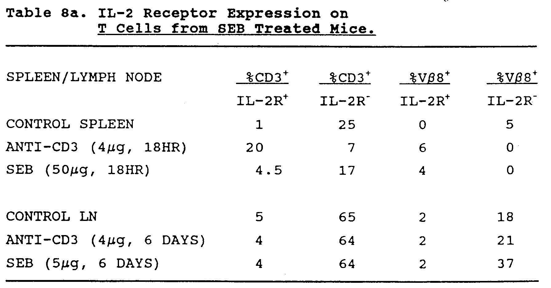
| Tumor regression in anti-CD3 and SEB treated mice. | |
| Group | Tumor Incidence (Day 28) |
| CONTROL | 13/16 |
| ANTI-CD3 (4µg) | 6/20 |
| SEB (50µg) | 5/21 p<0.01 |
| Measurement of the Effect of CD3
Treatment on the Immune System I. Peripheral Leukocyte Phenotype |
| Total Lymphocyte Count. |
| Total T-Cell Count. |
| Total Th and Ts counts. Helper/suppressor |
| Total NK Cell count. |
| CD3 and CD25 Expression. |
| II. Lymphokine Production |
| Interleukin-2 (IL-2) Level. |
| Soluble IL-2 receptor - a reflection of T-Cell Activation. |
| G-CSF and GM-CSF Levels. |
| TNF Level. |
| III. Cellular Cytotoxicity. |
| NK Cell Cytotoxicity on K562 Cell Line at 0 and 24 hours. |
| LAK Cell Cytotoxicity on Raji Cell Line at 0 and 24 hours. |
| Pre-LAK Cell (LAK Cell Cytotoxicity After 3-day Culture in IL-2) |
| IV. Cellular Proliferative Responses. |
| Anti-CD3 Induced Proliferation. |
| IL-2 Induced Proliferation. |
| Unidirectional MLC against pooled, Mitomycin C-Treated Lymphocytes. |
| PHA- and Con A- induced proliferation. |
| LAK = Lymphokine activated killer cells |
Claims (38)
- A composition for use in immunopotentiation in a human comprising an unconjugated monoclonal antibody that is directed to a T cell activation molecule or epitope on the cell surface of a human T cell, wherein the antibody is not directed to NDA5 or CD28.
- The composition of claim 1, wherein the monoclonal antibody is directed to a T cell activation molecule associated with the T cell receptor complex.
- The composition of claim 2, wherein the monoclonal antibody is directed to a T cell activation molecule variable or constant region epitope expressed on an antigen specific T cell receptor polymorphic TcR α, β, γ or δ chain.
- The composition of claim 2, wherein the monoclonal antibody is directed to CD3.
- The composition of claim 4, wherein the monoclonal antibody comprises OKT3 (ATCC CRL-8001).
- The composition of claim 1, wherein the monoclonal antibody is directed to a T cell surface antigen distinct from, and not physically associated on the cell surface with, the TcR.
- The composition of claim 6, wherein the monoclonal antibody is directed to Thy-1.
- The composition of claim 6, wherein the monoclonal antibody is directed to an activation epitope expressed on a member of the Ly-6 protein family.
- The composition of claim 6, wherein the monoclonal antibody is directed to human CD2.
- The composition of any preceding claim, wherein the monoclonal antibody is a bispecific antibody, wherein one arm is specific for a T cell activation epitope, and the other arm is specific for a T cell subset specific epitope.
- The composition of claim 10, wherein the monoclonal antibody comprises a union of monoclonal antibodies directed individually against CD3 and CD4.
- An immunopotentiating composition according to any preceding claim, for use in eliciting or enhancing a cellular or humoral immune response in a human subject wherein the composition is administered to the human subject in an amount effective to elicit or enhance a cellular or humoral immune response.
- An immunopotentiating composition according to any preceding claim further comprising a second selected compound for use in eliciting or enhancing a cellular or humoral immune response in a human subject to an epitope contained within said selected compound wherein the composition is administered to the human subject in an amount effective to elicit or enhance a cellular or humoral immune response against said epitope.
- The composition of claim 13, wherein the selected compound comprises a protein or peptide.
- The composition of claim 14, wherein the selected compound comprises a protein or peptide that includes an amino acid sequence against which the immune response is desired.
- The composition of claim 14 or 15, wherein the protein or peptide is from about 8 to about 100 amino acids in length.
- The composition of claim 16, wherein the protein or peptide is from about 8 to about 50 amino acids in length.
- The composition of any one of claims 14 to 17, wherein the selected compound comprises a protein or peptide derived from a tumor-specific or tumor-associated epitope.
- The composition of any one of claims 14 to 17, wherein the selected compound comprises a protein or peptide derived from a viral-specific or viral-associated epitope.
- The composition of claim 19, wherein the viral-specific or viral-associated epitope comprises an amino acid sequence found on the surface of an HIV virus or which is specific for HIV infected cells.
- The composition of claim 20, wherein the viral-specific or viral-associated epitope comprises an epitope from the gp120 envelope protein of HIV-1.
- The composition of claim 21, wherein the gp120 envelope protein epitope comprises peptides 18, T1, or T2.
- The composition of claim 19, wherein the viral-specific or viral-associated epitope comprises an amino acid sequence homologous to those expressed on the surface of human hepatitis virus.
- The composition of claim 19, wherein the viral-specific or viral-associated epitope comprises an amino acid sequence homologous to those expressed on the surface of viruses causing influenza.
- The composition of amy one of claims 14 to 17, wherein the selected compound comprises a protein or peptide with an amino acid sequence homologous to that derived from a gene in a bacteria, fungus, protozoal or metazoal parasite.
- A composition according to any preceding claim for administration to a human subject that has a tumor.
- A composition according to any preceding claim for administration to a human subject that is immunocompromised.
- A composition according to any preceding claim for administration to a human subject that has an infection.
- A composition according to any preceding claim for administration to a human subject deficient in hematopoietic bone marrow stem cells.
- A composition according to any preceding claim for administration to a human subject receiving a hematopoietic tissue transplant or graft.
- A composition according to any preceding claim for administration to a human subject in an amount between about 10 µg and about 1,000 µg.
- A composition according to any preceding claim for administration to a human subject in an amount between about 100 µg and about 400 µg.
- A composition according to any preceding claim for administration to a human subject as a bolus.
- A composition according to any preceding claim for administration to a human subject as a series of boluses.
- A composition according to any preceding claim for administration to a human subject as a series of boluses about every 14 days.
- A composition according to any preceding claim for use in eliciting or enhancing a cellular or humoral immune response in a human subject and for subsequently obtaining an antibody from said subject.
- A composition according to any preceding claim for use in eliciting or enhancing a cellular or humoral immune response in a human subject and for subsequently obtaining a T cell from said subject.
- Use of an immunopotentiating composition according to any preceding claim for the manufacture of a medicament for eliciting or enhancing a cellular or humoral immune response in a human subject when administered to a human subject in an amount effective to elicit or enhance a cellular or humoral immune response.
Priority Applications (1)
| Application Number | Priority Date | Filing Date | Title |
|---|---|---|---|
| EP98100138A EP0839536A1 (en) | 1989-10-27 | 1990-10-26 | Methods and compositions for promoting immunopotentiation |
Applications Claiming Priority (5)
| Application Number | Priority Date | Filing Date | Title |
|---|---|---|---|
| US42972989A | 1989-10-27 | 1989-10-27 | |
| US429729 | 1989-10-27 | ||
| US52430490A | 1990-05-16 | 1990-05-16 | |
| PCT/US1990/006177 WO1991006319A1 (en) | 1989-10-27 | 1990-10-26 | Methods and compositions for promoting immunopotentiation |
| US524304 | 2000-03-14 |
Related Child Applications (1)
| Application Number | Title | Priority Date | Filing Date |
|---|---|---|---|
| EP98100138A Division EP0839536A1 (en) | 1989-10-27 | 1990-10-26 | Methods and compositions for promoting immunopotentiation |
Publications (2)
| Publication Number | Publication Date |
|---|---|
| EP0497883A1 EP0497883A1 (en) | 1992-08-12 |
| EP0497883B1 true EP0497883B1 (en) | 1998-07-15 |
Family
ID=27028317
Family Applications (2)
| Application Number | Title | Priority Date | Filing Date |
|---|---|---|---|
| EP98100138A Withdrawn EP0839536A1 (en) | 1989-10-27 | 1990-10-26 | Methods and compositions for promoting immunopotentiation |
| EP90916853A Expired - Lifetime EP0497883B1 (en) | 1989-10-27 | 1990-10-26 | Compositions and their use for promoting immunopotentiation |
Family Applications Before (1)
| Application Number | Title | Priority Date | Filing Date |
|---|---|---|---|
| EP98100138A Withdrawn EP0839536A1 (en) | 1989-10-27 | 1990-10-26 | Methods and compositions for promoting immunopotentiation |
Country Status (8)
| Country | Link |
|---|---|
| US (2) | US6113901A (en) |
| EP (2) | EP0839536A1 (en) |
| JP (1) | JP2546544B2 (en) |
| AT (1) | ATE168272T1 (en) |
| AU (1) | AU6642390A (en) |
| CA (1) | CA2071478A1 (en) |
| DE (2) | DE69032484D1 (en) |
| WO (1) | WO1991006319A1 (en) |
Cited By (2)
| Publication number | Priority date | Publication date | Assignee | Title |
|---|---|---|---|---|
| US12221481B2 (en) | 2019-05-21 | 2025-02-11 | Novartis Ag | CD19 binding molecules and uses thereof |
| US12624118B2 (en) | 2019-05-21 | 2026-05-12 | Novartis Ag | Trispecific binding molecules against BCMA and uses thereof |
Families Citing this family (72)
| Publication number | Priority date | Publication date | Assignee | Title |
|---|---|---|---|---|
| US6685941B1 (en) | 1988-11-23 | 2004-02-03 | The Regents Of The University Of Michigan | Methods of treating autoimmune disease via CTLA-4Ig |
| DE69032484D1 (en) * | 1989-10-27 | 1998-08-20 | Arch Dev Corp | COMPOSITIONS AND THE USE THEREOF TO PROMOTE IMMUNOPOTENTIATION |
| US6406696B1 (en) | 1989-10-27 | 2002-06-18 | Tolerance Therapeutics, Inc. | Methods of stimulating the immune system with anti-CD3 antibodies |
| US5480895A (en) * | 1991-09-27 | 1996-01-02 | New York Society For The Relief Of The Ruptured And Crippled, Maintaining The Hospital For Special Surgery | Method of producing antibodies to a restricted population of T lymphocytes, antibodies produced therefrom and methods of use thereof |
| US20030108548A1 (en) * | 1993-06-01 | 2003-06-12 | Bluestone Jeffrey A. | Methods and materials for modulation of the immunosuppressive activity and toxicity of monoclonal antibodies |
| GB9607711D0 (en) * | 1996-04-13 | 1996-06-19 | Univ Sheffield | T-cell dependent vaccine |
| AU2596600A (en) | 1998-12-31 | 2000-07-31 | Chiron Corporation | Modified hiv env polypeptides |
| WO2000039304A2 (en) | 1998-12-31 | 2000-07-06 | Chiron Corporation | Polynucleotides encoding antigenic hiv type c polypeptides, polypeptides and uses thereof |
| EP1980617A1 (en) | 1998-12-31 | 2008-10-15 | Novartis Vaccines and Diagnostics, Inc. | Improved expression of HIV polypeptides and production of virus-like particles |
| US6737398B1 (en) * | 1999-09-30 | 2004-05-18 | National Jewish Medical And Research Center | Modulation of γδ T cells to regulate airway hyperresponsiveness |
| EP1246643A4 (en) | 1999-10-14 | 2005-05-11 | Jeffery A Ledbetter | Dna vaccines encoding antigen linked to a domain that binds cd40 |
| US7541184B2 (en) | 2000-02-24 | 2009-06-02 | Invitrogen Corporation | Activation and expansion of cells |
| US7572631B2 (en) | 2000-02-24 | 2009-08-11 | Invitrogen Corporation | Activation and expansion of T cells |
| US6867041B2 (en) * | 2000-02-24 | 2005-03-15 | Xcyte Therapies, Inc. | Simultaneous stimulation and concentration of cells |
| US20030235908A1 (en) * | 2000-02-24 | 2003-12-25 | Xcyte Therapies, Inc. | Activation and expansion of cells |
| US20030119185A1 (en) * | 2000-02-24 | 2003-06-26 | Xcyte Therapies, Inc. | Activation and expansion of cells |
| US6797514B2 (en) * | 2000-02-24 | 2004-09-28 | Xcyte Therapies, Inc. | Simultaneous stimulation and concentration of cells |
| US20020019048A1 (en) * | 2000-05-25 | 2002-02-14 | Ronald Berenson | Methods for restoring or enhancing T-cell immune surveillance following naturally or artificially induced immunosuppression |
| US8178098B2 (en) | 2001-04-03 | 2012-05-15 | National Jewish Health | Method to inhibit airway hyperresponsiveness using aerosolized T cell receptor antibodies |
| CA2452015C (en) | 2001-07-05 | 2012-07-03 | Chiron Corporation | Polynucleotides encoding antigenic hiv type c polypeptides, polypeptides and uses thereof |
| US20030138434A1 (en) * | 2001-08-13 | 2003-07-24 | Campbell Robert L. | Agents for enhancing the immune response |
| US20030170614A1 (en) | 2001-08-31 | 2003-09-11 | Megede Jan Zur | Polynucleotides encoding antigenic HIV type B polypeptides, polypeptides and uses thereof |
| US20050069549A1 (en) | 2002-01-14 | 2005-03-31 | William Herman | Targeted ligands |
| US20040151704A1 (en) * | 2002-06-28 | 2004-08-05 | Xcyte Therapies, Inc. | Compositions and methods for restoring immune repertoire in patients with immunological defects related to autoimmunity and organ or hematopoietic stem cell transplantation |
| US20040175373A1 (en) * | 2002-06-28 | 2004-09-09 | Xcyte Therapies, Inc. | Compositions and methods for eliminating undesired subpopulations of T cells in patients with immunological defects related to autoimmunity and organ or hematopoietic stem cell transplantation |
| US20050084967A1 (en) | 2002-06-28 | 2005-04-21 | Xcyte Therapies, Inc. | Compositions and methods for eliminating undesired subpopulations of T cells in patients with immunological defects related to autoimmunity and organ or hematopoietic stem cell transplantation |
| DE60334250D1 (en) * | 2003-05-08 | 2010-10-28 | Life Technologies Corp | GENERATION AND ISOLATION OF ANTIGEN-SPECIFIC T CELLS |
| CA2539716A1 (en) * | 2003-09-22 | 2005-04-07 | Xcyte Therapies, Inc. | Compositions and methods to accelerate hematologic recovery |
| EP1687066B1 (en) | 2003-11-14 | 2012-08-29 | Brigham and Women's Hospital, Inc. | Methods of modulating immunity |
| JP2007520566A (en) * | 2004-02-04 | 2007-07-26 | ザ トラスティーズ オブ コロンビア ユニヴァーシティ イン ザ シティ オブ ニューヨーク | Anti-CD3 and antigen-specific immunotherapy for autoimmune therapy |
| MXPA06014075A (en) | 2004-06-03 | 2007-03-15 | Novimmune Sa | Anti-cd3 antibodies and methods of use thereof. |
| AU2006267090B2 (en) | 2005-07-11 | 2013-03-07 | Provention Bio, Inc. | Methods for the treatment of autoimmune disorders using immunosuppressive monoclonal antibodies with reduced toxicity |
| AR057807A1 (en) * | 2005-09-12 | 2007-12-19 | Novimmune Sa | ANTI-CD3 ANTIBODY FORMULATIONS |
| EP1937309A4 (en) * | 2005-09-14 | 2010-01-20 | Univ Columbia | CD8 + T LYMPHOCYTES REGULATORS INDUCED BY ANTI-CD3 ANTIBODIES |
| US20070178113A1 (en) * | 2005-11-22 | 2007-08-02 | Backstrom B T | Superantigen conjugate |
| US20090258001A1 (en) * | 2006-06-06 | 2009-10-15 | Paul Ponath | Administration of anti-CD3 antibodies in the treatment of autoimmune diseases |
| MX2008015771A (en) | 2006-06-14 | 2009-01-27 | Macrogenics Inc | Methods for the treatment of autoimmune disorders using monoclonal antibodies with reduced toxicity. |
| KR20160009104A (en) | 2006-09-01 | 2016-01-25 | 가부시키가이샤 니콘 | Discharge lamp, light source apparatus, exposure apparatus and exposure apparatus manufacturing method |
| US20100015142A1 (en) * | 2006-12-21 | 2010-01-21 | Macrogenics Inc. | Methods for the treatment of lada and other adult- onset autoimmune using immunosuppressive monoclonal antibodies with reduced toxicity |
| US20080248025A1 (en) * | 2007-03-27 | 2008-10-09 | National Jewish Medical And Research Center | Gamma Delta T Cells and Methods of Treatment of Interleukin-17 Related Conditions |
| SG10201808730VA (en) † | 2007-04-03 | 2018-11-29 | Amgen Res Munich Gmbh | Cross-species-specific binding domain |
| EP2242513A2 (en) | 2008-01-18 | 2010-10-27 | Hadasit Medical Research Services & Development Ltd. | Combination therapy of beta-glycolipids and antibodies for the treatment of immune-related disorders |
| EP2531216B1 (en) * | 2010-02-04 | 2019-03-27 | The Trustees Of The University Of Pennsylvania | Icos critically regulates the expansion and function of inflammatory human th17 cells |
| KR20130066626A (en) | 2010-04-29 | 2013-06-20 | 나스벡스 엘티디. | Methods and compositions for treating hepatitis with anti-cd3 immune molecule therapy |
| US9018006B2 (en) | 2010-07-23 | 2015-04-28 | The University Of Toledo | Stable Tregs and related materials and methods |
| US20130273055A1 (en) | 2010-11-16 | 2013-10-17 | Eric Borges | Agents and methods for treating diseases that correlate with bcma expression |
| TWI638833B (en) | 2010-11-30 | 2018-10-21 | 中外製藥股份有限公司 | Cell damage induction treatment |
| TWI679212B (en) | 2011-11-15 | 2019-12-11 | 美商安進股份有限公司 | Binding molecules for e3 of bcma and cd3 |
| AU2013338153B2 (en) | 2012-10-29 | 2018-06-21 | The Board Of Trustees Of The University Of Arkansas | Novel mucosal adjuvants and delivery systems |
| US20150174242A1 (en) | 2013-12-19 | 2015-06-25 | Mayo Foundation For Medical Education And Research | Monovalent anti-cd3 adjuvants |
| KR102568808B1 (en) | 2014-04-07 | 2023-08-18 | 추가이 세이야쿠 가부시키가이샤 | Immunoactivating antigen-binding molecule |
| SG11201609370QA (en) | 2014-05-13 | 2016-12-29 | Chugai Pharmaceutical Co Ltd | T cell-redirected antigen-binding molecule for cells having immunosuppression function |
| WO2016059220A1 (en) | 2014-10-16 | 2016-04-21 | INSERM (Institut National de la Santé et de la Recherche Médicale) | Tcr-activating agents for use in the treatment of t-all |
| EP3812398A3 (en) | 2015-01-23 | 2021-07-21 | Sanofi | Anti-cd3 antibodies, anti-cd123 antibodies and bispecific antibodies specifically binding to cd3 and/or cd123 |
| EP3091032A1 (en) | 2015-05-08 | 2016-11-09 | Miltenyi Biotec GmbH | Humanized antibody or fragment thereof specific for cd3 |
| WO2017077382A1 (en) | 2015-11-06 | 2017-05-11 | Orionis Biosciences Nv | Bi-functional chimeric proteins and uses thereof |
| EP3378488A4 (en) * | 2015-11-18 | 2019-10-30 | Chugai Seiyaku Kabushiki Kaisha | METHOD FOR IMPROVING THE HUMORAL IMMUNE REACTION |
| WO2017086367A1 (en) | 2015-11-18 | 2017-05-26 | 中外製薬株式会社 | Combination therapy using t cell redirection antigen binding molecule against cell having immunosuppressing function |
| CN116769054A (en) | 2016-02-05 | 2023-09-19 | 奥里尼斯生物科学私人有限公司 | Bispecific signaling agents and uses thereof |
| TWI790206B (en) | 2016-07-18 | 2023-01-21 | 法商賽諾菲公司 | Bispecific antibody-like binding proteins specifically binding to cd3 and cd123 |
| CN110573172A (en) | 2017-02-06 | 2019-12-13 | 奥里尼斯生物科学有限公司 | Targeted engineered interferon and uses thereof |
| US20220348660A1 (en) | 2019-10-02 | 2022-11-03 | INSERM (Institut National de la Santé et de la Recherche Médicale) | Methods for the treatment of adult t-cell leukemia/lymphoma |
| CA3169827A1 (en) | 2020-03-10 | 2021-09-16 | Kunwar Shailubhai | Compositions of il-6/il-6r antibodies and methods of use thereof |
| EP4153314A4 (en) | 2020-05-19 | 2024-05-22 | The Regents of The University of California | Conjugate polypeptides and vaccines for inducing immune responses |
| EP4168537A1 (en) | 2020-06-17 | 2023-04-26 | Tiziana Life Sciences PLC | Compositions and methods for augmenting chimeric antigen receptor (car) t cell therapies |
| CN116406274B (en) | 2020-07-30 | 2026-02-17 | 迪赞纳生命科学公开有限公司 | CD3 antibodies for treatment of coronaviruses |
| US20220332822A1 (en) | 2021-04-16 | 2022-10-20 | Tiziana Life Sciences Plc | Subcutaneous administration of antibodies for the treatment of disease |
| WO2022251253A1 (en) | 2021-05-24 | 2022-12-01 | Provention Bio, Inc. | Methods for treating type 1 diabetes |
| IL311930A (en) | 2021-10-14 | 2024-06-01 | Brigham & Womens Hospital Inc | Methods to suppress microglial activation |
| EP4493592A1 (en) | 2022-03-14 | 2025-01-22 | LamKap Bio gamma AG | Bispecific gpc3xcd28 and gpc3xcd3 antibodies and their combination for targeted killing of gpc3 positive malignant cells |
| EP4574174A1 (en) | 2023-12-19 | 2025-06-25 | Jack Elands | Novel antibody-drug conjugates comprising an anti-(tcr)-cd3-complex binding antibody |
| WO2026096637A1 (en) | 2024-10-30 | 2026-05-07 | Tiziana Life Sciences Plc | Glp-1 agonists and foralumab combination therapies |
Citations (1)
| Publication number | Priority date | Publication date | Assignee | Title |
|---|---|---|---|---|
| US4658019A (en) * | 1979-04-26 | 1987-04-14 | Ortho Pharmaceutical Corporation | Complement-fixing monoclonal antibody to human T cells |
Family Cites Families (39)
| Publication number | Priority date | Publication date | Assignee | Title |
|---|---|---|---|---|
| JPS51133489A (en) * | 1975-05-14 | 1976-11-19 | Tokyo Daigaku | Process for producing microbial components of pseudomonas aeruginosa h aving antimicrobial and antitumor activities |
| US4361549A (en) * | 1979-04-26 | 1982-11-30 | Ortho Pharmaceutical Corporation | Complement-fixing monoclonal antibody to human T cells, and methods of preparing same |
| US4515893A (en) * | 1979-04-26 | 1985-05-07 | Ortho Pharmaceutical Corporation | Hybrid cell line for producing complement-fixing monoclonal antibody to human T cells |
| US4221794A (en) * | 1979-06-21 | 1980-09-09 | Newport Pharmaceuticals International, Inc. | Method of imparting immunomodulating and antiviral activity |
| US4395394A (en) * | 1979-10-26 | 1983-07-26 | Pfizer Inc. | Use of lipid amines formulated with fat or lipid emulsions as vaccine adjuvants |
| GB8314362D0 (en) * | 1983-05-24 | 1983-06-29 | Celltech Ltd | Polypeptide and protein products |
| US4695624A (en) * | 1984-05-10 | 1987-09-22 | Merck & Co., Inc. | Covalently-modified polyanionic bacterial polysaccharides, stable covalent conjugates of such polysaccharides and immunogenic proteins with bigeneric spacers, and methods of preparing such polysaccharides and conjugates and of confirming covalency |
| US4882317A (en) * | 1984-05-10 | 1989-11-21 | Merck & Co., Inc. | Covalently-modified bacterial polysaccharides, stable covalent conjugates of such polysaccharides and immunogenic proteins with bigeneric spacers and methods of preparing such polysaccharides and conjugataes and of confirming covalency |
| US5279960A (en) * | 1984-07-05 | 1994-01-18 | Enzon Corp. | 25 KD coccidial antigen of eimeria tenella |
| US4808700A (en) * | 1984-07-09 | 1989-02-28 | Praxis Biologics, Inc. | Immunogenic conjugates of non-toxic E. coli LT-B enterotoxin subunit and capsular polymers |
| NZ214503A (en) * | 1984-12-20 | 1990-02-26 | Merck & Co Inc | Covalently-modified neutral bacterial polysaccharides, stable covalent conjugates of such polysaccharides and immunogenic proteins, and methods of preparing such polysaccharides and conjugates |
| US5078998A (en) * | 1985-08-02 | 1992-01-07 | Bevan Michael J | Hybrid ligand directed to activation of cytotoxic effector T lymphocytes and target associated antigen |
| US4676980A (en) * | 1985-09-23 | 1987-06-30 | The United States Of America As Represented By The Secretary Of The Department Of Health And Human Services | Target specific cross-linked heteroantibodies |
| US4882424A (en) * | 1987-05-11 | 1989-11-21 | Dana-Farber Cancer Institute, Inc. | Activation antigen |
| US5639853A (en) * | 1987-09-29 | 1997-06-17 | Praxis Biologics, Inc. | Respiratory syncytial virus vaccines |
| WO1989006974A2 (en) * | 1988-02-01 | 1989-08-10 | Praxis Biologics, Inc. | T-cell epitope as carriers molecule for conjugate vaccines |
| FI105320B (en) * | 1988-04-04 | 2000-07-31 | Oncogen | Procedure for Preparation of Antibody Heteroconjugates for Use in Regulating Lymphocyte Activity and Diagnosis |
| JPH03504975A (en) * | 1988-06-14 | 1991-10-31 | セル―エスシーアイ コーポレイション | Heterofunctional cell immunizing agent, vaccine containing the same, and method for using the same |
| AU4334389A (en) * | 1988-10-14 | 1990-05-01 | Trustees Of Columbia University In The City Of New York, The | A differentiation antigen, nda5, associated with amplification of differentiation of t and b lymphocytes |
| WO1993019767A1 (en) * | 1992-04-07 | 1993-10-14 | The Regents Of The University Of Michigan | Cd28 pathway immunoregulation |
| CA2003455C (en) * | 1988-11-23 | 2000-02-22 | Craig B. Thompson | Immunotherapy involving cd28 stimulation |
| US5225538A (en) * | 1989-02-23 | 1993-07-06 | Genentech, Inc. | Lymphocyte homing receptor/immunoglobulin fusion proteins |
| AU648509B2 (en) * | 1989-07-14 | 1994-04-28 | Wyeth Holdings Corporation | Stable vaccine compositions containing interleukins |
| SE8903100D0 (en) * | 1989-09-20 | 1989-09-20 | Pharmacia Ab | NEW PHARMACEUTICAL AGENT |
| DE69032484D1 (en) * | 1989-10-27 | 1998-08-20 | Arch Dev Corp | COMPOSITIONS AND THE USE THEREOF TO PROMOTE IMMUNOPOTENTIATION |
| GB8928874D0 (en) * | 1989-12-21 | 1990-02-28 | Celltech Ltd | Humanised antibodies |
| ZA91463B (en) * | 1990-01-25 | 1992-09-30 | Bristol Myers Squibb Co | Method of activating cytolytic activity of lymphocytes using anti-cd28 antibody |
| CA2086325C (en) * | 1990-07-02 | 2010-10-05 | Peter S. Linsley | Ligand for cd28 receptor on b cells and methods |
| JPH06505396A (en) * | 1991-03-08 | 1994-06-23 | サイトメッド,インコーポレイテッド | Soluble CD28 protein and therapeutic methods using the same |
| US5770197A (en) * | 1991-06-27 | 1998-06-23 | Bristol-Myers Squibb Company | Methods for regulating the immune response using B7 binding molecules and IL4-binding molecules |
| EP0606217B2 (en) * | 1991-06-27 | 2008-12-03 | Bristol-Myers Squibb Company | Ctl4a receptor, fusion proteins containing it and uses thereof |
| AU4535893A (en) * | 1992-06-15 | 1994-01-04 | Regents Of The University Of California, The | Screening assay for the identification of immunosuppressive drugs |
| US5394321A (en) * | 1992-09-02 | 1995-02-28 | Electric Power Research Institute, Inc. | Quasi square-wave back-EMF permanent magnet AC machines with five or more phases |
| US5718883A (en) * | 1993-04-14 | 1998-02-17 | The United States Of America As Represented By The Secretary Of The Navy | Transgenic animal model for autoimmune diseases |
| US5885573A (en) * | 1993-06-01 | 1999-03-23 | Arch Development Corporation | Methods and materials for modulation of the immunosuppressive activity and toxicity of monoclonal antibodies |
| WO1994028912A1 (en) * | 1993-06-10 | 1994-12-22 | The Regents Of The University Of Michigan | Cd28 pathway immunosuppression |
| WO1995003408A1 (en) * | 1993-07-26 | 1995-02-02 | Dana-Farber Cancer Institute | B7-2: ctl a4/cd 28 counter receptor |
| AU7526894A (en) * | 1993-08-16 | 1995-03-14 | Arch Development Corporation | B7-2: ctla4/cd28 counter receptor |
| AUPQ960500A0 (en) * | 2000-08-23 | 2000-09-14 | Infinity Beautification Enterprises Pty Ltd | A planter box |
-
1990
- 1990-10-26 DE DE69032484A patent/DE69032484D1/en not_active Expired - Fee Related
- 1990-10-26 AU AU66423/90A patent/AU6642390A/en not_active Abandoned
- 1990-10-26 DE DE69032484T patent/DE69032484T4/en not_active Expired - Lifetime
- 1990-10-26 EP EP98100138A patent/EP0839536A1/en not_active Withdrawn
- 1990-10-26 WO PCT/US1990/006177 patent/WO1991006319A1/en not_active Ceased
- 1990-10-26 AT AT90916853T patent/ATE168272T1/en not_active IP Right Cessation
- 1990-10-26 JP JP2515665A patent/JP2546544B2/en not_active Expired - Lifetime
- 1990-10-26 EP EP90916853A patent/EP0497883B1/en not_active Expired - Lifetime
- 1990-10-26 CA CA002071478A patent/CA2071478A1/en not_active Abandoned
-
1994
- 1994-08-05 US US08/286,805 patent/US6113901A/en not_active Expired - Lifetime
-
1995
- 1995-06-02 US US08/458,462 patent/US6143297A/en not_active Expired - Fee Related
Patent Citations (1)
| Publication number | Priority date | Publication date | Assignee | Title |
|---|---|---|---|---|
| US4658019A (en) * | 1979-04-26 | 1987-04-14 | Ortho Pharmaceutical Corporation | Complement-fixing monoclonal antibody to human T cells |
Cited By (2)
| Publication number | Priority date | Publication date | Assignee | Title |
|---|---|---|---|---|
| US12221481B2 (en) | 2019-05-21 | 2025-02-11 | Novartis Ag | CD19 binding molecules and uses thereof |
| US12624118B2 (en) | 2019-05-21 | 2026-05-12 | Novartis Ag | Trispecific binding molecules against BCMA and uses thereof |
Also Published As
| Publication number | Publication date |
|---|---|
| DE69032484T4 (en) | 1999-09-16 |
| AU6642390A (en) | 1991-05-31 |
| EP0497883A1 (en) | 1992-08-12 |
| JPH05504554A (en) | 1993-07-15 |
| CA2071478A1 (en) | 1991-04-28 |
| US6143297A (en) | 2000-11-07 |
| ATE168272T1 (en) | 1998-08-15 |
| DE69032484D1 (en) | 1998-08-20 |
| DE69032484T2 (en) | 1999-03-11 |
| JP2546544B2 (en) | 1996-10-23 |
| EP0839536A1 (en) | 1998-05-06 |
| US6113901A (en) | 2000-09-05 |
| WO1991006319A1 (en) | 1991-05-16 |
Similar Documents
| Publication | Publication Date | Title |
|---|---|---|
| US6113901A (en) | Methods of stimulating or enhancing the immune system with anti-CD3 antibodies | |
| US6406696B1 (en) | Methods of stimulating the immune system with anti-CD3 antibodies | |
| Benjamin et al. | Mechanisms of monoclonal antibody‐facilitated tolerance induction: a possible role for the CD4 (L3T4) and CD11a (LFA‐1) molecules in self‐non‐self discrimination | |
| US6129916A (en) | Method of Increasing activation on proliferation of T cells using antibody-microbead conjugates | |
| US6056956A (en) | Non-depleting anti-CD4 monoclonal antibodies and tolerance induction | |
| Hafler et al. | Anti-CD4 and anti-CD2 monoclonal antibody infusions in subjects with multiple sclerosis. Immunosuppressive effects and human anti-mouse responses. | |
| US20120100156A1 (en) | Therapeutic Agent | |
| EP0474691B1 (en) | Monoclonal antibodies for inducing tolerance | |
| US5658569A (en) | Anti-HIV-1 neutralizing antibodies | |
| Scott | Antifluorescein affinity columns. Isolation and immunocompetence of lymphocytes that bind fluoresceinated antigens in vivo or in vitro. | |
| US7070785B2 (en) | Heat shock proteins for use in enhancing cellular factor production | |
| Truneh et al. | Humoral response of cynomolgus macaques to human soluble CD4: antibody reactivity restricted to xeno-human determinants | |
| US5529776A (en) | Anti-HIV-1 neutralizing antibodies | |
| JP2813630B2 (en) | Human immunodeficiency virus envelope polypeptide and its antibody | |
| CA2140150A1 (en) | A major histocompatibility complex antigen for use as vaccine against an immunodeficiency virus | |
| CA1337756C (en) | Means and method for preventing and treating graft failure in humans | |
| Cobbold | Cambridge zyxwvutsrqponmlkjihgfedcb | |
| Eibl et al. | Symposium in Immunology I and II | |
| Mu | Regulation of cytokine production by pertussis toxin and cyclophosphamide | |
| Sutherland et al. | Prolonged survival of renal allografts in outbred rabbits treated with donor specific F (ab') 2 alloantibody | |
| Ahmiedat | Induction of Transplantation Tolerance Using Monoclonal Antibodies to CD4: Experimental Studies Using a Rat Heterotopic Cardiac Allograft Model | |
| MXPA99003380A (en) | Compositions and methods for treating viral infections |
Legal Events
| Date | Code | Title | Description |
|---|---|---|---|
| PUAI | Public reference made under article 153(3) epc to a published international application that has entered the european phase |
Free format text: ORIGINAL CODE: 0009012 |
|
| 17P | Request for examination filed |
Effective date: 19920423 |
|
| AK | Designated contracting states |
Kind code of ref document: A1 Designated state(s): AT BE CH DE DK ES FR GB GR IT LI LU NL SE |
|
| RAP1 | Party data changed (applicant data changed or rights of an application transferred) |
Owner name: ARCH DEVELOPMENT CORPORATION |
|
| 17Q | First examination report despatched |
Effective date: 19950210 |
|
| GRAG | Despatch of communication of intention to grant |
Free format text: ORIGINAL CODE: EPIDOS AGRA |
|
| GRAG | Despatch of communication of intention to grant |
Free format text: ORIGINAL CODE: EPIDOS AGRA |
|
| GRAH | Despatch of communication of intention to grant a patent |
Free format text: ORIGINAL CODE: EPIDOS IGRA |
|
| GRAH | Despatch of communication of intention to grant a patent |
Free format text: ORIGINAL CODE: EPIDOS IGRA |
|
| GRAA | (expected) grant |
Free format text: ORIGINAL CODE: 0009210 |
|
| AK | Designated contracting states |
Kind code of ref document: B1 Designated state(s): AT BE CH DE DK ES FR GB GR IT LI LU NL SE |
|
| PG25 | Lapsed in a contracting state [announced via postgrant information from national office to epo] |
Ref country code: NL Free format text: LAPSE BECAUSE OF FAILURE TO SUBMIT A TRANSLATION OF THE DESCRIPTION OR TO PAY THE FEE WITHIN THE PRESCRIBED TIME-LIMIT Effective date: 19980715 Ref country code: GR Free format text: LAPSE BECAUSE OF FAILURE TO SUBMIT A TRANSLATION OF THE DESCRIPTION OR TO PAY THE FEE WITHIN THE PRESCRIBED TIME-LIMIT Effective date: 19980715 Ref country code: ES Free format text: THE PATENT HAS BEEN ANNULLED BY A DECISION OF A NATIONAL AUTHORITY Effective date: 19980715 Ref country code: BE Free format text: LAPSE BECAUSE OF FAILURE TO SUBMIT A TRANSLATION OF THE DESCRIPTION OR TO PAY THE FEE WITHIN THE PRESCRIBED TIME-LIMIT Effective date: 19980715 Ref country code: AT Free format text: LAPSE BECAUSE OF FAILURE TO SUBMIT A TRANSLATION OF THE DESCRIPTION OR TO PAY THE FEE WITHIN THE PRESCRIBED TIME-LIMIT Effective date: 19980715 |
|
| REF | Corresponds to: |
Ref document number: 168272 Country of ref document: AT Date of ref document: 19980815 Kind code of ref document: T |
|
| REG | Reference to a national code |
Ref country code: CH Ref legal event code: EP |
|
| ITF | It: translation for a ep patent filed | ||
| REF | Corresponds to: |
Ref document number: 69032484 Country of ref document: DE Date of ref document: 19980820 |
|
| PG25 | Lapsed in a contracting state [announced via postgrant information from national office to epo] |
Ref country code: SE Free format text: LAPSE BECAUSE OF FAILURE TO SUBMIT A TRANSLATION OF THE DESCRIPTION OR TO PAY THE FEE WITHIN THE PRESCRIBED TIME-LIMIT Effective date: 19981015 Ref country code: DK Free format text: LAPSE BECAUSE OF FAILURE TO SUBMIT A TRANSLATION OF THE DESCRIPTION OR TO PAY THE FEE WITHIN THE PRESCRIBED TIME-LIMIT Effective date: 19981015 |
|
| PG25 | Lapsed in a contracting state [announced via postgrant information from national office to epo] |
Ref country code: LU Free format text: LAPSE BECAUSE OF NON-PAYMENT OF DUE FEES Effective date: 19981026 |
|
| ET | Fr: translation filed | ||
| REG | Reference to a national code |
Ref country code: CH Ref legal event code: NV Representative=s name: BOVARD AG PATENTANWAELTE |
|
| NLV1 | Nl: lapsed or annulled due to failure to fulfill the requirements of art. 29p and 29m of the patents act | ||
| ET1 | Fr: translation filed ** revision of the translation of the patent or the claims | ||
| PLBE | No opposition filed within time limit |
Free format text: ORIGINAL CODE: 0009261 |
|
| STAA | Information on the status of an ep patent application or granted ep patent |
Free format text: STATUS: NO OPPOSITION FILED WITHIN TIME LIMIT |
|
| 26N | No opposition filed | ||
| PGFP | Annual fee paid to national office [announced via postgrant information from national office to epo] |
Ref country code: GB Payment date: 20001101 Year of fee payment: 11 |
|
| PGFP | Annual fee paid to national office [announced via postgrant information from national office to epo] |
Ref country code: DE Payment date: 20001106 Year of fee payment: 11 |
|
| PGFP | Annual fee paid to national office [announced via postgrant information from national office to epo] |
Ref country code: FR Payment date: 20001110 Year of fee payment: 11 |
|
| PGFP | Annual fee paid to national office [announced via postgrant information from national office to epo] |
Ref country code: CH Payment date: 20001113 Year of fee payment: 11 |
|
| PG25 | Lapsed in a contracting state [announced via postgrant information from national office to epo] |
Ref country code: GB Free format text: LAPSE BECAUSE OF NON-PAYMENT OF DUE FEES Effective date: 20011026 |
|
| PG25 | Lapsed in a contracting state [announced via postgrant information from national office to epo] |
Ref country code: LI Free format text: LAPSE BECAUSE OF NON-PAYMENT OF DUE FEES Effective date: 20011031 Ref country code: CH Free format text: LAPSE BECAUSE OF NON-PAYMENT OF DUE FEES Effective date: 20011031 |
|
| REG | Reference to a national code |
Ref country code: GB Ref legal event code: IF02 |
|
| REG | Reference to a national code |
Ref country code: CH Ref legal event code: PL |
|
| GBPC | Gb: european patent ceased through non-payment of renewal fee |
Effective date: 20011026 |
|
| PG25 | Lapsed in a contracting state [announced via postgrant information from national office to epo] |
Ref country code: FR Free format text: LAPSE BECAUSE OF NON-PAYMENT OF DUE FEES Effective date: 20020628 |
|
| PG25 | Lapsed in a contracting state [announced via postgrant information from national office to epo] |
Ref country code: DE Free format text: LAPSE BECAUSE OF NON-PAYMENT OF DUE FEES Effective date: 20020702 |
|
| REG | Reference to a national code |
Ref country code: FR Ref legal event code: ST |
|
| PG25 | Lapsed in a contracting state [announced via postgrant information from national office to epo] |
Ref country code: IT Free format text: LAPSE BECAUSE OF NON-PAYMENT OF DUE FEES;WARNING: LAPSES OF ITALIAN PATENTS WITH EFFECTIVE DATE BEFORE 2007 MAY HAVE OCCURRED AT ANY TIME BEFORE 2007. THE CORRECT EFFECTIVE DATE MAY BE DIFFERENT FROM THE ONE RECORDED. Effective date: 20051026 |