EP0490428A2 - Spectrometer with multichannel detection - Google Patents
Spectrometer with multichannel detection Download PDFInfo
- Publication number
- EP0490428A2 EP0490428A2 EP91203155A EP91203155A EP0490428A2 EP 0490428 A2 EP0490428 A2 EP 0490428A2 EP 91203155 A EP91203155 A EP 91203155A EP 91203155 A EP91203155 A EP 91203155A EP 0490428 A2 EP0490428 A2 EP 0490428A2
- Authority
- EP
- European Patent Office
- Prior art keywords
- skin surface
- slit
- spectrometer
- white standard
- line
- Prior art date
- Legal status (The legal status is an assumption and is not a legal conclusion. Google has not performed a legal analysis and makes no representation as to the accuracy of the status listed.)
- Granted
Links
- 238000001514 detection method Methods 0.000 title description 2
- 239000000049 pigment Substances 0.000 claims abstract description 23
- 238000000985 reflectance spectrum Methods 0.000 claims abstract description 20
- 230000003287 optical effect Effects 0.000 claims abstract description 18
- 238000011156 evaluation Methods 0.000 claims abstract description 17
- 238000003384 imaging method Methods 0.000 claims abstract description 8
- TZCXTZWJZNENPQ-UHFFFAOYSA-L barium sulfate Chemical compound [Ba+2].[O-]S([O-])(=O)=O TZCXTZWJZNENPQ-UHFFFAOYSA-L 0.000 claims description 8
- 238000001228 spectrum Methods 0.000 claims description 8
- 230000003595 spectral effect Effects 0.000 claims description 7
- 238000006073 displacement reaction Methods 0.000 claims description 3
- 230000003211 malignant effect Effects 0.000 abstract description 2
- 238000011835 investigation Methods 0.000 abstract 1
- 238000005259 measurement Methods 0.000 description 3
- 239000003086 colorant Substances 0.000 description 1
- 238000010586 diagram Methods 0.000 description 1
- 238000005516 engineering process Methods 0.000 description 1
- 238000002189 fluorescence spectrum Methods 0.000 description 1
- 238000005286 illumination Methods 0.000 description 1
- 238000010606 normalization Methods 0.000 description 1
- 125000006850 spacer group Chemical group 0.000 description 1
- 238000004611 spectroscopical analysis Methods 0.000 description 1
Images
Classifications
-
- A—HUMAN NECESSITIES
- A61—MEDICAL OR VETERINARY SCIENCE; HYGIENE
- A61B—DIAGNOSIS; SURGERY; IDENTIFICATION
- A61B5/00—Measuring for diagnostic purposes; Identification of persons
- A61B5/44—Detecting, measuring or recording for evaluating the integumentary system, e.g. skin, hair or nails
- A61B5/441—Skin evaluation, e.g. for skin disorder diagnosis
- A61B5/444—Evaluating skin marks, e.g. mole, nevi, tumour, scar
-
- A—HUMAN NECESSITIES
- A61—MEDICAL OR VETERINARY SCIENCE; HYGIENE
- A61B—DIAGNOSIS; SURGERY; IDENTIFICATION
- A61B5/00—Measuring for diagnostic purposes; Identification of persons
- A61B5/0059—Measuring for diagnostic purposes; Identification of persons using light, e.g. diagnosis by transillumination, diascopy, fluorescence
- A61B5/0062—Arrangements for scanning
- A61B5/0064—Body surface scanning
-
- A—HUMAN NECESSITIES
- A61—MEDICAL OR VETERINARY SCIENCE; HYGIENE
- A61B—DIAGNOSIS; SURGERY; IDENTIFICATION
- A61B5/00—Measuring for diagnostic purposes; Identification of persons
- A61B5/0059—Measuring for diagnostic purposes; Identification of persons using light, e.g. diagnosis by transillumination, diascopy, fluorescence
- A61B5/0075—Measuring for diagnostic purposes; Identification of persons using light, e.g. diagnosis by transillumination, diascopy, fluorescence by spectroscopy, i.e. measuring spectra, e.g. Raman spectroscopy, infrared absorption spectroscopy
-
- G—PHYSICS
- G01—MEASURING; TESTING
- G01J—MEASUREMENT OF INTENSITY, VELOCITY, SPECTRAL CONTENT, POLARISATION, PHASE OR PULSE CHARACTERISTICS OF INFRARED, VISIBLE OR ULTRAVIOLET LIGHT; COLORIMETRY; RADIATION PYROMETRY
- G01J3/00—Spectrometry; Spectrophotometry; Monochromators; Measuring colours
- G01J3/28—Investigating the spectrum
- G01J3/2823—Imaging spectrometer
-
- G—PHYSICS
- G01—MEASURING; TESTING
- G01J—MEASUREMENT OF INTENSITY, VELOCITY, SPECTRAL CONTENT, POLARISATION, PHASE OR PULSE CHARACTERISTICS OF INFRARED, VISIBLE OR ULTRAVIOLET LIGHT; COLORIMETRY; RADIATION PYROMETRY
- G01J3/00—Spectrometry; Spectrophotometry; Monochromators; Measuring colours
- G01J3/28—Investigating the spectrum
- G01J3/2803—Investigating the spectrum using photoelectric array detector
- G01J2003/2813—2D-array
-
- G—PHYSICS
- G01—MEASURING; TESTING
- G01N—INVESTIGATING OR ANALYSING MATERIALS BY DETERMINING THEIR CHEMICAL OR PHYSICAL PROPERTIES
- G01N21/00—Investigating or analysing materials by the use of optical means, i.e. using sub-millimetre waves, infrared, visible or ultraviolet light
- G01N21/17—Systems in which incident light is modified in accordance with the properties of the material investigated
- G01N21/47—Scattering, i.e. diffuse reflection
- G01N21/4738—Diffuse reflection, e.g. also for testing fluids, fibrous materials
Definitions
- the invention relates to a multichannel spectrometer, in particular for examining pigment marks, with an evaluation and display device and optical means for determining and imaging reflectance spectra of a skin surface on an evaluable image plane, taking into account white standards.
- Body color measurements are becoming increasingly important in many areas of technology.
- one is interested in obtaining objective measures for body colors, e.g. to gain diagnostic information about the skin surface.
- objective measures for body colors e.g. to gain diagnostic information about the skin surface.
- the light that is emitted from the skin surface of a body point is broken down into its spectrum with the aid of a monochromator and then calculated with predetermined standard spectral value curves to determine the color coordinates.
- predetermined standard spectral value curves to determine the color coordinates.
- a certain predetermined type of lighting must be assumed or the primary light spectrum must also be recorded and the reflectance spectrum of the body point corrected thereon.
- Such a spectrometer is known from DE-OS 27 26 606. This is essentially a so-called medical spectrophotometer with a spacer ring at the lens end for color measurement using the spectrometry of reflected light, i.e. the measurement of the reflectance of a skin surface, for example, in which a white standard alternates as a reference is used. Such a spectrometer makes it possible to determine the reflectance spectrum from a point on the surface.
- the object of the present invention is to provide an optical multichannel spectrometer for the contactless determination of the different reflectance spectra of the points of at least one line of a skin surface to be examined while continuously taking into account a primary light correction.
- the optical means a lens system directed towards the skin surface and the white standard and a slit diaphragm with a downstream one corresponding to a slit of the Slit diaphragm aligned diffraction grids, for the simultaneous generation of the reflectance spectrum of the points of a determinable line of the skin surface and the white standard on the image plane downstream of the diffraction grating and connected to the evaluation and display device.
- the lens system has an objective lens on an optical axis with means for sharp imaging of the white standard and at the same time the skin surface on the slit diaphragm.
- the slit diaphragm only allows a line-shaped image section to pass.
- the gratings of the diffraction grating are aligned parallel to the direction of the slit of the slit diaphragm.
- the white standard consists of, for example, pressed barium sulfate and can be attached in a small section in the area of the observed line of the skin surface to the pigment mole itself or next to it, e.g. be glued on.
- the multichannel spectrometer according to the invention is arranged such that it can be displaced and adjusted orthogonally to the slit direction and to the optical axis of the lens system, so that reflectance spectra according to the invention can be generated from virtually every line of the pigment mark.
- the multi-channel spectrometer according to the invention can have a housing and can be mounted in front of a low-light camera instead of a lens.
- the means for optically sharp imaging are advantageously formed by a linear displacement unit of the objective lens. It can also be provided that the objective lens is integrated in a zoom lens with a variable focal length.
- the arrangement of the lens system, the slit diaphragm and the diffraction grating according to the invention has the result that the diffraction of the parallel light beam at the diffraction grating in the image plane in a direction perpendicular to the direction of the slit results in the spectra of the individual slit image points and also perpendicular to the slit image and twice from the direct one Extending the zero-order slit image in mirror image to each other in two opposite directions.
- the correction i.e. the normalization of the reflectance spectra of e.g. Pigmentmal with regard to the spectral distribution of the primary light occurs via the reflectance spectrum of the white standard, which is also recorded at the same time, which is why practically any primary light can be used as long as the line being viewed is illuminated.
- a two-dimensional detector is arranged in the image plane, for the detection of the spectra of the individual slit image points of the skin surface and at the same time the white standard, in order to ultimately add the color coordinates for each point of the line under consideration via a subordinate evaluation and display device calculate and display.
- the calculation is carried out for each point of the line from the standardized reflectance spectra and tabulated standard spectral value curves, which can be stored in a memory of the evaluation device.
- a line-shaped section from the skin surface for example of a pigment mark
- To scan the complete Area only requires the corresponding repeated shifting of the spectrometer.
- it is often sufficient for diagnostic purposes if the color profile of a line is known, for example.
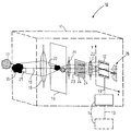
- FIG. 1 shows the basic structure and the mode of operation of the multi-channel spectrometer 10 according to the invention, which is arranged in a housing 11 and contains a two-dimensional detector 12, which is connected via a connecting line 13 to an evaluation and display device, for example arranged outside (dashed line) of the housing 11 14 is connected.
- the evaluation and display device 14 (solid line) can also be arranged in the housing 11.
- the optical axis 15 of the multichannel spectrometer 10 is directed at a skin surface outside the housing 11 and particularly at a pigment mark 16 located on the skin surface.
- a sample of pressed barium sulfate for example in tablet form, made of pressed barium sulfate as white standard is on the skin surface, not shown 17 attached. It can be provided that the white standard 17 is also arranged on or partially on the pigment 16, but does not substantially cover it.
- the pigment mark 16 and also the white standard 17 on the surface of the skin are illuminated in a spectrally broadband manner. However, it is also possible to provide spectrally narrowband lighting. The illumination of the respectively evaluated section or the evaluated line 25 must be even across the section.
- the pigment mark 16 and the white standard 17 are simultaneously imaged sharply into the slit region of a slit aperture 19.
- the distance of the objective lens 18 from the slit 20 can be variable in the direction of the optical axis 15 via a linear displacement unit, not shown.
- a zoom object can also be provided as an objective lens 18. Different magnifications can then advantageously be set without having to change the distance between the spectrometer and the skin surface.
- the slit aperture 19 has the gap 20 running horizontally in the plane of the drawing, which only has a line-shaped cutout 25 of the skin surface, i.e. of the pigment mark 16 and the white standard 18.
- the line-shaped intermediate image is projected into an evaluable image plane x, y via a system of two lenses 21 and 22 arranged downstream of the slit aperture 19.
- the correspondingly sensitive two-dimensional optical detector 12 is arranged in this image plane x, y, which is arranged downstream of the double lens system comprising the lenses 21 and 22.
- a diffraction grating 23 is arranged between the lenses 21 and 22, the grating 24 of said grating 24 running parallel to the direction of the slit 20 of the slit diaphragm 19.
- the double lens system comprising the lenses 21 and 22 is arranged between the slit aperture 19 and the detector 12 in such a way that the front focal point of the lens 21 lies in the slit plane 19 and the rear focal point of the lens 22 lies in the image plane x, y.
- the parallelism of the light beam in the double lens system is therefore determined by the distance between the lens 21 and the slit diaphragm 19. That is, it is necessary that the focal point of lens 21 facing the slit 20 lies exactly in the plane of the slit 20.
- the detector-side focal point of the lens 22 lies exactly in the image plane x, y, that is to say the detector 12.
- the diffraction of the parallel light beam of the intermediate image takes place at the diffraction grating 23 in a direction orthogonal to the direction of the slit 20 and the optical axis 15.
- This state of affairs is hatched in each case in FIG. 1 on the detector 12 and indicated in a diagram 26. The latter is shown in the correct position next to the detector 12 for better understanding.
- the correction i.e. Standardization of the reflectance spectra of the pigment 16 with respect to the spectral distribution of the primary light takes place via the reflectance spectrum of the white standard 17, which is also recorded, as is also shown hatched on the detector 12.
- the calculation of the color coordinates is carried out in the evaluation device of the evaluation and display device 14 for each point of the line 25 of the pigment mark 16 just under consideration from the standardized reflectance spectra and tabulated standard spectral value curves, which are stored in a memory of the evaluation device.
- the evaluation device can be a microprocessor.
- the housing 11 can advantageously be designed similar to a camera lens, as a result of which the multichannel spectrometer can be mounted in front of a low-light camera instead of a lens, with the aid of which the spectra can then be detected and transmitted to the evaluation and display device 14.
- a device for moving, ie raising or lowering the multichannel spectrometer in order to evaluate several lines 15 of a pigment mark is not shown in FIG. 1.
- a normal camera tripod can be used.
Landscapes
- Health & Medical Sciences (AREA)
- Life Sciences & Earth Sciences (AREA)
- Physics & Mathematics (AREA)
- Molecular Biology (AREA)
- Animal Behavior & Ethology (AREA)
- Pathology (AREA)
- Engineering & Computer Science (AREA)
- Biomedical Technology (AREA)
- Heart & Thoracic Surgery (AREA)
- Medical Informatics (AREA)
- Spectroscopy & Molecular Physics (AREA)
- Surgery (AREA)
- Biophysics (AREA)
- General Health & Medical Sciences (AREA)
- Public Health (AREA)
- Veterinary Medicine (AREA)
- Dermatology (AREA)
- General Physics & Mathematics (AREA)
- Nuclear Medicine, Radiotherapy & Molecular Imaging (AREA)
- Radiology & Medical Imaging (AREA)
- Spectrometry And Color Measurement (AREA)
- Investigating Or Analysing Materials By Optical Means (AREA)
- Measuring And Recording Apparatus For Diagnosis (AREA)
Abstract
Die Erfindung betrifft ein Vielkanalspektrometer (10), insbesondere zur Untersuchung von Pigmentmalen (16) mit einer Auswerte- und Anzeigeeinrichtung (14) und optischen Mitteln (18, 19, 21, 23, 22) zur Bestimmung und zur Abbildung von Remissionsspektren einer Hautoberfläche auf einer auswertbaren Bildebene (x, y) unter Berücksichtung eines Weiß-Standards (17). Bei einem solchen Spektrometer zur Untersuchung von u.U. maligner Pigmentmale (16) ist eine berührungslose optische Punkt für Punkt-Abtastung der Hautoberfläche wünschenswert, weshalb die optischen Mittel des Spektrometers (10) ein auf die Hautoberfläche und den Weiß-Standard (17) gerichtetes Linsensystem (18, 21, 22) und eine Spaltblende (19) mit einem nachgeordneten entsprechend einem Spalt (20) der Spaltblende (19) ausgerichteten Beugungsgitter (23) umfassen, zur gleichzeitigen Erzeugung des Remissionsspektrums der Punkte einer bestimmbaren Zeile (25) der Hautoberfäche und des Weiß-Standards (17) auf der dem Beugungsgitter (23) nachgeordneten mit der Auswerte- und Anzeigevorrichtung (14) verbundenen Bildebene (x, y). <IMAGE>The invention relates to a multichannel spectrometer (10), in particular for examining pigment marks (16) with an evaluation and display device (14) and optical means (18, 19, 21, 23, 22) for determining and imaging reflectance spectra of a skin surface an evaluable image plane (x, y) taking into account a white standard (17). With such a spectrometer for the investigation of possibly Malignant pigment (16) is a non-contact optical point for point scanning of the skin surface is desirable, which is why the optical means of the spectrometer (10) a lens system (18, 21, 22) directed towards the skin surface and the white standard (17) and one Slit diaphragm (19) with a downstream diffraction grating (23) aligned corresponding to a slit (20) of the slit diaphragm (19) for the simultaneous generation of the reflectance spectrum of the points of a determinable line (25) of the skin surface and the white standard (17) on the image plane (x, y) connected downstream of the diffraction grating (23) and connected to the evaluation and display device (14). <IMAGE>
Description
Die Erfindung betrifft ein Vielkanalspektrometer, insbesondere zur Untersuchung von Pigmentmalen, mit einer Auswerte- und Anzeigeeinrichtung und optischen Mitteln zur Bestimmung und zur Abbildung von Remissionsspektren einer Hautoberfläche auf einer auswertbaren Bildebene unter Berücksichtigung von Weiß-Standards.The invention relates to a multichannel spectrometer, in particular for examining pigment marks, with an evaluation and display device and optical means for determining and imaging reflectance spectra of a skin surface on an evaluable image plane, taking into account white standards.
Messungen von Körperfarben gewinnen in vielen Bereichen der Technik zunehmend an Bedeutung. Im medizinischen Bereich ist man daran interessiert, objektive Maßzahlen für Körperfarben zu gewinnen, um z.B. diagnostische Informationen über die Hautoberfläche zu gewinnen. Bei herkömmlichen Farbmeßgeräten wird das Licht, das von der Hautoberfläche eines Körperpunktes abgestrahlt wird, mit Hilfe eines Monochromators in sein Spektrum zerlegt und anschließend zur Bestimmung der Farbkoordinaten mit vorgegebenen Normspektralwertkurven verrechnet. Bei nicht selbstleuchtenden Objekten muß dabei von einer bestimmten vorgegebenen Beleuchtungsart ausgegangen oder das Primärlichtspektrum miterfaßt und das Remissionsspektrum des Körperpunktes darauf korrigiert werden.Body color measurements are becoming increasingly important in many areas of technology. In the medical field, one is interested in obtaining objective measures for body colors, e.g. to gain diagnostic information about the skin surface. In conventional color measuring devices, the light that is emitted from the skin surface of a body point is broken down into its spectrum with the aid of a monochromator and then calculated with predetermined standard spectral value curves to determine the color coordinates. In the case of non-self-illuminating objects, a certain predetermined type of lighting must be assumed or the primary light spectrum must also be recorded and the reflectance spectrum of the body point corrected thereon.
Ein derartiges Spektrometer ist aus der DE-OS 27 26 606 bekannt. Dabei handelt es sich im wesentlichen um ein sogenanntes medizinisches Spektralfotometer mit einem Distanzring am objektivseitigen Ende zur Farbmessung mit Hilfe der Spektrometrie von reflektiertem Licht, also der Messung der Remission (Reflektivität) einer z.B. Hautoberfläche, bei der auch noch als Referenz ein Weiß-Standard alternierend benutzt wird. Ein solches Spektrometer ermöglicht die Ermittlung des Remissionsspektrums von einem Punkt der Oberfläche.Such a spectrometer is known from DE-OS 27 26 606. This is essentially a so-called medical spectrophotometer with a spacer ring at the lens end for color measurement using the spectrometry of reflected light, i.e. the measurement of the reflectance of a skin surface, for example, in which a white standard alternates as a reference is used. Such a spectrometer makes it possible to determine the reflectance spectrum from a point on the surface.
Bei strukturierten Oberflächen, z.B. einer Hautoberfläche mit Pigmentmalen oder der Oberfläche eines Pigmentmals selbst, ist es notwendig, nicht nur einen definierten Punkt bezüglich seines Remissionsspektrums zu vermessen, sondern diese Informationen von einer Vielzahl von Hautoberflächenpunkten zu bestimmen. Im Idealfall ist es wünschenswert, von jedem Punkt der Hautoberfläche das Remissionsspektrum zu bestimmen. Eine aufwendige optische Punkt für Punkt-Abtastung der Oberfläche ist dazu notwendig. Bei der Untersuchung von u.U. maligner Pigmentmale ist ferner eine berührungsfreie Untersuchung wünschenswert.For structured surfaces, e.g. a skin surface with pigment marks or the surface of a pigment mark itself, it is not only necessary to measure a defined point with regard to its reflectance spectrum, but also to determine this information from a multiplicity of skin surface points. Ideally, it is desirable to determine the reflectance spectrum from every point on the skin surface. A complex optical point for point scanning of the surface is necessary for this. When examining Malignant pigment marks also require a non-contact examination.
Aus der DE-OS 38 15 743 ist eine Vorrichtung zur Messung und Auswertung von Eigenfluoreszenzspektren organischer Gewebeflächen bekannt. Bei dieser Vorrichtung wird vorgeschlagen, zur Erfassung der spektralen Verteilung von Fluoreszenzlicht der Hautoberfläche dieselbe spaltenförmig durch Projektion eines Spaltbildes auf die Hautoberfläche zu beleuchten. Dies hat zur Folge, daß für alle Punkte des von der Lichtquelle auf die Hautoberfläche projizierten Spaltbildes ein gemeinsames über alle Punkte gemitteltes Spektrum erfaßt wird. Eine, wie oben bereits ausgeführt wurde, wünschenswerte optische Punkt für Punkt-Abtastung der Hautoberfläche ist mit dieser Vorrichtung nicht möglich.From DE-OS 38 15 743 a device for measuring and evaluating self-fluorescence spectra of organic tissue areas is known. In this device, it is proposed to illuminate the spectral distribution of fluorescent light on the skin surface in the form of a column by projecting a slit image onto the skin surface. The result of this is that a common spectrum averaged over all points is recorded for all points of the slit image projected from the light source onto the skin surface. A desirable optical point for point scanning of the skin surface, as already stated above, is not possible with this device.
Aufgabe der vorliegenden Erfindung ist es, ein optisches Vielkanalspektrometer zu schaffen, zur berührungsfreien Ermittlung der unterschiedlichen Remissionsspektren der Punkte mindestens einer Zeile einer zu untersuchenden Hautoberfläche unter kontinuierlicher Berücksichtigung einer Primärlichtkorrektur.The object of the present invention is to provide an optical multichannel spectrometer for the contactless determination of the different reflectance spectra of the points of at least one line of a skin surface to be examined while continuously taking into account a primary light correction.
Erfindungsgemäß wird diese Aufgabe dadurch gelöst, daß die optischen Mittel ein auf die Hautoberfläche und den Weiß-Standard gerichtetes Linsensystem und eine Spaltblende mit einem nachgeordneten entsprechend einem Spalt der Spaltblende ausgerichteten Beugungsgitter umfassen, zur gleichzeitigen Erzeugung des Remissionsspektrums der Punkte einer bestimmbaren Zeile der Hautoberfläche und des Weiß-Standards auf der dem Beugungsgitter nachgeordneten mit der Auswerte- und Anzeigeeinrichtung verbundenen Bildebene.According to the invention, this object is achieved in that the optical means a lens system directed towards the skin surface and the white standard and a slit diaphragm with a downstream one corresponding to a slit of the Slit diaphragm aligned diffraction grids, for the simultaneous generation of the reflectance spectrum of the points of a determinable line of the skin surface and the white standard on the image plane downstream of the diffraction grating and connected to the evaluation and display device.
Gemäß einer bevorzugten Ausgestaltung der Erfindung ist vorgesehen, daß das Linsensystem auf einer optischen Achse eine Objektivlinse mit Mitteln zur scharfen Abbildung des Weiß-Standards und gleichzeitig der Hautoberfläche auf der Spaltblende aufweist. Die Spaltblende läßt nur einen zeilenförmigen Bildausschnitt passieren. Ein nachgeordnetes System von zwei Linsen, zwischen denen das Beugungsgitter angeordnet ist, erzeugt ein zeilenförmiges Zwischenbild und bildet es auf der Bildebene ab. Die Gitter des Beugungsgitters sind parallel zur Richtung des Spaltes der Spaltblende ausgerichtet.According to a preferred embodiment of the invention, it is provided that the lens system has an objective lens on an optical axis with means for sharp imaging of the white standard and at the same time the skin surface on the slit diaphragm. The slit diaphragm only allows a line-shaped image section to pass. A subordinate system of two lenses, between which the diffraction grating is arranged, creates a line-shaped intermediate image and images it on the image plane. The gratings of the diffraction grating are aligned parallel to the direction of the slit of the slit diaphragm.
Gemäß einer besonderen Ausgestaltung der Erfindung besteht der Weiß-Standard aus beispielsweise gepreßtem Bariumsulfat und kann im Bereich der beobachteten Zeile der Hautoberfläche in einem kleinen Abschnitt auf dem Pigmentmal selbst oder daneben auf der Hautoberfläche befestigt, z.B. aufgeklebt sein.According to a particular embodiment of the invention, the white standard consists of, for example, pressed barium sulfate and can be attached in a small section in the area of the observed line of the skin surface to the pigment mole itself or next to it, e.g. be glued on.
Zur Untersuchung der ganzen Fläche des Pigmentmals auf der Hautoberfläche kann vorteilhaft vorgesehen sein, daß das erfindungsgemäße Vielkanalspektrometer orthogonal zur Spaltrichtung und zur optischen Achse des Linsensystems verschiebbar und justierbar angeordnet ist, so daß erfindungsgemäße Remissionsspektren bestimmbar von quasi jeder Zeile des Pigmentmals erzeugt werden können.To examine the entire surface of the pigment mark on the surface of the skin, it can advantageously be provided that the multichannel spectrometer according to the invention is arranged such that it can be displaced and adjusted orthogonally to the slit direction and to the optical axis of the lens system, so that reflectance spectra according to the invention can be generated from virtually every line of the pigment mark.
Gemäß einer besonderen Ausgestaltung kann das erfindungsgemäße Vielkanalspektrometer ein Gehäuse haben und anstelle eines Objektivs vor einer Restlichtkamera montiert werden.According to a special embodiment, the multi-channel spectrometer according to the invention can have a housing and can be mounted in front of a low-light camera instead of a lens.
Die Mittel zur optisch scharfen Abbildung werden vorteilhaft durch eine Linearverschiebungseinheit der Objektivlinse gebildet. Es kann auch vorgesehen sein, daß die Objektivlinse in ein Zoomobjektiv mit variabler Brennweite integriert ist.The means for optically sharp imaging are advantageously formed by a linear displacement unit of the objective lens. It can also be provided that the objective lens is integrated in a zoom lens with a variable focal length.
Die erfindungsgemäße Anordnung des Linsensystems, der Spaltblende und des Beugungsgitters hat zur Folge, daß durch Beugung des parallelen Lichtstrahlbündels am Beugungsgitter in der Bildebene in einer Richtung senkrecht zur Richtung des Spaltes die Spektren der einzelnen Spaltbildpunkte entstehen und sich ebenfalls senkrecht zum Spaltbild und zweimal vom direkten Spaltbild nullter Ordnung ausgehend spiegelbildlich zueinander in zwei entgegengesetzten Richtungen erstrecken. Die Korrektur, d.h. die Normierung der Remissionsspektren des z.B. Pigmentmals bezüglich der spektralen Verteilung des Primärlichtes geschieht über das Remissionsspektrum des Weiß-Standards, das gleichzeitig miterfaßt wird, weshalb praktisch jedes Primärlicht verwendet werden kann, solange die gerade betrachtete Zeile beleuchtet ist.The arrangement of the lens system, the slit diaphragm and the diffraction grating according to the invention has the result that the diffraction of the parallel light beam at the diffraction grating in the image plane in a direction perpendicular to the direction of the slit results in the spectra of the individual slit image points and also perpendicular to the slit image and twice from the direct one Extending the zero-order slit image in mirror image to each other in two opposite directions. The correction, i.e. the normalization of the reflectance spectra of e.g. Pigmentmal with regard to the spectral distribution of the primary light occurs via the reflectance spectrum of the white standard, which is also recorded at the same time, which is why practically any primary light can be used as long as the line being viewed is illuminated.
Gemäß einer besonderen Ausgestaltung der Erfindung ist in der Bildebene ein zweidimensionaler Detektor angeordnet, zur Detektion der Spektren der einzelnen Spaltbildpunkte der Hautoberfläche und gleichzeitig des Weiß-Standards, um letztlich die Farbkoodinaten für jeden Punkt der jeweils betrachteten Zeile über eine nachgeordnete Auswerte- und Anzeigevorrichtung zu berechnen und anzuzeigen. Die Berechnung erfolgt für jeden Punkt der Zeile aus den normierten Remissionsspektren und tabellierten Normspektralwertkurven, die in einem Speicher der Auswertevorrichtung abgelegt sein können.According to a special embodiment of the invention, a two-dimensional detector is arranged in the image plane, for the detection of the spectra of the individual slit image points of the skin surface and at the same time the white standard, in order to ultimately add the color coordinates for each point of the line under consideration via a subordinate evaluation and display device calculate and display. The calculation is carried out for each point of the line from the standardized reflectance spectra and tabulated standard spectral value curves, which can be stored in a memory of the evaluation device.
Mit Hilfe des erfindungsgemäßen Vielkanalspektrometers kann somit berührungslos ein zeilenförmiger Ausschnitt aus der Hautoberfläche z.B. eines Pigmentmals für jeden Punkt simultan spektrometriert werden. Zur Abtastung der vollständigen Fläche bedarf es lediglich der entsprechenden mehrmaligen Verschiebung des Spektrometers. Häufig ist es jedoch zu diagnostischen Zwecken bereits ausreichend, wenn das Farbprofil beispielsweise einer Zeile bekannt ist.With the help of the multichannel spectrometer according to the invention, a line-shaped section from the skin surface, for example of a pigment mark, can thus be simultaneously spectrometrically measured for each point without contact. To scan the complete Area only requires the corresponding repeated shifting of the spectrometer. However, it is often sufficient for diagnostic purposes if the color profile of a line is known, for example.
Weitere vorteilhafte Ausgestaltungen der Erfindung ergeben sich aus den Unteransprüchen.Further advantageous embodiments of the invention result from the subclaims.
Ein Ausführungsbeispiel der Erfindung ist in der Zeichnung in der einzigen Fig. 1 dargestellt und wird im folgenden näher beschrieben.An embodiment of the invention is shown in the drawing in the single Fig. 1 and will be described in more detail below.
Fig. 1 zeigt den prinzipiellen Aufbau und die Funktionsweise des erfindungsgemäßen Vielkanalspektrometers 10, das in einem Gehäuse 11 angeordnet ist und einen zweidimensionalen Detektor 12 enthält, der über eine Verbindungsleitung 13 mit einer beispielsweise außerhalb (gestrichelte Linie) des Gehäuses 11 angeordneten Auswerte- und Anzeigeeinrichtung 14 verbunden ist. Die Auswerte- und Anzeigeeinrichtung 14 (ausgezogene Linie) kann ebenfalls im Gehäuse 11 angeordnet sein.1 shows the basic structure and the mode of operation of the
Die optische Achse 15 des Vielkanalspektrometers 10 ist auf eine Hautoberfläche außerhalb des Gehäuses 11 gerichtet und besonders auf ein auf der Hautoberfläche befindliches Pigmentmal 16. Neben dem Pigmentmal 16 ist auf der nicht dargestellten Hautoberfläche eine beispielsweise in Tablettenform ausgeführte Probe aus gepreßtem Bariumsulfat als Weiß-Standard 17 befestigt. Es kann vorgesehen sein, daß der Weiß-Standard 17 auch auf bzw. teilweise auf dem Pigmentmal 16 angeordnet ist, dieses jedoch nicht wesentlich abdeckt. Mit einer nicht dargestellten Lichtquelle wird das Pigmentmal 16 und auch der Weiß-Standard 17 auf der Hautoberfläche spektral breitbandig beleuchtet. Es ist jedoch auch möglich, eine spektral schmalbandige Beleuchtung vorzusehen. Die Ausleuchtung des jeweils ausgewerteten Abschnitts bzw. der ausgewerteten Zeile 25 muß über den Abschnitt hinweg gleichmäßig sein.The
Mit Hilfe einer im Gehäuse 11 befindlichen und z.B. verschiebbaren Objektivlinse 18 werden das Pigmentmal 16 und der Weiß-Standard 17 gleichzeitig scharf in den Spaltbereich einer Spaltblende 19 abgebildet. Der Abstand der Objektivlinse 18 vom Spalt 20 kann dazu über eine nicht dargestellte Linearverschiebungseinheit in Richtung der optischen Achse 15 variabel sein. Auch kann ein Zoomobjekt als Objektivlinse 18 vorgesehen sein. Vorteilhaft sind dann unterschiedliche Vergrößerungen einstellbar, ohne daß der Abstand zwischen Spektrometer und Hautoberfläche geändert werden muß. Die Spaltblende 19 weist den in der Zeichnungsebene horizontal verlaufenden Spalt 20 auf, der nur einen zeilenförmigen Ausschnitt 25 der Hautoberfläche, d.h. des Pigmentmals 16 und des Weiß-Standards 18 passieren läßt.With the help of a located in the
Über ein der Spaltblende 19 nachgeordnetes System von zwei Linsen 21 und 22 wird das zeilenförmige Zwischenbild in eine auswertbare Bildebene x, y projiziert. In dieser Bildebene x, y, die dem Doppellinsensystem aus den Linsen 21 und 22 nachgeordnet ist, ist der entsprechend empfindliche zweidimensionale optische Detektor 12 angeordnet. Zwischen den Linsen 21 und 22 ist ein Beugungsgitter 23 angeordnet, dessen Gitter 24 parallel zur Richtung des Spaltes 20 der Spaltblende 19 verlaufen. Das Doppellinsensystem aus den Linsen 21 und 22 ist derart zwischen der Spaltblende 19 und dem Detektor 12 angeordnet, daß der vorderseitige Brennpunkt der Linse 21 in der Spaltebene 19 und der rückseitige Brennpunkt der Linse 22 in der Bildebene x, y liegt. Die Parallelität des Lichtbündels im Doppellinsensystem wird also durch den Abstand zwischen der Linse 21 und der Spaltblende 19 besimmt. D.h. es ist notwendig, daß der dem Spalt 20 zugewandte Brennpunkt von Linse 21 genau in der Ebene des Spalts 20 liegt. Für den Detektor 12 und Linse 22 gilt, daß der detektorseitige Brennpunkt der Linse 22 genau in der Bildebene x, y, also des Detektors 12 liegt.The line-shaped intermediate image is projected into an evaluable image plane x, y via a system of two
Die Beugung des parallelen Lichtstrahlbündels des Zwischenbilds erfolgt am Beugungsgitter 23 in einer Richtung orthogonal zur Richtung des Spaltes 20 und zur optischen Achse 15. Dies hat zur Folge, daß in der Bildebene x, y auf dem zweidimensionalen Detektor 12 die Spektren der einzelnen Spaltbildpunkte sich ebenfalls orthogonal zum Spaltbild erstrecken und zwar zweimal vom direkten Spaltbild nullter Ordnung ausgehend spiegelbildlich zueinander in zwei entgegengesetzten Richtungen. Dieser Sachverhalt ist jeweils schraffiert in der Fig. 1 auf dem Detektor 12 und in einem Diagramm 26 angedeutet. Letzteres ist zum besseren Verständnis lagerichtig neben dem Detektor 12 dargestellt.The diffraction of the parallel light beam of the intermediate image takes place at the diffraction grating 23 in a direction orthogonal to the direction of the
Die Korrektur, d.h. Normierung der Remissionsspektren des Pigmentmals 16 bezüglich der spektralen Verteilung des Primärlichts geschieht über das Remissionsspektrum des Weiß-Standards 17, welches gleichzeitig miterfaßt wird, wie dies auch auf dem Detektor 12 schraffiert dargestellt ist. Die Berechnung der Farbkoordinaten wird in der Auswerteeinrichtung der Auswerte- und Anzeigeeinrichtung 14 für jeden Punkt der gerade betrachteten Zeile 25 des Pigmentmals 16 vorgenommen aus den normierten Remissionsspektren und tabellierten Normspektralwertkurven, die in einem Speicher der Auswerteeinrichtung gespeichert sind. Die Auswerteeinrichtung kann ein Mikroprozessor sein.The correction, i.e. Standardization of the reflectance spectra of the
Wie bereits erwähnt, kann vorteilhaft das Gehäuse 11 ähnlich einem Kameraobjektiv ausgebildet sein, wodurch das Vielkanalspektrometer anstelle eines Objektivs vor einer Restlichtkamera montierbar ist, mit deren Hilfe dann die Spektren detektiert und an die Auswerte- und Anzeigeeinrichtung 14 weitergegeben werden können.As already mentioned, the
Eine Vorrichtung zum Bewegen, also Anheben bzw. Senken des Vielkanalspektrometers, um mehrere Zeilen 15 eines Pigmentmals auszuwerten, ist in Fig. 1 nicht dargestellt. Beispielsweise kann ein normales Kamerastativ eingesetzt werden.A device for moving, ie raising or lowering the multichannel spectrometer in order to evaluate
Die in der vorstehenden Beschreibung, in der Zeichnung sowie in den Ansprüchen offenbarten Merkmale der Erfindung können sowohl einzeln als auch in beliebiger Kombination für die Verwirklichung der Erfindung in ihren verschiedenen Ausführungsformen wesentlich sein.The features of the invention disclosed in the above description, in the drawing and in the claims can be essential both individually and in any combination for realizing the invention in its various embodiments.
Claims (10)
dadurch gekennzeichnet, daß die optischen Mittel ein auf die Hautoberfläche und den Weiß-Standard (17) gerichtetes Linsensystem (18, 21, 22) und eine Spaltblende (19) mit einem nachgeordneten entsprechend einem Spalt (20) der Spaltblende (19) ausgerichteten Beugungsgitter (23) umfassen, zur gleichzeitigen Erzeugung des Remissionsspektrums der Punkte einer bestimmbaren Zeile (25) der Hautoberfläche und des Weiß-Standards (17) auf der dem Beugungsgitter (23) nachgeordneten mit der Auswerte- und Anzeigeeinrichtung (14) verbundenen Bildebene (x, y).Multi-channel spectrometer, in particular for examining pigment marks, with an evaluation and display device and optical means for determining and imaging reflectance spectra of a skin surface on an evaluable image plane, taking into account a white standard,
characterized in that the optical means comprise a lens system (18, 21, 22) directed towards the skin surface and the white standard (17) and a slit diaphragm (19) with a diffraction grating arranged downstream in accordance with a slit (20) of the slit diaphragm (19) (23), for the simultaneous generation of the reflectance spectrum of the points of a determinable line (25) of the skin surface and the white standard (17) on the image plane (x, downstream of the diffraction grating (23) connected to the evaluation and display device (14) y).
dadurch gekennzeichnet, daß das Linsensystem (18, 21, 22) auf einer optischen Achse (15) eine Objektivlinse (18) mit Mitteln zur scharfen Abbildung der Hautoberfläche und des Weiß-Standards (17) auf der Spaltblende (19) und ein System von zwei Linsen (21, 22) mit einem zwischengeschalteten Beugungsgitter (23) umfaßt, zur Erzeugung und Abbildung eines zeilenförmigen Zwischenbildes auf der Bildebene (x, y).Spectrometer according to claim 1,
characterized in that the lens system (18, 21, 22) on an optical axis (15) an objective lens (18) with means for sharply imaging the skin surface and the white standard (17) on the slit diaphragm (19) and a system of comprises two lenses (21, 22) with an intermediate diffraction grating (23) for generating and imaging a line-shaped intermediate image on the image plane (x, y).
dadurch gekennzeichnet, daß der Weiß-Standard (17) aus einer mindestens in der gerade ausgewerteten Zeile (25) der Hautoberfläche befestigten, z.B. verklebten Tablette aus beispielsweise gepreßtem Bariumsulfat besteht.Spectrometer according to claim 2,
characterized in that the white standard (17) consists of a tablet which is fastened, for example glued, at least in the line (25) of the skin surface which has just been evaluated and consists, for example, of pressed barium sulfate.
dadurch gekennzeichnet, daß die Tablette neben einem Pigmentmal (16) auf der Hautoberfläche oder auf einem kleineren Abschnitt des Pigmentmals (16) befestigt ist und zusammen mit dem Pigmentmal (16) von der Objektivlinse (18) abgebildet wird.Spectrometer according to claim 3,
characterized in that the tablet is attached next to a pigment mark (16) on the skin surface or on a smaller section of the pigment mark (16) and is imaged together with the pigment mark (16) by the objective lens (18).
dadurch gekennzeichnet, daß das Spektrometer (10) orthogonal zur Spaltrichtung (20) und zur optischen Achse (15) verschiebbar und justierbar ist, zur Erzeugung von Remissionsspektren von mehreren Zeilen (25) der zu untersuchenden Hautoberfläche.Spectrometer according to claim 4,
characterized in that the spectrometer (10) can be displaced and adjusted orthogonally to the slit direction (20) and to the optical axis (15) to generate reflectance spectra from several lines (25) of the skin surface to be examined.
dadurch gekennzeichnet, daß das Spektrometer (10) ein Gehäuse (11) aufweist, mit welchem es anstelle eines Objektivs vor einer Restlichtkamera montierbar ist.Spectrometer according to claim 5,
characterized in that the spectrometer (10) has a housing (11) with which it can be mounted in front of a low-light camera instead of an objective.
dadurch gekennzeichnet, daß die Mittel zur optisch scharfen Abbildung durch eine Linearverschiebungseinheit der Objektivlinse (18) gebildet werden, oder die Objektivlinse (18) in einem Zoomobjektiv mit variabler Brennweite integriert ist.Spectrometer according to claim 6,
characterized in that the means for optically sharp imaging are formed by a linear displacement unit of the objective lens (18), or the objective lens (18) is integrated in a zoom lens with variable focal length.
dadurch gekennzeichnet, daß die auswertbare Bildebene (x, y) durch einen zweidimensionalen Detektor (12) gebildet ist, zur Detektion der Spektren der einzelnen Punkte eines zeilenförmigen Spaltbildes der Hautoberfläche und des Weiß-Standards (17), die sich spiegelbildlich orthogonal zum Spaltbild erstrecken.Spectrometer according to one or more of the preceding claims,
characterized in that the evaluable image plane (x, y) is formed by a two-dimensional detector (12) for detecting the spectra of the individual points of a line-shaped slit image of the skin surface and the white standard (17), which extend in mirror image orthogonal to the slit image .
dadurch gekennzeichnet, daß dem Detektor (12) die Auswerte- und Anzeigeeinrichtung (14) zugeordnet ist, die die Berechnung und Anzeige der Farbkoordinaten für jeden Punkt der jeweils ausgewerteten Zeile (25) durchgeführt aus dem normierten Remissionsspektrum und tabellierten Normspektralwertkurven, die z.B. in der Auswerteeinrichtung gespeichert sind.Spectrometer according to claim 8,
characterized in that the detector (12) is assigned the evaluation and display device (14) which the calculation and display of the color coordinates for each point of the line (25) evaluated in each case is carried out from the standardized reflectance spectrum and tabulated standard spectral value curves, which are stored, for example, in the evaluation device.
dadurch gekennzeichnet, daß mindestens die jeweils zur Auswertung gelangende Zeile (25) der Hautoberfläche und des Weiß-Standards (17) mit Licht beleuchtet ist.Spectrometer according to one or more of the preceding claims,
characterized in that at least the line (25) of the skin surface and the white standard (17) that is being evaluated is illuminated with light.
Applications Claiming Priority (2)
| Application Number | Priority Date | Filing Date | Title |
|---|---|---|---|
| DE4039070A DE4039070A1 (en) | 1990-12-07 | 1990-12-07 | MULTI-CHANNEL SPECTROMETER |
| DE4039070 | 1990-12-07 |
Publications (3)
| Publication Number | Publication Date |
|---|---|
| EP0490428A2 true EP0490428A2 (en) | 1992-06-17 |
| EP0490428A3 EP0490428A3 (en) | 1992-08-26 |
| EP0490428B1 EP0490428B1 (en) | 1996-03-13 |
Family
ID=6419805
Family Applications (1)
| Application Number | Title | Priority Date | Filing Date |
|---|---|---|---|
| EP91203155A Expired - Lifetime EP0490428B1 (en) | 1990-12-07 | 1991-12-03 | Spectrometer with multichannel detection |
Country Status (4)
| Country | Link |
|---|---|
| US (1) | US5297555A (en) |
| EP (1) | EP0490428B1 (en) |
| JP (1) | JPH04294224A (en) |
| DE (2) | DE4039070A1 (en) |
Cited By (2)
| Publication number | Priority date | Publication date | Assignee | Title |
|---|---|---|---|---|
| WO1996011386A1 (en) * | 1994-10-10 | 1996-04-18 | Soliton Elektronik Ab | Method for a spectral based representation of image elements on a line |
| WO2002065069A2 (en) * | 2000-11-07 | 2002-08-22 | Hypermed, Inc. | Hyperspectral imaging calibration device |
Families Citing this family (13)
| Publication number | Priority date | Publication date | Assignee | Title |
|---|---|---|---|---|
| US5708504A (en) * | 1996-10-25 | 1998-01-13 | The United States Of America As Represented By The Secretary Of The Air Force | Interfering imaging spectrometer |
| US7202949B2 (en) * | 2003-09-30 | 2007-04-10 | The Trustees Of Boston University | Binocular spectrometer |
| US7315371B2 (en) * | 2004-01-23 | 2008-01-01 | P&P Optica Inc. | Multi-channel spectrum analyzer |
| JP4499476B2 (en) * | 2004-05-25 | 2010-07-07 | オリンパス株式会社 | Spectral image input apparatus and optical apparatus provided with the same |
| TWI245114B (en) * | 2004-10-11 | 2005-12-11 | Ind Tech Res Inst | Apparatus for measuring imaging spectrograph |
| GB0504279D0 (en) * | 2005-03-02 | 2005-04-06 | Carroll Charles C | Skin colour matching system and method |
| US7564547B2 (en) * | 2005-11-07 | 2009-07-21 | Wafermasters, Inc. | Spectroscopy system |
| TWI305832B (en) | 2006-12-13 | 2009-02-01 | Ind Tech Res Inst | Multi-channel imaging spectrometer |
| US8315692B2 (en) * | 2007-02-22 | 2012-11-20 | Sheinis Andrew I | Multi-spectral imaging spectrometer for early detection of skin cancer |
| JP5655437B2 (en) * | 2009-09-14 | 2015-01-21 | 株式会社リコー | Spectral characteristics acquisition device |
| US20130341509A1 (en) * | 2010-06-11 | 2013-12-26 | Chemimage Corporation | Portable system for detecting explosive materials using near infrared hyperspectral imaging and method for using thereof |
| US10154813B1 (en) | 2017-07-03 | 2018-12-18 | Spyros Kokolis | Method and apparatus for patient skin color monitoring and drug efficacy measurement |
| US10123738B1 (en) | 2017-07-03 | 2018-11-13 | Spyros Kokolis | Methods and apparatus for skin color patient monitoring |
Citations (4)
| Publication number | Priority date | Publication date | Assignee | Title |
|---|---|---|---|---|
| US3874799A (en) * | 1973-06-01 | 1975-04-01 | Color Control Inc | Method and apparatus for color spectrophotometry |
| DE2726606A1 (en) * | 1977-06-13 | 1978-12-21 | Max Planck Gesellschaft | MEDICAL SPECTRAL PHOTOMETER |
| DE2758141A1 (en) * | 1977-12-27 | 1979-06-28 | Ibm Deutschland | SPECTROPHOTOMETER |
| US4170987A (en) * | 1977-11-28 | 1979-10-16 | California Institute Of Technology | Medical diagnosis system and method with multispectral imaging |
Family Cites Families (3)
| Publication number | Priority date | Publication date | Assignee | Title |
|---|---|---|---|---|
| US4729372A (en) * | 1983-11-17 | 1988-03-08 | Lri L.P. | Apparatus for performing ophthalmic laser surgery |
| DE3815743A1 (en) * | 1988-05-07 | 1989-11-16 | Zeiss Carl Fa | DEVICE FOR MEASURING AND EVALUATING NATURAL FLUORESCENCE SPECTRES OF ORGANIC TISSUE SURFACES |
| US5014709A (en) * | 1989-06-13 | 1991-05-14 | Biologic Systems Corp. | Method and apparatus for high resolution holographic imaging of biological tissue |
-
1990
- 1990-12-07 DE DE4039070A patent/DE4039070A1/en not_active Withdrawn
-
1991
- 1991-12-02 US US07/803,313 patent/US5297555A/en not_active Expired - Fee Related
- 1991-12-03 EP EP91203155A patent/EP0490428B1/en not_active Expired - Lifetime
- 1991-12-03 DE DE59107547T patent/DE59107547D1/en not_active Expired - Fee Related
- 1991-12-05 JP JP3322038A patent/JPH04294224A/en active Pending
Patent Citations (4)
| Publication number | Priority date | Publication date | Assignee | Title |
|---|---|---|---|---|
| US3874799A (en) * | 1973-06-01 | 1975-04-01 | Color Control Inc | Method and apparatus for color spectrophotometry |
| DE2726606A1 (en) * | 1977-06-13 | 1978-12-21 | Max Planck Gesellschaft | MEDICAL SPECTRAL PHOTOMETER |
| US4170987A (en) * | 1977-11-28 | 1979-10-16 | California Institute Of Technology | Medical diagnosis system and method with multispectral imaging |
| DE2758141A1 (en) * | 1977-12-27 | 1979-06-28 | Ibm Deutschland | SPECTROPHOTOMETER |
Non-Patent Citations (1)
| Title |
|---|
| PHYSICS IN MEDECINE AND BIOLOGY. Bd. 34, Nr. 7, Juli 1989, LONDON GB Seiten 807 - 820; J.W. FEATHER ET AL: 'A portable scanning reflectance spectrophotometer using visible wavelengths for the rapid measurement of skin pigments' * |
Cited By (5)
| Publication number | Priority date | Publication date | Assignee | Title |
|---|---|---|---|---|
| WO1996011386A1 (en) * | 1994-10-10 | 1996-04-18 | Soliton Elektronik Ab | Method for a spectral based representation of image elements on a line |
| WO2002065069A2 (en) * | 2000-11-07 | 2002-08-22 | Hypermed, Inc. | Hyperspectral imaging calibration device |
| WO2002065069A3 (en) * | 2000-11-07 | 2004-02-19 | Hypermed Inc | Hyperspectral imaging calibration device |
| US6810279B2 (en) | 2000-11-07 | 2004-10-26 | Hypermed, Inc. | Hyperspectral imaging calibration device |
| US7013172B2 (en) | 2000-11-07 | 2006-03-14 | Hypermed, Inc. | Hyperspectral imaging calibration device |
Also Published As
| Publication number | Publication date |
|---|---|
| EP0490428A3 (en) | 1992-08-26 |
| EP0490428B1 (en) | 1996-03-13 |
| US5297555A (en) | 1994-03-29 |
| JPH04294224A (en) | 1992-10-19 |
| DE59107547D1 (en) | 1996-04-18 |
| DE4039070A1 (en) | 1992-06-11 |
Similar Documents
| Publication | Publication Date | Title |
|---|---|---|
| EP3891465B1 (en) | Optical measuring apparatus | |
| DE69330969T2 (en) | MULTI-SPECTRAL IMAGE ANALYSIS | |
| EP0116321B1 (en) | Infrared spectrometer | |
| EP0490428B1 (en) | Spectrometer with multichannel detection | |
| DE69314377T2 (en) | Portable spectrophotometer | |
| EP0279191B1 (en) | Device for contactless measurement of remission | |
| DE3713149A1 (en) | REMOTE MEASUREMENT SPECTROPHOTOMETER | |
| EP0778459B1 (en) | Spectrometer device | |
| DE69021786T2 (en) | Optical system for spectral analysis. | |
| DE60129247T2 (en) | OPTICAL SENSOR DEVICE AND METHOD FOR SPECTRAL ANALYSIS | |
| DE3843876C2 (en) | ||
| EP0209860A2 (en) | Apparatus for contactless reflection measurement | |
| EP1754032A1 (en) | Echelle spectrometer with improved use of the detector by means of two spectrometer arrangements | |
| DE102016212507A1 (en) | Chromatic confocal range sensor with camera part | |
| DE19961481B4 (en) | Centered sphere spectrometer | |
| DE202018102149U1 (en) | Optical measuring device | |
| DE102005024271B4 (en) | Grating spectrometer system and method for measured value acquisition | |
| EP1273951B1 (en) | Scanning microscope and method for the wavelength dependent detection | |
| DE10159234A1 (en) | Banknote testing device, spectrally separates light into two spectral components which can be detected respectively by individual detectors | |
| DE69005681T2 (en) | Transmission / reflection densitometer with two measuring heads. | |
| DE3244484A1 (en) | DEVICE FOR OPTIMIZING THE COUPLING OF TWO OPTICAL SYSTEMS FOR OBSERVING AND ANALYZING OBJECTS | |
| DE102019107963B4 (en) | Measuring light source and measuring arrangement for recording a reflection spectrum | |
| EP1588135B1 (en) | Array and method for the spectrally resolving detection of a sample | |
| DE10356729B4 (en) | color sensor | |
| EP1353210A2 (en) | Microscope including a device to determine the light intensity of an illumination beam |
Legal Events
| Date | Code | Title | Description |
|---|---|---|---|
| PUAI | Public reference made under article 153(3) epc to a published international application that has entered the european phase |
Free format text: ORIGINAL CODE: 0009012 |
|
| AK | Designated contracting states |
Kind code of ref document: A2 Designated state(s): DE FR GB |
|
| PUAL | Search report despatched |
Free format text: ORIGINAL CODE: 0009013 |
|
| AK | Designated contracting states |
Kind code of ref document: A3 Designated state(s): DE FR GB |
|
| 17P | Request for examination filed |
Effective date: 19930223 |
|
| 17Q | First examination report despatched |
Effective date: 19941128 |
|
| GRAA | (expected) grant |
Free format text: ORIGINAL CODE: 0009210 |
|
| AK | Designated contracting states |
Kind code of ref document: B1 Designated state(s): DE FR GB |
|
| REF | Corresponds to: |
Ref document number: 59107547 Country of ref document: DE Date of ref document: 19960418 |
|
| ET | Fr: translation filed | ||
| GBT | Gb: translation of ep patent filed (gb section 77(6)(a)/1977) |
Effective date: 19960528 |
|
| PGFP | Annual fee paid to national office [announced via postgrant information from national office to epo] |
Ref country code: GB Payment date: 19961202 Year of fee payment: 6 |
|
| PGFP | Annual fee paid to national office [announced via postgrant information from national office to epo] |
Ref country code: FR Payment date: 19961217 Year of fee payment: 6 |
|
| PLBE | No opposition filed within time limit |
Free format text: ORIGINAL CODE: 0009261 |
|
| STAA | Information on the status of an ep patent application or granted ep patent |
Free format text: STATUS: NO OPPOSITION FILED WITHIN TIME LIMIT |
|
| PGFP | Annual fee paid to national office [announced via postgrant information from national office to epo] |
Ref country code: DE Payment date: 19970221 Year of fee payment: 6 |
|
| 26N | No opposition filed | ||
| PG25 | Lapsed in a contracting state [announced via postgrant information from national office to epo] |
Ref country code: GB Free format text: LAPSE BECAUSE OF NON-PAYMENT OF DUE FEES Effective date: 19971203 |
|
| PG25 | Lapsed in a contracting state [announced via postgrant information from national office to epo] |
Ref country code: FR Free format text: THE PATENT HAS BEEN ANNULLED BY A DECISION OF A NATIONAL AUTHORITY Effective date: 19971231 |
|
| GBPC | Gb: european patent ceased through non-payment of renewal fee |
Effective date: 19971203 |
|
| PG25 | Lapsed in a contracting state [announced via postgrant information from national office to epo] |
Ref country code: DE Free format text: LAPSE BECAUSE OF NON-PAYMENT OF DUE FEES Effective date: 19980901 |
|
| REG | Reference to a national code |
Ref country code: FR Ref legal event code: ST |