EP0474769B1 - Verfahren zum nachweis von antikörpern gegen streptokinase - Google Patents
Verfahren zum nachweis von antikörpern gegen streptokinase Download PDFInfo
- Publication number
- EP0474769B1 EP0474769B1 EP90909412A EP90909412A EP0474769B1 EP 0474769 B1 EP0474769 B1 EP 0474769B1 EP 90909412 A EP90909412 A EP 90909412A EP 90909412 A EP90909412 A EP 90909412A EP 0474769 B1 EP0474769 B1 EP 0474769B1
- Authority
- EP
- European Patent Office
- Prior art keywords
- streptokinase
- antibodies
- antistreptokinase
- complex
- serum
- Prior art date
- Legal status (The legal status is an assumption and is not a legal conclusion. Google has not performed a legal analysis and makes no representation as to the accuracy of the status listed.)
- Expired - Lifetime
Links
- 229960005202 streptokinase Drugs 0.000 title claims abstract description 194
- 238000000034 method Methods 0.000 title claims abstract description 50
- 238000001514 detection method Methods 0.000 title claims abstract description 22
- 108010023197 Streptokinase Proteins 0.000 claims abstract description 186
- 210000002966 serum Anatomy 0.000 claims abstract description 69
- 108010044467 Isoenzymes Proteins 0.000 claims description 48
- 230000027455 binding Effects 0.000 claims description 21
- 102000004169 proteins and genes Human genes 0.000 claims description 19
- 108090000623 proteins and genes Proteins 0.000 claims description 19
- 102000013566 Plasminogen Human genes 0.000 claims description 16
- 108010051456 Plasminogen Proteins 0.000 claims description 16
- 238000001962 electrophoresis Methods 0.000 claims description 16
- 238000003556 assay Methods 0.000 claims description 12
- 108010088842 Fibrinolysin Proteins 0.000 claims description 5
- 241001465754 Metazoa Species 0.000 claims description 5
- 229940012957 plasmin Drugs 0.000 claims description 5
- 101710115418 Apolipoprotein(a) Proteins 0.000 claims description 4
- 102100040214 Apolipoprotein(a) Human genes 0.000 claims description 4
- 238000004440 column chromatography Methods 0.000 claims description 4
- 238000007824 enzymatic assay Methods 0.000 claims 2
- 230000002255 enzymatic effect Effects 0.000 claims 1
- 102000003855 L-lactate dehydrogenase Human genes 0.000 abstract description 131
- 108700023483 L-lactate dehydrogenases Proteins 0.000 abstract description 131
- 230000000694 effects Effects 0.000 description 39
- 239000000499 gel Substances 0.000 description 30
- 239000000523 sample Substances 0.000 description 21
- 102000023732 binding proteins Human genes 0.000 description 18
- 108091008324 binding proteins Proteins 0.000 description 18
- 235000018102 proteins Nutrition 0.000 description 14
- 230000003993 interaction Effects 0.000 description 13
- 239000002244 precipitate Substances 0.000 description 12
- 229920005654 Sephadex Polymers 0.000 description 10
- 239000012507 Sephadex™ Substances 0.000 description 10
- FAPWRFPIFSIZLT-UHFFFAOYSA-M Sodium chloride Chemical compound [Na+].[Cl-] FAPWRFPIFSIZLT-UHFFFAOYSA-M 0.000 description 10
- 108090000373 Tissue Plasminogen Activator Proteins 0.000 description 9
- 102000003978 Tissue Plasminogen Activator Human genes 0.000 description 9
- 238000004458 analytical method Methods 0.000 description 9
- 239000000872 buffer Substances 0.000 description 9
- 230000015572 biosynthetic process Effects 0.000 description 8
- 239000003814 drug Substances 0.000 description 8
- 238000001502 gel electrophoresis Methods 0.000 description 8
- 210000002381 plasma Anatomy 0.000 description 8
- 238000002965 ELISA Methods 0.000 description 7
- 229940024606 amino acid Drugs 0.000 description 7
- 235000001014 amino acid Nutrition 0.000 description 7
- 238000002474 experimental method Methods 0.000 description 7
- 239000003527 fibrinolytic agent Substances 0.000 description 7
- 239000000203 mixture Substances 0.000 description 7
- 229960000187 tissue plasminogen activator Drugs 0.000 description 7
- FTOAOBMCPZCFFF-UHFFFAOYSA-N 5,5-diethylbarbituric acid Chemical compound CCC1(CC)C(=O)NC(=O)NC1=O FTOAOBMCPZCFFF-UHFFFAOYSA-N 0.000 description 6
- 102000004506 Blood Proteins Human genes 0.000 description 6
- 108010017384 Blood Proteins Proteins 0.000 description 6
- 102000004420 Creatine Kinase Human genes 0.000 description 6
- 108010042126 Creatine kinase Proteins 0.000 description 6
- 150000001413 amino acids Chemical class 0.000 description 6
- 229940079593 drug Drugs 0.000 description 6
- 230000005764 inhibitory process Effects 0.000 description 6
- 238000012360 testing method Methods 0.000 description 6
- 241000283973 Oryctolagus cuniculus Species 0.000 description 5
- 230000004075 alteration Effects 0.000 description 5
- 125000000539 amino acid group Chemical group 0.000 description 5
- 229960002319 barbital Drugs 0.000 description 5
- 239000003153 chemical reaction reagent Substances 0.000 description 5
- 238000013508 migration Methods 0.000 description 5
- 230000005012 migration Effects 0.000 description 5
- 239000011780 sodium chloride Substances 0.000 description 5
- 238000010186 staining Methods 0.000 description 5
- 238000002560 therapeutic procedure Methods 0.000 description 5
- 210000001519 tissue Anatomy 0.000 description 5
- 102000004190 Enzymes Human genes 0.000 description 4
- 108090000790 Enzymes Proteins 0.000 description 4
- 241000287828 Gallus gallus Species 0.000 description 4
- 102000012479 Serine Proteases Human genes 0.000 description 4
- 108010022999 Serine Proteases Proteins 0.000 description 4
- 239000011543 agarose gel Substances 0.000 description 4
- AAOVKJBEBIDNHE-UHFFFAOYSA-N diazepam Chemical compound N=1CC(=O)N(C)C2=CC=C(Cl)C=C2C=1C1=CC=CC=C1 AAOVKJBEBIDNHE-UHFFFAOYSA-N 0.000 description 4
- 229940088598 enzyme Drugs 0.000 description 4
- 238000002523 gelfiltration Methods 0.000 description 4
- 229910052739 hydrogen Inorganic materials 0.000 description 4
- 239000011159 matrix material Substances 0.000 description 4
- 238000002156 mixing Methods 0.000 description 4
- 229930027945 nicotinamide-adenine dinucleotide Natural products 0.000 description 4
- BOPGDPNILDQYTO-NNYOXOHSSA-N nicotinamide-adenine dinucleotide Chemical compound C1=CCC(C(=O)N)=CN1[C@H]1[C@H](O)[C@H](O)[C@@H](COP(O)(=O)OP(O)(=O)OC[C@@H]2[C@H]([C@@H](O)[C@@H](O2)N2C3=NC=NC(N)=C3N=C2)O)O1 BOPGDPNILDQYTO-NNYOXOHSSA-N 0.000 description 4
- 238000002360 preparation method Methods 0.000 description 4
- 239000000047 product Substances 0.000 description 4
- 239000000243 solution Substances 0.000 description 4
- QTBSBXVTEAMEQO-UHFFFAOYSA-N Acetic acid Chemical compound CC(O)=O QTBSBXVTEAMEQO-UHFFFAOYSA-N 0.000 description 3
- 102000008186 Collagen Human genes 0.000 description 3
- 108010035532 Collagen Proteins 0.000 description 3
- BAWFJGJZGIEFAR-NNYOXOHSSA-O NAD(+) Chemical compound NC(=O)C1=CC=C[N+]([C@H]2[C@@H]([C@H](O)[C@@H](COP(O)(=O)OP(O)(=O)OC[C@@H]3[C@H]([C@@H](O)[C@@H](O3)N3C4=NC=NC(N)=C4N=C3)O)O2)O)=C1 BAWFJGJZGIEFAR-NNYOXOHSSA-O 0.000 description 3
- 108090000435 Urokinase-type plasminogen activator Proteins 0.000 description 3
- 102000003990 Urokinase-type plasminogen activator Human genes 0.000 description 3
- 206010000891 acute myocardial infarction Diseases 0.000 description 3
- 239000003146 anticoagulant agent Substances 0.000 description 3
- 230000001580 bacterial effect Effects 0.000 description 3
- 239000011230 binding agent Substances 0.000 description 3
- 239000008280 blood Substances 0.000 description 3
- 210000004369 blood Anatomy 0.000 description 3
- 238000004587 chromatography analysis Methods 0.000 description 3
- 229920001436 collagen Polymers 0.000 description 3
- 238000011161 development Methods 0.000 description 3
- LOKCTEFSRHRXRJ-UHFFFAOYSA-I dipotassium trisodium dihydrogen phosphate hydrogen phosphate dichloride Chemical compound P(=O)(O)(O)[O-].[K+].P(=O)(O)([O-])[O-].[Na+].[Na+].[Cl-].[K+].[Cl-].[Na+] LOKCTEFSRHRXRJ-UHFFFAOYSA-I 0.000 description 3
- HNDVDQJCIGZPNO-UHFFFAOYSA-N histidine Natural products OC(=O)C(N)CC1=CN=CN1 HNDVDQJCIGZPNO-UHFFFAOYSA-N 0.000 description 3
- 239000000463 material Substances 0.000 description 3
- 208000010125 myocardial infarction Diseases 0.000 description 3
- FSVCQIDHPKZJSO-UHFFFAOYSA-L nitro blue tetrazolium dichloride Chemical compound [Cl-].[Cl-].COC1=CC(C=2C=C(OC)C(=CC=2)[N+]=2N(N=C(N=2)C=2C=CC=CC=2)C=2C=CC(=CC=2)[N+]([O-])=O)=CC=C1[N+]1=NC(C=2C=CC=CC=2)=NN1C1=CC=C([N+]([O-])=O)C=C1 FSVCQIDHPKZJSO-UHFFFAOYSA-L 0.000 description 3
- 239000002953 phosphate buffered saline Substances 0.000 description 3
- 230000000717 retained effect Effects 0.000 description 3
- 238000004513 sizing Methods 0.000 description 3
- 241000894007 species Species 0.000 description 3
- 239000000758 substrate Substances 0.000 description 3
- 230000002537 thrombolytic effect Effects 0.000 description 3
- 229960005356 urokinase Drugs 0.000 description 3
- 239000011800 void material Substances 0.000 description 3
- UXFQFBNBSPQBJW-UHFFFAOYSA-N 2-amino-2-methylpropane-1,3-diol Chemical compound OCC(N)(C)CO UXFQFBNBSPQBJW-UHFFFAOYSA-N 0.000 description 2
- NUFBIAUZAMHTSP-UHFFFAOYSA-N 3-(n-morpholino)-2-hydroxypropanesulfonic acid Chemical compound OS(=O)(=O)CC(O)CN1CCOCC1 NUFBIAUZAMHTSP-UHFFFAOYSA-N 0.000 description 2
- RXGJTUSBYWCRBK-UHFFFAOYSA-M 5-methylphenazinium methyl sulfate Chemical compound COS([O-])(=O)=O.C1=CC=C2[N+](C)=C(C=CC=C3)C3=NC2=C1 RXGJTUSBYWCRBK-UHFFFAOYSA-M 0.000 description 2
- 229920000936 Agarose Polymers 0.000 description 2
- 241000283707 Capra Species 0.000 description 2
- 208000032843 Hemorrhage Diseases 0.000 description 2
- 108091006905 Human Serum Albumin Proteins 0.000 description 2
- 102000008100 Human Serum Albumin Human genes 0.000 description 2
- ROHFNLRQFUQHCH-UHFFFAOYSA-N Leucine Natural products CC(C)CC(N)C(O)=O ROHFNLRQFUQHCH-UHFFFAOYSA-N 0.000 description 2
- KDXKERNSBIXSRK-UHFFFAOYSA-N Lysine Natural products NCCCCC(N)C(O)=O KDXKERNSBIXSRK-UHFFFAOYSA-N 0.000 description 2
- 239000004472 Lysine Substances 0.000 description 2
- 206010061372 Streptococcal infection Diseases 0.000 description 2
- 208000007536 Thrombosis Diseases 0.000 description 2
- KZSNJWFQEVHDMF-UHFFFAOYSA-N Valine Natural products CC(C)C(N)C(O)=O KZSNJWFQEVHDMF-UHFFFAOYSA-N 0.000 description 2
- 125000003275 alpha amino acid group Chemical group 0.000 description 2
- 238000005349 anion exchange Methods 0.000 description 2
- 238000013459 approach Methods 0.000 description 2
- 230000006472 autoimmune response Effects 0.000 description 2
- 239000007982 barbital buffer Substances 0.000 description 2
- 239000013060 biological fluid Substances 0.000 description 2
- 239000003114 blood coagulation factor Substances 0.000 description 2
- 210000004027 cell Anatomy 0.000 description 2
- 230000008859 change Effects 0.000 description 2
- HVYWMOMLDIMFJA-DPAQBDIFSA-N cholesterol Chemical compound C1C=C2C[C@@H](O)CC[C@]2(C)[C@@H]2[C@@H]1[C@@H]1CC[C@H]([C@H](C)CCCC(C)C)[C@@]1(C)CC2 HVYWMOMLDIMFJA-DPAQBDIFSA-N 0.000 description 2
- 235000018417 cysteine Nutrition 0.000 description 2
- 230000034994 death Effects 0.000 description 2
- 230000007423 decrease Effects 0.000 description 2
- 238000013461 design Methods 0.000 description 2
- 230000020764 fibrinolysis Effects 0.000 description 2
- 230000003480 fibrinolytic effect Effects 0.000 description 2
- 230000001900 immune effect Effects 0.000 description 2
- 210000000987 immune system Anatomy 0.000 description 2
- 230000002163 immunogen Effects 0.000 description 2
- 238000011534 incubation Methods 0.000 description 2
- 230000006698 induction Effects 0.000 description 2
- 238000001990 intravenous administration Methods 0.000 description 2
- 238000005259 measurement Methods 0.000 description 2
- 210000004165 myocardium Anatomy 0.000 description 2
- 238000006386 neutralization reaction Methods 0.000 description 2
- COLNVLDHVKWLRT-UHFFFAOYSA-N phenylalanine Natural products OC(=O)C(N)CC1=CC=CC=C1 COLNVLDHVKWLRT-UHFFFAOYSA-N 0.000 description 2
- COLNVLDHVKWLRT-QMMMGPOBSA-N phenylalanine group Chemical group N[C@@H](CC1=CC=CC=C1)C(=O)O COLNVLDHVKWLRT-QMMMGPOBSA-N 0.000 description 2
- 230000036470 plasma concentration Effects 0.000 description 2
- 108090000765 processed proteins & peptides Proteins 0.000 description 2
- 230000009467 reduction Effects 0.000 description 2
- 239000011347 resin Substances 0.000 description 2
- 229920005989 resin Polymers 0.000 description 2
- 238000000926 separation method Methods 0.000 description 2
- 239000007787 solid Substances 0.000 description 2
- 239000004474 valine Substances 0.000 description 2
- MTCFGRXMJLQNBG-REOHCLBHSA-N (2S)-2-Amino-3-hydroxypropansäure Chemical compound OC[C@H](N)C(O)=O MTCFGRXMJLQNBG-REOHCLBHSA-N 0.000 description 1
- QKNYBSVHEMOAJP-UHFFFAOYSA-N 2-amino-2-(hydroxymethyl)propane-1,3-diol;hydron;chloride Chemical compound Cl.OCC(N)(CO)CO QKNYBSVHEMOAJP-UHFFFAOYSA-N 0.000 description 1
- BFSVOASYOCHEOV-UHFFFAOYSA-N 2-diethylaminoethanol Chemical compound CCN(CC)CCO BFSVOASYOCHEOV-UHFFFAOYSA-N 0.000 description 1
- XZKIHKMTEMTJQX-UHFFFAOYSA-N 4-Nitrophenyl Phosphate Chemical compound OP(O)(=O)OC1=CC=C([N+]([O-])=O)C=C1 XZKIHKMTEMTJQX-UHFFFAOYSA-N 0.000 description 1
- 101150035093 AMPD gene Proteins 0.000 description 1
- QTBSBXVTEAMEQO-UHFFFAOYSA-M Acetate Chemical compound CC([O-])=O QTBSBXVTEAMEQO-UHFFFAOYSA-M 0.000 description 1
- 102000009027 Albumins Human genes 0.000 description 1
- 108010088751 Albumins Proteins 0.000 description 1
- 102000002260 Alkaline Phosphatase Human genes 0.000 description 1
- 108020004774 Alkaline Phosphatase Proteins 0.000 description 1
- DCXYFEDJOCDNAF-UHFFFAOYSA-N Asparagine Natural products OC(=O)C(N)CC(N)=O DCXYFEDJOCDNAF-UHFFFAOYSA-N 0.000 description 1
- 241000894006 Bacteria Species 0.000 description 1
- 102000015081 Blood Coagulation Factors Human genes 0.000 description 1
- 108010039209 Blood Coagulation Factors Proteins 0.000 description 1
- UXVMQQNJUSDDNG-UHFFFAOYSA-L Calcium chloride Chemical compound [Cl-].[Cl-].[Ca+2] UXVMQQNJUSDDNG-UHFFFAOYSA-L 0.000 description 1
- 108010038061 Chymotrypsinogen Proteins 0.000 description 1
- KCXVZYZYPLLWCC-UHFFFAOYSA-N EDTA Chemical compound OC(=O)CN(CC(O)=O)CCN(CC(O)=O)CC(O)=O KCXVZYZYPLLWCC-UHFFFAOYSA-N 0.000 description 1
- 241000283074 Equus asinus Species 0.000 description 1
- 241000206602 Eukaryota Species 0.000 description 1
- 102000009123 Fibrin Human genes 0.000 description 1
- 108010073385 Fibrin Proteins 0.000 description 1
- BWGVNKXGVNDBDI-UHFFFAOYSA-N Fibrin monomer Chemical compound CNC(=O)CNC(=O)CN BWGVNKXGVNDBDI-UHFFFAOYSA-N 0.000 description 1
- 108010049003 Fibrinogen Proteins 0.000 description 1
- 102000008946 Fibrinogen Human genes 0.000 description 1
- 101100120327 Gallus gallus LD gene Proteins 0.000 description 1
- 108060003951 Immunoglobulin Proteins 0.000 description 1
- 102000001399 Kallikrein Human genes 0.000 description 1
- 108060005987 Kallikrein Proteins 0.000 description 1
- DCXYFEDJOCDNAF-REOHCLBHSA-N L-asparagine Chemical compound OC(=O)[C@@H](N)CC(N)=O DCXYFEDJOCDNAF-REOHCLBHSA-N 0.000 description 1
- CKLJMWTZIZZHCS-REOHCLBHSA-N L-aspartic acid Chemical compound OC(=O)[C@@H](N)CC(O)=O CKLJMWTZIZZHCS-REOHCLBHSA-N 0.000 description 1
- ROHFNLRQFUQHCH-YFKPBYRVSA-N L-leucine Chemical compound CC(C)C[C@H](N)C(O)=O ROHFNLRQFUQHCH-YFKPBYRVSA-N 0.000 description 1
- 125000002435 L-phenylalanyl group Chemical group O=C([*])[C@](N([H])[H])([H])C([H])([H])C1=C([H])C([H])=C([H])C([H])=C1[H] 0.000 description 1
- KZSNJWFQEVHDMF-BYPYZUCNSA-N L-valine Chemical compound CC(C)[C@H](N)C(O)=O KZSNJWFQEVHDMF-BYPYZUCNSA-N 0.000 description 1
- JVTAAEKCZFNVCJ-UHFFFAOYSA-M Lactate Chemical compound CC(O)C([O-])=O JVTAAEKCZFNVCJ-UHFFFAOYSA-M 0.000 description 1
- 208000012902 Nervous system disease Diseases 0.000 description 1
- 102000004316 Oxidoreductases Human genes 0.000 description 1
- 108090000854 Oxidoreductases Proteins 0.000 description 1
- 102000016387 Pancreatic elastase Human genes 0.000 description 1
- 108010067372 Pancreatic elastase Proteins 0.000 description 1
- 102000035195 Peptidases Human genes 0.000 description 1
- 108091005804 Peptidases Proteins 0.000 description 1
- 241000042032 Petrocephalus catostoma Species 0.000 description 1
- 108010094028 Prothrombin Proteins 0.000 description 1
- 102100027378 Prothrombin Human genes 0.000 description 1
- LCTONWCANYUPML-UHFFFAOYSA-M Pyruvate Chemical compound CC(=O)C([O-])=O LCTONWCANYUPML-UHFFFAOYSA-M 0.000 description 1
- 229920002684 Sepharose Polymers 0.000 description 1
- MTCFGRXMJLQNBG-UHFFFAOYSA-N Serine Natural products OCC(N)C(O)=O MTCFGRXMJLQNBG-UHFFFAOYSA-N 0.000 description 1
- BQCADISMDOOEFD-UHFFFAOYSA-N Silver Chemical compound [Ag] BQCADISMDOOEFD-UHFFFAOYSA-N 0.000 description 1
- 229920002472 Starch Polymers 0.000 description 1
- AYFVYJQAPQTCCC-UHFFFAOYSA-N Threonine Natural products CC(O)C(N)C(O)=O AYFVYJQAPQTCCC-UHFFFAOYSA-N 0.000 description 1
- 239000004473 Threonine Substances 0.000 description 1
- 206010066901 Treatment failure Diseases 0.000 description 1
- -1 Tris-barbital Chemical compound 0.000 description 1
- 102000004142 Trypsin Human genes 0.000 description 1
- 108090000631 Trypsin Proteins 0.000 description 1
- 102000018690 Trypsinogen Human genes 0.000 description 1
- 108010027252 Trypsinogen Proteins 0.000 description 1
- 229940099983 activase Drugs 0.000 description 1
- 230000004913 activation Effects 0.000 description 1
- 230000001154 acute effect Effects 0.000 description 1
- 229960003318 alteplase Drugs 0.000 description 1
- 239000000427 antigen Substances 0.000 description 1
- 102000036639 antigens Human genes 0.000 description 1
- 108091007433 antigens Proteins 0.000 description 1
- 229960001230 asparagine Drugs 0.000 description 1
- 235000009582 asparagine Nutrition 0.000 description 1
- 229940009098 aspartate Drugs 0.000 description 1
- 238000011888 autopsy Methods 0.000 description 1
- 230000004888 barrier function Effects 0.000 description 1
- 238000005452 bending Methods 0.000 description 1
- 230000008901 benefit Effects 0.000 description 1
- 230000023555 blood coagulation Effects 0.000 description 1
- 239000001110 calcium chloride Substances 0.000 description 1
- 235000011148 calcium chloride Nutrition 0.000 description 1
- 229910001628 calcium chloride Inorganic materials 0.000 description 1
- 230000000747 cardiac effect Effects 0.000 description 1
- 210000003169 central nervous system Anatomy 0.000 description 1
- 238000005119 centrifugation Methods 0.000 description 1
- 210000001175 cerebrospinal fluid Anatomy 0.000 description 1
- 238000006243 chemical reaction Methods 0.000 description 1
- 235000012000 cholesterol Nutrition 0.000 description 1
- 239000012504 chromatography matrix Substances 0.000 description 1
- 238000003776 cleavage reaction Methods 0.000 description 1
- 230000015271 coagulation Effects 0.000 description 1
- 238000005345 coagulation Methods 0.000 description 1
- 230000000052 comparative effect Effects 0.000 description 1
- 108091036078 conserved sequence Proteins 0.000 description 1
- 210000004351 coronary vessel Anatomy 0.000 description 1
- XUJNEKJLAYXESH-UHFFFAOYSA-N cysteine Natural products SCC(N)C(O)=O XUJNEKJLAYXESH-UHFFFAOYSA-N 0.000 description 1
- 125000000151 cysteine group Chemical group N[C@@H](CS)C(=O)* 0.000 description 1
- 150000001945 cysteines Chemical class 0.000 description 1
- 230000009089 cytolysis Effects 0.000 description 1
- 230000003247 decreasing effect Effects 0.000 description 1
- 230000001419 dependent effect Effects 0.000 description 1
- 230000008021 deposition Effects 0.000 description 1
- 238000003745 diagnosis Methods 0.000 description 1
- 238000002405 diagnostic procedure Methods 0.000 description 1
- 238000009792 diffusion process Methods 0.000 description 1
- 239000012470 diluted sample Substances 0.000 description 1
- 201000010099 disease Diseases 0.000 description 1
- 208000037265 diseases, disorders, signs and symptoms Diseases 0.000 description 1
- 231100000673 dose–response relationship Toxicity 0.000 description 1
- 238000005516 engineering process Methods 0.000 description 1
- 238000000605 extraction Methods 0.000 description 1
- 210000003608 fece Anatomy 0.000 description 1
- 229950003499 fibrin Drugs 0.000 description 1
- 229940012952 fibrinogen Drugs 0.000 description 1
- 238000001914 filtration Methods 0.000 description 1
- 239000012847 fine chemical Substances 0.000 description 1
- 235000013305 food Nutrition 0.000 description 1
- 208000019622 heart disease Diseases 0.000 description 1
- 238000004128 high performance liquid chromatography Methods 0.000 description 1
- 239000001257 hydrogen Substances 0.000 description 1
- 230000003053 immunization Effects 0.000 description 1
- 238000002649 immunization Methods 0.000 description 1
- 102000018358 immunoglobulin Human genes 0.000 description 1
- 230000000415 inactivating effect Effects 0.000 description 1
- 238000001802 infusion Methods 0.000 description 1
- 239000003112 inhibitor Substances 0.000 description 1
- 238000003780 insertion Methods 0.000 description 1
- 230000037431 insertion Effects 0.000 description 1
- 230000003834 intracellular effect Effects 0.000 description 1
- 229960000310 isoleucine Drugs 0.000 description 1
- 210000003292 kidney cell Anatomy 0.000 description 1
- 125000001909 leucine group Chemical group [H]N(*)C(C(*)=O)C([H])([H])C(C([H])([H])[H])C([H])([H])[H] 0.000 description 1
- 239000007788 liquid Substances 0.000 description 1
- 239000006194 liquid suspension Substances 0.000 description 1
- GKQWYZBANWAFMQ-UHFFFAOYSA-M lithium;2-hydroxypropanoate Chemical compound [Li+].CC(O)C([O-])=O GKQWYZBANWAFMQ-UHFFFAOYSA-M 0.000 description 1
- 210000004185 liver Anatomy 0.000 description 1
- 229940040511 liver extract Drugs 0.000 description 1
- 238000011068 loading method Methods 0.000 description 1
- 235000013372 meat Nutrition 0.000 description 1
- 230000001404 mediated effect Effects 0.000 description 1
- VMGAPWLDMVPYIA-HIDZBRGKSA-N n'-amino-n-iminomethanimidamide Chemical compound N\N=C\N=N VMGAPWLDMVPYIA-HIDZBRGKSA-N 0.000 description 1
- 230000007935 neutral effect Effects 0.000 description 1
- 230000003472 neutralizing effect Effects 0.000 description 1
- 238000011587 new zealand white rabbit Methods 0.000 description 1
- 239000013610 patient sample Substances 0.000 description 1
- 239000000825 pharmaceutical preparation Substances 0.000 description 1
- 230000000704 physical effect Effects 0.000 description 1
- 239000000049 pigment Substances 0.000 description 1
- 229920002401 polyacrylamide Polymers 0.000 description 1
- 229920000642 polymer Polymers 0.000 description 1
- 230000003389 potentiating effect Effects 0.000 description 1
- 230000008569 process Effects 0.000 description 1
- 102000004196 processed proteins & peptides Human genes 0.000 description 1
- 229940039716 prothrombin Drugs 0.000 description 1
- 238000000163 radioactive labelling Methods 0.000 description 1
- BOLDJAUMGUJJKM-LSDHHAIUSA-N renifolin D Natural products CC(=C)[C@@H]1Cc2c(O)c(O)ccc2[C@H]1CC(=O)c3ccc(O)cc3O BOLDJAUMGUJJKM-LSDHHAIUSA-N 0.000 description 1
- 238000011160 research Methods 0.000 description 1
- 238000012502 risk assessment Methods 0.000 description 1
- 210000003296 saliva Anatomy 0.000 description 1
- 230000007017 scission Effects 0.000 description 1
- 238000012216 screening Methods 0.000 description 1
- 229910052709 silver Inorganic materials 0.000 description 1
- 239000004332 silver Substances 0.000 description 1
- 210000002027 skeletal muscle Anatomy 0.000 description 1
- 239000001488 sodium phosphate Substances 0.000 description 1
- 229910000162 sodium phosphate Inorganic materials 0.000 description 1
- 230000000087 stabilizing effect Effects 0.000 description 1
- 238000010561 standard procedure Methods 0.000 description 1
- 235000019698 starch Nutrition 0.000 description 1
- 239000008107 starch Substances 0.000 description 1
- 239000000126 substance Substances 0.000 description 1
- 208000024891 symptom Diseases 0.000 description 1
- 210000001138 tear Anatomy 0.000 description 1
- 230000002381 testicular Effects 0.000 description 1
- 210000001550 testis Anatomy 0.000 description 1
- 230000001225 therapeutic effect Effects 0.000 description 1
- 229960000103 thrombolytic agent Drugs 0.000 description 1
- RYFMWSXOAZQYPI-UHFFFAOYSA-K trisodium phosphate Chemical compound [Na+].[Na+].[Na+].[O-]P([O-])([O-])=O RYFMWSXOAZQYPI-UHFFFAOYSA-K 0.000 description 1
- 239000012588 trypsin Substances 0.000 description 1
- 210000002700 urine Anatomy 0.000 description 1
- 125000002987 valine group Chemical group [H]N([H])C([H])(C(*)=O)C([H])(C([H])([H])[H])C([H])([H])[H] 0.000 description 1
- 238000005406 washing Methods 0.000 description 1
Images


Classifications
-
- G—PHYSICS
- G01—MEASURING; TESTING
- G01N—INVESTIGATING OR ANALYSING MATERIALS BY DETERMINING THEIR CHEMICAL OR PHYSICAL PROPERTIES
- G01N33/00—Investigating or analysing materials by specific methods not covered by groups G01N1/00 - G01N31/00
- G01N33/48—Biological material, e.g. blood, urine; Haemocytometers
- G01N33/50—Chemical analysis of biological material, e.g. blood, urine; Testing involving biospecific ligand binding methods; Immunological testing
- G01N33/86—Chemical analysis of biological material, e.g. blood, urine; Testing involving biospecific ligand binding methods; Immunological testing involving blood coagulating time or factors, or their receptors
-
- C—CHEMISTRY; METALLURGY
- C12—BIOCHEMISTRY; BEER; SPIRITS; WINE; VINEGAR; MICROBIOLOGY; ENZYMOLOGY; MUTATION OR GENETIC ENGINEERING
- C12Q—MEASURING OR TESTING PROCESSES INVOLVING ENZYMES, NUCLEIC ACIDS OR MICROORGANISMS; COMPOSITIONS OR TEST PAPERS THEREFOR; PROCESSES OF PREPARING SUCH COMPOSITIONS; CONDITION-RESPONSIVE CONTROL IN MICROBIOLOGICAL OR ENZYMOLOGICAL PROCESSES
- C12Q1/00—Measuring or testing processes involving enzymes, nucleic acids or microorganisms; Compositions therefor; Processes of preparing such compositions
- C12Q1/26—Measuring or testing processes involving enzymes, nucleic acids or microorganisms; Compositions therefor; Processes of preparing such compositions involving oxidoreductase
- C12Q1/32—Measuring or testing processes involving enzymes, nucleic acids or microorganisms; Compositions therefor; Processes of preparing such compositions involving oxidoreductase involving dehydrogenase
-
- G—PHYSICS
- G01—MEASURING; TESTING
- G01N—INVESTIGATING OR ANALYSING MATERIALS BY DETERMINING THEIR CHEMICAL OR PHYSICAL PROPERTIES
- G01N33/00—Investigating or analysing materials by specific methods not covered by groups G01N1/00 - G01N31/00
- G01N33/48—Biological material, e.g. blood, urine; Haemocytometers
- G01N33/50—Chemical analysis of biological material, e.g. blood, urine; Testing involving biospecific ligand binding methods; Immunological testing
- G01N33/53—Immunoassay; Biospecific binding assay; Materials therefor
- G01N33/573—Immunoassay; Biospecific binding assay; Materials therefor for enzymes or isoenzymes
-
- G—PHYSICS
- G01—MEASURING; TESTING
- G01N—INVESTIGATING OR ANALYSING MATERIALS BY DETERMINING THEIR CHEMICAL OR PHYSICAL PROPERTIES
- G01N2333/00—Assays involving biological materials from specific organisms or of a specific nature
- G01N2333/90—Enzymes; Proenzymes
- G01N2333/914—Hydrolases (3)
- G01N2333/948—Hydrolases (3) acting on peptide bonds (3.4)
- G01N2333/972—Plasminogen activators
-
- Y—GENERAL TAGGING OF NEW TECHNOLOGICAL DEVELOPMENTS; GENERAL TAGGING OF CROSS-SECTIONAL TECHNOLOGIES SPANNING OVER SEVERAL SECTIONS OF THE IPC; TECHNICAL SUBJECTS COVERED BY FORMER USPC CROSS-REFERENCE ART COLLECTIONS [XRACs] AND DIGESTS
- Y10—TECHNICAL SUBJECTS COVERED BY FORMER USPC
- Y10S—TECHNICAL SUBJECTS COVERED BY FORMER USPC CROSS-REFERENCE ART COLLECTIONS [XRACs] AND DIGESTS
- Y10S435/00—Chemistry: molecular biology and microbiology
- Y10S435/8215—Microorganisms
- Y10S435/911—Microorganisms using fungi
- Y10S435/925—Cephalosporium
Definitions
- This invention is directed to methods for the detection of antistreptokinase antibodies, that is, antibodies which recognize streptokinase and/or a complex of streptokinase with another protein.
- the methods of the invention are based upon the ability of streptokinase to simultaneously bind to other proteins such as lactate dehydrogenase (LD) subunit M and to antistreptokinase antibodies.
- LD lactate dehydrogenase
- the addition of exogenous streptokinase to samples containing antistreptokinase antibodies and a streptokinase binding protein results in the formation of a three-part complex which can be revealed by reporter methods which detect the streptokinase binding protein or its subunits.
- the methods of the invention are useful as diagnostic tests for the detection of serum antibodies to streptokinase.
- plasminogen by either streptokinase or TPA results in the formation of plasmin, a proteolytic enzyme that degrades fibrin, the principal component of the lattice which holds a blood clot together.
- Streptokinase is a naturally occurring product from the bacteria streptococci. Because streptokinase is a bacterial product and an antigen, many individuals who have had previous streptococcal infections (e.g., strep throats) have anti-streptokinase antibodies in their blood. These antibodies neutralize streptokinase when it is administered as a drug (Brogden, R.N., et al. , Drugs : 5 :357-445 (1973)). Anti-streptokinase titers between 2 to 402 U/ml in a random sample of 120 people has been reported (Bachmann, F., J. Lab. Clin. Med .
- Streptokinase is biochemically inert when bound to this antibody and the complex of streptokinase and antibody is rapidly cleared from the circulation (Fletcher, A.P. et al. , Clin. Invest. 37 :1306 (1958)).
- Such antistreptokinase antibodies may account for some treatment failures of streptokinase in myocardial infarction due to inadequate dosing with the drug.
- streptokinase it is necessary to begin thrombolytic therapy early after onset of myocardial infarction (within 4 hours) to achieve satisfactory clinical results. Consequently, the choice of which drug to use and how much to use should be made quickly.
- the dose of streptokinase must begin with a dosage in excess of that required to neutralize endogenous circulating antibodies to streptokinase (Brogden, R.N., et al. , Drugs : 5 :357-445 (1973)). Doses which are not in excess of the amount required to neutralize these endogenous antibodies are pharmacologically inactive. It is critical to determine the proper dose for streptokinase therapy.
- an appropriate initial neutralizing dose may be administered, followed by an infusion of the drug in an amount sufficient to maintain the level of free streptokinase required for the induction of a thrombolytic state.
- Such an assay would also be useful as part of routine cardiac risk assessment profiling, especially, after a patient has been treated with streptokinase.
- Eight to nine days after treatment the titer of antistreptokinase antibodies rapidly rises 50-fold to 100-fold in most patients, only returning to pretreatment levels 4-6 months later (Schmutzler, R. et al., Thrombolytic Therapy , in Poller Recent Advances in Blood Coagulation , p. 324, Churchill, London, 1969). Consequently, it is critical to know the titer of streptokinase antibodies in these patients before repeating a course of streptokinase within a few months of the original treatment.
- streptokinase binds to human, porcine, and chicken LD iso-enzyme subunit M, but not to the H or C subunits (Podlasek, S.J. et al. , Clin. Chem. 35 :69-73 (1989)).
- the binding of streptokinase to LD subunit M results in the formation of a streptokinase-LD complex in serum that contains LD activity (Podlasek, S.J. et al. , Clin. Chem. 35 :69-73 1989)).
- streptokinase-LD complex may be the immunogenic stimulus for the development of autoantibodies to LD after intravenous administration of streptokinase (Podlasek, S.J. and Mcpherson R.A., Clin.Chem. 34 (6) 1283 (Abstract 637) 1988)).
- streptokinase Podlasek, S.J. and Mcpherson R.A., Clin.Chem. 34 (6) 1283 (Abstract 637) 1988
- affinity of streptokinase for LD as the basis for the determination of antistreptokinase antibodies in the serum of an individual.
- a simple and unique method by which to determine the presence of antistreptokinase antibody in a subject is presented. This method further detects antibody which reacts with a complex between streptokinase and other proteins.
- lactate dehydrogenase (LD) and plasminogen (the natural target of streptokinase) culminated in the present invention which provides methods based on the interaction between streptokinase and a streptokinase binding protein (such as subunit M-containing LD).
- the method of the invention may be used as the basis for an assay to determine the presence of antistreptokinase antibodies in a subject's serum.
- the method is unique in that it takes advantage of this previously unrecognized and unsuspected structural similarity between LD and plasminogen.
- the method utilizes reagents, procedures, and technologies currently available in virtually all hospital laboratories, such as, for example, immunological, electrophoretic and chromatographic techniques. It is a rapid, convenient, and simple method.
- diagnostic assays involving the use of LD isoenzyme electrophoresis have been developed for the detection of antibodies to streptokinase in samples suspected of containing antibodies against streptokinase.
- Figure 1 is a gel electrophoresis pattern showing the interaction of streptokinase with different LD subunits.
- concentration of streptokinase added was 15,000 IU/ml.
- a tight band (complex) of LD activity remained at the electrophoretic origin (arrow) when streptokinase was mixed with serum (markers in panel A, lane b).
- Streptokinase altered the mobility of purified LD5 but not that of LD1 or LDX.
- Panel A lane a, serum containing normal LD isozymes 1, 2, 3, 4, and 5; lane b, serum plus streptokinase; lane c, streptokinase alone; lane d, semi-purified LD1; lane e, LD1 plus streptokinase; lane f, semi-purified LD5; lane g, LD5 plus streptokinase; lane h, serum alone.
- Panel B lane a, serum alone; lane b, semi-purified LDX; lane c, streptokinase alone; lane d, LDX plus streptokinase; lane e, serum alone.
- Figure 2 is a gel electrophoretic pattern showing the interaction of various streptokinase concentrations with LD isozymes in serum.
- Normal serum was mixed with streptokinase for one hour, electrophoresed, and developed for LD activity.
- the concentrations of streptokinase in each mixture were: lane a, none; lane b, 15,000 IU/ml; lane c, 7500 IU/ml; lane d, 150 IU/ml.
- the highest concentration of streptokinase trapped a large amount of LD activity at the origin (arrow).
- Figure 3 is a gel electrophoretic pattern showing a comparative sizing of LD streptokinase molecular complex by gel filtration.
- Figure 4 is a gel electrophoretic pattern showing the assay of antistreptokinase antibodies in serum of rabbits.
- Rabbit "A” was immunized with 45,000 IU of streptokinase on day zero and 180,000 IU streptokinase on day 28.
- Rabbit "B” was immunized with 45,000 IU streptokinase on day zero. Serum was obtained from rabbits "A” and "B” prior to immunization and at day 40.
- Serum samples were electrophoresed in the Beckman Paragon R system either without the addition of streptokinase (lanes A and B in both the pre-immune and immune columns) or after a one hr incubation with 15,000 IU/ml final concentration (lanes sA and sB in both the pre-immune and immune columns). Arrow indicates the origin.
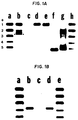
- Figure 5 is a gel electrophoretic pattern showing the assay of antistreptokinase antibodies in human serum.
- Lane 1 sample A mixed with streptokinase; lane 2, sample A alone; the presence of a precipitate at the electrophoretic origin of lane 1 indicates that the serum contains antistreptokinase antibodies.
- Arrow indicates the origin.
- the invention comprises methods which detect anti-streptokinase antibodies in a sample by the ability to form a complex between such antistreptokinase antibodies streptokinase and a streptokinase binding protein other than an antibody.
- This complex is herein termed the "three-part complex.”
- antistreptokinase antibody is meant two types of antibodies: antibodies which recognize (bind to) streptokinase and antibodies which recognize a complex containing streptokinase and a second (non-antibody) protein.
- the non-antibody streptokinase binding protein is a protein which contains the streptokinase consensus binding sequence Phe/Cys-Pro-Lys-any/none-Arg-Val-Ile/ValGly-any/none-Gly-Cys such as, for example, lactate dehydrogenase subunit M, plasminogen, plasmin, Lp(a).
- a “complex” between proteins is meant that two or more proteins are associated together in a non-covalent manner, through hydrogen bonding, ionic attraction and other noncovalent means, with such an affinity that the proteins in such complex are capable of extraction, analysis and measurement as a singular complex or entity.
- the formation of a "streptokinase binding protein-streptokinase-antistreptokinase antibody” complex in samples of the serum of an individual may be used as a rapid and specific assay for the presence of such antistreptokinase antibodies in the serum of an individual.
- LD which possesses at least one M subunit is used as the non-antibody binding protein.
- the complex which forms between streptokinase and LD upon the addition of exogenous streptokinase to a sample containing LD possesses such affinity that, if streptokinase antibodies are present in the sample, they also bind to the LD-streptokinase complex without dissociating it.
- the presence of the three part streptokinase binding protein-streptokinase-antistreptokinase antibody complex can be detected by any technique that reveals the presence of the complex.
- the three part complex can be detected by any technique that 'reveals the size, or a change in the size, of the LD isozymes in a serum sample, or changes in a distribution pattern of LD isozymes which occurs in the presence of streptokinase and serum.
- the presence of streptokinase antibodies may be predicted from physical properties of the LD isozyme pattern, for example, size or charge, after addition of streptokinase to a sample of the patient's serum.
- lactate dehydrogenase is a tetrameric enzyme which exists in at least five isoenzymic forms in an individual. Each subunit of the enzyme has a molecular weight of approximately 33,500 daltons but the isozymes vary in their content of one of three subunit types called type M, H or C. Serum LD isozyme analysis is frequently used in medical diagnosis and is a routine procedure in clinical laboratories. The electrophoretic separation of LD isozymes from serum may separate five bands of LD activity representing LD1, LD2, LD3, LD4 and LD5, respectively, with LD1 migrating fastest towards the anode and LD5 migrating fastest towards the cathode.
- LD1 is a tetramer of four H chains and LD5 is a tetramer of four M chains.
- the tetrameric structures of LD2, LD3 and LD4 consist of a mixture of subunit types, H3M, H2M2, and HM3, respectively.
- the C subunit is found in LD isolated from spermatozoa, called LDX.
- a prebound complex of streptokinase-streptokinase binding protein can be added directly to the serum.
- the length of time of the incubation need only be long enough to allow for binding of the anti-streptokinase antibodies to the streptokinase-streptokinase binding protein complex, for example, 15 min. to one hour.
- exogenous LD containing a M subunit may be added to the serum so as to ensure that sufficient M-containing LD is present in the serum to be tested.
- the final concentration of streptokinase in the assay is approximately 1500 IU/ml to 150,000 IU/ml, and most preferably, 15,000 IU/ml.
- the streptokinase is added to a small sample of serum, preferably less than 0.5 ml, incubated for 15-60 minutes, and analyzed (using, for example, electropnoresis or column chromatography techniques) under conditions that separate LD isozymes or complexes thereof. If electrophoresis is used, then after staining to inspect the resulting electrophoretic pattern. the presence or absence of antistreptokinase antibodies can be visually determined.
- the staining is an activity stain so that only protein bands containing some LD activity are revealed.
- any protocol which allows for the separation or detection of the three-part complex and/or LD isozymes is useful for the methods of the invention.
- any chromatographic, ELISA, or electrophoretic protocol which will differentiate between the complex containing streptokinase-LD and a three-part complex containing antistreptokinase antibody-streptokinase-LD is useful in the methods of the invention.
- One of skill in the art can design conditions and gel formulae that allow for this distinction. Gel electrophoresis is reviewed in Gel Electrophoresis of Proteins, A Practical Approach , B.D. Hames et al. , eds., IRL Press, Washington, 1981.
- the gel porosity and electrophoretic buffer conditions are such that migration of the antistreptokinase antibody-streptokinase-LD complex into the gel occurs only to a small extent or does not occur and the three-part complex remains at, or essentially at, the origin of the gel.
- the origin of the gel is that location where the sample was originally applied to the gel before electrophoresis. Complexes which are too large to migrate very far into the gel's structure, or which are electrically neutral relative to the anode and cathode, remain at, or essentially at, the origin.
- a preferred gel with an appropriate porosity is a 1% agarose gel. Electrophoresis of a 1% agarose gel in pH 8.2 barbital buffer at 100 V for a time sufficient to separate LD isozymes (for example, 20-28 min with the Beckman Paragon R electrophoresis system), followed by staining of LD activity in the gel reveals the presence of antistreptokinase antibodies by the presence of a precipitate of an LD complex at the origin of the electrophoretic gel. If no antistreptokinase antibodies are present, the complex of streptokinase with LD migrates into the gel and does not precipitate at, or remain at, the origin.
- Colorimetric staining for LD activity may be performed by, for example, incubating the electrophoresed gel in contact with substrate containing an appropriate amount of the LD substrates, for example, approximately 208 mM lithium lactate and 5.6 mM NAD+.
- the NADH generated due to LD activity in converting the lactate and NAD to pyruvate and NADH will appear at the site of the LD isozyme in the gel.
- the NADH and thus the site of the LD activity can be colorimetrically detected with a dye such as p -nitro blue tetrazolium (NBT) which is reduced to a colored product in the presence of NADH.
- NBT p -nitro blue tetrazolium
- Isoenzyme protein may also be identified with fluorescence, chemiluminescence, radiolabelling, or immunological techniques.
- gel matrices which can be used for the methods of the invention include agarose, polyacrylamide, starch, and combinations thereof.
- the conditions for separating the LD isozymes by electrophoresis are well known in the art and have been reviewed in, for example, Moses, G.C., Clin. Chem 34 :1885-1890 (1988); McKenzie, D. et al. , Clin. Chem. 29 :189-195 (1983); and Roman, W., Enzymologia 36 :189-219 (1969)).
- buffers which can be "used to perform LD isozyme analysis in agarose gels include barbital, barbital/EDTA, Tris-barbital, barbital/AMPD (2-amino-2-methyl-1,3-propanediol, and MOPSO (3-(N-morpholino)-2-hydroxypropane-sulfonic acid).
- buffers which can be used to run LD isozyme analysis with acetate gels includes tris-barbital.
- antistreptokinase antibodies may also be detected by assaying LD enzyme activity in the serum before and after the addition of concentrations of streptokinase similar to those used for electrophoretic determination of antibodies, 1500 IU/ml to 150,000 IU/ml, and most preferably, 15,000 IU/ml final concentration. It has been discovered that LD activity decreases when trapped in a complex containing. streptokinase and antistreptokinase antibodies. Therefore, a reduction of LD activity after addition of streptokinase is indicative of the presence of antistreptokinase antibodies in the sample.
- the presence of the antistreptokinase antibody-streptokinase-LD complex may also be assayed by a method which relies on column chromatography or filtration (either with a resin or a microfilter) to separate the antibody-containing LD complexes from complexes which do not contain antibody.
- a method which relies on column chromatography or filtration (either with a resin or a microfilter) to separate the antibody-containing LD complexes from complexes which do not contain antibody.
- column chromatography or filtration either with a resin or a microfilter
- one of skill in the art can design chromatographic conditions which separate non-antibody-containing LD complexes with a molecular weight of approximately 187,000 daltons (the sum of the molecular weight of the LD tetramer, 140,000 daltons and the molecular weight of streptokinase, 47,000 daltons) from larger complexes which would include the antibody.
- Such chromatography may be performed with any suitable chromatographic matrix, especially those which separate by size, such as, for example, Sepharose R , and Sephadex R and especially Sephadex R G-200. If the fraction containing serum proteins of molecular weight greater than 187,000 daltons is void of LD activity, the serum does not contain antistreptokinase antibodies. The appearance of LD activity in a fraction of molecular weight significantly greater than 187,000 daltons would indicate the presence of the LD-streptokinase-antistreptokinase complex and of anti-streptokinase antibodies in the serum of the individual being tested.
- streptokinase or portion thereof possessing affinity for both the antibodies found in serum and a non-antibody binding protein, especially LD subunit M, is useful in the methods of the invention.
- Such streptokinase is available commercially, for example, from Boehringer Mannheim Bio-chemicals, Indianapolis, IN and Sigma Chemical Co., St. Louis, M0.
- Preparations containing streptokinase in a composition which further contains albumin or collagen are also useful in the methods of the invention, for example, Streptase R from Hoechst-Roussel Pharmaceuticals, or Kabikinase R from Kabi-Vitrum.
- kits may comprise a carrier means being compartmentalized to receive in close confinement one or more container means such as vials, test tubes, and the like.
- container means such as vials, test tubes, and the like.
- Each of said container means comprises one of the separate elements to be used in the method.
- one of said container means may comprise streptokinase.
- a second container may comprise a detection system for LD.
- the carrier may also contain, in addition, a plurality of containers each of which comprises different, predetermined and known amounts of buffer and other solutions, gels or resins necessary for the iseparation of LD complexes or isozymes.
- the presence of antistreptokinase antibodies may be detected in biological fluids and tissues.
- Any sample containing the unknown amount of antistreptokinase antibodies can be used.
- serum is used.
- a sample is a liquid such as, for example, serum, urine, saliva, tear drops, cerebrospinal fluid, blood, plasma and the like.
- a solid or semi-solid such as, for example, tissues, feces, and the like may be used if they are first homogenized or otherwise placed in liquid suspension. Such methods are known in the art.
- antibodies to streptokinase may be present in a human's or animal's biological fluids or tissue, without such human or animal suffering from a streptococcal infection or previous exposure to streptokinase.
- any means of detecting LD and especially LD subunit M may be used.
- detectably labelled antibodies to the streptokinase binding protein or three-part complex, and especially to LD, or LD subunit M may be used to reveal the presence of the complex between LD and streptokinase, and the presence or absence of the antistreptokinase antibodies.
- the methods of the invention may be used to identify disease processes caused by or associated with an autoimmune response to the complexes as described herein and especially to identify the presence, in the serum of patients, of antistreptokinase antibodies which specifically recognize a complex between streptokinase and a serum streptokinase binding protein.
- LD isoenzymes were extracted by mincing the tissue in a volume of 145 mmol/L sodium chloride equal to the volume of the tissue and subsequently clarified by centrifugation at 5000 x g.
- LD5 was semi-purified by passing liver extract through a QAE-Sephadex anion-exchange column (bed volume 45 mL, 27 cm high, flow rate approximately 30 mL/h) equilibrated in 20 mmol/L Tris HCl, pH 8.2 (buffer A). LD5 passed directly through this column, free of LD1-LD4.
- LD1 was semi-purified by loading serum rich in that isoenzyme onto a similar column, washing off LD2-LD5 with buffer A containing 215 mmol of sodium chloride per liter, and then eluting LD1 free of the other LD isoenzymes with buffer A containing 260 mmol of sodium chloride per liter (Podlasek, S.J. et al. , Clin. Chem. 31 :527-32 (1985); Hsu, M-Y. et al. , Clin. Chem. 25 :1453-8 (1979)).
- LDX LD from spermatozoa which contains a subunit called "subunit C"
- one volume of testicular extract was mixed with four volumes of Isomune-LD solution A (goat anti-human LD-M antibody) and then immunoprecipitated with an equal volume of Isomune-LD solution B (polymer-bound donkey anti-goat immunoglobulin) to remove all M-subunit-containing LD isoenzymes.
- Isomune-LD solution A goat anti-human LD-M antibody
- Isomune-LD solution B polymer-bound donkey anti-goat immunoglobulin
- the two pharmaceutical preparations were also free of endogenous LD activity ( Figure 1A, lane c).
- the stabilizing components in these streptokinase preparations mixed separately with serum to achieve similar concentrations, showed no interaction with LD.
- Mixtures of streptokinase and human serum samples were also electrophoresed and evaluated for creatine kinase isoenzymes and for total stainable proteins. There were no alterations in electrophoretic migration of creatine kinase isoenzymes or of the major serum proteins resulting from the addition of streptokinase.
- LD1 or LD5 Semi-purified LD1 or LD5 (nine volumes) was mixed with one volume of streptokinase (final concentration 15000 IU/mL) and subjected to electrophoretic analysis to determine whether binding was specific for the H or M subunit of LD. Addition of streptokinase to LD1 (H tetramer) did not alter that isoenzyme's mobility ( Figure 1A, lane e) indicating that streptokinase and the H subunit did not interact.
- the LD-streptokinase-antistreptokinase antibody complexes formed in serum were evaluated by gel filtration, using Sephadex G-200.
- the fractions obtained from chromatographing a serum-streptokinase mixture were analyzed for LD isoenzyme content by electrophoresis.
- the complexes (precipitate at electrophoretic origin in Figure 3, lanes a-c) were eluted after the void volume, but before the normal tetraineric LD isoenyzmes ( Figure 3, lanes d-i).
- fibrinolytic agents urokinase and TPA, which bind to the same site on plasminogen as does streptokinase, were also added to serum to test for binding to LD. They failed to demonstrate any interaction with LD under conditions similar to those used with streptokinase.
- the region of plasminogen where streptokinase binds consists of a small loop of amino acids held together by a cysteine-cysteine linkage at its base (amino acid residues 557 to 565) (Collen, D., Thromb. Haemost. 43 :7-89 (1980)).
- a search of known LD sequences (Eventoff, W. et al., Proc. Natl. Acad. Sci. USA 74 :2677-81 (1977); Tsujibo, H. et al. , Eur. J. Biochem. 147 :9 15 (1985); Millan, J.L. et al. , Proc. Natl. Acad. Sci.
- Human LD-C and porcine LD-H neither of which interact with streptokinase, have sequences differing from that of human LD-M at amino acid residues 152, 154, and 155.
- the phenylalanine at position 152 is common to all three binders whereas the nonbinders all have leucine at position 152. This finding suggests that the phenylalanine at position 152 is pivotal for streptokinase binding, perhaps because of its relative bulk (compared with leucine in the nonbinders) and as such is somewhat homologous to cysteine at position 557 in plasminogen.
- lysine is common in both binders and one nonbinder (the other nonbinder, porcine LD-H, has a valine at position 154), suggesting that the amino acid at this position is not critical to streptokinase interaction.
- one nonbinder has threonine (human LD-C) and the other has the positively charged histidine (porcine LD-H), compared with asparagine in the binders. These differences do not (porcine LD-H and chicken LD-H) have the same amino acids at positions 154 (lysine) and 155 (histidine).
- streptokinase binding by streptokinase is potentially bivalent, owing to an internal duplication, with each half of the molecule homologous to the structure of a serine protease (Jackson, K.W., Biochemistry 21 :6620-5 (1982)).
- Each potential binding site on streptokinase includes the serine and aspartate, but not the histidine characteristic of the active site of serine proteases.
- the interaction of bivalent streptokinase with tetrameric LD would allow for very long chains or a matrix of complexes to form. Such extensive complexes are consistent with the precipitate that remains at the electrophoretic point of application ( Figure 2).
- these macro-complexes could be phagocytosed by cells of the immune system, thereby potentiating the induction of an autoimmune response against LD-M.
- Plasminogen and other molecular species that are only univalent could also participate in the matrix (as chain terminators) and thus be presented to the immune system as well in association with the foreign immunogen, streptokinase.
- Plasminogen is a serine protease of the coagulation scheme that acts extracellularly, and LD is an intracellular oxidoreductase that is expressed in essentially all cells.
- Arg-Ile-Val-Gly-Gly is conserved in the eukaryote serine proteases prothrombin, blood clotting factors IX, X, and XI, kallikrein, trypsinogen, chymotrypsinogen, and elastase, and also in bacterial trypsin (Dayhoff, M.0., Atlas of protein sequence and structure, Vol. 5 suppl. 3, Silver Spring, MD: The National Biomedical Research Foundation (1978); Yoshitake, S. et al. , Biochemistry 24 :3736-50(1985); Fujikawa, K. et al. , Biochemistry 25 :2417-24(19861).
- the arginine-isoleucine bond in this conserved sequence is a common cleavage site for conversion of these molecules to activated forms.
- LD isozymes which contain the M subunit all bound streptokinase as evidenced by the change in their migration but streptokinase alone did not result in a complex so big so as to precipitate at the electrophoretic origin.
- preimmune sample, sA or sB showed LD activity at the electrophoretic origin.
- immune sA and sB samples showed heavy LD activity at the origin.
- streptokinase Kabikinase, Kabivitrum, Inc.
- 9 parts of serum 9 parts were mixed with 9 parts of serum to a final streptokinase concentration of 15,000 IU/ml, incubated for 1 hour, and electrophoresed according to the manufacturer's instructions on a Paragon LD Gel Electrophoresis system (Beckman).
- Total LD activity was also measured in these serum samples with the Hitachi 737 system (Boehringer Mannheim) before and after addition of streptokinase.
- LD5 which was added to these samples demonstrated a stoichiometric relation between inhibition of LD5 activity and the presence of antistreptokinase antibodies.
- LD5 was purified by sequential Sephadex R G-200 gel filtration, QAE-Sephadex R chromatography and DEAE HPLC to yield a product with 280 U/mg specific activity. This material was stored at 4°C until use. In timed studies, inhibition occurred rapidly with the majority completed within 5 minutes. Increasing amounts of purified LD5 were plated added to a fixed volume of plasma from a volunteer blood donor with initial inhibition of 90% of original activity..
- This data indicates the inhibition of LD5 by STK is stoichiometrically mediated by the presence of antistreptokinase antibodies in the patient's serum. Scatchard plot analysis of the data yield an affinity constant of 2.5 x 10⁹ L/mol.
- Anti-streptokinase antibodies show considerable inter-individual titer difference, and the highest ones are able to neutralize streptokinase (STK) fibrinolytic therapy for acute myocardial infarction.
- Three methods for the detection of high titer anti-STK to guide choice of fibrinolytic drug have been developed. These include two standard methods (ELISA and fibrinolysis neutralization) and one using lactate dehydrogenase (LD:EC1.1.1.27) isoenzyme electrophoresis for rapid detection, based on the observations that STK binds to LD subunit M ( Clin. Chem. 35 :69 (1989)) and that anti-STK further alters electrophoretic pattern ( Clin. Chem.
- microtiter plates coated with 1 ⁇ g STK/well were incubated with diluted sample and then incubated with alkaline phosphatase-conjugated goat anti-human IgG to permit binding of the anti-IgG to any IgG present in the sample before color development with p -nitrophenyl phosphate (405 nm).
- a positive value was >0.06 for plasma and >0.08 for serum.
- Fibrinolysis neutralization was done by mixing STK with plasma then added to wells in a 1% agarose plate containing 13% citrated plasma. After diffusion, gels were soaked in 0.05 M CaCl2. Reduction in lysis zone was a positive result.
- LD-STK LD isoenzyme pattern alteration
- STK was added before electrophoresis.
- a positive result was a tight LD band at the origin, a wide band between LD2 and LD3, or marked decrease in LD activity.
- the fibrinolytic assay showed general agreement with ELISA establishing validity of ELISA.
- 43 were concordant and 17 discordant between ELISA and STK-LD. Only 2 of 20 serum samples were discordant.
- Four of 6 false negatives by STK-LD had high Lp(a) concentrations. Ten of 11 false positives were plasma samples.
- Detection of anti-STK by STK-LD correlates well with ELISA, although Lp(a) may interfere by competing for STK, and the preferred sample is serum.
- the strength of the STK-LD approach is its potential for use in acute care situations where rapid determinations are necessary for clinical utility.
Landscapes
- Health & Medical Sciences (AREA)
- Life Sciences & Earth Sciences (AREA)
- Engineering & Computer Science (AREA)
- Chemical & Material Sciences (AREA)
- Immunology (AREA)
- Molecular Biology (AREA)
- Hematology (AREA)
- Biomedical Technology (AREA)
- Urology & Nephrology (AREA)
- Biochemistry (AREA)
- Biotechnology (AREA)
- Organic Chemistry (AREA)
- Microbiology (AREA)
- General Health & Medical Sciences (AREA)
- Physics & Mathematics (AREA)
- Analytical Chemistry (AREA)
- Medicinal Chemistry (AREA)
- Zoology (AREA)
- General Physics & Mathematics (AREA)
- Pathology (AREA)
- Food Science & Technology (AREA)
- Proteomics, Peptides & Aminoacids (AREA)
- Cell Biology (AREA)
- Wood Science & Technology (AREA)
- Biophysics (AREA)
- Bioinformatics & Cheminformatics (AREA)
- General Engineering & Computer Science (AREA)
- Genetics & Genomics (AREA)
- Enzymes And Modification Thereof (AREA)
- Measuring Or Testing Involving Enzymes Or Micro-Organisms (AREA)
- Investigating Or Analysing Biological Materials (AREA)
- Preparation Of Compounds By Using Micro-Organisms (AREA)
- Medicines Containing Antibodies Or Antigens For Use As Internal Diagnostic Agents (AREA)
Claims (13)
- Verfahren zum Nachweis von Antikörpern gegen Streptokinase in einer Probe, wobei diese Probe eine LD-Untereinheit enthält, die eine Affinität für Streptokinase besitzt, welches Verfahren umfaßt, daß man:(a) die Probe mit einer Lösung in Kontakt bringt, die Streptokinase enthält;(b) die Streptokinase mit der LD-Untereinheit und den Antikörpern gegen Streptokinase einen Dreiwegkomplex bilden läßt;(c) entweder den Dreiwegkomplex zwischen LD, Streptokinase und Antikörpern gegen Streptokinase oder das Isoenzym-Muster von LD in der Probe nachweist; und(d) aus dem Vorhandensein oder Fehlen des Dreiwegkomplexes oder aus dem Isoenzym-Muster auf das Vorhandensein oder Fehlen der Antikörper gegen Streptokinase schließt.
- Verfahren zum Nachweis von Antikörpern gegen Streptokinase in einer Probe, wobei das Verfahren umfaßt, daß man:(a) die Probe mit einer Lösung in Kontakt bringt, die einen Komplex zwischen Streptokinase und einer LD-Untereinheit, welche eine Affinität für Streptokinase besitzt, enthält;(b) den Komplex mit den Antikörpern gegen Streptokinase einen Dreiwegkomplex bilden läßt;(c) entweder den Dreiwegkomplex zwischen LD, Streptokinase und Antikörpern gegen Streptokinase oder das Isoenzym-Muster von LD in der Probe nachweist; und(d) aus dem Vorhandensein oder Fehlen des Dreiwegkomplexes oder aus dem Isoenzym-Muster auf das Vorhandensein oder Fehlen der Antikörper gegen Streptokinase schließt.
- Verfahren nach Anspruch 1 oder Anspruch 2, worin die LD-Untereinheit die Untereinheit M ist.
- Verfahren nach einem der Ansprüche 1 bis 3, worin der Nachweis in Schritt (c) durch Elektrophorese, Säulenchromatographie oder durch Nachweis der enzymatischen Aktivität von LD erfolgt.
- Verfahren nach einem der Ansprüche 1 bis 4, worin die Probe menschliches oder tierisches Serum ist.
- Verfahren nach einem der Ansprüche 1 bis 5, worin der Antikörper gegen Streptokinase Streptokinase erkennt.
- Verfahren nach einem der Ansprüche 1 bis 5, worin der Antikörper gegen Streptokinase einen Komplex zwischen Streptokinase und einem anderen Protein erkennt.
- Verfahren nach Anspruch 7, worin das Protein eine Streptokinase-bindende Sequenz enthält.
- Verfahren nach Anspruch 8, worin das Protein LD, Plasminogen, Plasmin oder Lp(a) ist.
- Verfahren nach Anspruch 9, worin das Protein LD ist, die mindestens eine M-Untereinheit enthält.
- Kit zur Verwendung bei einem Verfahren nach einem der Ansprüche 1 bis 10, umfassend eine gefächerte Haltevorrichtung zur Aufnahme von einem oder mehreren Behältern in enger Umschließung; einen ersten Behälter mit Streptokinase; und ein Nachweissystem zum Nachweis der Gegenwart von LD.
- Kit nach Anspruch 11, worin das Nachweissystem einen zweiten Behälter mit einem Enzymtest für LD umfaßt.
- Kit nach Anspruch 12, worin der Enzymtest mit einer LD-Analyse auf einem Gel kompatibel ist.
Applications Claiming Priority (3)
| Application Number | Priority Date | Filing Date | Title |
|---|---|---|---|
| US360822 | 1982-03-23 | ||
| US36082289A | 1989-06-02 | 1989-06-02 | |
| PCT/US1990/003080 WO1990015153A1 (en) | 1989-06-02 | 1990-05-30 | A method for the detection of anti-streptokinase antibodies |
Publications (3)
| Publication Number | Publication Date |
|---|---|
| EP0474769A1 EP0474769A1 (de) | 1992-03-18 |
| EP0474769A4 EP0474769A4 (en) | 1992-04-22 |
| EP0474769B1 true EP0474769B1 (de) | 1995-11-08 |
Family
ID=23419531
Family Applications (1)
| Application Number | Title | Priority Date | Filing Date |
|---|---|---|---|
| EP90909412A Expired - Lifetime EP0474769B1 (de) | 1989-06-02 | 1990-05-30 | Verfahren zum nachweis von antikörpern gegen streptokinase |
Country Status (7)
| Country | Link |
|---|---|
| US (1) | US5342755A (de) |
| EP (1) | EP0474769B1 (de) |
| JP (1) | JP2846463B2 (de) |
| AT (1) | ATE130099T1 (de) |
| CA (1) | CA2056449A1 (de) |
| DE (1) | DE69023500D1 (de) |
| WO (1) | WO1990015153A1 (de) |
Families Citing this family (2)
| Publication number | Priority date | Publication date | Assignee | Title |
|---|---|---|---|---|
| US20030140920A1 (en) * | 2001-10-26 | 2003-07-31 | Dey L.P. | Albuterol inhalation soultion, system, kit and method for relieving symptoms of pediatric asthma |
| JP5603074B2 (ja) * | 2006-09-08 | 2014-10-08 | ピラマル イメージング ソシエテ アノニム | 18f標識物質のための化合物と方法 |
Family Cites Families (2)
| Publication number | Priority date | Publication date | Assignee | Title |
|---|---|---|---|---|
| US4376110A (en) * | 1980-08-04 | 1983-03-08 | Hybritech, Incorporated | Immunometric assays using monoclonal antibodies |
| DE3484505D1 (de) * | 1983-12-19 | 1991-05-29 | Daiichi Pure Chemicals Co Ltd | Immuntest. |
-
1990
- 1990-05-30 AT AT90909412T patent/ATE130099T1/de active
- 1990-05-30 EP EP90909412A patent/EP0474769B1/de not_active Expired - Lifetime
- 1990-05-30 CA CA002056449A patent/CA2056449A1/en not_active Abandoned
- 1990-05-30 US US07/777,319 patent/US5342755A/en not_active Expired - Fee Related
- 1990-05-30 WO PCT/US1990/003080 patent/WO1990015153A1/en not_active Ceased
- 1990-05-30 JP JP2508832A patent/JP2846463B2/ja not_active Expired - Lifetime
- 1990-05-30 DE DE69023500T patent/DE69023500D1/de not_active Expired - Lifetime
Also Published As
| Publication number | Publication date |
|---|---|
| WO1990015153A1 (en) | 1990-12-13 |
| EP0474769A4 (en) | 1992-04-22 |
| JPH04505664A (ja) | 1992-10-01 |
| CA2056449A1 (en) | 1990-12-03 |
| JP2846463B2 (ja) | 1999-01-13 |
| EP0474769A1 (de) | 1992-03-18 |
| ATE130099T1 (de) | 1995-11-15 |
| DE69023500D1 (de) | 1995-12-14 |
| US5342755A (en) | 1994-08-30 |
Similar Documents
| Publication | Publication Date | Title |
|---|---|---|
| Radek et al. | Affinity of human erythrocyte transglutaminase for a 42-kDa gelatin-binding fragment of human plasma fibronectin. | |
| Heeb et al. | Inhibition and complexation of activated protein C by two major inhibitors in plasma | |
| Cummins et al. | Cardiac specific troponin-I release in canine experimental myocardial infarction: development of a sensitive enzyme-linked immunoassay | |
| Moroz et al. | A rapid and sensitive 125I-fibrin solid-phase fibrinolytic assay for plasmin | |
| Epstein et al. | Radioimmunoassays for protein C and factor X: Plasma antigen levels in abnormal hemostatic states | |
| HU176133B (en) | Process and reagent for determining mb-creatinase-activity in body-liquid-samples | |
| Deogny et al. | Improved fibrin plate method for fibrinolytic activity measurements: use of bentonite precipitation and agar solidification | |
| KR20110104805A (ko) | 바이오마커 | |
| JPH0370184B2 (de) | ||
| JPH0614045B2 (ja) | 末端デオキシヌクレオチジル転移酵素の測定法 | |
| Dahl et al. | Enhancement of urokinase-induced plasminogen activation by the cationic protein of human eosinophil granulocytes | |
| EP0690991A1 (de) | Neue antikoagulant-cofaktor aktivität | |
| JP5080707B2 (ja) | 第vii因子活性化プロテアーゼの変異体および特異抗体を用いる検出方法 | |
| Sumi et al. | Urokinase-like plasminogen activator increased in plasma after alcohol drinking | |
| EP0436654A1 (de) | Immunochemische bestimmung von katalytisch aktiven serinproteasen | |
| CA1331158C (en) | Method, device and kit for measurement of tissue plasminogen activator activity | |
| US4267271A (en) | Radioimmunoassay for isoenzymes of creative kinase having a B-subunit and reagents therefor | |
| Kiyotaka et al. | Macro lactate dehydrogenase: an LDH-immunoglobulin M complex that inhibits lactate dehydrogenase activity in a patient's serum | |
| Ahmad et al. | Characterization of an acquired IgG inhibitor of coagulation factor XIII in a patient with systemic lupus erythematosus | |
| EP0474769B1 (de) | Verfahren zum nachweis von antikörpern gegen streptokinase | |
| Wallén et al. | The tissue activator of plasminogen | |
| Shakespeare et al. | The demonstration of urokinase antigen in whole blood | |
| JP3271157B2 (ja) | 新規な抗体、それを含むレニン活性物質及びそれを用いたプロレニン測定試薬 | |
| Ruiz‐Arguelles et al. | Serum antibodies to distinct epitopes of the tissue‐type plasminogen activator (t‐PA) in patients with systemic lupus erythematosus | |
| US4237219A (en) | Radioimmunoassay assay and reagents for MB isoenzyme of CK |
Legal Events
| Date | Code | Title | Description |
|---|---|---|---|
| PUAI | Public reference made under article 153(3) epc to a published international application that has entered the european phase |
Free format text: ORIGINAL CODE: 0009012 |
|
| 17P | Request for examination filed |
Effective date: 19920102 |
|
| AK | Designated contracting states |
Kind code of ref document: A1 Designated state(s): AT BE CH DE DK ES FR GB IT LI LU NL SE |
|
| A4 | Supplementary search report drawn up and despatched |
Effective date: 19920303 |
|
| AK | Designated contracting states |
Kind code of ref document: A4 Designated state(s): AT BE CH DE DK ES FR GB IT LI LU NL SE |
|
| 17Q | First examination report despatched |
Effective date: 19940317 |
|
| GRAA | (expected) grant |
Free format text: ORIGINAL CODE: 0009210 |
|
| AK | Designated contracting states |
Kind code of ref document: B1 Designated state(s): AT BE CH DE DK ES FR GB IT LI LU NL SE |
|
| PG25 | Lapsed in a contracting state [announced via postgrant information from national office to epo] |
Ref country code: IT Free format text: LAPSE BECAUSE OF FAILURE TO SUBMIT A TRANSLATION OF THE DESCRIPTION OR TO PAY THE FEE WITHIN THE PRESCRIBED TIME-LIMIT;WARNING: LAPSES OF ITALIAN PATENTS WITH EFFECTIVE DATE BEFORE 2007 MAY HAVE OCCURRED AT ANY TIME BEFORE 2007. THE CORRECT EFFECTIVE DATE MAY BE DIFFERENT FROM THE ONE RECORDED. Effective date: 19951108 Ref country code: CH Effective date: 19951108 Ref country code: LI Effective date: 19951108 Ref country code: NL Free format text: LAPSE BECAUSE OF FAILURE TO SUBMIT A TRANSLATION OF THE DESCRIPTION OR TO PAY THE FEE WITHIN THE PRESCRIBED TIME-LIMIT Effective date: 19951108 Ref country code: FR Effective date: 19951108 Ref country code: ES Free format text: THE PATENT HAS BEEN ANNULLED BY A DECISION OF A NATIONAL AUTHORITY Effective date: 19951108 Ref country code: AT Effective date: 19951108 Ref country code: DK Effective date: 19951108 Ref country code: BE Effective date: 19951108 |
|
| REF | Corresponds to: |
Ref document number: 130099 Country of ref document: AT Date of ref document: 19951115 Kind code of ref document: T |
|
| REF | Corresponds to: |
Ref document number: 69023500 Country of ref document: DE Date of ref document: 19951214 |
|
| PG25 | Lapsed in a contracting state [announced via postgrant information from national office to epo] |
Ref country code: SE Effective date: 19960208 |
|
| PG25 | Lapsed in a contracting state [announced via postgrant information from national office to epo] |
Ref country code: DE Effective date: 19960209 |
|
| NLV1 | Nl: lapsed or annulled due to failure to fulfill the requirements of art. 29p and 29m of the patents act | ||
| EN | Fr: translation not filed | ||
| REG | Reference to a national code |
Ref country code: CH Ref legal event code: PL |
|
| PG25 | Lapsed in a contracting state [announced via postgrant information from national office to epo] |
Ref country code: LU Free format text: LAPSE BECAUSE OF NON-PAYMENT OF DUE FEES Effective date: 19960531 |
|
| PLBE | No opposition filed within time limit |
Free format text: ORIGINAL CODE: 0009261 |
|
| STAA | Information on the status of an ep patent application or granted ep patent |
Free format text: STATUS: NO OPPOSITION FILED WITHIN TIME LIMIT |
|
| 26N | No opposition filed | ||
| PGFP | Annual fee paid to national office [announced via postgrant information from national office to epo] |
Ref country code: GB Payment date: 19990504 Year of fee payment: 10 |
|
| PG25 | Lapsed in a contracting state [announced via postgrant information from national office to epo] |
Ref country code: GB Free format text: LAPSE BECAUSE OF NON-PAYMENT OF DUE FEES Effective date: 20000530 |
|
| GBPC | Gb: european patent ceased through non-payment of renewal fee |
Effective date: 20000530 |