EP0465008B1 - A monoclonal antibody recognizing human pancreatic prophospholipase A2 - Google Patents
A monoclonal antibody recognizing human pancreatic prophospholipase A2 Download PDFInfo
- Publication number
- EP0465008B1 EP0465008B1 EP91305063A EP91305063A EP0465008B1 EP 0465008 B1 EP0465008 B1 EP 0465008B1 EP 91305063 A EP91305063 A EP 91305063A EP 91305063 A EP91305063 A EP 91305063A EP 0465008 B1 EP0465008 B1 EP 0465008B1
- Authority
- EP
- European Patent Office
- Prior art keywords
- propla2
- pla2
- monoclonal antibody
- human pancreatic
- prophospholipase
- Prior art date
- Legal status (The legal status is an assumption and is not a legal conclusion. Google has not performed a legal analysis and makes no representation as to the accuracy of the status listed.)
- Expired - Lifetime
Links
Images

Classifications
-
- C—CHEMISTRY; METALLURGY
- C07—ORGANIC CHEMISTRY
- C07K—PEPTIDES
- C07K16/00—Immunoglobulins [IG], e.g. monoclonal or polyclonal antibodies
- C07K16/40—Immunoglobulins [IG], e.g. monoclonal or polyclonal antibodies against enzymes
-
- A—HUMAN NECESSITIES
- A61—MEDICAL OR VETERINARY SCIENCE; HYGIENE
- A61K—PREPARATIONS FOR MEDICAL, DENTAL OR TOILETRY PURPOSES
- A61K39/00—Medicinal preparations containing antigens or antibodies
- A61K39/395—Antibodies; Immunoglobulins; Immune serum, e.g. antilymphocytic serum
-
- C—CHEMISTRY; METALLURGY
- C12—BIOCHEMISTRY; BEER; SPIRITS; WINE; VINEGAR; MICROBIOLOGY; ENZYMOLOGY; MUTATION OR GENETIC ENGINEERING
- C12P—FERMENTATION OR ENZYME-USING PROCESSES TO SYNTHESISE A DESIRED CHEMICAL COMPOUND OR COMPOSITION OR TO SEPARATE OPTICAL ISOMERS FROM A RACEMIC MIXTURE
- C12P21/00—Preparation of peptides or proteins
Definitions
- PLA2 Human pancreatic phospholipase A2 (EC 3.1.1.4; in the following account, phospholipase A2 is referred to as PLA2) is formed from prophospholipase A2 (hereinafter referred to as proPLA2) which is enzymatically inactive and is produced by the acinar cells of the pancreas. After production in the pancreas, the proPLA2 is secreted into the duodenum, and is therein decomposed by trypsin and converted into the active form PLA2. Both PLA2 and proPLA2 are known to be present in blood serum. Serum levels of PLA2 have previously been measured by enzymatic methods [Zieve L and Vogel WC, J. Lab. Clin.
- Serum levels of PLA2 and proPLA2 are known to increase not only in pancreatitis, as described above, but also, according to other research results, in cancer, articular rheumatism, external injuries and other disorders. Therefore, precise quantitation of serum levels of PLA2 and proPLA2 is regarded as an important datum with respect to the diagnosis and monitoring of patients with the aforesaid diseases.
- monoclonal antibodies against PLA2 have previously been developed. However, these monoclonal antibodies are known to display considerable cross-reactivity with proPLA2. Consequently, for example, radioimmunoassays performed with these antibodies cannot yield precise measurements of PLA2 levels. From the viewpoint of diagnosis and clinical monitoring of the aforesaid disorders, the development of monoclonal antibodies which permit accurate and specific measurement of serum levels of both PLA2 and proPLA2 is an important desideratum.
- a monoclonal antibody specifically recognizing human pancreatic prophospholipase A2 wherein said antibody is monoclonal antibody PR414 produced by hybridoma PR414 (FERM BP-2978).
- the cross-reactivity of the monoclonal antibody with human pancreatic phospholipase A2 is less than 1%.
- hybridomas which produce the above-mentioned monoclonal antibodies.
- the present invention comprises hybridomas producing the aforesaid monoclonal antibody.
- the monoclonal antibody of this invention which specifically recognizes proPLA2 can be prepared by the use of a hybridoma in accordance with conventional procedures. For example, by the use of the method of Grataroli, R et al. [Biochimie, 63 , 677 (1981)], proPLA2 is extracted and purified from pancreatic juice, and used to immunize mice or other suitable animals. Then, splenic or other antibody-producing cells are fused with myeloma cells to obtain hybridomas. Cells derived from mouse myeloma, for example, can be used as the aforesaid myeloma cells.
- the aforesaid cell fusion can be effected, for example, by a conventional procedure using polyethylene glycol.
- monoclonal antibodies against proPLA2 can be obtained by screening.
- This screening can be accomplished by conventional enzyme-linked immunosorbent assay (ELISA) methods.
- ELISA enzyme-linked immunosorbent assay
- purified proPLA2 is adsorbed onto the solid phase, and this is brought into contact with the supernate of the hybridoma culture; if antibodies recognizing proPLA2 are present in this supernate, then these antibodies bind to the proPLA2.
- an appropriate enzyme-labeled antibody for example, mouse anti-IgG antibody, is allowed to act and binds to the anti-proPLA2 antibodies.
- detection of the enzyme label permits screening for the antibodies recognizing proPLA2.
- hybridomas producing these antibodies are further screened for those producing antibodies displaying minimal cross-reactivity with PLA2, that is, monoclonal antibodies specifically recognizing proPLA2.
- monoclonal antibodies specifically recognizing proPLA2 For example, by means of ELISA or RIA (radioimmunoassay) using PLA2 and proPLA2, the hybridomas can be screened for monoclonal antibodies which bind specifically to proPLA2 and display almost no binding to PLA2.
- hybridomas producing monoclonal antibodies specifically recognizing proPLA2 are obtained.
- eight hybridoma cell lines, designated as PR304, PR306, PR307 PR312, PR314, PR318, PR414, and PR424, were obtained in the aforesaid manner.
- PR414 has been deposited with the Fermentation Research Institute Agency for Industrial Science and Technology, 1-3, Higashi 1-chome, Tsukuba-shi, Ibaraki-ken, Japan on June 21, 1990 under Accession number FERM BP-2978 in accordance with the Budapest Treaty.
- the monoclonal antibodies of the present invention are secreted into the supernate. Moreover, by transplanting the aforesaid hybridomas into the peritoneal cavities of mice, ascitic fluid containing high concentrations of the said monoclonal antibodies can be obtained.
- the use of these monoclonal antibodies permits accurate measurement of proPLA2 or PLA2. This measurement can be performed by generally known RIA or enzyme immunoassay procedures. For example, proPLA2 can be measured by the following method (competitive assay).
- a prescribed quantity of labelled (e.g., radiolabeled) proPLA2 is added to a sample containing a known quantity of proPLA2, then, the aforesaid monoclonal antibody specifically recognizing proPLA2 is added to the mixture.
- labeled or unlabeled proPLA2 binds to each monoclonal antibody molecule, thereby forming antibody-antigen complexes.
- Immunobead Bio-Rad Co.
- the bound materials are separated from the reaction mixture in the form of pellets, and the quantity of label on the pellets is measured.
- a calibration curve is prepared by varying the quantity of proPLA2 in the sample. By the use of this calibration curve, unknown quantities of proPLA2 can be measured in a similar manner.
- the PLA2 content of blood serum containing both proPLA2 and PLA2 can be measured in the following manner. First, anti-PLA2 monoclonal antibody having known cross-reactivity (x%) with proPLA2 is prepared. The quantity of proPLA2 in a sample containing unknown concentrations of both PLA2 and proPLA2 can be measured by the aforesaid method; let A denote the molar concentration of proPLA2 determined in this manner. Next, the same measurement is performed by the use of the aforesaid cross-reactive anti-PLA2 monoclonal antibody in place of anti-proPLA2 monoclonal antibody; let B denote the molar concentration so obtained.
- B is the sum of the value corresponding to PLA2 and x% of the value corresponding to proPLA2
- the concentration of PLA2 is expressed by the formula (B - (X/100)A).
- PLA2 was extracted and purified from pancreatic juice in accordance with the method of Nishijima et al. [J. Biochem., 94, 137 (1983)].
- ProPLA2 was extracted and purified from pancreatic juice in accordance with the method of Grataroli et al. [Biochimie, 63 , 677 (1981)], except that CM-Sepharose (registered trademark) chromatography was performed twice. The purities of both PLA2 and proPLA2 were verified by amino acid analysis.
- mAb1008 was used in the present invention.
- the affinity constant of mAb1008 was 4x10¹¹M ⁇ 1, and the cross-reactivity of this antibody with proPLA2 was 49%.
- mice Female BALB/c mice were immunized with pro-PLA2 in the following manner. First, 25 ⁇ g of proPLA2 emulsified with Freund's complete adjuvant was subcutaneously injected into the mice three times, at intervals of 3 weeks. In addition, physiological saline containing 50 ⁇ g of proPLA2 was administered intraperitoneally 4 days prior to the collection of splenic cells from these mice.
- mice splenic cells were collected from the aforesaid mice and fused with mouse myeloma cells (P3-Ns1-1-Ag4-1) by the use of 50% polyethylene glycol (molecular weight 4000, Merck) in accordance with the method of Galfre and Milstein [Methods Enzymol., 73 , 3 (1981)].
- the fused cells were suspended in HAT medium and inoculated into Costar 96-well Tissue Culture Cluster (trade name). Then, the supernates of the cultures in a total of 1503 wells wherein the hybridomas had been grown were screened by the following ELISA procedure.
- the wells of 96-well Immunoplates (Nunc Co.) were coated overnight at 4°C with 10 ng of proPLA2 or 10 ng of PLA2 in 50 ⁇ l of 0.05 M sodium bicarbonate buffer, pH 8.5. Next, the wells were blocked with 0.2% BSA-PBS and washed with 0.5% Tween (registered trademark) 80-PBS. Twenty-five microliter aliquots of supernate from each of the aforesaid 1503 hybridoma cultures were added to the aforesaid proPLA2-coated wells as well as PLA2-coated wells, after which these plates were incubated at 37°C for 1 hour.
- the antibody which bound to the proPLA2 or PLA2 on the wells was quantitated by means of a Vectastain ABC kit (Vector Co.), with the use of ABTS as a color-producing agent.
- the reaction mixture was left at room temperature, and allowed to color. Twelve minutes after the agent was added, the mixture was measured at 415 nm using a Corona MTP-32 microplate photometer.
- Wells displaying a large absorbance (greater than 1.0) in the ELISA procedure using proPLA2-coated plates and a small absorbance (less than 0.5) when using the PLA2-coated plates were selected. In this manner, hybridomas producing anti-proPLA2 mAb (i.e., binding to proPLA2 and having very low cross-reactivity with PLA2) were obtained (31 wells).
- mice hybridoma cell lines producing anti-proPLA2mAb (designated as PR304, PR306, PR307, PR312, PR314, PR318, PR414, and PR424) were established.
- the culture supernatants were used as the source of anti-proPLA2mAb in the Examples of the present invention.
- the characteristics of the anti-proPLA2mAb produced by aforesaid eight hybridoma cell lines are shown in Table 1.
- the immunoglobulin class as determined by the Ouchterlony diffusion method was IgG1 in every case.
- the affinity constants were determined from the Scatchard plots [Scatchard G, Ann. N. Y. Acad. Sci., 51 , 660 (1949)].
- mAbPR414 was used in the following experiments.
- the cross-reactivity of mAbPR414 with PLA2 is 0.9%.
- the assay buffer solution used for RIA was a 0.01M PBS (pH 7.4) containing 1 mM EDTA, 0.01% NaN3, and 0.2% BSA. Both 15I-proPLA2 and 15I-PLA2 were prepared by the chloramine T method.
- Calibration curves for proPLA2 and PLA2 were prepared by the following procedure. First, 100 ⁇ l of a standard solution that contains proPLA2 or PLA2 in a predetermined amount, 100 ⁇ l of a solution that contains 15I-proPLA2 or 15I-PLA2 (40,000 cpm), 100 ⁇ l of mAb solution (i.e., the aforesaid diluted supernatant containing mAb PR414, or the aforesaid diluted ascitic fluid containing mAb1008), and 200 ⁇ l of assay buffer solution, were mixed in a polystyrene tube, after which the mixture was incubated overnight at room temperature.
- mAb solution i.e., the aforesaid diluted supernatant containing mAb PR414, or the aforesaid diluted ascitic fluid containing mAb1008
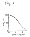
- FIG. 1 shows the calibration curve representing the relation between the measured radioactivity and the proPLA2 concentration of the standard solution.
- the ordinate B/B0 of the graph in Figure 1 is the ratio between the measured radioactivity B corresponding to the respective standard solutions and the radioactivity B0 corresponding to a zero concentration of proPLA2.
- the sensitivity of the calibration curve that is, the lowest measurable concentration (i.e., the concentration of proPLA2 corresponding to 90% inhibition) was 0.4 ng/ml, while the concentration of proPLA2 corresponding to 50% inhibition was 3.2 ng/ml.
- the calibration curve for RIA of PLA2 revealed that the concentration of PLA2 corresponding to 90% inhibition was 0.4 ng/ml, while that corresponding to 50% inhibition was 3.0 ng/ml.
- the aforesaid assays were performed in the same manner, using sample liquids containing human serum, or eluates obtained by ion exchange chromatography of human serum in place of the aforesaid standard solutions of various concentrations of proPLA2 or PLA2.
- the concentrations of proPLA2 and PLA2 in these sample liquids was determined by using the calibration curves for proPLA2 and PLA2, respectively, prepared in the manner indicated above.
- Example 2 Blood sera of the following healthy humans and patients were assayed by the same RIA procedure described in Example 1 above.
- the subjects comprised three healthy individuals, three pancreatitis patients (No. 1: patient with moderate pancreatitis following contrast radiography of the pancreatic duct; No. 2: patient with chronic pancreatitis in a period of exacerbation; No. 3: patient with acute necrotizing pancreatitis), one patient with pancreatic carcinoma, and one patient with chronic renal insufficiency.
- the results of the RIA studies are shown in Table 2.
- the serum levels of proPLA2 shown in Table 2 are considered to agree almost completely with the true serum concentrations of proPLA2.
- the monoclonal antibody used for the assay of PLA2 displayed 49% cross-reactivity with proPLA2
- the serum levels of PLA2 shown in Table 2 are proportionately higher than the actual values. According to the data in this table, the measured levels of both proPLA2 and PLA2 in the sera of the pancreatitis, pancreatic carcinoma and chronic renal insufficiency patients were higher than the corresponding values for the healthy individuals, except for pancreatitis patient No. 2.
- proPLA2 The measured levels of proPLA2 were higher (by a factor of approximately 2) than those of PLA2 in the healthy individuals as well as the pancreatic carcinoma patient and the chronic renal insufficiency patient. Furthermore, in two of the three pancreatitis patients (Nos. 2 and 3), the measured levels of PLA2 were higher than those of proPLA2.
- the aforesaid measured proPLA2 values were directly recorded as the serum concentrations of proPLA2.
- the serum concentrations of PLA2 were calculated on the basis of the measured PLA2 values.
- the serum levels of proPLA2 and PLA2 determined in this manner as well as the ratios of proPLA2 and PLA2 concentrations are shown in Table 3.
- ProPLA2 and PLA2 in human blood serum were isolated by ion exchange chromatography using S-Sepharose (registered trademark) (pharmacia), after which measurement was performed by RIA.
- S-Sepharose registered trademark
- RIA ion exchange chromatography
- 10 mM Tris-HCl (pH 7.0) containing 0.05% CHAPS was used and the entire chromatographic process was performed at room temperature.
- human sera i.e., the same lot of eight sample liquids as was used in Example 2
- diluted 5-fold with the aforesaid chromatographic buffer were injected into the column and separation was performed by the same procedure.
- the black dots represent concentrations of proPLA2 values measured by RIA using mAb PR414, while the black triangles represent concentrations of PLA2 measured by RIA using mAb1008.
- concentrations and relative proportions of proPLA2 and PLA2 in the sera of the eight subjects, calculated from the chromatographic peak regions, are shown in Table 4.
- Table 5 shows the recovery rate of immunoreactivity of proPLA2 and PLA2 by ion exchange chromatography, as determined by RIA. As is seen from the table, the recovery rate for every tested human serum was approximately 100% (86% - 104%). This indicates that RIA for proPLA2 using mAb PR414 measures the serum level of proPLA2 only, while RIA for PLA2 using mAb1008 measures the serum level of PLA2 plus the quantity corresponding to cross-reactivity with proPLA2.
- the serum levels of proPLA2 and PLA2 calculated by means of the monoclonal antibody of the present invention, shown in Table 3, are nearly identical to those shown in Table 4. Therefore, it is recognized that the use of the monoclonal antibody of the present invention permits extremely precise measurement of both proPLA2 and PLA2.
- proPLA2 and PLA2 reflect the degree of severity of pancreatitis.
- the serum levels of both proPLA2 and PLA2 were extremely high.
- the relative proportions of proPLA2 and PLA2 were nearly the same as in the healthy subjects.
- this result may be attributed to impaired excretion of proPLA2 and PLA2 from the kidneys. This characteristic differs from those manifested in pancreatitis patients, and therefore could possibly be utilized for differential diagnosis.
Landscapes
- Chemical & Material Sciences (AREA)
- Health & Medical Sciences (AREA)
- Life Sciences & Earth Sciences (AREA)
- Organic Chemistry (AREA)
- General Health & Medical Sciences (AREA)
- Medicinal Chemistry (AREA)
- Immunology (AREA)
- Genetics & Genomics (AREA)
- Molecular Biology (AREA)
- Engineering & Computer Science (AREA)
- Biochemistry (AREA)
- Microbiology (AREA)
- Zoology (AREA)
- Wood Science & Technology (AREA)
- Bioinformatics & Cheminformatics (AREA)
- Proteomics, Peptides & Aminoacids (AREA)
- Biophysics (AREA)
- Veterinary Medicine (AREA)
- Public Health (AREA)
- Animal Behavior & Ethology (AREA)
- Epidemiology (AREA)
- Biotechnology (AREA)
- Chemical Kinetics & Catalysis (AREA)
- General Chemical & Material Sciences (AREA)
- Pharmacology & Pharmacy (AREA)
- Mycology (AREA)
- General Engineering & Computer Science (AREA)
- Preparation Of Compounds By Using Micro-Organisms (AREA)
- Micro-Organisms Or Cultivation Processes Thereof (AREA)
- Peptides Or Proteins (AREA)
Abstract
Description
- The present invention relates to a monoclonal antibody recognizing human prophospholipase A₂ (hereinafter referred to as proPLA₂mAb), a method for the assay of human pancreatic prophospholipase A₂ using the monoclonal antibody, and a hybridoma producing the monoclonal antibody.
- Human pancreatic phospholipase A₂ (EC 3.1.1.4; in the following account, phospholipase A₂ is referred to as PLA₂) is formed from prophospholipase A₂ (hereinafter referred to as proPLA₂) which is enzymatically inactive and is produced by the acinar cells of the pancreas. After production in the pancreas, the proPLA₂ is secreted into the duodenum, and is therein decomposed by trypsin and converted into the active form PLA₂. Both PLA₂ and proPLA₂ are known to be present in blood serum. Serum levels of PLA₂ have previously been measured by enzymatic methods [Zieve L and Vogel WC, J. Lab. Clin. Med., 57, 586(1961); Doizaki WM and Zieve L, J. Lab. Clin. Med., 67, 108 (1966); Hashihira S et al., Jpn. Arch. Intern. Med., 24, 243 (1977); and Schröder T et al., Scand. J. Gastroent., 15, 633 (1980)]. Moreover, immunological assays of this enzyme have also been performed [Nishijima J et al., J. Biochem., 94, 137 (1983); and Eskola JU et al., Clin. Chem., 29, 1777 (1983)]. Elevated serum levels of PLA₂ have been reported as occurring in cases of acute pancreatitis (ibid.). Furthermore, monoclonal antibodies have been prepared against PLA₂ (Japanese Laid-Open Patent Publication No. 63-258898), and radioimmunoassays employing these antibodies have suggested that nearly all of the said enzyme in blood serum of healthy humans assumes the form of proPLA₂, while the quantity of active PLA₂ in healthy serum is extremely minute [Misaki et al., Ann. Jpn. Clin. Chem. Soc., 27, 109 (1987)]. The aforesaid report of Misaki et al. also suggests that proPLA₂ is transformed into active PLA₂ in the serum of some patients with acute pancreatitis. Büchler M et al. [Klin. Wochenschr, 67, 186 (1989)] report that both immunologically active PLA₂ levels (reflecting the quantities of both PLA₂ and proPLA₂) and PLA₂ catalytic activity (reflecting only the quantity of PLA₂, since proPLA₂ is inactive) are elevated in the serum of patients with necrotic pancreatitis, whereas only immunologically active PLA₂ levels are elevated in edematous pancreatitis patients.
- Serum levels of PLA₂ and proPLA₂ are known to increase not only in pancreatitis, as described above, but also, according to other research results, in cancer, articular rheumatism, external injuries and other disorders. Therefore, precise quantitation of serum levels of PLA₂ and proPLA₂ is regarded as an important datum with respect to the diagnosis and monitoring of patients with the aforesaid diseases. As mentioned above, monoclonal antibodies against PLA₂ have previously been developed. However, these monoclonal antibodies are known to display considerable cross-reactivity with proPLA₂. Consequently, for example, radioimmunoassays performed with these antibodies cannot yield precise measurements of PLA₂ levels. From the viewpoint of diagnosis and clinical monitoring of the aforesaid disorders, the development of monoclonal antibodies which permit accurate and specific measurement of serum levels of both PLA₂ and proPLA₂ is an important desideratum.
- According to one aspect of the present invention there is provided a monoclonal antibody specifically recognizing human pancreatic prophospholipase A₂, wherein said antibody is monoclonal antibody PR414 produced by hybridoma PR414 (FERM BP-2978).
- In a preferred embodiment, the cross-reactivity of the monoclonal antibody with human pancreatic phospholipase A₂ is less than 1%.
- According to another aspect of the present invention there is provided a method for the assay of human pancreatic prophospholipase A₂ of this invention comprising the steps of,
- (a) adding a prescribed quantity of labeled human pancreatic prophospholipase A₂ to a sample containing human pancreatic prophospholipase A₂ analyte,
- (b) adding the above-mentioned monoclonal antibody to the mixture obtained in process (a), thereby forming an antibody-antigen complex, and
- (c) calculating the quantity of human pancreatic prophospholipase A₂ in the sample from the result of measurement of the quantity of label bound in the complex obtained in process (b).
- Also provided are hybridomas which produce the above-mentioned monoclonal antibodies.
- Thus, the invention described herein makes possible the objectives of:
- (1) providing a monoclonal antibody against proPLA₂ which permit precise measurement of human serum levels of proPLA₂, and
- (2) providing a method for the assay of proPLA₂ by the use of the aforesaid monoclonal antibody.
- For a better understanding of the invention, and to show how the same may be carried into effect, reference will now be made to the accompanying drawings, in which:
- Figure 1 is a graph showing the calibration curve for measurement of proPLA₂ by radioimmunoassay by the use of the monoclonal antibody of the present invention.
- Figures 2(A) and 2(B) are graphs showing the elution profiles obtained when the blood sera of a healthy human and a pancreatitis patient, respectively, were subjected to ion exchange chromatography.
- The present invention comprises hybridomas producing the aforesaid monoclonal antibody.
- The monoclonal antibody of this invention which specifically recognizes proPLA₂ can be prepared by the use of a hybridoma in accordance with conventional procedures. For example, by the use of the method of Grataroli, R et al. [Biochimie, 63, 677 (1981)], proPLA₂ is extracted and purified from pancreatic juice, and used to immunize mice or other suitable animals. Then, splenic or other antibody-producing cells are fused with myeloma cells to obtain hybridomas. Cells derived from mouse myeloma, for example, can be used as the aforesaid myeloma cells. The aforesaid cell fusion can be effected, for example, by a conventional procedure using polyethylene glycol. Then, from the fused cells so obtained, monoclonal antibodies against proPLA₂ can be obtained by screening. This screening can be accomplished by conventional enzyme-linked immunosorbent assay (ELISA) methods. For example, purified proPLA₂ is adsorbed onto the solid phase, and this is brought into contact with the supernate of the hybridoma culture; if antibodies recognizing proPLA₂ are present in this supernate, then these antibodies bind to the proPLA₂. Next, an appropriate enzyme-labeled antibody, for example, mouse anti-IgG antibody, is allowed to act and binds to the anti-proPLA₂ antibodies. Thus, detection of the enzyme label permits screening for the antibodies recognizing proPLA₂.
- Next, the hybridomas producing these antibodies are further screened for those producing antibodies displaying minimal cross-reactivity with PLA₂, that is, monoclonal antibodies specifically recognizing proPLA₂. For example, by means of ELISA or RIA (radioimmunoassay) using PLA₂ and proPLA₂, the hybridomas can be screened for monoclonal antibodies which bind specifically to proPLA₂ and display almost no binding to PLA₂. Thus, hybridomas producing monoclonal antibodies specifically recognizing proPLA₂ are obtained. In the present invention, eight hybridoma cell lines, designated as PR304, PR306, PR307 PR312, PR314, PR318, PR414, and PR424, were obtained in the aforesaid manner. Among these cell lines, PR414 has been deposited with the Fermentation Research Institute Agency for Industrial Science and Technology, 1-3, Higashi 1-chome, Tsukuba-shi, Ibaraki-ken, Japan on June 21, 1990 under Accession number FERM BP-2978 in accordance with the Budapest Treaty.
- Under the conditions of conventional methods of cultivation, the monoclonal antibodies of the present invention are secreted into the supernate. Moreover, by transplanting the aforesaid hybridomas into the peritoneal cavities of mice, ascitic fluid containing high concentrations of the said monoclonal antibodies can be obtained. The use of these monoclonal antibodies permits accurate measurement of proPLA₂ or PLA₂. This measurement can be performed by generally known RIA or enzyme immunoassay procedures. For example, proPLA₂ can be measured by the following method (competitive assay). First, a prescribed quantity of labelled (e.g., radiolabeled) proPLA₂ is added to a sample containing a known quantity of proPLA₂, then, the aforesaid monoclonal antibody specifically recognizing proPLA₂ is added to the mixture. Thus, labeled or unlabeled proPLA₂ binds to each monoclonal antibody molecule, thereby forming antibody-antigen complexes. Then, for example, Immunobead (Bio-Rad Co.) was added to the sample, thus binding the said complexes. Then, the bound materials are separated from the reaction mixture in the form of pellets, and the quantity of label on the pellets is measured. In this manner, a calibration curve is prepared by varying the quantity of proPLA₂ in the sample. By the use of this calibration curve, unknown quantities of proPLA₂ can be measured in a similar manner.
- The PLA₂ content of blood serum containing both proPLA₂ and PLA₂ can be measured in the following manner. First, anti-PLA₂ monoclonal antibody having known cross-reactivity (x%) with proPLA₂ is prepared. The quantity of proPLA₂ in a sample containing unknown concentrations of both PLA₂ and proPLA₂ can be measured by the aforesaid method; let A denote the molar concentration of proPLA₂ determined in this manner. Next, the same measurement is performed by the use of the aforesaid cross-reactive anti-PLA₂ monoclonal antibody in place of anti-proPLA₂ monoclonal antibody; let B denote the molar concentration so obtained. Since B is the sum of the value corresponding to PLA₂ and x% of the value corresponding to proPLA₂, the concentration of PLA₂ is expressed by the formula (B - (X/100)A). Thus, by the use of the monoclonal antibody of the present invention, serum levels of both PLA₂ and proPLA₂ can be accurately determined.
- Next, the present invention will be explained in further detail with reference to specific examples. However, these examples are merely illustrative and by no means limit the scope of the present invention.
- PLA₂ was extracted and purified from pancreatic juice in accordance with the method of Nishijima et al. [J. Biochem., 94, 137 (1983)]. ProPLA₂ was extracted and purified from pancreatic juice in accordance with the method of Grataroli et al. [Biochimie, 63, 677 (1981)], except that CM-Sepharose (registered trademark) chromatography was performed twice. The purities of both PLA₂ and proPLA₂ were verified by amino acid analysis.
- Among the varieties of anti-PLA₂mAb obtained by the method of Misaki et al. (Japanese Laid-Open Patent Publication No. 63-258898), that designated as mAb1008 was used in the present invention. The affinity constant of mAb1008 was 4x10¹¹M⁻¹, and the cross-reactivity of this antibody with proPLA₂ was 49%.
- Female BALB/c mice were immunized with pro-PLA₂ in the following manner. First, 25 µg of proPLA₂ emulsified with Freund's complete adjuvant was subcutaneously injected into the mice three times, at intervals of 3 weeks. In addition, physiological saline containing 50 µg of proPLA₂ was administered intraperitoneally 4 days prior to the collection of splenic cells from these mice.
- Then, splenic cells were collected from the aforesaid mice and fused with mouse myeloma cells (P3-Ns1-1-Ag4-1) by the use of 50% polyethylene glycol (molecular weight 4000, Merck) in accordance with the method of Galfre and Milstein [Methods Enzymol., 73, 3 (1981)]. Next, the fused cells were suspended in HAT medium and inoculated into Costar 96-well Tissue Culture Cluster (trade name). Then, the supernates of the cultures in a total of 1503 wells wherein the hybridomas had been grown were screened by the following ELISA procedure.
- The wells of 96-well Immunoplates (Nunc Co.) were coated overnight at 4°C with 10 ng of proPLA₂ or 10 ng of PLA₂ in 50 µl of 0.05 M sodium bicarbonate buffer, pH 8.5. Next, the wells were blocked with 0.2% BSA-PBS and washed with 0.5% Tween (registered trademark) 80-PBS. Twenty-five microliter aliquots of supernate from each of the aforesaid 1503 hybridoma cultures were added to the aforesaid proPLA₂-coated wells as well as PLA₂-coated wells, after which these plates were incubated at 37°C for 1 hour. The antibody which bound to the proPLA₂ or PLA₂ on the wells was quantitated by means of a Vectastain ABC kit (Vector Co.), with the use of ABTS as a color-producing agent. In the process of the measurement, the reaction mixture was left at room temperature, and allowed to color. Twelve minutes after the agent was added, the mixture was measured at 415 nm using a Corona MTP-32 microplate photometer. Wells displaying a large absorbance (greater than 1.0) in the ELISA procedure using proPLA₂-coated plates and a small absorbance (less than 0.5) when using the PLA₂-coated plates were selected. In this manner, hybridomas producing anti-proPLA₂ mAb (i.e., binding to proPLA₂ and having very low cross-reactivity with PLA₂) were obtained (31 wells).
- Some of these ELISA positive hybridomas were cloned immediately after screening to further proliferation and these hybridomas were frozen subsequently. The approximate affinity of the antibodies produced by the selected hybridomas for proPLA₂ was estimated using the supernatant recovered when these hybridomas were frozen. The aforesaid affinity was determined by the competitive RIA procedure described in the section entitled "Description of the Preferred Embodiment". Hybridomas producing antibodies displaying high affinity were cloned, if necessary.
- In this manner, eight mouse hybridoma cell lines producing anti-proPLA₂mAb (designated as PR304, PR306, PR307, PR312, PR314, PR318, PR414, and PR424) were established. The culture supernatants were used as the source of anti-proPLA₂mAb in the Examples of the present invention.
- The characteristics of the anti-proPLA₂mAb produced by aforesaid eight hybridoma cell lines are shown in Table 1. The immunoglobulin class as determined by the Ouchterlony diffusion method was IgG₁ in every case. The affinity constants were determined from the Scatchard plots [Scatchard G, Ann. N. Y. Acad. Sci., 51, 660 (1949)].

- As is clear from Table 1, all of these monoclonal antibodies display high affinity for proPLA₂, with affinity constants ranging from 1.7x10¹⁰M⁻¹ to 2.8x10¹¹M⁻¹. Cross-reactivity with PLA₂ was estimated from the 50% inhibitory ratio of the calibration curve determined by competitive RIA by the use of the same procedure as that described above. MAbPR304 and mAbPR312 displayed significant cross-reactivity with PLA₂, but the cross-reactivity for the other six varieties of mAb was less than 1%.
- Among the aforesaid eight varieties of anti-proPLA₂ monoclonal antibodies, mAbPR414 was used in the following experiments. The cross-reactivity of mAbPR414 with PLA₂ is 0.9%.
- RIA for the measurement of proPLA₂ was performed with the aforesaid proPLA₂mAbPR414 (i.e., using the culture supernatant diluted 320-fold with assay buffer solution; the cross-reactivity of anti-proPLA₂ mAbPR414 with PLA₂ was 0.9%) and ¹⁵I-labeled proPLA₂. RIA for the measurement of PLA₂ was performed with the aforesaid anti-PLA₂mAb1008 (i.e., using ascitic fluid diluted 300,000-fold with assay buffer solution; the cross-reactivity of anti-PLA₂mAb1008 with proPLA₂ is 49%) and ¹⁵I-labeled PLA₂. The assay buffer solution used for RIA was a 0.01M PBS (pH 7.4) containing 1 mM EDTA, 0.01% NaN₃, and 0.2% BSA. Both ¹⁵I-proPLA₂ and ¹⁵I-PLA₂ were prepared by the chloramine T method.
- Calibration curves for proPLA₂ and PLA₂ were prepared by the following procedure. First, 100 µl of a standard solution that contains proPLA₂ or PLA₂ in a predetermined amount, 100 µl of a solution that contains ¹⁵I-proPLA₂ or ¹⁵I-PLA₂ (40,000 cpm), 100 µl of mAb solution (i.e., the aforesaid diluted supernatant containing mAb PR414, or the aforesaid diluted ascitic fluid containing mAb1008), and 200 µl of assay buffer solution, were mixed in a polystyrene tube, after which the mixture was incubated overnight at room temperature. Then, 100 µl of Immunobead (1 mg anti-mouse immunoglobulin/ml assay buffer, Bio-Rad Co.) was added, and the mixture was left undisturbed for 1 hour at room temperature. Next, the tube was centrifuged at 3,000 rpm for 5 minutes, and the radioactivity of the pellet so obtained was measured. Figure 1 shows the calibration curve representing the relation between the measured radioactivity and the proPLA₂ concentration of the standard solution. The ordinate B/B₀ of the graph in Figure 1 is the ratio between the measured radioactivity B corresponding to the respective standard solutions and the radioactivity B₀ corresponding to a zero concentration of proPLA₂.
- The sensitivity of the calibration curve, that is, the lowest measurable concentration (i.e., the concentration of proPLA₂ corresponding to 90% inhibition) was 0.4 ng/ml, while the concentration of proPLA₂ corresponding to 50% inhibition was 3.2 ng/ml. The calibration curve for RIA of PLA₂ (not shown) revealed that the concentration of PLA₂ corresponding to 90% inhibition was 0.4 ng/ml, while that corresponding to 50% inhibition was 3.0 ng/ml.
- In the following examples, the aforesaid assays were performed in the same manner, using sample liquids containing human serum, or eluates obtained by ion exchange chromatography of human serum in place of the aforesaid standard solutions of various concentrations of proPLA₂ or PLA₂. The concentrations of proPLA₂ and PLA₂ in these sample liquids was determined by using the calibration curves for proPLA₂ and PLA₂, respectively, prepared in the manner indicated above.
- Blood sera of the following healthy humans and patients were assayed by the same RIA procedure described in Example 1 above. The subjects comprised three healthy individuals, three pancreatitis patients (No. 1: patient with moderate pancreatitis following contrast radiography of the pancreatic duct; No. 2: patient with chronic pancreatitis in a period of exacerbation; No. 3: patient with acute necrotizing pancreatitis), one patient with pancreatic carcinoma, and one patient with chronic renal insufficiency. The results of the RIA studies are shown in Table 2.

- The serum levels of proPLA₂ shown in Table 2 are considered to agree almost completely with the true serum concentrations of proPLA₂. On the other hand, since the monoclonal antibody used for the assay of PLA₂ displayed 49% cross-reactivity with proPLA₂, the serum levels of PLA₂ shown in Table 2 are proportionately higher than the actual values. According to the data in this table, the measured levels of both proPLA₂ and PLA₂ in the sera of the pancreatitis, pancreatic carcinoma and chronic renal insufficiency patients were higher than the corresponding values for the healthy individuals, except for pancreatitis patient No. 2. The measured levels of proPLA₂ were higher (by a factor of approximately 2) than those of PLA₂ in the healthy individuals as well as the pancreatic carcinoma patient and the chronic renal insufficiency patient. Furthermore, in two of the three pancreatitis patients (Nos. 2 and 3), the measured levels of PLA₂ were higher than those of proPLA₂.
- The aforesaid measured proPLA₂ values were directly recorded as the serum concentrations of proPLA₂. Next, taking into account the aforesaid cross-reactivity of mAb1008, the serum concentrations of PLA₂ were calculated on the basis of the measured PLA₂ values. The serum levels of proPLA₂ and PLA₂ determined in this manner as well as the ratios of proPLA₂ and PLA₂ concentrations are shown in Table 3.

- ProPLA₂ and PLA₂ in human blood serum were isolated by ion exchange chromatography using S-Sepharose (registered trademark) (pharmacia), after which measurement was performed by RIA. As the buffer solution for chromatography, 10 mM Tris-HCl (pH 7.0) containing 0.05% CHAPS was used and the entire chromatographic process was performed at room temperature.
- First, proPLA₂ and PLA₂ standards (dissolved in chromatographic buffer solution containing 0.2% BSA), prepared for the purposes of column calibration, were injected into an S-Sepharose (registered trademark) column (
bed capacity 2 ml) equilibrated with the aforesaid chromatographic buffer solution. Then, the column was eluted with 40 ml of the chromatographic buffer solution under a 0-0.2 M linear gradient of sodium chloride, at a flow rate of approximately 12 ml/hour. After completion of the linear gradient elution, the column was again eluted with 0.5 M sodium chloride. One milliliter aliquots of all the eluates from the column were collected in polypropylene tubes containing 100 µl of 2% BSA-PBS. ProPLA₂ and PLA₂ standards were recovered from the S-Sephadex column at high recovery rates. The peak fractions were fraction 19 for proPLA₂ and fraction 26 for PLA₂. - Next, human sera (i.e., the same lot of eight sample liquids as was used in Example 2), diluted 5-fold with the aforesaid chromatographic buffer, were injected into the column and separation was performed by the same procedure.
- The proPLA₂ and PLA₂ content of the various eluted fractions of the aforesaid standards and human sera were measured by RIA using the same procedure as in Example 2. Except for a small peak which appeared in the vicinity of the
fraction 15 for the pancreatitis patients, all of the immunoreactive components were eluted with the same retention volumes as the standards. No immunoreactivity was detected in either the initial fraction or the 0.5 M sodium chloride fraction. Figures 2(A) and 2(B) show the elution profiles for the sera of one of the healthy individuals (No. 1) and one of the pancreatitis patients (No. 3), respectively. In these figures, the black dots represent concentrations of proPLA₂ values measured by RIA using mAb PR414, while the black triangles represent concentrations of PLA₂ measured by RIA using mAb1008. The concentrations and relative proportions of proPLA₂ and PLA₂ in the sera of the eight subjects, calculated from the chromatographic peak regions, are shown in Table 4.

- Table 5 shows the recovery rate of immunoreactivity of proPLA₂ and PLA₂ by ion exchange chromatography, as determined by RIA. As is seen from the table, the recovery rate for every tested human serum was approximately 100% (86% - 104%). This indicates that RIA for proPLA₂ using mAb PR414 measures the serum level of proPLA₂ only, while RIA for PLA₂ using mAb1008 measures the serum level of PLA₂ plus the quantity corresponding to cross-reactivity with proPLA₂. The serum levels of proPLA₂ and PLA₂ calculated by means of the monoclonal antibody of the present invention, shown in Table 3, are nearly identical to those shown in Table 4. Therefore, it is recognized that the use of the monoclonal antibody of the present invention permits extremely precise measurement of both proPLA₂ and PLA₂.
- The data in Tables 2-4 indicate that PLA₂ in the serum of healthy humans assumes the form of the enzymatically inactive proPLA₂ predominantly. This indicates that proPLA₂, which is synthesized in the pancreas and diffuses into the bloodstream, exists without activation in normal serum. Consequently, the cytotoxicity of PLA₂ is not manifested in healthy individuals. In the patient with comparatively moderate pancreatitis (No. 1), the serum level of proPLA₂ was higher than that of PLA₂, however, in the patient with chronic pancreatitis during a period of exacerbation (No. 2) as well as the patient with acute necrotizing pancreatitis (No. 3), the serum concentration of PLA₂ was higher than that of proPLA₂. This suggests that the relative proportions of proPLA₂ and PLA₂ reflect the degree of severity of pancreatitis. As regards the pancreatic carcinoma patient and the chronic renal insufficiency patient, the serum levels of both proPLA₂ and PLA₂ were extremely high. However, the relative proportions of proPLA₂ and PLA₂ were nearly the same as in the healthy subjects. In the renal insufficiency patient, this result may be attributed to impaired excretion of proPLA₂ and PLA₂ from the kidneys. This characteristic differs from those manifested in pancreatitis patients, and therefore could possibly be utilized for differential diagnosis.
Claims (8)
- A monoclonal antibody specifically recognizing human pancreatic prophospholipase A₂, wherein said antibody is monoclonal antibody PR414 produced by hybridoma PR414 (FERM BP-2978).
- A monoclonal antibody of claim 1, wherein the cross-reactivity of said monoclonal antibody with human pancreatic phospholipase A₂ is less than 1%.
- A method for the assay of human pancreatic prophospholipase A₂, comprising the steps of,(a) adding a prescribed quantity of labeled human pancreatic prophospholipase A₂ to a sample containing human pancreatic prophospholipase A₂ analyte,(b) adding a monoclonal antibody of claim 1 or 2 to the mixture obtained in process (a), thereby forming an antibody-antigen complex, and(c) calculating the quantity of human pancreatic prophospholipase A₂ in said sample from the result of measurement of the quantity of label bound in the complex obtained in process (b).
- A hybridoma which produces a monoclonal antibody of claim 1 or 2.
Applications Claiming Priority (2)
| Application Number | Priority Date | Filing Date | Title |
|---|---|---|---|
| JP2179137A JP3023147B2 (en) | 1990-07-05 | 1990-07-05 | Monoclonal antibody recognizing human pancreatic prophospholipase A (2) |
| JP179137/90 | 1990-07-05 |
Publications (3)
| Publication Number | Publication Date |
|---|---|
| EP0465008A2 EP0465008A2 (en) | 1992-01-08 |
| EP0465008A3 EP0465008A3 (en) | 1993-02-24 |
| EP0465008B1 true EP0465008B1 (en) | 1996-05-08 |
Family
ID=16060628
Family Applications (1)
| Application Number | Title | Priority Date | Filing Date |
|---|---|---|---|
| EP91305063A Expired - Lifetime EP0465008B1 (en) | 1990-07-05 | 1991-06-05 | A monoclonal antibody recognizing human pancreatic prophospholipase A2 |
Country Status (5)
| Country | Link |
|---|---|
| EP (1) | EP0465008B1 (en) |
| JP (1) | JP3023147B2 (en) |
| KR (1) | KR0173123B1 (en) |
| AT (1) | ATE137865T1 (en) |
| DE (1) | DE69119304T2 (en) |
Families Citing this family (1)
| Publication number | Priority date | Publication date | Assignee | Title |
|---|---|---|---|---|
| EP3533459A1 (en) * | 2018-03-02 | 2019-09-04 | Diaccurate | Anti-pla2-gib antibodies and the uses thereof |
Family Cites Families (2)
| Publication number | Priority date | Publication date | Assignee | Title |
|---|---|---|---|---|
| IE64356B1 (en) * | 1986-07-28 | 1995-07-26 | Bioscience Int Inc | Diagnosis severity prediction and monitoring of disease by immunoassay of free activation peptides of pancreatic zymogens |
| JPH0717680B2 (en) * | 1987-04-15 | 1995-03-01 | 塩野義製薬株式会社 | Monoclonal antibody against human pancreatic phospholipase A-2, its production method, hybridoma producing the monoclonal antibody, and human pancreatic phospholipase A-2 using the monoclonal antibody 2 ▼ Measuring method |
-
1990
- 1990-07-05 JP JP2179137A patent/JP3023147B2/en not_active Expired - Fee Related
-
1991
- 1991-06-05 AT AT91305063T patent/ATE137865T1/en not_active IP Right Cessation
- 1991-06-05 EP EP91305063A patent/EP0465008B1/en not_active Expired - Lifetime
- 1991-06-05 DE DE69119304T patent/DE69119304T2/en not_active Expired - Fee Related
- 1991-06-07 KR KR1019910009377A patent/KR0173123B1/en not_active Expired - Fee Related
Also Published As
| Publication number | Publication date |
|---|---|
| JPH0466100A (en) | 1992-03-02 |
| ATE137865T1 (en) | 1996-05-15 |
| JP3023147B2 (en) | 2000-03-21 |
| EP0465008A3 (en) | 1993-02-24 |
| KR920002169A (en) | 1992-02-28 |
| DE69119304T2 (en) | 1996-09-12 |
| DE69119304D1 (en) | 1996-06-13 |
| KR0173123B1 (en) | 1999-02-01 |
| EP0465008A2 (en) | 1992-01-08 |
Similar Documents
| Publication | Publication Date | Title |
|---|---|---|
| EP0288088B1 (en) | Detection of tumor necrosis factor; monoclonal antibody and kit | |
| EP0358739B1 (en) | Monoclonal antibodies against glycated albumin, hybrid cell lines producing these antibodies, and use thereof | |
| US5494791A (en) | Monoclonal antibodies against glycated low density lipoprotein | |
| JP2665850B2 (en) | Monoclonal antibody recognizing the C-terminus of hBNP | |
| EP0201187B1 (en) | Determination of glycated (glycosylated) hemoglobin in blood | |
| US4797473A (en) | Monoclonal antibodies to unreduced, nonenzymatically-glycated proteins | |
| EP0287030B1 (en) | Immunological methods and materials for detection of tumor associated antigens | |
| EP0288179B1 (en) | Ckmb assay, monoclonal antibodies for use therein and hybrid cell lines | |
| US5316914A (en) | Method for determining human collagen peptides by way of enzyme immunoassay | |
| JPH04503006A (en) | Blood coagulation factor XIIa beta monoclonal antibodies and immunoassays | |
| EP0606516B1 (en) | Anti-human ceruloplasmin monoclonal antibody | |
| AU772548B2 (en) | Monoclonal antibody against apolipoprotein A-I | |
| EP0465008A2 (en) | A monoclonal antibody recognizing human pancreatic prophospholipase A2 | |
| CA1321763C (en) | Monoclonal antibody specific to human pancreatic phospholipase a | |
| US5358850A (en) | Sandwich immunoassay of β-n-acetylglucosaminidase and monoclonal antibody used therein | |
| US5358849A (en) | Monoclonal antibody recognizing membrane phospholipase A2 and immunoassay of membrane phospholipase A2 | |
| KR100502275B1 (en) | Methods for quantitative determination of serum enzymes using immunoassay and methods for diagnosis of hepatic disease thereby | |
| HK1003795B (en) | Monoclonal antibodies recognizing alpha-hanp and corresponding hybridomas, their preparation and use | |
| HK1003795A1 (en) | Monoclonal antibodies recognizing alpha-hanp and corresponding hybridomas, their preparation and use | |
| EP0229794B1 (en) | Monoclonal antibodies to gamma interferon, their production and use | |
| EP0444787B1 (en) | Monoclonal antibodies against non-A 1C glycated hemoglobin | |
| EP0393640A1 (en) | A monoclonal antibody recognizing C-terminus of ANP | |
| US5241052A (en) | Carcinoma orosomucoid-related antigen, a monoclonal antibody thereto, and their uses | |
| US5130233A (en) | Method for determining the β-subunit of human prolyl hydroxylase by radioimmunoassay to detect hepatic disease | |
| EP0222923A1 (en) | Monoclonal antibody |
Legal Events
| Date | Code | Title | Description |
|---|---|---|---|
| PUAI | Public reference made under article 153(3) epc to a published international application that has entered the european phase |
Free format text: ORIGINAL CODE: 0009012 |
|
| AK | Designated contracting states |
Kind code of ref document: A2 Designated state(s): AT BE CH DE DK ES FR GB GR IT LI LU NL SE |
|
| RTI1 | Title (correction) | ||
| PUAL | Search report despatched |
Free format text: ORIGINAL CODE: 0009013 |
|
| AK | Designated contracting states |
Kind code of ref document: A3 Designated state(s): AT BE CH DE DK ES FR GB GR IT LI LU NL SE |
|
| 17P | Request for examination filed |
Effective date: 19930518 |
|
| 17Q | First examination report despatched |
Effective date: 19950224 |
|
| GRAH | Despatch of communication of intention to grant a patent |
Free format text: ORIGINAL CODE: EPIDOS IGRA |
|
| GRAA | (expected) grant |
Free format text: ORIGINAL CODE: 0009210 |
|
| AK | Designated contracting states |
Kind code of ref document: B1 Designated state(s): AT BE CH DE DK ES FR GB GR IT LI LU NL SE |
|
| PG25 | Lapsed in a contracting state [announced via postgrant information from national office to epo] |
Ref country code: IT Free format text: LAPSE BECAUSE OF FAILURE TO SUBMIT A TRANSLATION OF THE DESCRIPTION OR TO PAY THE FEE WITHIN THE PRE;WARNING: LAPSES OF ITALIAN PATENTS WITH EFFECTIVE DATE BEFORE 2007 MAY HAVE OCCURRED AT ANY TIME BEFORE 2007. THE CORRECT EFFECTIVE DATE MAY BE DIFFERENT FROM THE ONE RECORDED.SCRIBED TIME-LIMIT Effective date: 19960508 Ref country code: NL Free format text: LAPSE BECAUSE OF FAILURE TO SUBMIT A TRANSLATION OF THE DESCRIPTION OR TO PAY THE FEE WITHIN THE PRESCRIBED TIME-LIMIT Effective date: 19960508 Ref country code: BE Effective date: 19960508 Ref country code: AT Effective date: 19960508 Ref country code: ES Free format text: THE PATENT HAS BEEN ANNULLED BY A DECISION OF A NATIONAL AUTHORITY Effective date: 19960508 Ref country code: DK Effective date: 19960508 Ref country code: LI Free format text: LAPSE BECAUSE OF FAILURE TO SUBMIT A TRANSLATION OF THE DESCRIPTION OR TO PAY THE FEE WITHIN THE PRESCRIBED TIME-LIMIT Effective date: 19960508 Ref country code: GR Free format text: LAPSE BECAUSE OF FAILURE TO SUBMIT A TRANSLATION OF THE DESCRIPTION OR TO PAY THE FEE WITHIN THE PRESCRIBED TIME-LIMIT Effective date: 19960508 Ref country code: CH Free format text: LAPSE BECAUSE OF FAILURE TO SUBMIT A TRANSLATION OF THE DESCRIPTION OR TO PAY THE FEE WITHIN THE PRESCRIBED TIME-LIMIT Effective date: 19960508 |
|
| REF | Corresponds to: |
Ref document number: 137865 Country of ref document: AT Date of ref document: 19960515 Kind code of ref document: T |
|
| REF | Corresponds to: |
Ref document number: 69119304 Country of ref document: DE Date of ref document: 19960613 |
|
| ET | Fr: translation filed | ||
| PG25 | Lapsed in a contracting state [announced via postgrant information from national office to epo] |
Ref country code: LU Free format text: LAPSE BECAUSE OF NON-PAYMENT OF DUE FEES Effective date: 19960630 |
|
| PG25 | Lapsed in a contracting state [announced via postgrant information from national office to epo] |
Ref country code: SE Effective date: 19960808 |
|
| NLV1 | Nl: lapsed or annulled due to failure to fulfill the requirements of art. 29p and 29m of the patents act | ||
| REG | Reference to a national code |
Ref country code: CH Ref legal event code: PL |
|
| PLBE | No opposition filed within time limit |
Free format text: ORIGINAL CODE: 0009261 |
|
| STAA | Information on the status of an ep patent application or granted ep patent |
Free format text: STATUS: NO OPPOSITION FILED WITHIN TIME LIMIT |
|
| 26N | No opposition filed | ||
| REG | Reference to a national code |
Ref country code: GB Ref legal event code: IF02 |
|
| PGFP | Annual fee paid to national office [announced via postgrant information from national office to epo] |
Ref country code: FR Payment date: 20020618 Year of fee payment: 12 |
|
| PGFP | Annual fee paid to national office [announced via postgrant information from national office to epo] |
Ref country code: DE Payment date: 20020627 Year of fee payment: 12 |
|
| PGFP | Annual fee paid to national office [announced via postgrant information from national office to epo] |
Ref country code: GB Payment date: 20030523 Year of fee payment: 13 |
|
| PG25 | Lapsed in a contracting state [announced via postgrant information from national office to epo] |
Ref country code: DE Free format text: LAPSE BECAUSE OF NON-PAYMENT OF DUE FEES Effective date: 20040101 |
|
| PG25 | Lapsed in a contracting state [announced via postgrant information from national office to epo] |
Ref country code: FR Free format text: LAPSE BECAUSE OF NON-PAYMENT OF DUE FEES Effective date: 20040227 |
|
| REG | Reference to a national code |
Ref country code: FR Ref legal event code: ST |
|
| PG25 | Lapsed in a contracting state [announced via postgrant information from national office to epo] |
Ref country code: GB Free format text: LAPSE BECAUSE OF NON-PAYMENT OF DUE FEES Effective date: 20040605 |
|
| GBPC | Gb: european patent ceased through non-payment of renewal fee |
Effective date: 20040605 |