EP0458940B1 - Method for the quantitative detection of thyrotropine (tsh) or of anti-thyrotropine receptor autoantibodies (anti-tshr) using a recombinant thyrotropin receptor polypeptide and kit for conducting said method - Google Patents
Method for the quantitative detection of thyrotropine (tsh) or of anti-thyrotropine receptor autoantibodies (anti-tshr) using a recombinant thyrotropin receptor polypeptide and kit for conducting said method Download PDFInfo
- Publication number
- EP0458940B1 EP0458940B1 EP91901286A EP91901286A EP0458940B1 EP 0458940 B1 EP0458940 B1 EP 0458940B1 EP 91901286 A EP91901286 A EP 91901286A EP 91901286 A EP91901286 A EP 91901286A EP 0458940 B1 EP0458940 B1 EP 0458940B1
- Authority
- EP
- European Patent Office
- Prior art keywords
- tsh
- sequence
- receptor
- acid residues
- polypeptide
- Prior art date
- Legal status (The legal status is an assumption and is not a legal conclusion. Google has not performed a legal analysis and makes no representation as to the accuracy of the status listed.)
- Expired - Lifetime
Links
- 108090000765 processed proteins & peptides Proteins 0.000 title claims abstract description 64
- 229920001184 polypeptide Polymers 0.000 title claims abstract description 62
- 102000004196 processed proteins & peptides Human genes 0.000 title claims abstract description 62
- 102000015486 thyroid-stimulating hormone receptor activity proteins Human genes 0.000 title claims abstract description 18
- 108040006218 thyroid-stimulating hormone receptor activity proteins Proteins 0.000 title claims abstract description 18
- 102000005962 receptors Human genes 0.000 title claims description 66
- 238000000034 method Methods 0.000 title claims description 16
- 238000001514 detection method Methods 0.000 title claims description 10
- 125000000539 amino acid group Chemical group 0.000 claims abstract description 28
- 230000000694 effects Effects 0.000 claims abstract description 18
- 125000003275 alpha amino acid group Chemical group 0.000 claims abstract description 13
- 108020003175 receptors Proteins 0.000 claims description 65
- 210000001685 thyroid gland Anatomy 0.000 claims description 28
- 239000012528 membrane Substances 0.000 claims description 23
- 238000002360 preparation method Methods 0.000 claims description 14
- 150000001413 amino acids Chemical class 0.000 claims description 13
- 230000014509 gene expression Effects 0.000 claims description 13
- 150000007523 nucleic acids Chemical class 0.000 claims description 12
- 239000012472 biological sample Substances 0.000 claims description 11
- 108020004707 nucleic acids Proteins 0.000 claims description 7
- 102000039446 nucleic acids Human genes 0.000 claims description 7
- 239000013598 vector Substances 0.000 claims description 7
- 102000030621 adenylate cyclase Human genes 0.000 claims description 6
- 108060000200 adenylate cyclase Proteins 0.000 claims description 6
- 239000002773 nucleotide Substances 0.000 claims description 6
- 125000003729 nucleotide group Chemical group 0.000 claims description 6
- 238000006243 chemical reaction Methods 0.000 claims description 5
- 230000001413 cellular effect Effects 0.000 claims description 4
- 239000003153 chemical reaction reagent Substances 0.000 claims description 4
- 230000002163 immunogen Effects 0.000 claims description 4
- 230000001971 thyroidal effect Effects 0.000 claims description 4
- 239000012535 impurity Substances 0.000 claims description 3
- 238000005259 measurement Methods 0.000 claims description 2
- 239000012634 fragment Substances 0.000 abstract description 23
- 238000012217 deletion Methods 0.000 abstract description 7
- 230000037430 deletion Effects 0.000 abstract description 7
- 238000003780 insertion Methods 0.000 abstract description 4
- 230000037431 insertion Effects 0.000 abstract description 4
- 238000006467 substitution reaction Methods 0.000 abstract 1
- 108090000253 Thyrotropin Receptors Proteins 0.000 description 55
- 102000003911 Thyrotropin Receptors Human genes 0.000 description 54
- 210000004027 cell Anatomy 0.000 description 45
- 239000002299 complementary DNA Substances 0.000 description 19
- 101150098159 TSHR gene Proteins 0.000 description 16
- 108090000045 G-Protein-Coupled Receptors Proteins 0.000 description 15
- 230000027455 binding Effects 0.000 description 15
- 102000003688 G-Protein-Coupled Receptors Human genes 0.000 description 14
- 229940072221 immunoglobulins Drugs 0.000 description 11
- 108060003951 Immunoglobulin Proteins 0.000 description 10
- 235000001014 amino acid Nutrition 0.000 description 10
- 238000003556 assay Methods 0.000 description 10
- 238000006073 displacement reaction Methods 0.000 description 10
- 102000018358 immunoglobulin Human genes 0.000 description 10
- 238000010367 cloning Methods 0.000 description 9
- 210000000287 oocyte Anatomy 0.000 description 9
- 108090000623 proteins and genes Proteins 0.000 description 9
- 102000023108 LH Receptors Human genes 0.000 description 8
- 108010011942 LH Receptors Proteins 0.000 description 8
- 108010076504 Protein Sorting Signals Proteins 0.000 description 8
- 108020004999 messenger RNA Proteins 0.000 description 8
- IVOMOUWHDPKRLL-KQYNXXCUSA-N Cyclic adenosine monophosphate Chemical compound C([C@H]1O2)OP(O)(=O)O[C@H]1[C@@H](O)[C@@H]2N1C(N=CN=C2N)=C2N=C1 IVOMOUWHDPKRLL-KQYNXXCUSA-N 0.000 description 7
- IVOMOUWHDPKRLL-UHFFFAOYSA-N UNPD107823 Natural products O1C2COP(O)(=O)OC2C(O)C1N1C(N=CN=C2N)=C2N=C1 IVOMOUWHDPKRLL-UHFFFAOYSA-N 0.000 description 7
- 229940095074 cyclic amp Drugs 0.000 description 7
- 230000004988 N-glycosylation Effects 0.000 description 6
- 235000018102 proteins Nutrition 0.000 description 6
- 102000004169 proteins and genes Human genes 0.000 description 6
- 241000894007 species Species 0.000 description 6
- 210000001519 tissue Anatomy 0.000 description 6
- 102000003886 Glycoproteins Human genes 0.000 description 5
- 108090000288 Glycoproteins Proteins 0.000 description 5
- 108700026244 Open Reading Frames Proteins 0.000 description 5
- 241000269370 Xenopus <genus> Species 0.000 description 5
- 229940088597 hormone Drugs 0.000 description 5
- 239000005556 hormone Substances 0.000 description 5
- 239000013615 primer Substances 0.000 description 5
- 108020004414 DNA Proteins 0.000 description 4
- OHCQJHSOBUTRHG-KGGHGJDLSA-N FORSKOLIN Chemical compound O=C([C@@]12O)C[C@](C)(C=C)O[C@]1(C)[C@@H](OC(=O)C)[C@@H](O)[C@@H]1[C@]2(C)[C@@H](O)CCC1(C)C OHCQJHSOBUTRHG-KGGHGJDLSA-N 0.000 description 4
- 102000008175 FSH Receptors Human genes 0.000 description 4
- 108010060374 FSH Receptors Proteins 0.000 description 4
- 239000003446 ligand Substances 0.000 description 4
- 239000000523 sample Substances 0.000 description 4
- 238000012216 screening Methods 0.000 description 4
- 230000004936 stimulating effect Effects 0.000 description 4
- 241000283690 Bos taurus Species 0.000 description 3
- 108091026890 Coding region Proteins 0.000 description 3
- 208000015023 Graves' disease Diseases 0.000 description 3
- 206010028665 Myxoedema Diseases 0.000 description 3
- 238000000636 Northern blotting Methods 0.000 description 3
- 102000011923 Thyrotropin Human genes 0.000 description 3
- 108010061174 Thyrotropin Proteins 0.000 description 3
- 230000003321 amplification Effects 0.000 description 3
- 230000001363 autoimmune Effects 0.000 description 3
- 125000003178 carboxy group Chemical group [H]OC(*)=O 0.000 description 3
- 210000000170 cell membrane Anatomy 0.000 description 3
- 239000003795 chemical substances by application Substances 0.000 description 3
- 238000011534 incubation Methods 0.000 description 3
- 230000003834 intracellular effect Effects 0.000 description 3
- 238000004519 manufacturing process Methods 0.000 description 3
- 239000002609 medium Substances 0.000 description 3
- 238000003199 nucleic acid amplification method Methods 0.000 description 3
- 230000001817 pituitary effect Effects 0.000 description 3
- 238000003752 polymerase chain reaction Methods 0.000 description 3
- 230000001105 regulatory effect Effects 0.000 description 3
- 238000012163 sequencing technique Methods 0.000 description 3
- XLYOFNOQVPJJNP-UHFFFAOYSA-N water Substances O XLYOFNOQVPJJNP-UHFFFAOYSA-N 0.000 description 3
- 108091032973 (ribonucleotides)n+m Proteins 0.000 description 2
- APIXJSLKIYYUKG-UHFFFAOYSA-N 3 Isobutyl 1 methylxanthine Chemical compound O=C1N(C)C(=O)N(CC(C)C)C2=C1N=CN2 APIXJSLKIYYUKG-UHFFFAOYSA-N 0.000 description 2
- 208000023328 Basedow disease Diseases 0.000 description 2
- 239000003155 DNA primer Substances 0.000 description 2
- SUZLHDUTVMZSEV-UHFFFAOYSA-N Deoxycoleonol Natural products C12C(=O)CC(C)(C=C)OC2(C)C(OC(=O)C)C(O)C2C1(C)C(O)CCC2(C)C SUZLHDUTVMZSEV-UHFFFAOYSA-N 0.000 description 2
- ZHNUHDYFZUAESO-UHFFFAOYSA-N Formamide Chemical compound NC=O ZHNUHDYFZUAESO-UHFFFAOYSA-N 0.000 description 2
- 108091006027 G proteins Proteins 0.000 description 2
- 102000030782 GTP binding Human genes 0.000 description 2
- 108091000058 GTP-Binding Proteins 0.000 description 2
- 108091092195 Intron Proteins 0.000 description 2
- 241001465754 Metazoa Species 0.000 description 2
- 108091028043 Nucleic acid sequence Proteins 0.000 description 2
- 101710120037 Toxin CcdB Proteins 0.000 description 2
- 239000000556 agonist Substances 0.000 description 2
- 238000004458 analytical method Methods 0.000 description 2
- 239000000427 antigen Substances 0.000 description 2
- 230000000890 antigenic effect Effects 0.000 description 2
- 102000036639 antigens Human genes 0.000 description 2
- 108091007433 antigens Proteins 0.000 description 2
- 230000000903 blocking effect Effects 0.000 description 2
- 210000004899 c-terminal region Anatomy 0.000 description 2
- 230000003491 cAMP production Effects 0.000 description 2
- OHCQJHSOBUTRHG-UHFFFAOYSA-N colforsin Natural products OC12C(=O)CC(C)(C=C)OC1(C)C(OC(=O)C)C(O)C1C2(C)C(O)CCC1(C)C OHCQJHSOBUTRHG-UHFFFAOYSA-N 0.000 description 2
- 208000037265 diseases, disorders, signs and symptoms Diseases 0.000 description 2
- 238000002474 experimental method Methods 0.000 description 2
- 230000002068 genetic effect Effects 0.000 description 2
- 230000013595 glycosylation Effects 0.000 description 2
- 238000006206 glycosylation reaction Methods 0.000 description 2
- 238000009396 hybridization Methods 0.000 description 2
- 238000000338 in vitro Methods 0.000 description 2
- 238000002372 labelling Methods 0.000 description 2
- 230000001404 mediated effect Effects 0.000 description 2
- 230000004660 morphological change Effects 0.000 description 2
- 230000035772 mutation Effects 0.000 description 2
- 230000003169 placental effect Effects 0.000 description 2
- 230000008569 process Effects 0.000 description 2
- 238000000746 purification Methods 0.000 description 2
- 230000009870 specific binding Effects 0.000 description 2
- 210000001550 testis Anatomy 0.000 description 2
- 229960000874 thyrotropin Drugs 0.000 description 2
- 230000001748 thyrotropin Effects 0.000 description 2
- 108010032595 Antibody Binding Sites Proteins 0.000 description 1
- ILJQISGMGXRZQQ-IHRRRGAJSA-N Asp-Arg-Tyr Chemical compound [H]N[C@@H](CC(O)=O)C(=O)N[C@@H](CCCNC(N)=N)C(=O)N[C@@H](CC1=CC=C(O)C=C1)C(O)=O ILJQISGMGXRZQQ-IHRRRGAJSA-N 0.000 description 1
- 208000023275 Autoimmune disease Diseases 0.000 description 1
- 101000772269 Canis lupus familiaris Thyrotropin receptor Proteins 0.000 description 1
- 108020004705 Codon Proteins 0.000 description 1
- 108091035707 Consensus sequence Proteins 0.000 description 1
- 108020003215 DNA Probes Proteins 0.000 description 1
- 239000003298 DNA probe Substances 0.000 description 1
- 239000006144 Dulbecco’s modified Eagle's medium Substances 0.000 description 1
- 241000701959 Escherichia virus Lambda Species 0.000 description 1
- 102000012673 Follicle Stimulating Hormone Human genes 0.000 description 1
- 108010079345 Follicle Stimulating Hormone Proteins 0.000 description 1
- 108050005395 Glycoprotein hormone receptor Proteins 0.000 description 1
- 208000003807 Graves Disease Diseases 0.000 description 1
- ROHFNLRQFUQHCH-YFKPBYRVSA-N L-leucine Chemical compound CC(C)C[C@H](N)C(O)=O ROHFNLRQFUQHCH-YFKPBYRVSA-N 0.000 description 1
- ROHFNLRQFUQHCH-UHFFFAOYSA-N Leucine Natural products CC(C)CC(N)C(O)=O ROHFNLRQFUQHCH-UHFFFAOYSA-N 0.000 description 1
- 102000009151 Luteinizing Hormone Human genes 0.000 description 1
- 108010073521 Luteinizing Hormone Proteins 0.000 description 1
- 241000283973 Oryctolagus cuniculus Species 0.000 description 1
- 206010035226 Plasma cell myeloma Diseases 0.000 description 1
- 238000012300 Sequence Analysis Methods 0.000 description 1
- 108091081024 Start codon Proteins 0.000 description 1
- 108091036066 Three prime untranslated region Proteins 0.000 description 1
- 101000980463 Treponema pallidum (strain Nichols) Chaperonin GroEL Proteins 0.000 description 1
- 102000014384 Type C Phospholipases Human genes 0.000 description 1
- 108010079194 Type C Phospholipases Proteins 0.000 description 1
- 230000004913 activation Effects 0.000 description 1
- 230000001800 adrenalinergic effect Effects 0.000 description 1
- 230000001780 adrenocortical effect Effects 0.000 description 1
- 150000001412 amines Chemical class 0.000 description 1
- BFNBIHQBYMNNAN-UHFFFAOYSA-N ammonium sulfate Chemical compound N.N.OS(O)(=O)=O BFNBIHQBYMNNAN-UHFFFAOYSA-N 0.000 description 1
- 229910052921 ammonium sulfate Inorganic materials 0.000 description 1
- 235000011130 ammonium sulphate Nutrition 0.000 description 1
- 239000001166 ammonium sulphate Substances 0.000 description 1
- 238000004873 anchoring Methods 0.000 description 1
- 238000010171 animal model Methods 0.000 description 1
- 230000005784 autoimmunity Effects 0.000 description 1
- 238000000376 autoradiography Methods 0.000 description 1
- 230000037429 base substitution Effects 0.000 description 1
- 239000013060 biological fluid Substances 0.000 description 1
- 230000033228 biological regulation Effects 0.000 description 1
- 230000015572 biosynthetic process Effects 0.000 description 1
- 230000001593 cAMP accumulation Effects 0.000 description 1
- 238000004113 cell culture Methods 0.000 description 1
- 238000005119 centrifugation Methods 0.000 description 1
- 238000012512 characterization method Methods 0.000 description 1
- 210000004978 chinese hamster ovary cell Anatomy 0.000 description 1
- 230000002759 chromosomal effect Effects 0.000 description 1
- 230000000295 complement effect Effects 0.000 description 1
- 239000000470 constituent Substances 0.000 description 1
- 210000004748 cultured cell Anatomy 0.000 description 1
- 210000000805 cytoplasm Anatomy 0.000 description 1
- 239000010432 diamond Substances 0.000 description 1
- 230000004069 differentiation Effects 0.000 description 1
- 230000029087 digestion Effects 0.000 description 1
- 201000010099 disease Diseases 0.000 description 1
- 208000035475 disorder Diseases 0.000 description 1
- 230000003291 dopaminomimetic effect Effects 0.000 description 1
- 231100000673 dose–response relationship Toxicity 0.000 description 1
- 230000002255 enzymatic effect Effects 0.000 description 1
- 230000007717 exclusion Effects 0.000 description 1
- 230000001747 exhibiting effect Effects 0.000 description 1
- 108010052305 exodeoxyribonuclease III Proteins 0.000 description 1
- 229940028334 follicle stimulating hormone Drugs 0.000 description 1
- 238000010230 functional analysis Methods 0.000 description 1
- 230000004927 fusion Effects 0.000 description 1
- 239000000833 heterodimer Substances 0.000 description 1
- 238000002169 hydrotherapy Methods 0.000 description 1
- 239000000960 hypophysis hormone Substances 0.000 description 1
- 238000002347 injection Methods 0.000 description 1
- 239000007924 injection Substances 0.000 description 1
- 230000003993 interaction Effects 0.000 description 1
- 238000011835 investigation Methods 0.000 description 1
- 210000004901 leucine-rich repeat Anatomy 0.000 description 1
- 229940040129 luteinizing hormone Drugs 0.000 description 1
- 210000004698 lymphocyte Anatomy 0.000 description 1
- 230000007246 mechanism Effects 0.000 description 1
- 238000000520 microinjection Methods 0.000 description 1
- 238000012544 monitoring process Methods 0.000 description 1
- 230000003551 muscarinic effect Effects 0.000 description 1
- 201000000050 myeloid neoplasm Diseases 0.000 description 1
- 230000009871 nonspecific binding Effects 0.000 description 1
- 210000001672 ovary Anatomy 0.000 description 1
- 210000002826 placenta Anatomy 0.000 description 1
- 230000008488 polyadenylation Effects 0.000 description 1
- 238000001556 precipitation Methods 0.000 description 1
- 230000035755 proliferation Effects 0.000 description 1
- 238000000163 radioactive labelling Methods 0.000 description 1
- 230000036647 reaction Effects 0.000 description 1
- 230000009257 reactivity Effects 0.000 description 1
- 230000003252 repetitive effect Effects 0.000 description 1
- 238000002864 sequence alignment Methods 0.000 description 1
- 230000000862 serotonergic effect Effects 0.000 description 1
- 210000002966 serum Anatomy 0.000 description 1
- 230000019491 signal transduction Effects 0.000 description 1
- 108010048090 soybean lectin Proteins 0.000 description 1
- 239000007858 starting material Substances 0.000 description 1
- 230000000638 stimulation Effects 0.000 description 1
- 230000009897 systematic effect Effects 0.000 description 1
- 230000005030 transcription termination Effects 0.000 description 1
- 230000010474 transient expression Effects 0.000 description 1
- 238000013519 translation Methods 0.000 description 1
- 102000035160 transmembrane proteins Human genes 0.000 description 1
- 108091005703 transmembrane proteins Proteins 0.000 description 1
- 230000001960 triggered effect Effects 0.000 description 1
- 238000005406 washing Methods 0.000 description 1
- 238000001262 western blot Methods 0.000 description 1
Images


Classifications
-
- C—CHEMISTRY; METALLURGY
- C07—ORGANIC CHEMISTRY
- C07K—PEPTIDES
- C07K14/00—Peptides having more than 20 amino acids; Gastrins; Somatostatins; Melanotropins; Derivatives thereof
- C07K14/435—Peptides having more than 20 amino acids; Gastrins; Somatostatins; Melanotropins; Derivatives thereof from animals; from humans
- C07K14/705—Receptors; Cell surface antigens; Cell surface determinants
- C07K14/72—Receptors; Cell surface antigens; Cell surface determinants for hormones
- C07K14/723—G protein coupled receptor, e.g. TSHR-thyrotropin-receptor, LH/hCG receptor, FSH receptor
-
- C—CHEMISTRY; METALLURGY
- C07—ORGANIC CHEMISTRY
- C07K—PEPTIDES
- C07K16/00—Immunoglobulins [IGs], e.g. monoclonal or polyclonal antibodies
- C07K16/18—Immunoglobulins [IGs], e.g. monoclonal or polyclonal antibodies against material from animals or humans
- C07K16/28—Immunoglobulins [IGs], e.g. monoclonal or polyclonal antibodies against material from animals or humans against receptors, cell surface antigens or cell surface determinants
- C07K16/2869—Immunoglobulins [IGs], e.g. monoclonal or polyclonal antibodies against material from animals or humans against receptors, cell surface antigens or cell surface determinants against hormone receptors
-
- Y—GENERAL TAGGING OF NEW TECHNOLOGICAL DEVELOPMENTS; GENERAL TAGGING OF CROSS-SECTIONAL TECHNOLOGIES SPANNING OVER SEVERAL SECTIONS OF THE IPC; TECHNICAL SUBJECTS COVERED BY FORMER USPC CROSS-REFERENCE ART COLLECTIONS [XRACs] AND DIGESTS
- Y10—TECHNICAL SUBJECTS COVERED BY FORMER USPC
- Y10S—TECHNICAL SUBJECTS COVERED BY FORMER USPC CROSS-REFERENCE ART COLLECTIONS [XRACs] AND DIGESTS
- Y10S530/00—Chemistry: natural resins or derivatives; peptides or proteins; lignins or reaction products thereof
- Y10S530/827—Proteins from mammals or birds
- Y10S530/854—Glands
Definitions
- the invention relates to a method for the quantitative detection of thyrotropine (TSH) or of anti-thyrotropine receptor autoantibodies (anti-TSHr) in biological fluids using a recombinant thyrotropin receptor polypeptide, and to kits for conducting said method.
- TSH thyrotropine
- anti-TSHr anti-thyrotropine receptor autoantibodies
- Pituitary glycoproteins (Luteinizing hormone, LH; follicle stimulating hormone, FHS ; and thyroid stimulating hormone or thyrotropin, TSH) form a family of closely related hormones.
- the pituitary hormone thyrotropin is the main physiological agent regulating the thyroid gland. It stimulates the function and the proliferation of thyrocytes and induces the expression of differentiation (1). Most of its effects are mediated by cyclic AMP (cAMP) (1).
- cAMP cyclic AMP
- TSH is a heterodimer. All these hormones share an identical alpha subunit ; the beta subunit, despite sequence similarity, is specific for each (2).
- the activated TSH, FSH and LH-CG receptors stimulate adenylyl cyclase in their target cells via mechanisms mediated by the G protein Gs (3).
- the TSH receptor may be the target of autoimmune reactions leading to hyper- or hypo-stimulation of the thyroid gland by autoantibodies in Grave's disease and in idiopathic myxoedema, respectively (4).
- particulate membrane preparations and detergent-solubilised thyroid membranes often of porcine or bovine origin (4) and (31) or (32) have been used in such studies.
- Human receptor preparations are not only costly but are also difficult to reproduce identically.
- the known preparations cannot be considered to be "purified" receptors; they are enriched with respect to their receptor content but do not allow purification of the receptor to a degree which would enable a partial sequence analysis, and hence its cloning.
- a cDNA for the rat LH-CG receptor was isolated with use of a DNA probe generated in a polymerase chain reaction with oligonucleotide primers based on peptide sequences of purified receptor protein (15). Variants of the porcine LH-CG receptor were cloned by screening a ⁇ gt11 library with cDNA probes isolated with monoclonal antibodies (16).
- (34) describes the preparation of TSH anti-idiotypic antibodies as monoclonals which are reported to recognize the TSH receptor.
- the nature of the molecules recognized by the antibodies described in (34) cannot be clearly derived from the Western blot experiments, and the authors in (34) could not identify the nature of the antigen recognized by their antibodies.
- antiparatypical antibodies for detecting and treating autoimmune diseases, i.e. the use of antibodies which are raised against specific paratopes of other antibodies, i.e. against immunoglobulins.
- the use of antiparatypical antibodies is proposed in view of the difficulties in obtaining defined pure antigens as e.g. the TSH receptor.
- the present inventors have succeeded in cloning the TSH receptor and variants thereof, firstly by applying the technique described in (6) but with different sets of primers, and with human genomic DNA as the template, and secondly by use of a selected sequence amplified by this technique as a probe.
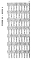
- Figure 1 is a sequence comparison of clone HGMP09 with a panel of G-protein coupled receptors ((6) and ref. therein). Only the sequence around transmembrane segment III of each receptor is shown in the one letter code. Residues conserved in HGMP09 and in more than 50 % of the other receptors are indicated by an asterisk. The "DRY” and "Asp113" residues (9) are indicated by ⁇ .
- Figure 2a shows the primary structure of the dog TSH receptor, as deduced from the nucleic acid sequence of dTSHr.
- the sequence was aligned (17) with full-length rat and pig LH-CG sequences (15, 16) and with HGMP09 partial sequence. Numbering is given from the first residue predicted in the mature polypeptide by von Heijne algorithm (11). Identical residues and conservative replacements in TSHr and LH-CGr are indicated by * and ., respectively. Sites for N glycosylation are underlined. Putative transmembrane segments are overlined.
- Lambda phages containing dTSHr inserts were subcloned in M13 and sequenced on both strands (Applied Biosystems model 370A) using a combination of forced cloning and exonuclease III deletions (21).
- Figure 2b is a dendogram constructed from the sequences of G-protein coupled receptors.
- the CLUSTAL algorithm (17) was used to construct a dendogram from the sequences of 22 receptors ((6) and references therein) including rat and pig LH-CG receptors (16, 17), HGMP09 and the TSH receptor.
- a segment corresponding to the known sequence of HGMP09 131 residues, extending from transmembrane segments II to V was used for comparison by the program.
- FIG. 3a shows TSH induced morphological changes in Y1 cells microinjected with TSH receptor mRNA.
- Y1 cells were microinjected with recombinant TSH receptor mRNA (0.1 pl at 0.25 ug/ul) (right) or water (left) as described (13) and incubated in control medium (upper panel) or with TSH (0.1nM) (lower panel).
- RO 201724 and isobutylmethylxanthine (10 -6 M each) were present in all incubations.
- FIG. 3b shows TSH induced CAMP accumulation in Xenopus oocytes microinjected with TSH receptor mRNA.
- Xenopus oocytes were handled as described (22) and injected with water (open symbols) or recombinant TSH receptor mRNA (13) (50 nl at 0.1 ug/ul) (filled symbols).
- After 3 days in control medium batches of 35 oocytes were incubated for 90 min. in medium supplemented with various concentrations of TSH (circles), LH (squares) or FSH (triangles).
- cAMP was determined as described (14).
- RO 201724 and isobutylmethylxanthine (10 -6 M each) were present in all incubations. Incubation of control oocytes in forskolin at 10 -4 M resulted in doubling of the cAMP concentration (not shown).
- FIG. 4 illustrates the displacement of 125 I TSH receptors expressed in cos7 cells.
- Cos7 cells were transfected with TSH receptor cDNA subcloned in pSVL (23). After 72 hours, cells were harvested and a membrane fraction was prepared (24). Membranes were similarly prepared from wild type cos7 cells and from dog thyrocytes in primary culture (20). Binding of 125 I TSH (TRAK Henning) was performed at 0°C for 120 min. in the presence of various concentrations of competitors (TSH-Armour, FSH and LH, UCB bioproducts). Bound radioactivity was separated by centrifugation and counted.
- Results are expressed as percent 125 I TSH bound by transfected cells in the absence of competitor (3,000 cpm) over nonspecific binding (radioactivity bound in the presence of 100nM cold TSH, 800 cpm).
- Open and filled circles represent displacement by cold TSH from cos7 and thyrocyte membranes respectively.
- Open and filled squares represent displacement from cos7 by LH and FSH, respectively.
- Diamonds represent control cos7 cells in presence of various amounts of cold TSH.
- Figure 5 shows the cDNA sequence coding for the dog TSH receptor, which was expressed in oocytes and culture cells.
- Figure 6 is a schematic representation of the dog thyrotropin receptor, showing the 7 putative transmembrane segments and the large NH2 terminal extracellular domain (to the exclusion of the signal peptide). The amino-acids deleted in the variant form are indicated in black. The five putative glycosylation sites are shown.
- Figure 7 shows the sequence alignment of the repeats constituting the extracellular domain of the thyrotropin receptor amino-acid sequence.
- the signal peptide as determined by Von Heijne algorithm is represented in italic.
- the repeat missing in the molecular varian of the receptor is indicated by the leftward arrow.
- Figure 8 shows the primary structure of the human TSH receptor as deduced from its cDNA sequence.
- the amino-acid sequence corresponds to the 2292 nucleotide open reading frame determined from the sequencing of two overlapping inserts in lamda gtll clones (see examples). It is aligned for comparison with the dog TSH receptor sequence (only non conserved amino-acids are indicated). Numbering starts from the first residue of the mature polypeptide as determined by von Heijne algorithm [11]. Potential N-glycosylation sites are underlined and putative transmembrane segments are overlined.
- Figure 9 shows the displacement by nonradioactive TSH of [ 125 I]TSH from human TSH receptors expressed in cos-7 cells. Results are expressed as percentage of the [ 125 I]-labelled TSH bound by transfected cells in the absence of competitor (1400 cpm) after correcting for -nonspecific binding (radioactivity bound in the presence of 100 nM unlabelled TSH, 240 cpm).
- Figure 10 represents the displacement by immunoglobulins of [ 125 I]TSH from human TSH receptor expressed in cos-7 cells. Results are expressed as described in the legend to -fig. 9. Immoglobulins were prepared (see examples) from a normal individual (N), from patients with idiopathic myxoedema (IM1, IM2) or Graves' disease (GD1, GD2). The concentration of immunoglobulins in the assay is indicated. The ability of IM1 and IM2 (1.5 mg/ml) to inhibit TSH-stimulated cAMP production in a human thyrocyte assay was 100 % and 85 %, respectively. The thyroid stimulating activity of GD1 and GD2 (1.5 mg/ml) was equivalent to that of 10 mU/ml of TSH, respectively.
- Figure 11 shows the primary structure of a TSH receptor according to the invention, in which a plurality of letters at any one site indicates the presence of one of the given amino acid residues at that site.
- Figure 12 illustrates the cDNA sequence of the cloned human TSH receptor.
- the invention relates to the use of such recombinant polypeptides in methods for the quantitative detection of thyrotropine (TSH) or of anti-thyrotropine receptor autoantibodies (anti-TSHr) in a biological sample, in which method such polypeptide is contacted with the biological sample suspected of containing TSH or anti-TSHr antibodies, and is either simultaneously or subseqently further contacted with labelled TSH, or with labelled anti-TSHr antibodies, and wherein after competition between the labelled and the unlabelled species in the reaction system the remaining bound labelled TSH or bound labelled anti-TSHr antibodies are measured.
- TSH thyrotropine
- anti-TSHr anti-thyrotropine receptor autoantibodies
- kits for carrying out such method which kits contain recombinant polypeptides as mentioned above as constituents of a kit component.
- TSH-receptor activity is meant either TSH-binding properties or anti-TSH receptor antibody-binding properties or ability to activate adenylyl cyclase or phospholipase C via G proteins when exposed to TSH or anti-TSHr antibodies. These properties are easily verified by contacting the polypeptide with for example labelled TSH or labelled anti-TSHr antibodies or by monitoring the adenylyl cyclase activity of a membrane preparation containing the polypeptide.
- the polypeptides of the invention include the entire TSH receptor as identified by the inventors, and fragments or variants of this polypeptide as defined below.
- the entire TSH receptor is composed of a signal peptide (20 residues) followed by a large putative extracellular domain (398 residues) containing 5 sites for N-glycosylation, connected to a 346 residue COOH domain containing seven putative transmembrane segments.
- the amino-acid sequence of the receptor is illustrated in fig. 11.
- the labelled anti-TSHr antibodies are polyclonal or monoclonal antibodies raised against a recombinant polypeptide possessing thyrotropin receptor activity as defined above.
- the sequence represented by [x] n in the above general formula corresponds to the signal sequence of the TSH receptor.
- This part of the polypeptide naturally ensures the transport to the cell membrane of the adjoining [y] and/or [z] fragments, after its production in the cell.
- the signal sequence does not need to be present in the polypeptide in cases where transport to the membrane is not required (for example in in vitro translation of the mRNA encoding the polypeptide), or may be replaced by other signal sequences to facilitate production of the recombinant receptor in certain host cells.
- the sequence represented by [z] P in the above general formula corresponds to the COOH domain of the entire polypeptide containing the seven putative transmembrane fragments I-VII, which show homology with the corresponding region of other G-protein coupled receptors.
- the polypeptides of the invention include, as indicated above, variants of the basic TSH receptor - sequence lacking part or all of the transmembrane domain. It is thought that these types of variants may exist naturally as a result of an alternative splicing phenomenom. By homology with other, known G-protein coupled receptors, the seven putative transmembrane segments have tentatively been identified as shown in Fig. 11 (numbered I to VII).
- the variant polypeptides of the invention include polypeptides missing some or all of the fragments I - VII; as defined above, which definition includes the putative extracellular and intracellular "loops" occuring between the transmembrane segments (see fig. 6).
- the transmembrane segment(s) missing may therefore, for example, be a segment selected from segments I to VII as indicated in fig. 11 or may be part of one of those segments, or may be a transmembrane segment in conjunction with its adjoining intracellular and/or extracellular loop.
- transmembrane domain it is also within the terms of the invention to replace some or all of the transmembrane domain by the transmembrane domain, or part of this domain, of a different receptor, thus giving rise to a hybrid receptor.
- This type of receptor is particularly interesting in cases where the extracellular part of the TSH receptor needs to be anchored in a cell membrane having characteristics which are different from, or even incompatible with, the transmembrane portion of the TSH receptor. It is also possible to use as the transmembrane domain in a hybrid receptor any amino-acid sequence exhibiting suitable anchoring properties. Such a sequence could be entirely synthetic or based on any transmembrane protein.
- the invention also embraces the use of polypeptides having thyrotropin receptor activity which lack the entire transmembrane domain.
- the polypeptide corresponds to the extracellular domain of the naturally occuring receptor. This-extracellular part of the receptor which is apparently responsible for ligand binding, is identified by the region [y] in the general formula.
- This extracellular part of the receptor [y] is characterised by an imperfect repeat structure which can be aligned as shown in Fig 7.
- the polypeptides of the invention include variants in which one or more of these repeats is missing.
- immunogenic amino-acid sequences may comprise for example 5, 6, 7, 8 or 9 consecutive amino-acids of the "y" sequence defined above.
- the immunogenic nature of the fragments of the invention is tested by injection of the fragment in question into a laboratory animal, followed by investigation of the reactivity between any antibodies thus formed and the immunising fragment.
- the invention encompasses the use of polypeptides in which the second repeat (marked by an arrow in fig 7) is missing.
- nucleic acid sequences coding for the polypeptides are those shown in figs. 5 and 12, and fragments of these sequences, as well as corresponding degenerate sequences.
- the nucleic acid fragments normally have at least 8 nucleotides and have preferably at least 12 or preferably at least 16 nucleotides.
- Such fragments, or their complementary sequences can be used as primers in the amplification of segments of DNA using the polymerase chain reaction, for example in the production of cDNA coding- for the polypeptides having thyrotropin receptor activity.
- the nucleic acid sequences of the invention coding for the entire TSH receptor are in a genetic environment other than that found naturally in thyroid cells.
- the genetic environment may be that of a Cos-7 cell, a CH0 cell or Y1 cells.
- polypeptides to be used in the present invention can be produced in several different ways.
- a host cell such as COS 7 cells, CHO cells, NIH3T3 cells, Xenopus oocytes or Y1 cells can be transformed by a vector containing a nucleic acid insert coding for the desired peptide, in conjunction with all the necessary regulatory elements such as promoter, transcription termination signals etc, or can be microinjected with recombinant mRNA transcribed from appropriate vectors containing the receptor encoded sequence . Expression of the insert normally leads to the insertion of the recombinant polypeptide in the membrane of the cell used as host.
- the receptor polypeptide can be used in this form, the receptor thus being present in a non-thyroidal eukaryotic cellular environment, or in a solubilised membrane fragment form.
- the non-thyroid cells expressing the recombinant receptor exhibit a receptor density of upto ten times that observed in thyroid cells.
- the correspondingly shorter nucleic acid sequence can be used to transform a suitable host cell, for example, a DNA coding for the putative extracellular region only, or one or more repeats of the repetitive portion of this region. It is also within the terms of the invention to synthesise the polypeptide chemically, by successive assembly of the required amino-acid residues. In cases where larger fragments are desired, it is possible to synthesise first a series of smaller fragments and to ultimately assemble these fragments to form the larger fragment.
- the invention also relates to the use of antibodies, both polyclonal and monoclonal, to the thyrotropin-receptor polypeptides.
- the antibodies of the invention are preferably in a purified form, and may be of animal origin e.g. rabbit or mouse.
- the TSH-receptor may be the target of auto-immune reactions giving rise to hyper- or hypo-stimulation of the thyroid gland by stimulating or blocking autoantibodies respectively.
- the antigenic nature of the polypeptides of the invention particularly the putative extracellular domain, permits the preparation of antibodies, which can be used in a variety of studies and assays.
- the TSH-receptor binds both TSH and anti-TSHr antibodies, thus it is possible in certain studies to replace TSH by anti-TSHr antibodies.
- the phenomenon of competition between labelled and unlabelled species is particularly useful in such assays.
- Use of specific fragments of the TSH receptor allows the preparation of antibodies against defined epitopes, and, by using a panel of such antibodies, allows further characterisation of the type of disorder present in auto-immune patients.
- An assay of the invention is a process for the quantitative detection of thyrotropine (TSH) or of anti-thyrotropine receptor antibodies (anti-TSHr) in a biological sample characterised in that a polypeptide according to the invention is contacted with the biological sample suspected of containing TSH or anti-TSHr antibodies and, either simultaneously or subsequently, contacted with labelled TSH, or with labelled anti-TSHr antibodies and the remaining, bound labelled TSH or bound labelled anti-TSHr antibodies after competition between the labelled and unlabelled species, is measured.
- TSH thyrotropine
- anti-TSHr anti-thyrotropine receptor antibodies
- the competition between the labelled TSH or labelled antibodies with the unlabelled TSH or antibodies present in the biological sample is measured as an indication of the concentration of that species in the sample.
- a receptor polypeptide to bind the TSH in the biological sample, and then after washing to add labelled anti-TSH antibodies which selectively detect the bound TSH.
- This type of assay can also be carried out using immobilized or solubilised receptor polypeptide to bind the anti-TSHr-antibody in a biological sample, followed by detection of the bound antibody by labelled anti immunoglobulins or protein A or protein G or any other agent capable of recognizing an antibody.
- the labels used in the assays of the invention are those conventionally used in the art, for example, radioactive labelling, enzymatic labelling, labelled anti-immunoglobulins, protein A, protein G, depending upon the type of assay.
- kits suitable for the detection of TSH or anti-TSHr antibodies are characterised in that they contain:
- kits for effecting the detection of autoantibodies directed against the TSH receptor by competition would include the recombinant polypeptide in immobilised or solubilised form, with labelled TSH or unlabelled TSH in combination with agents permitting the TSH to be labelled.
- a kit for effecting the detection of autoantibodies directed against the TSH receptor by competition would include the recombinant polypeptide in immobilised or solubilised form, with labelled TSH or unlabelled TSH in combination with agents permitting the TSH to be labelled.
- such a kit might include antibodies to the TSH receptor and means of labelling them, instead of the TSH.
- the primers used in this amplification were: and a plurality of nucleotides at any one site indicating the presence of one of the given nucleotides at that site.
- a dendrogram constructed from the alignment shown in fig. 1 demonstrated that it is equally distant from all receptors cloned to date (7); in particular, it does not contain the canonical Asp Arg Tyr (DRY) tripeptide close to transmembrane segment III (8) and lacks the Asp residue implicated in the binding of charged amines is adrenergic (Asp113), muscarinic, dopaminergic and serotonergic receptors (9).
- HGMP09 was used as a probe both in Northern blotting experiments with thyroid and non-thyroid tissues, and in screening of a dog thyroid cDNA library. HGMP09 did not hybridize to thyroid mRNA but identified a major 2.6 kb transcript in the ovary and the testis. However, under moderate conditions of stringency it hybridized to one out of 50,000 thyroid cDNA clones suggesting cross-hybridization with a relatively abundant putative receptor of the thyroid. From these characteristics, it was hypothesized that HGMP09 encoded a receptor fragment, distinct from the TSH receptor, but with sequence characteristics expected from close relatives like LH or FSH receptors.
- dTSHr cross-hybridizing clone
- the encoded polypeptide was unambiguously identified as the TSH receptor by expression of the cDNA in a variety of systems.
- Microinjection of recombinant mRNA in adrenocortical Y1 cells and in Xenopus oocytes conferred a TSH responsive phenotype to both systems.
- Y1 cells responded to TSH by a characteristic morphological change which is triggered by elevation of cAMP in the cytoplasm (12,13).
- Xenopus oocytes (fig. 3) displayed a dose-dependant increase in cAMP which was specific for stimulation by TSH and corresponded to the expected sensivity of the dog receptor to bovine TSH (half-maximal effect around 0.3 nM) (14).
- Transient expression of the receptor cDNA was obtained in Cos7 cells (fig 4). Specific binding of 125 I TSH to membranes was observed only in transfected cells.
- the displacement curve of the label by TSH presented characteristics very similar to that obtained with membranes from dog thyrocytes (half-maximal displacement at 0.4 nM and 0.16 nM for cos cells and thyrocytes, respectively) (fig. 4c). The slight rightward shift of the displacement curve obtained with Cos7 cell membranes may reflect the higher amount of receptors in this system.
- the cDNA coding for the dog TSH receptor was sequenced completely. The sequence is given in fig. 5.
- TSH receptor Comparison of the TSH receptor with the LH-CG receptor cloned recently (15, 16) reveals interesting common characteristics which make them members of a new subfamily of G-protein coupled receptors. They both display a long aminoterminal extension containing multiple sites for N glycosylation (five in the TSH receptor).
- the TSH receptor has an extra 52 residue insert close to the junction between the putative extracellular domain and the first transmembrane segment (fig. 2a).
- the overall sequence similarity between the extracellular domains of the TSH and LH-CG receptors is 45% (fig 2a).
- the similarity between a segment of soybean lectin and the rat LH receptor (15) is not conserved in the TSH receptor, which suggests that it may be fortuitous.
- the C-terminal half of the TSH receptor containing the transmembrane segments is 70% similar to both the pig and rat LH receptors (fig. 2a).
- the homology is particularly impressive in the transmembrane segments themselves, where stretches of up to 24 identical residues are observed in a row (transmembrane region III).
- the carboxyl terminal region of the third putative intracellular loop which is particularly short in TSH and LH receptors and which has been implicated in the interaction with Gas (8, 9), is identical in both receptor types. This pattern of similarities gives support to the view that the extracellular domain would be involved in the recognition of the ligands (4) , while the membrane-inserted domain would be responsible for the activation of Gas (15, 16).
- TSH and LH-CG receptors, and HGMP09 constitute clearly a distinct subfamily of G-protein coupled receptors.
- a sequence similarity dendrogram (17) including most of the G-protein coupled receptors cloned to date demonstrates both their close relationships and their distance from the bulk of the other receptors (fig. 2b).
- the complete sequence of the FSH receptor will reveal whether the known ability of LH-CG to stimulate the TSH receptor (18) is reflected by a higher sequence similarity of the extracellular domains of LH and TSH receptors.
- dTSHR3 was shorter, and consisted of 2200 and 1500 bp EcoRI fragments. Restriction analysis of the 2800 bp fragments of dTSHR1 and dTSHR2 revealed slight differences in the restriction map, the main discordance being the presence of a PstI restriction site in dTSHR1 and its absence in dTSGR2.
- dTSHR1 was sequenced completely and revealed an open reading frame of 764 codons which was identified as the thyrotropin receptor by expression of the cDNA in oocytes and cell cultures (see example I(b) + fig 5).
- dTSHR3 was shown by sequencing to be completely colinear with dTSHR1 but this clone lacked 600 bp at its 5' end. Because of the difference in the restriction map of dTSHR1 and dTSHR2, this latter clone was also sequenced on both strands.
- a total of 5 mutations, including two single base substitutions, one single base deletion, one single base insertion and one 5 base insertion were found scattered in the 2060 bp long 3' untranslated region (not shown).
- the main difference between dTSHR2 and dTSHR1 was located in the coding region, and consisted in a 75 bp deletion located 240 bp after the start codon.
- the corresponding 25 amino-acids deletion in the protein itself is located in the long NH2 terminal extracellular domain which is characteristic of the TSH receptor (25) and its recently cloned close relative, the LH receptor (15, 16) (fig. 6).
- the NH2 terminal part of the thyrotropin receptor is characterized by an imperfect repeat structure that can be aligned as indicated in fig. 7. These repeats are similar in structure to the leucine-rich repeats found in the various proteins comprising the family of leucine-rich glycoproteins (26, 15), and references therein).
- the deletion in the dTSHR2 clone corresponds exactly to one of these repeats, in a region of the protein where the repeat length is regular and their alignment unambiguous. The existence of such variant reinforces considerably the significance of this repeated structure and sets up interesting questions concerning its functional meaning and the structure of the chromosomal gene.
- the extracellular domains of TSH and LH receptors are apparently responsible for the ligand binding (4).
- the deleted repeat also contains one of the 5 consensus sequences for N-glycosylation. Glycosylation of the TSH receptor could be important for ligand binding or signal transduction. If, and to what extent, the lack of this repeat influences the binding capabilities and/or the function of the receptor variant, is not yet known. Comparison of cell lines expressing this variant with the presently available stable transfectants expressing the full size receptor should partially answer this question. The functional analysis of other in-vitro generated mutants of the TSH receptor will complete the study.
- a human lambda gtll cDNA library (29) was screened with a fragment of the dog TSHr (25). Out of the 218 clones scored as positive (1/6000), 24 were plaque-purified to homogeneity and the size of the inserts was determined. Two clones which harbored inserts of 2370 bp and 3050 bp, respectively, were subcloned as overlapping fragments in M13 derivatives and sequenced (fig 12). A total of 4272 bp were determined in which a 2292 bp open reading frame was identified. When translated into protein, the coding sequence showed an overall 90.3 % similarity with the dog TSHr (Fig. 8) [1].
- the coding sequence was inserted in the SV40-based vector pSVL, and the resulting construct transfected in Cos-7 cells (24).
- Membranes prepared from transfected cells demonstrated specific binding of [ 125 I]TSH (fig. 9).
- the unlabelled competitor TSH was bovine.
- the characteristics of the displacement curve with unlabelled TSH were similar to those observed with the dog TSHr assayed under similar conditions (half maximal displacement around 0.5 nM) (25).
Landscapes
- Health & Medical Sciences (AREA)
- Chemical & Material Sciences (AREA)
- Organic Chemistry (AREA)
- Life Sciences & Earth Sciences (AREA)
- Immunology (AREA)
- Proteomics, Peptides & Aminoacids (AREA)
- Molecular Biology (AREA)
- Endocrinology (AREA)
- Medicinal Chemistry (AREA)
- Biochemistry (AREA)
- Biophysics (AREA)
- General Health & Medical Sciences (AREA)
- Genetics & Genomics (AREA)
- Toxicology (AREA)
- Zoology (AREA)
- Cell Biology (AREA)
- Gastroenterology & Hepatology (AREA)
- Engineering & Computer Science (AREA)
- Biomedical Technology (AREA)
- Neurology (AREA)
- Peptides Or Proteins (AREA)
- Preparation Of Compounds By Using Micro-Organisms (AREA)
- Investigating Or Analysing Biological Materials (AREA)
Abstract
Description
- The invention relates to a method for the quantitative detection of thyrotropine (TSH) or of anti-thyrotropine receptor autoantibodies (anti-TSHr) in biological fluids using a recombinant thyrotropin receptor polypeptide, and to kits for conducting said method.
- The literature references indicated by numbers in parentheses in this specification are listed in the form of a bibliography at the end of the description.
- Pituitary glycoproteins (Luteinizing hormone, LH; follicle stimulating hormone, FHS ; and thyroid stimulating hormone or thyrotropin, TSH) form a family of closely related hormones.
- The pituitary hormone thyrotropin (TSH) is the main physiological agent regulating the thyroid gland. It stimulates the function and the proliferation of thyrocytes and induces the expression of differentiation (1). Most of its effects are mediated by cyclic AMP (cAMP) (1). As the other pituitary and placental glycoprotein hormones (FSH, LH, CG), TSH is a heterodimer. All these hormones share an identical alpha subunit ; the beta subunit, despite sequence similarity, is specific for each (2). The activated TSH, FSH and LH-CG receptors stimulate adenylyl cyclase in their target cells via mechanisms mediated by the G protein Gs (3). In man, the TSH receptor may be the target of autoimmune reactions leading to hyper- or hypo-stimulation of the thyroid gland by autoantibodies in Grave's disease and in idiopathic myxoedema, respectively (4).
- A prerequisite to studies of such diseases and to the elucidation of receptor structure and function is the availability of receptor preparations, particularly human, at a reasonable cost and in relative abundance.
- To date, particulate membrane preparations and detergent-solubilised thyroid membranes, often of porcine or bovine origin (4) and (31) or (32) have been used in such studies. Human receptor preparations are not only costly but are also difficult to reproduce identically. Furthermore, the known preparations cannot be considered to be "purified" receptors; they are enriched with respect to their receptor content but do not allow purification of the receptor to a degree which would enable a partial sequence analysis, and hence its cloning.
- Cloning and expression of the related LH-CG receptor has recently been achieved. A cDNA for the rat LH-CG receptor was isolated with use of a DNA probe generated in a polymerase chain reaction with oligonucleotide primers based on peptide sequences of purified receptor protein (15). Variants of the porcine LH-CG receptor were cloned by screening a λgt11 library with cDNA probes isolated with monoclonal antibodies (16).
- Attempts have been made to clone the TSH receptor (6) using a method which exploits the sequence similarity displayed by all_ known G-protein coupled receptors. A pair of oligonucleotide primers corresponding to transmembrane segments III and VI were used on cDNA from thyroid tissue under conditions allowing amplification of the primed sequences by the polymerase chain reaction. The method did not allow cloning of the TSH receptor but led instead to the cloning of four new members of the G-protein coupled receptor family.
- Various attempts to prepare monoclonal antibodies which recognize the TSH receptor are reported in the scientific literature as well. (33) describes monoclonal antibodies which are obtained from the fusion of human lymphocytes with myeloma cells and which are proposed as recognizing the TSH receptor because they interfere with TSH binding to human thyroid membranes and weakly stimulate cAMP production in FRTL-5 cells. However, all attempts to use said antibodies for the purification or cloning of the TSH receptor have failed.
- (34) describes the preparation of TSH anti-idiotypic antibodies as monoclonals which are reported to recognize the TSH receptor. However, the nature of the molecules recognized by the antibodies described in (34) cannot be clearly derived from the Western blot experiments, and the authors in (34) could not identify the nature of the antigen recognized by their antibodies.
- (35) describes the use of "antiparatypical antibodies" for detecting and treating autoimmune diseases, i.e. the use of antibodies which are raised against specific paratopes of other antibodies, i.e. against immunoglobulins. The use of antiparatypical antibodies is proposed in view of the difficulties in obtaining defined pure antigens as e.g. the TSH receptor.
- The present inventors have succeeded in cloning the TSH receptor and variants thereof, firstly by applying the technique described in (6) but with different sets of primers, and with human genomic DNA as the template, and secondly by use of a selected sequence amplified by this technique as a probe.
- Certain aspects of the invention are illustrated in the figures 1 to 12. Figures illustrating amino-acid sequences use the one-letter abbreviation system.
- Figure 1 is a sequence comparison of clone HGMP09 with a panel of G-protein coupled receptors ((6) and ref. therein). only the sequence around transmembrane segment III of each receptor is shown in the one letter code. Residues conserved in HGMP09 and in more than 50 % of the other receptors are indicated by an asterisk. The "DRY" and "Asp113" residues (9) are indicated by ^.
- Figure 2a shows the primary structure of the dog TSH receptor, as deduced from the nucleic acid sequence of dTSHr. The sequence was aligned (17) with full-length rat and pig LH-CG sequences (15, 16) and with HGMP09 partial sequence. Numbering is given from the first residue predicted in the mature polypeptide by von Heijne algorithm (11). Identical residues and conservative replacements in TSHr and LH-CGr are indicated by * and ., respectively. Sites for N glycosylation are underlined. Putative transmembrane segments are overlined. Lambda phages containing dTSHr inserts were subcloned in M13 and sequenced on both strands (Applied Biosystems model 370A) using a combination of forced cloning and exonuclease III deletions (21).
- Figure 2b is a dendogram constructed from the sequences of G-protein coupled receptors. The CLUSTAL algorithm (17) was used to construct a dendogram from the sequences of 22 receptors ((6) and references therein) including rat and pig LH-CG receptors (16, 17), HGMP09 and the TSH receptor. For each receptor, a segment corresponding to the known sequence of HGMP09 (131 residues, extending from transmembrane segments II to V) was used for comparison by the program.
- Figure 3a shows TSH induced morphological changes in Y1 cells microinjected with TSH receptor mRNA. Y1 cells were microinjected with recombinant TSH receptor mRNA (0.1 pl at 0.25 ug/ul) (right) or water (left) as described (13) and incubated in control medium (upper panel) or with TSH (0.1nM) (lower panel). RO 201724 and isobutylmethylxanthine (10-6 M each) were present in all incubations.
- Figure 3b shows TSH induced CAMP accumulation in Xenopus oocytes microinjected with TSH receptor mRNA. Xenopus oocytes were handled as described (22) and injected with water (open symbols) or recombinant TSH receptor mRNA (13) (50 nl at 0.1 ug/ul) (filled symbols). After 3 days in control medium, batches of 35 oocytes were incubated for 90 min. in medium supplemented with various concentrations of TSH (circles), LH (squares) or FSH (triangles). cAMP was determined as described (14). RO 201724 and isobutylmethylxanthine (10-6 M each) were present in all incubations. Incubation of control oocytes in forskolin at 10-4 M resulted in doubling of the cAMP concentration (not shown).
- Figure 4 illustrates the displacement of 125I TSH receptors expressed in cos7 cells. Cos7 cells were transfected with TSH receptor cDNA subcloned in pSVL (23). After 72 hours, cells were harvested and a membrane fraction was prepared (24). Membranes were similarly prepared from wild type cos7 cells and from dog thyrocytes in primary culture (20). Binding of 125I TSH (TRAK Henning) was performed at 0°C for 120 min. in the presence of various concentrations of competitors (TSH-Armour, FSH and LH, UCB bioproducts). Bound radioactivity was separated by centrifugation and counted. Results are expressed as percent 125I TSH bound by transfected cells in the absence of competitor (3,000 cpm) over nonspecific binding (radioactivity bound in the presence of 100nM cold TSH, 800 cpm). Open and filled circles represent displacement by cold TSH from cos7 and thyrocyte membranes respectively. Open and filled squares represent displacement from cos7 by LH and FSH, respectively. Diamonds represent control cos7 cells in presence of various amounts of cold TSH.
- Figure 5 shows the cDNA sequence coding for the dog TSH receptor, which was expressed in oocytes and culture cells.
- Figure 6 is a schematic representation of the dog thyrotropin receptor, showing the 7 putative transmembrane segments and the large NH2 terminal extracellular domain (to the exclusion of the signal peptide). The amino-acids deleted in the variant form are indicated in black. The five putative glycosylation sites are shown.
- Figure 7 shows the sequence alignment of the repeats constituting the extracellular domain of the thyrotropin receptor amino-acid sequence. The signal peptide, as determined by Von Heijne algorithm is represented in italic. The repeat missing in the molecular varian of the receptor is indicated by the leftward arrow.
- Figure 8 shows the primary structure of the human TSH receptor as deduced from its cDNA sequence. The amino-acid sequence corresponds to the 2292 nucleotide open reading frame determined from the sequencing of two overlapping inserts in lamda gtll clones (see examples). It is aligned for comparison with the dog TSH receptor sequence (only non conserved amino-acids are indicated). Numbering starts from the first residue of the mature polypeptide as determined by von Heijne algorithm [11]. Potential N-glycosylation sites are underlined and putative transmembrane segments are overlined.
- Figure 9 shows the displacement by nonradioactive TSH of [125I]TSH from human TSH receptors expressed in cos-7 cells. Results are expressed as percentage of the [125I]-labelled TSH bound by transfected cells in the absence of competitor (1400 cpm) after correcting for -nonspecific binding (radioactivity bound in the presence of 100 nM unlabelled TSH, 240 cpm).
- Figure 10 represents the displacement by immunoglobulins of [125I]TSH from human TSH receptor expressed in cos-7 cells. Results are expressed as described in the legend to -fig. 9. Immoglobulins were prepared (see examples) from a normal individual (N), from patients with idiopathic myxoedema (IM1, IM2) or Graves' disease (GD1, GD2). The concentration of immunoglobulins in the assay is indicated. The ability of IM1 and IM2 (1.5 mg/ml) to inhibit TSH-stimulated cAMP production in a human thyrocyte assay was 100 % and 85 %, respectively. The thyroid stimulating activity of GD1 and GD2 (1.5 mg/ml) was equivalent to that of 10 mU/ml of TSH, respectively.
- Figure 11 shows the primary structure of a TSH receptor according to the invention, in which a plurality of letters at any one site indicates the presence of one of the given amino acid residues at that site.
- Figure 12 illustrates the cDNA sequence of the cloned human TSH receptor.
- This invention relates to specific uses of recombinant polypeptides possessing thyrotropin receptor activity, which polypeptides are characterised in that they comprise an amino-acid sequence represented by the following general formula:
[X]n - [Y]m - [Z]p
wherein n = 0 or 1; m = 1; p = 0 or 1;
and X, Y and Z are defined as indicated below, said polypeptides being obtained by a process which comprises the expression of a nucleic acid coding for the polypeptide in a host cell transformed by a vector in which the said nucleic acid has been operationally inserted, which polypeptides, in the case of p = 1, further are associated with such a host cell, the receptor thus being present in a non-thyroidal cellular environment, or with a membrane preparation which is free of impurities associated with detergent-solubilized thyroid membrane preparations. More specially, the invention relates to the use of such recombinant polypeptides in methods for the quantitative detection of thyrotropine (TSH) or of anti-thyrotropine receptor autoantibodies (anti-TSHr) in a biological sample, in which method such polypeptide is contacted with the biological sample suspected of containing TSH or anti-TSHr antibodies, and is either simultaneously or subseqently further contacted with labelled TSH, or with labelled anti-TSHr antibodies, and wherein after competition between the labelled and the unlabelled species in the reaction system the remaining bound labelled TSH or bound labelled anti-TSHr antibodies are measured. - The invention further relates to kits for carrying out such method, which kits contain recombinant polypeptides as mentioned above as constituents of a kit component.
- By "TSH-receptor activity" is meant either TSH-binding properties or anti-TSH receptor antibody-binding properties or ability to activate adenylyl cyclase or phospholipase C via G proteins when exposed to TSH or anti-TSHr antibodies. These properties are easily verified by contacting the polypeptide with for example labelled TSH or labelled anti-TSHr antibodies or by monitoring the adenylyl cyclase activity of a membrane preparation containing the polypeptide. The polypeptides of the invention include the entire TSH receptor as identified by the inventors, and fragments or variants of this polypeptide as defined below. The entire TSH receptor is composed of a signal peptide (20 residues) followed by a large putative extracellular domain (398 residues) containing 5 sites for N-glycosylation, connected to a 346 residue COOH domain containing seven putative transmembrane segments. The amino-acid sequence of the receptor is illustrated in fig. 11.
- More particularly, the invention relates to a method for the quantitative detection of thyrotropine (TSH) or of anti-thyrotropine receptor autoantibodies (anti-TSHr) in a biological sample, characterized in that a recombinant polypeptide possessing thyrotropin receptor activity, which polypeptide i) is characterised in that it comprises an amino-acid sequence represented by the following general formula:
[X]n - [Y]m - [Z]p
wherein n = 0 or 1; m = 1; p = 0 or 1;
and X, Y and Z are defined as follows (using the one-letter amino-acid symbol and wherein a plurality of letters at any one site indicates the presence of any one of the given amino-acid residues at that site):Y = at least the minimum number of consecutive amino-acids of the following sequence necessary for the presentation of immunogenic properties: and Z = [I - II -IIi - III - IIIi - IV - V - VI - VII - VIIi] wherein the amino-acid sequences I - II - IIi - III - IIIi - IV - V - VI - VII - VIIi are independently present or absent and have the following meanings:
 or at least 12 consecutive amino-acid residues of this sequence;
 or at least 12 consecutive amino-acid residues of this sequence;
 or at least 2 consecutive amino-acid residues of this sequence;
 or at least 22 consecutive amino-acid residues of this sequence
 or at least 2 consecutive amino-acid residues of this sequence;
 or at least 12 consecutive amino-acid residues of this sequence;
 or at least 12 consecutive amino-acid residues of this sequence;
 or at least 12 consecutive amino-acid residues of this sequence;
 or at least 12 consecutive amino-acid residues of this sequence;
 or at least 12 consecutive amino-acid residues of this sequence;
it being understood that any of the above-specified amino-acids can be replaced or deleted, and that extra amino-acid residues may be inserted provided the thyrotropin receptor activity is maintained, said polypeptide being obtained by the expression of a nucleic acid coding for the polypeptide in a host cell transformed by a vector in which the said nucleic acid has been operationally inserted,
and which polypeptide, ii) in the case of p = 1, further is characterized in that it is associated with such a host cell, the receptor thus being present in a non-thyroidal cellular environment, or with a membrane preparation which is free of impurities associated with detergent-solubilized thyroid membrane preparations,
is contacted with the biological sample suspected of containing TSH or anti-TSHr antibodies, and that it is either simultaneously or subseqently contacted with labelled TSH, or with labelled anti-TSHr antibodies, and that after competition between the labelled and the unlabelled species in the reaction system the remaining bound labelled TSH or bound labelled anti-TSHr antibodies are measured. - Preferably, the labelled anti-TSHr antibodies are polyclonal or monoclonal antibodies raised against a recombinant polypeptide possessing thyrotropin receptor activity as defined above.
- The sequence represented by [x]n in the above general formula corresponds to the signal sequence of the TSH receptor. This part of the polypeptide naturally ensures the transport to the cell membrane of the adjoining [y] and/or [z] fragments, after its production in the cell. Clearly the signal sequence does not need to be present in the polypeptide in cases where transport to the membrane is not required (for example in in vitro translation of the mRNA encoding the polypeptide), or may be replaced by other signal sequences to facilitate production of the recombinant receptor in certain host cells.
- The sequence represented by [z]P in the above general formula corresponds to the COOH domain of the entire polypeptide containing the seven putative transmembrane fragments I-VII, which show homology with the corresponding region of other G-protein coupled receptors. The polypeptides of the invention include, as indicated above, variants of the basic TSH receptor - sequence lacking part or all of the transmembrane domain. It is thought that these types of variants may exist naturally as a result of an alternative splicing phenomenom. By homology with other, known G-protein coupled receptors, the seven putative transmembrane segments have tentatively been identified as shown in Fig. 11 (numbered I to VII). The variant polypeptides of the invention include polypeptides missing some or all of the fragments I - VII; as defined above, which definition includes the putative extracellular and intracellular "loops" occuring between the transmembrane segments (see fig. 6). The transmembrane segment(s) missing may therefore, for example, be a segment selected from segments I to VII as indicated in fig. 11 or may be part of one of those segments, or may be a transmembrane segment in conjunction with its adjoining intracellular and/or extracellular loop.
- It is also within the terms of the invention to replace some or all of the transmembrane domain by the transmembrane domain, or part of this domain, of a different receptor, thus giving rise to a hybrid receptor. This type of receptor is particularly interesting in cases where the extracellular part of the TSH receptor needs to be anchored in a cell membrane having characteristics which are different from, or even incompatible with, the transmembrane portion of the TSH receptor. It is also possible to use as the transmembrane domain in a hybrid receptor any amino-acid sequence exhibiting suitable anchoring properties. Such a sequence could be entirely synthetic or based on any transmembrane protein.
- It is to be noted that the invention also embraces the use of polypeptides having thyrotropin receptor activity which lack the entire transmembrane domain. In this case, the polypeptide corresponds to the extracellular domain of the naturally occuring receptor. This-extracellular part of the receptor which is apparently responsible for ligand binding, is identified by the region [y] in the general formula. A polypeptide lacking the entire transmembrane domain is respresented by the general formula [y]m, where m = 1, the [z] part of the sequence being absent. This extracellular part of the receptor [y], is characterised by an imperfect repeat structure which can be aligned as shown in Fig 7. The polypeptides of the invention include variants in which one or more of these repeats is missing. It is however important that sufficient aminoacids be present to allow formation of antibodies (monoclonal or polyclonal). Such immunogenic amino-acid sequences may comprise for example 5, 6, 7, 8 or 9 consecutive amino-acids of the "y" sequence defined above. The immunogenic nature of the fragments of the invention is tested by injection of the fragment in question into a laboratory animal, followed by investigation of the reactivity between any antibodies thus formed and the immunising fragment.
- In particular, the invention encompasses the use of polypeptides in which the second repeat (marked by an arrow in fig 7) is missing.
- Examples of nucleic acid sequences coding for the polypeptides are those shown in figs. 5 and 12, and fragments of these sequences, as well as corresponding degenerate sequences. The nucleic acid fragments normally have at least 8 nucleotides and have preferably at least 12 or preferably at least 16 nucleotides. Such fragments, or their complementary sequences can be used as primers in the amplification of segments of DNA using the polymerase chain reaction, for example in the production of cDNA coding- for the polypeptides having thyrotropin receptor activity.
- The nucleic acid sequences of the invention coding for the entire TSH receptor are in a genetic environment other than that found naturally in thyroid cells. For example, the genetic environment may be that of a Cos-7 cell, a CH0 cell or Y1 cells.
- The polypeptides to be used in the present invention can be produced in several different ways. For example, a host cell such as COS 7 cells, CHO cells, NIH3T3 cells, Xenopus oocytes or Y1 cells can be transformed by a vector containing a nucleic acid insert coding for the desired peptide, in conjunction with all the necessary regulatory elements such as promoter, transcription termination signals etc, or can be microinjected with recombinant mRNA transcribed from appropriate vectors containing the receptor encoded sequence . Expression of the insert normally leads to the insertion of the recombinant polypeptide in the membrane of the cell used as host. In this way, the receptor polypeptide can be used in this form, the receptor thus being present in a non-thyroidal eukaryotic cellular environment, or in a solubilised membrane fragment form. The non-thyroid cells expressing the recombinant receptor exhibit a receptor density of upto ten times that observed in thyroid cells.
- Furthermore, in the case where only a fragment of the polypeptide is required, the correspondingly shorter nucleic acid sequence can be used to transform a suitable host cell, for example, a DNA coding for the putative extracellular region only, or one or more repeats of the repetitive portion of this region. It is also within the terms of the invention to synthesise the polypeptide chemically, by successive assembly of the required amino-acid residues. In cases where larger fragments are desired, it is possible to synthesise first a series of smaller fragments and to ultimately assemble these fragments to form the larger fragment.
- The invention also relates to the use of antibodies, both polyclonal and monoclonal, to the thyrotropin-receptor polypeptides. The antibodies of the invention are preferably in a purified form, and may be of animal origin e.g. rabbit or mouse. As mentioned earlier, in man the TSH-receptor may be the target of auto-immune reactions giving rise to hyper- or hypo-stimulation of the thyroid gland by stimulating or blocking autoantibodies respectively. The antigenic nature of the polypeptides of the invention, particularly the putative extracellular domain, permits the preparation of antibodies, which can be used in a variety of studies and assays. The TSH-receptor binds both TSH and anti-TSHr antibodies, thus it is possible in certain studies to replace TSH by anti-TSHr antibodies. The phenomenon of competition between labelled and unlabelled species is particularly useful in such assays. Use of specific fragments of the TSH receptor allows the preparation of antibodies against defined epitopes, and, by using a panel of such antibodies, allows further characterisation of the type of disorder present in auto-immune patients.
- An assay of the invention is a process for the quantitative detection of thyrotropine (TSH) or of anti-thyrotropine receptor antibodies (anti-TSHr) in a biological sample characterised in that a polypeptide according to the invention is contacted with the biological sample suspected of containing TSH or anti-TSHr antibodies and, either simultaneously or subsequently, contacted with labelled TSH, or with labelled anti-TSHr antibodies and the remaining, bound labelled TSH or bound labelled anti-TSHr antibodies after competition between the labelled and unlabelled species, is measured.
- In this type of assay, the competition between the labelled TSH or labelled antibodies with the unlabelled TSH or antibodies present in the biological sample is measured as an indication of the concentration of that species in the sample.
- Alternatively, instead of using competition between two like-species to measure TSH, it is also possible to use a receptor polypeptide to bind the TSH in the biological sample, and then after washing to add labelled anti-TSH antibodies which selectively detect the bound TSH. This type of assay can also be carried out using immobilized or solubilised receptor polypeptide to bind the anti-TSHr-antibody in a biological sample, followed by detection of the bound antibody by labelled anti immunoglobulins or protein A or protein G or any other agent capable of recognizing an antibody.
- The labels used in the assays of the invention are those conventionally used in the art, for example, radioactive labelling, enzymatic labelling, labelled anti-immunoglobulins, protein A, protein G, depending upon the type of assay.
- The invention also embraces kits suitable for the detection of TSH or anti-TSHr antibodies. Such kits are characterised in that they contain:
- a) a polypeptide according to the invention and defined above, said polypeptide having thyrotropin receptor activity and being either in an immobilised or solibilised form ;
- b) at least one of the following reagents:
- i) labelled TSH
- ii) labelled anti-TSHr antibodies
- iii) reagents necessary for the measurement of adenylyl cyclase activity.
- For example, a kit for effecting the detection of autoantibodies directed against the TSH receptor by competition would include the recombinant polypeptide in immobilised or solubilised form, with labelled TSH or unlabelled TSH in combination with agents permitting the TSH to be labelled. Alternatively, such a kit might include antibodies to the TSH receptor and means of labelling them, instead of the TSH.
- The invention will be illustrated by the following examples:
- As most G protein-coupled receptor genes do not contain introns in their coding sequence, a similar strategy to that previously described (6) was used, but using different sets of degenerated primers and with human genomic DNA as starting material. Eleven clones displaying sequence similarity with G-protein coupled receptors where thus obtained (7). Out of these, one clone (HGMP09) which was amplified with primers corresponding to transmembrane segments II and VII, presented sequence characteristics suggesting that it belonged to a distinct subfamily of receptors.
- The primers used in this amplification were:and
 a plurality of nucleotides at any one site indicating the presence of one of the given nucleotides at that site.
- A dendrogram constructed from the alignment shown in fig. 1 demonstrated that it is equally distant from all receptors cloned to date (7); in particular, it does not contain the canonical Asp Arg Tyr (DRY) tripeptide close to transmembrane segment III (8) and lacks the Asp residue implicated in the binding of charged amines is adrenergic (Asp113), muscarinic, dopaminergic and serotonergic receptors (9).
- In the frame of a systematic screening for the expression of the new receptors in thyroid tissue, HGMP09 was used as a probe both in Northern blotting experiments with thyroid and non-thyroid tissues, and in screening of a dog thyroid cDNA library. HGMP09 did not hybridize to thyroid mRNA but identified a major 2.6 kb transcript in the ovary and the testis. However, under moderate conditions of stringency it hybridized to one out of 50,000 thyroid cDNA clones suggesting cross-hybridization with a relatively abundant putative receptor of the thyroid. From these characteristics, it was hypothesized that HGMP09 encoded a receptor fragment, distinct from the TSH receptor, but with sequence characteristics expected from close relatives like LH or FSH receptors. A full-length cross-hybridizing clone (dTSHr) was isolated and used as a probe in Northern blots of ten different dog tissues. It hybridized to a 4.9 kb transcript present only in the thyroid gland and in cultured thyrocytes. Interestingly, the signal was much stronger in cultured thyrocytes exposed for several days to the CAMP agonist forskolin than in thyroid tissue. This is a characteristic one would expect from the TSH receptor whose expression is known to be up-regulated by cAMP agonists in cultured cells (10). A 4,417 bp cDNA clone was sequenced completely. It contains an open reading frame of 764 aminoacids beginning with a 20 residue signal peptide, as predicted by Von Heijne algorithm (11) (fig.2a). Comparison to known G-protein coupled receptors (see hereunder and fig. 2b) and hydropathy profile analysis (not shown) demonstrated a 346 residue C-terminal structure with seven putative transmembrane domains preceded by 398 aminoacids constituting a large putative extracellular domain.
- The encoded polypeptide was unambiguously identified as the TSH receptor by expression of the cDNA in a variety of systems. Microinjection of recombinant mRNA in adrenocortical Y1 cells and in Xenopus oocytes conferred a TSH responsive phenotype to both systems. Y1 cells responded to TSH by a characteristic morphological change which is triggered by elevation of cAMP in the cytoplasm (12,13). Xenopus oocytes (fig. 3) displayed a dose-dependant increase in cAMP which was specific for stimulation by TSH and corresponded to the expected sensivity of the dog receptor to bovine TSH (half-maximal effect around 0.3 nM) (14). Transient expression of the receptor cDNA was obtained in Cos7 cells (fig 4). Specific binding of 125I TSH to membranes was observed only in transfected cells. The displacement curve of the label by TSH presented characteristics very similar to that obtained with membranes from dog thyrocytes (half-maximal displacement at 0.4 nM and 0.16 nM for cos cells and thyrocytes, respectively) (fig. 4c). The slight rightward shift of the displacement curve obtained with Cos7 cell membranes may reflect the higher amount of receptors in this system.
- The cDNA coding for the dog TSH receptor was sequenced completely. The sequence is given in fig. 5.
- Comparison of the TSH receptor with the LH-CG receptor cloned recently (15, 16) reveals interesting common characteristics which make them members of a new subfamily of G-protein coupled receptors. They both display a long aminoterminal extension containing multiple sites for N glycosylation (five in the TSH receptor). The TSH receptor has an extra 52 residue insert close to the junction between the putative extracellular domain and the first transmembrane segment (fig. 2a). The overall sequence similarity between the extracellular domains of the TSH and LH-CG receptors is 45% (fig 2a). The similarity between a segment of soybean lectin and the rat LH receptor (15) is not conserved in the TSH receptor, which suggests that it may be fortuitous. The C-terminal half of the TSH receptor containing the transmembrane segments is 70% similar to both the pig and rat LH receptors (fig. 2a). The homology is particularly impressive in the transmembrane segments themselves, where stretches of up to 24 identical residues are observed in a row (transmembrane region III). Also, the carboxyl terminal region of the third putative intracellular loop, which is particularly short in TSH and LH receptors and which has been implicated in the interaction with Gas (8, 9), is identical in both receptor types. This pattern of similarities gives support to the view that the extracellular domain would be involved in the recognition of the ligands (4), while the membrane-inserted domain would be responsible for the activation of Gas (15, 16). Together, the TSH and LH-CG receptors, and HGMP09 (there is strong preliminary evidence that HGMP09 could actually be the FSH receptor (7)) constitute clearly a distinct subfamily of G-protein coupled receptors. A sequence similarity dendrogram (17) including most of the G-protein coupled receptors cloned to date demonstrates both their close relationships and their distance from the bulk of the other receptors (fig. 2b). The complete sequence of the FSH receptor will reveal whether the known ability of LH-CG to stimulate the TSH receptor (18) is reflected by a higher sequence similarity of the extracellular domains of LH and TSH receptors.
- Screening of the dog thyroid cDNA library (30) with the HGMP09 clone (thought to be part of the FSH receptor), gave rise to 16 positive clones out of the 840,000 screened plaques. Hybridization was carried out at 42°C in 35% formamide and the filters were washed at 65°C in 2 x SSC, 0.1% SDS before autoradiography. 12 clones were purified to homogeneity and analyzed by EcoRI digestion. Three clones (dTSHR1, dTSHR2 and dTSHR3) were subcloned in M13mp18 and pBS vectors. dTSHR1 and dTSHR2 consisted of two EcoRI fragments of respectively 2800 and 1500 bp. dTSHR3 was shorter, and consisted of 2200 and 1500 bp EcoRI fragments. Restriction analysis of the 2800 bp fragments of dTSHR1 and dTSHR2 revealed slight differences in the restriction map, the main discordance being the presence of a PstI restriction site in dTSHR1 and its absence in dTSGR2. dTSHR1 was sequenced completely and revealed an open reading frame of 764 codons which was identified as the thyrotropin receptor by expression of the cDNA in oocytes and cell cultures (see example I(b) + fig 5). dTSHR3 was shown by sequencing to be completely colinear with dTSHR1 but this clone lacked 600 bp at its 5' end. Because of the difference in the restriction map of dTSHR1 and dTSHR2, this latter clone was also sequenced on both strands.
- The sequence revealed a number of mutations when compared with the dTSHR1 clone. A total of 5 mutations, including two single base substitutions, one single base deletion, one single base insertion and one 5 base insertion were found scattered in the 2060 bp long 3' untranslated region (not shown). However, the main difference between dTSHR2 and dTSHR1 was located in the coding region, and consisted in a 75 bp deletion located 240 bp after the start codon. The corresponding 25 amino-acids deletion in the protein itself is located in the long NH2 terminal extracellular domain which is characteristic of the TSH receptor (25) and its recently cloned close relative, the LH receptor (15, 16) (fig. 6). As in the LH receptor, the NH2 terminal part of the thyrotropin receptor is characterized by an imperfect repeat structure that can be aligned as indicated in fig. 7. These repeats are similar in structure to the leucine-rich repeats found in the various proteins comprising the family of leucine-rich glycoproteins (26, 15), and references therein). The deletion in the dTSHR2 clone corresponds exactly to one of these repeats, in a region of the protein where the repeat length is regular and their alignment unambiguous. The existence of such variant reinforces considerably the significance of this repeated structure and sets up interesting questions concerning its functional meaning and the structure of the chromosomal gene.
- The extracellular domains of TSH and LH receptors are apparently responsible for the ligand binding (4). The deleted repeat also contains one of the 5 consensus sequences for N-glycosylation. Glycosylation of the TSH receptor could be important for ligand binding or signal transduction. If, and to what extent, the lack of this repeat influences the binding capabilities and/or the function of the receptor variant, is not yet known. Comparison of cell lines expressing this variant with the presently available stable transfectants expressing the full size receptor should partially answer this question. The functional analysis of other in-vitro generated mutants of the TSH receptor will complete the study.
- The deletion of a full repeat gives also some insight on the structure of the TSH receptor gene. It is highly probable that the repeat unit corresponds to a complete exon, and it is therefore possible that all repeats would be separated by introns. It is interesting to note that most of the genes coding for G-protein coupled receptors are completely devoid of intronic structures. The functional or evolutionary significance of this observation is not known, but a highly fragmented exonic structure of the glycoprotein hormone receptor genes would be in clear contrast to the rest of the receptor family.
- A human lambda gtll cDNA library (29) was screened with a fragment of the dog TSHr (25). Out of the 218 clones scored as positive (1/6000), 24 were plaque-purified to homogeneity and the size of the inserts was determined. Two clones which harbored inserts of 2370 bp and 3050 bp, respectively, were subcloned as overlapping fragments in M13 derivatives and sequenced (fig 12). A total of 4272 bp were determined in which a 2292 bp open reading frame was identified. When translated into protein, the coding sequence showed an overall 90.3 % similarity with the dog TSHr (Fig. 8) [1]. It displayed all the characteristics described recently for the subfamily of G protein-coupled receptors binding glycoprotein hormones (25, 15, 16); a signal peptide (20 residues) followed by a large putative extracellular domain (398 residues) containing 5 sites for N-glycosylation, connected to a 346 residue carboxyl-terminal domain containing seven putative transmembrane segments (fig. 8). It has been suggested that the aminoterminal domain, which is not found in other G protein-coupled receptors, might correspond to the region involved in the binding of the bulky pituitary and placental glycoprotein hormones (25, 15, 16).
- When probed with the putative human TSHr, a Northern blot of RNA from human placenta, testis and thyroid revealeld two major 4.6 and 4.4 kb thyroid-specific transcrips. Minor thyroid-specific RNA species of smaller size were also observed. Although the former could simply correspond to multiple polyadenylation sites in the 3' region of the gene, this situation is reminiscent of what has been observed for the porcine LH-CG receptor. In this case, multiple transcripts were found to correspond to variants of the receptor cDNA lacking the potential to encode the membrane spanning segments (16). Whether this observation with important implications on receptor function and regulation also applies to the human TSHr will await sequencing of additional clones from the cDNA library.
- To provide definite proof that the clones isolated encoded a human TSH receptor, the coding sequence was inserted in the SV40-based vector pSVL, and the resulting construct transfected in Cos-7 cells (24). Membranes prepared from transfected cells demonstrated specific binding of [125I]TSH (fig. 9). The unlabelled competitor TSH was bovine. The characteristics of the displacement curve with unlabelled TSH were similar to those observed with the dog TSHr assayed under similar conditions (half maximal displacement around 0.5 nM) (25).
- From the sequence similarity with dog TSHr, the tissue specific expression of the corresponding transcripts and the binding studies on membranes from transfected COS-7 cells, it was concluded that a bona fide human TSHr has been cloned.
- To investigate the relevance of the cloned human TSHr to thyroid autoimmunity, competition was tested between [125]TSH and immunoglebulins prepared from patients, for binding to the recombinant receptor expressed in Cos-7 cells (fig 10). Immunoglobulins were prepared from the serum of patients or normal individuals by ammonium sulphate precipitation. They were dissolved in water and dialysed extensively against Dulbecco's modified Eagle medium. While immunoglobulins from normal individuals did not displace [125I]TSH, samples from two patients with idiopathic myxoedema clearly did, in a dose-dependant manner. The steep dose-response which was observed suggests that immunoglobulins from these patients present a very high affinity for the recombinant receptor. When samples from two patients with Graves' disease having high levels of thyroid stimulating immunoglobulins in the circulation were tested, one of them showed limited ability to displace labelled TSH under the conditions of the assay (fig.10). The difference in the potency of these two categories of immunoglobulins to displace TSH from the receptor expressed in Cos-7 cells may reflect differences in their affinity for a common antigen. Alternatively, despite previous studies suggesting that both stimulating and blocking antibodies would bind to the same part of the TSHr (26, 27), it may correspond to more basic differences in the actual nature of their respective antigenic targets. Studies where binding activity of a larger collection of immunoglobulins are compared to their ability to activate adenylate cyclase in permanently transfected cells will help to clarify this point.
-
- 1. J.E. Dumont, G. Vassart & S. Refetoff, in The Metabolic Bases of Inherited Diseases, C.R. Scrivers, A.L. Beaudet, W.S. Sly & D. Vale eds. McGraw-Hill, pp 1843-1880 (1989).
- 2. I.A. Kourides, J.A. Gurr & O. Wolw, Rec. Progr. Horm. Res. 40, 79-120 (1984)
- 3. F. Ribeiro-Neto, L. Birnbaumer and JM.B. Field, Mol. Endo. 1, pp 482-490 (1987).
- 4. B. Rees-Smith, S.M. McLachlan & J. Furmaniak, Endocrine Rev. 9, .106-121 (1988).
- 5. R.K. Saiki et al., Science 239, pp. 487-491 (1988).
- 6. F. Libert et al., Science 244, pp. 569-572 (1988).
- 7. M. Parmentier et al; to be published elsewhere.
- 8. B.F. O'Dowd, et al. J. Biol. Chem. 263, 15985-15992 (1988).
- 9. C. Strader, I.S. Sigal & R. Dixon.
FASEB J 3, 1825-1832 (1989) - 10. S. Lissitzky, G. Fayet and B. Verrier. Adv. Cyclic Nucl. Res. 5, 133-152 (1975).
- 11. G. von Heijne, Nucl. Acids Res. 14, 4683 (1986).
- 12. B.P. Schimmer, in Functionally Differentiated cell lines. pp 61-92. G. Sato, ed. AlanR. Riss Inc. (1981) N.Y.
- 13. C. Maenhaut & F. Libert, submitted. Y1 cells were grown as monolayers as described (12). 1 mm2 areas were marked on the bottom of the dishes and all cells in these areas were microinjected with mRNA at 0.25 ug/ul in water. mRNA was synthesized from TSH receptor cDNA subcloned in pSP64 (Promega). After 30 min;, TSH was added and the cells were photographed 120 min. later. The morphological changes (stable for 20 hours) were observed with TSH concentrations down to 0.1 nM. FSH, LH and hCG were ineffective (not shown).
- 14. J. Van Sande, P. Cochaux and J.E. Dumont. FEBS Lett. 150, 137-141 (1982).
- 15. K.C. McFarland et al., Science 245, 494 (1989).
- 16. H Loosfelt et al., ibid 245, 525, (1989).
- 17. D.G.. Higgins and P.M. Sharp, Gene, 73, 237-244 (1988).
- 18 J.G. Kenimer, J.M. Hershman & H.P. Higgins. J. Clin. Endpe. Metab. 40, 482 (1975).
- 19. T. Maniatis, E.F. Fritsch and J. Sambrook, (1982) In Molecular Cloning: A Laboratory Manual (Cold Spring Harbor Laboratory Press, New York).
- 20. P. Roger et al. Eur. J. Biochem. 152, 239-245 (1985).
- 21. F. Sanger, S. Nicklen & A.R. Coulson. Proc Natl. Acad. SCi. U.S.A. 74, 5463 (1977). S. Henikoff. Gene 28, 351 (1984).
- 22. B.K. Kobilka et al. J. Biol. Chem. 262, 7321 (1987).
- 23. G. Wong, Y.S. et al. Science 228, 810-815 (1985).
- 24. R.A.F. Dixon et al. Nature 326, 73-77 (1987).
- 25. Parmentier, M., Libert, F., Maenhaut, C., Lefort, A., G6rard, C., Perret, J., Van Sande, J, Dumont J.E. and Vassart, G. (1989). Submitted.
- 26. Takahashi, N., Takahashi, Y. and Putman, F.W. (1985). Proc. Natl. Acad. Sci. U.S.A. 82, 1906.
- 27. Davies Jones, E., Hashim, F., Creagh, F. Williams, S. and Rees Smith, B. (1985) Mol. Cell. Endo. 41, 257-265.
- 28. Amino, N., Watanabe, Y., Tamaki, H., Iwatani, Y; and Miyai, X. (1987) Clin. Endo. 27, 615-620.
- 29. Libert, F., Ruel, J., Ludgate, M., Swillens, S., Alexander, N., Vassart, G. and Dinsart, C. (1987) EMBO J.T, 4193-4196.
- 30. Lefort, A., Lecocq, R., Libert, F., Lamy, F., Swillens, S., Vassart, G. and Dumont, J.E. (1989) EMBO J. 8, 111-116.
- 31. Koizumi, Y. et al. (1982), Endocrinology, Vol. 110, No. 4,pp. 1381-1391.
- 32. Clinical Abstract, (1981), Vol. 29, No.2, page 294A.
- 33. Valente, W.A. et al. (1982), PNAS, Vol. 79, pp.6680-6684
- 34. Hill, B.L. and Erlanger, B.F. (1988), Endocrinology, Vol. 122, No.6, pp.2840-2850.
- 35 W087/05703
Claims (4)
- Method for the quantitative detection of thyrotropine (TSH) or of anti-thyrotropine receptor autoantibodies (anti-TSHr) in a biological sample, characterized in that a recombinant polypeptide possessing thyrotropin receptor activity, which polypeptide i) is characterised in that it comprises an amino-acid sequence represented by the following general formula:
[X]n - [Y]m - [Z]p
wherein n = 0 or 1; m = 1; p = 0 or 1;
and X, Y and Z are defined as follows (using the one-letter amino-acid symbol and wherein a plurality of letters at any one site indicates the presence of any one of the given amino-acid residues at that site):Y = at least the minimum number of consecutive amino-acids of the following sequence necessary for the presentation of immunogenic properties: and Z = [I - II -IIi - III - IIIi - IV - V - VI - VII - VIIi] wherein the amino-acid sequences I - II - IIi - III - IIIi - IV - V - VI - VII - VIIi are independently present or absent and have the following meanings:
 or at least 12 consecutive amino-acid residues of this sequence;
 or at least 12 consecutive amino-acid residues of this sequence;
 or at least 2 consecutive amino-acid residues of this sequence;
 or at least 22 consecutive amino-acid residues of this sequence
 or at least 2 consecutive amino-acid residues of this sequence;
 or at least 12 consecutive amino-acid residues of this sequence;
 or at least 12 consecutive amino-acid residues of this sequence;
 or at least 12 consecutive amino-acid residues of this sequence;
 or at least 12 consecutive amino-acid residues of this sequence;
 or at least 12 consecutive amino-acid residues of this sequence;
it being understood that any of the above-specified amino-acids can be replaced or deleted, and that extra amino-acid residues may be inserted provided the thyrotropin receptor activity is maintained, said polypeptide being obtained by the expression of a nucleic acid coding for the polypeptide in a host cell transformed by a vector in which the said nucleic acid has been operationally inserted,
and which polypeptide, ii) in the case of p = 1, further is characterized in that it is associated with such a host cell, the receptor thus being present in a non-thyroidal cellular environment, or with a membrane preparation which is free of impurities associated with detergent-solubilized thyroid membrane preparations,
is contacted with the biological sample suspected of containing TSH or anti-TSHr antibodies, and that it is either simultaneously or subseqently contacted with labelled TSH, or with labelled anti-TSHr antibodies, and that after competition between the labelled and the unlabelled species in the reaction system the remaining bound labelled TSH or bound labelled anti-TSHr antibodies are measured. - Method according to claim 1, wherein the labelled anti-TSHr antibodies are polyclonal or monoclonal antibodies raised against a recombinant polypeptide possessing thyrotropin receptor activity as defined in claim 1.
- Kit for the detection of TSH or anti-TSHr antibodies, characterized that is contains:a) a recombinant polypeptide possessing thyrotropin receptor activity as defined in claim 1, said polypeptide being present either in an immobilized or solubilized form;b) at least one of the following reagents:i) labelled TSH,ii) labelled anti-TSHr antibodies,iii) reagents necessary for the measurement of adenylyl cyclase activity.
- Kit according to claim 3, characterized in that said recombinant polypeptide is present in the form of intact cells previously operationally transformed by a nucleotide sequence coding for said polypeptide and consequently bearing said polypeptide in their membrane, or in the form of solubilized membranes of such cells.
Priority Applications (1)
| Application Number | Priority Date | Filing Date | Title |
|---|---|---|---|
| EP91901286A EP0458940B1 (en) | 1989-12-14 | 1990-12-12 | Method for the quantitative detection of thyrotropine (tsh) or of anti-thyrotropine receptor autoantibodies (anti-tshr) using a recombinant thyrotropin receptor polypeptide and kit for conducting said method |
Applications Claiming Priority (4)
| Application Number | Priority Date | Filing Date | Title |
|---|---|---|---|
| EP89403493A EP0433509B1 (en) | 1989-12-14 | 1989-12-14 | Polypeptides having thyrotropin receptor activity, nucleic acid sequences coding for such receptors and polypeptides, and applications of these polypeptides |
| EP89403493 | 1989-12-14 | ||
| EP91901286A EP0458940B1 (en) | 1989-12-14 | 1990-12-12 | Method for the quantitative detection of thyrotropine (tsh) or of anti-thyrotropine receptor autoantibodies (anti-tshr) using a recombinant thyrotropin receptor polypeptide and kit for conducting said method |
| PCT/EP1990/002154 WO1991009121A2 (en) | 1989-12-14 | 1990-12-12 | Polypeptides having thyrotropin-receptor activity, nucleic acid sequences coding for such receptors and polypeptides, and applications of these polypeptides |
Publications (2)
| Publication Number | Publication Date |
|---|---|
| EP0458940A1 EP0458940A1 (en) | 1991-12-04 |
| EP0458940B1 true EP0458940B1 (en) | 1996-09-25 |
Family
ID=8203015
Family Applications (2)
| Application Number | Title | Priority Date | Filing Date |
|---|---|---|---|
| EP89403493A Expired - Lifetime EP0433509B1 (en) | 1989-12-14 | 1989-12-14 | Polypeptides having thyrotropin receptor activity, nucleic acid sequences coding for such receptors and polypeptides, and applications of these polypeptides |
| EP91901286A Expired - Lifetime EP0458940B1 (en) | 1989-12-14 | 1990-12-12 | Method for the quantitative detection of thyrotropine (tsh) or of anti-thyrotropine receptor autoantibodies (anti-tshr) using a recombinant thyrotropin receptor polypeptide and kit for conducting said method |
Family Applications Before (1)
| Application Number | Title | Priority Date | Filing Date |
|---|---|---|---|
| EP89403493A Expired - Lifetime EP0433509B1 (en) | 1989-12-14 | 1989-12-14 | Polypeptides having thyrotropin receptor activity, nucleic acid sequences coding for such receptors and polypeptides, and applications of these polypeptides |
Country Status (10)
| Country | Link |
|---|---|
| US (1) | US6228597B1 (en) |
| EP (2) | EP0433509B1 (en) |
| JP (1) | JP3090682B2 (en) |
| AT (2) | ATE145240T1 (en) |
| AU (1) | AU641209B2 (en) |
| CA (1) | CA2046917C (en) |
| DE (2) | DE68927461T2 (en) |
| DK (1) | DK0458940T3 (en) |
| ES (2) | ES2092998T3 (en) |
| WO (1) | WO1991009121A2 (en) |
Families Citing this family (21)
| Publication number | Priority date | Publication date | Assignee | Title |
|---|---|---|---|---|
| ATE221123T1 (en) * | 1989-05-05 | 2002-08-15 | Genentech Inc | GLYCOPROTEIN HORMONE RECEPTOR MOLECULES |
| US6261800B1 (en) | 1989-05-05 | 2001-07-17 | Genentech, Inc. | Luteinizing hormone/choriogonadotropin (LH/CG) receptor |
| US5614363A (en) * | 1990-01-25 | 1997-03-25 | New England Medical Center Hospitals, Inc. | TSH receptor |
| CA2066263C (en) * | 1989-09-08 | 1999-11-02 | New England Medical Center Hospitals, Inc. | Tsh receptor |
| AU7257391A (en) * | 1989-12-20 | 1991-07-18 | Basil Rapoport | Recombinant thyrotropin receptor |
| EP0482598A3 (en) * | 1990-10-24 | 1993-05-19 | Ishihara Sangyo Kaisha, Ltd. | Peptide analog of human tsh receptor affective against autoimmune hyperthyroidism |
| EP0719858A3 (en) * | 1994-12-27 | 1997-12-29 | Tosoh Corporation | Myeloma cell line expressing recombinant human thyroid stimulating hormone receptor |
| DE19728991A1 (en) * | 1996-11-06 | 1999-02-11 | Brahms Diagnostica Gmbh | Receptor binding assay, recombinant fusion receptor suitable for the receptor binding assay, vector for its production and set of reagents for carrying out the receptor binding assay |
| DE19645729C1 (en) * | 1996-11-06 | 1998-06-04 | Brahms Diagnostica Gmbh | Receptor binding assay for detecting auto-antibodies to thyroid stimulating hormone receptor |
| DE19651093C2 (en) * | 1996-12-09 | 1999-06-10 | Brahms Diagnostica Gmbh | Receptor binding assay for the detection of TSH receptor autoantibodies and set of reagents for performing such a receptor binding assay |
| DE19801154A1 (en) * | 1998-01-14 | 1999-07-15 | Brahms Diagnostica Gmbh | Preparation of pure recombinant thyroid stimulating hormone receptor useful for detection of autoantibodies in human serum |
| DE19907094C1 (en) | 1999-02-19 | 2000-04-13 | Brahms Diagnostica Gmbh | Use of antibodies capable of blocking human thyroid-stimulating hormone receptor to treat hyperthyroidism and endocrine orbitopathy |
| ES2248133T3 (en) * | 1999-10-12 | 2006-03-16 | Ulrich Loos | PROCEDURE FOR THE DETERMINATION OF AUTO ANTIBODIES AGAINST THE TSH RECEIVER. |
| AU2002231353A1 (en) * | 2000-10-26 | 2002-05-06 | Deltagen, Inc. | Knockout mice thyroid stimulating hormone receptor (tsh-r) gene |
| GB2376016A (en) * | 2001-05-30 | 2002-12-04 | Nadir R Farid | Thyrotropin receptor (TSHR) domain cleavage catalysed by the metalloprotease ADAM 10 |
| CN1622958B (en) | 2001-08-23 | 2013-04-03 | Rsr有限公司 | Epitope regions of a thyrotrophin (TSH) receptor, uses thereof and antibodies thereto |
| WO2004048415A1 (en) * | 2002-11-26 | 2004-06-10 | B.R.A.H.M.S Aktiengesellschaft | Identification of tsh receptor autoantibodies using affinity-purified antibodies |
| CN101203612B (en) | 2005-06-24 | 2013-03-20 | 拜尔作物科学公司 | Methods for altering the reactivity of plant cell walls |
| US8097699B2 (en) | 2006-08-30 | 2012-01-17 | Rsr Limited | Crystal structure of thyroid stimulating hormone receptor |
| WO2008091981A2 (en) * | 2007-01-25 | 2008-07-31 | Cedars-Sinai Medical Center | Monoclonal antibody that suppresses thyrotropin receptor constitutive activity |
| GB201314052D0 (en) | 2013-08-06 | 2013-09-18 | Apitope Int Nv | Peptides |
Family Cites Families (7)
| Publication number | Priority date | Publication date | Assignee | Title |
|---|---|---|---|---|
| US4448764A (en) * | 1982-07-06 | 1984-05-15 | Pierce Chemical Company | Direct-acting iodinating reagent |
| HU195580B (en) * | 1985-11-20 | 1988-05-30 | Gyula Bako | Method for detecting antibodies for thyroid antigens |
| DE3609271A1 (en) * | 1986-03-19 | 1987-09-24 | Boehringer Mannheim Gmbh | METHOD AND REAGENT FOR DETECTING AUTOIMMUNE DISEASES, MEDICINES FOR TREATING AUTOIMMUNE DISEASES AND ANTIPARATYPICAL ANTIBODIES SUITABLE FOR THIS |
| US5144007A (en) * | 1988-11-03 | 1992-09-01 | La Jolla Cancer Research Foundation | Thyroid hormone receptor |
| CA2066263C (en) * | 1989-09-08 | 1999-11-02 | New England Medical Center Hospitals, Inc. | Tsh receptor |
| US5614363A (en) | 1990-01-25 | 1997-03-25 | New England Medical Center Hospitals, Inc. | TSH receptor |
| AU7257391A (en) * | 1989-12-20 | 1991-07-18 | Basil Rapoport | Recombinant thyrotropin receptor |
-
1989
- 1989-12-14 DE DE68927461T patent/DE68927461T2/en not_active Expired - Lifetime
- 1989-12-14 AT AT89403493T patent/ATE145240T1/en not_active IP Right Cessation
- 1989-12-14 EP EP89403493A patent/EP0433509B1/en not_active Expired - Lifetime
- 1989-12-14 ES ES89403493T patent/ES2092998T3/en not_active Expired - Lifetime
-
1990
- 1990-12-12 WO PCT/EP1990/002154 patent/WO1991009121A2/en active IP Right Grant
- 1990-12-12 AU AU70386/91A patent/AU641209B2/en not_active Expired
- 1990-12-12 CA CA002046917A patent/CA2046917C/en not_active Expired - Lifetime
- 1990-12-12 DE DE69028695T patent/DE69028695T2/en not_active Expired - Lifetime
- 1990-12-12 JP JP03501671A patent/JP3090682B2/en not_active Expired - Lifetime
- 1990-12-12 US US07/741,453 patent/US6228597B1/en not_active Expired - Lifetime
- 1990-12-12 EP EP91901286A patent/EP0458940B1/en not_active Expired - Lifetime
- 1990-12-12 AT AT91901286T patent/ATE143375T1/en not_active IP Right Cessation
- 1990-12-12 ES ES91901286T patent/ES2091317T3/en not_active Expired - Lifetime
- 1990-12-12 DK DK91901286.4T patent/DK0458940T3/en active
Non-Patent Citations (6)
| Title |
|---|
| 64th MEETING of the American Thyroid Association, 1989; FRAZIER-SEABROOK et al., AN T-51 * |
| BIOCHEMICAL & BIOPHYSICAL RESEARCH COMMUNICATIONS, vol. 165, no. 3, 29 December 1989, Academic Press Inc., Duluth, MN (US); F. LIBERT et al., pp. 1250-1255; and Y. NAGAYAMA et al., pp. 1184-1190 * |
| CHEMICAL ABSTRACTS, vol. 108, 1988; Columbus, OH (US); p. 349, AN 71722w * |
| MOLECULAR & CELLULAR ENDOCRINOLOGY, vol. 73, no. 1, 01 October 1990, Elsevier Scientific Publishers Ireland Ltd.; M. LUDGATE et al., pp. R13-R18 * |
| RADIOIMMUNOASSAY & RELATED PROCEDURES IN MEDICINE, 1982, Proceedings of a Symposium, 21-25 June 1982, Vienna (AT); P. KOTULLA et al., pp. 297-305 * |
| SCIENCE, vol. 245, 04 August 1989, Washington, DC (US); H. LOOSFELT et al., pp. 525-527 * |
Also Published As
| Publication number | Publication date |
|---|---|
| EP0433509B1 (en) | 1996-11-13 |
| EP0433509A3 (en) | 1991-07-24 |
| DE69028695D1 (en) | 1996-10-31 |
| CA2046917C (en) | 2003-07-01 |
| WO1991009121A3 (en) | 1991-09-19 |
| ES2092998T3 (en) | 1996-12-16 |
| DE68927461T2 (en) | 1997-03-20 |
| DE68927461D1 (en) | 1996-12-19 |
| EP0458940A1 (en) | 1991-12-04 |
| EP0433509A2 (en) | 1991-06-26 |
| AU7038691A (en) | 1991-07-18 |
| WO1991009121A2 (en) | 1991-06-27 |
| DK0458940T3 (en) | 1996-11-11 |
| JP3090682B2 (en) | 2000-09-25 |
| AU641209B2 (en) | 1993-09-16 |
| ES2091317T3 (en) | 1996-11-01 |
| DE69028695T2 (en) | 1997-02-20 |
| JPH04506752A (en) | 1992-11-26 |
| CA2046917A1 (en) | 1991-06-15 |
| ATE145240T1 (en) | 1996-11-15 |
| US6228597B1 (en) | 2001-05-08 |
| ATE143375T1 (en) | 1996-10-15 |
Similar Documents
| Publication | Publication Date | Title |
|---|---|---|
| EP0458940B1 (en) | Method for the quantitative detection of thyrotropine (tsh) or of anti-thyrotropine receptor autoantibodies (anti-tshr) using a recombinant thyrotropin receptor polypeptide and kit for conducting said method | |
| Roglić et al. | cDNA cloning of a novel G protein-coupled receptor with a large extracellular loop structure | |
| Nagayama et al. | The thyrotropin receptor 25 years after its discovery: new insight after its molecular cloning. | |
| EP0621874B1 (en) | Somatostatin receptors | |
| US7022489B2 (en) | Method of using cells expressing galanin receptor 3 (GALR3) | |
| US20050170461A1 (en) | Polypeptides, their production and use | |
| Marazziti et al. | Cloning of GPR37, a gene located on chromosome 7 encoding a putative G-protein-coupled peptide receptor, from a human frontal brain EST library | |
| US20030055238A1 (en) | Dna encoding a cholecystokinin receptor | |
| CA2116489A1 (en) | Pacap receptor protein, method for preparing said protein and use thereof | |
| Morel et al. | Molecular cloning and expression of rat V1a and V2 arginine vasopressin receptors | |
| WO1997000952A2 (en) | A human edg-2 receptor homolog | |
| WO1992002640A1 (en) | Cannabinoid receptor | |
| US6660496B1 (en) | Nucleic acid molecules encoding a KOR3 kappa opioid receptor and the methods of producing the encoded receptor | |
| AU689119B2 (en) | Cloning and recombinant production of CRF (corticotropin releasing factor) receptor(s) | |
| Civelli et al. | The next frontier in the molecular biology of the opioid system: The opioid receptors | |
| US6511827B1 (en) | Nucleotide encoding human galanin receptor 3 (GALR3) | |
| WO1997031945A1 (en) | Dna encoding a human imidazoline receptor | |
| US6881545B1 (en) | Polypeptides, their production and use | |
| EP1025128A1 (en) | Dna molecules encoding imidazoline receptive polypeptides and polypeptides encoded thereby | |
| Jung | Discovery and characterization of three genes encoding G protein-coupled receptors | |
| US20030139589A1 (en) | G protein coupled receptor A4 |
Legal Events
| Date | Code | Title | Description |
|---|---|---|---|
| PUAI | Public reference made under article 153(3) epc to a published international application that has entered the european phase |
Free format text: ORIGINAL CODE: 0009012 |
|
| AK | Designated contracting states |
Kind code of ref document: A1 Designated state(s): AT BE CH DE DK ES FR GB GR IT LI LU NL SE |
|
| 17P | Request for examination filed |
Effective date: 19911202 |
|
| 17Q | First examination report despatched |
Effective date: 19930804 |
|
| RAP1 | Party data changed (applicant data changed or rights of an application transferred) |
Owner name: HENNING BERLIN ANLAGEN GMBH |
|
| RAP1 | Party data changed (applicant data changed or rights of an application transferred) |
Owner name: B.R.A.H.M.S. DIAGNOSTICA GMBH |
|
| RAP3 | Party data changed (applicant data changed or rights of an application transferred) |
Owner name: B.R.A.H.M.S DIAGNOSTICA GMBH |
|
| GRAH | Despatch of communication of intention to grant a patent |
Free format text: ORIGINAL CODE: EPIDOS IGRA |
|
| GRAH | Despatch of communication of intention to grant a patent |
Free format text: ORIGINAL CODE: EPIDOS IGRA |
|
| GRAA | (expected) grant |
Free format text: ORIGINAL CODE: 0009210 |
|
| ITF | It: translation for a ep patent filed | ||
| AK | Designated contracting states |
Kind code of ref document: B1 Designated state(s): AT BE CH DE DK ES FR GB GR IT LI LU NL SE |
|
| PG25 | Lapsed in a contracting state [announced via postgrant information from national office to epo] |
Ref country code: GR Free format text: LAPSE BECAUSE OF FAILURE TO SUBMIT A TRANSLATION OF THE DESCRIPTION OR TO PAY THE FEE WITHIN THE PRESCRIBED TIME-LIMIT Effective date: 19960925 |
|
| REF | Corresponds to: |
Ref document number: 143375 Country of ref document: AT Date of ref document: 19961015 Kind code of ref document: T |
|
| ET | Fr: translation filed | ||
| ET | Fr: translation filed | ||
| REF | Corresponds to: |
Ref document number: 69028695 Country of ref document: DE Date of ref document: 19961031 |
|
| REG | Reference to a national code |
Ref country code: CH Ref legal event code: NV Representative=s name: REBMANN-KUPFER & CO. PATENTANWAELTE |
|
| REG | Reference to a national code |
Ref country code: ES Ref legal event code: FG2A Ref document number: 2091317 Country of ref document: ES Kind code of ref document: T3 |
|
| REG | Reference to a national code |
Ref country code: DK Ref legal event code: T3 |
|
| PG25 | Lapsed in a contracting state [announced via postgrant information from national office to epo] |
Ref country code: SE Effective date: 19961213 |
|
| PG25 | Lapsed in a contracting state [announced via postgrant information from national office to epo] |
Ref country code: DK Effective date: 19961225 |
|
| REG | Reference to a national code |
Ref country code: DK Ref legal event code: EBP |
|
| PG25 | Lapsed in a contracting state [announced via postgrant information from national office to epo] |
Ref country code: LU Free format text: LAPSE BECAUSE OF NON-PAYMENT OF DUE FEES Effective date: 19961231 |
|
| PG25 | Lapsed in a contracting state [announced via postgrant information from national office to epo] |
Ref country code: NL Effective date: 19970701 |
|
| PLBE | No opposition filed within time limit |
Free format text: ORIGINAL CODE: 0009261 |
|
| STAA | Information on the status of an ep patent application or granted ep patent |
Free format text: STATUS: NO OPPOSITION FILED WITHIN TIME LIMIT |
|
| NLV4 | Nl: lapsed or anulled due to non-payment of the annual fee |
Effective date: 19970701 |
|
| EUG | Se: european patent has lapsed |
Ref document number: 91901286.4 |
|
| 26N | No opposition filed | ||
| REG | Reference to a national code |
Ref country code: GB Ref legal event code: IF02 |
|
| PGFP | Annual fee paid to national office [announced via postgrant information from national office to epo] |
Ref country code: AT Payment date: 20091217 Year of fee payment: 20 Ref country code: CH Payment date: 20091224 Year of fee payment: 20 Ref country code: ES Payment date: 20091222 Year of fee payment: 20 |
|
| PGFP | Annual fee paid to national office [announced via postgrant information from national office to epo] |
Ref country code: FR Payment date: 20100108 Year of fee payment: 20 Ref country code: IT Payment date: 20091223 Year of fee payment: 20 Ref country code: GB Payment date: 20091218 Year of fee payment: 20 |
|
| PGFP | Annual fee paid to national office [announced via postgrant information from national office to epo] |
Ref country code: BE Payment date: 20100212 Year of fee payment: 20 Ref country code: DE Payment date: 20100223 Year of fee payment: 20 |
|
| REG | Reference to a national code |
Ref country code: CH Ref legal event code: PL |
|
| BE20 | Be: patent expired |
Owner name: *B.R.A.H.M.S DIAGNOSTICA G.M.B.H. Effective date: 20101212 |
|
| REG | Reference to a national code |
Ref country code: GB Ref legal event code: PE20 Expiry date: 20101211 |
|
| PG25 | Lapsed in a contracting state [announced via postgrant information from national office to epo] |
Ref country code: GB Free format text: LAPSE BECAUSE OF EXPIRATION OF PROTECTION Effective date: 20101211 |
|
| PG25 | Lapsed in a contracting state [announced via postgrant information from national office to epo] |
Ref country code: DE Free format text: LAPSE BECAUSE OF EXPIRATION OF PROTECTION Effective date: 20101212 |
|
| REG | Reference to a national code |
Ref country code: ES Ref legal event code: FD2A Effective date: 20130807 |
|
| PG25 | Lapsed in a contracting state [announced via postgrant information from national office to epo] |
Ref country code: ES Free format text: LAPSE BECAUSE OF EXPIRATION OF PROTECTION Effective date: 20101213 |