EP0447464B1 - Amplified dna assay - Google Patents
Amplified dna assay Download PDFInfo
- Publication number
- EP0447464B1 EP0447464B1 EP90900722A EP90900722A EP0447464B1 EP 0447464 B1 EP0447464 B1 EP 0447464B1 EP 90900722 A EP90900722 A EP 90900722A EP 90900722 A EP90900722 A EP 90900722A EP 0447464 B1 EP0447464 B1 EP 0447464B1
- Authority
- EP
- European Patent Office
- Prior art keywords
- dna
- gcn4
- primers
- double
- amplified
- Prior art date
- Legal status (The legal status is an assumption and is not a legal conclusion. Google has not performed a legal analysis and makes no representation as to the accuracy of the status listed.)
- Expired - Lifetime
Links
Images


Classifications
-
- C—CHEMISTRY; METALLURGY
- C12—BIOCHEMISTRY; BEER; SPIRITS; WINE; VINEGAR; MICROBIOLOGY; ENZYMOLOGY; MUTATION OR GENETIC ENGINEERING
- C12Q—MEASURING OR TESTING PROCESSES INVOLVING ENZYMES, NUCLEIC ACIDS OR MICROORGANISMS; COMPOSITIONS OR TEST PAPERS THEREFOR; PROCESSES OF PREPARING SUCH COMPOSITIONS; CONDITION-RESPONSIVE CONTROL IN MICROBIOLOGICAL OR ENZYMOLOGICAL PROCESSES
- C12Q1/00—Measuring or testing processes involving enzymes, nucleic acids or microorganisms; Compositions therefor; Processes of preparing such compositions
- C12Q1/68—Measuring or testing processes involving enzymes, nucleic acids or microorganisms; Compositions therefor; Processes of preparing such compositions involving nucleic acids
- C12Q1/6813—Hybridisation assays
- C12Q1/6834—Enzymatic or biochemical coupling of nucleic acids to a solid phase
-
- C—CHEMISTRY; METALLURGY
- C12—BIOCHEMISTRY; BEER; SPIRITS; WINE; VINEGAR; MICROBIOLOGY; ENZYMOLOGY; MUTATION OR GENETIC ENGINEERING
- C12Q—MEASURING OR TESTING PROCESSES INVOLVING ENZYMES, NUCLEIC ACIDS OR MICROORGANISMS; COMPOSITIONS OR TEST PAPERS THEREFOR; PROCESSES OF PREPARING SUCH COMPOSITIONS; CONDITION-RESPONSIVE CONTROL IN MICROBIOLOGICAL OR ENZYMOLOGICAL PROCESSES
- C12Q1/00—Measuring or testing processes involving enzymes, nucleic acids or microorganisms; Compositions therefor; Processes of preparing such compositions
- C12Q1/68—Measuring or testing processes involving enzymes, nucleic acids or microorganisms; Compositions therefor; Processes of preparing such compositions involving nucleic acids
- C12Q1/6844—Nucleic acid amplification reactions
- C12Q1/686—Polymerase chain reaction [PCR]
Definitions
- the present invention relates generally to the capture and detection of amplified target DNA in a sample. More particularly, the present invention relates to a single or multi-step amplified DNA assay (ADA) and the use thereof in the rapid capture and detection of target DNA in a sample, such as in the detection of a pathogen.
- ADA amplified DNA assay
- PCR polymerase chain reaction
- a sample containing the DNA of interest is repetitively cycled through three temperatures. This results successively in denaturation of the DNA, annealing of synthetic oligonucleotides at the boundaries of the sequence of interest and the extension of the oligonucleotides by the DNA polymerase from Thermus aquaticus (Taq) (2).
- the exponentially amplified DNA segment can then be detected by simple procedures such as staining with ethidium bromide after agarose gel electrophoresis, or by hybridisation or sequencing to ensure that it is the expected sequence (1-6).
- the PCR system should rapidly replace conventional procedures in many areas of mass screening (7).
- One of these is the detection of pathogens because of the generality of the technique and its extraordinar sensitivity.
- Testing blood smaples for human immunodeficiency virus (HIV) sequences is one such area in which preliminary studies have been reported (8).
- Other areas include epidemiology and human genetic applications such as HLA typing and screening for genetic diseases.
- current procedures for detection of the products of PCR reactions are not well suited to mass screening as they generally require gel electrophoresis.
- artefactual DNA molecules resulting from such events as dimerization of the primers or misincorporation of primers into irrelevant sequences can readily arise and so hybridisation or sequence information is necessary to identify a molecule with certainty.
- an assay system for detecting DNA amplified by the PCR procedure that is highly specific, rapid, readily, applicable to mass screening, suitable for any known sequence and uses equipment already available in many laboratories would be advantageous.
- GB-A-2 202 328 discloses a rapid and sensitive method for assaying nucleic acids by hybridization in which the detector probes are modified primers incorporated into copies of the target nucleic acid before hybridization reaction, as well as a reagent combination and a kit therefor. There is also provided a method for assaying nucleic acids by hybridization in which capturing probes are modified primers incorporated into copies of the target nucleic acids before hybridization.
- Yamane et al (1988) Nucleic Acids Symp. Ser. 1988 20 91-92 discloses a rapid and highly sensitive method for the detection of DNA.
- a specific sequence is amplified by the polymerase chain reaction with 2 different primers.
- One of the primers is modified with an affinity label (e.g. biotin) at the 5' end through the hydroxyl group.
- the other primer is modified for detection using radioisotopes or fluorphores at the 5' end. This method was used to detect human ⁇ -globin gene segments.
- the present invention provides a method for capturing and detecting target DNA, which method comprises subjecting said target DNA to a polymerase chain reaction using a set of oligonucleotide primers selected to be complementary to the strands of said target DNA and wherein one of the primers of said set of oligonucleotides bears a first ligand containing a site for a double-stranded DNA-specific binding protein and the other of the primers bears a second ligand or a label, contacting the amplified double-stranded DNA with a solid substrate having the double-stranded DNA-specific DNA binding protein for said first ligand immobilized thereon and detecting the second ligand or label while the amplified double-stranded DNA is bound to said solid substrate to indicate the presence of the amplified double-stranded DNA.
- the present method first optionally amplifies target DNA by the polymerase chain reaction procedure using a first set of oligonucleotide primers selected to be complementary to the strands of said target DNA.
- the first PCR is optional to the extent that there may be an amount of target DNA sufficient to enable the practitioner to proceed to the next step without a first amplification.
- the target DNA is amplified by the polymerase chain reaction procedure using a second set of oligonucleotide primers, the primers of said second set being selected to be complementary to the strands of said target DNA and being nested between the primers of said first set, and wherein one of the primers of said second set bears a first ligand containing a site for a double-stranded DNA-specific DNA-binding protein and the other of the primers of said second set bears a second ligand or label.
- the amplified DNA is contacted with a solid substrate having a double-stranded DNA-specific binding protein for said first ligand immobilized thereon.
- the invention includes a method for capturing and detecting target DNA in a sample, which method comprises amplifying said target DNA, if present in said sample, by a polymerase chain reaction procedure using a first set of oligonucleotide primers selected to be complementary to the strands of said target DNA, and detecting the amplified target DNA by a method according to the preceding paragraph wherein the set of primers employed in said method according to the preceding paragraph is nested between said first set of oligonucleotide primers.
- the first and second amplification steps can occur in either a single reaction mixture or two successive reaction mixtures.
- Another aspect of the present invention is directed to a test kit for capturing and detecting target DNA in a sample by the amplified DNA assay, said kit comprising in compartmental form, a first container containing a set of oligonucleotide primers selected to be complementary to the strands of said target DNA and wherein one of the primers of said set of oligonucleotides bears a first ligand containing a site for a double-stranded DNA-specific binding protein, and the other of the said primers bears a second ligand or a label; and, in said first container or in a second container, reagents for a polymerase chain reaction, a solid substrate coated with the double-stranded DNA-specific binding protein, and a means for detecting amplified double-stranded DNA bound to said solid substrate.
- the target DNA may be subjected directly to one or more cycles of PCR using the labelled second set of primers and then subjected to binding to the said substrate. This would be particularly useful where there is an abundance of target DNA and/or where the detection means is very sensitive.
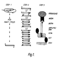
- Figure 1 shows the three basic steps of one embodiment of the ADA.
- a DNA segment is amplified from a biological sample via oligonucleotides a and b.
- specific ligands are incorporated into the amplified DNA segment through at least 3 further cycles of amplification via internally nested oligonucleotides.
- Oligonucleotide c. bears a molecule of biotin at its 5' terminus and oligonucleotide d. bears a 5' nucleotide sequence specifically recognised by the DNA binding protein, GCN4, of Saccharomyces cerevisiae.
- step 3 ligand bearing DNA segments are bound to a solid support coated with purified GCN4 produced in bacteria (GST-GCN4) and are detected via binding of avidin-peroxidase to the biotin of oligonucleotide c. and subsequent colorimetric detection of peroxidase activity.
- Figure 2 shows the structure of yeast GCN4 and GST-GCN4. At the top is the structure of the Saccharomyces cerevisiae GCN4 gene (9) with the coding region (281 amino acids) boxed and proposed transcription activation and DNA binding regions of the GCN4 protein indicated by hatching (10). Also indicated are the positions of oligonucleotides 1-3 used to amplify the GCN4 gene from yeast DNA by PCR.
- Fig.2 also shows the structure of genes encoding GST-GCN4 fusion proteins produced in E . coli by introducing fragments of the GCN4 gene into the plasmid expression vector pGEX-2T (11).
- the GCN4 gene was amplified from yeast DNA using oligonucleotides 2 and 3 or 1 and 3 to generate plasmids encoding partial (GST-GCN4 3.12) or full-length (GST-GCN4 6.8) versions of the GCN4 polypeptide fused to Schistosoma japonicum glutathione-S-transferase (GST).
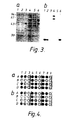
- Figure 3a shows fractionation by polyacrylamide gel electrophoresis of total proteins (lanes 1 and 4) from E .
- coli strain 7118 transfected with plasmids pGST-GCN4 3.12 (lanes 1-3) or pGST-GCN4 6.8 (lanes 4-6) and grown in the presence of 0.1 mM IPTG for 1 hour at 37°C. Also shown is material purified from lysed bacteria by one-step affinity chromatography (lanes 3 and 6) and soluble proteins remaining after incubation with glutathione-agarose beads (lanes 2 and 5).
- Figure 3b shows a gel retardation assay demonstrating that the mobility of a 32 P labelled DNA fragment containing a GCN4 binding site is decreased when it is mixed with purified GST-GCN4 3.12 (lanes 2 and 4) or GST-GCN4 6.8 (lane 6) in comparison to its mobility in the absence of protein (lane 1).
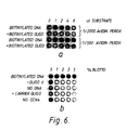
- Figure 4 shows the effect of added carrier DNA on the specificity of ADAs on DNA from HIV plasmid pHXBc2. In row A there was no added carrier DNA in the microtitre dish, while rows B, C and D contained 1 ⁇ g/ml, 0.1 ⁇ g/ml and 0.01 ⁇ g/ml of sonicated human DNA, respectively.
- Step 1 of the ADA was for 30 cycles as described in Example 1, below, using oligonucleotides a. and b. and a Sac 1 - Sal 1 fragment from pHXBc2 (12).
- Samples (5ml) from step 1 were then amplified for a further 10 cycles using oligonucleotides c. plus b. (columns 1-3), c. plus d. (columns 4-6) or c. plus d2 (columns 7-9).
- Samples (5 ⁇ l in 1, 4 and 7; 0.5 ⁇ l in 2, 5 and 8; 0.05 ⁇ l in 3, 6 and 9) were then added to wells of plates coated with purified GST-GCN4 3.12 (a) or GST-GCN4 6.8 (b).
- FIG. 1 shows ADAs on DNA from HIV-infected cells.
- Samples 1-3 human DNA ( ⁇ 100ng) from a Burkitt's lymphoma; samples 4-6, human DNA ( ⁇ 100ng) from HIV-infected cells; samples 7-9, no DNA; samples 10-12, DNA ( ⁇ 1ng) from plasmid pHXBc2.
- Step 1 of the ADA was for 35 cycles as described in Example 1 using oligonucleotides a. and b. Samples of 10 ⁇ l from step 1 were then amplified a further 6 cycles using oligonucleotides c. plus d.
- FIG. 6A shows the specificity of the one step ADA reaction. Competition of unreacted biotinylated oligonucleotides with the ADA substrate in a one step binding reaction.
- a PCR was performed using oligonucleotides cl and dl (0.2 ⁇ g) with 1ng of plasmid pHXBc2 in a 100 ⁇ l reaction mix, cycled 24 times.
- the control did not contain any plasmid DNA.
- the volumes of PCR reaction indicated were added to 50 ⁇ l of binding mix (without powdered milk) containing the dilutions of a 5 mg/ml avidin-peroxidase solution indicated.
- the volumes of PCR reaction indicated were added, and a further 8 ⁇ l of the control PCR was added to each well.
- the binding reactions and colour development are described in Example 3.
- Figure 6B shows the specificity of the one step binding reaction. The procedure was as for Fig. 6A, except that the wells contained the %(w/v) of non-fat powdered milk indicated.
- the top and bottom rows contained 5 ⁇ l/well of the PCR mix described above.
- oligonucleotide d was omitted.
- Row 3 there was no DNA in the PCR.
- Row 4 was as for row 3, but an unrelated oligonucleotide (1 ⁇ g/ml) was added.
- row 5 the wells were not coated with GCN4.
- Figure 7 shows the effect of annealing temperature on incorporation. PCRs were carried out with 1ng plasmid pHXBc2 and oligonucleotides al and bl (1 ⁇ g/ml), c2 and d2 (2 ⁇ g/ml) as indicated, and cycled under the conditions shown, 10 ⁇ l samples were fractionated on a 1.6% (w/v) agarose gel in the presence of 1 ⁇ g/ml ethidium bromide.
- a1 and b1 were 0.3 ⁇ g/ml.
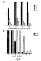
- Figure 8A shows the sensitivity with different oligonucleotide concentrations.
- Two step PCRs were carried out with oligonucleotides a2 and b2 (concentrations as indicated) and c2 and d2 (5 ⁇ g/ml) with the amounts of Plasmid indicated (at the right in ⁇ g) in a 20 ⁇ l reaction, cycled 30 times (95°/30 sec, 65°/60 sec) and then 12 times (95°/30 sec, 40°/60 sec, 65°/30 sec). 5 ⁇ l of the product was then analysed in an ADA with a one-step binding reaction.
- Figure 8B shows the sensitivity of ADA reactions. Comparison of the sensitivity of ADA reactions using a and b oligonucleotides with different spacings from c2 and d2.
- oligonucleotides were at 0.3 ⁇ g/ml.
- Figure 9 shows the ADA dependence on temperature shift.
- Two step PCRs were carried out with oligonucleotides a2, b2, c2 and d2 as in Fig. 3B.
- the DNA was from 5 x 10 3 HIV-infected CEM cells.
- the PCRs were cycled at 95°/30 sec, 65°/60 sec for the number of cycles indicated by the arrows, and then at 95°/30 sec, 40°/60 sec, 65°/30 sec for 0, 5 and 10 further cycles.
- the number of cycles indicated at the bottom is the total number for each sample.
- Figure 10 shows the detection of HIV in cultured cells.
- DNA from uninfected or HIV-infected CEM cells was used as the input DNA for PCR reactions containing oligonucleotides a2 and b2 (0.3 ⁇ g/ml) and c2 (2.5 ⁇ g/ml) and d2 (5 ⁇ g/ml) that were cycled 30 times (95°/30 sec, 65°/60 sec) followed by a further 10 (top 3 rows in right panel) or 15 (bottom 3 rows in right panel, and left panel) cycles (95°/30 sec, 40°/60 sec, 65°/30 sec). Plasmid DNA was used as a positive control. ADA reactions with a one-step binding reaction were carried out on 5 ⁇ l samples, and agarose gel electrophoresis on 10ml samples.
- the DNA samples analysed in the ADAs or by gel electrophoresis represented the material obtained from the number of cells indicated, or from the number of plasmid molecules indicated.
- Figure 11 shows the quantitation of the ADA reactions shown in Figure 10.
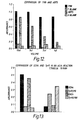
- Figure 12 shows the comparison of TMB and ABTS in an ADA mediated by GCN4-coated pins.
- Figure 13 shows a comparison of GCN4 and TyrR in an ADA reaction.
- Figure 14 shows the effect of thrombin cleavage of GCN4 in the ADA.
- Figure 15 shows the results of a clinical trial performed on Peripheral Blood Lymphocytes (PBL's) taken from patients positively diagnosed as suffering from AIDS or from negative controls. The method is as described in Figure 10 except 2 PCR's, 35 and 12 cycles respectively were used. Abbreviations are defined in Example 9.
- target DNA is first amplified by PCR using a first set of appropriate oligonucleotide primers in accordance with the known PCR procedure and then a second set of oligonucleotide primers, nested between the first two, are incorporated by a small number of additional cycles.
- the nucleotides in the second set of primers bear ligands; one can, for example, be biotinylated, and the other contains a site for a double stranded DNA binding protein.
- an immobilised affinity reagent such as a DNA binding protein and labelling with a second affinity reagent, for example avidin linked to horseradish peroxidase
- reaction with a chromogenic substrate allows detection of the amplified DNA.
- a system such as digoxigenin could be employed. Where there is sufficient target DNA without a first amplification or for other reason such as convenience or speed of assay, the target DNA may be directly subjected to incorporation by the labelled primers.
- target DNA any eukaryotic, prokaryotic or viral nucleic acid sequence and includes the identification of pathogens or the screening of human or other mammalian genetic disorders such as in cancer cells.
- target DNA encompasses RNA wherein by the action of reverse transcriptase, corresponding DNA is first synthesised, i.e. cDNA copied by reverse transcriptase from RNA.
- Target DNA also extends, therefore, to RNA viruses.
- Target DNA also extends to plant genetic sequences and to their pathogens.
- target DNA may vary depending on the particular circumstances and relative convenience.
- one embodiment of the subject invention is described in terms of detecting HIV sequences in blood.
- these and other target sequences may be isolated from other bodily fluids such as, but not limited to, saliva.
- the present invention extends to the detection of target DNA in any suitable biological fluid such as blood, saliva, lymph fluid, cell and tissue extracts, culture supernatants, plant sap and/or other fluids or tissue extracts, aerosols, various environmental locations (eg soil, water, etc.) and the like.
- the ADA procedure of this invention provides a very sensitive, specific, simple and convenient method for detecting specific DNA segments amplified by at least one PCR.
- the sensitivity of the method results from the combination of the inherent sensitivity of the PCR procedure itself (it can detect a single DNA molecule against a background of at least 10 6 human genomes (2)) and a sensitive novel method for detecting the amplified DNA.
- the data below show that molecules of the ligand-containing amplified DNA can readily be detected and only a small fraction of the product from a typical PCR reaction is necessary for detection.
- the specificity of the procedure reflects the fact that the ADA, in one embodiment, uses two successive PCR reactions with nested oligonucleotide primers. Only DNA molecules generated in the second step are detected in the final step because the ligands are only introduced during the second step. Alternatively, the target DNA may undergo the binding step directly without need of a first PCR. This specificity may be further increased, as in Example 2 herein, by the fact that GST-GCN4 only binds to double-stranded DNA - it does not recognise the single-stranded oligonucleotides.
- the second step utilises only a small number of cycles (for example 3-12 cycles), there is insufficient time for accumulation of significant amounts of primer-dimers derived from the oligonucleotides of the second set or other double stranded DNA artefacts.
- any such artefacts generated in the first PCR step for example by spurious priming at other places in the genome, are not amplified in the second step because they will not contain the nested sequences of the second set of primers.
- Primer-dimers formed in the first step will not be detected as they do not contain the ligands.
- PCR can be performed in one reaction mixture effectively resulting in a "single step ADA".
- This modification to the multi-step procedure previously outlined is predicated in part on a strong dependence of the thermal stability of an oligonucleotide duplex on its length and hence, oligonucleotide primers can be selected such that their incorporation in a PCR is critically dependent on the annealing temperature. Consequently, if one set of primers is considerably longer than a second set, then successive PCR reactions can be carried out in the one reaction mixture by incubating the mixture through first a high temperature and then a low temperature thermal cycle regime. (See Example 5).
- the present invention therefore, extends to both the multi-step and single step ADA.
- the one step ADA also has advantages in the binding step where the binding of the amplified product to the double-stranded DNA-binding protein immobilized to a solid substance occurs simultaneously to the binding of or to a detection complex.
- the amplified DNA is bound to GST-GCN4 immobilized in the wells of a microtitre dish while simultaneously binding to the avidin-peroxidase conjugate.
- a further aspect of the one step ADA relates to the use of single or multiple beads or pins coated with a double-stranded DNA-binding protein to transfer the amplified product from a reaction vessel, after washing and contacting immobilized amplified product to a detection complex, to detection substrate.
- the amplified DNA is transferred from a microtiter well by an array of GST-GCN4-coated beads or pins and, after washing and contacting with avidin-peroxidase, the beads are immersed in a microtiter dish containing ABTS substrate.
- the successive PCR reactions can be carried out in the one reaction mixture, simply by incubating the mixture through first a high temperature and then a low temperature thermal cycle regime.
- the protocol becomes greatly simplified, namely (1) the DNA sample is added to the PCR mixture, a drop of paraffin oil is added and the tube is placed on a thermal cycler and subjected to the two successive thermal regimes; (2) a sample is then placed in the GST-GCN4-coated microtiter well for simultaneous immobilization and binding; and (3) the dish is then washed and substrate added.
- This protocol is well suited to handle moderate numbers of samples. For example, the results for 50 samples can be obtained about 1 hour after completion of the PCRs.
- the amplified DNA binds to GST-GCN4 immobilized in the wells of a microtiter dish while simultaneously binding to the avidin-peroxidase conjugate. This decreases both the number of manipulations required and the time taken in handling samples, with no decrease in sensitivity or specificity.
- unincorporated biotinylated oligonucleotides compete with the amplified DNA for binding to avidin and it is necessary to ensure that the amount of biotin does not exceed the binding capacity of the avidin.
- the one step assay also provides a protocol where the PCR with two successive thermal regimes is itself performed in a modified microtiter dish.
- the amplified DNA molecules are then bound to GCN4 immobilized on polystyrene beads attached to the lid of a microtiter dish. While this procedure cannot take advantage of the simultaneous immobilization and avidin-peroxidase binding, it has the very considerable advantage that after pipetting individual DNA samples into the first microtiter well, 96 samples can be handled simultaneously in a manner analogous to the widely used "FAST ELISA" system.
- the sample may be treated in precisely the same manner as a routine enzyme-linked immunosorbent assay (ELISA), using the same equipment.
- ELISA enzyme-linked immunosorbent assay
- the ADA system described in detail herein is only one possible formulation that has many alternatives. Obviously, the approach could be used for detection of many other viral, bacterial, protozoan, fungal and mycoplasmal pathogens. Screening for hepatitis, tuberculosis, malaria and candida infections are among the obvious applications involving these disparate organisms. Similarly, this system could be used for the detection of cellular disorders such as cancers and the like.
- the outstandingly useful feature of the ADA approach is that it is only necessary to change the sequences of the oligonucleotides in order to detect any gene from any organism by a simple colour test.
- the kinetics of the detection steps should be identical as the same affinity reagents are interacting with the same ligands in all cases. This contrasts with the ELISA system where the affinity and kinetics are determined by the monoclonal antibodies, which differ for each situation.
- Another potential application lies in determining the genotypes of certain pathogens. For example, in Plasmodium falciparum, some genes contain variable regions defining different antigenic determinants surrounded by relatively conserved regions (13).
- the products of the first PCR step could be tested with several pairs of oligonucleotides corresponding to internal variable regions that define the different serotypes.
- the ADA described herein is also applicable to screening for genetic diseases such as cystic fibrosis and cancers amongst others.
- the double-stranded DNA - specific DNA binding protein is of the leucine zipper type i.e. GCN4.
- a range of other DNA binding proteins of this type could be used, including thrombin-cleaved GCN4 ( Figure 2, 14).
- the present invention extends to leucine zipper type DNA binding proteins such as GCN4 and/or its derivatives which includes GST-GCN4, thrombin-cleaved GCN4 and any other modifications thereof such as additions, deletion and/or substitution to the GCN4 amino acid and/or carbohydrate moieties provided said derivatives retain DNA binding activity
- DNA binding protein that could be used in the ADA is the TyrR protein of the "Helix turn Helix” type and which has a C-terminal DNA binding domain (Dr V. Argyropolous, Thesis submitted for degree of Doctor of Philosophy, The University of Melbourne, Parkville, Victoria, Australia).
- Other DNA binding proteins which may be used in the ADA are well known and include, for example, the "zinc finger” type. These binding proteins are reviewed by Struhl (19).
- the ADA system could be used with appropriate modification for virtually any application amenable to PCR itself (see for example references (1) to (8) and the Australian Patent Applications cited above).
- Major examples would include human genetics applications such as HLA typing and prenatal diagnosis of genetic disorders. The simplicity, specificity and generality of the approach should find many other applications.
- Another embodiment of the subject invention employs a single PCR to incorporate directly label and/or ligand bound primers into target DNA without first being amplified and then exposing labelled target DNA to the solid support prior to, or simultaneously to, detection. This provides an even more streamlined method of detecting target DNA and makes the first PCR an optional step depending on the circumstances.
- the present invention also extends to a conjugate consisting essentially of a support, GST-GCN4 immobilized on said support and an amplified double-stranded DNA bound at a first end to said GST-GCN4.
- the conjugate may further comprise at the second end of said double-stranded DNA, a label and preferably said label is an enzyme.
- the label is conjugated to the amplified DNA through an avidin-biotin bridge.
- the complete coding region of the GCN4 gene from S. cerevisiae (9) was synthesized by PCR on a crude DNA preparation using oligo 1 (GGAATTCTAATGTCCGAATATCAGCCA) and oligo 3 (GGAATTCAGCGTTCGCCAACTAATTTC) of GCN4, and incorporating Eco R1 sites at their 5' terminii (Fig.2). After cleaving with EcoRl, the DNA was ligated to Eco R1 cut DNA of the expression vector pGEX-2T (11).
- GCN4 A smaller portion of GCN4 was also isolated by PCR using oligo 3 above and oligo 2 (CGGATCCATGTTTGAGTATGAAAACC) containing a Bam H1 site at the 5' terminus (Fig.2) and insertion of the PCR product after cleavage with Bam H1 and Eco R1 into Bam H1 and Eco R1 cut pGEX-2T DNA.
- PCR reactions for amplification of p24 sequences from DNA isolated from HIV infected cells contained 50mM KCl, 10mM Tris pH 8.4, 2.5mM Mg Cl 2 , 0.25mM each dATP, dCTP, dGTP and dTTP, 0.01% gelatin, 1.5 units Taq DNA polymerase (Cetus), 4ng oligonucleotide primers a. and b. and 100ng purified DNA. Reaction mixes (100ml) were cycled approximately 30 times between 40°C, 70°C and 95°C for 1.5, 2.0 and 1.5 minutes respectively.
- Step 1 PCR reaction One-tenth of a Step 1 PCR reaction was subjected to at least 3 additional cycles of PCR under identical conditions except that the primers used were oligonucleotides b. and c., c. and d. or c. and d1.
- sequences (12) of the oligonucleotides corresponding to the p24 gene of HIV used were:
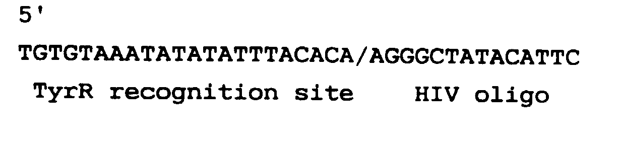
- Oligonucleotide c. was biotinylated at the 5' end. (nucleotides 5' to the slash correspond to an artificial GCN4 binding site (14), while those 3' to the slash correspond to positions 1641-1660). (nucleotides 5' to the slash correspond to an artificial GCN4 binding site (14), while those 3' to the slash correspond to positions 1641-1660).
- CEM cells were derived by culturing human peripheral blood cells from a patient with acute lymphoblastic leukaemia, and then infected with HTLV IIIb. DNA was purified using guanidine HCl and CsCl centrifugation.
- Microtiter trays (Dynatech Laboratories Inc.) were coated with purified GST-GCN4 fusion polypeptides at approximately l ⁇ g/ml in mouse tonicity phosphate-buffered saline (MTPBS) for 3 hours at 37°C (50 ⁇ l per well) and then blocked with 1% (w/v) bovine serum albumin (fraction V) (Flow Laboratories) in MTPBS for 1 hour at 37°C.
- MTPBS mouse tonicity phosphate-buffered saline
- MTPBS-Tw-20 MTPBS containing 0.05% (v/v) Tween-20, Trade Mark (MTPBS-Tw-20) and twice with MTPBS alone, before incubation at 20°C for 30 minutes with 50 ⁇ l of ligand bearing DNA diluted in MTPBS-Tw-20. Trays were washed as before and incubated again at 20°C for 30 minutes with 50 ⁇ l horseradish peroxidase -avidin D conjugate (Vector Laboratories, Inc.) at a concentration of 2.5 ⁇ g/ml in MTPBS-Tw-20.
- the approach to the detection of specifically amplified DNA is outlined in Figure 1.
- the ADA consists of three basic steps, the first two of which are different PCR reactions performed successively. A sample of the amplified DNA is then placed in a well of a microtitre dish for detection. As described above, there are many different possible permutations of the ADA of varying specificity and simplicity. For the sake of clarity, only one example of the theoretically most specific formulation is described in this section.
- Step 1 Amplification.
- Step 1 is simply a standard PCR reaction performed on any suitable DNA-containing extract relevant to the sequence of interest, for a large number of cycles.
- the oligonucleotides for step 1 (designated a and b in Figure 1) are limiting for this reaction.
- Step 1 simply amplifies the desired segment of DNA.
- Step 2 Sequence-specific ligand incorporation.
- This step achieves specificity and simultaneously incorporates ligands into the PCR products that can react with affinity reagents, and thereby be detected in step 3.
- two new oligonucleotides (designated c and d in Fig.1) are used for a second PCR reaction that can be cycled for as little as three cycles. This achieves specificity because oligonucleotides c and d are nested between oligonucleotides a and b. There are only a small number of cycles and therefore the only molecules that will form to a detectable extent are those generated by amplification of the correct sequence in step 1.
- Step 2 also incorporates the ligands.
- oligonucleotide c which is biotinylated
- oligonucleotide d which contains extra sequences encoding the recognition site for a double-stranded DNA binding protein, such as the yeast regulatory protein GCN4 (14).
- step 2 At least three cycles of step 2 are necessary to generate blunt-ended molecules with these ligands at both ends (Fig.1).
- This step attaches the amplified DNA to a solid phase by affinity binding to a double-stranded DNA-binding protein at one end, and is followed after washing by attachment of an enzyme by affinity binding at the other end for subsequent colour generation.
- a sample from step 2 is added to a well of a microtitre dish.
- the well has been pre-coated with the DNA-binding protein, for example a cloned fused polypeptide bearing DNA binding protein GCN4 (see below).
- GCN4 DNA binding protein
- This polypeptide specifically immobilises the amplified molecules because of its affinity for double stranded DNA containing the correct sequence, incorporated via oligonucleotide d.
- a solution of the other affinity reagent conjugated to an enzyme for example avidin linked to horseradish peroxidase, is added. This binds to the biotin linked to oligonucleotide c.
- a chromogenic substrate is added to the microtitre dish, allowed to develop and the absorbance is read in a microtitre-plate reader.
- Saccharomyces cerevisiae regulatory protein GCN4 has been expressed as a glutathione-S-transferase (GST) fusion protein as shown in Figure 2.
- GST glutathione-S-transferase
- Plasmid pGST-GCN4-3.12 contains most of the sequence of GCN4 from Saccharomyces cerevisiae, including the C-terminal DNA-binding region, inserted into the plasmid pGEX-2T (11), while the plasmid pGST-GCN4-6.8 contains the entire coding sequence of GCN4.
- the GST-GCN4 fused polypeptide contains the entire sequence of glutathione-S-transferase (GST) from Schistosoma japonicum, which allows purification of the molecule in one simple affinity step by binding to glutathione-agarose beads (11).
- GST-GCN4 polypeptides are abundant in Escherichia coli clones transformed with these plasmids.
- affinity purification each of the GST-GCN4 polypeptides was detected as two Coomassie-blue stained bands after polyacrylamide gel electrophoresis (Fig.3a). These purified proteins retain the ability to bind to the consensus GCN4 binding sequence as revealed by a gel retardation assay (Fig.3b).
- ADA Human Immunodeficiency Virus
- the fused polypeptide from clone GST-GCN4 3.12 (16) was purified by binding to glutathione-agarose as described (11).
- TyrR a DNA binding protein of the "Helix turn Helix” type, and which has a C-terminal DNA binding domain was provided for testing by Dr V. Argyropolous (18).
- PCR reactions for amplification of p24 sequences of HIV contained 50mM KCl, lOmM Tris pH 8.4, 2.5mM MgCl 2 0.25mM each dNTP, Taq polymerase (0.5 unit) and oligonucleotide primers at various concentrations. Reaction mixes (20 ⁇ l) were incubated under paraffin oil using the conditions described below. For routine use, the PCR mixes containing all components except DNA were stored as frozen aliquots.
- the plates were then drained, but not washed, and 50 ⁇ l/well of a mixture containing 10% (w/v) non-fat powdered milk in PBS, 4 ⁇ g/ml sonicated salmon DNA, 0.05% (v/v) Tween-20 and 50 ⁇ g/ml horseradish peroxidase-avidin D conjugate (Vector Laboratories Inc.) in PBS was added. Samples of the PCR reactions (1-10 ⁇ l) were then added and allowed to react for at least 20 min at RT.
- the beads on the lid of a "FAST ELISA" dish (Falcon plastics) with the corners cut off were coated with GST-GCN4 by placing them in 50 ⁇ l aliquots of GST-GCN4-PBS in a microtiter tray (Dynatech Laboratories Inc.) as above for 1 hr at 37°C, and then blocked in a solution containing 10% (w/v) non-fat powdered milk, 4 ⁇ g/ml salmon DNA and 0.05% (v/v) Tween-20 in MTPBS. The lid was then flicked to drain off excess solution and the beads placed in the microtiter dish containing the PCR samples.
- the beads were washed with PBS-0.05% (v/v) Tween 20. They were then reacted with 10 ⁇ g/ml peroxidase-avidin conjugate in 10% (w/v) powdered milk, 0.05% (v/v) Tween-20 in MTPBS for 20 min. They were then washed extensively with PBS and reacted with ABTS as above. Alternatively, they were reacted with 0.4mM Tetramethylbenzidine (TMB) in 0.1M NaAc, pH 5.5 plus 1.41mM hydrogen peroxide and read in a Titertek Multiskan MCC/340 scanner on mode 1 using filter number 7.
- TMB Tetramethylbenzidine
- Consensus oligonucleotides corresponding to sequences from the gag gene of HIV were selected after aligning available sequences from the HIV database.
- the oligonucleotides synthesized were:
- CEM cells were derived by culturing human peripheral blood cells from a patient with acute lymphoblastic leukaemia, and then infected with HIV isolate HTLV IIIb. DNA was purified using guanidine HC1 and CsC1 centrifugation.
- DNA was purified from peripheral blood leucocytes using guanidine HCl and phenol/chloroform/ethanol centrifugation.
- Plasmid pHXBc2 (12) encoding the GAG gene of HIV was used as a source of DNA for developing the reactions. Generally, 1 ⁇ l of a 1 ⁇ g/ml solution per 1000ml of PCR was used.
- the amplified DNA was first captured on GST-GCN4 immobilized in a microtiter well, the unincorporated substrates washed away and then avidin-peroxidase bound to the amplified DNA molecules.
- the amplified DNA was mixed with the avidin-peroxidase conjugate in the presence of protein and DNA carriers and these were bound to the immobilized GST-GCN4 in a single reaction mixture.
- oligonucleotides a, b, c and d are considerably longer than c and d so that they form duplexes that are considerably more stable than those of c and d, then annealing at a sufficiently high temperature should prevent incorporation of c and d, allowing separation of the reactions in a mixture containing all four oligonucleotides.
- Figure 7B shows that when the amounts of a1 and b1 were decreased sufficiently, the expected c2-d2 product could indeed be obtained when cycled with annealing. As expected, only the a1-b1 product was formed without annealing, and the amounts of this were not greatly affected by lowering the concentrations of a1 and b1 (Fig 7B, left panel).
- ADAs on the products of these reactions were also performed (data not shown).
- the amplified DNA In order for the amplified DNA to function in such an assay, it must contain a biotin moiety at one end and a GCN4 binding site at the other end. Only those reaction mixtures containing the short c-d product gave a significant colour reaction in the ADAs, confirming the structure of these molecules.
- Taq polymerase has a 5'->3' exonuclease activity and so it can translate nicks. These effects should all be lowered if the spacing of oligonucleotides a/c and b/d are increased. Accordingly, oligonucleotides a2 and b2 were synthesized corresponding to conserved positions considerably further away from c2 and d2 than are al and b1.
- oligonucleotides c2 and d2 were dependent on the concentration of oligonucleotides a2 and b2 (Fig 8a).
- the c2-d2 product could be detected either by an ADA reaction or by EtBr staining from about 100 fold less input plasmid DNA than with oligonucleotides a1 and b1 (Fig 8b).
- Production of the c2-d2 product and colour intensity in an ADA was dependent on the number of cycles both without and with annealing and there was no significant c2-d2 product without annealing, even after 40 cycles (Fig 9).
- HIV sequences could be detected in the DNA obtained from about 250 cells from an HIV-infected CEM culture while there was no significant background even with 100-fold more DNA from uninfected cells (Figs 10 and 11). It can be seen on the gel shown in Fig 10 that the two successive reactions with nested oligonucleotides are indeed vital to the specificity - there are many bands generated from uninfected DNA, but these do not register as positive in the ADA.
- One way to further simplify the ADA system is to perform the PCRs in a microtiter dish and then to capture and transfer the amplified DNA from each of the 96 wells to a second dish using an array of GST-GCN4-coated beads or pins.
- 1-10 ⁇ l samples of a PCR reaction performed with oligonucleotides c2 and d2 were made up to 20 ⁇ l in a microtiter dish and covered with a drop of paraffin oil.
- the beads of a FAST ELISA screening plate were coated with GST-GCN4, blocked with powdered milk-DNA and then immersed in the PCR samples for 20 min.
- the beads were washed, exposed to avidin-peroxidase, washed and placed in a microtiter dish containing ABTS substrate.
- the responses obtained were proportional to the amount of amplified DNA (Fig 12), and there was negligible background from equal amounts of a PCR mix incubated without substrate DNA.
- the colour intensity was lower by a factor of 2-3 than reactions performed using GST-GCN4-coated wells with the same material (data not shown) reflecting the lower surface area of beads.
- the sensitivity could be increased approximately 10-fold using TMB as the substrate, without any significant increase in background (Fig 12).
- amplified DNA molecules can be captured and transferred efficiently using GCN4 coated beads. This is surprising given that the coated beads were first dipped through a layer of paraffin oil, mimicking the conditions necessary for a PCR.
- PCR reactions could be performed in a microtiter dish and then transferred as above, reactions with oligonucleotides a1 and b1, c2 and d2 or all four oligonucleotides were set up as for Fig 2 and incubated in the wells of a flexible microtiter dish mounted on a hollow aluminum block through which water at the appropriate temperature was circulated. The top of the block was milled to fit the bottom of the dish and zinc oxide heat-sink cream was used to ensure thermal contact. Evaporation was prevented by a drop of paraffin oil. After 24 cycles with a 40°C annealing step, oligonucleotides a1-b1 and c2-d2 were incorporated into products of the expected size. Furthermore, the c2-d2 product gave an ADA reaction as expected (data not shown).
- a set of HIV-oligonucleotides with a GST-GCN4 site could be included in the same mix as a set of hepatitis B viral oligonucleotides marked with a second DNA binding protein site, so each could be read specifically from the one PCR.
- TyrR is a DNA binding protein of the "Helix turn Helix” type, and which has a C-terminal DNA binding domain was provided for testing by Dr V. Argyropolous (18).
- An oligonucleotide probe was manufactured which contained a TyrR recognition site and a HIV sequence, i.e. corresponding to the oligo "d" described in Example 3 except that the TyrR recognition site replaced the GCN4 binding site.
- the probe was of the sequence
- This probe was incorporated into the ADA test with the oligo "c" and reaction products tested on plates coated with GST-GCN4 or TyrR.
- PBL's Peripheral Blood Lymphocytes
- the PBL's were prepared from blood samples by lysis in Guanidine thiocyanate buffer (4M) and centrifugation.
- DNA was extracted and purified from PBL's using the same technique of guanidine thiocyanate centrifugation.
- Samples 20 and 46 were obtained from healthy humans.
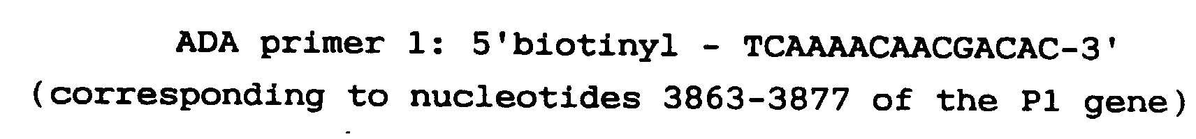
- the ADA can be used to detect DNA from M. pneumoniae, a pathogen which can cause severe respiratory tract infections.
- the first experiment used plasmid DNA containing the P1 gene of M. pneumoniae as described in Hu et al, Gene, 64 ,217, and one pair of ADA primers.
- the second experiment used clinical specimens (nasopharyngeal aspirate) to which had been added whole M. pneumoniae cells, and two nested pairs of primers.
Abstract
Description
in said first container or in a second container, reagents for a polymerase chain reaction, a solid substrate coated with the double-stranded DNA-specific binding protein, and a means for detecting amplified double-stranded DNA bound to said solid substrate.
- PCR
- Polymerase chain reaction
- DNA
- Deoxyribonucleic acid
- A
- Adenine
- T
- Thymine
- G
- Guanine
- C
- Cytosine
- GST
- Glutathione-S-transferase
- ADA
- Amplified DNA assay
- HIV
- Human immunodeficiency virus
- TMB
- Tetramethylbenzidine
- ABTS
- 2.2'-azino-bis-(3-ethylbenzthiazoline-6-sulphonic acid)
- MTPBS
- Mouse tonicity phosphate buffered saline
- RT
- Room temperature
- PBS
- Phosphate buffered saline
Figure 1 shows the three basic steps of one embodiment of the ADA. In
Figure 2 shows the structure of yeast GCN4 and GST-GCN4. At the top is the structure of the Saccharomyces cerevisiae GCN4 gene (9) with the coding region (281 amino acids) boxed and proposed transcription activation and DNA binding regions of the GCN4 protein indicated by hatching (10). Also indicated are the positions of oligonucleotides 1-3 used to amplify the GCN4 gene from yeast DNA by PCR. Fig.2 also shows the structure of genes encoding GST-GCN4 fusion proteins produced in E. coli by introducing fragments of the GCN4 gene into the plasmid expression vector pGEX-2T (11). The GCN4 gene was amplified from yeast
Figure 3a shows fractionation by polyacrylamide gel electrophoresis of total proteins (
Figure 4 shows the effect of added carrier DNA on the specificity of ADAs on DNA from HIV plasmid pHXBc2. In row A there was no added carrier DNA in the microtitre dish, while rows B, C and D contained 1µg/ml, 0.1 µg/ml and 0.01 µg/ml of sonicated human DNA, respectively.
Figure 5 shows ADAs on DNA from HIV-infected cells. Samples 1-3, human DNA (~100ng) from a Burkitt's lymphoma; samples 4-6, human DNA (~100ng) from HIV-infected cells; samples 7-9, no DNA; samples 10-12, DNA (~1ng) from plasmid pHXBc2.
Figure 6A shows the specificity of the one step ADA reaction. Competition of unreacted biotinylated oligonucleotides with the ADA substrate in a one step binding reaction. A PCR was performed using oligonucleotides cl and dl (0.2µg) with 1ng of plasmid pHXBc2 in a 100µl reaction mix, cycled 24 times. The control did not contain any plasmid DNA. For
Figure 6B shows the specificity of the one step binding reaction. The procedure was as for Fig. 6A, except that the wells contained the %(w/v) of non-fat powdered milk indicated. The top and bottom rows contained 5µl/well of the PCR mix described above. For
Figure 7 shows the effect of annealing temperature on incorporation. PCRs were carried out with 1ng plasmid pHXBc2 and oligonucleotides al and bl (1µg/ml), c2 and d2 (2µg/ml) as indicated, and cycled under the conditions shown, 10µl samples were fractionated on a 1.6% (w/v) agarose gel in the presence of 1µg/ml ethidium bromide. For A the concentrations of a1 and b1 were 0.3µg/ml. For B the concentrations of oligonucleotides a1 and c1 in the PCR were the following in successive tracks: a1= 6, 6, 2, 0.6, 2 µg/ml, b1= 3 µg/ml, c1= 20, 30, 10, 10, 20 µg/ml, d1= 5 µg/ml.
Figure 8A shows the sensitivity with different oligonucleotide concentrations. Two step PCRs were carried out with oligonucleotides a2 and b2 (concentrations as indicated) and c2 and d2 (5µg/ml) with the amounts of Plasmid indicated (at the right in µg) in a 20µl reaction, cycled 30 times (95°/30 sec, 65°/60 sec) and then 12 times (95°/30 sec, 40°/60 sec, 65°/30 sec). 5µl of the product was then analysed in an ADA with a one-step binding reaction.
Figure 8B shows the sensitivity of ADA reactions. Comparison of the sensitivity of ADA reactions using a and b oligonucleotides with different spacings from c2 and d2. The a and b oligonucleotides were at 0.3µg/ml.
Figure 9 shows the ADA dependence on temperature shift. Two step PCRs were carried out with oligonucleotides a2, b2, c2 and d2 as in Fig. 3B. The DNA was from 5 x 10³ HIV-infected CEM cells. The PCRs were cycled at 95°/30 sec, 65°/60 sec for the number of cycles indicated by the arrows, and then at 95°/30 sec, 40°/60 sec, 65°/30 sec for 0, 5 and 10 further cycles. The number of cycles indicated at the bottom is the total number for each sample.
Figure 10 shows the detection of HIV in cultured cells. DNA from uninfected or HIV-infected CEM cells was used as the input DNA for PCR reactions containing oligonucleotides a2 and b2 (0.3µg/ml) and c2 (2.5µg/ml) and d2 (5µg/ml) that were cycled 30 times (95°/30 sec, 65°/60 sec) followed by a further 10 (top 3 rows in right panel) or 15 (bottom 3 rows in right panel, and left panel) cycles (95°/30 sec, 40°/60 sec, 65°/30 sec). Plasmid DNA was used as a positive control. ADA reactions with a one-step binding reaction were carried out on 5µl samples, and agarose gel electrophoresis on 10ml samples. The DNA samples analysed in the ADAs or by gel electrophoresis represented the material obtained from the number of cells indicated, or from the number of plasmid molecules indicated.
Figure 11 shows the quantitation of the ADA reactions shown in Figure 10.
Figure 12 shows the comparison of TMB and ABTS in an ADA mediated by GCN4-coated pins.
Figure 13 shows a comparison of GCN4 and TyrR in an ADA reaction.
Figure 14 shows the effect of thrombin cleavage of GCN4 in the ADA.
Figure 15 shows the results of a clinical trial performed on Peripheral Blood Lymphocytes (PBL's) taken from patients positively diagnosed as suffering from AIDS or from negative controls. The method is as described in Figure 10 except 2 PCR's, 35 and 12 cycles respectively were used. Abbreviations are defined in Example 9.
(CCACCTAGCGGATGACTCATTTTTTTTCTTAGCG and
CGCTAAGAAAAAAAATGAGTC) and incubating them with Taq DNA polymerase (Cetus) in a reaction mix identical to that used for PCR except that dATP was replaced by 20mCi a32P-dATP (Amersham). After 5 minutes at 70°C, dATP was added to 0.25mM and the incubation continued for 5 minutes. Unincorporated a32P-dATP was removed by passage through a Sephadex G-10 (Trade Mark) spin column.


- -
- No DNA
- DO
- Human DNA (negative control)
- CC
- Positive control using CEM cells transfected with HIV
- CP
- Positive control using Plasmid DNA
- +
- Positive control using CEM cells having Plasmid DNA incorporated therein.
- 20-55
- Clinical specimens


| Experiment (i) | |
| Amount of plasmid DNA | Result |
| 56ng (7x10⁸ molecules) P1 plasmid: | ++ |
| 5.6ng (6x10⁷ molecules) P1 plasmid: | + |
| No plasmid control | - |
| Experiment (ii) | |
| Approx no. of M. pneumoniae genomes | Result |
| 5x10⁸ | +++ |
| 2.5x10⁸ | + |
| 108 | ++ |
| 107 | +/- |
| 5x10⁵ | - |
| 2.5x10⁵ | - |
| 105 | - |
| 104 | - |
| 0 | - |
Claims (21)
- A method for capturing and detecting target DNA which method comprises subjecting said target DNA to a polymerase chain reaction using a set of oligonucleotide primers selected to be complementary to the strands of said target DNA and wherein one of the primers of said set of oligonucleotides bears a first ligand containing a site for a double-stranded DNA-specific binding protein and the other of the primers bears a second ligand or a label, contacting the amplified double-stranded DNA with a solid substrate having the double-stranded DNA-specific DNA binding protein for said first ligand immobilized thereon and detecting the second ligand or label while the amplified double-stranded DNA is bound to said solid substrate to indicate the presence of the amplified double-stranded DNA.
- A method for capturing and detecting target DNA in a sample, which method comprises amplifying said target DNA, if present in said sample, by a polymerase chain reaction procedure using a first set of oligonucleotide primers selected to be complementary to the strands of said target DNA, and detecting the amplified tarqet DNA by a method according to claim 1 wherein the set of primers employed in said method according to claim 1 is nested between said first set of oligonucleotide primers.
- The method according to claim 2, wherein the first and second polymerase chain reactions occur in a single reaction mixture.
- The method according to any one of the preceding claims wherein the binding of the amplified DNA to the solid substrate occurs simultaneously to the binding to a detection complex.
- The method according to any one of the preceding claims wherein the double stranded DNA-specific DNA binding protein is of the leucine zipper type.
- The method according to claim 5 wherein the DNA binding protein is GCN4 and/or its derivatives.
- The method according to claim 6 wherein the DNA binding protein is GST-GCN4.
- The method according to any one of claims 1 to 4 wherein the double stranded DNA-specific DNA binding protein is of the helix turn helix type.
- The method according to claim 8, wherein the DNA binding protein is TyrR.
- The method according to any one of the preceding claims wherein the bound DNA is detected using a detection reagent comprising avidin-peroxidase.
- The method according to claim 10, wherein the binding moiety for the detection reagent is biotin.
- The method according to any one of the preceding claims wherein the target DNA is HIV DNA.
- The method according to any one of the preceding claims wherein the solid substrate coated with the double-stranded DNA-specific DNA binding protein is a well of a microtiter dish.
- The method according to any one of the preceding claims wherein the solid substrate coated with the double-stranded DNA-specific DNA-binding protein is a single or multiple bead or pin apparatus capable of transferring the amplified product with detection complex to a detection substrate.
- The method according to any one of the preceding claims wherein the target DNA is cDNA copied by reverse transcriptase from RNA.
- A test kit for capturing and detecting target DNA in a sample by the method according to any one of the preceding claims, said kit comprising in compartmental form, a first container containing a set of oligonucleotide primers selected to be complementary to the strands of said target DNA and wherein one of the primers of said set of oligonucleotides bears a first ligand containing a site for a double-stranded DNA-specific binding protein and the other of the said primers bears a second ligand or a label; and, in said first container or in a second container, reagents for a polymerase chain reaction, a solid substrate coated with the double-stranded DNA-specific binding protein, and a means for detecting amplified double-stranded DNA bound to said solid substrate.
- A test kit according to claim 16 further comprising a first set of primers capable of amplifying said target DNA, wherein during polymerase chain reaction said set of primers as defined in claim 16 is nested between said first set of primers.
- A conjugate for use in a method according to any one of claims 1 to 7 consisting essentially of a support, GST-GCN4 immobilized on said support, and an amplified double-stranded DNA bound at a first end to said GST-GCN4.
- The conjugate according to claim 18, wherein at a second end the DNA is conjugated to a label.
- The conjugate according to claim 19, wherein the label is an enzyme.
- The conjugate according to claim 20 wherein the label is conjugated to the amplified DNA through an avidin-biotin bridge.
Applications Claiming Priority (5)
| Application Number | Priority Date | Filing Date | Title |
|---|---|---|---|
| AUPJ188988 | 1988-12-09 | ||
| AU18/89 | 1988-12-09 | ||
| AUPJ508089 | 1989-07-04 | ||
| AU5080/89 | 1989-07-04 | ||
| PCT/AU1989/000526 WO1990006374A1 (en) | 1988-12-09 | 1989-12-08 | Amplified dna assay |
Publications (3)
| Publication Number | Publication Date |
|---|---|
| EP0447464A1 EP0447464A1 (en) | 1991-09-25 |
| EP0447464A4 EP0447464A4 (en) | 1992-06-03 |
| EP0447464B1 true EP0447464B1 (en) | 1998-08-05 |
Family
ID=25643595
Family Applications (1)
| Application Number | Title | Priority Date | Filing Date |
|---|---|---|---|
| EP90900722A Expired - Lifetime EP0447464B1 (en) | 1988-12-09 | 1989-12-08 | Amplified dna assay |
Country Status (11)
| Country | Link |
|---|---|
| EP (1) | EP0447464B1 (en) |
| JP (1) | JP3068848B2 (en) |
| AT (1) | ATE169341T1 (en) |
| AU (1) | AU633036B2 (en) |
| CA (1) | CA2004990A1 (en) |
| DE (1) | DE68928769T2 (en) |
| DK (1) | DK108091A (en) |
| ES (1) | ES2121750T3 (en) |
| HK (1) | HK1009705A1 (en) |
| NO (1) | NO912169L (en) |
| WO (1) | WO1990006374A1 (en) |
Families Citing this family (17)
| Publication number | Priority date | Publication date | Assignee | Title |
|---|---|---|---|---|
| US5753433A (en) * | 1909-12-05 | 1998-05-19 | Boehringer Mannheim Gmbh | Method for the sensitive detection of nucleic acids |
| AU629845B2 (en) * | 1988-08-30 | 1992-10-15 | Abbott Laboratories | Detection and amplification of target nucleic acid sequences |
| US5858652A (en) * | 1988-08-30 | 1999-01-12 | Abbott Laboratories | Detection and amplification of target nucleic acid sequences |
| JPH03123500A (en) * | 1989-10-06 | 1991-05-27 | Canon Inc | Fractionation of nucleic acid |
| WO1991015599A1 (en) * | 1990-04-05 | 1991-10-17 | Amrad Corporation Limited | A method for detecting dna |
| WO1993009250A1 (en) * | 1991-11-01 | 1993-05-13 | Adelaide Children's Hospital | Solid phase amplification process |
| US5440025A (en) * | 1992-03-12 | 1995-08-08 | University Of Massachusetts At Lowell | Process for separating nucleic acid polymers |
| FR2694768B1 (en) * | 1992-07-30 | 1994-12-23 | Pasteur Institut | Oligonucleotide sequences for the specific detection of mollicutes by amplification of conserved genes. |
| US6037127A (en) * | 1994-03-31 | 2000-03-14 | E. I. Du Pont De Nemours And Company | Method for detection of non-denatured nucleic acid fragments |
| CA2145719C (en) * | 1994-04-18 | 1998-06-30 | James G. Nadeau | Detection of nucleic acid amplification |
| US5547861A (en) * | 1994-04-18 | 1996-08-20 | Becton, Dickinson And Company | Detection of nucleic acid amplification |
| US5563036A (en) * | 1994-04-29 | 1996-10-08 | Tularik, Inc. | Transcription factor-DNA binding assay |
| US6953663B1 (en) * | 1995-11-29 | 2005-10-11 | Affymetrix, Inc. | Polymorphism detection |
| SE9601318D0 (en) * | 1996-04-04 | 1996-04-04 | Pharmacia Biosensor Ab | Method for nucleic acid analysis |
| IT1284635B1 (en) * | 1996-04-24 | 1998-05-21 | Clonit Spa | METHOD FOR THE DETECTION OF AMPLIFIED NUCLEIC ACIDS AND KITS FOR ITS USE. |
| US6143496A (en) | 1997-04-17 | 2000-11-07 | Cytonix Corporation | Method of sampling, amplifying and quantifying segment of nucleic acid, polymerase chain reaction assembly having nanoliter-sized sample chambers, and method of filling assembly |
| JPWO2009034842A1 (en) * | 2007-09-11 | 2010-12-24 | 株式会社カネカ | Nucleic acid detection method and nucleic acid detection kit |
Family Cites Families (10)
| Publication number | Priority date | Publication date | Assignee | Title |
|---|---|---|---|---|
| US4863849A (en) * | 1985-07-18 | 1989-09-05 | New York Medical College | Automatable process for sequencing nucleotide |
| EP0224126A3 (en) * | 1985-11-25 | 1989-02-01 | The University of Calgary | Covalently linked complementary oligodeoxynucleotides as universal nucleic acid sequencing primer linkers |
| AU622104B2 (en) * | 1987-03-11 | 1992-04-02 | Sangtec Molecular Diagnostics Ab | Method of assaying of nucleic acids, a reagent combination and kit therefore |
| IL85551A0 (en) * | 1987-04-01 | 1988-08-31 | Miles Inc | Rapid hybridization assay and reagent system used therein |
| CA1317535C (en) * | 1987-06-30 | 1993-05-11 | Nanibhushan Dattagupta | Assay of sequences using amplified genes |
| CA1339351C (en) * | 1987-10-15 | 1997-08-26 | Michael S. Urdea | Nucleic acid multimers and amplified nucleic acid hybridization assays using same |
| AU2735988A (en) * | 1987-12-21 | 1989-07-13 | Amoco Corporation | Target and background capture methods with amplification for affinity assays |
| CA1340807C (en) * | 1988-02-24 | 1999-11-02 | Lawrence T. Malek | Nucleic acid amplification process |
| NO165894C (en) * | 1988-05-24 | 1991-04-24 | Gunnar Paulsen | METHOD OF ANALYSIS OF GENES. |
| US5091310A (en) * | 1988-09-23 | 1992-02-25 | Cetus Corporation | Structure-independent dna amplification by the polymerase chain reaction |
-
1989
- 1989-12-08 WO PCT/AU1989/000526 patent/WO1990006374A1/en active IP Right Grant
- 1989-12-08 AT AT90900722T patent/ATE169341T1/en not_active IP Right Cessation
- 1989-12-08 DE DE68928769T patent/DE68928769T2/en not_active Expired - Fee Related
- 1989-12-08 CA CA002004990A patent/CA2004990A1/en not_active Abandoned
- 1989-12-08 ES ES90900722T patent/ES2121750T3/en not_active Expired - Lifetime
- 1989-12-08 AU AU46637/89A patent/AU633036B2/en not_active Ceased
- 1989-12-08 EP EP90900722A patent/EP0447464B1/en not_active Expired - Lifetime
- 1989-12-08 JP JP2500923A patent/JP3068848B2/en not_active Expired - Lifetime
-
1991
- 1991-06-06 NO NO91912169A patent/NO912169L/en not_active Application Discontinuation
- 1991-06-07 DK DK108091A patent/DK108091A/en unknown
-
1998
- 1998-09-08 HK HK98110528A patent/HK1009705A1/en not_active IP Right Cessation
Non-Patent Citations (3)
| Title |
|---|
| CHEMICAL ABSTRACTS, vol. 109, no. 19, 07 November 1988, Columbus, OH (US); A. YAMANE et al., p. 219, no. 164992v/ * |
| Nucleic Acids Research, Symposium Series No. 20, 1988, p.91-92 * |
| SYVANEN et al., pp. 11327-11338/ * |
Also Published As
| Publication number | Publication date |
|---|---|
| HK1009705A1 (en) | 1999-06-04 |
| DK108091A (en) | 1991-08-07 |
| AU4663789A (en) | 1990-06-26 |
| NO912169L (en) | 1991-08-06 |
| EP0447464A1 (en) | 1991-09-25 |
| WO1990006374A1 (en) | 1990-06-14 |
| JPH04502251A (en) | 1992-04-23 |
| AU633036B2 (en) | 1993-01-21 |
| ES2121750T3 (en) | 1998-12-16 |
| DE68928769T2 (en) | 1999-04-01 |
| DK108091D0 (en) | 1991-06-07 |
| ATE169341T1 (en) | 1998-08-15 |
| DE68928769D1 (en) | 1998-09-10 |
| NO912169D0 (en) | 1991-06-06 |
| EP0447464A4 (en) | 1992-06-03 |
| CA2004990A1 (en) | 1990-06-09 |
| JP3068848B2 (en) | 2000-07-24 |
Similar Documents
| Publication | Publication Date | Title |
|---|---|---|
| US5536648A (en) | Amplified DNA assay using a double stranded DNA binding protein | |
| EP0447464B1 (en) | Amplified dna assay | |
| Kemp et al. | Colorimetric detection of specific DNA segments amplified by polymerase chain reactions. | |
| EP0592035B1 (en) | Thermostable DNA polymerase composition comprising a temperature sensitive polymerase inhibitor, diagnostic test kits and methods of use | |
| EP0519338B1 (en) | Improved methods for nucleic acid amplification | |
| JP2846018B2 (en) | Amplification and detection of nucleic acid sequences | |
| EP0702090B1 (en) | Coamplification of target nucleic acids using volume exclusion agent, in reaction composition, test kit and test device useful therefor | |
| EP0269445B1 (en) | Process for detecting HTLVI and HTLVII viruses by hybridization | |
| EP0511712B1 (en) | Nucleic acid amplification and detection methods using rapid polymerase chain reaction cycle | |
| JPH0813280B2 (en) | Method for detecting mutant nucleic acid | |
| JPH09503910A (en) | Oligonucleotides and methods for the detection of Trachoma chlamydia | |
| IE904029A1 (en) | Nucleic Acid Amplification Employing Ligatable Hairpin Probe¹and Transcription | |
| JP2692702B2 (en) | Method for detecting herpes simplex virus and reagent therefor | |
| AU741141B2 (en) | Specific and sensitive method for detecting nucleic acids | |
| EP0533906A4 (en) | Detection of dna/rna by fluorescence polarization | |
| JP2547517B2 (en) | Probes for Mycobacterium avium, Mycobacterium intracellulare and Mycobacterium paratuberculosis | |
| US5569582A (en) | Rapid amplification and detection of nucleic acids | |
| US6007984A (en) | Detection of DNA/RNA by fluorescence polarization | |
| WO1994003635A1 (en) | Rapid amplification and detection of nucleic acids | |
| JPH0588A (en) | New nucleic acid fragment and detection of mycoplasma using the same | |
| JPH0458900A (en) | Detection of expectedly existing nucleic acid and kit therefor |
Legal Events
| Date | Code | Title | Description |
|---|---|---|---|
| PUAI | Public reference made under article 153(3) epc to a published international application that has entered the european phase |
Free format text: ORIGINAL CODE: 0009012 |
|
| 17P | Request for examination filed |
Effective date: 19910610 |
|
| AK | Designated contracting states |
Kind code of ref document: A1 Designated state(s): AT BE CH DE ES FR GB IT LI LU NL SE |
|
| A4 | Supplementary search report drawn up and despatched |
Effective date: 19920413 |
|
| AK | Designated contracting states |
Kind code of ref document: A4 Designated state(s): AT BE CH DE ES FR GB IT LI LU NL SE |
|
| 17Q | First examination report despatched |
Effective date: 19940510 |
|
| GRAG | Despatch of communication of intention to grant |
Free format text: ORIGINAL CODE: EPIDOS AGRA |
|
| GRAG | Despatch of communication of intention to grant |
Free format text: ORIGINAL CODE: EPIDOS AGRA |
|
| GRAH | Despatch of communication of intention to grant a patent |
Free format text: ORIGINAL CODE: EPIDOS IGRA |
|
| GRAH | Despatch of communication of intention to grant a patent |
Free format text: ORIGINAL CODE: EPIDOS IGRA |
|
| GRAA | (expected) grant |
Free format text: ORIGINAL CODE: 0009210 |
|
| AK | Designated contracting states |
Kind code of ref document: B1 Designated state(s): AT BE CH DE ES FR GB IT LI LU NL SE |
|
| REF | Corresponds to: |
Ref document number: 169341 Country of ref document: AT Date of ref document: 19980815 Kind code of ref document: T |
|
| REG | Reference to a national code |
Ref country code: CH Ref legal event code: EP |
|
| ITF | It: translation for a ep patent filed |
Owner name: ST. DR. CAVATTONI ING. A. RAIMONDI |
|
| REF | Corresponds to: |
Ref document number: 68928769 Country of ref document: DE Date of ref document: 19980910 |
|
| ET | Fr: translation filed | ||
| REG | Reference to a national code |
Ref country code: CH Ref legal event code: NV Representative=s name: E. BLUM & CO. PATENTANWAELTE |
|
| REG | Reference to a national code |
Ref country code: ES Ref legal event code: FG2A Ref document number: 2121750 Country of ref document: ES Kind code of ref document: T3 |
|
| PLBE | No opposition filed within time limit |
Free format text: ORIGINAL CODE: 0009261 |
|
| STAA | Information on the status of an ep patent application or granted ep patent |
Free format text: STATUS: NO OPPOSITION FILED WITHIN TIME LIMIT |
|
| 26N | No opposition filed | ||
| PGFP | Annual fee paid to national office [announced via postgrant information from national office to epo] |
Ref country code: SE Payment date: 19991119 Year of fee payment: 11 Ref country code: AT Payment date: 19991119 Year of fee payment: 11 |
|
| PGFP | Annual fee paid to national office [announced via postgrant information from national office to epo] |
Ref country code: NL Payment date: 19991122 Year of fee payment: 11 |
|
| PGFP | Annual fee paid to national office [announced via postgrant information from national office to epo] |
Ref country code: GB Payment date: 19991130 Year of fee payment: 11 |
|
| PGFP | Annual fee paid to national office [announced via postgrant information from national office to epo] |
Ref country code: ES Payment date: 19991209 Year of fee payment: 11 |
|
| PGFP | Annual fee paid to national office [announced via postgrant information from national office to epo] |
Ref country code: LU Payment date: 19991217 Year of fee payment: 11 Ref country code: CH Payment date: 19991217 Year of fee payment: 11 |
|
| PGFP | Annual fee paid to national office [announced via postgrant information from national office to epo] |
Ref country code: DE Payment date: 19991218 Year of fee payment: 11 |
|
| PGFP | Annual fee paid to national office [announced via postgrant information from national office to epo] |
Ref country code: FR Payment date: 19991230 Year of fee payment: 11 |
|
| PGFP | Annual fee paid to national office [announced via postgrant information from national office to epo] |
Ref country code: BE Payment date: 20000106 Year of fee payment: 11 |
|
| PG25 | Lapsed in a contracting state [announced via postgrant information from national office to epo] |
Ref country code: LU Free format text: LAPSE BECAUSE OF NON-PAYMENT OF DUE FEES Effective date: 20001208 Ref country code: GB Free format text: LAPSE BECAUSE OF NON-PAYMENT OF DUE FEES Effective date: 20001208 Ref country code: AT Free format text: LAPSE BECAUSE OF NON-PAYMENT OF DUE FEES Effective date: 20001208 |
|
| PG25 | Lapsed in a contracting state [announced via postgrant information from national office to epo] |
Ref country code: SE Free format text: LAPSE BECAUSE OF NON-PAYMENT OF DUE FEES Effective date: 20001209 |
|
| PG25 | Lapsed in a contracting state [announced via postgrant information from national office to epo] |
Ref country code: LI Free format text: LAPSE BECAUSE OF NON-PAYMENT OF DUE FEES Effective date: 20001231 Ref country code: CH Free format text: LAPSE BECAUSE OF NON-PAYMENT OF DUE FEES Effective date: 20001231 Ref country code: BE Free format text: LAPSE BECAUSE OF NON-PAYMENT OF DUE FEES Effective date: 20001231 |
|
| BERE | Be: lapsed |
Owner name: AMRAD CORP. LTD Effective date: 20001231 |
|
| PG25 | Lapsed in a contracting state [announced via postgrant information from national office to epo] |
Ref country code: NL Free format text: LAPSE BECAUSE OF NON-PAYMENT OF DUE FEES Effective date: 20010701 |
|
| GBPC | Gb: european patent ceased through non-payment of renewal fee |
Effective date: 20001208 |
|
| EUG | Se: european patent has lapsed |
Ref document number: 90900722.1 |
|
| REG | Reference to a national code |
Ref country code: CH Ref legal event code: PL |
|
| PG25 | Lapsed in a contracting state [announced via postgrant information from national office to epo] |
Ref country code: FR Free format text: LAPSE BECAUSE OF NON-PAYMENT OF DUE FEES Effective date: 20010831 |
|
| NLV4 | Nl: lapsed or anulled due to non-payment of the annual fee |
Effective date: 20010701 |
|
| REG | Reference to a national code |
Ref country code: FR Ref legal event code: ST |
|
| PG25 | Lapsed in a contracting state [announced via postgrant information from national office to epo] |
Ref country code: DE Free format text: LAPSE BECAUSE OF NON-PAYMENT OF DUE FEES Effective date: 20011002 |
|
| PG25 | Lapsed in a contracting state [announced via postgrant information from national office to epo] |
Ref country code: ES Free format text: LAPSE BECAUSE OF NON-PAYMENT OF DUE FEES Effective date: 20011209 |
|
| REG | Reference to a national code |
Ref country code: ES Ref legal event code: FD2A Effective date: 20020112 |
|
| PG25 | Lapsed in a contracting state [announced via postgrant information from national office to epo] |
Ref country code: IT Free format text: LAPSE BECAUSE OF NON-PAYMENT OF DUE FEES Effective date: 20051208 |