EP0445423B1 - Essai pour l'hépatite C - Google Patents
Essai pour l'hépatite C Download PDFInfo
- Publication number
- EP0445423B1 EP0445423B1 EP90125354A EP90125354A EP0445423B1 EP 0445423 B1 EP0445423 B1 EP 0445423B1 EP 90125354 A EP90125354 A EP 90125354A EP 90125354 A EP90125354 A EP 90125354A EP 0445423 B1 EP0445423 B1 EP 0445423B1
- Authority
- EP
- European Patent Office
- Prior art keywords
- polypeptide
- antibody
- polypeptides
- sample
- hcv
- Prior art date
- Legal status (The legal status is an assumption and is not a legal conclusion. Google has not performed a legal analysis and makes no representation as to the accuracy of the status listed.)
- Expired - Lifetime
Links
- 238000003556 assay Methods 0.000 title claims abstract description 88
- 208000005176 Hepatitis C Diseases 0.000 title 1
- 108090000765 processed proteins & peptides Proteins 0.000 claims abstract description 202
- 102000004196 processed proteins & peptides Human genes 0.000 claims abstract description 173
- 229920001184 polypeptide Polymers 0.000 claims abstract description 143
- 239000000427 antigen Substances 0.000 claims abstract description 71
- 108091007433 antigens Proteins 0.000 claims abstract description 64
- 102000036639 antigens Human genes 0.000 claims abstract description 64
- 238000002809 confirmatory assay Methods 0.000 claims abstract description 13
- 239000003153 chemical reaction reagent Substances 0.000 claims description 25
- 238000000034 method Methods 0.000 claims description 25
- 239000007787 solid Substances 0.000 claims description 24
- 238000012360 testing method Methods 0.000 claims description 23
- 238000001514 detection method Methods 0.000 claims description 14
- 230000000536 complexating effect Effects 0.000 claims description 10
- 239000012530 fluid Substances 0.000 claims description 10
- 238000002360 preparation method Methods 0.000 claims description 9
- 238000003018 immunoassay Methods 0.000 claims description 8
- 238000002820 assay format Methods 0.000 abstract description 7
- 241000711549 Hepacivirus C Species 0.000 description 92
- 239000000523 sample Substances 0.000 description 45
- 239000011324 bead Substances 0.000 description 37
- 238000006243 chemical reaction Methods 0.000 description 25
- 150000001413 amino acids Chemical class 0.000 description 21
- 210000002966 serum Anatomy 0.000 description 21
- DTQVDTLACAAQTR-UHFFFAOYSA-N Trifluoroacetic acid Chemical compound OC(=O)C(F)(F)F DTQVDTLACAAQTR-UHFFFAOYSA-N 0.000 description 16
- ZMXDDKWLCZADIW-UHFFFAOYSA-N N,N-Dimethylformamide Chemical compound CN(C)C=O ZMXDDKWLCZADIW-UHFFFAOYSA-N 0.000 description 15
- 239000003085 diluting agent Substances 0.000 description 13
- 208000015181 infectious disease Diseases 0.000 description 13
- 230000009257 reactivity Effects 0.000 description 13
- YMWUJEATGCHHMB-UHFFFAOYSA-N Dichloromethane Chemical compound ClCCl YMWUJEATGCHHMB-UHFFFAOYSA-N 0.000 description 12
- 239000004793 Polystyrene Substances 0.000 description 12
- 229920002223 polystyrene Polymers 0.000 description 12
- 241000283707 Capra Species 0.000 description 11
- 239000000020 Nitrocellulose Substances 0.000 description 11
- FJWGYAHXMCUOOM-QHOUIDNNSA-N [(2s,3r,4s,5r,6r)-2-[(2r,3r,4s,5r,6s)-4,5-dinitrooxy-2-(nitrooxymethyl)-6-[(2r,3r,4s,5r,6s)-4,5,6-trinitrooxy-2-(nitrooxymethyl)oxan-3-yl]oxyoxan-3-yl]oxy-3,5-dinitrooxy-6-(nitrooxymethyl)oxan-4-yl] nitrate Chemical compound O([C@@H]1O[C@@H]([C@H]([C@H](O[N+]([O-])=O)[C@H]1O[N+]([O-])=O)O[C@H]1[C@@H]([C@@H](O[N+]([O-])=O)[C@H](O[N+]([O-])=O)[C@@H](CO[N+]([O-])=O)O1)O[N+]([O-])=O)CO[N+](=O)[O-])[C@@H]1[C@@H](CO[N+]([O-])=O)O[C@@H](O[N+]([O-])=O)[C@H](O[N+]([O-])=O)[C@H]1O[N+]([O-])=O FJWGYAHXMCUOOM-QHOUIDNNSA-N 0.000 description 11
- 235000001014 amino acid Nutrition 0.000 description 11
- 229940024606 amino acid Drugs 0.000 description 11
- 238000010790 dilution Methods 0.000 description 11
- 239000012895 dilution Substances 0.000 description 11
- 229920001220 nitrocellulos Polymers 0.000 description 11
- 229940079938 nitrocellulose Drugs 0.000 description 11
- 239000011347 resin Substances 0.000 description 11
- 229920005989 resin Polymers 0.000 description 11
- 206010019786 Hepatitis non-A non-B Diseases 0.000 description 10
- 230000000890 antigenic effect Effects 0.000 description 10
- 210000004027 cell Anatomy 0.000 description 10
- 239000002299 complementary DNA Substances 0.000 description 10
- 239000000243 solution Substances 0.000 description 10
- 108010001336 Horseradish Peroxidase Proteins 0.000 description 9
- 238000002835 absorbance Methods 0.000 description 9
- 241000588724 Escherichia coli Species 0.000 description 8
- PXIPVTKHYLBLMZ-UHFFFAOYSA-N Sodium azide Chemical compound [Na+].[N-]=[N+]=[N-] PXIPVTKHYLBLMZ-UHFFFAOYSA-N 0.000 description 8
- FAPWRFPIFSIZLT-UHFFFAOYSA-M Sodium chloride Chemical compound [Na+].[Cl-] FAPWRFPIFSIZLT-UHFFFAOYSA-M 0.000 description 8
- 241000700605 Viruses Species 0.000 description 8
- RIIWUGSYXOBDMC-UHFFFAOYSA-N benzene-1,2-diamine;hydron;dichloride Chemical compound Cl.Cl.NC1=CC=CC=C1N RIIWUGSYXOBDMC-UHFFFAOYSA-N 0.000 description 8
- -1 deactivated alumina Chemical compound 0.000 description 8
- 230000004927 fusion Effects 0.000 description 8
- 108091028043 Nucleic acid sequence Proteins 0.000 description 7
- 240000004808 Saccharomyces cerevisiae Species 0.000 description 7
- QAOWNCQODCNURD-UHFFFAOYSA-N Sulfuric acid Chemical compound OS(O)(=O)=O QAOWNCQODCNURD-UHFFFAOYSA-N 0.000 description 7
- 239000008055 phosphate buffer solution Substances 0.000 description 7
- 230000004044 response Effects 0.000 description 7
- 230000035945 sensitivity Effects 0.000 description 7
- 238000005406 washing Methods 0.000 description 7
- QTBSBXVTEAMEQO-UHFFFAOYSA-N Acetic acid Chemical compound CC(O)=O QTBSBXVTEAMEQO-UHFFFAOYSA-N 0.000 description 6
- WEVYAHXRMPXWCK-UHFFFAOYSA-N Acetonitrile Chemical compound CC#N WEVYAHXRMPXWCK-UHFFFAOYSA-N 0.000 description 6
- RTZKZFJDLAIYFH-UHFFFAOYSA-N Diethyl ether Chemical compound CCOCC RTZKZFJDLAIYFH-UHFFFAOYSA-N 0.000 description 6
- 101000740205 Homo sapiens Sal-like protein 1 Proteins 0.000 description 6
- MHAJPDPJQMAIIY-UHFFFAOYSA-N Hydrogen peroxide Chemical compound OO MHAJPDPJQMAIIY-UHFFFAOYSA-N 0.000 description 6
- 102100037204 Sal-like protein 1 Human genes 0.000 description 6
- 229920004890 Triton X-100 Polymers 0.000 description 6
- 238000004458 analytical method Methods 0.000 description 6
- 230000015572 biosynthetic process Effects 0.000 description 6
- 239000012634 fragment Substances 0.000 description 6
- 208000006454 hepatitis Diseases 0.000 description 6
- 239000000463 material Substances 0.000 description 6
- 239000002904 solvent Substances 0.000 description 6
- 239000007983 Tris buffer Substances 0.000 description 5
- 230000001684 chronic effect Effects 0.000 description 5
- 231100000283 hepatitis Toxicity 0.000 description 5
- 230000001900 immune effect Effects 0.000 description 5
- 238000011534 incubation Methods 0.000 description 5
- 239000013642 negative control Substances 0.000 description 5
- 102000004169 proteins and genes Human genes 0.000 description 5
- 108090000623 proteins and genes Proteins 0.000 description 5
- SIKJAQJRHWYJAI-UHFFFAOYSA-N Indole Chemical compound C1=CC=C2NC=CC2=C1 SIKJAQJRHWYJAI-UHFFFAOYSA-N 0.000 description 4
- 230000001154 acute effect Effects 0.000 description 4
- 150000008064 anhydrides Chemical class 0.000 description 4
- 239000000872 buffer Substances 0.000 description 4
- 201000010099 disease Diseases 0.000 description 4
- 208000037265 diseases, disorders, signs and symptoms Diseases 0.000 description 4
- 239000003814 drug Substances 0.000 description 4
- 229940079593 drug Drugs 0.000 description 4
- 238000001990 intravenous administration Methods 0.000 description 4
- 235000018102 proteins Nutrition 0.000 description 4
- 238000007423 screening assay Methods 0.000 description 4
- 239000011780 sodium chloride Substances 0.000 description 4
- LENZDBCJOHFCAS-UHFFFAOYSA-N tris Chemical compound OCC(N)(CO)CO LENZDBCJOHFCAS-UHFFFAOYSA-N 0.000 description 4
- 108091003079 Bovine Serum Albumin Proteins 0.000 description 3
- 241001227713 Chiron Species 0.000 description 3
- 108020004705 Codon Proteins 0.000 description 3
- QMMFVYPAHWMCMS-UHFFFAOYSA-N Dimethyl sulfide Chemical compound CSC QMMFVYPAHWMCMS-UHFFFAOYSA-N 0.000 description 3
- OKKJLVBELUTLKV-UHFFFAOYSA-N Methanol Chemical compound OC OKKJLVBELUTLKV-UHFFFAOYSA-N 0.000 description 3
- 241000282577 Pan troglodytes Species 0.000 description 3
- DBMJMQXJHONAFJ-UHFFFAOYSA-M Sodium laurylsulphate Chemical compound [Na+].CCCCCCCCCCCCOS([O-])(=O)=O DBMJMQXJHONAFJ-UHFFFAOYSA-M 0.000 description 3
- 239000010836 blood and blood product Substances 0.000 description 3
- 239000007853 buffer solution Substances 0.000 description 3
- 239000007795 chemical reaction product Substances 0.000 description 3
- 238000000576 coating method Methods 0.000 description 3
- 230000008878 coupling Effects 0.000 description 3
- 238000010168 coupling process Methods 0.000 description 3
- 238000005859 coupling reaction Methods 0.000 description 3
- 230000002255 enzymatic effect Effects 0.000 description 3
- 239000000284 extract Substances 0.000 description 3
- 230000006872 improvement Effects 0.000 description 3
- 239000012071 phase Substances 0.000 description 3
- 230000008569 process Effects 0.000 description 3
- 238000003786 synthesis reaction Methods 0.000 description 3
- RMVRSNDYEFQCLF-UHFFFAOYSA-N thiophenol Chemical compound SC1=CC=CC=C1 RMVRSNDYEFQCLF-UHFFFAOYSA-N 0.000 description 3
- 230000003612 virological effect Effects 0.000 description 3
- YBJHBAHKTGYVGT-ZKWXMUAHSA-N (+)-Biotin Chemical compound N1C(=O)N[C@@H]2[C@H](CCCCC(=O)O)SC[C@@H]21 YBJHBAHKTGYVGT-ZKWXMUAHSA-N 0.000 description 2
- DHBXNPKRAUYBTH-UHFFFAOYSA-N 1,1-ethanedithiol Chemical compound CC(S)S DHBXNPKRAUYBTH-UHFFFAOYSA-N 0.000 description 2
- 102100036475 Alanine aminotransferase 1 Human genes 0.000 description 2
- 108010082126 Alanine transaminase Proteins 0.000 description 2
- IJGRMHOSHXDMSA-UHFFFAOYSA-N Atomic nitrogen Chemical compound N#N IJGRMHOSHXDMSA-UHFFFAOYSA-N 0.000 description 2
- 108010017384 Blood Proteins Proteins 0.000 description 2
- 102000004506 Blood Proteins Human genes 0.000 description 2
- 125000001433 C-terminal amino-acid group Chemical group 0.000 description 2
- 108020004414 DNA Proteins 0.000 description 2
- 102000004190 Enzymes Human genes 0.000 description 2
- 108090000790 Enzymes Proteins 0.000 description 2
- 108010010803 Gelatin Proteins 0.000 description 2
- 208000037262 Hepatitis delta Diseases 0.000 description 2
- 241000724709 Hepatitis delta virus Species 0.000 description 2
- 241000709721 Hepatovirus A Species 0.000 description 2
- QIVBCDIJIAJPQS-VIFPVBQESA-N L-tryptophane Chemical compound C1=CC=C2C(C[C@H](N)C(O)=O)=CNC2=C1 QIVBCDIJIAJPQS-VIFPVBQESA-N 0.000 description 2
- CSNNHWWHGAXBCP-UHFFFAOYSA-L Magnesium sulfate Chemical compound [Mg+2].[O-][S+2]([O-])([O-])[O-] CSNNHWWHGAXBCP-UHFFFAOYSA-L 0.000 description 2
- 241001465754 Metazoa Species 0.000 description 2
- 108091034117 Oligonucleotide Proteins 0.000 description 2
- 241000282579 Pan Species 0.000 description 2
- QIVBCDIJIAJPQS-UHFFFAOYSA-N Tryptophan Natural products C1=CC=C2C(CC(N)C(O)=O)=CNC2=C1 QIVBCDIJIAJPQS-UHFFFAOYSA-N 0.000 description 2
- KZSNJWFQEVHDMF-UHFFFAOYSA-N Valine Natural products CC(C)C(N)C(O)=O KZSNJWFQEVHDMF-UHFFFAOYSA-N 0.000 description 2
- JLCPHMBAVCMARE-UHFFFAOYSA-N [3-[[3-[[3-[[3-[[3-[[3-[[3-[[3-[[3-[[3-[[3-[[5-(2-amino-6-oxo-1H-purin-9-yl)-3-[[3-[[3-[[3-[[3-[[3-[[5-(2-amino-6-oxo-1H-purin-9-yl)-3-[[5-(2-amino-6-oxo-1H-purin-9-yl)-3-hydroxyoxolan-2-yl]methoxy-hydroxyphosphoryl]oxyoxolan-2-yl]methoxy-hydroxyphosphoryl]oxy-5-(5-methyl-2,4-dioxopyrimidin-1-yl)oxolan-2-yl]methoxy-hydroxyphosphoryl]oxy-5-(6-aminopurin-9-yl)oxolan-2-yl]methoxy-hydroxyphosphoryl]oxy-5-(6-aminopurin-9-yl)oxolan-2-yl]methoxy-hydroxyphosphoryl]oxy-5-(6-aminopurin-9-yl)oxolan-2-yl]methoxy-hydroxyphosphoryl]oxy-5-(6-aminopurin-9-yl)oxolan-2-yl]methoxy-hydroxyphosphoryl]oxyoxolan-2-yl]methoxy-hydroxyphosphoryl]oxy-5-(5-methyl-2,4-dioxopyrimidin-1-yl)oxolan-2-yl]methoxy-hydroxyphosphoryl]oxy-5-(4-amino-2-oxopyrimidin-1-yl)oxolan-2-yl]methoxy-hydroxyphosphoryl]oxy-5-(5-methyl-2,4-dioxopyrimidin-1-yl)oxolan-2-yl]methoxy-hydroxyphosphoryl]oxy-5-(5-methyl-2,4-dioxopyrimidin-1-yl)oxolan-2-yl]methoxy-hydroxyphosphoryl]oxy-5-(6-aminopurin-9-yl)oxolan-2-yl]methoxy-hydroxyphosphoryl]oxy-5-(6-aminopurin-9-yl)oxolan-2-yl]methoxy-hydroxyphosphoryl]oxy-5-(4-amino-2-oxopyrimidin-1-yl)oxolan-2-yl]methoxy-hydroxyphosphoryl]oxy-5-(4-amino-2-oxopyrimidin-1-yl)oxolan-2-yl]methoxy-hydroxyphosphoryl]oxy-5-(4-amino-2-oxopyrimidin-1-yl)oxolan-2-yl]methoxy-hydroxyphosphoryl]oxy-5-(6-aminopurin-9-yl)oxolan-2-yl]methoxy-hydroxyphosphoryl]oxy-5-(4-amino-2-oxopyrimidin-1-yl)oxolan-2-yl]methyl [5-(6-aminopurin-9-yl)-2-(hydroxymethyl)oxolan-3-yl] hydrogen phosphate Polymers Cc1cn(C2CC(OP(O)(=O)OCC3OC(CC3OP(O)(=O)OCC3OC(CC3O)n3cnc4c3nc(N)[nH]c4=O)n3cnc4c3nc(N)[nH]c4=O)C(COP(O)(=O)OC3CC(OC3COP(O)(=O)OC3CC(OC3COP(O)(=O)OC3CC(OC3COP(O)(=O)OC3CC(OC3COP(O)(=O)OC3CC(OC3COP(O)(=O)OC3CC(OC3COP(O)(=O)OC3CC(OC3COP(O)(=O)OC3CC(OC3COP(O)(=O)OC3CC(OC3COP(O)(=O)OC3CC(OC3COP(O)(=O)OC3CC(OC3COP(O)(=O)OC3CC(OC3COP(O)(=O)OC3CC(OC3COP(O)(=O)OC3CC(OC3COP(O)(=O)OC3CC(OC3COP(O)(=O)OC3CC(OC3COP(O)(=O)OC3CC(OC3CO)n3cnc4c(N)ncnc34)n3ccc(N)nc3=O)n3cnc4c(N)ncnc34)n3ccc(N)nc3=O)n3ccc(N)nc3=O)n3ccc(N)nc3=O)n3cnc4c(N)ncnc34)n3cnc4c(N)ncnc34)n3cc(C)c(=O)[nH]c3=O)n3cc(C)c(=O)[nH]c3=O)n3ccc(N)nc3=O)n3cc(C)c(=O)[nH]c3=O)n3cnc4c3nc(N)[nH]c4=O)n3cnc4c(N)ncnc34)n3cnc4c(N)ncnc34)n3cnc4c(N)ncnc34)n3cnc4c(N)ncnc34)O2)c(=O)[nH]c1=O JLCPHMBAVCMARE-UHFFFAOYSA-N 0.000 description 2
- 230000001745 anti-biotin effect Effects 0.000 description 2
- 230000027455 binding Effects 0.000 description 2
- 230000000903 blocking effect Effects 0.000 description 2
- 239000012888 bovine serum Substances 0.000 description 2
- 229940098773 bovine serum albumin Drugs 0.000 description 2
- 210000004899 c-terminal region Anatomy 0.000 description 2
- 125000003178 carboxy group Chemical group [H]OC(*)=O 0.000 description 2
- 239000003795 chemical substances by application Substances 0.000 description 2
- 239000011248 coating agent Substances 0.000 description 2
- 230000009137 competitive binding Effects 0.000 description 2
- 239000012141 concentrate Substances 0.000 description 2
- 238000012790 confirmation Methods 0.000 description 2
- 230000003111 delayed effect Effects 0.000 description 2
- 238000011161 development Methods 0.000 description 2
- 238000005516 engineering process Methods 0.000 description 2
- 238000000605 extraction Methods 0.000 description 2
- 239000000499 gel Substances 0.000 description 2
- 239000008273 gelatin Substances 0.000 description 2
- 229920000159 gelatin Polymers 0.000 description 2
- 235000019322 gelatine Nutrition 0.000 description 2
- 235000011852 gelatine desserts Nutrition 0.000 description 2
- HNDVDQJCIGZPNO-UHFFFAOYSA-N histidine Natural products OC(=O)C(N)CC1=CN=CN1 HNDVDQJCIGZPNO-UHFFFAOYSA-N 0.000 description 2
- 230000002163 immunogen Effects 0.000 description 2
- 238000010324 immunological assay Methods 0.000 description 2
- PZOUSPYUWWUPPK-UHFFFAOYSA-N indole Natural products CC1=CC=CC2=C1C=CN2 PZOUSPYUWWUPPK-UHFFFAOYSA-N 0.000 description 2
- RKJUIXBNRJVNHR-UHFFFAOYSA-N indolenine Natural products C1=CC=C2CC=NC2=C1 RKJUIXBNRJVNHR-UHFFFAOYSA-N 0.000 description 2
- 230000001939 inductive effect Effects 0.000 description 2
- 239000000203 mixture Substances 0.000 description 2
- 230000004048 modification Effects 0.000 description 2
- 238000012986 modification Methods 0.000 description 2
- 230000009871 nonspecific binding Effects 0.000 description 2
- 230000008520 organization Effects 0.000 description 2
- IWDCLRJOBJJRNH-UHFFFAOYSA-N p-cresol Chemical compound CC1=CC=C(O)C=C1 IWDCLRJOBJJRNH-UHFFFAOYSA-N 0.000 description 2
- 102000013415 peroxidase activity proteins Human genes 0.000 description 2
- 108040007629 peroxidase activity proteins Proteins 0.000 description 2
- 235000020030 perry Nutrition 0.000 description 2
- 239000013612 plasmid Substances 0.000 description 2
- 239000004033 plastic Substances 0.000 description 2
- 229920003023 plastic Polymers 0.000 description 2
- 229920000136 polysorbate Polymers 0.000 description 2
- 239000003755 preservative agent Substances 0.000 description 2
- 239000000047 product Substances 0.000 description 2
- 238000000746 purification Methods 0.000 description 2
- 238000011160 research Methods 0.000 description 2
- 238000004007 reversed phase HPLC Methods 0.000 description 2
- 238000012216 screening Methods 0.000 description 2
- 238000013207 serial dilution Methods 0.000 description 2
- 230000000405 serological effect Effects 0.000 description 2
- 235000019333 sodium laurylsulphate Nutrition 0.000 description 2
- 229910000162 sodium phosphate Inorganic materials 0.000 description 2
- 238000010532 solid phase synthesis reaction Methods 0.000 description 2
- 239000000758 substrate Substances 0.000 description 2
- XLYOFNOQVPJJNP-UHFFFAOYSA-N water Chemical compound O XLYOFNOQVPJJNP-UHFFFAOYSA-N 0.000 description 2
- 210000005253 yeast cell Anatomy 0.000 description 2
- 239000012138 yeast extract Substances 0.000 description 2
- RDEIXVOBVLKYNT-VQBXQJRRSA-N (2r,3r,4r,5r)-2-[(1s,2s,3r,4s,6r)-4,6-diamino-3-[(2r,3r,6s)-3-amino-6-(1-aminoethyl)oxan-2-yl]oxy-2-hydroxycyclohexyl]oxy-5-methyl-4-(methylamino)oxane-3,5-diol;(2r,3r,4r,5r)-2-[(1s,2s,3r,4s,6r)-4,6-diamino-3-[(2r,3r,6s)-3-amino-6-(aminomethyl)oxan-2-yl]o Chemical compound OS(O)(=O)=O.O1C[C@@](O)(C)[C@H](NC)[C@@H](O)[C@H]1O[C@@H]1[C@@H](O)[C@H](O[C@@H]2[C@@H](CC[C@@H](CN)O2)N)[C@@H](N)C[C@H]1N.O1C[C@@](O)(C)[C@H](NC)[C@@H](O)[C@H]1O[C@@H]1[C@@H](O)[C@H](O[C@@H]2[C@@H](CC[C@H](O2)C(C)N)N)[C@@H](N)C[C@H]1N.O1[C@H](C(C)NC)CC[C@@H](N)[C@H]1O[C@H]1[C@H](O)[C@@H](O[C@@H]2[C@@H]([C@@H](NC)[C@@](C)(O)CO2)O)[C@H](N)C[C@@H]1N RDEIXVOBVLKYNT-VQBXQJRRSA-N 0.000 description 1
- QRXMUCSWCMTJGU-UHFFFAOYSA-L (5-bromo-4-chloro-1h-indol-3-yl) phosphate Chemical compound C1=C(Br)C(Cl)=C2C(OP([O-])(=O)[O-])=CNC2=C1 QRXMUCSWCMTJGU-UHFFFAOYSA-L 0.000 description 1
- 108091032973 (ribonucleotides)n+m Proteins 0.000 description 1
- GEYOCULIXLDCMW-UHFFFAOYSA-N 1,2-phenylenediamine Chemical compound NC1=CC=CC=C1N GEYOCULIXLDCMW-UHFFFAOYSA-N 0.000 description 1
- ASOKPJOREAFHNY-UHFFFAOYSA-N 1-Hydroxybenzotriazole Chemical compound C1=CC=C2N(O)N=NC2=C1 ASOKPJOREAFHNY-UHFFFAOYSA-N 0.000 description 1
- JKMHFZQWWAIEOD-UHFFFAOYSA-N 2-[4-(2-hydroxyethyl)piperazin-1-yl]ethanesulfonic acid Chemical compound OCC[NH+]1CCN(CCS([O-])(=O)=O)CC1 JKMHFZQWWAIEOD-UHFFFAOYSA-N 0.000 description 1
- GNKZMNRKLCTJAY-UHFFFAOYSA-N 4'-Methylacetophenone Chemical compound CC(=O)C1=CC=C(C)C=C1 GNKZMNRKLCTJAY-UHFFFAOYSA-N 0.000 description 1
- 125000006181 4-methyl benzyl group Chemical group [H]C1=C([H])C(=C([H])C([H])=C1C([H])([H])[H])C([H])([H])* 0.000 description 1
- WLHCBQAPPJAULW-UHFFFAOYSA-N 4-methylbenzenethiol Chemical compound CC1=CC=C(S)C=C1 WLHCBQAPPJAULW-UHFFFAOYSA-N 0.000 description 1
- 229920000936 Agarose Polymers 0.000 description 1
- 102000002260 Alkaline Phosphatase Human genes 0.000 description 1
- 108020004774 Alkaline Phosphatase Proteins 0.000 description 1
- 239000004475 Arginine Substances 0.000 description 1
- DCXYFEDJOCDNAF-UHFFFAOYSA-N Asparagine Natural products OC(=O)C(N)CC(N)=O DCXYFEDJOCDNAF-UHFFFAOYSA-N 0.000 description 1
- 108010003415 Aspartate Aminotransferases Proteins 0.000 description 1
- 102000004625 Aspartate Aminotransferases Human genes 0.000 description 1
- 241000894006 Bacteria Species 0.000 description 1
- 208000017667 Chronic Disease Diseases 0.000 description 1
- 208000035473 Communicable disease Diseases 0.000 description 1
- 229920000742 Cotton Polymers 0.000 description 1
- 241000701022 Cytomegalovirus Species 0.000 description 1
- 229920002307 Dextran Polymers 0.000 description 1
- QOSSAOTZNIDXMA-UHFFFAOYSA-N Dicylcohexylcarbodiimide Chemical compound C1CCCCC1N=C=NC1CCCCC1 QOSSAOTZNIDXMA-UHFFFAOYSA-N 0.000 description 1
- KCXVZYZYPLLWCC-UHFFFAOYSA-N EDTA Chemical compound OC(=O)CN(CC(O)=O)CCN(CC(O)=O)CC(O)=O KCXVZYZYPLLWCC-UHFFFAOYSA-N 0.000 description 1
- 241000700721 Hepatitis B virus Species 0.000 description 1
- 206010019791 Hepatitis post transfusion Diseases 0.000 description 1
- 206010019799 Hepatitis viral Diseases 0.000 description 1
- 241000701044 Human gammaherpesvirus 4 Species 0.000 description 1
- 108060003951 Immunoglobulin Proteins 0.000 description 1
- 206010023126 Jaundice Diseases 0.000 description 1
- ODKSFYDXXFIFQN-BYPYZUCNSA-P L-argininium(2+) Chemical compound NC(=[NH2+])NCCC[C@H]([NH3+])C(O)=O ODKSFYDXXFIFQN-BYPYZUCNSA-P 0.000 description 1
- DCXYFEDJOCDNAF-REOHCLBHSA-N L-asparagine Chemical compound OC(=O)[C@@H](N)CC(N)=O DCXYFEDJOCDNAF-REOHCLBHSA-N 0.000 description 1
- CKLJMWTZIZZHCS-REOHCLBHSA-N L-aspartic acid Chemical compound OC(=O)[C@@H](N)CC(O)=O CKLJMWTZIZZHCS-REOHCLBHSA-N 0.000 description 1
- ZDXPYRJPNDTMRX-VKHMYHEASA-N L-glutamine Chemical compound OC(=O)[C@@H](N)CCC(N)=O ZDXPYRJPNDTMRX-VKHMYHEASA-N 0.000 description 1
- HNDVDQJCIGZPNO-YFKPBYRVSA-N L-histidine Chemical compound OC(=O)[C@@H](N)CC1=CN=CN1 HNDVDQJCIGZPNO-YFKPBYRVSA-N 0.000 description 1
- FFEARJCKVFRZRR-BYPYZUCNSA-N L-methionine Chemical compound CSCC[C@H](N)C(O)=O FFEARJCKVFRZRR-BYPYZUCNSA-N 0.000 description 1
- KZSNJWFQEVHDMF-BYPYZUCNSA-N L-valine Chemical compound CC(C)[C@H](N)C(O)=O KZSNJWFQEVHDMF-BYPYZUCNSA-N 0.000 description 1
- 206010067125 Liver injury Diseases 0.000 description 1
- 240000000366 Melilotus officinalis Species 0.000 description 1
- 235000017822 Melilotus officinalis Nutrition 0.000 description 1
- 241001529936 Murinae Species 0.000 description 1
- 241000283973 Oryctolagus cuniculus Species 0.000 description 1
- 241001494479 Pecora Species 0.000 description 1
- 208000037581 Persistent Infection Diseases 0.000 description 1
- 239000004743 Polypropylene Substances 0.000 description 1
- 229920001213 Polysorbate 20 Polymers 0.000 description 1
- 108010009736 Protein Hydrolysates Proteins 0.000 description 1
- VYPSYNLAJGMNEJ-UHFFFAOYSA-N Silicium dioxide Chemical compound O=[Si]=O VYPSYNLAJGMNEJ-UHFFFAOYSA-N 0.000 description 1
- 229930006000 Sucrose Natural products 0.000 description 1
- CZMRCDWAGMRECN-UGDNZRGBSA-N Sucrose Chemical compound O[C@H]1[C@H](O)[C@@H](CO)O[C@@]1(CO)O[C@@H]1[C@H](O)[C@@H](O)[C@H](O)[C@@H](CO)O1 CZMRCDWAGMRECN-UGDNZRGBSA-N 0.000 description 1
- 102000019197 Superoxide Dismutase Human genes 0.000 description 1
- 108010012715 Superoxide dismutase Proteins 0.000 description 1
- 239000002253 acid Substances 0.000 description 1
- 238000005903 acid hydrolysis reaction Methods 0.000 description 1
- PNEYBMLMFCGWSK-UHFFFAOYSA-N aluminium oxide Inorganic materials [O-2].[O-2].[O-2].[Al+3].[Al+3] PNEYBMLMFCGWSK-UHFFFAOYSA-N 0.000 description 1
- 125000000539 amino acid group Chemical group 0.000 description 1
- 238000005349 anion exchange Methods 0.000 description 1
- 150000001450 anions Chemical class 0.000 description 1
- 229940121375 antifungal agent Drugs 0.000 description 1
- 239000003429 antifungal agent Substances 0.000 description 1
- 239000006286 aqueous extract Substances 0.000 description 1
- ODKSFYDXXFIFQN-UHFFFAOYSA-N arginine Natural products OC(=O)C(N)CCCNC(N)=N ODKSFYDXXFIFQN-UHFFFAOYSA-N 0.000 description 1
- 229960001230 asparagine Drugs 0.000 description 1
- 235000009582 asparagine Nutrition 0.000 description 1
- 238000003149 assay kit Methods 0.000 description 1
- 125000001797 benzyl group Chemical group [H]C1=C([H])C([H])=C(C([H])=C1[H])C([H])([H])* 0.000 description 1
- 239000012472 biological sample Substances 0.000 description 1
- 230000005540 biological transmission Effects 0.000 description 1
- 229960002685 biotin Drugs 0.000 description 1
- 235000020958 biotin Nutrition 0.000 description 1
- 239000011616 biotin Substances 0.000 description 1
- 230000002051 biphasic effect Effects 0.000 description 1
- 210000004369 blood Anatomy 0.000 description 1
- 239000008280 blood Substances 0.000 description 1
- KGBXLFKZBHKPEV-UHFFFAOYSA-N boric acid Chemical compound OB(O)O KGBXLFKZBHKPEV-UHFFFAOYSA-N 0.000 description 1
- 239000004327 boric acid Substances 0.000 description 1
- 239000008366 buffered solution Substances 0.000 description 1
- 244000309466 calf Species 0.000 description 1
- 239000000969 carrier Substances 0.000 description 1
- 239000005018 casein Substances 0.000 description 1
- BECPQYXYKAMYBN-UHFFFAOYSA-N casein, tech. Chemical compound NCCCCC(C(O)=O)N=C(O)C(CC(O)=O)N=C(O)C(CCC(O)=N)N=C(O)C(CC(C)C)N=C(O)C(CCC(O)=O)N=C(O)C(CC(O)=O)N=C(O)C(CCC(O)=O)N=C(O)C(C(C)O)N=C(O)C(CCC(O)=N)N=C(O)C(CCC(O)=N)N=C(O)C(CCC(O)=N)N=C(O)C(CCC(O)=O)N=C(O)C(CCC(O)=O)N=C(O)C(COP(O)(O)=O)N=C(O)C(CCC(O)=N)N=C(O)C(N)CC1=CC=CC=C1 BECPQYXYKAMYBN-UHFFFAOYSA-N 0.000 description 1
- 235000021240 caseins Nutrition 0.000 description 1
- 238000005341 cation exchange Methods 0.000 description 1
- 238000004113 cell culture Methods 0.000 description 1
- 230000001413 cellular effect Effects 0.000 description 1
- 229920002678 cellulose Polymers 0.000 description 1
- 239000001913 cellulose Substances 0.000 description 1
- 230000002490 cerebral effect Effects 0.000 description 1
- 238000004587 chromatography analysis Methods 0.000 description 1
- 239000007979 citrate buffer Substances 0.000 description 1
- 238000003776 cleavage reaction Methods 0.000 description 1
- 239000013599 cloning vector Substances 0.000 description 1
- 239000012228 culture supernatant Substances 0.000 description 1
- 238000003745 diagnosis Methods 0.000 description 1
- 230000004069 differentiation Effects 0.000 description 1
- 230000029087 digestion Effects 0.000 description 1
- 238000007865 diluting Methods 0.000 description 1
- 239000000539 dimer Substances 0.000 description 1
- LOKCTEFSRHRXRJ-UHFFFAOYSA-I dipotassium trisodium dihydrogen phosphate hydrogen phosphate dichloride Chemical compound P(=O)(O)(O)[O-].[K+].P(=O)(O)([O-])[O-].[Na+].[Na+].[Cl-].[K+].[Cl-].[Na+] LOKCTEFSRHRXRJ-UHFFFAOYSA-I 0.000 description 1
- 231100000676 disease causative agent Toxicity 0.000 description 1
- 239000012153 distilled water Substances 0.000 description 1
- 238000001035 drying Methods 0.000 description 1
- 230000000694 effects Effects 0.000 description 1
- DEFVIWRASFVYLL-UHFFFAOYSA-N ethylene glycol bis(2-aminoethyl)tetraacetic acid Chemical compound OC(=O)CN(CC(O)=O)CCOCCOCCN(CC(O)=O)CC(O)=O DEFVIWRASFVYLL-UHFFFAOYSA-N 0.000 description 1
- 239000013604 expression vector Substances 0.000 description 1
- 239000012894 fetal calf serum Substances 0.000 description 1
- 230000001605 fetal effect Effects 0.000 description 1
- 125000000524 functional group Chemical group 0.000 description 1
- 239000011521 glass Substances 0.000 description 1
- ZDXPYRJPNDTMRX-UHFFFAOYSA-N glutamine Natural products OC(=O)C(N)CCC(N)=O ZDXPYRJPNDTMRX-UHFFFAOYSA-N 0.000 description 1
- 231100000234 hepatic damage Toxicity 0.000 description 1
- 230000002440 hepatic effect Effects 0.000 description 1
- 208000005252 hepatitis A Diseases 0.000 description 1
- 208000002672 hepatitis B Diseases 0.000 description 1
- 239000000413 hydrolysate Substances 0.000 description 1
- NPZTUJOABDZTLV-UHFFFAOYSA-N hydroxybenzotriazole Substances O=C1C=CC=C2NNN=C12 NPZTUJOABDZTLV-UHFFFAOYSA-N 0.000 description 1
- 230000008105 immune reaction Effects 0.000 description 1
- 230000028993 immune response Effects 0.000 description 1
- 238000003119 immunoblot Methods 0.000 description 1
- 230000000984 immunochemical effect Effects 0.000 description 1
- 102000018358 immunoglobulin Human genes 0.000 description 1
- 230000002458 infectious effect Effects 0.000 description 1
- 238000011081 inoculation Methods 0.000 description 1
- 229910010272 inorganic material Inorganic materials 0.000 description 1
- 239000011147 inorganic material Substances 0.000 description 1
- BPHPUYQFMNQIOC-NXRLNHOXSA-N isopropyl beta-D-thiogalactopyranoside Chemical compound CC(C)S[C@@H]1O[C@H](CO)[C@H](O)[C@H](O)[C@H]1O BPHPUYQFMNQIOC-NXRLNHOXSA-N 0.000 description 1
- 230000008818 liver damage Effects 0.000 description 1
- 239000012160 loading buffer Substances 0.000 description 1
- 210000004698 lymphocyte Anatomy 0.000 description 1
- 239000006166 lysate Substances 0.000 description 1
- 229910052943 magnesium sulfate Inorganic materials 0.000 description 1
- 235000019341 magnesium sulphate Nutrition 0.000 description 1
- 239000012528 membrane Substances 0.000 description 1
- 229930182817 methionine Natural products 0.000 description 1
- 239000011859 microparticle Substances 0.000 description 1
- 235000019799 monosodium phosphate Nutrition 0.000 description 1
- 238000006386 neutralization reaction Methods 0.000 description 1
- 229910052757 nitrogen Inorganic materials 0.000 description 1
- 239000002773 nucleotide Substances 0.000 description 1
- 125000003729 nucleotide group Chemical group 0.000 description 1
- 239000013610 patient sample Substances 0.000 description 1
- 239000008363 phosphate buffer Substances 0.000 description 1
- 239000002953 phosphate buffered saline Substances 0.000 description 1
- 210000002381 plasma Anatomy 0.000 description 1
- 229920002401 polyacrylamide Polymers 0.000 description 1
- 239000000256 polyoxyethylene sorbitan monolaurate Substances 0.000 description 1
- 235000010486 polyoxyethylene sorbitan monolaurate Nutrition 0.000 description 1
- 229920001155 polypropylene Polymers 0.000 description 1
- 238000012809 post-inoculation Methods 0.000 description 1
- 230000002335 preservative effect Effects 0.000 description 1
- 230000002265 prevention Effects 0.000 description 1
- 125000002924 primary amino group Chemical group [H]N([H])* 0.000 description 1
- 230000002035 prolonged effect Effects 0.000 description 1
- BDERNNFJNOPAEC-UHFFFAOYSA-N propan-1-ol Chemical compound CCCO BDERNNFJNOPAEC-UHFFFAOYSA-N 0.000 description 1
- 125000006239 protecting group Chemical group 0.000 description 1
- 239000011541 reaction mixture Substances 0.000 description 1
- 230000010076 replication Effects 0.000 description 1
- 230000002441 reversible effect Effects 0.000 description 1
- 230000007017 scission Effects 0.000 description 1
- 239000000741 silica gel Substances 0.000 description 1
- 229910002027 silica gel Inorganic materials 0.000 description 1
- AJPJDKMHJJGVTQ-UHFFFAOYSA-M sodium dihydrogen phosphate Chemical compound [Na+].OP(O)([O-])=O AJPJDKMHJJGVTQ-UHFFFAOYSA-M 0.000 description 1
- 239000001488 sodium phosphate Substances 0.000 description 1
- 235000011008 sodium phosphates Nutrition 0.000 description 1
- 239000007790 solid phase Substances 0.000 description 1
- 239000005720 sucrose Substances 0.000 description 1
- 235000011149 sulphuric acid Nutrition 0.000 description 1
- 208000024891 symptom Diseases 0.000 description 1
- 125000002088 tosyl group Chemical group [H]C1=C([H])C(=C([H])C([H])=C1C([H])([H])[H])S(*)(=O)=O 0.000 description 1
- JLEXUIVKURIPFI-UHFFFAOYSA-N tris phosphate Chemical compound OP(O)(O)=O.OCC(N)(CO)CO JLEXUIVKURIPFI-UHFFFAOYSA-N 0.000 description 1
- RYFMWSXOAZQYPI-UHFFFAOYSA-K trisodium phosphate Chemical compound [Na+].[Na+].[Na+].[O-]P([O-])([O-])=O RYFMWSXOAZQYPI-UHFFFAOYSA-K 0.000 description 1
- 239000004474 valine Substances 0.000 description 1
- 201000001862 viral hepatitis Diseases 0.000 description 1
- 239000003039 volatile agent Substances 0.000 description 1
- 239000006226 wash reagent Substances 0.000 description 1
Images


Classifications
-
- C—CHEMISTRY; METALLURGY
- C07—ORGANIC CHEMISTRY
- C07K—PEPTIDES
- C07K14/00—Peptides having more than 20 amino acids; Gastrins; Somatostatins; Melanotropins; Derivatives thereof
- C07K14/005—Peptides having more than 20 amino acids; Gastrins; Somatostatins; Melanotropins; Derivatives thereof from viruses
-
- C—CHEMISTRY; METALLURGY
- C07—ORGANIC CHEMISTRY
- C07K—PEPTIDES
- C07K16/00—Immunoglobulins [IGs], e.g. monoclonal or polyclonal antibodies
- C07K16/08—Immunoglobulins [IGs], e.g. monoclonal or polyclonal antibodies against material from viruses
- C07K16/10—Immunoglobulins [IGs], e.g. monoclonal or polyclonal antibodies against material from viruses from RNA viruses
- C07K16/1081—Togaviridae, e.g. flavivirus, rubella virus, hog cholera virus
- C07K16/109—Hepatitis C virus; Hepatitis G virus
-
- G—PHYSICS
- G01—MEASURING; TESTING
- G01N—INVESTIGATING OR ANALYSING MATERIALS BY DETERMINING THEIR CHEMICAL OR PHYSICAL PROPERTIES
- G01N33/00—Investigating or analysing materials by specific methods not covered by groups G01N1/00 - G01N31/00
- G01N33/48—Biological material, e.g. blood, urine; Haemocytometers
- G01N33/50—Chemical analysis of biological material, e.g. blood, urine; Testing involving biospecific ligand binding methods; Immunological testing
- G01N33/53—Immunoassay; Biospecific binding assay; Materials therefor
- G01N33/576—Immunoassay; Biospecific binding assay; Materials therefor for hepatitis
- G01N33/5767—Immunoassay; Biospecific binding assay; Materials therefor for hepatitis non-A, non-B hepatitis
-
- C—CHEMISTRY; METALLURGY
- C12—BIOCHEMISTRY; BEER; SPIRITS; WINE; VINEGAR; MICROBIOLOGY; ENZYMOLOGY; MUTATION OR GENETIC ENGINEERING
- C12N—MICROORGANISMS OR ENZYMES; COMPOSITIONS THEREOF; PROPAGATING, PRESERVING, OR MAINTAINING MICROORGANISMS; MUTATION OR GENETIC ENGINEERING; CULTURE MEDIA
- C12N2770/00—MICROORGANISMS OR ENZYMES; COMPOSITIONS THEREOF; PROPAGATING, PRESERVING, OR MAINTAINING MICROORGANISMS; MUTATION OR GENETIC ENGINEERING; CULTURE MEDIA ssRNA viruses positive-sense
- C12N2770/00011—Details
- C12N2770/24011—Flaviviridae
- C12N2770/24211—Hepacivirus, e.g. hepatitis C virus, hepatitis G virus
- C12N2770/24222—New viral proteins or individual genes, new structural or functional aspects of known viral proteins or genes
Definitions
- This invention relates generally to an assay for identifying the presence in a sample of an antibody which is immunologically reactive with a hepatitis C virus antigen and specifically to an assay for detecting a complex of an antibody and a polypeptide having at least one epitope of a hepatitis C virus antigen.
- Acute viral hepatitis is clinically diagnosed by a well-defined set of patient symptoms, including jaundice, hepatic tenderness, and an increase in the serum levels of alanine aminotransferase and aspartate aminotransferase. Additional serologic immunoassays are generally performed to diagnose the specific type of viral causative agent. Historically, patients presenting clinical hepatitis symptoms and not otherwise infected by hepatitis A, hepatitis B, Epstein-Barr or cytomegalovirus were clinically diagnosed as having non-A non-B hepatitis (NANBH) by default. The disease may result in chronic liver damage.
- NANBH non-A non-B hepatitis
- Each of the well-known, immunologically characterized hepatitis-inducing viruses belongs to a separate family of viruses and has a distinctive viral organization, protein structure, and mode of replication.
- HCV hepatitis C virus
- the cDNA sequences associated with HCV were isolated from a cDNA library prepared from the RNA obtained from pooled serum from a chimpanzee with chronic HCV infection.
- the cDNA library contained cDNA sequences of approximate mean size of about 200 base pairs.
- the cDNA library was screened for encoded epitopes expressed in clones that could bind to antibodies in sera from patients who had previously experienced NANBH.
- SOD superoxide dismutase fusion polypeptides
- C100-3 The most complex SOD fusion polypeptide described in the European Patent Application, designated C100-3, was described as containing 154 amino acids of human SOD at the aminoterminus, 5 amino acid residues derived from the expression of a synthetic DNA adapter containing a restriction site, EcoRI, 363 amino acids derived from the expression of a cloned HCV cDNA fragment, and 5 carboxy terminal amino acids derived from an MS2 cloning vector nucleotide sequence.
- the DNA sequence encoding this polypeptide was transformed into yeast cells using a plasmid. The transformed cells were cultured and expressed a 54,000 molecular weight polypeptide which was purified to about 80% purity by differential extraction.
- SOD-NANB5 ⁇ 1 ⁇ 1 and SOD-NANB81 were expressed in recombinant bacteria.
- the E.coli fusion polypeptides were purified by differential extraction and by chromatography using anion and cation exchange columns. The purification procedures were able to produce SOD-NANB5 ⁇ 1 ⁇ 1 as about 80% pure and SOD-NANB81 as about 50% pure.
- microtiter wells or polystyrene beads coated on microtiter wells or polystyrene beads and used to assay serum samples. Briefly, coated microtiter wells were incubated with a sample in a diluent. After incubation, the microtiter wells were washed and then developed using either a radioactively labelled sheep anti-human antibody or a mouse antihuman IgG-HRP (horseradish peroxidase) conjugate. These assays were used to detect both post acute phase and chronic phase HCV infection. Due to the preparative methods, assay specificity required adding yeast or E.coli extracts to the samples in order to prevent undesired immunological reactions with any yeast or E.coli antibodies present in samples.
- EP-A-0 388 232 discloses further HCV sequences and polypeptides.
- Ortho Diagnostic Systems Inc. have developed a research immunoenzyme assay to detect antibodies to HCV antigens.
- the Ortho assay procedure is a three-stage test for serum/plasma carried out in a microwell coated with the recombinant yeast/hepatitis C virus SOD fusion polypeptide C100-3.
- a test specimen is diluted directly in the test well and incubated for a specified length of time. If antibodies to HCV antigens are present in the specimen, antigen-antibody complexes will be formed on the microwell surface. If no antibodies are present, complexes will not be formed and the unbound serum or plasma proteins will be removed in a washing step.
- anti-human IgG murine monoclonal antibody horseradish peroxidase conjugate is added to the microwell.
- the conjugate binds specifically to the antibody portion of the antigen-antibody complexes. If antigen-antibody complexes are not present, the unbound conjugate will also be removed by a washing step.
- an enzyme detection system composed of o-phenylenediamine 2HCl (OPD) and hydrogen peroxide is added to the test well. If bound conjugate is present, the OPD will be oxidized, resulting in a colored end product. After formation of the colored end product, dilute sulfuric acid is added to the microwell to stop the color-forming detection reaction.
- OPD o-phenylenediamine 2HCl
- the intensity of the colored end product is measured with a microwell reader.
- the assay may be used to screen patient serum and plasma.
- HCV may be transmitted by contaminated blood and blood products. In transfused patients, as many as 10% will suffer from post-transfusion hepatitis. Of these, approximately 90% are the result of infections diagnosed as HCV.
- the prevention of transmission of HCV by blood and blood products requires reliable, sensitive and specific diagnosis and prognostic tools to identify HCV carriers as well as contaminated blood and blood products.
- an HCV assay which uses reliable and efficient reagents and methods to accurately detect the presence of HCV antibodies in samples.
- the present invention provides an improved assay for detecting the presence of an antibody to an HCV antigen in a sample by contacting the sample with polypeptide containing at least one epitope of an HCV antigen.
- One assay format provides a confirmatory assay for unequivocally identifying the presence of an antibody that is immunologically reactive with an HCV antigen. Briefly, a fluid sample is used to prepare first and second aliquots. The aliquots are then contacted with at least two polypeptides duplicative of a continuous amino acid sequence putatively contained in proteins expressed by clones containing HCV cDNA sequences containing at least one epitope of an HCV antigen under conditions suitable for complexing the antibody with the polypeptide. Finally, the antibody-antigen complex is detected.
- the improvement comprises contacting the first aliquot with recombinant polypeptide C100-3, and contacting the second aliquot with one or more polypeptides selected from the group consisting of p1, p35, p99, p1192, p1223, p1684, p1689, p1694, p1866, p1899, p380, p380.LG, p447, p607, p643 b, p666, p691 and p2302 provided that assays in which the second aliquot is contacted with p1689 as the only polypeptide are excluded.
- Preferred polypeptides are selected from the group consisting of p1684, p1689, p1866, p380, p643b, p666, p2302 and p380.LG.
- Another assay format provides a combination assay for detecting the presence of an antibody that is immunologically reactive with an HCV antigen in a fluid sample by contacting the sample with a polypeptide containing at least one epitope of an HCV antigen under conditions suitable for complexing the antibody with the polypeptide and detecting the antibody-polypeptide complex.
- the improvement comprises contacting the sample with a solid support containing commonly bound recombinant polypeptide C100-3 and a polypeptide selected from the group consisting of p1, p35, p99, p1192, p1223, p1684, p1694, p1866, p1899, p380, p380.LG, p447, p607, p643b, p666, p691 and p2302.
- Another assay format provide, an assay for identifying the presence of an antibody that is immunologically reactive with an HCV antigen in a fluid sample comprising contacting the sample with a polypeptide containing at least one epitope of in HCV antigen selected from the group consisting of p1, p35, p99, p1192, p1223, p1684, p1694, p1866, p1899, p380, p380.LG, p447, p607, p643b, p666, p691 and p2302. under conditions suitable for complexing the antibody with the polypeptide and detecting the antibody-polypeptide complex.
- Another assay format provides an immunodot assay for identifying the presence of an antibody that is immunologically reactive with an HCV antigen by concurrently contacting a sample with at least two polypeptides each containing distinct epitopes of an HCV antigen under conditions suitable for complexing the p1684, p1689, p1694, p1866, p1899, p380, p380.LG, p447, p607, p643b, p666, p691, p2302 and C100-3.
- the assay methods employing only p1689 and C100-3 are excluded.
- Preferred polypeptides are selected from the group consisting of p1684, p1694, p1684, p1866, p380, p643b, p666, p2302, p380.LG and C100-3.
- Another assay format provides a competition assay directed to the confirmation that positive results are not false by identifying the presence of an antibody that is immunologically reactive with an HCV antigen in a fluid sample where the sample is used to prepare first and second immunologically equivalent aliquots.
- the first aliquot is contacted with solid support containing a bound polypeptide which contains at least one epitope of an HCV antigen under conditions suitable for complexing with the antibody to form a detectable antibody-polypeptide complex and the second aliquot is first contacted with unbound polypeptide and then contacted with the same, solid support containing bound polypeptide.
- the improvement comprises selecting the polypeptide from the group consisting or p1, p35, p99, p1192, p1223, p1684, p1694, p1866, p1899, p380, p380.LG, p447, p607, p643b, p666, p691 and p2302.
- Samples may be obtained from different biological samples such as whole blood, serum, plasma, cerebral or spinal fluid, and lymphocyte or cell culture supernatants.
- Solid support materials may include cellulose materials, such as paper and nitrocellulose, natural and synthetic polymeric materials, such as polyacrylamide, polystyrene, and cotton, porous gels such is silica gel, agarose, dextran and gelatin, and inorganic materials such as deactivated alumina, magnesium sulfate and glass.
- Suitable solid support materials may be used in assays in a variety of wall known physical configurations, including microtiter wells, test tubes, beads, strips, membranes, and microparticles.
- a preferred solid support for a non-immunodot assay is a polystyrene bead.
- a preferred solid support for on immunodot assay is nitrocellulose.
- Suitable methods and reagents for detacting an antibiody-antigen complex in an assay of the present invention are commercially available or known in the relevant art. Representative methods may employ detection reagents such as enzymatic, radioisotopic, fluorescent, luminescent, or chemiluminescent reagents. These reagents may be used to prepare hapten-labelled antihapten detection systems according to known procedures, for example, a biotin-labelled antibiotin system may be used to detect in antibody-antigen complex.
- the present invention also encompasses assay kits including polypeptides which contain at least one epitope of an HCV antigen bound to a solid support as well as needed sample preparation reagents, wash reagents, detection reagents and signal producing reagents.
- FIGURES 1a and 1b illustrate the HCV genome.
- FIGURE 2 illustrates the use of antigenic polypeptides to identify the presence of antibodies in a chimpanzee inoculated with HCV.
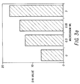
- FIGURES 3a and 3b illustrate the sensitivity increase using a combination assay format.
- FIGURE 4 illustrates a test cartridge for an immunodot assay.
- FIGURE 5 illustrates a seroconversion graph wherein the amount of anti-NS-5 S/N antibody, shown as the solid line between closed circles, and the amount of anti-HCV 2.0 S/CO antibody shown as a solid line between open squares, is plotted against days post presentation.
- the present invention is directed to an assay to detect an antibody to an HCV antigen in a sample.
- Human serum or plasma is diluted in a sample diluent and incubated with a polystyrene bead coated with a polypeptide that includes an HCV antigenic epitope. If antibodies are present in the sample they will form a complex with the antigenic polypeptide and become affixed to the polystyrene bead. After the complex has formed, unbound materials and reagents are removed by washing the bead and the bead-antigen-antibody complex is reacted with a solution containing horseradish peroxidase labeled goat antibodies directed against human antibodies.
- This peroxidase enzyme then binds to the antigen-antibody complex already fixed to the bead.
- the horseradish peroxidase is contacted with o-phenylenediamine and hydrogen peroxide which results in a yellow-orange color.
- the intensity of the color is proportional to the amount of antibody which initially binds to the antigen fixed to the bead.
- the preferred polypeptides having HCV antigenic epitopes were selected from portions of the HCV genome which encoded polypeptides which possessed amino acid sequences similar to other known immunologically reactive agents and which were identified as having some immunological reactivity.
- the immunological reactivity of a polypeptide was initially identified by reacting the cellular extract of E . coli clones which had been transformed with cDNA fragments of the HCV genome with HCV infected serum. The clones presumably expressed polypeptides encoded by the incorporated cDNA which were immunologically reactive with serum known to contain antibody to HCV antigens.
- An analysis of a given amino acid sequence however, only provides rough guides to predicting immunological reactivity.
- polypeptides having one or more than one epitope of an HCV antigen to detect the presence of an antibody to an HCV antigen is illustrated in Figure 2.
- the polypeptides of the present invention may be prepared using automated synthesizers.
- the synthesis of p1684 is provided below.
- the fully protected peptide-resin was assembled on a phenylacetamidomethyl (PAM) resin by stepwise solid phase synthesis (starting with the carboxyl terminal residue) according to the general procedure described by Barany, G. and Merrifield, R.B. in The Peptides , (Gross, E., and Meinhoeffer, T., eds.) 2, 1-284 (1980) Academic Press, New York, NY.
- PAM phenylacetamidomethyl
- the C-terminal amino acid valine (Val) was coupled to the solid support via an oxymethylphenylacetamidomethyl (OMPA) linkage to yield PAM resin which ensures improved stability to prolonged treatment with trifluoroacetic acid (TFA).
- OMPA oxymethylphenylacetamidomethyl
- TFA trifluoroacetic acid
- a BOC-Val-OCH2-Pam-resin (0.78 mmol/g, 0.13 g) was transferred to the reaction vessel of an Applied Biosystems Peptide Synthesizer, model 430A. All subsequent amino acids starting from the carboxyl terminal to N-terminus were coupled in a stepwise manner using Applied Biosystems' small scale rapid cycle protocol.
- Protected amino acids were coupled using preformed symmetric anhydride chemistry except for asparagine, glutamine, arginine and histidine which were double coupled using N-N'-dicyclohexylcarbodiimide (DCC)/1-hydroxybenzotriazole (HOBT) chemistry.
- DCC N-N'-dicyclohexylcarbodiimide
- HOBT hydroxybenzotriazole
- protected amino acids were coupled using preformed symmetric anhydrides dissolved in dimethylformamide (DMF).
- DMF dimethylformamide
- the symmetric anhydride of an individual amino acid was formed in methylene chloride followed by solvent exchange to DMF before transferring to the reaction vessel of the peptide synthesizer.
- the second coupling of symmetric anhydride was also conducted in DMF.
- N-amino group of all amino acids used was protected by a t-butyloxycarbonyl (t-BOC) linkage.
- the side chain functional groups of various amino acids were protected by the following groups: Arg-Tos (Tosyl) Lys-2ClZ (2-chlorobenzyloxycarbonyl) Thr,Ser-Bzl (Benzyl) Tyr-2BrZ (2-Bromobenzyloxycarbonyl) Cys-4MeBzl (4-Methylbenzyl) Asp,Glu-OBzl (0-Benzyl) His-DNP (Dinitrophenyl) The fully protected peptide-resin (0.28g) was allowed to swell in methylene chloride (CH2Cl2) for 5 minutes.
- CH2Cl2 methylene chloride
- the peptide-resin was transferred to a manual reaction vessel, treated twice with 5% thiophenol in DMF for twenty minutes each followed by six CH2Cl2 washes for one minute each, and then transferred to the reaction vessel of the synthesizer.
- the t-BOC protecting group was then removed using 60% TFA/CH2Cl2 according to the manufacturer's protocol and the partially deprotected peptide-resin was then dried overnight under house vacuum at room temperature.
- Partially deprotected peptide-resin was then treated with dimethyl sulfide (DMS (1 ml), p-cresol (1 ml), p-thiocresol (0.2 g) and HF (10 mL) at 0°C for one hour to cleave the peptide from the resin support.
- the HF/DMS and other volatiles were distilled off in vacuo at 0°C.
- the cleaved peptide and resin were washed three times with 15 ml aliquots of diethyl ether, and the cleaved peptide was extracted by washing three times each with 10 ml aliquots of 40% aqueous acetic acid and 15% aqueous acetic acid, respectively.
- the aqueous extracts were combined and washed three times with 15 ml aliquots of diethyl ether and then lyophilized to yield a crude peptide.
- the crude peptide was analyzed for purity using reversed-phase high performance liquid chromatography on a C4, 4.6 x 30mm column (Brownlee, Applied Biosystems, Inc., Foster City, California), flow rate one ml/minute employing 0.1% aqueous TFA (A) and 100% acetonitrile (B) as the solvent system.
- the preferred solvent gradient employed for this peptide analysis started with 30% B solvent.
- the column was maintained at 30% B for one minute followed by an increase over 20 minutes using a linear gradient to 55% B and maintained for one minute. Finally, the column was brought back to 30% B over a two minute period.
- the presence of peptide in the effluent was monitored simultaneously at 225 nm and 280 nm.
- the composition of the purified peptide was determined by acid hydrolysis. After removal of the acid, the hydrolysate was analyzed on a Beckmar 6300 amino acid analyzer.
- polypeptide p1689 is not used alone or in combination with only C100-3 in an assay of the present invention.
- polypeptides illustrated in Table 2 may also be prepared in a stepwise fashion or in a fragment coupling protocol using various side chain protection methodologies known to those skilled in the art.
- the polypeptides may also be prepared using enzymatic methodology.
- polypeptides useful in the practice of this invention may be prepared using recombinant technologies. Briefly, DNA sequences which encode the desired polypeptides are preferably assembled from fragments of the total desired sequence. The fragments are generally prepared using well known automated processes and apparatus. After the complete sequence has been prepared the desired sequence is incorporated into an expression vector which is transformed into a host cell. The DNA sequence is then expressed by the host cell to give the desired polypeptide which is harvested from the host cell or from the medium in which the host cell is cultured. In most cases, the manufactured DNA sequence is assembled using codons which are known to be best expressed in the host cell.
- the amino acid sequence for p1684 is reverse translated to give the codons listed in Table 3 which are optimized (where not inconsistent with assembly and synthesis of fragments) to facilitate high level expression in E . coli .
- Individual oligonucleotides are synthesized on Applied Biosystem 380A DNA synthesizer using methods and reagents recommended by the manufacturer. These purified oligonucleotides are annealed and ligated together to assemble the entire DNA sequence for digestion with BamH1 and Sal1, allowing ligation into pUC18. The resulting plasmid is suitably transformed into E . coli JM103 cells.
- Table 3 also lists preferred codons to express p1 and p1223.
- the nitrocellulose sheet containing the transferred proteins is incubated with a blocking solution for one hour and incubated overnight at 4 °C with HCV patients' sera diluted in TBS containing 5% E . coli JM103 lysate.
- the nitrocellulose sheet is washed three times in TBS, then incubated with HRPO-labeled goat anti-human IgG, diluted in TBS containing 10% fetal calf sera.
- the nitrocellulose is washed three times with TBS and the color is developed in TBS containing 2 mg/ml 4-chloro-1-napthol, 0.02% hydrogen peroxide and 17% methanol. Strong immunoreactive band formation with HCV patients' sera indicates that the synthetic polypeptide in expressed in E . coli in immunologically reactive form.
- Example 1 describes a confirmatory assay.
- Example 2 describes a combination assay.
- Example 3 describes a synthetic polypeptide-based assay.
- Example 4 describes an immunodot assay.
- Example 5 describes a competition assay.
- Example 6 describes an EIA assay in which peptides 380-436 and 447-483, 643-683 and 2302-2352 are used.
- Example 7 describes an EIA utilizing peptide p380.LG.
- Example 8 describes an EIA utilizing peptide 2302 (NS-5) compared to an EIA utilizing antigens NS3 (CKS-33C), NS4 (C-100) or CORE (CRS-CORE).
- Example 9 describes the PEPSCAN protocol followed.
- the confirmatory assay uses at least two polypeptides containing HCV antigenic epitopes which are preferably prepared and isolated from different sources.
- One polypeptide is used to screen serum or plasma samples.
- the other polypeptide is used to confirm the presence of a HCV antibody in a sample initially identified as containing a HCV antibody by the screening procedure.
- the screening procedure uses a recombinant C100-3 polypeptide.
- the C100-3 recombinant polypeptide is believed to contain multiple epitopes as well as an immunodominant region defined by the 1689-1806 amino acid sequence.
- the C100-3 polypeptide is expressed in recombinant yeast cells and isolated from the cell extract as described in EPA publication Number 0 318 216.
- Other recombinant polypeptides containing amino acid sequences essentially duplicative of C100-3 may also be used.
- the other peptide used in the confirmatory assay is a synthetic peptide selected from the group consisting of p1, p35, p99, p1192, p1223, p1684, p1689, p1694, p1866 and p1899, although p1689 cannot be used alone with C100-3 as part of the present invention.
- the peptide is p1684 or p1866.
- C100-3 and the synthetic peptides, p1684, p1694 or p1866 were separately coated onto polystyrene beads. A combination of synthetic peptides coated on a polystyrene bead may also be used if desired.
- the polystyrene beads are first washed with distilled water and propanol then incubated with crude or purified HCV synthetic peptides diluted to 0.1-20.0 ug/ml in a 0.1 M solution of an appropriate buffer containing about 0.4-0.5 M NaCl, about 0.0022% Triton® X-100 and adjusted to about pH 6.5-10.0.
- the following buffers, tris, NaH2PO4 ⁇ H2O, boric acid, and citrate buffers are preferred and are optimized for each peptide; preferred buffers, pH and coating concentration for the synthetic peptides are listed in Table 4. Successful coatings have also been accomplished with lower or higher pH.
- the beads are incubated in the antigen solution for about two hours at 38-42 °C, washed in phosphate buffer solution (PBS) and soaked in 0.1% Triton® X-100 in PBS for sixty minutes at 38-42 °C. The beads are then washed two times in PBS, overcoated with a solution of 5% (w/v) bovine serum albumin in PBS for sixty minutes and washed three times with PBS. Finally, the beads are overcoated with 5% (w/v) sucrose in PBS and dried under nitrogen or air.
- PBS phosphate buffer solution
- the peptides are each individually coated onto polystyrene beads and used in an antibody capture format. Ten microliters of sample are added to the wells of a reaction tray along with 400 ⁇ l of a sample diluent and a peptide coated bead.
- the sample diluent consists of about 10% (v/v), or less, bovine serum and about 20% (v/v), or less, goat serum in 20 mM Tris phosphate buffer containing 0.20%, or less, (v/v) Triton® X-100, 3% (w/v), or less, bovine serum albumin.
- yeast extracts which are added to the sample diluent (typically about 200 ⁇ g/ml). The addition of yeast extracts to the sample diluent is used to prevent false positive results.
- the final material is sterile filtered and filled in plastic bottles, and preserved with 0.1% sodium azide.
- the beads After one hour of incubation at 40 °C, the beads are washed and 200 ⁇ l of conjugate is added to the wells of the reaction tray.
- the preferred conjugate is goat anti-human IgG horseradish peroxidase conjugate.
- Concentrated conjugate is purchased from Kirkegaard and Perry Laboratories, Inc., Gaithersburg, Maryland, and is titered to determine a working concentration.
- a twenty-fold concentrate of the working conjugate solution is then prepared by diluting the concentrate in diluent.
- the conjugate diluent includes 10% (v/v) bovine serum, 10% (v/v) goat serum and 0.15% Triton®-X100 in 20 mM Tris buffer, pH 7.5 with 0.01% gentamicin sulfate, pink dye and antifungal agents as preservatives.
- the conjugate is sterile filtered and filled in plastic bottles.
- the beads After one hour of incubation with the conjugate at 40 °C, the beads are washed, exposed to the OPD substrate for thirty minutes at room temperature and the reaction terminated by the addition of 1 N H2SO4. The absorbance is read at 492 nm.
- Samples found to be repeatably reactive by a screening assay using the polypeptide C100-3 are tested in duplicate using p1684 or p1689 coated beads. Reactive specimens are considered confirmed samples. Samples not reacting with p1684 or p1689 are tested in duplicate with p1694 and p1866 beads. Samples reacting with one or both of these peptides are considered confirmed: Those specimens not reacting with any of these peptides are considered nonconfirmed.
- the cutoff for the assay should be at least 5-15 standard deviations above the absorbance value of a normal population mean. Consistent with these criteria, a cutoff for the assay may be selected which clearly separated most of the presumed "true negatives" from "true positive" specimens. A general cutoff value may be calculated as about 2.1 to 8 times the negative control mean absorbance value.
- Samples were collected from a population of intravenous drug users enrolled in an NIH-funded study. The population consisted of individuals who were acknowledged users of intravenous drugs selected over a two-year period from patients at the Edward Hines Jr. Veteran's Administration Hospital in Maywood, Illinois by Dr. Connie Pachucki and members of the Infectious Diseases staff.
- a proficiency panel comprised of neat and diluted human plasma including specimens containing antibodies to HCV C100-3 was provided by scientists at the Chiron Corporation (12 specimens). This panel contains specimens ranging from low to high reactivity in other assays, non-reactive presumed "true negative” specimens, and reactive specimens diluted to give low-level or negative results.
- the data further support the utility of the confirmatory strategy using synthetic peptides.
- the synthetic peptides serve as an independent source of antigen for use in immunoassays.
- the combination assay uses more than one polypeptide antigen coated on the same bead.
- the polystyrene beads described in Example 1 are contacted simultaneously with the polypeptide in appropriate buffer solutions. After the beads have been contacted with the polypeptides, the bead is treated further as described above.
- the increased sensitivity of an assay employing synthetic polypeptide compared to recombinant C100-3 polypeptide was demonstrated in a serial dilution study.
- the serial dilution study employed fifteen samples which were identified as having antibodies to HCV antigens using a recombinant C100-3 screening assay. Each positive sample was assayed using recombinant C100-3 polypeptide in one assay and p1689 polypeptide in a second assay, and the samples were then diluted twofold until the S/CO value was less than one. In twelve samples the p1689 polypeptide gave increased sensitivity (larger S/CO values) at all dilutions. In two samples, the p1689 polypeptide and the recombinant yeast C100-3 polypeptides were essentially equivalent. In one sample, the p1689 polypeptide gave a negative response to a positive sample at all dilutions.
- the antibodies, IgG, IgM and IgA were detected using affinity purified goat antibodies to human IgG, IgM and IgA coupled to horseradish peroxidase (Kirkegaard & Perry Laboratories, Inc., Gaithersburg, Maryland) which were used at working concentrations of 0.2 ug/ml Anti-IgG, 0.5 ug/ml Anti-IgM and 0.2 ug/ml Anti-IgA. Serum dilutions for each assay were 1:41 for IgG, 1:101 for IgM, and 1:41 for IgA.
- polypeptides that were used in the study include C100-3, p1694, p1684, p1689, and p1866.
- beads containing the polypeptides were incubated with diluted serum for one hour at 40 °C, the beads were washed and incubated with the appropriate goat antibody for one hour at 40°C. The beads were washed again and the assay was developed by incubating the beads with OPD for thirty minutes at room temperature. The color development was quenched with 1 N sulfuric acid and the results read at 492 nm.
- IgM antibody was detected in only three of the six chimps studied. The response of each of the three animals to C100-3, p1684, p1689 and p1694 was detected in 7 to 10 WPI whereas IgM antibody to p1866 was undetectable in two chimps and delayed in the third. All IgM responses were short lived with levels falling below positive (S/N less than 3.0) within 2 to 22 weeks.
- a positive IgA response (S/N greater than 3.0) was detected in only 2 of the 6 chimps and proved to be either biphasic or significantly later than the IgG or IgM response. Although these 2 chimps had chronic disease no conclusions regarding the significance of IgA antibodies can be made since sera from the three resolved chimps is available only through 30 to 40 WPI.
- results show the polypeptides when used to assay for antibodies to HCV antigens are useful to follow the progression of HCV infection and that the polypeptides exhibit unexpected sensitivity to different antibodies generated during the clinical progression of HCV infection.
- the immunodot assay of the invention excludes those assays relying only on a combination of C100-3 and p1689 polypeptides.
- the immunodot assay system uses a panel of purified synthetic polypeptides placed in an array on a nitrocellulose solid support.
- the prepared solid support is contacted with a sample and captures specific antibodies to HCV antigens.
- the captured antibodies are detected by a conjugate-specific reaction.
- the conjugate-specific reaction is quantified using a reflectance optics assembly within an instrument which has been described in U.S. Patent Application Serial No. 07/227,408, filed August 2, 1988.
- the related U.S. Patent Applications Serial Nos. 07/227,272, 07/227,586, and 07/227,590 further describe specific methods and apparatus useful to perform an immunodot assay. Briefly, a nitrocellulose-base test cartridge is treated with multiple antigenic polypeptides.
- test cartridge which may be used in an automated process for performing an immunodot assay described above is illustrated in Figure 4.
- Each polypeptide is contained within a specific reaction zone on the test cartridge. After all the antigenic polypeptides have been placed on the nitrocellulose, excess binding sites on the nitrocellulose are blocked. The test cartridge is then contacted with a sample such that each antigenic polypeptide in each reaction zone will react if the sample contains the appropriate antibody. After reaction, the test cartridge is washed and any antigen-antibody reactions are identified using suitable well known reagents.
- the synthetic polypeptides p1223, p1684, p1689 and p1866 were diluted into an aqueous buffered solution (polypeptide diluent: 0.03% Triton® X-100 and 0.1% sodium azide in 50 mM Hepes buffer, pH 7.6) and applied to a preassembled nitrocellulose test cartridge at about 40 ng in each reaction zone. After drying the cartridge overnight at room temperature, the nonspecific binding capacity of the nitro-cellulose phase was blocked.
- the blocking solution contained 1% porcine gelatin, 1% casein enzymatic hydrolysate, 5% Tween®-20, 0.1% sodium azide, 0.5 M sodium chloride and 20 mM Tris, pH 7.5.
- Test cartridges were incubated with samples 00642 and 423 (see Table 1) and ALT 27.
- the sample ALT 27 was obtained from a volunteer donor having elevated alanine aminotransferase levels. After sample incubation, sequential incubations with a biotin-conjugated goat anti-human immunoglobulin-specific antibody, an alkaline phosphatase-conjugated rabbit anti-biotin specific antibody, and 5-bromo-4-chloro-3-indolyl phosphate produced a colored product at the site of the reaction.
- a detectable reaction is defined by the formation of a visually discernable product at the antigen site on the array; when quantified by the instrument, a reflectance density (Dr) value of greater than or equal to approximately 0.0150 above background is obtained. None of the tested polypeptides elicited a detectable reaction with a negative control serum that was previously demonstrated negative for antibodies to HCV antigens using a recombinant C100-3 polypeptide.
- polypeptide p1684 Enhanced reactivity of polypeptide p1684 with sample 00642 was achieved through subtle modification of the antigen dilution (the modified polypeptide diluent was 0.5 M sodium chloride, 0.0022% Triton® X-100 and 0.1 M Tris/HCl, pH 8.5).
- the net reflectance (Dr) for a test cartridge containing the polypeptides p1223, p1684, p1689, p1694, and p1866 which indicate a positive or negative response is set out in Table 7.
- the synthetic peptides containing antigenic HCV epitopes are useful for competition assays.
- peptides other than p1689 representing epitopes within the C100-3 region such as p1694 or p1684 are solubilized and mixed with a specimen diluent to a final concentration of 0.5-50 ⁇ g/ml.
- Ten microliters of specimen or diluted specimen is added to a reaction well followed by 400 ⁇ l of the specimen diluent containing peptide and if desired, the mixture may be pre-incubated for about fifteen minutes to two hours.
- a bead coated with C100-3 antigen of HCV is then added to the reaction well and incubated for one hour at 40 °C.
- Samples containing antibodies to the C100-3 antigen generate a reduced signal caused by the competitive binding of the peptides to these antibodies in solution.
- the percentage of competitive binding may be calculated by comparing the absorbance value of the sample in the presence of a synthetic peptide to the absorbance value of the sample assayed in the absence of a synthetic peptide at the same dilution.
- EXAMPLE 8 EIA UTILIZING p2302.
- Beads were coated with either p2303 or "HCV2.0S/CO” according to the method of Example 1.
- the bead coated with "HCV 2.0/SCO” comprised antigen from the NS3 (CKS-33C), NS4 (C-100) and Core (CKS-CORE) regions of the HCV genome.
- CKS-33C NS3
- NS4 C-100
- Core CKS-CORE
- NS1 region of HCV genome from a.a. 600-720 was mapped with PEPSCAN analysis, which is serological analyses of series of overlapping peptides spanning the protein sequence to identify immunogenic domains.
- PEPSCAN analysis is serological analyses of series of overlapping peptides spanning the protein sequence to identify immunogenic domains.
- a total of 106 overlapping hexamer peptides were synthesized on polypropylene pins following the manufacturer's instructions (Cambridge Research Bioscience, Valley Stream, N.Y.).
- Fab dimers of IgG purified from sera of individuals seropositive for HCV were tested with these peptides.
- EIA performed as described by the manufacturer
- four peptide sequences were selected as illustrated in Table 10.
- Each of these peptides were synthesized by a stepwise solid phase synthesis, starting with the Carboxy terminus residue.
- a panel of sera positive for antibodies to C-100 protein of CHV was tested for their reactivity to NS1 peptide by microtiter EIA as described below.
- Sera from individuals seropositive for antibodies to HCV C-100 were serially diluted in 100 ⁇ l of a buffer containing 20MM sodium phosphate, pH 7.4, 0.15M NaCl, 20% normal goat serum, 10% fetal calf serum, 5 MM EDTA, 10MM EGTA, 50MM Tris, 0.2% Tween®-20 with sodium azide as preservative, pH 6.8.
- the diluted sera were reacted with peptides in microtiter wells for 3 hours at 37°C or overnight at ambient temperatures.
- the plates were washed and 100 ⁇ l of appropriately diluted goat anti-mouse (HH) Horseradish Peroxidase (HRPO)-conjugated antibody (Jackson immunochemicals, West Grove, PA) was added.
- HH goat anti-mouse
- HRPO Horseradish Peroxidase
- the plates were incubated at 37°C for 2 hours. After a final wash, 100 ⁇ l of O-phenylenediamine 2HCl (OPD) color reagent was added. The reaction was carried out at room temperature in the dark for 20-25 minutes, and stopped by the addition of 100 ⁇ l of IN H SOu. The absorbance of the reaction mixture recorded at 492 NM. A negative control which was previously confirmed to be negative for HCV infection was included with each plate in triplicate. The sample was considered reactive if the absorbance of the sample at a 1:2000 dilution was three times the absorbance of the negative control at the same dilution. Table 11 illustrates the reactivity of these samples with each of the peptides.
- OPD O-phenylenediamine 2HCl
- NUMBERS 607-627 643-663 666-683 691-714 15 - - + - 22 - - - - 23 - - - - 24 +/- - - - - 25 +/- - - - 32 +/- + + + + - 36 - - - - 46 - - - - 50 - - - - 65 - - - - 70 - + + - 71 - - + - 75 - - - - 89 - ++ - + 95 - ++ ++ + 100 - ++ - - 102 - - - - 108 - - - - 130 - - + - 137 - - - - LG + ++ +++ + 301060 - ++ +++ + PB3178 ++ + +++ + PB3180 ++ + + +++ ++ 300423 ++ - +++ + % POS
Landscapes
- Health & Medical Sciences (AREA)
- Life Sciences & Earth Sciences (AREA)
- Chemical & Material Sciences (AREA)
- Immunology (AREA)
- Organic Chemistry (AREA)
- Molecular Biology (AREA)
- General Health & Medical Sciences (AREA)
- Virology (AREA)
- Engineering & Computer Science (AREA)
- Medicinal Chemistry (AREA)
- Biochemistry (AREA)
- Hematology (AREA)
- Biophysics (AREA)
- Communicable Diseases (AREA)
- Proteomics, Peptides & Aminoacids (AREA)
- Genetics & Genomics (AREA)
- Biomedical Technology (AREA)
- Urology & Nephrology (AREA)
- Microbiology (AREA)
- Cell Biology (AREA)
- Gastroenterology & Hepatology (AREA)
- Biotechnology (AREA)
- Food Science & Technology (AREA)
- Physics & Mathematics (AREA)
- Analytical Chemistry (AREA)
- General Physics & Mathematics (AREA)
- Pathology (AREA)
- Peptides Or Proteins (AREA)
- Saccharide Compounds (AREA)
- Steroid Compounds (AREA)
- Measuring Or Testing Involving Enzymes Or Micro-Organisms (AREA)
Claims (13)
- Procédé de dosage pour identifier la présence d'un anticorps immunologiquement réactif avec un antigène de HCV dans un échantillon de liquide, comprenant :
la mise en contact de l'échantillon avec un polypeptide contenant au moins un épitope d'un antigène de HCV choisi dans le groupe consistant en p1, p35, p99, p1192, p1223, p1684, p1694, p1866, p1899, p380, p380.LG, p447, p607, p643a, p643b, p666, p691 et p2302 dans des conditions appropriées pour la complexation de l'anticorps avec le polypeptide, et la détection du complexe anticorps-polypeptide. - Procédé de dosage selon la revendication 1, dans lequel l'antigène est p1866, p380, p380.LG, p643b, p666 ou p2302.
- Procédé de dosage en combinaison pour détecter la présence d'un anticorps immunologiquement réactif avec un antigène de HCV dans un échantillon de liquide, comprenant :
la mise en contact de l'échantillon avec un support solide auquel sont liés simultanément le polypeptide recombiné C100-3 et un polypeptide choisi dans le groupe consistant en p1, p35, p99, p1192, p1223, p1684, p1694, p1866, p1899, p380, p380.LG, p447, p607, p643a, p643b, p666, p691 et p2302 dans des conditions appropriées pour la complexation de l'anticorps avec le polypeptide, et la détection du complexe anticorps-polypeptide. - Procédé de dosage de confirmation pour identifier la présence d'un anticorps dans un échantillon de liquide immunologiquement réactif avec un antigène de HCV, comprenant :
la mise en contact d'une première portion aliquote de l'échantillon avec un premier polypeptide recombiné C100-3 qui contient au moins un épitope d'un antigène de HCV dans des conditions appropriées pour la complexation de l'anticorps avec le polypeptide et la détection d'un premier complexe anticorps-antigène,
la mise en contact d'une seconde portion aliquote de l'échantillon avec un ou plusieurs polypeptides choisis dans le groupe consistant en p1, p35, p99, p1192, p1223, p1684, p1689, p1694, p1866, p1899, p380, p380.LG, p447, p607, p643a, p643b, p666, p691 et p2302 dans des conditions appropriées pour former un second complexe anticorps-antigène, et
la détection du second complexe anticorps-antigène,
à condition que les procédés de dosage employant p1689 seul pour former ledit second complexe anticorps-antigène soient exclus. - Procédé de dosage selon la revendication 4, dans lequel le second antigène est p1684, p1694, p380, p380.LG, p643b, p666 ou p2302.
- Procédé de dosage par immunotaches pour identifier la présence d'un anticorps immunologiquement réactif avec un antigène de HCV dans un échantillon de liquide, comprenant :
la mise en contact simultanée de l'échantillon avec au moins deux polypeptides contenant chacun des épitopes distincts d'un antigène de HCV dans des conditions appropriées pour la complexation de l'anticorps avec les polypeptides et dans lequel l'anticorps-polypeptide est détecté par réaction du complexe avec des réactifs producteurs de couleur,
lesdits polypeptides étant choisis dans le groupe consistant en p1, p35, p99, p1192, p1223, p1684, p1689, p1694, p1866, p1899, p380, p380.LG, p447, p607, p643a, p643b, p666, p691, p2302 et C100-3 liés à un support solide, à condition que les procédés de dosage employant seulement p1689 et C100-3 soient exclus. - Procédé de dosage par compétition pour identifier la présence d'un anticorps immunologiquement réactif avec un antigène de HCV dans un échantillon de liquide, comprenant la préparation de première et seconde portions aliquotes immunologiquement équivalentes de l'échantillon, la mise en contact de la première portion aliquote avec un support solide contenant un polypeptide lié qui contient au moins un épitope d'un antigène de HCV dans des conditions appropriées pour la complexation avec l'anticorps pour former un complexe anticorps-polypeptide détectable et la mise en contact de la seconde portion aliquote tout d'abord avec un polypeptide non lié puis avec le support solide contenant le polypeptide lié, ledit polypeptide étant choisi dans le groupe consistant en p1, p35, p99, p1192, p1223, p1684, p1694, p1866, p1899, p380, p380.LG, p447, p607, p643a, p643b, p666, p691 et p2302.
- Trousse d'immunodosage comprenant :
un polypeptide contenant au moins un épitope d'un antigène de HCV choisi dans le groupe consistant en p1, p35, p99, p1192, p1223, p1684, p1694, p1866, p1899, p380, p380.LG, p447, p607, p643a, p643b, p666, p691 et p2302,
un ou plusieurs réactifs de préparation d'échantillon, et
un ou plusieurs réactifs de détection et de production de signal. - Trousse d'immunodosage comprenant :
un support solide auquel sont liés simultanément le polypeptide recombiné C100-3 et un polypeptide contenant au moins un épitope d'un antigène de HCV choisi dans le groupe consistant en p1, p35, p99, p1192, p1223, p1684, p1694, p1866, p1899, p380, p380.LG, p447, p607, p643a, p643b, p666, p691 et p2302,
un ou plusieurs réactifs de préparation d'échantillon, et
un ou plusieurs réactifs de détection et de production de signal. - Trousse d'immunodosage comprenant :
le polypeptide recombiné C100-3 lié à un support solide,
un ou plusieurs polypeptides contenant au moins un épitope d'un anti-gène de HCV lié à un support solide choisi dans le groupe consistant en p1, p35, p99, p1192, p1223, p1684, p1689, p1694, p1866, p1899, p380, p380.LG, p447, p607, p643a, p643b, p666, p691 et p2302, à condition que la trousse ne contienne pas p1689 et C100-3 comme seuls polypeptides,
un ou plusieurs réactifs de préparation d'échantillon, et
un ou plusieurs réactifs de détection et de production de signal. - Trousse d'immunodosage comprenant :
le polypeptide recombiné C100-3 lié à un support solide,
un ou plusieurs polypeptides contenant au moins un épitope d'un anti-gène de HCV lié au même support solide que le polypeptide C100-3 choisi dans le groupe consistant en p1, p35, p99, p1192, p1223, p1684, p1689, p1694, p1866, p1899, p380, p380.LG, p447, p607, p643a, p643b, p666, p691 et p2302, à condition que la trousse ne contienne pas p1689 et C100-3 comme seuls polypeptides,
un ou plusieurs réactifs de préparation d'échantillon, et
un ou plusieurs réactifs de détection et de production de signal. - Trousse d'immunodosage comprenant :
une cartouche de test comprenant au moins deux polypeptides contenant chacun des épitopes distincts d'un antigène de HCV, lesdits polypeptides étant choisis dans le groupe consistant en p1, p35, p99, p1192, p1223, p1684, p1689, p1694, p1866, p1899, p380, p380.LG, p447, p607, p643a, p643b, p666, p691 et p2302, liés à un support solide, à condition que la cartouche de test ne contienne pas p1689 et C100-3 comme seuls polypeptides,
un ou plusieurs réactifs de préparation d'échantillon, et
un ou plusieurs réactifs de détection et de production de signal. - Trousse d'immunodosage comprenant :
un support solide contenant un polypeptide lié contenant au moins un épitope d'un antigène de HCV,
un polypeptide non lié contenant au moins un épitope d'un antigène de HCV,
un ou plusieurs réactifs de préparation d'échantillon, et
un ou plusieurs réactifs de détection et de production de signal,
ledit polypeptide étant choisi dans le groupe consistant en p1, p35, p99, p1192, p1223, p1684, p1694, p1866, p1899, p380, p380.LG, p447, p607, p643a, p643b, p666, p691 et p2302.
Applications Claiming Priority (4)
| Application Number | Priority Date | Filing Date | Title |
|---|---|---|---|
| US45616289A | 1989-12-22 | 1989-12-22 | |
| US456162 | 1989-12-22 | ||
| US61018090A | 1990-11-07 | 1990-11-07 | |
| US610180 | 1990-11-07 |
Publications (3)
| Publication Number | Publication Date |
|---|---|
| EP0445423A2 EP0445423A2 (fr) | 1991-09-11 |
| EP0445423A3 EP0445423A3 (en) | 1992-04-29 |
| EP0445423B1 true EP0445423B1 (fr) | 1995-09-20 |
Family
ID=27038137
Family Applications (1)
| Application Number | Title | Priority Date | Filing Date |
|---|---|---|---|
| EP90125354A Expired - Lifetime EP0445423B1 (fr) | 1989-12-22 | 1990-12-22 | Essai pour l'hépatite C |
Country Status (7)
| Country | Link |
|---|---|
| EP (1) | EP0445423B1 (fr) |
| JP (1) | JP3188717B2 (fr) |
| AT (1) | ATE128237T1 (fr) |
| AU (1) | AU638304B2 (fr) |
| CA (1) | CA2032907C (fr) |
| DE (1) | DE69022578T2 (fr) |
| ES (1) | ES2080099T3 (fr) |
Cited By (1)
| Publication number | Priority date | Publication date | Assignee | Title |
|---|---|---|---|---|
| US6150087A (en) * | 1991-06-24 | 2000-11-21 | Chiron Corporation | NANBV diagnostics and vaccines |
Families Citing this family (27)
| Publication number | Priority date | Publication date | Assignee | Title |
|---|---|---|---|---|
| US5683864A (en) * | 1987-11-18 | 1997-11-04 | Chiron Corporation | Combinations of hepatitis C virus (HCV) antigens for use in immunoassays for anti-HCV antibodies |
| HUT59964A (en) * | 1989-05-18 | 1992-07-28 | Chiron Corp | Process for producing new nanbv diagnostica for determining hepatitis c virus |
| US5308750A (en) * | 1989-12-22 | 1994-05-03 | Abbott Laboratories | Monoclonal antibodies to putative HCV E2/NS1 proteins and methods for using same |
| KR940000755B1 (ko) * | 1990-02-16 | 1994-01-29 | 유나이티드 바이오메디칼 인코오포레이티드 | Hcv에 대한 항체 검출, hcv 감염의 진단 및 백신으로서의 그 예방에 특히 적합한 합성 펩티드 |
| RU2130969C1 (ru) † | 1990-04-04 | 1999-05-27 | Чирон Корпорейшн | Композиция для диагностики гепатита c человека (варианты), способ и набор для обнаружения антител к вирусу гепатита c человека |
| DE69132332T2 (de) * | 1990-04-06 | 2000-11-30 | Genelabs Tech Inc | Hepatitis c-virus-epitope |
| CA2049679C (fr) * | 1990-08-24 | 2005-06-21 | Sushil G. Devare | Recherche des anticorps de l'hepatite c a l'aide d'antigenes recombinants |
| ATE206717T1 (de) * | 1990-11-03 | 2001-10-15 | Dade Behring Marburg Gmbh | Hcv-spezifische peptide, mittel dazu und ihre verwendung |
| CA2055149A1 (fr) * | 1990-11-08 | 1992-05-09 | Hiroaki Okamoto | Antigene, anticorps, systemes de detection, polynucleotides et polypeptides lies au virus de l'hepatite non a, non b |
| KR920703640A (ko) * | 1990-11-29 | 1992-12-18 | 테루카츄 아리마 | 비a비b형 간염 바이러스 항원 단백질 |
| US6007982A (en) * | 1990-12-14 | 1999-12-28 | Innogenetics N.V. | Synthetic antigens for the detection of antibodies to hepatitis C virus |
| ES2138784T3 (es) * | 1990-12-14 | 2000-01-16 | Innogenetics Nv | Antigenos sinteticos para la deteccion de anticuerpos contra el virus de la hepatitis c. |
| US5910404A (en) | 1990-12-14 | 1999-06-08 | Innogenetics N.V. | Synthetic antigens for the detection of antibodies to hepatitis C virus |
| AU1459792A (en) * | 1991-01-14 | 1992-08-27 | James N. Gamble Institute Of Medical Research | Basic structural immunogenic polypeptides having epitopes for hcv, antibodies, polynucleotide sequences, vaccines and methods |
| ATE167522T1 (de) * | 1991-01-31 | 1998-07-15 | Abbott Lab | Monoklonale antikörper gegen mögliche hcv- hüllregionen und methoden diese zu benutzen |
| US5574132A (en) * | 1991-04-05 | 1996-11-12 | Biochem Immunosystems Inc. | Peptides and mixtures thereof for detecting antibodies to hepatitis C virus (HCV) |
| DK0599913T3 (da) * | 1991-08-21 | 1999-08-16 | Abbott Lab | Monoklonale antistoffer mod formodet hepatitis C-virus NS5-proteiner og anvendelsesmetoder af samme |
| WO1993006247A1 (fr) * | 1991-09-16 | 1993-04-01 | Abbott Laboratories | Analyse de detection de l'hepatite c |
| CA2123875C (fr) | 1991-11-21 | 2005-05-24 | Peter Simmons | Depistage du virus de l'hepatite c |
| JPH05176773A (ja) * | 1991-12-27 | 1993-07-20 | Imuno Japan:Kk | オリゴヌクレオチド、オリゴペプタイド、ならびに抗c型肝 炎ウイルス抗体検出・鑑別方法 |
| GB9203803D0 (en) * | 1992-02-21 | 1992-04-08 | Wellcome Found | A recombinant polypeptide |
| US6709828B1 (en) | 1992-03-06 | 2004-03-23 | N.V. Innogenetics S.A. | Process for the determination of peptides corresponding to immunologically important epitopes and their use in a process for determination of antibodies or biotinylated peptides corresponding to immunologically important epitopes, a process for preparing them and compositions containing them |
| US6667387B1 (en) | 1996-09-30 | 2003-12-23 | N.V. Innogenetics S.A. | HCV core peptides |
| GB2294690B (en) * | 1994-11-01 | 1998-10-28 | United Biomedical Inc | Peptides effective for diagnosis and detection of hepatitis C infection |
| DE19504302A1 (de) * | 1995-02-09 | 1996-08-14 | Boehringer Mannheim Gmbh | Methode zur serologischen Typisierung mittels typspezifischer Antigene |
| DE19540105C1 (de) * | 1995-09-19 | 1997-02-20 | United Biomedical Inc | Peptidzusammensetzungen zur Diagnose und zum Nachweis von Hepatitis C Virus (HCV)-Infektion |
| CA2305847A1 (fr) | 1997-11-06 | 1999-05-20 | Innogenetics N.V. | Peptides multimeres derives de proteines d'enveloppe de virus d'hepatite c (hcv) a des fins de diagnostic et de vaccination |
Family Cites Families (2)
| Publication number | Priority date | Publication date | Assignee | Title |
|---|---|---|---|---|
| WO1989004669A1 (fr) * | 1987-11-18 | 1989-06-01 | Chiron Corporation | Diagnostic et vaccins du virus d'hepatite non-a non-b |
| WO1990011089A1 (fr) * | 1989-03-17 | 1990-10-04 | Chiron Corporation | Diagnostics et vaccins de nanbv (virus de l'hepatite non a, non b) |
-
1990
- 1990-12-21 CA CA002032907A patent/CA2032907C/fr not_active Expired - Fee Related
- 1990-12-21 AU AU68390/90A patent/AU638304B2/en not_active Ceased
- 1990-12-22 ES ES90125354T patent/ES2080099T3/es not_active Expired - Lifetime
- 1990-12-22 EP EP90125354A patent/EP0445423B1/fr not_active Expired - Lifetime
- 1990-12-22 DE DE69022578T patent/DE69022578T2/de not_active Expired - Fee Related
- 1990-12-22 AT AT90125354T patent/ATE128237T1/de not_active IP Right Cessation
- 1990-12-25 JP JP41824090A patent/JP3188717B2/ja not_active Expired - Fee Related
Cited By (1)
| Publication number | Priority date | Publication date | Assignee | Title |
|---|---|---|---|---|
| US6150087A (en) * | 1991-06-24 | 2000-11-21 | Chiron Corporation | NANBV diagnostics and vaccines |
Also Published As
| Publication number | Publication date |
|---|---|
| EP0445423A2 (fr) | 1991-09-11 |
| JP3188717B2 (ja) | 2001-07-16 |
| AU638304B2 (en) | 1993-06-24 |
| EP0445423A3 (en) | 1992-04-29 |
| AU6839090A (en) | 1991-06-27 |
| CA2032907C (fr) | 2002-05-14 |
| CA2032907A1 (fr) | 1991-06-23 |
| DE69022578T2 (de) | 1996-05-02 |
| JPH04253998A (ja) | 1992-09-09 |
| ES2080099T3 (es) | 1996-02-01 |
| ATE128237T1 (de) | 1995-10-15 |
| DE69022578D1 (de) | 1995-10-26 |
Similar Documents
| Publication | Publication Date | Title |
|---|---|---|
| EP0445423B1 (fr) | Essai pour l'hépatite C | |
| US6596476B1 (en) | Hepatitis C assay | |
| US5683864A (en) | Combinations of hepatitis C virus (HCV) antigens for use in immunoassays for anti-HCV antibodies | |
| EP0450931B1 (fr) | Combinaisons d'antigènes de l'hépatitis C virus (HCV) pour usage dans des échantillons immunologiques pour anticorps anti-HCV | |
| US5712087A (en) | Immunoassays for anti-HCV antibodies employing combinations of hepatitis C virus (HCV) antigens | |
| EP0489968A1 (fr) | Antigènes synthétiques pour la détection des anticorps contre le virus de l'hépatite C | |
| EP0642666B1 (fr) | Analyse de detection de l'hepatite c | |
| EP0472207B1 (fr) | Essai d'hépatite C utilisant des antigènes recombinants | |
| US5645983A (en) | Core antigen protein of hepatitis C virus, and diagnostic method and kit using the same | |
| EP0525910A1 (fr) | Peptide non-A, non-B | |
| US5670310A (en) | Methods and compositions for differential diagnosis of acute and chronic hepatitis c virus infection | |
| EP0744467A2 (fr) | Peptide multi-antigénique comprenant au moins deux peptides associés avec le virus de l'hépatite C | |
| EP0536838A2 (fr) | Peptides non-A, non-B | |
| WO1993011158A2 (fr) | Peptides non-a, non-b | |
| AU639560C (en) | Combinations of hepatitis C virus (HCV) antigens for use in immunoassays for anti-HCV antibodies | |
| WO1996041196A1 (fr) | Procede de detection d'un anticorps contre la deuxieme glycoproteine d'enveloppe du virus de l'hepatite c |
Legal Events
| Date | Code | Title | Description |
|---|---|---|---|
| PUAI | Public reference made under article 153(3) epc to a published international application that has entered the european phase |
Free format text: ORIGINAL CODE: 0009012 |
|
| AK | Designated contracting states |
Kind code of ref document: A2 Designated state(s): AT BE CH DE ES FR GB IT LI NL SE |
|
| PUAL | Search report despatched |
Free format text: ORIGINAL CODE: 0009013 |
|
| AK | Designated contracting states |
Kind code of ref document: A3 Designated state(s): AT BE CH DE ES FR GB IT LI NL SE |
|
| 17P | Request for examination filed |
Effective date: 19930108 |
|
| 17Q | First examination report despatched |
Effective date: 19940606 |
|
| GRAA | (expected) grant |
Free format text: ORIGINAL CODE: 0009210 |
|
| AK | Designated contracting states |
Kind code of ref document: B1 Designated state(s): AT BE CH DE ES FR GB IT LI NL SE |
|
| REF | Corresponds to: |
Ref document number: 128237 Country of ref document: AT Date of ref document: 19951015 Kind code of ref document: T |
|
| REF | Corresponds to: |
Ref document number: 69022578 Country of ref document: DE Date of ref document: 19951026 |
|
| ITF | It: translation for a ep patent filed |
Owner name: MODIANO & ASSOCIATI S.R.L. |
|
| REG | Reference to a national code |
Ref country code: ES Ref legal event code: FG2A Ref document number: 2080099 Country of ref document: ES Kind code of ref document: T3 |
|
| ET | Fr: translation filed | ||
| PLBE | No opposition filed within time limit |
Free format text: ORIGINAL CODE: 0009261 |
|
| STAA | Information on the status of an ep patent application or granted ep patent |
Free format text: STATUS: NO OPPOSITION FILED WITHIN TIME LIMIT |
|
| 26N | No opposition filed | ||
| REG | Reference to a national code |
Ref country code: GB Ref legal event code: IF02 |
|
| PGFP | Annual fee paid to national office [announced via postgrant information from national office to epo] |
Ref country code: GB Payment date: 20021104 Year of fee payment: 13 Ref country code: AT Payment date: 20021104 Year of fee payment: 13 |
|
| PGFP | Annual fee paid to national office [announced via postgrant information from national office to epo] |
Ref country code: NL Payment date: 20021112 Year of fee payment: 13 |
|
| PGFP | Annual fee paid to national office [announced via postgrant information from national office to epo] |
Ref country code: FR Payment date: 20021202 Year of fee payment: 13 |
|
| PGFP | Annual fee paid to national office [announced via postgrant information from national office to epo] |
Ref country code: SE Payment date: 20021203 Year of fee payment: 13 |
|
| PGFP | Annual fee paid to national office [announced via postgrant information from national office to epo] |
Ref country code: ES Payment date: 20021216 Year of fee payment: 13 |
|
| PGFP | Annual fee paid to national office [announced via postgrant information from national office to epo] |
Ref country code: CH Payment date: 20021223 Year of fee payment: 13 |
|
| PGFP | Annual fee paid to national office [announced via postgrant information from national office to epo] |
Ref country code: DE Payment date: 20021230 Year of fee payment: 13 |
|
| PGFP | Annual fee paid to national office [announced via postgrant information from national office to epo] |
Ref country code: BE Payment date: 20030116 Year of fee payment: 13 |
|
| PG25 | Lapsed in a contracting state [announced via postgrant information from national office to epo] |
Ref country code: GB Free format text: LAPSE BECAUSE OF NON-PAYMENT OF DUE FEES Effective date: 20031222 Ref country code: AT Free format text: LAPSE BECAUSE OF NON-PAYMENT OF DUE FEES Effective date: 20031222 |
|
| PG25 | Lapsed in a contracting state [announced via postgrant information from national office to epo] |
Ref country code: SE Free format text: LAPSE BECAUSE OF NON-PAYMENT OF DUE FEES Effective date: 20031223 Ref country code: ES Free format text: LAPSE BECAUSE OF NON-PAYMENT OF DUE FEES Effective date: 20031223 |
|
| PG25 | Lapsed in a contracting state [announced via postgrant information from national office to epo] |
Ref country code: LI Free format text: LAPSE BECAUSE OF NON-PAYMENT OF DUE FEES Effective date: 20031231 Ref country code: CH Free format text: LAPSE BECAUSE OF NON-PAYMENT OF DUE FEES Effective date: 20031231 Ref country code: BE Free format text: LAPSE BECAUSE OF NON-PAYMENT OF DUE FEES Effective date: 20031231 |
|
| BERE | Be: lapsed |
Owner name: *ABBOTT LABORATORIES Effective date: 20031231 |
|
| PG25 | Lapsed in a contracting state [announced via postgrant information from national office to epo] |
Ref country code: NL Free format text: LAPSE BECAUSE OF NON-PAYMENT OF DUE FEES Effective date: 20040701 Ref country code: DE Free format text: LAPSE BECAUSE OF NON-PAYMENT OF DUE FEES Effective date: 20040701 |
|
| EUG | Se: european patent has lapsed | ||
| GBPC | Gb: european patent ceased through non-payment of renewal fee |
Effective date: 20031222 |
|
| REG | Reference to a national code |
Ref country code: CH Ref legal event code: PL |
|
| PG25 | Lapsed in a contracting state [announced via postgrant information from national office to epo] |
Ref country code: FR Free format text: LAPSE BECAUSE OF NON-PAYMENT OF DUE FEES Effective date: 20040831 |
|
| NLV4 | Nl: lapsed or anulled due to non-payment of the annual fee |
Effective date: 20040701 |
|
| REG | Reference to a national code |
Ref country code: FR Ref legal event code: ST |
|
| REG | Reference to a national code |
Ref country code: ES Ref legal event code: FD2A Effective date: 20031223 |
|
| PG25 | Lapsed in a contracting state [announced via postgrant information from national office to epo] |
Ref country code: IT Free format text: LAPSE BECAUSE OF NON-PAYMENT OF DUE FEES;WARNING: LAPSES OF ITALIAN PATENTS WITH EFFECTIVE DATE BEFORE 2007 MAY HAVE OCCURRED AT ANY TIME BEFORE 2007. THE CORRECT EFFECTIVE DATE MAY BE DIFFERENT FROM THE ONE RECORDED. Effective date: 20051222 |