EP0441469A1 - Essai immunospécifique et bioluminescent d'ATP cellulaire - Google Patents
Essai immunospécifique et bioluminescent d'ATP cellulaire Download PDFInfo
- Publication number
- EP0441469A1 EP0441469A1 EP91300007A EP91300007A EP0441469A1 EP 0441469 A1 EP0441469 A1 EP 0441469A1 EP 91300007 A EP91300007 A EP 91300007A EP 91300007 A EP91300007 A EP 91300007A EP 0441469 A1 EP0441469 A1 EP 0441469A1
- Authority
- EP
- European Patent Office
- Prior art keywords
- cell
- process according
- detection
- sample
- solid carrier
- Prior art date
- Legal status (The legal status is an assumption and is not a legal conclusion. Google has not performed a legal analysis and makes no representation as to the accuracy of the status listed.)
- Withdrawn
Links
Images


Classifications
-
- C—CHEMISTRY; METALLURGY
- C12—BIOCHEMISTRY; BEER; SPIRITS; WINE; VINEGAR; MICROBIOLOGY; ENZYMOLOGY; MUTATION OR GENETIC ENGINEERING
- C12Q—MEASURING OR TESTING PROCESSES INVOLVING ENZYMES, NUCLEIC ACIDS OR MICROORGANISMS; COMPOSITIONS OR TEST PAPERS THEREFOR; PROCESSES OF PREPARING SUCH COMPOSITIONS; CONDITION-RESPONSIVE CONTROL IN MICROBIOLOGICAL OR ENZYMOLOGICAL PROCESSES
- C12Q1/00—Measuring or testing processes involving enzymes, nucleic acids or microorganisms; Compositions therefor; Processes of preparing such compositions
- C12Q1/66—Measuring or testing processes involving enzymes, nucleic acids or microorganisms; Compositions therefor; Processes of preparing such compositions involving luciferase
-
- G—PHYSICS
- G01—MEASURING; TESTING
- G01N—INVESTIGATING OR ANALYSING MATERIALS BY DETERMINING THEIR CHEMICAL OR PHYSICAL PROPERTIES
- G01N33/00—Investigating or analysing materials by specific methods not covered by groups G01N1/00 - G01N31/00
- G01N33/48—Biological material, e.g. blood, urine; Haemocytometers
- G01N33/50—Chemical analysis of biological material, e.g. blood, urine; Testing involving biospecific ligand binding methods; Immunological testing
- G01N33/53—Immunoassay; Biospecific binding assay; Materials therefor
- G01N33/543—Immunoassay; Biospecific binding assay; Materials therefor with an insoluble carrier for immobilising immunochemicals
Definitions
- the present invention is generally directed to the detection of living organisms in a fluid, especially in an aqueous physiological fluid.
- the present invention is specifically directed to the detection of the presence and concentration of specified living organisms in a physiological fluid sample through the use of a bioluminescent assay of certain detection metabolites in the cells of living organisms captured onto a solid phase by a cell capture system.
- Procedures for the rapid, positive identification of specific organisms in samples are important, particularly when the determination relates to a matter of health concern. In such cases a determination is required within a short time, generally a few hours.
- FA fluorescent-antibody
- the FA technique is more rapid than standard microbiological techniques, it is time consuming, taking up to 50 hours to conduct. There are also problems with false-positive reactions. As such, all positive results must be reconfirmed using conventional microbiological or culture testing. The process is also expensive, requiring special fluorescent microscopic equipment. Additionally, personnel must be well trained to recognize the positive fluorescent reactions, and fatigue is often a problem in reading large numbers of samples by the FA technique (Mattingly, et al. , supra .; and Silliker, et al., 1982, Food Technology , Vol. 36, p. 65).
- One of the more recent techniques for cell detection includes immunological tests such as ELISA (Enzyme Linked Immunosorbant Assay) as described in D'Aoust, J.Y., et al. (1986), J. Food Science , Vol. 51, pp. 484-488; and Swaminatham, B., et al. (1985), Food Technology , pp. 83-88.
- ELISA Enzyme Linked Immunosorbant Assay
- cells are captured on a solid phase immunospecifically, and a second signaling antibody is used to complete a sandwich. While this method is an improvement over the FA technique in that it requires less time, is less expensive to perform, and does not require technicians having the skills necessary to perform an FA technique, the immunological assay still lacks the sensitivity necessary to do one day testing.
- Nucleic acid based probe assay systems have also been developed that are sequence specific for the organisms of interest, as described in Fitts, R. (March 1985), Food Technology , pp. 95-100. Reference is also made to U.S. Patent 4,689,295 to Tabor et al. , which is directed to a method of detecting the presence of Salmonella utilizing a DNA probe. These procedures include cell extraction, hybridization and detection steps. However, the nucleic acid based probe assay also lacks adequate sensitivity to allow one day testing.
- ATP adenosine triphosphate
- Luminescent Assays Prospectives in Endocrinology and Chemical Chemistry , Ed. Serio and Pazzagli, Raven Press, New York, and DeLuca M. (1976) Advances in Enzymology , Ed. A. Meister, Vol. 44, pp. 37-68.
- Bacterial ATP levels may be used as a measure of bacterial cell numbers. This is an important consideration in fields of medical microbiology, food and dairy microbiology and environmental microbiology and the cosmetics industry.
- ATP can also be measured using a well-known firefly bioluminescence assay, which is described in U.S. Patent 3,745,090 to Chappel, et al .
- the bioluminescent assay is specific for ATP, thereby making it a suitable indicator of the presence of various life forms.
- the bioluminescent assay is performed by causing the release of ATP from living cells present in a concentrated sample aliquot, reacting the ATP with an enzyme resulting in a light reaction relating to the amount of ATP present, measuring the light, and relating the light reading to the concentration of bacteria present.
- the test is basically accomplished by reacting ATP with the firefly-luciferase enzyme and luciferin in the presence of a divalent metal ion such as magnesium or manganese.
- Luciferase is an enzyme which catalyzes the reaction of luciferin with ATP preferably in the presence of Mg++, with light emission being one of the results of the reaction.
- U.S. Patent 4,264,727 to Kolehmainen, et al. is directed to a method for assaying digoxin, a drug used to prevent thrombosis, in blood serum. The method is based on the utilization of sodium-potassium specific ATPase as a receptor for digoxin.
- the target organisms may be either prokaryotic or eukaryotic cells.
- the present invention is directed to a process for determining the presence of an antigenic cell substance in a fluid sample, comprising contacting the sample with a cell binding molecule designed to bind to the antigenic cell substance, the cell binding molecules being bound to a solid carrier which is insoluble in the fluid sample. This forms a complex between the cell binding molecule and the antigen.
- the solid carrier is separated from the fluid sample, and the cell walls of the antigenic cells are lysed to release one of several known cellular detection metabolites.
- a light producing enzyme is added to the treated sample and the presence of the detection metabolites is detected.
- the present invention is further directed to a process for determining the presence of an antigenic cell substance in a fluid sample comprising contacting the sample with an antibody to the antigenic cell substance.
- the antibody is bound to a solid carrier which is insoluble in the fluid sample.
- a complex is formed between the antibody and the antigen.
- the antibody-antigen complex is captured onto the solid carrier.
- the solid carrier is then separated from the fluid sample, and the cell walls of the antigenic cells are lysed to release cellular ATP.
- a firefly-luciferase reagent is added to the treated sample and the presence of ATP is detected by the firefly-luciferase reaction.
- the present invention is also directed to a process for determining the concentration of an antigenic cell substance in a fluid sample which includes, along with the steps described above, measuring the emitted light from the treated sample, calculating the concentration of the detection metabolite based on the emitted light and calculating the antigenic cell concentration in the sample.
- the present invention is also directed to a diagnostic kit for the detection of an antigenic cell substance in a sample.
- the present invention is specifically directed to utilizing an ATP detection method with the firefly-luciferase system after the immunospecific capture of the targeted cell to a solid phase carrier.
- a specific target cell type can be detected from a mixture of other cell types or sample components.
- This assay and testing method is also applicable to eukaryotic cell types. These methods can utilize either polyclonal or monoclonal antibodies to capture target cells.
- the assay provides improved sensitivity and specificity over other cell detection methods by incorporating the advantages of antibody specificity in the assay system.
- the reasoning for the improved sensitivity and specificity is as follows: the limit of sensitivity of an ELISA technique for Salmonella is reported to be 105 cells per ml. ( Swaminathem, et al. , supra ). It is estimated that the amount of ATP in a single bacterium is on the order of 40 x 10 ⁇ 18M ( Thore, et al. , supra ). Given that the sensitivity of commercial ATP assays on available instrumentation is on the order of 2 x 10 ⁇ 16M, a theoretical estimate of the maximum sensitivity of the ATP assay is 50 bacterial cells. Thus, the present invention still offers a significant improvement in sensitivity over other known methods because of a lower detection limit.
- the assay system measures only live cells.
- the assay system has enhanced sensitivity to living organisms. This is because most detection metabolites, specifically ATP, are rapidly broken down in dead cells. This has an advantage especially in food microbiology testing as live cells are generally the cause of potential problems in a food or beverage sample.
- the present invention has many applications, including novel methods and kits for the detection of the presence and concentration of specific cell types in a sample. Further, the present invention is useful in the analysis of food or beverage samples that may be contaminated with microorganisms such as Salmonella . Additionally, the assay system provides a method for the analysis of biological specimens such as body fluids and tissues for the presence of specific cell types, either prokaryotic or eukaryotic. The assay system of the present invention is distinct from other methods and is amenable to a wide range of formats in order to study various biological samples. The assay system is directed to all types of assay systems whereby a specific cell type is to be detected from a mixture of other cell types in the sample.
- samples as biological fluids, including body fluids, foods and liquids, and all various manipulations of foods and liquids necessary in general testing of consumable products. Additionally, this would include samples from bacteriological enrichment steps of cells of any source including consumable items, such as pre-enrichment or selective media growth. The capture of these cells is mediated by either polyclonal or monoclonal antibodies, or other cell binding molecules.
- the present invention is directed to an immunological assay technique that utilizes the cell binding specificity of an ELISA-type test, but is more sensitive due to the detection of certain detection metabolites, such as ATP, in captured cells.
- Cells are captured immunospecifically to a solid carrier and are then detected using a light producing enzyme for the detection of the detection metabolite.
- the present invention is further directed to all assay systems whereby a specific cell type is to be detected from a mixture of other cell types or sample components. This would include such samples as foods and liquids and all various manipulations of foods and liquids necessary in general testing of consumable products.
- the present invention describes the use of a specific cell capture process coupled with ATP detection as a method of cell detection.
- the term "antigen” or “antigenic cell substance,” “antigenic cell,” “antigenic substance” or “target cell” is intended to mean any prokaryotic or eukaryotic cellular organism which is amenable to capture with cell binding molecules.
- any macromolecular entity containing ATP or another detection metabolite, which can be captured with a cell binding molecule is also a target cell within the scope of this invention.
- bacteria is meant to include single-celled prokaryotes. It is also within the scope of the present invention to interchange the term "bacteria” with the terms "antigen” and "antigenic cell substance.”
- eukaryotic cell is intended to denote higher organisms in which the genetic material is enclosed by a nucleus.
- cell binding molecule is meant to include all types of molecules which will effectively bind an antigenic cell substance to a solid phase carrier.
- Cell binding molecules include antibodies and molecules engineered to look and work like antibodies for the purpose of forming complexes similar to antibody-antigen complexes.
- antibody or "specific binding protein” denotes any macromolecule that may, by specific interaction with a target cell component, bind the cell of interest in a capture or attachment technique.
- detection metabolite is meant to include any of a number of molecules within the cell of a living organism which can be used to identify the presence of the living organism in a sample. Although it is not a requirement, it is often a characteristic of the detection metabolite that it will disintegrate soon after the destruction of the cell. Thus, the detection of a detection metabolite often not only indicates the presence of the organism in the sample, it also determines whether the organism is still alive and active. This enhanced sensitivity to living organisms can be extremely useful, especially when the organism is only disease producing in its active form.
- detection metabolites include adenine nucleotides such as adenosine 5' monophosphate (AMP), adenosine 5' diphosphate (ADP) and adenosine 5' triphosphate (ATP).
- adenine nucleotides are important in biological energy metabolism. All living cells contain adenine nucleotides.
- the preferred adenine nucleotide and the preferred detection metabolite is ATP. Although ATP is the preferred detection metabolite for purposes of the present invention, other detection metabolites can be effectively utilized. (S. Yamashoji S., et al. (1989) Anal. Biochem , 181:149-152).
- Examples of other detection metabolites include NAD+, NADH, FMNH2, or FMN, which are all substrates for the bacterial luciferase enzyme (Wannlund, V. and M. DeLuca, 1983, Meth. Enzym ., Vol. 92, pp. 426-432).
- light producing enzyme is meant to include any enzyme which, upon catalyzing a reaction with a substrate, emits light as a by-product.
- An example of a light producing enzyme is luciferase.
- the term "monoclonal antibody” describes an antibody produced by culturing a single type of cell.
- solid phase or “solid carrier” is used herein to denote any support to which an antibody or cell-binding protein is bound or can be bound after antibody-cell reactions.
- the solid phase includes all variations of assay supports such as microtiter dishes, test tubes, polystyrene or plastic beads, resins, papers, plastics and magnetic particles.
- the solid phase includes a dipstick, i.e., a stick or probe made of wood or another non-reactive material to which an antibody is bound. The dipstick is intended to be manually placed in a fluid sample which includes an antigen for reaction of the antibody with the antigen.
- the present invention is specifically directed to the use of cell capture techniques coupled with ATP detection as a method of cell detection.
- cells are captured onto a solid phase by any number of means, washed of any contaminating components or non-specific cells and lysed to release ATP for assay by the firefly-luciferase enzyme system.
- detection metabolites, cell binding molecules and light-emitting enzymes as described above, the description of the present invention will focus on the preferred ATP detection method utilizing an antibody-antigen ELISA procedure and the firefly-luciferase enzyme assay system.
- the cell capture technique is conducted according to any of a number of well known enzyme immunoassay systems. Examples of some systems are described in Voller, A.D., et al. (1980), "Enzyme-linked Immunosorbant Assay," Manual of Clinical Immunology ,” 2nd ed., ed. N.R. Rose and H. Friedman. Am. Soc. for Microbiology, Washington, D.C., which is incorporated herein by reference for descriptions of the enzyme immunoassay systems.
- the immunoassay technique is based upon the formation of a complex between an antigenic cell substance in a fluid sample and a cell binding molecule, preferably an antibody or a system of antibodies, which permits the "capture" of the antigen onto a solid phase carrier or substrate.
- the antibodies may be either polyclonal or monoclonal antibodies. Any other macromolecular entity containing ATP which can be captured with an antibody is also a target and is covered in the invention.
- the support system includes all solid phase carriers to which an antibody or capturing molecule can be coupled. This includes all matrices of any of a number of compositions and designs to which antibodies have been coupled, adsorbed or attached to by chemical or physical means. The attachment may be direct or may include bridging molecules such as biotin, avidin (streptavidin), secondary antibodies, lectins, protein A or G, and any other antibody binding protein or nucleic acid.
- the cells are captured onto the solid phase by any of a number of means known to the art.
- the treated samples are washed one or several times with a suitable buffer solution, such as phosphate buffered saline, to rid the sample of any non-specific cells or other contaminating materials. It is within the scope of the present invention to use any buffer solution which will effectively wash the treated samples. Examples include phosphate buffered saline (Thorpe, R., supra .) (0.15M NaCl) which may contain a surfactant, or other buffers such as Tris (tris-hydroxymethylaminomethane) hydrochloride containing saline and a surfactant. In some instances washing may be effected by altering conditions of pH, ionic strength, or by including chaotrophic molecules.
- a suitable buffer solution such as phosphate buffered saline
- Cell lysis is conducted to release the detection metabolite (ATP) for assay.
- ATP detection metabolite
- Methods for extracting adenine nucleotides are referenced in Lundin and Thore, November 1975, Appl. Microbiol . vol. 30, No. 5, pp. 713-721 and Prioli and Brown, 1904, Anal. Appl. of Bioluminescence and Chemiluminesence , pp. 81-84, which are incorporated herein by reference for a description of nucleotide extraction procedures.
- Cell lysis is generally accomplished by the addition of a strong inorganic acid such as nitric acid, leaving a solution of free material, ATP and the acid.
- Suitable acids include perchloric acid, trichloroacetic acid (TCA) and sulphuric acid or any inorganic acid that is highly ionized.
- TCA trichloroacetic acid
- sulphuric acid any inorganic acid that is highly ionized.
- concentration of the acid depends on the type of acid and the type of bacterial cell to be ruptured.
- Other physical or chemical means of releasing ATP from a captured cell may also be utilized. These include detergents, organic solvents, boiling aqueous solutions, or other membrane reactive molecules that may release ATP from bacterial cells.
- Luminescence has emerged as a leading technological advancement for the rapid, sensitive and simplified detection and analysis of biological compounds such as ATP, NADH, FMN and other cell detection metabolites.
- Luminescence immunoassays can be performed using luminescent labels including luminol, isoluminol, acridium esters, trioxylate compounds and various luciferase enzymes.
- the preferred cell detection metabolite is ATP. Because it is the primary energy donor of living organisms, all living cells contain ATP. As a cell dies, it rapidly loses its stores of ATP. Thus, the measurement of ATP can be used as a means to determine cell viability.
- ATP is most conveniently measured using the firefly-luciferase system.
- Lundin, A., supra. Lundin, A., Bioluminescence and Chemiluminescence , 1981, Academic Press, Inc. pp. 187-196; L. Kricka, et al. 1983, Anal. Biochem 129:392-397: A. Lundin, 1976, Anal. Biochem 175:611-620).
- Other luciferase systems such as click beetle luciferase can also be used. (Wood, K.V. et al, 1989, Science , 44, 700-702.
- the light output from the reaction can be measured on simple instrumentation and can be quantitated.
- the light emission may be measured with luminometers using photomultipliers or diodes as light detectors or with photographic techniques, such as Polaroid cameras.
- the sensitivity can be increased by concentrating the cells before the cell lysis process.
- Such conventional techniques include filtration or centrifugation.
- a microtiter well or test tube is coated with an antibody.
- the antibody may either be polyclonal or monoclonal and must be specific to the cell type of interest, i.e., the target cell.
- the target cell which is a component of a fluid sample, which may include non-cellular sample components and non-specific cells, is then added to the microtiter well, where the target cell binds to the antibody. Unbound material is then washed away.
- This procedure is analogous to other referenced ELISA procedures. See, for example, D'Aoust, J. Y., et al. supra ., 1986.
- the captured cells are then lysed or ruptured, by means of an acid such as nitric acid or detergents, to release the cellular ATP.
- an acid such as nitric acid or detergents
- the presence and/or concentration of the ATP in solution is then measured by the firefly-luciferase reaction system.
- the present invention is illustrated by utilizing a dipstick format.
- the antibody is bound to a dipstick, a sterile stick having on its surface antibodies designed to react with a target antigenic cell of interest within a fluid sample housed in a test tube.
- a dipstick a sterile stick having on its surface antibodies designed to react with a target antigenic cell of interest within a fluid sample housed in a test tube.
- the specific antigenic cell is then bound to the dipstick, which is washed to remove all non-cellular sample components.
- the dipstick is then placed into a lysis solution to lyse the cells and release the cellular ATP.
- the ATP assay test is then conducted using the firefly luciferase-luciferin system.
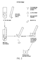
- FIG. 3 for a third embodiment employing a magnetic solid phase.
- the antibody is coated onto a magnetic particle which is added to a buffer solution.
- the fluid sample, containing the target cell is then added to the test tube in order to bind specific cells to the antibody coated magnetic particle.
- a magnetic field is then applied to the wall of the test tube in order to attract the magnetic particles to the wall.
- the remaining solution is then aspirated from the test tube, leaving the magnetic particles attached to the wall of the test tube.
- the cells are lysed and ATP is released from the cells bound to the antibody coated magnetic particles.
- the detection of the presence and concentration of the ATP is then performed via the firefly-luciferase assay.
- antibodies bound to the antigenic cell surface and the antibody-cell complexes can be captured on the magnetic particles. Extraction of ATP and detection would proceed as previously described.
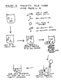
- Figure 4 illustrates another embodiment of the present invention utilizing a magnetic solid phase coated with avidin or streptavidin and a biotinylated cell-specific antibody.
- a fluid sample containing the target cells along with non-specific cells and non-cellular components is placed in a test tube.
- a biotinylated cell specific antibody is then added which binds to the target cell.
- the avidin or streptavidin coated magnetic particle is then bound to the biotinylated antibody forming a cell complex.
- the cell complex is then captured on the test tube wall via a magnet.
- the remaining fluid sample including the non-specific cell components and non-cellular components is aspirated leaving the magnetic particle complex magnetically bound to the test tube wall.
- ATP from the captured cells is released and is assayed by the firefly-luciferase technique.
- biotinylated antibody already bound to an avidin or streptavidin magnetic particle can be incubated with the sample in a single step procedure.
- FIG. 5 illustrates yet another embodiment of the present invention utilizing a magnetic solid phase with protein A.
- a fluid sample containing the target cells, non-specific cells and non-cellular material is placed in a test tube.
- a target cell specific antibody is then bound to the target cells.
- the antibody is then bound to the protein A magnetic particles forming a target cell complex.
- the cell complex is then retained on the test tube wall via a magnet in a similar fashion as that disclosed with respect to the embodiment illustrated in Figure 4.
- the fluid sample solution is then aspirated leaving the magnetic particle target cell complex bound to the test tube wall.
- Menz, E. T., supra ., 1986 which is incorporated herein by reference for a description of this process.
- ATP from the captured target cell is then released for assay with the firefly-luciferase technique.
- the present invention describes the use of specific cell capture techniques coupled with ATP detection as a method of target cell detection.
- target cells are captured onto a solid phase by any number of means, washed of any contaminating components or non-specific cells, and cellular ATP is released for assay by the firefly-luciferase enzyme system.
- the present invention is also directed to kits that utilize this method.
- a diagnostic kit for the detection of the presence of an antigenic cell substance in a sample would include the following:
- the kit can be conveniently modified to accommodate solid carriers, cell binding molecules, cell lysing solution and assay reagents described hereinabove and in the following examples.
- the present invention is preferably directed to the detection of contaminating organisms in a sample, such as a food or fluid sample.
- a food specimen thought to be contaminated with Salmonella cells is enriched for Salmonella cells by growth in an enrichment broth and added to a microtiter dish well that has been previously coated with antibodies to Salmonella cells.
- the Salmonella cells are allowed to bind to the antibodies and the plate is washed to remove non-specific cells or other materials.
- a lysis solution containing acid and/or detergent is then added to the well to lyse the bacterial cells.
- a kit for this purpose would contain an antibody coated solid phase or a variation, buffers, wash solutions, a cell lysing solution and ATP assay reagents to perform the assay.
- Example 1 was designed to disclose the capture and detection of Salmonella cells.
- a flat-bottomed ELISA plate (Nunc TM , Naperville, Ill) was coated with anti- Salmonella antibody (Kirkegard and Perry, Bac Trace TM , Catalog No. 01-91-99, Gathersburg, Md).
- One milligram of lyophilized antibody was reconstituted with 50% (v/v) glycerol.
- This glycerol solution was diluted 1:100 (22 uL to 2.2 mL) in 50 mM sodium carbonate, pH. 9.6. Fifty uL of the sodium carbonate diluted antibody was placed into each well of the microtiter dish and antibody was allowed to bind overnite (15 hours) at room temperature (23°C).
- a row of wells on the plate was not coated with antibody to serve as a minus antibody control.
- PBS phosphate buffered saline
- Each well was then blocked with 100 uL of a solution of PBS containing 1% (w/v) casein hydrolysate (Sigma, St. Louis, MO) and 5% (v/v) Tween-20 (Bio-Rad Laboratories, Richmond, CA) for 1 hour at 23°C. Following blocking, the plate was washed three times with PBS containing 0.05% Tween-20.
- Two overnight cultures of bacterial cells were prepared for use in the capture assay.
- the first was Salmonella typhimurium (American Type Culture Collection, ATCC 14028, Rockville, Md) which is the capture specific, and Klebsiella pneumoniae (ATCC 23357) which is a control, non-specific organism. Both cultures were grown overnight in flasks containing LB broth at 37°C at 250 rpm. Cells for each culture were serially diluted in PBS (10 ⁇ 1, 10 ⁇ 2, 10 ⁇ 3, 10 ⁇ 4, 10 ⁇ 5, 10 ⁇ 6, 10 ⁇ 7). Fifty uL of the dilutions were added to appropriate microtiter wells as shown in Table 1. The cells were allowed to incubate for 1.5 hours at 23°C. In addition, 50 uL of each of the cell dilutions was plated on LB agar (Maniatis, T. et al., supra .) in order to determine the number of cells in the original overnight cultures.
- the wells were individually aspirated into a sodium hypochlorite trap via vacuum, and each well was washed three times with 300 mL PBS, aspirating the PBS after each wash step.
- a non- Salmonella cell, Klebsiella shows a very low capture efficiency in this experiment. Even at 107 cells, less than 1 RLU is observed. This demonstrates the specificity of the proposed invention.
- the sensitivity of this experiment is on the order of 7 x 10⁴ Salmonella cells (twice the signal of the 0.2 RLU background). This sensitivity can likely be improved upon, by addition of a larger portion of the cell lysis solution to the light reaction. Only one-tenth of the lysate was added in this example.
- Example 2 was designed to disclose the capture and detection of the yeast Candida albicans using the present invention.
- IgG from antiserum to Candida albicans (Difco, Detroit, MI) was purified on a Protein A sepharose CL-4B (Pharmacia, Piscataway, NJ) column. Approximately 2.5 mL of antiserum in 0.2M sodium phosphate, pH 8.2 was applied to a 3 mL column previously equilibrated with 10 mM sodium phosphate, 0.15 M NaCl, pH 8.2 and washed with 10 column volumes of buffer. IgG was eluted from the column in 2 fractions (6 mL total) with 0.1M sodium citrate, pH 3.0. The eluted IgG (2 mg) was dialyzed against PBS and aliquots were stored frozen at -70°C.
- IgG was diluted 1:20 in 50 mM sodium carbonate, pH 9.6, and 50 uL was placed into flat-bottom microtiter dish (Nunc TM , Naperville, Ill) wells and allowed to adsorb overnight at 23°C. In the morning wells were rinsed and aspirated three times with 300 mL PBS containing 0.05% Tween-20. These wells and another row of wells (no IgG bound) were blocked with PBS containing 1% casein hydrolysate and 5% Tween-20 for 1.5 hours at 23°C. All wells were then rinsed three times with PBS containing 0.05% Tween-20.
- Candida albicans cells were grown in flasks containing LB broth overnight at 37°C at 250 rpm shaking. Cells were serially diluted in PBS (10 ⁇ 1, 10 ⁇ 2, 10 ⁇ 3, 10 ⁇ 4, 10 ⁇ 5, 10 ⁇ 6, 10 ⁇ 7) and 50 uL of each dilution and undiluted overnight controls were incubated and then plated on LB agar to determine cell numbers in the original overnight culture. Cells (50 uL of each dilution) were added to microtiter dish wells as shown in Table 3. The cells were allowed to incubate for 1.5 hours and then cells were removed by aspiration and the wells were rinsed three times with 300 uL PBS.
- Figure 7 illustrates a dose response of increasing RLU for increasing cells added when anti- Candida antibody is coated on the ELISA plate wells.
- the capture (RLU) is significantly less (about one hundred fold).
- the detection limit (at about 2 times background) is approximately 200 cells.
Landscapes
- Health & Medical Sciences (AREA)
- Life Sciences & Earth Sciences (AREA)
- Chemical & Material Sciences (AREA)
- Engineering & Computer Science (AREA)
- Immunology (AREA)
- Organic Chemistry (AREA)
- Molecular Biology (AREA)
- Zoology (AREA)
- General Health & Medical Sciences (AREA)
- Proteomics, Peptides & Aminoacids (AREA)
- Microbiology (AREA)
- Urology & Nephrology (AREA)
- Analytical Chemistry (AREA)
- Wood Science & Technology (AREA)
- Physics & Mathematics (AREA)
- Biochemistry (AREA)
- Hematology (AREA)
- Biomedical Technology (AREA)
- Biotechnology (AREA)
- Genetics & Genomics (AREA)
- General Engineering & Computer Science (AREA)
- Cell Biology (AREA)
- Bioinformatics & Cheminformatics (AREA)
- Biophysics (AREA)
- Food Science & Technology (AREA)
- Medicinal Chemistry (AREA)
- General Physics & Mathematics (AREA)
- Pathology (AREA)
- Measuring Or Testing Involving Enzymes Or Micro-Organisms (AREA)
Applications Claiming Priority (2)
| Application Number | Priority Date | Filing Date | Title |
|---|---|---|---|
| US46764990A | 1990-01-19 | 1990-01-19 | |
| US467649 | 1999-12-20 |
Publications (1)
| Publication Number | Publication Date |
|---|---|
| EP0441469A1 true EP0441469A1 (fr) | 1991-08-14 |
Family
ID=23856551
Family Applications (1)
| Application Number | Title | Priority Date | Filing Date |
|---|---|---|---|
| EP91300007A Withdrawn EP0441469A1 (fr) | 1990-01-19 | 1991-01-02 | Essai immunospécifique et bioluminescent d'ATP cellulaire |
Country Status (2)
| Country | Link |
|---|---|
| EP (1) | EP0441469A1 (fr) |
| AU (1) | AU6847890A (fr) |
Cited By (12)
| Publication number | Priority date | Publication date | Assignee | Title |
|---|---|---|---|---|
| EP0566010A1 (fr) * | 1992-04-13 | 1993-10-20 | BIOLAC GmbH Gesellschaft für industrielle Nutzung von Milchinhaltsstoffen | Procédé et kit d'analyse pour déterminer le nombre de germes bactériens dans le petit-lait et ses composants |
| WO1994028169A1 (fr) * | 1993-06-01 | 1994-12-08 | Celsis International Plc | Reactifs utilises en bioluminescence |
| US5759864A (en) * | 1995-06-23 | 1998-06-02 | Cedars Sinai Medical Center | Methods for reducing background binding in antibody preparations |
| US5798214A (en) * | 1994-07-13 | 1998-08-25 | The Secretary Of State For Defence In Her Britannic Majesty's Government Of The United Kingdom Of Great Britain And Northern Ireland Of Defence Evaluation And Research Agency | Capture asays |
| EP1223429A1 (fr) * | 2001-01-13 | 2002-07-17 | Dentognostics GmbH | Méthode pour la détection de micro-organismes vivants |
| WO2003012447A2 (fr) * | 2001-07-23 | 2003-02-13 | Institut Für Chemo-Und Biosensorik Münster E.V. | Dispositif et procede pour la mise en evidence de micro-organismes, de cellules eucaryotes ou d'organites vivants |
| WO2003025208A1 (fr) * | 2001-09-21 | 2003-03-27 | The Secretary Of State For Defence | Procede pour determiner la presence de bacteries resistant a des antibiotiques de lyse cellulaire |
| WO2005028680A2 (fr) * | 2003-09-12 | 2005-03-31 | Biocontrol Systems, Inc. | Methodes, compositions, et trousses pour concentration et detection de micro-organismes |
| EP1642985A1 (fr) * | 1999-05-13 | 2006-04-05 | The Secretary of State for Defence DSTL | Dosage cellulaire, méthode et réactifs |
| EP2060625A1 (fr) * | 2007-11-16 | 2009-05-20 | Dentognostics GMBH | Dispositif destiné à l'élution d'échantillons prévus pour l'analyse |
| US7897376B2 (en) | 2004-08-25 | 2011-03-01 | Sigma-Aldrich Co. | Method for extracting a target product from a host cell employing zwitterionic detergent combinations |
| US9116151B1 (en) * | 2009-04-27 | 2015-08-25 | University Of South Florida | Detection plate for ATP-bioluminescence immunoassay and method of manufacturing |
Citations (10)
| Publication number | Priority date | Publication date | Assignee | Title |
|---|---|---|---|---|
| US3660240A (en) * | 1969-05-05 | 1972-05-02 | Nasa | Flavin co-enzyme assay |
| US3745090A (en) * | 1970-08-04 | 1973-07-10 | Nasa | Method of detecting and counting bacteria in body fluids |
| EP0016552A1 (fr) * | 1979-02-28 | 1980-10-01 | Kendrew Biosystems, Inc. | Procédé de séparation magnétique de cellules et analogues et microsphères à utiliser dans ce procédé |
| US4282287A (en) * | 1980-01-24 | 1981-08-04 | Giese Roger W | Biochemical avidin-biotin multiple-layer system |
| DE3224484A1 (de) * | 1981-07-01 | 1983-01-20 | Yeda Research And Development Co., Ltd., Rehovot | Polymere mikrokuegelchen und verfahren zu ihrer herstellung |
| US4385113A (en) * | 1978-03-20 | 1983-05-24 | Nasa | Rapid, quantitative determination of bacteria in water |
| EP0153283A1 (fr) * | 1984-01-25 | 1985-08-28 | Symbicom Ab | Méthode de concentration et de détection de biomolécules et de cellules |
| WO1986007094A1 (fr) * | 1985-05-22 | 1986-12-04 | University Of Wales College Of Medicine | Agent d'extraction de cellules bacteriennes |
| WO1989002929A1 (fr) * | 1987-09-23 | 1989-04-06 | Life Science International Ab | Analyse luminometrique d'atp cellulaire |
| EP0363510A1 (fr) * | 1988-10-12 | 1990-04-18 | Biotest AG | Procédés de recherche et d'identification des anticorps d'érythrocyte à l'aide d'une méthode à phase solide |
-
1990
- 1990-12-21 AU AU68478/90A patent/AU6847890A/en not_active Abandoned
-
1991
- 1991-01-02 EP EP91300007A patent/EP0441469A1/fr not_active Withdrawn
Patent Citations (10)
| Publication number | Priority date | Publication date | Assignee | Title |
|---|---|---|---|---|
| US3660240A (en) * | 1969-05-05 | 1972-05-02 | Nasa | Flavin co-enzyme assay |
| US3745090A (en) * | 1970-08-04 | 1973-07-10 | Nasa | Method of detecting and counting bacteria in body fluids |
| US4385113A (en) * | 1978-03-20 | 1983-05-24 | Nasa | Rapid, quantitative determination of bacteria in water |
| EP0016552A1 (fr) * | 1979-02-28 | 1980-10-01 | Kendrew Biosystems, Inc. | Procédé de séparation magnétique de cellules et analogues et microsphères à utiliser dans ce procédé |
| US4282287A (en) * | 1980-01-24 | 1981-08-04 | Giese Roger W | Biochemical avidin-biotin multiple-layer system |
| DE3224484A1 (de) * | 1981-07-01 | 1983-01-20 | Yeda Research And Development Co., Ltd., Rehovot | Polymere mikrokuegelchen und verfahren zu ihrer herstellung |
| EP0153283A1 (fr) * | 1984-01-25 | 1985-08-28 | Symbicom Ab | Méthode de concentration et de détection de biomolécules et de cellules |
| WO1986007094A1 (fr) * | 1985-05-22 | 1986-12-04 | University Of Wales College Of Medicine | Agent d'extraction de cellules bacteriennes |
| WO1989002929A1 (fr) * | 1987-09-23 | 1989-04-06 | Life Science International Ab | Analyse luminometrique d'atp cellulaire |
| EP0363510A1 (fr) * | 1988-10-12 | 1990-04-18 | Biotest AG | Procédés de recherche et d'identification des anticorps d'érythrocyte à l'aide d'une méthode à phase solide |
Cited By (20)
| Publication number | Priority date | Publication date | Assignee | Title |
|---|---|---|---|---|
| EP0566010A1 (fr) * | 1992-04-13 | 1993-10-20 | BIOLAC GmbH Gesellschaft für industrielle Nutzung von Milchinhaltsstoffen | Procédé et kit d'analyse pour déterminer le nombre de germes bactériens dans le petit-lait et ses composants |
| US5677140A (en) * | 1992-04-13 | 1997-10-14 | Biolac Gmbh | Process and test kit for determining the bacterial count level in whey and whey products |
| WO1994028169A1 (fr) * | 1993-06-01 | 1994-12-08 | Celsis International Plc | Reactifs utilises en bioluminescence |
| AU672707B2 (en) * | 1993-06-01 | 1996-10-10 | Celsis International Plc | Reagents for use in bioluminescence |
| US5770391A (en) * | 1993-06-01 | 1998-06-23 | Celsis International Plc | Reagents and methods for use in bioluminescence |
| US5798214A (en) * | 1994-07-13 | 1998-08-25 | The Secretary Of State For Defence In Her Britannic Majesty's Government Of The United Kingdom Of Great Britain And Northern Ireland Of Defence Evaluation And Research Agency | Capture asays |
| US5759864A (en) * | 1995-06-23 | 1998-06-02 | Cedars Sinai Medical Center | Methods for reducing background binding in antibody preparations |
| EP1642985A1 (fr) * | 1999-05-13 | 2006-04-05 | The Secretary of State for Defence DSTL | Dosage cellulaire, méthode et réactifs |
| WO2002055733A2 (fr) * | 2001-01-13 | 2002-07-18 | Dentognostics Gmbh | Procede pour deceler des micro-organismes vivants |
| WO2002055733A3 (fr) * | 2001-01-13 | 2002-09-12 | Dentognostics Gmbh | Procede pour deceler des micro-organismes vivants |
| EP1223429A1 (fr) * | 2001-01-13 | 2002-07-17 | Dentognostics GmbH | Méthode pour la détection de micro-organismes vivants |
| WO2003012447A2 (fr) * | 2001-07-23 | 2003-02-13 | Institut Für Chemo-Und Biosensorik Münster E.V. | Dispositif et procede pour la mise en evidence de micro-organismes, de cellules eucaryotes ou d'organites vivants |
| WO2003012447A3 (fr) * | 2001-07-23 | 2003-08-07 | Inst Chemo Biosensorik | Dispositif et procede pour la mise en evidence de micro-organismes, de cellules eucaryotes ou d'organites vivants |
| WO2003025208A1 (fr) * | 2001-09-21 | 2003-03-27 | The Secretary Of State For Defence | Procede pour determiner la presence de bacteries resistant a des antibiotiques de lyse cellulaire |
| US7648830B2 (en) | 2001-09-21 | 2010-01-19 | The Secretary Of State For Defence In Her Britannic Majesty's Government Of The United Kingdom Of Great Britain And Northern Ireland | Method for determining the presence of bacteria resistant to cell lysing antibiotics |
| WO2005028680A2 (fr) * | 2003-09-12 | 2005-03-31 | Biocontrol Systems, Inc. | Methodes, compositions, et trousses pour concentration et detection de micro-organismes |
| WO2005028680A3 (fr) * | 2003-09-12 | 2005-05-06 | Biocontrol Systems Inc | Methodes, compositions, et trousses pour concentration et detection de micro-organismes |
| US7897376B2 (en) | 2004-08-25 | 2011-03-01 | Sigma-Aldrich Co. | Method for extracting a target product from a host cell employing zwitterionic detergent combinations |
| EP2060625A1 (fr) * | 2007-11-16 | 2009-05-20 | Dentognostics GMBH | Dispositif destiné à l'élution d'échantillons prévus pour l'analyse |
| US9116151B1 (en) * | 2009-04-27 | 2015-08-25 | University Of South Florida | Detection plate for ATP-bioluminescence immunoassay and method of manufacturing |
Also Published As
| Publication number | Publication date |
|---|---|
| AU6847890A (en) | 1991-07-25 |
Similar Documents
| Publication | Publication Date | Title |
|---|---|---|
| JP3706890B2 (ja) | 試料中の生物を検出する方法 | |
| EP0542790B1 (fr) | Procede et materiel de separation, concentration et analyse de cellules | |
| Brewster et al. | Filtration capture and immunoelectrochemical detection for rapid assay of Escherichia coli O157: H7 | |
| Boyacı et al. | Amperometric determination of live Escherichia coli using antibody-coated paramagnetic beads | |
| Amani et al. | A review approaches to identify enteric bacterial pathogens | |
| EP0441469A1 (fr) | Essai immunospécifique et bioluminescent d'ATP cellulaire | |
| CA2445509A1 (fr) | Techniques rapides de numeration et de typage microbiens | |
| US20090170144A1 (en) | Determination of viable microorganisms using coated paramagnetic beads | |
| Kim et al. | Rapid single-cell detection of pathogenic bacteria for in situ determination of food safety | |
| US7241626B2 (en) | Isolation and confirmation of analytes from test devices | |
| US20030059839A1 (en) | Method for detecting pathogens using immunoassays | |
| Stanley | A survey of some commercially available kits and reagents which include bioluminescence or chemiluminescence for their operation: Including immunoassays, hybridization, labels, probes, blots and ATP‐based rapid microbiology. Products from more than forty companies | |
| RU2165081C2 (ru) | Способ индикации микроорганизмов | |
| US6344332B1 (en) | Methods for the rapid detection of actively respiring microorganisms | |
| JPH04262258A (ja) | 細胞atpの免疫特異性的および生物発光的検定法 | |
| Wyatt | Antibody‐based detection of microbiological analytes in food: aspects of development | |
| Miotti | Rapid methods for the molecular diagnosis of infectious diseases: current trends and applications | |
| JP2002181823A (ja) | 微生物の検出方法 | |
| JP2001004631A (ja) | 磁気ビーズ固定化抗体を用いた微生物の迅速検定法 | |
| Chorti | Bioassays for In-Field Detection of Disease Biomarkers and Pathogen Contaminants | |
| Wolk et al. | Commercial methods for identification and susceptibility testing of fungi | |
| Colliva | CURRENT STATE OFADVANCEMENTS IN RAPID DETECTION TECHNOLOGY FOR PATHOGENIC BACTERIA IN FOOD | |
| Rodrigues et al. | Newer methods for microbiologic diagnosis of pediatric infections | |
| Feltham et al. | Present Trends and Future Prospects for Rapid Methods and Automation in the Clinical Laboratory | |
| AU742814B2 (en) | Method of detecting organisms in a sample |
Legal Events
| Date | Code | Title | Description |
|---|---|---|---|
| PUAI | Public reference made under article 153(3) epc to a published international application that has entered the european phase |
Free format text: ORIGINAL CODE: 0009012 |
|
| AK | Designated contracting states |
Kind code of ref document: A1 Designated state(s): AT BE CH DE DK ES FR GB GR IT LI LU NL SE |
|
| STAA | Information on the status of an ep patent application or granted ep patent |
Free format text: STATUS: THE APPLICATION IS DEEMED TO BE WITHDRAWN |
|
| 18D | Application deemed to be withdrawn |
Effective date: 19920215 |