EP0432218B1 - Method for detecting cellular pathology - Google Patents
Method for detecting cellular pathology Download PDFInfo
- Publication number
- EP0432218B1 EP0432218B1 EP89910554A EP89910554A EP0432218B1 EP 0432218 B1 EP0432218 B1 EP 0432218B1 EP 89910554 A EP89910554 A EP 89910554A EP 89910554 A EP89910554 A EP 89910554A EP 0432218 B1 EP0432218 B1 EP 0432218B1
- Authority
- EP
- European Patent Office
- Prior art keywords
- spectrin
- breakdown products
- spectrin breakdown
- biological sample
- cellular
- Prior art date
- Legal status (The legal status is an assumption and is not a legal conclusion. Google has not performed a legal analysis and makes no representation as to the accuracy of the status listed.)
- Expired - Lifetime
Links
- 238000000034 method Methods 0.000 title claims abstract description 36
- 230000001413 cellular effect Effects 0.000 title claims abstract description 9
- 230000007170 pathology Effects 0.000 title claims abstract description 7
- 102000005890 Spectrin Human genes 0.000 claims abstract description 55
- 108010019965 Spectrin Proteins 0.000 claims abstract description 55
- 230000015556 catabolic process Effects 0.000 claims abstract description 28
- 210000004027 cell Anatomy 0.000 claims abstract description 8
- 238000001514 detection method Methods 0.000 claims abstract description 7
- 238000006731 degradation reaction Methods 0.000 claims description 14
- 239000000523 sample Substances 0.000 claims description 12
- 239000012472 biological sample Substances 0.000 claims description 10
- 210000001519 tissue Anatomy 0.000 claims description 10
- 230000030833 cell death Effects 0.000 claims description 9
- 230000006800 cellular catabolic process Effects 0.000 claims description 9
- 230000003902 lesion Effects 0.000 claims description 9
- 108090000765 processed proteins & peptides Proteins 0.000 claims description 5
- 210000004369 blood Anatomy 0.000 claims description 4
- 239000008280 blood Substances 0.000 claims description 4
- 208000014674 injury Diseases 0.000 claims description 4
- 208000028867 ischemia Diseases 0.000 claims description 4
- 102000004196 processed proteins & peptides Human genes 0.000 claims description 4
- 230000001537 neural effect Effects 0.000 claims description 3
- 230000001575 pathological effect Effects 0.000 claims description 3
- 201000006938 muscular dystrophy Diseases 0.000 claims description 2
- 238000010186 staining Methods 0.000 claims description 2
- 208000024891 symptom Diseases 0.000 claims description 2
- 210000001175 cerebrospinal fluid Anatomy 0.000 claims 3
- 210000000653 nervous system Anatomy 0.000 claims 2
- 208000024827 Alzheimer disease Diseases 0.000 claims 1
- 241000124008 Mammalia Species 0.000 claims 1
- 208000018737 Parkinson disease Diseases 0.000 claims 1
- 231100000573 exposure to toxins Toxicity 0.000 claims 1
- 230000008733 trauma Effects 0.000 claims 1
- 101000933296 Homo sapiens Transcription factor TFIIIB component B'' homolog Proteins 0.000 abstract description 9
- 101001087412 Homo sapiens Tyrosine-protein phosphatase non-receptor type 18 Proteins 0.000 abstract description 9
- 102100026002 Transcription factor TFIIIB component B'' homolog Human genes 0.000 abstract description 9
- 210000004292 cytoskeleton Anatomy 0.000 abstract description 6
- 238000003018 immunoassay Methods 0.000 abstract description 3
- 230000005684 electric field Effects 0.000 abstract description 2
- 210000004556 brain Anatomy 0.000 description 17
- 102000007590 Calpain Human genes 0.000 description 13
- 108010032088 Calpain Proteins 0.000 description 13
- 210000001947 dentate gyrus Anatomy 0.000 description 13
- 239000000047 product Substances 0.000 description 11
- 102000004169 proteins and genes Human genes 0.000 description 9
- 108090000623 proteins and genes Proteins 0.000 description 9
- 210000001320 hippocampus Anatomy 0.000 description 7
- OYPRJOBELJOOCE-UHFFFAOYSA-N Calcium Chemical compound [Ca] OYPRJOBELJOOCE-UHFFFAOYSA-N 0.000 description 6
- PEDCQBHIVMGVHV-UHFFFAOYSA-N Glycerine Chemical compound OCC(O)CO PEDCQBHIVMGVHV-UHFFFAOYSA-N 0.000 description 6
- 241001465754 Metazoa Species 0.000 description 6
- 241000699670 Mus sp. Species 0.000 description 6
- 229910052791 calcium Inorganic materials 0.000 description 6
- 239000011575 calcium Substances 0.000 description 6
- UXVMQQNJUSDDNG-UHFFFAOYSA-L Calcium chloride Chemical compound [Cl-].[Cl-].[Ca+2] UXVMQQNJUSDDNG-UHFFFAOYSA-L 0.000 description 5
- 239000001110 calcium chloride Substances 0.000 description 5
- 229910001628 calcium chloride Inorganic materials 0.000 description 5
- 235000011148 calcium chloride Nutrition 0.000 description 5
- 238000002415 sodium dodecyl sulfate polyacrylamide gel electrophoresis Methods 0.000 description 5
- RYGMFSIKBFXOCR-UHFFFAOYSA-N Copper Chemical compound [Cu] RYGMFSIKBFXOCR-UHFFFAOYSA-N 0.000 description 4
- 241000700159 Rattus Species 0.000 description 4
- 238000003556 assay Methods 0.000 description 4
- 229910052802 copper Inorganic materials 0.000 description 4
- 239000010949 copper Substances 0.000 description 4
- 210000001353 entorhinal cortex Anatomy 0.000 description 4
- LYRCQNDYYRPFMF-UHFFFAOYSA-N trimethyltin Chemical compound C[Sn](C)C LYRCQNDYYRPFMF-UHFFFAOYSA-N 0.000 description 4
- QTBSBXVTEAMEQO-UHFFFAOYSA-N Acetic acid Chemical compound CC(O)=O QTBSBXVTEAMEQO-UHFFFAOYSA-N 0.000 description 3
- KCXVZYZYPLLWCC-UHFFFAOYSA-N EDTA Chemical compound OC(=O)CN(CC(O)=O)CCN(CC(O)=O)CC(O)=O KCXVZYZYPLLWCC-UHFFFAOYSA-N 0.000 description 3
- 241000283973 Oryctolagus cuniculus Species 0.000 description 3
- 208000027418 Wounds and injury Diseases 0.000 description 3
- 230000006378 damage Effects 0.000 description 3
- 230000007850 degeneration Effects 0.000 description 3
- 230000003412 degenerative effect Effects 0.000 description 3
- DNJIEGIFACGWOD-UHFFFAOYSA-N ethyl mercaptane Natural products CCS DNJIEGIFACGWOD-UHFFFAOYSA-N 0.000 description 3
- 239000000499 gel Substances 0.000 description 3
- 230000003447 ipsilateral effect Effects 0.000 description 3
- 210000002966 serum Anatomy 0.000 description 3
- 239000003053 toxin Substances 0.000 description 3
- 231100000765 toxin Toxicity 0.000 description 3
- 108700012359 toxins Proteins 0.000 description 3
- DGVVWUTYPXICAM-UHFFFAOYSA-N β‐Mercaptoethanol Chemical compound OCCS DGVVWUTYPXICAM-UHFFFAOYSA-N 0.000 description 3
- 206010003694 Atrophy Diseases 0.000 description 2
- 241000699694 Gerbillinae Species 0.000 description 2
- DHMQDGOQFOQNFH-UHFFFAOYSA-N Glycine Chemical compound NCC(O)=O DHMQDGOQFOQNFH-UHFFFAOYSA-N 0.000 description 2
- 241000699666 Mus <mouse, genus> Species 0.000 description 2
- MQUQNUAYKLCRME-INIZCTEOSA-N N-tosyl-L-phenylalanyl chloromethyl ketone Chemical compound C1=CC(C)=CC=C1S(=O)(=O)N[C@H](C(=O)CCl)CC1=CC=CC=C1 MQUQNUAYKLCRME-INIZCTEOSA-N 0.000 description 2
- 239000000020 Nitrocellulose Substances 0.000 description 2
- 229920002684 Sepharose Polymers 0.000 description 2
- FAPWRFPIFSIZLT-UHFFFAOYSA-M Sodium chloride Chemical compound [Na+].[Cl-] FAPWRFPIFSIZLT-UHFFFAOYSA-M 0.000 description 2
- FJWGYAHXMCUOOM-QHOUIDNNSA-N [(2s,3r,4s,5r,6r)-2-[(2r,3r,4s,5r,6s)-4,5-dinitrooxy-2-(nitrooxymethyl)-6-[(2r,3r,4s,5r,6s)-4,5,6-trinitrooxy-2-(nitrooxymethyl)oxan-3-yl]oxyoxan-3-yl]oxy-3,5-dinitrooxy-6-(nitrooxymethyl)oxan-4-yl] nitrate Chemical compound O([C@@H]1O[C@@H]([C@H]([C@H](O[N+]([O-])=O)[C@H]1O[N+]([O-])=O)O[C@H]1[C@@H]([C@@H](O[N+]([O-])=O)[C@H](O[N+]([O-])=O)[C@@H](CO[N+]([O-])=O)O1)O[N+]([O-])=O)CO[N+](=O)[O-])[C@@H]1[C@@H](CO[N+]([O-])=O)O[C@@H](O[N+]([O-])=O)[C@H](O[N+]([O-])=O)[C@H]1O[N+]([O-])=O FJWGYAHXMCUOOM-QHOUIDNNSA-N 0.000 description 2
- 239000002671 adjuvant Substances 0.000 description 2
- 238000004458 analytical method Methods 0.000 description 2
- 238000003149 assay kit Methods 0.000 description 2
- 230000037444 atrophy Effects 0.000 description 2
- 210000001772 blood platelet Anatomy 0.000 description 2
- 239000000872 buffer Substances 0.000 description 2
- 210000003169 central nervous system Anatomy 0.000 description 2
- 230000001086 cytosolic effect Effects 0.000 description 2
- 230000034994 death Effects 0.000 description 2
- 230000002638 denervation Effects 0.000 description 2
- 230000000694 effects Effects 0.000 description 2
- 239000012530 fluid Substances 0.000 description 2
- 239000011544 gradient gel Substances 0.000 description 2
- 230000000971 hippocampal effect Effects 0.000 description 2
- 239000007924 injection Substances 0.000 description 2
- 238000002347 injection Methods 0.000 description 2
- 210000003734 kidney Anatomy 0.000 description 2
- 210000004185 liver Anatomy 0.000 description 2
- 239000012528 membrane Substances 0.000 description 2
- 210000000110 microvilli Anatomy 0.000 description 2
- 210000004165 myocardium Anatomy 0.000 description 2
- 210000002569 neuron Anatomy 0.000 description 2
- 229920001220 nitrocellulos Polymers 0.000 description 2
- 229940079938 nitrocellulose Drugs 0.000 description 2
- 230000008569 process Effects 0.000 description 2
- 230000004044 response Effects 0.000 description 2
- 239000012723 sample buffer Substances 0.000 description 2
- 210000000225 synapse Anatomy 0.000 description 2
- XLYOFNOQVPJJNP-UHFFFAOYSA-N water Substances O XLYOFNOQVPJJNP-UHFFFAOYSA-N 0.000 description 2
- QRXMUCSWCMTJGU-UHFFFAOYSA-L (5-bromo-4-chloro-1h-indol-3-yl) phosphate Chemical compound C1=C(Br)C(Cl)=C2C(OP([O-])(=O)[O-])=CNC2=C1 QRXMUCSWCMTJGU-UHFFFAOYSA-L 0.000 description 1
- VADKRMSMGWJZCF-UHFFFAOYSA-N 2-bromophenol Chemical compound OC1=CC=CC=C1Br VADKRMSMGWJZCF-UHFFFAOYSA-N 0.000 description 1
- QFVHZQCOUORWEI-UHFFFAOYSA-N 4-[(4-anilino-5-sulfonaphthalen-1-yl)diazenyl]-5-hydroxynaphthalene-2,7-disulfonic acid Chemical compound C=12C(O)=CC(S(O)(=O)=O)=CC2=CC(S(O)(=O)=O)=CC=1N=NC(C1=CC=CC(=C11)S(O)(=O)=O)=CC=C1NC1=CC=CC=C1 QFVHZQCOUORWEI-UHFFFAOYSA-N 0.000 description 1
- SLXKOJJOQWFEFD-UHFFFAOYSA-N 6-aminohexanoic acid Chemical compound NCCCCCC(O)=O SLXKOJJOQWFEFD-UHFFFAOYSA-N 0.000 description 1
- 108010085238 Actins Proteins 0.000 description 1
- 102000007469 Actins Human genes 0.000 description 1
- 102000002260 Alkaline Phosphatase Human genes 0.000 description 1
- 108020004774 Alkaline Phosphatase Proteins 0.000 description 1
- 206010002660 Anoxia Diseases 0.000 description 1
- 241000976983 Anoxia Species 0.000 description 1
- 101710155556 Calcium-dependent protease Proteins 0.000 description 1
- 102100021238 Dynamin-2 Human genes 0.000 description 1
- 102000004190 Enzymes Human genes 0.000 description 1
- 108090000790 Enzymes Proteins 0.000 description 1
- VGGSQFUCUMXWEO-UHFFFAOYSA-N Ethene Chemical compound C=C VGGSQFUCUMXWEO-UHFFFAOYSA-N 0.000 description 1
- 239000005977 Ethylene Substances 0.000 description 1
- 239000004471 Glycine Substances 0.000 description 1
- 241000282412 Homo Species 0.000 description 1
- 101000817607 Homo sapiens Dynamin-2 Proteins 0.000 description 1
- 206010020880 Hypertrophy Diseases 0.000 description 1
- 206010021143 Hypoxia Diseases 0.000 description 1
- 241000699684 Meriones unguiculatus Species 0.000 description 1
- KWYHDKDOAIKMQN-UHFFFAOYSA-N N,N,N',N'-tetramethylethylenediamine Chemical compound CN(C)CCN(C)C KWYHDKDOAIKMQN-UHFFFAOYSA-N 0.000 description 1
- 102000008763 Neurofilament Proteins Human genes 0.000 description 1
- 108010088373 Neurofilament Proteins Proteins 0.000 description 1
- 102000035195 Peptidases Human genes 0.000 description 1
- 108091005804 Peptidases Proteins 0.000 description 1
- 239000004365 Protease Substances 0.000 description 1
- 229930006000 Sucrose Natural products 0.000 description 1
- CZMRCDWAGMRECN-UGDNZRGBSA-N Sucrose Chemical compound O[C@H]1[C@H](O)[C@@H](CO)O[C@@]1(CO)O[C@@H]1[C@H](O)[C@@H](O)[C@H](O)[C@@H](CO)O1 CZMRCDWAGMRECN-UGDNZRGBSA-N 0.000 description 1
- 206010042618 Surgical procedure repeated Diseases 0.000 description 1
- 239000007983 Tris buffer Substances 0.000 description 1
- 230000004913 activation Effects 0.000 description 1
- 238000007792 addition Methods 0.000 description 1
- 210000004100 adrenal gland Anatomy 0.000 description 1
- 238000001042 affinity chromatography Methods 0.000 description 1
- 230000004075 alteration Effects 0.000 description 1
- 229960002684 aminocaproic acid Drugs 0.000 description 1
- 230000007953 anoxia Effects 0.000 description 1
- 210000003050 axon Anatomy 0.000 description 1
- 230000003376 axonal effect Effects 0.000 description 1
- 230000000903 blocking effect Effects 0.000 description 1
- 230000017531 blood circulation Effects 0.000 description 1
- 230000036770 blood supply Effects 0.000 description 1
- 230000037396 body weight Effects 0.000 description 1
- 238000009835 boiling Methods 0.000 description 1
- 210000004958 brain cell Anatomy 0.000 description 1
- 230000000747 cardiac effect Effects 0.000 description 1
- 210000001715 carotid artery Anatomy 0.000 description 1
- 210000000170 cell membrane Anatomy 0.000 description 1
- 210000001638 cerebellum Anatomy 0.000 description 1
- 239000007795 chemical reaction product Substances 0.000 description 1
- 210000000172 cytosol Anatomy 0.000 description 1
- 230000006735 deficit Effects 0.000 description 1
- 238000000326 densiometry Methods 0.000 description 1
- 238000011161 development Methods 0.000 description 1
- 201000010099 disease Diseases 0.000 description 1
- 208000037265 diseases, disorders, signs and symptoms Diseases 0.000 description 1
- 238000002224 dissection Methods 0.000 description 1
- 239000000839 emulsion Substances 0.000 description 1
- 210000003743 erythrocyte Anatomy 0.000 description 1
- DEFVIWRASFVYLL-UHFFFAOYSA-N ethylene glycol bis(2-aminoethyl)tetraacetic acid Chemical compound OC(=O)CN(CC(O)=O)CCOCCOCCN(CC(O)=O)CC(O)=O DEFVIWRASFVYLL-UHFFFAOYSA-N 0.000 description 1
- 108010006620 fodrin Proteins 0.000 description 1
- 230000002518 glial effect Effects 0.000 description 1
- 239000011539 homogenization buffer Substances 0.000 description 1
- 238000003119 immunoblot Methods 0.000 description 1
- 238000000338 in vitro Methods 0.000 description 1
- 238000011534 incubation Methods 0.000 description 1
- 230000000968 intestinal effect Effects 0.000 description 1
- 230000002427 irreversible effect Effects 0.000 description 1
- 230000005923 long-lasting effect Effects 0.000 description 1
- 210000004072 lung Anatomy 0.000 description 1
- 210000004698 lymphocyte Anatomy 0.000 description 1
- 238000004519 manufacturing process Methods 0.000 description 1
- 239000003550 marker Substances 0.000 description 1
- 230000007246 mechanism Effects 0.000 description 1
- 238000012544 monitoring process Methods 0.000 description 1
- 230000003387 muscular Effects 0.000 description 1
- 230000017074 necrotic cell death Effects 0.000 description 1
- 210000001577 neostriatum Anatomy 0.000 description 1
- JPXMTWWFLBLUCD-UHFFFAOYSA-N nitro blue tetrazolium(2+) Chemical compound COC1=CC(C=2C=C(OC)C(=CC=2)[N+]=2N(N=C(N=2)C=2C=CC=CC=2)C=2C=CC(=CC=2)[N+]([O-])=O)=CC=C1[N+]1=NC(C=2C=CC=CC=2)=NN1C1=CC=C([N+]([O-])=O)C=C1 JPXMTWWFLBLUCD-UHFFFAOYSA-N 0.000 description 1
- 210000000578 peripheral nerve Anatomy 0.000 description 1
- 229920002401 polyacrylamide Polymers 0.000 description 1
- 230000002980 postoperative effect Effects 0.000 description 1
- 230000002028 premature Effects 0.000 description 1
- 230000002797 proteolythic effect Effects 0.000 description 1
- 230000000284 resting effect Effects 0.000 description 1
- 230000035945 sensitivity Effects 0.000 description 1
- 238000000926 separation method Methods 0.000 description 1
- 210000002460 smooth muscle Anatomy 0.000 description 1
- 239000011780 sodium chloride Substances 0.000 description 1
- 238000001179 sorption measurement Methods 0.000 description 1
- 241000894007 species Species 0.000 description 1
- 210000000952 spleen Anatomy 0.000 description 1
- 238000012453 sprague-dawley rat model Methods 0.000 description 1
- 239000007929 subcutaneous injection Substances 0.000 description 1
- 238000010254 subcutaneous injection Methods 0.000 description 1
- 210000001913 submandibular gland Anatomy 0.000 description 1
- 239000000758 substrate Substances 0.000 description 1
- 239000005720 sucrose Substances 0.000 description 1
- 239000013589 supplement Substances 0.000 description 1
- 230000000153 supplemental effect Effects 0.000 description 1
- 230000004083 survival effect Effects 0.000 description 1
- 238000012360 testing method Methods 0.000 description 1
- 210000001550 testis Anatomy 0.000 description 1
- 210000001541 thymus gland Anatomy 0.000 description 1
- -1 tissues Substances 0.000 description 1
- 238000012546 transfer Methods 0.000 description 1
- LENZDBCJOHFCAS-UHFFFAOYSA-N tris Chemical compound OCC(N)(CO)CO LENZDBCJOHFCAS-UHFFFAOYSA-N 0.000 description 1
Images



Classifications
-
- G—PHYSICS
- G01—MEASURING; TESTING
- G01N—INVESTIGATING OR ANALYSING MATERIALS BY DETERMINING THEIR CHEMICAL OR PHYSICAL PROPERTIES
- G01N33/00—Investigating or analysing materials by specific methods not covered by groups G01N1/00 - G01N31/00
- G01N33/48—Biological material, e.g. blood, urine; Haemocytometers
- G01N33/50—Chemical analysis of biological material, e.g. blood, urine; Testing involving biospecific ligand binding methods; Immunological testing
- G01N33/68—Chemical analysis of biological material, e.g. blood, urine; Testing involving biospecific ligand binding methods; Immunological testing involving proteins, peptides or amino acids
- G01N33/6893—Chemical analysis of biological material, e.g. blood, urine; Testing involving biospecific ligand binding methods; Immunological testing involving proteins, peptides or amino acids related to diseases not provided for elsewhere
-
- G—PHYSICS
- G01—MEASURING; TESTING
- G01N—INVESTIGATING OR ANALYSING MATERIALS BY DETERMINING THEIR CHEMICAL OR PHYSICAL PROPERTIES
- G01N33/00—Investigating or analysing materials by specific methods not covered by groups G01N1/00 - G01N31/00
- G01N33/48—Biological material, e.g. blood, urine; Haemocytometers
- G01N33/50—Chemical analysis of biological material, e.g. blood, urine; Testing involving biospecific ligand binding methods; Immunological testing
- G01N33/68—Chemical analysis of biological material, e.g. blood, urine; Testing involving biospecific ligand binding methods; Immunological testing involving proteins, peptides or amino acids
- G01N33/6893—Chemical analysis of biological material, e.g. blood, urine; Testing involving biospecific ligand binding methods; Immunological testing involving proteins, peptides or amino acids related to diseases not provided for elsewhere
- G01N33/6896—Neurological disorders, e.g. Alzheimer's disease
Definitions
- This invention relates to an in vitro method for the detection of cellular pathology, and more specifically to an assay for monitoring cytoskeleton breakdown products to determine cell necrosis.
- the structural integrity of cells is maintained in part by the cytoskeleton, a mesh-like structure composed primarily of proteins, which lies adjacent to the inner cell surface.
- the cytoskeleton of many cell types (a partial list includes neurons, lymphocytes, kidney, liver, cardiac and smooth muscle, and blood platelets) contain large amounts of a protein either identical to or closely related to brain spectrin (also known as fodrin).
- Calpain is an enzyme which in the presence of elevated calcium levels degrades spectrin, thereby leading to the disruption of the cytoskeleton.
- a variety of cellular insults e.g., toxins, anoxia, etc.
- disease states e.g., Alzheimer's, Parkinson's, muscular dystrophy
- a reliable method to detect degenerative events as soon as possible after their onset before pathological symptoms appear.
- Such method should have high sensitivity, wide ranging applicability and ease of administration.
- the present invention satisfies this need and provides related advantages.
- the invention provides a method for the detection of cellular pathology by means of an immunoassay to determine the presence of stable breakdown products, termed BDP's or BDP1 and BDP2, of the cytoskeleton component spectrin.
- BDP's stable breakdown products
- BDP1 and BDP2 stable breakdown products
- the components from a sample of spectrin-containing cells are physically separated, as by exposure to an electric field, in such a way that BDP and spectrin are separated.
- Antibodies reactive with BDP are then contacted with the separated sample, and their binding to that portion of the sample containing any BDP determined.
- Figure 1 A. Spectrin immunoreactivity in blotted samples of the contralateral (left lane) and ipsilateral (right lane) dentate gyrus two days after a unilateral lesion of the entorhinal cortex. Arrows indicate the alpha and beta spectrin subunits with apparent M r s of about 240 and 230 KDa respectively and two additional immunoreactive peptides (BDP1 and BDP2) with apparent M r s of about 155 and 150 KDa respectively.
- BDP1 and BDP2 two additional immunoreactive peptides
- Lane 1 Purified brain spectrin incubated under the following conditions. Lane 1: no additions; Lane 2: lmM CaCl2, 1.8 »g/ml calpain I, 10 minutes; Lane 3: 1mM CaCl2, 3 »g/ml calpain I, 30 minutes; Lane 4: 10 »g of dentate gyrus protein homogenate two days post-lesion. Lane 5: lmM CaCl2, 13 »g/ml calpain, 1.25 »g/ml colmodulin, 3 minutes; Lane 6: 1mM CaCl2, 13 »g/ml calpain, 1.25 »g/ml colmodulin, 30 minutes.
- BDP1 filled circles
- BDP2 open circles
- FIG. 1 Levels of BDP's in regions of the brain of control and Brindled mice, showing the effect of treatment with copper.
- the present invention relates to a sensitive and efficient method for the early detection of cellular death and degradation.
- the method involves detecting cellular death or degradation in a subject by analyzing a biological sample for the presence of spectrin breakdown products and comparing the quantity of spectrin breakdown products to the quantity of spectrin breakdown products in a normal subject, such that an increased level of spectrin breakdown products indicates cellular death or degradation in the subject.
- the method can be carried out, for example, by exposing the sample to an electrical gradient so as to separate the components in such a way that spectrin breakdown products are separated from spectrin, followed by staining the separated products so as to visualize the products and determine the presence of stain binding.
- the method involves an immuno assay using antibodies which recognize stable breakdown products of spectrin".
- Spectrin binds F-actin, and together they are generally associated with the inner face of the cell membrane, where they form a filamentous meshwork.
- Brain and many other tissues have been known for some time to express calcium-stimulated proteolytic activity.
- calpain a calcium-dependent protease, termed calpain
- Two forms of this protease have been identified in the brain and other tissues which are differentiated by their threshold for activation by calcium: calpain I requires micromolar calcium while calpain II is activated by concentrations of calcium between 0.1 and 0.5 mM. The two forms are differentially distributed in the brain. While calpain II is mainly localized in the cytoplasmic fraction of brain cells, the highest activity of calpain I is found in small processes.
- Brain spectrin can be purified to greater than 90% purity by the method of Davis and Bennett, J. Biol. Chem. 258:7757-7766 (1983), which is incorporated herein by reference.
- Calpain can be purified to a similar level of purity from rat erythrocyte cytosol according to the method of Seubert et al., Synapse 1:20-24 (1987), which is incorporated herein by reference.
- the gels were stained with Coomassie blue and restained with 7% acetic acid. See Fig. 1, Seubert et al. Synapse 1:20-24 (1987), which is incorporated herein by reference. Peptide bands of approximately 150,000 Daltons were found to increase as spectrin disappeared. The peptides of these bands were termed BDP's. The apparent stability of the BDP's towards further degradation suggested that antibodies directed against spectrin may recognize the BDP's in biological samples, such as tissues, fluids, etc.
- Antibodies to brain spectrin which are also reactive with BDP1 and BDP2, were raised in rabbits using well known procedures (See, for example, Hurn, B.A.L. and Chantler, S.M. Meth. Enz. 70:104-135 (1980) which is incorporated herein by reference).
- the anti-brain spectrin antibodies were purified from serum by brain spectrin-sepharose affinity chromatography using methods well-known in the art.
- brains were rapidly dissected in ice-cold homogenization buffer consisting of 0.32 M sucrose, 10mM Tris, 2mM EDTA, lmM ethylene glycol-bis B-amino-ethylether (EGTA) N,N,N′,N′-tetraacetic acid, 100 »M leupetin, 1 »g/ml N-tosyl-L-phenylalanine chloromethyl ketone (TPCK), pH 7.4
- TPCK N-tosyl-L-phenylalanine chloromethyl ketone
- the dissected dentate gyrus was homogenized in 500 »l of dissection buffer. An aliquot was added to one-third volume of 3X's SDS-PAGE sample buffer (consisting of 150mM Tris-Po4, 5%, SDS, 30% glycerol, 3.75mM EDTA, 3% mercaptoethanol, pH 6.8) and placed in a boiling water bath for three minutes.
- the protein concentration of the homogenate was determined by the method of Bradford, Anal. Biochem. 72:248-254 (1976), which is incorporated herein by reference, and the concentration of proteins in sample buffer adjusted to 0.33 mg/ml.
- Antibodies were produced by the following method: For each rabbit approximately 200 »g of purified brain spectrin was excised from SDS-polyacrylamide gels (after electrophoretic separation) and emulsified with Freund's complete adjuvant. Multiple subdermal injections were made and the procedure repeated again after 2 to 4 weeks, using Freund's Incomplete Adjuvant. After an additional two weeks subcutaneous injections of an emulsion containing approximately 100 »g of spectrin were made. This procedure was repeated approximately one month later. Ten days following this series of injections, approximately 20 ml of blood was drawn from the rabbit and the serum collected after allowing the blood to clot overnight at 4°C.
- Antibodies to brain spectrin were isolated from the serum by adsorption to brain spectrin which has been coupled to 6-amino hexanoic acid activated sepharose 4B (Sigma Chemical Co., St. Louis, MO). The specifically bound antibodies were then eluted in 0.2 M glycine, pH 2.8. The affinity purified antibodies were then equilibrated to pH 7.4 and frozen until use.
- Quantitation of the immunoreactive species was made using a soft laser scanning densitometer (Model #SLR504-XL, BioMed Instruments, Fullerton, CA). An integrator 4270 Varion, Sunnyvale, CA) summed the amount of reaction product in each band and expressed them as a % of the total in that sample.
- the anti-spectrin reactive species present in the dentate gyrus two days after a unilateral entorhinal lesion ar shown in Figure 1A.
- the homogenates of the ipsilateral dentate gyrus exhibited a marked increase in the amount of two peptides, termed BDP1 and BDP2, with apparent M r S of about 155,000 and 150,000 Daltons respectively.
- BDP1 and BDP2 The time course of the changes in BDP1 and BDP2 in the dentate gyrus following a unilateral entorhinal cortex lesion is shown in Figure 2.
- BDP's are usually below the limit of detection in samples from unoperated animals.
- a significant elevation of BDP's in the ipsilateral sample is evident as early as four hours post-lesion.
- the increase is maximal two days after the lesion, where the BDP's represent 25% of the total immunoreactivity. Two and even four weeks after the lesion, the amounts of BDP's were still significantly increased; the contralateral dentate gyri at two and four weeks showed no detectable BDP's and average BDP2 levels were less than 0.1%. Small increases in the amounts of BDP's were observed in the contralateral region during the first week following the lesion as compared to non-operated animals. These results indicate that removal of the main input to the dentate gyrus is followed by a rapid and longlasting increased degradation of the cytoskeletal protein brain spectrin.
- Example II Samples from the hippocampus, cortex and striatum of 12 day old mouse pups were processed as described in Example I.
- the experimental groups were control mice, Brindled mice, and Brindled mice receiving supplemental copper, a treatment which prevents the premature death which otherwise occurs.
- Brindled mice are characterized in that they have a copper deficit which, untreated causes normal degradation.
- spectrin BDP's are elevated in the pathological condition and this elevation is blocked by the copper supplement.
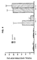
- Rats were injected (i.p.) with 10 mg TMT/kg body weight. At various times post treatment the dentate gyrus and CAl regions of the hippocampus were removed and analyzed for BDP's as described in Example I. Massive increases in BDP's were noted in regions most at risk to this toxin (See Balabin et al., Neurosa 26:337-361 (1988), which is incorporated herein by reference, as depicted in Figure 4.
- Spectrin is found in a variety of other tissues. For example, similar BDP's of spectrin have been observed in blood platelets (Fox et al., Blood 69 :537-545 (1987)) and intestinal brush border cells (Glenney et al., PNAS 79 :4002-4005 (1982)). The analysis described above can be extended to other tissues and fluids. The following tissue taken from rats have been examined using the methods described above, and found to exhibit spectrin and BDP's: submandibular gland, brush border, testes, thymus, skeletal muscular, heart muscle, lung, liver, spleen, adrenal gland, kidney, brain. Additionally humans, gerbils and mice have also been determined to contain spectrin and BDP's, suggesting that both are common to animals.
Landscapes
- Life Sciences & Earth Sciences (AREA)
- Health & Medical Sciences (AREA)
- Engineering & Computer Science (AREA)
- Biomedical Technology (AREA)
- Molecular Biology (AREA)
- Chemical & Material Sciences (AREA)
- Urology & Nephrology (AREA)
- Hematology (AREA)
- Immunology (AREA)
- Cell Biology (AREA)
- General Health & Medical Sciences (AREA)
- Biotechnology (AREA)
- Proteomics, Peptides & Aminoacids (AREA)
- Food Science & Technology (AREA)
- Medicinal Chemistry (AREA)
- Physics & Mathematics (AREA)
- Analytical Chemistry (AREA)
- Biochemistry (AREA)
- Microbiology (AREA)
- General Physics & Mathematics (AREA)
- Pathology (AREA)
- Neurology (AREA)
- Neurosurgery (AREA)
- Peptides Or Proteins (AREA)
- Investigating Or Analysing Biological Materials (AREA)
- Measuring Or Testing Involving Enzymes Or Micro-Organisms (AREA)
Applications Claiming Priority (3)
| Application Number | Priority Date | Filing Date | Title |
|---|---|---|---|
| US24015488A | 1988-09-02 | 1988-09-02 | |
| US240154 | 1988-09-02 | ||
| PCT/US1989/003748 WO1990002949A1 (en) | 1988-09-02 | 1989-08-30 | Method for detecting cellular pathology |
Publications (3)
| Publication Number | Publication Date |
|---|---|
| EP0432218A1 EP0432218A1 (en) | 1991-06-19 |
| EP0432218A4 EP0432218A4 (en) | 1992-01-22 |
| EP0432218B1 true EP0432218B1 (en) | 1995-06-28 |
Family
ID=22905332
Family Applications (1)
| Application Number | Title | Priority Date | Filing Date |
|---|---|---|---|
| EP89910554A Expired - Lifetime EP0432218B1 (en) | 1988-09-02 | 1989-08-30 | Method for detecting cellular pathology |
Country Status (7)
| Country | Link |
|---|---|
| EP (1) | EP0432218B1 (da) |
| JP (1) | JP2566327B2 (da) |
| AT (1) | ATE124537T1 (da) |
| CA (1) | CA1340182C (da) |
| DE (1) | DE68923285T2 (da) |
| DK (1) | DK33991A (da) |
| WO (1) | WO1990002949A1 (da) |
Families Citing this family (2)
| Publication number | Priority date | Publication date | Assignee | Title |
|---|---|---|---|---|
| US5118606A (en) | 1988-09-02 | 1992-06-02 | The Regents Of The University Of California | Methods for detecting cellular pathology by assaying spectrin and spectrin breakdown products |
| FR2745174B1 (fr) † | 1996-02-22 | 1998-04-30 | Oreal | Composition cosmetique de fixation et de brillance |
-
1989
- 1989-08-30 WO PCT/US1989/003748 patent/WO1990002949A1/en not_active Ceased
- 1989-08-30 JP JP1509837A patent/JP2566327B2/ja not_active Expired - Lifetime
- 1989-08-30 DE DE68923285T patent/DE68923285T2/de not_active Expired - Fee Related
- 1989-08-30 EP EP89910554A patent/EP0432218B1/en not_active Expired - Lifetime
- 1989-08-30 AT AT89910554T patent/ATE124537T1/de not_active IP Right Cessation
- 1989-09-05 CA CA000610248A patent/CA1340182C/en not_active Expired - Fee Related
-
1991
- 1991-02-27 DK DK033991A patent/DK33991A/da not_active Application Discontinuation
Non-Patent Citations (1)
| Title |
|---|
| PROC. NATL. ACAD. SCI, USA, Vol. 79, 1982, pages 4002-4005, see the Abstract and Introduction; J.R. GLENNEY et al.: "Erythroid spectrin, brain fodrin and intestinal brush border-proteins (TW-260/240) are related molecules containing a common calmodulin-binding subunit bound to a variant cell type-specific subunit". * |
Also Published As
| Publication number | Publication date |
|---|---|
| DK33991D0 (da) | 1991-02-27 |
| ATE124537T1 (de) | 1995-07-15 |
| EP0432218A4 (en) | 1992-01-22 |
| EP0432218A1 (en) | 1991-06-19 |
| DK33991A (da) | 1991-02-27 |
| DE68923285T2 (de) | 1995-11-09 |
| DE68923285D1 (de) | 1995-08-03 |
| JPH04504753A (ja) | 1992-08-20 |
| CA1340182C (en) | 1998-12-15 |
| WO1990002949A1 (en) | 1990-03-22 |
| JP2566327B2 (ja) | 1996-12-25 |
Similar Documents
| Publication | Publication Date | Title |
|---|---|---|
| EP0540591B1 (en) | Cell necrosis detection through assays for spectrin and breakdown products thereof | |
| Moir et al. | Relative increase in Alzheimer's disease of soluble forms of cerebral Aβ amyloid protein precursor containing the Kunitz protease inhibitory domain | |
| Jöns et al. | Identification of the binding interface involved in linkage of cytoskeletal protein 4.1 to the erythrocyte anion exchanger. | |
| Köpke et al. | Microtubule-associated protein tau. Abnormal phosphorylation of a non-paired helical filament pool in Alzheimer disease. | |
| Udvardy | Purification and characterization of a multiprotein component of the Drosophila 26 S (1500 kDa) proteolytic complex. | |
| Baudier et al. | Purification and characterization of a brain-specific protein kinase C substrate, neurogranin (p17). Identification of a consensus amino acid sequence between neurogranin and neuromodulin (GAP43) that corresponds to the protein kinase C phosphorylation site and the calmodulin-binding domain. | |
| De Graan et al. | Determination of changes in the phosphorylation state of the neuron‐specific protein kinase C substrate B‐50 (GAP43) by quantitative immunoprecipitation | |
| EP0783695B1 (en) | Cell tests and diagnostic kit for alzheimer's disease | |
| Wong et al. | An isoelectric variant of the 150,000-dalton neurofilament polypeptide. Evidence that phosphorylation state affects its association with the filament. | |
| Lusis et al. | Isolation, characterization, and radioimmunoassay of murine egasyn, a protein stabilizing glucuronidase membrane binding | |
| Bush et al. | An abnormality of plasma amyloid protein precursor in Alzheimer's disease | |
| WO1992000374A1 (en) | Protease clipsin and method of use | |
| Becker et al. | Sensitive immunoassay shows selective association of peripheral and integral membrane proteins of the inhibitory glycine receptor complex | |
| Benuck et al. | Changes in brain protease activity in aging | |
| Grundke-Iqbal et al. | Alzheimer paired helical filaments: cross-reacting polypeptide/s normally present in brain | |
| EP0432218B1 (en) | Method for detecting cellular pathology | |
| Dahl et al. | The glial fibrillary acidic protein and astrocytic 10-nanometer filaments | |
| DE68922340T2 (de) | Polyfunktionelle Protease. | |
| Borgese et al. | Concentration of NADH-cytochrome b5 reductase in erythrocytes of normal and methemoglobinemic individuals measured with a quantitative radioimmunoblotting assay. | |
| Nicoletti et al. | Changes of mitochondrial cytochrome c oxidase and FoF1 ATP synthase subunits in rat cerebral cortex during aging | |
| Yang et al. | Extravasation of plasma proteins can confound interpretation of proteomic studies of brain: a lesson from apo AI in mesial temporal lobe epilepsy | |
| Scherberich et al. | Routine diagnosis with PhastSystem compared to conventional electrophoresis: Automated sodium dodecyl sulfate‐polyacrylamide gel electrophoresis, silver staining and Western blotting of urinary proteins | |
| Zanetta et al. | Antibodies against the membrane‐bound acetylcholinesterase from adult rat brain | |
| Willmott et al. | Molecular characterisation and structural relationship of the synapse‐enriched glycoproteins gp65 and gp55 | |
| Cano et al. | The role of membrane lectins in Dictyostelium discoideum aggregation as ascertained by specific univalent antibodies against discoidin I |
Legal Events
| Date | Code | Title | Description |
|---|---|---|---|
| PUAI | Public reference made under article 153(3) epc to a published international application that has entered the european phase |
Free format text: ORIGINAL CODE: 0009012 |
|
| 17P | Request for examination filed |
Effective date: 19910220 |
|
| AK | Designated contracting states |
Kind code of ref document: A1 Designated state(s): AT BE CH DE FR GB IT LI LU NL SE |
|
| RIN1 | Information on inventor provided before grant (corrected) |
Inventor name: SEUBERT, PETER, A. Inventor name: LYNCH, GARY, S. |
|
| A4 | Supplementary search report drawn up and despatched |
Effective date: 19911202 |
|
| AK | Designated contracting states |
Kind code of ref document: A4 Designated state(s): AT BE CH DE FR GB IT LI LU NL SE |
|
| 17Q | First examination report despatched |
Effective date: 19930804 |
|
| GRAA | (expected) grant |
Free format text: ORIGINAL CODE: 0009210 |
|
| AK | Designated contracting states |
Kind code of ref document: B1 Designated state(s): AT BE CH DE FR GB IT LI LU NL SE |
|
| REF | Corresponds to: |
Ref document number: 124537 Country of ref document: AT Date of ref document: 19950715 Kind code of ref document: T |
|
| ITF | It: translation for a ep patent filed | ||
| REF | Corresponds to: |
Ref document number: 68923285 Country of ref document: DE Date of ref document: 19950803 |
|
| ET | Fr: translation filed | ||
| PLBE | No opposition filed within time limit |
Free format text: ORIGINAL CODE: 0009261 |
|
| STAA | Information on the status of an ep patent application or granted ep patent |
Free format text: STATUS: NO OPPOSITION FILED WITHIN TIME LIMIT |
|
| 26N | No opposition filed | ||
| PGFP | Annual fee paid to national office [announced via postgrant information from national office to epo] |
Ref country code: FR Payment date: 19980720 Year of fee payment: 10 |
|
| PGFP | Annual fee paid to national office [announced via postgrant information from national office to epo] |
Ref country code: SE Payment date: 19980721 Year of fee payment: 10 |
|
| PGFP | Annual fee paid to national office [announced via postgrant information from national office to epo] |
Ref country code: AT Payment date: 19980723 Year of fee payment: 10 |
|
| PGFP | Annual fee paid to national office [announced via postgrant information from national office to epo] |
Ref country code: GB Payment date: 19980724 Year of fee payment: 10 |
|
| PGFP | Annual fee paid to national office [announced via postgrant information from national office to epo] |
Ref country code: CH Payment date: 19980727 Year of fee payment: 10 |
|
| PGFP | Annual fee paid to national office [announced via postgrant information from national office to epo] |
Ref country code: NL Payment date: 19980728 Year of fee payment: 10 |
|
| PGFP | Annual fee paid to national office [announced via postgrant information from national office to epo] |
Ref country code: DE Payment date: 19980729 Year of fee payment: 10 |
|
| PGFP | Annual fee paid to national office [announced via postgrant information from national office to epo] |
Ref country code: BE Payment date: 19980813 Year of fee payment: 10 |
|
| PGFP | Annual fee paid to national office [announced via postgrant information from national office to epo] |
Ref country code: LU Payment date: 19980902 Year of fee payment: 10 |
|
| PG25 | Lapsed in a contracting state [announced via postgrant information from national office to epo] |
Ref country code: SE Free format text: THE PATENT HAS BEEN ANNULLED BY A DECISION OF A NATIONAL AUTHORITY Effective date: 19990830 Ref country code: LU Free format text: LAPSE BECAUSE OF NON-PAYMENT OF DUE FEES Effective date: 19990830 Ref country code: GB Free format text: LAPSE BECAUSE OF NON-PAYMENT OF DUE FEES Effective date: 19990830 Ref country code: AT Free format text: LAPSE BECAUSE OF NON-PAYMENT OF DUE FEES Effective date: 19990830 |
|
| PG25 | Lapsed in a contracting state [announced via postgrant information from national office to epo] |
Ref country code: LI Free format text: LAPSE BECAUSE OF NON-PAYMENT OF DUE FEES Effective date: 19990831 Ref country code: CH Free format text: LAPSE BECAUSE OF NON-PAYMENT OF DUE FEES Effective date: 19990831 Ref country code: BE Free format text: LAPSE BECAUSE OF NON-PAYMENT OF DUE FEES Effective date: 19990831 |
|
| BERE | Be: lapsed |
Owner name: THE REGENTS OF THE UNIVERSITY OF CALIFORNIA Effective date: 19990831 |
|
| PG25 | Lapsed in a contracting state [announced via postgrant information from national office to epo] |
Ref country code: NL Free format text: LAPSE BECAUSE OF NON-PAYMENT OF DUE FEES Effective date: 20000301 |
|
| REG | Reference to a national code |
Ref country code: CH Ref legal event code: PL |
|
| GBPC | Gb: european patent ceased through non-payment of renewal fee |
Effective date: 19990830 |
|
| PG25 | Lapsed in a contracting state [announced via postgrant information from national office to epo] |
Ref country code: FR Free format text: LAPSE BECAUSE OF NON-PAYMENT OF DUE FEES Effective date: 20000428 |
|
| EUG | Se: european patent has lapsed |
Ref document number: 89910554.8 |
|
| NLV4 | Nl: lapsed or anulled due to non-payment of the annual fee |
Effective date: 20000301 |
|
| PG25 | Lapsed in a contracting state [announced via postgrant information from national office to epo] |
Ref country code: DE Free format text: LAPSE BECAUSE OF NON-PAYMENT OF DUE FEES Effective date: 20000601 |
|
| REG | Reference to a national code |
Ref country code: FR Ref legal event code: ST |
|
| PG25 | Lapsed in a contracting state [announced via postgrant information from national office to epo] |
Ref country code: IT Free format text: LAPSE BECAUSE OF NON-PAYMENT OF DUE FEES;WARNING: LAPSES OF ITALIAN PATENTS WITH EFFECTIVE DATE BEFORE 2007 MAY HAVE OCCURRED AT ANY TIME BEFORE 2007. THE CORRECT EFFECTIVE DATE MAY BE DIFFERENT FROM THE ONE RECORDED. Effective date: 20050830 |