EP0419182B1 - New HCV isolate J-1 - Google Patents
New HCV isolate J-1 Download PDFInfo
- Publication number
- EP0419182B1 EP0419182B1 EP90310149A EP90310149A EP0419182B1 EP 0419182 B1 EP0419182 B1 EP 0419182B1 EP 90310149 A EP90310149 A EP 90310149A EP 90310149 A EP90310149 A EP 90310149A EP 0419182 B1 EP0419182 B1 EP 0419182B1
- Authority
- EP
- European Patent Office
- Prior art keywords
- hcv
- sequence
- sequences
- polynucleotide
- antibodies
- Prior art date
- Legal status (The legal status is an assumption and is not a legal conclusion. Google has not performed a legal analysis and makes no representation as to the accuracy of the status listed.)
- Expired - Lifetime
Links
- 239000002773 nucleotide Substances 0.000 claims abstract description 132
- 125000003729 nucleotide group Chemical group 0.000 claims abstract description 132
- 108090000765 processed proteins & peptides Proteins 0.000 claims abstract description 129
- 102000004196 processed proteins & peptides Human genes 0.000 claims abstract description 123
- 229920001184 polypeptide Polymers 0.000 claims abstract description 120
- 108091033319 polynucleotide Proteins 0.000 claims abstract description 63
- 102000040430 polynucleotide Human genes 0.000 claims abstract description 63
- 239000002157 polynucleotide Substances 0.000 claims abstract description 63
- 238000000034 method Methods 0.000 claims description 95
- 239000000523 sample Substances 0.000 claims description 72
- 239000000427 antigen Substances 0.000 claims description 62
- 108091007433 antigens Proteins 0.000 claims description 61
- 102000036639 antigens Human genes 0.000 claims description 61
- 150000001413 amino acids Chemical group 0.000 claims description 50
- 210000004369 blood Anatomy 0.000 claims description 23
- 239000008280 blood Substances 0.000 claims description 23
- 238000012360 testing method Methods 0.000 claims description 23
- 238000003018 immunoassay Methods 0.000 claims description 18
- 230000015572 biosynthetic process Effects 0.000 claims description 17
- 230000000890 antigenic effect Effects 0.000 claims description 12
- 230000000295 complement effect Effects 0.000 claims description 12
- 239000007787 solid Substances 0.000 claims description 10
- 239000013612 plasmid Substances 0.000 claims description 5
- 241000711549 Hepacivirus C Species 0.000 abstract description 208
- 229960005486 vaccine Drugs 0.000 abstract description 30
- 125000003275 alpha amino acid group Chemical group 0.000 abstract description 25
- 102000007056 Recombinant Fusion Proteins Human genes 0.000 abstract description 2
- 108010008281 Recombinant Fusion Proteins Proteins 0.000 abstract description 2
- 238000011161 development Methods 0.000 abstract description 2
- 230000014616 translation Effects 0.000 abstract 1
- 235000001014 amino acid Nutrition 0.000 description 50
- 239000002299 complementary DNA Substances 0.000 description 50
- 210000004027 cell Anatomy 0.000 description 44
- 108090000623 proteins and genes Proteins 0.000 description 44
- 239000013615 primer Substances 0.000 description 43
- 230000003612 virological effect Effects 0.000 description 40
- 241000700605 Viruses Species 0.000 description 38
- 239000012634 fragment Substances 0.000 description 38
- 239000013598 vector Substances 0.000 description 35
- 101710128560 Initiator protein NS1 Proteins 0.000 description 34
- 101710144127 Non-structural protein 1 Proteins 0.000 description 34
- 238000003752 polymerase chain reaction Methods 0.000 description 32
- 235000018102 proteins Nutrition 0.000 description 32
- 102000004169 proteins and genes Human genes 0.000 description 32
- 108020004414 DNA Proteins 0.000 description 27
- 238000003556 assay Methods 0.000 description 22
- 108091032973 (ribonucleotides)n+m Proteins 0.000 description 20
- 238000009396 hybridization Methods 0.000 description 19
- 239000000203 mixture Substances 0.000 description 19
- 108091035707 Consensus sequence Proteins 0.000 description 18
- 241000710831 Flavivirus Species 0.000 description 18
- 239000000463 material Substances 0.000 description 18
- 102000004190 Enzymes Human genes 0.000 description 16
- 108090000790 Enzymes Proteins 0.000 description 16
- 230000027455 binding Effects 0.000 description 16
- 150000007523 nucleic acids Chemical class 0.000 description 16
- 108091008146 restriction endonucleases Proteins 0.000 description 16
- 239000002245 particle Substances 0.000 description 15
- 108010076039 Polyproteins Proteins 0.000 description 14
- 230000000692 anti-sense effect Effects 0.000 description 14
- 210000002966 serum Anatomy 0.000 description 14
- 108020004635 Complementary DNA Proteins 0.000 description 13
- 238000006243 chemical reaction Methods 0.000 description 13
- 239000003795 chemical substances by application Substances 0.000 description 13
- 230000002163 immunogen Effects 0.000 description 13
- 238000011282 treatment Methods 0.000 description 13
- 239000003153 chemical reaction reagent Substances 0.000 description 12
- 238000010367 cloning Methods 0.000 description 12
- 238000003786 synthesis reaction Methods 0.000 description 12
- 101710144111 Non-structural protein 3 Proteins 0.000 description 11
- 108091028043 Nucleic acid sequence Proteins 0.000 description 11
- 240000004808 Saccharomyces cerevisiae Species 0.000 description 11
- 235000014680 Saccharomyces cerevisiae Nutrition 0.000 description 11
- 241000251539 Vertebrata <Metazoa> Species 0.000 description 11
- 102000039446 nucleic acids Human genes 0.000 description 11
- 108020004707 nucleic acids Proteins 0.000 description 11
- 210000002845 virion Anatomy 0.000 description 11
- 108091033380 Coding strand Proteins 0.000 description 10
- 101710144121 Non-structural protein 5 Proteins 0.000 description 10
- 238000004519 manufacturing process Methods 0.000 description 10
- 230000010076 replication Effects 0.000 description 10
- 241000588724 Escherichia coli Species 0.000 description 9
- 230000003321 amplification Effects 0.000 description 9
- 238000002955 isolation Methods 0.000 description 9
- 238000003199 nucleic acid amplification method Methods 0.000 description 9
- 238000012163 sequencing technique Methods 0.000 description 9
- 241000894007 species Species 0.000 description 9
- 108091026890 Coding region Proteins 0.000 description 8
- ZHNUHDYFZUAESO-UHFFFAOYSA-N Formamide Chemical compound NC=O ZHNUHDYFZUAESO-UHFFFAOYSA-N 0.000 description 8
- -1 for e.g. Proteins 0.000 description 8
- 230000001900 immune effect Effects 0.000 description 8
- 210000004962 mammalian cell Anatomy 0.000 description 8
- 238000002360 preparation method Methods 0.000 description 8
- 239000000047 product Substances 0.000 description 8
- 238000012216 screening Methods 0.000 description 8
- 238000001514 detection method Methods 0.000 description 7
- 238000003745 diagnosis Methods 0.000 description 7
- 208000015181 infectious disease Diseases 0.000 description 7
- 230000004048 modification Effects 0.000 description 7
- 238000012986 modification Methods 0.000 description 7
- 230000009466 transformation Effects 0.000 description 7
- 102000053602 DNA Human genes 0.000 description 6
- 241001131785 Escherichia coli HB101 Species 0.000 description 6
- LFQSCWFLJHTTHZ-UHFFFAOYSA-N Ethanol Chemical compound CCO LFQSCWFLJHTTHZ-UHFFFAOYSA-N 0.000 description 6
- TWRXJAOTZQYOKJ-UHFFFAOYSA-L Magnesium chloride Chemical compound [Mg+2].[Cl-].[Cl-] TWRXJAOTZQYOKJ-UHFFFAOYSA-L 0.000 description 6
- 108091034117 Oligonucleotide Proteins 0.000 description 6
- 238000012408 PCR amplification Methods 0.000 description 6
- 239000012491 analyte Substances 0.000 description 6
- 238000012512 characterization method Methods 0.000 description 6
- 230000000694 effects Effects 0.000 description 6
- 230000006870 function Effects 0.000 description 6
- 208000006454 hepatitis Diseases 0.000 description 6
- 231100000283 hepatitis Toxicity 0.000 description 6
- 238000010369 molecular cloning Methods 0.000 description 6
- 150000003839 salts Chemical class 0.000 description 6
- 238000012546 transfer Methods 0.000 description 6
- 241000701447 unidentified baculovirus Species 0.000 description 6
- 241000894006 Bacteria Species 0.000 description 5
- 241000238631 Hexapoda Species 0.000 description 5
- 239000004480 active ingredient Substances 0.000 description 5
- 239000002671 adjuvant Substances 0.000 description 5
- 239000011324 bead Substances 0.000 description 5
- 239000012472 biological sample Substances 0.000 description 5
- 239000000872 buffer Substances 0.000 description 5
- 239000000969 carrier Substances 0.000 description 5
- 238000013461 design Methods 0.000 description 5
- 239000013604 expression vector Substances 0.000 description 5
- 238000009472 formulation Methods 0.000 description 5
- 230000003472 neutralizing effect Effects 0.000 description 5
- 230000009257 reactivity Effects 0.000 description 5
- YBJHBAHKTGYVGT-ZKWXMUAHSA-N (+)-Biotin Chemical compound N1C(=O)N[C@@H]2[C@H](CCCCC(=O)O)SC[C@@H]21 YBJHBAHKTGYVGT-ZKWXMUAHSA-N 0.000 description 4
- 108020004705 Codon Proteins 0.000 description 4
- BWGNESOTFCXPMA-UHFFFAOYSA-N Dihydrogen disulfide Chemical compound SS BWGNESOTFCXPMA-UHFFFAOYSA-N 0.000 description 4
- 241000710781 Flaviviridae Species 0.000 description 4
- 102100034343 Integrase Human genes 0.000 description 4
- 241001465754 Metazoa Species 0.000 description 4
- 101710163270 Nuclease Proteins 0.000 description 4
- DBMJMQXJHONAFJ-UHFFFAOYSA-M Sodium laurylsulphate Chemical compound [Na+].CCCCCCCCCCCCOS([O-])(=O)=O DBMJMQXJHONAFJ-UHFFFAOYSA-M 0.000 description 4
- 101710172711 Structural protein Proteins 0.000 description 4
- 102000019197 Superoxide Dismutase Human genes 0.000 description 4
- 108010012715 Superoxide dismutase Proteins 0.000 description 4
- 108010067390 Viral Proteins Proteins 0.000 description 4
- 230000003302 anti-idiotype Effects 0.000 description 4
- 238000004113 cell culture Methods 0.000 description 4
- 238000004587 chromatography analysis Methods 0.000 description 4
- 238000010276 construction Methods 0.000 description 4
- 230000029087 digestion Effects 0.000 description 4
- 201000010099 disease Diseases 0.000 description 4
- 208000037265 diseases, disorders, signs and symptoms Diseases 0.000 description 4
- 230000004927 fusion Effects 0.000 description 4
- 108020001507 fusion proteins Proteins 0.000 description 4
- 102000037865 fusion proteins Human genes 0.000 description 4
- 210000004408 hybridoma Anatomy 0.000 description 4
- 238000011534 incubation Methods 0.000 description 4
- 239000012678 infectious agent Substances 0.000 description 4
- 108020004999 messenger RNA Proteins 0.000 description 4
- 244000005700 microbiome Species 0.000 description 4
- 229940083575 sodium dodecyl sulfate Drugs 0.000 description 4
- 235000019333 sodium laurylsulphate Nutrition 0.000 description 4
- 239000007790 solid phase Substances 0.000 description 4
- 239000000243 solution Substances 0.000 description 4
- 239000000758 substrate Substances 0.000 description 4
- 210000001519 tissue Anatomy 0.000 description 4
- 238000013519 translation Methods 0.000 description 4
- QKNYBSVHEMOAJP-UHFFFAOYSA-N 2-amino-2-(hydroxymethyl)propane-1,3-diol;hydron;chloride Chemical compound Cl.OCC(N)(CO)CO QKNYBSVHEMOAJP-UHFFFAOYSA-N 0.000 description 3
- 238000002965 ELISA Methods 0.000 description 3
- 238000012286 ELISA Assay Methods 0.000 description 3
- PEDCQBHIVMGVHV-UHFFFAOYSA-N Glycerine Chemical compound OCC(O)CO PEDCQBHIVMGVHV-UHFFFAOYSA-N 0.000 description 3
- 241000700721 Hepatitis B virus Species 0.000 description 3
- 208000005176 Hepatitis C Diseases 0.000 description 3
- 241000709721 Hepatovirus A Species 0.000 description 3
- 241000701044 Human gammaherpesvirus 4 Species 0.000 description 3
- 108060003951 Immunoglobulin Proteins 0.000 description 3
- 101800000515 Non-structural protein 3 Proteins 0.000 description 3
- 101800000508 Non-structural protein 5 Proteins 0.000 description 3
- 108010076504 Protein Sorting Signals Proteins 0.000 description 3
- 108010092799 RNA-directed DNA polymerase Proteins 0.000 description 3
- 108091081021 Sense strand Proteins 0.000 description 3
- 108020004682 Single-Stranded DNA Proteins 0.000 description 3
- 241000710924 Togaviridae Species 0.000 description 3
- 238000001042 affinity chromatography Methods 0.000 description 3
- NWMHDZMRVUOQGL-CZEIJOLGSA-N almurtide Chemical compound OC(=O)CC[C@H](C(N)=O)NC(=O)[C@H](C)NC(=O)CO[C@@H]([C@H](O)[C@H](O)CO)[C@@H](NC(C)=O)C=O NWMHDZMRVUOQGL-CZEIJOLGSA-N 0.000 description 3
- 238000004458 analytical method Methods 0.000 description 3
- 239000010836 blood and blood product Substances 0.000 description 3
- 239000001913 cellulose Substances 0.000 description 3
- 229920002678 cellulose Polymers 0.000 description 3
- 238000005119 centrifugation Methods 0.000 description 3
- 239000013599 cloning vector Substances 0.000 description 3
- 238000002405 diagnostic procedure Methods 0.000 description 3
- 238000000605 extraction Methods 0.000 description 3
- 239000012530 fluid Substances 0.000 description 3
- 102000006602 glyceraldehyde-3-phosphate dehydrogenase Human genes 0.000 description 3
- 108020004445 glyceraldehyde-3-phosphate dehydrogenase Proteins 0.000 description 3
- 208000029570 hepatitis D virus infection Diseases 0.000 description 3
- 230000002209 hydrophobic effect Effects 0.000 description 3
- 102000018358 immunoglobulin Human genes 0.000 description 3
- 230000000977 initiatory effect Effects 0.000 description 3
- 238000002372 labelling Methods 0.000 description 3
- 229910001629 magnesium chloride Inorganic materials 0.000 description 3
- 239000012528 membrane Substances 0.000 description 3
- 229910052751 metal Inorganic materials 0.000 description 3
- 239000002184 metal Substances 0.000 description 3
- 150000002739 metals Chemical class 0.000 description 3
- 230000035772 mutation Effects 0.000 description 3
- 210000004897 n-terminal region Anatomy 0.000 description 3
- 239000000546 pharmaceutical excipient Substances 0.000 description 3
- 230000002265 prevention Effects 0.000 description 3
- 230000001681 protective effect Effects 0.000 description 3
- 230000002285 radioactive effect Effects 0.000 description 3
- 238000003127 radioimmunoassay Methods 0.000 description 3
- 230000001105 regulatory effect Effects 0.000 description 3
- 230000028327 secretion Effects 0.000 description 3
- 238000000926 separation method Methods 0.000 description 3
- 239000000829 suppository Substances 0.000 description 3
- 238000002560 therapeutic procedure Methods 0.000 description 3
- 239000001226 triphosphate Substances 0.000 description 3
- 235000011178 triphosphate Nutrition 0.000 description 3
- 238000005406 washing Methods 0.000 description 3
- QMXCRMQIVATQMR-UHFFFAOYSA-N (2,5-dioxopyrrolidin-1-yl) 3-pyridin-2-ylsulfanylpropanoate Chemical compound O=C1CCC(=O)N1OC(=O)CCSC1=CC=CC=N1 QMXCRMQIVATQMR-UHFFFAOYSA-N 0.000 description 2
- 229920001817 Agar Polymers 0.000 description 2
- 102000007698 Alcohol dehydrogenase Human genes 0.000 description 2
- 108010021809 Alcohol dehydrogenase Proteins 0.000 description 2
- 241000701822 Bovine papillomavirus Species 0.000 description 2
- OYPRJOBELJOOCE-UHFFFAOYSA-N Calcium Chemical compound [Ca] OYPRJOBELJOOCE-UHFFFAOYSA-N 0.000 description 2
- HEDRZPFGACZZDS-UHFFFAOYSA-N Chloroform Chemical compound ClC(Cl)Cl HEDRZPFGACZZDS-UHFFFAOYSA-N 0.000 description 2
- 241000701022 Cytomegalovirus Species 0.000 description 2
- 102000004594 DNA Polymerase I Human genes 0.000 description 2
- 108010017826 DNA Polymerase I Proteins 0.000 description 2
- 239000003298 DNA probe Substances 0.000 description 2
- 238000009007 Diagnostic Kit Methods 0.000 description 2
- 108010067770 Endopeptidase K Proteins 0.000 description 2
- 241000620209 Escherichia coli DH5[alpha] Species 0.000 description 2
- WSFSSNUMVMOOMR-UHFFFAOYSA-N Formaldehyde Chemical compound O=C WSFSSNUMVMOOMR-UHFFFAOYSA-N 0.000 description 2
- 206010019791 Hepatitis post transfusion Diseases 0.000 description 2
- QNAYBMKLOCPYGJ-REOHCLBHSA-N L-alanine Chemical compound C[C@H](N)C(O)=O QNAYBMKLOCPYGJ-REOHCLBHSA-N 0.000 description 2
- FFEARJCKVFRZRR-BYPYZUCNSA-N L-methionine Chemical compound CSCC[C@H](N)C(O)=O FFEARJCKVFRZRR-BYPYZUCNSA-N 0.000 description 2
- GUBGYTABKSRVRQ-QKKXKWKRSA-N Lactose Natural products OC[C@H]1O[C@@H](O[C@H]2[C@H](O)[C@@H](O)C(O)O[C@@H]2CO)[C@H](O)[C@@H](O)[C@H]1O GUBGYTABKSRVRQ-QKKXKWKRSA-N 0.000 description 2
- 241000829100 Macaca mulatta polyomavirus 1 Species 0.000 description 2
- 241000124008 Mammalia Species 0.000 description 2
- 101710159910 Movement protein Proteins 0.000 description 2
- 108700015872 N-acetyl-nor-muramyl-L-alanyl-D-isoglutamine Proteins 0.000 description 2
- 239000000020 Nitrocellulose Substances 0.000 description 2
- 101710144128 Non-structural protein 2 Proteins 0.000 description 2
- 101710144117 Non-structural protein 4 Proteins 0.000 description 2
- 101710199667 Nuclear export protein Proteins 0.000 description 2
- 108090001074 Nucleocapsid Proteins Proteins 0.000 description 2
- 108010038807 Oligopeptides Proteins 0.000 description 2
- 102000015636 Oligopeptides Human genes 0.000 description 2
- 108700026244 Open Reading Frames Proteins 0.000 description 2
- ISWSIDIOOBJBQZ-UHFFFAOYSA-N Phenol Chemical compound OC1=CC=CC=C1 ISWSIDIOOBJBQZ-UHFFFAOYSA-N 0.000 description 2
- 239000004793 Polystyrene Substances 0.000 description 2
- 108020004511 Recombinant DNA Proteins 0.000 description 2
- 241000714474 Rous sarcoma virus Species 0.000 description 2
- 108010003723 Single-Domain Antibodies Proteins 0.000 description 2
- XSQUKJJJFZCRTK-UHFFFAOYSA-N Urea Chemical compound NC(N)=O XSQUKJJJFZCRTK-UHFFFAOYSA-N 0.000 description 2
- 241000700618 Vaccinia virus Species 0.000 description 2
- JLCPHMBAVCMARE-UHFFFAOYSA-N [3-[[3-[[3-[[3-[[3-[[3-[[3-[[3-[[3-[[3-[[3-[[5-(2-amino-6-oxo-1H-purin-9-yl)-3-[[3-[[3-[[3-[[3-[[3-[[5-(2-amino-6-oxo-1H-purin-9-yl)-3-[[5-(2-amino-6-oxo-1H-purin-9-yl)-3-hydroxyoxolan-2-yl]methoxy-hydroxyphosphoryl]oxyoxolan-2-yl]methoxy-hydroxyphosphoryl]oxy-5-(5-methyl-2,4-dioxopyrimidin-1-yl)oxolan-2-yl]methoxy-hydroxyphosphoryl]oxy-5-(6-aminopurin-9-yl)oxolan-2-yl]methoxy-hydroxyphosphoryl]oxy-5-(6-aminopurin-9-yl)oxolan-2-yl]methoxy-hydroxyphosphoryl]oxy-5-(6-aminopurin-9-yl)oxolan-2-yl]methoxy-hydroxyphosphoryl]oxy-5-(6-aminopurin-9-yl)oxolan-2-yl]methoxy-hydroxyphosphoryl]oxyoxolan-2-yl]methoxy-hydroxyphosphoryl]oxy-5-(5-methyl-2,4-dioxopyrimidin-1-yl)oxolan-2-yl]methoxy-hydroxyphosphoryl]oxy-5-(4-amino-2-oxopyrimidin-1-yl)oxolan-2-yl]methoxy-hydroxyphosphoryl]oxy-5-(5-methyl-2,4-dioxopyrimidin-1-yl)oxolan-2-yl]methoxy-hydroxyphosphoryl]oxy-5-(5-methyl-2,4-dioxopyrimidin-1-yl)oxolan-2-yl]methoxy-hydroxyphosphoryl]oxy-5-(6-aminopurin-9-yl)oxolan-2-yl]methoxy-hydroxyphosphoryl]oxy-5-(6-aminopurin-9-yl)oxolan-2-yl]methoxy-hydroxyphosphoryl]oxy-5-(4-amino-2-oxopyrimidin-1-yl)oxolan-2-yl]methoxy-hydroxyphosphoryl]oxy-5-(4-amino-2-oxopyrimidin-1-yl)oxolan-2-yl]methoxy-hydroxyphosphoryl]oxy-5-(4-amino-2-oxopyrimidin-1-yl)oxolan-2-yl]methoxy-hydroxyphosphoryl]oxy-5-(6-aminopurin-9-yl)oxolan-2-yl]methoxy-hydroxyphosphoryl]oxy-5-(4-amino-2-oxopyrimidin-1-yl)oxolan-2-yl]methyl [5-(6-aminopurin-9-yl)-2-(hydroxymethyl)oxolan-3-yl] hydrogen phosphate Polymers Cc1cn(C2CC(OP(O)(=O)OCC3OC(CC3OP(O)(=O)OCC3OC(CC3O)n3cnc4c3nc(N)[nH]c4=O)n3cnc4c3nc(N)[nH]c4=O)C(COP(O)(=O)OC3CC(OC3COP(O)(=O)OC3CC(OC3COP(O)(=O)OC3CC(OC3COP(O)(=O)OC3CC(OC3COP(O)(=O)OC3CC(OC3COP(O)(=O)OC3CC(OC3COP(O)(=O)OC3CC(OC3COP(O)(=O)OC3CC(OC3COP(O)(=O)OC3CC(OC3COP(O)(=O)OC3CC(OC3COP(O)(=O)OC3CC(OC3COP(O)(=O)OC3CC(OC3COP(O)(=O)OC3CC(OC3COP(O)(=O)OC3CC(OC3COP(O)(=O)OC3CC(OC3COP(O)(=O)OC3CC(OC3COP(O)(=O)OC3CC(OC3CO)n3cnc4c(N)ncnc34)n3ccc(N)nc3=O)n3cnc4c(N)ncnc34)n3ccc(N)nc3=O)n3ccc(N)nc3=O)n3ccc(N)nc3=O)n3cnc4c(N)ncnc34)n3cnc4c(N)ncnc34)n3cc(C)c(=O)[nH]c3=O)n3cc(C)c(=O)[nH]c3=O)n3ccc(N)nc3=O)n3cc(C)c(=O)[nH]c3=O)n3cnc4c3nc(N)[nH]c4=O)n3cnc4c(N)ncnc34)n3cnc4c(N)ncnc34)n3cnc4c(N)ncnc34)n3cnc4c(N)ncnc34)O2)c(=O)[nH]c1=O JLCPHMBAVCMARE-UHFFFAOYSA-N 0.000 description 2
- DZBUGLKDJFMEHC-UHFFFAOYSA-N acridine Chemical compound C1=CC=CC2=CC3=CC=CC=C3N=C21 DZBUGLKDJFMEHC-UHFFFAOYSA-N 0.000 description 2
- 235000004279 alanine Nutrition 0.000 description 2
- 125000003277 amino group Chemical group 0.000 description 2
- 230000002238 attenuated effect Effects 0.000 description 2
- 230000001580 bacterial effect Effects 0.000 description 2
- 230000001588 bifunctional effect Effects 0.000 description 2
- 230000003115 biocidal effect Effects 0.000 description 2
- 230000005540 biological transmission Effects 0.000 description 2
- 239000011616 biotin Substances 0.000 description 2
- 229960002685 biotin Drugs 0.000 description 2
- 235000020958 biotin Nutrition 0.000 description 2
- 210000004899 c-terminal region Anatomy 0.000 description 2
- 239000011575 calcium Substances 0.000 description 2
- 125000003178 carboxy group Chemical group [H]OC(*)=O 0.000 description 2
- 230000001413 cellular effect Effects 0.000 description 2
- 210000000349 chromosome Anatomy 0.000 description 2
- 230000002860 competitive effect Effects 0.000 description 2
- 239000002131 composite material Substances 0.000 description 2
- 239000000470 constituent Substances 0.000 description 2
- 230000008878 coupling Effects 0.000 description 2
- 239000007822 coupling agent Substances 0.000 description 2
- 238000010168 coupling process Methods 0.000 description 2
- 238000005859 coupling reaction Methods 0.000 description 2
- 125000000151 cysteine group Chemical group N[C@@H](CS)C(=O)* 0.000 description 2
- VHJLVAABSRFDPM-QWWZWVQMSA-N dithiothreitol Chemical compound SC[C@@H](O)[C@H](O)CS VHJLVAABSRFDPM-QWWZWVQMSA-N 0.000 description 2
- 239000003814 drug Substances 0.000 description 2
- 239000012636 effector Substances 0.000 description 2
- 230000007717 exclusion Effects 0.000 description 2
- 239000000499 gel Substances 0.000 description 2
- 230000003053 immunization Effects 0.000 description 2
- 238000002649 immunization Methods 0.000 description 2
- 230000000984 immunochemical effect Effects 0.000 description 2
- 230000005847 immunogenicity Effects 0.000 description 2
- 238000009169 immunotherapy Methods 0.000 description 2
- 230000002779 inactivation Effects 0.000 description 2
- 230000002458 infectious effect Effects 0.000 description 2
- 239000004615 ingredient Substances 0.000 description 2
- 238000002347 injection Methods 0.000 description 2
- 239000007924 injection Substances 0.000 description 2
- 238000003780 insertion Methods 0.000 description 2
- 230000037431 insertion Effects 0.000 description 2
- 230000000968 intestinal effect Effects 0.000 description 2
- 239000008101 lactose Substances 0.000 description 2
- 239000004816 latex Substances 0.000 description 2
- 229920000126 latex Polymers 0.000 description 2
- 239000003446 ligand Substances 0.000 description 2
- HQKMJHAJHXVSDF-UHFFFAOYSA-L magnesium stearate Chemical compound [Mg+2].CCCCCCCCCCCCCCCCCC([O-])=O.CCCCCCCCCCCCCCCCCC([O-])=O HQKMJHAJHXVSDF-UHFFFAOYSA-L 0.000 description 2
- 229930182817 methionine Natural products 0.000 description 2
- 235000013336 milk Nutrition 0.000 description 2
- 239000008267 milk Substances 0.000 description 2
- 210000004080 milk Anatomy 0.000 description 2
- 230000007935 neutral effect Effects 0.000 description 2
- 229920001220 nitrocellulos Polymers 0.000 description 2
- 238000007899 nucleic acid hybridization Methods 0.000 description 2
- 238000004806 packaging method and process Methods 0.000 description 2
- 239000012071 phase Substances 0.000 description 2
- 239000004033 plastic Substances 0.000 description 2
- 229920003023 plastic Polymers 0.000 description 2
- 229920002223 polystyrene Polymers 0.000 description 2
- 238000001556 precipitation Methods 0.000 description 2
- 239000002987 primer (paints) Substances 0.000 description 2
- ZCCUUQDIBDJBTK-UHFFFAOYSA-N psoralen Chemical compound C1=C2OC(=O)C=CC2=CC2=C1OC=C2 ZCCUUQDIBDJBTK-UHFFFAOYSA-N 0.000 description 2
- 238000010188 recombinant method Methods 0.000 description 2
- 230000000241 respiratory effect Effects 0.000 description 2
- 239000003161 ribonuclease inhibitor Substances 0.000 description 2
- FGDZQCVHDSGLHJ-UHFFFAOYSA-M rubidium chloride Chemical compound [Cl-].[Rb+] FGDZQCVHDSGLHJ-UHFFFAOYSA-M 0.000 description 2
- 210000003296 saliva Anatomy 0.000 description 2
- 238000002741 site-directed mutagenesis Methods 0.000 description 2
- 239000000126 substance Substances 0.000 description 2
- 238000006467 substitution reaction Methods 0.000 description 2
- KZNICNPSHKQLFF-UHFFFAOYSA-N succinimide Chemical compound O=C1CCC(=O)N1 KZNICNPSHKQLFF-UHFFFAOYSA-N 0.000 description 2
- JJAHTWIKCUJRDK-UHFFFAOYSA-N succinimidyl 4-(N-maleimidomethyl)cyclohexane-1-carboxylate Chemical compound C1CC(CN2C(C=CC2=O)=O)CCC1C(=O)ON1C(=O)CCC1=O JJAHTWIKCUJRDK-UHFFFAOYSA-N 0.000 description 2
- 239000000725 suspension Substances 0.000 description 2
- 238000010189 synthetic method Methods 0.000 description 2
- 229940124597 therapeutic agent Drugs 0.000 description 2
- 238000013518 transcription Methods 0.000 description 2
- 230000035897 transcription Effects 0.000 description 2
- 230000002103 transcriptional effect Effects 0.000 description 2
- GETQZCLCWQTVFV-UHFFFAOYSA-N trimethylamine Chemical compound CN(C)C GETQZCLCWQTVFV-UHFFFAOYSA-N 0.000 description 2
- 125000002264 triphosphate group Chemical class [H]OP(=O)(O[H])OP(=O)(O[H])OP(=O)(O[H])O* 0.000 description 2
- 238000005199 ultracentrifugation Methods 0.000 description 2
- 241000701161 unidentified adenovirus Species 0.000 description 2
- 238000011144 upstream manufacturing Methods 0.000 description 2
- 239000011534 wash buffer Substances 0.000 description 2
- 210000005253 yeast cell Anatomy 0.000 description 2
- DGVVWUTYPXICAM-UHFFFAOYSA-N β‐Mercaptoethanol Chemical compound OCCS DGVVWUTYPXICAM-UHFFFAOYSA-N 0.000 description 2
- YHQZWWDVLJPRIF-JLHRHDQISA-N (4R)-4-[[(2S,3R)-2-[acetyl-[(3R,4R,5S,6R)-3-amino-4-[(1R)-1-carboxyethoxy]-5-hydroxy-6-(hydroxymethyl)oxan-2-yl]amino]-3-hydroxybutanoyl]amino]-5-amino-5-oxopentanoic acid Chemical compound C(C)(=O)N([C@@H]([C@H](O)C)C(=O)N[C@H](CCC(=O)O)C(N)=O)C1[C@H](N)[C@@H](O[C@@H](C(=O)O)C)[C@H](O)[C@H](O1)CO YHQZWWDVLJPRIF-JLHRHDQISA-N 0.000 description 1
- YYGNTYWPHWGJRM-UHFFFAOYSA-N (6E,10E,14E,18E)-2,6,10,15,19,23-hexamethyltetracosa-2,6,10,14,18,22-hexaene Chemical compound CC(C)=CCCC(C)=CCCC(C)=CCCC=C(C)CCC=C(C)CCC=C(C)C YYGNTYWPHWGJRM-UHFFFAOYSA-N 0.000 description 1
- 102000040650 (ribonucleotides)n+m Human genes 0.000 description 1
- MIJDSYMOBYNHOT-UHFFFAOYSA-N 2-(ethylamino)ethanol Chemical compound CCNCCO MIJDSYMOBYNHOT-UHFFFAOYSA-N 0.000 description 1
- OSJPPGNTCRNQQC-UWTATZPHSA-N 3-phospho-D-glyceric acid Chemical compound OC(=O)[C@H](O)COP(O)(O)=O OSJPPGNTCRNQQC-UWTATZPHSA-N 0.000 description 1
- VXGRJERITKFWPL-UHFFFAOYSA-N 4',5'-Dihydropsoralen Natural products C1=C2OC(=O)C=CC2=CC2=C1OCC2 VXGRJERITKFWPL-UHFFFAOYSA-N 0.000 description 1
- LQILVUYCDHSGEU-UHFFFAOYSA-N 4-[(2,5-dioxopyrrol-1-yl)methyl]cyclohexane-1-carboxylic acid Chemical compound C1CC(C(=O)O)CCC1CN1C(=O)C=CC1=O LQILVUYCDHSGEU-UHFFFAOYSA-N 0.000 description 1
- WOJKKJKETHYEAC-UHFFFAOYSA-N 6-Maleimidocaproic acid Chemical compound OC(=O)CCCCCN1C(=O)C=CC1=O WOJKKJKETHYEAC-UHFFFAOYSA-N 0.000 description 1
- 229920000936 Agarose Polymers 0.000 description 1
- 108010088751 Albumins Proteins 0.000 description 1
- 102000009027 Albumins Human genes 0.000 description 1
- 102000002260 Alkaline Phosphatase Human genes 0.000 description 1
- 108020004774 Alkaline Phosphatase Proteins 0.000 description 1
- GUBGYTABKSRVRQ-XLOQQCSPSA-N Alpha-Lactose Chemical compound O[C@@H]1[C@@H](O)[C@@H](O)[C@@H](CO)O[C@H]1O[C@@H]1[C@@H](CO)O[C@H](O)[C@H](O)[C@H]1O GUBGYTABKSRVRQ-XLOQQCSPSA-N 0.000 description 1
- QGZKDVFQNNGYKY-UHFFFAOYSA-O Ammonium Chemical compound [NH4+] QGZKDVFQNNGYKY-UHFFFAOYSA-O 0.000 description 1
- 108010032595 Antibody Binding Sites Proteins 0.000 description 1
- 101100163849 Arabidopsis thaliana ARS1 gene Proteins 0.000 description 1
- 108090001008 Avidin Proteins 0.000 description 1
- 241000193830 Bacillus <bacterium> Species 0.000 description 1
- ZOXJGFHDIHLPTG-UHFFFAOYSA-N Boron Chemical compound [B] ZOXJGFHDIHLPTG-UHFFFAOYSA-N 0.000 description 1
- QCMYYKRYFNMIEC-UHFFFAOYSA-N COP(O)=O Chemical class COP(O)=O QCMYYKRYFNMIEC-UHFFFAOYSA-N 0.000 description 1
- 101100000858 Caenorhabditis elegans act-3 gene Proteins 0.000 description 1
- 101100098985 Caenorhabditis elegans cct-3 gene Proteins 0.000 description 1
- 101100439299 Caenorhabditis elegans cgt-3 gene Proteins 0.000 description 1
- 241000283707 Capra Species 0.000 description 1
- 108010039939 Cell Wall Skeleton Proteins 0.000 description 1
- 102100037633 Centrin-3 Human genes 0.000 description 1
- 241000699800 Cricetinae Species 0.000 description 1
- 241000699802 Cricetulus griseus Species 0.000 description 1
- FBPFZTCFMRRESA-KVTDHHQDSA-N D-Mannitol Chemical compound OC[C@@H](O)[C@@H](O)[C@H](O)[C@H](O)CO FBPFZTCFMRRESA-KVTDHHQDSA-N 0.000 description 1
- 102000012410 DNA Ligases Human genes 0.000 description 1
- 108010061982 DNA Ligases Proteins 0.000 description 1
- 108020003215 DNA Probes Proteins 0.000 description 1
- 239000003155 DNA primer Substances 0.000 description 1
- 230000004543 DNA replication Effects 0.000 description 1
- 230000007018 DNA scission Effects 0.000 description 1
- 101710118188 DNA-binding protein HU-alpha Proteins 0.000 description 1
- 108090000204 Dipeptidase 1 Proteins 0.000 description 1
- 101150013191 E gene Proteins 0.000 description 1
- 101710107327 Endochitinase 1 Proteins 0.000 description 1
- 101710204837 Envelope small membrane protein Proteins 0.000 description 1
- 241000283073 Equus caballus Species 0.000 description 1
- 241000701959 Escherichia virus Lambda Species 0.000 description 1
- 241000206602 Eukaryota Species 0.000 description 1
- KRHYYFGTRYWZRS-UHFFFAOYSA-M Fluoride anion Chemical compound [F-] KRHYYFGTRYWZRS-UHFFFAOYSA-M 0.000 description 1
- 241001200922 Gagata Species 0.000 description 1
- 108700028146 Genetic Enhancer Elements Proteins 0.000 description 1
- 108700007698 Genetic Terminator Regions Proteins 0.000 description 1
- WQZGKKKJIJFFOK-GASJEMHNSA-N Glucose Natural products OC[C@H]1OC(O)[C@H](O)[C@@H](O)[C@@H]1O WQZGKKKJIJFFOK-GASJEMHNSA-N 0.000 description 1
- 208000005331 Hepatitis D Diseases 0.000 description 1
- 241000724709 Hepatitis delta virus Species 0.000 description 1
- 206010019799 Hepatitis viral Diseases 0.000 description 1
- 101000880522 Homo sapiens Centrin-3 Proteins 0.000 description 1
- 108010001336 Horseradish Peroxidase Proteins 0.000 description 1
- 241000700588 Human alphaherpesvirus 1 Species 0.000 description 1
- HEFNNWSXXWATRW-UHFFFAOYSA-N Ibuprofen Chemical compound CC(C)CC1=CC=C(C(C)C(O)=O)C=C1 HEFNNWSXXWATRW-UHFFFAOYSA-N 0.000 description 1
- DGAQECJNVWCQMB-PUAWFVPOSA-M Ilexoside XXIX Chemical compound C[C@@H]1CC[C@@]2(CC[C@@]3(C(=CC[C@H]4[C@]3(CC[C@@H]5[C@@]4(CC[C@@H](C5(C)C)OS(=O)(=O)[O-])C)C)[C@@H]2[C@]1(C)O)C)C(=O)O[C@H]6[C@@H]([C@H]([C@@H]([C@H](O6)CO)O)O)O.[Na+] DGAQECJNVWCQMB-PUAWFVPOSA-M 0.000 description 1
- 108010054477 Immunoglobulin Fab Fragments Proteins 0.000 description 1
- 102000001706 Immunoglobulin Fab Fragments Human genes 0.000 description 1
- 101710203526 Integrase Proteins 0.000 description 1
- 102100024319 Intestinal-type alkaline phosphatase Human genes 0.000 description 1
- 101710184243 Intestinal-type alkaline phosphatase Proteins 0.000 description 1
- 241000235058 Komagataella pastoris Species 0.000 description 1
- ONIBWKKTOPOVIA-BYPYZUCNSA-N L-Proline Chemical compound OC(=O)[C@@H]1CCCN1 ONIBWKKTOPOVIA-BYPYZUCNSA-N 0.000 description 1
- HNDVDQJCIGZPNO-YFKPBYRVSA-N L-histidine Chemical compound OC(=O)[C@@H](N)CC1=CN=CN1 HNDVDQJCIGZPNO-YFKPBYRVSA-N 0.000 description 1
- QIVBCDIJIAJPQS-VIFPVBQESA-N L-tryptophane Chemical compound C1=CC=C2C(C[C@H](N)C(O)=O)=CNC2=C1 QIVBCDIJIAJPQS-VIFPVBQESA-N 0.000 description 1
- 108091026898 Leader sequence (mRNA) Proteins 0.000 description 1
- 102000003960 Ligases Human genes 0.000 description 1
- 108090000364 Ligases Proteins 0.000 description 1
- 239000000232 Lipid Bilayer Substances 0.000 description 1
- 206010067125 Liver injury Diseases 0.000 description 1
- 239000004472 Lysine Substances 0.000 description 1
- KDXKERNSBIXSRK-UHFFFAOYSA-N Lysine Natural products NCCCCC(N)C(O)=O KDXKERNSBIXSRK-UHFFFAOYSA-N 0.000 description 1
- 101710145006 Lysis protein Proteins 0.000 description 1
- 229930195725 Mannitol Natural products 0.000 description 1
- 101710085938 Matrix protein Proteins 0.000 description 1
- 101710127721 Membrane protein Proteins 0.000 description 1
- 241000713869 Moloney murine leukemia virus Species 0.000 description 1
- 241000699666 Mus <mouse, genus> Species 0.000 description 1
- 108700020354 N-acetylmuramyl-threonyl-isoglutamine Proteins 0.000 description 1
- 206010028980 Neoplasm Diseases 0.000 description 1
- 239000004677 Nylon Substances 0.000 description 1
- 108020005187 Oligonucleotide Probes Proteins 0.000 description 1
- 241000283973 Oryctolagus cuniculus Species 0.000 description 1
- 108010058846 Ovalbumin Proteins 0.000 description 1
- 229910019142 PO4 Inorganic materials 0.000 description 1
- 241000282579 Pan Species 0.000 description 1
- 241000282577 Pan troglodytes Species 0.000 description 1
- 108010087702 Penicillinase Proteins 0.000 description 1
- 108091005804 Peptidases Proteins 0.000 description 1
- 102000011755 Phosphoglycerate Kinase Human genes 0.000 description 1
- 108010022181 Phosphopyruvate Hydratase Proteins 0.000 description 1
- 206010035226 Plasma cell myeloma Diseases 0.000 description 1
- 239000002202 Polyethylene glycol Substances 0.000 description 1
- 108010020346 Polyglutamic Acid Proteins 0.000 description 1
- 108010039918 Polylysine Proteins 0.000 description 1
- 108010021757 Polynucleotide 5'-Hydroxyl-Kinase Proteins 0.000 description 1
- 102000008422 Polynucleotide 5'-hydroxyl-kinase Human genes 0.000 description 1
- ZLMJMSJWJFRBEC-UHFFFAOYSA-N Potassium Chemical compound [K] ZLMJMSJWJFRBEC-UHFFFAOYSA-N 0.000 description 1
- 241000288906 Primates Species 0.000 description 1
- ONIBWKKTOPOVIA-UHFFFAOYSA-N Proline Natural products OC(=O)C1CCCN1 ONIBWKKTOPOVIA-UHFFFAOYSA-N 0.000 description 1
- 239000004365 Protease Substances 0.000 description 1
- 241000589516 Pseudomonas Species 0.000 description 1
- 102100037486 Reverse transcriptase/ribonuclease H Human genes 0.000 description 1
- 108091028664 Ribonucleotide Proteins 0.000 description 1
- 241000702670 Rotavirus Species 0.000 description 1
- 108700031314 Rotavirus VP6 Proteins 0.000 description 1
- WINXNKPZLFISPD-UHFFFAOYSA-M Saccharin sodium Chemical compound [Na+].C1=CC=C2C(=O)[N-]S(=O)(=O)C2=C1 WINXNKPZLFISPD-UHFFFAOYSA-M 0.000 description 1
- 235000003534 Saccharomyces carlsbergensis Nutrition 0.000 description 1
- 241001123227 Saccharomyces pastorianus Species 0.000 description 1
- 101100097319 Schizosaccharomyces pombe (strain 972 / ATCC 24843) ala1 gene Proteins 0.000 description 1
- 229920002684 Sepharose Polymers 0.000 description 1
- FAPWRFPIFSIZLT-UHFFFAOYSA-M Sodium chloride Chemical compound [Na+].[Cl-] FAPWRFPIFSIZLT-UHFFFAOYSA-M 0.000 description 1
- 229920002472 Starch Polymers 0.000 description 1
- 108091081024 Start codon Proteins 0.000 description 1
- 230000024932 T cell mediated immunity Effects 0.000 description 1
- 239000004098 Tetracycline Substances 0.000 description 1
- BHEOSNUKNHRBNM-UHFFFAOYSA-N Tetramethylsqualene Natural products CC(=C)C(C)CCC(=C)C(C)CCC(C)=CCCC=C(C)CCC(C)C(=C)CCC(C)C(C)=C BHEOSNUKNHRBNM-UHFFFAOYSA-N 0.000 description 1
- 101001099217 Thermotoga maritima (strain ATCC 43589 / DSM 3109 / JCM 10099 / NBRC 100826 / MSB8) Triosephosphate isomerase Proteins 0.000 description 1
- ZMZDMBWJUHKJPS-UHFFFAOYSA-M Thiocyanate anion Chemical compound [S-]C#N ZMZDMBWJUHKJPS-UHFFFAOYSA-M 0.000 description 1
- RYYWUUFWQRZTIU-UHFFFAOYSA-N Thiophosphoric acid Chemical class OP(O)(S)=O RYYWUUFWQRZTIU-UHFFFAOYSA-N 0.000 description 1
- AYFVYJQAPQTCCC-UHFFFAOYSA-N Threonine Natural products CC(O)C(N)C(O)=O AYFVYJQAPQTCCC-UHFFFAOYSA-N 0.000 description 1
- 239000004473 Threonine Substances 0.000 description 1
- 108020004440 Thymidine kinase Proteins 0.000 description 1
- 108010034949 Thyroglobulin Proteins 0.000 description 1
- 102000009843 Thyroglobulin Human genes 0.000 description 1
- 102000004357 Transferases Human genes 0.000 description 1
- 108090000992 Transferases Proteins 0.000 description 1
- QIVBCDIJIAJPQS-UHFFFAOYSA-N Tryptophan Natural products C1=CC=C2C(CC(N)C(O)=O)=CNC2=C1 QIVBCDIJIAJPQS-UHFFFAOYSA-N 0.000 description 1
- 206010046865 Vaccinia virus infection Diseases 0.000 description 1
- 108010087302 Viral Structural Proteins Proteins 0.000 description 1
- IXKSXJFAGXLQOQ-XISFHERQSA-N WHWLQLKPGQPMY Chemical compound C([C@@H](C(=O)N[C@@H](CC=1C2=CC=CC=C2NC=1)C(=O)N[C@@H](CC(C)C)C(=O)N[C@@H](CCC(N)=O)C(=O)N[C@@H](CC(C)C)C(=O)N1CCC[C@H]1C(=O)NCC(=O)N[C@@H](CCC(N)=O)C(=O)N[C@@H](CC(O)=O)C(=O)N1CCC[C@H]1C(=O)N[C@@H](CCSC)C(=O)N[C@@H](CC=1C=CC(O)=CC=1)C(O)=O)NC(=O)[C@@H](N)CC=1C2=CC=CC=C2NC=1)C1=CNC=N1 IXKSXJFAGXLQOQ-XISFHERQSA-N 0.000 description 1
- 208000003152 Yellow Fever Diseases 0.000 description 1
- 230000021736 acetylation Effects 0.000 description 1
- 238000006640 acetylation reaction Methods 0.000 description 1
- 239000002253 acid Substances 0.000 description 1
- 239000008272 agar Substances 0.000 description 1
- 238000000246 agarose gel electrophoresis Methods 0.000 description 1
- 239000002168 alkylating agent Substances 0.000 description 1
- 230000004075 alteration Effects 0.000 description 1
- WNROFYMDJYEPJX-UHFFFAOYSA-K aluminium hydroxide Chemical compound [OH-].[OH-].[OH-].[Al+3] WNROFYMDJYEPJX-UHFFFAOYSA-K 0.000 description 1
- 150000001408 amides Chemical class 0.000 description 1
- 125000000539 amino acid group Chemical group 0.000 description 1
- BFNBIHQBYMNNAN-UHFFFAOYSA-N ammonium sulfate Chemical compound N.N.OS(O)(=O)=O BFNBIHQBYMNNAN-UHFFFAOYSA-N 0.000 description 1
- 229910052921 ammonium sulfate Inorganic materials 0.000 description 1
- 235000011130 ammonium sulphate Nutrition 0.000 description 1
- AVKUERGKIZMTKX-NJBDSQKTSA-N ampicillin Chemical compound C1([C@@H](N)C(=O)N[C@H]2[C@H]3SC([C@@H](N3C2=O)C(O)=O)(C)C)=CC=CC=C1 AVKUERGKIZMTKX-NJBDSQKTSA-N 0.000 description 1
- 229960000723 ampicillin Drugs 0.000 description 1
- 210000004102 animal cell Anatomy 0.000 description 1
- 125000000129 anionic group Chemical group 0.000 description 1
- 230000003466 anti-cipated effect Effects 0.000 description 1
- 210000000628 antibody-producing cell Anatomy 0.000 description 1
- 239000003443 antiviral agent Substances 0.000 description 1
- 238000000429 assembly Methods 0.000 description 1
- 230000000712 assembly Effects 0.000 description 1
- 238000000376 autoradiography Methods 0.000 description 1
- 210000003719 b-lymphocyte Anatomy 0.000 description 1
- 230000010310 bacterial transformation Effects 0.000 description 1
- 239000013602 bacteriophage vector Substances 0.000 description 1
- 230000008901 benefit Effects 0.000 description 1
- WQZGKKKJIJFFOK-VFUOTHLCSA-N beta-D-glucose Chemical compound OC[C@H]1O[C@@H](O)[C@H](O)[C@@H](O)[C@@H]1O WQZGKKKJIJFFOK-VFUOTHLCSA-N 0.000 description 1
- 102000005936 beta-Galactosidase Human genes 0.000 description 1
- 108010005774 beta-Galactosidase Proteins 0.000 description 1
- 102000006635 beta-lactamase Human genes 0.000 description 1
- 239000011230 binding agent Substances 0.000 description 1
- 102000023732 binding proteins Human genes 0.000 description 1
- 108091008324 binding proteins Proteins 0.000 description 1
- 210000000601 blood cell Anatomy 0.000 description 1
- 229940125691 blood product Drugs 0.000 description 1
- 229910052796 boron Inorganic materials 0.000 description 1
- KDPAWGWELVVRCH-UHFFFAOYSA-N bromoacetic acid Chemical compound OC(=O)CBr KDPAWGWELVVRCH-UHFFFAOYSA-N 0.000 description 1
- 210000004900 c-terminal fragment Anatomy 0.000 description 1
- 238000010804 cDNA synthesis Methods 0.000 description 1
- AIYUHDOJVYHVIT-UHFFFAOYSA-M caesium chloride Chemical compound [Cl-].[Cs+] AIYUHDOJVYHVIT-UHFFFAOYSA-M 0.000 description 1
- 229910052791 calcium Inorganic materials 0.000 description 1
- 229910001628 calcium chloride Inorganic materials 0.000 description 1
- 239000001506 calcium phosphate Substances 0.000 description 1
- 229910000389 calcium phosphate Inorganic materials 0.000 description 1
- 235000011010 calcium phosphates Nutrition 0.000 description 1
- 244000309466 calf Species 0.000 description 1
- 239000002775 capsule Substances 0.000 description 1
- 150000004657 carbamic acid derivatives Chemical class 0.000 description 1
- 239000004202 carbamide Substances 0.000 description 1
- 125000002091 cationic group Chemical group 0.000 description 1
- 239000006143 cell culture medium Substances 0.000 description 1
- 230000007910 cell fusion Effects 0.000 description 1
- 230000010261 cell growth Effects 0.000 description 1
- 239000013592 cell lysate Substances 0.000 description 1
- 210000004520 cell wall skeleton Anatomy 0.000 description 1
- 230000033077 cellular process Effects 0.000 description 1
- 210000003850 cellular structure Anatomy 0.000 description 1
- 230000008859 change Effects 0.000 description 1
- 239000002738 chelating agent Substances 0.000 description 1
- YTRQFSDWAXHJCC-UHFFFAOYSA-N chloroform;phenol Chemical compound ClC(Cl)Cl.OC1=CC=CC=C1 YTRQFSDWAXHJCC-UHFFFAOYSA-N 0.000 description 1
- 230000001684 chronic effect Effects 0.000 description 1
- 238000003776 cleavage reaction Methods 0.000 description 1
- 238000003759 clinical diagnosis Methods 0.000 description 1
- 238000012411 cloning technique Methods 0.000 description 1
- 150000001875 compounds Chemical class 0.000 description 1
- 239000003636 conditioned culture medium Substances 0.000 description 1
- 230000021615 conjugation Effects 0.000 description 1
- 230000001276 controlling effect Effects 0.000 description 1
- 238000007796 conventional method Methods 0.000 description 1
- 238000001816 cooling Methods 0.000 description 1
- 229920001577 copolymer Polymers 0.000 description 1
- 230000009260 cross reactivity Effects 0.000 description 1
- 230000002950 deficient Effects 0.000 description 1
- 239000005547 deoxyribonucleotide Substances 0.000 description 1
- 125000002637 deoxyribonucleotide group Chemical group 0.000 description 1
- 230000001419 dependent effect Effects 0.000 description 1
- 239000003599 detergent Substances 0.000 description 1
- 239000008121 dextrose Substances 0.000 description 1
- 238000010790 dilution Methods 0.000 description 1
- 239000012895 dilution Substances 0.000 description 1
- 239000000539 dimer Substances 0.000 description 1
- 238000009826 distribution Methods 0.000 description 1
- NAGJZTKCGNOGPW-UHFFFAOYSA-N dithiophosphoric acid Chemical class OP(O)(S)=S NAGJZTKCGNOGPW-UHFFFAOYSA-N 0.000 description 1
- PRAKJMSDJKAYCZ-UHFFFAOYSA-N dodecahydrosqualene Natural products CC(C)CCCC(C)CCCC(C)CCCCC(C)CCCC(C)CCCC(C)C PRAKJMSDJKAYCZ-UHFFFAOYSA-N 0.000 description 1
- 239000000975 dye Substances 0.000 description 1
- 238000001493 electron microscopy Methods 0.000 description 1
- 238000004520 electroporation Methods 0.000 description 1
- 239000003995 emulsifying agent Substances 0.000 description 1
- 239000000839 emulsion Substances 0.000 description 1
- 238000005516 engineering process Methods 0.000 description 1
- 239000003623 enhancer Substances 0.000 description 1
- 238000001976 enzyme digestion Methods 0.000 description 1
- 150000002148 esters Chemical class 0.000 description 1
- 210000003527 eukaryotic cell Anatomy 0.000 description 1
- 238000002474 experimental method Methods 0.000 description 1
- 238000000855 fermentation Methods 0.000 description 1
- 230000004151 fermentation Effects 0.000 description 1
- 239000007850 fluorescent dye Substances 0.000 description 1
- 238000005194 fractionation Methods 0.000 description 1
- 230000002538 fungal effect Effects 0.000 description 1
- 238000001502 gel electrophoresis Methods 0.000 description 1
- 238000012817 gel-diffusion technique Methods 0.000 description 1
- 230000002068 genetic effect Effects 0.000 description 1
- 238000010353 genetic engineering Methods 0.000 description 1
- 150000004676 glycans Chemical class 0.000 description 1
- 230000002414 glycolytic effect Effects 0.000 description 1
- 230000013595 glycosylation Effects 0.000 description 1
- 238000006206 glycosylation reaction Methods 0.000 description 1
- 239000001963 growth medium Substances 0.000 description 1
- ZRALSGWEFCBTJO-UHFFFAOYSA-O guanidinium Chemical compound NC(N)=[NH2+] ZRALSGWEFCBTJO-UHFFFAOYSA-O 0.000 description 1
- ZJYYHGLJYGJLLN-UHFFFAOYSA-N guanidinium thiocyanate Chemical compound SC#N.NC(N)=N ZJYYHGLJYGJLLN-UHFFFAOYSA-N 0.000 description 1
- 230000035931 haemagglutination Effects 0.000 description 1
- 238000010438 heat treatment Methods 0.000 description 1
- 229910001385 heavy metal Inorganic materials 0.000 description 1
- 231100000234 hepatic damage Toxicity 0.000 description 1
- 208000002672 hepatitis B Diseases 0.000 description 1
- HNDVDQJCIGZPNO-UHFFFAOYSA-N histidine Natural products OC(=O)C(N)CC1=CN=CN1 HNDVDQJCIGZPNO-UHFFFAOYSA-N 0.000 description 1
- 230000028996 humoral immune response Effects 0.000 description 1
- 235000011167 hydrochloric acid Nutrition 0.000 description 1
- ZMZDMBWJUHKJPS-UHFFFAOYSA-N hydrogen thiocyanate Natural products SC#N ZMZDMBWJUHKJPS-UHFFFAOYSA-N 0.000 description 1
- 230000007062 hydrolysis Effects 0.000 description 1
- 238000006460 hydrolysis reaction Methods 0.000 description 1
- 150000004679 hydroxides Chemical class 0.000 description 1
- 210000001822 immobilized cell Anatomy 0.000 description 1
- 230000028993 immune response Effects 0.000 description 1
- 210000000987 immune system Anatomy 0.000 description 1
- 238000010820 immunofluorescence microscopy Methods 0.000 description 1
- 229940072221 immunoglobulins Drugs 0.000 description 1
- 238000010324 immunological assay Methods 0.000 description 1
- 238000001114 immunoprecipitation Methods 0.000 description 1
- 230000004957 immunoregulator effect Effects 0.000 description 1
- 230000001024 immunotherapeutic effect Effects 0.000 description 1
- 230000006872 improvement Effects 0.000 description 1
- 238000000338 in vitro Methods 0.000 description 1
- 238000012606 in vitro cell culture Methods 0.000 description 1
- 238000011065 in-situ storage Methods 0.000 description 1
- 238000010348 incorporation Methods 0.000 description 1
- 150000007529 inorganic bases Chemical class 0.000 description 1
- 230000010354 integration Effects 0.000 description 1
- 230000003993 interaction Effects 0.000 description 1
- 230000002452 interceptive effect Effects 0.000 description 1
- 239000000543 intermediate Substances 0.000 description 1
- JDNTWHVOXJZDSN-UHFFFAOYSA-N iodoacetic acid Chemical compound OC(=O)CI JDNTWHVOXJZDSN-UHFFFAOYSA-N 0.000 description 1
- 239000003456 ion exchange resin Substances 0.000 description 1
- 229920003303 ion-exchange polymer Polymers 0.000 description 1
- JJWLVOIRVHMVIS-UHFFFAOYSA-N isopropylamine Chemical compound CC(C)N JJWLVOIRVHMVIS-UHFFFAOYSA-N 0.000 description 1
- 108010045069 keyhole-limpet hemocyanin Proteins 0.000 description 1
- 210000003734 kidney Anatomy 0.000 description 1
- 101150066555 lacZ gene Proteins 0.000 description 1
- 210000000265 leukocyte Anatomy 0.000 description 1
- 230000000670 limiting effect Effects 0.000 description 1
- 239000002502 liposome Substances 0.000 description 1
- 239000007788 liquid Substances 0.000 description 1
- 239000006193 liquid solution Substances 0.000 description 1
- 239000006194 liquid suspension Substances 0.000 description 1
- 230000008818 liver damage Effects 0.000 description 1
- 208000019423 liver disease Diseases 0.000 description 1
- 210000004880 lymph fluid Anatomy 0.000 description 1
- 239000006166 lysate Substances 0.000 description 1
- 229920002521 macromolecule Polymers 0.000 description 1
- ZLNQQNXFFQJAID-UHFFFAOYSA-L magnesium carbonate Chemical compound [Mg+2].[O-]C([O-])=O ZLNQQNXFFQJAID-UHFFFAOYSA-L 0.000 description 1
- 239000001095 magnesium carbonate Substances 0.000 description 1
- 229910000021 magnesium carbonate Inorganic materials 0.000 description 1
- 235000019359 magnesium stearate Nutrition 0.000 description 1
- 239000000594 mannitol Substances 0.000 description 1
- 235000010355 mannitol Nutrition 0.000 description 1
- 230000001404 mediated effect Effects 0.000 description 1
- 239000002609 medium Substances 0.000 description 1
- 229910052753 mercury Inorganic materials 0.000 description 1
- 230000011987 methylation Effects 0.000 description 1
- 238000007069 methylation reaction Methods 0.000 description 1
- JMUHBNWAORSSBD-WKYWBUFDSA-N mifamurtide Chemical compound CCCCCCCCCCCCCCCC(=O)OC[C@@H](OC(=O)CCCCCCCCCCCCCCC)COP(O)(=O)OCCNC(=O)[C@H](C)NC(=O)CC[C@H](C(N)=O)NC(=O)[C@H](C)NC(=O)[C@@H](C)O[C@H]1[C@H](O)[C@@H](CO)OC(O)[C@@H]1NC(C)=O JMUHBNWAORSSBD-WKYWBUFDSA-N 0.000 description 1
- 229960005225 mifamurtide Drugs 0.000 description 1
- 108700007621 mifamurtide Proteins 0.000 description 1
- 230000003278 mimic effect Effects 0.000 description 1
- 150000007522 mineralic acids Chemical class 0.000 description 1
- 229940035032 monophosphoryl lipid a Drugs 0.000 description 1
- 229940031348 multivalent vaccine Drugs 0.000 description 1
- 201000000050 myeloid neoplasm Diseases 0.000 description 1
- 210000004898 n-terminal fragment Anatomy 0.000 description 1
- 229920001778 nylon Polymers 0.000 description 1
- 239000002751 oligonucleotide probe Substances 0.000 description 1
- 238000002515 oligonucleotide synthesis Methods 0.000 description 1
- 231100000590 oncogenic Toxicity 0.000 description 1
- 230000002246 oncogenic effect Effects 0.000 description 1
- 210000000056 organ Anatomy 0.000 description 1
- 150000007524 organic acids Chemical class 0.000 description 1
- 235000005985 organic acids Nutrition 0.000 description 1
- 150000007530 organic bases Chemical class 0.000 description 1
- 239000003960 organic solvent Substances 0.000 description 1
- 230000008520 organization Effects 0.000 description 1
- 229940092253 ovalbumin Drugs 0.000 description 1
- 210000001672 ovary Anatomy 0.000 description 1
- 230000001590 oxidative effect Effects 0.000 description 1
- 239000006179 pH buffering agent Substances 0.000 description 1
- 230000001717 pathogenic effect Effects 0.000 description 1
- 239000008188 pellet Substances 0.000 description 1
- 229950009506 penicillinase Drugs 0.000 description 1
- 239000010452 phosphate Substances 0.000 description 1
- 235000011007 phosphoric acid Nutrition 0.000 description 1
- 150000003016 phosphoric acids Chemical class 0.000 description 1
- 230000026731 phosphorylation Effects 0.000 description 1
- 238000006366 phosphorylation reaction Methods 0.000 description 1
- 239000006187 pill Substances 0.000 description 1
- 229920000729 poly(L-lysine) polymer Polymers 0.000 description 1
- 229920002401 polyacrylamide Polymers 0.000 description 1
- 238000002264 polyacrylamide gel electrophoresis Methods 0.000 description 1
- 229920001515 polyalkylene glycol Polymers 0.000 description 1
- 229920001223 polyethylene glycol Polymers 0.000 description 1
- 229920002643 polyglutamic acid Polymers 0.000 description 1
- 229920000656 polylysine Polymers 0.000 description 1
- 229920000642 polymer Polymers 0.000 description 1
- 235000010482 polyoxyethylene sorbitan monooleate Nutrition 0.000 description 1
- 229920001282 polysaccharide Polymers 0.000 description 1
- 239000005017 polysaccharide Substances 0.000 description 1
- 229920000053 polysorbate 80 Polymers 0.000 description 1
- 239000004800 polyvinyl chloride Substances 0.000 description 1
- 229920000915 polyvinyl chloride Polymers 0.000 description 1
- 238000011176 pooling Methods 0.000 description 1
- 229910052700 potassium Inorganic materials 0.000 description 1
- 239000011591 potassium Substances 0.000 description 1
- 239000000843 powder Substances 0.000 description 1
- 239000002244 precipitate Substances 0.000 description 1
- 239000002243 precursor Substances 0.000 description 1
- MFDFERRIHVXMIY-UHFFFAOYSA-N procaine Chemical compound CCN(CC)CCOC(=O)C1=CC=C(N)C=C1 MFDFERRIHVXMIY-UHFFFAOYSA-N 0.000 description 1
- 229960004919 procaine Drugs 0.000 description 1
- 230000008569 process Effects 0.000 description 1
- 238000012545 processing Methods 0.000 description 1
- 230000000069 prophylactic effect Effects 0.000 description 1
- 238000011321 prophylaxis Methods 0.000 description 1
- 235000019419 proteases Nutrition 0.000 description 1
- 108020001580 protein domains Proteins 0.000 description 1
- 238000001742 protein purification Methods 0.000 description 1
- 230000017854 proteolysis Effects 0.000 description 1
- 238000000746 purification Methods 0.000 description 1
- 238000003259 recombinant expression Methods 0.000 description 1
- 238000010839 reverse transcription Methods 0.000 description 1
- 239000002336 ribonucleotide Substances 0.000 description 1
- 125000002652 ribonucleotide group Chemical group 0.000 description 1
- 210000003705 ribosome Anatomy 0.000 description 1
- 229940102127 rubidium chloride Drugs 0.000 description 1
- 230000007017 scission Effects 0.000 description 1
- 238000004062 sedimentation Methods 0.000 description 1
- 230000000405 serological effect Effects 0.000 description 1
- 239000011734 sodium Substances 0.000 description 1
- 229910052708 sodium Inorganic materials 0.000 description 1
- 239000011780 sodium chloride Substances 0.000 description 1
- 159000000000 sodium salts Chemical class 0.000 description 1
- 125000006850 spacer group Chemical group 0.000 description 1
- 230000009870 specific binding Effects 0.000 description 1
- 230000002269 spontaneous effect Effects 0.000 description 1
- 229940031439 squalene Drugs 0.000 description 1
- TUHBEKDERLKLEC-UHFFFAOYSA-N squalene Natural products CC(=CCCC(=CCCC(=CCCC=C(/C)CCC=C(/C)CC=C(C)C)C)C)C TUHBEKDERLKLEC-UHFFFAOYSA-N 0.000 description 1
- 239000008107 starch Substances 0.000 description 1
- 235000019698 starch Nutrition 0.000 description 1
- 229960002317 succinimide Drugs 0.000 description 1
- 238000001356 surgical procedure Methods 0.000 description 1
- 238000013268 sustained release Methods 0.000 description 1
- 239000012730 sustained-release form Substances 0.000 description 1
- 239000003826 tablet Substances 0.000 description 1
- 210000001138 tear Anatomy 0.000 description 1
- 229960000814 tetanus toxoid Drugs 0.000 description 1
- 229960002180 tetracycline Drugs 0.000 description 1
- 229930101283 tetracycline Natural products 0.000 description 1
- 235000019364 tetracycline Nutrition 0.000 description 1
- 150000003522 tetracyclines Chemical class 0.000 description 1
- 230000001225 therapeutic effect Effects 0.000 description 1
- 150000003568 thioethers Chemical class 0.000 description 1
- 125000003396 thiol group Chemical group [H]S* 0.000 description 1
- 229960002175 thyroglobulin Drugs 0.000 description 1
- 239000003053 toxin Substances 0.000 description 1
- 231100000765 toxin Toxicity 0.000 description 1
- 108700012359 toxins Proteins 0.000 description 1
- 230000002463 transducing effect Effects 0.000 description 1
- 238000010361 transduction Methods 0.000 description 1
- 230000026683 transduction Effects 0.000 description 1
- 238000001890 transfection Methods 0.000 description 1
- 238000000844 transformation Methods 0.000 description 1
- 230000001131 transforming effect Effects 0.000 description 1
- XETCRXVKJHBPMK-MJSODCSWSA-N trehalose 6,6'-dimycolate Chemical compound C([C@@H]1[C@H]([C@H](O)[C@@H](O)[C@@H](O[C@@H]2[C@@H]([C@@H](O)[C@H](O)[C@@H](COC(=O)C(CCCCCCCCCCC3C(C3)CCCCCCCCCCCCCCCCCC)C(O)CCCCCCCCCCCCCCCCCCCCCCCCC)O2)O)O1)O)OC(=O)C(C(O)CCCCCCCCCCCCCCCCCCCCCCCCC)CCCCCCCCCCC1CC1CCCCCCCCCCCCCCCCCC XETCRXVKJHBPMK-MJSODCSWSA-N 0.000 description 1
- 150000003626 triacylglycerols Chemical class 0.000 description 1
- QORWJWZARLRLPR-UHFFFAOYSA-H tricalcium bis(phosphate) Chemical compound [Ca+2].[Ca+2].[Ca+2].[O-]P([O-])([O-])=O.[O-]P([O-])([O-])=O QORWJWZARLRLPR-UHFFFAOYSA-H 0.000 description 1
- UNXRWKVEANCORM-UHFFFAOYSA-N triphosphoric acid Chemical compound OP(O)(=O)OP(O)(=O)OP(O)(O)=O UNXRWKVEANCORM-UHFFFAOYSA-N 0.000 description 1
- 238000002255 vaccination Methods 0.000 description 1
- 208000007089 vaccinia Diseases 0.000 description 1
- 201000001862 viral hepatitis Diseases 0.000 description 1
- 230000007919 viral pathogenicity Effects 0.000 description 1
- XLYOFNOQVPJJNP-UHFFFAOYSA-N water Substances O XLYOFNOQVPJJNP-UHFFFAOYSA-N 0.000 description 1
- 238000009736 wetting Methods 0.000 description 1
- 239000000080 wetting agent Substances 0.000 description 1
Images


Classifications
-
- C—CHEMISTRY; METALLURGY
- C07—ORGANIC CHEMISTRY
- C07K—PEPTIDES
- C07K14/00—Peptides having more than 20 amino acids; Gastrins; Somatostatins; Melanotropins; Derivatives thereof
- C07K14/005—Peptides having more than 20 amino acids; Gastrins; Somatostatins; Melanotropins; Derivatives thereof from viruses
-
- A—HUMAN NECESSITIES
- A61—MEDICAL OR VETERINARY SCIENCE; HYGIENE
- A61P—SPECIFIC THERAPEUTIC ACTIVITY OF CHEMICAL COMPOUNDS OR MEDICINAL PREPARATIONS
- A61P31/00—Antiinfectives, i.e. antibiotics, antiseptics, chemotherapeutics
- A61P31/12—Antivirals
-
- A—HUMAN NECESSITIES
- A61—MEDICAL OR VETERINARY SCIENCE; HYGIENE
- A61K—PREPARATIONS FOR MEDICAL, DENTAL OR TOILETRY PURPOSES
- A61K39/00—Medicinal preparations containing antigens or antibodies
-
- C—CHEMISTRY; METALLURGY
- C12—BIOCHEMISTRY; BEER; SPIRITS; WINE; VINEGAR; MICROBIOLOGY; ENZYMOLOGY; MUTATION OR GENETIC ENGINEERING
- C12N—MICROORGANISMS OR ENZYMES; COMPOSITIONS THEREOF; PROPAGATING, PRESERVING, OR MAINTAINING MICROORGANISMS; MUTATION OR GENETIC ENGINEERING; CULTURE MEDIA
- C12N2770/00—MICROORGANISMS OR ENZYMES; COMPOSITIONS THEREOF; PROPAGATING, PRESERVING, OR MAINTAINING MICROORGANISMS; MUTATION OR GENETIC ENGINEERING; CULTURE MEDIA ssRNA viruses positive-sense
- C12N2770/00011—Details
- C12N2770/24011—Flaviviridae
- C12N2770/24211—Hepacivirus, e.g. hepatitis C virus, hepatitis G virus
- C12N2770/24222—New viral proteins or individual genes, new structural or functional aspects of known viral proteins or genes
Definitions
- the present invention relates to new isolates of the viral class Hepatitis C, polypeptides, polynucleotides and antibodies derived therefrom, as well as the use of such polypeptides, polynucleotides and antibodies in assays (e.g., immunoassays, nucleic acid hybridization assays, etc.) and in the production of viral polypeptides.
- assays e.g., immunoassays, nucleic acid hybridization assays, etc.
- Non-A, Non-B hepatitis is a transmissible disease or family of diseases that are believed to be viral-induced, and that are distinguishable from other forms of viral-associated liver diseases, including that caused by the known hepatitis viruses, i.e., hepatitis A virus (HAV), hepatitis B virus (HBV), and delta hepatitis virus (HDV), as well as the hepatitis induced by cytomegalovirus (CMV) or Epstein-Barr virus (EBV).
- HAV hepatitis A virus
- HBV hepatitis B virus
- HDV delta hepatitis virus
- CMV cytomegalovirus
- EBV Epstein-Barr virus
- NANBH Newcastle disease virus
- NANBH Clinical diagnosis and identification of NANBH has been accomplished primarily by exclusion of other viral markers.
- methods used to detect putative NANBH antigens and antibodies are agar-gel diffusion, counterimmunoelectrophoresis, immunofluorescence microscopy, immune electron microscopy, radioimmunoassay, and enzyme-linked immunosorbent assay.
- none of these assays has proved to be sufficiently sensitive, specific, and reproducible to be used as a diagnostic test for NANBH.
- NANBH is caused by more than one infectious agent and unclear what the serological assays detect in the serum of patients with NANBH.
- Houghton et al. cloned the first virus definitively linked to NANBH. See, e.g., EPO Pub. No. 318,216; Houghton et al., Science 244 :359 (1989). Houghton et al. described therein the cloning of an isolate from a new viral class, hepatitis C virus (HCV), the prototype isolate described therein being named "HCV1". HCV is a Flavi-like virus, with an RNA genome. Houghton et al. described the production of recombinant proteins from HCV sequences that are useful as diagnostic reagents, as well as polynucleotides useful in diagnostic hybridization assays and in the cloning of additional HCV isolates.
- HCV hepatitis C virus
- HCV isolate While at least one HCV isolate has been identified which is useful in meeting the above needs, additional isolates, particularly those with divergent a genome, may prove to have unique applications.
- HCV1 New isolates of HCV has been characterized from Japanese blood donors who have been implicated as NANBH carriers. These isolates exhibit nucleotide and amino acid sequence heterogeneity with respect to the prototype isolate, HCV1, in several viral domains. It is believed that these distinct sequences are of in importance, particularly in diagnostic assays and in vaccine development.
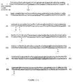
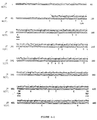
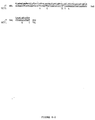
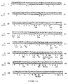
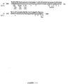
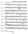
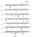
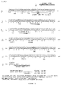
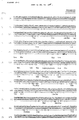
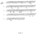
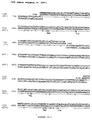
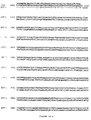
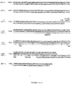
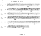
- the present invention provides a polynucleotide in substantially isolated form comprising a nucleotide sequence of at least 8 nucleotides from a J-1 HCV isolate, said J-1 HCV isolate having at least 90% nucleotide sequence homology with the J-1 sequence of any one of Figures 7 to 10 or 13 to 18, wherein said nucleotide sequence of at least 8 nucleotides is distinct from the nucleotide sequence of HCV isolate HCV-1.
- Yet another embodiment of the present invention provides a polypeptide of the invention immobilized on the solid support.
- an immunoassay for detecting the presence of anti-HCV antibodies in a test sample comprising: (a) incubating the test sample under conditions that allow the formation of antigen-antibody complexes with polypeptide of the invention, wherein the polypeptide is not immunologically cross-reactive with HCV-1 and (b) detecting any antigen-antibody complexes formed.
- Yet another embodiment of the present invention provides a method of detecting HCV polynucleotides in a test sample comprising: (a) providing a polypeptide of the invention as a probe; (b) contacting the test sample and the probe under conditions that allow for the formation of a polynucleotide duplex between the probe and its complement in the absence of substantial polynucleotide duplex formation between the probe and non-HCV polynucleotide sequences present in the test sample; and (c) detecting any polynucleotide duplexes comprising the probe.
- hepatitis C virus has been reserved by workers in the field for an heretofore unknown etiologic agent of NANBH. Accordingly, as used herein, "hepatitis C virus” (HCV) refers to an agent causative of NANBH, which was formerly referred to as NANBV and/or BB-NANBV from the class of the prototype isolate, HCV1, described by Houghton et al. See, e.g., EPO Pub. No. 318,216 and U.S. Patent App. Serial No. 355,002, filed 19 May 1989 (available in non-U.S. applications claiming priority therefrom), the disclosures of which are incorporated herein by reference.
- HCV hepatitis C virus
- HCV The nucleotide sequence and putative amino acid sequence of HCV1 is shown in Figure 6.
- HCV the disease caused by HCV, formerly called NANB hepatitis (NANBH), is called hepatitis C.
- NANBH NANB hepatitis
- NANBH and hepatitis C may be used interchangeably herein.
- HCV denotes a viral species of which pathogenic strains cause NANBH, as well as attenuated strains or defective interfering particles derived therefrom.
- HCV is a Flavi-like virus.
- the morphology and composition of Flavivirus particles are known, and are discussed by Brinton (1986) THE VIRUSES: THE TOGAVIRIDAE AND FLAVIVIRIDAE (Series eds. Fraenkel-Conrat and Wagner, vol eds. Schlesinger and Schlesinger, Plenum Press), p.327-374.
- Flaviviruses contain a central nucleocapsid surrounded by a lipid bilayer.
- Virions are spherical and have a diameter of about 40-50 nm. Their cores are about 25-30 nm in diameter.
- Within the outer surface of the virion envelope are projections that are about 5-10 nm long with terminal knobs about 2 nm in diameter.
- the HCV genome is comprised of RNA. It is known that RNA containing viruses have relatively high rates of spontaneous mutation, i.e., reportedly on the order of 10 -3 to 10 -4 per incorporated nucleotide. Therefore, there are multiple strains, which may be virulent or avirulent, within the HCV class or species.
- the genome of HCV isolates is comprised of a single ORF of approximately 9,000 nucleotides to approximately 12,000 nucleotides, encoding a polyprotein similar in size to that of HCV1, an encoded polyprotein of similar hydrophobic and antigenic character to that of HCV1, and the presence of co-linear peptide sequences that are conserved with HCV1.
- the genome is believed to be a positive-stranded RNA.
- Isolates of HCV comprise epitopes that are immunologically cross-reactive with epitopes in the HCV1 genome. At least some of these are epitopes unique to HCV when compared to other known Flaviviruses. The uniqueness of the epitope may be determined by its immunological reactivity with anti-HCV antibodies and lack of immunological reactivity with antibodies to other Flavivirus species. Methods for determining immunological reactivity are known in the art, for example, by radioimmunoassay, by ELISA assay, by hemagglutination, and several examples of suitable techniques for assays are provided herein.
- the overall homology of HCV isolates and HCV1 genomes at the nucleotide level probably will be about 40% or greater, probably about 60% or greater, and even more probably about 80% to about 90% or greater.
- the correspondence between the sequence from a new isolate and the HCV1 sequence can be determined by techniques known in the art. For example, they can be determined by a direct comparison of the sequence information of the polynucleotide from the new isolate and HCV1 sequences.
- homology can be determined by hybridization of the polynucleotides under conditions which form stable duplexes between homologous regions (for example, those which would be used prior to S 1 digestion), followed by digestion with single-stranded specific nuclease(s), followed by size determination of the digested fragments.
- putative HCV strains or isolates are identifiable by their homology at the polypeptide level.
- new HCV isolates are expected to be more than about 40% homologous, probably more than about 70% homologous, and even more probably more than about 80% homologous, and possibly even more than about 90% homologous at the polypeptide level.
- the techniques for determining amino acid sequence homology are known in the art. For example, the amino acid sequence may be determined directly and compared to the sequences provided herein. Alternatively the nucleotide sequence of the genomic material of the putative HCV may be determined, the amino acid sequence encoded therein can be determined, and the corresponding regions compared.
- the ORF of HCV1 is shown in Figure 12.
- the non-structural, core, and envelope domains of the polyprotein have been predicted for HCV1 ( Figure 5).
- the "C”, or core, polypeptide is believed to be encoded from the 5' terminus to about nucleotide 345 of HCV1.
- the putative "E”, or envelope, domain of HCV1 is believed to be encoded from about nucleotide 346 to about nucleotide 1050.
- Putative NS1, or non-structural one domain is thought to be encoded from about nucleotide 1051 to about nucleotide 1953.
- putative NS2 is thought to be encoded from about nucleotide 1954 to about nucleotide 3018, putative NS3 from about nucleotide 3019 to about nucleotide 4950, putative NS4 from about nucleotide 4951 to about nucleotide 6297, and putative NS5 from about nucleotide 6298 to the 3' terminus respectively.
- putative NS2 is thought to be encoded from about nucleotide 1954 to about nucleotide 3018, putative NS3 from about nucleotide 3019 to about nucleotide 4950, putative NS4 from about nucleotide 4951 to about nucleotide 6297, and putative NS5 from about nucleotide 6298 to the 3' terminus respectively.
- the above boundaries are approximations based on an analysis of the ORF. The exact boundaries can be determined by those skilled in the art in view of the disclosure herein.
- HCV/J1 or "J1” and “HCV/J7” or “J7” refer to new HCV isolates characterized by the nucleotide sequence disclosed herein, as well as related isolates that are substantially homologous thereto; i.e., at least about 90% or about 95% at the nucleotide level. It is believed that the sequences disclosed herein characterize an HCV subclass that is predominant in Japan and other Asian and/or Pacific rim countries. Additional J1 and J7 isolates can be obtained in view of the disclosure herein and EPO Pub. No. 318,216. In particular, the J1 and J7 nucleotide sequences disclosed herein, as well as the HCV1 sequences in Figure 12, can be used as primers or probes to clone additional domains of J1, J7, or additional isolates.
- nucleotide sequence "from" a designated sequence or source refers to a nucleotide sequence that is homologous (i.e., identical) to or complementary to the designated sequence or source, or a portion thereof.
- the J1 sequences provided herein are a minimum of 15 nucleotides preferably 20 nucleotides or longer. The maximum length is the complete viral genome.
- the sequence of the region from which the polynucleotide is derived is preferably homologous to or complementary to a sequence which is unique to an HCV genome or the J1 genome. Whether or not a sequence is unique to a genome can be determined by techniques known to those of skill in the art. For example, the sequence can be compared to sequences in databanks, e.g., Genebank, to determine whether it is present in the uninfected host or other organisms. The sequence can also be compared to the known sequences of other viral agents, including those which are known to induce hepatitis, e.g., HAV, HBV, and HDV, and to other members of the Flaviviridae.
- the correspondence or non-correspondence of the derived sequence to other sequences can also be determined by hybridization under the appropriate stringency conditions.
- Hybridization techniques for determining the complementarity of nucleic acid sequences are known in the art. See also, for example, Maniatis et al. (1982) MOLECULAR CLONING; A LABORATORY MANUAL (Cold Spring Harbor Press, Cold Spring Harbor, N.Y.).
- mismatches of duplex polynucleotides formed by hybridization can be determined by known techniques, including for example, digestion with a nuclease such as S1 that specifically digests single-stranded areas in duplex polynucleotides. Regions from which typical DNA sequences may be derived include, but are not limited to, regions encoding specific epitopes, as well as non-transcribed and/or non-translated regions.
- the J1 polynucleotide is not necessarily physically derived from the nucleotide sequence shown, but may be generated in any manner, including for example, chemical synthesis or DNA replication or reverse transcription or transcription. In addition, combinations of regions corresponding to that of the designated sequence may be modified in ways known in the art to be consistent with an intended use.
- the polynucleotides may also include one or more labels, which are known to those of skill in the art.
- amino acid sequence "from" a designated polypeptide or source of polypeptides means that the amino acid sequence is homologous (i.e., identical) to the sequence of the designated polypeptide, or a portion thereof.
- An amino acid sequence "from” a designated nucleic acid sequence refers to a polypeptide having an amino acid sequence identical to that of a polypeptide encoded in the sequence, or a portion thereof.
- the J1 amino acid sequences in the polypeptides of the present invention are at least 10 amino acids, more preferably at least about 15 amino acids, and most preferably at least about 20 amino acids.
- polypeptides of the present invention are not necessarily translated from a designated nucleic acid sequence; the polypeptides may be generated in any manner, including for example, chemical synthesis, or expression of a recombinant expression system, or isolation from virus.
- the polypeptides may include one or more analogs of amino acids or unnatural amino acids. Methods of inserting analogs of amino acids into a sequence are known in the art.
- the polypeptides may also include one or more labels, which are known to those of skill in the art.
- polynucleotide intends a polynucleotide of genomic, cDNA, semisynthetic, or synthetic origin which, by virtue of its origin or manipulation: (1) is linked to a polynucleotide other than that to which it is linked in nature, or (2) does not occur in nature.
- polynucleotide refers to a polymeric form of nucleotides of any length, either ribonucleotides or deoxyribonucleotides. This term refers only to the primary structure of the molecule. Thus, this term includes double- and single-stranded DNA, and RNA.
- modifications for example, labels which are known in the art, methylation, "caps", substitution of one or more of the naturally occurring nucleotides with an analog, internucleotide modifications such as, for example, those with uncharged linkages (e.g., methyl phosphonates, phosphotriesters, phosphoamidates, carbamates, etc.) and with charged linkages (e.g., phosphorothioates, phosphorodithioates, etc.), those containing pendant moieties, such as, for example proteins (including for e.g., nucleases, toxins, antibodies, signal peptides, poly-L-lysine, etc.), those with intercalators (e.g., acridine, psoralen, etc.), those containing chelators (e.g., metals, radioactive metals, boron, oxidative metals, etc.), those containing alkylators, those with modified linkages (e.g., alkylators, those with
- Polynucleotide refers to a composition comprising a specified polynucleotide that is substantially free of other components, such composition typically comprising at least about 70% of the specified polynucleotide, more typically at least about 80%, 90% or even 95% to 99% of the specified polynucleotide.
- Polypeptide refers to a composition comprising a specified polypeptide that is substantially free of other components, such composition typically comprising at least about 70% of the specified polypeptide, more typically at least about 80%, 90% or even 95% to 99% of the specified polypeptide.
- Recombinant host cells denote microorganisms or higher eukaryotic cell lines cultured as unicellular entities that can be, or have been, used as recipients for a recombinant vector or other transfer DNA, and include the progeny of the original cell which has been transformed. It is understood that the progeny of a single parental cell may not necessarily be completely identical in morphology or in genomic or total DNA complement as the original parent, due to natural, accidental, or deliberate mutation.
- a “replicon” is any genetic element, e.g., a plasmid, a chromosome, a virus, a cosmid, etc. that behaves as an autonomous unit of polynucleotide replication within a cell; i.e., capable of replication under its own control.
- a "cloning vector” is a replicon that can transform a selected host cell and in which another polynucleotide segment is attached, so as to bring about the replication and/or expression of the attached segment.
- cloning vectors include plasmids, virus (e.g., bacteriophage vector) and cosmids.
- An "integrating vector” is a vector that does not behave as a replicon in a selected host cell, but has the ability to integrate into a replicon (typically a chromosome) resident in the selected host to stably transform the host.
- An "expression vector” is a construct that can transform a selected host cell and provides for expression of a heterologous coding sequence in the selected host.
- Expression vectors can be either a cloning vector or an integrating vector.
- a “coding sequence” is a polynucleotide sequence which is transcribed into mRNA and/or translated into a polypeptide when placed under the control of appropriate regulatory sequences. The boundaries of the coding sequence are determined by a translation start codon at the 5'-terminus and a translation stop codon at the 3'-terminus.
- a coding sequence can include, but is not limited to mRNA, cDNA, and recombinant polynucleotide sequences.
- Control sequence refers to polynucleotide regulatory sequences which are necessary to effect the expression of coding sequences to which they are ligated. The nature of such control sequences differs depending upon the host organism. In prokaryotes, control sequences generally include promoter, ribosomal binding site, and terminators. In eukaryotes generally control sequences include promoters, terminators and, in some instances, enhancers. The term “control sequences” is intended to include, at a minimum, all components the presence of which are necessary for expression, and may also include additional advantageous components.
- Operaably linked refers to a juxtaposition wherein the components so described are in a relationship permitting them to function in their intended manner.
- a control sequence "operably linked" to a coding sequence is ligated in such a way that expression of the coding sequence is achieved under conditions compatible with the control sequences.
- An "open reading frame” or ORF is a region of a polynucleotide sequence which encodes a polypeptide; this region may represent a portion of a coding sequence or a total coding sequence.
- Immunologically cross-reactive refers to two or more epitopes or polypeptides that are bound by the same antibody. Cross-reactivity can be determined by any of a number of immunoassay techniques, such as a competition assay.
- an antibody refers to a polypeptide or group of polypeptides which comprise at least one epitope.
- An "antigen binding site” is formed from the folding of the variable domains of an antibody molecule(s) to form three-dimensional binding sites with an internal surface shape and charge distribution complementary to the features of an epitope of an antigen, which allows specific binding to form an antibody-antigen complex.
- An antigen binding site may be formed from a heavy- and/or light-chain domain (VH and VL, respectively), which form hypervariable loops which contribute to antigen binding.
- VH and VL light-chain domain
- the term “antibody” includes, without limitation, chimeric antibodies, altered antibodies,univalent antibodies, Fab proteins, and single-domain antibodies. In many cases, the biding phenomena of antibodies to antigens is equivalent to other ligand/anti-ligand binding.
- a "single domain antibody” is an antibody which is comprised of an HL domain, which binds specifically with a designated antigen.
- a dAb does not contain a VL domain, but may contain other antigen binding domains known to exist to antibodies, for example, the kappa and lambda domains.
- Methods for preparing dAbs are known in the art. See, for example, Ward et al, Nature 341 : 544 (1989).
- Antibodies may also be comprised of VH and VL domains, as well as other known antigen binding domains. Examples of these types of antibodies and methods for their preparation and known in the art (see, e.g., U.S. Patent No. 4,816,467, which is incorporated herein by reference), and include the following.
- “vertebrate antibodies” refers to antibodies which are tetramers or aggregates thereof, comprising light and heavy chains which are usually aggregated in a "Y" configuration and which may or may not have covalent linkages between the chains.
- the amino acid sequences of the chains are homologous with those sequences found in antibodies produced in vertebrates, whether in situ or in vitro (for example, in hybridomas).
- Vertebrate antibodies include, for example, purified polyclonal antibodies and monoclonal antibodies, methods for the preparation of which are described infra.
- Hybrid antibodies are antibodies where chains are separately homologous with reference to mammalian antibody chains and represent novel assemblies of them, so that two different antigens are precipitable by the tetramer or aggregate.
- one pair of heavy and light chains are homologous to those found in an antibody raised against a first antigen, while a second pair of chains are homologous to those found in an antibody raised against a second antibody. This results in the property of "divalence”, i.e., the ability to bind two antigens simultaneously.
- Such hybrids may also be formed using chimeric chains, as set forth below.
- Chimeric antibodies refers to antibodies in which the heavy and/or light chains are fusion proteins. Typically, one portion of the amino acid sequences of the chain is homologous to corresponding sequences in an antibody derived from a particular species or a particular class, while the remaining segment of the chain is homologous to the sequences derived from another species and/or class. Usually, the variable region of both light and heavy chains mimics the variable regions or antibodies derived from one species of vertebrates, while the constant portions are homologous to the sequences in the antibodies derived from another species of vertebrates. However, the definition is not limited to this particular example.
- altered antibodies refers to antibodies in which the naturally occurring amino acid sequence in a vertebrate antibody has been varies.
- antibodies can be redesigned to obtain desired characteristics.
- the possible variations are many, and range from the changing of one or more amino acids to the complete redesign of a region, for example, the constant region.
- Changes in the constant region in general, to attain desired cellular process characteristics, e.g., changes in complement fixation, interaction with membranes, and other effector functions. Changes in the variable region may be made to alter antigen binding characteristics.
- the antibody may also be engineered to aid the specific delivery of a molecule or substance to a specific cell or tissue site.
- the desired alterations may be made by known techniques in molecular biology, e.g., recombinant techniques, site-directed mutagenesis, etc.
- antibodies are aggregates comprised of a heavy-chain/light-chain dimer bound to the Fc (i.e., stem) region of a second heavy chain. This type of antibody escapes antigenic modulation. See, e.g., Glennie et al. Nature 295 : 712 (1982). Included also within the definition of antibodies are “Fab” fragments of antibodies.
- the “Fab” region refers to those portions of the heavy and light chains which are roughly equivalent, or analogous, to the sequences which comprise the branch portion of the heavy and light chains, and which have been shown to exhibit immunological binding to a specified antigen, but which lack the effector Fc portion.
- Fab includes aggregates of one heavy and one light chain (commonly known as Fab'), as well as tetramers containing the 2H and 2L chains (referred to as F(ab) 2 ), which are capable of selectively reacting with a designated antigen or antigen family.
- Fab antibodies may be divided into subsets analogous to those described above, i.e., “vertebrate Fab”, “hybrid Fab”, “chimeric Fab”, and “altered Fab”.
- Methods of producing Fab fragments of antibodies are known within the art and include, for example, proteolysis, and synthesis by recombinant techniques.
- Epitope refers to an antibody binding site usually defined by a polypeptide, but also by non-amino acid haptens.
- An epitope could comprise 3 amino acids in a spatial conformation which is unique to the epitope, generally an epitope consists of at least 5 such amino acids, and more usually, consists of at least 8-10 such amino acids.
- Antigen-antibody complex refers to the complex formed by an antibody that is specifically bound to an epitope on an antigen.
- Immunogenic polypeptide refers to a polypeptide that elicits a cellular and/or humoral immune response in a mammal, whether alone or linked to a carrier, in the presence or absence of an adjuvant.
- Polypeptide refers to a polymer of amino acids and does not refer to a specific length of the molecule. Thus, peptides, oligopeptides, and proteins are included within the definition of polypeptide. This term also does not refer to or exclude post-expression modifications of the polypeptide, for example, glycosylations, acetylations, phosphorylations and the like. Included within the definition are, for example, polypeptides containing one or more analogs of an amino acid (including, for example, unnatural amino acids, etc.), polypeptides with substituted linkages, as well as other modifications known in the art, both naturally occurring and non-naturally occurring.
- Transformation refers to the insertion of an exogenous polynucleotide into a host cell, irrespective of the method used for the insertion, for example, direct uptake, transduction, f-mating or electroporation.
- the exogenous polynucleotide may be maintained as a non-integrated vector, for example, a plasmid, or alternatively, may be integrated into the host genome.
- a "transformed" host cell refers to both the immediate cell that has undergone transformation and its progeny that maintain the originally exogenous polynucleotide.
- Treatment refers to prophylaxis and/or therapy.
- “Individual” refers to vertebrates, particularly members of the mammalian species, and includes but is not limited to domestic animals, sports animals, and primates, including humans.
- Sense strand refers to the strand of a double-stranded DNA molecule that is homologous to a mRNA transcript thereof.
- the "anti-sense strand” contains a sequence which is complementary to that of the "sense strand”.
- Antibody-containing body component refers to a component of an individual's body which is a source of the antibodies of interest.
- Antibody-containing body components are known in the art, and include but are not limited to, whole blood and components thereof, plasma, serum, spinal fluid, lymph fluid, the external sections of the respiratory, intestinal, and genitourinary tracts, tears, saliva, milk, white blood cells, and myelomas.
- Purified HCV isolate refers to a preparation of HCV particles which has been isolated from the cellular constituents with which the virus is normally associated, and from other types of viruses which may be present in the infected tissue.
- the techniques for isolating viruses are known to those of skill in the art, and include, for example, centrifugation and affinity chromatography.
- HCV particles is an entire virion, as well as particles which are intermediates in virion formation.
- HCV particles generally have one or more HCV proteins associated with the HCV nucleic acid.
- Probe refers to a polynucleotide which forms a hybrid structure with a sequence in a target polynucleotide, due to a complementarity of at least one region in the probe with a region in the target.
- Bio sample refers to a sample of tissue or fluid isolated from an individual, including but not limited to, for example, whole blood and components thereof, plasma, serum, spinal fluid, the external sections of the skin, respiratory, intestinal, and genitourinary tracts, tears saliva, milk, blood cells, tumors, organs, and also samples of in vitro cell culture constituents (including but not limited to conditioned medium resulting from the growth of cells in cell culture medium, putatively virally infected cells, recombinant cells, and cell components).
- the invention pertains to the isolation and characterization of a newly discovered isolate of HCV, J1, its nucleotide sequence, its protein sequence and resulting polynucleotides and polypeptides and antibodies derived therefrom.
- HCV HCV
- J7 another newly discovered isolate of HCV
- the nucleotide sequences derived from HCV J1 are useful as probes to diagnose the presence of virus in samples, and to isolate other naturally occurring variants of the virus. These nucleotide sequences also make available polypeptide sequences of HCV antigens encoded within the J1 genome and permits the production of polypeptides which are useful as standards or reagents in diagnostic tests and/or as components or vaccines. Antibodies, both polyclonal and monoclonal, directed against HCV epitodes contained within these polypeptide sequences are also useful for diagnostic tests, as therapeutic agents, for screening of antiviral agents, and for the isolation of the NANBH virus.
- probes derived from the sequences disclosed herein it is possible to isolate and sequence other portions of the J1 genome, thus giving rise to additional probes and polypeptides which are useful in the diagnosis and/or treatment, both prophylactic and therapeutic, of NANBH.
- HCV/J1 nucleotide sequences enable the construction of polynucleotide probes and polypeptides useful in diagnosing NANBH due to HCV infection and in screening blood donors as well as donated blood and blood products for infection.
- DNA oligomers of about 8-10 nucleotides, or larger, which are useful as hybridization probes to detect the presence of HCV RNA in, for example, sera of subjects suspected of harboring the virus, or for screening donated blood for the presence of the virus.
- the HCV/J1 sequences also allow the design and production of HCV specific polypeptides which are useful as diagnostic reagents for the presence of antibodies raised during NANBH.
- Antibodies to purified polypeptides derived from the HCV/J1 sequences may also be used to detect viral antigens in infected individuals and in blood.
- HCV/J1 sequences enable she design and production of polypeptides which may be used as vaccines against HCV and also for the production of antibodies, which in turn may be used for protection against the disease, and/or for therapy of HCV infected individuals.
- the disclosed HCV/J1 sequences enable further characterization of the HCV genome. Polynucleotide probes derived from these sequences, as well as from the HCV genome, may be used to screen cDNA libraries for additional viral cDNA sequences, which, in turn, may be used to obtain additional overlapping sequences. See, e.g., EPO Pub. No. 318,216.
- the HCV/J1 polynucleotide sequences, the polypeptides derived therefrom and the antibodies directed against these polypeptides, are useful in the isolation and identification of the BB-NANBV agent(s).
- antibodies directed against HCV epitopes contained in polypeptides derived from the HCV/J1 sequences may be used in processes based upon affinity chromatography to isolate the virus.
- the antibodies may be used to identify viral particles isolated by other techniques. The viral antigens and the genomic material within the isolated viral particles may then be further characterized.
- the information obtained from further sequencing of the HCV/J1 genome, as well as from further characterization of the HCV/J1 antigens and characterization of the genomes enable the design and synthesis of additional probes and polypeptides and antibodies which may be used for diagnosis, for prevention, and for therapy of HCV induced NANBH, and for screening for infected blood and blood-related products.
- HCV/J1 cDNA sequences permits the construction of expression vectors encoding antigenically active regions of the polypeptide encoded in either strand.
- antigenically active regions may be derived from coat or envelope antigens or from core antigens, or from antigens which are non-structural including, for example, polynucleotide binding proteins, polynucleotide polymerase(s), and other viral proteins required for the replication and/or assembly of the virus particle.
- Fragments encoding the desired polypeptides are derived from the cDNA clones using conventional restriction digestion or by synthetic methods, and are ligated into vectors which may, for example, contain portions of fusion sequences such as beta-galactosidase or superoxide dismutase (SOD). Methods and vectors which are useful for the production of polypeptides which contain fusion sequences of SOD are described in EPO Pub. No. 196,056. Vectors encoding fusion polypeptides of SOD and HCV polypeptides are described in EPO Pub. No. 318,216.
- HCV cDNA containing an open reading frame in either sense strand, can be obtained as a recombinant polypeptide, such as a mature or fusion protein.
- a polypeptide encoded in the cDNA can be provided by chemical synthesis.
- the DNA encoding the desired polypeptide may be ligated into expression vectors suitable for any convenient host. Both eukaryotic and prokaryotic host systems are presently used in forming recombinant polypeptides, and a summary of some of the more common control systems and host cell is given below.
- the polypeptide produced in such host cells is then isolated from lysed cells or from the culture medium and purified to the extent needed for its intended use. Purification may be by techniques known in the art, for example, differential extraction, salt fractionation, chromatography on ion exchange resins, affinity chromatography, centrifugation, and the like. See, for example, Methods in Enzymology for a variety of methods for purifying proteins.
- Such recombinant or synthetic HCV polypeptides can be used as diagnostics, or those which give rise to neutralizing antibodies may be formulated into vaccines. Antibodies raised against these polypeptides can also be used as diagnostics, or for passive immunotherapy. In addition, antibodies to these polypeptides are useful for isolating and identifying HCV particles.
- the HCV antigens may also be isolated from HCV virions.
- the virions may be grown in HCV infected cells in tissue culture, or in an infected host.
- polypeptides of the present invention may comprise a substantially complete viral domain, in many applications all that Is required is that the polypeptide comprise an antigenic or immunogenic region of the virus.
- An antigenic region of a polypeptide is generally relatively small--typically 8 to 10 amino acids or less in length. Fragments of as few as 5 amino acids may characterize an antigenic region. These segments may correspond to regions of HCV/J1 epitopes. Accordingly, using the cDNAs of HCV/J1 as a basis, DNAs encoding short segments of HCV/J1 polypeptides can be expressed recombinantly either as fusion proteins, or as isolated polypeptides. In addition, short amino acid sequences can be conveniently obtained by chemical synthesis.
- the polypeptide may be linked to a suitable carrier.
- a suitable carrier A number of techniques for obtaining such linkage are known in the art, including the formation of disulfide linkages using N-succinimidyl-3-(2-pyridyl-thio)propionate (SPDP) and succinimidyl 4-(N-maleimido-methyl)cyclohexane-1-carboxylate (SMCC) obtained from Pierce Company, Rockford, Illinois, (if the peptide lacks a sulfhydryl group, this can be provided by addition of a cysteine residue.) These reagents create a disulfide linkage between themselves and peptide cysteine residues on one protein and an amide linkage through the epsilon-amino on a lysine, or other free amino group in the other.
- SPDP N-succinimidyl-3-(2-pyridyl-thio)propionate
- SMCC succinimidyl
- disulfide/amide-forming agents A variety of such disulfide/amide-forming agents are known. See, for example, Immun. Rev. (1982) 62 :185.
- Other bifunctional coupling agents form a thioether rather than a disulfide linkage.
- Many of these thio-ether-forming agents are commercially available and include reactive esters of 6-maleimidocaproic acid, 2-bromoacetic acid, 2-iodoacetic acid, 4-(N-maleimido-methyl)cyclohexane-1-carboxylic acid, and the like.
- the carboxyl groups can be activated by combining them with succinimide or 1-hydroxyl-2-nitro-4-sulfonic acid, sodium salt.
- Suitable carriers are typically large, slowly metabolized macromolecules such as proteins; polysaccharides, such as latex functionalized sepharose, agarose, cellulose, cellulose beads and the like; polymeric amino acids, such as polyglutamic acid, polylysine, and the like; amino acid copolymers; and inactive virus particles.
- Especially useful protein substrates are serum albumins, keyhole limpet hemocyanin, immunoglobulin molecules, thyroglobulin, ovalbumin, tetanus toxoid, and other proteins well known to those skilled in the art.
- polypeptides comprising truncated HCV amino acid sequences encoding at least one viral epitope are useful immunological reagents.
- polypeptides comprising such truncated sequences can be used as reagents in an immunoassay.
- These polypeptides also are candidate subunit antigens in compositions for antiserum production or vaccines. While these truncated sequences can be produced by various known treatments of native viral protein, it is generally preferred to make synthetic or recombinant polypeptides comprising an HCV sequence.
- Polypeptides comprising these truncated HCV sequences can be made up entirely of HCV sequences (one or more epitopes, either contiguous or noncontiguous), or HCV sequences and heterologous sequences in a fusion protein.
- Useful heterologous sequences include sequences that provide for secretion from a recombinant host, enhance the immunological reactivity of the HCV epitope(s), or facilitate the coupling of the polypeptide to an immunoassay support or a vaccine carrier. See, e.g., EPO Pub. No. 116,201; U.S. Pat. No. 4,722,840; EPO Pub. No. 259,149; U.S. Pat. No. 4,629,783, the disclosures of which are incorporated herein by reference.
- the size of polypeptides comprising the truncated HCV sequences can vary widely, the minimum size being a sequence of sufficient size to provide an HCV epitope, while the maximum size is not critical. In some applications, the maximum size usually is not substantially greater than that required to provide the desired HCV epitopes and function(s) of the heterologous sequence, if any.
- the truncated HCV amino acid sequence will range from about 5 to about 100 amino acids in length. More typically, however, the HCV sequence will be a maximum of about 50 amino acids in length, preferably a maximum of about 30 amino acids. It is usually desirable to select HCV sequences of at least about 10, 12 or 15 amino acids, up to a maximum of about 20 or 25 amino acids.
- Truncated HCV amino acid sequences comprising epitopes can be identified in a number of ways.
- the entire viral protein sequence can be screened by preparing a series of short peptides that together span the entire protein sequence.
- 100-mer polypeptides it would be routine to test each polypeptide for the presence of epitope(s) showing a desired reactivity, and then testing progressively smaller and overlapping fragments from an identified 100-mer to map the epitope of interest. Screening such peptides in an immunoassay is within the skill of the art.
- NS5 of Flaviviruses encodes the virion polymerase, and that NSI corresponds with a complement fixation antigen which has been shown to be an effective vaccine in animals. Recently, it has been shown that a flaviviral protease function resides in NS3.
- Figure 11 is a schematic of putative domains of the HCV polyprotein.
- the expression of polypeptides containing these domains in a variety of recombinant host cells, including, for example, bacteria, yeast, insect, and vertebrate cells, should give rise to important immunological reagents which can be used for diagnosis, detection, and vaccines.
- non-structural protein region of the putative polyproteins of the HCV isolate described herein and of Flaviviruses appears to be generally similar, there is less similarity between the putative structural regions which are towards the N-terminus. In this region, there is a greater divergence in sequence, and in addition, the hydrophobic profile of the two regions show less similarity. This "divergence" begins in the N-terminal region of the putative NS1 domain in HCV, and extends to the presumed N-terminus. Nevertheless, it is still possible to predict the approximate locations of the putative nucleocapsid (N-terminal basic domain) and E (generally hydrophobic) domains within the HCV polyprotein.
- HCV polyprotein that could correspond with useful immunological reagents.
- E and NS1 proteins of Flaviviruses are known to have efficacy as protective vaccines. These regions, as well as some which are shown to be antigenic in the HCV1, for example those within putative NS3, C, and NS5, etc., should also provide diagnostic reagents.
- the immunogenicity of the HCV sequences may also be enhanced by preparing the sequences fused to or assembled with particle-forming proteins such as, for example, hepatitis B surface antigen or rotavirus VP6 antigen. Constructs wherein the HCV epitope is linked directly to the particle-forming protein coding sequences produce hybrids which are immunogenic with respect to the HCV epitope.
- all of the vectors prepared include epitopes specific to HBV, having various degrees of immunogenicity, such as, for example, the pre-S peptide.
- particles constructed from particle forming protein which include HCV sequences are immunogenic with respect to HCV and particle-form protein. See, e.g., U.S. Pat. No. 4,722,840; EPO Pub No. 175,261; EPO Pub. No. 259,149; Michelle et al. (1984) Int. Symposium on Viral Hepatitis.
- Vaccines may be prepared from one or more immunogenic polypeptides derived from HCV/J1.
- the observed homology between HCV and Flaviviruses provides information concerning the polypeptides which are likely to be most effective as vaccines, as well as the regions of the genome in which they are encoded.
- the general structure of the Flavivirus genome is discussed in Rice et al. (1986) in THE VIRUSES: THE TOGAVIRIDAE AND FLAVIVIRIDAE (Series eds. Fraenkel-Conrat and Wagner, Vol eds. Schlesinger and Schlesinger, Plenum Press).
- the flavivirus genomic RNA is believed to be the only virus-specific mRNA species, and it is translated into the three viral structural proteins, i.e., C, M, and E, as well as two large nonstructural proteins, NV4 and NV5, and a complex set of smaller nonstructural proteins. It is known that major neutralizing epitopes for Flaviviruses reside in the E (envelope) protein. Roehrig (1986) in THE VIRUSES: THE TOGAVIRIDAE AND FLAVIVIRIDAE (Series eds. Fraenkel-Conrat and Wagner, Vol eds. Schlesinger and Schlesinger, Plenum Press).
- vaccines may be comprised of recombinant polypeptides containing epitopes of HCV E. These polypeptides may be expressed in bacteria, yeast, or mammalian cells, or alternatively may be isolated from viral preparations. It is also anticipated that the other structural proteins may also contain epitopes which give rise to protective anti-HCV antibodies. Thus, polypeptides containing the epitopes of E, C, and M may also be used, whether singly or in combination, in HCV vaccines.
- multivalent vaccines against HCV may be comprised of one or more epitopes from one or more structural proteins, and/or one or more epitopes from one or more nonstructural proteins.
- These vaccines may be comprised of, for example, recombinant HCV polypeptides and/or polypeptides isolated from the virions.
- vaccines are contemplated comprising one or more of the following HCV proteins, or subunit antigens derived therefrom: E, NS1, C, NS2, NS3, NS4 and NS5.
- vaccines comprising E and/or NS1, or subunits thereof.
- inactivated HCV inactivated HCV in vaccines
- inactivation may be by the preparation of viral lysates, or by other means known in the art to cause inactivation of Flaviviruses, for example, treatment with organic solvents or detergents, or treatment with formalin.
- vaccines may also be prepared from attenuated HCV strains or from hybrid viruses such as vaccinia vectors known in the art [Brown et al. Nature 319 : 549-550 (1986)].
- vaccines which contain immunogenic polypeptide(s) as active ingredients is known to one skilled in the art.
- such vaccines are prepared as injectables, either as liquid solutions or suspensions; solid forms suitable for solution in, or suspension in, liquid prior to injection may also be prepared.
- the preparation may also be emulsified, or the protein encapsulated in liposomes.
- the active immunogenic ingredients are often mixed with excipients which are pharmaceutically acceptable and compatible with the active ingredient. Suitable excipients are, for example, water, saline, dextrose, glycerol, ethanol, or the like and combinations thereof.
- the vaccine may contain minor amounts of auxiliary substances such as wetting or emulsifying agents, pH buffering agents, and/or adjuvants which enhance the effectiveness of the vaccine.
- auxiliary substances such as wetting or emulsifying agents, pH buffering agents, and/or adjuvants which enhance the effectiveness of the vaccine.
- adjuvants which may be effective include but are not limited to: aluminum hydroxide, N-acetyl-muramyl-L-threonyl-D-isoglutamine (thr-MDP), N-acetyl-nor-muramyl-L-alanyl-D-isoglutamine (CGP 11637, referred to as nor-MDP), N-acetylmuramyl-L-alanyl-D-isoglutaminyl-L-alanine-2-(1′-2′-dipalmitoyl-sn-glycerol-3-hydroxyphosphoryloxy)-ethylamine (CGP 19835A,
- the vaccines are conventionally administered parenterally, by injection, usually, either subcutaneously or intramuscularly.
- Additional formulations which are suitable for other modes of administration include suppositories and, in some cases, oral formulations.
- suppositories traditional binders and carriers may include, for example, polyalkylene glycols or triglycerides; such suppositories may be formed from mixtures containing the active ingredient in the range of 0.5% to 10%, preferably 1%-2%.
- Oral formulations include such normally employed excipients as, for example, pharmaceutical grades of mannitol, lactose, starch, magnesium stearate, sodium saccharine, cellulose, magnesium carbonate, and the like. These compositions take the form of solutions, suspensions, tablets, pills, capsules, sustained release formulations or powders and contain 10%-95% of active ingredient, preferably 25%-70%.
- the proteins may be formulated into the vaccine as neutral or salt forms.
- Pharmaceutically acceptable salts include the acid addition salts (formed with free amino groups of the peptide) and which are formed with inorganic acids such as, for example, hydrochloric or phosphoric acids, or such organic acids such as acetic, oxalic, tartaric, maleic, and the like. Salts formed with the free carboxyl groups may also be derived from inorganic bases such as, for example, sodium, potassium, ammonium, calcium, or ferric hydroxides, and such organic bases as isopropylamine, trimethylamine, 2-ethylamino ethanol, histidine, procaine, and the like.
- the vaccines are administered in a manner compatible with the dosage formulation, and in such amount as will be prophylactically and/or therapeutically effective.
- the quantity to be administered which is generally in the range of 5 micrograms to 250 micrograms of antigen per dose, depends on the subject to be treated, capacity of the subject's immune system to synthesize antibodies, and the degree of protection desired. Precise amounts of active ingredient required to be administered may depend on the judgment of the practitioner and may be peculiar to each subject.
- the vaccine may be given in a single dose schedule, or preferably in a multiple dose schedule.
- a multiple dose schedule is one in which a primary course of vaccination may be with 1-10 separate doses, followed by other doses given at subsequent time intervals required to maintain and or reenforce the immune response, for example, at 1-4 months for a second dose, and if needed, a subsequent dose(s) after several months.
- the dosage regimen will also, at least in part, be determined by the need of the individual and be dependent upon the judgment of the practitioner.
- the vaccine containing the immunogenic HCV antigen(s) may be administered in conjunction with other immunoregulatory agents, for example, immune globulins.
- the immunogenic polypeptides prepared as described above are used to produce antibodies, both polyclonal and monoclonal. If polyclonal antibodies are desired, a selected mammal (e.g., mouse, rabbit, goat, horse, etc.) is immunized with an immunogenic polypeptide bearing an HCV epitope(s). Serum from the immunized animal is collected and treated according to known procedures. If serum containing polyclonal antibodies to an HCV epitope contains antibodies to other antigens, the polyclonal antibodies can be purified by immunoaffinity chromatography. Techniques for producing and processing polyclonal antisera are known in the art, see for example, Mayer and Walker, eds. (1987) IMMUNOCHEMICAL METHODS IN CELL AND MOLECULAR BIOLOGY (Academic Press, London).
- Monoclonal antibodies directed against HCV epitopes can also be readily produced by one skilled in the art.
- the general methodology for making monoclonal antibodies by hybridomas is well known.
- Immortal antibody-producing cell lines can be created by cell fusion, and also by other techniques such as direct transformation of B lymphocytes with oncogenic DNA, or transfection with Epstein-Barr virus. See, e.g., M. Schreier et al. (1980) HYBRIDOMA TECHNIQUES; Hammerling et al. (1981), MONOCLONAL ANTIBODIES AND T-CELL HYBRIDOMAS; Kennett et al. (1980) MONOCLONAL ANTIBODIES; see also , U.S. Patent Nos.
- Panels of monoclonal antibodies produced against HCV epitopes can be screened for various properties; i.e., for isotype, epitope affinity, etc.
- Antibodies both monoclonal and polyclonal, which are directed against HCV epitopes are particularly useful in diagnosis, and those which are neutralizing are useful in passive immunotherapy.
- Monoclonal antibodies in particular, may be used to raise anti-idiotype antibodies.
- Anti-idiotype antibodies are immunoglobulins which carry an "internal image" of the antigen of the infectious agent against which protection is desired. Techniques for raising anti-idiotype antibodies are known in the art. See, e,g,, Grzych (1985), Nature 316 :74; MacNamara et al. (1984), Science 226 :1325, Uytdehaag et al (1985), J. Immunol. 134 :1225. These anti-idiotype antibodies may also be useful for treatment and/or diagnosis of NANBH, as well as for an elucidation of the immunogenic regions of HCV antigens.
- oligomers of approximately 8 nucleotides or more can be prepared, either by excision or synthetically, which hybridize with the HCV genome and are useful in identification of the viral agent(s), further characterization of the viral genome(s), as well as in detection of the virus(es) in diseased individuals.
- the probes for HCV polynucleotides are a length which allows the detection of unique viral sequences by hybridization. While 6-8 nucleotides may be a workable length, sequences of about 10-12 nucleotides are preferred, and about 20 nucleotides appears optimal.
- probes can be prepared using routine methods, including automated oligonucleotide synthetic methods.
- useful probes for example, are the clones disclosed herein, as well as the various oligomers useful in probing cDNA libraries, set forth below.
- a complement to any unique portion of the HCV genome will be satisfactory.
- complete complementarity is desirable, though it may be unnecessary as the length of the fragment is increased.
- the biological sample to be analyzed such as blood or serum
- the biological sample to be analyzed may be treated, if desired, to extract the nucleic acids contained therein.
- the resulting nucleic acid from the sample may be subjected to gel electrophoresis or other size separation techniques; alternatively, the nucleic acid sample may be dot blotted without size separation.
- the probes are then labeled. Suitable labels, and methods for labeling probes are known in the art, and include, for example, radioactive labels incorporated by nick translation or kinasing, biotin, fluorescent probes, and chemiluminescent probes.
- the nucleic acids extracted from the sample are then treated with the labeled probe under hybridization conditions of suitable stringencies.
- the stringency of hybridization is determined by a number of factors during hybridization and during the washing procedure, including temperature, ionic strength, length of time, and concentration of formamide. These factors are outlined in, for example, Maniatis, T. (1982) MOLECULAR CLONING; A LABORATORY MANUAL (Cold Spring Harbor Press, Cold Spring Harbor, N.Y.).
- HCV genome sequences will be present in serum of infected individuals at relatively low levels, i.e., at approximately 10 2 -10 3 chimp infectious doses (CID) per ml.
- This level may require that amplification techniques be used in hybridization assays.
- amplification techniques are known in the art.
- the Enzo Biochemical Corporation "Bio-Bridge" system uses terminal deoxynucleotide transferase to add unmodified 3′-poly-dT-tails to a DNA probe. The poly dT-tailed probe is hybridized to the target nucleotide sequence, and then to a biotin-modified poly-A.
- EPO Pub. No. 204,510 describes a DNA hybridization assay in which analyte DNA is contacted with a probe that has a tail, such as a poly-dT tail, an amplifier strand that has a sequence that hybridizes to the tail of the probe, such as a poly-A sequence, and which is capable of binding a plurality of labeled strands.
- a particularly desirable technique may first involve amplification of the target HCV sequences in sera approximately 10,000-fold, i.e., to approximately 10 6 sequences/ml. This may be accomplished, for example, by the polymerase chain reactions (PCR) technique described which is by Saiki et al. (1986) Nature 324 :163, Mullis, U.S. Patent No. 4,683,195, and Mullis et al. U.S. Patent No. 4,683,202.
- the amplified sequence(s) may then be detected using a hybridization assay which is described in co-pending European Publication No. 317-077 and Japanese application No. 63-260347, which are assigned to the herein assignee, and are hereby incorporated herein by reference.
- hybridization assays which should detect sequences at the level of 10 6 /ml, utilize nucleic acid multimers which bind to single-stranded analyte nucleic acid, and which also bind to a multiplicity of single-stranded labeled oligonucleotides.
- a suitable solution phase sandwich assay which may be used with labeled polynucleotide probes, and the methods for the preparation of probes is described in EPO Pub. No. 225,807 which is hereby incorporated herein by reference.
- the probes can be packaged into diagnostic kits. Diagnostic kits include the probe DNA, which may be labeled; alternatively, the probe DNA may be unlabeled and the ingredients for labeling may be included in the kit in separate containers.
- the kit may also contain other suitably packaged reagents and materials needed for the particular hybridization protocol, for example, standards, wash buffers, as well as instructions for conducting the test.
- Both the HCV/J1 polypeptides which react immunologically with serum containing HCV antibodies and the antibodies raised against the HCV specific epitopes in these polypeptides are useful in immunoassays to detect presence of HCV antibodies, or the presence of the virus and/or viral antigens, in biological samples.
- Design of the immunoassays is subject to a great deal of variation, and a variety of these are known in the art.
- An immunoassay for anti-HCV antibody may utilize one viral epitope or several viral epitopes. When multiple epitopes are used, the epitopes may be derived from the same or different viral polypeptides, and may be in separate recombinant or natural polypeptides, or together in the same recombinant polypeptides.
- An immunoassay for viral antigen may use, for example, a monoclonal antibody directed towards a viral epitope, a combination of monoclonal antibodies directed towards epitopes of one viral polypeptide, monoclonal antibodies directed towards epitopes of different viral polypeptides, polyclonal antibodies directed towards the same viral antigen, polyclonal antibodies directed towards different viral antigens or a combination of monoclonal and polyclonal antibodies.
- Immunoassay protocols may be based, for example, upon competition, or direct reaction, or sandwich type assays. Protocols may also, for example, use solid supports, or may be by immunoprecipitation. Most assays involve the use of labeled antibody or polypeptide. The labels may be, for example, fluorescent, chemiluminescent, radioactive, or dye molecules. Assays which amplify the signals from the probe are also known. Examples of which are assays which utilize biotin and avidin, and enzyme-labeled and mediated immunoassays, such as ELISA assays.
- an immunoassay for anti-HCV antibody will involve selecting and preparing the test sample, such as a biological sample, and then incubating it with an antigenic (i.e., epitope-containing) HCV polypeptide under conditions that allow antigen-antibody complexes to form. Such conditions are well known in the art.
- the polypeptide is bound to a solid support to facilitate separation of the sample from the polypeptide after incubation.
- solid supports examples include nitrocellulose, in membrane or microtiter well form, polyvinylchloride, in sheets or microtiter wells, polystyrene latex, in beads or microtiter plates, polyvinylidine fluoride, known as ImmobulonTM, diazotized paper, nylon membranes, activated beads, and Protein A beads.
- the Dynatech, ImmulonTM 1 microtiter plate or the 0.25-inch polystyrene beads, which Spec finished by Precision Plastic Ball are used in the heterogeneous format.
- the solid support is typically washed after separating it from the test sample.
- test sample is incubated with antigen in solution, under conditions that will precipitate any antigen-antibody complexes that are formed, as is know in the art.
- the precipitated complexes are then separated from the test sample, for example, by centrifugation.
- the complexes formed comprising anti-HCV antibody are then detected by any of a number of techniques.
- the complexes can be detected with labeled anti-xenogeneic Ig or, if a competitive format is used, by measuring the amount of bound, labeled competing antibody.
- the test sample typically a biological sample
- anti-HCV antibodies again under conditions that allow the formation of antigen-antibody complexes.
- Various formats can be employed, such as a "sandwich" assay where antibody bound to a solid support is incubated with the test sample; washed;incubated with a second, labeled antibody to the analyte; and the support is washed again.
- Analyte is detected by determining if the second antibody is bound to the support.
- a competitive format which can be either heterogeneous or homogeneous
- a test sample is usually incubated with and antibody and a labeled, competing antigen either sequentially or simultaneously.
- the Flavivirus model for HCV allows predictions regarding the likely location of diagnostic epitopes for the virion structural proteins.
- the C, pre-M, M, and E domains are all likely to contain epitopes of significant potential for detecting viral antigens, and particularly for diagnosis.
- domains of the nonstructural proteins are expected to contain important diagnostic epitopes (e.g., NS5 encoding a putative polymerase; and NS1 encoding a putative complement-binding antigen).
- Recombinant polypeptides, or viral polypeptides, which include epitopes from these specific domains may be useful for the detection of viral antibodies in infections blood donors and infected patients.
- antibodies directed against the E and/or M proteins can be used in immunoassays for the detection of viral antigens in patients with HCV caused NANBH, and in infectious blood donors. Moreover, these antibodies may be extremely useful in detecting acute-phase donors and patients.
- Antigenic regions of the putative polyprotein can be mapped and identified by screening the antigenicity of bacterial expression products of HCV cDNAs which encode portions of the polyprotein.
- Other antigenic regions of HCV may be detected by expressing the portions of the HCV cDNAS in other expression systems, including yeast systems and cellular systems derived from insects and vertebrates.
- studies giving rise to an antigenicity index and hydrophobicity/hydrophilicity profile give rise to information concerning the probability of a region's antigenicity.
- Efficient detection systems may include the use of panels of epitopes. The epitopes in the panel may be constructed into one or multiple polypeptides.
- Kits suitable for immunodiagnosis and containing the appropriate labeled reagents are constructed by packaging the appropriate materials, including the polypeptides of the invention containing HCV epitopes or antibodies directed against HCV epitopes in suitable containers, along with the remaining reagents and materials required for the conduct of the assay (e.g., wash buffers, detection means like labeled anti-human Ig, labeled anti-HCV, or labeled HCV antigen), as well as a suitable set of assay instructions.
- the appropriate materials including the polypeptides of the invention containing HCV epitopes or antibodies directed against HCV epitopes in suitable containers, along with the remaining reagents and materials required for the conduct of the assay (e.g., wash buffers, detection means like labeled anti-human Ig, labeled anti-HCV, or labeled HCV antigen), as well as a suitable set of assay instructions.
- HCV/J1 nucleotide sequence information described herein may be used to gain further information on the sequence of the HCV genomes, and for identification and isolation of additional HCV isolates related to J1. This information, in turn, can lead to additional polynucleotide probes, polypeptides derived from the HCV genome, and antibodies directed against HCV epitopes which would be useful for the diagnosis and/or treatment of HCV caused NANBH.
- the HCV/J1 nucleotide sequence information herein is useful for the design of probes for the isolation of additional sequences which are derived from as yet undefined regions of the HCV genomes from which the J1 sequences are derived.
- labeled probes containing a sequence of approximately 8 or more nucleotides; and preferably 20 or more nucleotides, which are derived from regions close to the 5'-termini or 3'-termini of the family of HCV cDNA sequences disclosed in the examples may be used to isolate overlapping cDNA sequences from HCV cDNA libraries.
- sequences which overlap the cDNAs in the above-mentioned clones, but which also contain sequences derived from regions of the genome from which the cDNA in the above mentioned clones are not derived may then be used to synthesize probes for identification of other overlapping fragments which do not necessarily overlap the cDNAs described below.
- Methods for constructing cDNA libraries are known in the art. See, e.g. EPO Pub. No. 318,216. It is particularly preferred to prepare libraries from the serum of Japanese and other Asian patients diagnosed as having NANBH demonstrating antibody to HCV1 antigens; these are believed to be the most likely candidates for carriers of HCV/J1 or related isolates.
- HCV particles may be isolated from the sera from individuals with NANBH or from cell cultures by any of the methods known in the art, including for example, techniques based on size discrimination such as sedimentation or exclusion methods, or techniques based on density such as ultracentrifugation in density gradients, or precipitation with agents such as polyethylene glycol, or chromatography on a variety of materials such as anionic or cationic exchange materials, and materials which bind due to hydrophobicity.
- a preferred method of isolating HCV particles or antigen is by immunoaffinity columns.
- Techniques for immunoaffinity chromatography are known in the art, including techniques for affixing antibodies to solid supports so that they retain their immunoselective activity.
- the techniques may be those in which the antibodies are adsorbed to the support (see, for example, Kurstak in ENZYME IMMUNODIAGNOSIS, page 31-37), as well as those in which the antibodies are covalently linked to the support.
- the techniques are similar to those used for covalent linking of antigens to a solid support, described above.
- spacer groups may be included in the bifunctional coupling agents so that the antigen binding site of the antibody remains accessible.
- the antibodies may be monoclonal, or polyclonal, and it may be desirable to purify the antibodies before their use in the immunoassay.
- prokaryotic and eukaryotic host cells may be used for expression of desired coding sequences when appropriate control sequences which are compatible with the designated host are used.
- E. coli is most frequently used.
- Expression control sequences for prokaryotes include promoters, optionally containing operator portions, and ribosome binding sites.
- Transfer vectors compatible with prokaryotic hosts are commonly derived from, for example, pBR322, a plasmid containing operons conferring ampicillin and tetracycline resistance, and the various pUC vectors, which also contain sequences conferring antibiotic resistance markers. These markers may be used to obtain successful transformants by selection.
- prokaryotic control sequences include the Beta-lactamase (penicillinase) and lactose promoter systems (Chang et al.(1977), Nature 198 :1056, the tryptophan (trp) promoter system (Goeddel et al. (1980) Nucleic Acid Res. 8 :4057), and the lambda-derived P L promoter and N gene ribosome binding site (Shimatake et al. (1981) Nature 292 :128) and the hybrid tac promoter (De Boer et al. (1983) Proc. Natl. Acad. Sci. USA 292 :128) derived from sequences of the trp and lac UV5 promoters.
- the foregoing systems are particularly compatible with E. coli ; if desired, other prokaryotic hosts such as strains of Bacillus or Pseudomonas may be used, with corresponding control sequences.
- Eukaryotic hosts include yeast and mammalian cells in culture systems. Saccharomyces cerevisiae , Saccharomyces carlsbergensis , Klebsiela lactis and Pichia pastoris are the most commonly used yeast hosts, and are convenient fungal hosts.
- Yeast compatible vectors carry markers which permit selection of successful transformants by conferring prototrophy to auxotrophic mutants or resistance to heavy metals on wild-type strains.
- Yeast compatible vectors may employ the 2 micron origin of replication (Broach et al. (1983) Math Enz. 101:307), the combination of CEN3 and ARS1 or other means for assuring replication, such as sequences which will result in incorporation of an appropriate fragment into the host cell genome.
- Control sequences for yeast vectors are known in the art and include promoters for the synthesis of glycolytic enzymes (Hess et al. (1968) J. Adv. Enzyme Eng. 7 :149; Holland et al. (1978), J. Biol. Chem. 256 :1385), including the promoter for 3 phosphoglycerate kinase (Hitzeman (1980), J. Biol. Chem. 255 :2073). Terminators may also be included, such as those derived from the enolase gene (Holland (1981), J. Biol. Chem. 256 :1385).
- Particularly useful control systems are those which comprise the glyceraldehyde-3 phosphate dehydrogenase (GAPDH) promoter or alcohol dehydrogenase (ADH) regulatable promoter, terminators also derived from GAPDH, and if secretion is desired, leader sequence from yeast alpha factor.
- GAPDH glyceraldehyde-3 phosphate dehydrogenase
- ADH alcohol dehydrogenase
- the transcriptional regulatory region and the transcriptional initiation region which are operably linked may be such that they are not naturally associated in the wild-type organism.
- Mammalian cell lines available as hosts for expression are known in the art and include many immortalized cell lines available from the American Type Culture Collection (ATCC), including HeLa cells, Chinese hamster ovary (CHO) cells, baby hamster kidney (BHK) cells, and a number of other cell lines.
- ATCC American Type Culture Collection
- Suitable promoters for mammalian cells are also known in the art and include viral promoters such as that from Simian Virus 40 (SV40) (Fiers (1978), Nature 273 :113), Rous sarcoma virus (RSV), adenovirus (ADV), and bovine papilloma virus (BPV).
- Simian Virus 40 SV40
- RSV Rous sarcoma virus
- ADV adenovirus
- BDV bovine papilloma virus
- Mammalian cells may also require terminator sequences and poly A addition sequences; enhancer sequences which increase expression may also be included, and sequences which cause amplification of the gene may also be desirable. These sequences are known in the art.
- Vectors suitable for replication in mammalian cells may include viral replicons, or sequences which insure integration of the appropriate sequences encoding NANBV epitopes into the host genome.
- the vaccinia virus system can also be used to express foreign DNA in mammalian cells.
- the foreign DNA is usually inserted into the thymidine kinase gene of the vaccinia virus and then infected cells can be selected. This procedure is known in the art and further information can be found in these references [Mackett et al. J. Virol. 49 : 857-864 (1984) and Chapter 7 in DNA Cloning , Vol. 2, IRL Press].
- viral antigens can be expressed in insect cells by the Baculovirus system.
- a general guide to baculovirus expression by Summer and Smith is A Manual of Methods for Baculovirus Vectors and Insect Cell Culture Procedures (Texas Agricultural Experiment Station Bulletin No. 1555).
- To incorporate the heterologous gene into the Baculovirus genome the gene is first cloned into a transfer vector containing some Baculovirus sequences. THis transfer vector, when it is cotransfected with wild-type virus into insect cells, will recombine with the wild-type virus. Usually, the transfer vector will be engineered so that the heterologous gene will disrupt the wild-type Baculovirus polyhedron gene.
- the purified recombinant virus can be used to infect cells to express the heterologous gene.
- the foreign protein can be secreted into the medium if a signal peptide is linked in frame to the heterologous gene; otherwise, the protein will be bound in the cell lysates.
- Transformation may be by any known method for introducing polynucleotides into a host cell, including, for example packaging the polynucleotide in a virus and transducing a host cell with the virus, and by direct uptake of the polynucleotide.
- the transformation procedure used depends upon the host to be transformed.
- Bacterial transformation by direct uptake generally employs treatment with calcium or rubidium chloride (Cohen (1972), Proc. Natl. Acad. Sci. USA 69 :2110; Maniatis et al. (1982), MOLECULAR CLONING; A LABORATORY MANUAL (Cold Spring Harbor Press, Cold Spring Harbor, N.Y.).
- Yeast transformation by direct uptake may be carried out using the method of Hinnen et al. (1978) Proc. Natl. Acad. Sci. USA 75 : 1929. Mammalian transformations by direct uptake may be conducted using the calcium phosphate precipitation method of Graham and Van der Eb (1978), Virology 52 :546 or the various known modifications thereof.
- Vector construction employs techniques which are known in the art. Site-specific DNA cleavage is performed by treating with suitable restriction enzymes under conditions which generally are specified by the manufacturer of these commercially available enzymes. The cleaved fragments may be separated using polyacrylamide or agarose gel electrophoresis techniques, according to the general procedures found in Methods in Enzymology (1980) 65 :499-560. Sticky ended cleavage fragments may be blunt ended using E. coli DNA polymerase I (Klenow) in the presence of the appropriate deoxynucleotide triphosphates (dNTPs) present in the mixture. Treatment with S1 nuclease may also be used, resulting in the hydrolysis of any single stranded DNA portions.
- dNTPs deoxynucleotide triphosphates
- Ligations are carried out using standard buffer and temperature conditions using T4 DNA ligase and ATP; sticky end ligations require less ATP and less ligase than blunt end ligations.
- the vector fragment is often treated with bacterial alkaline phosphatase (BAP) or calf intestinal alkaline phosphatase to remove the 5′-phosphate and thus prevent religation of the vector; alternatively, restriction enzyme digestion of unwanted fragments can be used to prevent ligation.
- Ligation mixtures are transformed into suitable cloning hosts, such as E. coli , and successful transformants selected by, for example, antibiotic resistance, and screened for the correct construction.
- Synthetic oligonucleotides may be prepared using an automated oligonucleotide synthesizer as described by Warner (1984), DNA 3 :401. If desired, the synthetic strands may be labeled with 32 P by treatment with polynucleotide kinase in the presence of 32 P-ATP, using standard conditions for the reaction. DNA sequences, including those isolated from cDNA libraries, may be modified by known techniques, including, for example site directed mutagenesis, as described by Zoller (1982), Nucleic Acids Res. 10 :6487.
- DNA libraries may be probed using the procedure of Grunstein and Hogness (1975), Proc. Natl. Acad. Sci. USA 73 :3961. Briefly, in this procedure, the DNA to be probed is immobilized on nitrocellulose filters, denatured, and prehybridized with a buffer. The percentage of formamide in the buffer, as well as the time and temperature conditions of the prehybridization and subsequent hybridization steps depends on the stringency required. Oligomeric probes which require lower stringency conditions are generally used with low percentages of formamide, lower temperatures, and longer hybridization times.
- Probes containing more than 30 or 40 nucleotides such as those derived from cDNA or genomic sequences generally employ higher temperatures, e.g., about 40-42°C, and a high percentage, e.g., 50%, formamide.
- 5′- 32 P-labeled oligonucleotide probe is added to the buffer, and the filters are incubated in this mixture under hybridization conditions. After washing, the treated filters are subjected to autoradiography to show the location of the hybridized probe; DNA in corresponding locations on the original agar plates is used as the source of the desired DNA.
- An enzyme-linked immunosorbent assay can be used to measure either antigen or antibody concentrations. This method depends upon conjugation of an enzyme to either an antigen or an antibody, and uses the bound enzyme activity as a quantitative label.
- the known antigen is fixed to a solid phase (e.g., a microplate or plastic cup), incubated with test serum dilutions, washed, incubated with anti-immunoglobulin labeled with an enzyme, and washed again.
- Enzymes suitable for labeling are known in the art, and include, for example, horseradish peroxidase.
- Enzyme activity bound to the solid phase is measured by adding the specific substrate, and determining product formation or substrate utilization colorimetrically. The enzyme activity bound is a direct function of the amount of antibody bound.
- a known specific antibody is fixed to the solid phase, the test material containing antigen is added, after an incubation the solid phase is washed, and a second enzyme-labeled antibody is added. After washing, substrate is added, and enzyme activity is estimated colorimetrically, and related to antigen concentration.
- This example describes the cloning of the HCV/J1 and HCV/J7 nucleotide sequences.
- HCV/J1 and HCV/J7 Both blood samples which were used as a source of HCV virions were found to be positive in an anti-HCV antibody assay.
- THe HCV isolates from these samples were named HCV/J1 and HCV/J7.
- the infectivity of the blood sample containing the J1 isolate was confirmed by a prospective study of blood transfusion recipients.
- Dr. Tohru Katayama from the Department of Surgery at the National Tokyo Chest Hospital collected blood from patients who have contracted post-transfusion non-A, non-B hepatitis. He also collected blood samples from the respective blood donors of these patients. Next, these samples were assayed for antibodies to the C100-3 HCV1 antigen (EPO Pub. No. 318,216), and blood from one of the donors was found to be positive.
- the first set of primers were to isolate the sequence from the core and some of the envelope domain.
- the second set of primers were to isolate the sequences in the envelope domain.
- the third set of primers were to isolate a fragment which overlapped the putative envelope and non-structural one, NS1, domains.
- the fourth and fifth set of primers were used to isolate fragments from non-structural domains three and five, NS3 and NS5.
- the sequences for the various primers are shown below: The sequence of the primers for the C/E region were:
- the cDNA fragments amplified by the PCR method were gel isolated and cloned by blunt-end ligation into the SmaI site of pUC119 [Vieira, J. and Messing, J. (1987) Methods in Enzymology 153 , 3-11] or into the SnaBI site of charomid SB, a derivative of the cloning vector charomid 9-42 [Saito, I. and Stark, G. (1986) Proc. Natl. Acad. Sci. USA 83 : 8664-8668]. Clones which contain the fragments of the five viral domains were successfully constructed.
- nucleotide differences were determined how many of these nucleotide differences would lead to amino acid changes, using the sequence from the NS3 domain of the J1 isolate as an example. Out of the five nucleotide differences, three fall on the third position of the amino acid codon and do not change the amino acid sequence. Both of the remaining two nucleotide changes fall on the first position of the amino acid codon and generate amino acid changes of threonine to alanine and proline to alanine, all of which are small, neutral amino acid residues. Similarly, when analyzing the nucleotide differences in other domains, many silent and conserved mutations are found. These results suggest that nucleotide sequences of the HCV genomes in the plasma of a single healthy donor are heterogeneous at the nucleotide level.
- each sequence was compared to the HCV1 isolate in Figures 6 through 10.
- the fragment from the C/E region of the J7 isolate shows a 92.8%, 512/552, nucleotide and 97.4%, 150/154, amino acid homology to the HCV1 isolate.
- the fragment from the E domain of J1 shows a slightly lower nucleotide and amino acid homology to HCV1 in Figure 7 of 76.2% and 82.9%, respectively.
- the fragment from the J1 isolate which overlaps the envelope and non-structural one domains shows the lowest homology to HCV1, as seen in Figure 8, where the J1 isolate has a 71.5% nucleotide homology and a 73.5% amino acid homology to HCV1.
- Figure 9 shows a comparison of the fragment from the NS3 domain of J1 to HCV1. The homology between the nucleotides sequences is 79.8%, while the amino acid homology between the isolates is quite high, 92.2% or 179/194 amino acids.
- Figure 10 shows the homology between the NS5 sequences from J1 and HCV1. The sequences have a 84.3% nucleotide and 88.7% amino acid homology.
- HCV/J1 clone, J1-1519 was isolated using the essentially the techniques described above. However, the primers used in the isolation were J159S and 199A. The sequences of the oligomeric primers J159S and 199A, which follow, Were based upon those in J1-1216 and in HCV1.
- Clone J1-1519 is comprised of an HCV cDNA sequence of 367 nucleotides which spans most of the 5′-half of the NS1 region and which overlaps the E-region clone, J1-1216, by 31 nucleotides. Three independent 10 clones spanning this region were sequenced; the sequences in this region obtained from the three clones were identical.
- the sequence of the HCV cDNA in J1-1216 (shown in the figure as J1) and the amino acids encoded therein (shown above the nucleotide sequence) are shown in Figure 13.
- Figure 13 also shows the sequence differences between J1-1216 in the comparable region of the prototype HCV1 cDNA (indicated in the figure as PT), and the resulting changes in the encoded amino acids.
- the homology between the J1-1216 and HCV1 cDNA is approximately 70% at the nucleotide level, and about 75% at the amino acid level.
- a composite of the sequences from the putative core to NS1 region of the J1 isolate is shown in Figure 14; also shown in the figure are the amino acids encoded in the J1 sequence.
- the variation from the HCV1 prototype sequence is shown in the line below the J1 nucleotide sequence; the dashed lines indicate homologous sequences.
- the nonhomologous amino acid encoded in the HCV1 prototype sequence is shown below the HCV1 nucleotide sequence.
- the C200-C100 region includes nucleotides 3799 to 5321 of the prototype HCV1.
- RNA was extracted as described above, except that extraction was with guanidinium thiocyanate in the presence of Proteinase K and sodium dodecylsulfate (SDS) (Maniatis (1982), supra).
- RNA was transcribed into HCV cDNA by incubation in a 25 ⁇ l reaction comprised of 1 ⁇ M of each primer, 40 units of RNase inhibitor (RNASIN), 5 units of AMV reverse transcriptase, and salts and buffer necessary for the reaction.
- Amplification of a segment of the HCV cDNA from the designated region was performed utilizing pairs of synthetic oligomer 16-mer primers. PCR amplification was accomplished in three rounds (PCR I, PCR II, and PCRIII).
- the second and third rounds of PCR amplification utilized different sets of PCR primers; the first PCR reaction was diluted 10-fold and multiple rounds of PCR amplification were carried out with the new primers, so that ultimately up to 50% of the products of the first PCR reaction (PCR I) were reamplified.
- the primers used for the amplification of the regions were the following. These primers, with the exception of J1C200-3 which was derived from the J1 isolate sequence, were derived from the prototype HCV1 sequence.
- the sequences obtained by assymetric sequencing of the "NS1" region, the C200-C100 region, and the 5-1-1 region are shown in Figure 15, and Figure 16, respectively.
- the amino acids encoded in the J1 sequence are shown above the J1 nucleotide sequence.
- the differences between the J1 sequence and the HCV1 prototype nucleotide sequence is shown below the J1 sequence (the dashes indicate homologous nucleotides in both sequences).
- the encoded amino acids which differ in the HCV1 prototype sequence are shown below the HCV1 nucleotide sequence.
- HCV cDNAs from the NS1 region, the C200-C100 region, and the 5-1-1 region were cloned.
- a 300 bp and a 230 bp fragment from the putative NS1 region, were cloned into a derivative of the commercially available vector, pGEM-3Z, in host HB101, and deposited with the ATCC as AW-300bp.
- the derivative vectors maintain the original pGEM-3Z polylinkers, an intact Amp r gene, and the genes required for replication in E . coli .
- the HCV cDNA fragments may be removed with SacI and XbaI.
- HCV cDNAs containing 770 bp N-terminal fragments of C200 were cloned into pM1E in HB101, 12 clones were pooled and deposited with the ATCC as AW-770bp-N; the HCV cDNA may be removed from the vector with HaeII.
- the resultant HaeII fragment will contain vector DNA of 300 bp and 250 bp at the 5′ and 3′ ends, respectively.
- HCV cDNAs containing 700 bp C-terminal fragments of C200 were cloned into M13mp10 and maintained in host DH5 ⁇ -F′; cloning was into the vector polylinker site.
- HCV cDNA from J1 equivalent to the 5-1-1 region of HCV1 was cloned into mp19EcoR1 site, and maintained in DH5 ⁇ -F'.
- Several m13 phage superanants from this cloning were pooled and deposited with the ATCC as J1 5-1-1, on September 11, 1990.
- the HCV cDNAs may be obtained from the phage by treatment with EcoRI. Accession numbers for J1 5-1-1 and AW-700bp-N or AW-700bp-C may be obtained by telephoning the ATCC at (301) 881-2600.
- ATCC American Type Culture Collection
- HCV cDNA library containing sequences of the putative "NS1" region of the J1 isolate was created by directional cloning in ⁇ -gt22.
- the "NS1" region extends from about nucleotide 1460 to about nucleotide 2730 using the numbering system of the HCV1 prototype nucleic acid sequence, where nucleotide 1 is the first nucleotide of the initiating methionine codon for the putative polyprotein.
- the cloning was accomplished using essentially the method described by Han and Rutter in GENETIC ENGINEERING, Vol 10 (J.K.
- RNA is extracted with guanidium thiocyanate at a low temperature. The RNA is then converted to full length cDNA, which is cloned in a defined orientation relative to the lacZ promoter in ⁇ -phage.
- HCV cDNAs to J1 RNA were inserted into the NotI site of ⁇ -gt22. The presence of "NS1" sequences in the library was detected using as probe, Alx54.
- a 400 bp fragment of J1 HCV cDNA derived from the sequenced region was cloned into pGEM3z and maintained in HB101; the HCV cDNA may be removed from the vector with SacI and XbaI. Host cells transformed with the vector (JH-400bp) have been deposited with the ATCC.
- a pooled cDNA library was created from the J1 serum; the pooled library spans the J1 genome and is identified as HCV-J1 ⁇ gt22.
- the pooled cDNA library was created by pooling aliquots of 11 individual cDNA libraries, which had been prepared using the directional cloning technique described above, except that the libraries were created from primers which were designed to yield HCV cDNAs which spanned the genome.
- the primers were derived from the sequence of HCV1, and included JHC 67 and JHC 68.
- the HCV cDNAs were inserted into the NotI site of ⁇ -gt22.
- the pooled cDNA library, HCV-J1 ⁇ gt22 has been deposited with the ATCC.
- sequence of a region of the polynucleotide upstream of that shown in Figure 14 was determined. This region begins at nucleotide -267 with respect to the HCV-1 (See Figure 12) and extends for 560 nucleotides. Sequencing was accomplished by preparing HCV cDNA from RNA extracted from J1 serum, and amplifying the HCV cDNA using the PCR method.
- HCV cDNA from the J1 isolate was prepared by denaturing the precipitated RNA with 0.01M MeHgOH; after ten minutes at room temperature, 2-mercaptoethanol was added to sequester the mercury ions. Immediately, the mix for the first strand of cDNA synthesis was added, and incubation was continued for 1 hr at 37°C.
- the conditions for the synthesis of the anti-sense strand were the following: 50 mM Tris HCl, pH 8.3, 75 mM KCl, 3 mM MgCl 2 , 10 mM dithiothreitol, 500 ⁇ M each deoxynucleotide triphosphate, 250 pmol specific antisense cDNA primer r25, 250 units MMLV reverse transcriptase.
- the synthesis reaction components were added, and incubated for one hour at 14°C.
- the components for the second strand reaction were as follows: 14 mM Tris HCl, pH 8.3, 68 mM KCl, 7.5 mM ammonium sulfate, 3.5 mM MgCl 2 , 2.8 mM dithiothreitol, 25 units DNA polymerase I, and one unit RNase H.
- the reactions were terminated by heating the samples to 95°C for 10 minutes, followed by cooling on ice.
- the HCV cDNA was amplified by two rounds of PCR.
- the first round was accomplished using 20 ⁇ l of the cDNA mix.
- the conditions for the PCR reaction were as follows: 10 mM Tris HCl, pH 8.3, 50 mM KCl, 1.5 mM MgCl 2 , 0.002% gelating, 200 mM each of the deoxynucleotide triphosphates, and 2.5 units Amplitaq.
- the PCR thermal cycle was as follows: 94°C one minute, 50°C one minute, 72°C one minute, repeated 40 times followed by seven minutes at 72°C.
- the second round of PCR was accomplished using nested primers (i.e.
- primers which bound to an internal region of the first round of PCR amplified product to increase the specificity of the PCR products.
- One percent of the first PCR reaction was amplified essentially as the first round, except that the primers were substituted, and the second step in the PCR reaction was at 60°C instead of 50°C.
- the primers used for the first round of PCR were ALX90 and r14.
- the primers used for the second round of PCR were r14 and p14.
- sequences of the primers for the synthesis of HCV cDNA and for the PCR method were the following.
- the PCR products were gel purified, the material which migrated as having about 615 bp was isolated, and sequenced by a modification of the Sanger dideoxy chain termination method, using 32 P-ATP as label.
- the sequence replication was primed using P32 and R31 as primers; the double stranded DNA was melted for 3 minutes at 95°C prior to replication, and the synthesis of labeled dideoxy terminated polynucleotides was catalyzed by Bst polymerase (obtained from BioRad Corp.), according to the manufacturer's directions.
- the sequencing was performed using 500ng to 1 ⁇ g of PCR product per sequencing reaction.
- the primers P32 (sense) and R31 (antisense) were derived from nucleotides -137 to -115 and from nucleotides 192 to 173, respectively, of the HCV1 sequence.
- the sequences of the primers are the following.
- the sequence of the region in the J1 isolate which encompasses the 5′-untranslated region as well as a part of the region of the putative "Core” is shown in Figure 18.
- amino acids encoded in the J1 sequence are shown above the nucleotide sequence.
- sequence of the prototype HCV1 is shown below the J1 sequence; the dashes indicate sequence homology with J1.
- the differing amino acids encoded in the HCV1 sequence are shown below the HCV1 sequence.
- HCV cDNA fragment which is a representative of the 600 bp J1 sequence described above (TC 600bp) was cloned into pGEM3Z and maintained in host HB101; the HCV cDNA fragment may be removed with SacI and XbaI. This material is on deposit with the ATCC.
Abstract
Description
- The present invention relates to new isolates of the viral class Hepatitis C, polypeptides, polynucleotides and antibodies derived therefrom, as well as the use of such polypeptides, polynucleotides and antibodies in assays (e.g., immunoassays, nucleic acid hybridization assays, etc.) and in the production of viral polypeptides.
- Non-A, Non-B hepatitis (NANBH) is a transmissible disease or family of diseases that are believed to be viral-induced, and that are distinguishable from other forms of viral-associated liver diseases, including that caused by the known hepatitis viruses, i.e., hepatitis A virus (HAV), hepatitis B virus (HBV), and delta hepatitis virus (HDV), as well as the hepatitis induced by cytomegalovirus (CMV) or Epstein-Barr virus (EBV). NANBH was first identified in transfused individuals. Transmission from man to chimpanzee and serial passage in chimpanzees provided evidence that NANBH is due to a transmissible infectious agent or agents. Epidemiologic evidence is suggestive that there may be three types of NANBH: the water-borne epidemic type; the blood or needle associated type; and the sporadically occurring (community acquired) type. However, until recently, no transmissible agent responsible for NANBH had not been identified.
- Clinical diagnosis and identification of NANBH has been accomplished primarily by exclusion of other viral markers. Among the methods used to detect putative NANBH antigens and antibodies are agar-gel diffusion, counterimmunoelectrophoresis, immunofluorescence microscopy, immune electron microscopy, radioimmunoassay, and enzyme-linked immunosorbent assay. However, none of these assays has proved to be sufficiently sensitive, specific, and reproducible to be used as a diagnostic test for NANBH.
- Until recently there has been neither clarity nor agreement as to the identity or specificity of the antigen antibody systems associated with agents of NANBH. It is possible that NANBH is caused by more than one infectious agent and unclear what the serological assays detect in the serum of patients with NANBH.
- In the past, a number of candidate NANBH agents were postulated. See, e.g., Prince (1983) Ann. Rev. Microbiol. 37:217; Feinstone & Hoofnagle (1984) New Eng. J. Med. 311:185; Overby (1985) Curr. Heptol. 5:49; Overby (1986) Curr. Heptol. 6:65; Overby (1987) Curr. Heptol. 7:35; and Iwarson (1987) British Med. J. 295:946. However, there is no proof that any of these candidates represent the etiological agent of NANBH.
- In 1987, Houghton et al. cloned the first virus definitively linked to NANBH. See, e.g., EPO Pub. No. 318,216; Houghton et al., Science 244:359 (1989). Houghton et al. described therein the cloning of an isolate from a new viral class, hepatitis C virus (HCV), the prototype isolate described therein being named "HCV1". HCV is a Flavi-like virus, with an RNA genome. Houghton et al. described the production of recombinant proteins from HCV sequences that are useful as diagnostic reagents, as well as polynucleotides useful in diagnostic hybridization assays and in the cloning of additional HCV isolates.
- The demand for sensitive, specific methods for screening and identifying carriers of NANBH and NANBH contaminated blood or blood products is significant. Post-transfusion hepatitis (PTH) occurs in approximately 10% of transfused patients, and NANBH accounts for up to 90% of these cases. There is a frequent progression to chronic liver damage (25-55%).
- Patient care as well as the prevention of transmission of NANBH by blood and blood products or by close personal contact require reliable diagnostic and prognostic tools to detect nucleic acids, antigens and antibodies related to NANBH. In addition, there is also a need for effective vaccines and immunotherapeutic therapeutic agents for the prevention and/or treatment of the disease.
- While at least one HCV isolate has been identified which is useful in meeting the above needs, additional isolates, particularly those with divergent a genome, may prove to have unique applications.
- New isolates of HCV has been characterized from Japanese blood donors who have been implicated as NANBH carriers. These isolates exhibit nucleotide and amino acid sequence heterogeneity with respect to the prototype isolate, HCV1, in several viral domains. It is believed that these distinct sequences are of in importance, particularly in diagnostic assays and in vaccine development.
- In one embodiment, the present invention provides a polynucleotide in substantially isolated form comprising a nucleotide sequence of at least 8 nucleotides from a J-1 HCV isolate, said J-1 HCV isolate having at least 90% nucleotide sequence homology with the J-1 sequence of any one of Figures 7 to 10 or 13 to 18, wherein said nucleotide sequence of at least 8 nucleotides is distinct from the nucleotide sequence of HCV isolate HCV-1.
- In another embodiment of the present invention provides a purified polypeptide comprising a amino acid sequence which:
- (a) is encoded by a nucleotide sequence of the invention, said coding being in frame with the corresponding amino acid sequences set out in Figures 7 to 10 or 13 to 18;
- (b) comprises an antigenic determinant; and
- (c) contains at least one amino acid substitution compared to the amino acids encoded by the corresponding portion of the HCV-1 sequence.
-
- Yet another embodiment of the present invention provides a polypeptide of the invention immobilized on the solid support.
- In a further embodiment of the present invention, an immunoassay for detecting the presence of anti-HCV antibodies in a test sample is provided comprising: (a) incubating the test sample under conditions that allow the formation of antigen-antibody complexes with polypeptide of the invention, wherein the polypeptide is not immunologically cross-reactive with HCV-1 and (b) detecting any antigen-antibody complexes formed.
- Yet another embodiment of the present invention provides a method of detecting HCV polynucleotides in a test sample comprising: (a) providing a polypeptide of the invention as a probe; (b) contacting the test sample and the probe under conditions that allow for the formation of a polynucleotide duplex between the probe and its complement in the absence of substantial polynucleotide duplex formation between the probe and non-HCV polynucleotide sequences present in the test sample; and (c) detecting any polynucleotide duplexes comprising the probe.
- These and other embodiments of the present invention will be readily apparent to this of ordinary skill in the art in view of the following description.
-
- Figure 1 shows the consensus sequence of the coding strand of a fragment from the J7 C/E domain with the heterogeneities.
- Figure 2 shows the consensus sequence of the coding strand of a fragment from the J1 E domain with the heterogeneities.
- Figure 3 shows the consensus sequence of the coding strand of a fragment of the J1 E/NS1 domain with the heterogeneities.
- Figure 4 shows the consensus sequence of the coding strand of a fragment from the J1 NS3 domain with the heterogeneities.
- Figure 5 shows the consensus sequence of the coding strand of a fragment from the J1 NS5 domain with the heterogeneities.
- Figure 6 shows the homology of the J7 C/E consensus sequence with the nucleotide sequence of the same domain from HCV1.
- Figure 7 shows the homology of the J1 E consensus sequence with the nucleotide sequence of the same domain from HCV1.
- Figure 8 shows the homology of the J1 E/NS1 consensus sequence with the nucleotide sequence of the same domain from HCV1.
- Figure 9 shows the homology of the J1 NS3 consensus sequence with the nucleotide sequence of the same domain from HCV1.
- Figure 10 shows the homology of the J1 NS5 consensus sequence with the nucleotide sequence of the same domain from HCV1.
- Figure 11 shows the putative genomic organization of the HCV1 genome.
- Figure 12 shows the nucleotide sequence of the
ORF of HCV1. In the
figure nucleotide number 1 is the first A of the putative initiating methionine of the large ORF; nucleotides upstream of this nucleotide are numbered with negative numbers. - Figure 13 shows the consensus sequence of the coding strand of a fragment from the J1 NS1 domain (J1 1519) with the nucleotide sequence of the same domain from HCV1. Also shown are the amino acids encoded therein.
- Figure 14 shows a composite of the consensus sequence from the core to the NS1 domain of J1 with the nucleotide sequence of the same domain from HCV1. Also shown are the amino acids encoded therein.
- Figure 15 shows a consensus sequence of the coding strand of the NS1 domain of J1, as determined in Example IV. Also shown are the nucleotide sequence of the same domain from HCV1, and the amino acids encoded in the HCV1 and J1 sequences.
- Figure 16 shows a consensus sequence of a coding strand of the C200 region of the NS3-NS4 domain of J1. Also shown are the nucleotide sequence of the same domain from HCV1. Also shown are the amino acids encoded in the sequences.
- Figure 17 shows a consensus sequence of the coding strand of the NS1 domain of J1, as determined in Example V. Also shown are the nucleotide sequence of the same domain from HCV1, and the amino acids encoded in the sequences.
- Figure 18 shows a consensus sequence of the coding strand of the untranslated and core domains of J1. Also shown are the nucleotide sequence of the same domain from HCV1, and the amino acids encoded in the sequences.
-
- The practice of the present invention will employ, unless otherwise indicated, conventional techniques of molecular biology, microbiology, recombinant DNA techniques, and immunology, which are within the skill of the art. Such techniques are explained fully in the literature. See e.g., Maniatis, Fitsch & Sambrook, MOLECULAR CLONING; A LABORATORY MANUAL (1982); DNA CLONING, VOLUMES I AND II (D.N Glover ed. 1985); OLIGONUCLEOTIDE SYNTHESIS (M.J. Gait ed, 1984); NUCLEIC ACID HYBRIDIZATION (B.D. Names & S.J. Higgins eds. 1984); TRANSCRIPTION AND TRANSLATION (B.D. Hames & S.J. Higgins eds. 1984); ANIMAL CELL CULTURE (R.I. Freshney ed. 1986); IMMOBILIZED CELLS AND ENZYMES (IRL Press, 1986); B. Perbal, A PRACTICAL GUIDE TO MOLECULAR CLONING (1984); the series, METHODS IN ENZYMOLOGY (Academic Press, Inc.); GENE TRANSFER VECTORS FOR MAMMALIAN CELLS (J.H. Miller and M.P. Calos eds. 1987, Cold Spring Harbor Laboratory), Methods in Enzymology Vol. 154 and Vol. 155 (Wu and Grossman, and Wu, eds., respectively), Mayer and Walker, eds. (1987), IMMUNOCHEMICAL METHODS IN CELL AND MOLECULAR BIOLOGY (Academic Press, London), Scopes, (1987), PROTEIN PURIFICATION: PRINCIPLES AND PRACTICE, Second Edition (Springer-Verlag, N.Y.), and HANDBOOK OF EXPERIMENTAL IMMUNOLOGY, VOLUMES I-IV (D.M. Weir and C. C. Blackwell eds 1986). All patents, patent applications, and other publications mentioned herein, both supra and infra, are hereby incorporated herein by reference.
- The term "hepatitis C virus" has been reserved by workers in the field for an heretofore unknown etiologic agent of NANBH. Accordingly, as used herein, "hepatitis C virus" (HCV) refers to an agent causative of NANBH, which was formerly referred to as NANBV and/or BB-NANBV from the class of the prototype isolate, HCV1, described by Houghton et al. See, e.g., EPO Pub. No. 318,216 and U.S. Patent App. Serial No. 355,002, filed 19 May 1989 (available in non-U.S. applications claiming priority therefrom), the disclosures of which are incorporated herein by reference. The nucleotide sequence and putative amino acid sequence of HCV1 is shown in Figure 6. The terms HCV, NANBV, and BB-NANBV are used interchangeably herein. As an extension of this terminology, the disease caused by HCV, formerly called NANB hepatitis (NANBH), is called hepatitis C. The terms NANBH and hepatitis C may be used interchangeably herein. The term "HCV", as used herein, denotes a viral species of which pathogenic strains cause NANBH, as well as attenuated strains or defective interfering particles derived therefrom.
- HCV is a Flavi-like virus. The morphology and composition of Flavivirus particles are known, and are discussed by Brinton (1986) THE VIRUSES: THE TOGAVIRIDAE AND FLAVIVIRIDAE (Series eds. Fraenkel-Conrat and Wagner, vol eds. Schlesinger and Schlesinger, Plenum Press), p.327-374. Generally, with respect to morphology, Flaviviruses contain a central nucleocapsid surrounded by a lipid bilayer. Virions are spherical and have a diameter of about 40-50 nm. Their cores are about 25-30 nm in diameter. Along the outer surface of the virion envelope are projections that are about 5-10 nm long with terminal knobs about 2 nm in diameter.
- The HCV genome is comprised of RNA. It is known that RNA containing viruses have relatively high rates of spontaneous mutation, i.e., reportedly on the order of 10-3 to 10-4 per incorporated nucleotide. Therefore, there are multiple strains, which may be virulent or avirulent, within the HCV class or species.
- It is believed that the genome of HCV isolates is comprised of a single ORF of approximately 9,000 nucleotides to approximately 12,000 nucleotides, encoding a polyprotein similar in size to that of HCV1, an encoded polyprotein of similar hydrophobic and antigenic character to that of HCV1, and the presence of co-linear peptide sequences that are conserved with HCV1. In addition, the genome is believed to be a positive-stranded RNA.
- Isolates of HCV comprise epitopes that are immunologically cross-reactive with epitopes in the HCV1 genome. At least some of these are epitopes unique to HCV when compared to other known Flaviviruses. The uniqueness of the epitope may be determined by its immunological reactivity with anti-HCV antibodies and lack of immunological reactivity with antibodies to other Flavivirus species. Methods for determining immunological reactivity are known in the art, for example, by radioimmunoassay, by ELISA assay, by hemagglutination, and several examples of suitable techniques for assays are provided herein.
- It is also expected that the overall homology of HCV isolates and HCV1 genomes at the nucleotide level probably will be about 40% or greater, probably about 60% or greater, and even more probably about 80% to about 90% or greater. In addition that there are many corresponding contiguous sequences of at least about 13 nucleotides that are fully homologous. The correspondence between the sequence from a new isolate and the HCV1 sequence can be determined by techniques known in the art. For example, they can be determined by a direct comparison of the sequence information of the polynucleotide from the new isolate and HCV1 sequences. Alternatively, homology can be determined by hybridization of the polynucleotides under conditions which form stable duplexes between homologous regions (for example, those which would be used prior to S1 digestion), followed by digestion with single-stranded specific nuclease(s), followed by size determination of the digested fragments.
- Because of the evolutionary relationship of the strains or isolates of HCV, putative HCV strains or isolates are identifiable by their homology at the polypeptide level. Thus, new HCV isolates are expected to be more than about 40% homologous, probably more than about 70% homologous, and even more probably more than about 80% homologous, and possibly even more than about 90% homologous at the polypeptide level. The techniques for determining amino acid sequence homology are known in the art. For example, the amino acid sequence may be determined directly and compared to the sequences provided herein. Alternatively the nucleotide sequence of the genomic material of the putative HCV may be determined, the amino acid sequence encoded therein can be determined, and the corresponding regions compared.
- The ORF of HCV1 is shown in Figure 12. The non-structural, core, and envelope domains of the polyprotein have been predicted for HCV1 (Figure 5). The "C", or core, polypeptide is believed to be encoded from the 5' terminus to about nucleotide 345 of HCV1. The putative "E", or envelope, domain of HCV1 is believed to be encoded from about nucleotide 346 to about nucleotide 1050. Putative NS1, or non-structural one domain, is thought to be encoded from about nucleotide 1051 to about nucleotide 1953. For the remaining domains, putative NS2 is thought to be encoded from about nucleotide 1954 to about nucleotide 3018, putative NS3 from about nucleotide 3019 to about nucleotide 4950, putative NS4 from about nucleotide 4951 to about nucleotide 6297, and putative NS5 from about nucleotide 6298 to the 3' terminus respectively. The above boundaries are approximations based on an analysis of the ORF. The exact boundaries can be determined by those skilled in the art in view of the disclosure herein.
- "HCV/J1" or "J1" and "HCV/J7" or "J7" refer to new HCV isolates characterized by the nucleotide sequence disclosed herein, as well as related isolates that are substantially homologous thereto; i.e., at least about 90% or about 95% at the nucleotide level. It is believed that the sequences disclosed herein characterize an HCV subclass that is predominant in Japan and other Asian and/or Pacific rim countries. Additional J1 and J7 isolates can be obtained in view of the disclosure herein and EPO Pub. No. 318,216. In particular, the J1 and J7 nucleotide sequences disclosed herein, as well as the HCV1 sequences in Figure 12, can be used as primers or probes to clone additional domains of J1, J7, or additional isolates.
- As used herein, a nucleotide sequence "from" a designated sequence or source refers to a nucleotide sequence that is homologous (i.e., identical) to or complementary to the designated sequence or source, or a portion thereof. The J1 sequences provided herein are a minimum of 15 nucleotides preferably 20 nucleotides or longer. The maximum length is the complete viral genome.
- In some aspects of the invention, the sequence of the region from which the polynucleotide is derived is preferably homologous to or complementary to a sequence which is unique to an HCV genome or the J1 genome. Whether or not a sequence is unique to a genome can be determined by techniques known to those of skill in the art. For example, the sequence can be compared to sequences in databanks, e.g., Genebank, to determine whether it is present in the uninfected host or other organisms. The sequence can also be compared to the known sequences of other viral agents, including those which are known to induce hepatitis, e.g., HAV, HBV, and HDV, and to other members of the Flaviviridae. The correspondence or non-correspondence of the derived sequence to other sequences can also be determined by hybridization under the appropriate stringency conditions. Hybridization techniques for determining the complementarity of nucleic acid sequences are known in the art. See also, for example, Maniatis et al. (1982) MOLECULAR CLONING; A LABORATORY MANUAL (Cold Spring Harbor Press, Cold Spring Harbor, N.Y.). In addition, mismatches of duplex polynucleotides formed by hybridization can be determined by known techniques, including for example, digestion with a nuclease such as S1 that specifically digests single-stranded areas in duplex polynucleotides. Regions from which typical DNA sequences may be derived include, but are not limited to, regions encoding specific epitopes, as well as non-transcribed and/or non-translated regions.
- The J1 polynucleotide is not necessarily physically derived from the nucleotide sequence shown, but may be generated in any manner, including for example, chemical synthesis or DNA replication or reverse transcription or transcription. In addition, combinations of regions corresponding to that of the designated sequence may be modified in ways known in the art to be consistent with an intended use. The polynucleotides may also include one or more labels, which are known to those of skill in the art.
- An amino acid sequence "from" a designated polypeptide or source of polypeptides means that the amino acid sequence is homologous (i.e., identical) to the sequence of the designated polypeptide, or a portion thereof. An amino acid sequence "from" a designated nucleic acid sequence refers to a polypeptide having an amino acid sequence identical to that of a polypeptide encoded in the sequence, or a portion thereof. The J1 amino acid sequences in the polypeptides of the present invention are at least 10 amino acids, more preferably at least about 15 amino acids, and most preferably at least about 20 amino acids.
- The polypeptides of the present invention are not necessarily translated from a designated nucleic acid sequence; the polypeptides may be generated in any manner, including for example, chemical synthesis, or expression of a recombinant expression system, or isolation from virus. The polypeptides may include one or more analogs of amino acids or unnatural amino acids. Methods of inserting analogs of amino acids into a sequence are known in the art. The polypeptides may also include one or more labels, which are known to those of skill in the art.
- The term "recombinant polynucleotide" as used herein intends a polynucleotide of genomic, cDNA, semisynthetic, or synthetic origin which, by virtue of its origin or manipulation: (1) is linked to a polynucleotide other than that to which it is linked in nature, or (2) does not occur in nature.
- The term "polynucleotide" as used herein refers to a polymeric form of nucleotides of any length, either ribonucleotides or deoxyribonucleotides. This term refers only to the primary structure of the molecule. Thus, this term includes double- and single-stranded DNA, and RNA. It also includes known types of modifications, for example, labels which are known in the art, methylation, "caps", substitution of one or more of the naturally occurring nucleotides with an analog, internucleotide modifications such as, for example, those with uncharged linkages (e.g., methyl phosphonates, phosphotriesters, phosphoamidates, carbamates, etc.) and with charged linkages (e.g., phosphorothioates, phosphorodithioates, etc.), those containing pendant moieties, such as, for example proteins (including for e.g., nucleases, toxins, antibodies, signal peptides, poly-L-lysine, etc.), those with intercalators (e.g., acridine, psoralen, etc.), those containing chelators (e.g., metals, radioactive metals, boron, oxidative metals, etc.), those containing alkylators, those with modified linkages (e.g., alpha anomeric nucleic acids, etc.), as well as unmodified forms of the polynucleotide.
- "Purified polynucleotide" refers to a composition comprising a specified polynucleotide that is substantially free of other components, such composition typically comprising at least about 70% of the specified polynucleotide, more typically at least about 80%, 90% or even 95% to 99% of the specified polynucleotide.
- "Purified polypeptide" refers to a composition comprising a specified polypeptide that is substantially free of other components, such composition typically comprising at least about 70% of the specified polypeptide, more typically at least about 80%, 90% or even 95% to 99% of the specified polypeptide.
- "Recombinant host cells", "host cells", "cells", "cell lines", "cell cultures", and other such terms denote microorganisms or higher eukaryotic cell lines cultured as unicellular entities that can be, or have been, used as recipients for a recombinant vector or other transfer DNA, and include the progeny of the original cell which has been transformed. It is understood that the progeny of a single parental cell may not necessarily be completely identical in morphology or in genomic or total DNA complement as the original parent, due to natural, accidental, or deliberate mutation.
- A "replicon" is any genetic element, e.g., a plasmid, a chromosome, a virus, a cosmid, etc. that behaves as an autonomous unit of polynucleotide replication within a cell; i.e., capable of replication under its own control.
- A "cloning vector" is a replicon that can transform a selected host cell and in which another polynucleotide segment is attached, so as to bring about the replication and/or expression of the attached segment. Typically, cloning vectors include plasmids, virus (e.g., bacteriophage vector) and cosmids.
- An "integrating vector" is a vector that does not behave as a replicon in a selected host cell, but has the ability to integrate into a replicon (typically a chromosome) resident in the selected host to stably transform the host.
- An "expression vector" is a construct that can transform a selected host cell and provides for expression of a heterologous coding sequence in the selected host. Expression vectors can be either a cloning vector or an integrating vector.
- A "coding sequence" is a polynucleotide sequence which is transcribed into mRNA and/or translated into a polypeptide when placed under the control of appropriate regulatory sequences. The boundaries of the coding sequence are determined by a translation start codon at the 5'-terminus and a translation stop codon at the 3'-terminus. A coding sequence can include, but is not limited to mRNA, cDNA, and recombinant polynucleotide sequences.
- "Control sequence" refers to polynucleotide regulatory sequences which are necessary to effect the expression of coding sequences to which they are ligated. The nature of such control sequences differs depending upon the host organism. In prokaryotes, control sequences generally include promoter, ribosomal binding site, and terminators. In eukaryotes generally control sequences include promoters, terminators and, in some instances, enhancers. The term "control sequences" is intended to include, at a minimum, all components the presence of which are necessary for expression, and may also include additional advantageous components.
- "Operably linked" refers to a juxtaposition wherein the components so described are in a relationship permitting them to function in their intended manner. A control sequence "operably linked" to a coding sequence is ligated in such a way that expression of the coding sequence is achieved under conditions compatible with the control sequences.
- An "open reading frame" or ORF is a region of a polynucleotide sequence which encodes a polypeptide; this region may represent a portion of a coding sequence or a total coding sequence.
- "Immunologically cross-reactive" refers to two or more epitopes or polypeptides that are bound by the same antibody. Cross-reactivity can be determined by any of a number of immunoassay techniques, such as a competition assay.
- As used herein, the term "antibody" refers to a polypeptide or group of polypeptides which comprise at least one epitope. An "antigen binding site" is formed from the folding of the variable domains of an antibody molecule(s) to form three-dimensional binding sites with an internal surface shape and charge distribution complementary to the features of an epitope of an antigen, which allows specific binding to form an antibody-antigen complex. An antigen binding site may be formed from a heavy- and/or light-chain domain (VH and VL, respectively), which form hypervariable loops which contribute to antigen binding. The term "antibody" includes, without limitation, chimeric antibodies, altered antibodies,univalent antibodies, Fab proteins, and single-domain antibodies. In many cases, the biding phenomena of antibodies to antigens is equivalent to other ligand/anti-ligand binding.
- As used herein, a "single domain antibody" (dAb) is an antibody which is comprised of an HL domain, which binds specifically with a designated antigen. A dAb does not contain a VL domain, but may contain other antigen binding domains known to exist to antibodies, for example, the kappa and lambda domains. Methods for preparing dAbs are known in the art. See, for example, Ward et al, Nature 341: 544 (1989).
- Antibodies may also be comprised of VH and VL domains, as well as other known antigen binding domains. Examples of these types of antibodies and methods for their preparation and known in the art (see, e.g., U.S. Patent No. 4,816,467, which is incorporated herein by reference), and include the following. For example, "vertebrate antibodies" refers to antibodies which are tetramers or aggregates thereof, comprising light and heavy chains which are usually aggregated in a "Y" configuration and which may or may not have covalent linkages between the chains. In vertebrate antibodies, the amino acid sequences of the chains are homologous with those sequences found in antibodies produced in vertebrates, whether in situ or in vitro (for example, in hybridomas). Vertebrate antibodies include, for example, purified polyclonal antibodies and monoclonal antibodies, methods for the preparation of which are described infra.
- "Hybrid antibodies" are antibodies where chains are separately homologous with reference to mammalian antibody chains and represent novel assemblies of them, so that two different antigens are precipitable by the tetramer or aggregate. In hybrid antibodies, one pair of heavy and light chains are homologous to those found in an antibody raised against a first antigen, while a second pair of chains are homologous to those found in an antibody raised against a second antibody. This results in the property of "divalence", i.e., the ability to bind two antigens simultaneously. Such hybrids may also be formed using chimeric chains, as set forth below.
- "Chimeric antibodies" refers to antibodies in which the heavy and/or light chains are fusion proteins. Typically, one portion of the amino acid sequences of the chain is homologous to corresponding sequences in an antibody derived from a particular species or a particular class, while the remaining segment of the chain is homologous to the sequences derived from another species and/or class. Usually, the variable region of both light and heavy chains mimics the variable regions or antibodies derived from one species of vertebrates, while the constant portions are homologous to the sequences in the antibodies derived from another species of vertebrates. However, the definition is not limited to this particular example. Also included is any antibody in which either or both of the heavy or light chains are composed of combinations of sequences mimicking the sequences in antibodies of different sources, whether these sources be from differing classes or different species of origin, and whether or not the fusion point is at the variable/constant boundary. Thus, it is possible to produce antibodies in which neither the constant nor the variable region mimic know antibody sequences. It then becomes possible, for example, to construct antibodies whose variable region has a higher specific affinity for a particular antigen, or whose constant region can elicit enhanced complement fixation, or to make other improvements in properties possessed by a particular constant region.
- Another example is "altered antibodies", which refers to antibodies in which the naturally occurring amino acid sequence in a vertebrate antibody has been varies. Utilizing recombinant DNA techniques, antibodies can be redesigned to obtain desired characteristics. The possible variations are many, and range from the changing of one or more amino acids to the complete redesign of a region, for example, the constant region. Changes in the constant region, in general, to attain desired cellular process characteristics, e.g., changes in complement fixation, interaction with membranes, and other effector functions. Changes in the variable region may be made to alter antigen binding characteristics. The antibody may also be engineered to aid the specific delivery of a molecule or substance to a specific cell or tissue site. The desired alterations may be made by known techniques in molecular biology, e.g., recombinant techniques, site-directed mutagenesis, etc.
- Yet another example are "univalent antibodies", which are aggregates comprised of a heavy-chain/light-chain dimer bound to the Fc (i.e., stem) region of a second heavy chain. This type of antibody escapes antigenic modulation. See, e.g., Glennie et al. Nature 295: 712 (1982). Included also within the definition of antibodies are "Fab" fragments of antibodies. The "Fab" region refers to those portions of the heavy and light chains which are roughly equivalent, or analogous, to the sequences which comprise the branch portion of the heavy and light chains, and which have been shown to exhibit immunological binding to a specified antigen, but which lack the effector Fc portion. "Fab" includes aggregates of one heavy and one light chain (commonly known as Fab'), as well as tetramers containing the 2H and 2L chains (referred to as F(ab)2), which are capable of selectively reacting with a designated antigen or antigen family. Fab antibodies may be divided into subsets analogous to those described above, i.e., "vertebrate Fab", "hybrid Fab", "chimeric Fab", and "altered Fab". Methods of producing Fab fragments of antibodies are known within the art and include, for example, proteolysis, and synthesis by recombinant techniques.
- "Epitope" refers to an antibody binding site usually defined by a polypeptide, but also by non-amino acid haptens. An epitope could comprise 3 amino acids in a spatial conformation which is unique to the epitope, generally an epitope consists of at least 5 such amino acids, and more usually, consists of at least 8-10 such amino acids.
- "Antigen-antibody complex" refers to the complex formed by an antibody that is specifically bound to an epitope on an antigen.
- "Immunogenic polypeptide" refers to a polypeptide that elicits a cellular and/or humoral immune response in a mammal, whether alone or linked to a carrier, in the presence or absence of an adjuvant.
- "Polypeptide" refers to a polymer of amino acids and does not refer to a specific length of the molecule. Thus, peptides, oligopeptides, and proteins are included within the definition of polypeptide. This term also does not refer to or exclude post-expression modifications of the polypeptide, for example, glycosylations, acetylations, phosphorylations and the like. Included within the definition are, for example, polypeptides containing one or more analogs of an amino acid (including, for example, unnatural amino acids, etc.), polypeptides with substituted linkages, as well as other modifications known in the art, both naturally occurring and non-naturally occurring.
- "Transformation", as used herein, refers to the insertion of an exogenous polynucleotide into a host cell, irrespective of the method used for the insertion, for example, direct uptake, transduction, f-mating or electroporation. The exogenous polynucleotide may be maintained as a non-integrated vector, for example, a plasmid, or alternatively, may be integrated into the host genome.
- A "transformed" host cell refers to both the immediate cell that has undergone transformation and its progeny that maintain the originally exogenous polynucleotide.
- "Treatment" as used herein refers to prophylaxis and/or therapy.
- "Individual", refers to vertebrates, particularly members of the mammalian species, and includes but is not limited to domestic animals, sports animals, and primates, including humans.
- "Sense strand" refers to the strand of a double-stranded DNA molecule that is homologous to a mRNA transcript thereof. The "anti-sense strand" contains a sequence which is complementary to that of the "sense strand".
- "Antibody-containing body component" refers to a component of an individual's body which is a source of the antibodies of interest. Antibody-containing body components are known in the art, and include but are not limited to, whole blood and components thereof, plasma, serum, spinal fluid, lymph fluid, the external sections of the respiratory, intestinal, and genitourinary tracts, tears, saliva, milk, white blood cells, and myelomas.
- "Purified HCV" isolate refers to a preparation of HCV particles which has been isolated from the cellular constituents with which the virus is normally associated, and from other types of viruses which may be present in the infected tissue. The techniques for isolating viruses are known to those of skill in the art, and include, for example, centrifugation and affinity chromatography.
- An HCV "particle" is an entire virion, as well as particles which are intermediates in virion formation. HCV particles generally have one or more HCV proteins associated with the HCV nucleic acid.
- "Probe" refers to a polynucleotide which forms a hybrid structure with a sequence in a target polynucleotide, due to a complementarity of at least one region in the probe with a region in the target.
- "Biological sample" refers to a sample of tissue or fluid isolated from an individual, including but not limited to, for example, whole blood and components thereof, plasma, serum, spinal fluid, the external sections of the skin, respiratory, intestinal, and genitourinary tracts, tears saliva, milk, blood cells, tumors, organs, and also samples of in vitro cell culture constituents (including but not limited to conditioned medium resulting from the growth of cells in cell culture medium, putatively virally infected cells, recombinant cells, and cell components).
- The invention pertains to the isolation and characterization of a newly discovered isolate of HCV, J1, its nucleotide sequence, its protein sequence and resulting polynucleotides and polypeptides and antibodies derived therefrom. However, the isolation and characterization of another newly discovered isolate of HCV, J7, is also described herein, although it does not form part of the invention claimed. Isolates J1 and J7 are novel in their nucleotide and amino acid sequences, and are believed to be characteristic of HCV isolates from Japan and other Asian countries.
- The nucleotide sequences derived from HCV J1 are useful as probes to diagnose the presence of virus in samples, and to isolate other naturally occurring variants of the virus. These nucleotide sequences also make available polypeptide sequences of HCV antigens encoded within the J1 genome and permits the production of polypeptides which are useful as standards or reagents in diagnostic tests and/or as components or vaccines. Antibodies, both polyclonal and monoclonal, directed against HCV epitodes contained within these polypeptide sequences are also useful for diagnostic tests, as therapeutic agents, for screening of antiviral agents, and for the isolation of the NANBH virus. In addition, by utilizing probes derived from the sequences disclosed herein it is possible to isolate and sequence other portions of the J1 genome, thus giving rise to additional probes and polypeptides which are useful in the diagnosis and/or treatment, both prophylactic and therapeutic, of NANBH.
- The availability of the HCV/J1 nucleotide sequences enable the construction of polynucleotide probes and polypeptides useful in diagnosing NANBH due to HCV infection and in screening blood donors as well as donated blood and blood products for infection. For example, from the sequences it is possible to synthesize DNA oligomers of about 8-10 nucleotides, or larger, which are useful as hybridization probes to detect the presence of HCV RNA in, for example, sera of subjects suspected of harboring the virus, or for screening donated blood for the presence of the virus. The HCV/J1 sequences also allow the design and production of HCV specific polypeptides which are useful as diagnostic reagents for the presence of antibodies raised during NANBH. Antibodies to purified polypeptides derived from the HCV/J1 sequences may also be used to detect viral antigens in infected individuals and in blood.
- Knowledge of these HCV/J1 sequences also enable she design and production of polypeptides which may be used as vaccines against HCV and also for the production of antibodies, which in turn may be used for protection against the disease, and/or for therapy of HCV infected individuals. Moreover, the disclosed HCV/J1 sequences enable further characterization of the HCV genome. Polynucleotide probes derived from these sequences, as well as from the HCV genome, may be used to screen cDNA libraries for additional viral cDNA sequences, which, in turn, may be used to obtain additional overlapping sequences. See, e.g., EPO Pub. No. 318,216.
- The HCV/J1 polynucleotide sequences, the polypeptides derived therefrom and the antibodies directed against these polypeptides, are useful in the isolation and identification of the BB-NANBV agent(s). For example, antibodies directed against HCV epitopes contained in polypeptides derived from the HCV/J1 sequences may be used in processes based upon affinity chromatography to isolate the virus. Alternatively, the antibodies may be used to identify viral particles isolated by other techniques. The viral antigens and the genomic material within the isolated viral particles may then be further characterized.
- The information obtained from further sequencing of the HCV/J1 genome, as well as from further characterization of the HCV/J1 antigens and characterization of the genomes enable the design and synthesis of additional probes and polypeptides and antibodies which may be used for diagnosis, for prevention, and for therapy of HCV induced NANBH, and for screening for infected blood and blood-related products.
- The availability of HCV/J1 cDNA sequences permits the construction of expression vectors encoding antigenically active regions of the polypeptide encoded in either strand. These antigenically active regions may be derived from coat or envelope antigens or from core antigens, or from antigens which are non-structural including, for example, polynucleotide binding proteins, polynucleotide polymerase(s), and other viral proteins required for the replication and/or assembly of the virus particle. Fragments encoding the desired polypeptides are derived from the cDNA clones using conventional restriction digestion or by synthetic methods, and are ligated into vectors which may, for example, contain portions of fusion sequences such as beta-galactosidase or superoxide dismutase (SOD). Methods and vectors which are useful for the production of polypeptides which contain fusion sequences of SOD are described in EPO Pub. No. 196,056. Vectors encoding fusion polypeptides of SOD and HCV polypeptides are described in EPO Pub. No. 318,216. Any desired portion of the HCV cDNA containing an open reading frame, in either sense strand, can be obtained as a recombinant polypeptide, such as a mature or fusion protein. Alternatively, a polypeptide encoded in the cDNA can be provided by chemical synthesis.
- The DNA encoding the desired polypeptide, whether in fused or mature form, and whether or not containing a signal sequence to permit secretion, may be ligated into expression vectors suitable for any convenient host. Both eukaryotic and prokaryotic host systems are presently used in forming recombinant polypeptides, and a summary of some of the more common control systems and host cell is given below. The polypeptide produced in such host cells is then isolated from lysed cells or from the culture medium and purified to the extent needed for its intended use. Purification may be by techniques known in the art, for example, differential extraction, salt fractionation, chromatography on ion exchange resins, affinity chromatography, centrifugation, and the like. See, for example, Methods in Enzymology for a variety of methods for purifying proteins.
- Such recombinant or synthetic HCV polypeptides can be used as diagnostics, or those which give rise to neutralizing antibodies may be formulated into vaccines. Antibodies raised against these polypeptides can also be used as diagnostics, or for passive immunotherapy. In addition, antibodies to these polypeptides are useful for isolating and identifying HCV particles.
- The HCV antigens may also be isolated from HCV virions. The virions may be grown in HCV infected cells in tissue culture, or in an infected host.
- While the polypeptides of the present invention may comprise a substantially complete viral domain, in many applications all that Is required is that the polypeptide comprise an antigenic or immunogenic region of the virus. An antigenic region of a polypeptide is generally relatively small--typically 8 to 10 amino acids or less in length. Fragments of as few as 5 amino acids may characterize an antigenic region. These segments may correspond to regions of HCV/J1 epitopes. Accordingly, using the cDNAs of HCV/J1 as a basis, DNAs encoding short segments of HCV/J1 polypeptides can be expressed recombinantly either as fusion proteins, or as isolated polypeptides. In addition, short amino acid sequences can be conveniently obtained by chemical synthesis.
- In instances wherein the synthesized polypeptide is correctly configured so as to provide the correct epitope, but is too small to be immunogenic, the polypeptide may be linked to a suitable carrier. A number of techniques for obtaining such linkage are known in the art, including the formation of disulfide linkages using N-succinimidyl-3-(2-pyridyl-thio)propionate (SPDP) and succinimidyl 4-(N-maleimido-methyl)cyclohexane-1-carboxylate (SMCC) obtained from Pierce Company, Rockford, Illinois, (if the peptide lacks a sulfhydryl group, this can be provided by addition of a cysteine residue.) These reagents create a disulfide linkage between themselves and peptide cysteine residues on one protein and an amide linkage through the epsilon-amino on a lysine, or other free amino group in the other. A variety of such disulfide/amide-forming agents are known. See, for example, Immun. Rev. (1982) 62:185. Other bifunctional coupling agents form a thioether rather than a disulfide linkage. Many of these thio-ether-forming agents are commercially available and include reactive esters of 6-maleimidocaproic acid, 2-bromoacetic acid, 2-iodoacetic acid, 4-(N-maleimido-methyl)cyclohexane-1-carboxylic acid, and the like. The carboxyl groups can be activated by combining them with succinimide or 1-hydroxyl-2-nitro-4-sulfonic acid, sodium salt. Additional methods of coupling antigens employs the rotavirus/"binding peptide" system described in EPO Pub. No. 259,149, the disclosure of which is incorporated herein by reference. The foregoing list is not meant to be exhaustive, and modifications of the named compounds can clearly be used.
- Any carrier may be used which does not itself induce the production of antibodies harmful to the host. Suitable carriers are typically large, slowly metabolized macromolecules such as proteins; polysaccharides, such as latex functionalized sepharose, agarose, cellulose, cellulose beads and the like; polymeric amino acids, such as polyglutamic acid, polylysine, and the like; amino acid copolymers; and inactive virus particles. Especially useful protein substrates are serum albumins, keyhole limpet hemocyanin, immunoglobulin molecules, thyroglobulin, ovalbumin, tetanus toxoid, and other proteins well known to those skilled in the art.
- In addition to full-length viral proteins, polypeptides comprising truncated HCV amino acid sequences encoding at least one viral epitope are useful immunological reagents. For example, polypeptides comprising such truncated sequences can be used as reagents in an immunoassay. These polypeptides also are candidate subunit antigens in compositions for antiserum production or vaccines. While these truncated sequences can be produced by various known treatments of native viral protein, it is generally preferred to make synthetic or recombinant polypeptides comprising an HCV sequence. Polypeptides comprising these truncated HCV sequences can be made up entirely of HCV sequences (one or more epitopes, either contiguous or noncontiguous), or HCV sequences and heterologous sequences in a fusion protein. Useful heterologous sequences include sequences that provide for secretion from a recombinant host, enhance the immunological reactivity of the HCV epitope(s), or facilitate the coupling of the polypeptide to an immunoassay support or a vaccine carrier. See, e.g., EPO Pub. No. 116,201; U.S. Pat. No. 4,722,840; EPO Pub. No. 259,149; U.S. Pat. No. 4,629,783, the disclosures of which are incorporated herein by reference.
- The size of polypeptides comprising the truncated HCV sequences can vary widely, the minimum size being a sequence of sufficient size to provide an HCV epitope, while the maximum size is not critical. In some applications, the maximum size usually is not substantially greater than that required to provide the desired HCV epitopes and function(s) of the heterologous sequence, if any. Typically, the truncated HCV amino acid sequence will range from about 5 to about 100 amino acids in length. More typically, however, the HCV sequence will be a maximum of about 50 amino acids in length, preferably a maximum of about 30 amino acids. It is usually desirable to select HCV sequences of at least about 10, 12 or 15 amino acids, up to a maximum of about 20 or 25 amino acids.
- Truncated HCV amino acid sequences comprising epitopes can be identified in a number of ways. For example, the entire viral protein sequence can be screened by preparing a series of short peptides that together span the entire protein sequence. By starting with, for example, 100-mer polypeptides, it would be routine to test each polypeptide for the presence of epitope(s) showing a desired reactivity, and then testing progressively smaller and overlapping fragments from an identified 100-mer to map the epitope of interest. Screening such peptides in an immunoassay is within the skill of the art. It is also known to carry out a computer analysis of a protein sequence to identify potential epitopes, and then prepare oligopeptides comprising the identified regions for screening. It is appreciated by those of skill in the art that such computer analysis of antigenicity does not always identify an epitope that actually exists, and can also incorrectly identify a region of the protein as containing an epitope.
- The observed relationship of the putative polyproteins of HCV and the Flaviviruses allows a prediction of the putative domains of the HCV "non-structural" (NS) proteins. The locations of the individual NS proteins in the putative Flavivirus precursor polyprotein are fairly well-known. Moreover, these also coincide with observed gross fluctuations in the hydrophobicity profile of the polyprotein. It is established that NS5 of Flaviviruses encodes the virion polymerase, and that NSI corresponds with a complement fixation antigen which has been shown to be an effective vaccine in animals. Recently, it has been shown that a flaviviral protease function resides in NS3. Due to the observed similarities between HCV and the Flaviviruses, deductions concerning the approximate locations of the corresponding protein domains and functions in the HCV polyprotein are possible. Figure 11 is a schematic of putative domains of the HCV polyprotein. The expression of polypeptides containing these domains in a variety of recombinant host cells, including, for example, bacteria, yeast, insect, and vertebrate cells, should give rise to important immunological reagents which can be used for diagnosis, detection, and vaccines.
- Although the non-structural protein region of the putative polyproteins of the HCV isolate described herein and of Flaviviruses appears to be generally similar, there is less similarity between the putative structural regions which are towards the N-terminus. In this region, there is a greater divergence in sequence, and in addition, the hydrophobic profile of the two regions show less similarity. This "divergence" begins in the N-terminal region of the putative NS1 domain in HCV, and extends to the presumed N-terminus. Nevertheless, it is still possible to predict the approximate locations of the putative nucleocapsid (N-terminal basic domain) and E (generally hydrophobic) domains within the HCV polyprotein. From these predictions it may be possible to identify approximate regions of the HCV polyprotein that could correspond with useful immunological reagents. For example, the E and NS1 proteins of Flaviviruses are known to have efficacy as protective vaccines. These regions, as well as some which are shown to be antigenic in the HCV1, for example those within putative NS3, C, and NS5, etc., should also provide diagnostic reagents.
- The immunogenicity of the HCV sequences may also be enhanced by preparing the sequences fused to or assembled with particle-forming proteins such as, for example, hepatitis B surface antigen or rotavirus VP6 antigen. Constructs wherein the HCV epitope is linked directly to the particle-forming protein coding sequences produce hybrids which are immunogenic with respect to the HCV epitope. In addition, all of the vectors prepared include epitopes specific to HBV, having various degrees of immunogenicity, such as, for example, the pre-S peptide. Thus, particles constructed from particle forming protein which include HCV sequences are immunogenic with respect to HCV and particle-form protein. See, e.g., U.S. Pat. No. 4,722,840; EPO Pub No. 175,261; EPO Pub. No. 259,149; Michelle et al. (1984) Int. Symposium on Viral Hepatitis.
- Vaccines may be prepared from one or more immunogenic polypeptides derived from HCV/J1. The observed homology between HCV and Flaviviruses provides information concerning the polypeptides which are likely to be most effective as vaccines, as well as the regions of the genome in which they are encoded. The general structure of the Flavivirus genome is discussed in Rice et al. (1986) in THE VIRUSES: THE TOGAVIRIDAE AND FLAVIVIRIDAE (Series eds. Fraenkel-Conrat and Wagner, Vol eds. Schlesinger and Schlesinger, Plenum Press). The flavivirus genomic RNA is believed to be the only virus-specific mRNA species, and it is translated into the three viral structural proteins, i.e., C, M, and E, as well as two large nonstructural proteins, NV4 and NV5, and a complex set of smaller nonstructural proteins. It is known that major neutralizing epitopes for Flaviviruses reside in the E (envelope) protein. Roehrig (1986) in THE VIRUSES: THE TOGAVIRIDAE AND FLAVIVIRIDAE (Series eds. Fraenkel-Conrat and Wagner, Vol eds. Schlesinger and Schlesinger, Plenum Press). The corresponding HCV E gene and polypeptide encoding region may be predicted, based upon the homology to Flaviviruses. Thus, vaccines may be comprised of recombinant polypeptides containing epitopes of HCV E. These polypeptides may be expressed in bacteria, yeast, or mammalian cells, or alternatively may be isolated from viral preparations. It is also anticipated that the other structural proteins may also contain epitopes which give rise to protective anti-HCV antibodies. Thus, polypeptides containing the epitopes of E, C, and M may also be used, whether singly or in combination, in HCV vaccines.
- In addition to the above, it has been shown that immunization with NS1 (nonstructural protein 1), results in protection against yellow fever. Schlesinger et al (1986) J. Virol. 60:1153. This is true even though the immunization does not give rise to neutralizing antibodies. Thus, particularly since this protein appears to be highly conserved among Flaviviruses, it is likely that HCV NS1 will also be protective against HCV infection. Moreover, it also shows that nonstructural proteins may provide protection against viral pathogenicity, even if they do not cause the production of neutralizing antibodies.
- In view of the above, multivalent vaccines against HCV may be comprised of one or more epitopes from one or more structural proteins, and/or one or more epitopes from one or more nonstructural proteins. These vaccines may be comprised of, for example, recombinant HCV polypeptides and/or polypeptides isolated from the virions. In particular, vaccines are contemplated comprising one or more of the following HCV proteins, or subunit antigens derived therefrom: E, NS1, C, NS2, NS3, NS4 and NS5. Particularly preferred are vaccines comprising E and/or NS1, or subunits thereof. In addition, it may be possible to use inactivated HCV in vaccines; inactivation may be by the preparation of viral lysates, or by other means known in the art to cause inactivation of Flaviviruses, for example, treatment with organic solvents or detergents, or treatment with formalin. Moreover, vaccines may also be prepared from attenuated HCV strains or from hybrid viruses such as vaccinia vectors known in the art [Brown et al. Nature 319: 549-550 (1986)].
- The preparation of vaccines which contain immunogenic polypeptide(s) as active ingredients is known to one skilled in the art. Typically, such vaccines are prepared as injectables, either as liquid solutions or suspensions; solid forms suitable for solution in, or suspension in, liquid prior to injection may also be prepared. The preparation may also be emulsified, or the protein encapsulated in liposomes. The active immunogenic ingredients are often mixed with excipients which are pharmaceutically acceptable and compatible with the active ingredient. Suitable excipients are, for example, water, saline, dextrose, glycerol, ethanol, or the like and combinations thereof. In addition, if desired, the vaccine may contain minor amounts of auxiliary substances such as wetting or emulsifying agents, pH buffering agents, and/or adjuvants which enhance the effectiveness of the vaccine. Examples of adjuvants which may be effective include but are not limited to: aluminum hydroxide, N-acetyl-muramyl-L-threonyl-D-isoglutamine (thr-MDP), N-acetyl-nor-muramyl-L-alanyl-D-isoglutamine (CGP 11637, referred to as nor-MDP), N-acetylmuramyl-L-alanyl-D-isoglutaminyl-L-alanine-2-(1′-2′-dipalmitoyl-sn-glycerol-3-hydroxyphosphoryloxy)-ethylamine (CGP 19835A, referred to as MTP-PE), and RIBI, which contains three components extracted from bacteria, monophosphoryl lipid A, trehalose dimycolate and cell wall skeleton (MPL+TDM+CWS) in a 2% squalene/
Tween 80 emulsion. The effectiveness of an adjuvant may be determined by measuring the amount of antibodies directed against an immunogenic polypeptide containing an HCV antigenic sequence resulting from administration of this polypeptide in vaccines which are also comprised of the various adjuvants. - The vaccines are conventionally administered parenterally, by injection, usually, either subcutaneously or intramuscularly. Additional formulations which are suitable for other modes of administration include suppositories and, in some cases, oral formulations. For suppositories, traditional binders and carriers may include, for example, polyalkylene glycols or triglycerides; such suppositories may be formed from mixtures containing the active ingredient in the range of 0.5% to 10%, preferably 1%-2%. Oral formulations include such normally employed excipients as, for example, pharmaceutical grades of mannitol, lactose, starch, magnesium stearate, sodium saccharine, cellulose, magnesium carbonate, and the like. These compositions take the form of solutions, suspensions, tablets, pills, capsules, sustained release formulations or powders and contain 10%-95% of active ingredient, preferably 25%-70%.
- The proteins may be formulated into the vaccine as neutral or salt forms. Pharmaceutically acceptable salts include the acid addition salts (formed with free amino groups of the peptide) and which are formed with inorganic acids such as, for example, hydrochloric or phosphoric acids, or such organic acids such as acetic, oxalic, tartaric, maleic, and the like. Salts formed with the free carboxyl groups may also be derived from inorganic bases such as, for example, sodium, potassium, ammonium, calcium, or ferric hydroxides, and such organic bases as isopropylamine, trimethylamine, 2-ethylamino ethanol, histidine, procaine, and the like.
- The vaccines are administered in a manner compatible with the dosage formulation, and in such amount as will be prophylactically and/or therapeutically effective. The quantity to be administered, which is generally in the range of 5 micrograms to 250 micrograms of antigen per dose, depends on the subject to be treated, capacity of the subject's immune system to synthesize antibodies, and the degree of protection desired. Precise amounts of active ingredient required to be administered may depend on the judgment of the practitioner and may be peculiar to each subject.
- The vaccine may be given in a single dose schedule, or preferably in a multiple dose schedule. A multiple dose schedule is one in which a primary course of vaccination may be with 1-10 separate doses, followed by other doses given at subsequent time intervals required to maintain and or reenforce the immune response, for example, at 1-4 months for a second dose, and if needed, a subsequent dose(s) after several months. The dosage regimen will also, at least in part, be determined by the need of the individual and be dependent upon the judgment of the practitioner.
- In addition, the vaccine containing the immunogenic HCV antigen(s) may be administered in conjunction with other immunoregulatory agents, for example, immune globulins.
- The immunogenic polypeptides prepared as described above are used to produce antibodies, both polyclonal and monoclonal. If polyclonal antibodies are desired, a selected mammal (e.g., mouse, rabbit, goat, horse, etc.) is immunized with an immunogenic polypeptide bearing an HCV epitope(s). Serum from the immunized animal is collected and treated according to known procedures. If serum containing polyclonal antibodies to an HCV epitope contains antibodies to other antigens, the polyclonal antibodies can be purified by immunoaffinity chromatography. Techniques for producing and processing polyclonal antisera are known in the art, see for example, Mayer and Walker, eds. (1987) IMMUNOCHEMICAL METHODS IN CELL AND MOLECULAR BIOLOGY (Academic Press, London).
- Monoclonal antibodies directed against HCV epitopes can also be readily produced by one skilled in the art. The general methodology for making monoclonal antibodies by hybridomas is well known. Immortal antibody-producing cell lines can be created by cell fusion, and also by other techniques such as direct transformation of B lymphocytes with oncogenic DNA, or transfection with Epstein-Barr virus. See, e.g., M. Schreier et al. (1980) HYBRIDOMA TECHNIQUES; Hammerling et al. (1981), MONOCLONAL ANTIBODIES AND T-CELL HYBRIDOMAS; Kennett et al. (1980) MONOCLONAL ANTIBODIES; see also, U.S. Patent Nos. 4,341,761; 4,399,121; 4,427,783; 4,444,887; 4,466,917; 4,472,500; 4,491,632; and 4,493,890. Panels of monoclonal antibodies produced against HCV epitopes can be screened for various properties; i.e., for isotype, epitope affinity, etc.
- Antibodies, both monoclonal and polyclonal, which are directed against HCV epitopes are particularly useful in diagnosis, and those which are neutralizing are useful in passive immunotherapy. Monoclonal antibodies, in particular, may be used to raise anti-idiotype antibodies.
- Anti-idiotype antibodies are immunoglobulins which carry an "internal image" of the antigen of the infectious agent against which protection is desired. Techniques for raising anti-idiotype antibodies are known in the art. See, e,g,, Grzych (1985), Nature 316:74; MacNamara et al. (1984), Science 226:1325, Uytdehaag et al (1985), J. Immunol. 134:1225. These anti-idiotype antibodies may also be useful for treatment and/or diagnosis of NANBH, as well as for an elucidation of the immunogenic regions of HCV antigens.
- Using the HCV/J1 polynucleotide sequences as a basis, oligomers of approximately 8 nucleotides or more can be prepared, either by excision or synthetically, which hybridize with the HCV genome and are useful in identification of the viral agent(s), further characterization of the viral genome(s), as well as in detection of the virus(es) in diseased individuals. The probes for HCV polynucleotides (natural or derived) are a length which allows the detection of unique viral sequences by hybridization. While 6-8 nucleotides may be a workable length, sequences of about 10-12 nucleotides are preferred, and about 20 nucleotides appears optimal. These probes can be prepared using routine methods, including automated oligonucleotide synthetic methods. Among useful probes, for example, are the clones disclosed herein, as well as the various oligomers useful in probing cDNA libraries, set forth below. A complement to any unique portion of the HCV genome will be satisfactory. For use as probes, complete complementarity is desirable, though it may be unnecessary as the length of the fragment is increased.
- For use of such probes as diagnostics, the biological sample to be analyzed, such as blood or serum, may be treated, if desired, to extract the nucleic acids contained therein. The resulting nucleic acid from the sample may be subjected to gel electrophoresis or other size separation techniques; alternatively, the nucleic acid sample may be dot blotted without size separation. The probes are then labeled. Suitable labels, and methods for labeling probes are known in the art, and include, for example, radioactive labels incorporated by nick translation or kinasing, biotin, fluorescent probes, and chemiluminescent probes. The nucleic acids extracted from the sample are then treated with the labeled probe under hybridization conditions of suitable stringencies. Usually high stringency conditions are desirable in order to prevent false positives. The stringency of hybridization is determined by a number of factors during hybridization and during the washing procedure, including temperature, ionic strength, length of time, and concentration of formamide. These factors are outlined in, for example, Maniatis, T. (1982) MOLECULAR CLONING; A LABORATORY MANUAL (Cold Spring Harbor Press, Cold Spring Harbor, N.Y.).
- Generally, it is expected that the HCV genome sequences will be present in serum of infected individuals at relatively low levels, i.e., at approximately 102-103 chimp infectious doses (CID) per ml. This level may require that amplification techniques be used in hybridization assays. Such techniques are known in the art. For example, the Enzo Biochemical Corporation "Bio-Bridge" system uses terminal deoxynucleotide transferase to add unmodified 3′-poly-dT-tails to a DNA probe. The poly dT-tailed probe is hybridized to the target nucleotide sequence, and then to a biotin-modified poly-A. PCT App. No. 84/03520 and EPO Pub. No. 124,221 describe a DNA hybridization assay in which: (1) analyte is annealed to a single-stranded DNA probe that is complementary to an enzyme-labeled oligonucleotide; and (2) the resulting tailed duplex is hybridized to an enzyme-labeled oligonucleotide. EPO Pub. No. 204,510 describes a DNA hybridization assay in which analyte DNA is contacted with a probe that has a tail, such as a poly-dT tail, an amplifier strand that has a sequence that hybridizes to the tail of the probe, such as a poly-A sequence, and which is capable of binding a plurality of labeled strands.
- A particularly desirable technique may first involve amplification of the target HCV sequences in sera approximately 10,000-fold, i.e., to approximately 106 sequences/ml. This may be accomplished, for example, by the polymerase chain reactions (PCR) technique described which is by Saiki et al. (1986) Nature 324:163, Mullis, U.S. Patent No. 4,683,195, and Mullis et al. U.S. Patent No. 4,683,202. The amplified sequence(s) may then be detected using a hybridization assay which is described in co-pending European Publication No. 317-077 and Japanese application No. 63-260347, which are assigned to the herein assignee, and are hereby incorporated herein by reference. These hybridization assays, which should detect sequences at the level of 106/ml, utilize nucleic acid multimers which bind to single-stranded analyte nucleic acid, and which also bind to a multiplicity of single-stranded labeled oligonucleotides. A suitable solution phase sandwich assay which may be used with labeled polynucleotide probes, and the methods for the preparation of probes is described in EPO Pub. No. 225,807 which is hereby incorporated herein by reference.
- The probes can be packaged into diagnostic kits. Diagnostic kits include the probe DNA, which may be labeled; alternatively, the probe DNA may be unlabeled and the ingredients for labeling may be included in the kit in separate containers. The kit may also contain other suitably packaged reagents and materials needed for the particular hybridization protocol, for example, standards, wash buffers, as well as instructions for conducting the test.
- Both the HCV/J1 polypeptides which react immunologically with serum containing HCV antibodies and the antibodies raised against the HCV specific epitopes in these polypeptides are useful in immunoassays to detect presence of HCV antibodies, or the presence of the virus and/or viral antigens, in biological samples. Design of the immunoassays is subject to a great deal of variation, and a variety of these are known in the art. An immunoassay for anti-HCV antibody may utilize one viral epitope or several viral epitopes. When multiple epitopes are used, the epitopes may be derived from the same or different viral polypeptides, and may be in separate recombinant or natural polypeptides, or together in the same recombinant polypeptides.
- An immunoassay for viral antigen may use, for example, a monoclonal antibody directed towards a viral epitope, a combination of monoclonal antibodies directed towards epitopes of one viral polypeptide, monoclonal antibodies directed towards epitopes of different viral polypeptides, polyclonal antibodies directed towards the same viral antigen, polyclonal antibodies directed towards different viral antigens or a combination of monoclonal and polyclonal antibodies.
- Immunoassay protocols may be based, for example, upon competition, or direct reaction, or sandwich type assays. Protocols may also, for example, use solid supports, or may be by immunoprecipitation. Most assays involve the use of labeled antibody or polypeptide. The labels may be, for example, fluorescent, chemiluminescent, radioactive, or dye molecules. Assays which amplify the signals from the probe are also known. Examples of which are assays which utilize biotin and avidin, and enzyme-labeled and mediated immunoassays, such as ELISA assays.
- Typically, an immunoassay for anti-HCV antibody will involve selecting and preparing the test sample, such as a biological sample, and then incubating it with an antigenic (i.e., epitope-containing) HCV polypeptide under conditions that allow antigen-antibody complexes to form. Such conditions are well known in the art. In a heterogeneous format, the polypeptide is bound to a solid support to facilitate separation of the sample from the polypeptide after incubation. Examples of solid supports that can be used are nitrocellulose, in membrane or microtiter well form, polyvinylchloride, in sheets or microtiter wells, polystyrene latex, in beads or microtiter plates, polyvinylidine fluoride, known as Immobulon™, diazotized paper, nylon membranes, activated beads, and Protein A beads. Most preferably, the Dynatech,
Immulon™ 1 microtiter plate or the 0.25-inch polystyrene beads, which Spec finished by Precision Plastic Ball, are used in the heterogeneous format. The solid support is typically washed after separating it from the test sample. In a homogeneous format, the test sample is incubated with antigen in solution, under conditions that will precipitate any antigen-antibody complexes that are formed, as is know in the art. The precipitated complexes are then separated from the test sample, for example, by centrifugation. The complexes formed comprising anti-HCV antibody are then detected by any of a number of techniques. Depending on the format, the complexes can be detected with labeled anti-xenogeneic Ig or, if a competitive format is used, by measuring the amount of bound, labeled competing antibody. - In immunoassays where HCV polypeptides are the analyte, the test sample, typically a biological sample, is incubated with anti-HCV antibodies again under conditions that allow the formation of antigen-antibody complexes. Various formats can be employed, such as a "sandwich" assay where antibody bound to a solid support is incubated with the test sample; washed;incubated with a second, labeled antibody to the analyte; and the support is washed again. Analyte is detected by determining if the second antibody is bound to the support. In a competitive format, which can be either heterogeneous or homogeneous, a test sample is usually incubated with and antibody and a labeled, competing antigen either sequentially or simultaneously. These and other formats are well known in the art.
- The Flavivirus model for HCV allows predictions regarding the likely location of diagnostic epitopes for the virion structural proteins. The C, pre-M, M, and E domains are all likely to contain epitopes of significant potential for detecting viral antigens, and particularly for diagnosis. Similarly, domains of the nonstructural proteins are expected to contain important diagnostic epitopes (e.g., NS5 encoding a putative polymerase; and NS1 encoding a putative complement-binding antigen). Recombinant polypeptides, or viral polypeptides, which include epitopes from these specific domains may be useful for the detection of viral antibodies in infections blood donors and infected patients. In addition, antibodies directed against the E and/or M proteins can be used in immunoassays for the detection of viral antigens in patients with HCV caused NANBH, and in infectious blood donors. Moreover, these antibodies may be extremely useful in detecting acute-phase donors and patients.
- Antigenic regions of the putative polyprotein can be mapped and identified by screening the antigenicity of bacterial expression products of HCV cDNAs which encode portions of the polyprotein. Other antigenic regions of HCV may be detected by expressing the portions of the HCV cDNAS in other expression systems, including yeast systems and cellular systems derived from insects and vertebrates. In addition, studies giving rise to an antigenicity index and hydrophobicity/hydrophilicity profile give rise to information concerning the probability of a region's antigenicity. Efficient detection systems may include the use of panels of epitopes. The epitopes in the panel may be constructed into one or multiple polypeptides.
- Kits suitable for immunodiagnosis and containing the appropriate labeled reagents are constructed by packaging the appropriate materials, including the polypeptides of the invention containing HCV epitopes or antibodies directed against HCV epitopes in suitable containers, along with the remaining reagents and materials required for the conduct of the assay (e.g., wash buffers, detection means like labeled anti-human Ig, labeled anti-HCV, or labeled HCV antigen), as well as a suitable set of assay instructions.
- The HCV/J1 nucleotide sequence information described herein may be used to gain further information on the sequence of the HCV genomes, and for identification and isolation of additional HCV isolates related to J1. This information, in turn, can lead to additional polynucleotide probes, polypeptides derived from the HCV genome, and antibodies directed against HCV epitopes which would be useful for the diagnosis and/or treatment of HCV caused NANBH.
- The HCV/J1 nucleotide sequence information herein is useful for the design of probes for the isolation of additional sequences which are derived from as yet undefined regions of the HCV genomes from which the J1 sequences are derived. For example, labeled probes containing a sequence of approximately 8 or more nucleotides; and preferably 20 or more nucleotides, which are derived from regions close to the 5'-termini or 3'-termini of the family of HCV cDNA sequences disclosed in the examples may be used to isolate overlapping cDNA sequences from HCV cDNA libraries. These sequences which overlap the cDNAs in the above-mentioned clones, but which also contain sequences derived from regions of the genome from which the cDNA in the above mentioned clones are not derived, may then be used to synthesize probes for identification of other overlapping fragments which do not necessarily overlap the cDNAs described below. Methods for constructing cDNA libraries are known in the art. See, e.g. EPO Pub. No. 318,216. It is particularly preferred to prepare libraries from the serum of Japanese and other Asian patients diagnosed as having NANBH demonstrating antibody to HCV1 antigens; these are believed to be the most likely candidates for carriers of HCV/J1 or related isolates.
- HCV particles may be isolated from the sera from individuals with NANBH or from cell cultures by any of the methods known in the art, including for example, techniques based on size discrimination such as sedimentation or exclusion methods, or techniques based on density such as ultracentrifugation in density gradients, or precipitation with agents such as polyethylene glycol, or chromatography on a variety of materials such as anionic or cationic exchange materials, and materials which bind due to hydrophobicity.
- A preferred method of isolating HCV particles or antigen is by immunoaffinity columns. Techniques for immunoaffinity chromatography are known in the art, including techniques for affixing antibodies to solid supports so that they retain their immunoselective activity. The techniques may be those in which the antibodies are adsorbed to the support (see, for example, Kurstak in ENZYME IMMUNODIAGNOSIS, page 31-37), as well as those in which the antibodies are covalently linked to the support. Generally, the techniques are similar to those used for covalent linking of antigens to a solid support, described above. However, spacer groups may be included in the bifunctional coupling agents so that the antigen binding site of the antibody remains accessible. The antibodies may be monoclonal, or polyclonal, and it may be desirable to purify the antibodies before their use in the immunoassay.
- The general techniques used in extracting the genome from a virus, preparing and probing a cDNA library, sequencing clones, constructing expression vectors, transforming cells, performing immunological assays such as radioimmunoassays and ELISA assays, for growing cells in culture, and the like are known in the art and laboratory manuals are available describing these techniques. However, as a general guide, the following sets forth some sources currently available for such procedures, and for materials useful in carrying them out.
- Both prokaryotic and eukaryotic host cells may be used for expression of desired coding sequences when appropriate control sequences which are compatible with the designated host are used. Among prokaryotic hosts, E. coli is most frequently used. Expression control sequences for prokaryotes include promoters, optionally containing operator portions, and ribosome binding sites. Transfer vectors compatible with prokaryotic hosts are commonly derived from, for example, pBR322, a plasmid containing operons conferring ampicillin and tetracycline resistance, and the various pUC vectors, which also contain sequences conferring antibiotic resistance markers. These markers may be used to obtain successful transformants by selection. Commonly used prokaryotic control sequences include the Beta-lactamase (penicillinase) and lactose promoter systems (Chang et al.(1977), Nature 198:1056, the tryptophan (trp) promoter system (Goeddel et al. (1980) Nucleic Acid Res. 8:4057), and the lambda-derived PL promoter and N gene ribosome binding site (Shimatake et al. (1981) Nature 292:128) and the hybrid tac promoter (De Boer et al. (1983) Proc. Natl. Acad. Sci. USA 292:128) derived from sequences of the trp and lac UV5 promoters. The foregoing systems are particularly compatible with E. coli; if desired, other prokaryotic hosts such as strains of Bacillus or Pseudomonas may be used, with corresponding control sequences.
- Eukaryotic hosts include yeast and mammalian cells in culture systems. Saccharomyces cerevisiae, Saccharomyces carlsbergensis, Klebsiela lactis and Pichia pastoris are the most commonly used yeast hosts, and are convenient fungal hosts. Yeast compatible vectors carry markers which permit selection of successful transformants by conferring prototrophy to auxotrophic mutants or resistance to heavy metals on wild-type strains. Yeast compatible vectors may employ the 2 micron origin of replication (Broach et al. (1983) Math Enz. 101:307), the combination of CEN3 and ARS1 or other means for assuring replication, such as sequences which will result in incorporation of an appropriate fragment into the host cell genome. Control sequences for yeast vectors are known in the art and include promoters for the synthesis of glycolytic enzymes (Hess et al. (1968) J. Adv. Enzyme Eng. 7:149; Holland et al. (1978), J. Biol. Chem. 256:1385), including the promoter for 3 phosphoglycerate kinase (Hitzeman (1980), J. Biol. Chem. 255:2073). Terminators may also be included, such as those derived from the enolase gene (Holland (1981), J. Biol. Chem. 256:1385). Particularly useful control systems are those which comprise the glyceraldehyde-3 phosphate dehydrogenase (GAPDH) promoter or alcohol dehydrogenase (ADH) regulatable promoter, terminators also derived from GAPDH, and if secretion is desired, leader sequence from yeast alpha factor. In addition, the transcriptional regulatory region and the transcriptional initiation region which are operably linked may be such that they are not naturally associated in the wild-type organism. These systems are described in detail in EPO Pub. No. 120,551; EPO Pub. No. 116,201; and EPO Pub. No. 164,556 all of which are incorporated herein by reference.
- Mammalian cell lines available as hosts for expression are known in the art and include many immortalized cell lines available from the American Type Culture Collection (ATCC), including HeLa cells, Chinese hamster ovary (CHO) cells, baby hamster kidney (BHK) cells, and a number of other cell lines. Suitable promoters for mammalian cells are also known in the art and include viral promoters such as that from Simian Virus 40 (SV40) (Fiers (1978), Nature 273:113), Rous sarcoma virus (RSV), adenovirus (ADV), and bovine papilloma virus (BPV). Mammalian cells may also require terminator sequences and poly A addition sequences; enhancer sequences which increase expression may also be included, and sequences which cause amplification of the gene may also be desirable. These sequences are known in the art. Vectors suitable for replication in mammalian cells may include viral replicons, or sequences which insure integration of the appropriate sequences encoding NANBV epitopes into the host genome.
- The vaccinia virus system can also be used to express foreign DNA in mammalian cells. To express heterologous genes, the foreign DNA is usually inserted into the thymidine kinase gene of the vaccinia virus and then infected cells can be selected. This procedure is known in the art and further information can be found in these references [Mackett et al. J. Virol. 49: 857-864 (1984) and
Chapter 7 in DNA Cloning, Vol. 2, IRL Press]. - In addition, viral antigens can be expressed in insect cells by the Baculovirus system. A general guide to baculovirus expression by Summer and Smith is A Manual of Methods for Baculovirus Vectors and Insect Cell Culture Procedures (Texas Agricultural Experiment Station Bulletin No. 1555). To incorporate the heterologous gene into the Baculovirus genome the gene is first cloned into a transfer vector containing some Baculovirus sequences. THis transfer vector, when it is cotransfected with wild-type virus into insect cells, will recombine with the wild-type virus. Usually, the transfer vector will be engineered so that the heterologous gene will disrupt the wild-type Baculovirus polyhedron gene. This disruption enables easy selection of the recombinant virus since the cells infected with the recombinant virus will appear phenotypically different from the cells infected with the wild-type virus. The purified recombinant virus can be used to infect cells to express the heterologous gene. The foreign protein can be secreted into the medium if a signal peptide is linked in frame to the heterologous gene; otherwise, the protein will be bound in the cell lysates. For further information, see Smith et al Mol. & Cell. Biol. 3:2156-2165 (1983) or Luckow and Summers in Virology 17: 31-39 (1989).
- Transformation may be by any known method for introducing polynucleotides into a host cell, including, for example packaging the polynucleotide in a virus and transducing a host cell with the virus, and by direct uptake of the polynucleotide. The transformation procedure used depends upon the host to be transformed. Bacterial transformation by direct uptake generally employs treatment with calcium or rubidium chloride (Cohen (1972), Proc. Natl. Acad. Sci. USA 69:2110; Maniatis et al. (1982), MOLECULAR CLONING; A LABORATORY MANUAL (Cold Spring Harbor Press, Cold Spring Harbor, N.Y.). Yeast transformation by direct uptake may be carried out using the method of Hinnen et al. (1978) Proc. Natl. Acad. Sci. USA 75: 1929. Mammalian transformations by direct uptake may be conducted using the calcium phosphate precipitation method of Graham and Van der Eb (1978), Virology 52:546 or the various known modifications thereof.
- Vector construction employs techniques which are known in the art. Site-specific DNA cleavage is performed by treating with suitable restriction enzymes under conditions which generally are specified by the manufacturer of these commercially available enzymes. The cleaved fragments may be separated using polyacrylamide or agarose gel electrophoresis techniques, according to the general procedures found in Methods in Enzymology (1980) 65:499-560. Sticky ended cleavage fragments may be blunt ended using E. coli DNA polymerase I (Klenow) in the presence of the appropriate deoxynucleotide triphosphates (dNTPs) present in the mixture. Treatment with S1 nuclease may also be used, resulting in the hydrolysis of any single stranded DNA portions.
- Ligations are carried out using standard buffer and temperature conditions using T4 DNA ligase and ATP; sticky end ligations require less ATP and less ligase than blunt end ligations. When vector fragments are used as part of a ligation mixture, the vector fragment is often treated with bacterial alkaline phosphatase (BAP) or calf intestinal alkaline phosphatase to remove the 5′-phosphate and thus prevent religation of the vector; alternatively, restriction enzyme digestion of unwanted fragments can be used to prevent ligation. Ligation mixtures are transformed into suitable cloning hosts, such as E. coli, and successful transformants selected by, for example, antibiotic resistance, and screened for the correct construction.
- Synthetic oligonucleotides may be prepared using an automated oligonucleotide synthesizer as described by Warner (1984), DNA 3:401. If desired, the synthetic strands may be labeled with 32P by treatment with polynucleotide kinase in the presence of 32P-ATP, using standard conditions for the reaction. DNA sequences, including those isolated from cDNA libraries, may be modified by known techniques, including, for example site directed mutagenesis, as described by Zoller (1982), Nucleic Acids Res. 10:6487.
- DNA libraries may be probed using the procedure of Grunstein and Hogness (1975), Proc. Natl. Acad. Sci. USA 73:3961. Briefly, in this procedure, the DNA to be probed is immobilized on nitrocellulose filters, denatured, and prehybridized with a buffer. The percentage of formamide in the buffer, as well as the time and temperature conditions of the prehybridization and subsequent hybridization steps depends on the stringency required. Oligomeric probes which require lower stringency conditions are generally used with low percentages of formamide, lower temperatures, and longer hybridization times. Probes containing more than 30 or 40 nucleotides such as those derived from cDNA or genomic sequences generally employ higher temperatures, e.g., about 40-42°C, and a high percentage, e.g., 50%, formamide. Following prehybridization, 5′-32P-labeled oligonucleotide probe is added to the buffer, and the filters are incubated in this mixture under hybridization conditions. After washing, the treated filters are subjected to autoradiography to show the location of the hybridized probe; DNA in corresponding locations on the original agar plates is used as the source of the desired DNA.
- An enzyme-linked immunosorbent assay (ELISA) can be used to measure either antigen or antibody concentrations. This method depends upon conjugation of an enzyme to either an antigen or an antibody, and uses the bound enzyme activity as a quantitative label. To measure antibody, the known antigen is fixed to a solid phase (e.g., a microplate or plastic cup), incubated with test serum dilutions, washed, incubated with anti-immunoglobulin labeled with an enzyme, and washed again. Enzymes suitable for labeling are known in the art, and include, for example, horseradish peroxidase. Enzyme activity bound to the solid phase is measured by adding the specific substrate, and determining product formation or substrate utilization colorimetrically. The enzyme activity bound is a direct function of the amount of antibody bound.
- To measure antigen, a known specific antibody is fixed to the solid phase, the test material containing antigen is added, after an incubation the solid phase is washed, and a second enzyme-labeled antibody is added. After washing, substrate is added, and enzyme activity is estimated colorimetrically, and related to antigen concentration.
- This example describes the cloning of the HCV/J1 and HCV/J7 nucleotide sequences.
- Both blood samples which were used as a source of HCV virions were found to be positive in an anti-HCV antibody assay. THe HCV isolates from these samples were named HCV/J1 and HCV/J7. The infectivity of the blood sample containing the J1 isolate was confirmed by a prospective study of blood transfusion recipients. Dr. Tohru Katayama from the Department of Surgery at the National Tokyo Chest Hospital collected blood from patients who have contracted post-transfusion non-A, non-B hepatitis. He also collected blood samples from the respective blood donors of these patients. Next, these samples were assayed for antibodies to the C100-3 HCV1 antigen (EPO Pub. No. 318,216), and blood from one of the donors was found to be positive.
- Isolation of the RNA from the blood samples began by pelleting virions in the blood sample by ultracentrifugation [Bradley, D.W., McCaustland, K.A., Cook E.H., Schable, C.A., Ebert, J.W. and Maynard, J.E. (1985) Gastroenterology 88, 773-779]. RNA was then extracted from the pellet by the guanidinium/cesium chloride method [Maniatis T., Fritsch, E.F., and Sambrook J. (1982) "Molecular Cloning: A Laboratory Manual", Cold Spring Harbor Laboratory, Cold Spring Harbor] and further purified by phenol/chloroform extraction in the presence of urea, [Berk, A.J. Lee,F., Harrison, T., Williams, J. and Sharp, P.A. (1979) Cell 17, 935-944].
- Five pairs of synthetic oligonucleotide primers were designed from the C/E, E, E/NS1, NS3, and NS5 domains of the nucleotide sequence of HCV1 to isolate fragments from the J1 and J7 genome. The first set of primers were to isolate the sequence from the core and some of the envelope domain. The second set of primers were to isolate the sequences in the envelope domain. The third set of primers were to isolate a fragment which overlapped the putative envelope and non-structural one, NS1, domains. The fourth and fifth set of primers were used to isolate fragments from non-structural domains three and five, NS3 and NS5. The sequences for the various primers are shown below:
The sequence of the primers for the C/E region were: -
21S 5′ -
CGTGCCCCCGCAAGACTGCT 3′ -
J80A 5′ -
CCGTCCTCCAGAACCCGGAC 3′ -
71S 5′ -
GCCGACCTCATGGGGTACAT 3′ -
J132A 5′ -
AACTGCGACACCACTAAGGC 3′ -
127S 5′ -
TGGCATGGGATATGATGATG 3′ -
166A 5′ -
TTGAACTTGTGGTGATAGAA 3′ -
464S 5′ -
GGCTATACCGGCGACTTCGA 3′ -
526A 5′ -
GACATGCATGTCATGATGTA 3′ -
870S 5′ -
GCTGGAAAGAGGGTCTACTA 3′ -
917A 5′ -
GTTCTTACTGCCCAGTTGAA 3′ - 1 µg of the antisense primers, 166A, 526A, or 917A, was added to 10 units of reverse transcriptase (Biorad) to synthesize cDNA fragments from the isolated RNA as the template. The cDNA fragments were then amplified by a standard polymerase chain reaction [Saiki, R.K., Scharf, S., Faloona, F., Mullis, K.B., Horn G.T., Erlich, H.A., and Arnheim, N. (1985) Science 230, 1350-1354] after 1 µg of the appropriate sense primer, 21S, 71S, 127S, 464S or 870S, was added.
- The cDNA fragments amplified by the PCR method were gel isolated and cloned by blunt-end ligation into the SmaI site of pUC119 [Vieira, J. and Messing, J. (1987) Methods in Enzymology 153, 3-11] or into the SnaBI site of charomid SB, a derivative of the cloning vector charomid 9-42 [Saito, I. and Stark, G. (1986) Proc. Natl. Acad. Sci. USA 83: 8664-8668]. Clones which contain the fragments of the five viral domains were successfully constructed.
- From the PCR reaction of the Japanese isolates, J1 and J7, three independent clones from each region, C/E, E, E/NS1, NS3, and NS5, have been sequenced by the dideoxy chain termination method.
- Sequence from all regions except C/E has been isolated from the J1 isolate. Sequence from only the C/E region has been isolated from the J7 isolate. Surprisingly, fragments isolated from both isolates are neither longer or shorter than what would be predicted from the HCV1 genome. However, there is heterogeneity between clones containing sequence from the same region. Consequently, a consensus sequence was constructed for each of the domains, C/E, E, E/NS1, NS3 and NS5, as shown respectively in Figures 1 through 5. These differences may be explained as artifacts which occur randomly during the PCR amplification [Saiki, R.K., Scharf, S., Faloona, F., Mullis, K.B., Horn, G.T., Erlich, H.A., and Arnheim, N. (1985) Science 230, 1350-1354]. Another explanation is that more than one virus genome is present in the plasma of a single healthy carrier and that these genomes are heterogeneous at the nucleotide level.
- To clarify this point, it was determined how many of these nucleotide differences would lead to amino acid changes, using the sequence from the NS3 domain of the J1 isolate as an example. Out of the five nucleotide differences, three fall on the third position of the amino acid codon and do not change the amino acid sequence. Both of the remaining two nucleotide changes fall on the first position of the amino acid codon and generate amino acid changes of threonine to alanine and proline to alanine, all of which are small, neutral amino acid residues. Similarly, when analyzing the nucleotide differences in other domains, many silent and conserved mutations are found. These results suggest that nucleotide sequences of the HCV genomes in the plasma of a single healthy donor are heterogeneous at the nucleotide level.
- In addition, once the consensus sequences for each of the fragments were compiled each sequence was compared to the HCV1 isolate in Figures 6 through 10. In Figure 6 the fragment from the C/E region of the J7 isolate shows a 92.8%, 512/552, nucleotide and 97.4%, 150/154, amino acid homology to the HCV1 isolate. The fragment from the E domain of J1 shows a slightly lower nucleotide and amino acid homology to HCV1 in Figure 7 of 76.2% and 82.9%, respectively. The fragment from the J1 isolate which overlaps the envelope and non-structural one domains shows the lowest homology to HCV1, as seen in Figure 8, where the J1 isolate has a 71.5% nucleotide homology and a 73.5% amino acid homology to HCV1. Figure 9 shows a comparison of the fragment from the NS3 domain of J1 to HCV1. The homology between the nucleotides sequences is 79.8%, while the amino acid homology between the isolates is quite high, 92.2% or 179/194 amino acids. Figure 10 shows the homology between the NS5 sequences from J1 and HCV1. The sequences have a 84.3% nucleotide and 88.7% amino acid homology.
- The vectors described in the examples above were deposited with the Patent Microorganism Depository, Fermentation Institute, Agency of Industrial Science and Technology at 1-3, Higashi 1-chome Tsukuba-chi,
Ibaragiken 305, Japan, and will be maintained under the provisions of the Budapest Treaty. The accession numbers and dates of the deposit are listed below, onpage 68. - An HCV/J1 clone, J1-1519, was isolated using the essentially the techniques described above. However, the primers used in the isolation were J159S and 199A. The sequences of the oligomeric primers J159S and 199A, which follow, Were based upon those in J1-1216 and in HCV1.
-
J159S 5′ - ACT GCC CTG AAC
TGC AAT GA 3′ -
199A 5′ - AAT CCA GTT GAG
TTC ATC CA 3′ - Clone J1-1519 is comprised of an HCV cDNA sequence of 367 nucleotides which spans most of the 5′-half of the NS1 region and which overlaps the E-region clone, J1-1216, by 31 nucleotides. Three independent 10 clones spanning this region were sequenced; the sequences in this region obtained from the three clones were identical. The sequence of the HCV cDNA in J1-1216 (shown in the figure as J1) and the amino acids encoded therein (shown above the nucleotide sequence) are shown in Figure 13. Figure 13 also shows the sequence differences between J1-1216 in the comparable region of the prototype HCV1 cDNA (indicated in the figure as PT), and the resulting changes in the encoded amino acids. The homology between the J1-1216 and HCV1 cDNA is approximately 70% at the nucleotide level, and about 75% at the amino acid level.
- A composite of the sequences from the putative core to NS1 region of the J1 isolate is shown in Figure 14; also shown in the figure are the amino acids encoded in the J1 sequence. The variation from the HCV1 prototype sequence is shown in the line below the J1 nucleotide sequence; the dashed lines indicate homologous sequences. The nonhomologous amino acid encoded in the HCV1 prototype sequence is shown below the HCV1 nucleotide sequence.
- Cloned material containing the J1/1519 HCV cDNA (ps1-1519) has been maintained in DH5α, and deposited with the Patent Microorganism Depository.
- Several regions of the J1 isolate, including the C200-C100 region from the putative NS3-NS4 region (which encompasses the region encoding the 5-1-1 polypeptide in HSV1 (See EPO Pub. No. 318,216), and the putative NS1 - E region, were amplified using the PCR method. The C200-C100 region includes
nucleotides 3799 to 5321 of the prototype HCV1. RNA was extracted as described above, except that extraction was with guanidinium thiocyanate in the presence of Proteinase K and sodium dodecylsulfate (SDS) (Maniatis (1982), supra). The RNA was transcribed into HCV cDNA by incubation in a 25 µl reaction comprised of 1 µM of each primer, 40 units of RNase inhibitor (RNASIN), 5 units of AMV reverse transcriptase, and salts and buffer necessary for the reaction. Amplification of a segment of the HCV cDNA from the designated region was performed utilizing pairs of synthetic oligomer 16-mer primers. PCR amplification was accomplished in three rounds (PCR I, PCR II, and PCRIII). The second and third rounds of PCR amplification (PCR II) utilized different sets of PCR primers; the first PCR reaction was diluted 10-fold and multiple rounds of PCR amplification were carried out with the new primers, so that ultimately up to 50% of the products of the first PCR reaction (PCR I) were reamplified. The primers used for the amplification of the regions were the following. These primers, with the exception of J1C200-3 which was derived from the J1 isolate sequence, were derived from the prototype HCV1 sequence. -
- PCR I
511/16A (sense, derived from nucleotides starting at number 1528 of HCV1) - 5' AAC AGG CTG CGT GGT C 3'
- 511/16B (anti-sense, derived from nucleotides ending at
5260 of HCV1)
- 5' AGT TGG TCT GGA CAG C 3'
- PCR II
511/35A (sense, the HCV portion derived from nucleotides starting at number 5057 of HCV1, the restriction enzyme site is underlined) - 5' CTTGAATTC TCG TCT TGT CCG GGA AGC CGG CAA TC 3'
- 511/35B (anti-sense, the HCV portion derived from
nucleotides ending at number 5233 of HCV1; the
restriction enzyme site is underlined)
- 5' CTTGAATTC CCT CTG CCT GAC GGG ACG CGG TCT GC 3'
- PCRIII
511/35A (see supra) - VSNrc7 (antisense, derived from nucleotides ending at
number 5804 of HCV1)
- 5' GTA GTG CGT GGG GGA AAC AT 3'
-
-
- PCR I
J1(E2)3 (sense, the HCV portion derived from nucleotides starting at number 953 of HCV1, the restriction enzyme site is underlined) - 5' CTTAGAATTC TGG CAT GGG ATA TGA TGA TG 3'
- J1(E)4 (sense, the HCV portion derived from nucleotides
starting at number 1087 of HCV1, the restriction enzyme
site is underlined)
- 5' CTTAGAATTC TCC ATG GTG GGG AAC TGG GC 3'
- J1rc12 (anti-sense, the HCV portion derived from
nucleotides ending at 1995 of HCV1, the restriction
enzyme site is underlined)
- 5' CTTGAATTC TAA CGG GCT GAG CTC GGA 3'
- J1rc13 (anti-sense, the HCV portion derived from
nucleotides ending at 1941 of HCV1, the restriction
enzyme site is underlined)
- 5′ CTTAGAATTC CGT CCA GTT GCA
GGC AGC TTC 3′ - PCR II
J1rc13 (see supra) - J1IZ-1 (sense, the HCV portion is derived from
nucleotides starting at number 1641 of HCV1, the
restriction enzyme site is underlined)
- 5′ CTTGAATTC CAA CTG GTT CGG
CTG TAC A 3′ - J1IZ-2 (sense, the HCV portion is derived from
nucleotides starting at number 1596 of HCV1, the
restriction enzyme site is underlined)
- 5′ TGA GAC GGA CGT
GCT GCT CCT 3′ -
-
- PCR I
J1C200-1 (sense, derived from nucleotides starting at number 3478 of HCV1) - 5′ TCC TAC TTG
AAA GGC TC 3′ - J1C200-3 (anti-sense, derived from nucleotides ending at
number 4402 of HCV1)
- 5′ GGA TCC AAG CTG
AAA TCG AC 3′ - J1rc52 (anti-sense, the HCV portion derived from
nucleotides ending at 5853 of HCV1, the restriction
enzyme site is underlined)
- 5′ CTTAGAATTC GAG GCT GCT GAG ATA GGC AGT 3'
- 511/16A (see above).
- PCR II
J1C200-2 (sense, the HCV portion derived from nucleotides starting at number 3557 of HCV1, the restriction enzyme site is underlined) - 5′ CTTGAATTC CCC GTG GAG TGG CTA AGG
CGG TGG ACT 3′ - J1C200-4 (anti-sense, the HCV portion derived from
nucleotides ending at 4346 of HCV1, the restriction
enzyme site is underlined)
- 5′ CTTGAATTC TCG AAG TCG CCG GTA TAG
CCG GTC ATG 3′ - 511/35A (see above)
- J1rc51 (anti-sense, the HCV portion derived from
nucleotides ending at 5826 of HCV1, the restriction
enzyme site is underlined)
- 5′ CTTAGAATTC GGC AGC TGC ATC GCT
CTC CGG CAC 3′ The amplified HCV cDNAs were either sequenced directly
without cloning, and/or were cloned. Sequencing was
accomplished using an assymetric PCR technique,
essentially as described in Shyamala and Ames, J.
Bacteriology 171:1602 (1989). In this technique,
amplification of the cDNA is carried out with a limiting
concentration of one of the primers (usually in a ratio
of about 1:50) in order to get preferential amplification
of one strand. The preferentially amplified strand is
then sequenced by the dideoxy chain termination method.The primers used for assymetric sequencing by
the PCR method were the following. For the NS1 region:
J1lZ-1 and J1rc13 (sequenced with both); J1lZ-2, J1rc13
(confirmed on both strands). For the NS3-NS4 region,
which includes the C200-C100 N-terminal region, C200-C100
C-terminal region, and the 5-1-1 region: J1C200-2
and J1C200-7 (for the N-terminal region of C200-C100),
and J1C200-4 and J1C200-6 (for the C200-C100 C-terminal
region); and 511/35A and hep 4 (for the 5-1-1 region).
The sequences for J1C200-2, J1C200-4, and 511/35A are
shown supra; the sequences of hep 4, J1C200-6, and
J1C200-7 are the following.
- hep 4 (derived from nucleotides starting at number 5415
of HCV1)
- 5′ TT GGC TAG TGG TTA GTG GGC
TGG TGA CAG 3′ - J1C200-6 (the HCV portion derived from nucleotides
starting at number 3875 of HCV1, the restriction enzyme
site is underlined)
- 5′ CTTGAATTC CGT ACT CCA CCT ACG GCA
AGT TCC TT 3′ - J1C200-7 (the HCV portion derived from nucleotides
starting at number 3946 of HCV1, the restriction enzyme
site is underlined)
- 5′ CTTGAATTC GTG GCA TCC GTG GAG TGG
CAC TCG TC 3′ -
- The sequences obtained by assymetric sequencing of the "NS1" region, the C200-C100 region, and the 5-1-1 region are shown in Figure 15, and Figure 16, respectively. In the figures, the amino acids encoded in the J1 sequence are shown above the J1 nucleotide sequence. The differences between the J1 sequence and the HCV1 prototype nucleotide sequence is shown below the J1 sequence (the dashes indicate homologous nucleotides in both sequences). The encoded amino acids which differ in the HCV1 prototype sequence are shown below the HCV1 nucleotide sequence.
- HCV cDNAs from the NS1 region, the C200-C100 region, and the 5-1-1 region were cloned. A 300 bp and a 230 bp fragment from the putative NS1 region, were cloned into a derivative of the commercially available vector, pGEM-3Z, in host HB101, and deposited with the ATCC as AW-300bp. The derivative vectors maintain the original pGEM-3Z polylinkers, an intact Amp r gene, and the genes required for replication in E. coli. The HCV cDNA fragments may be removed with SacI and XbaI. HCV cDNAs containing 770 bp N-terminal fragments of C200 were cloned into pM1E in HB101, 12 clones were pooled and deposited with the ATCC as AW-770bp-N; the HCV cDNA may be removed from the vector with HaeII. The resultant HaeII fragment will contain vector DNA of 300 bp and 250 bp at the 5′ and 3′ ends, respectively. HCV cDNAs containing 700 bp C-terminal fragments of C200 (AW-700bp-C) were cloned into M13mp10 and maintained in host DH5α-F′; cloning was into the vector polylinker site. The resultant phage were pooled, and deposited with the ATCC on September 11, 1990 as AW-700bp-N or AW-700bp-C. HCV cDNA from J1 equivalent to the 5-1-1 region of HCV1 was cloned into mp19EcoR1 site, and maintained in DH5α-F'. Several m13 phage superanants from this cloning were pooled and deposited with the ATCC as J1 5-1-1, on September 11, 1990. The HCV cDNAs may be obtained from the phage by treatment with EcoRI. Accession numbers for J1 5-1-1 and AW-700bp-N or AW-700bp-C may be obtained by telephoning the ATCC at (301) 881-2600.
- The above-described cloned material was deposited with the American Type Culture Collection (ATCC).
- An HCV cDNA library containing sequences of the putative "NS1" region of the J1 isolate was created by directional cloning in λ-gt22. The "NS1" region extends from about nucleotide 1460 to about nucleotide 2730 using the numbering system of the HCV1 prototype nucleic acid sequence, where
nucleotide 1 is the first nucleotide of the initiating methionine codon for the putative polyprotein. The cloning was accomplished using essentially the method described by Han and Rutter in GENETIC ENGINEERING, Vol 10 (J.K. Setlow, Ed., Plenum Publishing Co., 1988), except that the primers for the synthesis of the first and second strand of HCV cDNA were JHC67 and JHC68, respectively, and the source of RNA was the J1 plasma. In this method the RNA is extracted with guanidium thiocyanate at a low temperature. The RNA is then converted to full length cDNA, which is cloned in a defined orientation relative to the lacZ promoter in λ-phage. Using this method, the HCV cDNAs to J1 RNA were inserted into the NotI site of λ-gt22. The presence of "NS1" sequences in the library was detected using as probe, Alx54. - The sequence of a region of "NS1" downstream from the region shown in Figure 14, but which overlaps the region by about 20 nucleotides, was determined using the assymetric sequencing technique described above, but substituting as primers for PCR amplification,
Alx 61 andAlx 62. The resulting sequence is shown in Figure 17. (It should be noted that the PCR amplification was of a region from about nucleotide 1930 to about nucleotide 2340; this region is also encompassed in the sequence shown in Figure 15). The sequences of the primers and probes used to obtain the HCV cDNA library in λ-gt22, and to sequence the portion of the "NS1" region were the following. -
JHC 67 - 5′ GACGC GGCCG CCTCC
GTGTC CAGCG CGT 3′ -
JHC 68 - 5′ CGTGC GGCCG CAAGA
CTGCT AGCCG AGGT 3′ -
ALX 61 - 5′ ACCTG CCACT GTGTA
GTGGT CAGCA GTAAC 3′ -
ALX 62 - 5′ ACGGA CGTCT
TCGTC CTTAACAATA CCAGG 3′ - ALX 54
- 5′ GAACT TTGCG
ATCTG GAAGACAGGG ACAGG 3′ -
- A 400 bp fragment of J1 HCV cDNA derived from the sequenced region was cloned into pGEM3z and maintained in HB101; the HCV cDNA may be removed from the vector with SacI and XbaI. Host cells transformed with the vector (JH-400bp) have been deposited with the ATCC.
- A pooled cDNA library was created from the J1 serum; the pooled library spans the J1 genome and is identified as HCV-J1 λ gt22. The pooled cDNA library was created by pooling aliquots of 11 individual cDNA libraries, which had been prepared using the directional cloning technique described above, except that the libraries were created from primers which were designed to yield HCV cDNAs which spanned the genome. The primers were derived from the sequence of HCV1, and included
JHC 67 andJHC 68. The HCV cDNAs were inserted into the NotI site of λ-gt22. The pooled cDNA library, HCV-J1 λ gt22, has been deposited with the ATCC. - The sequence of a region of the polynucleotide upstream of that shown in Figure 14 was determined. This region begins at nucleotide -267 with respect to the HCV-1 (See Figure 12) and extends for 560 nucleotides. Sequencing was accomplished by preparing HCV cDNA from RNA extracted from J1 serum, and amplifying the HCV cDNA using the PCR method.
- RNA was extracted from 100 µl of serum following treatment with proteinase K and sodium dodecylsulfate (SDS). The samples were extracted with phenol-chloroform, and the RNA precipitated with ethanol.
- HCV cDNA from the J1 isolate was prepared by denaturing the precipitated RNA with 0.01M MeHgOH; after ten minutes at room temperature, 2-mercaptoethanol was added to sequester the mercury ions. Immediately, the mix for the first strand of cDNA synthesis was added, and incubation was continued for 1 hr at 37°C. The conditions for the synthesis of the anti-sense strand were the following: 50 mM Tris HCl, pH 8.3, 75 mM KCl, 3 mM MgCl2, 10 mM dithiothreitol, 500µM each deoxynucleotide triphosphate, 250 pmol specific antisense cDNA primer r25, 250 units MMLV reverse transcriptase. In order to synthesize the second strand (sense), the synthesis reaction components were added, and incubated for one hour at 14°C. The components for the second strand reaction were as follows: 14 mM Tris HCl, pH 8.3, 68 mM KCl, 7.5 mM ammonium sulfate, 3.5 mM MgCl2, 2.8 mM dithiothreitol, 25 units DNA polymerase I, and one unit RNase H. The reactions were terminated by heating the samples to 95°C for 10 minutes, followed by cooling on ice.
- The HCV cDNA was amplified by two rounds of PCR. The first round was accomplished using 20 µl of the cDNA mix. The conditions for the PCR reaction were as follows: 10 mM Tris HCl, pH 8.3, 50 mM KCl, 1.5 mM MgCl2, 0.002% gelating, 200 mM each of the deoxynucleotide triphosphates, and 2.5 units Amplitaq. The PCR thermal cycle was as follows: 94°C one minute, 50°C one minute, 72°C one minute, repeated 40 times followed by seven minutes at 72°C. The second round of PCR was accomplished using nested primers (i.e. primers which bound to an internal region of the first round of PCR amplified product) to increase the specificity of the PCR products. One percent of the first PCR reaction was amplified essentially as the first round, except that the primers were substituted, and the second step in the PCR reaction was at 60°C instead of 50°C. The primers used for the first round of PCR were ALX90 and r14. The primers used for the second round of PCR were r14 and p14.
- The sequences of the primers for the synthesis of HCV cDNA and for the PCR method were the following.
- r25
- 5′ ACC TTA CCC AAA TTG
CGC GAC CTA 3′ - ALX90
- 5′ CCA TGA ATC ACT CCC CTG TGA
GGA ACT A 3′ - r14
- 5′ GGG CCC CCAG CTA
GGC CGA GA 3′ - p14
- 5′ AAC TAC TGT CTT CAC
GCA GAA AGC 3′ -
- The PCR products were gel purified, the material which migrated as having about 615 bp was isolated, and sequenced by a modification of the Sanger dideoxy chain termination method, using 32P-ATP as label. In the modified method, the sequence replication was primed using P32 and R31 as primers; the double stranded DNA was melted for 3 minutes at 95°C prior to replication, and the synthesis of labeled dideoxy terminated polynucleotides was catalyzed by Bst polymerase (obtained from BioRad Corp.), according to the manufacturer's directions. The sequencing was performed using 500ng to 1 µg of PCR product per sequencing reaction.
- The primers P32 (sense) and R31 (antisense) were derived from nucleotides -137 to -115 and from nucleotides 192 to 173, respectively, of the HCV1 sequence. The sequences of the primers are the following.
- P32 primer
- 5′ AAC CCG CTC AAT GCC
TGG AGA TT 3′ - R31 primer
- 5′ GGC CGX CGA GCC
TTG GGG AT 3′
where X = A or G -
- The sequence of the region in the J1 isolate which encompasses the 5′-untranslated region as well as a part of the region of the putative "Core" is shown in Figure 18. In the figure, amino acids encoded in the J1 sequence are shown above the nucleotide sequence. The sequence of the prototype HCV1 is shown below the J1 sequence; the dashes indicate sequence homology with J1. The differing amino acids encoded in the HCV1 sequence are shown below the HCV1 sequence.
- An HCV cDNA fragment which is a representative of the 600 bp J1 sequence described above (TC 600bp) was cloned into pGEM3Z and maintained in host HB101; the HCV cDNA fragment may be removed with SacI and XbaI. This material is on deposit with the ATCC.
- Patent Microorganism Depository-deposited under Budapest Treaty terms.
Deposited Materials Accession Number Deposit Date E. coli DH5/pS1-8791a BP-2593 9/15/1989 (This clone contains 427 bp of the HS5 domain of J1) E. coli HB101/pU1-1216c BP-2594 9/15/1989 (This clone contains 351 bp of the E/NS1 domains of J1) E. coli HB101/pU1-4652d BP-2595 9/15/1989 (This clone contains 583 bp of the NS3 domain of J1) E.coli DH5α/pS1-713c BP-2637 11/1/1989 (This clone contains 580 bp of the E domain of J1) E. coli DH5α/pS7-28c BP-2638 11/1/1989 (This clone contains 552 bp of the C/E domain of J7) E. coli DH5α/ps1-1519 BP3081 8/30/90 - The following vectors described in the Examples were deposited with the American Type Culture Collection (ATCC), 12301 Parklawn Dr., Rockville, Maryland 20852, and have been assigned the following Accession Numbers. The deposits were made under the terms of the Budapest Treaty.
Deposited Materials Accession Number Deposit Date TC-600BP (in E. coli HB101/pGEM3Z) 68393 9/11/90 JH-400bp (in E. coli HB101/pGEM3Z) 68394 9/11/90 AW-300bp (in E. coli HB101/pGEM3Z) 68392 9/11/90 AW-770bp-N (in E. coli HB101/pM1E) 68395 9/11/90 AW-700bp-C or AW-700bp-N (in E. coli DH5α-F'/M13mp10) J1 5-1-1 (in E. coli DH5α-F'/M13mp10) HCV-J1 λ gt22 40884 9/6/90 - These deposits are provided for the convenience of those skilled in the art. These deposits are neither an admission that such deposits are required to practice the present invention nor that equivalent embodiments are not within the skill of the art in view of the present disclosure. The public availability of these deposits is not a grant of a license to make, use or sell the deposited materials under this or any other patent. The nucleic acid sequences of the deposited materials are incorporated in to present disclosure by reference, and are controlling if in conflict with any sequences described herein.
- While the present invention has been described by way specific examples for the benefit of those in the field, the scope of the invention is not limited as additional embodiments will be apparent to those of skill in the art from the present disclosure.
Claims (14)
- A polynucleotide in substantially isolated form comprising a nucleotide sequence of at least 15 nucleotides from a J-1 HCV isolate, said J-1 HCV isolate having at least 90% nucleotide sequence homology with the J-1 sequence of any one of Figures 7 to 10 or 13 to 18, wherein said nucleotide sequence of at least 15 nucleotides is distinct from the nucleotide sequence of HCV isolate HCV-1
- A polynucleotide according to claim 1 wherein the J-1 HCV isolate has at least 95% homology with the J-1 HCV sequence of any one of Figures 7 to 10 or 13 to 18.
- A polynucleotide according to claim 1 wherein the J-1 HCV isolate has 100% homology with the J-1 HCV sequence of any one of Figures 7 to 10 or 13 to 18.
- A polynucleotide according to any one of the preceding claims which comprises at least 20 nucleotides.
- A method of detecting HCV polynucleotides in a test sample comprising:(a) providing a polynucleotide as defined in any one of claims 1 to 4 as a probe;(b) contacting the test sample and the probe under conditions that allow for the formation of a polynucleotide duplex between the probe and its complement in the absence of substantial polynucleotide duplex formation between the probe and non-HCV polynucleotide sequences present in test sample; and(c) detecting any polynucleotide duplexes comprising the probe.
- A polynucleotide comprising a sequence of at least 15 nucleotides from a J-1 HCV isolate present in any one of plasmids pS1-8791a, bU1-1216c, bU1-4652d, pS1-713c, pS1-1519, TC-600BP, JH-400BP, AW-300BP, AW-770-BP-N, or J1 5-1-1 deposited under accession numbers BP-2593, BP-2594, BP-2595, BP-2637, BP-3081, 68393, 68394, 68392, 68395 and 40884 respectively, wherein said nucleotide sequence of at least 15 nucleotides is distinct from the nucleotide sequence of HCV isolate HCV-1.
- A purified polypeptide comprising an amino acid sequence which:(a) is encoded by a nucleotide sequence as defined in any one of claims 1 to 4 or in the HCV sequences deposited and defined in claim 6, said coding being in frame with the corresponding amino acid sequences set out in Figures 7 to 10 or 13 to 18;(b) comprises an antigenic determinant; and(c) is distinct from the sequence of the polypeptides encoded by the HCV isolate HCV-1
- A polypeptide according to claim 7 which comprises at least 10 amino acids.
- A polypeptide according to claim 7 which comprises at least 15 amino acids.
- A polypeptide according to any one of claims 7 to 9 immobilised on a solid support.
- An immunoassay for detecting the presence of anti-HCV antibodies in a test sample which comprises:(a) incubating the test sample under conditions that allow the formation of an antigen-antibody complex to be formed with a polypeptide as defined in any one of claims 7 to 10, wherein the polypeptide is not immunologically cross-reactive with HCV-1; and(b) detecting any antigen antibody complexes formed.
- An immunoassay according to claim 11 wherein the test sample comprises human blood or a fraction thereof.
Priority Applications (1)
| Application Number | Priority Date | Filing Date | Title |
|---|---|---|---|
| EP99101746A EP0939128A3 (en) | 1989-09-15 | 1990-09-17 | New HCV isolates |
Applications Claiming Priority (4)
| Application Number | Priority Date | Filing Date | Title |
|---|---|---|---|
| US40804589A | 1989-09-15 | 1989-09-15 | |
| US408045 | 1989-09-15 | ||
| US45614289A | 1989-12-21 | 1989-12-21 | |
| US456142 | 1989-12-21 |
Related Child Applications (2)
| Application Number | Title | Priority Date | Filing Date |
|---|---|---|---|
| EP99101746A Division EP0939128A3 (en) | 1989-09-15 | 1990-09-17 | New HCV isolates |
| EP99101746.8 Division-Into | 1999-02-11 |
Publications (3)
| Publication Number | Publication Date |
|---|---|
| EP0419182A1 EP0419182A1 (en) | 1991-03-27 |
| EP0419182B1 true EP0419182B1 (en) | 2000-01-19 |
| EP0419182B2 EP0419182B2 (en) | 2009-07-22 |
Family
ID=27020120
Family Applications (2)
| Application Number | Title | Priority Date | Filing Date |
|---|---|---|---|
| EP90310149A Expired - Lifetime EP0419182B2 (en) | 1989-09-15 | 1990-09-17 | New HCV isolate J-1 |
| EP99101746A Withdrawn EP0939128A3 (en) | 1989-09-15 | 1990-09-17 | New HCV isolates |
Family Applications After (1)
| Application Number | Title | Priority Date | Filing Date |
|---|---|---|---|
| EP99101746A Withdrawn EP0939128A3 (en) | 1989-09-15 | 1990-09-17 | New HCV isolates |
Country Status (11)
| Country | Link |
|---|---|
| EP (2) | EP0419182B2 (en) |
| JP (3) | JP3156200B2 (en) |
| AT (1) | ATE188998T1 (en) |
| CA (1) | CA2065287C (en) |
| DE (1) | DE69033425T3 (en) |
| DK (1) | DK0419182T4 (en) |
| ES (1) | ES2141081T5 (en) |
| GR (1) | GR3033100T3 (en) |
| IE (2) | IE903363A1 (en) |
| PT (1) | PT95329B (en) |
| WO (1) | WO1991004262A1 (en) |
Cited By (1)
| Publication number | Priority date | Publication date | Assignee | Title |
|---|---|---|---|---|
| EP0527788A1 (en) | 1990-04-04 | 1993-02-24 | Chiron Corporation | Hepatitis c virus protease |
Families Citing this family (38)
| Publication number | Priority date | Publication date | Assignee | Title |
|---|---|---|---|---|
| US7166287B1 (en) | 1989-12-18 | 2007-01-23 | Glaxo Wellcome Inc. | Viral agent |
| GB2239245B (en) | 1989-12-18 | 1993-11-10 | Wellcome Found | Post-transfusional non-A non-B hepatitis viral polypeptides |
| US6596476B1 (en) * | 1989-12-22 | 2003-07-22 | Abbott Laboratories | Hepatitis C assay |
| JP3050395B2 (en) * | 1990-06-12 | 2000-06-12 | 国立感染症研究所長 | Hepatitis C virus antigen polypeptide, method for producing the same, and method for detecting antibody |
| US5747339A (en) | 1990-06-25 | 1998-05-05 | Research Foundation For Microbial Diseases Of Osaka | Non-A, non-B hepatitis virus genomic CDNA and antigen polypeptide |
| ATE150086T1 (en) * | 1990-07-11 | 1997-03-15 | Shionogi & Co | CDNS SEQUENCE AND DETECTION OF HEPATITIS C VIRUS |
| WO1992001714A1 (en) * | 1990-07-24 | 1992-02-06 | Yamanouchi Pharmaceutical Co., Ltd. | Non-a non-b hepatitis virus antigen |
| DK0484787T3 (en) * | 1990-11-03 | 2002-02-04 | Dade Behring Marburg Gmbh | HCV-specific peptides, method of obtaining them and their use |
| CA2055149A1 (en) * | 1990-11-08 | 1992-05-09 | Hiroaki Okamoto | Non-a, non-b hepatitis virus related antigen, antibody, detection systems, polynucleotides and polypeptides |
| EP0516859A1 (en) * | 1990-11-29 | 1992-12-09 | Toray Industries, Inc. | Non-a non-b hepatitis virus antigen protein |
| US5574132A (en) * | 1991-04-05 | 1996-11-12 | Biochem Immunosystems Inc. | Peptides and mixtures thereof for detecting antibodies to hepatitis C virus (HCV) |
| CA2065690A1 (en) * | 1991-04-10 | 1992-10-11 | Hiroaki Okamoto | Non-a, non-b hepatitis related antigens, detection systems, polynucleotides and polypeptides |
| US6538126B1 (en) * | 1991-06-10 | 2003-03-25 | Lucky Limited | Hepatitis C diagnostics and vaccines |
| RO117329B1 (en) * | 1991-06-24 | 2002-01-30 | Chiron Corp Emeryville | Polypeptides containing a hcv sequence |
| ES2198514T3 (en) * | 1991-08-27 | 2004-02-01 | F. Hoffmann-La Roche Ag | PRIMERS AND PROBES FOR THE DETECTION OF HEPATITIS C. |
| PL171489B1 (en) | 1991-09-13 | 1997-05-30 | Chiron Corp | Method of obtaining an immunogenous composition capable to immunologically cross-react with many hcv isolates |
| ES2145746T5 (en) * | 1991-09-16 | 2005-04-16 | Abbott Laboratories | HEPATITIS C DETECTION ANALYSIS C. |
| US6667387B1 (en) | 1996-09-30 | 2003-12-23 | N.V. Innogenetics S.A. | HCV core peptides |
| US6709828B1 (en) | 1992-03-06 | 2004-03-23 | N.V. Innogenetics S.A. | Process for the determination of peptides corresponding to immunologically important epitopes and their use in a process for determination of antibodies or biotinylated peptides corresponding to immunologically important epitopes, a process for preparing them and compositions containing them |
| MA22842A1 (en) * | 1992-03-27 | 1993-10-01 | Smithkline Beecham Biolog | PROCESS FOR THE PREPARATION OF VACCINE COMPOSITIONS. |
| US6620414B2 (en) | 1992-03-27 | 2003-09-16 | Smithkline Beecham Biologicals (S.A.) | Hepatitis vaccines containing 3-0-deacylated monophoshoryl lipid A |
| US6995146B2 (en) | 1992-09-10 | 2006-02-07 | Isis Pharmaceuticals, Inc. | Compositions and methods for treatment of hepatitis C virus-associated diseases |
| US6174868B1 (en) | 1992-09-10 | 2001-01-16 | Isis Pharmaceuticals, Inc. | Compositions and methods for treatment of hepatitis C virus-associated diseases |
| US6391542B1 (en) | 1992-09-10 | 2002-05-21 | Isis Pharmaceuticals, Inc. | Compositions and methods for treatment of Hepatitis C virus-associated diseases |
| US6433159B1 (en) | 1992-09-10 | 2002-08-13 | Isis Pharmaceuticals, Inc. | Compositions and methods for treatment of Hepatitis C virus associated diseases |
| US7255997B1 (en) | 1993-04-27 | 2007-08-14 | N.V. Innogenetics S.A. | Sequences of hepatitis C virus genotypes and their use as therapeutic and diagnostic agents |
| DE69435023T2 (en) * | 1993-04-27 | 2008-09-11 | N.V. Innogenetics S.A. | SEQUENCES OF THE GENOTYPE OF HEPATITIS C-VIRUS AND THEIR USE AS MEDICAMENTS AND DIAGNOSTICS |
| WO1996013590A2 (en) | 1994-10-21 | 1996-05-09 | Innogenetics N.V. | New sequences of hepatitis c virus genotypes and their use as prophylactic, therapeutic and diagnostic agents |
| EP1178116A1 (en) * | 2000-08-03 | 2002-02-06 | Hybrigenics S.A. | Sid nucleic acids and polypeptides selected from a pathogenic strain of hepatitis C virus and applications thereof |
| US7022830B2 (en) | 2000-08-17 | 2006-04-04 | Tripep Ab | Hepatitis C virus codon optimized non-structural NS3/4A fusion gene |
| EP1947185B1 (en) * | 2000-08-17 | 2011-07-20 | Tripep Ab | HCV NS3/4A encoding nucleic acid |
| JP2002125688A (en) * | 2000-10-30 | 2002-05-08 | Tosoh Corp | Oligonucleotide for detecting hepatitis c virus in high sensitivity and method for detecting the same |
| DE10112748A1 (en) * | 2001-03-14 | 2002-09-19 | Transmit Technologietransfer | Invention relating to HCV diseases |
| US7196183B2 (en) | 2001-08-31 | 2007-03-27 | Innogenetics N.V. | Hepatitis C virus genotype, and its use as prophylactic, therapeutic and diagnostic agent |
| WO2004024182A2 (en) * | 2002-09-13 | 2004-03-25 | Intercell Ag | Method for isolating hepatitis c virus peptides |
| ES2596753T3 (en) | 2003-08-29 | 2017-01-11 | Fujirebio Europe N.V. | New clade of the hepatitis C virus and prototype sequences thereof |
| US7473773B2 (en) | 2004-01-07 | 2009-01-06 | Third Wave Technologies, Inc. | Determination of hepatitis C virus genotype |
| CN105697424B (en) * | 2016-01-19 | 2019-01-08 | 江苏大学 | A kind of anti-vortex device of pumping plant |
Family Cites Families (15)
| Publication number | Priority date | Publication date | Assignee | Title |
|---|---|---|---|---|
| FR2480779B2 (en) * | 1979-08-30 | 1986-07-18 | Anvar | VECTOR CONTAINING A NUCLEOTIDE SEQUENCE OF THE SURFACE ANTIGEN OF HEPATITIS B VIRUS AND METHOD FOR MANUFACTURING AN IMMUNOGENIC MOLECULE USING THE SAME |
| US4870026A (en) * | 1982-09-16 | 1989-09-26 | The General Hospital Corporation | Non-A, non-B. hepatitis, virus, methods of identification purification, characterization, diagnosis and immunization |
| US4673634A (en) * | 1985-03-08 | 1987-06-16 | The United States Of America As Represented By The Department Of Health And Human Services | Purified antigen from non-A, non-B hepatitis causing factor |
| US4683202A (en) | 1985-03-28 | 1987-07-28 | Cetus Corporation | Process for amplifying nucleic acid sequences |
| US4683195A (en) | 1986-01-30 | 1987-07-28 | Cetus Corporation | Process for amplifying, detecting, and/or-cloning nucleic acid sequences |
| US4868105A (en) | 1985-12-11 | 1989-09-19 | Chiron Corporation | Solution phase nucleic acid sandwich assay |
| US5032511A (en) * | 1987-03-31 | 1991-07-16 | Mitsubishi Kasei Corporation | DNA fragments coding for antigens specific to non-A non-B hepatitis, expression vectors containing said DNA fragments, transformants and process for producing said antigens |
| JPH01124387A (en) * | 1987-11-10 | 1989-05-17 | Mitsubishi Kasei Corp | Manifestation vector having dna coding non-a non-b hepatitis specific antigen, transformant and production of said antigen |
| JP2590885B2 (en) * | 1987-03-31 | 1997-03-12 | 三菱化学株式会社 | DNA fragment |
| CA1339351C (en) | 1987-10-15 | 1997-08-26 | Michael S. Urdea | Nucleic acid multimers and amplified nucleic acid hybridization assays using same |
| CN1049686C (en) * | 1987-11-18 | 2000-02-23 | 希龙股份有限公司 | Nanbv diagnostics and vaccines |
| JP2500880Y2 (en) * | 1988-05-02 | 1996-06-12 | カシオ計算機株式会社 | Liquid crystal display |
| HUT54896A (en) * | 1989-03-17 | 1991-04-29 | Chiron Corp | Process for producing aquous diagnosticum and vaccine of nanbv |
| JPH0330676A (en) * | 1989-06-28 | 1991-02-08 | Sanwa Kagaku Kenkyusho Co Ltd | Dna of non-a, non-b hepatitis virus, the clone and its preparation |
| EP0416725A3 (en) * | 1989-07-14 | 1991-03-20 | Chugai Seiyaku Kabushiki Kaisha | Blood-borne non-a, non-b hepatitis specific protein, dna encoding it, and process for its production |
-
1990
- 1990-09-14 JP JP51318890A patent/JP3156200B2/en not_active Expired - Fee Related
- 1990-09-14 CA CA002065287A patent/CA2065287C/en not_active Expired - Lifetime
- 1990-09-14 WO PCT/US1990/005242 patent/WO1991004262A1/en active Application Filing
- 1990-09-17 DK DK90310149T patent/DK0419182T4/en active
- 1990-09-17 ES ES90310149T patent/ES2141081T5/en not_active Expired - Lifetime
- 1990-09-17 DE DE69033425T patent/DE69033425T3/en not_active Expired - Lifetime
- 1990-09-17 IE IE336390A patent/IE903363A1/en not_active IP Right Cessation
- 1990-09-17 EP EP90310149A patent/EP0419182B2/en not_active Expired - Lifetime
- 1990-09-17 AT AT90310149T patent/ATE188998T1/en not_active IP Right Cessation
- 1990-09-17 EP EP99101746A patent/EP0939128A3/en not_active Withdrawn
- 1990-09-17 PT PT95329A patent/PT95329B/en not_active IP Right Cessation
-
1996
- 1996-10-24 JP JP8282753A patent/JPH09187285A/en not_active Withdrawn
-
2000
- 2000-03-29 GR GR20000400789T patent/GR3033100T3/en unknown
-
2002
- 2002-02-19 IE IE20020123A patent/IE20020123A1/en not_active Application Discontinuation
-
2005
- 2005-01-26 JP JP2005018955A patent/JP2005143512A/en not_active Withdrawn
Cited By (1)
| Publication number | Priority date | Publication date | Assignee | Title |
|---|---|---|---|---|
| EP0527788A1 (en) | 1990-04-04 | 1993-02-24 | Chiron Corporation | Hepatitis c virus protease |
Also Published As
| Publication number | Publication date |
|---|---|
| EP0419182B2 (en) | 2009-07-22 |
| DE69033425D1 (en) | 2000-02-24 |
| EP0419182A1 (en) | 1991-03-27 |
| DK0419182T4 (en) | 2009-11-23 |
| ES2141081T3 (en) | 2000-03-16 |
| ES2141081T5 (en) | 2009-12-11 |
| DE69033425T3 (en) | 2010-02-18 |
| DK0419182T3 (en) | 2000-04-17 |
| ATE188998T1 (en) | 2000-02-15 |
| CA2065287A1 (en) | 1991-03-16 |
| JPH09187285A (en) | 1997-07-22 |
| GR3033100T3 (en) | 2000-08-31 |
| IE903363A1 (en) | 1991-04-10 |
| PT95329B (en) | 1998-04-30 |
| JP2005143512A (en) | 2005-06-09 |
| CA2065287C (en) | 1999-12-21 |
| PT95329A (en) | 1991-08-14 |
| WO1991004262A1 (en) | 1991-04-04 |
| JP3156200B2 (en) | 2001-04-16 |
| IE20020123A1 (en) | 2003-05-28 |
| EP0939128A2 (en) | 1999-09-01 |
| JPH05506773A (en) | 1993-10-07 |
| EP0939128A3 (en) | 2004-06-09 |
| DE69033425T2 (en) | 2000-06-08 |
Similar Documents
| Publication | Publication Date | Title |
|---|---|---|
| EP0419182B1 (en) | New HCV isolate J-1 | |
| US5959092A (en) | HCV isolates | |
| EP0388232B1 (en) | NANBV diagnostics and vaccines | |
| EP0318216B1 (en) | NANBV diagnostics and vaccines | |
| US5350671A (en) | HCV immunoassays employing C domain antigens | |
| IE83377B1 (en) | New HCV isolate J-1 | |
| AU624105C (en) | NANBV diagnostics and vaccines | |
| AU640920C (en) | Nanbv diagnostics and vaccines | |
| IL143675A (en) | Polynucleotide capable of hybridizing to a complement of a genomic sequence of hepatitis c virus, polynucleotide probe comprising said polynucleotide and methods using said probe | |
| CA2483289A1 (en) | Hepatitis c diagnostics and vaccines | |
| LV10306B (en) | Nanbv diagnostics and vaccines | |
| IE84606B1 (en) | NANBV diagnostics and vaccines |
Legal Events
| Date | Code | Title | Description |
|---|---|---|---|
| PUAI | Public reference made under article 153(3) epc to a published international application that has entered the european phase |
Free format text: ORIGINAL CODE: 0009012 |
|
| AK | Designated contracting states |
Kind code of ref document: A1 Designated state(s): AT BE CH DE DK ES FR GB GR IT LI LU NL SE |
|
| 17P | Request for examination filed |
Effective date: 19910913 |
|
| 17Q | First examination report despatched |
Effective date: 19940419 |
|
| GRAG | Despatch of communication of intention to grant |
Free format text: ORIGINAL CODE: EPIDOS AGRA |
|
| GRAG | Despatch of communication of intention to grant |
Free format text: ORIGINAL CODE: EPIDOS AGRA |
|
| GRAG | Despatch of communication of intention to grant |
Free format text: ORIGINAL CODE: EPIDOS AGRA |
|
| GRAH | Despatch of communication of intention to grant a patent |
Free format text: ORIGINAL CODE: EPIDOS IGRA |
|
| RIC1 | Information provided on ipc code assigned before grant |
Free format text: 6C 12N 15/51 A, 6C 07K 4/02 B, 6A 61K 39/29 -, 6G 01N 33/576 -, 6C 12Q 1/68 -, 6C 12Q 1/70 - |
|
| GRAH | Despatch of communication of intention to grant a patent |
Free format text: ORIGINAL CODE: EPIDOS IGRA |
|
| GRAA | (expected) grant |
Free format text: ORIGINAL CODE: 0009210 |
|
| AK | Designated contracting states |
Kind code of ref document: B1 Designated state(s): AT BE CH DE DK ES FR GB GR IT LI LU NL SE |
|
| REF | Corresponds to: |
Ref document number: 188998 Country of ref document: AT Date of ref document: 20000215 Kind code of ref document: T |
|
| REG | Reference to a national code |
Ref country code: CH Ref legal event code: EP Ref country code: CH Ref legal event code: NV Representative=s name: PATENTANWAELTE SCHAAD, BALASS, MENZL & PARTNER AG |
|
| ITF | It: translation for a ep patent filed |
Owner name: ING. A. GIAMBROCONO & C. S.R.L. |
|
| REF | Corresponds to: |
Ref document number: 69033425 Country of ref document: DE Date of ref document: 20000224 |
|
| REG | Reference to a national code |
Ref country code: ES Ref legal event code: FG2A Ref document number: 2141081 Country of ref document: ES Kind code of ref document: T3 |
|
| REG | Reference to a national code |
Ref country code: DK Ref legal event code: T3 |
|
| ET | Fr: translation filed | ||
| PLBQ | Unpublished change to opponent data |
Free format text: ORIGINAL CODE: EPIDOS OPPO |
|
| PLBI | Opposition filed |
Free format text: ORIGINAL CODE: 0009260 |
|
| PLBF | Reply of patent proprietor to notice(s) of opposition |
Free format text: ORIGINAL CODE: EPIDOS OBSO |
|
| 26 | Opposition filed |
Opponent name: INNOGENETICS N.V. Effective date: 20001019 |
|
| NLR1 | Nl: opposition has been filed with the epo |
Opponent name: INNOGENETICS N.V. |
|
| PLBF | Reply of patent proprietor to notice(s) of opposition |
Free format text: ORIGINAL CODE: EPIDOS OBSO |
|
| PLBF | Reply of patent proprietor to notice(s) of opposition |
Free format text: ORIGINAL CODE: EPIDOS OBSO |
|
| PLBF | Reply of patent proprietor to notice(s) of opposition |
Free format text: ORIGINAL CODE: EPIDOS OBSO |
|
| PLBF | Reply of patent proprietor to notice(s) of opposition |
Free format text: ORIGINAL CODE: EPIDOS OBSO |
|
| REG | Reference to a national code |
Ref country code: GB Ref legal event code: IF02 |
|
| RDAF | Communication despatched that patent is revoked |
Free format text: ORIGINAL CODE: EPIDOSNREV1 |
|
| APBP | Date of receipt of notice of appeal recorded |
Free format text: ORIGINAL CODE: EPIDOSNNOA2O |
|
| APBQ | Date of receipt of statement of grounds of appeal recorded |
Free format text: ORIGINAL CODE: EPIDOSNNOA3O |
|
| APAA | Appeal reference recorded |
Free format text: ORIGINAL CODE: EPIDOS REFN |
|
| PLAQ | Examination of admissibility of opposition: information related to despatch of communication + time limit deleted |
Free format text: ORIGINAL CODE: EPIDOSDOPE2 |
|
| PLAR | Examination of admissibility of opposition: information related to receipt of reply deleted |
Free format text: ORIGINAL CODE: EPIDOSDOPE4 |
|
| PLBQ | Unpublished change to opponent data |
Free format text: ORIGINAL CODE: EPIDOS OPPO |
|
| PLAB | Opposition data, opponent's data or that of the opponent's representative modified |
Free format text: ORIGINAL CODE: 0009299OPPO |
|
| R26 | Opposition filed (corrected) |
Opponent name: INNOGENETICS N.V. Effective date: 20001019 |
|
| NLR1 | Nl: opposition has been filed with the epo |
Opponent name: INNOGENETICS N.V. |
|
| APBU | Appeal procedure closed |
Free format text: ORIGINAL CODE: EPIDOSNNOA9O |
|
| APBW | Interlocutory revision of appeal recorded |
Free format text: ORIGINAL CODE: EPIDOSNIRAPO |
|
| APAH | Appeal reference modified |
Free format text: ORIGINAL CODE: EPIDOSCREFNO |
|
| RAP2 | Party data changed (patent owner data changed or rights of a patent transferred) |
Owner name: NOVARTIS VACCINES AND DIAGNOSTICS, INC. Owner name: OYA, AKIRA, DR., DIRECTOR GENERAL OF THE NATIONAL |
|
| NLT2 | Nl: modifications (of names), taken from the european patent patent bulletin |
Owner name: INSTITUTE OF HEALTH OF JAPAN ON BEHALF OF THE NATI Effective date: 20070124 |
|
| PLBP | Opposition withdrawn |
Free format text: ORIGINAL CODE: 0009264 |
|
| NLT1 | Nl: modifications of names registered in virtue of documents presented to the patent office pursuant to art. 16 a, paragraph 1 |
Owner name: DR. A. OYA, DIRECTOR GENERAL OF THE NATIONAL INSTI Owner name: NOVARTIS VACCINES AND DIAGNOSTICS, INC. |
|
| APBP | Date of receipt of notice of appeal recorded |
Free format text: ORIGINAL CODE: EPIDOSNNOA2O |
|
| APAH | Appeal reference modified |
Free format text: ORIGINAL CODE: EPIDOSCREFNO |
|
| APBQ | Date of receipt of statement of grounds of appeal recorded |
Free format text: ORIGINAL CODE: EPIDOSNNOA3O |
|
| REG | Reference to a national code |
Ref country code: FR Ref legal event code: CD |
|
| APBU | Appeal procedure closed |
Free format text: ORIGINAL CODE: EPIDOSNNOA9O |
|
| PUAH | Patent maintained in amended form |
Free format text: ORIGINAL CODE: 0009272 |
|
| STAA | Information on the status of an ep patent application or granted ep patent |
Free format text: STATUS: PATENT MAINTAINED AS AMENDED |
|
| 27A | Patent maintained in amended form |
Effective date: 20090722 |
|
| AK | Designated contracting states |
Kind code of ref document: B2 Designated state(s): AT BE CH DE DK ES FR GB GR IT LI LU NL SE |
|
| REG | Reference to a national code |
Ref country code: CH Ref legal event code: AEN Free format text: BREVET MAINTENU DANS UNE FORME MODIFIEE |
|
| NLR2 | Nl: decision of opposition |
Effective date: 20090722 |
|
| REG | Reference to a national code |
Ref country code: CH Ref legal event code: PFA Owner name: OYA, AKIRA, DR., DIRECTOR GENERAL OF THE NATIONAL Free format text: CHIRON CORPORATION#4560 HORTON STREET#EMERYVILLE, CALIFORNIA 94608 (US) $ OYA, AKIRA, DR., DIRECTOR GENERAL OF THE NATIONAL INSTITUTE OF HEALTH OF JAPAN, ON BEHALF OF THE NATIONAL INSTITUTE OF JAPAN#2-10-35 KAMIOSAKI#SHINAGAWA-KU, TOKYO 141 (JP) -TRANSFER TO- OYA, AKIRA, DR., DIRECTOR GENERAL OF THE NATIONAL INSTITUTE OF HEALTH OF JAPAN, ON BEHALF OF THE NATIONAL INSTITUTE OF JAPAN#2-10-35 KAMIOSAKI#SHINAGAWA-KU, TOKYO 141 (JP) $ NOVARTIS VACCINES AND DIAGNOSTICS, INC.#4560 HORTON STREET#EMERYVILLE, CA 94608 (US) |
|
| PGFP | Annual fee paid to national office [announced via postgrant information from national office to epo] |
Ref country code: DK Payment date: 20090914 Year of fee payment: 20 |
|
| REG | Reference to a national code |
Ref country code: SE Ref legal event code: RPEO |
|
| REG | Reference to a national code |
Ref country code: GR Ref legal event code: EP Ref document number: 20090402633 Country of ref document: GR |
|
| REG | Reference to a national code |
Ref country code: DK Ref legal event code: T4 |
|
| PGFP | Annual fee paid to national office [announced via postgrant information from national office to epo] |
Ref country code: AT Payment date: 20090911 Year of fee payment: 20 Ref country code: LU Payment date: 20090929 Year of fee payment: 20 Ref country code: GB Payment date: 20090916 Year of fee payment: 20 Ref country code: SE Payment date: 20090910 Year of fee payment: 20 Ref country code: NL Payment date: 20090903 Year of fee payment: 20 |
|
| REG | Reference to a national code |
Ref country code: ES Ref legal event code: DC2A Date of ref document: 20091021 Kind code of ref document: T5 |
|
| NLR3 | Nl: receipt of modified translations in the netherlands language after an opposition procedure | ||
| PGFP | Annual fee paid to national office [announced via postgrant information from national office to epo] |
Ref country code: ES Payment date: 20091006 Year of fee payment: 20 Ref country code: DE Payment date: 20090910 Year of fee payment: 20 Ref country code: CH Payment date: 20091006 Year of fee payment: 20 |
|
| PGFP | Annual fee paid to national office [announced via postgrant information from national office to epo] |
Ref country code: BE Payment date: 20090917 Year of fee payment: 20 |
|
| PGFP | Annual fee paid to national office [announced via postgrant information from national office to epo] |
Ref country code: IT Payment date: 20090916 Year of fee payment: 20 Ref country code: FR Payment date: 20091012 Year of fee payment: 20 |
|
| PGFP | Annual fee paid to national office [announced via postgrant information from national office to epo] |
Ref country code: GR Payment date: 20090819 Year of fee payment: 20 |
|
| REG | Reference to a national code |
Ref country code: NL Ref legal event code: V4 Effective date: 20100917 |
|
| BE20 | Be: patent expired |
Owner name: OYA AKIRA DIRECTOR GENERAL OF THE NATIONAL INSTITU Effective date: 20100917 Owner name: NOVARTIS VACCINES AND DIAGNOSTICS INC. Effective date: 20100917 |
|
| REG | Reference to a national code |
Ref country code: CH Ref legal event code: PL |
|
| REG | Reference to a national code |
Ref country code: DK Ref legal event code: EUP |
|
| REG | Reference to a national code |
Ref country code: GB Ref legal event code: PE20 Expiry date: 20100916 |
|
| EUG | Se: european patent has lapsed | ||
| PG25 | Lapsed in a contracting state [announced via postgrant information from national office to epo] |
Ref country code: GB Free format text: LAPSE BECAUSE OF EXPIRATION OF PROTECTION Effective date: 20100916 |
|
| PG25 | Lapsed in a contracting state [announced via postgrant information from national office to epo] |
Ref country code: NL Free format text: LAPSE BECAUSE OF EXPIRATION OF PROTECTION Effective date: 20100917 |
|
| PG25 | Lapsed in a contracting state [announced via postgrant information from national office to epo] |
Ref country code: DE Free format text: LAPSE BECAUSE OF EXPIRATION OF PROTECTION Effective date: 20100917 |
|
| REG | Reference to a national code |
Ref country code: ES Ref legal event code: FD2A Effective date: 20130726 |
|
| PG25 | Lapsed in a contracting state [announced via postgrant information from national office to epo] |
Ref country code: ES Free format text: LAPSE BECAUSE OF EXPIRATION OF PROTECTION Effective date: 20100918 |