EP0409176A2 - Method and materials for detecting pathology from alterations in estrogen metabolism - Google Patents
Method and materials for detecting pathology from alterations in estrogen metabolism Download PDFInfo
- Publication number
- EP0409176A2 EP0409176A2 EP90113694A EP90113694A EP0409176A2 EP 0409176 A2 EP0409176 A2 EP 0409176A2 EP 90113694 A EP90113694 A EP 90113694A EP 90113694 A EP90113694 A EP 90113694A EP 0409176 A2 EP0409176 A2 EP 0409176A2
- Authority
- EP
- European Patent Office
- Prior art keywords
- estrone
- metabolites
- metabolite
- estrogen
- labeled
- Prior art date
- Legal status (The legal status is an assumption and is not a legal conclusion. Google has not performed a legal analysis and makes no representation as to the accuracy of the status listed.)
- Granted
Links
Images


Classifications
-
- G—PHYSICS
- G01—MEASURING; TESTING
- G01N—INVESTIGATING OR ANALYSING MATERIALS BY DETERMINING THEIR CHEMICAL OR PHYSICAL PROPERTIES
- G01N33/00—Investigating or analysing materials by specific methods not covered by groups G01N1/00 - G01N31/00
- G01N33/48—Biological material, e.g. blood, urine; Haemocytometers
- G01N33/50—Chemical analysis of biological material, e.g. blood, urine; Testing involving biospecific ligand binding methods; Immunological testing
- G01N33/53—Immunoassay; Biospecific binding assay; Materials therefor
- G01N33/574—Immunoassay; Biospecific binding assay; Materials therefor for cancer
- G01N33/57484—Immunoassay; Biospecific binding assay; Materials therefor for cancer involving compounds serving as markers for tumor, cancer, neoplasia, e.g. cellular determinants, receptors, heat shock/stress proteins, A-protein, oligosaccharides, metabolites
-
- G—PHYSICS
- G01—MEASURING; TESTING
- G01N—INVESTIGATING OR ANALYSING MATERIALS BY DETERMINING THEIR CHEMICAL OR PHYSICAL PROPERTIES
- G01N33/00—Investigating or analysing materials by specific methods not covered by groups G01N1/00 - G01N31/00
- G01N33/48—Biological material, e.g. blood, urine; Haemocytometers
- G01N33/50—Chemical analysis of biological material, e.g. blood, urine; Testing involving biospecific ligand binding methods; Immunological testing
- G01N33/74—Chemical analysis of biological material, e.g. blood, urine; Testing involving biospecific ligand binding methods; Immunological testing involving hormones or other non-cytokine intercellular protein regulatory factors such as growth factors, including receptors to hormones and growth factors
- G01N33/743—Steroid hormones
-
- G—PHYSICS
- G01—MEASURING; TESTING
- G01N—INVESTIGATING OR ANALYSING MATERIALS BY DETERMINING THEIR CHEMICAL OR PHYSICAL PROPERTIES
- G01N2333/00—Assays involving biological materials from specific organisms or of a specific nature
- G01N2333/90—Enzymes; Proenzymes
-
- G—PHYSICS
- G01—MEASURING; TESTING
- G01N—INVESTIGATING OR ANALYSING MATERIALS BY DETERMINING THEIR CHEMICAL OR PHYSICAL PROPERTIES
- G01N2800/00—Detection or diagnosis of diseases
- G01N2800/52—Predicting or monitoring the response to treatment, e.g. for selection of therapy based on assay results in personalised medicine; Prognosis
Definitions
- the present invention relates to the diagnosis of pathology in mammals, and particularly to the comparison of the levels of estrogen metabolites and the correlation of metabolite levels with the onset or presence of various pathological conditions in mammals and particularly in humans.
- the diagnosis of the above pathological conditions and abnormalities is generally performed after the patient has experienced some abnormal physical response, e.g., lack of energy, headaches, rectal bleeding, lumps, etc., or as preliminarily detected during an annual physical examination.
- diagnostic procedures and/or other protocols are thereafter initiated and evaluated to qualify the pathological state as well as to quantify the extent of advancement of the pathological state or condition. Diagnostic procedures may involve X-rays, e.g., mammography for breast cancer, etc.
- a pathological state has been found to exist and has been qualified as to the specific pathological state, there may be remedial procedures to reduce the impact of the pathological state, such as drug, radiation therapy, chemotherapy, dietary and other lifestyle factors, and the like protocol, or alternately, to eliminate the pathological state, e.g., by surgical procedure.
- remedial procedures such as drug, radiation therapy, chemotherapy, dietary and other lifestyle factors, and the like protocol, or alternately, to eliminate the pathological state, e.g., by surgical procedure.
- the effectiveness of the remedial procedure is difficult to timely assess. For example, in the surgical removal of cancerous growth, only subsequent biopsies of proximate tissue may demonstrate total removal, and then, not necessarily on a 100 percent certain basis, let alone the possibility of metastasis.
- Tests have been developed wherein a cellular sample is isolated and tested for individual functionality by diverse methods. Such procedures are costly and time-consuming and are not specific to a particular pathological state. Also, the results of such tests are difficult to interpret, let alone correlate. For example, although mammography may delineate the size, location, etc., of a lump in the breast in a female, the results will not always qualify whether the lump is cancerous or benign. Such pathological evaluation is effected by pathological observation of the actual cellular structure after biopsy or surgical removal of the lump.
- a method and associated materials for detecting pathology from alterations in estrogen metabolism in mammals are disclosed.
- the method comprises isolating at least two distinct metabolites of estrone from a biological sample taken from the mammal under examination, determining the quantity of each of the said metabolites in the sample, correlating the quantities of each of said metabolites with each other to arrive at a quotient of said metabolites, and comparing said quotient with an extrinsic quotient derived either previously from the mammal under test, as by the previous performance of the within test, or from the testing of other subjects of the same species, to detect any alterations in said estrogen metabolism.
- the metabolites of estrone include products of enzymatic hydroxylation, products of enzymatic reduction of estrone, products of enzymatic oxidation of estrone and products of enzymatic methylation of estrone.
- the metabolites of estrone broadly comprise 2-hydroxyestrone, 4-hydroxyestrone, 15 ⁇ -hydroxyestrone, 16 ⁇ -hydroxyestrone, 2-methoxyestrone, and estriol, 17 ⁇ -estriol, epiestriol, 16-ketoestradiol, and 15 ⁇ -hydroxyestradiol, and more particularly, consist essentially of 2-hydroxyestrone and estriol.
- estrone As used herein, the ter “hydroxylation products of estrone” applies not only to the various hydroxyl-substituted estrones, but to subsequent metabolites thereof, as may be formed, for example, by the oxidation or reduction of the substituted estrones with the enzyme 17-oxidoreductase.
- the present method comprises the quantitation of the levels of the products of enzymatic estrone hydroxylation, and particularly the levels of 2-hydroxyestrone (2OHE1) and estriol (E3), and the determination of a quotient or index defined by the formula [2OHE1]/[E3].
- the use of the Index determined by the method of the present invention eliminates the need for corrections to accommodate biological variations as between subjects, as well as variations due to the menstrual cycle in female subjects, and circadian rhythm in both male and female subjects.
- the present method thereby provides the basis for diagnostic tests offering clinically significant data with reduced procedural complexity.
- the present method and the specific protocols that may be derived therefrom as set forth below and later on herein, are useful for detecting pathologies in which an alteration in estrogen metabolism is implicated, such as autoimmune diseases, liver diseases, osteoporosis, endometriosis, obesity, atherosclerosis, endometrial cancer, breast cancer and heart disease.
- the present method and associated test kits may be used to monitor the course of therapy, or test the efficiency of new drugs and the like.
- the present invention also relates to a method for determining the presence of stimulated, spontaneous, or idiopathic pathological states in mammals, by measuring the activity and presence of the noted metabolites of estrone. More particularly, the quantity or activity of the metabolites of estrone may be followed directly by the assay techniques discussed later on, through the use of an appropriately labeled quantity of the metabolites. Alternately, the estrone metabolites can be used to raise binding partners or antibodies that could in turn, be labeled and introduced into a medium such as serum, to test for the presence of metabolites of estrone therein, and to thereby assess the state of the host from which the medium was drawn.
- both the metabolites of estrone and any antibodies that may be raised thereto are capable of use in connection with various diagnostic techniques, including immunoassays, such as a radioimmunoassay, using for example, an antibody to the hydroxylation products that has been labeled by either radioactive addition, reduction with sodium borohydride, or radioiodination.
- immunoassays such as a radioimmunoassay, using for example, an antibody to the hydroxylation products that has been labeled by either radioactive addition, reduction with sodium borohydride, or radioiodination.
- a control quantity of an estrone metabolite, its antibody, or the like may be prepared and labeled with an enzyme, a specific binding partner and/or a radioactive element, and may then be introduced into a urine, blood, saliva or other physiological fluid sample of a mammal believed to be in a pathological state. After the labeled material or its binding partner(s) has had an opportunity to react with sites within the sample, the resulting mass may be examined by known techniques, which may vary with the nature of the label attached.
- radioactive label such as the isotopes 14C, 131I, 3H, 125I, 57Co, 59Fe and 35S
- known currently available counting procedures may be utilized.
- detection may be accomplished by any of the presently utilized colorimetric, spectrophotometric, thermometric, amperometric, fluorospectro-photometric or gasometric techniques known in the art.
- the materials developed and useful in accordance with the present invention includes an assay system which may be prepared in the form of a test kit for the quantitative analysis of the extent of the presence of the estrone metabolite.
- the system or test kit may comprise a labeled component prepared by one of the radioactive and/or enzymatic techniques discussed herein, directly or indirectly coupling a label to the estrone metabolite; and one or more additional immunochemical reagents, at least one of which is a free or immobilized ligand, capable either of binding with the labeled component, its binding partner, one of the components to be determined or their binding partner.
- a still further object of the present invention is to provide a simple method for sequentially determining the course of a known pathological state or condition in a mammal.
- Still another object of the present invention is to provide a method for determining the effectiveness of a surgical procedure on a mammal to eradicate a pathological state or condition, or to detect recurrent disease.
- a still further object of the present invention is to provide a method for monitoring the effectiveness of a drug or dietary regime or like protocol on a mammal having a known pathological state or condition.
- a still further object of the present invention is to provide a method for determining the reproductive status or the course of pregnancy in a mammal.
- the present invention relates to a method and associated materials for detecting pathology from alterations in estrogen metabolism in mammals.
- the present method comprises isolating at least two distinct metabolites of estrone from a biological sample taken from the mammal under examination, determining the quantity of each of the said estrone metabolites in the sample, correlating the quantities of each of said metabolites with each other to arrive at a quotient of said metabolites, and comparing said quotient with an extrinsic quotient derived either previously from the mammal under test, as by the previous performance of the within test, or from the testing of other subjects of the same species, to determine any alterations in said estrogen metabolism and to detect pathology therefrom.
- the metabolites of estrone that may be measured and monitored in accordance with the present invention include and may be selected from the products of enzymatic hydroxylation of estrone, the products of the enzymatic reduction of estrone, the products of the enzymatic oxidation of estrone and the products of the enzymatic methylation of estrone.
- products of enzymatic hydroxylation of estrone include such hydroxylated estrones as may be further enzymatically oxidized or reduced, as by the action of 17-oxidoreductase.
- estrone metabolites of interest herein include 2-hydroxyestrone, 4-hydroxyestrone, 15 ⁇ -hydroxyestrone, 16 ⁇ -hydroxyestrone, 2-methoxyestrone, estriol, 17 ⁇ -estriol, epiestriol, 16-ketoestradiol, and 15 ⁇ -hydroxyestradiol.
- the products of enzymatic hydroxylation broadly comprise 2-hydroxyestrone, 16 ⁇ -hydroxyestrone, 2-methoxyestrone, and estriol, and more particularly, 2-hydroxyestrone and estriol.
- the present invention comprises the measurement and quantitation of the products of enzymatic estrone hydroxylation, and the determination of an algorithmic relationship between certain of these hydroxylation products that provides a reliable indication of pathological condition.
- a ratio or quotient may be determined between the metabolites 2-hydroxyestrone (2OHE1) and estriol (E3), that is expressed as [2OHE1]/[E3] and which may be compared with either prior or subsequent such ratios taken from the measurement of the respective levels of these estrogen metabolites in the same subject, or in subjects of the same species.
- the physician or the patient may periodically monitor the index to note any changes that would suggest the desirability of more specific testing for one of the conditions or pathologies that are known to cause fluctuations in estrogen metabolism.
- the individual index or quotient may be compared with a continuum of similar data developed from subjects both healthy and afflicted with specific pathologies to attempt to identify in an approximate qualitative fashion, the pathological state of the particular subject under test.
- the present invention also extends to the quantitation and analysis of other pathways including the 4- hydroxylation, 15-hydroxylation and 16 ⁇ -hydroxylation pathways, which 16 ⁇ -hydroxylation results in the formation of 160-hydroxyestrone (16 ⁇ OHE1) and estriol (E3), which retain estrogen agonist activity (Fishman, J., et al., J. CLIN. ENDOCRINOL. METAB., 51 :611-615 (1980)).
- the specific enzymes involved in the reaction pathway of FIGURE 1A include 17-oxidoreductase which is implicated in the conversion of estradiol to estrone, 16 ⁇ -hydroxyestrone to estriol, 15 ⁇ -hydroxyestrone to 15 ⁇ -hydroxyestradiol, and of epiestriol to 16-ketoestriol; 2-hydroxylase implicated in the hydroxylation of estrone to 2-hydroxyestrone; catechol-O-methyltransferase, involved in the conversion of 2-hydroxyestrone to 2-methoxyestrone; 15 ⁇ -hydroxylase which is involved in the hydroxylation of estrone to 15 ⁇ -hydroxyestrone; 4-hydroxylase which is involved in the production of 4-hydroxyestrone; and 16 ⁇ -hydroxylase, which is involved in the hydroxylation of estrone to 16 ⁇ -hydroxyestrone.
- the pathological conditions include osteoporosis, endometriosis, obesity, atherosclerosis, endometrial cancer, breast cancer, heart disease, myocardial infarction, trauma, vascular thrombosis, infertility, gastrointestinal disease, autoimmune disease, central nervous system disease, etc., and any pathological state or condition affecting the estrogen metabolism of a mammal, it being understood by one skilled in the art that the specific pathological state or condition existing in a test mammal is generally qualified after a positive determination of the existence of a pathological state or condition in accordance with the method of the present invention.
- the term "mammals" as used herein includes homo sapiens as well as domesticated animals, e.g., race horses.
- the present invention also relates to a variety of diagnostic applications, including methods for determining the presence of disease by reference to the ability to detect alterations in estrogen metabolism which are affected by the presence of various pathological conditions in mammals. Also, the present method may be employed to monitor those events in which the level of estrogen metabolites is directly implicated, such as the fertility of the male and female of the species and the course of pregnancy. The present method may therefore be employed as an adjunct to therapy for impotence or infertility, or with respect to possible irregularities during pregnancy, the female menstrual cycle, or at the onset or during menopause. As mentioned earlier, the metabolites of estrone can be used to produce antibodies by a variety of known techniques, and such antibodies could then be isolated and utilized as in tests for alterations in estrogen metabolism in suspect mammals.
- Antibody(ies) to the metabolites of estrone can be produced and isolated by standard methods including the well known hybridoma techniques. Both polyclonal and monoclonal antibodies are included; however, monoclonal antibodies are preferred. Monoclonal antibodies may be prepared in accordance with the teachings of Kohler, G., et al., NATURE 256 :495-497 (1975), which reference is cited herein by way of illustration only, and not by way of limitation. In particular, antibodies may be prepared that cross-react with more than one metabolite, and, for example, a monoclonal antibody could be prepared that would bind to E3 and 16OHE1 for use in determining ratios that are based in part on 16OHE1 and E3. For convenience, the antibody(ies) to the metabolites of estrone will be referred to herein as Ab1 and antibody(ies) raised in another species as Ab2.
- the presence of metabolites of estrone in mammals can be ascertained by the usual immunological procedures applicable to such determinations.
- a number of useful procedures are known. Four such procedures which are especially useful utilize either the metabolite of estrone labeled with a detectable label, antibody Ab1 labeled with a detectable label, or antibody Ab2 labeled with a detectable label or a chemical conjugate with the hydroxylation product of estrone labeled with a detectable label.
- the procedures may be summarized by the following equations wherein the asterisk indicates that the particle is labeled, and "OHE" in this instance stands for all metabolites of estrone:
- the hydroxylation product of estrone forms complexes with one or more antibody(ies) or binding partners and one member of the complex is labeled with a detectable label.
- a complex has formed and, if desired, the amount thereof, can be determined by known methods applicable to the detection of labels.
- Ab2 a characteristic property of Ab2 is that it will react with Ab1. This is because antibodies raised in one mammalian species have been used in another species as an antigen to raise antibodies such as Ab2. For example, Ab2 may be raised in goats using rabbit antibodies as antigens. Ab2 therefore would be anti-rabbit antibody raised in goats.
- Ab1 will be referred to as a primary or anti-estrogen metabolite antibody, and Ab2 will be referred to as a secondary or anti-Ab1 antibody.
- the labels most commonly employed for these studies are radioactive elements, enzymes, chemicals which fluoresce when exposed to ultraviolet light, and others.
- fluorescent materials are known and can be utilized as labels. These include, for example, fluorescein, rhodamine and auramine.
- a particular detecting material is anti-rabbit antibody prepared in goats and conjugated with fluorescein through an isothiocyanate.
- the hydroxylated products of estrone or carrier of same, or their binding partner(s) can also be labeled with a radioactive element or with an enzyme.
- the radioactive label can be detected by any of the currently available counting procedures.
- the preferred isotope may be selected from 14C, 131I, 3H, 125I, 57Co, 59Fe and 35S.
- Enzyme labels are likewise useful, and can be detected by any of the presently utilized colorimetric, spectrophotometric, fluorospectrophotometric, thermometric, amperometric or gasometric techniques.
- the enzyme is conjugated to the estrogens or their binding partners or carrier molecules by reaction with bridging molecules such as carbodiimides, diisocyanates, glutaraldehyde and the like.
- a particular assay system developed and utilized in accordance with the present invention is known as a receptor assay.
- the material to be assayed is appropriately labeled and then certain cellular test colonies are inoculated with a quantity of both the labeled and unlabeled material after which binding studies are conducted to determine the extent to which the labeled material binds to the cell receptors. In this way, differences in affinity between materials can be ascertained.
- a purified quantity of the hydroxylated products of estrone may be radiolabeled, after which binding studies would be carried out using for example, MCF-7 breast cancer cells (ATCC Accession No. HTB 22). Solutions would then be prepared that contain various quantities of labeled and unlabeled MCF-7 cell samples would then be inoculated and thereafter incubated. The resulting cell monolayers would then be washed, solubilized and then counted in a scintillation counter for a length of time sufficient to yield a standard error of ⁇ 5%. These data are then subjected to Scatchard analysis after which observations and conclusions regarding material activity can be drawn. While the foregoing protocol is exemplary, it illustrates the manner in which a receptor assay may be performed and utilized, in the instance where the cellular binding ability of the assayed material may serve as a distinguishing characteristic.
- a variation of the exemplary receptor assay presented above is disclosed in U.S. Patent No. 4,818,684.
- a competitive protocol is used which employs monoclonal antibodies capable of binding to a steroid receptor which in turn are competitively bound by anti-steroid antibodies capable of binding to the steroid.
- the assay proceeds by the competition between the anti-steroid antibodies and the receptor.
- the presence or amount of monoclonal antibody anti-steroid antibody complex thus formed is related to the amount of steroid receptor present in the assayed material.
- test kits suitable for use by a medical specialist may be prepared to determine the presence or absence of hydroxylation products of estrone in a suspected mammal.
- one class of such kits will contain at least a labeled component selected from the hydroxylation products of estrone or their binding partners, for instance an antibody specific thereto, and directions, of course, depending upon the method selected, e.g., "competitive", “sandwich", “DASP” and the like.
- the kits may also contain peripheral reagents such as buffers, stabilizers, etc.
- a test kit may be prepared for the demonstration of the altered estrogen metabolism of a mammalian host, comprising:
- the diagnostic test kit may comprise:
- test kit may be prepared and used for the purposes stated above, which operates according to a predetermined protocol (e.g. "competitive”, “sandwich”, “double antibody”, homogeneous enzyme immunoassay, simultaneous multiple analyte immunoassay, etc.), and comprises:
- Urine samples (30 mL) were obtained between 8:00 and 12:00 AM prior to initiation of radiometric testing (see below), which was conducted within 12 days after the reported onset of menses. Smokers reported consuming 15-30 cigarettes daily, while control subjects reported complete abstention from active cigarette smoking. Cotinine, a major metabolite of nicotine, was undetectable in plasma of non-smokers and ranged between 190 and 400 ng/mL in smokers (assay kindly performed by Dr. Nancy Haley of the American Health Foundation, Valhalla, NY).
- Ascorbic acid was obtained from Sigma (St. Louis, MO). Unlabeled E1, E3 and E2 were obtained from Steraloids (Wilton, NH), while unlabeled 16 ⁇ OHE1 and 2OHE1 were synthesized by standard techniques. Non-radioactive steroids were recrystallized prior to use. Sep-pak cartridges (Waters Assoc., Milford, MA), Glusulase ( ⁇ -glucuronidase and aryl-sulfatase) (Boehringer Mannheim, Indianapolis, IN), and anti-E1 antibodies (Endocrine Sciences, Tarzana, CA) were obtained commercially.
- Antibodies to 16 ⁇ -OHE1, E2 and E3 were raised as described previously (Fishman, J., et al., SCIENCE, 204 :1089-1091 (1979); Ikegawa, S., et al., J. STEROID BIOCHEM., 18 :329-332 (1983)), while the anti-2OHE1 antiserum was a gift of Dr. Kanbegawa (Teikyo University, Japan) and was raised against bovine serum albumin linked to 2-hydroxyestrone 17-carboxymethyloxime.
- Each urine sample (1 or 2 mL) contained approximately 3000 dpm of stably labeled steroid used to determine recovery, and was applied to a C18 Sep-pak cartridge followed by elution with 5 mL of methanol. The eluate was dried in vacuo and hydrolyzed with Glusulase for 12 h at 37°C. The hydrolyzed steroids were again extracted on a C18 Sep-pak cartridge and eluted with 5 mL of methanol. Aliquots of this final eluate were taken for determination of recovery (averaging 90-95%) and duplicate radioimmunoassay (100 ⁇ L).
- Urinary 2OHE1 was significantly elevated in smokers, compared with non-smokers (17.2 ⁇ 2.4 vs. 9.4 ⁇ 1.2 ⁇ g/g creatinine, P ⁇ 0.02). A parallel reduction in urinary E3 was also observed in smokers (10.7 ⁇ 1.1 vs. 15.6 ⁇ 1.8 ⁇ g/g, creatinine P ⁇ 0.05). In contrast, no significant differences were observed in urinary levels of E2, E1 or 16 ⁇ OHE1. As a result of these altered urinary levels, 2OHE1 in smokers constituted 31.1% of total measured estrogens, compared with only 18.2% in non-smokers. Increased excretion of 2OHE1 thus occurred largely at the expense of decreased excretion of the 16 ⁇ -hydroxylated metabolite, E3.
- Figure 2A demonstrates the sharp differences in the catechol estrogen index between the two groups. While the values for non-smokers were tightly grouped around the mean value of 0.59 ⁇ 0.08 (range 0.15 to 1.10), the mean for smokers was significantly increased to 1.67 ⁇ 0.21 (range 0.80 to 2.82, P ⁇ 0.001). Also, there was little overlap between the two groups, and values for several smokers were 3-5 times greater than the mean for non-smokers.
- the urinary estrogen ratios were relatively constant throughout the menstrual cycle, despite large variations in estrogen production.
- urinary estrogen ratios such as [2OHE1]/[E3] are sensitive to inter- and intra-assay variation (typically 5 to 10%) (Fishman, J., et al., SCIENCE, 204 :1089-1091 (1979)), significant differences in the means of well-defined populations should still be apparent, as they are in these premenopausal female smokers compared with matched non-smokers. Combined radiometric and urinary assays in men have also demonstrated the usefulness of the catechol estrogen index (Michnovicz, J. J., et al. METABOLISM 38 :537-541 (1989).
- estradiol 2-hydroxylation radiometrically To measure estradiol 2-hydroxylation radiometrically (Fishman, J., et al., PROC. NATL. ACAD. SCI. USA, 77 :4957-4960 (1980); Michnovicz, J. J., et al., N. ENGL. J. MED., 315 :1305-1209 (1986)), each subject received 6 ⁇ Ci of [2-3H]-estradiol intravenously (New England Nuclear Corporation, Boston; specific activity 25.3 Ci/mmol). Blood and urine samples collected over the next 48 hours were lyophilized and counted for 3H2O.
- the extent of the reaction for each subject was calculated by dividing the total 3H present in the body water volume by the 3H administered as [2-3H]-estradiol.
- the body water volume was estimated using a bioelectrical impedance device (Model BIA-103, RJL Systems, Detroit).
- Cotinine a major metabolite of nicotine and a biochemical marker of tobacco consumption, was measured by radioimmunoassay (Haley, N. J., AM. J. PUBLIC HEALTH, 73 :1204-1207 (1983)). With an assay sensitivity of 0.4 ng/mL, cotinine was undetectable in the serum of all nonsmokers, and ranged from 89 to 487 ng/mL of serum in the smokers.
- Fig. 3A The results of the radiometric assay in smokers and nonsmokers are shown in Fig. 3A.
- the average extent of estradiol 2-hydroxylation for 16 nonsmokers was 24.6% ⁇ 1.9% of the administered dose.
- the extent of reaction averaged 43.4 ⁇ 1.9%, an increase of approximately 70% (P ⁇ .001).
- Fig. 3A The data shown in Fig. 3A indicate that values for 2-hydroxylation in many smokers overlapped with those of the nonsmoking population. Some lean individuals smoking over two packs per day, and thus expected to show a higher extent of reaction (Fishman, J., et al., CLIN. PHARMACOL. THER., 22 :721-728 (1977)), were sometimes still within the normal range. This suggests the presence of other variables, which might counteract the inducing effects of tobacco. Values from men who reported substantial ethanol use (habitual consumption on weekends of at least 12 cans of beer or its equivalent in hard liquor) are indicated in Fig. 3A with a circle. The values for four of five such individuals fell below their respective means.
- a urinary catechol estrogen index defined by [2OHE1]/[E3], previously shown to correlate with the extent of 2-hydroxylation developed in accordance with the present invention, was calculated in female smokers. This index minimizes the effect of variations in the absolute amount of estrogen present in a random urine sample (Michnovicz, J. J., et al., STEROIDS, 52 (1988)). Results are shown for both groups in Fig. 3B. The average for the smokers (1.46 ⁇ 0.19) was significantly greater than that of the nonsmokers (0.81 ⁇ 0.11, P .006). More variation in this ratio was found among the smokers, as well as considerable overlap with the values of nonsmokers.
- This example is taken from a study performed by Galbraith et al. and identified herein as Reference (3) respecting the activity of the histamine H2-receptor antagonist Cimetidine, which has been widely used to treat peptic ulceration.
- the study performed by Galbraith et al. was motivated by the prior noted activity of Cimetidine to cause gynecomastia and sexual dysfunction in certain men, in combination with demonstrated activity as an inhibitor of the cytochrome P-450-dependent biotransformation of certain drugs.
- the study comprised the radiometric analysis of urine and serum samples taken from nine normal male volunteers who had received twice daily oral doses of 800 mg per dose of Cimetidine for a period of two weeks. The study showed that, during this time, the 16 ⁇ -hydroxylation of estradiol was unaffected; however, the urinary excretion of 2-hydroxyestrone decreased by approximately 25% (P ⁇ 0.0002), and the serum concentration of estradiol increased by approximately 20% (P ⁇ 0.04).
- Table 4 Effect of Cimetidine on the Urinary Excretion of Estrogens* Before Cimetidine After Cimetidine P Value** nmol/nmol of creatinine Estrone 3.80 ⁇ 0.34 3.32 ⁇ 0.26 0.03 Estradiol 1.28 ⁇ 0.15 1.25 ⁇ 0.10 NS 16 ⁇ -Hydroxyestrone 1.08 ⁇ 0.11 1.05 ⁇ 0.08 NS 2-Hydroxyestrone 1.71 ⁇ 0.20 1.27 ⁇ 0.19 0.0002 Total estrogens*** 10.7 ⁇ 1.1 9.9 ⁇ 0.9 0.02 2-Hydroxyestrone/estriol 0.74 ⁇ 0.18 0.47 ⁇ 0.10 0.015 * Timed overnight urine samples were collected and assayed, as described in Methods, before and after treatment with cimetidine (800 mg orally
- Results are expressed as means ⁇ SEM. The number of measurements was nine in each case. ** The P values were calculated with the use of paired Student t-tests, NS denotes not significant. *** Total estrogens comprises estrone, estradiol, estriol, 16 ⁇ -hydroxyestrone, and 2-hydroxyestrone.
- indole-3-carbinol (I3C) was administered to humans at a rate of 500 mg daily for one week.
- I3C indole-3-carbinol
- the components of the index may be monitored and may correspondingly offer data to the clinician respecting the pathological state of patients who are being administered a potential therapeutic agent such as I3C or drugs or agents that may mimic the activity of I3C or other therapeutic agents having effect on estrogen metabolism as part of their activity profile.
- a potential therapeutic agent such as I3C or drugs or agents that may mimic the activity of I3C or other therapeutic agents having effect on estrogen metabolism as part of their activity profile.
Abstract
Description
- The Applicants are authors or co-authors of the following articles directed to the subject matter of the present invention: (1) [Applicants Michnovicz, Hershcopf, Bradlow and Fishman are co-authors with Hiroshi Naganuma] "Increased Urinary Catechol Estrogen Excretion In Female Smokers", STEROIDS 52/1-2 at 69-83 (July-August, 1988); (2) [Applicants co-authored with Nancy J. Haley] "Cigarette Smoking Alters Hepatic Estrogen Metabolism in Men: Implications for Atherosclerosis", METABOLISM 38 (6) at 537-541 (June, 1989); (3) [Applicant Michnovicz co-authored with Richard A. Galbraith] "The Effects of Cimetidine on the Oxidative Metabolism of Estradiol", N. ENGL. J. MED., 321/5:269-274 (August 3, 1989); (4) [Applicant Michnovicz co-authored with Richard A. Galbraith] "Effects of Exogenous Thyroxine on C-2 and C-16 Hydroxylations of Estradiol in Humans", STEROIDS, 55:22-26 (January, 1990); and (5) [Applicants Michnovicz and Bradlow co-authored] "Induction of Estradiol Metabolism by Dietary Indole-3-Carbinol in Humans", J. NATL. CANCER INS. (Published June, 1990). All of the above listed articles are incorporated herein by reference.
- The research leading to the present invention was funded in part by grants from the National Cancer Institute, the National Institutes of Health, and the Council For Tobacco Research, USA.
- The present invention relates to the diagnosis of pathology in mammals, and particularly to the comparison of the levels of estrogen metabolites and the correlation of metabolite levels with the onset or presence of various pathological conditions in mammals and particularly in humans.
- The metabolism of estrogen has been the subject of numerous studies seeking to correlate the presence of particular pathologies such as endometrial cancer and breast disease, with the presence and amount of certain estrogen metabolites. Thus, studies have focused on metabolites of estrone such as 2α-hydroxyestrone, estradiol and estriol, and have speculated to some extent as to the role, if any, that these metabolites might play in certain pathological scenarios. Particularly in the instance of 16α-hydroxyestrone, these studies have suggested a direct pathophysiological involvement. See, for example, Fishman et al., J. CLIN. ENDOCRINOL. METAB. 51(3):611-615 (1980). However, the constitutive nature of this metabolite has discouraged its further consideration for either diagnostic or therapeutic purposes.
- At present, the diagnosis of the above pathological conditions and abnormalities is generally performed after the patient has experienced some abnormal physical response, e.g., lack of energy, headaches, rectal bleeding, lumps, etc., or as preliminarily detected during an annual physical examination. Once evidencing such abnormal physical response, diagnostic procedures and/or other protocols are thereafter initiated and evaluated to qualify the pathological state as well as to quantify the extent of advancement of the pathological state or condition. Diagnostic procedures may involve X-rays, e.g., mammography for breast cancer, etc.
- Additionally, once a pathological state has been found to exist and has been qualified as to the specific pathological state, there may be remedial procedures to reduce the impact of the pathological state, such as drug, radiation therapy, chemotherapy, dietary and other lifestyle factors, and the like protocol, or alternately, to eliminate the pathological state, e.g., by surgical procedure. In any event, the effectiveness of the remedial procedure is difficult to timely assess. For example, in the surgical removal of cancerous growth, only subsequent biopsies of proximate tissue may demonstrate total removal, and then, not necessarily on a 100 percent certain basis, let alone the possibility of metastasis.
- Tests have been developed wherein a cellular sample is isolated and tested for individual functionality by diverse methods. Such procedures are costly and time-consuming and are not specific to a particular pathological state. Also, the results of such tests are difficult to interpret, let alone correlate. For example, although mammography may delineate the size, location, etc., of a lump in the breast in a female, the results will not always qualify whether the lump is cancerous or benign. Such pathological evaluation is effected by pathological observation of the actual cellular structure after biopsy or surgical removal of the lump.
- While disease-associated changes in estrogen metabolism have been noted earlier, the nature of the metabolites has discouraged efforts to apply this phenomenon toward the development of a simple diagnostic test. This is because the level of estrogen and its metabolism fluctuates in response to the menstrual cycle in females as well as to circadian rhythm in both men and women. The need continues to exist for the development of a safe and preferably non-invasive, reliable and inexpensive test for the assessment of the above and other pathological states and abnormalities wherein the metabolism of estrogen may be implicated.
- A method and associated materials for detecting pathology from alterations in estrogen metabolism in mammals are disclosed. The method comprises isolating at least two distinct metabolites of estrone from a biological sample taken from the mammal under examination, determining the quantity of each of the said metabolites in the sample, correlating the quantities of each of said metabolites with each other to arrive at a quotient of said metabolites, and comparing said quotient with an extrinsic quotient derived either previously from the mammal under test, as by the previous performance of the within test, or from the testing of other subjects of the same species, to detect any alterations in said estrogen metabolism. The metabolites of estrone include products of enzymatic hydroxylation, products of enzymatic reduction of estrone, products of enzymatic oxidation of estrone and products of enzymatic methylation of estrone. The metabolites of estrone broadly comprise 2-hydroxyestrone, 4-hydroxyestrone, 15α-hydroxyestrone, 16α-hydroxyestrone, 2-methoxyestrone, and estriol, 17β-estriol, epiestriol, 16-ketoestradiol, and 15α-hydroxyestradiol, and more particularly, consist essentially of 2-hydroxyestrone and estriol.
- As used herein, the ter "hydroxylation products of estrone" applies not only to the various hydroxyl-substituted estrones, but to subsequent metabolites thereof, as may be formed, for example, by the oxidation or reduction of the substituted estrones with the enzyme 17-oxidoreductase.
- In a particular embodiment, the present method comprises the quantitation of the levels of the products of enzymatic estrone hydroxylation, and particularly the levels of 2-hydroxyestrone (2OHE₁) and estriol (E₃), and the determination of a quotient or index defined by the formula [2OHE₁]/[E₃].
- The use of the Index determined by the method of the present invention eliminates the need for corrections to accommodate biological variations as between subjects, as well as variations due to the menstrual cycle in female subjects, and circadian rhythm in both male and female subjects. The present method thereby provides the basis for diagnostic tests offering clinically significant data with reduced procedural complexity.
- The present method and the specific protocols that may be derived therefrom as set forth below and later on herein, are useful for detecting pathologies in which an alteration in estrogen metabolism is implicated, such as autoimmune diseases, liver diseases, osteoporosis, endometriosis, obesity, atherosclerosis, endometrial cancer, breast cancer and heart disease. Likewise, the present method and associated test kits may be used to monitor the course of therapy, or test the efficiency of new drugs and the like.
- Accordingly, the present invention also relates to a method for determining the presence of stimulated, spontaneous, or idiopathic pathological states in mammals, by measuring the activity and presence of the noted metabolites of estrone. More particularly, the quantity or activity of the metabolites of estrone may be followed directly by the assay techniques discussed later on, through the use of an appropriately labeled quantity of the metabolites. Alternately, the estrone metabolites can be used to raise binding partners or antibodies that could in turn, be labeled and introduced into a medium such as serum, to test for the presence of metabolites of estrone therein, and to thereby assess the state of the host from which the medium was drawn.
- Thus, both the metabolites of estrone and any antibodies that may be raised thereto, are capable of use in connection with various diagnostic techniques, including immunoassays, such as a radioimmunoassay, using for example, an antibody to the hydroxylation products that has been labeled by either radioactive addition, reduction with sodium borohydride, or radioiodination.
- In an exemplary immunoassay, a control quantity of an estrone metabolite, its antibody, or the like may be prepared and labeled with an enzyme, a specific binding partner and/or a radioactive element, and may then be introduced into a urine, blood, saliva or other physiological fluid sample of a mammal believed to be in a pathological state. After the labeled material or its binding partner(s) has had an opportunity to react with sites within the sample, the resulting mass may be examined by known techniques, which may vary with the nature of the label attached.
- In the instance where a radioactive label, such as the isotopes ¹⁴C, ¹³¹I, ³H, ¹²⁵I, ⁵⁷Co, ⁵⁹Fe and ³⁵S are used, known currently available counting procedures may be utilized. In the instance where the label is an enzyme, detection may be accomplished by any of the presently utilized colorimetric, spectrophotometric, thermometric, amperometric, fluorospectro-photometric or gasometric techniques known in the art.
- The materials developed and useful in accordance with the present invention includes an assay system which may be prepared in the form of a test kit for the quantitative analysis of the extent of the presence of the estrone metabolite. The system or test kit may comprise a labeled component prepared by one of the radioactive and/or enzymatic techniques discussed herein, directly or indirectly coupling a label to the estrone metabolite; and one or more additional immunochemical reagents, at least one of which is a free or immobilized ligand, capable either of binding with the labeled component, its binding partner, one of the components to be determined or their binding partner.
- Accordingly, it is a first object of the present invention to provide a method for determining whether a pathological state or condition exists in a mammal.
- It is a further object of the present invention to provide a method as aforesaid exists that may be performed in a facile and inexpensive manner.
- It is a yet further object of the present invention to provide a dependable method for determining whether a pathological state or condition exists in a mammal with minimal, if any, false readings.
- A still further object of the present invention is to provide a simple method for sequentially determining the course of a known pathological state or condition in a mammal.
- Still another object of the present invention is to provide a method for determining the effectiveness of a surgical procedure on a mammal to eradicate a pathological state or condition, or to detect recurrent disease.
- A still further object of the present invention is to provide a method for monitoring the effectiveness of a drug or dietary regime or like protocol on a mammal having a known pathological state or condition.
- A still further object of the present invention is to provide a method for determining the reproductive status or the course of pregnancy in a mammal. Other objects and advantages will become apparent to those skilled in the art from a review of the ensuing description which proceeds with reference to the following illustrative drawings.
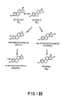
- FIGURE 1A is a flow diagram depicting the metabolic pathways of estrone.
- FIGURE 1B is a flow diagram depicting the metabolic pathways of estradiol.
- FIGURES 2A and 2B are graphs of the results of the measurement of 2-hydroxylation of estrone as between female smokers and nonsmokers. FIGURE 2A is a scattergram presentation of the ratios determined in accordance with the present invention, and FIGURE 2B represents confirmatory radiometric measurements of the same groups compared with the ratio data presented in FIGURE 2A.
- FIGURES 3A and 3B are graphs of the results of radiometric measurements of the percentage of estradiol 2-hydroxylation of groups of male smokers and nonsmokers. FIGURE 3A depicts the radiometric measurements and FIGURE 3B depicts the comparison of ratios from the same groups.
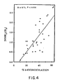
- FIGURE 4 is a graph plotting the radiometric extent of 2-hydroxylation versus urinary [2OHE₁]/[E₃] for each individual, showing a strong correlation between the two measures (r = 0.71,P < .002). The line represents the least squares fit to the points.
- The present invention relates to a method and associated materials for detecting pathology from alterations in estrogen metabolism in mammals. The present method comprises isolating at least two distinct metabolites of estrone from a biological sample taken from the mammal under examination, determining the quantity of each of the said estrone metabolites in the sample, correlating the quantities of each of said metabolites with each other to arrive at a quotient of said metabolites, and comparing said quotient with an extrinsic quotient derived either previously from the mammal under test, as by the previous performance of the within test, or from the testing of other subjects of the same species, to determine any alterations in said estrogen metabolism and to detect pathology therefrom.
- The metabolites of estrone that may be measured and monitored in accordance with the present invention include and may be selected from the products of enzymatic hydroxylation of estrone, the products of the enzymatic reduction of estrone, the products of the enzymatic oxidation of estrone and the products of the enzymatic methylation of estrone. As mentioned earlier, products of enzymatic hydroxylation of estrone include such hydroxylated estrones as may be further enzymatically oxidized or reduced, as by the action of 17-oxidoreductase.
- Accordingly, and as depicted in FIGURE 1A, the estrone metabolites of interest herein include 2-hydroxyestrone, 4-hydroxyestrone, 15α-hydroxyestrone, 16α-hydroxyestrone, 2-methoxyestrone, estriol, 17β-estriol, epiestriol, 16-ketoestradiol, and 15α-hydroxyestradiol. The products of enzymatic hydroxylation broadly comprise 2-hydroxyestrone, 16α-hydroxyestrone, 2-methoxyestrone, and estriol, and more particularly, 2-hydroxyestrone and estriol.
- More particularly, the present invention comprises the measurement and quantitation of the products of enzymatic estrone hydroxylation, and the determination of an algorithmic relationship between certain of these hydroxylation products that provides a reliable indication of pathological condition.
- Accordingly, in one embodiment a ratio or quotient may be determined between the metabolites 2-hydroxyestrone (2OHE₁) and estriol (E₃), that is expressed as
[2OHE₁]/[E₃]
and which may be compared with either prior or subsequent such ratios taken from the measurement of the respective levels of these estrogen metabolites in the same subject, or in subjects of the same species. In the former instance, the physician or the patient may periodically monitor the index to note any changes that would suggest the desirability of more specific testing for one of the conditions or pathologies that are known to cause fluctuations in estrogen metabolism. In the latter instance, the individual index or quotient may be compared with a continuum of similar data developed from subjects both healthy and afflicted with specific pathologies to attempt to identify in an approximate qualitative fashion, the pathological state of the particular subject under test. - As stated earlier herein, the theory of the invention was derived in part from studies of estrogen metabolism in smokers. Several epidemiological studies have indicated that cigarette smoking substantially alters the turnover of estrogen in women (Baron, A. J., AM. J. EPIDEMIOL., 119:9-22 (1984)). Female smokers have been found to have decreased endometrial cancer (Weiss, N. S., et al., MATURITAS, 2:185-190 (1980); Tyler, C. W., et al., AM. J. OBSTET. GYNECOL., 151:899-905 (1985); Lesko, S. M., et al., N. ENGL. J. MED., 313:593-596 (1985); Baron, J. A., et al., JNCI, 77:677-680 (1986)), decreased endometriosis (Cramer, D. W., et al., JAMA, 255:1904-1908 (1986)), decreased benign breast disease (Berkowitz, G. S., et al., J. EPIDEMIOL. COMMUNITY HEALTH, 39:308-313 (1985)), increased osteoporosis (Daniell, H. W., ARCH. INTERN. MED., 136:298-304 (1976); Jensen, J., et al., N. ENGL. J. MED., 313:973-975 (1985)), and earlier natural menopause (Willett, W. L., AM. J. EPIDEMIOL., 117:651-658 (1983)).
- lt was recently reported that smoking is associated with a significant increase in estradiol 2-hydroxylation as measured by an in vivo radiometric procedure in premenopausal females (Michnovicz, J. J., et al., N. ENGL. J. MED., 315:1305-1309 (1986)). This major irreversible biotransformation is depicted in the estrogen metabolic reaction pathway of FIGURE 1B and follows the conversion of estradiol (E₂) to estrone (E₁ and yields the catechol estrogens 2-hydroxyestrone (2OHE₁) and 2-methoxyestrone (2MeOE₁), both possessing minimal peripheral estrogenic activity (Fishman, J., et al., J. BIOL. CHEM., 235:3104-3107 (1960); Martucci, C. P., et al, ENDOCRINOLOGY, 105:1288-1292 (1979); Jellinck, P. H., et al., ENDOCRINOLOGY, 108:1848-1854 (1981)).
- The present invention also extends to the quantitation and analysis of other pathways including the 4- hydroxylation, 15-hydroxylation and 16α-hydroxylation pathways, which 16α-hydroxylation results in the formation of 160-hydroxyestrone (16αOHE₁) and estriol (E₃), which retain estrogen agonist activity (Fishman, J., et al., J. CLIN. ENDOCRINOL. METAB., 51:611-615 (1980)). The specific enzymes involved in the reaction pathway of FIGURE 1A include 17-oxidoreductase which is implicated in the conversion of estradiol to estrone, 16α-hydroxyestrone to estriol, 15α-hydroxyestrone to 15α-hydroxyestradiol, and of epiestriol to 16-ketoestriol; 2-hydroxylase implicated in the hydroxylation of estrone to 2-hydroxyestrone; catechol-O-methyltransferase, involved in the conversion of 2-hydroxyestrone to 2-methoxyestrone; 15α-hydroxylase which is involved in the hydroxylation of estrone to 15α-hydroxyestrone; 4-hydroxylase which is involved in the production of 4-hydroxyestrone; and 16α-hydroxylase, which is involved in the hydroxylation of estrone to 16α-hydroxyestrone.
- A similar relationship was postulated to exist in the male mammalian population, as, for example, men have a higher mortality rate from ischemic heart disease than age-matched women (Kuller, L. H., et al., AM. J. EPIDEMIOL., 104:425-426 (1976); Heller, R. F., et al., BR. MED. J., 1:472-474 (1978); Kannel, W. B., et al., ANN. INTERN. MED., 85:477-452 (1976); Bush, T. L., et al., EPIDEMIOL. REV., 7:80-104 (1985)). In addition, the incidence of heart disease increases significantly in women after menopause, coincident with the decline in ovarian estrogen production (Kannel, W. B., et al., ANN. INTERN. MED. 85:477-452 (1976)). These epidemiologic data point to estrogen as a protective factor against heart disease (Bush, T. L., et al., EPIDEMIOL. REV., 7:80-104 (1985)). Premenopausal females have greater mean high-density lipoprotein-cholesterol (HDL-C) than age-matched males (Rifkind, B. M., et al., LIPIDS, 14:105-112 (1979); Conner, S. L., et al., CIRCULATION, 65:1290-1298 (1980); Wahl, P. W., et al., ATHEROSCLEROSIS, 39:111-124 (1981)), except under such androgenizing conditions as polycystic ovary syndrome (Wild, R. A., et al., J. CLIN. ENDOCRINOL. METAB., 61:946-951 (1985). Administration of estrogens to both men and women produces favorable changes in lipoproteins, with increased HDL-C and decreased low-density lipoprotein-cholesterol (LDL-C) and total cholesterol.
- As a result of the above and as confirmed by the results of the experiments set forth in Examples I-V, below, it has been determined that the index or quotient of the products of enzymatic estrogen metabolism varies with the presence and extent of pathology and is sufficiently independent of other systemic factors that it can serve as a reliable indicator of pathological status.
- The pathological conditions, the nonspecific existence of which are identified by the present invention include osteoporosis, endometriosis, obesity, atherosclerosis, endometrial cancer, breast cancer, heart disease, myocardial infarction, trauma, vascular thrombosis, infertility, gastrointestinal disease, autoimmune disease, central nervous system disease, etc., and any pathological state or condition affecting the estrogen metabolism of a mammal, it being understood by one skilled in the art that the specific pathological state or condition existing in a test mammal is generally qualified after a positive determination of the existence of a pathological state or condition in accordance with the method of the present invention. The term "mammals" as used herein includes homo sapiens as well as domesticated animals, e.g., race horses.
- The present invention also relates to a variety of diagnostic applications, including methods for determining the presence of disease by reference to the ability to detect alterations in estrogen metabolism which are affected by the presence of various pathological conditions in mammals. Also, the present method may be employed to monitor those events in which the level of estrogen metabolites is directly implicated, such as the fertility of the male and female of the species and the course of pregnancy. The present method may therefore be employed as an adjunct to therapy for impotence or infertility, or with respect to possible irregularities during pregnancy, the female menstrual cycle, or at the onset or during menopause. As mentioned earlier, the metabolites of estrone can be used to produce antibodies by a variety of known techniques, and such antibodies could then be isolated and utilized as in tests for alterations in estrogen metabolism in suspect mammals.
- Antibody(ies) to the metabolites of estrone can be produced and isolated by standard methods including the well known hybridoma techniques. Both polyclonal and monoclonal antibodies are included; however, monoclonal antibodies are preferred. Monoclonal antibodies may be prepared in accordance with the teachings of Kohler, G., et al., NATURE 256:495-497 (1975), which reference is cited herein by way of illustration only, and not by way of limitation. In particular, antibodies may be prepared that cross-react with more than one metabolite, and, for example, a monoclonal antibody could be prepared that would bind to E₃ and 16OHE₁ for use in determining ratios that are based in part on 16OHE₁ and E₃. For convenience, the antibody(ies) to the metabolites of estrone will be referred to herein as Ab₁ and antibody(ies) raised in another species as Ab₂.
- The presence of metabolites of estrone in mammals can be ascertained by the usual immunological procedures applicable to such determinations. A number of useful procedures are known. Four such procedures which are especially useful utilize either the metabolite of estrone labeled with a detectable label, antibody Ab₁ labeled with a detectable label, or antibody Ab₂ labeled with a detectable label or a chemical conjugate with the hydroxylation product of estrone labeled with a detectable label. The procedures may be summarized by the following equations wherein the asterisk indicates that the particle is labeled, and "OHE" in this instance stands for all metabolites of estrone:
- A OHE* +′Ab: = OHE*Ab₁
- B. OHE + Ab* = OHEAb₁*
- C. OHE + Ab₁ + Ab₂* = OHEAb₁Ab₂*
- D. Carrier*OHE + Ab₁ = Carrier*OHEAb₁
- The procedures and their application are all familiar to those skilled in the art and are presented herein as illustrative and not restrictive of procedures that may be utilized within the scope of the present invention. The "competitive" procedure, Procedure A, is described in U.S. Patent Nos. 3,654,090 and 3,850,752. Procedure C, the "sandwich" procedure, is described in U.S. Patent Nos. RE 31,006 and 4,016,043. Still other procedures are known such as the "double antibody", or "DASP" procedure. A further diagnostic procedure employs multiple labeled compounds in a single solution for simultaneous radioimmune assay. In this procedure disclosed in U.S. Patent No. 4,762,028 to Olson, a composition may be prepared with two or more analytes in a coordinated compound having the formula: radioisotope-chelator-analyte.
- In each instance, the hydroxylation product of estrone forms complexes with one or more antibody(ies) or binding partners and one member of the complex is labeled with a detectable label. The fact that a complex has formed and, if desired, the amount thereof, can be determined by known methods applicable to the detection of labels.
- It will be seen from the above, that a characteristic property of Ab₂ is that it will react with Ab₁. This is because antibodies raised in one mammalian species have been used in another species as an antigen to raise antibodies such as Ab₂. For example, Ab₂ may be raised in goats using rabbit antibodies as antigens. Ab₂ therefore would be anti-rabbit antibody raised in goats. For purposes of this description and claims, Ab₁ will be referred to as a primary or anti-estrogen metabolite antibody, and Ab₂ will be referred to as a secondary or anti-Ab₁ antibody.
- The labels most commonly employed for these studies are radioactive elements, enzymes, chemicals which fluoresce when exposed to ultraviolet light, and others.
- A number of fluorescent materials are known and can be utilized as labels. These include, for example, fluorescein, rhodamine and auramine. A particular detecting material is anti-rabbit antibody prepared in goats and conjugated with fluorescein through an isothiocyanate.
- The hydroxylated products of estrone or carrier of same, or their binding partner(s) can also be labeled with a radioactive element or with an enzyme. The radioactive label can be detected by any of the currently available counting procedures. The preferred isotope may be selected from ¹⁴C, ¹³¹I, ³H, ¹²⁵I, ⁵⁷Co, ⁵⁹Fe and ³⁵S.
- Enzyme labels are likewise useful, and can be detected by any of the presently utilized colorimetric, spectrophotometric, fluorospectrophotometric, thermometric, amperometric or gasometric techniques. The enzyme is conjugated to the estrogens or their binding partners or carrier molecules by reaction with bridging molecules such as carbodiimides, diisocyanates, glutaraldehyde and the like.
- Many enzymes which can be used in these procedures are known and can be utilized. The preferred are peroxidase, β-glucuronidase, β-D-glucosidase, β-D-galactosidase, urease, glucose oxidase plus peroxidase, hexokinase plus GPDase, glucose oxidase plus alkaline phosphatase, NAD oxidoreductase plus luciferase, phosphofructokinase plus phosphoenol pyruvate carboxylase, aspartate aminotransferase plus phosphoenol pyruvate decarboxylase, and alkaline phosphatase. U.S. Patent Nos. 3,654,090; 3,850,752; and 4,016,043 are referred to by way of example for their disclosure of alternative labeling material and methods.
- A particular assay system developed and utilized in accordance with the present invention, is known as a receptor assay. In a receptor assay, the material to be assayed is appropriately labeled and then certain cellular test colonies are inoculated with a quantity of both the labeled and unlabeled material after which binding studies are conducted to determine the extent to which the labeled material binds to the cell receptors. In this way, differences in affinity between materials can be ascertained.
- Accordingly, a purified quantity of the hydroxylated products of estrone may be radiolabeled, after which binding studies would be carried out using for example, MCF-7 breast cancer cells (ATCC Accession No. HTB 22). Solutions would then be prepared that contain various quantities of labeled and unlabeled MCF-7 cell samples would then be inoculated and thereafter incubated. The resulting cell monolayers would then be washed, solubilized and then counted in a scintillation counter for a length of time sufficient to yield a standard error of <5%. These data are then subjected to Scatchard analysis after which observations and conclusions regarding material activity can be drawn. While the foregoing protocol is exemplary, it illustrates the manner in which a receptor assay may be performed and utilized, in the instance where the cellular binding ability of the assayed material may serve as a distinguishing characteristic.
- A variation of the exemplary receptor assay presented above is disclosed in U.S. Patent No. 4,818,684. In this assay, a competitive protocol is used which employs monoclonal antibodies capable of binding to a steroid receptor which in turn are competitively bound by anti-steroid antibodies capable of binding to the steroid. The assay proceeds by the competition between the anti-steroid antibodies and the receptor. The presence or amount of monoclonal antibody anti-steroid antibody complex thus formed is related to the amount of steroid receptor present in the assayed material.
- In a further embodiment of this invention, commercial test kits suitable for use by a medical specialist may be prepared to determine the presence or absence of hydroxylation products of estrone in a suspected mammal. For example, one class of such kits will contain at least a labeled component selected from the hydroxylation products of estrone or their binding partners, for instance an antibody specific thereto, and directions, of course, depending upon the method selected, e.g., "competitive", "sandwich", "DASP" and the like. The kits may also contain peripheral reagents such as buffers, stabilizers, etc. Accordingly, a test kit may be prepared for the demonstration of the altered estrogen metabolism of a mammalian host, comprising:
- (a) a predetermined amount of at least one labeled immunochemically reactive component obtained by the direct or indirect attachment of the present metabolites of estrone or a specific binding partner thereto, to a detectable label;
- (b) other reagents; and
- (c) directions for use of said kit.
- More specifically, the diagnostic test kit may comprise:
- (a) a known amount of at least one of the metabolites of estrone as described above (or a binding partner) generally bound to a solid phase to form an immunosorbent, or in the alternative, bound to a suitable tag, or plural such end products, etc. (or their binding partners) one of each;
- (b) if necessary, other reagents; and
- (c) directions for use of said test kit.
- In a further variation, the test kit may be prepared and used for the purposes stated above, which operates according to a predetermined protocol (e.g. "competitive", "sandwich", "double antibody", homogeneous enzyme immunoassay, simultaneous multiple analyte immunoassay, etc.), and comprises:
- (a) a labeled component which has been obtained by coupling at least one of the metabolites of estrone to a detectable label;
- (b) one or more additional immunochemical reagents of which at least one reagent is a ligand or an immobilized ligand, which ligand is selected from the group consisting of:
- (i) a ligand capable of binding with the labeled component (a);
- (ii) a ligand capable of binding with a binding partner of the labeled component (a);
- (iii) a ligand capable of binding with at least one of the component(s) to be determined; and
- (iv) a ligand capable of binding with at least one of the binding partners of at least one of the component(s) to be determined; and
- (c) directions for the performance of a protocol for the detection and/or determination of one or more components of an immunochemical reaction between the metabolites of estrone and a specific binding partner thereto.
- The following examples set forth the investigations of female and male smokers in an effort to identify a relationship between estrogen metabolism and the pathological states of the test subjects. As a result of the following tests, the relationship between the presence and ratio of the enzymatically hydroxylated estrone products 2-hydroxyestrone and estriol was identified and confirmed. The specific materials and techniques set forth below are exemplary only and may vary, so that the following is presented by way of illustration and not limitation.
- In the present example, a fairly complete profile of estrogen metabolites was examined in smokers and non-smokers, including assays of urinary 16αOHE₁ and 2OHE₁, as well as the classical estrogens E₁, E₂, and E₃. The resulting data suggest that catechol estrogens constitute a significantly greater proportion of the total measured urinary estrogens in female smokers compared to non-smokers, and led to the determination of the catechol estrogen index that comprises the quotient of the present invention.
- A total of 15 smokers and 14 non-smokers participated in this study; radiometric data from some of these subjects appeared in an earlier report (Michnovicz, J. J., et al., N. ENGL. J. MED., 315:1305-1309 (1986)). All aspects of this study were approved by the Rockefeller University Institutional Review Board. The subjects were healthy, non-obese premenopausal women, ages 21-44, with no history of ongoing menstrual dysfunction, oral contraceptive use, dysthyroidism, or unusual diets. Body-mass index (weight(kg)/height(m)²) ranged from 19-23, with no significant differences in the means of the two groups. All subjects denied use of prescription or illicit drugs except occasional aspirin or acetaminophen for at least 2 months before the study. Urine samples (30 mL) were obtained between 8:00 and 12:00 AM prior to initiation of radiometric testing (see below), which was conducted within 12 days after the reported onset of menses. Smokers reported consuming 15-30 cigarettes daily, while control subjects reported complete abstention from active cigarette smoking. Cotinine, a major metabolite of nicotine, was undetectable in plasma of non-smokers and ranged between 190 and 400 ng/mL in smokers (assay kindly performed by Dr. Nancy Haley of the American Health Foundation, Valhalla, NY).
- The radiolabeled 2-hydroxy-[6,7-³H]estrone (sp. act. 45.6 Ci/mmol), [2,4,6,7-³H]estrone (sp. act. 87.5 Ci/mmol), [2,4,6,7-³H]estradiol (sp. act. 97.8 Ci/mmol), and [2,4,6,7-³H]estriol (sp. act. 94.0 Ci/mmol) were purchased from New England Nuclear (Boston, MA). 16-αHydroxy-[6,7-³H]estrone (sp. act. 58 Ci/mmol) was prepared as previously described (S. Ikegawa, et al., STEROIDS 39:557-567 (1979)). Ascorbic acid was obtained from Sigma (St. Louis, MO). Unlabeled E₁, E₃ and E₂ were obtained from Steraloids (Wilton, NH), while unlabeled 16αOHE₁ and 2OHE₁ were synthesized by standard techniques. Non-radioactive steroids were recrystallized prior to use. Sep-pak cartridges (Waters Assoc., Milford, MA), Glusulase (β-glucuronidase and aryl-sulfatase) (Boehringer Mannheim, Indianapolis, IN), and anti-E₁ antibodies (Endocrine Sciences, Tarzana, CA) were obtained commercially.
- Antibodies to 16α-OHE₁, E₂ and E₃ were raised as described previously (Fishman, J., et al., SCIENCE, 204:1089-1091 (1979); Ikegawa, S., et al., J. STEROID BIOCHEM., 18:329-332 (1983)), while the anti-2OHE₁ antiserum was a gift of Dr. Kanbegawa (Teikyo University, Japan) and was raised against bovine serum albumin linked to 2-hydroxyestrone 17-carboxymethyloxime. Cross-reactivities of the 2OHE₁ antiserum with E₁, E₂ and E₃, 16αOHE₁, 2MeOE₁, and 2-hydroxyestradiol (2OHE₂) were 1.3%, 0.6%, 0.01%, 0.03%, 0.11% and 1.7%, respectively.
- Each urine sample (1 or 2 mL) contained approximately 3000 dpm of stably labeled steroid used to determine recovery, and was applied to a C₁₈ Sep-pak cartridge followed by elution with 5 mL of methanol. The eluate was dried in vacuo and hydrolyzed with Glusulase for 12 h at 37°C. The hydrolyzed steroids were again extracted on a C₁₈ Sep-pak cartridge and eluted with 5 mL of methanol. Aliquots of this final eluate were taken for determination of recovery (averaging 90-95%) and duplicate radioimmunoassay (100 µL). In order to prevent auto-oxidation of catecholestrogens, ascorbic acid (1%) was used in all stages of the assay. Radiolabeled steroids and antisera were incubated with the specimens overnight at 4°C, and radioactivity was measured with a Packard model 3000 scintillation counter. Estrogen levels are expressed relative to urinary creatinine concentration. Statistical analysis was performed with an unpaired t-test. Unless otherwise specified, all values are reported as means ± SEM.
- The radiometric method has been described in detail previously (Michnovicz, J. J., et al., N. ENGL. J. MED., 315:1303-1309 (1986); Fishman, J., et al., PROC. NATL. ACAD. SCI. USA, 77:4957-4960 (1980)). Briefly, subjects received 6 µCi of [2-³H]estradiol intravenously (NEN, sp. act. 25.3 Ci/mmol). Blood and urine samples collected over the next 48 h were lyophilized and counted for ³H₂O. The extent of the reaction was calculated by dividing the ³H specific activity of the total body water volume by the amount of ³H in the administered dose.
- The amounts of the various urinary estrogen metabolites in female smokers and non-smokers are presented in Table 1 below, along with the extent of 2-hydroxylation as determined by the radiometric technique in each subject. Total estrogen excretion, defined as the sum of the measured urinary metabolites in these random follicular-phase samples, did not differ significantly between smokers and non-smokers (55.3 ± 4.2 vs. 51.7 ± 4.5 µg/g creatinine). Although an attempt was made to collect all urine samples within 12 days after the reported onset of menses, estrogen excretion in a few subjects appeared somewhat higher than the average, suggesting that these individuals were approaching the preovulatory rise in plasma estradiol. Nevertheless, it is apparent that smoking in general exerted little effect on total mid-follicular estrogen excretion. Table 1 demonstrates that the average extent of estradiol 2-hydroxylation measured radiometrically was elevated approximately 50% in smokers (51.6 ± 2.7% vs. 33.3 ± 1.7%).
Table 1 Comparison of Estrogen Metabolism in Smokers and Non-Smokers Smokers (n=13) Non-Smokers (n=13) P Value Extent of 2-Hydroxylation 52.2 ± 2.6% 33.6 ± 1.2% <0.001 E₂ 5.5 ± 0.7 3.6 ± 0.3* <0.02 E₁ 17.5 ± 1.2 18.4 ± 2.3 NS 16αOHE₁ 5.6 ± 0.7 5.8 ± 0.7 NS E₃ 10.7 ± 1.1 15.6 ± 1.8 <0.05 2OHE₁ 17.5 ± 2.9 8.6 ± 1.0 <0.01 Total Estrogens 56.8 ± 5.5 52.0 ± 5.8 NS Values are mean ± SEM. * Estrogen metabolites expressed in µg.g creatinine. - Urinary 2OHE₁ was significantly elevated in smokers, compared with non-smokers (17.2 ± 2.4 vs. 9.4 ± 1.2 µg/g creatinine, P < 0.02). A parallel reduction in urinary E₃ was also observed in smokers (10.7 ± 1.1 vs. 15.6 ± 1.8 µg/g, creatinine P<0.05). In contrast, no significant differences were observed in urinary levels of E₂, E₁ or 16αOHE₁. As a result of these altered urinary levels, 2OHE₁ in smokers constituted 31.1% of total measured estrogens, compared with only 18.2% in non-smokers. Increased excretion of 2OHE₁ thus occurred largely at the expense of decreased excretion of the 16α-hydroxylated metabolite, E₃.
- Various ratios of 2-hydroxylated to 16α-hydroxylated metabolites were investigated as a means of differentiating the urinary profiles of smokers and non-smokers. A urinary catechol estrogen index, defined by [2OHE₁]/[E₃], was chosen as a ratio of the major metabolite from each pathway.
- Figure 2A demonstrates the sharp differences in the catechol estrogen index between the two groups. While the values for non-smokers were tightly grouped around the mean value of 0.59 ± 0.08 (range 0.15 to 1.10), the mean for smokers was significantly increased to 1.67 ± 0.21 (range 0.80 to 2.82, P <0.001). Also, there was little overlap between the two groups, and values for several smokers were 3-5 times greater than the mean for non-smokers.
- The urinary estrogen ratios were relatively constant throughout the menstrual cycle, despite large variations in estrogen production. For example, in two women from whom urine samples were obtained on each day of a normal menstrual cycle, the means + SEM (n=27 and 30) of various estrogen ratios for each subject were as follows: [2OHE₁]/[ΣAll] 0.165 ± 0.004 and 0.155 ± 0.004; [16αOHE₁ + E₃]/[ΣAll] 0.391 ± 0.012 and 0.485 ± 0.007 [2OHE₁]/[E₃] 0.689 ± 0.034 and 0.433 ± 0.014.
- The above data demonstrate in premenopausal women that mid-follicular urinary estrogen excretion patterns of smokers differ significantly from those of non-smokers. The principal alteration involves increased 2OHE₁ excretion and concomitantly decreased E₃ excretion among smokers, with no significant difference in the total of these five measured estrogens. These data are in agreement with the earlier finding of increased estrogen 2-hydroxylation measured radiometrically in female smokers (Michnovicz, J. J., et al., N. ENGL. J. MED., 315:1305-1309 (1986)).
- MacMahon et al. (N. ENGL. J. MED., 307:1062-1065 (1982)) previously reported that cigarette smokers had decreased urinary excretion of E₂, E₁ and E₃ during the luteal but not the follicular phase of the menstrual cycle. They concluded that smokers had decreased estrogen production in the luteal phase. The present study did not detect a significant difference in total urinary estrogen excretion in the follicular phase. However, the shift toward catechol estrogen excretion in smokers indicates that the exclusion of 2OHE₁ from the previous analysis precluded an accurate estimate of total estrogen production. The present data show that among the classical estrogens only E₃ is reduced in the follicular phase of smokers. Since the relative ratios of the various metabolites do not change appreciably during the menstrual cycle, the difference should presumably be observed in both the follicular and luteal phases.
- The discovery and analysis of the urinary catechol estrogen index, [2OHE₁]/[E₃], suggests that randomly collected urine samples may provide a reliable estimate of increased 2-hydroxylation in selected populations. Urinary assays would obviate the need to perform in vivo radiometric testing when comparing 2- and 16αhydroxylation in populations suspected of differing in estrogen metabolism. Although urinary estrogen ratios such as [2OHE₁]/[E₃] are sensitive to inter- and intra-assay variation (typically 5 to 10%) (Fishman, J., et al., SCIENCE, 204:1089-1091 (1979)), significant differences in the means of well-defined populations should still be apparent, as they are in these premenopausal female smokers compared with matched non-smokers. Combined radiometric and urinary assays in men have also demonstrated the usefulness of the catechol estrogen index (Michnovicz, J. J., et al. METABOLISM 38:537-541 (1989).
- The above experiment was unable to measure urinary 2MeOE₁, another potentially important metabolite in the 2-hydroxylative pathway (see Figure 1). Nevertheless, published data suggest that the relative amounts of this metabolite in the urine of premenopausal females are only about 10-15% of 2OHE₁ (Adlercreutz, H., et al. J. STEROID BIOCHEM., 24:289-296 (1986)). Thus, changes in the excretion of this metabolite in smokers are unlikely to affect the validity of the urinary catechol estrogen index, defined solely by [2OHE₁]/[E₃], in detecting differences in metabolism among selected populations.
- Although both smoking and obesity possibly affect estrogen production, the metabolic changes outlined above may also modify hormone-dependent disease in humans. In addition to smoking and altered body weight, other factors influencing the extent of 2-hydroxylation in humans include short-term (Anderson, K. E., et al., J. CLIN. ENDOCRINOL. METAB., 59:103-107 (1984)) and long-term (Adlercreutz, H., et al., J. STEROID BIOCHEM., 24:289-295 (1986)) dietary changes, dysthyroidism (Fishman, J., et al., J. CLIN. ENDOCRINOL. METAB., 25:365-368 (1964)), and liver disease (Zumoff, B., et al., J. CLIN. INVEST., 47:20-25 (1968)). The present studies and the resulting identification of the quotient or index of the invention shed light on the control of estrogen metabolism and should be useful in formulating safer therapeutic approaches toward reducing risk for estrogen-related disease.
- In this series of experiments, groups of male smokers and non-smokers were examined by analysis of the urinary estrogen catechol index of the present invention in conjunction with in vivo radiometric analysis for purposes of corroboration. The procedures and results of the experiments are set forth below.
- All of the subjects (n=36) were healthy, non-obese males, 19 to 57 years of age, using no medications except occasional aspirin or acetaminophen. All procedures were approved by the Rockefeller University Institutional Review Board and were performed after obtaining written informed consent from the participant. Physical examination and routine blood tests were normal for each subject. There were no significant differences in the mean age, weight, height or body mass index between the smokers and nonsmokers (Table 2). Nonsmokers denied active smoking, and smokers were included only if they reported the daily inhalation of at least 15 cigarettes (mean 24 ± 4,
range 15 to 55).Table 2 Clinical Data of 20 Smokers and 16 Nonsmoking Controls Subjects Age Height Weight BMI* (1 yr) (m) (kg) (kg/m₂) Smokers 32.6 ± 2.6 1.78 ± 0.02 72.2 ± 1.7 22.8 ± 0.3 (19-57) (1.60-1.91) (55.5-85.7) (19.7-25.3) Nonsmokers 28.6 ± 1.2 1.78 ± 0.02 75.4 ± 2.3 23.7 ± 0.4 (23-40) (1.63-1.92) (60.0-90.8) (20.2-26.8) - To measure estradiol 2-hydroxylation radiometrically (Fishman, J., et al., PROC. NATL. ACAD. SCI. USA, 77:4957-4960 (1980); Michnovicz, J. J., et al., N. ENGL. J. MED., 315:1305-1209 (1986)), each subject received 6 µCi of [2-³H]-estradiol intravenously (New England Nuclear Corporation, Boston; specific activity 25.3 Ci/mmol). Blood and urine samples collected over the next 48 hours were lyophilized and counted for ³H₂O. The extent of the reaction for each subject was calculated by dividing the total ³H present in the body water volume by the ³H administered as [2-³H]-estradiol. The body water volume was estimated using a bioelectrical impedance device (Model BIA-103, RJL Systems, Detroit).
- Cotinine, a major metabolite of nicotine and a biochemical marker of tobacco consumption, was measured by radioimmunoassay (Haley, N. J., AM. J. PUBLIC HEALTH, 73:1204-1207 (1983)). With an assay sensitivity of 0.4 ng/mL, cotinine was undetectable in the serum of all nonsmokers, and ranged from 89 to 487 ng/mL of serum in the smokers.
- Random urine samples (30 mL) obtained between 8 AM and 12 noon prior to the radiometric test were stored for future analysis at -60°C in the presence of ascorbic acid (1%). Frozen urine samples were not available for assay in three of the subjects (two smokers and one nonsmoker). Radioimmunoassays of urinary estrogen metabolites were performed following enzymatic hydrolysis of conjugates, as previously described (Michnovicz, J. J., et al., STEROIDS, 52 (1988)). The interassay coefficients of variability for the urinary estrogen assays varied between 5.0% and 10.0%. Statistical significance was determined using Student's t test, and all values are reported as mean ± SEM unless otherwise specified.
- The results of the radiometric assay in smokers and nonsmokers are shown in Fig. 3A. The average extent of estradiol 2-hydroxylation for 16 nonsmokers was 24.6% ± 1.9% of the administered dose. For the 20 smokers, the extent of reaction averaged 43.4 ± 1.9%, an increase of approximately 70% (P < .001). Cotinine levels in the smokers averaged 242 ± 22 ng/mL (range 89 to 487 ng/mL). Individual cotinine values did not correlate well with the extent of 2-hydroxylation in this group of heavy smokers (P=.61).
- The data shown in Fig. 3A indicate that values for 2-hydroxylation in many smokers overlapped with those of the nonsmoking population. Some lean individuals smoking over two packs per day, and thus expected to show a higher extent of reaction (Fishman, J., et al., CLIN. PHARMACOL. THER., 22:721-728 (1977)), were sometimes still within the normal range. This suggests the presence of other variables, which might counteract the inducing effects of tobacco. Values from men who reported substantial ethanol use (habitual consumption on weekends of at least 12 cans of beer or its equivalent in hard liquor) are indicated in Fig. 3A with a circle. The values for four of five such individuals fell below their respective means.
- Urinary estrogen metabolites were measured to determine whether the radiometric data were reflected in endogenous estrogen excretion patterns. Values of urinary estrogen metabolites in both populations are summarized in Table 2. A significant elevation in the excretion of 2OHE₁ was found in the smokers (10.4 ± 1.3 µg/g creatinine v 6.3 ± 0.7, P = .011), as well as a trend toward reduced E₃, consistent with the metabolic shift measured radiometrically.
- A urinary catechol estrogen index defined by [2OHE₁]/[E₃], previously shown to correlate with the extent of 2-hydroxylation developed in accordance with the present invention, was calculated in female smokers. This index minimizes the effect of variations in the absolute amount of estrogen present in a random urine sample (Michnovicz, J. J., et al., STEROIDS, 52 (1988)). Results are shown for both groups in Fig. 3B. The average for the smokers (1.46 ± 0.19) was significantly greater than that of the nonsmokers (0.81 ± 0.11, P = .006). More variation in this ratio was found among the smokers, as well as considerable overlap with the values of nonsmokers. A strong correlation was found between the urinary catechol estrogen index and the radiometric extent of reaction from each subject (r = 0.71, P = .001; Fig. 4).
Table 3 Summary of Urinary Estrogen Metabolites Smokers Nonsmokers P E₂ 3.9 ± 0.4 3.4 ± 0.3 NS E₁ 9.6 ± 0.8 9.7 ± 1.0 NS 16αOHE₁ 3.3 ± 0.5 2.5 ± 0.2 NS E₃ 7.9 ± 0.8 9.0 ± 0.9 NS 2OHE₁ 10.4 ± 1.3 6.3 ± 0.7 .011 Total estrogens* 35.2 ± 2.7 30.9 ± 1.9 NS [2OHE₁]/[E₃] 1.46 ± 0.19 0.81 ± 0.11 .006 Estrogen metabolites (mean ± SEM) are expressed as µg/g creatinine. * This represents the sum of E₁, E₂, E₃, 16αOHE₁, and 2OHE₁. - The radiometric data reported here demonstrate that induction of estradiol 2-hydroxylation is associated with moderately heavy tobacco use in men. Urinary excretion of endogenous 2-hydroxyestrone is significantly increased in male smokers, both in absolute amount and relative to estriol, suggesting that the enhanced 2-hydroxylation reduces the bioavailability of the active estrogen metabolites 160-hydroxyestrone and estriol. Because 2-hydroxyestrogens are weak agonists (Martucci, C. P., et al., ENDOCRINOLOGY, 105:1288-1292 (1979)), their increased formation is likely to reduce the overall estrogenic stimulation in the liver, the predominant site of 2-hydroxylation (Barbieri, R. L., et al., STEROIDS, 32:529-538 (1978).
- In addition to identifying the anti-estrogen effects of smoking in both males and females and postulating, as the underlying physiological and biochemical bases and implications of these effects, the above results further confirm that the quotient determined in accordance with the invention provides consistent information regarding pathological status that is valid for both males and females and is independent of physiological fluctuations.
- This example is taken from a study performed by Galbraith et al. and identified herein as Reference (3) respecting the activity of the histamine H₂-receptor antagonist Cimetidine, which has been widely used to treat peptic ulceration. The study performed by Galbraith et al. was motivated by the prior noted activity of Cimetidine to cause gynecomastia and sexual dysfunction in certain men, in combination with demonstrated activity as an inhibitor of the cytochrome P-450-dependent biotransformation of certain drugs. The investigators accordingly sought to determine whether Cimetidine also might inhibit the cytochrome P-450-dependent metabolism of estradiol.
- The study comprised the radiometric analysis of urine and serum samples taken from nine normal male volunteers who had received twice daily oral doses of 800 mg per dose of Cimetidine for a period of two weeks. The study showed that, during this time, the 16α-hydroxylation of estradiol was unaffected; however, the urinary excretion of 2-hydroxyestrone decreased by approximately 25% (P<0.0002), and the serum concentration of estradiol increased by approximately 20% (P<0.04).
- A review of this study and, in particular, Table 2 of the study appearing on page 271 thereof, illustrates the predictive value of the index determined from the ratio of 2OHE₁/E₃. Table 2 is reproduced herein below as Table 4.
Table 4 Effect of Cimetidine on the Urinary Excretion of Estrogens* Before Cimetidine After Cimetidine P Value** nmol/nmol of creatinine Estrone 3.80 ± 0.34 3.32 ± 0.26 0.03 Estradiol 1.28 ± 0.15 1.25 ± 0.10 NS 16α-Hydroxyestrone 1.08 ± 0.11 1.05 ± 0.08 NS 2-Hydroxyestrone 1.71 ± 0.20 1.27 ± 0.19 0.0002 Total estrogens*** 10.7 ± 1.1 9.9 ± 0.9 0.02 2-Hydroxyestrone/estriol 0.74 ± 0.18 0.47 ± 0.10 0.015 * Timed overnight urine samples were collected and assayed, as described in Methods, before and after treatment with cimetidine (800 mg orally twice a day for 14 days. Results are expressed as means ± SEM. The number of measurements was nine in each case. ** The P values were calculated with the use of paired Student t-tests, NS denotes not significant. *** Total estrogens comprises estrone, estradiol, estriol, 16α-hydroxyestrone, and 2-hydroxyestrone. - As noted in the Galbraith et al. article, the ratio of 2-hydroxyestrone to estriol was reduced by approximately 40% after Cimetidine treatment. The authors go on to state that Cimetidine appears to possess therapeutic value in the inhibition of estrogen-dependent disease.
- The above data are presented herein to substantiate that the index and quotient of the present invention possess predictive value with respect to pathology that goes beyond the scope and instances set forth in Examples 1 and 2.
- This experiment is taken from a publication by Michnovicz and Galbraith listed herein as Reference (4). In this instance, the authors examined the effect of a brief period of hyperthyroidism on estradiol hydroxylation at both the C-2 and C-16α sites. An in vivo radiometric assay with healthy male volunteers was employed, and a two-week course of thyroxine (4.3 mcg/kg/d) was administered. While the primary thrust of this study revealed that the C-2 pathway may be regulated whereas the C-16α pathway appears to be refractory, the data collected and analyzed in Table 1 of the publication at page 23 thereof, which table is reproduced as Table 5 hereinbelow, illustrated again in cumulative fashion the validity and possible value of the index of 2OHE₁/E₃.
Table 5 Effect of Thyroxine in Urinary Estrogensª Metabolite Preb Postc P Value E₂ 2.78 ± 0.23 3.03 ± 0.27 NS E₁ 7.36 ± 0.39 7.30 ± 0.74 NS 16αOHE₁ 2.07 ± 0.24 2.24 ± 0.17 NS E₃ 5.84 ± 0.88 5.37 ± 0.78 NS 2OHE₁ 2.88 ± 0.32 5.30 ± 0.85 0.01 Total estrogens 20.93 ± 1.58 23.24 ± 2.41 NS [2OHE₁]/[E₃] 0.56 ± 0.09 1.05 ± 0.14 0.01 ª Estrogen metabolites (means ± SEM) are expressed as µg creatinine. b Pre: baseline study c Post: following two weeks of thyroxine.1.5 - As can be seen from the table, a significant reduction in the index was noted, and it was found that urinary 2-hydroxyestrone was significantly greater after thyroxine administration and that the index nearly doubled, mostly due to the elevated excretion of 2OHE₁. Clearly, the condition of hyperthyroidism would be demonstrably reflected in the elevation of the index of the present invention, so that the index would serve to accurately predict this particular pathology. Accordingly, the data presented in Michnovicz et al. is further corroborative of the scope and value of the index of the present invention.
- In this study by two of the inventors herein, which study was just recently published, certain dietary indoles were examined for their induction of hepatic estradiol 2-hydroxylation. In particular, indole-3-carbinol (I3C) was administered to humans at a rate of 500 mg daily for one week. The results of this study reveal that I3C significantly increased the extent of estradiol 2-hydroxylation and indicated that I3C strongly influences estradiol metabolism in humans.
- In conjunction with this study, certain comparative data were developed between the administration of I3C to women and men, and the administration of a high fiber diet to women. The comparative results are presented in Table 6A, B and C below, wherein Table 6A represents the data from women taking I3C, Table 6B represents the data respecting the women who were placed on a high fiber diet, and Table 6C represents the data gathered from the sample of men who were administered I3C.
Table 6A Women Taking I3C (N=5) Metabolite Pre Post P Value [2OHE₁]/[E₁] 1.15 ± 0.24 1.53 ± 0.20 <0.05 [E₃]/[E₁] 1.04 ± 0.20 0.74 ± 0.11 NS [2OHE₁]/[E₃] 1.27 ± 0.30 2.35 ± 0.50 <0.005 Table 6B Women Going on High Fiber Diet (N=6) Metabolite Pre Post P Value [E₃] 9.3 ± 2.8 8.2 ± 3.2 NS [E₁] 10.4 ± 2.7 10.8 ± 2.7 NS [2OHE₁] 11.5 ± 3.2 12.8 ± 4.1 NS [2OHE₁]/[E₃] 1.32 ± 0.35 1.63 ± 0.40 <0.05 Table 6C Men Taking I3C (N=7) Metabolite Pre Post P Value [E₁] 8.4 ± 1.8 8.9 ± 2.3 NS [E₃] 6.4 ± 1.8 4.2 ± 1.3 <0.006 [2OHE₁] 6.6 ± 2.3 8.2 ± 3.5 NS [2OHE₁]/[E₃] 1.06 ± 0.11 2.04 ± 0.35 <0.02 - The study by Michnovicz et al. as to indole-3-carbinol confirmed earlier data with respect to the therapeutic and prophylactic value of ingestion of foods containing this compound. In this respect, both I3C and the high fiber diet as illustrated in the above unpublished data presented in Tables 6A-C above, confirmed that I3C and high fiber are potent inducers of estradiol 2-hydroxylation in similar fashion to the effect evidenced by smoking, and as presented in Example 1 herein. The data presented in Tables 6A-C include comparisons of the C-2 pathway index, which is likewise confirmatory of this therapeutic effect. Thus, the data presented as to the index in Tables 6A-C are cumulative and confirmatory of the predictive diagnostic value of the index of the present invention and its corresponding therapeutic relevance.
- Clearly, the components of the index may be monitored and may correspondingly offer data to the clinician respecting the pathological state of patients who are being administered a potential therapeutic agent such as I3C or drugs or agents that may mimic the activity of I3C or other therapeutic agents having effect on estrogen metabolism as part of their activity profile.
- This invention may be embodied in other forms or carried out in other ways without departing from the spirit or essential characteristics thereof. The present disclosure is, therefore, to be considered as in all respects illustrative and not restrictive, the scope of the invention being indicated by the appended Claims, and all changes which come within the meaning and range of equivalency are intended to be embraced therein.
Claims (28)
Applications Claiming Priority (4)
| Application Number | Priority Date | Filing Date | Title |
|---|---|---|---|
| US549290 | 1983-11-07 | ||
| US38106489A | 1989-07-17 | 1989-07-17 | |
| US381064 | 1989-07-17 | ||
| US54929090A | 1990-07-11 | 1990-07-11 |
Publications (3)
| Publication Number | Publication Date |
|---|---|
| EP0409176A2 true EP0409176A2 (en) | 1991-01-23 |
| EP0409176A3 EP0409176A3 (en) | 1991-10-30 |
| EP0409176B1 EP0409176B1 (en) | 1999-04-28 |
Family
ID=27009231
Family Applications (1)
| Application Number | Title | Priority Date | Filing Date |
|---|---|---|---|
| EP90113694A Expired - Lifetime EP0409176B1 (en) | 1989-07-17 | 1990-07-17 | Method and materials for detecting pathology from alterations in estrogen metabolism |
Country Status (6)
| Country | Link |
|---|---|
| EP (1) | EP0409176B1 (en) |
| JP (1) | JPH03215745A (en) |
| AT (1) | ATE179522T1 (en) |
| AU (1) | AU641409B2 (en) |
| CA (1) | CA2021309C (en) |
| DE (1) | DE69033072T2 (en) |
Cited By (3)
| Publication number | Priority date | Publication date | Assignee | Title |
|---|---|---|---|---|
| WO1997011374A1 (en) * | 1995-09-19 | 1997-03-27 | Immuna Care Corporation | Method of cancer detection |
| US5962242A (en) * | 1995-09-19 | 1999-10-05 | Immuna Care Corporation | Methods of predicting estrogen-dependent osteopenia |
| WO2023234790A1 (en) * | 2022-06-02 | 2023-12-07 | Gdanski Uniwersytet Medyczny | APPLICATION OF HYDROGEN PEROXIDE AND 17β-ESTRADIOL AND ITS METABOLITES AS BIOMARKERS IN A METHOD OF DIAGNOSING NEURODEGENERATIVE DISEASES IN VITRO |
Families Citing this family (1)
| Publication number | Priority date | Publication date | Assignee | Title |
|---|---|---|---|---|
| CN113189334B (en) * | 2021-04-27 | 2022-04-26 | 华南农业大学 | Application of COMT protein in prediction of good response of crab eating monkey to superovulation |
Citations (3)
| Publication number | Priority date | Publication date | Assignee | Title |
|---|---|---|---|---|
| EP0046004A1 (en) * | 1980-08-07 | 1982-02-17 | Syva Company | Concentrating zone method in heterogeneous immunoassays |
| EP0179037A2 (en) * | 1984-09-19 | 1986-04-23 | Mayo Foundation | Nuclear binding assay for steroid receptor functionality in cancerous cells |
| EP0200960A1 (en) * | 1985-05-08 | 1986-11-12 | Abbott Laboratories | Total estriol fluorescence polarization immunoassay |
-
1990
- 1990-07-17 AU AU59051/90A patent/AU641409B2/en not_active Expired
- 1990-07-17 CA CA002021309A patent/CA2021309C/en not_active Expired - Lifetime
- 1990-07-17 AT AT90113694T patent/ATE179522T1/en not_active IP Right Cessation
- 1990-07-17 EP EP90113694A patent/EP0409176B1/en not_active Expired - Lifetime
- 1990-07-17 JP JP2190349A patent/JPH03215745A/en active Pending
- 1990-07-17 DE DE69033072T patent/DE69033072T2/en not_active Expired - Lifetime
Patent Citations (3)
| Publication number | Priority date | Publication date | Assignee | Title |
|---|---|---|---|---|
| EP0046004A1 (en) * | 1980-08-07 | 1982-02-17 | Syva Company | Concentrating zone method in heterogeneous immunoassays |
| EP0179037A2 (en) * | 1984-09-19 | 1986-04-23 | Mayo Foundation | Nuclear binding assay for steroid receptor functionality in cancerous cells |
| EP0200960A1 (en) * | 1985-05-08 | 1986-11-12 | Abbott Laboratories | Total estriol fluorescence polarization immunoassay |
Non-Patent Citations (11)
| Title |
|---|
| American Journal of Epidemiology vol. 119, no. 1, January 1984, Baltimore Md. pages 9 - 22; J.A. Baron: "Smoking and estrogen related disease" * |
| ANNALS N Y. ACAD. SCI., vol., no., 1990, page 252 - 258, J.H.H. THIJSSEN AND M.A. BLANKENSTEIN 'UPTAKE AND METABOLISM OF ESTRADIOL BY NORMAL AND ABNORMAL BREAST TISSUES' * |
| EUR. J. CANCER CLIN. ONC., vol.24, no.7, 1988, page 1171 - 1178, Y.J. ABUL-HAJJ ET AL. 'METABOLISM OF ESTRADIOL BY HUMAN BREAST CANCER' * |
| J. NATL. CANC. INST. , vol.86, no.14, 20.07.94, page 1076 - 1082, H. ADLERCREUTZ ET AL. 'ESTROGEN METABOLISM AND EXCRETION IN ORIENTAL AND CAUCASIAN WOMEN' * |
| J. STEROID BIOCHEM., vol.34, no.1-6, 1989, page 527 - 530, H. ADLERCREUTZ ET AL. 'DIET AND URINARY ESTROGEN PROFILE IN PREMENOPAUSAL OMNIVOROUS AND VEGETARIAN WOMEN AND IN PREMENOPAUSAL WOMEN WITH BREAST CANCER' * |
| J. steroid. Biochem. vol. 24, no. 1, 1986, Oxford GB pages 289 - 296; H. Adlercreutz et al.: "Urinary estrogen profile determination in young finnish vegetarian and omnivorous women" * |
| Metabolism vol. 38, no. 6, June 1989, pages 537 - 541; J.J. Michnovicz et al.: "Cigarette smoking alters hepatic estrogen metabolism in men: Implicationsfor atherosclerosis" * |
| Steroids vol. 52, no. 1-2, August 1988, Stoneham Mass. pages 69 - 83; J.J. Michnovicz et al.: "Increased urinary catechol estrogen excretion in female smokers" * |
| Steroids vol. 55, no. 1, January 1990, Stoneham Mass. pages 22 - 26; J.J. Michnovicz et al.: "Effects of exogenous thyroxine on C-2 and C-16¡ hydroxylations of estradiol in humans" * |
| The New England Journal of Medicine vol. 315, no. 21, 20 November 1986, Boston Mass. pages 1305 - 1309; J.J. Michnovicz et al.: "Increased 2-hydroxylation of estradiol as a possible mechanism for the anti-estrogenic effect of cigarette smoking" * |
| The New England Journal of Medicine vol. 321, no. 5, 03 August 1989, Boston Mass. pages 269 - 274; R.A. Galbraith et al.: "The effects of cimetidine on the oxidative metabolism of estradiol" * |
Cited By (4)
| Publication number | Priority date | Publication date | Assignee | Title |
|---|---|---|---|---|
| WO1997011374A1 (en) * | 1995-09-19 | 1997-03-27 | Immuna Care Corporation | Method of cancer detection |
| US5854009A (en) * | 1995-09-19 | 1998-12-29 | Immuna Care Corporation | Method of detecting estrogen-sensitive pathologies |
| US5962242A (en) * | 1995-09-19 | 1999-10-05 | Immuna Care Corporation | Methods of predicting estrogen-dependent osteopenia |
| WO2023234790A1 (en) * | 2022-06-02 | 2023-12-07 | Gdanski Uniwersytet Medyczny | APPLICATION OF HYDROGEN PEROXIDE AND 17β-ESTRADIOL AND ITS METABOLITES AS BIOMARKERS IN A METHOD OF DIAGNOSING NEURODEGENERATIVE DISEASES IN VITRO |
Also Published As
| Publication number | Publication date |
|---|---|
| EP0409176A3 (en) | 1991-10-30 |
| ATE179522T1 (en) | 1999-05-15 |
| EP0409176B1 (en) | 1999-04-28 |
| JPH03215745A (en) | 1991-09-20 |
| CA2021309C (en) | 2003-04-01 |
| AU641409B2 (en) | 1993-09-23 |
| CA2021309A1 (en) | 1991-01-18 |
| DE69033072T2 (en) | 1999-10-28 |
| AU5905190A (en) | 1991-01-17 |
| DE69033072D1 (en) | 1999-06-02 |
Similar Documents
| Publication | Publication Date | Title |
|---|---|---|
| Tunn et al. | Simultaneous measurement of cortisol in serum and saliva after different forms of cortisol administration | |
| Fishman et al. | Increased estrogen-16α-hydroxylase activity in women with breast and endometrial cancer | |
| Lipton et al. | Aromatase activity in primary and metastatic human breast cancer | |
| WIlke et al. | Total testosterone, free-androgen index, calculated free testosterone, and free testosterone by analog RIA compared in hirsute women and in otherwise-normal women with altered binding of sex-hormone-binding globulin. | |
| CLARK et al. | Treatment of men with idiopathic oligozoospermic infertility using the aromatase inhibitor, testolactone results of a double‐blinded, randomized, placebo‐controlled trial with crossover | |
| FI67760B (en) | FOERFARANDE FOER BESTAEMNING AV CONCENTRATIONS AV EN FRI LIGAND FRAON EN BIOLOGISK VAETSKA | |
| Schmidt et al. | Androgen receptor in hirsutism and acne | |
| Campbell et al. | Urinary excretion of prostaglandin E2, prostaglandin F2 alpha, and thromboxane B2 in normotensive and hypertensive subjects on varying sodium intakes. | |
| HEIKINHEIMO et al. | Distribution of RU 486 and its demethylated metabolites in humans | |
| Bryan | Role of tryptophan metabolites in urinary bladder cancer | |
| Tschöp et al. | A time-resolved fluorescence immunoassay for the measurement of testosterone in saliva: monitoring of testosterone replacement therapy with testosterone buciclate | |
| English et al. | A radioimmunoassay procedure for dexamethasone: plasma and urine levels in man | |
| CA2021309C (en) | Method and materials for detecting pathology from alterations in estrogen metabolism | |
| AU602864B2 (en) | Method for measuring free ligands in biological fluids, and assay for measuring same | |
| EP0866969B1 (en) | Method of cancer detection | |
| Prodi et al. | 17β-estradiol, 5α-dihydrotestosterone, progesterone and Cortisol receptors in normal and neoplastic human endometrium | |
| Stanczyk et al. | Influence of estrogen on prostacyclin and thromboxane balance in postmenopausal women | |
| Ahokoski et al. | Hormonal effects of MPV‐2213ad, a new selective aromatase inhibitor, in healthy male subjects. A phase I study | |
| Holdaway et al. | Progesterone and oestrogen receptors in human breast cancer | |
| Naganuma et al. | Radioimmunoassay of 16α-hydroxyestrone in human urine | |
| US5962242A (en) | Methods of predicting estrogen-dependent osteopenia | |
| WO1996007905A1 (en) | A method for diagnosing responsiveness to hormone therapy | |
| Holt et al. | Enzyme-linked immunochemical measurement of estrogen receptor in gynecologic tumors, and an overview of steroid receptors in ovarian carcinoma. | |
| Lundgren et al. | Effect of oral high-dose progestins on the disposition of antipyrine, digitoxin, and warfarin in patients with advanced breast cancer | |
| JPS59155761A (en) | Method of measuring steroid hormone glucuronic acid compound |
Legal Events
| Date | Code | Title | Description |
|---|---|---|---|
| PUAI | Public reference made under article 153(3) epc to a published international application that has entered the european phase |
Free format text: ORIGINAL CODE: 0009012 |
|
| AK | Designated contracting states |
Kind code of ref document: A2 Designated state(s): AT BE CH DE DK ES FR GB GR IT LI LU NL SE |
|
| PUAL | Search report despatched |
Free format text: ORIGINAL CODE: 0009013 |
|
| AK | Designated contracting states |
Kind code of ref document: A3 Designated state(s): AT BE CH DE DK ES FR GB GR IT LI LU NL SE |
|
| 17P | Request for examination filed |
Effective date: 19920414 |
|
| 17Q | First examination report despatched |
Effective date: 19940930 |
|
| GRAG | Despatch of communication of intention to grant |
Free format text: ORIGINAL CODE: EPIDOS AGRA |
|
| GRAG | Despatch of communication of intention to grant |
Free format text: ORIGINAL CODE: EPIDOS AGRA |
|
| GRAH | Despatch of communication of intention to grant a patent |
Free format text: ORIGINAL CODE: EPIDOS IGRA |
|
| GRAH | Despatch of communication of intention to grant a patent |
Free format text: ORIGINAL CODE: EPIDOS IGRA |
|
| GRAA | (expected) grant |
Free format text: ORIGINAL CODE: 0009210 |
|
| AK | Designated contracting states |
Kind code of ref document: B1 Designated state(s): AT BE CH DE DK ES FR GB GR IT LI LU NL SE |
|
| PG25 | Lapsed in a contracting state [announced via postgrant information from national office to epo] |
Ref country code: NL Free format text: LAPSE BECAUSE OF FAILURE TO SUBMIT A TRANSLATION OF THE DESCRIPTION OR TO PAY THE FEE WITHIN THE PRESCRIBED TIME-LIMIT Effective date: 19990428 Ref country code: GR Free format text: LAPSE BECAUSE OF NON-PAYMENT OF DUE FEES Effective date: 19990428 Ref country code: ES Free format text: THE PATENT HAS BEEN ANNULLED BY A DECISION OF A NATIONAL AUTHORITY Effective date: 19990428 Ref country code: LI Free format text: LAPSE BECAUSE OF FAILURE TO SUBMIT A TRANSLATION OF THE DESCRIPTION OR TO PAY THE FEE WITHIN THE PRESCRIBED TIME-LIMIT Effective date: 19990428 Ref country code: CH Free format text: LAPSE BECAUSE OF FAILURE TO SUBMIT A TRANSLATION OF THE DESCRIPTION OR TO PAY THE FEE WITHIN THE PRESCRIBED TIME-LIMIT Effective date: 19990428 Ref country code: BE Free format text: LAPSE BECAUSE OF FAILURE TO SUBMIT A TRANSLATION OF THE DESCRIPTION OR TO PAY THE FEE WITHIN THE PRESCRIBED TIME-LIMIT Effective date: 19990428 Ref country code: SE Free format text: THE PATENT HAS BEEN ANNULLED BY A DECISION OF A NATIONAL AUTHORITY Effective date: 19990428 Ref country code: AT Free format text: LAPSE BECAUSE OF FAILURE TO SUBMIT A TRANSLATION OF THE DESCRIPTION OR TO PAY THE FEE WITHIN THE PRESCRIBED TIME-LIMIT Effective date: 19990428 |
|
| REF | Corresponds to: |
Ref document number: 179522 Country of ref document: AT Date of ref document: 19990515 Kind code of ref document: T |
|
| REG | Reference to a national code |
Ref country code: CH Ref legal event code: EP |
|
| ITF | It: translation for a ep patent filed |
Owner name: STUDIO TORTA S.R.L. |
|
| REF | Corresponds to: |
Ref document number: 69033072 Country of ref document: DE Date of ref document: 19990602 |
|
| PG25 | Lapsed in a contracting state [announced via postgrant information from national office to epo] |
Ref country code: LU Free format text: LAPSE BECAUSE OF NON-PAYMENT OF DUE FEES Effective date: 19990717 |
|
| ET | Fr: translation filed | ||
| PG25 | Lapsed in a contracting state [announced via postgrant information from national office to epo] |
Ref country code: DK Free format text: LAPSE BECAUSE OF FAILURE TO SUBMIT A TRANSLATION OF THE DESCRIPTION OR TO PAY THE FEE WITHIN THE PRESCRIBED TIME-LIMIT Effective date: 19990728 |
|
| REG | Reference to a national code |
Ref country code: CH Ref legal event code: PL |
|
| PLBE | No opposition filed within time limit |
Free format text: ORIGINAL CODE: 0009261 |
|
| STAA | Information on the status of an ep patent application or granted ep patent |
Free format text: STATUS: NO OPPOSITION FILED WITHIN TIME LIMIT |
|
| 26N | No opposition filed | ||
| REG | Reference to a national code |
Ref country code: GB Ref legal event code: IF02 |
|
| PGFP | Annual fee paid to national office [announced via postgrant information from national office to epo] |
Ref country code: FR Payment date: 20090630 Year of fee payment: 20 |
|
| PGFP | Annual fee paid to national office [announced via postgrant information from national office to epo] |
Ref country code: DE Payment date: 20090724 Year of fee payment: 20 Ref country code: GB Payment date: 20090710 Year of fee payment: 20 |
|
| PGFP | Annual fee paid to national office [announced via postgrant information from national office to epo] |
Ref country code: IT Payment date: 20090723 Year of fee payment: 20 |
|
| REG | Reference to a national code |
Ref country code: GB Ref legal event code: PE20 Expiry date: 20100716 |
|
| PG25 | Lapsed in a contracting state [announced via postgrant information from national office to epo] |
Ref country code: GB Free format text: LAPSE BECAUSE OF EXPIRATION OF PROTECTION Effective date: 20100716 |
|
| PG25 | Lapsed in a contracting state [announced via postgrant information from national office to epo] |
Ref country code: DE Free format text: LAPSE BECAUSE OF EXPIRATION OF PROTECTION Effective date: 20100717 |