EP0405578B1 - Enzyme immunoassay for antigen and solid phase used therefor - Google Patents
Enzyme immunoassay for antigen and solid phase used therefor Download PDFInfo
- Publication number
- EP0405578B1 EP0405578B1 EP90112454A EP90112454A EP0405578B1 EP 0405578 B1 EP0405578 B1 EP 0405578B1 EP 90112454 A EP90112454 A EP 90112454A EP 90112454 A EP90112454 A EP 90112454A EP 0405578 B1 EP0405578 B1 EP 0405578B1
- Authority
- EP
- European Patent Office
- Prior art keywords
- solid phase
- antibody
- antigen
- biotin
- bound
- Prior art date
- Legal status (The legal status is an assumption and is not a legal conclusion. Google has not performed a legal analysis and makes no representation as to the accuracy of the status listed.)
- Expired - Lifetime
Links
Images


Classifications
-
- G—PHYSICS
- G01—MEASURING; TESTING
- G01N—INVESTIGATING OR ANALYSING MATERIALS BY DETERMINING THEIR CHEMICAL OR PHYSICAL PROPERTIES
- G01N33/00—Investigating or analysing materials by specific methods not covered by groups G01N1/00 - G01N31/00
- G01N33/48—Biological material, e.g. blood, urine; Haemocytometers
- G01N33/50—Chemical analysis of biological material, e.g. blood, urine; Testing involving biospecific ligand binding methods; Immunological testing
- G01N33/53—Immunoassay; Biospecific binding assay; Materials therefor
- G01N33/575—Immunoassay; Biospecific binding assay; Materials therefor for cancer
- G01N33/57565—Immunoassay; Biospecific binding assay; Materials therefor for cancer involving carcinoembryonic antigen [CEA]
-
- G—PHYSICS
- G01—MEASURING; TESTING
- G01N—INVESTIGATING OR ANALYSING MATERIALS BY DETERMINING THEIR CHEMICAL OR PHYSICAL PROPERTIES
- G01N33/00—Investigating or analysing materials by specific methods not covered by groups G01N1/00 - G01N31/00
- G01N33/48—Biological material, e.g. blood, urine; Haemocytometers
- G01N33/50—Chemical analysis of biological material, e.g. blood, urine; Testing involving biospecific ligand binding methods; Immunological testing
- G01N33/53—Immunoassay; Biospecific binding assay; Materials therefor
- G01N33/543—Immunoassay; Biospecific binding assay; Materials therefor with an insoluble carrier for immobilising immunochemicals
- G01N33/54306—Solid-phase reaction mechanisms
-
- G—PHYSICS
- G01—MEASURING; TESTING
- G01N—INVESTIGATING OR ANALYSING MATERIALS BY DETERMINING THEIR CHEMICAL OR PHYSICAL PROPERTIES
- G01N33/00—Investigating or analysing materials by specific methods not covered by groups G01N1/00 - G01N31/00
- G01N33/48—Biological material, e.g. blood, urine; Haemocytometers
- G01N33/50—Chemical analysis of biological material, e.g. blood, urine; Testing involving biospecific ligand binding methods; Immunological testing
- G01N33/53—Immunoassay; Biospecific binding assay; Materials therefor
- G01N33/543—Immunoassay; Biospecific binding assay; Materials therefor with an insoluble carrier for immobilising immunochemicals
- G01N33/54353—Immunoassay; Biospecific binding assay; Materials therefor with an insoluble carrier for immobilising immunochemicals with ligand attached to the carrier via a chemical coupling agent
-
- G—PHYSICS
- G01—MEASURING; TESTING
- G01N—INVESTIGATING OR ANALYSING MATERIALS BY DETERMINING THEIR CHEMICAL OR PHYSICAL PROPERTIES
- G01N33/00—Investigating or analysing materials by specific methods not covered by groups G01N1/00 - G01N31/00
- G01N33/48—Biological material, e.g. blood, urine; Haemocytometers
- G01N33/50—Chemical analysis of biological material, e.g. blood, urine; Testing involving biospecific ligand binding methods; Immunological testing
- G01N33/53—Immunoassay; Biospecific binding assay; Materials therefor
- G01N33/575—Immunoassay; Biospecific binding assay; Materials therefor for cancer
- G01N33/5757—Immunoassay; Biospecific binding assay; Materials therefor for cancer involving oncofoetal proteins
-
- G—PHYSICS
- G01—MEASURING; TESTING
- G01N—INVESTIGATING OR ANALYSING MATERIALS BY DETERMINING THEIR CHEMICAL OR PHYSICAL PROPERTIES
- G01N33/00—Investigating or analysing materials by specific methods not covered by groups G01N1/00 - G01N31/00
- G01N33/48—Biological material, e.g. blood, urine; Haemocytometers
- G01N33/50—Chemical analysis of biological material, e.g. blood, urine; Testing involving biospecific ligand binding methods; Immunological testing
- G01N33/58—Chemical analysis of biological material, e.g. blood, urine; Testing involving biospecific ligand binding methods; Immunological testing involving labelled substances
- G01N33/581—Chemical analysis of biological material, e.g. blood, urine; Testing involving biospecific ligand binding methods; Immunological testing involving labelled substances with enzyme label (including co-enzymes, co-factors, enzyme inhibitors or substrates)
-
- G—PHYSICS
- G01—MEASURING; TESTING
- G01N—INVESTIGATING OR ANALYSING MATERIALS BY DETERMINING THEIR CHEMICAL OR PHYSICAL PROPERTIES
- G01N33/00—Investigating or analysing materials by specific methods not covered by groups G01N1/00 - G01N31/00
- G01N33/48—Biological material, e.g. blood, urine; Haemocytometers
- G01N33/50—Chemical analysis of biological material, e.g. blood, urine; Testing involving biospecific ligand binding methods; Immunological testing
- G01N33/68—Chemical analysis of biological material, e.g. blood, urine; Testing involving biospecific ligand binding methods; Immunological testing involving proteins, peptides or amino acids
- G01N33/6878—Chemical analysis of biological material, e.g. blood, urine; Testing involving biospecific ligand binding methods; Immunological testing involving proteins, peptides or amino acids in epitope analysis
Definitions
- the present invention relates to a method for measuring an amount of an antigen in a sample and a solid phase used therefor. More particularly, the present invention relates to an enzyme immunoassay for measuring an amount of an antigen in a sample which comprises reacting an antigen in a sample either with a biotinylated antibody, an enzyme-labelled antibody and an avidin-immobilized solid phase or with an enzyme-labelled antibody and a biotinylated antibody-bound avidin-immobilized solid phase to form a biotinylated antibody/antigen/enzyme-labelled antibody complex on the avidin-immobilized solid phase, and measuring an enzyme activity on the solid phase, and a solid phase used therefor.
- a trace substance such as a tumor marker or a hormone. Since measurement of such a trace substance requires high specificity and sensitivity, an immunological method utilizing an antigen-antibody reaction which is endowed with high specificity and sensitivity has widely been used.
- various immunological methods including a fluorescent antibody method, a radioimmunoassay, an enzyme immunoassay and the like.
- the enzyme immunoassay where an enzyme is used for labelling has advantages such as a wide range of application, easiness in handling of reagents, capability of treating a large number of a sample, etc. and is becoming a dominant method for measuring a trace substance.
- Japanese Patent First Publication No. 229368/1988 disloses the application of the avidin-biotin reaction for a B/F separation in the field of clinical chemistry.
- This method comprises reacting an antibody to be determined with a labelled antigen, a biotinylated antigen and an avidin-immobilized solid phase to form an avidin/biotinylated antigen/antibody/labelled antigen complex on the solid phase and measuring the label to determine an amount of the antibody.
- a reagent suitable for measuring an amount of an antigen using the avidin-biotin reaction there is commercially available a reagent for measuring ferritin from Nippon Mediphysics Co. Ltd., which comprises an avidin-immobilized solid phase, a biotinylated antibody and an antibody labelled with a radioisotope.
- the measurement with this reagent still takes a long time as 3 to 4 hours.
- a similar method has been reported in the Program Abstract, general subject No. 41 of the Proceeding of the 28th Annual Meeting of Japan Society Clinical Chemistry held on November 18 and 19, 1988.
- This method comprises adding an enzyme-labelled antibody and a biotinylated antibody to a sample containing an antigen to conduct the antigen-antibody reaction in a liquid phase, adding thereto an avidin-immobilized solid phase to allow the resulting sandwich complex of enzyme-labelled antibody/antigen/biotinylated antibody to be adsorbed to the solid phase, and measuring the enzyme activity to determine an amount of the antigen.
- the antigen-antibody reaction proceeds in the liquid phase, and hence, reaches to equilibrium rapidly and further the avidin-biotin reaction on the solid phase proceeds more rapidly than the antigen-antibody reaction on the solid phase, which shortens the reaction time.
- a fluorescent substance to compensate the insufficiency of sensitivity due to the shortening of the reaction time (to about 10 minutes).
- the antibody titer is possibly damaged since the biotinylated antibody is prepared by linking a biotin derivative to an amino group of an antibody and further an amount of avidin on the solid phase is possibly deficient due to difficulty in linking avidin to the solid phase.
- EP-A-0 072 902 discloses a solid phase method for determing carcinoembryonal antigen (CEA) by incubating the sample to be investigated with a first CEA antibody bound on a water-insoluble carrier and with a second CEA antibody, to which peroxidase is bound directly or via a biotin-avidin bridge.
- CEA carcinoembryonal antigen
- WO-87/04794 reveals a reagent for detecting an analyte in a sample fluid which comprises an inert particle, a plurality of avidin molecules noncovalently attached to the surface of the particle, and derivatized biotin molecules complexed with at least a portion of the avidin molecules.
- US-A-4 282 287 discloses a process of modifying the surface properties of a surface, wherein layers of first and second materials are applied to a surface to be modified, the first material comprising avidin and the second material comprising a noncovalent, biotin-modified extender.
- FR-A-2 382 450 a product of the formula R1 - S - S - A - Z is disclosed, wherein R1 represents specific pyridine derivatives, A represents a hydrocarbon residue with 1 to 10 carbon atoms, and Z is a specific group comprising carboxyl and carboimid bindings.
- R1 represents specific pyridine derivatives
- A represents a hydrocarbon residue with 1 to 10 carbon atoms
- Z is a specific group comprising carboxyl and carboimid bindings.
- the above product can be used as an agent for biofunctional bindings and for the formation of thiols.
- a capture membrane wherein biotin is bound to the membrane and a complex comprising an antibody and antigens bound to the enzyme and to the streptavidin/biotin complex.
- streptavidin may be bound to a biotin-coated membrane and biotinylated BSA is bound to a streptavidin-coated membrane.
- a immumological substance detecting reagent comprising an immunological homolog specific for the substance to be detected, a water-soluble polymer having a net charge not greater than zero, and means attaching the water-soluble polymer to the homolog.
- the attaching means may be represented by a biotin/avidin complex.
- the present inventors have intensively studied as to preparation of the biotinylated antibody without deteriorating the antibody titer and as to means for increasing an amount of avidin on the solid phase in order to overcome the problem of insufficient sensitivity as mentioned above, and as a result, have found that the first problem can be solved by linking the biotin derivative to a thiol group of an Fab' fragment of the antibody and the second problem can be solved by linking avidin to the solid phase via a biotinylated high molecular weight substance to increase sensitivity, that the sensitivity can be further increased if both means are used together, that even when a conventional biotinylated antibody and avidin-immobilized solid phase are used, the sensitivity can be increased if they are used in the form of a biotinylated antibody-bound avidin-immobilized solid phase in which said biotinylated antibody is bound to the avidin-immobilized solid phase, and that this latter method can advantageously be carried out for the measurement of an antigen only
- an enzyme immunoassay for an antigen using a solid phase which employs (a') a biotinylated antibody-immobilized solid phase, i.e. a biotinylated antibody, in which a biotin derivative is bound to a thiol group of an Fab' fragment of the antibody, [which antibody is immobilized on to a solid phase] via a substance selected from the group consisting of avidin, streptoavidin and a derivative thereof, wherein said substance is bound to the biotin moiety of said biotinylated antibody and is also bound to the solid phase via a linkage between another biotin derivative and a high molecular weight substance bound to said solid phase, and (b') an enzyme-labelled antibody wherein both of the antibodies (a') and (b') are directed to the same antigen to be determined but recognize different epitopes of said antigen.
- a biotinylated antibody-immobilized solid phase i.e. a biotinylated antibody
- the enzyme Immunoassay of the invention is carried out by simultaneously reacting the enzyme-labelled antibody (b'), a sample containing an antigen to be determined and the solid phase (a').
- the invention furthermore provides a solid phase for enzyme immunoassay which comprises a solid phase immobilized with a biotinylated antibody in which a biotin derivative is bound to a thiol group of an Fab' fragment of the antibody being directed to the antigen to be determined via a substance selected from the group consisting of avidin, streptoavidin and a derivative thereof wherein said substance is bound to the biotin moiety of the biotinylated antibody and is also bound to the solid phase via a linkage between another biotin derivative and a high molecular weight substance bound to the solid phase (hereinafter referred to as "Solid Phase II").
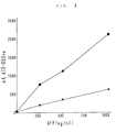
- Fig. 1 is a calibration curve for an antigen (AFP) in a sample measured by enzyme immunoassay using Solid Phase II of the present invention employing a biotin-Fab'.
- Fig. 2 is a calibration curve for an antigen (CEA) in a sample measured by enzyme immunoassay using Solid Phase II of the present invention employing a biotin-Fab'.
- Fig. 3 is a calibration curve for an antigen (AFP) in a sample measured by enzyme immunoassay using Solid Phase II of the present invention employing a biotin-Fab' or a conventional avidin-immobilized solid phase in which the sensitivity in case of Solid Phase II of the present invention and the sensitivity in case of the conventional solid phase are compared to each other.
- AFP antigen
- Solid Phase I has a structure wherein a solid phase is immobilized with a high molecular weight substance/biotin/avidin (or streptoavidin or a derivative thereof) complex, in which a high molecular weight substance is biotinylated and avidin, streptoavidin or a derivative thereof is bound to the biotinylated moiety thereof, in other words, a structure wherein avidin, streptoavidin or a derivative thereof is immobilized to a solid phase via the biotinylated high molecular weight substance.
- Solid Phase II of the present invention has a structure wherein the biotinylated antibody is bound to Solid Phase I at the moiety of avidin, etc. thereon, or to a solid phase to which a substance selected from the group consisting of avidin, streptoavidin and a derivative thereof is directly bound, through avidin-biotin linkage.
- Solid Phase I can be prepared by, for example, biotinylating a high molecular weight substance with a biotin derivative (hereinafter referred to as "biotinylating reagent"), linking said biotinylated high molecular weight substance to a solid phase via the high molecular weight substance moiety thereof and further linking avidin, streptoavidin or a derivative thereof (e.g. a neutralized avidin in which free amino group(s) is/are pre-blocked by, for example, acetylation with acetic anhydride) to the biotin moiety.
- the high molecular weight substance can be bound to the solid phase by various methods including physical adsorption, covalent bonding and the like.
- Solid Phase II of the present invention can be prepared by, for example, linking a biotinylated antibody in which the biotin derivative is bound to a thiol group of and Fab' fragment of an antibody to Solid Phase I or to a solid phase to which avidin, streptoavidin or a derivative thereof is directly bound, through avidin-biotin linkage.
- the high molecular weight substance used in the present invention has a functional group reactive with the biotinylating reagent and is capable of being physically adsorbed or covalently bound to the solid phase, including proteins [e.g. BSA, ovalbumin (hereinafter referred to as "OA") immunoglobulin, collagen, gelatin, casein, etc.], polysaccharides (e.g. dextran, pullulan, etc.) and the like.
- proteins e.g. BSA, ovalbumin (hereinafter referred to as "OA") immunoglobulin, collagen, gelatin, casein, etc.]
- polysaccharides e.g. dextran, pullulan, etc.
- the biotinylating reagent used in the present invention is a biotin derivative having a functional group reactive with the high molecular weight substance to form a covalent bond and includes, for example, biotin-N-hydroxysuccinimide ester (hereinafter referred to as "NHS-biotin”), N-biotinyl-6-aminocaproyl-N-hydroxysulfosuccinimide ester (hereinafter referred to as "NHS-LC-biotin”), sulfosuccinimidyl-2-(biotinamido)ethyl-1,3-dithiopropionate (hereinafter referred to as "NHS-SS-biotin”), and the like [cf. Analytical Biochemistry: 171, 1-32 (1988)].
- NHS-biotin biotin-N-hydroxysuccinimide ester
- NHS-LC-biotin N-biotinyl-6-aminocaproyl-N-
- the biotinylating reagent and the high molecular weight substance can be bound to each other by reacting one mole of the high molecular weight substance with preferably about 1 to about 100 moles of the biotinylating reagent, depending on a kind of the biotinylating reagent and the high molecular weight substance.
- the reaction can be conducted under such conditions that the high molecular weight substance and the biotinylating reagent are bound to each other sufficiently and the moiety on the high molecular weight substance where the solid phase is bound is not inactivated, for example, under the conditions of pH of the solution ranging from about 4 to about 10, the reaction temperature of about 4° to about 45°C and the reaction time of about 10 minutes to about 24 hours.
- the thus prepared biotinylated high molecular weight substance can be bound to the solid phase by the usual method such as the physical adsorption or the covalent bond but the method should not interfere the bond between the biotin moiety of the resulting biotinylated high molecular weight substance-immobilized solid phase and avidin, streptoavidin or a derivative thereof.
- the method for physical adsorption can be carried out by immersing the solid phase into a buffered solution of the biotinylated high molecular weight substance (1 to 1000 »g/ml).
- the method for covalent bonding can be carried out by, for example, reacting the biotinylated high molecular weight substance in the form of a buffered solution (1 to 1000 »g/ml; pH 5 to 7) with a solid phase containing carboxyl group which is pretreated with a buffered solution of 1-ethyl-3-(3-dimethylaminopropyl)carbodiimide hydrochloride (5 to 200 mg/ml) for 1 to 24 hours.
- Solid Phase I The resulting solid phase to which the biotinylated high molecular weight substance is bound is then immersed into a buffered solution of avidin, streptoavidin or a derivative thereof (1 to 1000 »g/ml) for 1 to 20 hours to give Solid Phase I.
- Solid Phase II of the present invention can be prepared from Solid Phase I or a solid phase which is prepared by immersing a conventional solid phase into a buffered solution of avidin, streptoavidn or a derivative thereof (1 to 1000 »g/ml) for 1 to 20 hours, that is, by immersing the solid phase into a buffered solution of the biotinylated antibody (1 to 1000 picomoles/ml) for 1 to 20 hours.
- the solid phase material used in Solid Phase I and Solid Phase II of the present invention may be any conventional material which can immobilize the complex stably and has a shape suitable for washing and measurement, including beads, plate, test tube, latex etc. made of polystyrene, polypropylene, polyvinyl chloride, glass, ceramics, agarose and the like, or any other substances conventionally used in enzyme immunoassay procedures.
- the present invention also provides a sandwich immunoassay for an antigen using the biotinylated antibody, the enzyme-labelled antibody and the solid phase to which avidin, streptoavidin or a derivative thereof is immobilized wherein said biotinylated antibody is an antibody in which the biotin derivative is bound to a thiol group of the Fab' fragment of the antibody.
- biotinylated antibody can be prepared by firstly producing the Fab' fragment in accordance with the known method, e.g.
- the biotin derivative used in the above procedure for preparing the biotinylated antibody is a biotinylating reagent having a functional group reactive with a thiol group such as iodoacetyl group or maleimide group, including N-iodoacetyl-N-biotinylhexylenediamine, 3-(N-maleimidobutyryl)biocytin (MBB), 3-(N-maleimidopropionyl)biocytin, 3-(N-maleimidocaproyl)biocytin, N-biotinyl-N-(6-maleimidohexanoyl)hydrazide, and the like.
- a thiol group such as iodoacetyl group or maleimide group
- An amount of the Fab' fragment and the biotinylating reagent used in the above reaction may vary depending on a kind of the biotinylating reagent but preferably about 5 to about 20 moles amount of the biotinylating reagent is used per one mole of the Fab' fragment.
- the reaction is carried out under such conditions that the Fab' fragment and the biotinylating reagent are bound to each other sufficiently and the antibody titer is not deteriorated, for example, under the conditions of pH of the solution ranging from about 6 to about 7, the reaction temperature of about 4° to about 40°C, preferably about 20° to about 30°C, and the reaction time of about 1 to about 24 hours.
- Solid Phase II of the present invention in which the biotinylated antibody prepared as mentioned above or the conventional biotinylated antibody is previously bound to Solid Phase I or the solid phase to which avidin, streptoavidin or a derivative thereof is directly immobilized, is used in the immunoassay for an antigen.
- a sample solution (1 to 100 »l, preferably 5 to 50 »l) and a buffered solution of the enzyme-labelled antibody (50 to 1000 »l, preferably 100 to 500 »l) are added to Solid Phase II to form an antigen/enzyme-labelled antibody complex on the solid phase.
- the reaction time in the above immunoassay is about 1 to about 20 minutes, preferably about 5 to about 15 minutes for each reaction of the antigen-antibody reaction and the avidin-biotin reaction when Solid Phase I is used.
- the reaction time may be about 1 to about 30 minutes, preferably about 5 to about 20 minutes.
- Solid Phase II of the present invention the antigen-antibody reaction may be carried out for about 1 to about 30 minutes, preferably about 5 to about 20 minutes.
- the solid phase is washed and a substrate solution (50 to 1000 »l, preferably 100 to 500 »l) is added, followed by enzyme reaction for 1 to 30 minutes, preferably 5 to 20 minutes, in case of colorimetry, or for 15 seconds to 15 minutes, preferably 1 to 5 minutes, in case of fluorometry.
- a substrate solution 50 to 1000 »l, preferably 100 to 500 »l
- enzyme reaction for 1 to 30 minutes, preferably 5 to 20 minutes, in case of colorimetry, or for 15 seconds to 15 minutes, preferably 1 to 5 minutes, in case of fluorometry.
- the reaction solution is directly, or after addition of an appropriate amount of a reagent for terminating the reaction, subjected to colorimetry or fluorometry.
- a concentration of the antigen in the sample is then calculated from a calibration curve which has previously been prepared using antigen solutions of known concentration.
- the above all reactions are carried out at 10° to 45°C, preferably at 20° to 40°C.
- the biotin-avidin reaction and the antigen-antibody reaction are carried out at pH 5 to 9, preferably at around neutral pH, and the enzyme activity is measured at around optimal pH to the enzyme for labelling.
- the enzyme for labelling may be any enzyme which is usually employed in enzyme immunoassay and includes, for example, ⁇ -D-galactosidase ( ⁇ -Gal), peroxidase (POD), alkaliphosphatase (ALP), and the like.
- Measurement of the enzyme activity on the solid phase is conducted by adding a suitable amount of substrate to the reaction system and then measuring the enzyme activity by the conventional method such as colorimetry or fluorometry.
- the substrate employed in the measurement of the enzyme activity includes orthonitrophenyl- ⁇ -D-galactopyranoside, 4-methylumbelliferyl- ⁇ -D-galactopyranoside, etc.
- an amount of the antigen present in a sample such as human serum, plasma, urine etc. can be measured with a high sensitivity and in an extremely short period of time.
- a hybridoma was prepared by a polyethylene glycol method using spleen cells of BALB/c mouse immunized with AFP and mouse myeloma cells FO.
- the obtained hybridoma was cloned twice by a limiting dilution method to give anti-AFP monoclonal antibody-producing hybridoma NHAFP-14.
- This hybridoma was cultured on ASF culture medium 103 (manufactured by Ajinomoto K.K.) and an anti-AFP monoclonal antibody (MNHAFP-14) was purified from the culture supernatant using protein A-agarose column.
- the anti-AFP monoclonal antibody solution (MNHAFP-14; 10 mg/2 ml) obtained in the above process (a) was dialyzed against 0.1 M acetate buffer (pH 4.2), thereto was added a pepsin solution (manufactured by Boehringer Mannheim GmbH; 20 mg/ml 0.1 M acetate buffer; 100 »l) and the mixture was reacted at 37°C for 20 hours.
- the reaction solution was centrifuged and the supernatant was subjected to Sephacryl®-S-200 column ( ⁇ 1.5 cm x 50 cm, eluent: 0.1 M phosphate buffer, pH 6.0) to give an F(ab')2 fraction.
- Example 1 (b) Using the anti-AFP monoclonal antibody (MNHAFP-18) obtained in the above process (a), the procedure in Example 1 (b) was repeated to give the Fab'.
- This solid phase was washed with a physiological saline solution several times, immersed into 1 % bovine serum albumin (BSA) solution (in 0.1 M phosphate buffer, pH 7.0) and then allowed to stand at room temperature for 20 hours to give the desired avidin-immobilized solid phase.
- BSA bovine serum albumin
- Human AFP (manufactured by Daco Co.) was dissolved in human serum with no AFP and the solution was adjusted to AFP concentrations ranging from 0 to 1000 ng/ml.
- This sample 50 »l was added to a test tube, and thereto was added an antibody solution (400 »l) which was prepared by adding 10 mM phosphate buffer (pH 7.0, containing 0.15 M sodium chloride and 0.5 % BSA) to a mixture of the biotin-Fab' (Fab' concentration of 160»g/ml; 50 »l) and the POD-Fab' (POD activity of 26.1 IU/ml; 500 »l) so that the total volume became 10 ml, and the mixture was incubated at 37°C for 10 minutes.
- 10 mM phosphate buffer pH 7.0, containing 0.15 M sodium chloride and 0.5 % BSA
- the reaction solution was added the avidin-immobilized solid phase prepared in the Preparation and the mixture was incubated at 37°C for 10 minutes.
- the reaction solution was removed by suction, and a washing solution (physiological saline; 2 ml) was added to the solid phase and, after shaking, the washing solution was removed by suction. This washing procedure was repeated further two times.
- a substrate solution (ABTS 1 mg/ml, 0.01 % hydrogen peroxide, 0.1 M citrate buffer, pH 4.2; 500 »l) was added to the resulting solid phase, and the mixture was reacted at 37°C for 15 minutes.
- 5 mM sodium aside (2 ml) was added to the mixture was added 5 mM sodium aside (2 ml) as a reaction terminater and the absorbance at 415 nm was measured.
- BSA (manufactured by Oriental Yeast K.K., F-V; 30 mg) was dissolved in 0.1 M phosphate buffer (pH 7.0; 1 ml), and thereto was added an NHS-SS-biotin solution (manufactured by Pierce Chemical Co., 16.8 mg/ml in 0.1 M phosphate buffer, pH 7.0; 1 ml) and the mixture was reacted at 30°C for 1 hour. The reaction solution was centrifuged and the supernatant was subjected to Sephadex® G-25 column ( ⁇ 1.5 cm x 30 cm, eluent: 50 mM carbonate buffer, pH 9.6) to give a biotin-BSA fraction.
- the biotin-BSA prepared in the above process (a) was diluted with 50 mM carbonate buffer (pH 9.6) to adjust the biotin-BSA concentration to 10 »g/ml.
- This solution (100 »l) was poured into each well of a plate (manufactured by Nunc Co., Immunoplate I, 96-well) and allowed to react at room temperature for 1 hour for immobilization.
- an avidin solution (Avidin-D, manufactured by Vector Laboratories, Inc., 50 »g/ml in 0.1 M phosphate buffer, pH 7.0; 100 »l) was poured into each well and allowed to react at 37°C for 1 hour.
- 3 % BSA solution in 50 mM carbonate buffer, pH 9.6; 250 »l was added and treated at 37°C for 1 hour to give Solid Phase I.
- Example 2 (b) Using the anti-AFP monoclonal antibody (MNHAFP-18) prepared in Example 2 (a), the procedure in Example 2 (b) was repeated to give the Fab'.
- ⁇ -Gal (manufactured by Oriental Yeast K.K., 10 mg) was dissolved in 0.1 M phosphate buffer (pH 6.0, 2 ml), and thereto was added an o-phenylenedimaleimide solution (manufactured by Aldrich Co., 25 mg/ml in N,N'-dimethylformamide; 100 »l) and the mixture was reacted at 30°C for 20 minutes. The reaction solution was centrifuged and the supernatant was subjected to Sephadex® G-25 column ( ⁇ 1.5 cm x 30 cm, eluent: 0.1 M phosphate buffer, pH 6.0) to give a ⁇ -Gal-maleimide fraction.
- the sample (20 »l) prepared in Reference Example 1 was poured into each well of Solid Phase I and thereto was added an antibody solution (100 »l), and the mixture was incubated at 37°C for 15 minutes.
- the antibody solution used in this procedure was prepared by adding 0.01 M phosphate buffer (pH 7.0, containing 0.1 M sodium chloride and 0.2 % BSA) to the biotin-Fab' (16 »g) prepared in Example 1 and the ⁇ -Gal-Fab' (20 U) and making the total volume of 10 ml. After completion of the reaction, the reaction solution was removed by suction and then a washing solution (physiological saline solution) was added to the well, followed by removal of the washing solution by suction. This washing procedure was repeated further four times.
- a substrate solution [o-nitrophenyl- ⁇ -D-galactopyranoside 3.01 g/l in 50 mM phosphate buffer (containing 75 mM sodium chloride, 0.04 % MgCl2, 0.05 % BSA, 6 % ethylene glycol); 100 »l] was added to the resulting well and the mixture was reacted at 37°C for 10 minutes, and then the reaction was terminated by adding 1 % sodium carbonate (100 »l). The absorbances at 415 nm (main) and at 600 nm (sub) were measured.
- biotin-Fab' (anti-CEA antibody) was prepared using the procedure in Example 1 except that an anti-CEA monoclonal antibody (MA3564F manufactured by Oriental Yeast K.K.) was used instead of the anti-AFP monoclonal antibody.
- an anti-CEA monoclonal antibody MA3564F manufactured by Oriental Yeast K.K.
- the desired POD-Fab' (anti-CEA antibody) was prepared using the procedure in Example 2 except that an anti-CEA monoclonal antibody (239-1 manufactured by Morinaga Seikagaku Kenkyusho) instead of the anti-AFP monoclonal antibody.
- Human CEA (manufactured by Morinaga Seikagaku Kenkyusho) was dissolved in human serum with no CEA and the solution was adjusted to CEA concentrations ranging from 0 to 50 ng/ml.
- This sample 50 »l was poured into each well of the solid phase prepared in the above process (c), thereto was added an antibody solution (100 »l) prepared by adding 0.01 M phosphate buffer (pH 7.0, containing 0.1 M sodium chloride and 0.2 % BSA) to the biotin-Fab' (10 »g) and the POD-Fab' (15 IU) so that the total volume became 10 ml and the mixture was incubated at 37°C for 15 minutes.
- 0.01 M phosphate buffer pH 7.0, containing 0.1 M sodium chloride and 0.2 % BSA
- Human AFP (manufactured by Dako Co.) was dissolved in human serum with no AFP and the solution was adjusted to AFP concentrations ranging from 0 to 500 ng/ml.
- This sample (20 »l) was poured into each well of Solid Phase II of the present invention prepared in Example 4, and thereto was added an enzyme-labelled antibody solution (100 »l) prepared in Example 3 by adding 0.01 M phosphate buffer (pH 7.0, containing 0.1 M sodium chloride and 0.2 % BSA) to the ⁇ -Gal-Fab' (20 U) so that the total volume became 10 ml, and the mixture was incubated at 37°C for 15 minutes. Thereafter, the procedure in Reference Example 3 was repeated to conduct the AFP immunoassay except that the absorbances at 415 nm (main) and at 650 nm (sub) were measured. The results are shown in Fig. 1.
- Human CEA (manufactured by Morinaga Seikagaku Kenkyusho) was dissolved in human serum with no CEA and the solution was adjusted to CEA concentrations ranging from 0 to 50 ng/ml.
- This sample 50 »l was poured into each well of Solid Phase II of the present invention prepared in Example 5, and thereto was added an enzyme-labelled antibody solution (100 »l) prepared in Reference Example 5 (b) by adding 0.01 M phosphate buffer (pH 7.0, containing 0.1 M sodium chloride and 0.2 % BSA) to the POD-Fab' (15 IU) so that the total volume became 10 ml, and the mixture was incubated at 37°C for 15 minutes. Thereafter, the procedure in Reference Example 5 (d) was repeated to conduct the CEA immunoassay. The results are shown in Fig. 2.
- Human AFP (manufactured by Dako Co.) was dissolved in human serum with no AFP and the solution was adjusted to AFP concentrations ranging from 0 to 1000 ng/ml.
- This sample (20 »l) was poured into each well of Solid Phase II of the present invention prepared in Example 6, and thereto was added an enzyme-labelled antibody solution (100 »l) prepared in Example 3 by adding 0.01 M phosphate buffer (pH 7.0, containing 0.1 M sodium chloride and 0.2 % BSA) to the ⁇ -Gal-Fab' (20 U) so that the total volume became 10 ml, and the mixture was incubated at 37°C for 15 minutes. Thereafter, the procedure in Reference Example 3 was repeated to conduct the AFP immunoassay except that the absorbances at 415 nm (main) and at 650 nm (sub) were measured. The results are shown in Fig. 3 as a circle.
Landscapes
- Health & Medical Sciences (AREA)
- Life Sciences & Earth Sciences (AREA)
- Immunology (AREA)
- Engineering & Computer Science (AREA)
- Chemical & Material Sciences (AREA)
- Molecular Biology (AREA)
- Biomedical Technology (AREA)
- Hematology (AREA)
- Urology & Nephrology (AREA)
- Food Science & Technology (AREA)
- General Physics & Mathematics (AREA)
- Cell Biology (AREA)
- Biotechnology (AREA)
- Medicinal Chemistry (AREA)
- Physics & Mathematics (AREA)
- Analytical Chemistry (AREA)
- Biochemistry (AREA)
- General Health & Medical Sciences (AREA)
- Microbiology (AREA)
- Pathology (AREA)
- Proteomics, Peptides & Aminoacids (AREA)
- Chemical Kinetics & Catalysis (AREA)
- Peptides Or Proteins (AREA)
- Measuring Or Testing Involving Enzymes Or Micro-Organisms (AREA)
- Medicines Containing Antibodies Or Antigens For Use As Internal Diagnostic Agents (AREA)
- Heterocyclic Carbon Compounds Containing A Hetero Ring Having Oxygen Or Sulfur (AREA)
Description
- The present invention relates to a method for measuring an amount of an antigen in a sample and a solid phase used therefor. More particularly, the present invention relates to an enzyme immunoassay for measuring an amount of an antigen in a sample which comprises reacting an antigen in a sample either with a biotinylated antibody, an enzyme-labelled antibody and an avidin-immobilized solid phase or with an enzyme-labelled antibody and a biotinylated antibody-bound avidin-immobilized solid phase to form a biotinylated antibody/antigen/enzyme-labelled antibody complex on the avidin-immobilized solid phase, and measuring an enzyme activity on the solid phase, and a solid phase used therefor.
- In a modern medical field, it has become more and more important to measure a trace substance such as a tumor marker or a hormone. Since measurement of such a trace substance requires high specificity and sensitivity, an immunological method utilizing an antigen-antibody reaction which is endowed with high specificity and sensitivity has widely been used. There have been known various immunological methods including a fluorescent antibody method, a radioimmunoassay, an enzyme immunoassay and the like. Among others, the enzyme immunoassay where an enzyme is used for labelling has advantages such as a wide range of application, easiness in handling of reagents, capability of treating a large number of a sample, etc. and is becoming a dominant method for measuring a trace substance.
- In the enzyme immunoassay for measuring an amount of an antigen, a sandwich method has widely been used where in order to separate an enzyme-labelled antibody reacted with an antigen in a sample and an unreacted enzyme-linked antibody from each other (hereinafter referred to as "B/F separation") the reaction solution is further subjected to an antigen-antibody reaction with an antibody immobilized on a solid phase and after a B/F separation the activity of an enzyme bound to the solid phase is measured. However, the antigen-antibody reaction on a solid phase in such a B/F separation requires a long period of time for equilibrium, which results in time-consuming in measurement and makes the method unsuitable in the field of clinical chemistry where rapid and short-time measurement is required. In recent years, it has been contemplated to use another specific reaction such as avidin-biotin reaction in place of an antigen-antibody reaction in order to conduct the reaction on a solid phase more speedy.
- The application of such specific binding of avidin-biotin to the immunological procedure is well known in histochemistry as described in, for example, Anderson, G. W. et al., J. Amer. Chem. Soc. 86: 1839 (1964). In this method, a biotinylated antibody is bound to, for example, an antigen in a tissue, and an avidin-labelled enzyme is further reacted therewith to form a antibody/biotin/avidin/enzyme complex in said tissue, and then localization of an antigen in said tissue is determined from the resulting enzyme reaction product. However, this method can not be used for the measurment of an antigen in a sample.
- Japanese Patent First Publication No. 229368/1988 disloses the application of the avidin-biotin reaction for a B/F separation in the field of clinical chemistry. This method comprises reacting an antibody to be determined with a labelled antigen, a biotinylated antigen and an avidin-immobilized solid phase to form an avidin/biotinylated antigen/antibody/labelled antigen complex on the solid phase and measuring the label to determine an amount of the antibody.
- On the other hand, as a reagent suitable for measuring an amount of an antigen using the avidin-biotin reaction, there is commercially available a reagent for measuring ferritin from Nippon Mediphysics Co. Ltd., which comprises an avidin-immobilized solid phase, a biotinylated antibody and an antibody labelled with a radioisotope. However, the measurement with this reagent still takes a long time as 3 to 4 hours. A similar method has been reported in the Program Abstract, general subject No. 41 of the Proceeding of the 28th Annual Meeting of Japan Society Clinical Chemistry held on November 18 and 19, 1988. This method comprises adding an enzyme-labelled antibody and a biotinylated antibody to a sample containing an antigen to conduct the antigen-antibody reaction in a liquid phase, adding thereto an avidin-immobilized solid phase to allow the resulting sandwich complex of enzyme-labelled antibody/antigen/biotinylated antibody to be adsorbed to the solid phase, and measuring the enzyme activity to determine an amount of the antigen.
- In this method, the antigen-antibody reaction proceeds in the liquid phase, and hence, reaches to equilibrium rapidly and further the avidin-biotin reaction on the solid phase proceeds more rapidly than the antigen-antibody reaction on the solid phase, which shortens the reaction time. In this method, there is also used a fluorescent substance to compensate the insufficiency of sensitivity due to the shortening of the reaction time (to about 10 minutes). However, it does still not give a substantial solution of the problem of insufficient sensitivity because the antibody titer is possibly damaged since the biotinylated antibody is prepared by linking a biotin derivative to an amino group of an antibody and further an amount of avidin on the solid phase is possibly deficient due to difficulty in linking avidin to the solid phase.
- EP-A-0 072 902 discloses a solid phase method for determing carcinoembryonal antigen (CEA) by incubating the sample to be investigated with a first CEA antibody bound on a water-insoluble carrier and with a second CEA antibody, to which peroxidase is bound directly or via a biotin-avidin bridge.
- WO-87/04794 reveals a reagent for detecting an analyte in a sample fluid which comprises an inert particle, a plurality of avidin molecules noncovalently attached to the surface of the particle, and derivatized biotin molecules complexed with at least a portion of the avidin molecules.
- US-A-4 282 287 discloses a process of modifying the surface properties of a surface, wherein layers of first and second materials are applied to a surface to be modified, the first material comprising avidin and the second material comprising a noncovalent, biotin-modified extender.
- In FR-A-2 382 450 a product of the formula R¹ - S - S - A - Z is disclosed, wherein R¹ represents specific pyridine derivatives, A represents a hydrocarbon residue with 1 to 10 carbon atoms, and Z is a specific group comprising carboxyl and carboimid bindings. The above product can be used as an agent for biofunctional bindings and for the formation of thiols.
- In WO-A-90/04786 a capture membrane is disclosed wherein biotin is bound to the membrane and a complex comprising an antibody and antigens bound to the enzyme and to the streptavidin/biotin complex. Thereby the streptavidin may be bound to a biotin-coated membrane and biotinylated BSA is bound to a streptavidin-coated membrane.
- In EP-A-0 077 671 a immumological substance detecting reagent is described comprising an immunological homolog specific for the substance to be detected, a water-soluble polymer having a net charge not greater than zero, and means attaching the water-soluble polymer to the homolog. Thereby, the attaching means may be represented by a biotin/avidin complex.
- Under the circumstances, the present inventors have intensively studied as to preparation of the biotinylated antibody without deteriorating the antibody titer and as to means for increasing an amount of avidin on the solid phase in order to overcome the problem of insufficient sensitivity as mentioned above, and as a result, have found that the first problem can be solved by linking the biotin derivative to a thiol group of an Fab' fragment of the antibody and the second problem can be solved by linking avidin to the solid phase via a biotinylated high molecular weight substance to increase sensitivity, that the sensitivity can be further increased if both means are used together, that even when a conventional biotinylated antibody and avidin-immobilized solid phase are used, the sensitivity can be increased if they are used in the form of a biotinylated antibody-bound avidin-immobilized solid phase in which said biotinylated antibody is bound to the avidin-immobilized solid phase, and that this latter method can advantageously be carried out for the measurement of an antigen only by addition of the enzyme-labelled antibody, which is advantageous in view of easier procedure of the method.
- According to the present invention there is provided an enzyme immunoassay for an antigen using a solid phase, which employs (a') a biotinylated antibody-immobilized solid phase, i.e. a biotinylated antibody, in which a biotin derivative is bound to a thiol group of an Fab' fragment of the antibody, [which antibody is immobilized on to a solid phase] via a substance selected from the group consisting of avidin, streptoavidin and a derivative thereof, wherein said substance is bound to the biotin moiety of said biotinylated antibody and is also bound to the solid phase via a linkage between another biotin derivative and a high molecular weight substance bound to said solid phase, and (b') an enzyme-labelled antibody wherein both of the antibodies (a') and (b') are directed to the same antigen to be determined but recognize different epitopes of said antigen.
- According to a favourable embodiment the enzyme Immunoassay of the invention is carried out by simultaneously reacting the enzyme-labelled antibody (b'), a sample containing an antigen to be determined and the solid phase (a').
- The invention furthermore provides a solid phase for enzyme immunoassay which comprises a solid phase immobilized with a biotinylated antibody in which a biotin derivative is bound to a thiol group of an Fab' fragment of the antibody being directed to the antigen to be determined via a substance selected from the group consisting of avidin, streptoavidin and a derivative thereof wherein said substance is bound to the biotin moiety of the biotinylated antibody and is also bound to the solid phase via a linkage between another biotin derivative and a high molecular weight substance bound to the solid phase (hereinafter referred to as "Solid Phase II").
-
- Fig. 1 is a calibration curve for an antigen (AFP) in a sample measured by enzyme immunoassay using Solid Phase II of the present invention employing a biotin-Fab'.
- Fig. 2 is a calibration curve for an antigen (CEA) in a sample measured by enzyme immunoassay using Solid Phase II of the present invention employing a biotin-Fab'.
- Fig. 3 is a calibration curve for an antigen (AFP) in a sample measured by enzyme immunoassay using Solid Phase II of the present invention employing a biotin-Fab' or a conventional avidin-immobilized solid phase in which the sensitivity in case of Solid Phase II of the present invention and the sensitivity in case of the conventional solid phase are compared to each other.
- Solid Phase I has a structure wherein a solid phase is immobilized with a high molecular weight substance/biotin/avidin (or streptoavidin or a derivative thereof) complex, in which a high molecular weight substance is biotinylated and avidin, streptoavidin or a derivative thereof is bound to the biotinylated moiety thereof, in other words, a structure wherein avidin, streptoavidin or a derivative thereof is immobilized to a solid phase via the biotinylated high molecular weight substance. Solid Phase II of the present invention has a structure wherein the biotinylated antibody is bound to Solid Phase I at the moiety of avidin, etc. thereon, or to a solid phase to which a substance selected from the group consisting of avidin, streptoavidin and a derivative thereof is directly bound, through avidin-biotin linkage.
- Solid Phase I can be prepared by, for example, biotinylating a high molecular weight substance with a biotin derivative (hereinafter referred to as "biotinylating reagent"), linking said biotinylated high molecular weight substance to a solid phase via the high molecular weight substance moiety thereof and further linking avidin, streptoavidin or a derivative thereof (e.g. a neutralized avidin in which free amino group(s) is/are pre-blocked by, for example, acetylation with acetic anhydride) to the biotin moiety. The high molecular weight substance can be bound to the solid phase by various methods including physical adsorption, covalent bonding and the like.
- Solid Phase II of the present invention can be prepared by, for example, linking a biotinylated antibody in which the biotin derivative is bound to a thiol group of and Fab' fragment of an antibody to Solid Phase I or to a solid phase to which avidin, streptoavidin or a derivative thereof is directly bound, through avidin-biotin linkage.
- The high molecular weight substance used in the present invention has a functional group reactive with the biotinylating reagent and is capable of being physically adsorbed or covalently bound to the solid phase, including proteins [e.g. BSA, ovalbumin (hereinafter referred to as "OA") immunoglobulin, collagen, gelatin, casein, etc.], polysaccharides (e.g. dextran, pullulan, etc.) and the like.
- The biotinylating reagent used in the present invention is a biotin derivative having a functional group reactive with the high molecular weight substance to form a covalent bond and includes, for example, biotin-N-hydroxysuccinimide ester (hereinafter referred to as "NHS-biotin"), N-biotinyl-6-aminocaproyl-N-hydroxysulfosuccinimide ester (hereinafter referred to as "NHS-LC-biotin"), sulfosuccinimidyl-2-(biotinamido)ethyl-1,3-dithiopropionate (hereinafter referred to as "NHS-SS-biotin"), and the like [cf. Analytical Biochemistry: 171, 1-32 (1988)].
- The biotinylating reagent and the high molecular weight substance can be bound to each other by reacting one mole of the high molecular weight substance with preferably about 1 to about 100 moles of the biotinylating reagent, depending on a kind of the biotinylating reagent and the high molecular weight substance. The reaction can be conducted under such conditions that the high molecular weight substance and the biotinylating reagent are bound to each other sufficiently and the moiety on the high molecular weight substance where the solid phase is bound is not inactivated, for example, under the conditions of pH of the solution ranging from about 4 to about 10, the reaction temperature of about 4° to about 45°C and the reaction time of about 10 minutes to about 24 hours.
- The thus prepared biotinylated high molecular weight substance can be bound to the solid phase by the usual method such as the physical adsorption or the covalent bond but the method should not interfere the bond between the biotin moiety of the resulting biotinylated high molecular weight substance-immobilized solid phase and avidin, streptoavidin or a derivative thereof. For example, the method for physical adsorption can be carried out by immersing the solid phase into a buffered solution of the biotinylated high molecular weight substance (1 to 1000 »g/ml). The method for covalent bonding can be carried out by, for example, reacting the biotinylated high molecular weight substance in the form of a buffered solution (1 to 1000 »g/ml; pH 5 to 7) with a solid phase containing carboxyl group which is pretreated with a buffered solution of 1-ethyl-3-(3-dimethylaminopropyl)carbodiimide hydrochloride (5 to 200 mg/ml) for 1 to 24 hours.
- The resulting solid phase to which the biotinylated high molecular weight substance is bound is then immersed into a buffered solution of avidin, streptoavidin or a derivative thereof (1 to 1000 »g/ml) for 1 to 20 hours to give Solid Phase I.
- Solid Phase II of the present invention can be prepared from Solid Phase I or a solid phase which is prepared by immersing a conventional solid phase into a buffered solution of avidin, streptoavidn or a derivative thereof (1 to 1000 »g/ml) for 1 to 20 hours, that is, by immersing the solid phase into a buffered solution of the biotinylated antibody (1 to 1000 picomoles/ml) for 1 to 20 hours.
- The solid phase material used in Solid Phase I and Solid Phase II of the present invention may be any conventional material which can immobilize the complex stably and has a shape suitable for washing and measurement, including beads, plate, test tube, latex etc. made of polystyrene, polypropylene, polyvinyl chloride, glass, ceramics, agarose and the like, or any other substances conventionally used in enzyme immunoassay procedures.
- The present invention also provides a sandwich immunoassay for an antigen using the biotinylated antibody, the enzyme-labelled antibody and the solid phase to which avidin, streptoavidin or a derivative thereof is immobilized wherein said biotinylated antibody is an antibody in which the biotin derivative is bound to a thiol group of the Fab' fragment of the antibody. Such biotinylated antibody can be prepared by firstly producing the Fab' fragment in accordance with the known method, e.g. the method described in "Zoku Seikagaku Jikken Koza 5 Men-eki Seikagaku Kenkyuho (Biochemical Experiment Course, second series, Volume 5, Method of Study in Immunological Biochemistry)", Tokyo Kagaku Dojin, 1986, pages 89-112, and then linking the biotin derivative to thiol group of the Fab' fragment.
- The biotin derivative used in the above procedure for preparing the biotinylated antibody is a biotinylating reagent having a functional group reactive with a thiol group such as iodoacetyl group or maleimide group, including N-iodoacetyl-N-biotinylhexylenediamine, 3-(N-maleimidobutyryl)biocytin (MBB), 3-(N-maleimidopropionyl)biocytin, 3-(N-maleimidocaproyl)biocytin, N-biotinyl-N-(6-maleimidohexanoyl)hydrazide, and the like.
- An amount of the Fab' fragment and the biotinylating reagent used in the above reaction may vary depending on a kind of the biotinylating reagent but preferably about 5 to about 20 moles amount of the biotinylating reagent is used per one mole of the Fab' fragment. The reaction is carried out under such conditions that the Fab' fragment and the biotinylating reagent are bound to each other sufficiently and the antibody titer is not deteriorated, for example, under the conditions of pH of the solution ranging from about 6 to about 7, the reaction temperature of about 4° to about 40°C, preferably about 20° to about 30°C, and the reaction time of about 1 to about 24 hours.
- According to the enzyme Immunoassay of the present invention, Solid Phase II of the present invention, in which the biotinylated antibody prepared as mentioned above or the conventional biotinylated antibody is previously bound to Solid Phase I or the solid phase to which avidin, streptoavidin or a derivative thereof is directly immobilized, is used in the immunoassay for an antigen. In this case, a sample solution (1 to 100 »l, preferably 5 to 50 »l) and a buffered solution of the enzyme-labelled antibody (50 to 1000 »l, preferably 100 to 500 »l) are added to Solid Phase II to form an antigen/enzyme-labelled antibody complex on the solid phase.
- The reaction time in the above immunoassay is about 1 to about 20 minutes, preferably about 5 to about 15 minutes for each reaction of the antigen-antibody reaction and the avidin-biotin reaction when Solid Phase I is used. In case of the single step procedure to conduct the reactions simultaneously, the reaction time may be about 1 to about 30 minutes, preferably about 5 to about 20 minutes. When Solid Phase II of the present invention is used, the antigen-antibody reaction may be carried out for about 1 to about 30 minutes, preferably about 5 to about 20 minutes.
- After completion of the reaction, the solid phase is washed and a substrate solution (50 to 1000 »l, preferably 100 to 500 »l) is added, followed by enzyme reaction for 1 to 30 minutes, preferably 5 to 20 minutes, in case of colorimetry, or for 15 seconds to 15 minutes, preferably 1 to 5 minutes, in case of fluorometry. After completion of the reaction, the reaction solution is directly, or after addition of an appropriate amount of a reagent for terminating the reaction, subjected to colorimetry or fluorometry. A concentration of the antigen in the sample is then calculated from a calibration curve which has previously been prepared using antigen solutions of known concentration.
- The above all reactions are carried out at 10° to 45°C, preferably at 20° to 40°C. The biotin-avidin reaction and the antigen-antibody reaction are carried out at pH 5 to 9, preferably at around neutral pH, and the enzyme activity is measured at around optimal pH to the enzyme for labelling.
- The enzyme for labelling may be any enzyme which is usually employed in enzyme immunoassay and includes, for example, β-D-galactosidase (β-Gal), peroxidase (POD), alkaliphosphatase (ALP), and the like. Measurement of the enzyme activity on the solid phase is conducted by adding a suitable amount of substrate to the reaction system and then measuring the enzyme activity by the conventional method such as colorimetry or fluorometry. The substrate employed in the measurement of the enzyme activity includes orthonitrophenyl-β-D-galactopyranoside, 4-methylumbelliferyl-β-D-galactopyranoside, etc. in case of the labelling enzyme being β-Gal; hydrogen peroxide with 2,2′-azino-bis(3-ethylbenzothiazoline-6-sulfonic acid) (ABTS), hydrogen peroxide with 3,3′,5,5′-tetramethylbenzidine, hydrogen peroxide with o-phenylenediamine, etc. in case of POD; paranitrophenylphosphoric acid, (4-methyl)umbelliferylphosphoric acid, etc. in case of ALP.
- In accordance with the method of the present invention, an amount of the antigen present in a sample such as human serum, plasma, urine etc. can be measured with a high sensitivity and in an extremely short period of time. By combining the method and the solid phase of the present invention, the sensitivity is more improved and the desired immunoassay can be effected more suitably.
- The present invention is illustrated in more detail by the following Examples, Preparaion and Comparative Examples but should not be construed to be limited thereto.
- A hybridoma was prepared by a polyethylene glycol method using spleen cells of BALB/c mouse immunized with AFP and mouse myeloma cells FO. The obtained hybridoma was cloned twice by a limiting dilution method to give anti-AFP monoclonal antibody-producing hybridoma NHAFP-14. This hybridoma was cultured on ASF culture medium 103 (manufactured by Ajinomoto K.K.) and an anti-AFP monoclonal antibody (MNHAFP-14) was purified from the culture supernatant using protein A-agarose column.
- The anti-AFP monoclonal antibody solution (MNHAFP-14; 10 mg/2 ml) obtained in the above process (a) was dialyzed against 0.1 M acetate buffer (pH 4.2), thereto was added a pepsin solution (manufactured by Boehringer Mannheim GmbH; 20 mg/ml 0.1 M acetate buffer; 100 »l) and the mixture was reacted at 37°C for 20 hours. The reaction solution was centrifuged and the supernatant was subjected to Sephacryl®-S-200 column (φ 1.5 cm x 50 cm, eluent: 0.1 M phosphate buffer, pH 6.0) to give an F(ab')₂ fraction. To the F(ab')₂ fraction was added 2-mercaptoethylamine hydrochloride at a final concentration of 10 mmol/l and the mixture was reacted at 37°C for 1 hour. The reaction solution was centrifuged and the supernatant was subjected to Sephacryl®-S-200 column (φ 1.5 cm x 50 cm, eluent: 0.1 M phosphate buffer, pH 6.0) to give an Fab' fraction.
- In accordance with the method described in Anal. Biochem. 149, 529-536 (1985), 3-(N-maleimidobutyryl)biocytin (MBB) was synthesized. To the Fab' (1.66 mg/1.3 ml) prepared in the above process (a) was added the MBB solution (0.5 mg/ml in 0.1 M phosphate buffer, pH 6.0; 0.5 ml) and the mixture was reacted at room temperature for 2 hours. The reaction solution was centrifuged and the supernatant was subjected to Sephadex®-G-25 column (φ 1.5 cm x 30 cm, eluent: 0.1 M phosphate buffer, pH 6.0) to give the desired biotin-Fab'.
-
- In the same manner as described in Example 1 (a), there was prepared an anti-AFP monoclonal antibody (MNHAFP-18).
- Using the anti-AFP monoclonal antibody (MNHAFP-18) obtained in the above process (a), the procedure in Example 1 (b) was repeated to give the Fab'.
- POD (manufactured by Boehringer Mannheim GmbH; 6 mg) was dissolved in 0.1 M phosphate buffer (0.8 ml; pH 7.0), and thereto was added N-(γ-maleimidobutyryloxy)succinimide (manufactured by Dojin Kagaku K. K.; 80 mg/ml N,N'-dimethylformamide; 50 »l) and the mixture was reacted at 30°C for 1 hour. The reaction solution was centrifuged and the supernatant was subjected to Sephadex® G-25 column (φ 1.5 cm x 30 cm, eluent: 0.1 M phosphate buffer, pH 6.0) to give a POD-maleimide fraction.
- To the Fab' (1 mg) prepared in the above process (b) was added the POD-maleimide (1 mg) prepared in the above process (c) and the mixture was reacted in a cold chamber for 20 hours. The reaction solution was centrifuged and the supernatant was subjected to Sephacryl®-S-200 column (φ 1.5 cm x 50 cm, eluent: 0.1 M phosphate buffer, pH 6.5) to give the desired POD-Fab' fraction.
- Avidin (manufactured by Wako Jun-yaku K.K.; an agent for biochemistry, 5 mg) was dissolved in 20 mM carbonate buffer (pH 9.6, containing 0.15 M sodium chloride; 10 ml). About 30 polystyrene beads (type C, manufactured by Sumitomo Bakelite Co., Ltd., as a solid phase) were added to the above solution and allowed to stand at room temperature for 20 hours to adsorb avidin onto the solid phase. This solid phase was washed with a physiological saline solution several times, immersed into 1 % bovine serum albumin (BSA) solution (in 0.1 M phosphate buffer, pH 7.0) and then allowed to stand at room temperature for 20 hours to give the desired avidin-immobilized solid phase.
- Human AFP (manufactured by Daco Co.) was dissolved in human serum with no AFP and the solution was adjusted to AFP concentrations ranging from 0 to 1000 ng/ml. This sample (50 »l) was added to a test tube, and thereto was added an antibody solution (400 »l) which was prepared by adding 10 mM phosphate buffer (pH 7.0, containing 0.15 M sodium chloride and 0.5 % BSA) to a mixture of the biotin-Fab' (Fab' concentration of 160»g/ml; 50 »l) and the POD-Fab' (POD activity of 26.1 IU/ml; 500 »l) so that the total volume became 10 ml, and the mixture was incubated at 37°C for 10 minutes. Thereafter, to the reaction solution was added the avidin-immobilized solid phase prepared in the Preparation and the mixture was incubated at 37°C for 10 minutes. After completion of the reaction, the reaction solution was removed by suction, and a washing solution (physiological saline; 2 ml) was added to the solid phase and, after shaking, the washing solution was removed by suction. This washing procedure was repeated further two times. After washing, a substrate solution (ABTS 1 mg/ml, 0.01 % hydrogen peroxide, 0.1 M citrate buffer, pH 4.2; 500 »l) was added to the resulting solid phase, and the mixture was reacted at 37°C for 15 minutes. To the mixture was added 5 mM sodium aside (2 ml) as a reaction terminater and the absorbance at 415 nm was measured.
- BSA (manufactured by Oriental Yeast K.K., F-V; 30 mg) was dissolved in 0.1 M phosphate buffer (pH 7.0; 1 ml), and thereto was added an NHS-SS-biotin solution (manufactured by Pierce Chemical Co., 16.8 mg/ml in 0.1 M phosphate buffer, pH 7.0; 1 ml) and the mixture was reacted at 30°C for 1 hour. The reaction solution was centrifuged and the supernatant was subjected to Sephadex® G-25 column (φ 1.5 cm x 30 cm, eluent: 50 mM carbonate buffer, pH 9.6) to give a biotin-BSA fraction.
- The biotin-BSA prepared in the above process (a) was diluted with 50 mM carbonate buffer (pH 9.6) to adjust the biotin-BSA concentration to 10 »g/ml. This solution (100 »l) was poured into each well of a plate (manufactured by Nunc Co., Immunoplate I, 96-well) and allowed to react at room temperature for 1 hour for immobilization. After washing the wells with a physiological saline solution several times, an avidin solution (Avidin-D, manufactured by Vector Laboratories, Inc., 50 »g/ml in 0.1 M phosphate buffer, pH 7.0; 100 »l) was poured into each well and allowed to react at 37°C for 1 hour. After washing the wells with a physiological saline solution several times, 3 % BSA solution (in 50 mM carbonate buffer, pH 9.6; 250 »l) was added and treated at 37°C for 1 hour to give Solid Phase I.
- Using the anti-AFP monoclonal antibody (MNHAFP-18) prepared in Example 2 (a), the procedure in Example 2 (b) was repeated to give the Fab'.
- β-Gal (manufactured by Oriental Yeast K.K., 10 mg) was dissolved in 0.1 M phosphate buffer (pH 6.0, 2 ml), and thereto was added an o-phenylenedimaleimide solution (manufactured by Aldrich Co., 25 mg/ml in N,N'-dimethylformamide; 100 »l) and the mixture was reacted at 30°C for 20 minutes. The reaction solution was centrifuged and the supernatant was subjected to Sephadex® G-25 column (φ 1.5 cm x 30 cm, eluent: 0.1 M phosphate buffer, pH 6.0) to give a β-Gal-maleimide fraction.
- To the Fab' (1 mg) prepared in the above process (a) was added the β-Gal-maleimide (3.9 mg) prepared in the process (b) and the mixture was reacted at 30°C for 30 minutes. The reaction solution was centrifuged and the supernatant was subjected to Sepharose® 6B column (φ 1.5 cm x 50 cm, eluent: 10 mM phosphate buffer, pH 6.5, containing 0.1 % BSA) to give a β-Gal-Fab' fragment.
- The sample (20 »l) prepared in Reference Example 1 was poured into each well of Solid Phase I and thereto was added an antibody solution (100 »l), and the mixture was incubated at 37°C for 15 minutes. The antibody solution used in this procedure was prepared by adding 0.01 M phosphate buffer (pH 7.0, containing 0.1 M sodium chloride and 0.2 % BSA) to the biotin-Fab' (16 »g) prepared in Example 1 and the β-Gal-Fab' (20 U) and making the total volume of 10 ml. After completion of the reaction, the reaction solution was removed by suction and then a washing solution (physiological saline solution) was added to the well, followed by removal of the washing solution by suction. This washing procedure was repeated further four times. After washing, a substrate solution [o-nitrophenyl-β-D-galactopyranoside 3.01 g/l in 50 mM phosphate buffer (containing 75 mM sodium chloride, 0.04 % MgCl₂, 0.05 % BSA, 6 % ethylene glycol); 100 »l] was added to the resulting well and the mixture was reacted at 37°C for 10 minutes, and then the reaction was terminated by adding 1 % sodium carbonate (100 »l). The absorbances at 415 nm (main) and at 600 nm (sub) were measured.
- The procedures in Reference Example 3 were repeated except that the conventional avidin-immobilized solid phase, which was prepared in accordance with the procedure in Reference Example 2 except that the biotin-BSA was not immobilized, was used instead of Solid Phase I.
- The desired biotin-Fab' (anti-CEA antibody) was prepared using the procedure in Example 1 except that an anti-CEA monoclonal antibody (MA3564F manufactured by Oriental Yeast K.K.) was used instead of the anti-AFP monoclonal antibody.
- The desired POD-Fab' (anti-CEA antibody) was prepared using the procedure in Example 2 except that an anti-CEA monoclonal antibody (239-1 manufactured by Morinaga Seikagaku Kenkyusho) instead of the anti-AFP monoclonal antibody.
- The procedures in Reference Example 2 using the biotin-BSA were repeated to give Solid Phase I of the present invention. The procedures in Reference Example 4 were also repeated to give the conventional avidin-immobilized solid phase as a control.
- Human CEA (manufactured by Morinaga Seikagaku Kenkyusho) was dissolved in human serum with no CEA and the solution was adjusted to CEA concentrations ranging from 0 to 50 ng/ml. This sample (50 »l) was poured into each well of the solid phase prepared in the above process (c), thereto was added an antibody solution (100 »l) prepared by adding 0.01 M phosphate buffer (pH 7.0, containing 0.1 M sodium chloride and 0.2 % BSA) to the biotin-Fab' (10 »g) and the POD-Fab' (15 IU) so that the total volume became 10 ml and the mixture was incubated at 37°C for 15 minutes. After completion of the reaction, the reaction solution was removed by suction and a washing solution (physiological saline solution) was added to the well, followed by removal of the washing solution by suction. This washing procedure was repeated further four times. After washing, a substrate solution (ABTS 1 mg/ml, 0.01 % hydrogen peroxide, in 0.1 M citrate buffer, pH 4.2; 100 »l) was added to the resulting well and the mixture was reacted at 37°C for 10 minutes. To the mixture was added 5 mM sodium azide (100 »l) as a reaction terminater and the absorbances at 415 nm (main) and at 650 nm (sub) were measured.
- To Solid Phase I prepared in accordance with the procedure in Reference Example 2 (a) was added the biotin-Fab' solution [1.6 »g/ml in 10 mM phosphate buffer, pH 7.0, containing 0.15 M NaCl and 0.1 % BSA; 100 »l] prepared in Example 1 and the mixture was reacted at 37°C for 1 hour to give Solid Phase II of the present invention for AFP immunoassay.
- To Solid Phase I prepared in accordance with the procedure in Reference Example 2 (b) was added the biotin-Fab' solution [1 »g/ml in 10 mM phosphate buffer, pH 7.0; containing 0.15 M NaCl and 0.1 % BSA); 100 »l] prepared in Reference Example 5 (a) and the mixture was reacted at 37°C for 1 hour to give Solid Phase II of the present invention for CEA immunoassay.
- To the conventional avidin-immobilized solid phase prepared in accordance with the procedure in Reference Example 4 was added the biotin-Fab' solution [1.6 »g/ml in 10 mM phosphate buffer, pH 7.0, containing 0.15 M NaCl and 0.1 % BSA; 100 »l] prepared in Example 1 and the mixture was reacted at 37°C for 1 hour to give Solid Phase II of the present invention for AFP immunoassay.
- Human AFP (manufactured by Dako Co.) was dissolved in human serum with no AFP and the solution was adjusted to AFP concentrations ranging from 0 to 500 ng/ml. This sample (20 »l) was poured into each well of Solid Phase II of the present invention prepared in Example 4, and thereto was added an enzyme-labelled antibody solution (100 »l) prepared in Example 3 by adding 0.01 M phosphate buffer (pH 7.0, containing 0.1 M sodium chloride and 0.2 % BSA) to the β-Gal-Fab' (20 U) so that the total volume became 10 ml, and the mixture was incubated at 37°C for 15 minutes. Thereafter, the procedure in Reference Example 3 was repeated to conduct the AFP immunoassay except that the absorbances at 415 nm (main) and at 650 nm (sub) were measured. The results are shown in Fig. 1.
- Human CEA (manufactured by Morinaga Seikagaku Kenkyusho) was dissolved in human serum with no CEA and the solution was adjusted to CEA concentrations ranging from 0 to 50 ng/ml. This sample (50 »l) was poured into each well of Solid Phase II of the present invention prepared in Example 5, and thereto was added an enzyme-labelled antibody solution (100 »l) prepared in Reference Example 5 (b) by adding 0.01 M phosphate buffer (pH 7.0, containing 0.1 M sodium chloride and 0.2 % BSA) to the POD-Fab' (15 IU) so that the total volume became 10 ml, and the mixture was incubated at 37°C for 15 minutes. Thereafter, the procedure in Reference Example 5 (d) was repeated to conduct the CEA immunoassay. The results are shown in Fig. 2.
- Human AFP (manufactured by Dako Co.) was dissolved in human serum with no AFP and the solution was adjusted to AFP concentrations ranging from 0 to 1000 ng/ml. This sample (20 »l) was poured into each well of Solid Phase II of the present invention prepared in Example 6, and thereto was added an enzyme-labelled antibody solution (100 »l) prepared in Example 3 by adding 0.01 M phosphate buffer (pH 7.0, containing 0.1 M sodium chloride and 0.2 % BSA) to the β-Gal-Fab' (20 U) so that the total volume became 10 ml, and the mixture was incubated at 37°C for 15 minutes. Thereafter, the procedure in Reference Example 3 was repeated to conduct the AFP immunoassay except that the absorbances at 415 nm (main) and at 650 nm (sub) were measured. The results are shown in Fig. 3 as a circle.
- The procedure in Reference Example 4 using the conventional avidin-immobilized solid phase was also repeated to conduct the AFP immunoassay. The results are shown in Fig. 3 as a triangle.
- As is clear from the results shown in Fig. 3, the method using Solid Phase II of the present invention had about 3 times higher sensitivity than that using the conventional avidin-immobilized solid phase.
Claims (4)
- An enzyme immunoassay for an antigen using a solid phase, characterized in that it comprises using (a') a biotinylated antibody in which a biotin derivative is bound to a thiol group of an Fab' fragment of the antibody, which antibody is immobilized to a solid phase via a substance selected from the group consisting of avidin, streptoavidin and a derivative thereof, wherein said substance is bound to the biotin moiety of said biotinylated antibody and is also bound to the solid phase via a linkage between another biotin derivative and a high molecular weight substance bound to said solid phase, and (b') an enzyme-labelled antibody, wherein both of the antibodies (a') and (b') are directed to the same antigen to be determined but recognize different epitopes of said antigen.
- The immunoassay according to Claim 1, wherein the immunoassay is carried out by simultaneously reacting the enzyme-labelled antibody (b'), a sample containing an antigen to be determined and the solid phase (a').
- A solid phase for enzyme immunoassay which comprises a biotinylated antibody in which a biotin derivative is bound to a thiol group of an Fab' fragment of the antibody being directed to the antigen to be determined, [which antibody is immobilized on to a solid phase] via a substance selected from the group consisting of avidin, streptoavidin and a derivative thereof, wherein said substance is bound to the biotin moiety of the biotinylated antibody and is also bound to the solid phase via a linkage between another biotin derivative and a high molecular weight substance bound to the solid phase.
- The solid phase according to Claim 3, wherein said high molecular weight substance is selected from the group consisting of bovine serum albumin, ovalbumin, immunoglobulin, collagen, gelatin, casein, pullulan and dextran.
Priority Applications (1)
| Application Number | Priority Date | Filing Date | Title |
|---|---|---|---|
| EP94109637A EP0620438A1 (en) | 1989-06-29 | 1990-06-29 | Enzyme immunoassay for antigen and solid phase used therefor |
Applications Claiming Priority (4)
| Application Number | Priority Date | Filing Date | Title |
|---|---|---|---|
| JP167697/89 | 1989-06-29 | ||
| JP16769789 | 1989-06-29 | ||
| JP70341/90 | 1990-03-20 | ||
| JP2070341A JP2619549B2 (en) | 1989-06-29 | 1990-03-20 | Antigen quantification method and solid phase used for it |
Related Child Applications (1)
| Application Number | Title | Priority Date | Filing Date |
|---|---|---|---|
| EP94109637.2 Division-Into | 1990-06-29 |
Publications (3)
| Publication Number | Publication Date |
|---|---|
| EP0405578A2 EP0405578A2 (en) | 1991-01-02 |
| EP0405578A3 EP0405578A3 (en) | 1991-05-29 |
| EP0405578B1 true EP0405578B1 (en) | 1995-08-30 |
Family
ID=26411503
Family Applications (2)
| Application Number | Title | Priority Date | Filing Date |
|---|---|---|---|
| EP94109637A Withdrawn EP0620438A1 (en) | 1989-06-29 | 1990-06-29 | Enzyme immunoassay for antigen and solid phase used therefor |
| EP90112454A Expired - Lifetime EP0405578B1 (en) | 1989-06-29 | 1990-06-29 | Enzyme immunoassay for antigen and solid phase used therefor |
Family Applications Before (1)
| Application Number | Title | Priority Date | Filing Date |
|---|---|---|---|
| EP94109637A Withdrawn EP0620438A1 (en) | 1989-06-29 | 1990-06-29 | Enzyme immunoassay for antigen and solid phase used therefor |
Country Status (8)
| Country | Link |
|---|---|
| US (1) | US5378608A (en) |
| EP (2) | EP0620438A1 (en) |
| JP (1) | JP2619549B2 (en) |
| DE (1) | DE69021953T2 (en) |
| DK (1) | DK0405578T3 (en) |
| ES (1) | ES2078923T3 (en) |
| FI (1) | FI101022B (en) |
| NO (1) | NO178047C (en) |
Families Citing this family (21)
| Publication number | Priority date | Publication date | Assignee | Title |
|---|---|---|---|---|
| US6274325B1 (en) * | 1990-06-25 | 2001-08-14 | Boehringer Mannheim Gmbh | Method for carrying out a homogeneous-immunoassay based on agglutination |
| JP2968039B2 (en) * | 1990-11-29 | 1999-10-25 | 株式会社京都第一科学 | Chemiluminescence immunoassay |
| DE4140142A1 (en) * | 1991-12-05 | 1993-06-09 | Boehringer Mannheim Gmbh, 6800 Mannheim, De | MULTIVALENT DEXTRANREAGENT FOR USE IN PRECIPITATION TESTS |
| ES2051247B1 (en) * | 1992-11-27 | 1994-12-16 | Univ Cordoba | PROCEDURE FOR THE DETECTION AND MEASUREMENT OF SWINE LUTEOTROPIC HORMONE LEVELS. |
| US5747352A (en) * | 1994-05-23 | 1998-05-05 | Beckman Instruments, Inc. | Reagents and methods for the rapid and quantitative assay of pharmacological agents |
| DE19530078A1 (en) * | 1995-08-16 | 1997-02-20 | Bayer Ag | Optical solid phase biosensor based on streptavidin and biotin |
| DE19534988A1 (en) * | 1995-09-21 | 1997-03-27 | Forssmann Wolf Georg | Process for the production and use of synthetic, biotinylated peptides |
| US5958704A (en) * | 1997-03-12 | 1999-09-28 | Ddx, Inc. | Sensing system for specific substance and molecule detection |
| US6303325B1 (en) | 1998-05-29 | 2001-10-16 | Dade Behring Inc. | Method for detecting analytes |
| EP1052510B2 (en) | 1998-11-06 | 2018-08-01 | LSI Medience Corporation | Novel complexes containing crosslinked avidin, process for preparing and analytical method using the same |
| CA2446246A1 (en) * | 2001-05-03 | 2002-11-14 | Immunetics, Inc. | Systems and methods for detection of analytes in biological fluids |
| US20040209294A1 (en) * | 2002-09-02 | 2004-10-21 | National Food Research Institute | Method to produce a receptor chip using biotinylated protein |
| CA2515213A1 (en) * | 2003-02-11 | 2004-08-26 | University Of Washington | Stimuli-responsive polymer conjugates and related methods |
| US20040248223A1 (en) * | 2003-06-05 | 2004-12-09 | Idexx Laboratories, Inc. | Reduction of interfering peroxidase activity in peroxidase based detection methods |
| KR101457716B1 (en) * | 2006-11-01 | 2014-11-03 | 베크만 컬터, 인코포레이티드 | Coupling surface for affinity analysis |
| US7981688B2 (en) | 2007-03-08 | 2011-07-19 | University Of Washington | Stimuli-responsive magnetic nanoparticles and related methods |
| US8426214B2 (en) * | 2009-06-12 | 2013-04-23 | University Of Washington | System and method for magnetically concentrating and detecting biomarkers |
| US9080933B2 (en) | 2009-11-09 | 2015-07-14 | University Of Washington Through Its Center For Commercialization | Stimuli-responsive polymer diagnostic assay comprising magnetic nanoparticles and capture conjugates |
| US20110117668A1 (en) * | 2009-11-09 | 2011-05-19 | University Of Washington Through Its Center For Commercialization | Self-powered smart diagnostic devices |
| KR101814385B1 (en) | 2010-04-14 | 2018-01-04 | 에이껜 가가꾸 가부시끼가이샤 | Complex of labeled probe and water-soluble carrier |
| WO2013016653A1 (en) | 2011-07-28 | 2013-01-31 | Cell Signaling Technology, Inc. | Multi component detection |
Family Cites Families (17)
| Publication number | Priority date | Publication date | Assignee | Title |
|---|---|---|---|---|
| SE430062B (en) * | 1977-03-04 | 1983-10-17 | Pharmacia Fine Chemicals Ab | COUPLING OR TIOLATION REAGENTS |
| JPS5510590A (en) * | 1978-05-04 | 1980-01-25 | Wellcome Found | Enzyme immunity quantity analysis |
| US4282287A (en) * | 1980-01-24 | 1981-08-04 | Giese Roger W | Biochemical avidin-biotin multiple-layer system |
| SE8003732L (en) * | 1980-05-19 | 1981-11-20 | Pharmacia Diagnostics Ab | SET FOR DETERMINATION METHODS INVOLVING BIOSPECIFIC AFFINITY REACTIONS |
| EP0072902B1 (en) * | 1981-08-21 | 1985-04-24 | F. HOFFMANN-LA ROCHE & CO. Aktiengesellschaft | Method for the determination of carcinoembryonic antigen (cea) and suitable antibody solution for the determination |
| US4434150A (en) * | 1981-10-19 | 1984-02-28 | Ortho Diagnostic Systems, Inc. | Immunological reagents employing polymeric backbone possessing reactive functional groups |
| US4429008B1 (en) * | 1981-12-10 | 1995-05-16 | Univ California | Thiol reactive liposomes |
| GB8307156D0 (en) * | 1983-03-15 | 1983-04-20 | Celltech Ltd | Heterogeneous binding assays |
| US4496654A (en) * | 1983-04-08 | 1985-01-29 | Quidel | Detection of HCG with solid phase support having avidin coating |
| US4935339A (en) * | 1985-05-07 | 1990-06-19 | Nichols Institute Diagnostics | Delayed solid phase immunologic assay |
| WO1987004794A1 (en) * | 1986-02-06 | 1987-08-13 | Microbiological Associates, Inc. | Latex agglutination using avidin/biotin system |
| US4778751A (en) * | 1986-05-12 | 1988-10-18 | Diagnostic Products Corporation | Method for measuring antigens or antibodies in biological fluids using ligand labeled antigens or ligand labeled antibodies |
| US4798795A (en) * | 1987-02-17 | 1989-01-17 | Hoechst Celanese Corporation | Biotinylating agents |
| JPH07107537B2 (en) * | 1987-08-03 | 1995-11-15 | イーストマン コダック カンパニー | Avidin- and biotin-fixing reagents, analytical elements and their use |
| US4916075A (en) * | 1987-08-19 | 1990-04-10 | Ohmicron Corporation | Differential homogeneous immunosensor device |
| JPS6488367A (en) * | 1987-09-30 | 1989-04-03 | Japan Chem Res | Immobilized antibody |
| JPH03501777A (en) * | 1988-10-17 | 1991-04-18 | モレキユラー デヴアイシズ コーポレイシヨン | Hapten-induced capture membrane and diagnostic assay using the membrane |
-
1990
- 1990-03-20 JP JP2070341A patent/JP2619549B2/en not_active Expired - Fee Related
- 1990-06-22 US US07/542,038 patent/US5378608A/en not_active Expired - Fee Related
- 1990-06-27 FI FI903228A patent/FI101022B/en not_active IP Right Cessation
- 1990-06-28 NO NO902899A patent/NO178047C/en unknown
- 1990-06-29 EP EP94109637A patent/EP0620438A1/en not_active Withdrawn
- 1990-06-29 ES ES90112454T patent/ES2078923T3/en not_active Expired - Lifetime
- 1990-06-29 DE DE69021953T patent/DE69021953T2/en not_active Expired - Fee Related
- 1990-06-29 DK DK90112454.5T patent/DK0405578T3/en active
- 1990-06-29 EP EP90112454A patent/EP0405578B1/en not_active Expired - Lifetime
Also Published As
| Publication number | Publication date |
|---|---|
| JPH03128460A (en) | 1991-05-31 |
| EP0405578A3 (en) | 1991-05-29 |
| DK0405578T3 (en) | 1995-09-18 |
| DE69021953D1 (en) | 1995-10-05 |
| JP2619549B2 (en) | 1997-06-11 |
| US5378608A (en) | 1995-01-03 |
| ES2078923T3 (en) | 1996-01-01 |
| NO902899L (en) | 1991-01-02 |
| NO178047B (en) | 1995-10-02 |
| NO902899D0 (en) | 1990-06-28 |
| NO178047C (en) | 1996-01-10 |
| FI903228A0 (en) | 1990-06-27 |
| EP0620438A1 (en) | 1994-10-19 |
| DE69021953T2 (en) | 1996-04-04 |
| EP0405578A2 (en) | 1991-01-02 |
| FI101022B (en) | 1998-03-31 |
Similar Documents
| Publication | Publication Date | Title |
|---|---|---|
| US5378608A (en) | Enzyme immunoassay for antigen and solid phase used therefor | |
| US5395754A (en) | Membrane-based immunoassay method | |
| JP2848457B2 (en) | Measurement method using oligomerization receptor | |
| JP3524401B2 (en) | Enzyme-antibody complex and method for producing the same | |
| US4828985A (en) | Antibodies against the complex of a small molecule and its binding protein, their preparation and their use in diagnostic methods | |
| JP4559729B2 (en) | Pregnancy detection method | |
| JPS5923251A (en) | Method of measuring polyvalent antigen and measuring reagent | |
| JPH0823560B2 (en) | Method for measuring immunologically bindable substance and diagnostic agent | |
| JPH02156155A (en) | Measurement and reagent for specifically bonding substance | |
| JP3781934B2 (en) | Enzyme-protein complex | |
| EP0124366B1 (en) | Method of measuring biological ligands | |
| US20040248216A1 (en) | Method of examining cancer by assaying autoantibody against mdm2 and reagent therefor | |
| US5437981A (en) | Method for the immunological determination of ligands | |
| JP3847983B2 (en) | Reagent for immunological analysis, immunological analysis method, and kit for immunological analysis | |
| EP1052510B1 (en) | Novel complexes containing crosslinked avidin, process for preparing and analytical method using the same | |
| AU601674B2 (en) | A method for determining a ligand | |
| KR940008091B1 (en) | Immunological Detection of Ligands | |
| CA2150029A1 (en) | Immobilization of chemically cross-linked proteins on solid supports | |
| Guesdon | Amplification systems for enzyme immunoassay | |
| JP3667435B2 (en) | Non-specific reaction inhibitor, suppression method and measurement kit | |
| JPH0466870A (en) | Novel labeling body and immunoassay using the same | |
| JPH04249770A (en) | Cancer diagnostic agent | |
| JPH1114626A (en) | Method for measuring ca125 | |
| Drouhet et al. | Jean-Luc Guesdon¹, Zoilo Pires de Camargo2 | |
| JPH09178747A (en) | Antibody measurement method |
Legal Events
| Date | Code | Title | Description |
|---|---|---|---|
| PUAI | Public reference made under article 153(3) epc to a published international application that has entered the european phase |
Free format text: ORIGINAL CODE: 0009012 |
|
| AK | Designated contracting states |
Kind code of ref document: A2 Designated state(s): BE CH DE DK ES FR GB IT LI NL SE |
|
| 17P | Request for examination filed |
Effective date: 19901220 |
|
| PUAL | Search report despatched |
Free format text: ORIGINAL CODE: 0009013 |
|
| AK | Designated contracting states |
Kind code of ref document: A3 Designated state(s): BE CH DE DK ES FR GB IT LI NL SE |
|
| 17Q | First examination report despatched |
Effective date: 19931112 |
|
| GRAA | (expected) grant |
Free format text: ORIGINAL CODE: 0009210 |
|
| AK | Designated contracting states |
Kind code of ref document: B1 Designated state(s): BE CH DE DK ES FR GB IT LI NL SE |
|
| XX | Miscellaneous (additional remarks) |
Free format text: TEILANMELDUNG 94109637.2 EINGEREICHT AM 29/06/90. |
|
| ITF | It: translation for a ep patent filed | ||
| REG | Reference to a national code |
Ref country code: DK Ref legal event code: T3 |
|
| REF | Corresponds to: |
Ref document number: 69021953 Country of ref document: DE Date of ref document: 19951005 |
|
| ET | Fr: translation filed | ||
| REG | Reference to a national code |
Ref country code: ES Ref legal event code: FG2A Ref document number: 2078923 Country of ref document: ES Kind code of ref document: T3 |
|
| PLBI | Opposition filed |
Free format text: ORIGINAL CODE: 0009260 |
|
| PLBF | Reply of patent proprietor to notice(s) of opposition |
Free format text: ORIGINAL CODE: EPIDOS OBSO |
|
| PG25 | Lapsed in a contracting state [announced via postgrant information from national office to epo] |
Ref country code: DK Effective date: 19960629 |
|
| REG | Reference to a national code |
Ref country code: DK Ref legal event code: EBP |
|
| PG25 | Lapsed in a contracting state [announced via postgrant information from national office to epo] |
Ref country code: SE Effective date: 19960630 Ref country code: LI Effective date: 19960630 Ref country code: CH Effective date: 19960630 Ref country code: BE Effective date: 19960630 |
|
| PG25 | Lapsed in a contracting state [announced via postgrant information from national office to epo] |
Ref country code: ES Free format text: LAPSE BECAUSE OF THE APPLICANT RENOUNCES Effective date: 19960701 |
|
| 26 | Opposition filed |
Opponent name: BOEHRINGER MANNHEIM GMBH Effective date: 19960528 |
|
| NLR1 | Nl: opposition has been filed with the epo |
Opponent name: BOEHRINGER MANNHEIM GMBH |
|
| PLBF | Reply of patent proprietor to notice(s) of opposition |
Free format text: ORIGINAL CODE: EPIDOS OBSO |
|
| BERE | Be: lapsed |
Owner name: NIPPON SHOJI K.K. Effective date: 19960630 |
|
| PG25 | Lapsed in a contracting state [announced via postgrant information from national office to epo] |
Ref country code: NL Effective date: 19970101 |
|
| PLBF | Reply of patent proprietor to notice(s) of opposition |
Free format text: ORIGINAL CODE: EPIDOS OBSO |
|
| REG | Reference to a national code |
Ref country code: CH Ref legal event code: PL |
|
| EUG | Se: european patent has lapsed |
Ref document number: 90112454.5 |
|
| NLV4 | Nl: lapsed or anulled due to non-payment of the annual fee |
Effective date: 19970101 |
|
| PLBL | Opposition procedure terminated |
Free format text: ORIGINAL CODE: EPIDOS OPPC |
|
| PLBM | Termination of opposition procedure: date of legal effect published |
Free format text: ORIGINAL CODE: 0009276 |
|
| STAA | Information on the status of an ep patent application or granted ep patent |
Free format text: STATUS: OPPOSITION PROCEDURE CLOSED |
|
| 27C | Opposition proceedings terminated |
Effective date: 19990124 |
|
| REG | Reference to a national code |
Ref country code: ES Ref legal event code: FD2A Effective date: 19991007 |
|
| PGFP | Annual fee paid to national office [announced via postgrant information from national office to epo] |
Ref country code: DE Payment date: 20010619 Year of fee payment: 12 |
|
| PGFP | Annual fee paid to national office [announced via postgrant information from national office to epo] |
Ref country code: GB Payment date: 20010622 Year of fee payment: 12 |
|
| PGFP | Annual fee paid to national office [announced via postgrant information from national office to epo] |
Ref country code: FR Payment date: 20010629 Year of fee payment: 12 |
|
| REG | Reference to a national code |
Ref country code: GB Ref legal event code: IF02 |
|
| PG25 | Lapsed in a contracting state [announced via postgrant information from national office to epo] |
Ref country code: GB Free format text: LAPSE BECAUSE OF NON-PAYMENT OF DUE FEES Effective date: 20020629 |
|
| PG25 | Lapsed in a contracting state [announced via postgrant information from national office to epo] |
Ref country code: DE Free format text: LAPSE BECAUSE OF NON-PAYMENT OF DUE FEES Effective date: 20030101 |
|
| GBPC | Gb: european patent ceased through non-payment of renewal fee |
Effective date: 20020629 |
|
| PG25 | Lapsed in a contracting state [announced via postgrant information from national office to epo] |
Ref country code: FR Free format text: LAPSE BECAUSE OF NON-PAYMENT OF DUE FEES Effective date: 20030228 |
|
| REG | Reference to a national code |
Ref country code: FR Ref legal event code: ST |
|
| PG25 | Lapsed in a contracting state [announced via postgrant information from national office to epo] |
Ref country code: IT Free format text: LAPSE BECAUSE OF NON-PAYMENT OF DUE FEES;WARNING: LAPSES OF ITALIAN PATENTS WITH EFFECTIVE DATE BEFORE 2007 MAY HAVE OCCURRED AT ANY TIME BEFORE 2007. THE CORRECT EFFECTIVE DATE MAY BE DIFFERENT FROM THE ONE RECORDED. Effective date: 20050629 |