JP4559729B2 - Pregnancy detection method - Google Patents
Pregnancy detection method Download PDFInfo
- Publication number
- JP4559729B2 JP4559729B2 JP2003517576A JP2003517576A JP4559729B2 JP 4559729 B2 JP4559729 B2 JP 4559729B2 JP 2003517576 A JP2003517576 A JP 2003517576A JP 2003517576 A JP2003517576 A JP 2003517576A JP 4559729 B2 JP4559729 B2 JP 4559729B2
- Authority
- JP
- Japan
- Prior art keywords
- antibody
- ita
- assay
- hcg
- binds
- Prior art date
- Legal status (The legal status is an assumption and is not a legal conclusion. Google has not performed a legal analysis and makes no representation as to the accuracy of the status listed.)
- Expired - Lifetime
Links
Images

Classifications
-
- G—PHYSICS
- G01—MEASURING; TESTING
- G01N—INVESTIGATING OR ANALYSING MATERIALS BY DETERMINING THEIR CHEMICAL OR PHYSICAL PROPERTIES
- G01N33/00—Investigating or analysing materials by specific methods not covered by groups G01N1/00 - G01N31/00
- G01N33/48—Biological material, e.g. blood, urine; Haemocytometers
- G01N33/50—Chemical analysis of biological material, e.g. blood, urine; Testing involving biospecific ligand binding methods; Immunological testing
- G01N33/74—Chemical analysis of biological material, e.g. blood, urine; Testing involving biospecific ligand binding methods; Immunological testing involving hormones or other non-cytokine intercellular protein regulatory factors such as growth factors, including receptors to hormones and growth factors
- G01N33/76—Human chorionic gonadotropin including luteinising hormone, follicle stimulating hormone, thyroid stimulating hormone or their receptors
-
- Y—GENERAL TAGGING OF NEW TECHNOLOGICAL DEVELOPMENTS; GENERAL TAGGING OF CROSS-SECTIONAL TECHNOLOGIES SPANNING OVER SEVERAL SECTIONS OF THE IPC; TECHNICAL SUBJECTS COVERED BY FORMER USPC CROSS-REFERENCE ART COLLECTIONS [XRACs] AND DIGESTS
- Y10—TECHNICAL SUBJECTS COVERED BY FORMER USPC
- Y10S—TECHNICAL SUBJECTS COVERED BY FORMER USPC CROSS-REFERENCE ART COLLECTIONS [XRACs] AND DIGESTS
- Y10S436/00—Chemistry: analytical and immunological testing
- Y10S436/804—Radioisotope, e.g. radioimmunoassay
-
- Y—GENERAL TAGGING OF NEW TECHNOLOGICAL DEVELOPMENTS; GENERAL TAGGING OF CROSS-SECTIONAL TECHNOLOGIES SPANNING OVER SEVERAL SECTIONS OF THE IPC; TECHNICAL SUBJECTS COVERED BY FORMER USPC CROSS-REFERENCE ART COLLECTIONS [XRACs] AND DIGESTS
- Y10—TECHNICAL SUBJECTS COVERED BY FORMER USPC
- Y10S—TECHNICAL SUBJECTS COVERED BY FORMER USPC CROSS-REFERENCE ART COLLECTIONS [XRACs] AND DIGESTS
- Y10S436/00—Chemistry: analytical and immunological testing
- Y10S436/815—Test for named compound or class of compounds
- Y10S436/817—Steroids or hormones
- Y10S436/818—Human chorionic gonadotropin
-
- Y—GENERAL TAGGING OF NEW TECHNOLOGICAL DEVELOPMENTS; GENERAL TAGGING OF CROSS-SECTIONAL TECHNOLOGIES SPANNING OVER SEVERAL SECTIONS OF THE IPC; TECHNICAL SUBJECTS COVERED BY FORMER USPC CROSS-REFERENCE ART COLLECTIONS [XRACs] AND DIGESTS
- Y10—TECHNICAL SUBJECTS COVERED BY FORMER USPC
- Y10S—TECHNICAL SUBJECTS COVERED BY FORMER USPC CROSS-REFERENCE ART COLLECTIONS [XRACs] AND DIGESTS
- Y10S436/00—Chemistry: analytical and immunological testing
- Y10S436/824—Immunological separation techniques
-
- Y—GENERAL TAGGING OF NEW TECHNOLOGICAL DEVELOPMENTS; GENERAL TAGGING OF CROSS-SECTIONAL TECHNOLOGIES SPANNING OVER SEVERAL SECTIONS OF THE IPC; TECHNICAL SUBJECTS COVERED BY FORMER USPC CROSS-REFERENCE ART COLLECTIONS [XRACs] AND DIGESTS
- Y10—TECHNICAL SUBJECTS COVERED BY FORMER USPC
- Y10T—TECHNICAL SUBJECTS COVERED BY FORMER US CLASSIFICATION
- Y10T436/00—Chemistry: analytical and immunological testing
- Y10T436/13—Tracers or tags
Abstract
Description
本発明は女性の妊娠検出のための方法を提供する。特に、本発明の方法は妊娠に関連する生体マーカーに対し生体サンプルをスクリーニングすることを含む。本発明の1つの特徴は1回のアッセイにおいて、ITAの異なるエピトープと特異的に結合する2種のキャプチャー抗体を組み合わせて使用することにより、前記生体マーカーに対する前記アッセイの感度が向上するという発見と関係があることである。
本発明の1つの態様において、女性の妊娠検出の方法は以下の工程を包含する。即ち、化学ルミネッセンスアッセイにおいて、前記女性の生体サンプルを、侵入性トロホブラスト抗原(ITA)と結合する抗体と接触させる工程を含み、前記アッセイはITAの異なるエピトープと特異的に結合する少なくとも2種の抗体を含み、前記2種の抗体の少なくとも1つに連結されている標識は検出可能なシグナルを生成し、検出可能なシグナルの存在が女性の妊娠を示す。
本発明の別の態様において、女性の妊娠検出の方法は、前記女性の生体サンプルを、1回のアッセイにおいて、ITAに結合する抗体と接触させる工程を含む。前記アッセイはITAの異なるエピトープと特異的に結合する少なくとも2種の抗体を含み、前記2種の抗体の少なくとも1つと連結されている標識が検出可能なシグナルを生成する。
The present invention provides a method for female pregnancy detection. In particular, the methods of the present invention include screening a biological sample for biomarkers associated with pregnancy. One feature of the present invention is the discovery that the combination of two capture antibodies that specifically bind to different epitopes of ITA in a single assay improves the sensitivity of the assay to the biomarker. There is a relationship.
In one embodiment of the present invention, a method for detecting pregnancy in a woman includes the following steps. That is, in a chemiluminescence assay, the method comprises contacting the female biological sample with an antibody that binds to an invasive trophoblast antigen (ITA), the assay comprising at least two antibodies that specifically bind to different epitopes of ITA. And a label linked to at least one of the two antibodies produces a detectable signal, and the presence of the detectable signal indicates female pregnancy.
In another embodiment of the present invention, a method for detecting female pregnancy comprises contacting the female biological sample with an antibody that binds to ITA in a single assay. The assay includes at least two antibodies that specifically bind to different epitopes of ITA, and a label linked to at least one of the two antibodies produces a detectable signal.
本発明の別の態様において、女性の妊娠検出方法は、前記女性の生体サンプルを、1回のアッセイにおいて、少なくとも1種の妊娠マーカーと結合する抗体と接触させる工程を含む。前記アッセイは前記マーカーの異なるエピトープと特異的に結合する少なくとも2種のキャプチャー抗体と、そのキャプチャー抗体と結合するエピトープと異なる前記マーカーのエピトープと特異的に結合する少なくとも1種の検出抗体とを含む。そして、前記検出抗体と結合する標識が検出可能なシグナルを生成する。特定の態様において、妊娠マーカーはITA又はヒトコリオン性腺刺激ホルモン(hCG)又はそれらのフラグメントを含む。本発明の他の態様において、前記妊娠マーカーはITA及びhCGの組み合わせ、又はそのフラグメントを含む。
本発明の別の態様において、女性の妊娠検出方法は、前記女性の生体サンプルを1回のアッセイにおいてITAと結合する抗体と接触させる工程を含む。前記アッセイはITAの異なるエピトープと特異的に結合する少なくとも2種のキャプチャー抗体と、前記キャプチャー抗体と結合するエピトープと異なるITAのエピトープと結合する少なくとも1つの検出抗体とを含む。そして、前記検出抗体に連結されている標識は検出シグナルを生成する。
上記方法は前記生体サンプルを、hCGのような追加のマーカーの少なくとも1つと結合する抗体又はその抗体のフラグメントと接触させることにより実施することもできる。
In another aspect of the present invention, a method for detecting female pregnancy comprises contacting the female biological sample with an antibody that binds to at least one pregnancy marker in a single assay. The assay includes at least two capture antibodies that specifically bind to different epitopes of the marker and at least one detection antibody that specifically binds to an epitope of the marker that is different from the epitope that binds to the capture antibody. . A label that binds to the detection antibody generates a detectable signal. In certain embodiments, the pregnancy marker comprises ITA or human corionic gonadotropin (hCG) or a fragment thereof. In another embodiment of the invention, the pregnancy marker comprises a combination of ITA and hCG, or a fragment thereof.
In another embodiment of the present invention, a female pregnancy detection method comprises contacting the female biological sample with an antibody that binds to ITA in a single assay. The assay comprises at least two capture antibodies that specifically bind to different epitopes of ITA and at least one detection antibody that binds to an epitope of ITA that differs from the epitope that binds to the capture antibody. The label linked to the detection antibody generates a detection signal.
The method can also be performed by contacting the biological sample with an antibody or fragment of an antibody that binds to at least one additional marker such as hCG.
本発明のさらにもう一つの態様において、女性の妊娠検出方法は、1回のアッセイにおいて、前記女性の生体サンプルを、ITAとhCGと結合する抗体と接触させる工程を含む。前記アッセイはITA及びhCGの異なるエピトープと特異的に結合する少なくとも2種のキャプチャー抗体と、前記キャプチャー抗体と結合するエピトープと異なるITA及びhCGのエピトープと結合する少なくとも1つの検出抗体とを含む。そして、前記検出抗体と結合する標識は検出可能なシグナルを生成する。
本発明の特定の態様において、前述の方法の前記キャプチャー抗体及び検出抗体はモノクロナール抗体である。キャプチャー抗体の具体例としては、B152、クローン827及びクローン820と命名されたモノクロナール抗体が含まれる。検出抗体の具体例としては、B207と命名されたモノクロナール抗体が含まれる。
本発明の別の態様において、女性の妊娠検出方法は、a)前記女性の生体サンプルをB152と命名されたキャプチャー抗体と接触する工程と、b)前記生体サンプルをB207と命名された検出抗体と接触させる工程とを含む。この場合、前記キャプチャー抗体と検出抗体とは異なるITAのエピトープを認識し、結合する。そして前記検出抗体は検出可能な化学ルミネッセンスシグナルを生成する標識と連結されている。
In yet another embodiment of the present invention, a method for detecting a female pregnancy comprises contacting the female biological sample with an antibody that binds ITA and hCG in a single assay. The assay comprises at least two capture antibodies that specifically bind to different epitopes of ITA and hCG and at least one detection antibody that binds to an epitope of ITA and hCG that differs from the epitope that binds to the capture antibody. The label that binds to the detection antibody then generates a detectable signal.
In a particular embodiment of the invention, the capture antibody and detection antibody of the aforementioned method are monoclonal antibodies. Specific examples of capture antibodies include monoclonal antibodies designated B152,
In another embodiment of the present invention, a method for detecting female pregnancy comprises: a) contacting the female biological sample with a capture antibody designated B152; and b) detecting antibody designated B207. Contacting. In this case, the capture antibody and the detection antibody recognize and bind to different epitopes of ITA. The detection antibody is then linked to a label that produces a detectable chemiluminescence signal.
本発明のさらにもう一つの態様において、女性の妊娠検出方法は、前記女性の生体サンプルを、1回のアッセイにおいて、ITA及びhCGと結合する抗体と接触させる工程を含む。この場合、前記アッセイはITA及びhCGの異なるエピトープと特異的に結合するB152及びクローン827と命名された少なくとも2種のキャプチャー抗体と、前記キャプチャー抗体と結合したエピトープと異なるITA及びhCGのエピトープに結合するB207と命名された少なくとも1つの検出抗体とを含む。そして、前記検出抗体は検出可能なシグナルを生成する標識と連結されている。
本発明の方法において使用される生体サンプルは、液体又は組織サンプルを含む。液体サンプルは尿、全血、血清、血漿、又は羊水を含む。組織サンプルは膣組織、又は胎盤組織を含む。
本発明のいくつかの態様において、前記検出可能なシグナルは、アクリジニウムエステルのルミネッセンスを測定することにより得られる。
前述の方法は排卵後又はin vitro受精後約7日以内に実施できる。いくつかの態様において、前記方法は排卵の約5日以内、又はin vitro受精の約4日以内に実施できる。
前述の方法の前記アッセイは自動化してもよい。
ここに記述されているいかなる特徴又は特徴の組み合わせも、本発明の範囲内に包含される。但し、そのようないかなる組み合わせに含まれる特徴は、文脈、本明細書及び当業者の知識から明らかなように、相互に矛盾しない。
本発明の更なる利益及び特徴は、以下の詳細な説明及び請求の範囲において明白である。
In yet another embodiment of the present invention, a female pregnancy detection method comprises contacting the female biological sample with an antibody that binds to ITA and hCG in a single assay. In this case, the assay binds to at least two capture antibodies named B152 and
Biological samples used in the methods of the present invention include liquid or tissue samples. Liquid samples include urine, whole blood, serum, plasma, or amniotic fluid. The tissue sample includes vaginal tissue or placental tissue.
In some embodiments of the invention, the detectable signal is obtained by measuring the luminescence of an acridinium ester.
The method described above can be performed within about 7 days after ovulation or after in vitro fertilization. In some embodiments, the method can be performed within about 5 days of ovulation or within about 4 days of in vitro fertilization.
The assay of the foregoing method may be automated.
Any feature or combination of features described herein is included within the scope of the present invention. However, the features included in any such combination are not inconsistent with each other, as will be apparent from the context, the specification and the knowledge of those skilled in the art.
Additional benefits and features of the present invention are apparent in the following detailed description and claims.
別の定義をされていない限り、ここで用いられているすべての技術的及び科学的な用語は本発明が属する当業者により共通に理解されていると同一の意味をもつ。本発明の目的上、以下の用語は以下に定義する。
ここで用いられる“侵入性トロホブラスト抗原”(ITA)は、妊婦の胎盤のトロホブラスト細胞から分泌された糖タンパクホルモンである。ITAはまた過剰グリコシル化された(hyperglycosylated)hCGといわれる。ITAはC5hCGに似ており、絨毛癌患者から得られるニックh-hCGである。ITAは定義されているようにITAのフラグメント又はITAの変異体を含む。特にITAはITAと類似した生体活性又は発現形式を示す分子及びニックhCG、hCGのαサブユニット、hCGのβサブユニット、又はそれらのいかなる組み合わせを含めて、通常のグリコシル化されたhCGと比較して異常な炭水化物濃度を示す分子を含む。
ITAのイソ型の例は57%三触角(triantennary)N連鎖オリゴ糖及び68%六糖類O連鎖オリゴ糖を含む。別のITAイソ型は48%三触角(triantennary)N連鎖オリゴ糖及び100%六糖類型O連鎖オリゴ糖である。正常の妊娠において、より複雑な三触角(triantennary)N連鎖オリゴ糖(0−30%)の割合は相対的に小さいこと、及びより大きい六糖類O連鎖糖単位(0−20%)の割合が分かった。
Unless defined otherwise, all technical and scientific terms used herein have the same meaning as commonly understood by one of ordinary skill in the art to which this invention belongs. For purposes of the present invention, the following terms are defined below.
As used herein, “invasive trophoblast antigen” (ITA) is a glycoprotein hormone secreted from trophoblast cells of the pregnant placenta. ITA is also referred to as hyperglycosylated hCG. ITA is similar to C5hCG and is a nick h-hCG obtained from a choriocarcinoma patient. ITA includes ITA fragments or ITA variants as defined. In particular, ITA is compared to normal glycosylated hCG, including molecules that exhibit biological activity or expression similar to ITA, and nick hCG, hCG α subunit, hCG β subunit, or any combination thereof. Containing molecules that exhibit abnormal carbohydrate concentrations.
Examples of ITA isoforms include 57% triantennary N-linked oligosaccharides and 68% hexasaccharide O-linked oligosaccharides. Another ITA isoform is 48% triantennary N-linked oligosaccharide and 100% hexasaccharide type O-linked oligosaccharide. In normal pregnancy, the proportion of more complex triantennary N-linked oligosaccharides (0-30%) is relatively small and the proportion of larger hexasaccharide O-linked sugar units (0-20%) I understood.
ここで用いられる、“妊娠マーカー”は、妊娠に関連している発現形式又は生体活性を有する分子と定義する。前記妊娠マーカーはITA、hCG、及びそのフラグメントを含む。妊娠マーカーの他の例としては、βサブユニットhCG、βコアhCG、非共役のエストリオール(UE3)、α-フェトプロテイン(AFP)、レプチン、プロレニン、レニン、DHEA-S、白血球酸フォスファターゼ、インヒビン、妊娠関連血漿タンパクA(PAPP-A)、AFP-L3、P43、スーパーオキシドジスムターゼ(SOD)、proMBP、胎児のDNA、インシュリン様成長因子結合タンパク3(IGFBP3)、CA125、胎盤ラクトゲン、Hp2FF、血清シアリルトランスフェラーゼ、s100bタンパク、シュワンガーズシャフツ(schwangers chafts) タンパク1(SP1)、アクチビンA/フォリスタチン、胎児抗原(FA-2)、胎盤アルカリホスファターゼ(PALP)が挙げられるがこれに限定されるものではない。
本発明の1つの態様において、ITAはITAのフラグメントを含む。例えば、hCG調製物と比較してより大きいニッキングがITA調製物において観察される。例えば、ITAはそのβサブユニット上の類似の部位にてニックされ、又は切断され、また会合してフリーのαサブユニット及びニックされたフリーの過剰グリコシル化(hyperglycosylated)されたβサブユニットを形成してもよい。ITAのニックされたフリーβサブユニットは、痕跡量の過剰グリコシル化(hyperglycosylated)糖部分とともにジスルフィド結合ペプチドを含むβサブユニットのコアフラグメントにさらに分解することもできる。
As used herein, “pregnancy marker” is defined as a molecule with an expression pattern or biological activity associated with pregnancy. The pregnancy marker includes ITA, hCG, and fragments thereof. Other examples of pregnancy markers include β subunit hCG, β core hCG, unconjugated estriol (UE3), α-fetoprotein (AFP), leptin, prorenin, renin, DHEA-S, leukocyte acid phosphatase, inhibin, Pregnancy-related plasma protein A (PAPP-A), AFP-L3, P43, superoxide dismutase (SOD), proMBP, fetal DNA, insulin-like growth factor binding protein 3 (IGFBP3), CA125, placental lactogen, Hp2FF, serum sialyl Examples include, but are not limited to, transferase, s100b protein, schwangers chafts protein 1 (SP1), activin A / follistatin, fetal antigen (FA-2), placental alkaline phosphatase (PALP). Absent.
In one embodiment of the invention, the ITA comprises a fragment of ITA. For example, greater nicking is observed in ITA preparations compared to hCG preparations. For example, ITA is nicked or cleaved at similar sites on its β subunit and associates to form a free α subunit and a nicked free hyperglycosylated β subunit May be. The nicked free β subunit of ITA can also be further broken down into a core fragment of the β subunit that contains a disulfide-linked peptide with trace amounts of hyperglycosylated sugar moieties.
ここで用いられる“抗体”は、免疫グロブリン遺伝子又はそれらのフラグメントにより実質的にコードされたポリペプチドであり、抗原を特異的に認識し、それと結合する。前記認識された免疫グロブリン遺伝子は前記免疫グロブリンの可変領域遺伝子とともに、κ、λ、α、γ、δ、ε、及びμの定常領域遺伝子を含む。抗体はFab'、F(ab)2、Fabc、及びFvフラグメントなどのフラグメントを含む。ここで用いられる“抗体”という語はまた、全抗体の変異によって産生されたものでも、DNA組換え手法を用いたデノボ合成された抗体フラグメントでもよく、さらに現在の常用されている技術により作られた“ヒト化”(humanized)抗体も含む。
前記抗体がタンパクとの結合反応において機能する場合、抗体はタンパクと、“特異的に結合する”又は、“免疫反応する”。抗体が、タンパクと結合するためには、前記タンパクは前記抗体と接触する。従って、注目している抗原を含んでいることが疑われるサンプルを抗体に接触させることによって前記抗体は前記抗原と特異的に結合することをができる。前記抗体が前記タンパクと結合すると、タンパク及び他の物質の不均一な集団の存在下、サンプル中の前記タンパクの存在が決定できる。かくして、所定の免疫測定条件下、特定の抗体が選択的に特定のタンパクに結合し、前記サンプル中の存在する他のタンパクとは有意に結合しない。そのような条件下でタンパクと特異的に結合するためには、特定のタンパクに特異性を有するものとして選ばれた抗体が必要である。ペプチドが抗体に免疫反応性であるか否かを決定するいくつかの方法は当該技術分野において公知である。
As used herein, an “antibody” is a polypeptide substantially encoded by an immunoglobulin gene or fragments thereof that specifically recognizes and binds to an antigen. The recognized immunoglobulin genes include κ, λ, α, γ, δ, ε, and μ constant region genes, as well as the immunoglobulin variable region genes. Antibodies include fragments such as Fab ′, F (ab) 2 , Fabc, and Fv fragments. The term “antibody” as used herein may also be an antibody fragment produced by mutation of a whole antibody, or a de novo synthesized antibody fragment using DNA recombination techniques. Also includes “humanized” antibodies.
An antibody “specifically binds” or “immunoreacts” with a protein if the antibody functions in a binding reaction with the protein. In order for an antibody to bind to a protein, the protein contacts the antibody. Therefore, the antibody can specifically bind to the antigen by contacting the antibody with a sample suspected of containing the antigen of interest. When the antibody binds to the protein, the presence of the protein in the sample can be determined in the presence of a heterogeneous population of proteins and other substances. Thus, under certain immunoassay conditions, a specific antibody selectively binds to a specific protein and does not significantly bind to other proteins present in the sample. In order to specifically bind to a protein under such conditions, an antibody selected as having specificity for a specific protein is required. Several methods for determining whether a peptide is immunoreactive with an antibody are known in the art.
ここで用いられている“キャプチャー抗体”は固体基質のような基質に付着する抗体、好ましくはモノクロナール抗体である。前記キャプチャー抗体はITAやhCGのような抗原の特定かつ独特のエピトープと特異的に結合するように選択されたものである。
ここに開示されているように、1つのキャプチャー抗体はB152と命名されており、磁性粒子を含む固体基質に付着してもよい。モノクロナール抗体B152は特異的にITAと結合する。前記モノクロナール抗体B152を産生するハイブリドーマは1998年2月3日に、特許手続上の微生物の寄託の国際的承認に関するブタペスト条約の規約の下、米国メリーランド20852ロックビル、パークローンドライブ12301の米国種培養コレクション(ATCC)に、寄託された。前記ハイブリドーマには、ATCC受託番号HB-12467が付与された。前記B152抗体は、以下に開示されているようにC5hCGに対して産生したものである。WO98/10282,Prenatal Screening for Down's Syndrome Using Hyperglycosylated Gonadotropin;Cole et al.,(1998)Hyperglycosylated hCG,aPotential Alternative to hCG in Down Syndrome Screening,Prenatal Diagnosis,18:926-933; Cole et al.,(1999)Hyperglycosylated Human Chorionic Gonadotropin(Invasive Trophoblast Antigen) Immunoassay:A New Basis for Gertational Down Syndrome Screening,45:2109-2119。前記B152モノクロナール抗体を産生するハイブリドーマはコロンビア大学より得た。
As used herein, a “capture antibody” is an antibody attached to a substrate, such as a solid substrate, preferably a monoclonal antibody. The capture antibody is selected to specifically bind to a specific and unique epitope of an antigen such as ITA or hCG.
As disclosed herein, one capture antibody is designated B152 and may be attached to a solid substrate containing magnetic particles. Monoclonal antibody B152 specifically binds to ITA. The hybridoma producing the monoclonal antibody B152 was incorporated on February 3, 1998 under the terms of the Budapest Treaty on the International Approval of Deposits of Microorganisms for Patent Proceedings in the United States of Maryland 20852 Rockville, Park Lane Drive 12301. Deposited in the Seed Culture Collection (ATCC). The hybridoma was assigned ATCC deposit number HB-12467. The B152 antibody was raised against C5hCG as disclosed below. WO98 / 10282, Prenatal Screening for Down's Syndrome Using Hyperglycosylated Gonadotropin; Cole et al., (1998) Hyperglycosylated hCG, aPotential Alternative to hCG in Down Syndrome Screening, Prenatal Diagnosis, 18: 926-933; Cole et al., (1999) Hyperglycosylated Human Chorionic Gonadotropin (Invasive Trophoblast Antigen) Immunoassay: A New Basis for Gertational Down Syndrome Screening, 45: 2109-2119. The hybridoma producing the B152 monoclonal antibody was obtained from Columbia University.
本発明の方法を実施するために用いた別のキャプチャー抗体は、メイン州ソーコのバイオデザインインターナショナルから市場において入手可能な(カタログ番号E45550M)、モノクロナール抗体であるクローン820である。クローン820はhCGに対するモノクロナール抗体である。クローン820は無処置のhCG(交叉反応性が100%である)と特異的に結合する。前記交叉反応性はβhCGについて1.0%未満、α-hCGについて1.0%未満、黄体形成ホルモンについて0.1%未満、甲状腺刺激ホルモンについて0.1%未満、及び小胞刺激ホルモンについて0.1%未満である。しかし、以下に実施例2で記載のように、クローン820はITAとも特異的に結合しうる。前記ITA標準は前記クローン820と反応したためである。クローン820はマウスで生成され、IgG1アイソタイプである。前記ハイブリドーマはミエローマ細胞を、Balb/cマウスからの脾臓細胞と融合することにより調製される。精製されたクローン820はpH7.2、0.15M食塩水、0.015Mのリン酸カリウム塩バッファーに、濃度5.64mg/mLで、液体型に貯蔵される。防腐剤は0.1%アジ化ナトリウムである。
Another capture antibody used to carry out the method of the present invention is
本発明の方法を実施するために用いた別のキャプチャー抗体はメイン州ソーコのバイオデザインインターナショナルから市場において入手可能な(カタログ番号E45575M)、モノクロナール抗体クローン827である。クローン827はhCGのβサブユニットに対するモノクロナール抗体である。クローン827はβ-hCG(交叉反応性が100%である)と特異的に結合する。前記交叉反応性は無処置のhCGについて0.5%、α-hCGについて0.1%未満、黄体形成ホルモンについて0.1%未満、甲状腺刺激ホルモンについて0.1%未満、及び小胞刺激ホルモンについて0.1%未満である。しかし、以下に実施例3で記載のように、クローン827はITAとも特異的に結合しうる。前記ITA標準は前記クローン827と反応したためである。クローン827はマウスで生成され、IgG1アイソタイプである。前記ハイブリドーマはミエローマ細胞をBalb/cマウスからの、脾臓細胞と融合することにより調製される。精製されたクローン827はpH7.2、0.15M食塩水、0.015Mのリン酸カリウム塩バッファーに、濃度4.44mg/mLで、液体型に貯蔵される。防腐剤は0.1%アジ化ナトリウムである。
Another capture antibody used to carry out the method of the invention is the
ここで用いられている“検出抗体”は、キャプチャー抗体の結合部位又はエピトープと異なる結合部位又はエピトープにおいて抗原と結合する抗体、好ましくはモノクロナール抗体と、定義される。この技術分野において理解されているように、関連のある抗原に所望される交叉反応性の大きさに依存して、検出抗体の特異性は変わりうる。例えば、そしてここで議論されるように、2種以上の抗原がアッセイされるコンビネーションアッセイにおいて、各々の抗原に特異的に結合する2種のキャプチャー抗体と、両方の抗原分子上の類似又は同一のエピトープと結合する1種の検出抗体とを用いることが望ましいであろう。
本発明の特定の態様において、検出抗体はhCGのβサブユニット又はITAのβサブユニットを認識するモノクロナール抗体である。1例はB207と命名されたモノクロナール抗体である。モノクロナール抗体B207はhCGのβサブユニットに対して生じているが、ITAのβサブユニットと交叉反応する。前記B207モノクロナール抗体を産生するハイブリドーマは特許手続上の微生物の寄託の国際的承認に関するブタペスト条約の規約の下、米国メリーランド20852ロックビル、パークローンドライブ12301の米国種培養コレクション(ATCC)に、寄託された。前記ハイブリドーマはATCC受託番号PTA1626が付与された。B207mAbはクリケスキー(Krichevsky)ら(1994)の文献によりて開発され、記述されている。The Development of a Panel of Monoclonal Antibodies to Human Luteinizing Hormone and its Application to Immunological Mapping and Two-Site Assays,Endocrine,2:511-520;WO99/41584,Methods for Predicting Pregnancy Outcome in a Subject by hCG Assay;and WO00/70094,Methods for Predicting Pregnancy Outcome in a Subject by hCG Assay;O'Connor et al.,(1998) Differential Urinary Gonadotrophin Prpfiles in Early Pregnancy and Early Pregnancy Loss,Prenatal Diagnosis,18:1232-1240。B207mAb用のハイブリドーマはコロンビア大学より得た。
As used herein, a “detection antibody” is defined as an antibody, preferably a monoclonal antibody, that binds an antigen at a binding site or epitope that differs from the binding site or epitope of the capture antibody. As understood in the art, depending on the amount of cross-reactivity desired for the relevant antigen, the specificity of the detection antibody can vary. For example, and as discussed herein, in a combination assay where two or more antigens are assayed, two capture antibodies that specifically bind to each antigen and similar or identical on both antigen molecules It may be desirable to use one detection antibody that binds to the epitope.
In a particular embodiment of the invention, the detection antibody is a monoclonal antibody that recognizes the β subunit of hCG or the β subunit of ITA. One example is a monoclonal antibody designated B207. Monoclonal antibody B207 is raised against the β subunit of hCG but cross-reacts with the β subunit of ITA. The hybridoma producing the B207 monoclonal antibody is subject to the American Species Culture Collection (ATCC) of Perclone Drive 12301, Maryland 20852 Rockville, under the terms of the Budapest Treaty on the International Approval of Deposit of Microorganisms in Patent Procedures. Deposited. The hybridoma was assigned ATCC accession number PTA1626. B207 mAb was developed and described by the literature of Krichevsky et al. (1994). The Development of a Panel of Monoclonal Antibodies to Human Luteinizing Hormone and its Application to Immunological Mapping and Two-Site Assays, Endocrine, 2: 511-520; WO99 / 41584, Methods for Predicting Pregnancy Outcome in a Subject by hCG Assay; and WO00 / 70094, Methods for Predicting Pregnancy Outcome in a Subject by hCG Assay; O'Connor et al., (1998) Differential Urinary Gonadotrophin Prpfiles in Early Pregnancy and Early Pregnancy Loss, Prenatal Diagnosis, 18: 1232-1240. Hybridoma for B207mAb was obtained from Columbia University.
ここで記載されているように検出抗体は標識と連結されている。本発明の方法を実施する際用いられる検出抗体の濃度はあらかじめ決めてあり、検出シグナルを生成するに必要な検出抗体の量を決定する実験を行うことにより最適化される。テスト抗原分子の全て又は実質的に全てと検出抗体が結合することを確実にするに十分な濃度の前記検出抗体を提供することは当業者に理解されうる。言い換えると、本発明の装置のシグナル対ノイズ比率を改良するために、前記アッセイ中で検出抗体の非特異的な結合を増加させずに、可能な限り多くの検出抗体を用いるのが好ましい。
本発明の特定の態様において、キャプチャー抗体は抗原の2種の異なるエピトープと特異的に結合するモノクロナール抗体である。例えば、前記2種のキャプチャー抗体はITAのβサブユニット上のエピトープと、ITAのαサブユニット上のエピトープとに結合してもよい。これとは別に、前記2種のキャプチャー抗体はhCGのαサブユニット上のエピトープとβサブユニット上のエピトープとに結合してもよい。ITAについてキャプチャー抗体の具体例としては、ここで記載されているようにモノクロナール抗体B152、クローン820、及びクローン827が挙げられている。他の抗原については、他の抗体を従来の免疫学的な技術を用いて生成し、スクリーニングしてもよい。加えて、前記検出抗体は、前記キャプチャー抗体と前記抗原との結合を妨げないエピトープにおいてその抗原と結合するモノクロナール抗体であってもよい。前記検出抗体は相対的に前記キャプチャー抗体より特異性が低くてもよい。例えば、前記検出抗体は第一の抗原と抗原性において類似する別の抗原と交叉反応できる。1例としてはhCGのβサブユニット上のエピトープとITAのβサブユニット上のエピトープと結合する検出抗体が挙げられる。本発明の1態様においては、前記検出抗体はここで記載されているようにB207と命名されている。
The detection antibody is linked to a label as described herein. The concentration of the detection antibody used in carrying out the method of the present invention is predetermined and is optimized by conducting an experiment to determine the amount of detection antibody necessary to generate a detection signal. One skilled in the art can appreciate that the detection antibody is provided at a concentration sufficient to ensure that the detection antibody binds to all or substantially all of the test antigen molecule. In other words, to improve the signal-to-noise ratio of the device of the present invention, it is preferable to use as many detection antibodies as possible without increasing non-specific binding of detection antibodies in the assay.
In certain embodiments of the invention, the capture antibody is a monoclonal antibody that specifically binds to two different epitopes of the antigen. For example, the two types of capture antibodies may bind to an epitope on the β subunit of ITA and an epitope on the α subunit of ITA. Alternatively, the two types of capture antibodies may bind to an epitope on the α subunit of hCG and an epitope on the β subunit. Specific examples of capture antibodies for ITA include monoclonal antibody B152,
“標識”は分光、光化学、生化学、免疫化学、又は化学的手段により検出可能な組成物である。言い換えると、標識は本発明の方法を実施する際に、検出可能なシグナルを生成する。例えば、有用な標識には蛍光塗料や、化学ルミネッセンス化合物、放射性同位体、電子密度試薬、酵素、着色粒子、ビオチン、ジオキシゲニンが含まれる。標識はしばしば放射能や、蛍光性の光、色素、酵素活性のような測定可能なシグナルを生成する。これにより結合された標識の量を定量することができる。
化学ルミネッセンス化合物の例には、ルシフェリンや、ルミノール誘導体、ピロガロール、イソルミノール、エクオリン、環状アリルヒドラジド、ジオキセタン、ロジウムキレート(電子的化学ルミネッセンス)、シュウ酸エステル、熱化学ルミネッセンス標識、アクリジニウムなどが挙げられる。これらの標識は例えば抗-ITA抗体に、当業者に良く知られた技術を用いてタンパク結合される。(米国特許第5,284,952号明細書を参照。この開示は参考文献によりここの全文中で組み込まれている)。1態様において、B207のような検出抗体は、米国特許第5,284,952,5,110,932号明細書及び第5,338,847号明細書に記載された手法を使用することによりアクリジニウムエステルで標識してもよい。この開示は参考文献によりここの全文中で組み込まれている。
A “label” is a composition detectable by spectroscopic, photochemical, biochemical, immunochemical, or chemical means. In other words, the label produces a detectable signal when performing the method of the invention. For example, useful labels include fluorescent paints, chemiluminescent compounds, radioisotopes, electron density reagents, enzymes, colored particles, biotin, dioxygenin. Labels often produce measurable signals such as radioactivity, fluorescent light, dyes, and enzyme activity. This allows the amount of bound label to be quantified.
Examples of chemiluminescent compounds include luciferin, luminol derivatives, pyrogallol, isoluminol, aequorin, cyclic allyl hydrazide, dioxetane, rhodium chelates (electrochemiluminescence), oxalate esters, thermochemiluminescent labels, acridinium, etc. . These labels are, for example, protein bound to anti-ITA antibodies using techniques well known to those skilled in the art. (See US Pat. No. 5,284,952, the disclosure of which is incorporated herein by reference in its entirety). In one embodiment, a detection antibody such as B207 may be labeled with an acridinium ester by using the techniques described in US Pat. Nos. 5,284,952,5,110,932 and 5,338,847. This disclosure is incorporated herein by reference in its entirety.
標識に用いられる蛍光材料の例には、フルオレセインや、フルオレサミン、フルオレッセイン イソチオシアネート、アンベリフェロン(umbelliferone)、ローダミン、テキサス赤塗料、フタロシアニン、クマリン、スクワライン(squaraine)、アントラセン、エリトロシン、ユーロピウムキレートなどが挙げられる。
標識に用いられる放射性同位体の例としては14C、3H、32P、18F、及び125Iが挙げられる。
開発され、本発明のアッセイに使用できる例示的な酵素は、米国特許第3,654,090号明細書、同3,791,932号明細書、同3,839,153号明細書、同3,850,752号明細書、同3,817,837号明細書、同3,879,262号明細書、Journal of Immunological Methods1:247(1972)、及びthe Journal of Immunology109:129(1972)に記載されており、これらの開示はここで参考として全体として組み込まれている。酵素の他の例には、アルカリホスファターゼや、βガラクトシダーゼ、セイヨウワサビペルオキシターゼ、グルコニダーゼ、ホスファターゼ、ペプチダーゼ、アルカリホスファターゼなどが挙げられるが、これに限定されるものではない。本発明において有用な補酵素には検出可能な生成物、例えば光、を生成する分子反応は酵素が反応試剤を触媒するのを促進する分子及びもしくはタンパクが含まれる。補酵素にはFAD、及びNADが挙げられるが、これに限定されるものでない。
着色粒子の例にはコロイド金や、青色ラテックスが含まれる。
Examples of fluorescent materials used for labeling include fluorescein, fluoresamine, fluorescein isothiocyanate, umbelliferone, rhodamine, Texas red paint, phthalocyanine, coumarin, squaraine, anthracene, erythrosine, europium chelate Etc.
Examples of radioisotopes used for labeling include 14 C, 3 H, 32 P, 18 F, and 125 I.
Exemplary enzymes that have been developed and can be used in the assays of the present invention are described in U.S. Pat.Nos. 3,654,090, 3,791,932, 3,839,153, 3,850,752, 3,817,837, 3,879,262. No., Journal of Immunological Methods 1: 247 (1972), and the Journal of Immunology 109: 129 (1972), the disclosures of which are hereby incorporated by reference in their entirety. Other examples of enzymes include, but are not limited to, alkaline phosphatase, β-galactosidase, horseradish peroxidase, gluconidase, phosphatase, peptidase, alkaline phosphatase and the like. Coenzymes useful in the present invention include molecules and / or proteins in which the molecular reaction that produces a detectable product, such as light, facilitates the enzyme to catalyze the reaction reagent. Examples of coenzymes include, but are not limited to, FAD and NAD.
Examples of the colored particles include colloidal gold and blue latex.
他の標識は、分光光度活性な物質の非活性先駆物質(英国特許第1,392,403号明細書及び仏国特許第2,201,299号明細書。これらの特許は米国特許第3,880,934号明細書に対応する。)、及び電子スピン共鳴部分(米国特許第3,850,578号明細書)を含む。
ここで記載されているように、本発明の方法を実施するために用いられる前記アッセイの特定なパラメータは、前記方法を実施する前に決定される。例えば、溶液の成分、及びその濃度(例えば、キャプチャー抗体及び検出抗体の濃度)、前記アッセイの実験条件、例えばバッファー溶液や、pH、イオン強度、温度、インキュベーション時間、固相担体、(前記担体と種々の抗体との間の結合化学、及び前記検出抗体と前記標識との間の結合化学)は通常の実験を実施して本発明の方法を最適化することにより、好ましくは事前に決定される。
本発明は、一部において、ここで開示されているように、抗体の組み合わせに対するITAの女性のレベル又は濃度を測定することにより、増加した感度及び精度で妊娠を検出できるという発見に基づいている。ここで開示されている方法は妊婦がダウン症候群の胎児を妊娠しているかどうかを決定するにも有用かもしれない。
Other labels are non-active precursors of spectrophotoactive substances (UK Patent 1,392,403 and French Patent 2,201,299, which correspond to US Pat. No. 3,880,934). And an electron spin resonance portion (US Pat. No. 3,850,578).
As described herein, the specific parameters of the assay used to perform the method of the invention are determined prior to performing the method. For example, solution components and their concentrations (eg, capture antibody and detection antibody concentrations), experimental conditions for the assay, such as buffer solution, pH, ionic strength, temperature, incubation time, solid phase carrier (with the carrier) The binding chemistry between the various antibodies and the binding chemistry between the detection antibody and the label) are preferably determined in advance by performing routine experiments to optimize the method of the invention. .
The present invention is based, in part, on the discovery that pregnancy can be detected with increased sensitivity and accuracy by measuring female levels or concentrations of ITA to antibody combinations, as disclosed herein. . The methods disclosed herein may also be useful in determining whether a pregnant woman is pregnant with a Down syndrome fetus.
ここで開示されている妊娠検出方法は、女性の生体サンプルを、ITAと結合する抗体に、単独で又は他の抗原又は生体マーカー(妊娠マーカー)と組み合わせて接触させる工程及び測定された抗原(例えばITA)の量を、決定されている標準と比較して、女性が妊娠している可能性を反映する工程を含む。
高まった尿又は血清hCG濃度は、妊娠の普通のマーカーであり、妊娠の適正な閾値は約25mlU/mL〜約100mIU/mL(IUは国際単位を意味する。25mlU/mLはhCGの約1.79ng/mLに対応する。)の範囲である。従って、hCG濃度は、試験される女性が妊娠していることの表示である。ITA濃度はhCG濃度の増加のより先に増加することが信じられている。従って、生体サンプル内のITA濃度の測定することにより、hCG濃度が増加する前に妊娠を検出するマーカーが得られる。
本発明の方法の実施に有用な生体サンプルは、全血、血清、尿、血漿、及び羊水が挙げられるがこれに限定されるものではない。加えて、前記サンプルは、組織サンプル、例えば、妊婦の胎盤、膣及び子宮の組織をも含みうる。本発明の1つの態様において生体サンプルは尿である。サンプルは当業者に公知のいかなる通常の方法により妊婦から得られる。例えば、血清サンプルは通常の静脈注射技術を用いて妊婦の一定量の血液を取り出すことにより得られる。羊水サンプルは注射針と注射器を用いて妊婦から羊水を取り出すことにより得ることができる。尿サンプルは妊婦から得ることができる。
The pregnancy detection methods disclosed herein comprise contacting a female biological sample with an antibody that binds to ITA, alone or in combination with other antigens or biomarkers (pregnancy markers) and measured antigens (eg, Comparing the amount of ITA) with the determined standard to reflect the likelihood that the woman is pregnant.
Elevated urine or serum hCG concentration is a common marker of pregnancy, and a reasonable threshold for pregnancy is about 25 mlU / mL to about 100 mIU / mL (IU means international units. 25 mlU / mL is about 1.79 ng of hCG. corresponds to / mL). Thus, hCG concentration is an indication that the woman being tested is pregnant. It is believed that ITA concentration increases prior to increase in hCG concentration. Therefore, by measuring the ITA concentration in the biological sample, a marker for detecting pregnancy before the hCG concentration increases can be obtained.
Biological samples useful for carrying out the methods of the invention include, but are not limited to, whole blood, serum, urine, plasma, and amniotic fluid. In addition, the sample may also include tissue samples, such as pregnant placenta, vagina and uterine tissue. In one embodiment of the invention, the biological sample is urine. The sample is obtained from the pregnant woman by any conventional method known to those skilled in the art. For example, a serum sample can be obtained by removing a certain amount of blood from a pregnant woman using conventional intravenous injection techniques. An amniotic fluid sample can be obtained by removing amniotic fluid from a pregnant woman using a needle and syringe. Urine samples can be obtained from pregnant women.
ITAに対し生体サンプルをスクリーニングすることは、前記サンプルを特異的にITAに結合する抗体と接触させることにより行うことができる。
1つの態様において、“サンドイッチ”型免疫反応アッセイを使用して、サンプル中のITAを測定する。本発明の方法は特異的にITAに結合するキャプチャー抗体を利用してもよい。前記キャプチャー抗体は固相基体又は液相に連結してもよい。適当な基体にはマイクロタイタープレートのウェルや、キュベット、ニトロセルロース、ナイロン膜が挙げられるが、これに限定されるものではない。本発明の1つの態様において、キャプチャー抗体がマイクロタイタープレートのウェル、又はキュベットの中で常磁性粒子に連結している。例えば、ビオチン連結キャプチャー抗体は周知のアビジン-ビオチン結合反応を介して、ストレプトアビジン、コーティング、常磁性粒子と連結できる。前記キャプチャー抗体を前記アッセイの固相に連結する他の方法は当業者に公知である。本発明の1つの態様において、前記キャプチャー抗体はB152と命名されている。B152モノクロナール抗体は以下に記載されているように特異的にITAと結合する。WO98/10282,Prenatal Screening for Down's Syndrome Using Hyperglycosylated Gonadapropin; WO99/41584hCG,Methods for Predicting Pregnancy Outcome in Subject by hCG Assay; WO00/70094,Methods for Predicting Pregnancy Outcome in a Subject by hCG Assay;O'Connor et al.,(1998) Differential Urinary Gonadotrophin Profiles in Early Pregnancy and Early Pregnancy Loss,Prenatal Diagnosis,18:1232-1240;Cole et al.,(1999)Hyperglycosylated Human Chorionic Gonadotropin(Invasive Trophoblast Antigen) Immunoassay:A New Basis for Gestational Down Syndrome Screening,Clinical Chemistry,45:2109-2119;Cole et al.,(1999) Urinary Screening Tests for Fetal Down Syndrome:II.Hyperglycosylated hCG:a Potential Screening Test for Fetal Down Syndrome,Prenatal Diagonosis,19:488-490。
Screening a biological sample for ITA can be performed by contacting the sample with an antibody that specifically binds to ITA.
In one embodiment, a “sandwich” type immune response assay is used to measure ITA in a sample. The method of the present invention may utilize a capture antibody that specifically binds to ITA. The capture antibody may be linked to a solid phase substrate or a liquid phase. Suitable substrates include, but are not limited to, microtiter plate wells, cuvettes, nitrocellulose, and nylon membranes. In one embodiment of the invention, the capture antibody is linked to paramagnetic particles in a well of a microtiter plate, or in a cuvette. For example, a biotin-linked capture antibody can be linked to streptavidin, coating, or paramagnetic particles via the well-known avidin-biotin binding reaction. Other methods of linking the capture antibody to the solid phase of the assay are known to those skilled in the art. In one embodiment of the invention, the capture antibody is designated B152. The B152 monoclonal antibody specifically binds to ITA as described below. WO98 / 10282, Prenatal Screening for Down's Syndrome Using Hyperglycosylated Gonadapropin; WO99 / 41584hCG, Methods for Predicting Pregnancy Outcome in Subject by hCG Assay; WO00 / 70094, Methods for Predicting Pregnancy Outcome in a Subject by hCG Assay; O'Connor et al. , (1998) Differential Urinary Gonadotrophin Profiles in Early Pregnancy and Early Pregnancy Loss, Prenatal Diagnosis, 18: 1232-1240; Cole et al., (1999) Hyperglycosylated Human Chorionic Gonadotropin (Invasive Trophoblast Antigen) Immunoassay: A New Basis for Gestational Down Syndrome Screening, Clinical Chemistry, 45: 2109-2119; Cole et al., (1999) Urinary Screening Tests for Fetal Down Syndrome: II.Hyperglycosylated hCG: a Potential Screening Test for Fetal Down Syndrome, Prenatal Diagonosis, 19: 488-490 .
前記サンドイッチ免疫アッセイの実施において、ITAは、検出可能な標識に連結した検出抗体に対し暴露してもよい。適切な標識の例は上述の通りであり、標識の1例はアクリジニウムエステルである。抗体に標識を連結する方法は当業者に周知である。例えば“スルホニルクロリドエステル”としてのアクリジニウムは、抗体のようなタンパク中のリジンのεアミノ基のリシン(lysly)部分とアクリジニウムエステルとの反応により、前記検出抗体と架橋しうる。反応生成物は、次いでセファロースビーズでの粒径排除クロマトグラフィー(size exclusion chromatography)により分離される。1つの検出抗体はB207と命名された。B207はクリシェブスキー(Krichevsky)ら(1994)の、The development of a Panel of Monoclonal Antibodies to Human Luteinizing Hormone and its Application to Immunological Mapping and Two-Site Assays,Tndocrine,2:511-520。
本発明の特定の態様において、前記サンドイッチ免疫アッセイは化学ルミネッセンス免疫アッセイである。ここで開示されている前記アッセイのITA濃度の感度の範囲は約1-300ng/mLである。しかし約0.1ng/mLの感度も含んでいる。本発明の方法に用いた前記抗体のhCG、β-hCG、ニックhCG抗体との交叉反応は約4.5%未満であってよい。
In performing the sandwich immunoassay, the ITA may be exposed to a detection antibody linked to a detectable label. Examples of suitable labels are as described above, and one example of a label is an acridinium ester. Methods for linking labels to antibodies are well known to those skilled in the art. For example, acridinium as a “sulfonyl chloride ester” can be cross-linked with the detection antibody by reaction of a lysly moiety of the ε-amino group of lysine in a protein such as an antibody with an acridinium ester. The reaction products are then separated by size exclusion chromatography on Sepharose beads. One detection antibody was designated B207. B207, Krichevsky et al. (1994), The development of a Panel of Monoclonal Antibodies to Human Luteinizing Hormone and its Application to Immunological Mapping and Two-Site Assays, Tndocrine, 2: 511-520.
In a particular embodiment of the invention, the sandwich immunoassay is a chemiluminescence immunoassay. The range of sensitivity of the ITA concentration of the assay disclosed herein is about 1-300 ng / mL. However, it includes a sensitivity of about 0.1 ng / mL. The cross-reaction of the antibody used in the method of the present invention with hCG, β-hCG, nick hCG antibody may be less than about 4.5%.
特定のモノクロナール抗体がここで開示されているが、ここで記載されているようなITAに対するキャプチャー抗体及び検出抗体として用いられている他のモノクロナール抗体は当業者に知られる通常の方法により生成することができる。例えば以下を参照せよ。Kohler and Milstein,(1975)Nature,256:495-97;or Sambrook et al.(2001)Molecular Cloning:A Laboratory Manual,3rd edition,Cold Sprong Harbor Laboratory Press。要するにマウスのような動物を担体タンパクと連結してもよいITAなどの抗原、又はそのフラグメントで注射する。前記動物は1種以上の抗原注射により追加免疫され、融合前約3日間静脈(IV)ブースターにより超免疫化(hyperimmunized)される。マウス由来の脾臓細胞を単離し、標準的方法により骨髄腫細胞と融合する。標準的方法によればハイブリドーマは、標準的ヒボキサンチン/アミノプテリン/チミン(HAT)培地にて選択される。前記抗原の異なるエピトープを認識する抗体を分泌するハイブリドーマを、通常の免疫学的技術を用いて同定し、培養され、サブクローニングする。次に前記抗体は当業者に知られた方法を用いて所望の特異性、又は交叉反応のスクリーニングをされる。 Although specific monoclonal antibodies are disclosed herein, other monoclonal antibodies used as capture and detection antibodies against ITA as described herein are generated by conventional methods known to those skilled in the art. can do. For example, see below. Kohler and Milstein, (1975) Nature , 256:. 495-97; or Sambrook et al (2001) Molecular Cloning: A Laboratory Manual, 3 rd edition, Cold Sprong Harbor Laboratory Press. In short, animals such as mice are injected with an antigen such as ITA or a fragment thereof that may be linked to a carrier protein. The animals are boosted by injection of one or more antigens and hyperimmunized with intravenous (IV) boosters for about 3 days prior to fusion. Spleen cells from mice are isolated and fused with myeloma cells by standard methods. According to standard methods, hybridomas are selected in standard hiboxanthin / aminopterin / thymine (HAT) medium. Hybridomas secreting antibodies that recognize different epitopes of the antigen are identified, cultured and subcloned using conventional immunological techniques. The antibody is then screened for the desired specificity or cross-reaction using methods known to those skilled in the art.
本発明の1つの態様は本発明の方法を実施するために化学ルミネッセンスアッセイのサンドイッチ免疫アッセイを使用するが、ELISA及びRIAのような他の免疫アッセイも用いられる。前記アッセイがアッセイされる生体サンプル中のITA濃度の大きさを与えることが当業者に周知のように、前記アッセイのパラメータ及び構成が、決定され、最適化される。加えて、本発明のある態様はITAを捕捉する薬剤として抗体を利用するが、ITAは抗体でない他の化学試薬又は分子を用いて本発明のアッセイにおいても捕捉できる。例えばそのような薬剤はITAの炭水化物特性を認識し、それによって、ここで記載されているキャプチャー抗体と同様にして固相にITAを結合させることができる。
本発明のいくつかの態様において、結果の反復可能性を改良し、かつ前記アッセイの実施に要する時間とコストを削減するために、できるだけ実用的となるように本発明の方法を自動化することは望ましい。本発明の方法を実施するために用いた自動化されたアッセイにより、ユーザーは少なくとも1時間あたり約80回の試験、好ましくは1時間あたり約100回以上の試験を行うことができる。
One embodiment of the present invention uses a chemiluminescent assay sandwich immunoassay to perform the methods of the present invention, although other immunoassays such as ELISA and RIA are also used. The parameters and configuration of the assay are determined and optimized, as is well known to those skilled in the art to provide a magnitude of ITA concentration in the biological sample to be assayed. In addition, although certain embodiments of the present invention utilize antibodies as agents that capture ITA, ITAs can also be captured in the assays of the present invention using other chemical reagents or molecules that are not antibodies. For example, such agents can recognize the carbohydrate properties of ITA, thereby allowing ITA to bind to the solid phase in a manner similar to the capture antibody described herein.
In some aspects of the invention, automating the methods of the invention to be as practical as possible to improve the repeatability of the results and reduce the time and cost of performing the assay. desirable. The automated assay used to practice the method of the present invention allows the user to perform at least about 80 tests per hour, preferably about 100 or more tests per hour.
本発明の方法を実施するために、通常の自動化されていない、アッセイ装置も用いる者もいるかもしれない。例えば、通常のマイクロタイタープレートは前記アッセイを実施する際に用いる種々の溶液の保管に用いることができる。前記装置により前記生体サンプルを抗体の組合せに暴露されることになる。前記抗体はアッセイされる抗原の異なるエピトープを認識できる。アッセイ中において溶液を加え、及び除去する際、本装置はまた結合した抗原を基体に保持させる。
ここで記載のように、非制限的な例として、マイクロタイタープレートのウェルにはストレプトアビジンでコートした磁性粒子を含む溶液を装入できる。ビオチン結合したキャプチャー抗体(例えば連結ビオチンB152mAb)を含む溶液を、キャプチャー抗体と磁性粒子の連結を可能にするため、ウェルに加えられる。ここで記載されているように、キャプチャー抗体の濃度は、サンプル中で利用可能な試験抗原のすべて、実質的に全てと結合できるように経験的に選択される(予測される抗原濃度に基づいて)。その点において、生体サンプル中の典型的な抗原濃度はナノグラムから低マイクログラム範囲にあり(例えば1ng/ml-5μg/ml)、これによってキャプチャー抗体濃度はマイクログラム範囲の低から高まである(例えば1-100μg/ml)。前記サンプルはウェルに加える。前記サンプルが、関心のある抗原(例えばITA)を含有する場合、前記抗原は前記キャプチャー抗体と結合する。前記プレートは磁性粒子に固定するために磁場にさらされ、前記溶液はウェルから除かれる。しかし前記抗原は除かれない。磁場により固定された磁性粒子に結合している抗体に、抗原は結合しているためである。標識(例えばアクリジニウムで標識したB207mAb)に連結された検出抗体を含む溶液は、結合した抗原を含むウェルに添加される。別途指摘されているように、検出抗体の濃度は好ましくは試験抗原分子(例えばITA)の全て、実質的に全てが前記検出抗体と結合するように選択される。かくして、前記検出抗体は少なくとも試験抗原の予測濃度より1オーダー大きい濃度で提供することができる。例えば、試験抗原が100ng/mlの予測濃度を有する場合、前記検出抗体濃度は1000ng/ml(1μg/ml)となり得る。ここで記載されているように経験的に決定、最適化された十分な時間(約10分から約8又はそれ以上の時間)の後、前記プレートは磁場にさらされ、その後溶液は除かれ、サンプルは洗浄される。ウェルに残っている標識の量が、次いで測定される(例えば、分光計により)。測定値は定量的又は定性的であり得る。定量的な結果は通常好ましい。前記測定値は、次いで、基準又は閾値と比較される。
Some may also use conventional, non-automated assay devices to perform the methods of the present invention. For example, ordinary microtiter plates can be used to store various solutions used in performing the assay. The device will expose the biological sample to a combination of antibodies. The antibody can recognize different epitopes of the antigen being assayed. As the solution is added and removed during the assay, the device also causes the bound antigen to be retained on the substrate.
As described herein, as a non-limiting example, the wells of a microtiter plate can be loaded with a solution containing magnetic particles coated with streptavidin. A solution containing a biotin-conjugated capture antibody (eg, linked biotin B152 mAb) is added to the wells to allow the capture antibody and magnetic particles to be linked. As described herein, the concentration of capture antibody is empirically selected to bind to substantially all of the test antigen available in the sample (based on the expected antigen concentration). ). In that regard, typical antigen concentrations in biological samples are in the nanogram to low microgram range (eg, 1 ng / ml-5 μg / ml), which causes the capture antibody concentration to be in the microgram range from low to high (eg, 1-100 μg / ml). The sample is added to the well. If the sample contains an antigen of interest (eg, ITA), the antigen binds to the capture antibody. The plate is exposed to a magnetic field to immobilize the magnetic particles and the solution is removed from the well. However, the antigen is not removed. This is because the antigen is bound to the antibody bound to the magnetic particles fixed by the magnetic field. A solution containing a detection antibody linked to a label (eg, B207 mAb labeled with acridinium) is added to a well containing bound antigen. As pointed out elsewhere, the concentration of the detection antibody is preferably selected such that substantially all of the test antigen molecule (eg, ITA) binds to the detection antibody. Thus, the detection antibody can be provided at a concentration that is at least one order of magnitude higher than the expected concentration of the test antigen. For example, if the test antigen has a predicted concentration of 100 ng / ml, the detected antibody concentration can be 1000 ng / ml (1 μg / ml). After a sufficient amount of time empirically determined and optimized as described herein (from about 10 minutes to about 8 or more hours), the plate is exposed to a magnetic field, after which the solution is removed and the sample Is washed. The amount of label remaining in the well is then measured (eg, by a spectrometer). The measured value can be quantitative or qualitative. Quantitative results are usually preferred. The measured value is then compared to a reference or threshold.
ニコルズアドバンテージ(The Nichols Advantage)(登録商標)免疫アッセイ系は、本発明の方法を実施するために用いられ得る十分に自動化された化学ルミネッセンス系である。前記系は固相化学ルミネッセンス免疫アッセイを行うためのベンチトップ機器である。ステプトアビジンでコートした磁性粒子及びビオチン標識された抗体は前記アッセイ系にて使用できる。アクリジニウムエステルは典型的にシグナル検出のための化学ルミネッセンス標識である。前記アドバンテージ免疫学的アッセイ系は異なる形式を使える柔撓性を有し、各個々のアッセイに対するインキュベーション時間を最適化する。前記系は3つの異なるアッセイ形式を支持する。1)抗体及び固相が同時に、前記サンプルとインキュベートされる同時アッセイ形式、2)抗体を、前記サンプルとともにインキュベートし、ストレプトアビジンでコートした磁性粒子を添加し、次いでさらにインキュベーション段階を有する連続的アッセイ形式、3)1つの抗体及び固相を抗原に結合した次いで洗浄し、及び標識された抗体の添加後インキュベーションすることを含む2段階アッセイ形式。前記系の他の特徴は、オンボード冷凍、プライマリーチューブサンプリング、自動凝固、及びバブル検出、要時使用の試薬カートリッジを含む。 The Nichols Advantage® immunoassay system is a fully automated chemiluminescence system that can be used to practice the methods of the present invention. The system is a bench top instrument for performing solid phase chemiluminescence immunoassays. Step-avidin-coated magnetic particles and biotin-labeled antibodies can be used in the assay system. Acridinium esters are typically chemiluminescent labels for signal detection. The advantage immunological assay system has the flexibility to use different formats and optimizes the incubation time for each individual assay. The system supports three different assay formats. 1) Simultaneous assay format in which antibody and solid phase are incubated with said sample simultaneously 2) Continuous assay with antibody incubated with said sample, magnetic particles coated with streptavidin added and then further incubation step Format, 3) A two-step assay format that involves binding one antibody and a solid phase to the antigen, then washing, and incubating after the addition of the labeled antibody. Other features of the system include on-board refrigeration, primary tube sampling, automatic coagulation, and bubble detection, on-demand reagent cartridges.
本発明の方法の実施において、反応が成功したことを確実にするためにアッセイにおいてコントロールが与えられる。例えばコントロールとしては生体サンプル中に存在する他の被検体に対する、ポリクロナール抗体溶液が与えられ得る。特定の例はサンプル中のプロゲステロン、又はそれらの代謝物の存在を検出することである。本方法が実施され、サンプル及びコントロールに対する結果が陰性である場合、又はサンプルが陽性であり、コントロールが陰性である(例えば検出可能なシグナルがない)場合は、女性はまず妊娠していないか、試験プロトコルエラーか、試験材料がいくつかの方法において損なわれていることを意味する可能性が高い。それとは別に、シグナルがサンプル反応域及びコントロールにおいて検出された場合、女性は妊娠している可能性が高い。
以下の実施例は妊娠を検出するために用いられるアッセイ及び方法を説明するために示されている。方法と結果はスクリーニングされた抗原だけでなく、用いられているアッセイのパラメータに依存して変わり得る。これらの実施例は決して本発明の範囲を限定することを意図するものでない。
In practicing the method of the invention, controls are provided in the assay to ensure that the reaction is successful. For example, as a control, a polyclonal antibody solution can be given to other analytes present in the biological sample. A particular example is detecting the presence of progesterone, or metabolites thereof, in a sample. If the method is performed and the results for the sample and control are negative, or if the sample is positive and the control is negative (eg, no detectable signal), then the woman is not first pregnant, It is likely to mean a test protocol error or that the test material has been compromised in some ways. Alternatively, if a signal is detected in the sample reaction zone and control, the woman is likely pregnant.
The following examples are presented to illustrate the assays and methods used to detect pregnancy. Methods and results can vary depending on the antigen being screened as well as the parameters of the assay being used. These examples are in no way intended to limit the scope of the invention.
ITA化学ルミネッセンスアッセイ
以下で示された方法はニコルスアドバンテージ(a Nichols Advantage)(登録商標)アッセイ系(カリフォルニア州サン・フアン・カプストラノ(San Juan Capistrano,CA)所在のニコルス・インスチチュート・ダイアグノスチックス(NID))において実施した。
ここで記載されているように一連の溶液が提供され、個々のバイアル又は容器に保管されている。アッセイバッファー溶液は0.5Mリン酸塩バッファー生理食塩水(PBS;pH7.6)中の、4%のプロテアーゼフリーのウシ血清アルブミン(BSA)を含有する。キャプチャー抗体溶液は、pH7.4において、4.2μg/mL(即ち、0.42μg/test)のビオチン連結キャプチャー抗体(B152)、0.5MPBS中の0.5%プロテアーゼフリーのBSA、6%正常マウス血清、及び0.1%マウスγグロブリンを含有する。磁性粒子の溶液は、正常マウス血清中、4mg/mLのステプトアビジンでコートされた磁性粒子 (M270;Dynal Biotech,Inc.,Lake Success,NY) を含有する。検出抗体溶液は、pH6.0において、0.1MPBS中約0.1μg/testのアクリジニウムエステルで標識された検出抗体(B207)、及び0.4%BSAを含有する。洗浄溶液は、PBS中にTween(登録商標)などの界面活性剤を、防腐剤として0.1%アジ化ナトリウムとともに含有する。
前記アッセイは、15μLのサンプル又は基準物質(ITA基準物質など)、260μLのアッセイバッファー、70μLのキャプチャー抗体溶液、及び25μLの磁性粒子溶液をプレートのウェル又はキュベットに添加することにより実施した。この溶液を30分間37℃においてインキュベートした。
ITA Chemiluminescence Assay The method shown below is a Nichols Advantage® assay system (Nichols Institute Diagnostics, San Juan Capistrano, Calif.). (NID)).
A series of solutions are provided and stored in individual vials or containers as described herein. The assay buffer solution contains 4% protease-free bovine serum albumin (BSA) in 0.5 M phosphate buffer saline (PBS; pH 7.6). The capture antibody solution was 4.2 μg / mL (i.e. 0.42 μg / test) biotin-linked capture antibody (B152), 0.5% protease-free BSA in 0.5 M PBS, 6% normal mouse serum, and 0.1% at pH 7.4. Contains% mouse gamma globulin. The magnetic particle solution contains magnetic particles (M270; Dynal Biotech, Inc., Lake Success, NY) coated with 4 mg / mL steptoavidin in normal mouse serum. The detection antibody solution contains detection antibody (B207) labeled with about 0.1 μg / test acridinium ester in 0.1 M PBS at pH 6.0, and 0.4% BSA. The wash solution contains a surfactant such as Tween® in PBS with 0.1% sodium azide as a preservative.
The assay was performed by adding 15 μL of sample or reference material (such as ITA reference material), 260 μL of assay buffer, 70 μL of capture antibody solution, and 25 μL of magnetic particle solution to the plate well or cuvette. This solution was incubated for 30 minutes at 37 ° C.
インキュベーション後、前記プレートは、磁場にさらし、ITA/キャプチャー抗体/磁性粒子複合体を固定した。上清を除去し、ウェルを洗浄液で洗浄した。実験的に決定し、最適化された十分な洗浄の後、前記プレートは前記磁場から除去し、ウェルに50μLの検出抗体溶液及び250μL正常マウス血清を添加した。前記溶液を約10分間37℃においてインキュベートした。その後、前記プレートを再度磁場にさらし、検出抗体/ITA/キャプチャー抗体/磁性粒子複合体を固定した。上清を除去し、ウェルを洗浄した。HClのような希酸中に過酸化水素を含有する酸性溶液、及び希水酸化ナトリウムを含有する塩基溶液を、次いでアクリジニウムエステルのシグナルを引き起こすために、ウェルに、添加した。その後、検出されたシグナルの量を分光計により測定し、データを記録した。
前記検出シグナルが前記アッセイの感度範囲を超えた場合、pH7.4において0.5MPBSの0.1%プロテアーゼフリーのBSAを含有する希釈剤により前記サンプルを希釈した。
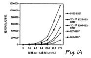
前記アッセイは6種のITA標準物質を用いてキャリブレーションをとった。前記ITA標準物質の濃度は、約1.3ng/mL、約2.5ng/mL、約8.2ng/mL、約22.8ng/mL、約91.7ng/mL、約271ng/mLである。それぞれの標準物質に対する算出された相対的な光単位(RLU)はそれぞれ914RLU、1,630RLU、4,873RLU、12,794RLU、48,149RLU、135,384RLUであった。基準線RLU(即ち、ITA濃度が0ng/mLの時)は314RLUである。このアッセイ結果は黒抜きダイヤ(◆)により示した図1A及び図1Bにおいて表されている。
上述のように、女性の尿サンプルをITAに対しスクリーニングした。尿サンプルは1,095RLUの検出可能なシグナルを示した。ITA標準物質のデータに基づき、これは1.6ng/mLのITA濃度に相関していた。
After incubation, the plate was exposed to a magnetic field to immobilize the ITA / capture antibody / magnetic particle complex. The supernatant was removed and the wells were washed with a washing solution. After extensive washing, determined experimentally and optimized, the plate was removed from the magnetic field and 50 μL of detection antibody solution and 250 μL normal mouse serum were added to the wells. The solution was incubated for about 10 minutes at 37 ° C. Thereafter, the plate was again exposed to a magnetic field to fix the detection antibody / ITA / capture antibody / magnetic particle complex. The supernatant was removed and the wells were washed. An acidic solution containing hydrogen peroxide in a dilute acid, such as HCl, and a base solution containing dilute sodium hydroxide were then added to the wells to cause an acridinium ester signal. Thereafter, the amount of detected signal was measured with a spectrometer and the data was recorded.
When the detection signal exceeded the sensitivity range of the assay, the sample was diluted with a diluent containing 0.5 M PBS 0.1% protease-free BSA at pH 7.4.
The assay was calibrated using 6 ITA standards. The concentration of the ITA standard is about 1.3 ng / mL, about 2.5 ng / mL, about 8.2 ng / mL, about 22.8 ng / mL, about 91.7 ng / mL, about 271 ng / mL. The calculated relative light units (RLU) for each standard were 914RLU, 1,630RLU, 4,873RLU, 12,794RLU, 48,149RLU, 135,384RLU, respectively. The baseline RLU (ie, when the ITA concentration is 0 ng / mL) is 314 RLU. The results of this assay are represented in FIG. 1A and FIG. 1B indicated by the black diamond (♦).
Female urine samples were screened for ITA as described above. The urine sample showed a detectable signal of 1,095 RLU. Based on ITA reference data, this correlated with an ITA concentration of 1.6 ng / mL.
ITA/無処置のhCGアッセイ
コンビネーションアッセイ(“コンボ(combo)”アッセイ)を、実施例1に記載されたB152-B207アッセイを用いて、再び実施した。但し、B152キャプチャー抗体及びITAによる最初のインキュベーションの間、無処置のhCGに特異的に結合する追加のキャプチャー抗体をウェルに添加した。用いた抗体はクローン820と命名された(Biodesign International,(Saco,Maine)より購入。Cat.No.E45550M)。このアッセイはここで、”コンボ820/B152-B207”という。特に、このコンボアッセイにおいて、前記キャプチャー抗体はクローン820及びB152モノクロナール抗体であり、検出抗体はB207モノクロナール抗体である。このアッセイ結果は黒抜き三角(▲)により示した図1A及び図1Bにおいて説明されている。
予想外にも、ITAの検出シグナルはB152-B207を単独で用いた検出シグナルより実質的に大きいことが明らかとなった。その点、6種のITA標準物質は、1,850RLU(1.3ng/mL)、3,178RLU(2.4ng/mL)、10,940RLU(8.4ng/mL)、31,119RLU(23.1ng/mL)、123,118RLU(90.0ng/mL)、及び341,532RLU(271.8ng/mL)のシグナルを示した。
これらの結果は、コンボアッセイ820/B152-B207がB152-B207アッセイ単独よりも約2-3倍大きい感度を示すことを示唆しているように見える。従って、現在利用されているアッセイよりも、より早い時点で、生体サンプル中のより小さいITA濃度を検出できる可能性がある。
精製されたモノクロナール抗体クローン820を単独で利用するアッセイは結果的にコンボアッセイ820/B152-B207又はB152-B207アッセイより大きな検出シグナルを発するように見えたことも注目に値する。前記結果はアステリスク(*)により示した図1A及び図1Bにおいて表されている。
ITA / no treatment hCG assay A combination assay ("combo" assay) was performed again using the B152-B207 assay described in Example 1. However, during the initial incubation with B152 capture antibody and ITA, additional capture antibody that specifically binds to intact hCG was added to the wells. The antibody used was named clone 820 (purchased from Biodesign International, (Saco, Maine). Cat. No. E45550M). This assay is referred to herein as “
Unexpectedly, the ITA detection signal was found to be substantially greater than the detection signal using B152-B207 alone. In that regard, the six ITA standards are 1,850 RLU (1.3 ng / mL), 3,178 RLU (2.4 ng / mL), 10,940 RLU (8.4 ng / mL), 31,119 RLU (23.1 ng / mL), 123,118 RLU ( 90.0 ng / mL) and 341,532 RLU (271.8 ng / mL).
These results appear to suggest that the
It is also noteworthy that the assay utilizing the purified
ITA/遊離したβhCGアッセイ
コンビネーションアッセイ(”コンボ”アッセイ)を、また実施例1で記載されているB152-B207アッセイを用いて実施した。但し、遊離したβhCGに対する追加のキャプチャー抗体を、B152キャプチャー抗体及びITAによる最初のインキュベーションの間、ウェルに添加した。使用した抗体はクローン#827と命名した(Biodesign International,(Saco,Maine)より購入。Cat.No.E45575M)。このアッセイはここで、”コンボ827/B152-B207”と呼ぶ。特にこのコンボアッセイにおいて、前記キャプチャー抗体はクローン827及びB152モノクロナール抗体であり、検出抗体はB207モノクロナール抗体である。このアッセイの結果は黒抜き四角(■)により示した図1A及び図1Bにおいて説明されている。
更に予期外なことには、ITAの検出シグナルは単独のB152-B207アッセイ又はコンボ820/B152-B207アッセイを用いた検出シグナルより実質上大きいと思われる。この点、6種のITA標準物質は次のシグナル、8,033RLU(1.3ng/mL ITA); 16,957RLU(2.5ng/mL ITA); 55,264RLU(8.2ng/mL ITA); 142,512RLU(22.9ng/mL ITA); 441,900RLU(92.2ng/mL ITA); 842,974RLU(267.9ng/mL ITA)を示した。
これらの結果はコンボアッセイ827/B152-B207はB152-B207アッセイ単独に比べ約6〜12倍大きい感度を示すように思われることを示唆する。従って、現在利用可能なアッセイにより得られるより、初期時点における生体サンプルのITAのより小さい濃度の検出が可能であろう。
また、精製されたクローン827を単独で利用するアッセイはコンボアッセイ827/B152-B207又はB152-B207アッセイより、結果的により大きく検出可能なシグナルとなるらしいことも注目に値する。前記結果はX(X)により示した図1A及び図1Bにおいて示されている。前記827-B207アッセイは前記B152-B207アッセイよりおよそ8〜22倍高感度と思われる。
ITA / Free βhCG Assay A combination assay (“combo” assay) was also performed using the B152-B207 assay described in Example 1. However, additional capture antibody against free βhCG was added to the wells during the initial incubation with B152 capture antibody and ITA. The antibody used was named clone # 827 (purchased from Biodesign International, (Saco, Maine). Cat. No. E45575M). This assay is referred to herein as “
Even more unexpectedly, the detection signal of ITA appears to be substantially greater than the detection signal using a single B152-B207 assay or a
These results suggest that the
It is also noteworthy that assays that use purified
排卵5日後の前記女性は性交後妊娠したか否か知ることを望んでいた。彼女は尿を産婦人科医に預けた。前記サンプルは実施例1-3に記載されたアッセイのいずれかを用いてITAに対しアッセイした。前記サンプルにおいて測定されたITA濃度は約1.0ng/mLであった。産婦人科医は前記女性が妊娠していることを確認した。 The woman, 5 days after ovulation, wanted to know if she became pregnant after intercourse. She deposited urine with an obstetrician and gynecologist. The samples were assayed for ITA using any of the assays described in Examples 1-3. The ITA concentration measured in the sample was about 1.0 ng / mL. The obstetrician and gynecologist confirmed that the woman was pregnant.
不妊治療受けている前記女性は結果的にin vitro受精した少なくとも1つの胎芽を移植される。移植後約3-4日後、移植が成功したか否かを決定するために前記女性は彼女の医師に尿サンプルを預けた。前記サンプルをITAについてスクリーニングした。前記サンプル中のITA濃度は約0.3ng/mLであった。前記医師は前記女性が妊娠していることを確認した。 The woman undergoing infertility treatment is eventually transplanted with at least one embryo fertilized in vitro. About 3-4 days after the transplant, the woman deposited a urine sample with her doctor to determine if the transplant was successful. The sample was screened for ITA. The ITA concentration in the sample was about 0.3 ng / mL. The doctor confirmed that the woman was pregnant.
排卵後の7日後の前記女性は、性交後妊娠しているか否か知ることを望んでいた。彼女は実施例1-3に記載されているいずれか1つのアッセイを利用した“家庭用妊娠キット“を入手した。前記キットはサンプルカップ、ピペット、及びアッセイ装置が含まれていた。前記アッセイ装置は、サンプル用のウェル、キャプチャー抗体溶液の保管用容器、検出抗体溶液の保存用容器、コントロール溶液保存用容器、アッセイの操作を制御するための電子部品、及び写真フィルムや光真空管のようなシグナル検出器を含んでいた。前記シグナル検出器はアッセイにより生成されたシグナルを検出し、好ましくは約0.18ng/mLより大きいITA濃度に対応するシグナルを検出するように設定されていた。前記サンプルをピペットで取って前記キットのサンプルウェルに入れた。前記アッセイを前記サンプルについて実施した。前記サンプルに対する光真空管は光を放射しないがコントロール溶液に対する光真空管は光を放射する。これによって、前記アッセイが成功したが、彼女が妊娠していないことを確認した。
種々の刊行物、及び/又は参考文献をここで引用したが、これらの内容はここで参考として取り込まれている。
本発明について種々の特定の実施例や態様に関連して記載したが、本発明がそれらに限定されることなく、また別紙の特許請求の範囲内において種々に実施できることは理解される。
The woman seven days after ovulation wanted to know if she was pregnant after intercourse. She obtained a “home pregnancy kit” that utilized any one of the assays described in Examples 1-3. The kit included a sample cup, pipette, and assay device. The assay device includes a sample well, a capture antibody solution storage container, a detection antibody solution storage container, a control solution storage container, an electronic component for controlling the operation of the assay, and a photographic film or a light vacuum tube. Such a signal detector. The signal detector was configured to detect the signal produced by the assay, and preferably detect a signal corresponding to an ITA concentration greater than about 0.18 ng / mL. The sample was pipetted into the sample well of the kit. The assay was performed on the sample. The light vacuum tube for the sample does not emit light, but the light vacuum tube for the control solution emits light. This confirmed that the assay was successful but she was not pregnant.
Various publications and / or references are cited herein, the contents of which are hereby incorporated by reference.
While the invention has been described in connection with various specific embodiments and embodiments, it will be understood that the invention is not limited thereto and can be practiced in various ways within the scope of the appended claims.
Claims (10)
(1)女性の生体サンプルを、(i)少なくとも2種のキャプチャー抗体であって、前記抗体の一方が、B152が結合するエピトープと同じITAのエピトープと特異的に結合し、かつ、他方が、クローン820又はクローン827が結合するエピトープと同じhCGのエピトープに特異的に結合するキャプチャー抗体、及び(ii)前記キャプチャー抗体が結合するエピトープと異なるITA及びhCGのエピトープに結合する単一検出抗体に接触させる工程であって、前記検出抗体が、B207が結合するエピトープと同じエピトープに結合することができ、1回のアッセイにおいて、検出シグナルを生成するのに有効な標識に連結されている工程、及び
(2)hCG及びITAを表す標識によって生成されるシグナルを検出する工程であって、前記検出シグナルの存在が、女性の妊娠を示す工程、
を含むことを特徴とする方法。A method for detecting pregnancy of a woman, comprising the following steps:
(1) A female biological sample, (i) at least two types of capture antibodies, wherein one of the antibodies specifically binds to the same epitope of ITA as the epitope to which B152 binds, and the other Contact with a capture antibody that specifically binds to the same hCG epitope as the epitope to which clone 820 or clone 827 binds , and (ii) a single detection antibody that binds to a different ITA and hCG epitope than the epitope to which the capture antibody binds And wherein said detection antibody is capable of binding to the same epitope to which B207 binds and is linked to a label effective to generate a detection signal in a single assay, and (2) a step of detecting a signal generated by a label representing hCG and ITA, wherein the presence of the detection signal indicates female pregnancy Process,
A method comprising the steps of:
Applications Claiming Priority (2)
| Application Number | Priority Date | Filing Date | Title |
|---|---|---|---|
| US09/918,297 US6627457B2 (en) | 2001-07-30 | 2001-07-30 | Methods for detecting pregnancy |
| PCT/US2002/024038 WO2003012433A1 (en) | 2001-07-30 | 2002-07-30 | Methods for detecting pregnancy |
Publications (3)
| Publication Number | Publication Date |
|---|---|
| JP2004537728A JP2004537728A (en) | 2004-12-16 |
| JP2004537728A5 JP2004537728A5 (en) | 2006-01-05 |
| JP4559729B2 true JP4559729B2 (en) | 2010-10-13 |
Family
ID=25440144
Family Applications (1)
| Application Number | Title | Priority Date | Filing Date |
|---|---|---|---|
| JP2003517576A Expired - Lifetime JP4559729B2 (en) | 2001-07-30 | 2002-07-30 | Pregnancy detection method |
Country Status (6)
| Country | Link |
|---|---|
| US (1) | US6627457B2 (en) |
| EP (1) | EP1419384B1 (en) |
| JP (1) | JP4559729B2 (en) |
| AT (1) | ATE430939T1 (en) |
| DE (1) | DE60232248D1 (en) |
| WO (1) | WO2003012433A1 (en) |
Families Citing this family (23)
| Publication number | Priority date | Publication date | Assignee | Title |
|---|---|---|---|---|
| US6927034B2 (en) * | 1998-02-03 | 2005-08-09 | The Trustees Of Columbia University In The City Of New York | Methods for detecting trophoblast malignancy by HCG assay |
| US7198954B1 (en) | 1999-02-03 | 2007-04-03 | The Trustees Of Columbia University In The City Of New York | Methods for predicting pregnancy outcome in a subject by hCG assay |
| US6500627B1 (en) * | 1998-02-03 | 2002-12-31 | The Trustees Of Columbia University In The City Of New York | Methods for predicting pregnancy outcome in a subject by HCG assay |
| US20040176914A1 (en) * | 2001-04-13 | 2004-09-09 | Biosite Incorporated | Methods and compositions for measuring biologically active natriuretic peptides and for improving their therapeutic potential |
| US20030219734A1 (en) * | 2001-04-13 | 2003-11-27 | Biosite Incorporated | Polypeptides related to natriuretic peptides and methods of their identification and use |
| US20030199000A1 (en) * | 2001-08-20 | 2003-10-23 | Valkirs Gunars E. | Diagnostic markers of stroke and cerebral injury and methods of use thereof |
| US7524635B2 (en) * | 2003-04-17 | 2009-04-28 | Biosite Incorporated | Methods and compositions for measuring natriuretic peptides and uses thereof |
| US7632647B2 (en) | 2001-04-13 | 2009-12-15 | Biosite Incorporated | Use of B-type natriuretic peptide as a prognostic indicator in acute coronary syndromes |
| EP1666881B1 (en) | 2001-05-04 | 2010-02-17 | Biosite Incorporated | Diagnostic markers of acute coronary syndromes and methods of use thereof |
| CA2457775A1 (en) * | 2001-08-20 | 2003-02-27 | Biosite Incorporated | Diagnostic markers of stroke and cerebral injury and methods of use thereof |
| US7608406B2 (en) * | 2001-08-20 | 2009-10-27 | Biosite, Inc. | Diagnostic markers of stroke and cerebral injury and methods of use thereof |
| US20040209307A1 (en) * | 2001-08-20 | 2004-10-21 | Biosite Incorporated | Diagnostic markers of stroke and cerebral injury and methods of use thereof |
| US20040219509A1 (en) * | 2001-08-20 | 2004-11-04 | Biosite, Inc. | Diagnostic markers of stroke and cerebral injury and methods of use thereof |
| US20050148096A1 (en) * | 2004-01-07 | 2005-07-07 | Cole Laurence A. | Methods for detecting pregnancy |
| US7572639B2 (en) * | 2004-06-03 | 2009-08-11 | Stc.Unm | Method and apparatus for predicting pregnancy outcome |
| US7666683B1 (en) | 2004-10-13 | 2010-02-23 | Stc.Unm | Rapid method of diagnosing a normal pregnancy with high accuracy |
| US20060105411A1 (en) * | 2004-11-15 | 2006-05-18 | Cole Laurence A | Method of detecting early pregnancy at high accuracy by measuring hCG and hyperglycosylated hCG concentrations equally |
| CA2615460A1 (en) * | 2005-08-08 | 2007-02-15 | Onconon, Llc | Antibody compositions, methods for treating neoplastic disease and methods for regulating fertility |
| BRPI0708677A2 (en) * | 2006-03-09 | 2011-06-21 | Hoffmann La Roche | anti-drug antibody assay |
| GB0717043D0 (en) * | 2007-04-10 | 2007-10-10 | Inverness Medical Switzerland | Assay device |
| US20100129935A1 (en) * | 2008-11-25 | 2010-05-27 | Sarah Daniel Maddison | Pregnancy testing method |
| US8278109B2 (en) | 2010-02-12 | 2012-10-02 | Church & Dwight Co., Inc. | Hyperglycosylated hCG detection device |
| JP7128827B2 (en) * | 2016-10-24 | 2022-08-31 | エフ.ホフマン-ラ ロシュ アーゲー | Fixed analyte |
Family Cites Families (16)
| Publication number | Priority date | Publication date | Assignee | Title |
|---|---|---|---|---|
| DK420475A (en) * | 1974-11-11 | 1976-07-19 | Carter Wallace | PREGNANCY DETECTION PROCEDURE |
| GB2112779B (en) | 1981-12-11 | 1986-10-15 | Welsh Nat School Med | Aryl acridinium esters as luminescent labelling materials |
| US4933275A (en) * | 1985-10-24 | 1990-06-12 | The General Hospital Corporation | Method for the detection of a polypeptide subunit in the presence of a quaternary protein containing the subunit |
| DE3633497A1 (en) * | 1986-10-02 | 1988-04-14 | Hoechst Ag | IMMUNOMETRIC DETERMINATION PROCEDURE |
| US4804626A (en) * | 1986-10-22 | 1989-02-14 | The General Hospital Corporation | Immunometric assay for the detection of human chorionic gonadotropin |
| US5506150A (en) | 1987-07-09 | 1996-04-09 | 3I Research Exploitation Limited | Prenatal screening for down's syndrome |
| US5356817A (en) | 1988-06-09 | 1994-10-18 | Yale University | Methods for detecting the onset, progression and regression of gynecologic cancers |
| US6352862B1 (en) | 1989-02-17 | 2002-03-05 | Unilever Patent Holdings B.V. | Analytical test device for imuno assays and methods of using same |
| US6339143B1 (en) | 1993-05-13 | 2002-01-15 | The Trustees Of Columbia University In The City Of New York | Methods of using antibodies against hormone-related determinants |
| US5660990A (en) | 1995-08-18 | 1997-08-26 | Immunivest Corporation | Surface immobilization of magnetically collected materials |
| JP3950481B2 (en) * | 1996-09-06 | 2007-08-01 | エール ユニバーシティ | Prenatal screening for Down syndrome using hyperglycosylated gonadotropins |
| US6500627B1 (en) | 1998-02-03 | 2002-12-31 | The Trustees Of Columbia University In The City Of New York | Methods for predicting pregnancy outcome in a subject by HCG assay |
| US6927034B2 (en) | 1998-02-03 | 2005-08-09 | The Trustees Of Columbia University In The City Of New York | Methods for detecting trophoblast malignancy by HCG assay |
| DK1076824T3 (en) | 1998-04-29 | 2006-09-18 | Nicholas John Wald | Prenatal screening for Down syndrome |
| JP3819612B2 (en) * | 1998-09-30 | 2006-09-13 | 大日精化工業株式会社 | Method for immunological measurement of β-hCG |
| US6127186A (en) | 1999-01-12 | 2000-10-03 | Pandian; M. R. | Methods of determining an increased risk of a woman carrying a downs syndrome affected fetus by measuring an analyte in a biological sample |
-
2001
- 2001-07-30 US US09/918,297 patent/US6627457B2/en not_active Expired - Lifetime
-
2002
- 2002-07-30 JP JP2003517576A patent/JP4559729B2/en not_active Expired - Lifetime
- 2002-07-30 EP EP02750353A patent/EP1419384B1/en not_active Expired - Lifetime
- 2002-07-30 WO PCT/US2002/024038 patent/WO2003012433A1/en active Application Filing
- 2002-07-30 DE DE60232248T patent/DE60232248D1/en not_active Expired - Lifetime
- 2002-07-30 AT AT02750353T patent/ATE430939T1/en not_active IP Right Cessation
Also Published As
| Publication number | Publication date |
|---|---|
| EP1419384B1 (en) | 2009-05-06 |
| US20030022381A1 (en) | 2003-01-30 |
| EP1419384A1 (en) | 2004-05-19 |
| ATE430939T1 (en) | 2009-05-15 |
| JP2004537728A (en) | 2004-12-16 |
| EP1419384A4 (en) | 2006-01-25 |
| US6627457B2 (en) | 2003-09-30 |
| WO2003012433A1 (en) | 2003-02-13 |
| DE60232248D1 (en) | 2009-06-18 |
Similar Documents
| Publication | Publication Date | Title |
|---|---|---|
| JP4559729B2 (en) | Pregnancy detection method | |
| US8753825B2 (en) | Methods and kits for detecting ITA in a biological sample | |
| US20070166776A1 (en) | Immunoassay and kit for an early and simultaneous detection of biochemical markers in a patient's sample | |
| WO2013132347A2 (en) | Improved elisa immunoassay for calprotectin | |
| WO2013132338A2 (en) | Competitive immunoassay for calprotectin | |
| EP0405578B1 (en) | Enzyme immunoassay for antigen and solid phase used therefor | |
| EP3248001B1 (en) | Diagnostic devices and methods for mitigating hook effect and use thereof | |
| JP2000508075A (en) | Luminescence-specific binding assay | |
| US5223440A (en) | Ex vivo product of conception test to determine abortion | |
| US20030027234A1 (en) | Methods for detecting Down's syndrome | |
| US20050148096A1 (en) | Methods for detecting pregnancy | |
| US20040248216A1 (en) | Method of examining cancer by assaying autoantibody against mdm2 and reagent therefor | |
| CN110133271B (en) | Method for covalently binding antibody or antigen binding fragment thereof to particle surface | |
| JP3847983B2 (en) | Reagent for immunological analysis, immunological analysis method, and kit for immunological analysis | |
| US20060105411A1 (en) | Method of detecting early pregnancy at high accuracy by measuring hCG and hyperglycosylated hCG concentrations equally | |
| Wani et al. | An automated flow immunosensor based on kinetic exclusion analysis for measurement of a free β-subunit of human chorionic gonadotropin in serum | |
| CN117616277A (en) | Thyroglobulin immunoassay and kit for same | |
| AU2002346529B2 (en) | Immunoassay and kit for an early and simulataneous detection of biochemical markers in a patient's sample | |
| JP2716103B2 (en) | Elimination of false results in immunoassay | |
| KR20230110262A (en) | Myocardial troponin I detection methods and kits | |
| JPH1114626A (en) | Method for measuring ca125 | |
| EP2960654A1 (en) | A method for detecting EP3 antibodies | |
| JP2002267665A (en) | Method of measuring human c-peptide | |
| JPH05340947A (en) | Sandwich immunoassay and measuring kit | |
| JPH1194831A (en) | Detection of cancer by measurement of autoimmune to hormone receptor |
Legal Events
| Date | Code | Title | Description |
|---|---|---|---|
| A521 | Request for written amendment filed |
Free format text: JAPANESE INTERMEDIATE CODE: A523 Effective date: 20050801 |
|
| A621 | Written request for application examination |
Free format text: JAPANESE INTERMEDIATE CODE: A621 Effective date: 20050801 |
|
| A977 | Report on retrieval |
Free format text: JAPANESE INTERMEDIATE CODE: A971007 Effective date: 20080606 |
|
| A131 | Notification of reasons for refusal |
Free format text: JAPANESE INTERMEDIATE CODE: A131 Effective date: 20080616 |
|
| A601 | Written request for extension of time |
Free format text: JAPANESE INTERMEDIATE CODE: A601 Effective date: 20080916 |
|
| A602 | Written permission of extension of time |
Free format text: JAPANESE INTERMEDIATE CODE: A602 Effective date: 20080924 |
|
| A521 | Request for written amendment filed |
Free format text: JAPANESE INTERMEDIATE CODE: A523 Effective date: 20081216 |
|
| A131 | Notification of reasons for refusal |
Free format text: JAPANESE INTERMEDIATE CODE: A131 Effective date: 20090706 |
|
| A521 | Request for written amendment filed |
Free format text: JAPANESE INTERMEDIATE CODE: A523 Effective date: 20091006 |
|
| TRDD | Decision of grant or rejection written | ||
| A01 | Written decision to grant a patent or to grant a registration (utility model) |
Free format text: JAPANESE INTERMEDIATE CODE: A01 Effective date: 20100628 |
|
| A01 | Written decision to grant a patent or to grant a registration (utility model) |
Free format text: JAPANESE INTERMEDIATE CODE: A01 |
|
| A61 | First payment of annual fees (during grant procedure) |
Free format text: JAPANESE INTERMEDIATE CODE: A61 Effective date: 20100723 |
|
| R150 | Certificate of patent or registration of utility model |
Ref document number: 4559729 Country of ref document: JP Free format text: JAPANESE INTERMEDIATE CODE: R150 Free format text: JAPANESE INTERMEDIATE CODE: R150 |
|
| FPAY | Renewal fee payment (event date is renewal date of database) |
Free format text: PAYMENT UNTIL: 20130730 Year of fee payment: 3 |
|
| R250 | Receipt of annual fees |
Free format text: JAPANESE INTERMEDIATE CODE: R250 |
|
| R250 | Receipt of annual fees |
Free format text: JAPANESE INTERMEDIATE CODE: R250 |
|
| R250 | Receipt of annual fees |
Free format text: JAPANESE INTERMEDIATE CODE: R250 |
|
| R250 | Receipt of annual fees |
Free format text: JAPANESE INTERMEDIATE CODE: R250 |
|
| R250 | Receipt of annual fees |
Free format text: JAPANESE INTERMEDIATE CODE: R250 |
|
| R250 | Receipt of annual fees |
Free format text: JAPANESE INTERMEDIATE CODE: R250 |
|
| R250 | Receipt of annual fees |
Free format text: JAPANESE INTERMEDIATE CODE: R250 |
|
| R250 | Receipt of annual fees |
Free format text: JAPANESE INTERMEDIATE CODE: R250 |
|
| R250 | Receipt of annual fees |
Free format text: JAPANESE INTERMEDIATE CODE: R250 |
|
| EXPY | Cancellation because of completion of term |