EP0395522A1 - Gerät zur Kontrolle der Strahlung mit einer modulierbaren aktiven Oberfläche - Google Patents
Gerät zur Kontrolle der Strahlung mit einer modulierbaren aktiven Oberfläche Download PDFInfo
- Publication number
- EP0395522A1 EP0395522A1 EP90401141A EP90401141A EP0395522A1 EP 0395522 A1 EP0395522 A1 EP 0395522A1 EP 90401141 A EP90401141 A EP 90401141A EP 90401141 A EP90401141 A EP 90401141A EP 0395522 A1 EP0395522 A1 EP 0395522A1
- Authority
- EP
- European Patent Office
- Prior art keywords
- radiation
- converter
- strip
- transparent
- detector
- Prior art date
- Legal status (The legal status is an assumption and is not a legal conclusion. Google has not performed a legal analysis and makes no representation as to the accuracy of the status listed.)
- Withdrawn
Links
- 230000005855 radiation Effects 0.000 title claims abstract description 43
- 238000012544 monitoring process Methods 0.000 claims 1
- 210000000481 breast Anatomy 0.000 abstract description 20
- 238000009607 mammography Methods 0.000 description 4
- 238000005259 measurement Methods 0.000 description 4
- 238000003745 diagnosis Methods 0.000 description 2
- 230000003054 hormonal effect Effects 0.000 description 2
- 230000010354 integration Effects 0.000 description 2
- 238000012806 monitoring device Methods 0.000 description 2
- 230000001575 pathological effect Effects 0.000 description 2
- 208000004434 Calcinosis Diseases 0.000 description 1
- 230000000711 cancerogenic effect Effects 0.000 description 1
- 231100000315 carcinogenic Toxicity 0.000 description 1
- 230000001010 compromised effect Effects 0.000 description 1
- 201000010759 hypertrophy of breast Diseases 0.000 description 1
- 238000009434 installation Methods 0.000 description 1
- 230000003449 preventive effect Effects 0.000 description 1
- 238000002601 radiography Methods 0.000 description 1
- 230000000284 resting effect Effects 0.000 description 1
- 238000012216 screening Methods 0.000 description 1
- 230000000392 somatic effect Effects 0.000 description 1
Images

Classifications
-
- A—HUMAN NECESSITIES
- A61—MEDICAL OR VETERINARY SCIENCE; HYGIENE
- A61B—DIAGNOSIS; SURGERY; IDENTIFICATION
- A61B6/00—Apparatus or devices for radiation diagnosis; Apparatus or devices for radiation diagnosis combined with radiation therapy equipment
- A61B6/42—Arrangements for detecting radiation specially adapted for radiation diagnosis
- A61B6/4208—Arrangements for detecting radiation specially adapted for radiation diagnosis characterised by using a particular type of detector
- A61B6/4225—Arrangements for detecting radiation specially adapted for radiation diagnosis characterised by using a particular type of detector using image intensifiers
-
- A—HUMAN NECESSITIES
- A61—MEDICAL OR VETERINARY SCIENCE; HYGIENE
- A61B—DIAGNOSIS; SURGERY; IDENTIFICATION
- A61B6/00—Apparatus or devices for radiation diagnosis; Apparatus or devices for radiation diagnosis combined with radiation therapy equipment
- A61B6/50—Apparatus or devices for radiation diagnosis; Apparatus or devices for radiation diagnosis combined with radiation therapy equipment specially adapted for specific body parts; specially adapted for specific clinical applications
- A61B6/502—Apparatus or devices for radiation diagnosis; Apparatus or devices for radiation diagnosis combined with radiation therapy equipment specially adapted for specific body parts; specially adapted for specific clinical applications for diagnosis of breast, i.e. mammography
-
- G—PHYSICS
- G01—MEASURING; TESTING
- G01T—MEASUREMENT OF NUCLEAR OR X-RADIATION
- G01T1/00—Measuring X-radiation, gamma radiation, corpuscular radiation, or cosmic radiation
- G01T1/16—Measuring radiation intensity
- G01T1/20—Measuring radiation intensity with scintillation detectors
Definitions
- the invention relates to radiology systems, in particular mammographs and more particularly in such systems, a device which makes it possible to control the dose of radiation received by the person being observed as well as the duration of the exposure so as to obtain an image with optimal contrast.
- Radiography systems of the mammography type include, as shown in FIG. 1, a source 10 of X-ray radiation carried by a bracket 11 disposed at the top of a vertical plate 12.
- the latter comprises an assembly 13 on which the breast 16 to be examined rests by means of a horizontal tablet 15.
- a ball, transparent to X-radiation and movable vertically on the plate 12, is used to compress the breast.
- the plate 12 is mounted on a vertical column 9 resting on the ground and moves vertically on said column using an appropriate mechanical device.
- the assembly 13 has on its upper part and under the shelf 15, a tunnel in which is housed a cassette 18 consisting of a black box containing a film 14 sensitive to direct X-ray radiation or to photonic radiation emitted by a screen (not shown) receiving X-ray radiation. It is on this film 14 that the latent image of the breast is formed after an appropriate exposure time; the development of the film gives an X-ray picture.
- the blackening of the film must be correct and "normalized" for a very wide range of opacity of the object.
- the blackening can be controlled by a radiation control device which is placed under the cassette in the lower part 8 of the assembly 13.
- This control device also called an expander, essentially consists of a radiation detector. X-ray which delivers an electrical signal proportional to the flow of the dose of X-ray which passes through the sensitive film.
- This electrical signal which translates the intensity of the X-radiation, is integrated during the exposure time and the signal resulting from this integration is compared at each instant to a predetermined threshold signal which is a function of the characteristics of the sensitive film. As soon as the integrated signal reaches this threshold, the signal indicating equality controls the stopping of the source, which ends the installation.
- This radiation control device is that, for a wide range of variation in the X-ray dose rates which lead to differences in exposure, on the one hand, an exposure of the sensitive film corresponding to optimal contrast and, on the other hand, better control of the average dose received by the patient, a dose which is an important factor in assessing the carcinogenic risk.
- the detector In a radiation monitoring device, it is important that the detector receives only the radiation that has passed through the breast because the reception of unattenuated X-rays would falsify the measurement. Also, the receiving surface of such a detector is limited by the size of the smallest breast to be examined. Such a limitation considerably limits the advantages that can be drawn from this device and constitutes an error factor in certain circumstances because the area of the object corresponding to the size of the detector may be different from that which is examined. Indeed, the position of the detector is generally fixed while the area to be examined can have a variable position relative to that of the detector and there is therefore no desired overlap for optimal measurement.
- the measurement signal is not representative of the breast as a whole and can lead to underexposed images in the case where the detector is under an adipose part of the breast or overexposed in the case where the detector is under a fibrous part or under a pathological opacity.
- the object of the present invention is therefore to produce a radiation control device which adapts to the different sizes of the object to be radiographed, in particular when it is a breast.
- the invention proposes, on the one hand, to enlarge the dimensions of the detector until possibly reaching those of the sensitive film and, on the other hand, to use masks of different dimensions which are placed between the cassette and detector.
- the invention relates to a radiation monitoring device in a radiology system which comprises at least one X-ray source and an X-ray detector having passed through an object to be observed of the type comprising a converter of X-ray radiation into X-ray radiation. photonics and a photomultiplier tube of the photons emitted by the converter, said detector supplying a signal electric which serves to control the exposure time of the object, characterized in that it further comprises at least one mask, opaque to light radiation but transparent to X-ray, which is interposed between the radiation converter and the photomultiplier, said mask having at least one zone transparent to light radiation, the shape and surface of the transparent zone being adapted to the dimensions of the object to be observed or to the type of examination which is carried out.
- the mask is supported by a band which is associated with a mechanism for moving said band so as to place said transparent zone in an optimal position relative to the object.
- this strip comprises several zones transparent to light radiation which are successively arranged in the direction of movement of the strip and whose dimensions and shape are different from one zone to the next so as to adapt to the dimensions and the shape of the object as well as the type of examination carried out.
- the invention will be described in its application to a mammography but it can be implemented in other radiology systems where use is made of a radiation controller of the type comprising a converter of radiation from X-ray to photon radiation and a photomultiplier tube of the photons emitted by the converter.
- a radiation controller of the type comprising a converter of radiation from X-ray to photon radiation and a photomultiplier tube of the photons emitted by the converter.
- the invention proposes to control the surface of the active area of the radiation converter by interposing an opaque mask between the latter and the photomultiplier tube. light radiation which presents transparent windows whose dimensions and shape vary according to those of the breast to be examined.
- the invention provides an automatic device for moving masks.
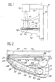
- the automatic device is disposed in the lower part 8 of the assembly 13 under the cassette 18.
- the latter comprises, in known manner, a sensitive film 14 inside the black box that constitutes the cassette and a zone transparent to radiation X which is located in the bottom wall of the cassette and near the outside edge of the part 8 closest to the patient.
- the radiation converter consists of a screen 22 which emits photons towards a photomultiplier 23 when it receives X-radiation passing through the transparent zone 21.
- the screen 22 is fixed on a support 24 secured to a frame 25 essentially constituted by the lower part 8 of the assembly 13.
- the photons emitted by the screen 22 are focused at the input 23 ′ of the photomultiplier 23 by a conduit 26 comprising in particular the walls 27 and 28 opaque to light but transparent to X-rays.
- a strip 29 In front of the screen 22 is disposed a strip 29 which is supported and driven in movement along the arrow 30 by a drive motor 34 and support rollers 31, 32 and 33.
- a mechanism 35 makes it possible to set up the strip 29 and adjust the tension.
- the strip moves in a black box 36 comprising the walls 28, 37 and 38 so that the outside light does not come to disturb the measurement of the photomultiplier 23.
- the strip 29 is closed on itself and is opaque to light radiation over its entire surface with the exception of zones 39, 40, 41, 42 and 43 (FIG. 3).
- the zones 39, 40 and 41 have a general shape of a semicircle whose diameter varies from one zone to the next, the zone 39 of smaller diameter being provided for example for the examination of a breast of small dimensions, zone 40 for examining a medium breast and zone 41 for examining a large breast.
- the zones 42 and 43 which are rectangular in shape and symmetrically arranged on either side of the longitudinal axis of symmetry 44 of the strip, are used during a stereotaxic examination to reproduce two views on the same film .
- the strip 29 it is possible to create a number of windows, such as 39.40 and 41, greater than three in order to better cover the disparity in the dimensions of the breasts.
- these areas can have shapes other than that of a semicircle.
- the maximum dimensions of the window 41 or of the rectangle encompassing the windows 42 and 43 are those of the screen 22 associated with the photomultiplier 23.
- the choice of window which is suitable for the breast observed is made by the practitioner and the electrical control circuit of the drive motor 34 is provided to set up the window chosen in front of the screen 22, the base of the semicircle being situated towards the outer edge 44 of the part 8 of the assembly 13, the as close as possible to the patient.
- the invention which has just been described therefore allows the practitioner to adapt the exposure time and therefore the dose of radiation emitted as a function, in particular, of the dimensions of the breast to be examined.
Landscapes
- Health & Medical Sciences (AREA)
- Life Sciences & Earth Sciences (AREA)
- Engineering & Computer Science (AREA)
- Medical Informatics (AREA)
- Physics & Mathematics (AREA)
- High Energy & Nuclear Physics (AREA)
- Molecular Biology (AREA)
- General Health & Medical Sciences (AREA)
- Surgery (AREA)
- Veterinary Medicine (AREA)
- Nuclear Medicine, Radiotherapy & Molecular Imaging (AREA)
- Optics & Photonics (AREA)
- Pathology (AREA)
- Radiology & Medical Imaging (AREA)
- Biomedical Technology (AREA)
- Heart & Thoracic Surgery (AREA)
- Biophysics (AREA)
- Animal Behavior & Ethology (AREA)
- Public Health (AREA)
- General Physics & Mathematics (AREA)
- Spectroscopy & Molecular Physics (AREA)
- Dentistry (AREA)
- Oral & Maxillofacial Surgery (AREA)
- Apparatus For Radiation Diagnosis (AREA)
Applications Claiming Priority (2)
| Application Number | Priority Date | Filing Date | Title |
|---|---|---|---|
| FR8905663 | 1989-04-28 | ||
| FR8905663A FR2646517B1 (fr) | 1989-04-28 | 1989-04-28 | Dispositif de controle de radiation a surface active modulable |
Publications (1)
| Publication Number | Publication Date |
|---|---|
| EP0395522A1 true EP0395522A1 (de) | 1990-10-31 |
Family
ID=9381238
Family Applications (1)
| Application Number | Title | Priority Date | Filing Date |
|---|---|---|---|
| EP90401141A Withdrawn EP0395522A1 (de) | 1989-04-28 | 1990-04-26 | Gerät zur Kontrolle der Strahlung mit einer modulierbaren aktiven Oberfläche |
Country Status (3)
| Country | Link |
|---|---|
| US (1) | US5050200A (de) |
| EP (1) | EP0395522A1 (de) |
| FR (1) | FR2646517B1 (de) |
Families Citing this family (2)
| Publication number | Priority date | Publication date | Assignee | Title |
|---|---|---|---|---|
| US5709206A (en) * | 1995-11-27 | 1998-01-20 | Teboul; Michel | Imaging system for breast sonography |
| US6892484B2 (en) | 2000-10-13 | 2005-05-17 | Mcginty James J. | Apparatus and method for mammography film image viewing |
Citations (2)
| Publication number | Priority date | Publication date | Assignee | Title |
|---|---|---|---|---|
| US3824397A (en) * | 1971-05-11 | 1974-07-16 | Philips Corp | Device for x-ray photography, in particular for mammography |
| EP0158838A1 (de) * | 1984-03-16 | 1985-10-23 | Fuji Photo Film Co., Ltd. | Verfahren und Vorrichtung zur Aufzeichnung und zum Auslesen von Strahlungsbildern |
Family Cites Families (2)
| Publication number | Priority date | Publication date | Assignee | Title |
|---|---|---|---|---|
| DE3209683A1 (de) * | 1982-03-17 | 1983-09-22 | Philips Patentverwaltung Gmbh, 2000 Hamburg | Roentgenzielgeraet mit einer einrichtung zur filmnahen einblendung |
| DE8436281U1 (de) * | 1984-12-11 | 1986-04-10 | Siemens AG, 1000 Berlin und 8000 München | Primärstrahlenblende für Röntgenuntersuchungsgeräte |
-
1989
- 1989-04-28 FR FR8905663A patent/FR2646517B1/fr not_active Expired - Lifetime
-
1990
- 1990-04-26 EP EP90401141A patent/EP0395522A1/de not_active Withdrawn
- 1990-04-26 US US07/514,963 patent/US5050200A/en not_active Expired - Fee Related
Patent Citations (2)
| Publication number | Priority date | Publication date | Assignee | Title |
|---|---|---|---|---|
| US3824397A (en) * | 1971-05-11 | 1974-07-16 | Philips Corp | Device for x-ray photography, in particular for mammography |
| EP0158838A1 (de) * | 1984-03-16 | 1985-10-23 | Fuji Photo Film Co., Ltd. | Verfahren und Vorrichtung zur Aufzeichnung und zum Auslesen von Strahlungsbildern |
Also Published As
| Publication number | Publication date |
|---|---|
| FR2646517B1 (fr) | 1991-08-16 |
| US5050200A (en) | 1991-09-17 |
| FR2646517A1 (fr) | 1990-11-02 |
Similar Documents
| Publication | Publication Date | Title |
|---|---|---|
| US7352887B2 (en) | Scatter rejection for composite medical imaging systems | |
| US7092482B2 (en) | Signal profiling for medical imaging systems | |
| US20040202279A1 (en) | Method and apparatus for blocking radiographic scatter | |
| EP0395523A1 (de) | Gerät zur Kontrolle der Strahlung mit einer modulierbaren aktiven für ionisierende Strahlung empfindlichen Oberfläche | |
| KR102206196B1 (ko) | 엑스선 촬영 장치 및 그 제어 방법 | |
| FR2877829A1 (fr) | Configuration de balayage a fentes reposant sur un detecteur a panneau plat. | |
| EP0928438B1 (de) | Abnehmbare vorrichtung zur digitalen bilderfassung in der radiologie | |
| FR2549248A1 (fr) | Porte-cassette escamotable pour appareil d'examen radiologique et radiographique | |
| FR2967887A1 (fr) | Mammographe compact, et procede de mammographie associe | |
| FR2808180A1 (fr) | Dispositif de prise d'images radiologiques, procede et programme de commande associes au dispositif | |
| US7564938B2 (en) | Scatter rejection for composite medical imaging systems | |
| CN107518909A (zh) | 医用图像诊断装置 | |
| EP0941501B1 (de) | Röntgengerät mit einer Bildaufnahmekassette | |
| FR2823969A1 (fr) | Procede de prelevement d'un tissu au cours d'un examen par rayons x et dispositif de mise en oeuvre | |
| JPH0370435B2 (de) | ||
| EP0395522A1 (de) | Gerät zur Kontrolle der Strahlung mit einer modulierbaren aktiven Oberfläche | |
| JP3604968B2 (ja) | 放射線撮影装置 | |
| US20050195938A1 (en) | Method and apparatus to generate an x-ray image of the female breast | |
| FR2825611A1 (fr) | Appareil de mammographie et support de sein pour un tel appareil | |
| FR2662596A1 (fr) | Dispositif de masquage pour cellule d'exposeur automatique. | |
| FR2541470A1 (fr) | Dispositif pour la formation d'images radiologiques | |
| EP0068928B1 (de) | Tisch für Röntgenprüfung mit einem Auswahlgerät | |
| FR2807602A1 (fr) | Dispositif et procede de traitement de lumiere, cassette de prise d'images, module de mesure de dose et appareil de radiologie | |
| US20240057959A1 (en) | Method and apparatus for breast imaging | |
| FR2646515A1 (fr) | Cassette de radiologie avec cellule detectrice d'exposeur automatique incorporee |
Legal Events
| Date | Code | Title | Description |
|---|---|---|---|
| PUAI | Public reference made under article 153(3) epc to a published international application that has entered the european phase |
Free format text: ORIGINAL CODE: 0009012 |
|
| AK | Designated contracting states |
Kind code of ref document: A1 Designated state(s): DE GB NL |
|
| 17P | Request for examination filed |
Effective date: 19901211 |
|
| 17Q | First examination report despatched |
Effective date: 19930504 |
|
| STAA | Information on the status of an ep patent application or granted ep patent |
Free format text: STATUS: THE APPLICATION IS DEEMED TO BE WITHDRAWN |
|
| 18D | Application deemed to be withdrawn |
Effective date: 19930915 |