EP0388738B1 - Plasmodium merozoite rhoptries antigen and derivatives - Google Patents
Plasmodium merozoite rhoptries antigen and derivatives Download PDFInfo
- Publication number
- EP0388738B1 EP0388738B1 EP90104561A EP90104561A EP0388738B1 EP 0388738 B1 EP0388738 B1 EP 0388738B1 EP 90104561 A EP90104561 A EP 90104561A EP 90104561 A EP90104561 A EP 90104561A EP 0388738 B1 EP0388738 B1 EP 0388738B1
- Authority
- EP
- European Patent Office
- Prior art keywords
- polypeptide
- dna
- antigenic polypeptide
- process according
- antigen
- Prior art date
- Legal status (The legal status is an assumption and is not a legal conclusion. Google has not performed a legal analysis and makes no representation as to the accuracy of the status listed.)
- Expired - Lifetime
Links
- 210000003936 merozoite Anatomy 0.000 title claims abstract description 33
- 239000000427 antigen Substances 0.000 title description 65
- 102000036639 antigens Human genes 0.000 title description 65
- 108091007433 antigens Proteins 0.000 title description 65
- 241000224016 Plasmodium Species 0.000 title description 11
- 108090000765 processed proteins & peptides Proteins 0.000 claims abstract description 155
- 102000004196 processed proteins & peptides Human genes 0.000 claims abstract description 144
- 229920001184 polypeptide Polymers 0.000 claims abstract description 131
- 238000000034 method Methods 0.000 claims abstract description 100
- 244000045947 parasite Species 0.000 claims abstract description 34
- 230000000890 antigenic effect Effects 0.000 claims abstract description 30
- 241000223960 Plasmodium falciparum Species 0.000 claims abstract description 29
- 201000004792 malaria Diseases 0.000 claims abstract description 28
- 239000000203 mixture Substances 0.000 claims abstract description 28
- 239000013598 vector Substances 0.000 claims abstract description 26
- 230000008569 process Effects 0.000 claims abstract description 25
- 210000003463 organelle Anatomy 0.000 claims abstract description 14
- 230000002163 immunogen Effects 0.000 claims abstract description 12
- 239000002671 adjuvant Substances 0.000 claims abstract description 9
- 241000124008 Mammalia Species 0.000 claims abstract description 8
- 230000003053 immunization Effects 0.000 claims abstract description 7
- 238000004519 manufacturing process Methods 0.000 claims abstract description 6
- 238000002649 immunization Methods 0.000 claims abstract description 5
- 125000003275 alpha amino acid group Chemical group 0.000 claims description 33
- 239000012634 fragment Substances 0.000 claims description 25
- 210000003743 erythrocyte Anatomy 0.000 claims description 16
- 238000002360 preparation method Methods 0.000 claims description 11
- 230000009545 invasion Effects 0.000 claims description 9
- 229960005486 vaccine Drugs 0.000 claims description 9
- 230000015572 biosynthetic process Effects 0.000 claims description 5
- 230000002401 inhibitory effect Effects 0.000 claims description 4
- 108020004511 Recombinant DNA Proteins 0.000 claims description 3
- 239000001963 growth medium Substances 0.000 claims description 3
- 238000001727 in vivo Methods 0.000 claims description 3
- 230000001939 inductive effect Effects 0.000 claims description 3
- 230000008105 immune reaction Effects 0.000 claims description 2
- 238000002156 mixing Methods 0.000 claims description 2
- 230000001902 propagating effect Effects 0.000 claims 1
- 238000010189 synthetic method Methods 0.000 claims 1
- 230000001131 transforming effect Effects 0.000 claims 1
- 108020004414 DNA Proteins 0.000 abstract description 106
- 108091028043 Nucleic acid sequence Proteins 0.000 abstract description 2
- 244000005700 microbiome Species 0.000 abstract description 2
- 108090000623 proteins and genes Proteins 0.000 description 52
- FAPWRFPIFSIZLT-UHFFFAOYSA-M Sodium chloride Chemical compound [Na+].[Cl-] FAPWRFPIFSIZLT-UHFFFAOYSA-M 0.000 description 33
- 239000000872 buffer Substances 0.000 description 32
- 210000004027 cell Anatomy 0.000 description 31
- 239000000499 gel Substances 0.000 description 30
- XLYOFNOQVPJJNP-UHFFFAOYSA-N water Chemical compound O XLYOFNOQVPJJNP-UHFFFAOYSA-N 0.000 description 26
- 239000007983 Tris buffer Substances 0.000 description 24
- LENZDBCJOHFCAS-UHFFFAOYSA-N tris Chemical compound OCC(N)(CO)CO LENZDBCJOHFCAS-UHFFFAOYSA-N 0.000 description 24
- 102000004169 proteins and genes Human genes 0.000 description 22
- 235000018102 proteins Nutrition 0.000 description 21
- TWRXJAOTZQYOKJ-UHFFFAOYSA-L Magnesium chloride Chemical compound [Mg+2].[Cl-].[Cl-] TWRXJAOTZQYOKJ-UHFFFAOYSA-L 0.000 description 20
- 239000002609 medium Substances 0.000 description 20
- 229920001817 Agar Polymers 0.000 description 18
- 239000008272 agar Substances 0.000 description 18
- 239000002773 nucleotide Substances 0.000 description 18
- 125000003729 nucleotide group Chemical group 0.000 description 18
- 239000000243 solution Substances 0.000 description 18
- 241000588724 Escherichia coli Species 0.000 description 17
- 230000004927 fusion Effects 0.000 description 17
- 239000011780 sodium chloride Substances 0.000 description 16
- KCXVZYZYPLLWCC-UHFFFAOYSA-N EDTA Chemical compound OC(=O)CN(CC(O)=O)CCN(CC(O)=O)CC(O)=O KCXVZYZYPLLWCC-UHFFFAOYSA-N 0.000 description 13
- 239000000523 sample Substances 0.000 description 13
- ZMXDDKWLCZADIW-UHFFFAOYSA-N N,N-Dimethylformamide Chemical compound CN(C)C=O ZMXDDKWLCZADIW-UHFFFAOYSA-N 0.000 description 12
- ISWSIDIOOBJBQZ-UHFFFAOYSA-N Phenol Chemical compound OC1=CC=CC=C1 ISWSIDIOOBJBQZ-UHFFFAOYSA-N 0.000 description 12
- DBMJMQXJHONAFJ-UHFFFAOYSA-M Sodium laurylsulphate Chemical compound [Na+].CCCCCCCCCCCCOS([O-])(=O)=O DBMJMQXJHONAFJ-UHFFFAOYSA-M 0.000 description 12
- 235000019333 sodium laurylsulphate Nutrition 0.000 description 12
- 238000012360 testing method Methods 0.000 description 12
- 235000001014 amino acid Nutrition 0.000 description 10
- 239000012876 carrier material Substances 0.000 description 10
- 239000013604 expression vector Substances 0.000 description 10
- 229910001629 magnesium chloride Inorganic materials 0.000 description 10
- YBYRMVIVWMBXKQ-UHFFFAOYSA-N phenylmethanesulfonyl fluoride Chemical compound FS(=O)(=O)CC1=CC=CC=C1 YBYRMVIVWMBXKQ-UHFFFAOYSA-N 0.000 description 10
- 239000011347 resin Substances 0.000 description 10
- 229920005989 resin Polymers 0.000 description 10
- 241000282693 Cercopithecidae Species 0.000 description 9
- LFQSCWFLJHTTHZ-UHFFFAOYSA-N Ethanol Chemical compound CCO LFQSCWFLJHTTHZ-UHFFFAOYSA-N 0.000 description 9
- 239000000020 Nitrocellulose Substances 0.000 description 9
- 238000007792 addition Methods 0.000 description 9
- 238000011534 incubation Methods 0.000 description 9
- 229920001220 nitrocellulos Polymers 0.000 description 9
- 150000001413 amino acids Chemical class 0.000 description 8
- 238000005859 coupling reaction Methods 0.000 description 8
- RGWHQCVHVJXOKC-SHYZEUOFSA-N dCTP Chemical compound O=C1N=C(N)C=CN1[C@@H]1O[C@H](CO[P@](O)(=O)O[P@](O)(=O)OP(O)(O)=O)[C@@H](O)C1 RGWHQCVHVJXOKC-SHYZEUOFSA-N 0.000 description 8
- HAAZLUGHYHWQIW-KVQBGUIXSA-N dGTP Chemical compound C1=NC=2C(=O)NC(N)=NC=2N1[C@H]1C[C@H](O)[C@@H](COP(O)(=O)OP(O)(=O)OP(O)(O)=O)O1 HAAZLUGHYHWQIW-KVQBGUIXSA-N 0.000 description 8
- NHVNXKFIZYSCEB-XLPZGREQSA-N dTTP Chemical compound O=C1NC(=O)C(C)=CN1[C@@H]1O[C@H](COP(O)(=O)OP(O)(=O)OP(O)(O)=O)[C@@H](O)C1 NHVNXKFIZYSCEB-XLPZGREQSA-N 0.000 description 8
- 239000012528 membrane Substances 0.000 description 8
- 230000036961 partial effect Effects 0.000 description 8
- 239000006228 supernatant Substances 0.000 description 7
- 239000000725 suspension Substances 0.000 description 7
- HEDRZPFGACZZDS-UHFFFAOYSA-N Chloroform Chemical compound ClC(Cl)Cl HEDRZPFGACZZDS-UHFFFAOYSA-N 0.000 description 6
- 241000701959 Escherichia virus Lambda Species 0.000 description 6
- ZHNUHDYFZUAESO-UHFFFAOYSA-N Formamide Chemical compound NC=O ZHNUHDYFZUAESO-UHFFFAOYSA-N 0.000 description 6
- PEDCQBHIVMGVHV-UHFFFAOYSA-N Glycerine Chemical compound OCC(O)CO PEDCQBHIVMGVHV-UHFFFAOYSA-N 0.000 description 6
- DHMQDGOQFOQNFH-UHFFFAOYSA-N Glycine Chemical compound NCC(O)=O DHMQDGOQFOQNFH-UHFFFAOYSA-N 0.000 description 6
- 125000003412 L-alanyl group Chemical group [H]N([H])[C@@](C([H])([H])[H])(C(=O)[*])[H] 0.000 description 6
- CSNNHWWHGAXBCP-UHFFFAOYSA-L Magnesium sulfate Chemical compound [Mg+2].[O-][S+2]([O-])([O-])[O-] CSNNHWWHGAXBCP-UHFFFAOYSA-L 0.000 description 6
- OKKJLVBELUTLKV-UHFFFAOYSA-N Methanol Chemical compound OC OKKJLVBELUTLKV-UHFFFAOYSA-N 0.000 description 6
- 108091034117 Oligonucleotide Proteins 0.000 description 6
- HEMHJVSKTPXQMS-UHFFFAOYSA-M Sodium hydroxide Chemical compound [OH-].[Na+] HEMHJVSKTPXQMS-UHFFFAOYSA-M 0.000 description 6
- ROOXNKNUYICQNP-UHFFFAOYSA-N ammonium persulfate Chemical compound [NH4+].[NH4+].[O-]S(=O)(=O)OOS([O-])(=O)=O ROOXNKNUYICQNP-UHFFFAOYSA-N 0.000 description 6
- 238000003776 cleavage reaction Methods 0.000 description 6
- SUYVUBYJARFZHO-RRKCRQDMSA-N dATP Chemical compound C1=NC=2C(N)=NC=NC=2N1[C@H]1C[C@H](O)[C@@H](COP(O)(=O)OP(O)(=O)OP(O)(O)=O)O1 SUYVUBYJARFZHO-RRKCRQDMSA-N 0.000 description 6
- 238000001962 electrophoresis Methods 0.000 description 6
- 239000000284 extract Substances 0.000 description 6
- 125000001360 methionine group Chemical group N[C@@H](CCSC)C(=O)* 0.000 description 6
- 239000000843 powder Substances 0.000 description 6
- 230000007017 scission Effects 0.000 description 6
- 210000003046 sporozoite Anatomy 0.000 description 6
- 102000002260 Alkaline Phosphatase Human genes 0.000 description 5
- 108020004774 Alkaline Phosphatase Proteins 0.000 description 5
- YMWUJEATGCHHMB-UHFFFAOYSA-N Dichloromethane Chemical compound ClCCl YMWUJEATGCHHMB-UHFFFAOYSA-N 0.000 description 5
- 241000282414 Homo sapiens Species 0.000 description 5
- KWYHDKDOAIKMQN-UHFFFAOYSA-N N,N,N',N'-tetramethylethylenediamine Chemical compound CN(C)CCN(C)C KWYHDKDOAIKMQN-UHFFFAOYSA-N 0.000 description 5
- 239000011543 agarose gel Substances 0.000 description 5
- 125000000539 amino acid group Chemical group 0.000 description 5
- 238000006243 chemical reaction Methods 0.000 description 5
- 230000008878 coupling Effects 0.000 description 5
- 238000010168 coupling process Methods 0.000 description 5
- SUYVUBYJARFZHO-UHFFFAOYSA-N dATP Natural products C1=NC=2C(N)=NC=NC=2N1C1CC(O)C(COP(O)(=O)OP(O)(=O)OP(O)(O)=O)O1 SUYVUBYJARFZHO-UHFFFAOYSA-N 0.000 description 5
- DEFVIWRASFVYLL-UHFFFAOYSA-N ethylene glycol bis(2-aminoethyl)tetraacetic acid Chemical compound OC(=O)CN(CC(O)=O)CCOCCOCCN(CC(O)=O)CC(O)=O DEFVIWRASFVYLL-UHFFFAOYSA-N 0.000 description 5
- 238000010353 genetic engineering Methods 0.000 description 5
- 239000012145 high-salt buffer Substances 0.000 description 5
- 238000002347 injection Methods 0.000 description 5
- 239000007924 injection Substances 0.000 description 5
- 239000012160 loading buffer Substances 0.000 description 5
- 239000006166 lysate Substances 0.000 description 5
- 229920002401 polyacrylamide Polymers 0.000 description 5
- 238000012163 sequencing technique Methods 0.000 description 5
- 239000007858 starting material Substances 0.000 description 5
- BHNQPLPANNDEGL-UHFFFAOYSA-N 2-(4-octylphenoxy)ethanol Chemical compound CCCCCCCCC1=CC=C(OCCO)C=C1 BHNQPLPANNDEGL-UHFFFAOYSA-N 0.000 description 4
- 108020004705 Codon Proteins 0.000 description 4
- 102000003960 Ligases Human genes 0.000 description 4
- 108090000364 Ligases Proteins 0.000 description 4
- 108010022999 Serine Proteases Proteins 0.000 description 4
- 102000012479 Serine Proteases Human genes 0.000 description 4
- XSQUKJJJFZCRTK-UHFFFAOYSA-N Urea Chemical compound NC(N)=O XSQUKJJJFZCRTK-UHFFFAOYSA-N 0.000 description 4
- 238000000246 agarose gel electrophoresis Methods 0.000 description 4
- 238000005119 centrifugation Methods 0.000 description 4
- 238000010367 cloning Methods 0.000 description 4
- 238000000502 dialysis Methods 0.000 description 4
- 210000000973 gametocyte Anatomy 0.000 description 4
- 238000009396 hybridization Methods 0.000 description 4
- MYWUZJCMWCOHBA-VIFPVBQESA-N methamphetamine Chemical compound CN[C@@H](C)CC1=CC=CC=C1 MYWUZJCMWCOHBA-VIFPVBQESA-N 0.000 description 4
- IWDCLRJOBJJRNH-UHFFFAOYSA-N p-cresol Chemical compound CC1=CC=C(O)C=C1 IWDCLRJOBJJRNH-UHFFFAOYSA-N 0.000 description 4
- 239000013612 plasmid Substances 0.000 description 4
- 238000007747 plating Methods 0.000 description 4
- SCVFZCLFOSHCOH-UHFFFAOYSA-M potassium acetate Chemical compound [K+].CC([O-])=O SCVFZCLFOSHCOH-UHFFFAOYSA-M 0.000 description 4
- 125000006239 protecting group Chemical group 0.000 description 4
- 238000000746 purification Methods 0.000 description 4
- 238000003259 recombinant expression Methods 0.000 description 4
- 108091008146 restriction endonucleases Proteins 0.000 description 4
- 238000012552 review Methods 0.000 description 4
- 229920006395 saturated elastomer Polymers 0.000 description 4
- 235000020183 skimmed milk Nutrition 0.000 description 4
- 238000012546 transfer Methods 0.000 description 4
- 239000011534 wash buffer Substances 0.000 description 4
- 238000001262 western blot Methods 0.000 description 4
- OSBLTNPMIGYQGY-UHFFFAOYSA-N 2-amino-2-(hydroxymethyl)propane-1,3-diol;2-[2-[bis(carboxymethyl)amino]ethyl-(carboxymethyl)amino]acetic acid;boric acid Chemical compound OB(O)O.OCC(N)(CO)CO.OC(=O)CN(CC(O)=O)CCN(CC(O)=O)CC(O)=O OSBLTNPMIGYQGY-UHFFFAOYSA-N 0.000 description 3
- OPIFSICVWOWJMJ-AEOCFKNESA-N 5-bromo-4-chloro-3-indolyl beta-D-galactoside Chemical compound O[C@@H]1[C@@H](O)[C@@H](O)[C@@H](CO)O[C@H]1OC1=CNC2=CC=C(Br)C(Cl)=C12 OPIFSICVWOWJMJ-AEOCFKNESA-N 0.000 description 3
- QTBSBXVTEAMEQO-UHFFFAOYSA-N Acetic acid Chemical compound CC(O)=O QTBSBXVTEAMEQO-UHFFFAOYSA-N 0.000 description 3
- HRPVXLWXLXDGHG-UHFFFAOYSA-N Acrylamide Chemical compound NC(=O)C=C HRPVXLWXLXDGHG-UHFFFAOYSA-N 0.000 description 3
- 102000012410 DNA Ligases Human genes 0.000 description 3
- 108010061982 DNA Ligases Proteins 0.000 description 3
- 241000255925 Diptera Species 0.000 description 3
- 241000238631 Hexapoda Species 0.000 description 3
- 241001465754 Metazoa Species 0.000 description 3
- 208000009182 Parasitemia Diseases 0.000 description 3
- 208000030852 Parasitic disease Diseases 0.000 description 3
- 108010076504 Protein Sorting Signals Proteins 0.000 description 3
- 241000282696 Saimiri sciureus Species 0.000 description 3
- 239000008051 TBE buffer Substances 0.000 description 3
- JLCPHMBAVCMARE-UHFFFAOYSA-N [3-[[3-[[3-[[3-[[3-[[3-[[3-[[3-[[3-[[3-[[3-[[5-(2-amino-6-oxo-1H-purin-9-yl)-3-[[3-[[3-[[3-[[3-[[3-[[5-(2-amino-6-oxo-1H-purin-9-yl)-3-[[5-(2-amino-6-oxo-1H-purin-9-yl)-3-hydroxyoxolan-2-yl]methoxy-hydroxyphosphoryl]oxyoxolan-2-yl]methoxy-hydroxyphosphoryl]oxy-5-(5-methyl-2,4-dioxopyrimidin-1-yl)oxolan-2-yl]methoxy-hydroxyphosphoryl]oxy-5-(6-aminopurin-9-yl)oxolan-2-yl]methoxy-hydroxyphosphoryl]oxy-5-(6-aminopurin-9-yl)oxolan-2-yl]methoxy-hydroxyphosphoryl]oxy-5-(6-aminopurin-9-yl)oxolan-2-yl]methoxy-hydroxyphosphoryl]oxy-5-(6-aminopurin-9-yl)oxolan-2-yl]methoxy-hydroxyphosphoryl]oxyoxolan-2-yl]methoxy-hydroxyphosphoryl]oxy-5-(5-methyl-2,4-dioxopyrimidin-1-yl)oxolan-2-yl]methoxy-hydroxyphosphoryl]oxy-5-(4-amino-2-oxopyrimidin-1-yl)oxolan-2-yl]methoxy-hydroxyphosphoryl]oxy-5-(5-methyl-2,4-dioxopyrimidin-1-yl)oxolan-2-yl]methoxy-hydroxyphosphoryl]oxy-5-(5-methyl-2,4-dioxopyrimidin-1-yl)oxolan-2-yl]methoxy-hydroxyphosphoryl]oxy-5-(6-aminopurin-9-yl)oxolan-2-yl]methoxy-hydroxyphosphoryl]oxy-5-(6-aminopurin-9-yl)oxolan-2-yl]methoxy-hydroxyphosphoryl]oxy-5-(4-amino-2-oxopyrimidin-1-yl)oxolan-2-yl]methoxy-hydroxyphosphoryl]oxy-5-(4-amino-2-oxopyrimidin-1-yl)oxolan-2-yl]methoxy-hydroxyphosphoryl]oxy-5-(4-amino-2-oxopyrimidin-1-yl)oxolan-2-yl]methoxy-hydroxyphosphoryl]oxy-5-(6-aminopurin-9-yl)oxolan-2-yl]methoxy-hydroxyphosphoryl]oxy-5-(4-amino-2-oxopyrimidin-1-yl)oxolan-2-yl]methyl [5-(6-aminopurin-9-yl)-2-(hydroxymethyl)oxolan-3-yl] hydrogen phosphate Polymers Cc1cn(C2CC(OP(O)(=O)OCC3OC(CC3OP(O)(=O)OCC3OC(CC3O)n3cnc4c3nc(N)[nH]c4=O)n3cnc4c3nc(N)[nH]c4=O)C(COP(O)(=O)OC3CC(OC3COP(O)(=O)OC3CC(OC3COP(O)(=O)OC3CC(OC3COP(O)(=O)OC3CC(OC3COP(O)(=O)OC3CC(OC3COP(O)(=O)OC3CC(OC3COP(O)(=O)OC3CC(OC3COP(O)(=O)OC3CC(OC3COP(O)(=O)OC3CC(OC3COP(O)(=O)OC3CC(OC3COP(O)(=O)OC3CC(OC3COP(O)(=O)OC3CC(OC3COP(O)(=O)OC3CC(OC3COP(O)(=O)OC3CC(OC3COP(O)(=O)OC3CC(OC3COP(O)(=O)OC3CC(OC3COP(O)(=O)OC3CC(OC3CO)n3cnc4c(N)ncnc34)n3ccc(N)nc3=O)n3cnc4c(N)ncnc34)n3ccc(N)nc3=O)n3ccc(N)nc3=O)n3ccc(N)nc3=O)n3cnc4c(N)ncnc34)n3cnc4c(N)ncnc34)n3cc(C)c(=O)[nH]c3=O)n3cc(C)c(=O)[nH]c3=O)n3ccc(N)nc3=O)n3cc(C)c(=O)[nH]c3=O)n3cnc4c3nc(N)[nH]c4=O)n3cnc4c(N)ncnc34)n3cnc4c(N)ncnc34)n3cnc4c(N)ncnc34)n3cnc4c(N)ncnc34)O2)c(=O)[nH]c1=O JLCPHMBAVCMARE-UHFFFAOYSA-N 0.000 description 3
- 229910001870 ammonium persulfate Inorganic materials 0.000 description 3
- 230000001580 bacterial effect Effects 0.000 description 3
- KGBXLFKZBHKPEV-UHFFFAOYSA-N boric acid Chemical compound OB(O)O KGBXLFKZBHKPEV-UHFFFAOYSA-N 0.000 description 3
- 239000004327 boric acid Substances 0.000 description 3
- UDSAIICHUKSCKT-UHFFFAOYSA-N bromophenol blue Chemical compound C1=C(Br)C(O)=C(Br)C=C1C1(C=2C=C(Br)C(O)=C(Br)C=2)C2=CC=CC=C2S(=O)(=O)O1 UDSAIICHUKSCKT-UHFFFAOYSA-N 0.000 description 3
- 239000003153 chemical reaction reagent Substances 0.000 description 3
- 229960003964 deoxycholic acid Drugs 0.000 description 3
- 238000010790 dilution Methods 0.000 description 3
- 239000012895 dilution Substances 0.000 description 3
- 210000003527 eukaryotic cell Anatomy 0.000 description 3
- 102000037865 fusion proteins Human genes 0.000 description 3
- 108020001507 fusion proteins Proteins 0.000 description 3
- 238000001502 gel electrophoresis Methods 0.000 description 3
- 238000007429 general method Methods 0.000 description 3
- 239000011521 glass Substances 0.000 description 3
- 238000002955 isolation Methods 0.000 description 3
- 238000002372 labelling Methods 0.000 description 3
- 229910052943 magnesium sulfate Inorganic materials 0.000 description 3
- 235000019341 magnesium sulphate Nutrition 0.000 description 3
- 229930182817 methionine Natural products 0.000 description 3
- ZIUHHBKFKCYYJD-UHFFFAOYSA-N n,n'-methylenebisacrylamide Chemical compound C=CC(=O)NCNC(=O)C=C ZIUHHBKFKCYYJD-UHFFFAOYSA-N 0.000 description 3
- 238000001556 precipitation Methods 0.000 description 3
- 150000003839 salts Chemical class 0.000 description 3
- FHHPUSMSKHSNKW-SMOYURAASA-M sodium deoxycholate Chemical compound [Na+].C([C@H]1CC2)[C@H](O)CC[C@]1(C)[C@@H]1[C@@H]2[C@@H]2CC[C@H]([C@@H](CCC([O-])=O)C)[C@@]2(C)[C@@H](O)C1 FHHPUSMSKHSNKW-SMOYURAASA-M 0.000 description 3
- 239000012089 stop solution Substances 0.000 description 3
- 239000000126 substance Substances 0.000 description 3
- 238000006467 substitution reaction Methods 0.000 description 3
- 230000009466 transformation Effects 0.000 description 3
- DGVVWUTYPXICAM-UHFFFAOYSA-N β‐Mercaptoethanol Chemical compound OCCS DGVVWUTYPXICAM-UHFFFAOYSA-N 0.000 description 3
- LLXVXPPXELIDGQ-UHFFFAOYSA-N (2,5-dioxopyrrolidin-1-yl) 3-(2,5-dioxopyrrol-1-yl)benzoate Chemical compound C=1C=CC(N2C(C=CC2=O)=O)=CC=1C(=O)ON1C(=O)CCC1=O LLXVXPPXELIDGQ-UHFFFAOYSA-N 0.000 description 2
- PMJWDPGOWBRILU-UHFFFAOYSA-N (2,5-dioxopyrrolidin-1-yl) 4-[4-(2,5-dioxopyrrol-1-yl)phenyl]butanoate Chemical compound O=C1CCC(=O)N1OC(=O)CCCC(C=C1)=CC=C1N1C(=O)C=CC1=O PMJWDPGOWBRILU-UHFFFAOYSA-N 0.000 description 2
- JKMHFZQWWAIEOD-UHFFFAOYSA-N 2-[4-(2-hydroxyethyl)piperazin-1-yl]ethanesulfonic acid Chemical compound OCC[NH+]1CCN(CCS([O-])(=O)=O)CC1 JKMHFZQWWAIEOD-UHFFFAOYSA-N 0.000 description 2
- -1 9-fluorenylmethyloxycarbonyl Chemical group 0.000 description 2
- 229920000936 Agarose Polymers 0.000 description 2
- 241000894006 Bacteria Species 0.000 description 2
- 125000001433 C-terminal amino-acid group Chemical group 0.000 description 2
- 241000283707 Capra Species 0.000 description 2
- 108091026890 Coding region Proteins 0.000 description 2
- QOSSAOTZNIDXMA-UHFFFAOYSA-N Dicylcohexylcarbodiimide Chemical compound C1CCCCC1N=C=NC1CCCCC1 QOSSAOTZNIDXMA-UHFFFAOYSA-N 0.000 description 2
- 102000004190 Enzymes Human genes 0.000 description 2
- 108090000790 Enzymes Proteins 0.000 description 2
- 241000206602 Eukaryota Species 0.000 description 2
- KRHYYFGTRYWZRS-UHFFFAOYSA-N Fluorane Chemical compound F KRHYYFGTRYWZRS-UHFFFAOYSA-N 0.000 description 2
- 239000004471 Glycine Substances 0.000 description 2
- ZRALSGWEFCBTJO-UHFFFAOYSA-N Guanidine Chemical compound NC(N)=N ZRALSGWEFCBTJO-UHFFFAOYSA-N 0.000 description 2
- KFZMGEQAYNKOFK-UHFFFAOYSA-N Isopropanol Chemical compound CC(C)O KFZMGEQAYNKOFK-UHFFFAOYSA-N 0.000 description 2
- 125000000570 L-alpha-aspartyl group Chemical group [H]OC(=O)C([H])([H])[C@]([H])(N([H])[H])C(*)=O 0.000 description 2
- 125000003440 L-leucyl group Chemical group O=C([*])[C@](N([H])[H])([H])C([H])([H])C(C([H])([H])[H])([H])C([H])([H])[H] 0.000 description 2
- 125000002842 L-seryl group Chemical group O=C([*])[C@](N([H])[H])([H])C([H])([H])O[H] 0.000 description 2
- 102000016943 Muramidase Human genes 0.000 description 2
- 108010014251 Muramidase Proteins 0.000 description 2
- 108010062010 N-Acetylmuramoyl-L-alanine Amidase Proteins 0.000 description 2
- JOCBASBOOFNAJA-UHFFFAOYSA-N N-tris(hydroxymethyl)methyl-2-aminoethanesulfonic acid Chemical compound OCC(CO)(CO)NCCS(O)(=O)=O JOCBASBOOFNAJA-UHFFFAOYSA-N 0.000 description 2
- 239000004677 Nylon Substances 0.000 description 2
- 108091005804 Peptidases Proteins 0.000 description 2
- 102000035195 Peptidases Human genes 0.000 description 2
- 240000004808 Saccharomyces cerevisiae Species 0.000 description 2
- 235000014680 Saccharomyces cerevisiae Nutrition 0.000 description 2
- PXIPVTKHYLBLMZ-UHFFFAOYSA-N Sodium azide Chemical compound [Na+].[N-]=[N+]=[N-] PXIPVTKHYLBLMZ-UHFFFAOYSA-N 0.000 description 2
- 239000007994 TES buffer Substances 0.000 description 2
- 241000251539 Vertebrata <Metazoa> Species 0.000 description 2
- 238000001042 affinity chromatography Methods 0.000 description 2
- BFNBIHQBYMNNAN-UHFFFAOYSA-N ammonium sulfate Chemical compound N.N.OS(O)(=O)=O BFNBIHQBYMNNAN-UHFFFAOYSA-N 0.000 description 2
- 229910052921 ammonium sulfate Inorganic materials 0.000 description 2
- 235000011130 ammonium sulphate Nutrition 0.000 description 2
- 229960000723 ampicillin Drugs 0.000 description 2
- 239000008346 aqueous phase Substances 0.000 description 2
- 210000003567 ascitic fluid Anatomy 0.000 description 2
- 102000005936 beta-Galactosidase Human genes 0.000 description 2
- 108010005774 beta-Galactosidase Proteins 0.000 description 2
- 230000003115 biocidal effect Effects 0.000 description 2
- 230000004071 biological effect Effects 0.000 description 2
- 230000000903 blocking effect Effects 0.000 description 2
- 210000004369 blood Anatomy 0.000 description 2
- 239000008280 blood Substances 0.000 description 2
- OSGAYBCDTDRGGQ-UHFFFAOYSA-L calcium sulfate Chemical compound [Ca+2].[O-]S([O-])(=O)=O OSGAYBCDTDRGGQ-UHFFFAOYSA-L 0.000 description 2
- 239000004202 carbamide Substances 0.000 description 2
- 230000004709 cell invasion Effects 0.000 description 2
- 239000006285 cell suspension Substances 0.000 description 2
- 239000013522 chelant Substances 0.000 description 2
- 238000011097 chromatography purification Methods 0.000 description 2
- 238000010276 construction Methods 0.000 description 2
- 239000003431 cross linking reagent Substances 0.000 description 2
- MGHPNCMVUAKAIE-UHFFFAOYSA-N diphenylmethanamine Chemical compound C=1C=CC=CC=1C(N)C1=CC=CC=C1 MGHPNCMVUAKAIE-UHFFFAOYSA-N 0.000 description 2
- LOKCTEFSRHRXRJ-UHFFFAOYSA-I dipotassium trisodium dihydrogen phosphate hydrogen phosphate dichloride Chemical compound P(=O)(O)(O)[O-].[K+].P(=O)(O)([O-])[O-].[Na+].[Na+].[Cl-].[K+].[Cl-].[Na+] LOKCTEFSRHRXRJ-UHFFFAOYSA-I 0.000 description 2
- 108010030074 endodeoxyribonuclease MluI Proteins 0.000 description 2
- 238000005516 engineering process Methods 0.000 description 2
- 229940088598 enzyme Drugs 0.000 description 2
- 230000002068 genetic effect Effects 0.000 description 2
- 210000000987 immune system Anatomy 0.000 description 2
- 208000015181 infectious disease Diseases 0.000 description 2
- PHTQWCKDNZKARW-UHFFFAOYSA-N isoamylol Chemical compound CC(C)CCO PHTQWCKDNZKARW-UHFFFAOYSA-N 0.000 description 2
- XIXADJRWDQXREU-UHFFFAOYSA-M lithium acetate Chemical compound [Li+].CC([O-])=O XIXADJRWDQXREU-UHFFFAOYSA-M 0.000 description 2
- 239000004325 lysozyme Substances 0.000 description 2
- 229960000274 lysozyme Drugs 0.000 description 2
- 235000010335 lysozyme Nutrition 0.000 description 2
- 239000000463 material Substances 0.000 description 2
- 235000013336 milk Nutrition 0.000 description 2
- 239000008267 milk Substances 0.000 description 2
- 210000004080 milk Anatomy 0.000 description 2
- SHDMMLFAFLZUEV-UHFFFAOYSA-N n-methyl-1,1-diphenylmethanamine Chemical compound C=1C=CC=CC=1C(NC)C1=CC=CC=C1 SHDMMLFAFLZUEV-UHFFFAOYSA-N 0.000 description 2
- 229920001778 nylon Polymers 0.000 description 2
- 230000003287 optical effect Effects 0.000 description 2
- 239000002245 particle Substances 0.000 description 2
- 238000010647 peptide synthesis reaction Methods 0.000 description 2
- 239000012071 phase Substances 0.000 description 2
- 239000002953 phosphate buffered saline Substances 0.000 description 2
- 235000011056 potassium acetate Nutrition 0.000 description 2
- 239000002243 precursor Substances 0.000 description 2
- 210000001236 prokaryotic cell Anatomy 0.000 description 2
- 235000019833 protease Nutrition 0.000 description 2
- 238000011160 research Methods 0.000 description 2
- 230000000717 retained effect Effects 0.000 description 2
- 210000001563 schizont Anatomy 0.000 description 2
- 239000013049 sediment Substances 0.000 description 2
- 210000002966 serum Anatomy 0.000 description 2
- 238000010532 solid phase synthesis reaction Methods 0.000 description 2
- 241000894007 species Species 0.000 description 2
- 238000003786 synthesis reaction Methods 0.000 description 2
- NLIVDORGVGAOOJ-MAHBNPEESA-M xylene cyanol Chemical compound [Na+].C1=C(C)C(NCC)=CC=C1C(\C=1C(=CC(OS([O-])=O)=CC=1)OS([O-])=O)=C\1C=C(C)\C(=[NH+]/CC)\C=C/1 NLIVDORGVGAOOJ-MAHBNPEESA-M 0.000 description 2
- MRXDGVXSWIXTQL-HYHFHBMOSA-N (2s)-2-[[(1s)-1-(2-amino-1,4,5,6-tetrahydropyrimidin-6-yl)-2-[[(2s)-4-methyl-1-oxo-1-[[(2s)-1-oxo-3-phenylpropan-2-yl]amino]pentan-2-yl]amino]-2-oxoethyl]carbamoylamino]-3-phenylpropanoic acid Chemical compound C([C@H](NC(=O)N[C@H](C(=O)N[C@@H](CC(C)C)C(=O)N[C@@H](CC=1C=CC=CC=1)C=O)C1NC(N)=NCC1)C(O)=O)C1=CC=CC=C1 MRXDGVXSWIXTQL-HYHFHBMOSA-N 0.000 description 1
- QRXMUCSWCMTJGU-UHFFFAOYSA-L (5-bromo-4-chloro-1h-indol-3-yl) phosphate Chemical compound C1=C(Br)C(Cl)=C2C(OP([O-])(=O)[O-])=CNC2=C1 QRXMUCSWCMTJGU-UHFFFAOYSA-L 0.000 description 1
- 125000003088 (fluoren-9-ylmethoxy)carbonyl group Chemical group 0.000 description 1
- OAKPWEUQDVLTCN-NKWVEPMBSA-N 2',3'-Dideoxyadenosine-5-triphosphate Chemical compound C1=NC=2C(N)=NC=NC=2N1[C@H]1CC[C@@H](CO[P@@](O)(=O)O[P@](O)(=O)OP(O)(O)=O)O1 OAKPWEUQDVLTCN-NKWVEPMBSA-N 0.000 description 1
- NZQLLBQLFWYNQV-WKUSAUFCSA-N 2-[2-[bis(carboxymethyl)amino]ethyl-(carboxymethyl)amino]acetic acid;(2s,3s)-1,4-bis(sulfanyl)butane-2,3-diol Chemical compound SC[C@@H](O)[C@H](O)CS.OC(=O)CN(CC(O)=O)CCN(CC(O)=O)CC(O)=O NZQLLBQLFWYNQV-WKUSAUFCSA-N 0.000 description 1
- FMYBFLOWKQRBST-UHFFFAOYSA-N 2-[bis(carboxymethyl)amino]acetic acid;nickel Chemical compound [Ni].OC(=O)CN(CC(O)=O)CC(O)=O FMYBFLOWKQRBST-UHFFFAOYSA-N 0.000 description 1
- MLONYBFKXHEPCD-UHFFFAOYSA-N 2-amino-2-(hydroxymethyl)propane-1,3-diol Chemical compound OCC(N)(CO)CO.OCC(N)(CO)CO MLONYBFKXHEPCD-UHFFFAOYSA-N 0.000 description 1
- 125000006185 3,4-dimethyl benzyl group Chemical group [H]C1=C(C([H])=C(C(=C1[H])C([H])([H])[H])C([H])([H])[H])C([H])([H])* 0.000 description 1
- ZKHQWZAMYRWXGA-UHFFFAOYSA-N Adenosine triphosphate Natural products C1=NC=2C(N)=NC=NC=2N1C1OC(COP(O)(=O)OP(O)(=O)OP(O)(O)=O)C(O)C1O ZKHQWZAMYRWXGA-UHFFFAOYSA-N 0.000 description 1
- 244000153158 Ammi visnaga Species 0.000 description 1
- 235000010585 Ammi visnaga Nutrition 0.000 description 1
- USFZMSVCRYTOJT-UHFFFAOYSA-N Ammonium acetate Chemical compound N.CC(O)=O USFZMSVCRYTOJT-UHFFFAOYSA-N 0.000 description 1
- 239000005695 Ammonium acetate Substances 0.000 description 1
- 108010087765 Antipain Proteins 0.000 description 1
- 235000014469 Bacillus subtilis Nutrition 0.000 description 1
- 108010017384 Blood Proteins Proteins 0.000 description 1
- 102000004506 Blood Proteins Human genes 0.000 description 1
- 241000283690 Bos taurus Species 0.000 description 1
- 108091003079 Bovine Serum Albumin Proteins 0.000 description 1
- UXVMQQNJUSDDNG-UHFFFAOYSA-L Calcium chloride Chemical compound [Cl-].[Cl-].[Ca+2] UXVMQQNJUSDDNG-UHFFFAOYSA-L 0.000 description 1
- CURLTUGMZLYLDI-UHFFFAOYSA-N Carbon dioxide Chemical compound O=C=O CURLTUGMZLYLDI-UHFFFAOYSA-N 0.000 description 1
- 241000700198 Cavia Species 0.000 description 1
- OLVPQBGMUGIKIW-UHFFFAOYSA-N Chymostatin Natural products C=1C=CC=CC=1CC(C=O)NC(=O)C(C(C)CC)NC(=O)C(C1NC(N)=NCC1)NC(=O)NC(C(O)=O)CC1=CC=CC=C1 OLVPQBGMUGIKIW-UHFFFAOYSA-N 0.000 description 1
- 108700010070 Codon Usage Proteins 0.000 description 1
- 238000007399 DNA isolation Methods 0.000 description 1
- 239000003155 DNA primer Substances 0.000 description 1
- 238000001712 DNA sequencing Methods 0.000 description 1
- QMMFVYPAHWMCMS-UHFFFAOYSA-N Dimethyl sulfide Chemical compound CSC QMMFVYPAHWMCMS-UHFFFAOYSA-N 0.000 description 1
- 108010067770 Endopeptidase K Proteins 0.000 description 1
- 241000283086 Equidae Species 0.000 description 1
- 241001596967 Escherichia coli M15 Species 0.000 description 1
- 241001302584 Escherichia coli str. K-12 substr. W3110 Species 0.000 description 1
- 229920001917 Ficoll Polymers 0.000 description 1
- 239000001828 Gelatine Substances 0.000 description 1
- SXRSQZLOMIGNAQ-UHFFFAOYSA-N Glutaraldehyde Chemical compound O=CCCCC=O SXRSQZLOMIGNAQ-UHFFFAOYSA-N 0.000 description 1
- 239000007995 HEPES buffer Substances 0.000 description 1
- 238000012404 In vitro experiment Methods 0.000 description 1
- CKLJMWTZIZZHCS-REOHCLBHSA-N L-aspartic acid Chemical compound OC(=O)[C@@H](N)CC(O)=O CKLJMWTZIZZHCS-REOHCLBHSA-N 0.000 description 1
- 125000001176 L-lysyl group Chemical group [H]N([H])[C@]([H])(C(=O)[*])C([H])([H])C([H])([H])C([H])([H])C(N([H])[H])([H])[H] 0.000 description 1
- 125000000769 L-threonyl group Chemical group [H]N([H])[C@]([H])(C(=O)[*])[C@](O[H])(C([H])([H])[H])[H] 0.000 description 1
- 125000003798 L-tyrosyl group Chemical group [H]N([H])[C@]([H])(C(=O)[*])C([H])([H])C1=C([H])C([H])=C(O[H])C([H])=C1[H] 0.000 description 1
- 125000003580 L-valyl group Chemical group [H]N([H])[C@]([H])(C(=O)[*])C(C([H])([H])[H])(C([H])([H])[H])[H] 0.000 description 1
- 108010054278 Lac Repressors Proteins 0.000 description 1
- 108010023244 Lactoperoxidase Proteins 0.000 description 1
- 102000045576 Lactoperoxidases Human genes 0.000 description 1
- GDBQQVLCIARPGH-UHFFFAOYSA-N Leupeptin Natural products CC(C)CC(NC(C)=O)C(=O)NC(CC(C)C)C(=O)NC(C=O)CCCN=C(N)N GDBQQVLCIARPGH-UHFFFAOYSA-N 0.000 description 1
- 239000006142 Luria-Bertani Agar Substances 0.000 description 1
- 108010052285 Membrane Proteins Proteins 0.000 description 1
- 102000018697 Membrane Proteins Human genes 0.000 description 1
- 241000699670 Mus sp. Species 0.000 description 1
- CHJJGSNFBQVOTG-UHFFFAOYSA-N N-methyl-guanidine Natural products CNC(N)=N CHJJGSNFBQVOTG-UHFFFAOYSA-N 0.000 description 1
- 238000005481 NMR spectroscopy Methods 0.000 description 1
- 108010038807 Oligopeptides Proteins 0.000 description 1
- 102000015636 Oligopeptides Human genes 0.000 description 1
- 241000283973 Oryctolagus cuniculus Species 0.000 description 1
- 241001494479 Pecora Species 0.000 description 1
- 240000009188 Phyllostachys vivax Species 0.000 description 1
- 241000223821 Plasmodium malariae Species 0.000 description 1
- 206010035501 Plasmodium malariae infection Diseases 0.000 description 1
- 241001505293 Plasmodium ovale Species 0.000 description 1
- 206010035502 Plasmodium ovale infection Diseases 0.000 description 1
- 239000002202 Polyethylene glycol Substances 0.000 description 1
- 108010039918 Polylysine Proteins 0.000 description 1
- 241000700159 Rattus Species 0.000 description 1
- 102000007056 Recombinant Fusion Proteins Human genes 0.000 description 1
- 108010008281 Recombinant Fusion Proteins Proteins 0.000 description 1
- 108091081062 Repeated sequence (DNA) Proteins 0.000 description 1
- 102000006382 Ribonucleases Human genes 0.000 description 1
- 108010083644 Ribonucleases Proteins 0.000 description 1
- MEFKEPWMEQBLKI-AIRLBKTGSA-N S-adenosyl-L-methioninate Chemical compound O[C@@H]1[C@H](O)[C@@H](C[S+](CC[C@H](N)C([O-])=O)C)O[C@H]1N1C2=NC=NC(N)=C2N=C1 MEFKEPWMEQBLKI-AIRLBKTGSA-N 0.000 description 1
- 241000282695 Saimiri Species 0.000 description 1
- 241000607142 Salmonella Species 0.000 description 1
- 241000293869 Salmonella enterica subsp. enterica serovar Typhimurium Species 0.000 description 1
- 108010071390 Serum Albumin Proteins 0.000 description 1
- 102000007562 Serum Albumin Human genes 0.000 description 1
- VMHLLURERBWHNL-UHFFFAOYSA-M Sodium acetate Chemical compound [Na+].CC([O-])=O VMHLLURERBWHNL-UHFFFAOYSA-M 0.000 description 1
- 238000002105 Southern blotting Methods 0.000 description 1
- 210000001744 T-lymphocyte Anatomy 0.000 description 1
- 239000007984 Tris EDTA buffer Substances 0.000 description 1
- 241000700618 Vaccinia virus Species 0.000 description 1
- 241000700605 Viruses Species 0.000 description 1
- 238000002441 X-ray diffraction Methods 0.000 description 1
- CTCBPRXHVPZNHB-VQFZJOCSSA-N [[(2r,3s,4r,5r)-5-(6-aminopurin-9-yl)-3,4-dihydroxyoxolan-2-yl]methoxy-hydroxyphosphoryl] phosphono hydrogen phosphate;(2r,3r,4s,5r)-2-(6-aminopurin-9-yl)-5-(hydroxymethyl)oxolane-3,4-diol Chemical compound C1=NC=2C(N)=NC=NC=2N1[C@@H]1O[C@H](CO)[C@@H](O)[C@H]1O.C1=NC=2C(N)=NC=NC=2N1[C@@H]1O[C@H](COP(O)(=O)OP(O)(=O)OP(O)(O)=O)[C@@H](O)[C@H]1O CTCBPRXHVPZNHB-VQFZJOCSSA-N 0.000 description 1
- HDRRAMINWIWTNU-NTSWFWBYSA-N [[(2s,5r)-5-(2-amino-6-oxo-3h-purin-9-yl)oxolan-2-yl]methoxy-hydroxyphosphoryl] phosphono hydrogen phosphate Chemical compound C1=2NC(N)=NC(=O)C=2N=CN1[C@H]1CC[C@@H](COP(O)(=O)OP(O)(=O)OP(O)(O)=O)O1 HDRRAMINWIWTNU-NTSWFWBYSA-N 0.000 description 1
- ARLKCWCREKRROD-POYBYMJQSA-N [[(2s,5r)-5-(4-amino-2-oxopyrimidin-1-yl)oxolan-2-yl]methoxy-hydroxyphosphoryl] phosphono hydrogen phosphate Chemical compound O=C1N=C(N)C=CN1[C@@H]1O[C@H](COP(O)(=O)OP(O)(=O)OP(O)(O)=O)CC1 ARLKCWCREKRROD-POYBYMJQSA-N 0.000 description 1
- 230000006978 adaptation Effects 0.000 description 1
- 229960001570 ademetionine Drugs 0.000 description 1
- 238000001261 affinity purification Methods 0.000 description 1
- WNROFYMDJYEPJX-UHFFFAOYSA-K aluminium hydroxide Chemical compound [OH-].[OH-].[OH-].[Al+3] WNROFYMDJYEPJX-UHFFFAOYSA-K 0.000 description 1
- 229910021502 aluminium hydroxide Inorganic materials 0.000 description 1
- ILRRQNADMUWWFW-UHFFFAOYSA-K aluminium phosphate Chemical compound O1[Al]2OP1(=O)O2 ILRRQNADMUWWFW-UHFFFAOYSA-K 0.000 description 1
- 229940001007 aluminium phosphate Drugs 0.000 description 1
- 229910000147 aluminium phosphate Inorganic materials 0.000 description 1
- 125000003277 amino group Chemical group 0.000 description 1
- 235000019257 ammonium acetate Nutrition 0.000 description 1
- 229940043376 ammonium acetate Drugs 0.000 description 1
- AVKUERGKIZMTKX-NJBDSQKTSA-N ampicillin Chemical compound C1([C@@H](N)C(=O)N[C@H]2[C@H]3SC([C@@H](N3C2=O)C(O)=O)(C)C)=CC=CC=C1 AVKUERGKIZMTKX-NJBDSQKTSA-N 0.000 description 1
- 238000004458 analytical method Methods 0.000 description 1
- 230000000078 anti-malarial effect Effects 0.000 description 1
- 239000002518 antifoaming agent Substances 0.000 description 1
- SDNYTAYICBFYFH-TUFLPTIASA-N antipain Chemical compound NC(N)=NCCC[C@@H](C=O)NC(=O)[C@H](C(C)C)NC(=O)[C@H](CCCN=C(N)N)NC(=O)N[C@H](C(O)=O)CC1=CC=CC=C1 SDNYTAYICBFYFH-TUFLPTIASA-N 0.000 description 1
- 235000003704 aspartic acid Nutrition 0.000 description 1
- 125000001797 benzyl group Chemical group [H]C1=C([H])C([H])=C(C([H])=C1[H])C([H])([H])* 0.000 description 1
- OQFSQFPPLPISGP-UHFFFAOYSA-N beta-carboxyaspartic acid Natural products OC(=O)C(N)C(C(O)=O)C(O)=O OQFSQFPPLPISGP-UHFFFAOYSA-N 0.000 description 1
- 210000004204 blood vessel Anatomy 0.000 description 1
- 229940098773 bovine serum albumin Drugs 0.000 description 1
- 125000004744 butyloxycarbonyl group Chemical group 0.000 description 1
- 239000001110 calcium chloride Substances 0.000 description 1
- 229910001628 calcium chloride Inorganic materials 0.000 description 1
- 229940041514 candida albicans extract Drugs 0.000 description 1
- 235000011089 carbon dioxide Nutrition 0.000 description 1
- 125000003178 carboxy group Chemical group [H]OC(*)=O 0.000 description 1
- 239000000969 carrier Substances 0.000 description 1
- 230000008859 change Effects 0.000 description 1
- 238000012512 characterization method Methods 0.000 description 1
- 238000004587 chromatography analysis Methods 0.000 description 1
- 108010086192 chymostatin Proteins 0.000 description 1
- 238000004440 column chromatography Methods 0.000 description 1
- 238000007796 conventional method Methods 0.000 description 1
- 238000001816 cooling Methods 0.000 description 1
- 229920001577 copolymer Polymers 0.000 description 1
- 210000004748 cultured cell Anatomy 0.000 description 1
- ATDGTVJJHBUTRL-UHFFFAOYSA-N cyanogen bromide Chemical compound BrC#N ATDGTVJJHBUTRL-UHFFFAOYSA-N 0.000 description 1
- 125000000151 cysteine group Chemical group N[C@@H](CS)C(=O)* 0.000 description 1
- 230000009089 cytolysis Effects 0.000 description 1
- URGJWIFLBWJRMF-JGVFFNPUSA-N ddTTP Chemical compound O=C1NC(=O)C(C)=CN1[C@@H]1O[C@H](COP(O)(=O)OP(O)(=O)OP(O)(O)=O)CC1 URGJWIFLBWJRMF-JGVFFNPUSA-N 0.000 description 1
- 230000007423 decrease Effects 0.000 description 1
- 238000012217 deletion Methods 0.000 description 1
- 230000037430 deletion Effects 0.000 description 1
- 238000001514 detection method Methods 0.000 description 1
- 239000003599 detergent Substances 0.000 description 1
- 238000011161 development Methods 0.000 description 1
- 125000006286 dichlorobenzyl group Chemical group 0.000 description 1
- 239000005546 dideoxynucleotide Substances 0.000 description 1
- HPNMFZURTQLUMO-UHFFFAOYSA-N diethylamine Chemical compound CCNCC HPNMFZURTQLUMO-UHFFFAOYSA-N 0.000 description 1
- 230000029087 digestion Effects 0.000 description 1
- 239000000539 dimer Substances 0.000 description 1
- SWSQBOPZIKWTGO-UHFFFAOYSA-N dimethylaminoamidine Natural products CN(C)C(N)=N SWSQBOPZIKWTGO-UHFFFAOYSA-N 0.000 description 1
- 206010013023 diphtheria Diseases 0.000 description 1
- 229960003983 diphtheria toxoid Drugs 0.000 description 1
- BNIILDVGGAEEIG-UHFFFAOYSA-L disodium hydrogen phosphate Chemical compound [Na+].[Na+].OP([O-])([O-])=O BNIILDVGGAEEIG-UHFFFAOYSA-L 0.000 description 1
- 229940079593 drug Drugs 0.000 description 1
- 239000003814 drug Substances 0.000 description 1
- 238000002651 drug therapy Methods 0.000 description 1
- 239000000839 emulsion Substances 0.000 description 1
- 210000002472 endoplasmic reticulum Anatomy 0.000 description 1
- 230000002255 enzymatic effect Effects 0.000 description 1
- 238000009585 enzyme analysis Methods 0.000 description 1
- ZMMJGEGLRURXTF-UHFFFAOYSA-N ethidium bromide Chemical compound [Br-].C12=CC(N)=CC=C2C2=CC=C(N)C=C2[N+](CC)=C1C1=CC=CC=C1 ZMMJGEGLRURXTF-UHFFFAOYSA-N 0.000 description 1
- 229960005542 ethidium bromide Drugs 0.000 description 1
- 230000001747 exhibiting effect Effects 0.000 description 1
- 238000000605 extraction Methods 0.000 description 1
- 230000004720 fertilization Effects 0.000 description 1
- 235000013305 food Nutrition 0.000 description 1
- 108010074605 gamma-Globulins Proteins 0.000 description 1
- 229920000159 gelatin Polymers 0.000 description 1
- 235000019322 gelatine Nutrition 0.000 description 1
- 238000002523 gelfiltration Methods 0.000 description 1
- 150000004676 glycans Chemical class 0.000 description 1
- 229930004094 glycosylphosphatidylinositol Natural products 0.000 description 1
- 210000002288 golgi apparatus Anatomy 0.000 description 1
- 230000036541 health Effects 0.000 description 1
- 238000010438 heat treatment Methods 0.000 description 1
- 108060003552 hemocyanin Proteins 0.000 description 1
- 210000003494 hepatocyte Anatomy 0.000 description 1
- 238000004128 high performance liquid chromatography Methods 0.000 description 1
- 125000000487 histidyl group Chemical group [H]N([H])C(C(=O)O*)C([H])([H])C1=C([H])N([H])C([H])=N1 0.000 description 1
- 229910000040 hydrogen fluoride Inorganic materials 0.000 description 1
- 125000002887 hydroxy group Chemical group [H]O* 0.000 description 1
- 230000036039 immunity Effects 0.000 description 1
- 238000003119 immunoblot Methods 0.000 description 1
- 238000010166 immunofluorescence Methods 0.000 description 1
- 239000012133 immunoprecipitate Substances 0.000 description 1
- 238000000338 in vitro Methods 0.000 description 1
- 239000000411 inducer Substances 0.000 description 1
- 230000006698 induction Effects 0.000 description 1
- CDAISMWEOUEBRE-GPIVLXJGSA-N inositol Chemical group O[C@H]1[C@H](O)[C@@H](O)[C@H](O)[C@H](O)[C@@H]1O CDAISMWEOUEBRE-GPIVLXJGSA-N 0.000 description 1
- 239000002917 insecticide Substances 0.000 description 1
- 238000003780 insertion Methods 0.000 description 1
- 230000037431 insertion Effects 0.000 description 1
- 238000007689 inspection Methods 0.000 description 1
- 230000016507 interphase Effects 0.000 description 1
- PGLTVOMIXTUURA-UHFFFAOYSA-N iodoacetamide Chemical compound NC(=O)CI PGLTVOMIXTUURA-UHFFFAOYSA-N 0.000 description 1
- 238000004255 ion exchange chromatography Methods 0.000 description 1
- ZXEKIIBDNHEJCQ-UHFFFAOYSA-N isobutanol Chemical class CC(C)CO ZXEKIIBDNHEJCQ-UHFFFAOYSA-N 0.000 description 1
- 238000001155 isoelectric focusing Methods 0.000 description 1
- BPHPUYQFMNQIOC-NXRLNHOXSA-N isopropyl beta-D-thiogalactopyranoside Chemical compound CC(C)S[C@@H]1O[C@H](CO)[C@H](O)[C@H](O)[C@H]1O BPHPUYQFMNQIOC-NXRLNHOXSA-N 0.000 description 1
- 108010045069 keyhole-limpet hemocyanin Proteins 0.000 description 1
- 229940057428 lactoperoxidase Drugs 0.000 description 1
- GDBQQVLCIARPGH-ULQDDVLXSA-N leupeptin Chemical compound CC(C)C[C@H](NC(C)=O)C(=O)N[C@@H](CC(C)C)C(=O)N[C@H](C=O)CCCN=C(N)N GDBQQVLCIARPGH-ULQDDVLXSA-N 0.000 description 1
- 108010052968 leupeptin Proteins 0.000 description 1
- 230000000670 limiting effect Effects 0.000 description 1
- 239000007788 liquid Substances 0.000 description 1
- 239000007791 liquid phase Substances 0.000 description 1
- YFVGRULMIQXYNE-UHFFFAOYSA-M lithium;dodecyl sulfate Chemical compound [Li+].CCCCCCCCCCCCOS([O-])(=O)=O YFVGRULMIQXYNE-UHFFFAOYSA-M 0.000 description 1
- 210000004185 liver Anatomy 0.000 description 1
- 238000011068 loading method Methods 0.000 description 1
- 230000007774 longterm Effects 0.000 description 1
- 230000002101 lytic effect Effects 0.000 description 1
- UEGPKNKPLBYCNK-UHFFFAOYSA-L magnesium acetate Chemical compound [Mg+2].CC([O-])=O.CC([O-])=O UEGPKNKPLBYCNK-UHFFFAOYSA-L 0.000 description 1
- 239000011654 magnesium acetate Substances 0.000 description 1
- 235000011285 magnesium acetate Nutrition 0.000 description 1
- 229940069446 magnesium acetate Drugs 0.000 description 1
- 229940124735 malaria vaccine Drugs 0.000 description 1
- 239000003550 marker Substances 0.000 description 1
- 230000035800 maturation Effects 0.000 description 1
- 108020004999 messenger RNA Proteins 0.000 description 1
- 229910052751 metal Inorganic materials 0.000 description 1
- 239000002184 metal Substances 0.000 description 1
- 230000000813 microbial effect Effects 0.000 description 1
- 238000012986 modification Methods 0.000 description 1
- 230000004048 modification Effects 0.000 description 1
- 238000010369 molecular cloning Methods 0.000 description 1
- 229910000402 monopotassium phosphate Inorganic materials 0.000 description 1
- 235000019796 monopotassium phosphate Nutrition 0.000 description 1
- 229920005615 natural polymer Polymers 0.000 description 1
- FEMOMIGRRWSMCU-UHFFFAOYSA-N ninhydrin Chemical compound C1=CC=C2C(=O)C(O)(O)C(=O)C2=C1 FEMOMIGRRWSMCU-UHFFFAOYSA-N 0.000 description 1
- JPXMTWWFLBLUCD-UHFFFAOYSA-N nitro blue tetrazolium(2+) Chemical compound COC1=CC(C=2C=C(OC)C(=CC=2)[N+]=2N(N=C(N=2)C=2C=CC=CC=2)C=2C=CC(=CC=2)[N+]([O-])=O)=CC=C1[N+]1=NC(C=2C=CC=CC=2)=NN1C1=CC=C([N+]([O-])=O)C=C1 JPXMTWWFLBLUCD-UHFFFAOYSA-N 0.000 description 1
- 108020004707 nucleic acids Proteins 0.000 description 1
- 102000039446 nucleic acids Human genes 0.000 description 1
- 150000007523 nucleic acids Chemical class 0.000 description 1
- 238000004806 packaging method and process Methods 0.000 description 1
- 239000008188 pellet Substances 0.000 description 1
- 108010091212 pepstatin Proteins 0.000 description 1
- 229950000964 pepstatin Drugs 0.000 description 1
- FAXGPCHRFPCXOO-LXTPJMTPSA-N pepstatin A Chemical compound OC(=O)C[C@H](O)[C@H](CC(C)C)NC(=O)[C@H](C)NC(=O)C[C@H](O)[C@H](CC(C)C)NC(=O)[C@H](C(C)C)NC(=O)[C@H](C(C)C)NC(=O)CC(C)C FAXGPCHRFPCXOO-LXTPJMTPSA-N 0.000 description 1
- 150000002989 phenols Chemical class 0.000 description 1
- KKHBCIDRVKMURK-UHFFFAOYSA-N phenyl(phenylmethoxy)methanol Chemical compound C=1C=CC=CC=1C(O)OCC1=CC=CC=C1 KKHBCIDRVKMURK-UHFFFAOYSA-N 0.000 description 1
- 150000003905 phosphatidylinositols Chemical class 0.000 description 1
- 229940080469 phosphocellulose Drugs 0.000 description 1
- 150000004713 phosphodiesters Chemical class 0.000 description 1
- PJNZPQUBCPKICU-UHFFFAOYSA-N phosphoric acid;potassium Chemical compound [K].OP(O)(O)=O PJNZPQUBCPKICU-UHFFFAOYSA-N 0.000 description 1
- 229920001223 polyethylene glycol Polymers 0.000 description 1
- 229920000656 polylysine Polymers 0.000 description 1
- 238000006116 polymerization reaction Methods 0.000 description 1
- 229920001282 polysaccharide Polymers 0.000 description 1
- 239000005017 polysaccharide Substances 0.000 description 1
- 239000001267 polyvinylpyrrolidone Substances 0.000 description 1
- 229920000036 polyvinylpyrrolidone Polymers 0.000 description 1
- 235000013855 polyvinylpyrrolidone Nutrition 0.000 description 1
- 230000004481 post-translational protein modification Effects 0.000 description 1
- 239000002244 precipitate Substances 0.000 description 1
- 125000002924 primary amino group Chemical group [H]N([H])* 0.000 description 1
- 239000013615 primer Substances 0.000 description 1
- 239000000047 product Substances 0.000 description 1
- 230000001681 protective effect Effects 0.000 description 1
- 239000012460 protein solution Substances 0.000 description 1
- 239000001397 quillaja saponaria molina bark Substances 0.000 description 1
- 230000002285 radioactive effect Effects 0.000 description 1
- 238000000163 radioactive labelling Methods 0.000 description 1
- 239000011541 reaction mixture Substances 0.000 description 1
- 238000011084 recovery Methods 0.000 description 1
- 230000002829 reductive effect Effects 0.000 description 1
- 238000004366 reverse phase liquid chromatography Methods 0.000 description 1
- 210000003079 salivary gland Anatomy 0.000 description 1
- 239000012266 salt solution Substances 0.000 description 1
- 238000005070 sampling Methods 0.000 description 1
- 229930182490 saponin Natural products 0.000 description 1
- 150000007949 saponins Chemical class 0.000 description 1
- 238000012216 screening Methods 0.000 description 1
- 230000001568 sexual effect Effects 0.000 description 1
- 239000001632 sodium acetate Substances 0.000 description 1
- 235000017281 sodium acetate Nutrition 0.000 description 1
- 239000001509 sodium citrate Substances 0.000 description 1
- NLJMYIDDQXHKNR-UHFFFAOYSA-K sodium citrate Chemical compound O.O.[Na+].[Na+].[Na+].[O-]C(=O)CC(O)(CC([O-])=O)C([O-])=O NLJMYIDDQXHKNR-UHFFFAOYSA-K 0.000 description 1
- 229940083575 sodium dodecyl sulfate Drugs 0.000 description 1
- 238000002415 sodium dodecyl sulfate polyacrylamide gel electrophoresis Methods 0.000 description 1
- 239000012064 sodium phosphate buffer Substances 0.000 description 1
- 239000007787 solid Substances 0.000 description 1
- 239000007790 solid phase Substances 0.000 description 1
- 238000007711 solidification Methods 0.000 description 1
- 230000008023 solidification Effects 0.000 description 1
- 230000007480 spreading Effects 0.000 description 1
- 238000003892 spreading Methods 0.000 description 1
- 239000008223 sterile water Substances 0.000 description 1
- 210000002784 stomach Anatomy 0.000 description 1
- 235000000346 sugar Nutrition 0.000 description 1
- 150000008163 sugars Chemical class 0.000 description 1
- 229920001059 synthetic polymer Polymers 0.000 description 1
- 125000000999 tert-butyl group Chemical group [H]C([H])([H])C(*)(C([H])([H])[H])C([H])([H])[H] 0.000 description 1
- 229960000814 tetanus toxoid Drugs 0.000 description 1
- 238000002560 therapeutic procedure Methods 0.000 description 1
- 238000013519 translation Methods 0.000 description 1
- 230000007723 transport mechanism Effects 0.000 description 1
- 239000013638 trimer Substances 0.000 description 1
- PIEPQKCYPFFYMG-UHFFFAOYSA-N tris acetate Chemical compound CC(O)=O.OCC(N)(CO)CO PIEPQKCYPFFYMG-UHFFFAOYSA-N 0.000 description 1
- 108010050327 trypticase-soy broth Proteins 0.000 description 1
- 241000701447 unidentified baculovirus Species 0.000 description 1
- 229940125575 vaccine candidate Drugs 0.000 description 1
- 238000005406 washing Methods 0.000 description 1
- 210000005253 yeast cell Anatomy 0.000 description 1
- 239000012138 yeast extract Substances 0.000 description 1
Images


Classifications
-
- C—CHEMISTRY; METALLURGY
- C07—ORGANIC CHEMISTRY
- C07K—PEPTIDES
- C07K14/00—Peptides having more than 20 amino acids; Gastrins; Somatostatins; Melanotropins; Derivatives thereof
- C07K14/435—Peptides having more than 20 amino acids; Gastrins; Somatostatins; Melanotropins; Derivatives thereof from animals; from humans
- C07K14/44—Peptides having more than 20 amino acids; Gastrins; Somatostatins; Melanotropins; Derivatives thereof from animals; from humans from protozoa
- C07K14/445—Plasmodium
-
- A—HUMAN NECESSITIES
- A61—MEDICAL OR VETERINARY SCIENCE; HYGIENE
- A61K—PREPARATIONS FOR MEDICAL, DENTAL OR TOILETRY PURPOSES
- A61K39/00—Medicinal preparations containing antigens or antibodies
- A61K39/002—Protozoa antigens
- A61K39/015—Hemosporidia antigens, e.g. Plasmodium antigens
-
- A—HUMAN NECESSITIES
- A61—MEDICAL OR VETERINARY SCIENCE; HYGIENE
- A61K—PREPARATIONS FOR MEDICAL, DENTAL OR TOILETRY PURPOSES
- A61K39/00—Medicinal preparations containing antigens or antibodies
-
- Y—GENERAL TAGGING OF NEW TECHNOLOGICAL DEVELOPMENTS; GENERAL TAGGING OF CROSS-SECTIONAL TECHNOLOGIES SPANNING OVER SEVERAL SECTIONS OF THE IPC; TECHNICAL SUBJECTS COVERED BY FORMER USPC CROSS-REFERENCE ART COLLECTIONS [XRACs] AND DIGESTS
- Y02—TECHNOLOGIES OR APPLICATIONS FOR MITIGATION OR ADAPTATION AGAINST CLIMATE CHANGE
- Y02A—TECHNOLOGIES FOR ADAPTATION TO CLIMATE CHANGE
- Y02A50/00—TECHNOLOGIES FOR ADAPTATION TO CLIMATE CHANGE in human health protection, e.g. against extreme weather
- Y02A50/30—Against vector-borne diseases, e.g. mosquito-borne, fly-borne, tick-borne or waterborne diseases whose impact is exacerbated by climate change
Definitions
- the natural life cycle of P. falciparum has three different stages.
- mosquitoes introduce sporozoites into the blood vessels of vertebrates during the intake of food. These sporozoites travel via the bloodstream to the liver and invade the hepatocytes of the host.
- merozoites develop from these sporozoites. These merozoites pass through several multiplication cycles in erythrocytes of the host and then develop to gametocytes.
- the gametocytes which are the sexual stage of the parasite, are taken up by mosquitoes when they feed. After fertilization in the stomach of the insect the gametocytes develop into sporozoites which then travel to the salivary glands of the insect. From there the cycle can begin again.
- Vaccines can be produced in principle against any of the different stages of the malaria parasite.
- Various merozoite antigens have been used to induce immunity against malaria. None of these antigens has been found to be the ideal vaccine candidate.
- MABs monoclonal antibodies
- MAB 2.13 strongly inhibits invasion of erythrocytes by malaria parasites.
- Western blot analysis (Towbin et al., Proc. Natl. Acad. Sci. USA 76 , 4350-4354 [1979]) of protein extracts from whole parasites using MAB 2.13 revealed 4 major bands with a relative molecular mass of 65 kD, 70 kD, 78 kD and 80 kD, respectively, whereby 1 kD equals 1'000 Dalton.
- Affinity purification of the protein extract from whole parasites followed by gelelectrophoretic analysis revealed two additional bands with a relative molecular mass of 40 kD and 42 kD.
- the natural form of the antigen recognized by MAB 2.13 is the 80 kD species, whereas the other bands represent processed forms of this antigen. Whether the processed forms of the antigen occur also in nature, viz. in the parasite, is not clear. Immunofluorescence studies showed that the antigen recognized by MAB 2.13 is associated with the rhoptry organelles of Plasmodium merozoites. More specifically the MAB 2.13 recognizes a determinant or epitope on a polypeptide associated with the rhoptry organelles of Plasmodium merozoites.
- the rhoptries of Plasmodium are a pair of pear-shaped, electron-dense bodies situated at the apical end of the merozoite. Electron microscopic studies have implicated these organelles as having a key role in the invasion of erythrocytes by the parasite. It was found that by blocking the said determinant the MAB 2.13 is able to inhibit erythrocyte invasion.
- the other monoclonal antibody MAB 7.12 has similar properties to MAB 2.13 but does not block erythrocyte invasion as efficiently as MAB 2.13.
- an antigen is established by generating a MAB reacting specifically with said antigen, one is still far away from an unambiguous definition of the antigen per se, e.g. by the molecular weight of the natural form of the antigen and by its amino acid composition or preferably by its amino acid sequence. This is mostly due to the low abundance of these antigens in the parasite and thus to the difficulty to isolate sufficient amounts of the protein from the parasite allowing the characterization of the antigen. This has led to efforts to molecularly clone the gene encoding the protein recognized as antigen by the MABs 2.13 and 7.12.
- an expression gene library of the parasite genome has first to be prepared, preferably a lambda phage expression gene library such as the one described in the Example (below) in which parasite DNA is expressed as a fusion polypeptides with ⁇ -galactosidase.
- the expression gene library is then screened for expression products reacting with MAB 2.13 and/or 7.12 and the DNA of a recombinant phage capable of expressing a polypeptide comprising the epitopes recognized by MAB 2.13 and/or 7.12 is isolated (Young et al., Proc. Natl. Acad. Sci. USA 80 , 1194-1198 [1983]).
- the nucleotide sequence coding for the said polypeptide can be determined using methods well known in the art.
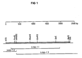
- nucleotide sequence (1) This nucleotide sequence represents a partial nucleotide sequence of the gene coding for the 2.13 antigen, which gene has the nucleotide sequence (2): Using the genetic code it was found that in the nucleotide sequence (1) two reading frames were interrupted by several stop codons and thus most likely do not code for a P. falciparum polypeptide. The third reading frame starting with nucleotides 3 to 5, viz.
- GAT as first codon encoding the amino acid aspartic acid (abreviated "D” in accordance with the single-letter system described by Dayhoff, in “Atlas of Protein Sequence and Structure", National Biomedical Research Foundation, Washington D.C., 1972) is open and codes for a polypeptide having the amino acid sequence (I)
- the amino acid sequence (I) codes for a polypeptide containing 591 amino acid residues. If the natural form of the P. falciparum antigen recognized by MABs 2.13 and 7.12, viz. the 2.13 antigen, has indeed a molecular weight of 80'000 Dalton, the amino acid sequence (I) represents about three quarters of the amino acid sequence of the said antigen.
- the antigenic determinant is formed by a specific molecular configuration of one or more sub-sequences in the amino acid sequence of the 2.13 antigen or the polypeptide comprising the amino acid sequence (I) or a partial sequence thereof, which partial sequence comprises the sub-sequence recognized by MAB 2.13.
- the nucleotide sequence (2) codes for a polypeptide containing 782 amino acid residues and having the following amino acid sequence (II):
- polypeptides comprising the amino acid sequence (II) or partial sequences thereof comprising the amino acid sequence forming the antigenic determinant recognized by MAB 2.13 such as e.g. the amino acid sequence (I).
- polypeptides can be prepared in substantially pure form i.e. free from contaminating malaria parasite derived materials.
- the amino acid sequence (II) may comprise other antigenic determinants which are immunologically cross-reactive with determinants on the 2.13 antigen of the rhoptry organelles of Plasmodium merozoites.
- the said determinants on the 2.13 antigen may be defined by labelling the 2.13 antigen in merozoites, e.g. by using the lactoperoxidase labelling method (Hogg, Proc. Natl. Acad. Sci. USA 71 , 489-492 [1974]) or by some other method known in the art and determining the amino acid residues labelled.
- Peptides comprising the amino acid residues determined in this way may be used as vaccines in accordance with the method disclosed in European Patent No. 44710.
- the detailed three-dimensional structure of the 2.13 antigen or of a sub-sequence thereof forming an antigenic surface determinant may be defined by computer-assisted X-ray or NMR analysis. Based on the structural information obtained in this way novel polypeptides comprising a sub-sequence forming about the same three-dimensional structure and thus mimicking an antigenic determinant may be designed (for a review see Blundell et al., Nature 326 , 347-352 [1987]).
- the said novel polypeptides may have an amino acid sequence which is different from the amino acid sequence of the 2.13 antigen or the sub-sequences thereof forming the antigenic surface determinant mentioned above.
- amino acid residues of the said novel polypeptide may also be modified in order to stabilize the three-dimensional configuration of the sub-sequence forming the antigenic determinant. Since the said sub-sequence in the novel polypeptide has about the same three-dimensional structure as the antigenic determinant on the 2.13 antigen the novel polypeptide immunologically cross-reacts with the determinant present on the 2.13 antigen.
- the present invention is concerned with antigenic polypeptides having a determinant or determinants immunologically cross-reactive with determinants on the Plasmodium falciparum polypeptide associated with the rhoptry organelles of the merozoite form of the malaria parasite which Plasmodium falciparum polypeptide has the amino acid sequence (II).
- the determinants on the Plasmodium falciparum polypeptide associated with the rhoptry organelles of the merozoites have to be accessible for an immune reaction of mammals against the malaria parasite.
- the invention is also concerned with a polypeptide comprising the amino acid sequence (II) or a partial sequence thereof such as the amino acid sequence (I) which polypeptide immunologically cross-reacts with determinants on the Plasmodium falciparum polypeptide defined above.
- a polypeptide of the present invention is the Plasmodium merozoite antigen associated with the rhoptry organelles of Plasmodium merozoites having a relative molecular mass of approximately 80kD which antigen is recognized by MABs 2.13 and 7.12.
- the preferred polypeptides are capable of inducing antibodies in a mammalian host, which antibodies are capable of inhibiting the invasion of erythrocytes by the merozoite form of the malaria parasite in vivo.
- the polypeptides may be in unglycosylated form or may be glycosylated. Other posttranslational modifications are possible such as covalently attaching a glycosylphosphatidyl inositol moiety.
- the polypeptide may also be an anti-idiotypic antibody or a fragment thereof having an antigenic determinant which is immunologically cross-reactive with the surface determinants mentioned above.
- An anti-idiotypic antibody is an antibody directed against the binding site of an antibody directed against a given antigenic determinant and thus the antigenic site of the said anti-idiotypic antibody has a similar molecular configuration as the antigenic determinant (Linthicum et al., BioEssays 3 , 213-217 [1985]).
- the polypeptides of the present invention are in substantially pure form.
- the invention relates also to polypeptides having an amino acid sequence derived from the amino acid sequences indicated above by additions, deletions, insertions or amino acid substitutions, provided that these polypeptides are still cross-reactive with determinants of the polypeptide associated with the rhoptry organelles of Plasmodium merozoites comprising the amino acid sequence (I).
- the invention is also concerned with DNAs coding for a polypeptide in accordance with the invention and with recombinant vectors containing such a DNA, especially expression vectors, i.e.
- the DNA which codes for a polypeptide in accordance with the present invention is bound to an expression control sequence in such a way that the polypeptide which is encoded by the DNA can be expressed.
- the preferred DNA comprises the nucleotide sequence (2) or a partial sequence thereof such as the nucleotide sequence (1).
- the present invention is concerned with unicellular host organisms which contain such a recombinant vector or an expression vector and with a process for the production of such organisms.
- the present invention is concerned with a process for the production of the polypeptides and with the use of these polypeptides for the immunization of mammals against malaria.
- the amino acid sequence of the polypeptides in accordance with the invention can differ from the amino acid sequences given above.
- Examples of such amino acid substitutions are Ala/Ser, Val/Ile, Asp/Glu, Thr/Ser, Ala/Gly, Ala/Thr, Ser/Asn, Ala/Val, Ser/Gly, Tyr/Phe, Ala/Pro, Lys/Arg, Asp/Asn, Leu/Ile, Leu/Val, Ala/Glu and vice versa (see Doolittle, in "The Proteins", Eds. Neurath, H, and Hill, R.L., Academic Press, New York [1979]).
- the polypeptides in accordance with the invention can be covalently bound to a carrier material or can be adsorbed thereon.
- Suitable carrier materials are natural or synthetic polymeric compounds such as e.g. copolymers of one or more amino acids (e.g. polylysine) or sugars (e.g. polysaccharides).
- serum proteins e.g. gammaglobulin, serum albumin
- toxoids e.g. diphtheria or tetanus toxoid.
- suitable carrier materials are known to the person skilled in the art.
- the covalent bonding of the polypeptides in accordance with the invention to the carrier materials can be effected in a known manner, e.g. directly by the formation of a peptide or ester bond between free carboxyl, amino or hydroxyl groups of the polypeptide and the corresponding groups on the carrier material or indirectly by using conventional, bifunctional reagents such as e.g. m-maleimidobenzoyl-N-hydroxysuccinimide ester (MBS) or succinimidyl 4-(p-maleimidophenyl)butyrate (SMPB).
- MBS m-maleimidobenzoyl-N-hydroxysuccinimide ester
- SMPB succinimidyl 4-(p-maleimidophenyl)butyrate
- C2 ⁇ 7-dialkanals such as e.g. glutaraldehyde (Avrameas, Immunochem. 6 , 43-52 [1969])
- the carrier material having the polypeptides bonded thereon can be separated from non-bonded polypeptides and, if desired, from excess reagents by known methods (e.g. dialysis or column chromatography).
- the peptides of the present invention can be produced by conventional methods of peptide synthesis in the liquid phase or, preferably, on the solid phase, such as the methods of Merrifield (J. Am. Chem. Soc. 85 , 2149-2154 [1963]) or by other equivalent methods of the state of the art.
- the solid phase synthesis begins with the C-terminal amino acid of the peptide to be synthesized, which is coupled in protected form to an appropriate resin.
- the starting material can be produced by coupling an amino acid, which is protected at the amino group, to a chloromethylated or a hydroxymethylated resin via a benzyl ester bridge or via an amide bond to a benzhydrylamine (BHA) resin, a methylbenzhydrylamine (MBHA) resin or a benzyloxybenzyl alcohol resin.
- BHA benzhydrylamine
- MBHA methylbenzhydrylamine
- Protecting groups include e.g. the 9-fluorenylmethyloxycarbonyl (Fmoc), the tertiary butyloxycarbonyl (Boc), the benzyl (Bzl), the t-butyl (But), the 2-chlorobenzyloxycarbonyl (2Cl-Z), the dichlorobenzyl (Dcb) and the 3,4-dimethylbenzyl (Dmb) group.
- the protected amino acids are coupled stepwise in the desired sequence to the C-terminal amino acid bonded to the resin.
- the complete peptide can thus be synthesized.
- small peptides can be synthesized and can then be joined together to give the desired peptide.
- Suitable coupling reagents belong to the state of the art, with dicyclohexylcarbodiimide (DCC) being especially preferred.
- Each protected amino acid or peptide is added in excess to the solid phase synthesis reaction vessel and the coupling reaction can be carried out in dimethylformamide (DMF) or methylene chloride (CH2Cl2) or a mixture of both.
- the coupling reaction is repeated before the N-terminal ⁇ -amino protecting group is removed for the purpose of coupling the next amino acid.
- the yield of each coupling step can be followed, preferably according to the ninhydrin method.
- the coupling reactions and the washing steps can be carried out automatically.
- the cleavage of the peptide from the carrier material can be achieved by methods which are well-known in peptide chemistry, e.g. by reaction with hydrogen fluoride (HF) in the presence of p-cresol and dimethyl sulphide for 1 hour at 0°C, followed possibly by a second reaction with HF in the presence of p-cresol for 2 hours at 0°C.
- HF hydrogen fluoride
- the cleavage of the peptides from chloromethylated or hydroxymethylated carrier materials gives peptides having a free C-terminus; the cleavage of peptides from benzylhydrylamine or methylbenzylhydrylamine carriers gives peptides having an amidated C-terminus.
- polypeptides of the present invention can also be produced using methods well known in the art of recombinant DNA technology (Maniatis et al. in "Molecular Cloning - A Laboratory Manual", Cold Spring Harbor Laboratory [1982]).
- a DNA which codes for such a polypeptide can be synthesized by conventional chemical methods, e.g. by the phosphotriester method which is described by Narang et al. in Meth. Enyzmol. 68 , 90-108 [1979], or by the phosphodiester method (Brown et al., Meth. Enzymol. 68 , 109-151 [1979]).
- the nucleotide sequence of the DNA can be partially or fully identical to the nucleotide sequence which codes for the natural polypeptide in the rhoptry organelles of Plasmodium merozoites. Since the genetic code is degenerate, there exists on the other hand the possibility that a partially or completely different nucleotide sequence codes for the same polypeptide.
- the codons selected can be adapted to the preferred codon usage of the host used to express the recombinant polypeptide (Grosjean et al., Gene 18 , 199-209 [1982]). Care must be taken that the DNA thus-obtained does not contain partial sequences which make the construction of the expression vector difficult, e.g. by introducing an undesired restriction enzyme cleavage site, or which prevents the expression of the polypeptide.
- a DNA coding for a polypeptide of the present invention can also be obtained by cloning parasite DNA into an expression phage vector, e.g. into the ⁇ phage vector gt11 which is obtainable from the American Type Culture Collection (ATCC), 12301 Parklawn Drive, Rockville, Maryland, USA. These phage vectors are transfected into suitable host cells, e.g. into E. coli Y1088 containing the plasmid pMC9 (available e.g. from ATCC). The expression gene library obtained in this way is then screened using polyclonal or monoclonal antibodies as described by Young et al., Proc. Natl. Acad. Sci. USA 80 , 1194-1198 [1983] and in the Example.
- an expression phage vector e.g. into the ⁇ phage vector gt11 which is obtainable from the American Type Culture Collection (ATCC), 12301 Parklawn Drive, Rockville, Maryland, USA.
- ATCC American Type Culture Collection
- the DNA is obtained by cleaving DNA of a Plasmodium parasite strain with one or more restriction endonucleases and cloning the fragments into a suitable vector according to procedures well known in the art (Maniatis et al., supra).
- the parasite DNA library is then screened with a suitable probe.
- suitable probes are oligonucleotides which correspond to a partial sequence of the DNA coding for the 2.13 antigen or the DNA having the nucleotide sequence (1).
- the manner in which these probes are selected and used is known to the person skilled in the art. Examples of such probes are oligonucleotides having the sequence TTGGTGCAGGAGGTGCT or AAGCTACCTATACATGT.
- Clones containing a DNA insert hybridizing to the probe are grown up and the DNA is isolated. Subsequently, the DNA fragment can be inserted into a suitable vector, preferably into a vector which provides the necessary expression signals. Examples of such expression vectors are described in European Patent Application, Publication No. 186 069 which was published on 2nd July 1986.
- the polypeptides of the present invention can, after corresponding adaptation of the nucleotide sequence, also be produced using other suitable expression vectors known to the person skilled in the art.
- the recombinant vector which contains a DNA coding for a polypeptide in accordance with the invention is subsequently introduced into a suitable unicellular host organism.
- Suitable unicellular host organisms are prokaryotic or eukaryotic cells.
- prokaryotic cells are microorganisms, e.g. bacterial cells, such as E. coli cells or B. subtilis cells, which are capable of expressing polypeptides encoded by the recombinant expression vectors.
- a large number of eukaryotic cells suitable as unicellular host organisms for recombinant vectors are known to the person skilled in the art. Examples for such eukaryotic cells are yeast cells (e.g.
- Saccharomyces cerevisiae and cultured cells of higher eukaryotes, such as insect cells (e.g. using the baculovirus vector system; for a review see Doerfler, Curr. Top. Microbiol. Immunol. 131 , 51-68 [1986] or Yong Kang, Adv. in Virus Research 35 , 177-192 [1988]) and vertebrate cells (for a review see Rigby, Genetic Engineering, Vol. 3, R. Williamson, ed., Academic Press, New York, pp. 83-141 [1982]).
- the preferred unicellular host organism is E. coli M15 (described as DZ291 by Villarejo et al. in J. Bacteriol.
- E. coli 294 E. coli RR1 and E. coli W3110 (all available from ATCC and other sources).
- E. coli RR1 E. coli RR1
- E. coli W3110 all available from ATCC and other sources.
- the polypeptide may be in glycosylated form.
- the manner in which the expression of the polypeptides in accordance with the invention is effected depends on the recombinant expression vector and on the host organism used. In the case of bacteria and yeast the host organisms which contain the recombinant expression vector are grown up under conditions which are optimal for the growth of the host organism. Towards the end of the exponential growth, when the increase in the number of cells per unit time decreases, the expression of the polypeptide of the present invention is induced, i.e. the DNA coding for the polypeptide is transcribed and the transcribed mRNA is translated.
- the induction can be effected by adding an inducer or a derepressor to the growth medium or by altering a physical parameter, e.g. by a temperature change.
- the expression is controlled by the lac repressor.
- IPTG ispropyl- ⁇ -D-thiogalactopyranoside
- the polypeptide produced in the unicellular host organisms can be secreted by special transport mechanisms or can be isolated by breaking open the unicellular organism.
- the host organism can be broken open by mechanical (Charm et al., Meth. Enzymol. 22 , 476-556 [1971]), enzymatic (lysozyme treatment) or chemical (e.g. detergent treatment, urea or guanidine ⁇ HCl treatment) means or by a combination thereof.
- polypeptides which are secreted from the cells are synthesized in the form of a precursor molecule.
- the mature polypeptide results by cleavage of the so-called signal peptide.
- signal peptide As prokaryotic host organisms are not capable of cleaving eukaryotic signal peptides from precursor molecules, eukaryotic polypeptides must be expressed directly in their mature form in prokaryotic host organisms.
- the translation start signal AUG which corresponds to the codon ATG on the level of the DNA, causes all polypeptides synthesized in a prokaryotic host organism to have a methionine residue at the N-terminus. In certain expression systems this N-teminal methionine residue is cleaved off.
- N-terminal methionine has often no influence on the biological activity of a polypeptide (see Winnacker, in "Gene und Klone", p. 255, Verlag Chemie, Weinheim, BRD [1985]).
- the N-terminal methionine can also be cleaved off by means of peptidases which are specific for the N-terminal methionine. Miller et al., Proc. Natl. Acad. Sci. U.S.A. 84 , 2718-2722 [1987] have described the isolation of such a peptidase from Salmonella typhimurium.
- the present invention is accordingly concerned with polypeptides with or without a N-terminal methionine residue.
- polypeptides in accordance with the present invention can be purified by known methods such as, for example, by centrifugation at different velocities, by precipitation with ammonium sulfate, by dialysis (at normal pressure or at reduced pressure), by preparative isoelectric focusing, by preparative gel electrophoresis or by various chromatographic methods such as gel filtration, high performance liquid chromatography (HPLC), ion exchange chromatography, reverse phase chromatography and affinity chromatography (e.g. on SepharoseTM Blue CL-6B, on phosphocellulose, on carrier-bound monoclonal antibodies directed against the polypeptide or on metal chelate resins such as those described in European Patent Application, Publication No. 253 303.
- HPLC high performance liquid chromatography
- ion exchange chromatography reverse phase chromatography
- affinity chromatography e.g. on SepharoseTM Blue CL-6B, on phosphocellulose, on carrier-bound monoclonal antibodies directed against the polypeptide or on metal chelate resin
- the preferred purification method in the present invention is affinity chromatographic purification.
- the purification of the polypeptides in accordance with the invention on carrier-bound monoclonal antibodies 2.13 and 7.12 is especially preferred.
- polypeptides of the present invention can be present in the form of multimers, e.g. in the form of dimers, trimers or tetramers.
- the subunits of these multimers may be linked together by covalent or noncovalent bonds. Multimers often result when polypeptides are produced in prokaryotic host organisms, for example by the formation of disulphide bridges between cysteine residues.
- polypeptides of the present invention can also be prepared in form of fusion polypeptides.
- a polypeptide of the present invention is covalently linked via a peptide bond to one or more fusion partners.
- fusion partners are peptides or polypeptides, preferably such peptides or polypeptides which contribute to improve the properties of the fusion polypeptide in comparison to those of the polypeptide without the fusion partner.
- Suitable fusion partners are polypeptides having an antigenic determinant or determinants immunologically cross-reactive with other antigens of the malaria parasite, preferably antigens from another stage of the malaria parasite and thereby improving the efficacy of vaccines comprising said fusion polypeptide.
- Antigens from the sporozoite stage such as the circumsporozoite (CS) protein or other polypeptides comprising repeat sequences such as the tetrapeptide repeat Asn-Ala-Asn-Pro present in the P. falciparum CS protein are preferred.
- the fusion partner may also be a T cell epitope such as one of those described by Good et al. (Science 235 , 1059-1062 [1987]).
- the fusion partner may also be an affinity peptide and thereby contribute to the ease of recovery of the fusion polypeptide.
- An example of such a fusion partner is an affinity peptide which binds preferably to an affinity chromatography carrier material.
- affinity peptides are peptides containing at least two histidine residues. Such affinity peptides bind selectively to nitrilotriacetic acid-nickel chelate resins (see e.g. European Patent Application, Publ. No. 253 303). Fusion polypeptides which contain such an affinity peptide can therefore be separated selectively from the remaining polypeptides by means of such resins. Fusion proteins can be produced by conventional peptide synthesis methods or by genetic engineering whereby DNA fragments which code for a polypeptide of the present invention are linked with one or more DNA fragments which code for said fusion partner. Preferably the fusion polypeptides are prepared by genetic engineering, e.g. by introducing a DNA coding for a polypeptide according to the present invention into an expression vector which comprises the necessary expression control sequences and a DNA coding for said fusion partner.
- polypeptides of the present invention may also be used diagnostically to detect antibodies directed against the determinant of the polypeptide associated with the rhoptry organelles of Plasmodium merozoites having the amino acid sequence (I) according to methods well known in the art.
- the present invention is also concerned with immunogenic compositions which contain a polypeptide in accordance with the present invention as well as a suitable adjuvant.
- suitable adjuvants are aluminium hydroxide and aluminium phosphate.
- Other suitable adjuvants or delivery systems e.g. Salmonella thyphimurium or Vaccinia virus
- Salmonella thyphimurium or Vaccinia virus for use in human beings and animals are known to the person skilled in the art (WHO Techn. Rep. Series 595 , 1-40 [1987]; Warren et al., Ann. Rev. Immunol.
- polypeptides or immunogenic compositions in accordance with the invention can be present as lyophilizates for reconstitution with sterile water or a salt solution, preferably phosphate buffered saline.
- a salt solution preferably phosphate buffered saline.
- the antibodies in accordance with the invention recognize the natural equivalent of the polypeptide in the malaria parasite and can therefore be used therapeutically for blocking red blood cell invasion by the merozoites, for passive immunization or for diagnostic purposes.
- Antibodies against the polypeptides in accordance with the invention can be produced in monkeys, rabbits, horses, goats, guinea pigs, rats, mice, cows, sheep etc, and also in human beings.
- the antiserum or the purified antibody can be used as required.
- the antibodies can be purified in a known manner, e.g. by precipitation with ammonium sulfate. It is also possible to produce monoclonal antibodies which are directed against the polypeptide of the present invention in accordance with the method developed by Köhler et al. (Nature, 256 , 495-497 [1975]). Polyclonal or monoclonal antibodies can also be used for the affinity-chromatographic purification of the polypeptides of the present invention or of their natural equivalent, the 2.13 antigen.
- the polypeptides and immunogenic compositions in accordance with the invention can be used as vaccines for the immunization of mammals against malaria.
- the immune system of a mammal preferably a human
- an immunizing amount of said polypeptide or immunogenic composition is stimulated with an immunizing amount of said polypeptide or immunogenic composition.
- the mode of administration, the dosage as well as the number of injections can be optimized in a manner known to the person skilled in the art. Typically, several injections are administered over a long time period in order to obtain a high titer of antibodies against the malaria antigen, i.e. against the polypeptide of the present invention.
- Phage buffer per litre:
- the DNA solution is treated with a tenth by volume of 5 M lithium acetate and two volumes of isopropanol, mixed well and placed on dry ice for 10 minutes.
- the precipitated DNA is centrifuged for 10 minutes at 12,000 RPM (20°C) in an Eppendorf bench centrifuge and the supernatant is carefully removed.
- the sediment is washed once with 80% (v/v) ethanol and subsequently dried for 5 minutes in a vacuum centrifuge.
- the DNA is dissolved in water and processed further.
- the dried DNA is dissolved in 1 x DNA gel loading buffer and heated to 65°C for 5 minutes.
- 100 ml of 1 x TBE buffer are mixed with agarose (800 mg for a 0.8% gel or 1.2 g for a 1.2% gel) and boiled until the agarose has dissolved completely.
- 2 ⁇ l of ethidium bromide solution (10 mg/ml) are added and the gel solution is poured into a horizontal gel electrophoresis apparatus. After solidification of the gel the samples are applied to the gel and the DNA is separated for 2 hours at 150 volt constant voltage.
- Commercial standard mixtures of DNA fragments of defined length are used as size markers.
- the DNA bands are visualized under 300 nm UV light.
- the DNA is separated on an agarose gel (Methode 2).
- a trough is cut in front of the band to be purified and a piece of dialysis tubing inserted at the end of the trough.
- the trough is filled with 1 x TBE buffer and the DNA electroeluted onto the dialysis tubing at 200 V for 10 to 15 minutes. It is then back-eluted into the trough for 45 seconds.
- the DNA solution is extracted twice with water-saturated isobutanol, twice with phenol/chloroform/isoamyl alcohol (25:24:1) and is then precipitated by the addition of 1/10th volume 3 M sodium acetate pH 5.2 and 2 volumes ethanol. After standing at -70°C for 15 minutes the precipitate is pelleted at 12,000 g for 10 minutes, dried and resuspended in 1 x TE buffer.
- a bacterial culture (e.g. E. coli Y1088 available e.g. from ATCC) is infected on an agar plate (Maniatis et al., supra, pp. 70-71) with lambda phages. Thereby, lytic plaques are formed in the lawn of bacteria.
- An agar cylinder (diameter 5 mm) containing a plaque is cut from the agar with a inverted Pasteur pipette. The agar cylinder is transferred into an Eppendorf test tube containing 500 ⁇ l of SM buffer and the test tube is shaken for 5 minutes. The phage suspension is centrifuged (5 minutes at 12,000 RPM, 20°C) and the supernatant is transferred into a fresh test tube.
- 1 ⁇ l of the phage suspension is diluted with 1 ml of SM buffer. 1, 10 and 100 ⁇ l of this solution are added to 50 ⁇ l of a cell suspension of E. coli Y1090 cells containing the plasmid pMC9 (available e.g. from ATCC) which had been Mg++-treated in accordance with Morrison, (Methods Enzymol. 68 , 326-331 [1979]). After a 30 minutes incubation at room temperature the solution is added to 3 ml of 0.8% (w/v) agar in LB medium and the mixture is poured onto LB-ampicillin agar plates (LB medium, 40 ⁇ g/ml ampicillin). Depending on the titre, some plates (e.g.
- those with the 1:1,000 dilution have individual plaques which, when they are positive in the antibody reaction or the DNA hybridization, can be isolated.
- the phages from the plaques can be grown up and e.g. used for the isolation of phage DNA.
- plaque forming units e.g. 10000
- infect 100 ⁇ l of plating cells methode 5
- the top agar which should exhibit confluent lysis, is then scraped into 5 ml phage buffer and allowed to stand for 2 hours at room temperature.
- the top agar is finally pelleted at 4,000 g for 10 minutes and the supernatant retained as phage stock.
- An individual colony of the E. coli strain to be used (e.g. Y1090 available e.g. from ATCC) is inoculated into 5 ml LB medium and grown overnight at 37°C. The suspension is then diluted 50-fold and grown at 37°C for a further 2 hours before pelleting the cells at 4,000 g for 10 minutes at 4°C and resuspending the cells in 1/10th volume of ice cold 10 mM magnesium sulfate. The cells may be stored at 4°C for up to 2 weeks. The cells are used for generating phage lambda plaques as follows. An appropriate number of plaque forming phage units is added to 100 ⁇ l of plating cells. The suspension is left at room temperature for 20 minutes, then 3 ml of liquid BBL top agar (at 45°C) is added and the mixture poured on to an 80 mm bottom agar plate.
- Y1090 available e.g. from ATCC
- 3 ml LB medium is inoculated with E. coli cells and shaken at 37°C overnight. 1 ml of this saturated culture is used to inoculate 50 ml LB medium. The culture is shaken until the optical density at 600 nm (OD600) has reached a value of 0.2. The cells are sedimented (5 minutes at 6,000 RPM, room temperature) and resuspended in 25 ml of ice-cold 100 mM magnesium chloride. The cells are again centrifuged off (see above) and suspended in 5 ml of 100 mM calcium chloride. The competent cells are left at 4°C for at least 30 minutes. For the transformation 10 ⁇ l of DNA solution containing 1 to 10 ng DNA are added to 100 ⁇ l of competent cells and incubated first for 20 minutes on ice, then at 42°C for 2 minutes and finally again on ice for 20 minutes.
- plasmid DNA which can be selectionized for antibiotic resistance
- 1 ml of LB medium is added to the transformation mixture and the incubation is carried out at 37°C for one hour.
- the cells are centrifuged off for 3 minutes at 6,000 RPM (room temperature) and resuspended in 100 ⁇ l of LB medium. These 100 ⁇ l are distributed uniformly on a LB agar plate which contains the antibiotic required for the selection and likewise incubated at 37°C overnight.
- E. coli strains TG-1, JM 101 or JM 103 can be used as hosts for cloning vectors of the M13 type for sequencing DNA according to Messing, Meth. Enzymol. 101 , 20-78 [1983].
- a vector of the M13 type which vector comprises a DNA fragment to be sequenced
- white plaques result. These white plaques are picked with a toothpick and resuspended in 3 ml of LB medium.
- a further 60 ⁇ l of a saturated host culture are added and the mixture is shaken at 37°C for 6 hours. 1.5 ml of culture are transferred into an Eppendorf test tube and centrifuged (5 minutes at 12,000 RPM, 20°C).
- 1.2 ml of supernatant are transferred into a new test tube and mixed with 300 ⁇ l of 20% (w/v) polyethylene glycol, 2.5 M sodium chloride solution and incubated at room temperature for 20 minutes. The remainder of the culture is stored at 4°C or used for the preparation of "mini-lysate" DNA (Method 10).
- the phages are precipitated by centrifugation (10 minutes at 12,000 RPM, 20°C). The sediment is dissolved in 100 ⁇ l of 1 x TES buffer and extracted with 100 ⁇ l of saturated phenol. The phases are separated by centrifugation (5 minutes at 12,000 RPM). 80 ⁇ l of the aqueous phase are transferred into a new reagent test tube, the DNA is precipitated (Method 1) and dissolved in 12 ⁇ l of water.
- 1 ⁇ l of the DNA prepared according to Method 8 are mixed with 6 ⁇ l of water, 2 ⁇ l of 1 x TM buffer, and 1 ⁇ l of oligonucleotide primer and heated at 65°C for 2 minutes. Thereafter, the solution is allowed to cool slowly to 35°C. In the meanwhile, 4 test tubes each containing 2 ⁇ l of the stop solutions A o , G o , T o and C o are prepared.
- the stop solutions have the following composition: A o : 1 ⁇ M ddATP, 100 ⁇ M dCTP, 100 ⁇ M dGTP, 100 ⁇ M dTTP C o : 160 ⁇ M ddCTP, 12.5 ⁇ M dCTP, 125 ⁇ M dGTP, 125 ⁇ M dTTP G o : 250 ⁇ M ddGTP, 125 ⁇ M dCTP, 12.5 ⁇ M dGTP, 125 ⁇ M dTTP T o : 400 ⁇ M ddTTP, 100 ⁇ M dCTP, 100 ⁇ M dGTP, 25 ⁇ M dTTP.
- the DNA is now applied to a 0.4 mm gel of the following composition: 34 ml H2O 3 ml 10 x TBE-buffer 28.8 g urea 3.6 g acrylamide 180 mg bisacrylamide 360 ⁇ l 10% ammonium persulfate 50 ⁇ l TEMED
- the DNA is separated for 1 to 6 hours by electrophoresis at 40 watt constant output.
- the glass plates are separated and the gel is fixed for 5 minutes in 10% (v/v) acetic acid and 10% (v/v) methanol.
- the gel is then washed twice with 10% (v/v) methanol for 5 minutes, mounted on Whatman 3M paper (Whatman Ltd., Maidstone, England) and dried in a gel dryer.
- the dried gel is autoradiographed for 20 hours using X-ray Film (e.g. KODAK-XAR, Eastman Kodak Co., Rochester, New York, USA).
- X-ray Film e.g. KODAK-XAR
- bacterial culture e.g. E. coli TG-1 containing a vector of the M13 type; see Method 8
- E. coli TG-1 containing a vector of the M13 type; see Method 8
- the supernatant is carefully sucked off.
- the sedimented cells are resuspended in 500 ⁇ l of 50 mM Tris/HCl [pH 7.6], 5 mM EDTA.
- After the addition of a small spatula tip of lysozyme the suspension is incubated at room temperature for 5 minutes. 15 ⁇ l of 25% (w/v) lithium dodecylsulfate solution and 30 ⁇ l of 5 M potassium acetate are then added and the suspension is mixed carefully.
- the DNA to be labelled (up to 100 ng) is diluted to 7 ⁇ l volume with H2O, heated to 95°C for 2 minutes and quick cooled on ice. After adding 5 ⁇ l of ⁇ -[32P]dCTP (3,000 Ci/mmol), 12 ⁇ l of LS buffer and 3 units of Klenow Polymerase (Maniatis et al., supra, pp 113-114) the reaction is incubated for 4 to 16 hours at room temperature.
- the labelled DNA may be used directly for hybridisations. Immediately prior to its use in hybridisations the DNA must be heated to 95°C for 10 minutes.
- the filter containing DNA is incubated for 1 hour at 65°C in 0.5 M sodium phosphate buffer [pH 7], 7% SDS. To this is added approximately 107 cpm of radioactive sample (method 11). After incubation at 65°C overnight the filters are washed twice for 30 minutes in 0.2 x SSC, 0.1% SDS at 65°C. The filters are dried and exposed against X-ray film (e.g. Kodak XAR).
- X-ray film e.g. Kodak XAR
- the solutions are mixed, 30 ⁇ l TEMED are added and poured onto the separating gel. A probe comb is inserted prior to the polymerization. 190 mM glycine, 25 mM Tris [pH 7.6], 1% (w/v) SDS is used as the electrophoresis buffer. Commercial molecular weight standards can be used as size markers.
- nitrocellulose sheet is again washed as above and subsequently placed in 5 ml of alkaline phosphatase buffer containing 33 ⁇ l of NBT and 16.5 ⁇ l BCIP (50 mg/ml each in dimethylformamide) and mixed well. After the bands have been made visible the nitrocellulose sheet is stored in water in order to prevent an overexposure. Pre-stained marker proteins can be used as molecular weight markers.
- plasmid or parasite DNA per slot are separated by electrophoresis on an agarose gel (Method 2).
- the gel is first photographed and then incubated for 15 minutes in 0.5 M sodium hydroxide, 1.5 M sodium chloride solution. After that the gel is neutralized by incubating it twice for 15 minutes in 0.02 M sodium hydroxide, 1 M ammonium acetate and then placed on a clean glass plate.
- a nylon membrane (Hybond N, available from Amersham) is placed on the gel, followed by three sheets of Whatman 3 MM and about 20 papertowels. The assembly is weighed down from above with a 500 g weight. After 3 hours the membrane is removed and dried at room temperature. The dried membrane is exposed to U.V. light, face-down on a tansilluminator for 5 minutes and may then be treated further in accordance with Method 12.
- P. falciparum cells (K1 isolate) were grown in 10 culture dishes as described by Trager et al., Science 193 , 673-675 [1976] and subsequently washed in culture medium containing 0.1% saponin. The washed parasites were resuspended in 2 ml of 10 mM EDTA [pH 8.0], 0.5% (w/v) SDS. After the addition of 50 mg of proteinase K the mixture was incubated at 65°C for 10 minutes and subsequently treated with 2 ml of phenol (saturated with Tris/HCl [pH 8.0]). The phases were mixed by shaking and again separated by centrifugation (10 minutes at 6,000 RPM, 20°C).
- the phenol extraction was repeated twice (an interphase should no longer be visible).
- the DNA was precipitated according to Method 1, washed with ethanol and dried.
- the DNA was dissolved in 2 ml of water and sheard mechanically, i.e. by squeezing the DNA solution 80 times through a syringe with a 0.5 x 16 mm needle. Thereafter, 0.2 volumes of 5 x EcoRl methylase buffer (50 mM Tris/HCl [pH 7.5], 0.25 M NaCl, 50 mM EDTA, 25 mM ⁇ -mercaptoethanol, 0.4 mM S-adenosylmethionine) were added.
- DNA 10 ⁇ g of DNA were methylated at 37°C for 30 minutes with 50 units of EcoRl methylase (New England Biolabs, Beverly, Massachusetts, USA).
- the DNA was extracted once with phenol as described above and precipitated in accordance with Method 1.
- the DNA was dissolved in 200 ⁇ l of T4 polymerase buffer and, after the addition of 5 ⁇ l of 5 mM dATP, dCTP, dGTP and dTTP as well as 10 units of T4 polymerase, incubated at 37°C for 30 minutes.
- the DNA was again extracted with phenol and precipitated in accordance with Method 1.
- the DNA was dissolved in 50 ⁇ l of ligase buffer.
- the DNA obtained was dissolved in 50 ⁇ l of water and, after the addition of 6 ⁇ l of 10 x ligase buffer, 2 ⁇ l of dephosphorylated lambda arms (Promega Biotech Corp.) and 6 Weiss units of T4-DNA ligase, ligated at 14°C overnight.
- the DNA was precipitated (Method 1) and dissolved in 5 ⁇ l of water.
- 20 ⁇ l of "Packaging Extract” (Genofit, S.A., Geneva, Switzerland) the DNA was packed in phage particles at 20°C for 2 hours according to the directions of the manufacturer.
- 500 ⁇ l of SM buffer as well as 50 ⁇ l of chloroform the gene bank was ready for the antibody test.
- the monoclonal antibodies 2.13 and 7.12 were produced as described by Hall et al. (supra). These MABs recognize polypeptides of 80-65 kD in Western blots.
- E. coli cells strain Y1090 were incubated in 3 ml of LB medium at 37°C overnight. On the next morning the cells were diluted 50-fold in LB medium and grown for 2 hours at 37°C. They were then sedimented (10 minutes at 7,000 g, 20°C) and resuspended in 5 ml 10 mM magnesium sulfate. Thirty batches of 104 phage particles from the gene bank were added to 100 ⁇ l of this cell suspension and incubation was carried out at room temperature for 30 minutes. 3 ml of 0.8% agar solution in BBL medium, warmed to 42°C, were added to each batch and mixed well.
- the soft agar having the infected cells was distributed on BBL agar plates (diameter 82 mm). The agar plates were then incubated at 42°C for 3,5 hours. A nitrocellulose filter, which had first been immersed in 100 mM IPTG solution and dried, was placed on each dish and incubation was carried out at 37°C for 3 hours. The position of the filter relative to the dish was marked. The filters were incubated for 30 minutes in 1 x TBS, 5% skimmed milk powder containing 0.01% Antifoam A (Sigma, St. Louis, Missouri, USA). Monoclonal antibody ascitic fluid containing MAB 2.13 was diluted 1:1,000 with 1 x TBS/5% (w/v) milk powder.
- the filters were incubated in the presence of said MAB ascitic fluid at room temperature for 30 minutes and then washed once for 5 minutes in 1 x TBS, twice for 5 minutes in 1 x TBS, 0.05% Nonidet P40 (Sigma) and once again in 1 x TBS for 5 minutes. This was followed by an incubation for 30 minutes with a 1:7,500 dilution of alkaline phosphatase conjugated goat anti-mouse IgG (Promega Biotech Corp.) in 1 x TBS, 5% (w/v) milk powder.
- the filters were again washed as above and the filters developed colorimetrically at room temperature using 5 ml of alkaline phosphatase buffer containing 33 ⁇ l NBT and 16.5 ⁇ l BCIP (P+S Biochemicals, England; catalogue No. P3771). Plaques which were positive were identified and picked from the Petri dishes on the basis of the markings. The phages in the individual plaques were plated out in soft agar in different dilutions according to Method 4 and positive plaques were again identified as described above. An individual, positive plaque ( ⁇ 2.13) was picked, grown up according to Grossberger, Nucleic Acids Res. 15 , 6737 [1987] and the DNA was isolated.
- the clone ⁇ 2.13 was plaque purified (Method 4) and a plate lysate phage stock was prepared (Method 5). The cloned phage ⁇ 2.13 was then lysogenised into E. coli strain Y1090 as follows. To 10 ⁇ l of Y1090 plating cells (Method 6) was added 106 plaque forming units of the phage ⁇ 2.13. LB medium (200 ⁇ l) was added and the mixture left for 3 hours at 30°C. The suspension was then streaked out on to an LB plate and grown overnight at 30°C. Colonies were isolated and picked on to replica plates, one grown at 30°C and the other grown at 42°C. Those colonies which grew at 30°C but not at 42°C were assumed to be lysogens of ⁇ 2.13.
- a lysogen of ⁇ 2.13 was grown overnight at 30°C in 5 ml LB medium. It was then diluted 10-fold with LB medium and grown for 3 hours at 30°C. Synthesis of the fusion protein was induced by adding IPTG to a final concentration 100 ⁇ M and moving the culture to 42°C. After 30 minutes 3 ml of this culture was pelleted at 12,000 g for 5 minutes and resuspended in 100 ⁇ l of SDS-PAGE loading buffer. The sample was boiled for 5 minutes and then loaded on to a 10% SDS-polyacrylamide gel (Method 13) and run at a constant voltage of 100 V overnight.
- the ⁇ 2.13 DNA (2 ⁇ g) was dissolved in high salt buffer (20 ⁇ l), digested at 37°C for 1 hour with 11 units EcoRI and analysed on a 0.8% agarose gel (Method 2). No insert was observed.
- the ⁇ 2.13 DNA (2 ⁇ g) was then dissolved in high salt buffer (20 ⁇ l) and digested at 37°C for 1 hour with 10 units MluI.
- a 3.9 kb DNA fragment was observed by agarose gel electrophoresis (Method 2) indicating a cloned insert of 1.7 kb.
- the ⁇ 2.13 DNA (2 ⁇ g) was then digested in core buffer (20 ⁇ l) with 11 units EcoRI for 1 hour at 37°C.
- the enzyme was heat inactivated by a 10 minutes incubation at 65°C.
- the reaction mixture was incubated at 37°C for 1 hour and the enzyme then heat inactivated at 65°C for 10 minutes.
- An EcoRI/KpnI fragment of 2.7 kb was observed by agarose gel electrophoresis (Method 2).
- this fragment was ligated into the sequencing vectors M13 mp18 and M13 mp19 (e.g. available from New England BioLabs, Beverly, Massachusetts, USA). This was done by digesting 2 ⁇ g of each vector with EcoRI and KpnI as described for ⁇ 2.13 above. The digested vectors M13 mp18 and M13 mp19 (40 ng) were each added to the EcoRI/KpnI digested ⁇ 2.13 DNA (100 ng) in 20 ⁇ l ligase buffer with 1 unit T4 DNA ligase and incubated at 15°C overnight. Competent TG-1 cells were transformed with the ligated DNA (Method 7).
- M13 mp18 and M13 mp19 e.g. available from New England BioLabs, Beverly, Massachusetts, USA. This was done by digesting 2 ⁇ g of each vector with EcoRI and KpnI as described for ⁇ 2.13 above. The digested vectors M13 mp18 and M13 mp19 (40 ng) were
- DNA (5 ⁇ g) from the subclone of M13 mp18 containing the KpnI/EcoRI fragment of ⁇ 2.13 was taken up in 20 ⁇ l of high salt buffer and treated with the restriction enzymes EcoRI (10 units) and salI (10 units) for 1 hour at 37°C. The DNA was analysed by agarose gel electrophoresis (Method 2) and a 1.7 kb fragment was isolated (Method 3).
- DNA 100 ng was radioactively labelled (Method 11) and hybridised (Method 12) to a Southern blot (Method 15) of P. falciparum (K1 strain) DNA (3 ⁇ g) which has been digested with HindIII (3 units) in 20 ⁇ l medium salt buffer at 37°C for 1 hour. Two DNA fragments of about 3 kb and 11 kb were observed. These two DNA fragments were also present in the gene bank of K1 DNA HindIII fragments present in the vector ⁇ NM1149 (Goman et al., Mol. Biochem. Parasitol. 5 , 391-400 [1982]).
- the coding sequence reveals a number of points of interest in the structure of the 2.13 antigen. At its N-terminus it has a putative signal peptide sequence (see arrow in Fig. 2A), suggesting that this antigen is directed through the endoplasmic reticulum and the Golgi apparatus to its point of destination.
- the 2.13 antigen has no transmembrane domains or oligopeptide repeats.
- the most interesting features are sequences which could form positively-charged amphiphilic ⁇ -helices (underlined in Fig. 2A,B). Such structures could be used by the protein to penetrate the membrane of the erythrocyte during invasion by the merozoite.
- Purified monoclonal antibody 2.13 was coupled to cyanogen bromide activated SepharoseTM 4B (Pharmacia) using the method recommended by the manfacturer.
- Cell pellets from bulk cultures of P. falciparum were harvested and extracted with at least 5 volumes of 1% (w/v) Nonidet P40, 2% (w/v) sodium deoxycholate, 500 mM Tris/HCl (pH 8.0), 5 mM EDTA, 5 mM EGTA, 5 mM iodoacetamide, 0.2 mM PMSF and 10 ⁇ g/ml each of pepstatin, chymostatin, antipain, leupeptin, at 4°C for 1 hour.
- the SepharoseTM 4B-bound MAB 2.13 column was then washed with at least 5 volumes of wash buffer (1% Nonidet P40, 0.5% (w/v) sodium deoxycholate, 50 mM Tris/HCl (pH 8.0), 5 mM EDTA, 5 mM EGTA) followed by 5 column volumes of wash buffer containing additionally 0.9% NaCl and finally re-equilibrated in salt-free wash buffer before eluting with 50 mM diethylamine in wash buffer at pH 11.5.
- wash buffer 1% Nonidet P40, 0.5% (w/v) sodium deoxycholate, 50 mM Tris/HCl (pH 8.0), 5 mM EDTA, 5 mM EGTA
- the protein solution was neutralised with solid glycine and then further purified by loading the protein on a polyacrylamide gel and eluting the bands with a relative molecular weight of 65 to 80 kD and 40 to 42 kD, i.e. the bands representing the affinity purified 2.13 antigen (see above), after electrophoresis by electroelution using methods well known in the art.
- the purified protein was used for immunizing monkeys.
- the antigen was emulsified in Freund's complete adjuvant, whereas for the second and third injections the antigen was emulsified in Freund's incomplete adjuvant.
- a second group of four S. sciureus monkeys serving as control group was injected in a similar way with Freund's adjuvant mixed with phosphate buffered saline. 60 days after the first injection the animals were injected intravascularly with 3.5 x 10⁷ parasitized red blood cells of a S.
Abstract
Description
- Malaria in human beings is caused by four species of Plasmodium, namely by P. falciparum, P. vivax, P. ovale and P. malariae. According to a report of the World Health Organisation (WHO) from the year 1986, there are worldwide almost 100 million cases of malaria infection. Of these about 1 million, mostly cases of young children which are infected with P. falciparum, are fatal. Because of the appearance of drug-resistant parasites and of insecticide-resistant mosquito vectors, malaria is spreading (Bruce-Chwatt, Essential Malariology, 2nd edition, Heinemann, London [1986]).
- Recent technical advances have raised hopes that it would soon be possible to produce an antimalaria vaccine which would counteract the growing spread of malaria. Firstly, new methods in the development of malaria vaccines, e.g. the cloning of genes and their expression in microbial host organisms as well as the use of monoclonal antibodies for antigen identification, can be used. Secondly, long-term cultures of P. falciparum in human red blood cells (Trager et al., Science 193, 673-675 [1976]) have provided a ready source of material for the study of the malaria parasite. More recently, it has become possible to maintain all stages in the life cycle of the parasite in the laboratory (Ponnudurai et al., Trans. R. Soc. Trop. Med. Hyg. 76, 812-818 [1982]; Mazier et al., Science 227, 440-442 [1985]).
- The natural life cycle of P. falciparum has three different stages. In the first stage, mosquitoes introduce sporozoites into the blood vessels of vertebrates during the intake of food. These sporozoites travel via the bloodstream to the liver and invade the hepatocytes of the host. In the second stage, merozoites develop from these sporozoites. These merozoites pass through several multiplication cycles in erythrocytes of the host and then develop to gametocytes. The gametocytes, which are the sexual stage of the parasite, are taken up by mosquitoes when they feed. After fertilization in the stomach of the insect the gametocytes develop into sporozoites which then travel to the salivary glands of the insect. From there the cycle can begin again.
- Sporozoites, merozoites and gametocytes have different antigens (Scaife, Genetic Engineering 7, 57-90 [1988]). Vaccines can be produced in principle against any of the different stages of the malaria parasite. Various merozoite antigens have been used to induce immunity against malaria. None of these antigens has been found to be the ideal vaccine candidate. Here we present a novel schizont/merozoite antigen of the malaria parasite. This antigen is recognized by two monoclonal antibodies (MABs) described by Hall et al. (Mol. Biochem. Parasitol. 7, 247-265 [1983]). One of these monoclonal antibodies MAB 2.13 strongly inhibits invasion of erythrocytes by malaria parasites. Western blot analysis (Towbin et al., Proc. Natl. Acad. Sci. USA 76, 4350-4354 [1979]) of protein extracts from whole parasites using MAB 2.13 revealed 4 major bands with a relative molecular mass of 65 kD, 70 kD, 78 kD and 80 kD, respectively, whereby 1 kD equals 1'000 Dalton. Affinity purification of the protein extract from whole parasites followed by gelelectrophoretic analysis revealed two additional bands with a relative molecular mass of 40 kD and 42 kD. Most likely the natural form of the antigen recognized by MAB 2.13, the so-called 2.13 antigen, is the 80 kD species, whereas the other bands represent processed forms of this antigen. Whether the processed forms of the antigen occur also in nature, viz. in the parasite, is not clear. Immunofluorescence studies showed that the antigen recognized by MAB 2.13 is associated with the rhoptry organelles of Plasmodium merozoites. More specifically the MAB 2.13 recognizes a determinant or epitope on a polypeptide associated with the rhoptry organelles of Plasmodium merozoites. The rhoptries of Plasmodium are a pair of pear-shaped, electron-dense bodies situated at the apical end of the merozoite. Electron microscopic studies have implicated these organelles as having a key role in the invasion of erythrocytes by the parasite. It was found that by blocking the said determinant the MAB 2.13 is able to inhibit erythrocyte invasion. The other monoclonal antibody MAB 7.12 has similar properties to MAB 2.13 but does not block erythrocyte invasion as efficiently as MAB 2.13.
- Campbell et al., Am. J. Trop. Med. Hyg. 33, 1051-1054 (1984) have described rhoptry-associated antigens of 78, 63, 42 and 40 kD present in protein extracts from the El Salvador and Malaysian strains of the P. falciparum parasite. Howard et al., Am. J. Trop. Med. Hyg. 33, 1055-1059 (1984) found proteins of 82, 70, 67, 39 and 37 kD in an immunoprecipitate from merozoites of a Vietnamese strain of P. falciparum. Schofield et al., Mol. Biochem. Parasitol. 18, 183-195 (1986) described four monoclonal antibodies recognizing antigens present in rhoptries of P. falciparum merozoites. Western blot analysis of a parasite extract using these antibodies yielded protein bands of 80, 66 and 42 kD. Two of the monoclonal antibodies described by Schofield et al. are capable of inhibiting the invasion of erythrocytes by merozoites in vitro. Clark et al. (Parasitol. Res. 73, 425-434 [1987]) have described two proteins having a molecular weight of 82'000 daltons and 65'000 daltons, respectively associated with the rhoptry organelles of Plasmodium falciparum merozoites. Braun-Breton et al. (Nature 332, 457-459 [1988]) have shown that cleavage of a phosphatidylinositol membrane anchor activates a serine protease associated with the soluble form of a Plasmodium falciparum membrane protein of a relative molecular mass of 76'000 dalton present in merozoite or isolated schizont membranes. In vitro experiments have shown that this serine protease appears to be involved in the process of red blood cell invasion by malaria merozoites and plays an important role in merozoite maturation. There is no experimental evidence that the proteins described in the above references are in any way related to the protein recognized by MABs 2.13 or 7.12. Moreover it is not known whether the proteins described in the above references are capable of inducing antibodies in a mammalian host, which antibodies are capable of inhibiting the invasion of erythrocytes by the merozoite form of the malaria parasite in vivo as it has been shown to be the case for the 2.13 antigen and thus the polypeptides disclosed for the first time in the present invention (see example below).
- Once the presence of an antigen is established by generating a MAB reacting specifically with said antigen, one is still far away from an unambiguous definition of the antigen per se, e.g. by the molecular weight of the natural form of the antigen and by its amino acid composition or preferably by its amino acid sequence. This is mostly due to the low abundance of these antigens in the parasite and thus to the difficulty to isolate sufficient amounts of the protein from the parasite allowing the characterization of the antigen. This has led to efforts to molecularly clone the gene encoding the protein recognized as antigen by the MABs 2.13 and 7.12. Although the art of genetic engineering is well advanced today it is still not straight-forward to clone a specific gene out of the multitude of genes present in an organism such as Plasmodium falciparum, especially when no probe consisting of an oligonucleotide sequence representing a short segment of the gene is available. When only a MAB is available an expression gene library of the parasite genome has first to be prepared, preferably a lambda phage expression gene library such as the one described in the Example (below) in which parasite DNA is expressed as a fusion polypeptides with β-galactosidase. The expression gene library is then screened for expression products reacting with MAB 2.13 and/or 7.12 and the DNA of a recombinant phage capable of expressing a polypeptide comprising the epitopes recognized by MAB 2.13 and/or 7.12 is isolated (Young et al., Proc. Natl. Acad. Sci. USA 80, 1194-1198 [1983]). The nucleotide sequence coding for the said polypeptide can be determined using methods well known in the art. An example of such a nucleotide sequence is the nucleotide sequence (1):

This nucleotide sequence represents a partial nucleotide sequence of the gene coding for the 2.13 antigen, which gene has the nucleotide sequence (2):

Using the genetic code it was found that in the nucleotide sequence (1) two reading frames were interrupted by several stop codons and thus most likely do not code for a P. falciparum polypeptide. The third reading frame starting with nucleotides 3 to 5, viz. GAT as first codon encoding the amino acid aspartic acid (abreviated "D" in accordance with the single-letter system described by Dayhoff, in "Atlas of Protein Sequence and Structure", National Biomedical Research Foundation, Washington D.C., 1972) is open and codes for a polypeptide having the amino acid sequence (I)

The amino acid sequence (I) codes for a polypeptide containing 591 amino acid residues. If the natural form of the P. falciparum antigen recognized by MABs 2.13 and 7.12, viz. the 2.13 antigen, has indeed a molecular weight of 80'000 Dalton, the amino acid sequence (I) represents about three quarters of the amino acid sequence of the said antigen. Due to the method used to clone the DNA coding for the amino acid sequence (I) the said amino acid sequence must comprise the antigenic determinant (= epitope) recognized by MAB 2.13. The antigenic determinant is formed by a specific molecular configuration of one or more sub-sequences in the amino acid sequence of the 2.13 antigen or the polypeptide comprising the amino acid sequence (I) or a partial sequence thereof, which partial sequence comprises the sub-sequence recognized by MAB 2.13. - The nucleotide sequence (2) codes for a polypeptide containing 782 amino acid residues and having the following amino acid sequence (II):


- The calculated molecular weight of this polypeptide is in good agreement with the apparent molecular weight determined for the P. falciparum antigen recognized by the MABs 2.13 and 7.12, i.e. 80'000 Dalton.
- Since the present invention provides the amino acid sequence of the 2.13 antigen it has now become possible to prepare large amounts of polypeptides comprising the amino acid sequence (II) or partial sequences thereof comprising the amino acid sequence forming the antigenic determinant recognized by MAB 2.13 such as e.g. the amino acid sequence (I). These polypeptides can be prepared in substantially pure form i.e. free from contaminating malaria parasite derived materials.
- In addition to the sub-sequences recognized by MAB 2.13 the amino acid sequence (II) may comprise other antigenic determinants which are immunologically cross-reactive with determinants on the 2.13 antigen of the rhoptry organelles of Plasmodium merozoites. The said determinants on the 2.13 antigen may be defined by labelling the 2.13 antigen in merozoites, e.g. by using the lactoperoxidase labelling method (Hogg, Proc. Natl. Acad. Sci. USA 71, 489-492 [1974]) or by some other method known in the art and determining the amino acid residues labelled. Peptides comprising the amino acid residues determined in this way may be used as vaccines in accordance with the method disclosed in European Patent No. 44710.
- The detailed three-dimensional structure of the 2.13 antigen or of a sub-sequence thereof forming an antigenic surface determinant may be defined by computer-assisted X-ray or NMR analysis. Based on the structural information obtained in this way novel polypeptides comprising a sub-sequence forming about the same three-dimensional structure and thus mimicking an antigenic determinant may be designed (for a review see Blundell et al., Nature 326, 347-352 [1987]). The said novel polypeptides may have an amino acid sequence which is different from the amino acid sequence of the 2.13 antigen or the sub-sequences thereof forming the antigenic surface determinant mentioned above. Some of the amino acid residues of the said novel polypeptide may also be modified in order to stabilize the three-dimensional configuration of the sub-sequence forming the antigenic determinant. Since the said sub-sequence in the novel polypeptide has about the same three-dimensional structure as the antigenic determinant on the 2.13 antigen the novel polypeptide immunologically cross-reacts with the determinant present on the 2.13 antigen.
- Thus, the present invention is concerned with antigenic polypeptides having a determinant or determinants immunologically cross-reactive with determinants on the Plasmodium falciparum polypeptide associated with the rhoptry organelles of the merozoite form of the malaria parasite which Plasmodium falciparum polypeptide has the amino acid sequence (II). In order to be useful as a malaria vaccine the determinants on the Plasmodium falciparum polypeptide associated with the rhoptry organelles of the merozoites have to be accessible for an immune reaction of mammals against the malaria parasite. The invention is also concerned with a polypeptide comprising the amino acid sequence (II) or a partial sequence thereof such as the amino acid sequence (I) which polypeptide immunologically cross-reacts with determinants on the Plasmodium falciparum polypeptide defined above. An example for a polypeptide of the present invention is the Plasmodium merozoite antigen associated with the rhoptry organelles of Plasmodium merozoites having a relative molecular mass of approximately 80kD which antigen is recognized by MABs 2.13 and 7.12. The preferred polypeptides are capable of inducing antibodies in a mammalian host, which antibodies are capable of inhibiting the invasion of erythrocytes by the merozoite form of the malaria parasite in vivo. The polypeptides may be in unglycosylated form or may be glycosylated. Other posttranslational modifications are possible such as covalently attaching a glycosylphosphatidyl inositol moiety. The polypeptide may also be an anti-idiotypic antibody or a fragment thereof having an antigenic determinant which is immunologically cross-reactive with the surface determinants mentioned above. An anti-idiotypic antibody is an antibody directed against the binding site of an antibody directed against a given antigenic determinant and thus the antigenic site of the said anti-idiotypic antibody has a similar molecular configuration as the antigenic determinant (Linthicum et al., BioEssays 3, 213-217 [1985]). Preferably the polypeptides of the present invention are in substantially pure form.
- The invention relates also to polypeptides having an amino acid sequence derived from the amino acid sequences indicated above by additions, deletions, insertions or amino acid substitutions, provided that these polypeptides are still cross-reactive with determinants of the polypeptide associated with the rhoptry organelles of Plasmodium merozoites comprising the amino acid sequence (I). The invention is also concerned with DNAs coding for a polypeptide in accordance with the invention and with recombinant vectors containing such a DNA, especially expression vectors, i.e. recombinant vectors, in which the DNA which codes for a polypeptide in accordance with the present invention is bound to an expression control sequence in such a way that the polypeptide which is encoded by the DNA can be expressed. The preferred DNA comprises the nucleotide sequence (2) or a partial sequence thereof such as the nucleotide sequence (1). Moreover, the present invention is concerned with unicellular host organisms which contain such a recombinant vector or an expression vector and with a process for the production of such organisms. Furthermore, the present invention is concerned with a process for the production of the polypeptides and with the use of these polypeptides for the immunization of mammals against malaria.
- As certain substitutions in the amino acid sequence of a polypeptide have little or no influence on the tertiary structure or the biological activity of the polypeptide, the amino acid sequence of the polypeptides in accordance with the invention can differ from the amino acid sequences given above. Examples of such amino acid substitutions are Ala/Ser, Val/Ile, Asp/Glu, Thr/Ser, Ala/Gly, Ala/Thr, Ser/Asn, Ala/Val, Ser/Gly, Tyr/Phe, Ala/Pro, Lys/Arg, Asp/Asn, Leu/Ile, Leu/Val, Ala/Glu and vice versa (see Doolittle, in "The Proteins", Eds. Neurath, H, and Hill, R.L., Academic Press, New York [1979]).
- The polypeptides in accordance with the invention can be covalently bound to a carrier material or can be adsorbed thereon. Suitable carrier materials are natural or synthetic polymeric compounds such as e.g. copolymers of one or more amino acids (e.g. polylysine) or sugars (e.g. polysaccharides). Other suitable carrier materials are natural polypeptides such as hemocyanins (e.g. KLH = keyhole limpet hemocyanin), serum proteins (e.g. gammaglobulin, serum albumin) and toxoids (e.g. diphtheria or tetanus toxoid). Other suitable carrier materials are known to the person skilled in the art.
- The covalent bonding of the polypeptides in accordance with the invention to the carrier materials can be effected in a known manner, e.g. directly by the formation of a peptide or ester bond between free carboxyl, amino or hydroxyl groups of the polypeptide and the corresponding groups on the carrier material or indirectly by using conventional, bifunctional reagents such as e.g. m-maleimidobenzoyl-N-hydroxysuccinimide ester (MBS) or succinimidyl 4-(p-maleimidophenyl)butyrate (SMPB). These and other bifunctional reagents are commercially obtainable, e.g. from Pierce Chemical Company, Rockford, Illinois, U.S.A. Further, C₂₋₇-dialkanals, such as e.g. glutaraldehyde (Avrameas, Immunochem. 6, 43-52 [1969]), can be used. The carrier material having the polypeptides bonded thereon can be separated from non-bonded polypeptides and, if desired, from excess reagents by known methods (e.g. dialysis or column chromatography).
- The peptides of the present invention can be produced by conventional methods of peptide synthesis in the liquid phase or, preferably, on the solid phase, such as the methods of Merrifield (J. Am. Chem. Soc. 85, 2149-2154 [1963]) or by other equivalent methods of the state of the art.
- The solid phase synthesis begins with the C-terminal amino acid of the peptide to be synthesized, which is coupled in protected form to an appropriate resin. The starting material can be produced by coupling an amino acid, which is protected at the amino group, to a chloromethylated or a hydroxymethylated resin via a benzyl ester bridge or via an amide bond to a benzhydrylamine (BHA) resin, a methylbenzhydrylamine (MBHA) resin or a benzyloxybenzyl alcohol resin. These resins are commercially obtainable and their production and use are well-known.
- General methods for the protection and removal of protecting groups from amino acids, which can be used in this invention, are described in "The Peptides", Vol. 2 (edited by E. Gross and J. Meienhofer, Academic Press, New York, 1-284 [1979]). Protecting groups include e.g. the 9-fluorenylmethyloxycarbonyl (Fmoc), the tertiary butyloxycarbonyl (Boc), the benzyl (Bzl), the t-butyl (But), the 2-chlorobenzyloxycarbonyl (2Cl-Z), the dichlorobenzyl (Dcb) and the 3,4-dimethylbenzyl (Dmb) group.
- After removal of the α-amino protecting group the protected amino acids are coupled stepwise in the desired sequence to the C-terminal amino acid bonded to the resin. The complete peptide can thus be synthesized. As an alternative thereto, small peptides can be synthesized and can then be joined together to give the desired peptide. Suitable coupling reagents belong to the state of the art, with dicyclohexylcarbodiimide (DCC) being especially preferred.
- Each protected amino acid or peptide is added in excess to the solid phase synthesis reaction vessel and the coupling reaction can be carried out in dimethylformamide (DMF) or methylene chloride (CH₂Cl₂) or a mixture of both. In cases of incomplete coupling, the coupling reaction is repeated before the N-terminal α-amino protecting group is removed for the purpose of coupling the next amino acid. The yield of each coupling step can be followed, preferably according to the ninhydrin method. The coupling reactions and the washing steps can be carried out automatically.
- The cleavage of the peptide from the carrier material can be achieved by methods which are well-known in peptide chemistry, e.g. by reaction with hydrogen fluoride (HF) in the presence of p-cresol and dimethyl sulphide for 1 hour at 0°C, followed possibly by a second reaction with HF in the presence of p-cresol for 2 hours at 0°C. The cleavage of the peptides from chloromethylated or hydroxymethylated carrier materials gives peptides having a free C-terminus; the cleavage of peptides from benzylhydrylamine or methylbenzylhydrylamine carriers gives peptides having an amidated C-terminus.
- Alternatively, the polypeptides of the present invention can also be produced using methods well known in the art of recombinant DNA technology (Maniatis et al. in "Molecular Cloning - A Laboratory Manual", Cold Spring Harbor Laboratory [1982]). Thereby a DNA which codes for such a polypeptide can be synthesized by conventional chemical methods, e.g. by the phosphotriester method which is described by Narang et al. in Meth. Enyzmol. 68, 90-108 [1979], or by the phosphodiester method (Brown et al., Meth. Enzymol. 68, 109-151 [1979]). In both methods long oligonucleotides are first synthesized and then joined together in a predetermined way. The nucleotide sequence of the DNA can be partially or fully identical to the nucleotide sequence which codes for the natural polypeptide in the rhoptry organelles of Plasmodium merozoites. Since the genetic code is degenerate, there exists on the other hand the possibility that a partially or completely different nucleotide sequence codes for the same polypeptide. The codons selected can be adapted to the preferred codon usage of the host used to express the recombinant polypeptide (Grosjean et al., Gene 18, 199-209 [1982]). Care must be taken that the DNA thus-obtained does not contain partial sequences which make the construction of the expression vector difficult, e.g. by introducing an undesired restriction enzyme cleavage site, or which prevents the expression of the polypeptide.
- A DNA coding for a polypeptide of the present invention can also be obtained by cloning parasite DNA into an expression phage vector, e.g. into the λ phage vector gt11 which is obtainable from the American Type Culture Collection (ATCC), 12301 Parklawn Drive, Rockville, Maryland, USA. These phage vectors are transfected into suitable host cells, e.g. into E. coli Y1088 containing the plasmid pMC9 (available e.g. from ATCC). The expression gene library obtained in this way is then screened using polyclonal or monoclonal antibodies as described by Young et al., Proc. Natl. Acad. Sci. USA 80, 1194-1198 [1983] and in the Example.
- Preferably the DNA is obtained by cleaving DNA of a Plasmodium parasite strain with one or more restriction endonucleases and cloning the fragments into a suitable vector according to procedures well known in the art (Maniatis et al., supra). The parasite DNA library is then screened with a suitable probe. Such suitable probes are oligonucleotides which correspond to a partial sequence of the DNA coding for the 2.13 antigen or the DNA having the nucleotide sequence (1). The manner in which these probes are selected and used is known to the person skilled in the art. Examples of such probes are oligonucleotides having the sequence TTGGTGCAGGAGGTGCT or AAGCTACCTATACATGT. Clones containing a DNA insert hybridizing to the probe are grown up and the DNA is isolated. Subsequently, the DNA fragment can be inserted into a suitable vector, preferably into a vector which provides the necessary expression signals. Examples of such expression vectors are described in European Patent Application, Publication No. 186 069 which was published on 2nd July 1986. The polypeptides of the present invention can, after corresponding adaptation of the nucleotide sequence, also be produced using other suitable expression vectors known to the person skilled in the art.
- The recombinant vector which contains a DNA coding for a polypeptide in accordance with the invention is subsequently introduced into a suitable unicellular host organism. Suitable unicellular host organisms are prokaryotic or eukaryotic cells. Examples for prokaryotic cells are microorganisms, e.g. bacterial cells, such as E. coli cells or B. subtilis cells, which are capable of expressing polypeptides encoded by the recombinant expression vectors. A large number of eukaryotic cells suitable as unicellular host organisms for recombinant vectors are known to the person skilled in the art. Examples for such eukaryotic cells are yeast cells (e.g. Saccharomyces cerevisiae) and cultured cells of higher eukaryotes, such as insect cells (e.g. using the baculovirus vector system; for a review see Doerfler, Curr. Top. Microbiol. Immunol. 131, 51-68 [1986] or Yong Kang, Adv. in Virus Research 35, 177-192 [1988]) and vertebrate cells (for a review see Rigby, Genetic Engineering, Vol. 3, R. Williamson, ed., Academic Press, New York, pp. 83-141 [1982]). The preferred unicellular host organism is E. coli M15 (described as DZ291 by Villarejo et al. in J. Bacteriol. 120, 466-474 [1974]). Other suitable host organisms are E. coli 294, E. coli RR1 and E. coli W3110 (all available from ATCC and other sources). Depending on the host organism used the polypeptide may be in glycosylated form.
- The manner in which the expression of the polypeptides in accordance with the invention is effected depends on the recombinant expression vector and on the host organism used. In the case of bacteria and yeast the host organisms which contain the recombinant expression vector are grown up under conditions which are optimal for the growth of the host organism. Towards the end of the exponential growth, when the increase in the number of cells per unit time decreases, the expression of the polypeptide of the present invention is induced, i.e. the DNA coding for the polypeptide is transcribed and the transcribed mRNA is translated. The induction can be effected by adding an inducer or a derepressor to the growth medium or by altering a physical parameter, e.g. by a temperature change. In the recombinant expression vector used in the present invention the expression is controlled by the lac repressor. By adding ispropyl-β-D-thiogalactopyranoside (IPTG) the expression control sequence is derepressed and thereby the synthesis of the polypeptide is induced.
- The polypeptide produced in the unicellular host organisms can be secreted by special transport mechanisms or can be isolated by breaking open the unicellular organism. The host organism can be broken open by mechanical (Charm et al., Meth. Enzymol. 22, 476-556 [1971]), enzymatic (lysozyme treatment) or chemical (e.g. detergent treatment, urea or guanidine·HCl treatment) means or by a combination thereof.
- In eukaryotes, polypeptides which are secreted from the cells are synthesized in the form of a precursor molecule. The mature polypeptide results by cleavage of the so-called signal peptide. As prokaryotic host organisms are not capable of cleaving eukaryotic signal peptides from precursor molecules, eukaryotic polypeptides must be expressed directly in their mature form in prokaryotic host organisms. The translation start signal AUG, which corresponds to the codon ATG on the level of the DNA, causes all polypeptides synthesized in a prokaryotic host organism to have a methionine residue at the N-terminus. In certain expression systems this N-teminal methionine residue is cleaved off. It has, however, been found that the presence or absence of the N-terminal methionine has often no influence on the biological activity of a polypeptide (see Winnacker, in "Gene und Klone", p. 255, Verlag Chemie, Weinheim, BRD [1985]). In cases where the N-terminal methionine is troublesome, it can also be cleaved off by means of peptidases which are specific for the N-terminal methionine. Miller et al., Proc. Natl. Acad. Sci. U.S.A. 84, 2718-2722 [1987] have described the isolation of such a peptidase from Salmonella typhimurium. The present invention is accordingly concerned with polypeptides with or without a N-terminal methionine residue.
- The polypeptides in accordance with the present invention can be purified by known methods such as, for example, by centrifugation at different velocities, by precipitation with ammonium sulfate, by dialysis (at normal pressure or at reduced pressure), by preparative isoelectric focusing, by preparative gel electrophoresis or by various chromatographic methods such as gel filtration, high performance liquid chromatography (HPLC), ion exchange chromatography, reverse phase chromatography and affinity chromatography (e.g. on Sepharose™ Blue CL-6B, on phosphocellulose, on carrier-bound monoclonal antibodies directed against the polypeptide or on metal chelate resins such as those described in European Patent Application, Publication No. 253 303.
- The preferred purification method in the present invention is affinity chromatographic purification. The purification of the polypeptides in accordance with the invention on carrier-bound monoclonal antibodies 2.13 and 7.12 is especially preferred.
- The polypeptides of the present invention can be present in the form of multimers, e.g. in the form of dimers, trimers or tetramers. The subunits of these multimers may be linked together by covalent or noncovalent bonds. Multimers often result when polypeptides are produced in prokaryotic host organisms, for example by the formation of disulphide bridges between cysteine residues.
- The polypeptides of the present invention can also be prepared in form of fusion polypeptides. In such fusion polypeptides a polypeptide of the present invention is covalently linked via a peptide bond to one or more fusion partners. Such fusion partners are peptides or polypeptides, preferably such peptides or polypeptides which contribute to improve the properties of the fusion polypeptide in comparison to those of the polypeptide without the fusion partner. Suitable fusion partners are polypeptides having an antigenic determinant or determinants immunologically cross-reactive with other antigens of the malaria parasite, preferably antigens from another stage of the malaria parasite and thereby improving the efficacy of vaccines comprising said fusion polypeptide. Several such antigens are known (see review of Scaife, supra). Antigens from the sporozoite stage such as the circumsporozoite (CS) protein or other polypeptides comprising repeat sequences such as the tetrapeptide repeat Asn-Ala-Asn-Pro present in the P. falciparum CS protein are preferred. The fusion partner may also be a T cell epitope such as one of those described by Good et al. (Science 235, 1059-1062 [1987]). Alternatively the fusion partner may also be an affinity peptide and thereby contribute to the ease of recovery of the fusion polypeptide. An example of such a fusion partner is an affinity peptide which binds preferably to an affinity chromatography carrier material. Examples of such affinity peptides are peptides containing at least two histidine residues. Such affinity peptides bind selectively to nitrilotriacetic acid-nickel chelate resins (see e.g. European Patent Application, Publ. No. 253 303). Fusion polypeptides which contain such an affinity peptide can therefore be separated selectively from the remaining polypeptides by means of such resins. Fusion proteins can be produced by conventional peptide synthesis methods or by genetic engineering whereby DNA fragments which code for a polypeptide of the present invention are linked with one or more DNA fragments which code for said fusion partner. Preferably the fusion polypeptides are prepared by genetic engineering, e.g. by introducing a DNA coding for a polypeptide according to the present invention into an expression vector which comprises the necessary expression control sequences and a DNA coding for said fusion partner.
- The polypeptides of the present invention may also be used diagnostically to detect antibodies directed against the determinant of the polypeptide associated with the rhoptry organelles of Plasmodium merozoites having the amino acid sequence (I) according to methods well known in the art.
- The present invention is also concerned with immunogenic compositions which contain a polypeptide in accordance with the present invention as well as a suitable adjuvant. Examples for suitable adjuvants are aluminium hydroxide and aluminium phosphate. Other suitable adjuvants or delivery systems (e.g. Salmonella thyphimurium or Vaccinia virus) for use in human beings and animals are known to the person skilled in the art (WHO Techn. Rep. Series 595, 1-40 [1987]; Warren et al., Ann. Rev. Immunol. 4, 369-388 [1986]; Morein, Nature 332, 287-288 [1988]; Klausner, BIO/TECHNOLOGY 6, 773-777 [1988] and Bromford,
Parasitology Today 5, 41-46 [1989]). - The polypeptides or immunogenic compositions in accordance with the invention can be present as lyophilizates for reconstitution with sterile water or a salt solution, preferably phosphate buffered saline. By introducing the polypeptides and immunogenic compositions in accordance with the invention into mammals their immune system is activated, i.e. the production of antibodies against the polypeptide is induced. The present invention is also concerned with such antibodies. The antibodies in accordance with the invention recognize the natural equivalent of the polypeptide in the malaria parasite and can therefore be used therapeutically for blocking red blood cell invasion by the merozoites, for passive immunization or for diagnostic purposes.
- Antibodies against the polypeptides in accordance with the invention can be produced in monkeys, rabbits, horses, goats, guinea pigs, rats, mice, cows, sheep etc, and also in human beings. The antiserum or the purified antibody can be used as required. The antibodies can be purified in a known manner, e.g. by precipitation with ammonium sulfate. It is also possible to produce monoclonal antibodies which are directed against the polypeptide of the present invention in accordance with the method developed by Köhler et al. (Nature, 256, 495-497 [1975]). Polyclonal or monoclonal antibodies can also be used for the affinity-chromatographic purification of the polypeptides of the present invention or of their natural equivalent, the 2.13 antigen.
- The polypeptides and immunogenic compositions in accordance with the invention can be used as vaccines for the immunization of mammals against malaria. Thereby the immune system of a mammal, preferably a human, is stimulated with an immunizing amount of said polypeptide or immunogenic composition. The mode of administration, the dosage as well as the number of injections can be optimized in a manner known to the person skilled in the art. Typically, several injections are administered over a long time period in order to obtain a high titer of antibodies against the malaria antigen, i.e. against the polypeptide of the present invention.
- Having now generally described this invention, the same may be more readily understood by reference to the following example when considered in connection with the following drawings, wherein
- Fig. 1
- shows a restriction map of the gene coding for the 2.13 antigen and the relative positions of the cloned fragments of the said gene in λ rap-1.1, λ rap-1.2 and λ rap-1.3.
- Fig. 2A,B
- shows the nucleotide sequence (2) of the gene coding for the 2.13 antigen and the amino acid sequence (II) derived from the coding region.
- It should be understood that the example is for illustrative purposes only and should not be construed as limiting this invention in any way to the specific embodiment recited therein. For the sake of clarity the example is preceded by a list of abbreviations, a list of buffers and media and a collection of general methods used in the example.
-
- ATP
- adenosine triphosphate
- bp
- base pair
- BCIP
- 5-bromo-4-chloro-3-indolyl phosphate
- BSA
- bovine serum albumin
- cpm
- impulse per minute
- dATP
- deoxyadenosine triphosphate
- dCTP
- deoxycytidine triphosphate
- dGTP
- deoxyguanosine triphosphate
- dTTP
- deoxythymidine triphosphate
- DTT
- dithiothreitol
- EDTA
- ethylenediaminetetraacetic acid
- EGTA
- ethylene glycol bis(2-aminoethyl ether)-N,N,N′,N′-tetraacetic acid
- HEPES
- [4-(2-hydroxyethyl)-1-piperazineethanesulfonic acid]
- IPTG
- isopropyl-β-D-thiogalactopyranoside
- kb
- 1,000 base pairs
- kD
- kilodalton (= 1,000 Dalton)
- M
- molar
- mM
- millimolar
- ml
- millilitre
- nm
- nanometre
- NBT
- nitro blue tetrazolium
- PFU
- plaque-forming units
- PMSF
- phenylmethylsulfonyl fluoride
- RPM
- revolutions per minute
- SDS
- sodium dodecylsulfate
- TEMED
- N,N,N′,N′-tetramethylethylenediamine
- Tris
- tris(hydroxymethyl)aminomethane
- X-Gal
- 5-bromo-4-chloro-3-indolyl-β-D-galactoside
- 2 g polyvinylpyrrolidone
2 g Ficoll (Pharmacia, Uppsala, Sweden)
2 g BSA
100 mg sodium azide - 1 x TBE (see below for composition)
20% glycerol
0.1% bromophenol blue
0.1% xylenecyanol - 80% (w/v) formamide
50 mM Tris/boric acid [pH 8.3]
1 mM EDTA
0.1% xylenecyanol
0.1% bromophenol blue - 10 mM Tris/HCl [pH 7.4]
10 mM magnesium chloride
50 mM sodium chloride - 50 mM Tris/HCl [pH 7.8]
10 mM magnesium chloride
20 mM DTT
10 mM dATP - 440 mM HEPES [pH 6.6]
110 mM Tris/HCl [pH 8.0]
11 mM magnesium chloride
22 mM β-mercaptoethanol
44 µM dATP
44 µM dTTP
44 µM dGTP
11 O.D.₂₆₀ units of random hexanucleotides (Pharmacia) - 10 g Bactotrypton
5 g yeast extract
10 g sodium chloride - 5% SDS
5 mM Tris/HCl [pH 6.8]
200 mM DTT
20% glycerol
0.1% bromophenol blue - 175.3 g sodium chloride
82.2 g sodium citrate [pH 7.0] - 10 mM sodium chloride
10 mM magnesium chloride
mM Tris/HCl [pH 7.4] - 0.33 M Tris/acetate [pH 7,9]
0.66 M potassium acetate
0.10 M magnesium acetate
5 mM DTT
1 mg/ml BSA - 0.89 M Tris/boric acid [PH 8.0]
0.89 M boric acid
20 mM EDTA - 0.5 M Tris/HCl [pH 7.4]
1.5 M sodium chloride
- 1 M Tris/HCl [pH 8.0]
100 mM EDTA - 10 g trypticase (BBL, Division of Becton Dickinson and Co., Cockeysville, Maryland, USA)
5 g sodium chloride - 100 mM Tris/HCl [pH 9.5]
100 mM sodium chloride
5 mM magnesium chloride - 50 mM Tris/HCl [pH 7.5]
10 mM magnesium chloride
100 mM sodium chloride - 10 mM Tris/HCl [pH 7.5]
10 mM magnesium chloride
10 mM sodium chloride - 50 mM Tris/HCl [pH 8.0]
10 mM magnesium chloride
50 mM sodium chloride - 20 mM Tris/HCl [pH 7.5]
10 mM sodium chloride
0.1 mM EDTA - 40 mM Tris/HCl [pH 7.5]
10 mM magnesium chloride
50 mM sodium chloride - 3 g potassium dihydrogen phosphate
7 g disodium hydrogen phosphate
5 g sodium chloride
0.095 g magnesium sulfate
0.011 g calcium sulfate
0.01 g gelatine - The DNA solution is treated with a tenth by volume of 5 M lithium acetate and two volumes of isopropanol, mixed well and placed on dry ice for 10 minutes. The precipitated DNA is centrifuged for 10 minutes at 12,000 RPM (20°C) in an Eppendorf bench centrifuge and the supernatant is carefully removed. The sediment is washed once with 80% (v/v) ethanol and subsequently dried for 5 minutes in a vacuum centrifuge. The DNA is dissolved in water and processed further.
- The dried DNA is dissolved in 1 x DNA gel loading buffer and heated to 65°C for 5 minutes. 100 ml of 1 x TBE buffer are mixed with agarose (800 mg for a 0.8% gel or 1.2 g for a 1.2% gel) and boiled until the agarose has dissolved completely. After cooling 2 µl of ethidium bromide solution (10 mg/ml) are added and the gel solution is poured into a horizontal gel electrophoresis apparatus. After solidification of the gel the samples are applied to the gel and the DNA is separated for 2 hours at 150 volt constant voltage. Commercial standard mixtures of DNA fragments of defined length are used as size markers. The DNA bands are visualized under 300 nm UV light.
- The DNA is separated on an agarose gel (Methode 2). A trough is cut in front of the band to be purified and a piece of dialysis tubing inserted at the end of the trough. The trough is filled with 1 x TBE buffer and the DNA electroeluted onto the dialysis tubing at 200 V for 10 to 15 minutes. It is then back-eluted into the trough for 45 seconds. The DNA solution is extracted twice with water-saturated isobutanol, twice with phenol/chloroform/isoamyl alcohol (25:24:1) and is then precipitated by the addition of 1/10th volume 3 M sodium acetate pH 5.2 and 2 volumes ethanol. After standing at -70°C for 15 minutes the precipitate is pelleted at 12,000 g for 10 minutes, dried and resuspended in 1 x TE buffer.
- A bacterial culture (e.g. E. coli Y1088 available e.g. from ATCC) is infected on an agar plate (Maniatis et al., supra, pp. 70-71) with lambda phages. Thereby, lytic plaques are formed in the lawn of bacteria. An agar cylinder (
diameter 5 mm) containing a plaque is cut from the agar with a inverted Pasteur pipette. The agar cylinder is transferred into an Eppendorf test tube containing 500 µl of SM buffer and the test tube is shaken for 5 minutes. The phage suspension is centrifuged (5 minutes at 12,000 RPM, 20°C) and the supernatant is transferred into a fresh test tube. 1 µl of the phage suspension is diluted with 1 ml of SM buffer. 1, 10 and 100 µl of this solution are added to 50 µl of a cell suspension of E. coli Y1090 cells containing the plasmid pMC9 (available e.g. from ATCC) which had been Mg⁺⁺-treated in accordance with Morrison, (Methods Enzymol. 68, 326-331 [1979]). After a 30 minutes incubation at room temperature the solution is added to 3 ml of 0.8% (w/v) agar in LB medium and the mixture is poured onto LB-ampicillin agar plates (LB medium, 40 µg/ml ampicillin). Depending on the titre, some plates (e.g. those with the 1:1,000 dilution) have individual plaques which, when they are positive in the antibody reaction or the DNA hybridization, can be isolated. The phages from the plaques can be grown up and e.g. used for the isolation of phage DNA. - An appropriate number of plaque forming units (e.g. 10000) is used to infect 100 µl of plating cells (methode 5) and these are plated on to a freshly prepared BBL plate and grown a 37°C overnight. The top agar, which should exhibit confluent lysis, is then scraped into 5 ml phage buffer and allowed to stand for 2 hours at room temperature. The top agar is finally pelleted at 4,000 g for 10 minutes and the supernatant retained as phage stock.
- An individual colony of the E. coli strain to be used (e.g. Y1090 available e.g. from ATCC) is inoculated into 5 ml LB medium and grown overnight at 37°C. The suspension is then diluted 50-fold and grown at 37°C for a further 2 hours before pelleting the cells at 4,000 g for 10 minutes at 4°C and resuspending the cells in 1/10th volume of ice cold 10 mM magnesium sulfate. The cells may be stored at 4°C for up to 2 weeks. The cells are used for generating phage lambda plaques as follows. An appropriate number of plaque forming phage units is added to 100 µl of plating cells. The suspension is left at room temperature for 20 minutes, then 3 ml of liquid BBL top agar (at 45°C) is added and the mixture poured on to an 80 mm bottom agar plate.
- 3 ml LB medium is inoculated with E. coli cells and shaken at 37°C overnight. 1 ml of this saturated culture is used to inoculate 50 ml LB medium. The culture is shaken until the optical density at 600 nm (OD₆₀₀) has reached a value of 0.2. The cells are sedimented (5 minutes at 6,000 RPM, room temperature) and resuspended in 25 ml of ice-cold 100 mM magnesium chloride. The cells are again centrifuged off (see above) and suspended in 5 ml of 100 mM calcium chloride. The competent cells are left at 4°C for at least 30 minutes. For the transformation 10 µl of DNA solution containing 1 to 10 ng DNA are added to 100 µl of competent cells and incubated first for 20 minutes on ice, then at 42°C for 2 minutes and finally again on ice for 20 minutes.
- When using vectors of the M13 type (Yanisch-Perron et al., Gene 33, 103-119 [1985]), there are now added 50 µl of 10% (w/v) X-Gal in dimethylformamide, 10 µl of 100 mM IPTG in water and 50 µl of a saturated E. coli TG-1 culture (E. coli TG-1 is available from Amersham, Little Chalfont, England; alternatively
E. coli JM 101 available from ATCC or E. coli JM 103 can be used). After mixing well 3 ml of 0.8% (w/v) agar in BBL medium are added and the mixture is poured on to a BBL agar plate. The agar plates are then incubated at 37°C overnight. - When using plasmid DNA, which can be selectionized for
antibiotic resistance 1 ml of LB medium is added to the transformation mixture and the incubation is carried out at 37°C for one hour. The cells are centrifuged off for 3 minutes at 6,000 RPM (room temperature) and resuspended in 100 µl of LB medium. These 100 µl are distributed uniformly on a LB agar plate which contains the antibiotic required for the selection and likewise incubated at 37°C overnight. - E. coli strains TG-1,
JM 101 or JM 103 can be used as hosts for cloning vectors of the M13 type for sequencing DNA according to Messing, Meth. Enzymol. 101, 20-78 [1983]. When a vector of the M13 type, which vector comprises a DNA fragment to be sequenced, is transformed into one of the mentioned host cells, then white plaques result. These white plaques are picked with a toothpick and resuspended in 3 ml of LB medium. A further 60 µl of a saturated host culture are added and the mixture is shaken at 37°C for 6 hours. 1.5 ml of culture are transferred into an Eppendorf test tube and centrifuged (5 minutes at 12,000 RPM, 20°C). 1.2 ml of supernatant are transferred into a new test tube and mixed with 300 µl of 20% (w/v) polyethylene glycol, 2.5 M sodium chloride solution and incubated at room temperature for 20 minutes. The remainder of the culture is stored at 4°C or used for the preparation of "mini-lysate" DNA (Method 10). The phages are precipitated by centrifugation (10 minutes at 12,000 RPM, 20°C). The sediment is dissolved in 100 µl of 1 x TES buffer and extracted with 100 µl of saturated phenol. The phases are separated by centrifugation (5 minutes at 12,000 RPM). 80 µl of the aqueous phase are transferred into a new reagent test tube, the DNA is precipitated (Method 1) and dissolved in 12 µl of water. - 1 µl of the DNA prepared according to Method 8 are mixed with 6 µl of water, 2 µl of 1 x TM buffer, and 1 µl of oligonucleotide primer and heated at 65°C for 2 minutes. Thereafter, the solution is allowed to cool slowly to 35°C. In the meanwhile, 4 test tubes each containing 2 µl of the stop solutions Ao, Go, To and Co are prepared. The stop solutions have the following composition:
Ao : 1 µM ddATP, 100 µM dCTP, 100 µM dGTP, 100 µM dTTP
Co : 160 µM ddCTP, 12.5 µM dCTP, 125 µM dGTP, 125 µM dTTP
Go : 250 µM ddGTP, 125 µM dCTP, 12.5 µM dGTP, 125 µM dTTP
To : 400 µM ddTTP, 100 µM dCTP, 100 µM dGTP, 25 µM dTTP. - 1.5 units of Klenow polymerase (Pharmacia), 0.5 µl of [³⁵S]-dATP (6000 Ci/mmol), 0.8 µl 0.1 M DTT, 1.6 µl 1 x TM buffer and 4.8 µl H₂O are pipetted into the test tubes containing the DNA and mixed well. In each case 4 µl of this solution are mixed with the stop solution and incubated at 30°C for 20 minutes. 2 µl of 0.25 mM dATP is added to each of the four test tubes, mixed and again incubated for 20 minutes. Finally, the reaction is stopped by adding 2 µl of formamide mix and heating to 80°C for 3 minutes. The DNA is now applied to a 0.4 mm gel of the following composition:
34 ml H₂O
3 ml 10 x TBE-buffer
28.8 g urea
3.6 g acrylamide
180 mg bisacrylamide
360 µl 10% ammonium persulfate
50 µl TEMED
The DNA is separated for 1 to 6 hours by electrophoresis at 40 watt constant output. The glass plates are separated and the gel is fixed for 5 minutes in 10% (v/v) acetic acid and 10% (v/v) methanol. The gel is then washed twice with 10% (v/v) methanol for 5 minutes, mounted on Whatman 3M paper (Whatman Ltd., Maidstone, England) and dried in a gel dryer. The dried gel is autoradiographed for 20 hours using X-ray Film (e.g. KODAK-XAR, Eastman Kodak Co., Rochester, New York, USA). - About 1 to 2 ml of bacterial culture (e.g. E. coli TG-1 containing a vector of the M13 type; see Method 8) are centrifuged for 5 minutes at 12,000 RPM (20°C). The supernatant is carefully sucked off. The sedimented cells are resuspended in 500 µl of 50 mM Tris/HCl [pH 7.6], 5 mM EDTA. After the addition of a small spatula tip of lysozyme the suspension is incubated at room temperature for 5 minutes. 15 µl of 25% (w/v) lithium dodecylsulfate solution and 30 µl of 5 M potassium acetate are then added and the suspension is mixed carefully. After incubation on ice for 15 minutes the sample is centrifuged for 15 minutes at 12,000 RPM (4°C). The supernatant is decanted into a new test tube and treated with 50 µl of RNase solution (10 mg/ml). After incubation at 37°C for minutes the sample is extracted once with phenol and once with chloroform (in each case the same volumes). The DNA in the aqueous phase is precipitated (Method 1) and finally dissolved in 100 µl of water.
- The DNA to be labelled (up to 100 ng) is diluted to 7 µl volume with H₂O, heated to 95°C for 2 minutes and quick cooled on ice. After adding 5 µl of α-[³²P]dCTP (3,000 Ci/mmol), 12 µl of LS buffer and 3 units of Klenow Polymerase (Maniatis et al., supra, pp 113-114) the reaction is incubated for 4 to 16 hours at room temperature. The labelled DNA may be used directly for hybridisations. Immediately prior to its use in hybridisations the DNA must be heated to 95°C for 10 minutes.
- The filter containing DNA is incubated for 1 hour at 65°C in 0.5 M sodium phosphate buffer [pH 7], 7% SDS. To this is added approximately 10⁷ cpm of radioactive sample (method 11). After incubation at 65°C overnight the filters are washed twice for 30 minutes in 0.2 x SSC, 0.1% SDS at 65°C. The filters are dried and exposed against X-ray film (e.g. Kodak XAR).
-
- 60 ml separating gel
- 15 ml 1.5 M Tris/HCl [pH 8.8],
0.4% (w/v) SDS, 8mM EDTA.
20 ml 29% (w/v) acrylamide,
1% (w/v) bisacrylamide in water
29 ml water.
500 µl 10% (w/v) ammonium persulfate in water. - The solutions are mixed. Immediately before pouring between 2 glass plates 100 µl of TEMED are added. After the separating gel has polymerized the collecting gel having the following composition is poured in:
- 20 ml collecting gel:
- 5 ml 0.5 M Tris/HCl [pH 6.8],
0.4% (w/v) SDS, 8 mM EDTA.
3 ml 29% (w/v) acrylamide,
1% (w/v) bisacrylamide in water.
12 ml water.
250 µl 10% (w/v) ammonium persulfate in water. - The solutions are mixed, 30 µl TEMED are added and poured onto the separating gel. A probe comb is inserted prior to the polymerization. 190 mM glycine, 25 mM Tris [pH 7.6], 1% (w/v) SDS is used as the electrophoresis buffer. Commercial molecular weight standards can be used as size markers.
- Up to 100 µl of a protein sample are separated on a 12% SDS polyacrylamide gel overnight at 100 V constant voltage. The gel is removed and placed in transfer buffer. A sheet of nitrocellulose filter paper is moistened with water and placed on the gel. Gel and nitrocellulose sheet are covered with Whatman 3MM paper and then a sponge is placed on each of them. The sandwich which is thus obtained is then introduced into an electrophoresis apparatus, whereby the nitrocellulose sheet is directed towards the positive pole. The transfer of the proteins is effected by electrophoresis at 300 mA constant current for 2 hours. After the transfer the nitrocellulose sheet is shaken for 10 minutes in 1 x TBS buffer. Subsequently, it is pre-incubated for 30 minutes in 1 x TBS, 5% (w/v) skimmed milk powder. An antibody directed against the protein to be detected is diluted in the ratio 1:1,000 in 1 x TBS, 5% (w/v) skimmed milk powder and incubated for one hour with the nitrocellulose sheet. Thereafter, the sheet is washed five times for three minutes in fresh 1 x TBS and subsequently incubated for one hour with alkaline phosphatase-conjugated goat-anti-rabbit IgG-Fc-fragments (Promega Biotech Corp., Madison, Wisconsin, USA) which were diluted 1:7,500 in 1 x TBS containing 5% (w/v) skimmed milk powder. The nitrocellulose sheet is again washed as above and subsequently placed in 5 ml of alkaline phosphatase buffer containing 33 µl of NBT and 16.5 µl BCIP (50 mg/ml each in dimethylformamide) and mixed well. After the bands have been made visible the nitrocellulose sheet is stored in water in order to prevent an overexposure. Pre-stained marker proteins can be used as molecular weight markers.
- About 10 µg of plasmid or parasite DNA per slot are separated by electrophoresis on an agarose gel (Method 2). The gel is first photographed and then incubated for 15 minutes in 0.5 M sodium hydroxide, 1.5 M sodium chloride solution. After that the gel is neutralized by incubating it twice for 15 minutes in 0.02 M sodium hydroxide, 1 M ammonium acetate and then placed on a clean glass plate. A nylon membrane (Hybond N, available from Amersham) is placed on the gel, followed by three sheets of Whatman 3 MM and about 20 papertowels. The assembly is weighed down from above with a 500 g weight. After 3 hours the membrane is removed and dried at room temperature. The dried membrane is exposed to U.V. light, face-down on a tansilluminator for 5 minutes and may then be treated further in accordance with Method 12.
- P. falciparum cells (K1 isolate) were grown in 10 culture dishes as described by Trager et al., Science 193, 673-675 [1976] and subsequently washed in culture medium containing 0.1% saponin. The washed parasites were resuspended in 2 ml of 10 mM EDTA [pH 8.0], 0.5% (w/v) SDS. After the addition of 50 mg of proteinase K the mixture was incubated at 65°C for 10 minutes and subsequently treated with 2 ml of phenol (saturated with Tris/HCl [pH 8.0]). The phases were mixed by shaking and again separated by centrifugation (10 minutes at 6,000 RPM, 20°C). The phenol extraction was repeated twice (an interphase should no longer be visible). The DNA was precipitated according to
Method 1, washed with ethanol and dried. The DNA was dissolved in 2 ml of water and sheard mechanically, i.e. by squeezing the DNA solution 80 times through a syringe with a 0.5 x 16 mm needle. Thereafter, 0.2 volumes of 5 x EcoRl methylase buffer (50 mM Tris/HCl [pH 7.5], 0.25 M NaCl, 50 mM EDTA, 25 mM β-mercaptoethanol, 0.4 mM S-adenosylmethionine) were added. 10 µg of DNA were methylated at 37°C for 30 minutes with 50 units of EcoRl methylase (New England Biolabs, Beverly, Massachusetts, USA). The DNA was extracted once with phenol as described above and precipitated in accordance withMethod 1. The DNA was dissolved in 200 µl of T4 polymerase buffer and, after the addition of 5 µl of 5 mM dATP, dCTP, dGTP and dTTP as well as 10 units of T4 polymerase, incubated at 37°C for 30 minutes. The DNA was again extracted with phenol and precipitated in accordance withMethod 1. The DNA was dissolved in 50 µl of ligase buffer. After the addition of 0.01 OD₂₆₀-units of phosphorylated EcoRI oligonucleotide adaptors (New England Biolabs) and 2 µl of T4-DNA ligase (12 Weiss units, New England Biolabs) the adaptors were ligated to the DNA at 14°C overnight. The DNA was precipitated according toMethod 1, dissolved in 20 µl of 1 x DNA gel loading buffer and separated on a 0.8% agarose gel (Method 2). DNA fragments having a length of 2 to 6 kb were isolated in accordance with Method 3. The DNA obtained was dissolved in 50 µl of water and, after the addition of 6 µl of 10 x ligase buffer, 2 µl of dephosphorylated lambda arms (Promega Biotech Corp.) and 6 Weiss units of T4-DNA ligase, ligated at 14°C overnight. The DNA was precipitated (Method 1) and dissolved in 5 µl of water. After the addition of 20 µl of "Packaging Extract" (Genofit, S.A., Geneva, Switzerland) the DNA was packed in phage particles at 20°C for 2 hours according to the directions of the manufacturer. After the addition of 500 µl of SM buffer as well as 50 µl of chloroform the gene bank was ready for the antibody test. - The monoclonal antibodies 2.13 and 7.12 were produced as described by Hall et al. (supra). These MABs recognize polypeptides of 80-65 kD in Western blots.
- E. coli cells strain Y1090 were incubated in 3 ml of LB medium at 37°C overnight. On the next morning the cells were diluted 50-fold in LB medium and grown for 2 hours at 37°C. They were then sedimented (10 minutes at 7,000 g, 20°C) and resuspended in 5 ml 10 mM magnesium sulfate. Thirty batches of 10⁴ phage particles from the gene bank were added to 100 µl of this cell suspension and incubation was carried out at room temperature for 30 minutes. 3 ml of 0.8% agar solution in BBL medium, warmed to 42°C, were added to each batch and mixed well. The soft agar having the infected cells was distributed on BBL agar plates (diameter 82 mm). The agar plates were then incubated at 42°C for 3,5 hours. A nitrocellulose filter, which had first been immersed in 100 mM IPTG solution and dried, was placed on each dish and incubation was carried out at 37°C for 3 hours. The position of the filter relative to the dish was marked. The filters were incubated for 30 minutes in 1 x TBS, 5% skimmed milk powder containing 0.01% Antifoam A (Sigma, St. Louis, Missouri, USA). Monoclonal antibody ascitic fluid containing MAB 2.13 was diluted 1:1,000 with 1 x TBS/5% (w/v) milk powder. The filters were incubated in the presence of said MAB ascitic fluid at room temperature for 30 minutes and then washed once for 5 minutes in 1 x TBS, twice for 5 minutes in 1 x TBS, 0.05% Nonidet P40 (Sigma) and once again in 1 x TBS for 5 minutes. This was followed by an incubation for 30 minutes with a 1:7,500 dilution of alkaline phosphatase conjugated goat anti-mouse IgG (Promega Biotech Corp.) in 1 x TBS, 5% (w/v) milk powder. The filters were again washed as above and the filters developed colorimetrically at room temperature using 5 ml of alkaline phosphatase buffer containing 33 µl NBT and 16.5 µl BCIP (P+S Biochemicals, England; catalogue No. P3771). Plaques which were positive were identified and picked from the Petri dishes on the basis of the markings. The phages in the individual plaques were plated out in soft agar in different dilutions according to Method 4 and positive plaques were again identified as described above. An individual, positive plaque (λ2.13) was picked, grown up according to Grossberger, Nucleic Acids Res. 15, 6737 [1987] and the DNA was isolated.
- The clone λ2.13 was plaque purified (Method 4) and a plate lysate phage stock was prepared (Method 5). The cloned phage λ2.13 was then lysogenised into E. coli strain Y1090 as follows. To 10 µl of Y1090 plating cells (Method 6) was added 10⁶ plaque forming units of the phage λ2.13. LB medium (200 µl) was added and the mixture left for 3 hours at 30°C. The suspension was then streaked out on to an LB plate and grown overnight at 30°C. Colonies were isolated and picked on to replica plates, one grown at 30°C and the other grown at 42°C. Those colonies which grew at 30°C but not at 42°C were assumed to be lysogens of λ2.13.
- A lysogen of λ2.13 was grown overnight at 30°C in 5 ml LB medium. It was then diluted 10-fold with LB medium and grown for 3 hours at 30°C. Synthesis of the fusion protein was induced by adding IPTG to a final concentration 100 µM and moving the culture to 42°C. After 30 minutes 3 ml of this culture was pelleted at 12,000 g for 5 minutes and resuspended in 100 µl of SDS-PAGE loading buffer. The sample was boiled for 5 minutes and then loaded on to a 10% SDS-polyacrylamide gel (Method 13) and run at a constant voltage of 100 V overnight. The sample was then blotted on to nitrocellulose paper and immunologically detected using antisera (Method 14). Both polyclonal serum against β-galactosidase and the monoclonal antibody 2.13 recognized a broad band showing the presence of a fusion protein with an apparent molecular weight of 150-170 kD. A polyclonal serum raised against the purified antigen also recognized these bands very strongly.
- The λ2.13 DNA (2 µg) was dissolved in high salt buffer (20 µl), digested at 37°C for 1 hour with 11 units EcoRI and analysed on a 0.8% agarose gel (Method 2). No insert was observed. The λ2.13 DNA (2 µg) was then dissolved in high salt buffer (20 µl) and digested at 37°C for 1 hour with 10 units MluI. A 3.9 kb DNA fragment was observed by agarose gel electrophoresis (Method 2) indicating a cloned insert of 1.7 kb. A further digestion of λ2.13 DNA (2 µg) in high salt buffer (20 µl) with 11 units EcoRI and 10 units MluI at 37°C for 1 hour yielded a DNA fragment of 3.4 kb suggesting that the EcoRI site at the 5′end of the insert is retained but the EcoRI site at the 3′end of the insert is lost.
- The λ2.13 DNA (2 µg) was then digested in core buffer (20 µl) with 11 units EcoRI for 1 hour at 37°C. The enzyme was heat inactivated by a 10 minutes incubation at 65°C. After adding 16 µl water, 2 µl 140 mM β-mercaptoethanol and 2 µl 0.2% Triton™ X-100 the DNA was further digested by adding 20 units KpnI. The reaction mixture was incubated at 37°C for 1 hour and the enzyme then heat inactivated at 65°C for 10 minutes. An EcoRI/KpnI fragment of 2.7 kb was observed by agarose gel electrophoresis (Method 2). For sequencing this fragment was ligated into the sequencing vectors M13 mp18 and M13 mp19 (e.g. available from New England BioLabs, Beverly, Massachusetts, USA). This was done by digesting 2 µg of each vector with EcoRI and KpnI as described for λ2.13 above. The digested vectors M13 mp18 and M13 mp19 (40 ng) were each added to the EcoRI/KpnI digested λ2.13 DNA (100 ng) in 20 µl ligase buffer with 1 unit T4 DNA ligase and incubated at 15°C overnight. Competent TG-1 cells were transformed with the ligated DNA (Method 7). White plaques were isolated, amplified and sufficient DNA for the sequence determination was prepared (Method 8). A mini-lysate preparation of RF DNA (Messing, supra; see also Method 10) from the M13 mp18 subclone (0.5 µg) was dissolved in 20 µl of Medium salt buffer and digested with 3 units HindIII. Similarly this DNA was also digested with BamHI. Both digests showed the presence of a HindIII site and a BamHI site in the cloned DNA insert. The DNA sequence of the insert was determined according to Method 9 using the information obtained from a restriction enzyme analysis of the DNA. The insert had the nucleotide sequence (1) shown above.
- Approximately 60% of the gene coding for the 2.13 antigen is already comprised in λ2.13. The following method was used to obtain the full gene encoding the 2.13 antigen. DNA (5 µg) from the subclone of M13 mp18 containing the KpnI/EcoRI fragment of λ2.13 was taken up in 20 µl of high salt buffer and treated with the restriction enzymes EcoRI (10 units) and salI (10 units) for 1 hour at 37°C. The DNA was analysed by agarose gel electrophoresis (Method 2) and a 1.7 kb fragment was isolated (Method 3). Some of this DNA (100 ng) was radioactively labelled (Method 11) and hybridised (Method 12) to a Southern blot (Method 15) of P. falciparum (K1 strain) DNA (3 µg) which has been digested with HindIII (3 units) in 20 µl medium salt buffer at 37°C for 1 hour. Two DNA fragments of about 3 kb and 11 kb were observed. These two DNA fragments were also present in the gene bank of K1 DNA HindIII fragments present in the vector λNM1149 (Goman et al., Mol. Biochem. Parasitol. 5, 391-400 [1982]). By screening this gene bank using the SalI/EcoRI fragment mentioned above DNAs containing the remainder of the gene were obtained (any other gene bank of P. falciparum could be used for this purpose). The location of these fragments and their relation to the parasite gene appears in Figure 1. They were sequenced by the dideoxynucleotide chain termination method after subcloning into the appropriate M13 vectors. Instability of some of the sub-fragments made it necessary to use several synthetic oligonucleotide primers to complete the sequence. The entire sequence of the gene appears in Figure 2.
- The coding sequence reveals a number of points of interest in the structure of the 2.13 antigen. At its N-terminus it has a putative signal peptide sequence (see arrow in Fig. 2A), suggesting that this antigen is directed through the endoplasmic reticulum and the Golgi apparatus to its point of destination. The 2.13 antigen has no transmembrane domains or oligopeptide repeats. The most interesting features are sequences which could form positively-charged amphiphilic α-helices (underlined in Fig. 2A,B). Such structures could be used by the protein to penetrate the membrane of the erythrocyte during invasion by the merozoite.
- It is said in the introductory part of the specification that Braun-Breton et al. have defined a rhoptry antigen which behaves on polyacrylamide gels similarly to 2.13 antigen. It is believed to be protective to Saimiri monkeys and is thought to be a serine protease activated by phospholiphase C, suggesting that it is attached to the membrane by a glycosylphosphoinositol (GPI) linker. Inspection of the amino acid sequence of the 2.13 antigen shows that none of the motifs conserved in serine protease is present, nor does it have a potential target for attachment of a GPI linker. We conclude that the rhoptry antigen of Braun-Breton et al. is different from 2.13 antigen.
- Purified monoclonal antibody 2.13 was coupled to cyanogen bromide activated Sepharose™ 4B (Pharmacia) using the method recommended by the manfacturer. Cell pellets from bulk cultures of P. falciparum were harvested and extracted with at least 5 volumes of 1% (w/v) Nonidet P40, 2% (w/v) sodium deoxycholate, 500 mM Tris/HCl (pH 8.0), 5 mM EDTA, 5 mM EGTA, 5 mM iodoacetamide, 0.2 mM PMSF and 10 µg/ml each of pepstatin, chymostatin, antipain, leupeptin, at 4°C for 1 hour. Fresh PMSF was added to 0.2 mM and the extract centrifuged at 100,000 g for 3 hours at 4°C. The supernatant had fresh PMSF added to 0.2 mM and was passed through a 10 ml column of Sepharose™ 4B-bound MAB 2.13 equilibrated with 1% (w/v) Nonidet P40, 2% (w/v) sodium deoxycholate, 50 mM Tris/HCl (pH 8.0), 5 mM EDTA, 5 mM EGTA. The Sepharose™ 4B-bound MAB 2.13 column was then washed with at least 5 volumes of wash buffer (1% Nonidet P40, 0.5% (w/v) sodium deoxycholate, 50 mM Tris/HCl (pH 8.0), 5 mM EDTA, 5 mM EGTA) followed by 5 column volumes of wash buffer containing additionally 0.9% NaCl and finally re-equilibrated in salt-free wash buffer before eluting with 50 mM diethylamine in wash buffer at pH 11.5. The protein solution was neutralised with solid glycine and then further purified by loading the protein on a polyacrylamide gel and eluting the bands with a relative molecular weight of 65 to 80 kD and 40 to 42 kD, i.e. the bands representing the affinity purified 2.13 antigen (see above), after electrophoresis by electroelution using methods well known in the art. The purified protein was used for immunizing monkeys.
- Each of four S. sciureus monkeys was injected at
day 0, day 24 and day 48 subcutaneously with one ml of emulsion containing a total of 65 µg purified 2.13 antigen (= vaccine). For the first injection the antigen was emulsified in Freund's complete adjuvant, whereas for the second and third injections the antigen was emulsified in Freund's incomplete adjuvant. A second group of four S. sciureus monkeys serving as control group was injected in a similar way with Freund's adjuvant mixed with phosphate buffered saline. 60 days after the first injection the animals were injected intravascularly with 3.5 x 10⁷ parasitized red blood cells of a S. sciureus monkey which had been infected with the Palo Alto strain of P. falciparum. The subsequent parasitemia was evaluated by daily sampling of blood obtained by taking blood from the ear of the monkeys. Percentage of parasitemia was determined by scoring the number of parasitized red blood cells under the microscope in 200 optical fields each containing approximately 200 red blood cells. Animals exhibiting 20% parasitemia received drug therapy. It was found that 3 out of 4 monkeys did not require therapy and were therefore protected by the vaccine containing the 2.13 antigen. - Modifications and variations of this invention may be made without departing from the spirit and scope, as will be apparent to those skilled in the art. Specific embodiments herein are offered only as examples, and the invention is to be limited only by the terms of the claims.
Claims (17)
- A process for the preparation of an antigenic polypeptide having the amino acid sequenceand of fragments thereof, which fragments are immunologically cross-reactive with determinants associated with the rhoptry organelles of the merozoite form of the malaria parasite Plasmodium falciparum, which process comprises:
 a) causing a culture of a unicellular host organism comprising a recombinant DNA coding for the said antigenic polypeptide or the fragments thereof to grow up and express the encoded polypeptide and;b) isolating the polypeptide produced from the culture.
 - A process according to claim 1 wherein the antigenic polypeptide has the amino acid sequence

- A process according to claim 1 or 2, wherein the antigenic polypeptide is capable of inducing antibodies in a mammalian host, which antibodies are capable of inhibiting the invasion of mammalian erythrocytes by the merozoite form of the malaria parasite in vivo.
- A process according to any one of claim 1 or 2, wherein the antigenic polypeptide is obtained in unglycosylated form.
- A process according to any one of claim 1 or 2, wherein the antigenic polypeptide is additionally purified to homogeneity.
- A process for the preparation of an antigenic polypeptide defined in any one of claims 1 to 5, which process comprises the use of conventional peptide synthetic methods.
- A process for the preparation of a unicellular host organism capable of expressing an antigenic polypeptide as defined in claim 1 or 2, which process comprises transforming a unicellular host organism in a known manner with a recombinant vector comprising a recombinant DNA encoding the said antigenic polypeptide and propagating the transformed host organism in a culture medium.
- A process for the preparation of an antibody directed against a Plasmodium falciparum polypeptide as defined in claim 1 or 2, which process comprises injecting an antigenic polypeptide obtained by a process according to any one of claims 1 to 6 into a mammal capable of producing an immune reaction against the said antigenic polypeptide and isolating the antibody formed in a known manner.
- A process for the preparation of an immunogenic composition capable of eliciting the formation of antibodies directed against determinants associated with the rhoptry organelles of the merozoite form of the malaria parasite Plasmodium falciparum, which process comprises mixing a polypeptide obtained by a process according to any one of claims 1 to 6 with a pharmaceutically acceptable adjuvant.
- Immunogenic compositions which contain an antigenic polypeptide obtained by a process according to any one of claims 1 to 6 and a pharmaceutically acceptable adjuvant.
- Immunogenic compositions in accordance with claims 10 as a vaccine.
- The use of a polypeptide obtained by a process according to any one of claims 1 to 6 for the manufacture of an immunogenic composition for the immunization of mammals against malaria.
- An antigenic polypeptide prepared by a process according to any one of claims 1 to 6.
- A unicellular host organism capable of expressing an antigenic polypeptide as defined in claim 1 or 2, prepared by a process according to claim 7.
- An antibody directed against a polypeptide as defined in any one of claim 1 or 2, prepared by a process according to claim 8.
Applications Claiming Priority (4)
| Application Number | Priority Date | Filing Date | Title |
|---|---|---|---|
| GB8905857 | 1989-03-14 | ||
| GB898905857A GB8905857D0 (en) | 1989-03-14 | 1989-03-14 | Plasmodium merozoite rhoptries antigen |
| GB898919064A GB8919064D0 (en) | 1989-03-14 | 1989-08-22 | Plasmodium merozoite rhoptries antigen |
| GB8919064 | 1989-08-22 |
Publications (2)
| Publication Number | Publication Date |
|---|---|
| EP0388738A1 EP0388738A1 (en) | 1990-09-26 |
| EP0388738B1 true EP0388738B1 (en) | 1993-11-24 |
Family
ID=26295091
Family Applications (1)
| Application Number | Title | Priority Date | Filing Date |
|---|---|---|---|
| EP90104561A Expired - Lifetime EP0388738B1 (en) | 1989-03-14 | 1990-03-09 | Plasmodium merozoite rhoptries antigen and derivatives |
Country Status (13)
| Country | Link |
|---|---|
| EP (1) | EP0388738B1 (en) |
| JP (1) | JPH0347088A (en) |
| AT (1) | ATE97693T1 (en) |
| AU (1) | AU633306B2 (en) |
| CA (1) | CA2011031C (en) |
| DE (1) | DE69004721T2 (en) |
| DK (1) | DK0388738T3 (en) |
| ES (1) | ES2059855T3 (en) |
| GB (1) | GB2230009B (en) |
| IE (1) | IE64212B1 (en) |
| IL (1) | IL93659A0 (en) |
| MY (1) | MY106275A (en) |
| PT (1) | PT93416B (en) |
Family Cites Families (3)
| Publication number | Priority date | Publication date | Assignee | Title |
|---|---|---|---|---|
| AU569722B2 (en) * | 1983-01-28 | 1988-02-18 | Saramane Pty Ltd | Expression of plasmodium falciparum polypeptides from cloned cdna |
| NZ227943A (en) * | 1988-02-12 | 1991-08-27 | Saramane Pty Ltd | An antigen derived from the neck region of the rhoptries of merozoites and vaccine against plasmodium falciparum |
| TW201218598A (en) * | 2010-10-29 | 2012-05-01 | xiang-yu Li | which combines a power supply and a voltage boost/buck circuit to regulate an output voltage of the power supply into a target voltage |
-
1990
- 1990-02-27 CA CA002011031A patent/CA2011031C/en not_active Expired - Fee Related
- 1990-03-06 IL IL93659A patent/IL93659A0/en unknown
- 1990-03-09 EP EP90104561A patent/EP0388738B1/en not_active Expired - Lifetime
- 1990-03-09 AU AU51215/90A patent/AU633306B2/en not_active Ceased
- 1990-03-09 ES ES90104561T patent/ES2059855T3/en not_active Expired - Lifetime
- 1990-03-09 DE DE90104561T patent/DE69004721T2/en not_active Expired - Fee Related
- 1990-03-09 DK DK90104561.7T patent/DK0388738T3/en active
- 1990-03-09 AT AT90104561T patent/ATE97693T1/en not_active IP Right Cessation
- 1990-03-13 IE IE89190A patent/IE64212B1/en not_active IP Right Cessation
- 1990-03-13 GB GB9005589A patent/GB2230009B/en not_active Revoked
- 1990-03-13 PT PT93416A patent/PT93416B/en not_active IP Right Cessation
- 1990-03-14 JP JP2063946A patent/JPH0347088A/en active Pending
- 1990-03-14 MY MYPI90000399A patent/MY106275A/en unknown
Also Published As
| Publication number | Publication date |
|---|---|
| GB2230009A (en) | 1990-10-10 |
| EP0388738A1 (en) | 1990-09-26 |
| CA2011031A1 (en) | 1990-09-14 |
| IE900891L (en) | 1990-09-14 |
| PT93416B (en) | 1996-02-29 |
| DK0388738T3 (en) | 1994-01-17 |
| IL93659A0 (en) | 1990-12-23 |
| IE64212B1 (en) | 1995-07-26 |
| JPH0347088A (en) | 1991-02-28 |
| ES2059855T3 (en) | 1994-11-16 |
| DE69004721T2 (en) | 1994-03-17 |
| AU5121590A (en) | 1990-11-01 |
| GB2230009B (en) | 1993-03-31 |
| GB9005589D0 (en) | 1990-05-09 |
| ATE97693T1 (en) | 1993-12-15 |
| DE69004721D1 (en) | 1994-01-05 |
| CA2011031C (en) | 2000-07-18 |
| PT93416A (en) | 1990-11-07 |
| AU633306B2 (en) | 1993-01-28 |
| MY106275A (en) | 1995-04-29 |
Similar Documents
| Publication | Publication Date | Title |
|---|---|---|
| Fidock et al. | Plasmodium falciparum liver stage antigen-1 is well conserved and contains potent B and T cell determinants. | |
| Eichinger et al. | Circumsporozoite protein of Plasmodium berghei: gene cloning and identification of the immunodominant epitopes | |
| US5231168A (en) | Malaria antigen | |
| HOLDER et al. | Immunization against Plasmodium falciparum with recombinant polypeptides produced in Escherichia coli | |
| Ling et al. | The combined epidermal growth factor‐like modules of Plasmodium yoelii Merozoite Surface Protein‐1 are required for a protective immune response to the parasite | |
| US5677438A (en) | Coccidiosis vaccine | |
| AU584881B2 (en) | Improvements in or relating to the production of malaria vaccines | |
| US20070098738A1 (en) | MSP-3-like family of genes | |
| Burns et al. | An unusual tryptophan-rich domain characterizes two secreted antigens of Plasmodium yoelii-infected erythrocytes | |
| Anders et al. | Candidate antigens for an asexual blood stage vaccine against falciparum malaria | |
| US5543323A (en) | Plasmodium merozoite rhoptries antigenic polypeptides | |
| US5061788A (en) | Recombinant malarial polypeptides | |
| US5116755A (en) | Asexual blood stage antigens of plasmodium falciparum which encode a rhoptry protein | |
| US6100067A (en) | Molecules containing at least one peptide sequence carrying one or several epitopes characteristic of a protein produced by P. falciparum at the sporozoite stage and in the hepatocytes | |
| EP0388738B1 (en) | Plasmodium merozoite rhoptries antigen and derivatives | |
| US5225534A (en) | Recombinant malarial polypeptides | |
| AU645482B2 (en) | A malaria antigen | |
| US20020006408A1 (en) | Vaccines against eimeria mediated disorder | |
| IE910966A1 (en) | Plasmodium Sporozoite Antigen | |
| Trager et al. | Malaria vaccine | |
| JPH04501661A (en) | malaria antigen | |
| Scaife et al. | Antigens of Plasmodiwn falciparum blood stages with clinical interest cloned and expressed in E. coli | |
| EP0872486A1 (en) | Eimeria proteins as vaccines | |
| PT100757A (en) | POLYPEPTIDES FOR INDUCING "IN VIVO" ANTIBODIES ABLE TO INHIBIT THE INVASION OF RED GLOBULES BY MERZOITOS OF "P. FALCIPARUM", APARENTED PRODUCTS AND ITS APPLICATION IN THE PRODUCTION OF VACCINATION COMPOSITIONS | |
| Debus et al. | Malaria Vaccine |
Legal Events
| Date | Code | Title | Description |
|---|---|---|---|
| PUAI | Public reference made under article 153(3) epc to a published international application that has entered the european phase |
Free format text: ORIGINAL CODE: 0009012 |
|
| 17P | Request for examination filed |
Effective date: 19900309 |
|
| AK | Designated contracting states |
Kind code of ref document: A1 Designated state(s): AT BE CH DE DK ES FR GB IT LI NL SE |
|
| 17Q | First examination report despatched |
Effective date: 19911106 |
|
| GRAA | (expected) grant |
Free format text: ORIGINAL CODE: 0009210 |
|
| AK | Designated contracting states |
Kind code of ref document: B1 Designated state(s): AT BE CH DE DK ES FR GB IT LI NL SE |
|
| REF | Corresponds to: |
Ref document number: 97693 Country of ref document: AT Date of ref document: 19931215 Kind code of ref document: T |
|
| REF | Corresponds to: |
Ref document number: 69004721 Country of ref document: DE Date of ref document: 19940105 |
|
| REG | Reference to a national code |
Ref country code: DK Ref legal event code: T3 |
|
| ITF | It: translation for a ep patent filed |
Owner name: BARZANO' E ZANARDO MILANO S.P.A. |
|
| ET | Fr: translation filed | ||
| PLBE | No opposition filed within time limit |
Free format text: ORIGINAL CODE: 0009261 |
|
| STAA | Information on the status of an ep patent application or granted ep patent |
Free format text: STATUS: NO OPPOSITION FILED WITHIN TIME LIMIT |
|
| REG | Reference to a national code |
Ref country code: ES Ref legal event code: FG2A Ref document number: 2059855 Country of ref document: ES Kind code of ref document: T3 |
|
| 26N | No opposition filed | ||
| EAL | Se: european patent in force in sweden |
Ref document number: 90104561.7 |
|
| REG | Reference to a national code |
Ref country code: CH Ref legal event code: PUE Owner name: F. HOFFMANN-LA ROCHE AG TRANSFER- SARAMANE PTY LTD |
|
| NLS | Nl: assignments of ep-patents |
Owner name: SARAMANE PTY LTD |
|
| PGFP | Annual fee paid to national office [announced via postgrant information from national office to epo] |
Ref country code: DK Payment date: 19980311 Year of fee payment: 9 Ref country code: AT Payment date: 19980311 Year of fee payment: 9 |
|
| PGFP | Annual fee paid to national office [announced via postgrant information from national office to epo] |
Ref country code: SE Payment date: 19980317 Year of fee payment: 9 |
|
| PGFP | Annual fee paid to national office [announced via postgrant information from national office to epo] |
Ref country code: NL Payment date: 19980326 Year of fee payment: 9 |
|
| PGFP | Annual fee paid to national office [announced via postgrant information from national office to epo] |
Ref country code: ES Payment date: 19980327 Year of fee payment: 9 |
|
| REG | Reference to a national code |
Ref country code: FR Ref legal event code: TP |
|
| REG | Reference to a national code |
Ref country code: GB Ref legal event code: 732E |
|
| PG25 | Lapsed in a contracting state [announced via postgrant information from national office to epo] |
Ref country code: AT Free format text: LAPSE BECAUSE OF NON-PAYMENT OF DUE FEES Effective date: 19990309 |
|
| PG25 | Lapsed in a contracting state [announced via postgrant information from national office to epo] |
Ref country code: SE Free format text: LAPSE BECAUSE OF NON-PAYMENT OF DUE FEES Effective date: 19990310 Ref country code: ES Free format text: LAPSE BECAUSE OF NON-PAYMENT OF DUE FEES Effective date: 19990310 |
|
| PG25 | Lapsed in a contracting state [announced via postgrant information from national office to epo] |
Ref country code: DK Free format text: LAPSE BECAUSE OF NON-PAYMENT OF DUE FEES Effective date: 19990331 |
|
| PG25 | Lapsed in a contracting state [announced via postgrant information from national office to epo] |
Ref country code: NL Free format text: LAPSE BECAUSE OF NON-PAYMENT OF DUE FEES Effective date: 19991001 |
|
| EUG | Se: european patent has lapsed |
Ref document number: 90104561.7 |
|
| NLV4 | Nl: lapsed or anulled due to non-payment of the annual fee |
Effective date: 19991001 |
|
| EUG | Se: european patent has lapsed |
Ref document number: 90104561.7 |
|
| PGFP | Annual fee paid to national office [announced via postgrant information from national office to epo] |
Ref country code: FR Payment date: 20000228 Year of fee payment: 11 |
|
| PGFP | Annual fee paid to national office [announced via postgrant information from national office to epo] |
Ref country code: DE Payment date: 20000306 Year of fee payment: 11 |
|
| REG | Reference to a national code |
Ref country code: DK Ref legal event code: EBP |
|
| PGFP | Annual fee paid to national office [announced via postgrant information from national office to epo] |
Ref country code: GB Payment date: 20000308 Year of fee payment: 11 |
|
| PGFP | Annual fee paid to national office [announced via postgrant information from national office to epo] |
Ref country code: CH Payment date: 20000313 Year of fee payment: 11 |
|
| PGFP | Annual fee paid to national office [announced via postgrant information from national office to epo] |
Ref country code: BE Payment date: 20000518 Year of fee payment: 11 |
|
| PG25 | Lapsed in a contracting state [announced via postgrant information from national office to epo] |
Ref country code: GB Free format text: LAPSE BECAUSE OF NON-PAYMENT OF DUE FEES Effective date: 20010309 |
|
| PG25 | Lapsed in a contracting state [announced via postgrant information from national office to epo] |
Ref country code: LI Free format text: LAPSE BECAUSE OF NON-PAYMENT OF DUE FEES Effective date: 20010331 Ref country code: CH Free format text: LAPSE BECAUSE OF NON-PAYMENT OF DUE FEES Effective date: 20010331 Ref country code: BE Free format text: LAPSE BECAUSE OF NON-PAYMENT OF DUE FEES Effective date: 20010331 |
|
| REG | Reference to a national code |
Ref country code: ES Ref legal event code: FD2A Effective date: 20010503 |
|
| BERE | Be: lapsed |
Owner name: SARAMANE PTY LTD Effective date: 20010331 |
|
| GBPC | Gb: european patent ceased through non-payment of renewal fee |
Effective date: 20010309 |
|
| REG | Reference to a national code |
Ref country code: CH Ref legal event code: PL |
|
| PG25 | Lapsed in a contracting state [announced via postgrant information from national office to epo] |
Ref country code: FR Free format text: LAPSE BECAUSE OF NON-PAYMENT OF DUE FEES Effective date: 20011130 |
|
| REG | Reference to a national code |
Ref country code: FR Ref legal event code: ST |
|
| PG25 | Lapsed in a contracting state [announced via postgrant information from national office to epo] |
Ref country code: DE Free format text: LAPSE BECAUSE OF NON-PAYMENT OF DUE FEES Effective date: 20020101 |
|
| PG25 | Lapsed in a contracting state [announced via postgrant information from national office to epo] |
Ref country code: IT Free format text: LAPSE BECAUSE OF NON-PAYMENT OF DUE FEES;WARNING: LAPSES OF ITALIAN PATENTS WITH EFFECTIVE DATE BEFORE 2007 MAY HAVE OCCURRED AT ANY TIME BEFORE 2007. THE CORRECT EFFECTIVE DATE MAY BE DIFFERENT FROM THE ONE RECORDED. Effective date: 20050309 |