EP0374184B1 - Cell growth inhibitors and methods of treating cancer and cell proliferative diseases - Google Patents
Cell growth inhibitors and methods of treating cancer and cell proliferative diseases Download PDFInfo
- Publication number
- EP0374184B1 EP0374184B1 EP88908058A EP88908058A EP0374184B1 EP 0374184 B1 EP0374184 B1 EP 0374184B1 EP 88908058 A EP88908058 A EP 88908058A EP 88908058 A EP88908058 A EP 88908058A EP 0374184 B1 EP0374184 B1 EP 0374184B1
- Authority
- EP
- European Patent Office
- Prior art keywords
- hydroxyphenyl
- compound
- methyl
- medicament
- hydroxypropionate
- Prior art date
- Legal status (The legal status is an assumption and is not a legal conclusion. Google has not performed a legal analysis and makes no representation as to the accuracy of the status listed.)
- Expired - Lifetime
Links
- 206010028980 Neoplasm Diseases 0.000 title claims abstract description 26
- 230000002062 proliferating effect Effects 0.000 title claims abstract description 21
- 201000011510 cancer Diseases 0.000 title claims abstract description 16
- 201000010099 disease Diseases 0.000 title description 5
- 208000037265 diseases, disorders, signs and symptoms Diseases 0.000 title description 5
- 238000000034 method Methods 0.000 title description 3
- 239000003966 growth inhibitor Substances 0.000 title 1
- 150000001875 compounds Chemical class 0.000 claims abstract description 114
- PDFUCDWETSQSSU-UHFFFAOYSA-N methyl 2-hydroxy-3-(4-hydroxyphenyl)propanoate Chemical compound COC(=O)C(O)CC1=CC=C(O)C=C1 PDFUCDWETSQSSU-UHFFFAOYSA-N 0.000 claims abstract description 64
- 239000002246 antineoplastic agent Substances 0.000 claims abstract description 9
- 238000002360 preparation method Methods 0.000 claims description 33
- 210000001519 tissue Anatomy 0.000 claims description 32
- 230000027455 binding Effects 0.000 claims description 31
- 206010006187 Breast cancer Diseases 0.000 claims description 29
- 239000003814 drug Substances 0.000 claims description 26
- 230000005764 inhibitory process Effects 0.000 claims description 26
- 229940011871 estrogen Drugs 0.000 claims description 25
- 239000000262 estrogen Substances 0.000 claims description 25
- 125000003118 aryl group Chemical group 0.000 claims description 22
- 230000012010 growth Effects 0.000 claims description 22
- 125000000217 alkyl group Chemical group 0.000 claims description 20
- 125000004432 carbon atom Chemical group C* 0.000 claims description 17
- 208000026310 Breast neoplasm Diseases 0.000 claims description 16
- 125000000956 methoxy group Chemical group [H]C([H])([H])O* 0.000 claims description 16
- NGSWKAQJJWESNS-UHFFFAOYSA-N 4-coumaric acid Chemical compound OC(=O)C=CC1=CC=C(O)C=C1 NGSWKAQJJWESNS-UHFFFAOYSA-N 0.000 claims description 14
- PDFUCDWETSQSSU-SECBINFHSA-N methyl 4-hydroxyphenyllactate Natural products COC(=O)[C@H](O)CC1=CC=C(O)C=C1 PDFUCDWETSQSSU-SECBINFHSA-N 0.000 claims description 12
- 210000004291 uterus Anatomy 0.000 claims description 12
- NJGBTKGETPDVIK-UHFFFAOYSA-N raspberry ketone Chemical compound CC(=O)CCC1=CC=C(O)C=C1 NJGBTKGETPDVIK-UHFFFAOYSA-N 0.000 claims description 11
- 210000005075 mammary gland Anatomy 0.000 claims description 9
- 150000003839 salts Chemical class 0.000 claims description 9
- 206010004446 Benign prostatic hyperplasia Diseases 0.000 claims description 8
- 208000004403 Prostatic Hyperplasia Diseases 0.000 claims description 8
- 230000002401 inhibitory effect Effects 0.000 claims description 8
- OITXOTZGDJLEBU-UHFFFAOYSA-N methyl 2-(4-hydroxyphenoxy)acetate Chemical compound COC(=O)COC1=CC=C(O)C=C1 OITXOTZGDJLEBU-UHFFFAOYSA-N 0.000 claims description 8
- OCNIKEFATSKIBE-UHFFFAOYSA-N p-hydroxyphenylbut-3-ene-2-one Chemical compound CC(=O)C=CC1=CC=C(O)C=C1 OCNIKEFATSKIBE-UHFFFAOYSA-N 0.000 claims description 8
- IMMXELILLOENSZ-UHFFFAOYSA-N 1-(4-hydroxyphenyl)pentan-3-one Chemical compound CCC(=O)CCC1=CC=C(O)C=C1 IMMXELILLOENSZ-UHFFFAOYSA-N 0.000 claims description 7
- MAEVSPLUELJOMM-UHFFFAOYSA-N caffeic acid methyl ester Natural products COC(=O)C=CC1=CC=C(O)C=C1O MAEVSPLUELJOMM-UHFFFAOYSA-N 0.000 claims description 7
- 238000004519 manufacturing process Methods 0.000 claims description 7
- OCNYGKNIVPVPPX-UHFFFAOYSA-N methyl 3-(3,4-dihydroxyphenyl)prop-2-enoate Chemical compound COC(=O)C=CC1=CC=C(O)C(O)=C1 OCNYGKNIVPVPPX-UHFFFAOYSA-N 0.000 claims description 7
- BUTPBQBOUBSEBK-UHFFFAOYSA-N propyl 2-hydroxy-3-(4-hydroxyphenyl)propanoate Chemical compound CCCOC(=O)C(O)CC1=CC=C(O)C=C1 BUTPBQBOUBSEBK-UHFFFAOYSA-N 0.000 claims description 6
- HLWDVUCRWRFECP-UHFFFAOYSA-N butyl 2-hydroxy-3-(4-hydroxyphenyl)propanoate Chemical compound CCCCOC(=O)C(O)CC1=CC=C(O)C=C1 HLWDVUCRWRFECP-UHFFFAOYSA-N 0.000 claims description 5
- HLWDVUCRWRFECP-GFCCVEGCSA-N latifolicin A Natural products CCCCOC(=O)[C@H](O)Cc1ccc(O)cc1 HLWDVUCRWRFECP-GFCCVEGCSA-N 0.000 claims description 5
- 125000004108 n-butyl group Chemical group [H]C([H])([H])C([H])([H])C([H])([H])C([H])([H])* 0.000 claims description 5
- PWWCDTYUYPOAIU-UHFFFAOYSA-N 3-(4-Hydroxyphenyl)-1-phenyl-2-propen-1-one Natural products C1=CC(O)=CC=C1C=CC(=O)C1=CC=CC=C1 PWWCDTYUYPOAIU-UHFFFAOYSA-N 0.000 claims description 4
- PWWCDTYUYPOAIU-DHZHZOJOSA-N 4-hydroxychalcone Chemical compound C1=CC(O)=CC=C1\C=C\C(=O)C1=CC=CC=C1 PWWCDTYUYPOAIU-DHZHZOJOSA-N 0.000 claims description 4
- 208000025421 tumor of uterus Diseases 0.000 claims description 4
- 206010046766 uterine cancer Diseases 0.000 claims description 4
- 239000003112 inhibitor Substances 0.000 abstract description 48
- 230000003211 malignant effect Effects 0.000 abstract description 21
- 230000010261 cell growth Effects 0.000 abstract description 20
- 239000000126 substance Substances 0.000 abstract description 8
- 239000003795 chemical substances by application Substances 0.000 abstract description 7
- 230000000069 prophylactic effect Effects 0.000 abstract description 2
- 210000004027 cell Anatomy 0.000 description 49
- 230000000694 effects Effects 0.000 description 28
- 241000700159 Rattus Species 0.000 description 26
- VOXZDWNPVJITMN-ZBRFXRBCSA-N 17β-estradiol Chemical compound OC1=CC=C2[C@H]3CC[C@](C)([C@H](CC4)O)[C@@H]4[C@@H]3CCC2=C1 VOXZDWNPVJITMN-ZBRFXRBCSA-N 0.000 description 25
- JVGVDSSUAVXRDY-UHFFFAOYSA-N 3-(4-hydroxyphenyl)lactic acid Chemical compound OC(=O)C(O)CC1=CC=C(O)C=C1 JVGVDSSUAVXRDY-UHFFFAOYSA-N 0.000 description 16
- 230000004663 cell proliferation Effects 0.000 description 16
- 210000000172 cytosol Anatomy 0.000 description 16
- 238000003556 assay Methods 0.000 description 12
- 210000004881 tumor cell Anatomy 0.000 description 11
- XEKOWRVHYACXOJ-UHFFFAOYSA-N Ethyl acetate Chemical compound CCOC(C)=O XEKOWRVHYACXOJ-UHFFFAOYSA-N 0.000 description 9
- LFQSCWFLJHTTHZ-UHFFFAOYSA-N Ethanol Chemical compound CCO LFQSCWFLJHTTHZ-UHFFFAOYSA-N 0.000 description 8
- 230000000259 anti-tumor effect Effects 0.000 description 6
- 229940088597 hormone Drugs 0.000 description 6
- 239000005556 hormone Substances 0.000 description 6
- 206010020718 hyperplasia Diseases 0.000 description 6
- 150000002576 ketones Chemical class 0.000 description 6
- 239000002609 medium Substances 0.000 description 6
- 125000002496 methyl group Chemical group [H]C([H])([H])* 0.000 description 6
- 210000004940 nucleus Anatomy 0.000 description 6
- 239000008188 pellet Substances 0.000 description 6
- 230000001836 utereotrophic effect Effects 0.000 description 6
- 230000006820 DNA synthesis Effects 0.000 description 5
- KCXVZYZYPLLWCC-UHFFFAOYSA-N EDTA Chemical compound OC(=O)CN(CC(O)=O)CCN(CC(O)=O)CC(O)=O KCXVZYZYPLLWCC-UHFFFAOYSA-N 0.000 description 5
- 229920005654 Sephadex Polymers 0.000 description 5
- 239000012507 Sephadex™ Substances 0.000 description 5
- RGLYKWWBQGJZGM-ISLYRVAYSA-N diethylstilbestrol Chemical compound C=1C=C(O)C=CC=1C(/CC)=C(\CC)C1=CC=C(O)C=C1 RGLYKWWBQGJZGM-ISLYRVAYSA-N 0.000 description 5
- 229960000452 diethylstilbestrol Drugs 0.000 description 5
- 238000010790 dilution Methods 0.000 description 5
- 239000012895 dilution Substances 0.000 description 5
- 102000015694 estrogen receptors Human genes 0.000 description 5
- 108010038795 estrogen receptors Proteins 0.000 description 5
- 210000004185 liver Anatomy 0.000 description 5
- 230000035755 proliferation Effects 0.000 description 5
- 230000000638 stimulation Effects 0.000 description 5
- 108091003079 Bovine Serum Albumin Proteins 0.000 description 4
- RTZKZFJDLAIYFH-UHFFFAOYSA-N Diethyl ether Chemical compound CCOCC RTZKZFJDLAIYFH-UHFFFAOYSA-N 0.000 description 4
- 241001465754 Metazoa Species 0.000 description 4
- 239000000872 buffer Substances 0.000 description 4
- 238000004587 chromatography analysis Methods 0.000 description 4
- 150000002148 esters Chemical class 0.000 description 4
- 229960005309 estradiol Drugs 0.000 description 4
- 229930182833 estradiol Natural products 0.000 description 4
- 230000003149 estradiol stimulation Effects 0.000 description 4
- 125000001495 ethyl group Chemical group [H]C([H])([H])C([H])([H])* 0.000 description 4
- 238000002474 experimental method Methods 0.000 description 4
- 239000012091 fetal bovine serum Substances 0.000 description 4
- 238000004128 high performance liquid chromatography Methods 0.000 description 4
- 125000001449 isopropyl group Chemical group [H]C([H])([H])C([H])(*)C([H])([H])[H] 0.000 description 4
- 238000001819 mass spectrum Methods 0.000 description 4
- 239000000463 material Substances 0.000 description 4
- 125000004123 n-propyl group Chemical group [H]C([H])([H])C([H])([H])C([H])([H])* 0.000 description 4
- 230000004044 response Effects 0.000 description 4
- 125000000999 tert-butyl group Chemical group [H]C([H])([H])C(*)(C([H])([H])[H])C([H])([H])[H] 0.000 description 4
- QTBSBXVTEAMEQO-UHFFFAOYSA-N Acetic acid Chemical compound CC(O)=O QTBSBXVTEAMEQO-UHFFFAOYSA-N 0.000 description 3
- 108020004414 DNA Proteins 0.000 description 3
- 239000006144 Dulbecco’s modified Eagle's medium Substances 0.000 description 3
- 239000002253 acid Substances 0.000 description 3
- 230000033228 biological regulation Effects 0.000 description 3
- 239000012888 bovine serum Substances 0.000 description 3
- 238000012512 characterization method Methods 0.000 description 3
- 230000007423 decrease Effects 0.000 description 3
- 231100000673 dose–response relationship Toxicity 0.000 description 3
- 230000001076 estrogenic effect Effects 0.000 description 3
- 239000003446 ligand Substances 0.000 description 3
- 230000036210 malignancy Effects 0.000 description 3
- 230000009871 nonspecific binding Effects 0.000 description 3
- 230000036961 partial effect Effects 0.000 description 3
- 238000000159 protein binding assay Methods 0.000 description 3
- 230000025053 regulation of cell proliferation Effects 0.000 description 3
- 230000009870 specific binding Effects 0.000 description 3
- QAIPRVGONGVQAS-DUXPYHPUSA-N trans-caffeic acid Chemical compound OC(=O)\C=C\C1=CC=C(O)C(O)=C1 QAIPRVGONGVQAS-DUXPYHPUSA-N 0.000 description 3
- 125000000026 trimethylsilyl group Chemical group [H]C([H])([H])[Si]([*])(C([H])([H])[H])C([H])([H])[H] 0.000 description 3
- 230000004614 tumor growth Effects 0.000 description 3
- RIYFMOODQIZJBJ-UHFFFAOYSA-N 1-(4-hydroxyphenyl)butan-2-one 4-(4-hydroxyphenyl)butan-2-one Chemical compound OC1=CC=C(C=C1)CCC(C)=O.OC1=CC=C(C=C1)CC(CC)=O RIYFMOODQIZJBJ-UHFFFAOYSA-N 0.000 description 2
- 201000009273 Endometriosis Diseases 0.000 description 2
- 206010020880 Hypertrophy Diseases 0.000 description 2
- RJKFOVLPORLFTN-LEKSSAKUSA-N Progesterone Chemical compound C1CC2=CC(=O)CC[C@]2(C)[C@@H]2[C@@H]1[C@@H]1CC[C@H](C(=O)C)[C@@]1(C)CC2 RJKFOVLPORLFTN-LEKSSAKUSA-N 0.000 description 2
- FAPWRFPIFSIZLT-UHFFFAOYSA-M Sodium chloride Chemical compound [Na+].[Cl-] FAPWRFPIFSIZLT-UHFFFAOYSA-M 0.000 description 2
- NKANXQFJJICGDU-QPLCGJKRSA-N Tamoxifen Chemical compound C=1C=CC=CC=1C(/CC)=C(C=1C=CC(OCCN(C)C)=CC=1)/C1=CC=CC=C1 NKANXQFJJICGDU-QPLCGJKRSA-N 0.000 description 2
- 230000008485 antagonism Effects 0.000 description 2
- 230000001413 cellular effect Effects 0.000 description 2
- 239000003610 charcoal Substances 0.000 description 2
- QAIPRVGONGVQAS-UHFFFAOYSA-N cis-caffeic acid Natural products OC(=O)C=CC1=CC=C(O)C(O)=C1 QAIPRVGONGVQAS-UHFFFAOYSA-N 0.000 description 2
- 239000000470 constituent Substances 0.000 description 2
- 238000001816 cooling Methods 0.000 description 2
- 230000002596 correlated effect Effects 0.000 description 2
- 210000000805 cytoplasm Anatomy 0.000 description 2
- 230000003247 decreasing effect Effects 0.000 description 2
- 230000007812 deficiency Effects 0.000 description 2
- 230000002950 deficient Effects 0.000 description 2
- 150000002170 ethers Chemical class 0.000 description 2
- 239000000284 extract Substances 0.000 description 2
- -1 methoxy compound Chemical class 0.000 description 2
- XELZGAJCZANUQH-UHFFFAOYSA-N methyl 1-acetylthieno[3,2-c]pyrazole-5-carboxylate Chemical compound CC(=O)N1N=CC2=C1C=C(C(=O)OC)S2 XELZGAJCZANUQH-UHFFFAOYSA-N 0.000 description 2
- 230000017095 negative regulation of cell growth Effects 0.000 description 2
- VWMVAQHMFFZQGD-UHFFFAOYSA-N p-Hydroxybenzyl acetone Natural products CC(=O)CC1=CC=C(O)C=C1 VWMVAQHMFFZQGD-UHFFFAOYSA-N 0.000 description 2
- 230000003389 potentiating effect Effects 0.000 description 2
- 230000002265 prevention Effects 0.000 description 2
- 102000005962 receptors Human genes 0.000 description 2
- 108020003175 receptors Proteins 0.000 description 2
- 230000021014 regulation of cell growth Effects 0.000 description 2
- 210000002966 serum Anatomy 0.000 description 2
- 239000011780 sodium chloride Substances 0.000 description 2
- 239000000725 suspension Substances 0.000 description 2
- 238000012360 testing method Methods 0.000 description 2
- XLYOFNOQVPJJNP-UHFFFAOYSA-N water Substances O XLYOFNOQVPJJNP-UHFFFAOYSA-N 0.000 description 2
- ACEAELOMUCBPJP-UHFFFAOYSA-N (E)-3,4,5-trihydroxycinnamic acid Natural products OC(=O)C=CC1=CC(O)=C(O)C(O)=C1 ACEAELOMUCBPJP-UHFFFAOYSA-N 0.000 description 1
- 0 *=C(C=Cc1ccccc1)c1ccccc1N=O Chemical compound *=C(C=Cc1ccccc1)c1ccccc1N=O 0.000 description 1
- PARHMNZPOUVEIQ-UHFFFAOYSA-N 1-(4-hydroxyphenyl)butan-2-one Chemical compound CCC(=O)CC1=CC=C(O)C=C1 PARHMNZPOUVEIQ-UHFFFAOYSA-N 0.000 description 1
- NIGIGWPNXVNZRK-UHFFFAOYSA-N 4-(2-methoxyethoxy)phenol Chemical compound COCCOC1=CC=C(O)C=C1 NIGIGWPNXVNZRK-UHFFFAOYSA-N 0.000 description 1
- NGSWKAQJJWESNS-ZZXKWVIFSA-M 4-Hydroxycinnamate Natural products OC1=CC=C(\C=C\C([O-])=O)C=C1 NGSWKAQJJWESNS-ZZXKWVIFSA-M 0.000 description 1
- PKGWLCZTTHWKIZ-UHFFFAOYSA-N 4-Hydroxypheoxyacetate Chemical compound OC(=O)COC1=CC=C(O)C=C1 PKGWLCZTTHWKIZ-UHFFFAOYSA-N 0.000 description 1
- DFYRUELUNQRZTB-UHFFFAOYSA-N Acetovanillone Natural products COC1=CC(C(C)=O)=CC=C1O DFYRUELUNQRZTB-UHFFFAOYSA-N 0.000 description 1
- 108010067770 Endopeptidase K Proteins 0.000 description 1
- 108090000371 Esterases Proteins 0.000 description 1
- 241000282412 Homo Species 0.000 description 1
- UFHFLCQGNIYNRP-UHFFFAOYSA-N Hydrogen Chemical compound [H][H] UFHFLCQGNIYNRP-UHFFFAOYSA-N 0.000 description 1
- 206010025323 Lymphomas Diseases 0.000 description 1
- 201000004458 Myoma Diseases 0.000 description 1
- JEYWNNAZDLFBFF-UHFFFAOYSA-N Nafoxidine Chemical compound C1CC2=CC(OC)=CC=C2C(C=2C=CC(OCCN3CCCC3)=CC=2)=C1C1=CC=CC=C1 JEYWNNAZDLFBFF-UHFFFAOYSA-N 0.000 description 1
- 102000008297 Nuclear Matrix-Associated Proteins Human genes 0.000 description 1
- 108010035916 Nuclear Matrix-Associated Proteins Proteins 0.000 description 1
- 208000026062 Tissue disease Diseases 0.000 description 1
- MKYQPGPNVYRMHI-UHFFFAOYSA-N Triphenylethylene Chemical group C=1C=CC=CC=1C=C(C=1C=CC=CC=1)C1=CC=CC=C1 MKYQPGPNVYRMHI-UHFFFAOYSA-N 0.000 description 1
- 239000007983 Tris buffer Substances 0.000 description 1
- 108090000631 Trypsin Proteins 0.000 description 1
- 102000004142 Trypsin Human genes 0.000 description 1
- 230000009471 action Effects 0.000 description 1
- 238000004458 analytical method Methods 0.000 description 1
- 239000005557 antagonist Substances 0.000 description 1
- 229940046836 anti-estrogen Drugs 0.000 description 1
- 230000001833 anti-estrogenic effect Effects 0.000 description 1
- 230000001028 anti-proliverative effect Effects 0.000 description 1
- 230000004071 biological effect Effects 0.000 description 1
- 210000004556 brain Anatomy 0.000 description 1
- 210000004899 c-terminal region Anatomy 0.000 description 1
- 229940074360 caffeic acid Drugs 0.000 description 1
- 235000004883 caffeic acid Nutrition 0.000 description 1
- 239000002775 capsule Substances 0.000 description 1
- 239000000969 carrier Substances 0.000 description 1
- 238000004113 cell culture Methods 0.000 description 1
- 210000003855 cell nucleus Anatomy 0.000 description 1
- 210000003679 cervix uteri Anatomy 0.000 description 1
- 238000003776 cleavage reaction Methods 0.000 description 1
- GKIRPKYJQBWNGO-OCEACIFDSA-N clomifene Chemical compound C1=CC(OCCN(CC)CC)=CC=C1C(\C=1C=CC=CC=1)=C(\Cl)C1=CC=CC=C1 GKIRPKYJQBWNGO-OCEACIFDSA-N 0.000 description 1
- 229960003608 clomifene Drugs 0.000 description 1
- 238000004440 column chromatography Methods 0.000 description 1
- 230000002508 compound effect Effects 0.000 description 1
- 230000001086 cytosolic effect Effects 0.000 description 1
- 230000001419 dependent effect Effects 0.000 description 1
- UREBDLICKHMUKA-CXSFZGCWSA-N dexamethasone Chemical compound C1CC2=CC(=O)C=C[C@]2(C)[C@]2(F)[C@@H]1[C@@H]1C[C@@H](C)[C@@](C(=O)CO)(O)[C@@]1(C)C[C@@H]2O UREBDLICKHMUKA-CXSFZGCWSA-N 0.000 description 1
- 229960003957 dexamethasone Drugs 0.000 description 1
- 210000000188 diaphragm Anatomy 0.000 description 1
- LOKCTEFSRHRXRJ-UHFFFAOYSA-I dipotassium trisodium dihydrogen phosphate hydrogen phosphate dichloride Chemical compound P(=O)(O)(O)[O-].[K+].P(=O)(O)([O-])[O-].[Na+].[Na+].[Cl-].[K+].[Cl-].[Na+] LOKCTEFSRHRXRJ-UHFFFAOYSA-I 0.000 description 1
- 230000006806 disease prevention Effects 0.000 description 1
- 229940079593 drug Drugs 0.000 description 1
- 239000000839 emulsion Substances 0.000 description 1
- 239000006274 endogenous ligand Substances 0.000 description 1
- 239000000328 estrogen antagonist Substances 0.000 description 1
- 239000002024 ethyl acetate extract Substances 0.000 description 1
- 239000002038 ethyl acetate fraction Substances 0.000 description 1
- 238000001704 evaporation Methods 0.000 description 1
- 230000008020 evaporation Effects 0.000 description 1
- 238000002290 gas chromatography-mass spectrometry Methods 0.000 description 1
- 239000005337 ground glass Substances 0.000 description 1
- 230000009036 growth inhibition Effects 0.000 description 1
- 125000004051 hexyl group Chemical group [H]C([H])([H])C([H])([H])C([H])([H])C([H])([H])C([H])([H])C([H])([H])* 0.000 description 1
- 238000000265 homogenisation Methods 0.000 description 1
- 229910052739 hydrogen Inorganic materials 0.000 description 1
- 239000001257 hydrogen Substances 0.000 description 1
- 238000001727 in vivo Methods 0.000 description 1
- 238000012623 in vivo measurement Methods 0.000 description 1
- 230000002779 inactivation Effects 0.000 description 1
- 238000011534 incubation Methods 0.000 description 1
- 230000000977 initiatory effect Effects 0.000 description 1
- 238000002347 injection Methods 0.000 description 1
- 239000007924 injection Substances 0.000 description 1
- 230000003993 interaction Effects 0.000 description 1
- 238000007918 intramuscular administration Methods 0.000 description 1
- 238000001990 intravenous administration Methods 0.000 description 1
- 238000002955 isolation Methods 0.000 description 1
- 125000000468 ketone group Chemical group 0.000 description 1
- 201000010260 leiomyoma Diseases 0.000 description 1
- 208000032839 leukemia Diseases 0.000 description 1
- 239000007788 liquid Substances 0.000 description 1
- 239000006193 liquid solution Substances 0.000 description 1
- 239000006194 liquid suspension Substances 0.000 description 1
- 238000012423 maintenance Methods 0.000 description 1
- 210000004962 mammalian cell Anatomy 0.000 description 1
- 238000005259 measurement Methods 0.000 description 1
- 230000007246 mechanism Effects 0.000 description 1
- AUJXJFHANFIVKH-UHFFFAOYSA-N methyl 3-(4-hydroxy-3-methoxyphenyl)prop-2-enoate Chemical compound COC(=O)C=CC1=CC=C(O)C(OC)=C1 AUJXJFHANFIVKH-UHFFFAOYSA-N 0.000 description 1
- 230000003278 mimic effect Effects 0.000 description 1
- 238000004264 monolayer culture Methods 0.000 description 1
- 229950002366 nafoxidine Drugs 0.000 description 1
- 231100000252 nontoxic Toxicity 0.000 description 1
- 230000003000 nontoxic effect Effects 0.000 description 1
- 210000000299 nuclear matrix Anatomy 0.000 description 1
- 210000000056 organ Anatomy 0.000 description 1
- 125000004430 oxygen atom Chemical group O* 0.000 description 1
- 210000000496 pancreas Anatomy 0.000 description 1
- 125000000951 phenoxy group Chemical group [H]C1=C([H])C([H])=C(O*)C([H])=C1[H] 0.000 description 1
- 239000002953 phosphate buffered saline Substances 0.000 description 1
- 230000001817 pituitary effect Effects 0.000 description 1
- 230000003449 preventive effect Effects 0.000 description 1
- 239000000186 progesterone Substances 0.000 description 1
- 229960003387 progesterone Drugs 0.000 description 1
- 210000002307 prostate Anatomy 0.000 description 1
- 230000002829 reductive effect Effects 0.000 description 1
- 230000034932 regulation of DNA biosynthetic process Effects 0.000 description 1
- 230000001105 regulatory effect Effects 0.000 description 1
- 238000011160 research Methods 0.000 description 1
- 230000002441 reversible effect Effects 0.000 description 1
- 230000007017 scission Effects 0.000 description 1
- 230000035945 sensitivity Effects 0.000 description 1
- 239000002356 single layer Substances 0.000 description 1
- 239000000243 solution Substances 0.000 description 1
- 239000002904 solvent Substances 0.000 description 1
- 210000000952 spleen Anatomy 0.000 description 1
- 150000003431 steroids Chemical class 0.000 description 1
- 238000007920 subcutaneous administration Methods 0.000 description 1
- 125000001424 substituent group Chemical group 0.000 description 1
- 239000003826 tablet Substances 0.000 description 1
- 229960001603 tamoxifen Drugs 0.000 description 1
- 239000012085 test solution Substances 0.000 description 1
- 238000011287 therapeutic dose Methods 0.000 description 1
- 238000002560 therapeutic procedure Methods 0.000 description 1
- 239000003104 tissue culture media Substances 0.000 description 1
- 230000008467 tissue growth Effects 0.000 description 1
- 230000000699 topical effect Effects 0.000 description 1
- NGSWKAQJJWESNS-ZZXKWVIFSA-N trans-4-coumaric acid Chemical class OC(=O)\C=C\C1=CC=C(O)C=C1 NGSWKAQJJWESNS-ZZXKWVIFSA-N 0.000 description 1
- 230000002103 transcriptional effect Effects 0.000 description 1
- LENZDBCJOHFCAS-UHFFFAOYSA-N tris Chemical compound OCC(N)(CO)CO LENZDBCJOHFCAS-UHFFFAOYSA-N 0.000 description 1
- 239000012588 trypsin Substances 0.000 description 1
- 230000004565 tumor cell growth Effects 0.000 description 1
Images




Classifications
-
- A—HUMAN NECESSITIES
- A61—MEDICAL OR VETERINARY SCIENCE; HYGIENE
- A61K—PREPARATIONS FOR MEDICAL, DENTAL OR TOILETRY PURPOSES
- A61K31/00—Medicinal preparations containing organic active ingredients
- A61K31/075—Ethers or acetals
- A61K31/085—Ethers or acetals having an ether linkage to aromatic ring nuclear carbon
-
- A—HUMAN NECESSITIES
- A61—MEDICAL OR VETERINARY SCIENCE; HYGIENE
- A61K—PREPARATIONS FOR MEDICAL, DENTAL OR TOILETRY PURPOSES
- A61K31/00—Medicinal preparations containing organic active ingredients
- A61K31/12—Ketones
-
- A—HUMAN NECESSITIES
- A61—MEDICAL OR VETERINARY SCIENCE; HYGIENE
- A61K—PREPARATIONS FOR MEDICAL, DENTAL OR TOILETRY PURPOSES
- A61K31/00—Medicinal preparations containing organic active ingredients
- A61K31/185—Acids; Anhydrides, halides or salts thereof, e.g. sulfur acids, imidic, hydrazonic or hydroximic acids
- A61K31/19—Carboxylic acids, e.g. valproic acid
-
- A—HUMAN NECESSITIES
- A61—MEDICAL OR VETERINARY SCIENCE; HYGIENE
- A61K—PREPARATIONS FOR MEDICAL, DENTAL OR TOILETRY PURPOSES
- A61K31/00—Medicinal preparations containing organic active ingredients
- A61K31/21—Esters, e.g. nitroglycerine, selenocyanates
- A61K31/215—Esters, e.g. nitroglycerine, selenocyanates of carboxylic acids
-
- A—HUMAN NECESSITIES
- A61—MEDICAL OR VETERINARY SCIENCE; HYGIENE
- A61P—SPECIFIC THERAPEUTIC ACTIVITY OF CHEMICAL COMPOUNDS OR MEDICINAL PREPARATIONS
- A61P13/00—Drugs for disorders of the urinary system
- A61P13/02—Drugs for disorders of the urinary system of urine or of the urinary tract, e.g. urine acidifiers
-
- A—HUMAN NECESSITIES
- A61—MEDICAL OR VETERINARY SCIENCE; HYGIENE
- A61P—SPECIFIC THERAPEUTIC ACTIVITY OF CHEMICAL COMPOUNDS OR MEDICINAL PREPARATIONS
- A61P15/00—Drugs for genital or sexual disorders; Contraceptives
-
- A—HUMAN NECESSITIES
- A61—MEDICAL OR VETERINARY SCIENCE; HYGIENE
- A61P—SPECIFIC THERAPEUTIC ACTIVITY OF CHEMICAL COMPOUNDS OR MEDICINAL PREPARATIONS
- A61P35/00—Antineoplastic agents
-
- C—CHEMISTRY; METALLURGY
- C07—ORGANIC CHEMISTRY
- C07C—ACYCLIC OR CARBOCYCLIC COMPOUNDS
- C07C59/00—Compounds having carboxyl groups bound to acyclic carbon atoms and containing any of the groups OH, O—metal, —CHO, keto, ether, groups, groups, or groups
- C07C59/40—Unsaturated compounds
- C07C59/42—Unsaturated compounds containing hydroxy or O-metal groups
- C07C59/48—Unsaturated compounds containing hydroxy or O-metal groups containing six-membered aromatic rings
Definitions
- This invention relates to the use of new and useful compounds which include methyl p-hydroxyphenyllactate (MeHPLA), its analogues, chemical derivatives and chemically related compounds for the manufacture of agents and inhibitors of proliferative cell growth.
- MeHPLA methyl p-hydroxyphenyllactate
- Type I sites represent the classical estrogen receptor and nuclear Type II sites appear to mediate a specific nuclear response to estrogenic hormones.
- Type I receptor sites bind estradiol and this receptor-estrogen complex interacts with nuclear acceptor sites before the initiation of the transcriptional events that are associated with estrogen stimulation of tissue growth.
- Type II sites bind estrogen with a higher capacity and a lower affinity than the classical estrogen receptor and do not appear to be translocated from the cytoplasm to the nucleus.
- Type II sites remain in the cytoplasm after hormone administration.
- Nuclear Type II sites appear to mediate a specific nuclear response to estrogenic hormones and are highly correlated with uterine cellular hypertrophy and hyperplasia. Additionally, nuclear Type II sites are highly stimulated in malignant tissues such as mouse mammary tumors and human breast cancer. This observation is consistent with the findings that highly proliferative tissue has an increased number of nuclear Type II sites. Because the stimulation of nuclear Type II sites is closely correlated with the stimulation of uterine growth, it has been postulated that the Type II sites are the location for the mechanisms by which estrogens cause uterotropic stimulation. Furthermore, the presence of Type II sites on the nuclear matrix suggests a potential role in the regulation of DNA synthesis.
- Nuclear Type II sites are constituents of many, if not all, non-malignant cells. Normally, Type II sites in non-malignant cells are occupied by methyl p-hydroxyphenyllactate (MeHPLA or methyl 3-(4-hydroxyphenyl)-2-hydroxypropionate). When MeHPLA binds to Type II sites, cell growth and proliferation of non-malignant tissues are slowed down or stopped. Conversely, malignant cells metabolise MeHPLA and thus there is insufficient binding to the nuclear Type II sites and the regulation of cell growth and proliferation is lost. Consequently all tumor cell populations examined have very high levels of unbound nuclear Type II sites. These sites should represent targets for the analogues of MeHPLA.
- MeHPLA methyl p-hydroxyphenyllactate
- This invention makes use of compounds which are not metabolized by malignant cells but which bind to nuclear Type II sites with high affinity. These compounds are very effective inhibitors of tumor cell proliferation and DNA synthesis. Since nuclear Type II sites have been observed in a variety of tumor cells, it is likely that analogues of MeHPLA and chemically related compounds will be effective inhibitors of a broad spectrum of tumors. Consequently, any tumor which contains nuclear Type II sites should respond to treatment with MeHPLA, its analogues, derivatives and chemically related compounds, including cancers of the pancreas, cervix, liver, brain, pituitary, prostate and other organ or tissue sites, as well as other cancers, such as leukemias, lymphomas, stromal myomas and leiomyomas, among others.
- MeHPLA will also block estrogen stimulation of normal cell growth such as that in the rat uterus (Table I)
- analogues of this compound may also be useful for the treatment of uterine hyperplasia, cervical hyperplasia, endometriosis and benign prostatic hypertrophy. Because non-proliferating non-malignant cells normally have their Type II sites bound with MeHPLA, the effects of the proposed compounds on non-malignant cell populations will be minimal to non-existent. For this reason, MeHPLA, its analogues, derivatives and chemically related compounds are also useful as propylactic agents in the inhibition and prevention of cancer and non-malignant cell growth.
- Type II sites The precise physiological role of Type II sites is unknown, but inhibition of the nuclear Type II sites is associated with antagonism of uterotropic responses to estrogen. This is true for steroid antagonists such as dexamethasone, progesterone and triphenylethylene derivatives such as nafoxidine and clomiphene. While there is at least one endogenous inhibitor of estradiol binding to nuclear Type II sites, the specific inhibitor for the nuclear Type II sites has not been identified previous to this invention. Furthermore, the inhibitor of the present invention is specific to nuclear Type II sites and does not interfere with estradiol binding to cytoplasmic or nuclear Type I estrogen receptors.
- the inhibitor is identified as methyl 3-(4-hydroxyphenyl)-2-hydroxypropionate and is an important regulator of cell growth and proliferation in normal and malignant tissues.
- the inhibitor is also known as methyl p-hydroxyphenyllactate or MeHPLA. These terms may be used interchangeably.
- Cell growth inhibition by this compound resides in its ability to interact with the high-affinity nuclear binding sites in normal and malignant cells which may be involved in the regulation of cellular proliferation and DNA synthesis.
- MeHPLA is bound to nuclear Type II sites, cell growth and proliferation are inhibited.
- the endogenous 3-(4-hydroxyphenyl)-2-hydroxypropionic acid inhibits the cell growth much less effectively. This activity correlates with its low binding affinity for nuclear Type II sites.
- MeHPLA is a normal constituent of mammalian cells, but metabolized by malignant cells
- MeHPLA, structural analogues and chemically related compounds as described herein, which are not metabolized by tumors should be useful in the prevention of malignancy. These compounds should possess little if any side effects, and if taken in a low level maintenance dose should inhibit the proliferation of malignant cells.
- MePHLA is such a potent inhibitor of cell growth, this compound, as well as its analogues and chemically related compounds were used as potential antitumor agents.
- the present invention utilizes the potent antitumor activity of these compounds.
- An object of the present invention is to provide for the preparation of a medicament for the treatment of cancer.
- An additional object of the present invention is to provide for the preparation of a medicament for the inhibition of the growth of proliferating cells which include a Type II nuclear estrogen binding site, such as estrogen responsive tissues.
- An additional object of the present invention is to provide for the preparation of a medicament for the treatment of human breast cancer, and other malignancies which contain unbound nuclear Type II sites.
- Another object of the present invention is to provide for the preparation of a medicament for the treatment of benign prostatic hyperplasia, cervical hyperplasia, uterine hyperplasia and endometriosis.
- methyl p-hydroxyphenyllactate and pharmaceutically acceptable salts thereof for the preparation of a medicament for the treatment of cancer.
- a compound for the preparation of a medicament for the treatment of cancer wherein said compound has a formula selected from wherein R1 is selected from H, alkyl groups containing from 1 to 6 carbon atoms, and aryl groups; R2 and R3 are each selected from H, OH and OCH3 and R4 is H or alkyl group containing from 1 to 6 carbon atoms.
- Preferred compounds which may be used to practice the present invention may be selected from methyl 3-(4-hydroxyphenyl)-2-hydroxypropionate, n-propyl 3-(4-hydroxyphenyl)-2-hydroxypropionate, n-butyl 3-(4-hydroxyphenyl)-2-hydroxypropionate, 3-(4-hydroxyphenyl)-2-propenoic acid, 4-(4-hydroxyphenyl)-2-butanone, 1-(4-hydroxyphenyl)-3-pentanone, methyl (4-hydroxyphenoxy)acetate, and methyl 3-(3,4-dihydroxyphenyl)-2-propenoate.
- the present invention also provides the use of methyl p-hydroxyphenyllactate for the preparation of a medicament for the inhibition of the growth of proliferating cells which include a Type II nuclear estrogen binding site. Further, the invention provides the use of a compound for the preparation of a medicament for the inhibition of the growth of proliferating cells which include a Type II nuclear estrogen binding site wherein said compound has a formula selected from wherein R1 is selected from H, alkyl groups containing from 1 to 6 carbon atoms, and aryl groups; R2 and R3 are each selected from H, OH and OCH3 and R4 is H or alkyl group containing from 1 to 6 carbon atoms.
- Preferred compounds which may be used to practice the present invention may be selected from methyl 3-(4-hydroxyphenyl)-2-hydroxypropionate, n-propyl 3-(4-hydroxyphenyl)-2-hydroxypropionate, n-butyl 3-(4-hydroxyphenyl)-2-hydroxypropionate, 3-(4-hydroxyphenyl)-2-propenoic acid, 4-(4-hydroxyphenyl)-2-butanone, 1-(4-hydroxyphenyl)-3-pentanone, methyl (4-hydroxyphenoxy)acetate, and methyl 3-(3,4-dihydroxyphenyl)-2-propenoate.
- An additional aspect of the present invention relates to the inhibition of the proliferative growth of estrogen responsive tissues such as uterus, mammary gland, uterine tumors and mammary tumors.
- the above-mentioned compounds have been used for the treatment of human breast cancer cells. The compounds inhibit the growth of human breast cancer cells.
- Another specific aspect of the present invention provides the use of the above-mentioned compounds for the preparation of a medicament for the treatment of benign prostatic hyperplasia.
- Another aspect of the present invention provides the use of the above-mentioned compounds for the preparation of a medicament for use as an antitumor agent.
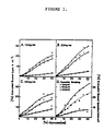
- Figure 1 demonstrates the effect of dilution on [3H]estradiol binding in uterine nuclear fractions from estradiol-17 ⁇ implanted adult ovariectomized rats.
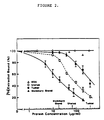
- Figure 2 demonstrates that tumor cytosol preparations contain lower levels of inhibitor activity for nuclear Type II site, than cytosol preparations from uterus or mammary gland.
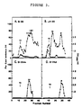
- Figure 3 shows the chromatographic profile of rat uterine cytosol inhibitor preparations on Sephadex G-25 (panels A, C, and D) or LH-20 (panel B) columns.
- Figure 4 demonstrates the chromatographic profile of bovine serum extract on a preparative LH-20 column.
- Figure 5 demonstrates the chromatographic profile of ⁇ and ⁇ Type II ligand by HPLC.
- Figure 6 demonstrates the mass spectra of the trimethylsilyl derivatives of the ⁇ peak (A) purified from fetal bovine serum and authentic p-hydroxyphenyllactic acid (B;HPLA).
- Figure 7 represents the mass spectra of the trimethylsilyl derivatives of the ⁇ peak (A) purified from fetal bovine serum and authentic methyl p-hydroxyphenyllactate (B;MeHPLA).
- Figure 8 demonstrates the competition of type II ligand ⁇ and ⁇ peaks and authentic HPLA and MeHPLA for Type II sites.
- Figure 9 represents the competition of authentic HPLA and MeHPLA for [3H]estradiol binding to nuclear Type II sites.
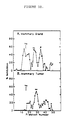
- Figure 10 represents the chromatographic profile of mouse mammary gland (A) and mouse mammary tumor (B) cytosols on LH-20 columns.
- Figure 11 represents the chromatography of human breast cancer cytosol on an LH-20 column.
- Figure 12 demonstrates the effects of authentic HPLA and MeHPLA on MCF-7 human breast cancer cell growth.
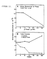
- Figure 13 represents the effects of LH-20 ⁇ peak fractions on the growth of MCF-7 human breast cancer cells in culture.
- Panel A shows the dose response curve for the ⁇ peak from rat liver.
- Panel B shows the time course of inhibition following the addition of 33 ⁇ l/ml of the LH-20 ⁇ peak fractions.
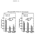
- Figure 14 demonstrates the effects of MeHPLA (A) and HPLA (B) on uterine growth.
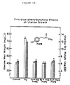
- Figure 15 demonstrates the effects of p-hydroxyphenylbutanone [1-(4-hydroxyphenyl)-3-butanone] on uterine growth.
- MeHPLA methyl p-hydroxyphenyllactate
- chemically related compounds refers to the derivatives and analogues of p-coumaric acid, p-hydroxyphenylbutanone, (4-hydroxyphenoxy)acetate and the arylpropenaldehydes, alkyl arylethenyl ketones, aryl arylethenyl ketones, aryl butenaldehydes, alkyl arylpropenyl ketones and arylpropenyl ketones which are structurally related to MeHPLA and disclosed herein.
- chemically related compounds include the cis and trans isomers of said compounds and their esters, ethers, ketones and derivatives containing S or N in place of O atoms.
- these structurally related analogues and derivatives include compounds where R1 represents the methyl, ethyl, n-propyl, n-butyl, isopropyl, tert-butyl or aryl group and R2 and R3 each represent H, OH or OCH3 groups and R4 is H or an alkyl group of 1 to 6 carbon atoms.
- R1 represents the methyl, ethyl, n-propyl, n-butyl, isopropyl, tert-butyl or aryl group
- R2 and R3 each represent H, OH or OCH3 groups
- R4 is H or an alkyl group of 1 to 6 carbon atoms.
- Specific analogues of each class of these structurally related compounds to MeHPLA have been demonstrated to possess biological activity (Table I) as defined herein and therefore mimic MeHPLA as an effective inhibitor of cell proliferation and tumor cell growth.
- the term "individual” is meant to include animals and humans.
- biologically inhibiting or “inhibition” of the growth of proliferating cells is meant to include partial or total growth inhibition and also is meant to include decreases in the rate of proliferation or growth of the cells.
- the biologically inhibitory dose of the compounds used in the present invention may be determined by assessing the effects of the text compound on malignant cell growth in tissue culture (see Figure 12), uterine growth in the animal (see Figures 14 and 15) or tumor growth in the animal as previously described by Markaverich et al., Cancer Research 43 :3208-3211 (1983), or any other method known to those of ordinary skill in the art.
- prophylactic agent is meant to include agents which may be used for partial or total inhibition or prevention of disease and the spread of disease and also is meant to include agents which may be used as a precaution against disease and for preventive treatment of disease.
- Administration of the compounds useful in the present invention may be by topical, parenteral, oral, intranasal, intravenous, intramuscular, subcutaneous, or any other suitable means.
- the dosage administered is dependent upon the age, weight, kind of concurrent treatment, if any, and nature of the malignancy.
- The-effective compound useful in the present invention may be employed in such forms as capsules, tablets, liquid solutions, suspensions, or elixirs, for oral administration, or sterile liquid forms such as solutions, suspensions or emulsions.
- Any inert carrier is preferably used, such as saline, or phosphate-buffered saline, or any such carrier in which the compounds used in the present invention have suitable solubility properties.
- the compounds used in the present invention may be administered in a biologically effective carrier.
- the biologically effective carriers may include any solvent with which the compounds are compatible and which are non-toxic to the individuals treated at the amounts administered.
- agent is meant to include agents which decrease cell growth, or inhibit the proliferation of tumor cells when administered to said tumor cells in an effective dose.
- MeHPLA antitumor agent
- MeHPLA analogues are derivatives of the general formula
- R1 is CH3, R2 is H, R3 is OH and R4 is H. This is a naturally occurring endogenous compound which was isolated and characterized and identified as the present invention.
- analogues include the compounds in which the R1 group has been replaced by an ethyl, n-propyl, n-butyl, isopropyl, tert-butyl or aryl group; R2 and/or R3 have been replaced with an H, OH or OCH group and R4 is H or an alkyl group of 1 to 6 carbon atoms.
- R1 group has been replaced by an ethyl, n-propyl, n-butyl, isopropyl, tert-butyl or aryl group
- R2 and/or R3 have been replaced with an H, OH or OCH group
- R4 is H or an alkyl group of 1 to 6 carbon atoms.
- Each of these esters can exist in the D and L form.
- Another group of derivative compounds includes those with the formula: Examples of these compounds are p-coumaric acid, 3-(4-hydroxyphenyl)-2-propenoic acid, and its esters. These substances exist as cis and trans isomers.
- R1 and R2 are hydrogen and R3 is OH.
- Additional esters include compounds wherein R1 is methyl, ethyl, n-propyl, n-butyl, isopropyl, tert-butyl or aryl and R2 and/or R3 is a H, OH or OCH3 group.
- Additional analogues include caffeic acid, 3-(3,4-dihydroxyphenyl)-2-propenoic acid, wherein R1 is H and R2 and R3 are both OH. Additionally the methoxy compound is very effective as an antitumor agent.
- Ketone derivatives include compounds with a methyl, ethyl, n-propyl, n-butyl, isopropyl, tert-butyl or aryl group at the R1 position, H, OH or OCH3 group at the R2 and R3 positions; and most preferably H at the R2 position and OH at the R3 position.
- the number of CH2 groups between the aromatic entity and the keto group can be varied.
- Specific examples of compounds are 1-(4-hydroxyphenyl)-3-pentanone and 1-(4-hydroxyphenyl)-3-butanone. These compounds have been shown to bind to Type II sites and to have antitumor proliferative activity in the uterotropic assay.
- ether-linked analogues of (4-hydrozyphenoxy)acetic acid for example, methyl (4-hydroxyphenoxy)acetate. All of these compounds bind to the Type II binding sites.
- ether-linked compounds include analogues wherein R1 is H, a C1 to C6 alkyl carbon chain or an aryl group, R2 and R3 are H, OH or OCH3.
- phenoxy compounds include the ether compounds, for example, 2-(4-hydroxyphenoxy)ethyl methyl ether, wherein R1 can be H or any C1 to C6 alkyl carbon chain or an aryl group, R2 and R3 are H, OH or OCH3 in the formula: Additionally useful is the compound of the formula: wherein R1 is a C1 to C6 alkyl chain or an aryl group, R2 and R3 are H, OH or OCH3.
- R1 is selected from H, alkyl groups containing from 1 to 6 carbon atoms, and aryl groups; and R2 and R3 are each selected from H, OH and OCH3. Where R1 is an aryl group it may be substituted or unsubstituted.
- the present invention provides the use of the above-mentioned compounds for the manufacture of a medicament for the treatment of cancer, for the manufacture of a medicament for inhibiting the growth of proliferating cells, or for treating benign prostatic hyperplasia.
- Preferred compounds of the above group which may be used to practice the present invention are: Most preferred compounds of this group for practicing the present invention are 3-(4-hydroxyphenyl)-1-phenyl-2-propen-1-one and 4-(4-hydroxyphenyl)-3-buten-2-one, analogues, chemical derivatives and chemical related compounds and pharmaceutically acceptable salts thereof.
- Another embodiment of the present invention includes the use, for the manufacture of a medicament for treating cancer of MeHPLA, its analogues, chemical derivatives or chemically related compounds.
- This compound can be any of the above-described antitumor compounds.
- these antitumor agents are also useful as inhibitors of cell growth and proliferation in those cells which include a Type II nuclear estrogen binding site.
- These compounds bind to Type II nuclear estrogen binding sites and regulate cell growth.
- Specific proliferating cells which are sensitive to the binding of these compounds include estrogen responsive tissues such as uterus, mammary gland, uterine tumors and mammary tumors.
- the above-described compounds inhibit the proliferative capacity of human breast cancer cells and thus provide an effective therapy for this disease.
- Benign prostatic hyperplasia is another example of a proliferative tissue disease in which the above-described compounds can successfully be used in the treatment.
- Tissue was dissected from host animals, weighed, rinsed in saline, and homogenized in 10 mM Tris-1.5 mM EDTA, by approximately four 30-sec bursts in a Sorval Omnimixer with intermittent cooling for about 5 min.
- This crude homogenate was transferred to a Kontes ground glass homogenizer and further homogenized with intermittent cooling until no tissue aggregates were visible. It was imperative that the homogenate to kept cold, about 4°C through all homogenisation steps.
- the final homogenate was centrifuged at 800xg for approximately 20 min to obtain the crude nuclear pellet, which was then washed by gentle resuspension in a Dounce homogenizer and then centrifuged at 800xg for approximately 7 min.
- fresh tissue equivalent is meant the amount of material which represents the initial concentration in the fresh uterine tissue.
- rat tissues possess an endogenous ligand which blocks [3H]estradiol binding to nuclear Type II estrogen binding sites; however, this compound does not interfere with [3H]estradiol binding to the estrogen receptor.
- this inhibitor activity was assessed in various tissue cytosols and LH-20 column chromatography fractions from these cytosols.
- Uterine nuclei were prepared from estrogen-implanted, adult-ovariectomized rats. The washed nuclear pellet was diluted to 10 mg of fresh uterine equivalents/ml. At this concentration the effects of the endogenous inhibitor were minimal, and nuclear Type II sites bound maximum quantities of [3H]estradiol.
- Figure 1 shows the effects of the dilution on [3H]estradiol binding in uterine nuclear fractions from estradiol-17 ⁇ implanted adult ovariectomized rats.
- the uterine nuclei were prepared as previously described in Markaverich, B. M. et al., J. Biol. Chem. 258 :11663-11671 (1983).
- uterine nuclei after preparation were diluted in 10 mM Tris, 1.5 mM EDTA buffer in final volumes equivalent to 10 , 20 or 40 mg fresh uterine equivalents per ml, and assayed for estrogen binding sites by [3H]estradiol exchange at 37°C for 30 minutes. Specific binding was determined by subtraction of non-specific binding, i.e., non-competable with 300-fold excess diethylstilbestrol, from the total quantity of [3H]estradiol bound. The results shown in Figure 1, panel D demonstrate the specific binding measured at 10, 20 or 40 mg nuclear equivalents per ml corrected for the dilution effect (pmoles/uterus).
- rat uterine nuclear preparations contain a factor which inhibits [3H]estradiol binding to nuclear Type II sites and that the interaction of this factor with Type II sites is decreased by dilution.
- Using an "inhibition assay" it was found that rat uterine cytosol contained a factor which inhibits [3H]estradiol binding to nuclear Type II sites.
- acid-precipitated, boiled cytosol from rat uterus, mouse mammary tumor and normal mouse mammary gland was diluted as shown on Figure 2 and evaluated for inhibitor activity.
- Figure 2 shows that this factor is very effective and completely inhibits the Type II site at very dilute cytosol concentrations.
- the results represented by Figure 2 represent the mean ⁇ standard error of the mean for triplicate determinations in four replicate experiments for each preparation. This inhibitor activity was stable to acid and was not destroyed by trypsin, proteinase K, RNAse or DNAse.
- the inhibitor responsible for this activity was characterized as methyl 3-(4-hydroxyphenyl)-2-hydroxypropionate.
- the identification and characterization procedure involved chromatography of cytosol preparations on Sephadex G-25 and Sephadex LH-20 columns ( Figure 3). Column fractions were assayed for inhibitory activity by incubating with dilute nuclear fractions from estradiol implanted rats. Under these conditions maximum [3H]estradiol binding to nuclear Type II sites was observed in the absence of inhibitor activity. Sephadex G-25 chromatography resulted in two major peaks ( ⁇ and ⁇ ) of inhibitor activity eluting in the inclusion volume of the column. Chromatography of the same cytosol preparation on Sephadex LH-20 also revealed two peaks showing inhibitor activity.
- the inhibitor molecules for nuclear Type II sites were isolated from bovine serum. Approximately 500 ml of bovine serum were diluted with an equal volume of HPLC grade water and the pH was adjusted to 1.0 with high purity HCl. The acidified serum was extracted three times with equal volumes of ethyl acetate and the pooled ethyl acetate extracts were dried under vacuum at 60°C. Following evaporation, the extract was redissolved in 100 ml of ethyl acetate and washed with an equal volume of acidified HPLC grade water (pH 1.0).
- Binding assays have shown that the mouse mammary tumor and human breast cancer preparations had high levels of free nuclear Type II sites relative to non-malignant tissues.
- Figures 2 and 10 demonstrate that the normal rat mammary glands contain very high levels of total inhibitor activity relative to mouse mammary tumors.
- Figure 11 shows that human breast cancer contains low levels of inhibitor.
- malignant tissues have high levels of free nuclear Type II sites and are deficient in the inhibitor activity. This deficiency in inhibitor activity explains the high levels of free nuclear Type II sites observed in these tumor tissue populations as well as their rapid rate of proliferation, cell growth and DNA synthesis.
- the high inhibitor activity found in the normal tissues was characterized as previously described on the LH-20 column and revealed that the rat uterus and normal mouse mammary gland contained both ⁇ and ⁇ inhibitor peak components. Comparison of the ⁇ and ⁇ peaks seen in the mouse mammary tumors with those observed in normal tissues showed a high correlation between a deficiency in the ⁇ peak component and increased unbound nuclear Type II sites in mouse mammary tumor tissue. In the rat and mouse mammary tumor tissues which have been analyzed, the ⁇ inhibitor component is consistently very low and, in some cases, undetectable. The low to undetectable ⁇ inhibitor level is also observed in human breast cancer tissue.
- MCF-7 human breast cancer cell line in tissue culture was used.
- the MCF-7 cells have both Type I and nuclear Type II estrogen receptor sites and respond in a proliferative fashion to estrogenic hormones. Furthermore, they are inhibited by well-known anti-estrogens such as Tamoxifen.
- the MCF-7 cells were plated at 5 x 10⁵ cells/dish in 30 mm petri dishes and grown in Dulbecco's Modified Eagles Medium containing about 10% charcoal stripped fetal bovine serum for approximately 48 hours.
- the cells attached to the plastic dishes and then underwent exponential growth with a cell-doubling time of approximately 24 hours.
- the plated cells were allowed to attach for approximately 48 hours and the medium was replaced ("day zero").
- the cells were allowed to grow exponentially for about 6 days.
- the cells were treated with doses ranging from 1-10 ⁇ g/ml of the compound of interest, for example, methyl p-hydroxyphenyllactate, in 10 ⁇ l of ethanol.
- the medium was changed at about 2 and 4 days.
- the control and test solutions were also re-added when the medium was changed.
- the cells were harvested, counted on a hemocytometer and DNA content per dish was determined by the Burton assay (Burton, K., Biochem. J. 62 :315-323, 1956). Results are expressed as cells/dish or DNA content ( ⁇ g/dish) at 6 days following treatment. The results are shown in Figure 12.
- characterization and demonstration of the antitumor activity of the ⁇ inhibitor component was seen in examples using rat liver isolates.
- the ⁇ inhibitor material was suspended in 2 ml of tissue culture medium and added to cultures of MCF-7 human breast cancer cell lines.
- the MCF-7 cells were plated in 3 ml of DMEM containing 10% charcoal stripped fetal bovine serum and allowed to attach to the surface of petri dishes. Following the establishment of monolayer cultures, approximately 24 to 36 hours, the floating cells were removed by aspiration, and 3 ml of fresh DMEM were added to each culture. At this time various amounts, ranging from 7 to 32 ⁇ l/ml, of the pooled ⁇ peak fractions or control medium were added to the cultures.
- the cells were cultured for an additional 24 hours. At the end of this culture period, the floating cells were removed by aspiration and the attached cells resuspended in DMEM following mild trypsinization. The cells were then counted on a hemocytometer. With the increasing concentrations of the ⁇ peak inhibitor material, the cell number was reduced in a dose dependent fashion within about 24 hours ( Figure 13A). The control samples did not exhibit a similar decrease. The time course of the inhibition activity was also examined. Approximately 32 ⁇ l of control medium or ⁇ peak inhibitor material was added to the cells and the floating cells were removed. Within approximately four hours of inhibitor addition, the inhibition of cell growth had begun. Maximum inhibition was observed in approximately 12 hours (Figure 13B).
- tumor cell proliferation is very rapid because the tumor cell metabolizes or inactivates the ⁇ peak inhibitor. This is supported by the observation that methyl p-hydroxyphenyllactate is found bound to Type II sites in normal tissues but is not found in malignant tissue.
- Cell proliferation is regulated by ligand binding to nuclear Type II sites. The number of unbound sites determines the rate of proliferation since tumor cells have an increased number of unbound nuclear Type II sites and thus are not inhibited, so tumor cell proliferation is dramatically accelerated. These cells are brought back into regulation and cell proliferation is decreased by administering a therapeutic dose of the inhibitors described in the present invention.
- the rat uterus is exquisitively sensitive to estrogen, and this hormone stimulates uterine cellular hypertrophy, hyperplasia and DNA synthesis within 24 hours following a single injection.
- Estradiol stimulation of nuclear Type II sites is a prerequisite for these responses.
- This assay includes injecting immature female rats with saline-ethanol vehicle, estradiol-17 ⁇ and the test compound of interest. Control rats were injected only with saline-ethanol vehicle and estradiol-17 ⁇ . The rats were sacrificed 24 hours later and the uterine wet and dry weights were determined. The wet and dry weight measurements are well defined biochemical end points of estrogen action and are a direct index of changes in cell proliferation and DNA synthesis.
Landscapes
- Health & Medical Sciences (AREA)
- Chemical & Material Sciences (AREA)
- Public Health (AREA)
- Veterinary Medicine (AREA)
- Medicinal Chemistry (AREA)
- Pharmacology & Pharmacy (AREA)
- Life Sciences & Earth Sciences (AREA)
- Animal Behavior & Ethology (AREA)
- General Health & Medical Sciences (AREA)
- Organic Chemistry (AREA)
- Epidemiology (AREA)
- Nuclear Medicine, Radiotherapy & Molecular Imaging (AREA)
- General Chemical & Material Sciences (AREA)
- Chemical Kinetics & Catalysis (AREA)
- Engineering & Computer Science (AREA)
- Bioinformatics & Cheminformatics (AREA)
- Emergency Medicine (AREA)
- Endocrinology (AREA)
- Reproductive Health (AREA)
- Urology & Nephrology (AREA)
- Pharmaceuticals Containing Other Organic And Inorganic Compounds (AREA)
- Acyclic And Carbocyclic Compounds In Medicinal Compositions (AREA)
- Medicines That Contain Protein Lipid Enzymes And Other Medicines (AREA)
Abstract
Description
- This invention relates to the use of new and useful compounds which include methyl p-hydroxyphenyllactate (MeHPLA), its analogues, chemical derivatives and chemically related compounds for the manufacture of agents and inhibitors of proliferative cell growth.
- There are two types of nuclear estrogen binding sites in normal and malignant tissues. Type I sites represent the classical estrogen receptor and nuclear Type II sites appear to mediate a specific nuclear response to estrogenic hormones. After estrogen administration, Type I receptor sites bind estradiol and this receptor-estrogen complex interacts with nuclear acceptor sites before the initiation of the transcriptional events that are associated with estrogen stimulation of tissue growth. In contrast, Type II sites bind estrogen with a higher capacity and a lower affinity than the classical estrogen receptor and do not appear to be translocated from the cytoplasm to the nucleus. Thus, although the levels of nuclear Type II sites are increased by estrogen administration, Type II sites remain in the cytoplasm after hormone administration. Nuclear Type II sites appear to mediate a specific nuclear response to estrogenic hormones and are highly correlated with uterine cellular hypertrophy and hyperplasia. Additionally, nuclear Type II sites are highly stimulated in malignant tissues such as mouse mammary tumors and human breast cancer. This observation is consistent with the findings that highly proliferative tissue has an increased number of nuclear Type II sites. Because the stimulation of nuclear Type II sites is closely correlated with the stimulation of uterine growth, it has been postulated that the Type II sites are the location for the mechanisms by which estrogens cause uterotropic stimulation. Furthermore, the presence of Type II sites on the nuclear matrix suggests a potential role in the regulation of DNA synthesis.
- Nuclear Type II sites are constituents of many, if not all, non-malignant cells. Normally, Type II sites in non-malignant cells are occupied by methyl p-hydroxyphenyllactate (MeHPLA or methyl 3-(4-hydroxyphenyl)-2-hydroxypropionate). When MeHPLA binds to Type II sites, cell growth and proliferation of non-malignant tissues are slowed down or stopped. Conversely, malignant cells metabolise MeHPLA and thus there is insufficient binding to the nuclear Type II sites and the regulation of cell growth and proliferation is lost. Consequently all tumor cell populations examined have very high levels of unbound nuclear Type II sites. These sites should represent targets for the analogues of MeHPLA.
- This invention makes use of compounds which are not metabolized by malignant cells but which bind to nuclear Type II sites with high affinity. These compounds are very effective inhibitors of tumor cell proliferation and DNA synthesis. Since nuclear Type II sites have been observed in a variety of tumor cells, it is likely that analogues of MeHPLA and chemically related compounds will be effective inhibitors of a broad spectrum of tumors. Consequently, any tumor which contains nuclear Type II sites should respond to treatment with MeHPLA, its analogues, derivatives and chemically related compounds, including cancers of the pancreas, cervix, liver, brain, pituitary, prostate and other organ or tissue sites, as well as other cancers, such as leukemias, lymphomas, stromal myomas and leiomyomas, among others. Since MeHPLA will also block estrogen stimulation of normal cell growth such as that in the rat uterus (Table I), analogues of this compound may also be useful for the treatment of uterine hyperplasia, cervical hyperplasia, endometriosis and benign prostatic hypertrophy. Because non-proliferating non-malignant cells normally have their Type II sites bound with MeHPLA, the effects of the proposed compounds on non-malignant cell populations will be minimal to non-existent. For this reason, MeHPLA, its analogues, derivatives and chemically related compounds are also useful as propylactic agents in the inhibition and prevention of cancer and non-malignant cell growth.
- The precise physiological role of Type II sites is unknown, but inhibition of the nuclear Type II sites is associated with antagonism of uterotropic responses to estrogen. This is true for steroid antagonists such as dexamethasone, progesterone and triphenylethylene derivatives such as nafoxidine and clomiphene. While there is at least one endogenous inhibitor of estradiol binding to nuclear Type II sites, the specific inhibitor for the nuclear Type II sites has not been identified previous to this invention. Furthermore, the inhibitor of the present invention is specific to nuclear Type II sites and does not interfere with estradiol binding to cytoplasmic or nuclear Type I estrogen receptors.
- The inhibitor is identified as methyl 3-(4-hydroxyphenyl)-2-hydroxypropionate and is an important regulator of cell growth and proliferation in normal and malignant tissues. The inhibitor is also known as methyl p-hydroxyphenyllactate or MeHPLA. These terms may be used interchangeably. Cell growth inhibition by this compound resides in its ability to interact with the high-affinity nuclear binding sites in normal and malignant cells which may be involved in the regulation of cellular proliferation and DNA synthesis. When MeHPLA is bound to nuclear Type II sites, cell growth and proliferation are inhibited. The endogenous 3-(4-hydroxyphenyl)-2-hydroxypropionic acid inhibits the cell growth much less effectively. This activity correlates with its low binding affinity for nuclear Type II sites.
- Since MeHPLA is a normal constituent of mammalian cells, but metabolized by malignant cells, MeHPLA, structural analogues and chemically related compounds as described herein, which are not metabolized by tumors should be useful in the prevention of malignancy. These compounds should possess little if any side effects, and if taken in a low level maintenance dose should inhibit the proliferation of malignant cells.
Because MePHLA is such a potent inhibitor of cell growth, this compound, as well as its analogues and chemically related compounds were used as potential antitumor agents. The present invention utilizes the potent antitumor activity of these compounds. - An object of the present invention is to provide for the preparation of a medicament for the treatment of cancer.
- An additional object of the present invention is to provide for the preparation of a medicament for the inhibition of the growth of proliferating cells which include a Type II nuclear estrogen binding site, such as estrogen responsive tissues.
- An additional object of the present invention is to provide for the preparation of a medicament for the treatment of human breast cancer, and other malignancies which contain unbound nuclear Type II sites.
- Another object of the present invention is to provide for the preparation of a medicament for the treatment of benign prostatic hyperplasia, cervical hyperplasia, uterine hyperplasia and endometriosis.
- Thus, there is provided in accordance with one aspect of the present invention the use of methyl p-hydroxyphenyllactate and pharmaceutically acceptable salts thereof for the preparation of a medicament for the treatment of cancer. Also provided is the use of a compound for the preparation of a medicament for the treatment of cancer wherein said compound has a formula selected from

wherein R₁ is selected from H, alkyl groups containing from 1 to 6 carbon atoms, and aryl groups; R₂ and R₃ are each selected from H, OH and OCH₃ and R₄ is H or alkyl group containing from 1 to 6 carbon atoms. Preferred compounds which may be used to practice the present invention may be selected from
methyl 3-(4-hydroxyphenyl)-2-hydroxypropionate,
n-propyl 3-(4-hydroxyphenyl)-2-hydroxypropionate,
n-butyl 3-(4-hydroxyphenyl)-2-hydroxypropionate,
3-(4-hydroxyphenyl)-2-propenoic acid,
4-(4-hydroxyphenyl)-2-butanone,
1-(4-hydroxyphenyl)-3-pentanone,
methyl (4-hydroxyphenoxy)acetate, and
methyl 3-(3,4-dihydroxyphenyl)-2-propenoate. - The present invention also provides the use of methyl p-hydroxyphenyllactate for the preparation of a medicament for the inhibition of the growth of proliferating cells which include a Type II nuclear estrogen binding site. Further, the invention provides the use of a compound for the preparation of a medicament for the inhibition of the growth of proliferating cells which include a Type II nuclear estrogen binding site wherein said compound has a formula selected from

wherein R₁ is selected from H, alkyl groups containing from 1 to 6 carbon atoms, and aryl groups; R₂ and R₃ are each selected from H, OH and OCH₃ and R₄ is H or alkyl group containing from 1 to 6 carbon atoms. Preferred compounds which may be used to practice the present invention may be selected from
methyl 3-(4-hydroxyphenyl)-2-hydroxypropionate,
n-propyl 3-(4-hydroxyphenyl)-2-hydroxypropionate,
n-butyl 3-(4-hydroxyphenyl)-2-hydroxypropionate,
3-(4-hydroxyphenyl)-2-propenoic acid,
4-(4-hydroxyphenyl)-2-butanone,
1-(4-hydroxyphenyl)-3-pentanone,
methyl (4-hydroxyphenoxy)acetate, and
methyl 3-(3,4-dihydroxyphenyl)-2-propenoate. - An additional aspect of the present invention relates to the inhibition of the proliferative growth of estrogen responsive tissues such as uterus, mammary gland, uterine tumors and mammary tumors. In one specific aspect, the above-mentioned compounds have been used for the treatment of human breast cancer cells. The compounds inhibit the growth of human breast cancer cells.
- Another specific aspect of the present invention provides the use of the above-mentioned compounds for the preparation of a medicament for the treatment of benign prostatic hyperplasia.
- Another aspect of the present invention provides the use of the above-mentioned compounds for the preparation of a medicament for use as an antitumor agent.
- Other and further objects, features and advantages will be apparent from the following description of the presently preferred embodiments of the invention given for the purpose of disclosure.
- Figure 1 demonstrates the effect of dilution on [³H]estradiol binding in uterine nuclear fractions from estradiol-17 β implanted adult ovariectomized rats.
- Figure 2 demonstrates that tumor cytosol preparations contain lower levels of inhibitor activity for nuclear Type II site, than cytosol preparations from uterus or mammary gland.
- Figure 3 shows the chromatographic profile of rat uterine cytosol inhibitor preparations on Sephadex G-25 (panels A, C, and D) or LH-20 (panel B) columns.
- Figure 4 demonstrates the chromatographic profile of bovine serum extract on a preparative LH-20 column.
- Figure 5 demonstrates the chromatographic profile of α and β Type II ligand by HPLC.
- Figure 6 demonstrates the mass spectra of the trimethylsilyl derivatives of the α peak (A) purified from fetal bovine serum and authentic p-hydroxyphenyllactic acid (B;HPLA).
- Figure 7 represents the mass spectra of the trimethylsilyl derivatives of the β peak (A) purified from fetal bovine serum and authentic methyl p-hydroxyphenyllactate (B;MeHPLA).
- Figure 8 demonstrates the competition of type II ligand α and β peaks and authentic HPLA and MeHPLA for Type II sites.
- Figure 9 represents the competition of authentic HPLA and MeHPLA for [³H]estradiol binding to nuclear Type II sites.
- Figure 10 represents the chromatographic profile of mouse mammary gland (A) and mouse mammary tumor (B) cytosols on LH-20 columns.
- Figure 11 represents the chromatography of human breast cancer cytosol on an LH-20 column.
- Figure 12 demonstrates the effects of authentic HPLA and MeHPLA on MCF-7 human breast cancer cell growth.
- Figure 13 represents the effects of LH-20 β peak fractions on the growth of MCF-7 human breast cancer cells in culture. Panel A shows the dose response curve for the β peak from rat liver. Panel B shows the time course of inhibition following the addition of 33 µl/ml of the LH-20 β peak fractions.
- Figure 14 demonstrates the effects of MeHPLA (A) and HPLA (B) on uterine growth.
- Figure 15 demonstrates the effects of p-hydroxyphenylbutanone [1-(4-hydroxyphenyl)-3-butanone] on uterine growth.
- "Methyl 3-(4-hydroxyphenyl)-2-hydroxypropionate" is also known as methyl p-hydroxyphenyllactate or MeHPLA. The term "MeHPLA" is meant to also include its analogues, chemical derivatives, and chemically related compounds which bind to the nuclear Type II receptors and by so doing inhibit cell proliferation.
- The term "chemically related compounds" refers to the derivatives and analogues of p-coumaric acid, p-hydroxyphenylbutanone, (4-hydroxyphenoxy)acetate and the arylpropenaldehydes, alkyl arylethenyl ketones, aryl arylethenyl ketones, aryl butenaldehydes, alkyl arylpropenyl ketones and arylpropenyl ketones which are structurally related to MeHPLA and disclosed herein. These chemically related compounds include the cis and trans isomers of said compounds and their esters, ethers, ketones and derivatives containing S or N in place of O atoms. More specifically these structurally related analogues and derivatives include compounds where R₁ represents the methyl, ethyl, n-propyl, n-butyl, isopropyl, tert-butyl or aryl group and R₂ and R₃ each represent H, OH or OCH₃ groups and R₄ is H or an alkyl group of 1 to 6 carbon atoms. Specific analogues of each class of these structurally related compounds to MeHPLA have been demonstrated to possess biological activity (Table I) as defined herein and therefore mimic MeHPLA as an effective inhibitor of cell proliferation and tumor cell growth.
- The term "individual" is meant to include animals and humans.
- The term "biologically inhibiting" or "inhibition" of the growth of proliferating cells is meant to include partial or total growth inhibition and also is meant to include decreases in the rate of proliferation or growth of the cells. The biologically inhibitory dose of the compounds used in the present invention may be determined by assessing the effects of the text compound on malignant cell growth in tissue culture (see Figure 12), uterine growth in the animal (see Figures 14 and 15) or tumor growth in the animal as previously described by Markaverich et al., Cancer Research 43:3208-3211 (1983), or any other method known to those of ordinary skill in the art.
- The term "prophylactic agent" is meant to include agents which may be used for partial or total inhibition or prevention of disease and the spread of disease and also is meant to include agents which may be used as a precaution against disease and for preventive treatment of disease.
- Administration of the compounds useful in the present invention may be by topical, parenteral, oral, intranasal, intravenous, intramuscular, subcutaneous, or any other suitable means. The dosage administered is dependent upon the age, weight, kind of concurrent treatment, if any, and nature of the malignancy. The-effective compound useful in the present invention may be employed in such forms as capsules, tablets, liquid solutions, suspensions, or elixirs, for oral administration, or sterile liquid forms such as solutions, suspensions or emulsions. Any inert carrier is preferably used, such as saline, or phosphate-buffered saline, or any such carrier in which the compounds used in the present invention have suitable solubility properties.
- The compounds used in the present invention may be administered in a biologically effective carrier. The biologically effective carriers may include any solvent with which the compounds are compatible and which are non-toxic to the individuals treated at the amounts administered.
- The term "antitumor agent" is meant to include agents which decrease cell growth, or inhibit the proliferation of tumor cells when administered to said tumor cells in an effective dose.
- One specific embodiment of this invention is an antitumor agent including MeHPLA, its analogues, chemical derivatives or chemically related compounds. Specific examples of MeHPLA analogues are derivatives of the general formula

One specific example of this type of compound is methyl 3-(4-hydroxyphenyl)-2-hydroxypropionate wherein R₁ is CH₃, R₂ is H, R₃ is OH and R₄ is H. This is a naturally occurring endogenous compound which was isolated and characterized and identified as the present invention. Other examples of analogues include the compounds in which the R₁ group has been replaced by an ethyl, n-propyl, n-butyl, isopropyl, tert-butyl or aryl group; R₂ and/or R₃ have been replaced with an H, OH or OCH group and R₄ is H or an alkyl group of 1 to 6 carbon atoms. Each of these esters can exist in the D and L form. - Another group of derivative compounds includes those with the formula:

Examples of these compounds are p-coumaric acid, 3-(4-hydroxyphenyl)-2-propenoic acid, and its esters. These substances exist as cis and trans isomers. In coumaric acid R₁ and R₂ are hydrogen and R₃ is OH. Additional esters include compounds wherein R₁ is methyl, ethyl, n-propyl, n-butyl, isopropyl, tert-butyl or aryl and R₂ and/or R₃ is a H, OH or OCH₃ group. Additional analogues include caffeic acid, 3-(3,4-dihydroxyphenyl)-2-propenoic acid, wherein R₁ is H and R₂ and R₃ are both OH. Additionally the methoxy compound is very effective as an antitumor agent. - Other compounds with antitumor activity are the derivatives of 1-(4-hydroxyphenyl)-3-butanone, such as compounds with the formulae:

Ketone derivatives include compounds with a methyl, ethyl, n-propyl, n-butyl, isopropyl, tert-butyl or aryl group at the R₁ position, H, OH or OCH₃ group at the R₂ and R₃ positions; and most preferably H at the R₂ position and OH at the R₃ position. - Additionally, as can be seen by the formulae, the number of CH₂ groups between the aromatic entity and the keto group can be varied. Specific examples of compounds are 1-(4-hydroxyphenyl)-3-pentanone and 1-(4-hydroxyphenyl)-3-butanone. These compounds have been shown to bind to Type II sites and to have antitumor proliferative activity in the uterotropic assay.
- Another group of compounds which show antitumor proliferative activity in the rat uterus is described by the formula:

These compounds are ether-linked analogues of (4-hydrozyphenoxy)acetic acid, for example, methyl (4-hydroxyphenoxy)acetate. All of these compounds bind to the Type II binding sites. These ether-linked compounds include analogues wherein R₁ is H, a C₁ to C₆ alkyl carbon chain or an aryl group, R₂ and R₃ are H, OH or OCH₃. An additional variation on the phenoxy compounds include the ether compounds, for example, 2-(4-hydroxyphenoxy)ethyl methyl ether, wherein R₁ can be H or any C₁ to C₆ alkyl carbon chain or an aryl group, R₂ and R₃ are H, OH or OCH₃ in the formula:
Additionally useful is the compound of the formula:
wherein R₁ is a C₁ to C₆ alkyl chain or an aryl group, R₂ and R₃ are H, OH or OCH₃. - Another group of compounds which show tumor anti-proliferative action is described by the general formulas:

wherein R₁ is selected from H, alkyl groups containing from 1 to 6 carbon atoms, and aryl groups; and R₂ and R₃ are each selected from H, OH and OCH₃. Where R₁ is an aryl group it may be substituted or unsubstituted. Thus, the present invention provides the use of the above-mentioned compounds for the manufacture of a medicament for the treatment of cancer, for the manufacture of a medicament for inhibiting the growth of proliferating cells, or for treating benign prostatic hyperplasia. Preferred compounds of the above group which may be used to practice the present invention are:
Most preferred compounds of this group for practicing the present invention are 3-(4-hydroxyphenyl)-1-phenyl-2-propen-1-one and 4-(4-hydroxyphenyl)-3-buten-2-one, analogues, chemical derivatives and chemical related compounds and pharmaceutically acceptable salts thereof. Another embodiment of the present invention includes the use, for the manufacture of a medicament for treating cancer of MeHPLA, its analogues, chemical derivatives or chemically related compounds. This compound, can be any of the above-described antitumor compounds. In addition to being used in medicaments for the treatment of cancer, these antitumor agents are also useful as inhibitors of cell growth and proliferation in those cells which include a Type II nuclear estrogen binding site. These compounds bind to Type II nuclear estrogen binding sites and regulate cell growth. Specific proliferating cells which are sensitive to the binding of these compounds include estrogen responsive tissues such as uterus, mammary gland, uterine tumors and mammary tumors. The above-described compounds inhibit the proliferative capacity of human breast cancer cells and thus provide an effective therapy for this disease. - Benign prostatic hyperplasia is another example of a proliferative tissue disease in which the above-described compounds can successfully be used in the treatment.
- Tissue was dissected from host animals, weighed, rinsed in saline, and homogenized in 10 mM Tris-1.5 mM EDTA, by approximately four 30-sec bursts in a Sorval Omnimixer with intermittent cooling for about 5 min. This crude homogenate was transferred to a Kontes ground glass homogenizer and further homogenized with intermittent cooling until no tissue aggregates were visible. It was imperative that the homogenate to kept cold, about 4°C through all homogenisation steps. The final homogenate was centrifuged at 800xg for approximately 20 min to obtain the crude nuclear pellet, which was then washed by gentle resuspension in a Dounce homogenizer and then centrifuged at 800xg for approximately 7 min. The final washed nuclear pellet was resuspended in 10 mM Tris-1.5 mM EDTA in a concentration ranging from 5 to 50 mg of fresh tissue equivalents/ml. By the term "fresh tissue equivalent" is meant the amount of material which represents the initial concentration in the fresh uterine tissue.
- To assess total and nonspecific binding, aliquots of the nuclear suspension were incubated at 37°C for 30 min with various concentrations ranging from 2 to 40 nM of [³H]estradiol with and without a large molar excess of diethylstilbestrol. The diethylstilbestrol concentration ranged from 0.6 to 12.0 µM. Following incubation, the nuclear pellets were resuspended in about 1 ml of 10mM Tris-1.5mM EDTA buffer and centrifuged at 800xg for about 7 min. The pellet was extracted with 1 ml of ethanol at about 30°C for about 30 min, and counted in 4 ml of Liquifluor. Specific binding was determined by subtraction of nonspecific binding; i.e., not competable by diethylstilbestrol from the total quantity of [³H]estradiol bound. Results were expressed as sites/cell, assuming 8 pg of DNA/cell nucleus and that each binding site binds one molecule of [³H]estradiol.
- A variety of rat tissues possess an endogenous ligand which blocks [³H]estradiol binding to nuclear Type II estrogen binding sites; however, this compound does not interfere with [³H]estradiol binding to the estrogen receptor. In order to assess the levels of this inhibitor activity in various tissue cytosols and LH-20 column chromatography fractions from these cytosols, the following inhibition assay was used. Uterine nuclei were prepared from estrogen-implanted, adult-ovariectomized rats. The washed nuclear pellet was diluted to 10 mg of fresh uterine equivalents/ml. At this concentration the effects of the endogenous inhibitor were minimal, and nuclear Type II sites bound maximum quantities of [³H]estradiol. Aliquots of these nuclei and the inhibitor sample were incubated at about 4°C for approximately 60 min in the presence of 40 nM of [³H]estradiol with and without 12 µM diethylstilbestrol. Under these conditions, nuclear Type II sites were quantitatively measured without interference from Type I sites. The nuclear pellets were resuspended in 1 ml of 10 mM Tris-1.5 mM EDTA and centrifuged, ethanol extracted, and counted. The results were expressed as the percentage of [³H]estradiol bound as compared to the buffer control, or as the percentage of inhibition where 100% bound was 0% inhibition and was equivalent to approximately 45,000 cpm.
- The numbers of nuclear Type II sites measured in the binding assay increased significantly, four-to eight-fold, with nuclear dilution. Thus, more nuclear Type II sites are measured in dilute than in concentrated nuclei. Figure 1 shows the effects of the dilution on [³H]estradiol binding in uterine nuclear fractions from estradiol-17β implanted adult ovariectomized rats. The uterine nuclei were prepared as previously described in Markaverich, B. M. et al., J. Biol. Chem. 258:11663-11671 (1983). Briefly, uterine nuclei after preparation were diluted in 10 mM Tris, 1.5 mM EDTA buffer in final volumes equivalent to 10 , 20 or 40 mg fresh uterine equivalents per ml, and assayed for estrogen binding sites by [³H]estradiol exchange at 37°C for 30 minutes. Specific binding was determined by subtraction of non-specific binding, i.e., non-competable with 300-fold excess diethylstilbestrol, from the total quantity of [³H]estradiol bound. The results shown in Figure 1, panel D demonstrate the specific binding measured at 10, 20 or 40 mg nuclear equivalents per ml corrected for the dilution effect (pmoles/uterus). These results suggest that rat uterine nuclear preparations contain a factor which inhibits [³H]estradiol binding to nuclear Type II sites and that the interaction of this factor with Type II sites is decreased by dilution. Using an "inhibition assay" it was found that rat uterine cytosol contained a factor which inhibits [³H]estradiol binding to nuclear Type II sites. Briefly, acid-precipitated, boiled cytosol from rat uterus, mouse mammary tumor and normal mouse mammary gland was diluted as shown on Figure 2 and evaluated for inhibitor activity. Figure 2 shows that this factor is very effective and completely inhibits the Type II site at very dilute cytosol concentrations. The results represented by Figure 2 represent the mean ± standard error of the mean for triplicate determinations in four replicate experiments for each preparation. This inhibitor activity was stable to acid and was not destroyed by trypsin, proteinase K, RNAse or DNAse.
- The inhibitor responsible for this activity was characterized as methyl 3-(4-hydroxyphenyl)-2-hydroxypropionate. The identification and characterization procedure involved chromatography of cytosol preparations on Sephadex G-25 and Sephadex LH-20 columns (Figure 3). Column fractions were assayed for inhibitory activity by incubating with dilute nuclear fractions from estradiol implanted rats. Under these conditions maximum [³H]estradiol binding to nuclear Type II sites was observed in the absence of inhibitor activity. Sephadex G-25 chromatography resulted in two major peaks (α and β) of inhibitor activity eluting in the inclusion volume of the column. Chromatography of the same cytosol preparation on Sephadex LH-20 also revealed two peaks showing inhibitor activity. When the LH-20 peaks were individually rechromatographed on the Sephadex G-25, the LH-20 α and β peaks eluted in positions identical to the components previously seen on the Sephadex G-25. Further characterization of the compounds with the inhibition assay demonstrated that the β compound was much more inhibitory than was the α component and thus is a preferred compound for use in cell growth regulation.
- The inhibitor molecules for nuclear Type II sites were isolated from bovine serum. Approximately 500 ml of bovine serum were diluted with an equal volume of HPLC grade water and the pH was adjusted to 1.0 with high purity HCl. The acidified serum was extracted three times with equal volumes of ethyl acetate and the pooled ethyl acetate extracts were dried under vacuum at 60°C. Following evaporation, the extract was redissolved in 100 ml of ethyl acetate and washed with an equal volume of acidified HPLC grade water (pH 1.0). This ethyl acetate fraction was then dried under vacuum, redissolved in the buffer, and chromatographed on a preparative LH-20 column to separate the α and β components (Figure 4). The column fractions were collected and α and β peaks were separately pooled, adjusted to pH 1.0, extracted with ethyl acetate, dried, and further purified by HPLC on an Ultrasphere-octyl (C8) reversed phase column (Figure 5). Structural identification was determined by gas chromatography-mass spectrometry of the trimethylsilyl derivatives of the α and β peak components (Figures 6 and 7). The a peak component mass spectrum was identical to that of HPLA (Figure 6). The identity was confirmed with known authentic samples of p-hydroxyphenyllactic acid. The β peak component mass spectrum was identified as methyl p-hydroxyphenyllactate (Figure 7).
- Once the peaks were identified, the relative binding with nuclear Type II sites was more precisely determined by competition analysis with known amounts of the two authentic compounds. From Figures 8 and 9 it can be seen that MeHPLA is approximately 20-fold more effective in inhibiting [³H]estradiol binding to nuclear Type II sites than the free acid (HPLA).
- Binding assays have shown that the mouse mammary tumor and human breast cancer preparations had high levels of free nuclear Type II sites relative to non-malignant tissues. Figures 2 and 10 demonstrate that the normal rat mammary glands contain very high levels of total inhibitor activity relative to mouse mammary tumors. Figure 11 shows that human breast cancer contains low levels of inhibitor. Thus, the evidence shows that malignant tissues have high levels of free nuclear Type II sites and are deficient in the inhibitor activity. This deficiency in inhibitor activity explains the high levels of free nuclear Type II sites observed in these tumor tissue populations as well as their rapid rate of proliferation, cell growth and DNA synthesis. The high inhibitor activity found in the normal tissues was characterized as previously described on the LH-20 column and revealed that the rat uterus and normal mouse mammary gland contained both α and β inhibitor peak components. Comparison of the α and β peaks seen in the mouse mammary tumors with those observed in normal tissues showed a high correlation between a deficiency in the β peak component and increased unbound nuclear Type II sites in mouse mammary tumor tissue. In the rat and mouse mammary tumor tissues which have been analyzed, the β inhibitor component is consistently very low and, in some cases, undetectable. The low to undetectable β inhibitor level is also observed in human breast cancer tissue. Examination of a variety of normal tissues, such as rat serum, spleen, diaphragm, liver and uterus, however, shows the presence of both the α and β inhibitor peak components. These results demonstrate that the active inhibitor component, with respect to the regulation of cell proliferation, is the β inhibitor peak component.
- To assay for tumor growth sensitivity the MCF-7 human breast cancer cell line in tissue culture was used. One skilled in the art will recognize that this is an excellent model system to assess the effects of hormones and drugs on human cancer cell growth and proliferation. The MCF-7 cells have both Type I and nuclear Type II estrogen receptor sites and respond in a proliferative fashion to estrogenic hormones. Furthermore, they are inhibited by well-known anti-estrogens such as Tamoxifen. The MCF-7 cells were plated at 5 x 10⁵ cells/dish in 30 mm petri dishes and grown in Dulbecco's Modified Eagles Medium containing about 10% charcoal stripped fetal bovine serum for approximately 48 hours. During this interval, the cells attached to the plastic dishes and then underwent exponential growth with a cell-doubling time of approximately 24 hours. The plated cells were allowed to attach for approximately 48 hours and the medium was replaced ("day zero"). The cells were allowed to grow exponentially for about 6 days. At day zero the cells were treated with doses ranging from 1-10 µg/ml of the compound of interest, for example, methyl p-hydroxyphenyllactate, in 10 µl of ethanol. The medium was changed at about 2 and 4 days. The control and test solutions were also re-added when the medium was changed. On day 6 the cells were harvested, counted on a hemocytometer and DNA content per dish was determined by the Burton assay (Burton, K., Biochem. J. 62:315-323, 1956). Results are expressed as cells/dish or DNA content (µg/dish) at 6 days following treatment. The results are shown in Figure 12.
- Cultures of rat uterine or mammary tumor cells with cytosol preparations from rat uterus or liver containing α and β components showed cell growth inhibition within about 24 hours. Conversely, when the cells were cultured with cytosol preparations from mouse mammary tumors which contained the a but not the β component, there was no substantial inhibition of cell growth across the wide range of concentrations tested. These data, consistent with the results of the competition experiments shown on Figures 8 and 9, demonstrated that the cytosol preparations from a normal mammary gland contain 15- to 20-fold more total inhibitor activity than those from tumors (Figure 2). One major difference between cytosol preparations from normal and malignant tissues was that the malignant tissues were deficient in the β peak inhibitor component (Figure 10). Hence, the β peak component was the most important compound with respect to potential regulation of cell proliferation. As described in the present invention the β peak component was identified as methyl p-hydroxyphenyllactate.
- Further, characterization and demonstration of the antitumor activity of the β inhibitor component was seen in examples using rat liver isolates. After the isolation, the β inhibitor material was suspended in 2 ml of tissue culture medium and added to cultures of MCF-7 human breast cancer cell lines. The MCF-7 cells were plated in 3 ml of DMEM containing 10% charcoal stripped fetal bovine serum and allowed to attach to the surface of petri dishes. Following the establishment of monolayer cultures, approximately 24 to 36 hours, the floating cells were removed by aspiration, and 3 ml of fresh DMEM were added to each culture. At this time various amounts, ranging from 7 to 32 µl/ml, of the pooled β peak fractions or control medium were added to the cultures. The cells were cultured for an additional 24 hours. At the end of this culture period, the floating cells were removed by aspiration and the attached cells resuspended in DMEM following mild trypsinization. The cells were then counted on a hemocytometer. With the increasing concentrations of the β peak inhibitor material, the cell number was reduced in a dose dependent fashion within about 24 hours (Figure 13A). The control samples did not exhibit a similar decrease. The time course of the inhibition activity was also examined. Approximately 32 µl of control medium or β peak inhibitor material was added to the cells and the floating cells were removed. Within approximately four hours of inhibitor addition, the inhibition of cell growth had begun. Maximum inhibition was observed in approximately 12 hours (Figure 13B).
- The effects of inhibition were reversible. It took approximately 24 hours for the cells to recover and about 7 to 24 days after the removal of the β peak inhibitor to regrow to a full monolayer.
- It is postulated that tumor cell proliferation is very rapid because the tumor cell metabolizes or inactivates the β peak inhibitor. This is supported by the observation that methyl p-hydroxyphenyllactate is found bound to Type II sites in normal tissues but is not found in malignant tissue. Cell proliferation is regulated by ligand binding to nuclear Type II sites. The number of unbound sites determines the rate of proliferation since tumor cells have an increased number of unbound nuclear Type II sites and thus are not inhibited, so tumor cell proliferation is dramatically accelerated. These cells are brought back into regulation and cell proliferation is decreased by administering a therapeutic dose of the inhibitors described in the present invention.
- In addition to testing purified biological preparations, the authentic compounds, methyl p-hydroxyphenyllactate and hydroxyphenyllactic acid, were added to cell cultures. As can be seen in Figure 12, methyl p-hydroxyphenyllactate inhibited MCF-7 human breast cancer cells, but hydroxyphenyllactic acid did not. Furthermore, it can be seen that the methyl p-hydroxyphenyllactate inhibited MCF-7 breast cancer cells in a similar dose-dependent fashion. Results obtained by using the authentic compound were in agreement with the data from biological preparations.
- The rat uterus is exquisitively sensitive to estrogen, and this hormone stimulates uterine cellular hypertrophy, hyperplasia and DNA synthesis within 24 hours following a single injection. Estradiol stimulation of nuclear Type II sites is a prerequisite for these responses. This assay includes injecting immature female rats with saline-ethanol vehicle, estradiol-17β and the test compound of interest. Control rats were injected only with saline-ethanol vehicle and estradiol-17β. The rats were sacrificed 24 hours later and the uterine wet and dry weights were determined. The wet and dry weight measurements are well defined biochemical end points of estrogen action and are a direct index of changes in cell proliferation and DNA synthesis.
- The results of these experiments with various compounds are shown in Table I.
- Additional in vivo measurements using the uterotropic assay show the utility of these compounds for inhibiting cell proliferation. Low doses of methyl p-hydroxyphenyllactate, but not hydroxyphenyllactic acid, block estradiol stimulation of uterine growth in the immature rat (Figure 14 and Table I). However, higher doses of hydroxyphenyllactic acid showed some partial antagonism. This is not surprising since it is known that hydroxyphenyllactic acid binds to nuclear Type II sites with a 20-fold lower affinity than methyl p-hydroxyphenyllactate.
- These results demonstrate that the bound/unbound ratio of nuclear Type II sites is important in the regulation of cell growth and that the primary bound inhibitor in normal cells is methyl p-hydroxyphenyllactate. Since the data demonstrated that tumor cells have the ability to inactivate methyl p-hydroxyphenyllactate, the analogues described in this invention were synthesized to avoid this inactivation. Thus, as can be seen in Table I, administration of compounds with various side-chains and various substituents on the aromatic ring resulted in inhibition of uterine growth. One compound 1-(4-hydroxyphenyl)-3-butanone (p-hydroxyphenylbutanone) which includes a C-terminal methyl group is not subject to the esterase cleavage since the methyl group is attached by a C-C bond. This compound is more stable and thus a better inhibitor in culture and in vivo. Furthermore, experiments with 1-(4-hydroxyphenyl)-3-butanone demonstrated that it binds to the nuclear Type II sites with a high affinity and blocks estradiol stimulation of uterine growth when injected into immature rats (Figure 15). Thus 1-(4-hydroxyphenyl)-3-butanone is an effective inhibitor of tumor growth and regulator of cell proliferation.
Table I MeHPLA Analogue and Related Compound Effects on Uterine Growth and Nuclear Type II Site Binding Inhibition COMPOUND GROWTHa INHIBITION (%) TYPE IIb INHIBITION methyl 3-(4-hydroxyphenyl) 2-hydroxypropionate 90 0.8 3-(4-hydroxyphenyl)-2- hydroxypropionic acid 0 80.0 1-(4-hydroxyphenyl)-3-butanone 96 2.0 methyl 3-(3,4-dihydroxyphenyl)-2- propenoate 70 1.0 methyl 3-(4-hydroxy-3-methoxyphenyl)-2-propenoate 56 6.0 methyl (4-hydroxyphenoxy) acetate 70 0.8 a Determined by the ability of the compound (10µg) to block estradiol stimulation in the rat uterotropic assay. b The concentration (nM) times 10⁻² of the compound to inhibit nuclear Type II binding sites by 50%.
Claims (24)
- The use of methyl p-hydroxyphenyllactate and pharmaceutically acceptable salts thereof for the preparation of a medicament for the treatment of cancer.
- The use of a compound for the preparation of a medicament for the treatment of cancer wherein said compound has a formula selected from
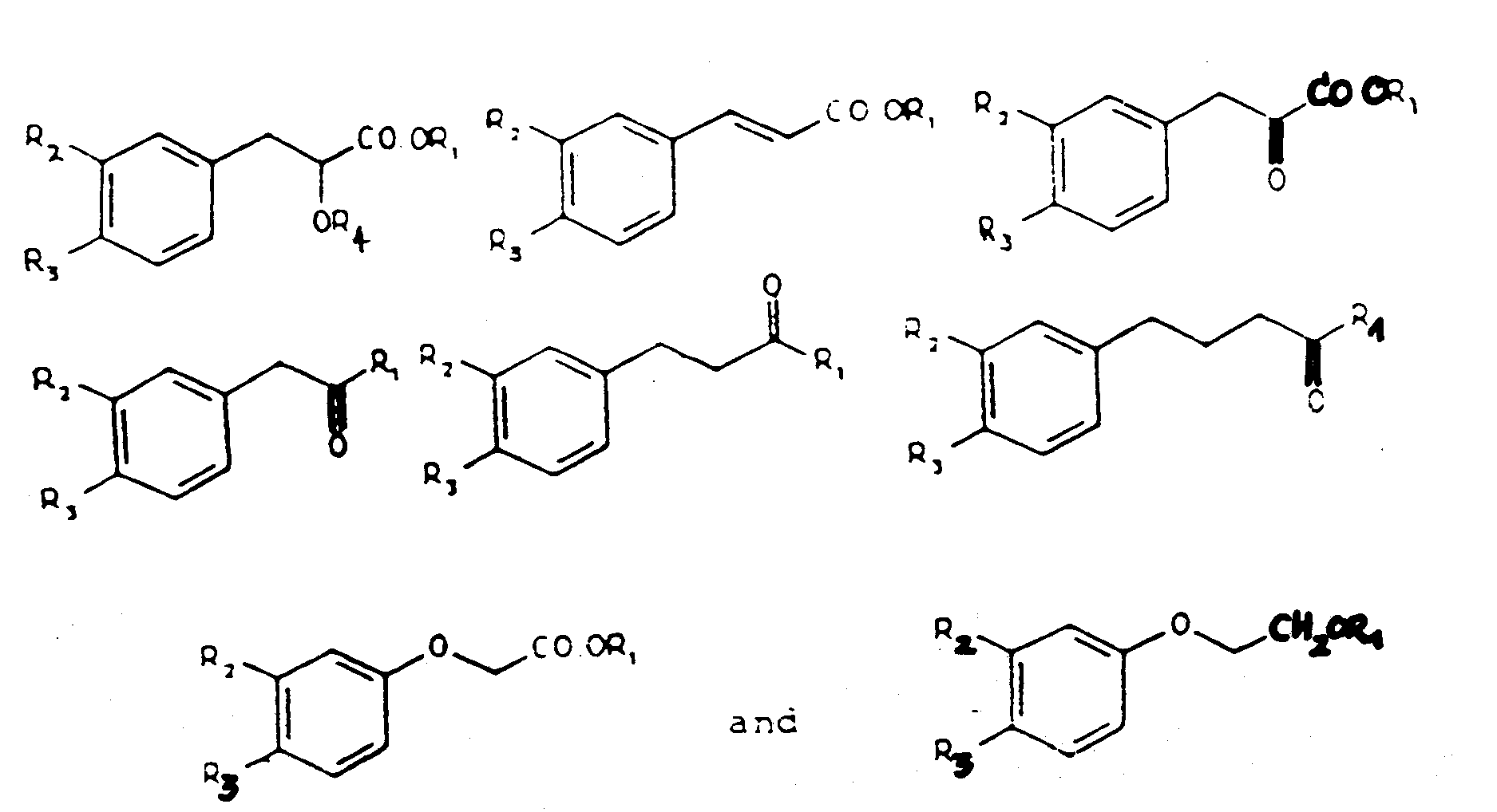
R₁ is selected from H, alkyl groups containing 1 to 6 carbon atoms and aryl groups;
R₂ and R₃ are each selected from H, OH and OCH₃; and
R₄ is H or an alkyl group of 1 to 6 carbon atoms. - The use of a compound according to claim 2, wherein the compound is selected from
methyl 3-(4-hydroxyphenyl)-2-hydroxypropionate,
n-propyl 3-(4-hydroxyphenyl)-2-hydroxypropionate,
n-butyl 3-(4-hydroxyphenyl)-2-hydroxypropionate,
3-(4-hydroxyphenyl)-2-propenoic acid,
1-(4-hydroxyphenyl)-3-butanone,
1-(4-hydroxyphenyl)-3-pentanone,
methyl-(4-hydroxyphenoxy)acetate, and
methyl 3-(3,4-dihydroxyphenyl)-2-propenoate. - The use of methyl p-hydroxyphenyllactate for the preparation of a medicament for the inhibition of the growth of proliferating cells which include a Type II nuclear estrogen binding site.
- The use of a compound for preparation of a medicament for the inhibition of the growth of proliferating cells which include a Type II nuclear estrogen binding site wherein said compound has a formula selected from

R₁ is selected from H, alkyl groups containing 1 to 6 carbon atoms and aryl groups;
R₂ and R₃ are each selected from H, OH, and OCH₃; and
R₄ is H or an alkyl group of 1 to 6 carbon atoms. - The use of a compound according to claim 5, wherein said compound is selected from
methyl 3-(4-hydroxyphenyl)-2-hydroxypropionate,
n-propyl 3-(4-hydroxyphenyl)-2-hydroxypropionate,
n-butyl 3-(4-hydroxyphenyl)-2-hydroxypropionate,
3-(4-hydroxyphenyl)-2-propenoic acid,
1-(4-hydroxyphenyl)-3-butanone,
1-(4-hydroxyphenyl)-3-pentanone,
methyl-(4-hydroxyphenoxy)acetate, and
methyl 3-(3,4-dihydroxyphenyl)-2-propenoate. - The use according to any one of claims 4 to 6, wherein said proliferating cells are estrogen responsive tissues selected from the group consisting of uterus, mammary gland, uterine tumors and mammary tumors.
- The use according to claim 7, wherein said estrogen responsive tissue comprises human breast cancer cells.
- The use of methyl p-hydroxyphenyllactate for the preparation of a medicament for the treatment of benign prostatic hyperplasia.
- The use of a compound for the preparation of a medicament for the treatment of benign prostatic hyperplasia wherein said compound has a formula selected from

R₂ and R₃ are selected from H, OH and OCH₃ ; and
R₄ is selected from H and alkyl groups of 1 to 6 carbons. - The use according claim 10, wherein said compound is selected from
methyl 3-(4-hydroxyphenyl)-2-hydroxypropionate, n-propyl 3-(4-hydroxyphenyl)-2-hydroxypropionate, n-butyl 3-(4-hydroxyphenyl)-2-hydrozypropionate,
3-(4-hydrozyphenyl)-2-propenoic acid,
1-(4-hydroxyphenyl)-3-butanone,
1-(4-hydroxyphenyl)-3-pentanone,
methyl (4-hydroxyphenoxy)acetate and
methyl 3-(3,4-dihydroxyphenyl)-2-propenoate. - The use of methyl p-hydroxyphenyllactate for the preparation of a medicament for use as an antitumor agent.
- The use of a compound for the preparation of a medicament for use as an antitumor agent wherein said compound has a formula selected from

R₁ is selected from H, alkyl groups containing 1 to 6 carbon atoms and aryl groups;
R₂ and R₃ are each selected from H, OH and OCH₃; and
R₄ is H or an alkyl group of 1 to 6 carbon atoms. - The use according to claim 13, wherein said compound is selected from
methyl 3-(4-hydroxyphenyl)-2-hydroxypropionate,
n-propyl 3-(4-hydroxyphenyl)-2-hydroxypropionate,
n-butyl 3-(4-hydroxyphenyl)-2-hydroxypropionate,
3-(4-hydroxyphenyl)-2-propenoic acid,
1-(4-hydroxyphenyl)-3-butanone,
1-(4-hydroxyphenyl)-3-pentanone,
methyl-(4-hydroxyphenoxy)acetate, and
methyl 3-(3,4-dihydroxyphenyl)-2-propenoate. - A compound of the formula
 R₁ is CH₃;R₂ is H;R₃ is OH; andR₄ is CH₃.
 - The use of a compound for the manufacture of a medicament for the treatment of cancer wherein said compound has a formula selected from

R₁ is selected from H, alkyl groups containing 1 to 6 carbon atoms, and aryl groups; and
R₂ and R₃ are each selected from H, OH and OCH₃. - The use according to claim 16, wherein said compound has a formula selected from

- The use according to claim 16, wherein said compound is selected from
3-(4-hydroxyphenyl)-1-phenyl-2-propen-1-one and
4-(4-hydroxyphenyl)-3-buten-2-one and
pharmaceutically acceptable salts thereof. - The use of a compound for the manufacture of a medicament for inhibiting the growth of proliferating cells wherein said compound has a formula selected from

R₁ is selected from H, alkyl groups containing 1 to 6 carbon atoms, and aryl groups; and
R₂ and R₃ are each selected from H, OH and OCH₃. - The use according to claim 19, wherein said compound has a formula selected from

- The use according to claim 19, wherein said compound is selected from
3-(4-hydroxyphenyl)-1-phenyl-2-propen-1-one and
4-(4-hydroxyphenyl)-3-buten-2-one and
pharmaceutically acceptable salts thereof. - The use of a compound for the manufacture of a medicament for treating benign prostatic hyperplasia wherein said compound has a formula selected from

R₁ is selected from H, alkyl groups containing 1 to 6 carbon atoms, and aryl groups; and
R₂ and R₃ are each selected from H, OH and OCH₃. - The use according to claim 22, wherein said compound has a formula selected from

- The use according to claim 22, wherein said compound is selected from
3-(4-hydroxyphenyl)-1-phenyl-2-propen-1-one and
4-(4-hydroxyphenyl)-3-buten-2-one and
pharmaceutically acceptable salts thereof.
Priority Applications (1)
| Application Number | Priority Date | Filing Date | Title |
|---|---|---|---|
| AT88908058T ATE99931T1 (en) | 1987-07-28 | 1988-07-20 | CELL GROWTH INHIBITORS AND METHODS FOR CANCER TREATMENT. |
Applications Claiming Priority (2)
| Application Number | Priority Date | Filing Date | Title |
|---|---|---|---|
| US7919987A | 1987-07-28 | 1987-07-28 | |
| US79199 | 1987-07-28 |
Publications (2)
| Publication Number | Publication Date |
|---|---|
| EP0374184A1 EP0374184A1 (en) | 1990-06-27 |
| EP0374184B1 true EP0374184B1 (en) | 1994-01-12 |
Family
ID=22149049
Family Applications (1)
| Application Number | Title | Priority Date | Filing Date |
|---|---|---|---|
| EP88908058A Expired - Lifetime EP0374184B1 (en) | 1987-07-28 | 1988-07-20 | Cell growth inhibitors and methods of treating cancer and cell proliferative diseases |
Country Status (8)
| Country | Link |
|---|---|
| US (1) | US5216024A (en) |
| EP (1) | EP0374184B1 (en) |
| JP (1) | JPH03501249A (en) |
| AT (1) | ATE99931T1 (en) |
| AU (1) | AU625313B2 (en) |
| CA (1) | CA1332742C (en) |
| DE (1) | DE3887151T2 (en) |
| WO (1) | WO1989000849A1 (en) |
Families Citing this family (20)
| Publication number | Priority date | Publication date | Assignee | Title |
|---|---|---|---|---|
| US4698225A (en) * | 1985-04-15 | 1987-10-06 | Morrison David G | Granular binding agent for pellets |
| US6515009B1 (en) | 1991-09-27 | 2003-02-04 | Neorx Corporation | Therapeutic inhibitor of vascular smooth muscle cells |
| US5811447A (en) | 1993-01-28 | 1998-09-22 | Neorx Corporation | Therapeutic inhibitor of vascular smooth muscle cells |
| US6251920B1 (en) | 1993-05-13 | 2001-06-26 | Neorx Corporation | Prevention and treatment of cardiovascular pathologies |
| US6306421B1 (en) | 1992-09-25 | 2001-10-23 | Neorx Corporation | Therapeutic inhibitor of vascular smooth muscle cells |
| US6663881B2 (en) | 1993-01-28 | 2003-12-16 | Neorx Corporation | Therapeutic inhibitor of vascular smooth muscle cells |
| US5981568A (en) | 1993-01-28 | 1999-11-09 | Neorx Corporation | Therapeutic inhibitor of vascular smooth muscle cells |
| US6491938B2 (en) | 1993-05-13 | 2002-12-10 | Neorx Corporation | Therapeutic inhibitor of vascular smooth muscle cells |
| DE69435137D1 (en) * | 1993-05-13 | 2008-10-16 | Poniard Pharmaceuticals Inc | PREVENTION AND TREATMENT OF PATHOLOGIES ASSOCIATED WITH AN ABNORMAL PROLIFERATION GLATTER MUSCLE CELLS |
| IL112205A0 (en) * | 1994-01-06 | 1995-03-15 | Res Dev Foundation | Curcumin, analogues of curcumin and novel uses thereof |
| EP0777472A2 (en) | 1994-08-31 | 1997-06-11 | Eli Lilly And Company | Methods for identifying and treating resistant tumors |
| US5710179A (en) * | 1995-04-05 | 1998-01-20 | Kabushiki Kaisha Hayashibara Seibutsu Kagaku Kenkyujo | Antitumor agent |
| US7611533B2 (en) | 1995-06-07 | 2009-11-03 | Cook Incorporated | Coated implantable medical device |
| JPH11508919A (en) * | 1995-06-19 | 1999-08-03 | オントジエン・コーポレイシヨン | Arylacrylic acid derivatives useful as protein tyrosine phosphatase inhibitors |
| US6613083B2 (en) | 2001-05-02 | 2003-09-02 | Eckhard Alt | Stent device and method |
| GB0505509D0 (en) * | 2005-03-17 | 2005-04-27 | Ic Innovations Ltd | Compounds |
| JP5119923B2 (en) * | 2005-09-02 | 2013-01-16 | 宇部興産株式会社 | Process for producing optically active (S or R) -α-hydroxy acid and optically active (R or S) -α-hydroxy acid ester |
| US20100010002A1 (en) * | 2006-09-01 | 2010-01-14 | Piramal Life Sciences Limited | Anticancer use of caffeic acid and its derivatives |
| CN102697757B (en) * | 2012-05-18 | 2014-06-04 | 广州军区广州总医院 | Application of p-hydroxy benzylidene acetone in preparation of drugs for preventing and/or treating encephalopathy |
| CN113480418B (en) * | 2021-07-13 | 2022-09-06 | 上海健康医学院 | A compound, and its application in the preparation of a drug for treating cancer |
Family Cites Families (18)
| Publication number | Priority date | Publication date | Assignee | Title |
|---|---|---|---|---|
| US2451149A (en) * | 1942-11-16 | 1948-10-12 | Nipa Lab Ltd | Manufacture of materials resistant to or active against microorganisms |
| GB1438652A (en) * | 1972-07-19 | 1976-06-09 | Shell Int Research | Hydroxyl phenyl-substituted ketones and their preparation |
| US4124724A (en) * | 1975-12-08 | 1978-11-07 | Agoro John W | Crystalline caffeic acid derivatives and compositions and method for treating snakebite |
| NL7804290A (en) * | 1977-05-12 | 1978-11-14 | Henkel Kgaa | IGNITION RETARDANT FOR COSMETIC PREPARATIONS. |
| GB1591063A (en) * | 1977-07-11 | 1981-06-10 | Ici Ltd | Monoalkylation of dihydroxybenzenes |
| JPS54160740A (en) * | 1978-06-09 | 1979-12-19 | Takeda Chem Ind Ltd | Anti-fibrotic agent |
| US4219569A (en) * | 1978-10-10 | 1980-08-26 | The Upjohn Company | Process for treating inflammation |
| FR2481270A1 (en) * | 1980-04-24 | 1981-10-30 | Roussel Uclaf | NOVEL SUBSTITUTED DERIVATIVES OF 4-PHENYL 4-OXO 2-HYDROXY BUTANOIC ACID, PROCESS FOR THEIR PREPARATION AND APPLICATIONS AS MEDICAMENTS |
| IT1171604B (en) * | 1981-10-22 | 1987-06-10 | Roussel Maestretti Spa | 4-FENYL-4-OSSOBUTEN-2-OICO ACID DERIVATIVES EQUIPPED WITH PHARMACOLOGICAL PROPERTIES AND THEIR PREPARATION PROCEDURE |
| DE3150233A1 (en) * | 1981-12-18 | 1983-06-30 | Basf Ag, 6700 Ludwigshafen | METHOD FOR PRODUCING 2- (HYDROXIPHENOXI) CARBONIC ACID ESTERS |
| JPS59104340A (en) * | 1982-12-07 | 1984-06-16 | Ube Ind Ltd | Preparation of benzylacetone |
| GB2141626B (en) * | 1983-04-25 | 1987-01-28 | Kao Corp | Cinnamic acid derivatives for lightening melanin pigmentation of skin |
| JPS60222437A (en) * | 1984-04-17 | 1985-11-07 | Ube Ind Ltd | Production of 1-phenyl-2-alkanone |
| US4733002A (en) * | 1984-05-23 | 1988-03-22 | Green Cross Corporation | Lipoxygenase inhibitor |
| JPS60248645A (en) * | 1984-05-23 | 1985-12-09 | Green Cross Corp:The | Caffeic acid derivative |
| JPH0623130B2 (en) * | 1984-07-10 | 1994-03-30 | 三井東圧化学株式会社 | Method for producing α-keto acid |
| IT1190340B (en) * | 1986-06-06 | 1988-02-16 | Roussel Maestretti Spa | DERIVATIVES OF 4-FENYL 4-BONE 2-BUTENOIC ACID, THEIR PREPARATION PROCEDURE AND THEIR USE AS MEDICINAL PRODUCTS |
| EP0270690B1 (en) * | 1986-06-21 | 1991-05-15 | Dainippon Ink And Chemicals, Inc. | Drug for treatment and prophylaxis of kidney and liver diseases |
-
1988
- 1988-07-15 US US07/219,680 patent/US5216024A/en not_active Expired - Fee Related
- 1988-07-20 AU AU23152/88A patent/AU625313B2/en not_active Ceased
- 1988-07-20 AT AT88908058T patent/ATE99931T1/en not_active IP Right Cessation
- 1988-07-20 WO PCT/US1988/002476 patent/WO1989000849A1/en not_active Ceased
- 1988-07-20 EP EP88908058A patent/EP0374184B1/en not_active Expired - Lifetime
- 1988-07-20 DE DE3887151T patent/DE3887151T2/en not_active Expired - Fee Related
- 1988-07-20 JP JP63506993A patent/JPH03501249A/en active Pending
- 1988-07-27 CA CA000573139A patent/CA1332742C/en not_active Expired - Fee Related
Also Published As
| Publication number | Publication date |
|---|---|
| WO1989000849A1 (en) | 1989-02-09 |
| DE3887151T2 (en) | 1994-06-30 |
| AU2315288A (en) | 1989-03-01 |
| CA1332742C (en) | 1994-10-25 |
| JPH03501249A (en) | 1991-03-22 |
| DE3887151D1 (en) | 1994-02-24 |
| US5216024A (en) | 1993-06-01 |
| EP0374184A1 (en) | 1990-06-27 |
| ATE99931T1 (en) | 1994-01-15 |
| AU625313B2 (en) | 1992-07-09 |
Similar Documents
| Publication | Publication Date | Title |
|---|---|---|
| US5216024A (en) | Cell growth inhibitors and methods of treating cancer and cell proliferative diseases | |
| Leclercq et al. | Physiological and pharmacological effects of estrogens in breast cancer | |
| Hsieh et al. | Estrogenic effects of genistein on the growth of estrogen receptor-positive human breast cancer (MCF-7) cells in vitro and in vivo | |
| CA2084208A1 (en) | Growth inhibitors and methods of treating cancer and cell proliferative diseases | |
| Du et al. | Dynamic changes of type I, III and IV collagen synthesis and distribution of collagen-producing cells in carbon tetrachloride-induced rat liver fibrosis | |
| CZ20011979A3 (en) | Preparation for treating cancer and method of using natural vegetable oils | |
| JP2010530880A (en) | Liquiritigenin and derivatives as selective estrogen receptor beta agonists | |
| Markaverich et al. | An endogenous inhibitor of [3H] estradiol binding to nuclear type II estrogen binding sites in normal and malignant tissues | |
| Markaverich et al. | Methyl p-hydroxyphenyllactate and nuclear type II binding sites in malignant cells: metabolic fate and mammary tumor growth | |
| Suto et al. | Persistent estrogen responsiveness of ras oncogene-transformed mouse mammary epithelial cells | |
| Judge et al. | Progesterone-specific stimulation of triglyceride biosynthesis in a breast cancer cell line (T-47D) | |
| Xiao et al. | Estrogen receptor mediates the effects of pseudoprotodiocsin on adipogenesis in 3T3-L1 cells | |
| Markaverich et al. | Estrogen regulation of methyl p-hydroxyphenyllactate hydrolysis: correlation with estrogen stimulation of rat uterine growth | |
| Brünner et al. | Endocrine sensitivity of the receptor‐positive T61 human breast carcinoma serially grown in nude mice | |
| Robinson et al. | In vitro estrogenic actions in rat and human cells of hydroxylated derivatives of D16726 (zindoxifene), an agent with known antimammary cancer activity in vivo | |
| JP2002503713A (en) | E-2- [4- (4-Chloro-1,2-diphenyl-but-1-enyl) phenoxy] ethanol and pharmaceutical composition thereof | |
| Hibner et al. | Effects of raloxifene hydrochloride on endometrial cancer cells in vitro | |
| Chatterjee et al. | N-Nitrosodiethylamine-induced nodule-like alveolar lesion and its prevention by a retinoid in BALB/c mouse mammary glands in the whole organ in culture | |
| Tsai et al. | Antiestrogen modulation of the growth and properties of ovarian-autonomous and ovarian-dependent mammary tumors in rats | |
| BAUER et al. | Trimethylmethoxyphenyl‐retinoic acid (Ro 10–1670) inhibits mitogen‐induced DNA‐synthesis in peripheral blood lymphocytes in vitro | |
| Huseby et al. | Lack of influence of hypophysectomy on estrogen-induced DNA synthesis in Leydig cells of BALB/c mice | |
| Koike et al. | Maitotoxin, a calcium channel activator, increases prolactin release from rat pituitary tumor 7315a cells by a mechanism that may involve leukotriene production | |
| Dao | Estrogen synthesis in human breast tumor and its inhibition by testololactone and bromoandrostenedione | |
| Copland et al. | Inhibition of estrogen stimulated mitogenesis by 3-phenylacetylamino-2, 6-piperidinedione and its para-hydroxy analog | |
| Shah et al. | Effect of histones, other basic proteins and some antibiotics on the transplantability of mouse mammary tumours |
Legal Events
| Date | Code | Title | Description |
|---|---|---|---|
| PUAI | Public reference made under article 153(3) epc to a published international application that has entered the european phase |
Free format text: ORIGINAL CODE: 0009012 |
|
| 17P | Request for examination filed |
Effective date: 19900124 |
|
| AK | Designated contracting states |
Kind code of ref document: A1 Designated state(s): AT BE CH DE FR GB IT LI LU NL SE |
|
| 17Q | First examination report despatched |
Effective date: 19910313 |
|
| GRAA | (expected) grant |
Free format text: ORIGINAL CODE: 0009210 |
|
| AK | Designated contracting states |
Kind code of ref document: B1 Designated state(s): AT BE CH DE FR GB IT LI LU NL SE |
|
| PG25 | Lapsed in a contracting state [announced via postgrant information from national office to epo] |
Ref country code: SE Effective date: 19940112 Ref country code: NL Effective date: 19940112 Ref country code: LI Effective date: 19940112 Ref country code: CH Effective date: 19940112 Ref country code: BE Effective date: 19940112 Ref country code: AT Effective date: 19940112 |
|
| REF | Corresponds to: |
Ref document number: 99931 Country of ref document: AT Date of ref document: 19940115 Kind code of ref document: T |
|
| ITF | It: translation for a ep patent filed | ||
| REF | Corresponds to: |
Ref document number: 3887151 Country of ref document: DE Date of ref document: 19940224 |
|
| REG | Reference to a national code |
Ref country code: CH Ref legal event code: PL |
|
| ET | Fr: translation filed | ||
| NLV1 | Nl: lapsed or annulled due to failure to fulfill the requirements of art. 29p and 29m of the patents act | ||
| PG25 | Lapsed in a contracting state [announced via postgrant information from national office to epo] |
Ref country code: GB Effective date: 19940720 |
|
| PG25 | Lapsed in a contracting state [announced via postgrant information from national office to epo] |
Ref country code: LU Free format text: LAPSE BECAUSE OF NON-PAYMENT OF DUE FEES Effective date: 19940731 |
|
| PLBE | No opposition filed within time limit |
Free format text: ORIGINAL CODE: 0009261 |
|
| STAA | Information on the status of an ep patent application or granted ep patent |
Free format text: STATUS: NO OPPOSITION FILED WITHIN TIME LIMIT |
|
| 26N | No opposition filed | ||
| GBPC | Gb: european patent ceased through non-payment of renewal fee |
Effective date: 19940720 |
|
| PG25 | Lapsed in a contracting state [announced via postgrant information from national office to epo] |
Ref country code: FR Effective date: 19950331 |
|
| PG25 | Lapsed in a contracting state [announced via postgrant information from national office to epo] |
Ref country code: DE Effective date: 19950401 |
|
| REG | Reference to a national code |
Ref country code: FR Ref legal event code: ST |
|
| PG25 | Lapsed in a contracting state [announced via postgrant information from national office to epo] |
Ref country code: IT Free format text: LAPSE BECAUSE OF NON-PAYMENT OF DUE FEES;WARNING: LAPSES OF ITALIAN PATENTS WITH EFFECTIVE DATE BEFORE 2007 MAY HAVE OCCURRED AT ANY TIME BEFORE 2007. THE CORRECT EFFECTIVE DATE MAY BE DIFFERENT FROM THE ONE RECORDED. Effective date: 20050720 |