EP0369413A2 - Interferon-gamma binding proteins - Google Patents
Interferon-gamma binding proteins Download PDFInfo
- Publication number
- EP0369413A2 EP0369413A2 EP89121094A EP89121094A EP0369413A2 EP 0369413 A2 EP0369413 A2 EP 0369413A2 EP 89121094 A EP89121094 A EP 89121094A EP 89121094 A EP89121094 A EP 89121094A EP 0369413 A2 EP0369413 A2 EP 0369413A2
- Authority
- EP
- European Patent Office
- Prior art keywords
- protein
- ifn
- gamma
- cells
- sequence shown
- Prior art date
- Legal status (The legal status is an assumption and is not a legal conclusion. Google has not performed a legal analysis and makes no representation as to the accuracy of the status listed.)
- Granted
Links
Images


Classifications
-
- C—CHEMISTRY; METALLURGY
- C07—ORGANIC CHEMISTRY
- C07K—PEPTIDES
- C07K14/00—Peptides having more than 20 amino acids; Gastrins; Somatostatins; Melanotropins; Derivatives thereof
- C07K14/435—Peptides having more than 20 amino acids; Gastrins; Somatostatins; Melanotropins; Derivatives thereof from animals; from humans
- C07K14/705—Receptors; Cell surface antigens; Cell surface determinants
- C07K14/715—Receptors; Cell surface antigens; Cell surface determinants for cytokines; for lymphokines; for interferons
- C07K14/7156—Receptors; Cell surface antigens; Cell surface determinants for cytokines; for lymphokines; for interferons for interferons [IFN]
-
- A—HUMAN NECESSITIES
- A61—MEDICAL OR VETERINARY SCIENCE; HYGIENE
- A61P—SPECIFIC THERAPEUTIC ACTIVITY OF CHEMICAL COMPOUNDS OR MEDICINAL PREPARATIONS
- A61P37/00—Drugs for immunological or allergic disorders
-
- A—HUMAN NECESSITIES
- A61—MEDICAL OR VETERINARY SCIENCE; HYGIENE
- A61P—SPECIFIC THERAPEUTIC ACTIVITY OF CHEMICAL COMPOUNDS OR MEDICINAL PREPARATIONS
- A61P43/00—Drugs for specific purposes, not provided for in groups A61P1/00-A61P41/00
-
- C—CHEMISTRY; METALLURGY
- C07—ORGANIC CHEMISTRY
- C07K—PEPTIDES
- C07K14/00—Peptides having more than 20 amino acids; Gastrins; Somatostatins; Melanotropins; Derivatives thereof
- C07K14/435—Peptides having more than 20 amino acids; Gastrins; Somatostatins; Melanotropins; Derivatives thereof from animals; from humans
- C07K14/705—Receptors; Cell surface antigens; Cell surface determinants
- C07K14/715—Receptors; Cell surface antigens; Cell surface determinants for cytokines; for lymphokines; for interferons
-
- C—CHEMISTRY; METALLURGY
- C07—ORGANIC CHEMISTRY
- C07K—PEPTIDES
- C07K16/00—Immunoglobulins [IGs], e.g. monoclonal or polyclonal antibodies
- C07K16/18—Immunoglobulins [IGs], e.g. monoclonal or polyclonal antibodies against material from animals or humans
- C07K16/28—Immunoglobulins [IGs], e.g. monoclonal or polyclonal antibodies against material from animals or humans against receptors, cell surface antigens or cell surface determinants
- C07K16/2866—Immunoglobulins [IGs], e.g. monoclonal or polyclonal antibodies against material from animals or humans against receptors, cell surface antigens or cell surface determinants against receptors for cytokines, lymphokines, interferons
-
- A—HUMAN NECESSITIES
- A61—MEDICAL OR VETERINARY SCIENCE; HYGIENE
- A61K—PREPARATIONS FOR MEDICAL, DENTAL OR TOILETRY PURPOSES
- A61K38/00—Medicinal preparations containing peptides
Definitions
- the present invention relates to proteins having interferon-gamma (IFN-gamma) binding activity, to proteins substantially homologous therewith or fragments thereof, to DNA molecules, in particular cDNA molecules, encoding these proteins or fragments thereof, and to monoclonal antibodies to the human IFN-gamma receptor used for isolating said cDNA clones.
- the invention further relates to the cloning of said human IFN-gamma binding proteins and their production by recombinant DNA techniques and to pharmaceutical compositions comprising them.
- Interferon-gamma is a lymphokine produced by activated T-lymphocytes. It exerts antiviral activity, growth inhibitory effect and several immunoregulatory activities on a variety of cell types and is of potential clinical value. However, together with its positive biological activities, IFN-gamma has been shown to provoke undesirable effects and to be involved in the development of autoimmune diseases. Thus, IFN-gamma was present in newly diagnosed diabetic children and in muscle biopsies from patients with polymyositis. It was also found to cause exacerbation of autoimmune diseases such as multiple sclerosis and psoriasis.
- IFN-gamma receptors having different molecular weight ranges were isolated from different cells by extraction followed by affinity chromatography on an immobilized IFN-gamma column.
- polyclonal antibodies to the IFN-gamma receptors were described.
- the present invention relates to monoclonal antibodies against human IFN-gamma receptor, in particular the IFN-gamma receptor of non-immune cells, eg. WISH, HeLa or FS-11 cells, having a molecular weight of aoout 90,000 Da.
- non-immune cells eg. WISH, HeLa or FS-11 cells
- the invention in another aspect, relates to proteins having IFN-gamma binding activity or proteins substantially homologous therewith or fragments thereof, said proteins comprising the amino acid sequence shown in Figure 9 or Figure 13.
- the invention encompasses larger proteins wherein these amino acid sequences are part of the total sequence of the protein molecule as well as fragments comprising essentially only part of said amino acid sequences.
- Homologous proteins or fragments thereof are included within the scope of the invention provided that they show the same biological activity of the proteins of the invention.
- the invention further relates to DNA molecules comprising a recombinant DNA molecule or a cDNA molecule comprising the nucleotide sequence coding for the proteins of the invention or fragments thereof, to replicable expression vehicles comprising them and host cells transformed therewith, and to a process for producing said proteins or fragments thereof by culturing said transformant cells in a suitable culture medium and harvesting the protein or a fragment thereof either from the cells or from the culture supernatant.
- cDNA clones encoding a portion of the human IFN-gamma binding proteins were isolated with the aid of monoclonal antibodies to the human IFN-gamma receptor. These antibodies were characterized by their ability to block the binding of 125-IFN-gamma to its receptor on human cells.
- a cDNA HeLa expression library in lambda gt 1 was screened by the antibodies and 5 positive clones were obtained from 106 recombinants.
- Four of the clones had an insert of 0.5 Kb and one was of 0.7 Kb. The 0.5 Kb fragments cross hybridized, but did not hybridize to the 0.7 Kb clone, as determined by Southern blots.
- Probes were prepared from the 0.5 Kb and 0.7 Kb inserts and a cDNA human placenta library in lambda gt11 was screened. Ten positive clones were obtained from 106 recombinants. Nine of the clones had an insert size of 1.15-2.3 Kb and they all cross hybridized, while one of the clones had an insert size of 1.8 Kb and it hybridized only to itself. Two clones of 1.8Kb and 2.3Kb were linked to expression vehicles, transfected into bacterial cells and the proteins encoded by them were expressed.
- probes may be prepared from the cDNA sequences of the invention and used for screening any cDNA library, e.g., colon, liver or kidney library or for isolation of the genomic DNA coding for the proteins of the invention by known methods, e.g. by colony hybridization techniques under stringent conditions. Positive clones are then inserted into appropriately constructed expression vectors by techniques well known in the art. Double-stranded cDNA is linked to plasmid vectors by homopolymeric tailing or by restriction linking involving the use of synthetic DNA linkers or blunt-ended ligation techniques. DNA ligases are used to ligate the DNA molecules and undesirable joining is avoided by treatment with alkaline phosphatase.
- an expression vector should comprise also specific nucleotide sequences containing transcriptional and translational regulatory information linked to the DNA coding for the desired protein in such a way as to permit gene expression and production of the protein.
- the gene must be preceded by a promoter in order to be transcribed. There are a variety of such promoters in use, which work with different efficiencies (strong and weak promoters).
- the DNA molecule comprising the nucleotide sequence coding for a protein of the invention or a fragment thereof preceded by a nucleotide sequence of a signal peptide and the operably linked transcriptional and translational regulatory signals is inserted into a vector which is capable of integrating the desired gene sequences into the host cell chromosome.
- the cells which have stably integrated the introduced DNA into their chromosomes can be selected by also introducing one or more markers which allow for selection of host cells which contain the expression vector.
- the introduced DNA molecule will be incorporated into a plasmid or viral vector capable of autonomous replication in the recipient host.
- Factors of importance in selecting a particular plasmid or viral vector include the ease with which recipient cells that contain the vector may be recognized and selected from those recipient cells which do not contain the vector; the number of copies of the vector which are desired in a particular host and whether it is desirable to be able to "shuttle" the vector between host cells of different species.
- the DNA construct(s) may be introduced into an appropriate host cell by any of a variety of suitable means: transformation, transfection, conjugation, protoplast fusion, electroporation, calcium phosphate precipitation, direct microinjection, etc.
- Host cells to be used in this invention may be either prokaryotic or eukaryotic.
- Preferred prokaryotic hosts include bacteria, such as E. coli . Under such conditions, the protein will not be glycosylated.
- the prokaryotic host must be compatible with the replicon and control sequences in the expression plasmid.
- Preferred eukaryotic hosts are mammalian cells, e.g., human, monkey, mouse and chinese hamster ovary (CHO) cells, because they provide post-translational modifications to protein molecules including correct folding or glycosylation at correct sites. Also yeast and insect cells can carry out post-translational peptide modifications including glycosylation.
- Yeast recognizes leader sequences on cloned mammalian gene products and secretes peptides bearing leader sequences (i.e. pre-peptides).
- the host cells After the introduction of the vector, the host cells are grown in a selective medium, which selects for the growth of vector-containing cells. Expression of the cloned gene sequence(s) results in the production of the desired protein or a fragment thereof. The expressed protein is then isolated and purified by any conventional procedure involving extraction, precipitation, chromatography, electrophoresis, or the like.
- the monoclonal antibodies against the human IFN-gamma receptor provided by the invention are useful for the purification of the receptor and for isolation of the cDNA clones of the invention. They bind IFN-gamma and inhibit its biological activity, thus differing from prior art monoclonal antibodies (Aguet, M. and Merlin, G. (1987) J. Exp. Med. 165 , pp. 988-999) not reported to inhibit IFN-gamma biological activitiy.
- the monoclonal antibodies against the human IFN-gamma receptor of the invention were developed by injecting mice with a preparation of receptor which was purified from solubilized placental membranes by ligand affinity chromatography. Three antibodies were identified by their ability to block the binding of 125I-IFN-gamma to its receptor on HeLa cells at 4°C. One of these antibodies blocked several biological activities of IFN-gamma, including its antiviral activity, its ability to induce HLA-DR surface antigens and its ability to protect cells from NK-cell mediated cytotoxicity. This antibody exhibited higher binding capacity to cells at 37°C and was significantly less displaceable by an excess of IFN-gamma as compared with the other two antibodies. Immunoaffinity chromatography of solubilized crude placental membrane preparation yielded a purified receptor which exhibited a molecular weight of about 88,000. The purified receptor retained its ability to bind 125I-IFN-gamma in solution.
- mice were immunized subcutaneously with a preparation of human IFN-gamma receptor obtained from placental membranes. This preparation was purified on IFN-gamma coupled to Affigel 10, then on Sephacryl 5-300 (Novick, D. et al (1987) J. Biol. Chem. 262 , p.8483) and finally adsorbed on agarose beads coupled to monoclonal anti-IFN-gamma antibodies (Novick, D. et al (1983) EMBO J. 2 , p. 1527). Two injections were given in complete Freund's adjuvant and the other two were given in 1 week intervals without an adjuvant. Each mouse received ⁇ 30 ⁇ g of affinity purified receptor per injection.
- Hybridoma supernatants were tested for the presence of anti-IFN-gamma receptor antibodies both by competitive inhibition of binding of 125I-IFN-gamma to HeLa cells and by neutralization of antiviral activity of IFN-gamma on WISH cells.
- HeLa cells (ATCC H229, CCL2.1) were seeded in 96-well microtiter plates (50,000 cells/well) in the presence of dexamethasone (10 ⁇ 6M). After 24 hrs, medium was discarded, cells were washed with ice cold phosphate-buffered saline containing Ca2+, Mg2+ (PBS) and sodium azide (0.02%). Hybridoma supernatants (50 ⁇ l/well) were added, and the plates were left for 2 hours at 4°C.
- 125I-IFN- gamma produced by CHO cells purified by affinity chromatography and labeled by the known chloramine-T method, was added to each well (50 ⁇ l, 200,000 cpm) and the plates were left for 2 hrs at 4°C. The plates were then washed 4 times with PBS 2%, harvested with NaOH (0.75M, 125 ⁇ l) and the content of each well was counted.
- a competitive inhibition of binding was performed in 24-well plates (Costar) on HeLa cells (250,000 cells/well without dexamethasone).
- the assay was done in the same manner as described for the one in 96 well microtiter plates, except for the volumes (250 ⁇ l of serially diluted hybridoma supernatants and 250 ⁇ l of 125I-IFN-gamma, 200,000 cpm). Cells were harvested with trypsin (250 ⁇ l), the wells were further washed with PBS (2x150 ⁇ l) and the combined cells and washings were counted.
- Hybridoma supernatants (50 ⁇ l) were added to cultures of WISH cells (ATCC CCL25) in 96-well plates, incubated for 2 hours at 37°C and followed by the addition of IFN-gamma (20 U/ml, 50 ⁇ l). The plates were incubated overnight at 37°C, vesicular stomatitis virus was added, the plates were further incubated overnight and the extent of the cytopathic effect was determined by staining with crystal violet (Rubinstein et al. (1981) J. Virol. 37 , p.775). For determination of neutralizing titer, hybridoma supernatants (or ascitic fluids) were serially diluted prior to the neutralization assay. One neutralizing unit is defined as the amount of antibody sufficient for neutralizing one unit of IFN-gamma. The NIH reference standard of IFN-gamma Gg23-901-503 was used in all assays.
- Hybridoma supernatants were screened for the presence of anti IFN-gamma receptor antibodies as described above. Three out of 468 hybridomas screened were found to inhibit the binding of 125-IFN-gamma to HeLa cells, and one out of the three was also positive in the neutralization assay. The positive clones were further grown and subcloned, and the cells were injected into mice for generation of ascitic fluids. The immune response in mice, the screening and the extent of antibody production in tissue culture and in ascitic fluids were all followed both by the binding assay (Table I) and by the neutralization assay (Table II).
- antibody No. 177 From the three monoclonal antibodies against the human interferon-gamma receptor developed here, antibody No. 177 and all its subclones were characterized by their ability to bind specifically to a solubilized IFN-gamma receptor, to inhibit the binding of 125I-IFN-gamma to cells (at 4°C), to block the antiviral activity of IFN-gamma and the induction of HLA-DR by IFN-gamma and to prevent the induction of resistance to NK-CMC by IFN-gamma.
- Two other monoclonal antibodies (Nos. 37 and 183) inhibited the binding of 125I-IFN-gamma to cells at 4°C but were unable to block the biological activities of IFN-gamma.
- Hybridoma 177-1 subcloned from hybridoma 177, was deposited on 14.11.1988 with the Collection Nationale de Cultures de Microorganismes (CNCM), Paris, France, and has the identifying characteristics CNCM I-814.
- the test was done as described in Example 2a. The results are shown in Table I. However, only antibody 177-1 inhibited the biological activities of IFN-gamma. Therefore comparative binding studies were performed at 37°C in the presence of sodium azide (to prevent internalization). Antibody 177-1 had a significantly higher binding capacity to cells as compared with the other two antibodies. This is shown in Figure 1, where HeLa cells were incubated at 37°C (in the presence of NaN3) with monoclonal antibody No. 37-1 183-2 and 177-1 followed by 125I-goat anti- mouse serum. Background counts (in the absence of anti-receptor antibody, 200 cpm) were subtracted. Binding at 4°C with 20 ⁇ g/ml of the various antibodies is shown as well.
- Antibodies 37-1 and 183-2 exhibited only a marginal blocking effect (Fig. 3). Incubation of HeLa cells with any of the three antibodies in the absence of IFN-gamma did not induce HLA-DR antigens. As a positive control the neutralizing monoclonal anti-IFN-gamma antibody No. 166-5 ( ⁇ - - ⁇ )) described in Novick, D. et al (1983) EMB0 J. 2 , p. 1527 was used. It inhibited the IFN-gamma induced HLA-DR, whereas no such inhibition was observed with a monoclonal anti-IFN-gamma antibody No. 7 described in Novick, D. et al (1982) J. Immunol. 129 , p.
- HLA-DR IFN-gamma class II MHC antigens
- the plates were then washed with cold PBS (3x100 ⁇ l), and fixed with formaldehyde (3.5% in PBS, 100 ⁇ l) for 30 min at 0°C.
- the plates were then rinsed with cold PBS and incubated with a solution of BSA (100 ⁇ l, 0.5% in 5mM Tris-HCl, 150mM NaCl, pH7.5) for 30 min at 0°C.
- BSA 100 ⁇ l, 0.5% in 5mM Tris-HCl, 150mM NaCl, pH7.5
- the plates were then rinsed with cold PBS and incubated for 1 hr at room temperature with monoclonal anti HLA-DR (L-243 ascitic fluid diluted 1:500 in 50 ⁇ l RPMI-1640 medium containing 0,1% BSA and 0.1% sodium azide).
- the plates were then rinsed with PBS and incubated for 30 minutes at room temperature with 125I-protein A (105cpm/well in 50 ⁇ l RPMI-1640 medium containing 0.1% BSA and 0.1% sodium azide). Excess of radioactivity was removed by washing with PBS containing 0,05% Tween 20 (3x200 ⁇ l). The cells were then solubilized with NaOH (0.75Nm 200 ⁇ l) and counted.
- the binding of anti-receptor antibodies to cells at 37°C and competition by IFN-gamma was performed as follows: HeLa cells (3x10⁵/well) were seeded and grown for 24 hours at 37°C in 24 well plates. Medium was discarded, various concentrations of anti-receptor antibodies (250 ⁇ l, in RPMI containing 10% FBS and 0,04% sodium azide) were added together with IFN-gamma (0-6 ⁇ g/ml) and the plates were incubated for 3hrs. Following two washings with PBS-2%, 125I-goat anti mouse serum (250 ⁇ l, 100,000 cpm) was added. The plates were left for 5hrs at room temperature, washed with PBS-2% (3x1ml), harvested with trypsin and counted.
- NK-CMC natural killer cell mediated cytotoxicity
- the blocking of IFN-gamma induced resistance to NK cell-mediated cytoto- xicity by anti-receptor antibody was tested as follows: U-937 target (T) cells (3.5x10⁵) were preincubated for 3hr at 37°C with or without monoclonal anti-receptor antibody (50-100ng in 750 ⁇ l RPMI 1640 medium containing 10% FBS). IFN-gamma (1000 units in 250 ⁇ l medium) was added and incubation was further continued for 9hrs.
- the cells were then labelled with 51[Cr]Na2CrO4 (0.5mCi, 1.5hr), washed and incubated in triplicates (l04cells, 50 ⁇ l/well) for 4hrs at 37°C with the effector cells (E, nylon vool non-adherent peripheral blood mononuclear cells 100 ⁇ l/well) at the indicated E:T ratios.
- the cells were then spun and the supernatant was counted (C). Spontaneous release (S; up to 6% of total cpm) was measured in supernatant of target cells alone and total cpm (T) was measured by adding Triton X-100 (1%, 100 ⁇ l) to labelled target cells. Percent cytotoxicity was determined according to the formula: % cytotoxicity 100 x
- An immunoadsorbent was prepared from an immunoglobulin fraction of ascitic fluids of mice containing monoclonal antibodies secreted by the hybridomas of the invention (e.g. of hybridomas 177 or 183).
- the ascitic fluids were precipitated with ammonium sulfate at 4°C (final concentration 50% saturation). The precipitate was collected by centrifugation, redisolved in water and dialysed against saline. About 10 mg of immunoglobulins were bound to polyacrylic hydrazide-agarose (Biomakor).
- a solubilized placental membrane preparation was loaded to the antibody column at 4°C at a flow rate of 0.2 ml/min.
- the column was washed with PBS containing 0.1% Triton X-100 (40 ml) and eluted by citric acid (50 mM, pH2) containing 0.05% Triton X-100 and 0.02% sodium azide. Eluted fractions were neutralized by Hepes buffer (1M, pH 8.5) and kept at 4°C.
- the column was monitored by binding of radiolabeled IFN-gamma and a purification of 4250 fold was achieved in one step (Table III).
- the binding of radiolabeled IFN-gamma to a soluble receptor for the monitoring of the purification method was performed as follows: Aliquots (20-40 ⁇ l) of the solubilized receptor from various purification steps were mixed with 125I-IFN-gamma (250 units) either with or without labeled IFN-gamma (100,000 units) in PBS containing 0.1% BSA (200 ⁇ l). The mixture was incubated for 2 hours at 4°C, rabbit IGg (0.1% in PBS, 0.5ml) was then added, followed by PEG-8000 (22% in PBS, 0.5ml). The mixture was left for 10 minutes at 4°C and then passed through a 0.45 ⁇ filter (25mm HAWP, Millipore).
- the filters were washed with cold PEG-8000 solution (8% in PBS), and counted. Background counts were determined in the presence of excess unlabeled IFN-gamma and were subtracted. Binding is expressed in pmoles of 125I-IFN-gamma.
- the nitrocellulose was incubated for 2 hours at room temperature with a mixture of the three anti-IFN-gamma receptor monoclonal antibodies (immunoglobulin fraction of ascitic fluids 10mg/ml diluted 1:150 in the blocking buffer). Following washings in 0.05% Tween-20 in PBS, the nitrocellulose sheet was incubated for 3 hours at room temperature with 125I-goat anti-mouse serum (0.7x10⁶ cpm/ml, in the blocking buffer). The sheet was then washed, dried and autoradiographed.
- a mixture of the three anti-IFN-gamma receptor monoclonal antibodies immunoglobulin fraction of ascitic fluids 10mg/ml diluted 1:150 in the blocking buffer. Following washings in 0.05% Tween-20 in PBS, the nitrocellulose sheet was incubated for 3 hours at room temperature with 125I-goat anti-mouse serum (0.7x10⁶ cpm/ml, in the blocking buffer). The sheet was then was
- Filters were marked and transfered to 10% low fat milk (1%), 0.05% Tween 20 in PBS for blocking (2 hrs at room temperature).
- the two sets of filters were washed in PBS containing 0.05% Tween 20 and incubated with monoclonal anti-IFN-gamma receptor antibodies obtained in Example 3 (monoclonal antibody 177-1 was preferably used), (20 ⁇ g/ml in blocking solution) for 3 hrs at room temperature.
- the filters were washed 5 times with PBS-Tween and positive clones were identified by 125I-goat anti-mouse F(ab)2 (7 x 10⁵ cpm/ml in blocking solution) following an overnight incubation at 4°C and extensive washings with PBS-Tween.
- Fig. 6A shows the screening of a cDNA HeLa expression library
- Fig. 6B shows the subcloning of a positive clone.
- DNA stocks were prepared from 500 ml E. coli 1088 infected with positive phages. DNA was purified by CsCl gradient followed by phenol-chloroform extraction.
- the purified lambda gt11 DNA containing positive cDNA clones were digested with EcoR1 and size-fractionated on 1% agarose gel.
- probes were prepared from one of the 0.5Kb clones (15-21-1) and from the 0.7 Kb clon (18-4-3), using the multiprime DNA labelling systems kits (AMERSHAM).
- Th technique is based on the use of random sequence hexanucleotide to prim DNA synthesis on denatured template DNA at numerous sites along it length (Feinberg A.P.
- the purified lambda gt11 DNA containing positive cDNA clones were digested with EcoR1 and size-fractionated on l% agarose gel.
- Nine of th clones isolated by the 0.7Kb probe had an insert size of 1.15-2.3 Kb an they all cross-hybridized, while one of the clones isolated by the 0.5K probe had an insert size of 1.8Kb and hybridized only to itself Cross-hybridization was determined by Southern blots.
- the clones were further characterized by digestion with restrictio enzymes.
- the 1.8Kb fragment (No. 39) was cut by restriction enzymes a shown by the restriction map of Figure 7.
- a KpnI site was found at distance of 1.1 Kb from the EcoR1 site of the lambda gt11 (19.6 Kb fro the left end).
- a SacI site was found at a distance of 0 Kb from the EcoR site of lambda gt11.
- the 2.3Kb fragment (No. 76) was cut by restriction enzymes as shown i the restriction map of Figure 8.
- a Sa1 site was found in the middle o the 2.3 Kb insert.
- An XbaI site was found very near the EcoR1 site of th lambda gt11 (19.6Kb from the right end).
- DNA of the 0.5 Kb, 0.7 Kb, 1.8 Kb and 2.3 Kb positive clones purified o CsC1 gradient was cut by EcoR1 restriction enzyme.
- DNA of the Bluescrip plasmid vector of Stratagene Cloning System (San Diego, California) wa cut as well by EcoR1, then dephosphorylated and run on a preparativ agarose gel. The band of DNA was extracted from the gel phenol-chlorofor extractions. Both the clone and the vector were nov ready for ligatio with the help of T4 ligase.
- E. coli (TG1) competent bacteria were use for transformation with the ligated vector.
- Vector was added to th bacteria at 4°C, folloved by a heat shock (42°C), removed to ice, then a room temperature and at 37°C. Finally the bacteria were plated on LB Ampicillin (Amp). Colonies were picked and grown in LB + Amp.
- Fo sequencing a single stranded DNA was prepared as follovs: A starter o E. coli TG1 transformed bacteria with the ligated vector was grovn in 2T medium and Ampicillin folloved by the addition of a helper virus. The DN was precipitated by polyethylene glycol and extracted by phenol chloroform. Finally the DNA was suspended in Tris-EDTA, ready fo sequencing with the aid of the Sequenase Kit (USB).
- Figure 9 shows the nucleotide sequence of the 0.5 Kb cDNA segment and it translated amino acid sequence.
- Figure 10 shows the complementary stran of the 0.7 Kb cDNA segment and its translated amino acid sequence
- Figure 11 shows two partial nucleotide and translated amino aci sequences of the 1.8 Kb cDNA segment.
- Figure 12 shows partial nucleotid sequence of the 2.3 Kb cDNA and Figure 13 a partial translated amino aci sequence thereof.
- An immunoadsorbent was prepared as in Example 7.
- the crude E. col extract obtained in Example 14 (100mg of the lysogen) was spun (10,000xg and the supernatant was applied to the antibody column (3 mg Igs/O.3m agarose) at 4°C.
- the column was washed with PBS containing 0.1% Trito X-100 and eluted by citric acid (50mM, pH 2) containing 0.05% Trito X-100 and 0.02% sodium azide.
- Five 0.5ml fractions were collected int HEPES buffer (1M, pH 8.5) and kept at 4°C. Fractions No. 1 and No. contained 75% of the eluted protein.
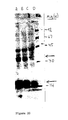
- nitrocellulose was incubated for 2 hrs at roo temperature with anti-IFN- receptor monoclonal antibodies (immuno globulin fraction of ascitic fluids 10mg/ml diluted 1:500 in the blockin buffer). Following washings in 0.05% Tween-20 in PBS, the nitrocellulos sheet was incubated overnight at 4°C with 125I-goat anti-mouse seru (0.7x10⁶ cpm/ml in the blocking buffer). The sheet was then washed, drie and autoradiographed. As shown in Fig. 12, a band of Mr of about 130,00 was obtained both in the crude and the affinity purified fractions.
- I Figure 15 Lane A - molecular weight markers; Lane B - Immunoaffinit purified lysogen; Lane C - crude lysogen.
- Preparations of the affinity purified fused protein (5 ⁇ g) or of pur ⁇ -galactosidase were mixed with 123I-IFN-gamma (1000 units, 5x10⁵ cpm) i the presence or absence of unlabelled IFN-gamma (1000 fold), and th mixture was left for 2 hrs at room temperature.
- Di Succinyl Suberat (DSS) was added to a final concentration of 0.3mM.
- the cross linking wa stopped after 15 min at 4°C by the addition of 1M Tris-HCl buffer.
- Th mixture was immunoprecipitated with rabbit anti- ⁇ -galactosidase seru (1:200, 2 hrs at room temperature), followed by the addition of Prot Sepharose beads.
- Lane F Pur ⁇ -galactosidase cross-linked to 125I-IFN-gamma
- the 1.8 Kb insert of lambda gt11 clone N° 39 isolated from the huma placenta cDNA library with the 0.5 Kb probe was cu out by EcoRI and inserted in the EcoRI site of the KS Bluescript vecto (BS) from Stratagene Cloning Systems (La Jolla, Cal.).
- BS-39cDNA the orientation was such that the AhaIII site at the 3′-end o the sense strand of the 1.8 Kb insert (Fig.
- Lane A Molecular weight markers
- Lane B Crude extract of non-induced bacteria No. 39.
- Lane C and D Crude extract of IPTG-induced bacteria No. 39 after 60 min. and 45 min. of induction, respectively. Blot incubated with antibody No. 177-1.
- the size of the protein may be smaller than that of the natural produc of the 39 cDNA as only a fragment of this cDNA was used in the construct
- This protein comprises the sequences shown in Figure 11.
- the sequence o fragment BalI-Kpn I (Fig. 11B) comprises the sequence shown in Figure 9.
- the 2.3 Kb insert of lambda gt11 clone N° 76 isolated from the huma placenta cDNA library with the 0.7 Kb probe was cu out with EcoRI and inserted in the EcoRI site of the KS Bluescrip vector, so that the XbaI site at the 3′-end of the sense strand of th 2.3 Kb insert (Fig. 8) is close to the BamHI site and T7 RNA polymeras promoter of the BS vector whereas the 5′-end is close to the HindIII sit and the T3 RNA polymerase promoter of the plasmid BS vector.
- Th expresssion plasmid TL-IFN- ⁇ c was cut with EcoRI and HindIII an religated with a synthetic ECORI-HindIII linker (Fig. 19).
- the resultin plasmid was recut by HindIII and BamHI and ligated to the HindIII-BamH 2.3 Kb cDNA excised from the above BS clone 76cDNA to yield TL-76cDN (Fig. 19).
- This plasmid was transfected into E. coli JM101 i q an cultures were induced with IPTG. A 34,000 and a 17,000 Mr protei products were identified which reacted with monoclonal antibody 183 o immunoblots (Fig. 20).
- This protein comprises the sequence shown i Figure 13. Extraction from the cell culture, immunoaffinit chromatography, Western blotting and 125I-IFN-gamma cross-linking ver performed as above.
- Lane A B - Crude extract of IPTG-induced bacteria No. 76 after 120 min. and 180 min., respectively.
- Lane C Crude extract of non-induced bacteria No. 76.
- Lane D - Molecular weight markers. Blot incubated with a mixture of antibodies No. 177-1 and 183.
- the IFN-gamma binding proteins of the present invention can be use either alone or together for modulating the activity and protectin against the deleterious effects of IFN-gamma by systemic or loca administration. They will be useful in the treatment of condition wherein excess of IFN-gamma is endogenously formed or is exogenousl administered.
- IFN-gamma activity by IFN-gamm binding protein is beneficial in cases of undesired production o endogenous IFN-gamma which may occur in local inflammation, systemi inflammation such as occcuring in septic shock and in various autoimmun and inflammatory diseases, including but not restricted to rheumatoi arthritis, multiple sclerosis, juvenile diabetes at its onset polymyositis, Behcet disease, thyroiditis, Lupus erythematosus an dermatitis.
- Modulation of IFN-gamma activity by the IFN-gamma bindin proteins is also beneficial in cases of administration of exogenou IFN-gamma whenever side effects due to overdose or to patient' sensitivity to IFN-gamma is diagnosed.
- the IFN-gamm binding proteins can be used to prolong or even enhance the antiviral anti-inflammatory, anticellular or any other activity of both endogenou and exogenous IFN-gamma.
- the proteins of the invention can be formulated according to know methods to prepare pharmaceutically useful compositions in a mixture wit a pharmaceutically acceptable carrier vehicle. Suitable vehicles an their formulations are described, for example, in Remington' Pharmaceutical Sciences by E.W. Martin.
- compositions of the invention are prepared fo administration by mixing the protein with physiologically acceptabl carriers, stabilizers and excipients, and prepared in dosage form, e.g by lyophilization in dosage vials.
- the amount of active compound to b administered will depend on the route of administration, the disease t be treated and the condition of the patient.
- the vay of administratio can be via any of the accepted modes of administration for similar agent and will depend on the condition to be treated.
Abstract

Description
- The present invention relates to proteins having interferon-gamma (IFN-gamma) binding activity, to proteins substantially homologous therewith or fragments thereof, to DNA molecules, in particular cDNA molecules, encoding these proteins or fragments thereof, and to monoclonal antibodies to the human IFN-gamma receptor used for isolating said cDNA clones. The invention further relates to the cloning of said human IFN-gamma binding proteins and their production by recombinant DNA techniques and to pharmaceutical compositions comprising them.
- Interferon-gamma is a lymphokine produced by activated T-lymphocytes. It exerts antiviral activity, growth inhibitory effect and several immunoregulatory activities on a variety of cell types and is of potential clinical value. However, together with its positive biological activities, IFN-gamma has been shown to provoke undesirable effects and to be involved in the development of autoimmune diseases. Thus, IFN-gamma was present in newly diagnosed diabetic children and in muscle biopsies from patients with polymyositis. It was also found to cause exacerbation of autoimmune diseases such as multiple sclerosis and psoriasis.
- It is therefore desirable to find ways to eliminate or antagonize the undesirable activities or effects of IFN-gamma endogenously formed in excess or exogenously administered, and particularly to block its action, for controlling the progression of autoimmune processes.
- In European Patent Application No. 240975 of the same applicant, whose contents are herein incorporated by reference, IFN-gamma receptors having different molecular weight ranges were isolated from different cells by extraction followed by affinity chromatography on an immobilized IFN-gamma column. In the same application polyclonal antibodies to the IFN-gamma receptors were described.
- In one aspect, the present invention relates to monoclonal antibodies against human IFN-gamma receptor, in particular the IFN-gamma receptor of non-immune cells, eg. WISH, HeLa or FS-11 cells, having a molecular weight of aoout 90,000 Da.
- In another aspect, the invention relates to proteins having IFN-gamma binding activity or proteins substantially homologous therewith or fragments thereof, said proteins comprising the amino acid sequence shown in Figure 9 or Figure 13. The invention encompasses larger proteins wherein these amino acid sequences are part of the total sequence of the protein molecule as well as fragments comprising essentially only part of said amino acid sequences. Homologous proteins or fragments thereof are included within the scope of the invention provided that they show the same biological activity of the proteins of the invention.
- The invention further relates to DNA molecules comprising a recombinant DNA molecule or a cDNA molecule comprising the nucleotide sequence coding for the proteins of the invention or fragments thereof, to replicable expression vehicles comprising them and host cells transformed therewith, and to a process for producing said proteins or fragments thereof by culturing said transformant cells in a suitable culture medium and harvesting the protein or a fragment thereof either from the cells or from the culture supernatant.
- According to the invention, cDNA clones encoding a portion of the human IFN-gamma binding proteins were isolated with the aid of monoclonal antibodies to the human IFN-gamma receptor. These antibodies were characterized by their ability to block the binding of ¹²⁵-IFN-gamma to its receptor on human cells. A cDNA HeLa expression library in
lambda gt 1 was screened by the antibodies and 5 positive clones were obtained from 10⁶ recombinants. Four of the clones had an insert of 0.5 Kb and one was of 0.7 Kb. The 0.5 Kb fragments cross hybridized, but did not hybridize to the 0.7 Kb clone, as determined by Southern blots. The inserts were ligated to a Bluescript plasmid vector and E. coli TG1 competent bacteria were transformed. Colonies containing the inserts from two of the clones (0.5 Kb and 0.7 Kb) were further grown and the single-stranded DNA obtained with the aid of a helper virus was used for sequencing. One open-reading frame was revealed in the sequencing and no significant homology to known DNA sequences was observed. Figure 9 shows the nucleotide sequence of clone 15-21-1 of 0.5 Kb and Figure 10 shows the nucleotide sequence of the complementary strand of clone 18-4-3 of 0.7 Kb. - The proteins encoded by the inserts were isolated and characterized. Lysogens were prepared and the induced proteins were purified by anti-IFN-gamma receptor immunoadsorbent. The size of the proteins in the eluted fractions was determined by SDS-PAGE followed by silver staining and by Western blotting. The fused protein originating from the 0.5 Kb clone had a Mr of aoout 130,000 (Mr of β-galactosidase = 114,000). Binding of ¹²⁵I-IFN-gamma to the fused protein was demonstrated in solution and was inhibited by an excess of unlabeled IFN-gamma. No such binding was detected with β-galactosidase alone. Cross-linking experiments of ¹²⁵IFN-gamma to this fused protein followed by immunoprecipitation resulted in a complex of Mr=155,000 (Mr of IFN-gamma is 25,000), as visualised by SDS-PAGE and autoradiography. This indicates that this 0.5Kb cDNA fragment is coding for at least part of the ligand binding domain of a human IFN-gamma binding protein.
- Probes were prepared from the 0.5 Kb and 0.7 Kb inserts and a cDNA human placenta library in lambda gt11 was screened. Ten positive clones were obtained from 10⁶ recombinants. Nine of the clones had an insert size of 1.15-2.3 Kb and they all cross hybridized, while one of the clones had an insert size of 1.8 Kb and it hybridized only to itself. Two clones of 1.8Kb and 2.3Kb were linked to expression vehicles, transfected into bacterial cells and the proteins encoded by them were expressed.
- Other probes may be prepared from the cDNA sequences of the invention and used for screening any cDNA library, e.g., colon, liver or kidney library or for isolation of the genomic DNA coding for the proteins of the invention by known methods, e.g. by colony hybridization techniques under stringent conditions. Positive clones are then inserted into appropriately constructed expression vectors by techniques well known in the art. Double-stranded cDNA is linked to plasmid vectors by homopolymeric tailing or by restriction linking involving the use of synthetic DNA linkers or blunt-ended ligation techniques. DNA ligases are used to ligate the DNA molecules and undesirable joining is avoided by treatment with alkaline phosphatase.
- In order to be capable of expressing a desired protein, an expression vector should comprise also specific nucleotide sequences containing transcriptional and translational regulatory information linked to the DNA coding for the desired protein in such a way as to permit gene expression and production of the protein. The gene must be preceded by a promoter in order to be transcribed. There are a variety of such promoters in use, which work with different efficiencies (strong and weak promoters).
- The DNA molecule comprising the nucleotide sequence coding for a protein of the invention or a fragment thereof preceded by a nucleotide sequence of a signal peptide and the operably linked transcriptional and translational regulatory signals is inserted into a vector which is capable of integrating the desired gene sequences into the host cell chromosome. The cells which have stably integrated the introduced DNA into their chromosomes can be selected by also introducing one or more markers which allow for selection of host cells which contain the expression vector.
- In a preferred embodiment, the introduced DNA molecule will be incorporated into a plasmid or viral vector capable of autonomous replication in the recipient host. Factors of importance in selecting a particular plasmid or viral vector include the ease with which recipient cells that contain the vector may be recognized and selected from those recipient cells which do not contain the vector; the number of copies of the vector which are desired in a particular host and whether it is desirable to be able to "shuttle" the vector between host cells of different species. once the vector or DNA sequence containing the construct(s) has been prepared for expression, the DNA construct(s) may be introduced into an appropriate host cell by any of a variety of suitable means: transformation, transfection, conjugation, protoplast fusion, electroporation, calcium phosphate precipitation, direct microinjection, etc.
- Host cells to be used in this invention may be either prokaryotic or eukaryotic. Preferred prokaryotic hosts include bacteria, such as E. coli. Under such conditions, the protein will not be glycosylated. The prokaryotic host must be compatible with the replicon and control sequences in the expression plasmid.
- Preferred eukaryotic hosts are mammalian cells, e.g., human, monkey, mouse and chinese hamster ovary (CHO) cells, because they provide post-translational modifications to protein molecules including correct folding or glycosylation at correct sites. Also yeast and insect cells can carry out post-translational peptide modifications including glycosylation. A number of recombinant DNA strategies exist which utilize strong promoter sequences and high copy number of plasmids which can be utilized for production of the desired proteins in yeast. Yeast recognizes leader sequences on cloned mammalian gene products and secretes peptides bearing leader sequences (i.e. pre-peptides).
- After the introduction of the vector, the host cells are grown in a selective medium, which selects for the growth of vector-containing cells. Expression of the cloned gene sequence(s) results in the production of the desired protein or a fragment thereof. The expressed protein is then isolated and purified by any conventional procedure involving extraction, precipitation, chromatography, electrophoresis, or the like.
- The monoclonal antibodies against the human IFN-gamma receptor provided by the invention are useful for the purification of the receptor and for isolation of the cDNA clones of the invention. They bind IFN-gamma and inhibit its biological activity, thus differing from prior art monoclonal antibodies (Aguet, M. and Merlin, G. (1987) J. Exp. Med. 165, pp. 988-999) not reported to inhibit IFN-gamma biological activitiy.
- The monoclonal antibodies against the human IFN-gamma receptor of the invention were developed by injecting mice with a preparation of receptor which was purified from solubilized placental membranes by ligand affinity chromatography. Three antibodies were identified by their ability to block the binding of ¹²⁵I-IFN-gamma to its receptor on HeLa cells at 4°C. One of these antibodies blocked several biological activities of IFN-gamma, including its antiviral activity, its ability to induce HLA-DR surface antigens and its ability to protect cells from NK-cell mediated cytotoxicity. This antibody exhibited higher binding capacity to cells at 37°C and was significantly less displaceable by an excess of IFN-gamma as compared with the other two antibodies. Immunoaffinity chromatography of solubilized crude placental membrane preparation yielded a purified receptor which exhibited a molecular weight of about 88,000. The purified receptor retained its ability to bind ¹²⁵I-IFN-gamma in solution.
- The invention is illustrated by, but not limited to, the following examples:
- BALB/c mice were immunized subcutaneously with a preparation of human IFN-gamma receptor obtained from placental membranes. This preparation was purified on IFN-gamma coupled to
Affigel 10, then on Sephacryl 5-300 (Novick, D. et al (1987) J. Biol. Chem. 262, p.8483) and finally adsorbed on agarose beads coupled to monoclonal anti-IFN-gamma antibodies (Novick, D. et al (1983) EMBO J. 2, p. 1527). Two injections were given in complete Freund's adjuvant and the other two were given in 1 week intervals without an adjuvant. Each mouse received ∼30µg of affinity purified receptor per injection. The last boost was given intraperitoneally 4 days before fusion. Sera were checked for their ability to block the binding of ¹²⁵I-IFN-gamma to HeLa cells as described hereinafter. Spleen cells (200x10⁶) from a mouse exhibiting a titer of 1:500 in this assay were fused with 40x10⁶ NSO/1 myeloma cells. Hybridomas were selected in Dulbecco's modified Eagle's medium, supplemented with 1mM pyruvate, 2mM glutamine, penicillin (10 units/ml), streptomycin 20µg/ml, fungizone 250µg/ml, 10% fetal bovine serum (FBS), and containing HAT. Hybridomas that were found to secrete anti-IFN-gamma receptor antibodies were cloned by the limiting dilution method. - Hybridoma supernatants were tested for the presence of anti-IFN-gamma receptor antibodies both by competitive inhibition of binding of ¹²⁵I-IFN-gamma to HeLa cells and by neutralization of antiviral activity of IFN-gamma on WISH cells.
- HeLa cells (ATCC H229, CCL2.1) were seeded in 96-well microtiter plates (50,000 cells/well) in the presence of dexamethasone (10⁻⁶M). After 24 hrs, medium was discarded, cells were washed with ice cold phosphate-buffered saline containing Ca²⁺, Mg²⁺ (PBS) and sodium azide (0.02%). Hybridoma supernatants (50µl/well) were added, and the plates were left for 2 hours at 4°C. Following two washings with ice cold PBS containing 2% FBS and 0.02% sodium azide (PBS-2%), ¹²⁵I-IFN- gamma produced by CHO cells, purified by affinity chromatography and labeled by the known chloramine-T method, was added to each well (50 µl, 200,000 cpm) and the plates were left for 2 hrs at 4°C. The plates were then washed 4 times with
PBS 2%, harvested with NaOH (0.75M, 125µl) and the content of each well was counted. - A competitive inhibition of binding was performed in 24-well plates (Costar) on HeLa cells (250,000 cells/well without dexamethasone). The assay was done in the same manner as described for the one in 96 well microtiter plates, except for the volumes (250µl of serially diluted hybridoma supernatants and 250µl of ¹²⁵I-IFN-gamma, 200,000 cpm). Cells were harvested with trypsin (250µl), the wells were further washed with PBS (2x150µl) and the combined cells and washings were counted.
- Hybridoma supernatants (50µl) were added to cultures of WISH cells (ATCC CCL25) in 96-well plates, incubated for 2 hours at 37°C and followed by the addition of IFN-gamma (20 U/ml, 50 µl). The plates were incubated overnight at 37°C, vesicular stomatitis virus was added, the plates were further incubated overnight and the extent of the cytopathic effect was determined by staining with crystal violet (Rubinstein et al. (1981) J. Virol. 37, p.775). For determination of neutralizing titer, hybridoma supernatants (or ascitic fluids) were serially diluted prior to the neutralization assay. One neutralizing unit is defined as the amount of antibody sufficient for neutralizing one unit of IFN-gamma. The NIH reference standard of IFN-gamma Gg23-901-503 was used in all assays.
- Hybridoma supernatants were screened for the presence of anti IFN-gamma receptor antibodies as described above. Three out of 468 hybridomas screened were found to inhibit the binding of ¹²⁵-IFN-gamma to HeLa cells, and one out of the three was also positive in the neutralization assay. The positive clones were further grown and subcloned, and the cells were injected into mice for generation of ascitic fluids. The immune response in mice, the screening and the extent of antibody production in tissue culture and in ascitic fluids were all followed both by the binding assay (Table I) and by the neutralization assay (Table II).
- From the three monoclonal antibodies against the human interferon-gamma receptor developed here, antibody No. 177 and all its subclones were characterized by their ability to bind specifically to a solubilized IFN-gamma receptor, to inhibit the binding of ¹²⁵I-IFN-gamma to cells (at 4°C), to block the antiviral activity of IFN-gamma and the induction of HLA-DR by IFN-gamma and to prevent the induction of resistance to NK-CMC by IFN-gamma. Two other monoclonal antibodies (Nos. 37 and 183) inhibited the binding of ¹²⁵I-IFN-gamma to cells at 4°C but were unable to block the biological activities of IFN-gamma. All biological activities were determined at 37°C. Since binding is a prerequisite to biological activity, antibody No. 177 was tested to see whether it had a higher affinity for the receptor as compared with the other two antibodies and whether IFN-gamma could displace the antibodies from the receptor at 37°C.
Table I Inhibition of ¹²⁵I-IFN-gamma to HeLa cells by anti-receptor antibodies. Sample Inhibition in microplates Inhibition in 24-well plates antibody dilution cpm % antibody dilution cpm % Immune serum (mouse) 1:500 50 83 1:500 550 61 Neg. control serum 1:500 350 0 1:500 1400 0 Hybridoma 37A undiluted 112 68 1:100 250 83 1:2500 890 40 Hybridoma 177A undiluted 76 78 1:50 900 39 1:250 1080 27 Hybridoma 183A undiluted 96 73 1:250 370 75 1:1250 1020 31 Negative hybridoma undiluted 350 0 - 1475 0 Ascitic fluid immunoglobulins (16mg/ml) 183-2 1:200000 560 62 A Hybridoma supernatants from the first screen were tested once in microplates, whereas supernatants of positive clones that were grown in larger amounts were tested in triplicates in 24 well plates. In the latter case the counts ranged within ±15%. - Indeed it was found that the two other antibodies could be displaced from cell surface by an excess of IFN-gamma whereas almost no such displacement of antibody No. 177 was observed. It is noteworthy that none of the antibodies exhibited antiviral activity or HLA-DR inducing activity when incubated vith cells in the absence of IFN-gamma.
- Hybridoma 177-1, subcloned from hybridoma 177, was deposited on 14.11.1988 with the Collection Nationale de Cultures de Microorganismes (CNCM), Paris, France, and has the identifying characteristics CNCM I-814.
- The three monoclonal antibodies No. 37, 177 and 183, inhibited the binding of ¹²⁵I-IFN-gamma to cells at 4°C. The test was done as described in Example 2a. The results are shown in Table I. However, only antibody 177-1 inhibited the biological activities of IFN-gamma. Therefore comparative binding studies were performed at 37°C in the presence of sodium azide (to prevent internalization). Antibody 177-1 had a significantly higher binding capacity to cells as compared with the other two antibodies. This is shown in Figure 1, where HeLa cells were incubated at 37°C (in the presence of NaN₃) with monoclonal antibody No. 37-1183-2
 and 177-1
 followed by ¹²⁵I-goat anti- mouse serum. Background counts (in the absence of anti-receptor antibody, 200 cpm) were subtracted. Binding at 4°C with 20µg/ml of the various antibodies is shown as well.
- Subsequent addition of IFN-gamma caused a significant displacement of the bound antibodies No. 37-1 and 183-2 and only minimal displacement of the neutralizing antibody No. 177-1. The inhibition of the binding of anti-receptor antibodies by IFN-gamma at 37°C is shown in Figure 2, where HeLa cells were incubated with anti-receptor antibody no. 37-1183-2
 or 177-1
 together with various concentrations of IFN-gamma. The cells were then washed and incubated with ¹²⁵I-goat anti-mouse serum. Maximal binding gave 3500 cpm. Background counts (in the absence of anti-receptor antibodies, 250 cpm) were subtracted.
- All subclones of antibody No. 177 blocked the antiviral activity of IFN-gamma. The neutralizing titer of the various subclones ranged for 4000-30,000 units/ml. No such blocking activity was observed with the other two antibodies. The results are shown in Table II.In a control study none of these antibodies were found to block the antiviral activity of IFN-gamma. None of the antibodies had an intrinsic capacity to elicit an antiviral state in the cells. The anti-receptor monoclonal antibodies were tested for their ability to block the induction of HLA-DR antigens in HeLa cells by IFN-gamma. Once again antibody No. 177-1exhibited high blocking activity and 50% inhibition was observed at ascitic fluid dilution of 1:20,000. Antibodies 37-1
 and 183-2
 exhibited only a marginal blocking effect (Fig. 3). Incubation of HeLa cells with any of the three antibodies in the absence of IFN-gamma did not induce HLA-DR antigens. As a positive control the neutralizing monoclonal anti-IFN-gamma antibody No. 166-5 (□- -□)) described in Novick, D. et al (1983) EMB0 J. 2, p. 1527 was used. It inhibited the IFN-gamma induced HLA-DR, whereas no such inhibition was observed with a monoclonal anti-IFN-gamma antibody No. 7 described in Novick, D. et al (1982) J. Immunol. 129, p. 2244 (not shown). The maximal induction of HLA-DR (in the absence of antibodies) was 2700±100 cpm. Basal level (in the absence of IFN-gamma) was 200 cpm and was subtracted from all readings.
Table II Neutralization of IFN-gamma activity in WISH cells by anti-receptor antibodies. Sample Titer (units/ml) Immune serum (mouse) 35000 Control serum <60 Hybridoma 37 <60 Hybridoma 177 2000 Hybridoma 177-1A 4000 Hybridoma 177-10A 30000 Hybridoma 183 <60 Ascitic fluid 177-10A 60000 A A subclone of antibody No. 177. - The inhibition of IFN-gamma class II MHC antigens (HLA-DR) by anti-receptors antibodies was performed as follows: HeLa cells (5x10⁴ cells/well) were seeded in 96-well plates and incubated for 3 hrs at 37°C in RPMI 1640 medium (100µl, containing 1% FBS, RPMI-1%). Various monoclonal antibodies were added in serial two-fold dilutions (in 50µl RPMI-1%), and the plates were further incubated at 37°C for 3 hrs. IFN-gamma (60 units/ml, 50µl RPMI-1%) was then added and the plates were further incubated for 40 hrs at 37°C. The plates were then washed with cold PBS (3x100µl), and fixed with formaldehyde (3.5% in PBS, 100µl) for 30 min at 0°C. The plates were then rinsed with cold PBS and incubated with a solution of BSA (100µl, 0.5% in 5mM Tris-HCl, 150mM NaCl, pH7.5) for 30 min at 0°C. The plates were then rinsed with cold PBS and incubated for 1 hr at room temperature with monoclonal anti HLA-DR (L-243 ascitic fluid diluted 1:500 in 50µl RPMI-1640 medium containing 0,1% BSA and 0.1% sodium azide). The plates were then rinsed with PBS and incubated for 30 minutes at room temperature with ¹²⁵I-protein A (10⁵cpm/well in 50µl RPMI-1640 medium containing 0.1% BSA and 0.1% sodium azide). Excess of radioactivity was removed by washing with PBS containing 0,05% Tween 20 (3x200µl). The cells were then solubilized with NaOH (0.75Nm 200µl) and counted.
- The binding of anti-receptor antibodies to cells at 37°C and competition by IFN-gamma was performed as follows: HeLa cells (3x10⁵/well) were seeded and grown for 24 hours at 37°C in 24 well plates. Medium was discarded, various concentrations of anti-receptor antibodies (250µl, in RPMI containing 10% FBS and 0,04% sodium azide) were added together with IFN-gamma (0-6µg/ml) and the plates were incubated for 3hrs. Following two washings with PBS-2%, ¹²⁵I-goat anti mouse serum (250µl, 100,000 cpm) was added. The plates were left for 5hrs at room temperature, washed with PBS-2% (3x1ml), harvested with trypsin and counted.
- Resistance to natural killer cell mediated cytotoxicity (NK-CMC) induced by IFN-gamma was prevented by incubation of the target cells U-937 (ATCC CRL1593) with F(ab′)₂ fragments prepared from antibody No, 177-1 together with IFN-gamma.
- The prevention was dose-dependent and was apparent in several effector cell: target cell ratios (E:T). No such inhibition was observed when antibody 37-1 or its F(ab′)₂ portion were incubated with the target cells. A similar extent of inhibition of IFN-gamma-treated U-937 target cells by the anti-receptor antibodies (177-1) was obtained when the NK effector cells were activated by preincubation with IFN-alpha (data not shown). The results of the inhibition of IFN-gamma-induced anti NK effect by anti- receptor antibody are shown in Figure 4, where U-937 cells were preincubated with anti-receptor antibodies No. 37-1177-1
 or no antibody
 followed by addition of IFN-gamma. Control cells were not treated with IFN-gamma
 The cells were then labeled with [⁵¹Cr]-Na₂Cro₄ and mixed with effector cells at the indicated E:T ratios. Spontaneous cytotoxicity was less than 6%.
- The blocking of IFN-gamma induced resistance to NK cell-mediated cytoto- xicity by anti-receptor antibody was tested as follows: U-937 target (T) cells (3.5x10⁵) were preincubated for 3hr at 37°C with or without monoclonal anti-receptor antibody (50-100ng in 750µl RPMI 1640 medium containing 10% FBS). IFN-gamma (1000 units in 250µl medium) was added and incubation was further continued for 9hrs. The cells were then labelled with ⁵¹[Cr]Na₂CrO₄ (0.5mCi, 1.5hr), washed and incubated in triplicates (l0⁴cells, 50µl/well) for 4hrs at 37°C with the effector cells (E, nylon vool non-adherent peripheral blood mononuclear cells 100µl/well) at the indicated E:T ratios. The cells were then spun and the supernatant was counted (C). Spontaneous release (S; up to 6% of total cpm) was measured in supernatant of target cells alone and total cpm (T) was measured by adding Triton X-100 (1%, 100µl) to labelled target cells. Percent cytotoxicity was determined according to the formula:
% cytotoxicity 100 x
- An immunoadsorbent was prepared from an immunoglobulin fraction of ascitic fluids of mice containing monoclonal antibodies secreted by the hybridomas of the invention (e.g. of hybridomas 177 or 183). The ascitic fluids were precipitated with ammonium sulfate at 4°C (
final concentration 50% saturation). The precipitate was collected by centrifugation, redisolved in water and dialysed against saline. About 10 mg of immunoglobulins were bound to polyacrylic hydrazide-agarose (Biomakor). - A solubilized placental membrane preparation was loaded to the antibody column at 4°C at a flow rate of 0.2 ml/min. The column was washed with PBS containing 0.1% Triton X-100 (40 ml) and eluted by citric acid (50 mM, pH2) containing 0.05% Triton X-100 and 0.02% sodium azide. Eluted fractions were neutralized by Hepes buffer (1M, pH 8.5) and kept at 4°C. The column was monitored by binding of radiolabeled IFN-gamma and a purification of 4250 fold was achieved in one step (Table III). Analysis of the purified receptor preparation by SDS-PAGE using 7.5% polyacrylamide gel under reducing conditions and silver staining revealed the presence of a major band corresponding to a molecular weight of about 88,000. This purified IFN-gamma receptor retained its binding activity.
- The results of the SDS-PAGE of the immunoaffinity-purified IFN-gamma receptor are shown in Figure 5, wherein aliquots of solubilized membrane receptor (lane C, µg), immunoaffinity purified receptor (lane B, µg), sample medium alone (lane D) and molecular weight markers (lane A, phosphorylase 94,000; bovine serum albumin 67,000; ovalbumin 43,000 and carbonic anhydrase 30,000) were electrophoresed in the presence of β-mercaptoethanol in polyacrylamide gel. Protein bands were visualized by silver staining.
Table III Immunoaffinity chromatography of placental IFN-gamma receptor Step Protein (mg) ¹²⁵I-IFN-gamma binding (pmol) Specific activity (pmol/mg) Purification (fold) Solubilized membranes 140.6 0.58 0.004 - Eluate (fractions 1-3) 0.02 0.34 17 4250 - The binding of radiolabeled IFN-gamma to a soluble receptor for the monitoring of the purification method was performed as follows: Aliquots (20-40µl) of the solubilized receptor from various purification steps were mixed with ¹²⁵I-IFN-gamma (250 units) either with or without labeled IFN-gamma (100,000 units) in PBS containing 0.1% BSA (200µl). The mixture was incubated for 2 hours at 4°C, rabbit IGg (0.1% in PBS, 0.5ml) was then added, followed by PEG-8000 (22% in PBS, 0.5ml). The mixture was left for 10 minutes at 4°C and then passed through a 0.45µ filter (25mm HAWP, Millipore). The filters were washed with cold PEG-8000 solution (8% in PBS), and counted. Background counts were determined in the presence of excess unlabeled IFN-gamma and were subtracted. Binding is expressed in pmoles of ¹²⁵I-IFN-gamma.
- Samples of affinity-purified receptor (500ng/slot) were analyzed by SDS-PAGE under reducing conditions and electroblotted onto nitrocellulose sheets (BA 85, Schleicher and Schuell) at 60 volt, 250mA in 25mM Tris HCl/ 10mM glycine buffer (pH 8.5)/20% methanol for 2 hours at 4°C. After electroblotting the nitrocellulose sheet was incubated overnight with 5% non-fat milk in PBS containing 0.05% Tween-20 and 0.02% sodium azide (blocking buffer). The nitrocellulose was incubated for 2 hours at room temperature with a mixture of the three anti-IFN-gamma receptor monoclonal antibodies (immunoglobulin fraction of ascitic fluids 10mg/ml diluted 1:150 in the blocking buffer). Following washings in 0.05% Tween-20 in PBS, the nitrocellulose sheet was incubated for 3 hours at room temperature with ¹²⁵I-goat anti-mouse serum (0.7x10⁶ cpm/ml, in the blocking buffer). The sheet was then washed, dried and autoradiographed.
- Western blotting of the load fraction, effluent and eluate was performed. Analysis of an aliquot from the load fraction which consisted of solubilized placental membranes revealed a single band of molecular veight 88,000. When this membrane preparation was passed on the immunoaffinity column, the 88,000 band could not be detected in the effluent fraction.
- 1x10⁶ recombinants of different inserts from a cDNA HeLa library in lambda gt11 (Clontech Laboratories, Inc. U.S.A.) were screened with the aid of anti-IFN-gamma receptor monoclonal antibodies. Phages were adsorbed to Escherichia coli strain Y1090, plated at a density of 25,000 p.f.u./9 cm petri dish and grown at 42°C for 4 hours. 30 minutes after transfer of plates to 37°C, nitrocellulose filters previously soaked in 10mM isopropylthiogalactosidase (IPTG) were overlaid on plaques and further incubated for 6 hours at 37°C, after which a second filter was applied for 10 hours.
- Filters were marked and transfered to 10% low fat milk (1%), 0.05
% Tween 20 in PBS for blocking (2 hrs at room temperature). The two sets of filters were washed in PBS containing 0.05% Tween 20 and incubated with monoclonal anti-IFN-gamma receptor antibodies obtained in Example 3 (monoclonal antibody 177-1 was preferably used), (20 µg/ml in blocking solution) for 3 hrs at room temperature. The filters were washed 5 times with PBS-Tween and positive clones were identified by ¹²⁵I-goat anti-mouse F(ab)2 (7 x 10⁵ cpm/ml in blocking solution) following an overnight incubation at 4°C and extensive washings with PBS-Tween. Positive clones were picked into 1 ml TMG (10mM Tris-HCl, pH 7.5, 1 mM Mg SO4, 0.02% gelatin) containing 100 µl of chloroform and the phages were further subcloned by the same procedure described above. In Figure 6, Fig. 6A shows the screening of a cDNA HeLa expression library and Fig. 6B shows the subcloning of a positive clone. DNA stocks were prepared from 500 ml E. coli 1088 infected with positive phages. DNA was purified by CsCl gradient followed by phenol-chloroform extraction. - The purified lambda gt11 DNA containing positive cDNA clones wer digested with EcoR1 and size-fractionated on 1% agarose gel. Four of th clones had an insert size of 0.5Kb and one was of 0.7 Kb. probes wer prepared from one of the 0.5Kb clones (15-21-1) and from the 0.7 Kb clon (18-4-3), using the multiprime DNA labelling systems kits (AMERSHAM). Th technique is based on the use of random sequence hexanucleotide to prim DNA synthesis on denatured template DNA at numerous sites along it length (Feinberg A.P. amd Vogelstein B., A Technique for radiolabellin DNA restriction endonuclease fragments to high specific activity, Anal Biochem. (1983) 132:6-13, and (1984) 137:266). Cross hybridization amo the clones was checked by Southern blots using the above mentione probes. The 0.5 Kb probe hybridized to all four 0.5 Kb clones but not t the 0.7Kb clone, while the 0.7 Kb probe hybridized only to the 0.7 K clone.
- The EcoRI insert of the 0.5 Kb clone 15-21-1 was subcloned in Bluescript vector and E. coli TG1 competent bacteria were transforme therewith. This transformed bacteria was deposited with the Collectio Nationale de Cultures de Microorganismes (C.N.C.M.), Paris, France, o 14.11.88 under the Budapest Treaty and it has the identifyin characteristics C.N.C.M. 5-815.
- 1 x 10⁶ recombinants from human placenta cDNA library in lambda gt1 (Clontech Laboratories Inc., U.S.A.) were screened with the aid of th above- mentioned DNA probes prepared from the 0.5 Kb and 0.7 Kb clone (Example 10). Phages were adsorbed to E. coli strain 41088, plated at density of 25,000 p.f.u/9 cm petri dish and grown at 37°C overnight. sets of nitrocellulose filters were overlaid and immersed in a tra containing DNA-denaturing solution. The filters were washed, fixed an prehybridized to allow non-specific sites to be saturated by unlabelle DNA. Then the filters were hybridized with the ³²P-labelled probe overnight at 67°C, washed and autoradiographed. 10 positive clones wer obtained and picked up. DNA from the positive clones was purified by CsC and followed by phenol-chloroform extraction.
- The purified lambda gt11 DNA containing positive cDNA clones wer digested with EcoR1 and size-fractionated on l% agarose gel. Nine of th clones isolated by the 0.7Kb probe had an insert size of 1.15-2.3 Kb an they all cross-hybridized, while one of the clones isolated by the 0.5K probe had an insert size of 1.8Kb and hybridized only to itself Cross-hybridization was determined by Southern blots.
- The clones were further characterized by digestion with restrictio enzymes. The 1.8Kb fragment (No. 39) was cut by restriction enzymes a shown by the restriction map of Figure 7. A KpnI site was found at distance of 1.1 Kb from the EcoR1 site of the lambda gt11 (19.6 Kb fro the left end). A SacI site was found at a distance of 0 Kb from the EcoR site of lambda gt11. From a Southern blot experiment in which the 1.8K fragment was cut by KpnI and run on a gel and the probe was the 0.5 K fragment (15-21-1), it was deduced that this 0.5Kb fragment is located i a way that its KpnI site is near the right end of the lambda gt11.
- The 2.3Kb fragment (No. 76) was cut by restriction enzymes as shown i the restriction map of Figure 8. A Sa1 site was found in the middle o the 2.3 Kb insert. An XbaI site was found very near the EcoR1 site of th lambda gt11 (19.6Kb from the right end).
- DNA of the 0.5 Kb, 0.7 Kb, 1.8 Kb and 2.3 Kb positive clones purified o CsC1 gradient was cut by EcoR1 restriction enzyme. DNA of the Bluescrip plasmid vector of Stratagene Cloning System (San Diego, California) wa cut as well by EcoR1, then dephosphorylated and run on a preparativ agarose gel. The band of DNA was extracted from the gel phenol-chlorofor extractions. Both the clone and the vector were nov ready for ligatio with the help of T4 ligase. E. coli (TG1) competent bacteria were use for transformation with the ligated vector. Vector was added to th bacteria at 4°C, folloved by a heat shock (42°C), removed to ice, then a room temperature and at 37°C. Finally the bacteria were plated on LB Ampicillin (Amp). Colonies were picked and grown in LB + Amp. Fo sequencing, a single stranded DNA was prepared as follovs: A starter o E. coli TG1 transformed bacteria with the ligated vector was grovn in 2T medium and Ampicillin folloved by the addition of a helper virus. The DN was precipitated by polyethylene glycol and extracted by phenol chloroform. Finally the DNA was suspended in Tris-EDTA, ready fo sequencing with the aid of the Sequenase Kit (USB).
- Figure 9 shows the nucleotide sequence of the 0.5 Kb cDNA segment and it translated amino acid sequence. Figure 10 shows the complementary stran of the 0.7 Kb cDNA segment and its translated amino acid sequence Figure 11 shows two partial nucleotide and translated amino aci sequences of the 1.8 Kb cDNA segment. Figure 12 shows partial nucleotid sequence of the 2.3 Kb cDNA and Figure 13 a partial translated amino aci sequence thereof.
- The technique used is described in DNA cloning, A Practical Approach Vol. 1, Ch. 2, Edited by D.M. Glover, IRL Press.
- 1) Generation of a lambda gt11 recombinant lysogen in E. coli Y1089. E coli Y1089 cells were grovn to saturation and infected with the lambd gt11 recombinant phage containing the 0.5 Kb clone described abov (15-21-1) at 32°C. The cells were plated and incubated at 32°C (at thi temperature, the temperature-sensitive phage repressor is functional) Single colonies were tested for temperature sensitivity at 42°C. Cell from single colonies were spotted onto two plates: one plate wa incubated at 42°C and the second at 32°C. Clones which grow at 32°C bu not at 42°C are lysogens.
- 2) Preparation of a crude lysate from lambda gt11 recombinant lysogen L Medium was innoculated with a single colony of the E. coli Y108 recombinant lysogen (15-21-1) and grown at 32°C. When the optical densit of the culture at 600nm was 0.5 the temperature was rapidly increased t 42°C and incubated at 42°C for 20 min. Then IPTG was added to a fina concentration of 10mM and the culture was incubated for 75 min at 37°C The cells were harvested by centrifugation, suspended in a protein buffe (10mM Hepes, 150 mM NaCl, 0.l% Triton X-100, 1mm PMSF and 20TI Aprotinin) and frozen in liquid nitrogen. Following first thawin lysosyme was added to a final concentration of 0.3mg/ml and DNase wa added to a final concentration of 5-10 µg/ml. By repeating quick thawin and freezing three times a complete lysis of the induced lysogen wa obtained. The resulting crude extract was spun before application on a immunoaffinity column.
- An immunoadsorbent was prepared as in Example 7. The crude E. col extract obtained in Example 14 (100mg of the lysogen) was spun (10,000xg and the supernatant was applied to the antibody column (3 mg Igs/O.3m agarose) at 4°C. The column was washed with PBS containing 0.1% Trito X-100 and eluted by citric acid (50mM, pH 2) containing 0.05% Trito X-100 and 0.02% sodium azide. Five 0.5ml fractions were collected int HEPES buffer (1M, pH 8.5) and kept at 4°C. Fractions No. 1 and No. contained 75% of the eluted protein. Analysis of the purified lysat preparation by SDS-PAGE under reducing conditions and silver stainin revealed the presence of a major band corresponding to a molecular weigh of about 130,000 (the molecular weight of β-galactosidase is 114,000) The results are shown in Figure 14: Lane A - β-galactosidase; Lane B lysate elution fraction; Lane C - load fraction (crude lysogen); Lane D molecular weight markers.
- Samples of either crude E. coli extracts containing the induced fuse protein (100µg/slot) or of affinity-purified fused protein (1µg/slot were analyzed by SDS-PAGE under reducing conditions and electroblotte onto nitrocellulose sheets at 60 volt, 250 mA in 25mM Tris HCl/10m glycine buffer (pH 8.5)/20% methanol for 2 hrs at 4°C. After electro blotting the nitrocellulose sheet was incubated overnight with 10 non-fat milk in PBS containing 0.05% Tween-20 and 0.02% sodium azid (blocking buffer). The nitrocellulose was incubated for 2 hrs at roo temperature with anti-IFN- receptor monoclonal antibodies (immuno globulin fraction of ascitic fluids 10mg/ml diluted 1:500 in the blockin buffer). Following washings in 0.05% Tween-20 in PBS, the nitrocellulos sheet was incubated overnight at 4°C with ¹²⁵I-goat anti-mouse seru (0.7x10⁶ cpm/ml in the blocking buffer). The sheet was then washed, drie and autoradiographed. As shown in Fig. 12, a band of Mr of about 130,00 was obtained both in the crude and the affinity purified fractions. I Figure 15: Lane A - molecular weight markers; Lane B - Immunoaffinit purified lysogen; Lane C - crude lysogen.
- Preparations of the affinity purified fused protein (5µg) or of pur β-galactosidase were mixed with ¹²³I-IFN-gamma (1000 units, 5x10⁵ cpm) i the presence or absence of unlabelled IFN-gamma (1000 fold), and th mixture was left for 2 hrs at room temperature. Di Succinyl Suberat (DSS) was added to a final concentration of 0.3mM. The cross linking wa stopped after 15 min at 4°C by the addition of 1M Tris-HCl buffer. Th mixture was immunoprecipitated with rabbit anti-β-galactosidase seru (1:200, 2 hrs at room temperature), followed by the addition of Prot Sepharose beads. The beads were washed twice with PBS containing 0.05
Tween 20 and once with PBS, suspended in a sample buffer and th supernatant was analyzed by SDS-PAGE folloved by autoradiography. A demonstrated in Fig. 16, lane E, a complex of M.W.=155,000 was obtaine when the fused protein coded by the 0.5Kb clone was cross-linked t ¹²⁵I-IFN-gamma. The band was abolished by the addition of an excess o unlabeled IFN-gamma (Lane D). No such band was observed whe cross-linking was performed with pure β-galactosidase itself (Lane F) In Figure 16: Lane A - molecular weight markers; Lanes B and C - pur lysogen (0.7Kb) cross-linked to ¹²⁵I-IFN-gamma in the presence or absenc of an excess of unlabeled IFN-gamma, respectively; Lanes B and E - pur lysogen (0.5Kb) cross-linked to ¹²⁵I-IFN-gamma in the presence or absenc of an excess of unlabeled IFN-gamma, respectively; Lane F - pur β-galactosidase cross-linked to ¹²⁵I-IFN-gamma - The 1.8 Kb insert of lambda gt11 clone N° 39 isolated from the huma placenta cDNA library with the 0.5 Kb probe (as in Example 12) was cu out by EcoRI and inserted in the EcoRI site of the KS Bluescript vecto (BS) from Stratagene Cloning Systems (La Jolla, Cal.). In the resultin BS-39cDNA, the orientation was such that the AhaIII site at the 3′-end o the sense strand of the 1.8 Kb insert (Fig. 7) is close to the HindII site and T3 RNA polymerase promoter of the BS vector, whereas the 5′-en is close to the BamHI site and T7 RNA polymerase promoter of the B vector (Fig. 17). As expression vector we used plasmid TL-IFN-α described by Chernajovsky Y., et al. in Biochemical Engineering III Annals N.Y. Acad. Sci., Vol 413, pp 88-96, 1983. This plasmid whic contains a tryp-lac promoter and a ribosomal binding site followed by a EcoRI site, was cut by EcoRI and HindIII and ligated with a syntheti EcoRI-BamHI linker (containing the initiator ATG) to the BamHI-HindII fragment of 85-39cDNA to yield plasmid TL-39cDNA (Fig. 17). Thi construction adds 9 codons to the coding sequence of the 39 cDN fragment. The TL-39cDNA plasmid was transfected into E. coli JM101 i and cultures were induced with isopropyl thiogalactosidase (IPTG) a described by Chernajovsky et al. (ibidem). Harvesting of the cells an extraction of the protein, immunoaffinity chromatography, wester blotting and cross-linking of ¹²⁵I-IFN-gamma were performed as i Examples 14 to 17, respectively. For the cross-linking of ¹²⁵I-IFN gamma, either rabbit anti-IFN-gamma serum or mouse anti-IFN-gamm receptor monoclonal antibody are used for immunoprecipitation.
- In Figure 18: Lane A - Molecular weight markers; Lane B - Crude extract of non-induced bacteria No. 39. Lane C and D - Crude extract of IPTG-induced bacteria No. 39 after 60 min. and 45 min. of induction, respectively. Blot incubated with antibody No. 177-1. Lanes E-H - as A-D, but incubated with antibody No. 183. Lanes I-L - as A-D, but incubated with anti-IFN-gamma antibody (negative control).
- As shown in Figure 18, a single band of Mr of 32,000 was obtained whe the blot was incubated with antibody No. 177-1 (Lanes C and D). When th blot was incubated with antibody No. 183 a major band of Mr of 32,000 an a minor band of Mr of 17,000 were obtained (Lanes G and H).
- The size of the protein may be smaller than that of the natural produc of the 39 cDNA as only a fragment of this cDNA was used in the construct This protein comprises the sequences shown in Figure 11. The sequence o fragment BalI-Kpn I (Fig. 11B) comprises the sequence shown in Figure 9.
- The 2.3 Kb insert of lambda gt11 clone N° 76 isolated from the huma placenta cDNA library with the 0.7 Kb probe (as in Example 12) was cu out with EcoRI and inserted in the EcoRI site of the KS Bluescrip vector, so that the XbaI site at the 3′-end of the sense strand of th 2.3 Kb insert (Fig. 8) is close to the BamHI site and T7 RNA polymeras promoter of the BS vector whereas the 5′-end is close to the HindIII sit and the T3 RNA polymerase promoter of the plasmid BS vector. Th expresssion plasmid TL-IFN-αc was cut with EcoRI and HindIII an religated with a synthetic ECORI-HindIII linker (Fig. 19). The resultin plasmid was recut by HindIII and BamHI and ligated to the HindIII-BamH 2.3 Kb cDNA excised from the above BS clone 76cDNA to yield TL-76cDN (Fig. 19). This plasmid was transfected into E. coli JM101 iq an cultures were induced with IPTG. A 34,000 and a 17,000 Mr protei products were identified which reacted with monoclonal antibody 183 o immunoblots (Fig. 20). This protein comprises the sequence shown i Figure 13. Extraction from the cell culture, immunoaffinit chromatography, Western blotting and ¹²⁵I-IFN-gamma cross-linking ver performed as above.
- In Figure 20: Lane A, B - Crude extract of IPTG-induced bacteria No. 76 after 120 min. and 180 min., respectively. Lane C - Crude extract of non-induced bacteria No. 76. Lane D - Molecular weight markers. Blot incubated with a mixture of antibodies No. 177-1 and 183.
- As shown in Figure 20, induced proteins of Mr of 17,000 and 32,000 were obtained when the blot was incubated with a mixture of antibodies No. 177-1 and 183 (Lanes A and B).
- The IFN-gamma binding proteins of the present invention, the protein substantially homologous therewith or fragments thereof, can be use either alone or together for modulating the activity and protectin against the deleterious effects of IFN-gamma by systemic or loca administration. They will be useful in the treatment of condition wherein excess of IFN-gamma is endogenously formed or is exogenousl administered. Thus, modulation of IFN-gamma activity by IFN-gamm binding protein is beneficial in cases of undesired production o endogenous IFN-gamma which may occur in local inflammation, systemi inflammation such as occcuring in septic shock and in various autoimmun and inflammatory diseases, including but not restricted to rheumatoi arthritis, multiple sclerosis, juvenile diabetes at its onset polymyositis, Behcet disease, thyroiditis, Lupus erythematosus an dermatitis. Modulation of IFN-gamma activity by the IFN-gamma bindin proteins is also beneficial in cases of administration of exogenou IFN-gamma whenever side effects due to overdose or to patient' sensitivity to IFN-gamma is diagnosed. Alternatively the IFN-gamm binding proteins can be used to prolong or even enhance the antiviral anti-inflammatory, anticellular or any other activity of both endogenou and exogenous IFN-gamma.
- The proteins of the invention can be formulated according to know methods to prepare pharmaceutically useful compositions in a mixture wit a pharmaceutically acceptable carrier vehicle. Suitable vehicles an their formulations are described, for example, in Remington' Pharmaceutical Sciences by E.W. Martin.
- The pharmaceutical compositions of the invention are prepared fo administration by mixing the protein with physiologically acceptabl carriers, stabilizers and excipients, and prepared in dosage form, e.g by lyophilization in dosage vials. The amount of active compound to b administered will depend on the route of administration, the disease t be treated and the condition of the patient. The vay of administratio can be via any of the accepted modes of administration for similar agent and will depend on the condition to be treated.
Claims (32)
Applications Claiming Priority (2)
| Application Number | Priority Date | Filing Date | Title |
|---|---|---|---|
| IL8837888A IL88378A (en) | 1988-11-14 | 1988-11-14 | Human interferon-gamma binding proteins their manufacture and pharmaceutical compositions comprising them |
| IL88378 | 1988-11-14 |
Publications (3)
| Publication Number | Publication Date |
|---|---|
| EP0369413A2 true EP0369413A2 (en) | 1990-05-23 |
| EP0369413A3 EP0369413A3 (en) | 1990-09-26 |
| EP0369413B1 EP0369413B1 (en) | 1995-10-04 |
Family
ID=11059418
Family Applications (1)
| Application Number | Title | Priority Date | Filing Date |
|---|---|---|---|
| EP89121094A Expired - Lifetime EP0369413B1 (en) | 1988-11-14 | 1989-11-14 | Interferon-gamma binding proteins |
Country Status (9)
| Country | Link |
|---|---|
| US (1) | US5763210A (en) |
| EP (1) | EP0369413B1 (en) |
| JP (1) | JP3104187B2 (en) |
| AT (1) | ATE128728T1 (en) |
| AU (1) | AU620466B2 (en) |
| CA (1) | CA2002833C (en) |
| DE (1) | DE68924464T2 (en) |
| ES (1) | ES2080060T3 (en) |
| IL (1) | IL88378A (en) |
Cited By (5)
| Publication number | Priority date | Publication date | Assignee | Title |
|---|---|---|---|---|
| WO1991005799A1 (en) * | 1989-10-23 | 1991-05-02 | Schering Corporation | Polypeptide inhibitors of gamma interferon |
| WO1991016431A1 (en) * | 1990-04-24 | 1991-10-31 | Schering Corporation | Soluble, truncated gamma-interferon receptors |
| GB2304342A (en) * | 1995-08-18 | 1997-03-19 | Univ Manchester | Pharmaceutical comprising either an inhibitor or a stimulator of interferon gamma |
| US6558661B1 (en) | 1992-12-29 | 2003-05-06 | Genentech, Inc. | Treatment of inflammatory bowel disease with IFN-γ inhibitors |
| US7220413B2 (en) | 1995-08-18 | 2007-05-22 | Renovo Limited | Pharmaceutical composition containing inhibitors of interferon-γ |
Families Citing this family (4)
| Publication number | Priority date | Publication date | Assignee | Title |
|---|---|---|---|---|
| IL91562A0 (en) * | 1989-09-07 | 1990-04-29 | Yeda Res & Dev | Interferon-gamma receptor fragment and its production |
| US7704944B2 (en) * | 1997-08-14 | 2010-04-27 | Yeda Research And Development Company Ltd. | Interleukin-18 binding proteins, their preparation and use for the treatment of sepsis |
| WO2004034988A2 (en) | 2002-10-16 | 2004-04-29 | Amgen Inc. | Human anti-ifn-ϝ neutralizing antibodies as selective ifn-ϝ pathway inhibitors |
| WO2018045273A2 (en) * | 2016-09-02 | 2018-03-08 | The Brigham And Women's Hospital, Inc. | Compositions and methods for treating neoplasias |
Citations (1)
| Publication number | Priority date | Publication date | Assignee | Title |
|---|---|---|---|---|
| EP0240975A2 (en) * | 1986-04-08 | 1987-10-14 | Yeda Research And Development Company Limited | Human gamma interferon-specific receptor protein, antibody against said protein, methods for obtaining said protein and said antibody and compositions containing said protein and antibody |
Family Cites Families (1)
| Publication number | Priority date | Publication date | Assignee | Title |
|---|---|---|---|---|
| AU625866B2 (en) * | 1989-04-19 | 1992-07-16 | F. Hoffmann-La Roche Ag | Soluble interferon-gamma receptors and methods for their production |
-
1988
- 1988-11-14 IL IL8837888A patent/IL88378A/en not_active IP Right Cessation
-
1989
- 1989-11-14 AT AT89121094T patent/ATE128728T1/en not_active IP Right Cessation
- 1989-11-14 DE DE68924464T patent/DE68924464T2/en not_active Expired - Lifetime
- 1989-11-14 EP EP89121094A patent/EP0369413B1/en not_active Expired - Lifetime
- 1989-11-14 ES ES89121094T patent/ES2080060T3/en not_active Expired - Lifetime
- 1989-11-14 AU AU44698/89A patent/AU620466B2/en not_active Expired
- 1989-11-14 JP JP01296033A patent/JP3104187B2/en not_active Expired - Lifetime
- 1989-11-14 CA CA002002833A patent/CA2002833C/en not_active Expired - Lifetime
-
1994
- 1994-03-03 US US08/205,358 patent/US5763210A/en not_active Expired - Lifetime
Patent Citations (1)
| Publication number | Priority date | Publication date | Assignee | Title |
|---|---|---|---|---|
| EP0240975A2 (en) * | 1986-04-08 | 1987-10-14 | Yeda Research And Development Company Limited | Human gamma interferon-specific receptor protein, antibody against said protein, methods for obtaining said protein and said antibody and compositions containing said protein and antibody |
Non-Patent Citations (3)
| Title |
|---|
| CELL, vol. 55, no. 10, 21st October 1988, pages 273-280, Cell Press, Cambridge, Massachusetts, US; M. AGUET et al.: "Molecular cloning and expression of the human interferon-gamma receptor" * |
| Proc. Natl. Acad. Sci USA 85, 4837-4841 (1988);J. Immunol.140, 4231-4237 (1988); Progress in Immunology (Melchers et al. Eds) 1989, pages 641 and 646 * |
| THE JOURNAL OF BIOLOGICAL CHEMISTRY, vol. 263, no. 6, 25th February 1988, pages 2632-2637, The American Society for Biochemistry and Molecular Biology, Inc., US; D.G. FISCHER et al.: "Two molecular forms of the human interferon-gamma receptor: Ligand binding, internalization, and down-regulation" * |
Cited By (7)
| Publication number | Priority date | Publication date | Assignee | Title |
|---|---|---|---|---|
| WO1991005799A1 (en) * | 1989-10-23 | 1991-05-02 | Schering Corporation | Polypeptide inhibitors of gamma interferon |
| WO1991016431A1 (en) * | 1990-04-24 | 1991-10-31 | Schering Corporation | Soluble, truncated gamma-interferon receptors |
| US6558661B1 (en) | 1992-12-29 | 2003-05-06 | Genentech, Inc. | Treatment of inflammatory bowel disease with IFN-γ inhibitors |
| US7300656B2 (en) | 1992-12-29 | 2007-11-27 | Genentech, Inc. | Treatment of inflammatory bowel disease with IFN-gamma inhibitors |
| US7449176B2 (en) | 1992-12-29 | 2008-11-11 | Genentech, Inc. | Treatment of inflammatory bowel disease with IFN-γ inhibitors |
| GB2304342A (en) * | 1995-08-18 | 1997-03-19 | Univ Manchester | Pharmaceutical comprising either an inhibitor or a stimulator of interferon gamma |
| US7220413B2 (en) | 1995-08-18 | 2007-05-22 | Renovo Limited | Pharmaceutical composition containing inhibitors of interferon-γ |
Also Published As
| Publication number | Publication date |
|---|---|
| EP0369413A3 (en) | 1990-09-26 |
| CA2002833A1 (en) | 1990-05-14 |
| AU620466B2 (en) | 1992-02-20 |
| JP3104187B2 (en) | 2000-10-30 |
| EP0369413B1 (en) | 1995-10-04 |
| ES2080060T3 (en) | 1996-02-01 |
| AU4469889A (en) | 1990-05-31 |
| IL88378A (en) | 2000-11-21 |
| CA2002833C (en) | 2001-10-30 |
| DE68924464T2 (en) | 1996-03-28 |
| US5763210A (en) | 1998-06-09 |
| DE68924464D1 (en) | 1995-11-09 |
| IL88378A0 (en) | 1989-06-30 |
| ATE128728T1 (en) | 1995-10-15 |
| JPH02291269A (en) | 1990-12-03 |
Similar Documents
| Publication | Publication Date | Title |
|---|---|---|
| EP0413908B2 (en) | Soluble extracellular fragment of the human IFN-beta 2/IL-6 receptor, its preparation and pharmaceutical compositions containing it | |
| US8138323B2 (en) | Isolated cDNA encoding tumor necrosis factor binding protein II, its purification, and vectors, hosts and processes using such cDNA | |
| KR100687388B1 (en) | Interleukin-18 binding proteins, their preparation and use | |
| EP0417563B1 (en) | TNF-binding proteins | |
| DK169627B1 (en) | Monoclonal antibodies against human interleukin 4, hybridomers which produce such monoclonal antibodies, a process and a method for detecting human interleukin 4, and a process for immunopurifying human interleukin 4 | |
| EP0617126B1 (en) | Polypeptide capable of inhibiting the binding between human IL-6 and its receptor | |
| WO1994009137A1 (en) | Antibodies against type 2 tumor necrosis factor receptor | |
| US6475983B1 (en) | Water-soluble polypeptides having a high affinity for α and β interferons | |
| EP0369413A2 (en) | Interferon-gamma binding proteins | |
| US6458932B1 (en) | Interferon-α/β binding protein, its preparation and use | |
| JP2005200422A (en) | Interferon alpha/beta binding protein and production method therefor | |
| US5723438A (en) | Method of antiviral use of soluble LDL receptor | |
| EP0625990B1 (en) | Glycoprotein complexes and glycoproteins | |
| EP0553667A1 (en) | Soluble LDL receptor, its production and use | |
| IL90488A (en) | Ifn-beta2/il-6 receptor its preparation and pharmaceutical compositions containing it |
Legal Events
| Date | Code | Title | Description |
|---|---|---|---|
| PUAI | Public reference made under article 153(3) epc to a published international application that has entered the european phase |
Free format text: ORIGINAL CODE: 0009012 |
|
| AK | Designated contracting states |
Kind code of ref document: A2 Designated state(s): AT BE CH DE ES FR GB IT LI LU NL SE |
|
| PUAL | Search report despatched |
Free format text: ORIGINAL CODE: 0009013 |
|
| AK | Designated contracting states |
Kind code of ref document: A3 Designated state(s): AT BE CH DE ES FR GB IT LI LU NL SE |
|
| 17P | Request for examination filed |
Effective date: 19910214 |
|
| 17Q | First examination report despatched |
Effective date: 19921002 |
|
| RHK1 | Main classification (correction) |
Ipc: C12N 15/12 |
|
| GRAA | (expected) grant |
Free format text: ORIGINAL CODE: 0009210 |
|
| AK | Designated contracting states |
Kind code of ref document: B1 Designated state(s): AT BE CH DE ES FR GB IT LI LU NL SE |
|
| REF | Corresponds to: |
Ref document number: 128728 Country of ref document: AT Date of ref document: 19951015 Kind code of ref document: T |
|
| ET | Fr: translation filed | ||
| REF | Corresponds to: |
Ref document number: 68924464 Country of ref document: DE Date of ref document: 19951109 |
|
| ITF | It: translation for a ep patent filed |
Owner name: SOCIETA' ITALIANA BREVETTI S.P.A. |
|
| REG | Reference to a national code |
Ref country code: ES Ref legal event code: FG2A Ref document number: 2080060 Country of ref document: ES Kind code of ref document: T3 |
|
| PLBE | No opposition filed within time limit |
Free format text: ORIGINAL CODE: 0009261 |
|
| STAA | Information on the status of an ep patent application or granted ep patent |
Free format text: STATUS: NO OPPOSITION FILED WITHIN TIME LIMIT |
|
| 26N | No opposition filed | ||
| REG | Reference to a national code |
Ref country code: GB Ref legal event code: IF02 |
|
| REG | Reference to a national code |
Ref country code: CH Ref legal event code: PCAR Free format text: ISLER & PEDRAZZINI AG;POSTFACH 1772;8027 ZUERICH (CH) |
|
| PGFP | Annual fee paid to national office [announced via postgrant information from national office to epo] |
Ref country code: DE Payment date: 20081107 Year of fee payment: 20 Ref country code: LU Payment date: 20081119 Year of fee payment: 20 Ref country code: NL Payment date: 20081103 Year of fee payment: 20 Ref country code: CH Payment date: 20081117 Year of fee payment: 20 |
|
| PGFP | Annual fee paid to national office [announced via postgrant information from national office to epo] |
Ref country code: AT Payment date: 20081112 Year of fee payment: 20 Ref country code: ES Payment date: 20081216 Year of fee payment: 20 |
|
| PGFP | Annual fee paid to national office [announced via postgrant information from national office to epo] |
Ref country code: BE Payment date: 20081110 Year of fee payment: 20 Ref country code: SE Payment date: 20081107 Year of fee payment: 20 Ref country code: IT Payment date: 20081126 Year of fee payment: 20 |
|
| PGFP | Annual fee paid to national office [announced via postgrant information from national office to epo] |
Ref country code: FR Payment date: 20081112 Year of fee payment: 20 |
|
| PGFP | Annual fee paid to national office [announced via postgrant information from national office to epo] |
Ref country code: GB Payment date: 20081112 Year of fee payment: 20 |
|
| BE20 | Be: patent expired |
Owner name: *YEDA RESEARCH AND DEVELOPMENT CY LTD Effective date: 20091114 |
|
| REG | Reference to a national code |
Ref country code: CH Ref legal event code: PL |
|
| REG | Reference to a national code |
Ref country code: GB Ref legal event code: PE20 Expiry date: 20091113 |
|
| NLV7 | Nl: ceased due to reaching the maximum lifetime of a patent |
Effective date: 20091114 |
|
| REG | Reference to a national code |
Ref country code: ES Ref legal event code: FD2A Effective date: 20091116 |
|
| PG25 | Lapsed in a contracting state [announced via postgrant information from national office to epo] |
Ref country code: NL Free format text: LAPSE BECAUSE OF EXPIRATION OF PROTECTION Effective date: 20091114 |
|
| PG25 | Lapsed in a contracting state [announced via postgrant information from national office to epo] |
Ref country code: GB Free format text: LAPSE BECAUSE OF EXPIRATION OF PROTECTION Effective date: 20091113 Ref country code: ES Free format text: LAPSE BECAUSE OF EXPIRATION OF PROTECTION Effective date: 20091116 |