EP0366232A2 - Method for producing multi-slice nmr images - Google Patents
Method for producing multi-slice nmr images Download PDFInfo
- Publication number
- EP0366232A2 EP0366232A2 EP89308241A EP89308241A EP0366232A2 EP 0366232 A2 EP0366232 A2 EP 0366232A2 EP 89308241 A EP89308241 A EP 89308241A EP 89308241 A EP89308241 A EP 89308241A EP 0366232 A2 EP0366232 A2 EP 0366232A2
- Authority
- EP
- European Patent Office
- Prior art keywords
- nmr
- pulse
- slices
- image
- excitation
- Prior art date
- Legal status (The legal status is an assumption and is not a legal conclusion. Google has not performed a legal analysis and makes no representation as to the accuracy of the status listed.)
- Granted
Links
Images


Classifications
-
- G—PHYSICS
- G01—MEASURING; TESTING
- G01R—MEASURING ELECTRIC VARIABLES; MEASURING MAGNETIC VARIABLES
- G01R33/00—Arrangements or instruments for measuring magnetic variables
- G01R33/20—Arrangements or instruments for measuring magnetic variables involving magnetic resonance
- G01R33/44—Arrangements or instruments for measuring magnetic variables involving magnetic resonance using nuclear magnetic resonance [NMR]
- G01R33/48—NMR imaging systems
- G01R33/483—NMR imaging systems with selection of signals or spectra from particular regions of the volume, e.g. in vivo spectroscopy
- G01R33/4833—NMR imaging systems with selection of signals or spectra from particular regions of the volume, e.g. in vivo spectroscopy using spatially selective excitation of the volume of interest, e.g. selecting non-orthogonal or inclined slices
- G01R33/4835—NMR imaging systems with selection of signals or spectra from particular regions of the volume, e.g. in vivo spectroscopy using spatially selective excitation of the volume of interest, e.g. selecting non-orthogonal or inclined slices of multiple slices
Definitions
- the field of the invention is nuclear magnetic resonance imaging methods and systems.
- An illustrative embodiment of the invention relates to a method for producing a larger number of images in a given scan time.
- any nucleus which possesses a magnetic moment attempts to align itself with the direction of the magnetic field in which it is located. In doing so, however, the nucleus precesses around this direction at a characteristic angular frequency (Larmor frequency) which is dependent on the strength of the magnetic field and on the properties of the specific nuclear species (the magnetogyric constant ⁇ of the nucleus). Nuclei which exhibit this phenomena are referred to herein as "spins".
- polarizing field B Z When a substance such as human tissue is subjected to a uniform magnetic field (polarizing field B Z ), the individual magnetic moments of the spins in the tissue attempt to align with this polarizing field, but precess about it in random order at their characteristic Larmor frequency. A net magnetic moment Mz is produced in the direction of the polarizing field, but the randomly oriented magnetic components in the perpendicular, or transverse, plane (x-y plane) cancel one another.
- the net aligned moment, Mz may be rotated, or "tipped", into the z-y plane to produce a net transverse magnetic moment M l , which is rotating, or spinning, in the x-y plane at the Larmor frequency.
- the degree to which the net magnetic moment Mz is tipped, and hence, the magnitude of the net transverse magnetic moment M l depends primarily on the length of time and magnitude of the applied excitation field B1 .
- the practical value of this phenomenon resides in the signal which is emitted by the excited spins after the excitation signal B1 is terminated.
- the excited nuclei induce an oscillating sine wave signal in a receiving coil.
- the frequency of this signal is the Larmor frequency, and its initial amplitude, A0, is determined by the magnitude of the transverse magnetic moment M1.
- the decay constant 1/T*2 depends on the homogeneity of the magnetic field and on T2, which is referred to as the "spin-spin relaxation” constant, or the “transverse relaxation” constant.
- the T2 constant is inversely proportional to the exponential rate at which the aligned precession of the spins would dephase after removal of the excitation signal B1 in a perfectly homogeneous field.
- T1 spin-lattice relaxation process
- T1 time constant
- T2 the longitudinal relaxation process as it describes the recovery of the net magnetic moment M to its equilibrium value along the axis of magnetic polarization (z).
- the T1 time constant is longer than T2, much longer in most substances of medical interest. If the net magnetic moment M is not given sufficient time to relax to its equilibrium value, the amplitude A of the NMR signal produced in a subsequent pulse sequence will be reduced.
- the NMR measurements of particular relevance to the present invention are called "pulsed NMR measurements". Such NMR measurements are divided into a period of excitation and a period of signal emission. Such measurements are performed in a cyclic manner in which the NMR measurement is repeated many times to accumulate different data during each cycle or to make the same measurement at different locations in the subject.
- a wide variety of preparative excitation techniques are known which involve the application of one or more RF excitation pulses (B1) of varying magnitude, frequency content and duration.
- Such RF excitation pulses may have a narrow frequency spectrum (selective excitation pulse), or they may have a broad frequency spectrum (nonselective excitation pulse) which produces transverse magnetization M1 over a range of resonant frequencies.
- the prior art is replete with excitation techniques that are designed to take advantage of particular NMR phenomena and which overcome particular problems in the NMR measurement process.
- NMR magnetic resonance
- NMR nuclear-spin distribution
- T1 spin lattice
- T2 spin-spin
- the maximum number of slices which can be acquired in a given scan without extending the total scan time is determined by the duration of each pulse sequence and the relaxation time required between successive excitations of the spin in any single slice. If more relaxation time is allocated, then time is available to acquire more NMR data from other slices. Of course, the total scan time is proportionally increased and the usual practice is to keep the allocated relaxation time at the minimum amount needed to enable adequate NMR signals and contrast to be produced. And, of course, efforts are always made to reduce the length of each pulse sequence, but ultimately, a limit is reached for any given NMR measurement cycle.
- One aspect of the present invention relates to a method for increasing the number of slices which can be imaged with a given NMR pulse sequence, relaxation time and total scan time.
- Another aspect of the present invention is an improvement to any NMR 2DFT pulse sequence in which the image is reconstructed from the acquired NMR data using a Fourier transformation for which NMR data is simultaneously acquired from a plurality of slices by employing RF excitation pulses which are phase modulated such that the reconstructed image array includes images of each slice that are offset from each other in the phase encoding direction and can be separately displayed or stored.
- a further aspect of the invention acquires NMR data from a plurality of slices simultaneously and reconstructs an image from such data in which each slice is shown separately.
- R(t) represents the RF excitation pulse envelope for a given slice in the NMR pulse sequence
- excitation with an RF pulse having the following envelope will produce a reconstructed image for that slice which is offset in the phase encoding direction (y direction) by a distance y0:
- R′ (t,k y ) R(t)e ik y y 0
- k y denotes the current value of the index in the phase encoding direction and is proportional to the area under the phase encoding gradient pulse.
- R′(t,k y ) varies from view to view in contrast to normal 2DFT methods in which only the phase encoding gradient pulses vary from view to view.
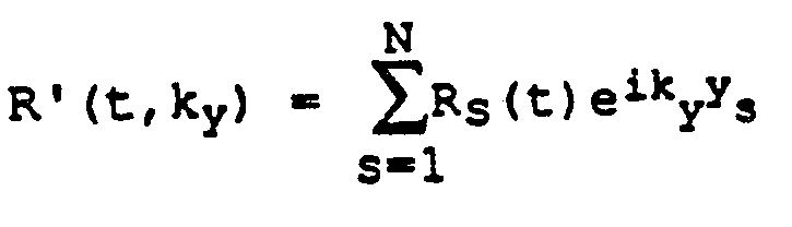
- Data from a series of slices can be acquired simultaneously by combining a corresponding number of such phase-offset modulation RF excitations. If R5(t) is the RF excitation pulse envelope for the sth slice in a desired set of N slices, then a composite RF excitation pulse R′(t,k y ) will simultaneously excite all N slices with offsets y s :
- a 2DFT reconstruction of the acquired NMR data will produce an image containing all N slices.
- the center of the image of the sth slice will be offset from the center of the entire reconstructed image in the y direction by an amount Y s .
- the N slices may be displayed without overlap.
- a still further aspect of the invention acquires NMR data simultaneously from a set of slices and reconstructs an image of each with an existing NMR system.
- the transmit frequency synthesizers on existing NMR systems can be controlled to produce the composite RF excitation R′(t,k y ).
- the NMR data is acquired and processed using existing NMR system hardware and software.
- the set of slice images which are produced in the image array may be easily separated for separate display or storage.
- a host computer system generally designated 100 which includes a main computer 101 (such as a Data General MV4000).
- the computer has associated therewith an interface 102 through which a plurality of computer peripheral devices and other NMR system components are coupled.
- a magnetic tape drive 104 which may be utilized under the direction of the main computer for archiving patient data and images to tape.
- Processed patient data may also be stored in an image disc storage device designated 110.
- An array processor 106 is utilized for preprocessing data and for image reconstruction.
- image processor 108 The function of image processor 108 is to provide interactive image display manipulation such as magnification, image comparison, gray-scale adjustment and realtime data display.
- the computer system is provided with a means to store raw data (i.e. before image construction) utilizing a disc data storage system designated 112.
- An operator console 116 is also coupled to the computer by means of interface 102 and provides the operator with the means to input data pertinent to a patient study as well as additional data necessary for proper NMR system operation, such as calibrating, initiating and terminating scans.
- the operator console is also used to display images stored on discs or magnetic tape.
- the computer system exercises control over the NMR system by means of system control 118 and gradient amplifier system 128.
- the computer 100 communicates with system control 118 by means of a serial digital communication network 103 (such as the Ethernet network) in a manner well known to those skilled in the art.
- the system control 118 includes several subsystems such as the pulse control module (PCM) 120, a radio frequency transceiver 122, a status control module (SCM) 124, and the power supplies generally designated 126 necessary to energize the components.
- the PCM 120 utilizes control signals provided by main computer 101 to generate digital timing and control signals such as the digital waveforms which control gradient coil excitation, as well as RF envelope waveforms utilized in the transceiver 122 for modulating the RF excitation pulses.
- the gradient waveforms are applied to the gradient amplifier system 128 generally comprised of G x , G y and G z amplifiers 130, 132 and 134, respectively.
- Each amplifier 130, 132 and 134 is utilized to excite a corresponding gradient coil in an assembly generally designated 136 and which is part of a magnet assembly 146.
- the gradient coils When energized, the gradient coils generate magnetic field gradients G x , G y and G z of the magnetic field in the same direction as the main polarizing magnetic field, wherein the gradients are directed in mutually orthogonal X-, Y- and Z-axis directions of a Cartesian coordinate system.
- the gradient magnetic fields are utilized in combination with radio frequency pulses generated by transceiver 122, RF amp 128 and RF coil 138 to encode spatial information into the NMR signals emanating from the region of the patient being studied.
- Waveforms and control signals provided by the pulse control module 120 are utilized by the transceiver subsystem 122 for RF carrier modulation and mode control.
- the transmitter In the transmit mode, the transmitter provides a radio frequency waveform modulated in accordance with the control signals to an RF power amplifier 123 which then energizes RF coils 138 which are situated within main magnet assembly 146.
- the NMR signals radiated by the excited nuclei in the patient are sensed by the same or a different RF coil than is used for transmitting.
- the signals are detected, amplified, demodulated, filtered, and digitized in the receiver section of the transceiver 122.
- the processed signals are transmitted to the main computer 101 for processing by means of a dedicated, unidirectional, high-speed digital link 105 which links interface 102 and transceiver 122.
- the PCM 120 and SCM 124 are independent subsystems both of which communicate with main computer 101, peripheral system, such as patient positioning system 152, as well as to one another by means of serial communications link 103.
- the PCM 120 and SCM 124 are each comprised of a 16-bit microprocessor (such as Intel 8086) for processing commands from the main computer 101.
- the SCM 124 includes means for acquiring information regarding patient cradle position, and the position of the moveable patient alignment light fan beam (not shown). This information is used by main computer 101 to modify image display and reconstruction parameters.
- the SCM 124 also initiates functions such as actuation of the patient transport and alignment systems.
- the gradient coil assembly 136 and the RF transmit and receiver coils 138 are mounted within the bore of the magnet utilized to produce the polarizing magnetic field.
- the magnet forms a part of the main magnet assembly which includes the patient alignment system 148, a shim coil power supply 140, and a main magnet power supply 142.
- the shim power supply 140 is utilized to energize shim coils associated with the main magnet and which are used to correct inhomogeneities in the polarizing magnetic field.
- main magnet power supply 142 is utilized to continuously energize the magnet.
- the main power supply 412 is utilized to bring the polarizing field produced by the magnet to the proper operating strength and is then disconnected.
- the patient alignment system 148 operates in combination with a patient cradle and transport system 150 and patient positioning system 152.
- the NMR system components comprised of the main magnet assembly, the gradient coil assembly, and the RF transmit and receiver coils, as well as the patient-handling devices, are enclosed in an RF-shielded room generally designated 144.
- the transceiver 122 includes components which produce the RF excitation field B1 through power amplifier 123 at a coil 138A and components which receive the resulting NMR signal induced in a coil 138B.
- the base, or carrier, frequency of the RF excitation field is produced by a frequency synthesizer 200 which receives a set of digital signals (CF) through the communications link 103 from the main computer 101. These digital signals indicate the frequency which is to be produced at an output 201 at a resolution of one Hertz.
- CF digital signals
- the commanded RF carrier is applied to a modulator 202 where it is modulated in response to a signal R(t) received through line 203 from the PCM 120.
- the signal R(t) is an analog signal that defines the envelope of the RF excitation pulse to be produced. It is produced in the PCM 120 by sequentially reading out a series of stored digital values that represent the desired envelope to a digital-to-analog converter that drives the line 203. The stored digital values may, in turn, be changed by the computer 100 to enable any desired RF pulse envelope to be produced.
- the resulting RF excitation signal is turned on and off in response to a control signal which is also received from the PCM 120 through line 204.
- the magnitude of the RF excitation pulse output through line 205 is attenuated by a transmit attenuator circuit 206 which receives a digital signal, TA, from the main computer 101 through communications link 103.
- the attenuated RF excitation pulses are applied to the power amplifier 123 that drives the RF transmitter coil 138A.
- the NMR signal produced by the subject is picked up by the receiver coil 138B and applied to the input of a receiver 207.
- the receiver 207 amplifies the NMR signal and this is attenuated by an amount determined by a digital attenuation signal (RA) received from the main computer 101 through link 103.
- RA digital attenuation signal
- the receiver 207 is also turned on and off by a signal through line 208 from the PCM 120 such that the NMR signal is acquired only over the time intervals required by the particular acquisition being performed.
- the received NMR signal is demodulated by a quadrature therefrom 209 to produce two signals I and Q that are coupled through filters to a pair of analog to digital converters indicated collectively at 215.
- These A/D converters are enabled by the control line 208 to produce digitized NMR data only when a valid signal is present. This data is output to the main computer 101 through line 105.
- the filters at the inputs to the A/D converters are controlled by the main computer 101 to limit the bandwidth of I and Q.
- the quadrature detector 209 also receives an RF reference signal from a second frequency synthesizer 210 and this is employed by the quadrature detector 209 to sense the amplitude of that component of the NMR signal which is in phase with the transmitter RF carrier and the amplitude of that component of the NMR signal which is in quadrature therewith.
- the frequency synthesizer 210 receives digital signals (CF) through the link 103 which determine the frequency of the demodulation signal.
- CF digital signals
- the phase of the two synthesizers 200 and 210 is locked together through a line 211, and in most measurements, the frequency of the two synthesizers is the same.
- Fig. 3 depicts two views of a conventional imaging pulse sequence of the type known as two-dimensional Fourier transform (2DFT), and which is frequently also referred to as two-dimensional "spin-warp" imaging.
- This pulse sequence is useful in obtaining, in a well-known manner, imaging NMR data to reconstruct images of an object being investigated.
- the two views are indicated at "A” and "B” and they are identical with the exception of the phase encoding gradient field G y .
- Each view is a pulse sequence which utilizes phase-alternated RF excitation pulses which, as disclosed and claimed in U.S. Patent No. 4,443,760, produce phase-alternated NMR signals S1(t) and S1′(t) to cancel certain baseline errors in the NMR system.
- Pulse control module 120 provides the needed control signals to the transmit frequency synthesizer 200 and modulator 202 so that the resulting excitation pulse is of the correct phase and frequency to excite nuclear spins only in a predetermined region, or slice, of the object being imaged.
- the excitation pulse is amplitude modulated by a (sin x)/x function.
- the frequency of the synthesizer 200 is dependent on the strength of the applied polarizing magnetic field B0 and the particular NMR species being imaged in accordance with the well-known Larmor equation.
- the pulse control module 120 also applies activating signals to the gradient amplifier 134 to generate, in this case, the G z gradient pulse.
- G x , G y and G z gradient pulses are applied simultaneously in interval 2.
- the G z gradient in interval 2 is a rephasing pulse typically selected such that the time integral of the gradient waveform over interval 2 is approximately equal to -1/2 of the time integral of the G z gradient waveform over interval 1.
- the function of the negative G z pulse is to rephase the nuclear spins excited in the selected slice during interval 1.
- the G y gradient pulse is a phase encoding pulse selected to have a different amplitude in each of Views A, B, ..., etc., to encode spatial information in the direction of the gradient.
- the number of different G y gradient amplitudes is typically selected to equal at least the number of pixel resolution elements the reconstructed image will have in the phase encoding (Y) direction.
- Y phase encoding
- 128, 256, or 512 different gradient amplitudes G y are selected and in the typical NMR system, the G y values are incremented a fixed amount from one view to the next until the NMR scan is complete.
- the G x gradient pulse in interval 2 is a dephasing pulse needed to dephase the excited nuclear spins by a predetermined amount to delay the time of occurrence of a spin-echo signal S1(t) in interval 4.
- the spin-echo signal is produced typically by the application of a 180° RF pulse in interval 3.
- the 180° RF pulse is a spatially selective pulse which reverses the direction of spin dephasing so as to produce the spin-echo signal.
- the spin-echo signal is sampled in interval 4 in the presence of a gradient pulse G x to encode spatial information in the direction (X) of this gradient.
- baseline error components are eliminated by using an additional NMR measurement in each view.
- This second measurement is substantially identical to the first with the exception that the RF excitation pulse in interval 5 of View A is selected to be 180° out of phase (as suggested by the minus sign) relative to the selective 90° excitation pulse in interval 1 of View A.
- the spin-echo signal S1′(t) in interval 8 is 180° out of phase with the spin-echo signal S1(t) in interval 4. If the signal S1′(t) is subtracted from S1(t), only those components of the signals with reversed sign in the signal S1′(t) are retained. The baseline error components thus cancel.
- the process described above with reference to View A is repeated for View B and so on for all amplitudes of the phase encoding G y gradient.
- the NMR data which is collected during this scan is stored in the computer system 100 where it is processed to produce image data suitable for controlling a CRT display.
- a complete scan of this type may be used to reconstruct a two-dimensional image of one slice through the patient. The location of this slice is determined by the center frequency of the selective 90° excitation pulse and the 180° reversal pulse, and its thickness is determined by the bandwidth of the pulses and their accompanying gradient pulse strengths.
- Fig. 3 depicts a single slice method wherein the duration of the sequence is the same as the repetition time T r .
- T r must be chosen, for contrast reasons, to be much longer than the actual time required for the sequence to acquire the data.
- other slices may be excited during the relaxation period so that multiple planes are imaged for the same scan time.
- T SCAN n ⁇ NEX ⁇ T r .
- T r the pulse sequence repetition time
- n the number of different G y phase encoding values used
- NEX the number of excitations at each G y
- Fig. 4 The use of a pulse sequences to acquire data from multiple planes is illustrated in Fig. 4, where NMR data is acquired from ten contiguous slices 1-10 distributed along the z axis.
- the data may be acquired in any order merely by changing the center frequency of the selective 90° excitation pulse to the frequency ( ⁇ 1 - ⁇ 10) of the next slice from which data is to be acquired.
- the center frequency may be changed from view to view either sequentially as shown in Fig. 4, or in the following sequence ⁇ 1, ⁇ 3, ⁇ 5, ⁇ 7, ⁇ 9, ⁇ 2, ⁇ 4, ⁇ 6, ⁇ 8, ⁇ 10.
- Acquisition of NMR data from ten slices in order to produce ten 256 by 256 pixel images might, for example, require a scan time of four minutes.
- the usual NMR scan produces a data set for each slice which is comprised of two, two-dimensional arrays of digital numbers, one array representing the amplitudes of the signals from the I output of the quadrature detector and the other array representing the Q signals (Fig. 2).
- each value in these two arrays is acquired when the phase encoding gradient field G y is at one of its discrete values (- ⁇ G y n/2 through + ⁇ G y n/2) and at one of the discrete sampling times (t1 through t T ).
- Such an image array is illustrated in Fig. 6 where the number of rows along the y axis is equal to the number (n) of NMR signals acquired with different phase encoding gradient values (G y ) and the number of columns along the x axis is equal to the number (m) of samples taken of each NMR signal during the read period of the pulse sequence.
- these pixel values can be mapped directly to a display memory array from which a human perceptible image can be produced which has n x m pixels.
- phase wrap it is known to produce data arrays which are larger than the image arrays that are mapped to the display memory.
- An example is to acquire twice the number of phase encoding values as before but with ⁇ G y halved. In this manner, the same total range of G y is covered but with twice the sampling in y, so that a 2n x m array of data is obtained.
- the resulting image array after Fourier transformation is 2n x m in size and the image occupies the entire, vertical, 2n space. As shown in Fig. 7, only the middle n x m portion of this 2n x m image array is mapped to the display memory and the top and bottom n/2 x m portions are thus unused. Any wrap around images which may occur on the upper or lower boundaries of the image are thus clipped off by this over sampling technique.
- This technique is important to the practice of the present embodiment in that the Fourier transformation algorithms in existing NMR systems are capable of processing 2n x m arrays.
- the present embodiment is an improvement to these well known techniques which enables NMR data to be acquired from two or more times as many slices in a given scan time.
- the inventive method may be used with nearly any existing pulse sequence which employs a slice selection RF excitation pulse as part of its imaging strategy, and it can be used with any NMR imaging system which employs a Fourier transformation of NMR data arrays into image arrays as described above.
- y is the vertical position in an image which is reconstructed with a Fourier transform
- the reconstructed image can be offset, or shifted upward, by a desired amount, y o .
- the image reconstructed from the NMR data can be offset in the y direction by multiplying the NMR data with a view dependent phase factor. This can be achieved by phase modulating the RF excitation pulse. For example, if R(t) represents the RF excitation pulse envelope for a given excitation, then excitation with R′(t, ky) in accordance with the following expression will result in the reconstruction of the same image offset by y o .
- R′(t,k y ) R(t)e ik y y 0 (4)
- the present embodiment makes use of this fact to excite two or more separate slices in the region of interest at the same time, acquire the NMR signal from both slices, and then reconstruct one image file that contains image data of both slices.
- one slice image is offset from the position of all other slice images such that they do not overlap.
- the separate slice images can, therefore, be displayed separately. In this manner, for a given scan time two or more times as many slices may be acquired and imaged.
- the embodiment is implemented by altering the frequency and phase content of the 90° and 180° RF excitation pulses.
- the mid-frequency of the envelope R1(t) is set to excite the first desired slice and the mid-frequency of the envelope R2(t) is set to excite the second desired slice.
- the values of y1 and y2 are selected such that the resulting two reconstructed images are offset from each other and do not overlap in the image array.
- each of the ten pulse sequences acquires NMR data from two different slices. While the invention is not limited to any particular grouping of slices, in the preferred embodiment the slices are grouped contiguously as follows: Pulse Sequence Slices R1(t) R2(t) 1 1 and 2 ⁇ 1 ⁇ 2 2 5 and 6 ⁇ 5 ⁇ 6 3 9 and 10 ⁇ 9 ⁇ 10 4 13 and 14 ⁇ 13 ⁇ 14 5 17 and 18 ⁇ 17 ⁇ 18 6 3 and 4 ⁇ 3 ⁇ 4 7 7 and 8 ⁇ 7 ⁇ 8 8 11 and 12 ⁇ 11 ⁇ 12 9 15 and 16 ⁇ 15 ⁇ 16 10 19 and 20 ⁇ 19 ⁇ 20
- NMR data can be acquired from all twenty slices in ten pulse sequences instead of the usual ten slices.
- the sequence is repeated with 2n different values of phase encoding gradient G y and consequent values of k y until the scan is complete.
- a 2n x m array of data from both the I and Q outputs of the quadrature detector 209 (Fig. 2) is thus obtained for each pair of slices.
- the I and Q 2n x m data arrays undergo complex Fourier transformations to produce a 2n x m image array.
- the 2n x m image array now contains two separate n x m images of the paired slices. These can be separately mapped to the n x m display memory or they can be separately stored for later use.
- the invention is not limited to the simultaneous acquisition of data from two slices. Additional frequency and phase components can be added to the RF excitation signals to excite numerous slices at the same time.
- the phase offsets (y1, y2 . . . y) of these components must be large enough that the reconstructed images will not overlap, and the data arrays and image arrays must be large enough to store all of the offset images. This can be assured by increasing the total number of acquisitions by the same factor N by which the slices are combined in accordance with the teachings of the present invention.
- N-fold signal averaging i.e. multiple NEX
- that sequence can be easily altered to produce N times as many slices in each reconstructed image, each with the expected signal-to-noise advantage of N fold averaging.
- the reconstructed image of the object which is obtained during conventional scanning does not fill the entire vertical extent, or field of view, of the n x m matrix.
- This can be achieved either by suitable choice of the field of view relative to the object extent, or by limiting the extent of the image using, for example, selective presaturating RF pulses to desensitize spins near the top and bottom of the object. It may be possible, therefore, to adequately represent the reconstructed object with half or less of the n vertical picture elements. In such cases, the invention may be practiced without requiring N-fold averaging. For example, if the object is only n/2 elements high, then two slices may be simultaneously excited and reconstructed in the n x m matrix for separation into two n/2 x m pictures.
Landscapes
- Physics & Mathematics (AREA)
- Optics & Photonics (AREA)
- Spectroscopy & Molecular Physics (AREA)
- High Energy & Nuclear Physics (AREA)
- Condensed Matter Physics & Semiconductors (AREA)
- General Physics & Mathematics (AREA)
- Magnetic Resonance Imaging Apparatus (AREA)
- Image Processing (AREA)
- Image Analysis (AREA)
Abstract
Description
- The field of the invention is nuclear magnetic resonance imaging methods and systems. An illustrative embodiment of the invention relates to a method for producing a larger number of images in a given scan time.
- Any nucleus which possesses a magnetic moment attempts to align itself with the direction of the magnetic field in which it is located. In doing so, however, the nucleus precesses around this direction at a characteristic angular frequency (Larmor frequency) which is dependent on the strength of the magnetic field and on the properties of the specific nuclear species (the magnetogyric constant γ of the nucleus). Nuclei which exhibit this phenomena are referred to herein as "spins".
- When a substance such as human tissue is subjected to a uniform magnetic field (polarizing field BZ), the individual magnetic moments of the spins in the tissue attempt to align with this polarizing field, but precess about it in random order at their characteristic Larmor frequency. A net magnetic moment Mz is produced in the direction of the polarizing field, but the randomly oriented magnetic components in the perpendicular, or transverse, plane (x-y plane) cancel one another. If, however, the substance, or tissue, is subjected to a magnetic field (excitation field B₁) which is in the x-y plane and which is near the Larmor frequency, the net aligned moment, Mz, may be rotated, or "tipped", into the z-y plane to produce a net transverse magnetic moment Ml, which is rotating, or spinning, in the x-y plane at the Larmor frequency. The degree to which the net magnetic moment Mz is tipped, and hence, the magnitude of the net transverse magnetic moment Ml depends primarily on the length of time and magnitude of the applied excitation field B₁ .
- The practical value of this phenomenon resides in the signal which is emitted by the excited spins after the excitation signal B₁ is terminated. In simple systems the excited nuclei induce an oscillating sine wave signal in a receiving coil. The frequency of this signal is the Larmor frequency, and its initial amplitude, A₀, is determined by the magnitude of the transverse magnetic moment M₁. The amplitude, A, of the emission signal decays in an exponential fashion with time, t:
A =A₀e -t/T*₂ - The decay constant 1/T*₂ depends on the homogeneity of the magnetic field and on T₂, which is referred to as the "spin-spin relaxation" constant, or the "transverse relaxation" constant. The T₂ constant is inversely proportional to the exponential rate at which the aligned precession of the spins would dephase after removal of the excitation signal B₁ in a perfectly homogeneous field.
- Another important factor which contributes to the amplitude A of the NMR signal is referred to as the spin-lattice relaxation process which is characterized by the time constant T₁. This is also called the longitudinal relaxation process as it describes the recovery of the net magnetic moment M to its equilibrium value along the axis of magnetic polarization (z). The T₁ time constant is longer than T₂, much longer in most substances of medical interest. If the net magnetic moment M is not given sufficient time to relax to its equilibrium value, the amplitude A of the NMR signal produced in a subsequent pulse sequence will be reduced.
- The NMR measurements of particular relevance to the present invention are called "pulsed NMR measurements". Such NMR measurements are divided into a period of excitation and a period of signal emission. Such measurements are performed in a cyclic manner in which the NMR measurement is repeated many times to accumulate different data during each cycle or to make the same measurement at different locations in the subject. A wide variety of preparative excitation techniques are known which involve the application of one or more RF excitation pulses (B₁) of varying magnitude, frequency content and duration. Such RF excitation pulses may have a narrow frequency spectrum (selective excitation pulse), or they may have a broad frequency spectrum (nonselective excitation pulse) which produces transverse magnetization M₁ over a range of resonant frequencies. The prior art is replete with excitation techniques that are designed to take advantage of particular NMR phenomena and which overcome particular problems in the NMR measurement process.
- When utilizing NMR to produce images, a technique is employed to obtain NMR signals from specific locations in the subject. Typically, the region which is to be imaged (region of interest) is subjected to a sequence of NMR measurement cycles which vary according to the particular localization method being used. The received NMR signals are digitized and processed to reconstruct the image using one of many well known reconstruction techniques. To perform such a scan, it is, of course, necessary to elicit NMR signals from specific locations in the subject. This is accomplished by employing magnetic fields (Gx, Gy, and Gz) which have the same direction as the polarizing field B₀, but which have a gradient along the respective x, y and z axes. By controlling the strength of these gradients during each NMR cycle, the spatial distribution of spin excitation can be controlled and the location of the NMR signals can be identified.
- NMR has rapidly developed into an imaging modality which is utilized to obtain tomographic, projection and volumetric images of anatomical features of live human subjects. Such images depict the nuclear-spin distribution (typically protons associated with water and fat), modified by specific NMR properties of tissues, such as spin lattice (T₁) and spin-spin (T₂) relaxation time constants. They are of medical diagnostic value because they depict anatomy and allow tissue characterization.
- One of the difficulties in using NMR in medical applications is the time required to acquire sufficient NMR data from which an image can be reconstructed. Many pulse sequences must be executed in order to acquire sufficient NMR data, and as indicated above, sufficient relaxation time must be allowed between successive excitations of the spin in order to produce NMR signals of appropriate amplitude A. As a result, it is common practice to acquire NMR data in a time-sequential fashion from a set of spatially-disparate slices in the region of interest so that spin magnetization can relax in one slice while the other slices are being excited. Such techniques are commonly referred to as multi-slice, two-dimensional methods. In most cases a variant of the "spin warp" data collection method is used wherein the raw data space is filled rectilinearly, and the image is produced by a simple two-dimensional Fourier transformation. Such methods are known as 2DFT methods.
- The maximum number of slices which can be acquired in a given scan without extending the total scan time is determined by the duration of each pulse sequence and the relaxation time required between successive excitations of the spin in any single slice. If more relaxation time is allocated, then time is available to acquire more NMR data from other slices. Of course, the total scan time is proportionally increased and the usual practice is to keep the allocated relaxation time at the minimum amount needed to enable adequate NMR signals and contrast to be produced. And, of course, efforts are always made to reduce the length of each pulse sequence, but ultimately, a limit is reached for any given NMR measurement cycle.
- One aspect of the present invention relates to a method for increasing the number of slices which can be imaged with a given NMR pulse sequence, relaxation time and total scan time.
- Another aspect of the present invention is an improvement to any NMR 2DFT pulse sequence in which the image is reconstructed from the acquired NMR data using a Fourier transformation for which NMR data is simultaneously acquired from a plurality of slices by employing RF excitation pulses which are phase modulated such that the reconstructed image array includes images of each slice that are offset from each other in the phase encoding direction and can be separately displayed or stored.
- A further aspect of the invention acquires NMR data from a plurality of slices simultaneously and reconstructs an image from such data in which each slice is shown separately. If R(t) represents the RF excitation pulse envelope for a given slice in the NMR pulse sequence, then excitation with an RF pulse having the following envelope will produce a reconstructed image for that slice which is offset in the phase encoding direction (y direction) by a distance y₀:
R′ (t,ky) = R(t)eik y y₀
where ky denotes the current value of the index in the phase encoding direction and is proportional to the area under the phase encoding gradient pulse. Thus, R′(t,ky ) varies from view to view in contrast to normal 2DFT methods in which only the phase encoding gradient pulses vary from view to view. - Data from a series of slices can be acquired simultaneously by combining a corresponding number of such phase-offset modulation RF excitations. If R₅(t) is the RF excitation pulse envelope for the sth slice in a desired set of N slices, then a composite RF excitation pulse R′(t,ky) will simultaneously excite all N slices with offsets ys:

- A 2DFT reconstruction of the acquired NMR data will produce an image containing all N slices. The center of the image of the sth slice will be offset from the center of the entire reconstructed image in the y direction by an amount Ys . Thus, by suitable choice of ys and a suitable reconstruction field of view in the y direction, the N slices may be displayed without overlap.
- A still further aspect of the invention acquires NMR data simultaneously from a set of slices and reconstructs an image of each with an existing NMR system. The transmit frequency synthesizers on existing NMR systems can be controlled to produce the composite RF excitation R′(t,ky). Similarly, the NMR data is acquired and processed using existing NMR system hardware and software. The set of slice images which are produced in the image array may be easily separated for separate display or storage.
- A better understanding of the invention will appear from the following description. In the description, reference is made to the accompanying drawings which form a part hereof, and in which there is shown by way of illustration a preferred embodiment of the invention.
- In the drawings:-
- Fig. 1 is a block diagram of an NMR system in which the present invention may be practised;
- Fig. 2 is an electrical block diagram of the transceiver which forms part of the NMR system of Fig. 1;
- Fig. 3 is a graphic representation of a conventional pulse sequence which may be adapted to incorporate the present invention;
- Fig. 4 is a graphic representation of a multiplanar region of interest which is scanned by the NMR system of Fig. 1;
- Fig. 5 is a graphic representation of an n x m NMR data array which is acquired by the NMR system of Fig. 1;
- Fig. 6 is a graphic representation of an n x m image array which is produced from the NMR data array of Fig. 5 in the NMR system of Fig. 1;
- Fig. 7 is a graphic representation of an 2n x m NMR data array which is acquired by the NMR system of Fig. 1; and
- Fig. 8 is a graphic representation of a 2n x m image array which is produced from the data array of Fig. 7 when a method in accordance with the present invention is employed in the NMR system of Fig. 1.
- Referring first to Fig. 1, there is shown in block diagram form the major components of an NMR system which is sold by the General Electric Company under the trademark "SIGNA". The overall operation of the system is under the control of a host computer system generally designated 100 which includes a main computer 101 (such as a Data General MV4000). The computer has associated therewith an
interface 102 through which a plurality of computer peripheral devices and other NMR system components are coupled. Among the computer peripheral devices is amagnetic tape drive 104 which may be utilized under the direction of the main computer for archiving patient data and images to tape. Processed patient data may also be stored in an image disc storage device designated 110. Anarray processor 106 is utilized for preprocessing data and for image reconstruction. The function of image processor 108 is to provide interactive image display manipulation such as magnification, image comparison, gray-scale adjustment and realtime data display. The computer system is provided with a means to store raw data (i.e. before image construction) utilizing a disc data storage system designated 112. Anoperator console 116 is also coupled to the computer by means ofinterface 102 and provides the operator with the means to input data pertinent to a patient study as well as additional data necessary for proper NMR system operation, such as calibrating, initiating and terminating scans. The operator console is also used to display images stored on discs or magnetic tape. - The computer system exercises control over the NMR system by means of
system control 118 and gradient amplifier system 128. Thecomputer 100 communicates withsystem control 118 by means of a serial digital communication network 103 (such as the Ethernet network) in a manner well known to those skilled in the art. Thesystem control 118 includes several subsystems such as the pulse control module (PCM) 120, aradio frequency transceiver 122, a status control module (SCM) 124, and the power supplies generally designated 126 necessary to energize the components. ThePCM 120 utilizes control signals provided bymain computer 101 to generate digital timing and control signals such as the digital waveforms which control gradient coil excitation, as well as RF envelope waveforms utilized in thetransceiver 122 for modulating the RF excitation pulses. The gradient waveforms are applied to the gradient amplifier system 128 generally comprised of Gx, Gy and Gz amplifiers 130, 132 and 134, respectively. Eachamplifier - The gradient magnetic fields are utilized in combination with radio frequency pulses generated by
transceiver 122, RF amp 128 andRF coil 138 to encode spatial information into the NMR signals emanating from the region of the patient being studied. Waveforms and control signals provided by thepulse control module 120 are utilized by thetransceiver subsystem 122 for RF carrier modulation and mode control. In the transmit mode, the transmitter provides a radio frequency waveform modulated in accordance with the control signals to anRF power amplifier 123 which then energizes RF coils 138 which are situated within main magnet assembly 146. The NMR signals radiated by the excited nuclei in the patient are sensed by the same or a different RF coil than is used for transmitting. The signals are detected, amplified, demodulated, filtered, and digitized in the receiver section of thetransceiver 122. The processed signals are transmitted to themain computer 101 for processing by means of a dedicated, unidirectional, high-speeddigital link 105 which links interface 102 andtransceiver 122. - The
PCM 120 andSCM 124 are independent subsystems both of which communicate withmain computer 101, peripheral system, such aspatient positioning system 152, as well as to one another by means of serial communications link 103. ThePCM 120 andSCM 124 are each comprised of a 16-bit microprocessor (such as Intel 8086) for processing commands from themain computer 101. TheSCM 124 includes means for acquiring information regarding patient cradle position, and the position of the moveable patient alignment light fan beam (not shown). This information is used bymain computer 101 to modify image display and reconstruction parameters. TheSCM 124 also initiates functions such as actuation of the patient transport and alignment systems. - The
gradient coil assembly 136 and the RF transmit andreceiver coils 138 are mounted within the bore of the magnet utilized to produce the polarizing magnetic field. The magnet forms a part of the main magnet assembly which includes thepatient alignment system 148, a shimcoil power supply 140, and a mainmagnet power supply 142. Theshim power supply 140 is utilized to energize shim coils associated with the main magnet and which are used to correct inhomogeneities in the polarizing magnetic field. In the case of a resistive magnet, mainmagnet power supply 142 is utilized to continuously energize the magnet. In the case of a superconductive magnet, the main power supply 412 is utilized to bring the polarizing field produced by the magnet to the proper operating strength and is then disconnected. In the case of a permanent magnet,power supply 142 would not be needed. Thepatient alignment system 148 operates in combination with a patient cradle andtransport system 150 andpatient positioning system 152. To minimize interference from external sources, the NMR system components comprised of the main magnet assembly, the gradient coil assembly, and the RF transmit and receiver coils, as well as the patient-handling devices, are enclosed in an RF-shielded room generally designated 144. - Referring particularly to Figs. 1 and 2, the
transceiver 122 includes components which produce the RF excitation field B₁ throughpower amplifier 123 at acoil 138A and components which receive the resulting NMR signal induced in acoil 138B. The base, or carrier, frequency of the RF excitation field is produced by afrequency synthesizer 200 which receives a set of digital signals (CF) through the communications link 103 from themain computer 101. These digital signals indicate the frequency which is to be produced at an output 201 at a resolution of one Hertz. As discussed in U.S. Patent Nos. 4,602,226; 4,694,254; and 4,682,106, which are incorporated herein by reference, the commanded RF carrier is applied to amodulator 202 where it is modulated in response to a signal R(t) received throughline 203 from thePCM 120. The signal R(t) is an analog signal that defines the envelope of the RF excitation pulse to be produced. It is produced in thePCM 120 by sequentially reading out a series of stored digital values that represent the desired envelope to a digital-to-analog converter that drives theline 203. The stored digital values may, in turn, be changed by thecomputer 100 to enable any desired RF pulse envelope to be produced. The resulting RF excitation signal is turned on and off in response to a control signal which is also received from thePCM 120 throughline 204. The magnitude of the RF excitation pulse output throughline 205 is attenuated by a transmitattenuator circuit 206 which receives a digital signal, TA, from themain computer 101 through communications link 103. The attenuated RF excitation pulses are applied to thepower amplifier 123 that drives theRF transmitter coil 138A. - Referring still to Figs. 1 and 2 the NMR signal produced by the subject is picked up by the
receiver coil 138B and applied to the input of areceiver 207. Thereceiver 207 amplifies the NMR signal and this is attenuated by an amount determined by a digital attenuation signal (RA) received from themain computer 101 through link 103. Thereceiver 207 is also turned on and off by a signal throughline 208 from thePCM 120 such that the NMR signal is acquired only over the time intervals required by the particular acquisition being performed. - The received NMR signal is demodulated by a quadrature therefrom 209 to produce two signals I and Q that are coupled through filters to a pair of analog to digital converters indicated collectively at 215. These A/D converters are enabled by the
control line 208 to produce digitized NMR data only when a valid signal is present. This data is output to themain computer 101 throughline 105. The filters at the inputs to the A/D converters are controlled by themain computer 101 to limit the bandwidth of I and Q. Thequadrature detector 209 also receives an RF reference signal from asecond frequency synthesizer 210 and this is employed by thequadrature detector 209 to sense the amplitude of that component of the NMR signal which is in phase with the transmitter RF carrier and the amplitude of that component of the NMR signal which is in quadrature therewith. Thefrequency synthesizer 210 receives digital signals (CF) through the link 103 which determine the frequency of the demodulation signal. The phase of the twosynthesizers line 211, and in most measurements, the frequency of the two synthesizers is the same. - Reference is made to Fig. 3 which depicts two views of a conventional imaging pulse sequence of the type known as two-dimensional Fourier transform (2DFT), and which is frequently also referred to as two-dimensional "spin-warp" imaging. This pulse sequence is useful in obtaining, in a well-known manner, imaging NMR data to reconstruct images of an object being investigated. The two views are indicated at "A" and "B" and they are identical with the exception of the phase encoding gradient field Gy. Each view is a pulse sequence which utilizes phase-alternated RF excitation pulses which, as disclosed and claimed in U.S. Patent No. 4,443,760, produce phase-alternated NMR signals S₁(t) and S₁′(t) to cancel certain baseline errors in the NMR system.
- Referring now to View A in Fig. 3, there is shown in interval 1 (indicated along the horizontal axis) a selective 90° RF excitation pulse applied in the presence of a positive Gz magnetic field gradient pulse.
Pulse control module 120 provides the needed control signals to the transmitfrequency synthesizer 200 andmodulator 202 so that the resulting excitation pulse is of the correct phase and frequency to excite nuclear spins only in a predetermined region, or slice, of the object being imaged. Typically, the excitation pulse is amplitude modulated by a (sin x)/x function. The frequency of thesynthesizer 200 is dependent on the strength of the applied polarizing magnetic field B₀ and the particular NMR species being imaged in accordance with the well-known Larmor equation. Thepulse control module 120 also applies activating signals to thegradient amplifier 134 to generate, in this case, the Gz gradient pulse. - Continuing with reference to Fig. 3, Gx, Gy and Gz gradient pulses are applied simultaneously in
interval 2. The Gz gradient ininterval 2 is a rephasing pulse typically selected such that the time integral of the gradient waveform overinterval 2 is approximately equal to -1/2 of the time integral of the Gz gradient waveform overinterval 1. The function of the negative Gz pulse is to rephase the nuclear spins excited in the selected slice duringinterval 1. The Gy gradient pulse is a phase encoding pulse selected to have a different amplitude in each of Views A, B, ..., etc., to encode spatial information in the direction of the gradient. The number of different Gy gradient amplitudes is typically selected to equal at least the number of pixel resolution elements the reconstructed image will have in the phase encoding (Y) direction. Typically, 128, 256, or 512 different gradient amplitudes Gy are selected and in the typical NMR system, the Gy values are incremented a fixed amount from one view to the next until the NMR scan is complete. - The Gx gradient pulse in
interval 2 is a dephasing pulse needed to dephase the excited nuclear spins by a predetermined amount to delay the time of occurrence of a spin-echo signal S₁(t) ininterval 4. The spin-echo signal is produced typically by the application of a 180° RF pulse ininterval 3. As is known, the 180° RF pulse is a spatially selective pulse which reverses the direction of spin dephasing so as to produce the spin-echo signal. The spin-echo signal is sampled ininterval 4 in the presence of a gradient pulse Gx to encode spatial information in the direction (X) of this gradient. - As indicated above, baseline error components are eliminated by using an additional NMR measurement in each view. This second measurement is substantially identical to the first with the exception that the RF excitation pulse in
interval 5 of View A is selected to be 180° out of phase (as suggested by the minus sign) relative to the selective 90° excitation pulse ininterval 1 of View A. As a result, the spin-echo signal S₁′(t) ininterval 8 is 180° out of phase with the spin-echo signal S₁(t) ininterval 4. If the signal S₁′(t) is subtracted from S₁(t), only those components of the signals with reversed sign in the signal S₁′(t) are retained. The baseline error components thus cancel. - The process described above with reference to View A is repeated for View B and so on for all amplitudes of the phase encoding Gy gradient. The NMR data which is collected during this scan is stored in the
computer system 100 where it is processed to produce image data suitable for controlling a CRT display. A complete scan of this type may be used to reconstruct a two-dimensional image of one slice through the patient. The location of this slice is determined by the center frequency of the selective 90° excitation pulse and the 180° reversal pulse, and its thickness is determined by the bandwidth of the pulses and their accompanying gradient pulse strengths. - The sequence illustrated in Fig. 3 depicts a single slice method wherein the duration of the sequence is the same as the repetition time Tr. However, often Tr must be chosen, for contrast reasons, to be much longer than the actual time required for the sequence to acquire the data. In such cases, other slices may be excited during the relaxation period so that multiple planes are imaged for the same scan time. Fig. 4 illustrates this multiplanar technique, where the sequence time T₅ is now the time required to perform one sequence (Tr in Fig. 3). In this example Ts = Tr/10 so that 10 slices are imaged. The time required to perform the scan TSCAN depends on the pulse sequence repetition time (Tr), the number of different Gy phase encoding values used (n), and the number of excitations at each Gy (NEX):
TSCAN = n·NEX·Tr.
For a NEX = 2 scan, such as that shown in Fig. 3, with a Tr of 500 msec and n = 256 phase encodings, the total scan time is 256 seconds. The number of slices which may be acquired in this scan depends on the duration (Ts) of each pulse sequence. - The use of a pulse sequences to acquire data from multiple planes is illustrated in Fig. 4, where NMR data is acquired from ten contiguous slices 1-10 distributed along the z axis. The data may be acquired in any order merely by changing the center frequency of the selective 90° excitation pulse to the frequency (ω₁ - ω₁₀) of the next slice from which data is to be acquired. For example, the center frequency may be changed from view to view either sequentially as shown in Fig. 4, or in the following sequence ω₁, ω₃, ω₅, ω₇, ω₉, ω₂, ω₄, ω₆, ω₈, ω₁₀. Acquisition of NMR data from ten slices in order to produce ten 256 by 256 pixel images might, for example, require a scan time of four minutes.
- The usual NMR scan produces a data set for each slice which is comprised of two, two-dimensional arrays of digital numbers, one array representing the amplitudes of the signals from the I output of the quadrature detector and the other array representing the Q signals (Fig. 2). As shown in Fig. 5, each value in these two arrays is acquired when the phase encoding gradient field Gy is at one of its discrete values (-ΔGyn/2 through +ΔGyn/2) and at one of the discrete sampling times (t₁ through tT). By performing a complex Fourier transform on the I and Q data arrays, an array of values which indicates the intensity of pixels in a two-dimensional image is produced.
- Such an image array is illustrated in Fig. 6 where the number of rows along the y axis is equal to the number (n) of NMR signals acquired with different phase encoding gradient values (Gy) and the number of columns along the x axis is equal to the number (m) of samples taken of each NMR signal during the read period of the pulse sequence. After adding an offset to each pixel value and limiting the range of values to that of the display device being used, these pixel values can be mapped directly to a display memory array from which a human perceptible image can be produced which has n x m pixels.
- To overcome a problem known as "phase wrap", it is known to produce data arrays which are larger than the image arrays that are mapped to the display memory. An example is to acquire twice the number of phase encoding values as before but with ΔGy halved. In this manner, the same total range of Gy is covered but with twice the sampling in y, so that a 2n x m array of data is obtained. The resulting image array after Fourier transformation is 2n x m in size and the image occupies the entire, vertical, 2n space. As shown in Fig. 7, only the middle n x m portion of this 2n x m image array is mapped to the display memory and the top and bottom n/2 x m portions are thus unused. Any wrap around images which may occur on the upper or lower boundaries of the image are thus clipped off by this over sampling technique. This technique is important to the practice of the present embodiment in that the Fourier transformation algorithms in existing NMR systems are capable of processing 2n x m arrays.
- The present embodiment is an improvement to these well known techniques which enables NMR data to be acquired from two or more times as many slices in a given scan time. The inventive method may be used with nearly any existing pulse sequence which employs a slice selection RF excitation pulse as part of its imaging strategy, and it can be used with any NMR imaging system which employs a Fourier transformation of NMR data arrays into image arrays as described above.
- If F(ky) is the Fourier transform of a function f(y), then we have the relationship:
f(y) = ∫F(ky)e-ik y ydky (1) - From a well known theorem in Fourier analysis, we also know the following: f(y-y₀) = ∫F′(ky)e-ik y ydky (2) where: F′(ky) = F(ky)eik y y o (3)
- If y is the vertical position in an image which is reconstructed with a Fourier transform, then by simply multiplying F by a ky-dependent phase factor, the reconstructed image can be offset, or shifted upward, by a desired amount, yo.
- As indicated above, in NMR imaging y represents the phase encoding direction and ky is proportional to the area under the Gy phase encoding magnetic field gradient pulses. Thus, in accordance with equation (3), the image reconstructed from the NMR data can be offset in the y direction by multiplying the NMR data with a view dependent phase factor. This can be achieved by phase modulating the RF excitation pulse. For example, if R(t) represents the RF excitation pulse envelope for a given excitation, then excitation with R′(t, ky) in accordance with the following expression will result in the reconstruction of the same image offset by yo.
R′(t,ky) = R(t)eik y y₀ (4) - The present embodiment makes use of this fact to excite two or more separate slices in the region of interest at the same time, acquire the NMR signal from both slices, and then reconstruct one image file that contains image data of both slices. By selecting the offsets large enough, one slice image is offset from the position of all other slice images such that they do not overlap. The separate slice images can, therefore, be displayed separately. In this manner, for a given scan time two or more times as many slices may be acquired and imaged.
- Referring again to Fig. 3, the embodiment is implemented by altering the frequency and phase content of the 90° and 180° RF excitation pulses. In a preferred embodiment, two slices are excited simultaneously by these excitation pulses (N=2), however, it can be appreciated that this number may be increased if more than two slices are to be excited and produced in the same reconstructed image. The envelope for these RF excitation pulses is as follows:
R′(t,ky) = R₁(t)e ik y y₁ + R₂(t)eik y y₂ (5) - The mid-frequency of the envelope R₁(t) is set to excite the first desired slice and the mid-frequency of the envelope R₂(t) is set to excite the second desired slice. The values of y₁ and y₂ are selected such that the resulting two reconstructed images are offset from each other and do not overlap in the image array. These RF excitation pulses are produced by the transmit
frequency synthesizer 200 andmodulator 202 as described above with reference to Fig. 2. - If NMR data for twenty slices is to be acquired during the scan, each of the ten pulse sequences according to the preferred embodiment of the present invention acquires NMR data from two different slices. While the invention is not limited to any particular grouping of slices, in the preferred embodiment the slices are grouped contiguously as follows:
Pulse Sequence Slices R₁(t) R₂(t) 1 1 and 2 ω₁ ω₂ 2 5 and 6 ω₅ ω₆ 3 9 and 10 ω₉ ω₁₀ 4 13 and 14 ω₁₃ ω₁₄ 5 17 and 18 ω₁₇ ω₁₈ 6 3 and 4 ω₃ ω₄ 7 7 and 8 ω₇ ω₈ 8 11 and 12 ω₁₁ ω₁₂ 9 15 and 16 ω₁₅ ω₁₆ 10 19 and 20 ω₁₉ ω₂₀ - It should be apparent that NMR data can be acquired from all twenty slices in ten pulse sequences instead of the usual ten slices. The sequence is repeated with 2n different values of phase encoding gradient Gy and consequent values of ky until the scan is complete. A 2n x m array of data from both the I and Q outputs of the quadrature detector 209 (Fig. 2) is thus obtained for each pair of slices.
- Processing of the acquired NMR data is carried out in the usual fashion. The I and
Q 2n x m data arrays undergo complex Fourier transformations to produce a 2n x m image array. As shown in Fig. 8, however, the 2n x m image array now contains two separate n x m images of the paired slices. These can be separately mapped to the n x m display memory or they can be separately stored for later use. - It should be apparent to those skilled in the art that the invention is not limited to the simultaneous acquisition of data from two slices. Additional frequency and phase components can be added to the RF excitation signals to excite numerous slices at the same time. Of course, the phase offsets (y₁, y₂ . . . y) of these components must be large enough that the reconstructed images will not overlap, and the data arrays and image arrays must be large enough to store all of the offset images. This can be assured by increasing the total number of acquisitions by the same factor N by which the slices are combined in accordance with the teachings of the present invention. Thus, if N-fold signal averaging (i.e. multiple NEX) acquisitions are performed in a conventional scan for signal to noise improvement, that sequence can be easily altered to produce N times as many slices in each reconstructed image, each with the expected signal-to-noise advantage of N fold averaging.
- In some cases, the reconstructed image of the object which is obtained during conventional scanning does not fill the entire vertical extent, or field of view, of the n x m matrix. This can be achieved either by suitable choice of the field of view relative to the object extent, or by limiting the extent of the image using, for example, selective presaturating RF pulses to desensitize spins near the top and bottom of the object. It may be possible, therefore, to adequately represent the reconstructed object with half or less of the n vertical picture elements. In such cases, the invention may be practiced without requiring N-fold averaging. For example, if the object is only n/2 elements high, then two slices may be simultaneously excited and reconstructed in the n x m matrix for separation into two n/2 x m pictures.
Claims (5)
applying a composite RF excitation pulse during each NMR pulse sequence which contains a plurality of pulse envelope terms multiplied by a sequence number dependent phase factor, and which excites spins in a plurality of slices which are selected by the frequency of a respective one of said plurality of pulse envelope terms in the composite RF excitation pulse;
performing a Fourier transform on the NMR data set which is acquired by the set of NMR pulse sequences to produce an image array; and
separating the slice images contained in the image array to produce a separate image array for each of said plurality of slices.
Rs(t) is the pulse envelope for each term,
N is the number of slices in which spins are simultaneously excited,
ky is a sequence number dependent factor, and
Ys is a desired amount of offset which enables the slice image to be separated.
applying a composite RF excitation pulse during each NMR pulse sequence which contains a plurality of pulse envelope terms multiplied by a sequence number dependent phase factor, and which excites spins in a plurality of slices which are selected by the frequency of a respective one of said plurality of pulse envelope terms in the composite RF excitation pulse;
performing a Fourier transform on the NMR data set which is acquired by the set of NMR pulse sequences to produce an image array; and
separating the slice images contained in the image array to produce a separate image array for each of said plurality of slices.

Rs(t) is the pulse envelope for each term,
N is the number of slices in which spins are simultaneously excited,
ky is a sequence number dependent factor, and
ys is a desired amount of offset which enables the slice image to be separated.
Applications Claiming Priority (2)
| Application Number | Priority Date | Filing Date | Title |
|---|---|---|---|
| US07/234,721 US4843322A (en) | 1988-08-15 | 1988-08-15 | Method for producing multi-slice NMR images |
| US234721 | 1999-01-21 |
Publications (3)
| Publication Number | Publication Date |
|---|---|
| EP0366232A2 true EP0366232A2 (en) | 1990-05-02 |
| EP0366232A3 EP0366232A3 (en) | 1991-02-06 |
| EP0366232B1 EP0366232B1 (en) | 1995-11-08 |
Family
ID=22882526
Family Applications (1)
| Application Number | Title | Priority Date | Filing Date |
|---|---|---|---|
| EP89308241A Expired - Lifetime EP0366232B1 (en) | 1988-08-15 | 1989-08-14 | Method for producing multi-slice nmr images |
Country Status (5)
| Country | Link |
|---|---|
| US (1) | US4843322A (en) |
| EP (1) | EP0366232B1 (en) |
| JP (1) | JPH02142536A (en) |
| DE (1) | DE68924753T2 (en) |
| IL (1) | IL90810A (en) |
Cited By (2)
| Publication number | Priority date | Publication date | Assignee | Title |
|---|---|---|---|---|
| EP1014102A3 (en) * | 1998-12-24 | 2001-10-04 | Marconi Electronic Systems Limited | Multislice magnetic resonance imaging using an array of receiving coils |
| US7180289B2 (en) | 2002-06-10 | 2007-02-20 | Koninklijke Philips Electronics N.V. | Magnetic resonance imaging method |
Families Citing this family (33)
| Publication number | Priority date | Publication date | Assignee | Title |
|---|---|---|---|---|
| US4885538A (en) * | 1988-08-19 | 1989-12-05 | The Regents Of The University Of California | Low data rate low noise serial digital communication link for magnetic resonance imaging systems |
| DE3926889A1 (en) * | 1989-08-16 | 1991-02-21 | Spectrospin Ag | METHOD FOR SIMULTANEOUSLY MEASURING NMR SIGNALS, IN PARTICULAR FOR DETERMINING FLOW SPEEDS IN NUCLEAR SPIN TOMOGRAPHY USING THE MULTIPLE SLICE FOURIER FLOW (= MUFF) METHOD |
| DE3937428A1 (en) * | 1989-11-10 | 1991-05-16 | Philips Patentverwaltung | Nuclear spin tomography method for generating separated fat and water images and arrangement for carrying out the method |
| US5222500A (en) * | 1990-06-22 | 1993-06-29 | Kabushiki Kaisha Toshiba | Magnetic resonance imaging method and apparatus for the same |
| US5237273A (en) * | 1991-05-22 | 1993-08-17 | General Electric Company | Interleaved acquisition of multi-slice NMR data |
| US5304931A (en) * | 1991-08-09 | 1994-04-19 | Flamig Duane P | Magnetic resonance imaging techniques |
| US5202631A (en) * | 1991-08-09 | 1993-04-13 | Steven E. Harms | Magnetic resonance imaging techniques utilizing multiple shaped radiofrequency pulse sequences |
| DE4137217C2 (en) * | 1991-11-13 | 1993-10-07 | Hennig Juergen | Magnetic resonance imaging |
| US5422572A (en) * | 1993-08-06 | 1995-06-06 | Toshiba America Mri, Inc. | Method and apparatus for substantially simultaneously exciting a plurality of slices in NMR imaging |
| DE4435104A1 (en) * | 1994-09-30 | 1996-04-04 | Siemens Ag | Pulse sequence with multi-layer excitation |
| DE4435464A1 (en) * | 1994-10-04 | 1996-04-11 | Philips Patentverwaltung | Multislice MR procedure |
| US5510713A (en) * | 1995-06-30 | 1996-04-23 | General Electric Company | Technique for selective fat saturation in MR imaging |
| DE19607023A1 (en) * | 1996-02-24 | 1997-08-28 | Philips Patentverwaltung | MR procedure with reduced movement artifacts |
| DE19814950A1 (en) * | 1998-04-03 | 1999-10-07 | Univ Ludwigs Albert | Nuclear magnetic resonance process for patient examinations, especially for brain excitation |
| US6560477B1 (en) | 2000-03-17 | 2003-05-06 | The Regents Of The University Of California | Joint imaging system utilizing magnetic resonance imaging and associated methods |
| US6998841B1 (en) * | 2000-03-31 | 2006-02-14 | Virtualscopics, Llc | Method and system which forms an isotropic, high-resolution, three-dimensional diagnostic image of a subject from two-dimensional image data scans |
| US7242190B1 (en) | 2001-01-26 | 2007-07-10 | Fonar Corporation | Driven equilibrium and fast-spin echo scanning |
| US7157909B1 (en) | 2001-01-26 | 2007-01-02 | Fonar Corporation | Driven equilibrium and fast-spin echo scanning |
| US7339375B1 (en) | 2001-01-26 | 2008-03-04 | Fonar Corporation | Driven equilibrium and fast-spin echo scanning |
| WO2002082376A2 (en) * | 2001-04-06 | 2002-10-17 | Regents Of The University Of California | Method for analyzing mri diffusion data |
| DE10152734B4 (en) * | 2001-10-25 | 2005-12-29 | Siemens Ag | Apparatus and method for magnetic resonance imaging with simultaneous measurement of two adjacent layers |
| US6980001B2 (en) * | 2002-05-20 | 2005-12-27 | The University Of Sheffield At Western Bank | Methods & apparatus for magnetic resonance imaging |
| US20030220558A1 (en) * | 2002-05-22 | 2003-11-27 | Reed Busse | Automatic field of view optimization for maximization of resolution and elimination of aliasing artifact |
| US7230424B1 (en) | 2005-06-17 | 2007-06-12 | Fonar Corporation | Magnetic resonance imaging |
| US7710115B2 (en) * | 2007-09-14 | 2010-05-04 | The Board Of Trustees Of The Leland Stanford Junior University | Independent phase modulation for efficient dual-band 3D imaging |
| JP2009160342A (en) * | 2008-01-10 | 2009-07-23 | Ge Medical Systems Global Technology Co Llc | Magnetic resonance imaging apparatus, and rf pulse transmission method and program |
| US7928729B2 (en) * | 2008-08-07 | 2011-04-19 | The Board Of Trustees Of The Leland Stanford Junior University | Distortion-free magnetic resonance imaging near metallic implants |
| DE102009011290B4 (en) * | 2009-03-02 | 2013-08-14 | Siemens Aktiengesellschaft | MR imaging of a volume section with overlapping displacement of a measurement layer |
| US8970217B1 (en) | 2010-04-14 | 2015-03-03 | Hypres, Inc. | System and method for noise reduction in magnetic resonance imaging |
| EP2572210A1 (en) * | 2010-05-21 | 2013-03-27 | Commissariat à l'Énergie Atomique et aux Énergies Alternatives | Method and apparatus for correcting bl - inhomogeneity in slice - selective mri using composite rf pulses |
| US9389294B2 (en) | 2013-03-12 | 2016-07-12 | The Board Of Trustees Of The Leland Stanford Junior University | Distortion-free magnetic resonance imaging near metallic implants |
| CN106725480B (en) * | 2013-06-28 | 2020-09-15 | 上海联影医疗科技有限公司 | Magnetic resonance image acquisition and reconstruction method and device |
| US10369385B2 (en) | 2015-04-14 | 2019-08-06 | The Board Of Trustees Of The Leland Stanford Junior University | Quantitative MRI measurements near metal objects |
Family Cites Families (13)
| Publication number | Priority date | Publication date | Assignee | Title |
|---|---|---|---|---|
| CA1147806A (en) * | 1980-03-14 | 1983-06-07 | William A. Edelstein | Methods of producing image information from objects |
| US4593247A (en) * | 1983-11-25 | 1986-06-03 | General Electric Company | Method of NMR imaging with off-center zoom scan |
| US4682106A (en) * | 1985-03-21 | 1987-07-21 | General Electric Company | Methods of, and apparatus for, proton decoupling in nuclear magnetic resonance spectroscopy |
| US4602226A (en) * | 1985-03-21 | 1986-07-22 | General Electric Company | Apparatus for the gated modulation of a radio-frequency carrier signal |
| US4694254A (en) * | 1985-06-10 | 1987-09-15 | General Electric Company | Radio-frequency spectrometer subsystem for a magnetic resonance imaging system |
| US4703267A (en) * | 1985-07-17 | 1987-10-27 | Advanced Nmr Systems, Inc. | High dynamic range in NMR data acquisition |
| US4663591A (en) * | 1985-08-16 | 1987-05-05 | General Electric Company | Method for reducing image artifacts due to periodic signal variations in NMR imaging |
| US4706026A (en) * | 1985-08-16 | 1987-11-10 | General Electric Company | Method for reducing image artifacts due to periodic signal variations in NMR imaging |
| US4698592A (en) * | 1985-08-16 | 1987-10-06 | The Regents Of The University Of California | MRI of chemical shift spectra within limited inner volume |
| US4665367A (en) * | 1985-08-16 | 1987-05-12 | Technicare Corporation | Multiplexed magnetic resonance imaging of volumetric regions |
| JPS62231640A (en) * | 1986-03-31 | 1987-10-12 | 株式会社東芝 | Magnetic resonance imaging apparatus |
| US4751462A (en) * | 1987-05-26 | 1988-06-14 | General Electric Company | Method for acquiring NMR data which is subject to periodic variations |
| DE3823961A1 (en) * | 1988-07-15 | 1990-01-18 | Philips Patentverwaltung | Nuclear spin tomography method and nuclear spin tomography for performing the method |
-
1988
- 1988-08-15 US US07/234,721 patent/US4843322A/en not_active Expired - Lifetime
-
1989
- 1989-06-30 IL IL90810A patent/IL90810A/en not_active IP Right Cessation
- 1989-08-14 DE DE68924753T patent/DE68924753T2/en not_active Expired - Fee Related
- 1989-08-14 JP JP1208041A patent/JPH02142536A/en active Granted
- 1989-08-14 EP EP89308241A patent/EP0366232B1/en not_active Expired - Lifetime
Cited By (2)
| Publication number | Priority date | Publication date | Assignee | Title |
|---|---|---|---|---|
| EP1014102A3 (en) * | 1998-12-24 | 2001-10-04 | Marconi Electronic Systems Limited | Multislice magnetic resonance imaging using an array of receiving coils |
| US7180289B2 (en) | 2002-06-10 | 2007-02-20 | Koninklijke Philips Electronics N.V. | Magnetic resonance imaging method |
Also Published As
| Publication number | Publication date |
|---|---|
| DE68924753D1 (en) | 1995-12-14 |
| JPH0420618B2 (en) | 1992-04-03 |
| IL90810A (en) | 1992-03-29 |
| IL90810A0 (en) | 1990-01-18 |
| EP0366232A3 (en) | 1991-02-06 |
| EP0366232B1 (en) | 1995-11-08 |
| DE68924753T2 (en) | 1996-07-18 |
| JPH02142536A (en) | 1990-05-31 |
| US4843322A (en) | 1989-06-27 |
Similar Documents
| Publication | Publication Date | Title |
|---|---|---|
| EP0366232B1 (en) | Method for producing multi-slice nmr images | |
| US5168226A (en) | Acquisition of multiple images in fast spin echo nmr scans | |
| US5151656A (en) | Correction of nmr data acquired by an echo-planar technique | |
| US5229717A (en) | Simultaneous two-contrast fast spin echo NMR imaging | |
| US5122747A (en) | Spiral three-dimensional fourier transform NMR scan | |
| EP0445151B1 (en) | Reducing motion artifacts in nmr images | |
| US5711300A (en) | Real time in vivo measurement of temperature changes with NMR imaging | |
| EP0582967B1 (en) | NMR angiography using fast pulse sequences with preparatory pulses | |
| US4734646A (en) | Method for obtaining T1-weighted and T2-weighted NMR images for a plurality of selected planes in the course of a single scan | |
| EP0413511A2 (en) | NMR imaging | |
| US5226418A (en) | Phase correction of complex - difference processed magnetic resonance angiograms | |
| US5212448A (en) | Nmr pulse sequence with burst rf excitation pulses having separately controlled phase | |
| US5749834A (en) | Intersecting multislice MRI data acquistion method | |
| US5347216A (en) | Fast NMR image acquisition with spectrally selective inversion pulse | |
| US5237273A (en) | Interleaved acquisition of multi-slice NMR data | |
| EP0188006A2 (en) | Method for reversing residual transverse magnetization due to phase-encoding magnetic field gradients,and apparatus | |
| US5170122A (en) | NMR imaging using flow compensated SSFP pulse sequences | |
| US5422576A (en) | Magnetic resonance angiography using fast spin echo pulse sequence | |
| US4968935A (en) | Selective rephasing of NMR signals to suppress motion artifacts | |
| US5168227A (en) | High resolution imaging using short te and tr pulse sequences with asymmetric nmr echo acquisition | |
| US5101156A (en) | Rapid flow measurement using an nmr imaging system | |
| US5185574A (en) | NMR measurements using recursive RF excitation | |
| US6157192A (en) | Recovery of signal void arising from field inhomogeneities in echo planar imaging | |
| EP0470843A2 (en) | Measuring the motion of spins in NMR systems | |
| US5233302A (en) | Masking motion ghost artifacts in NMR images |
Legal Events
| Date | Code | Title | Description |
|---|---|---|---|
| PUAI | Public reference made under article 153(3) epc to a published international application that has entered the european phase |
Free format text: ORIGINAL CODE: 0009012 |
|
| AK | Designated contracting states |
Kind code of ref document: A2 Designated state(s): DE FR GB NL |
|
| PUAL | Search report despatched |
Free format text: ORIGINAL CODE: 0009013 |
|
| AK | Designated contracting states |
Kind code of ref document: A3 Designated state(s): DE FR GB NL |
|
| 17P | Request for examination filed |
Effective date: 19910715 |
|
| 17Q | First examination report despatched |
Effective date: 19930730 |
|
| GRAA | (expected) grant |
Free format text: ORIGINAL CODE: 0009210 |
|
| AK | Designated contracting states |
Kind code of ref document: B1 Designated state(s): DE FR GB NL |
|
| PG25 | Lapsed in a contracting state [announced via postgrant information from national office to epo] |
Ref country code: NL Free format text: LAPSE BECAUSE OF FAILURE TO SUBMIT A TRANSLATION OF THE DESCRIPTION OR TO PAY THE FEE WITHIN THE PRESCRIBED TIME-LIMIT Effective date: 19951108 Ref country code: FR Effective date: 19951108 |
|
| REF | Corresponds to: |
Ref document number: 68924753 Country of ref document: DE Date of ref document: 19951214 |
|
| NLV1 | Nl: lapsed or annulled due to failure to fulfill the requirements of art. 29p and 29m of the patents act | ||
| EN | Fr: translation not filed | ||
| PG25 | Lapsed in a contracting state [announced via postgrant information from national office to epo] |
Ref country code: GB Effective date: 19960814 |
|
| PLBE | No opposition filed within time limit |
Free format text: ORIGINAL CODE: 0009261 |
|
| STAA | Information on the status of an ep patent application or granted ep patent |
Free format text: STATUS: NO OPPOSITION FILED WITHIN TIME LIMIT |
|
| 26N | No opposition filed | ||
| GBPC | Gb: european patent ceased through non-payment of renewal fee |
Effective date: 19960814 |
|
| PG25 | Lapsed in a contracting state [announced via postgrant information from national office to epo] |
Ref country code: DE Effective date: 19970501 |