EP0365657B1 - Dna encoding androgen receptor protein - Google Patents
Dna encoding androgen receptor protein Download PDFInfo
- Publication number
- EP0365657B1 EP0365657B1 EP89905317A EP89905317A EP0365657B1 EP 0365657 B1 EP0365657 B1 EP 0365657B1 EP 89905317 A EP89905317 A EP 89905317A EP 89905317 A EP89905317 A EP 89905317A EP 0365657 B1 EP0365657 B1 EP 0365657B1
- Authority
- EP
- European Patent Office
- Prior art keywords
- androgen receptor
- dna
- receptor protein
- dna molecule
- human
- Prior art date
- Legal status (The legal status is an assumption and is not a legal conclusion. Google has not performed a legal analysis and makes no representation as to the accuracy of the status listed.)
- Expired - Lifetime
Links
Images


Classifications
-
- C—CHEMISTRY; METALLURGY
- C07—ORGANIC CHEMISTRY
- C07K—PEPTIDES
- C07K16/00—Immunoglobulins [IG], e.g. monoclonal or polyclonal antibodies
- C07K16/18—Immunoglobulins [IG], e.g. monoclonal or polyclonal antibodies against material from animals or humans
- C07K16/28—Immunoglobulins [IG], e.g. monoclonal or polyclonal antibodies against material from animals or humans against receptors, cell surface antigens or cell surface determinants
- C07K16/2869—Immunoglobulins [IG], e.g. monoclonal or polyclonal antibodies against material from animals or humans against receptors, cell surface antigens or cell surface determinants against hormone receptors
-
- C—CHEMISTRY; METALLURGY
- C07—ORGANIC CHEMISTRY
- C07K—PEPTIDES
- C07K14/00—Peptides having more than 20 amino acids; Gastrins; Somatostatins; Melanotropins; Derivatives thereof
- C07K14/435—Peptides having more than 20 amino acids; Gastrins; Somatostatins; Melanotropins; Derivatives thereof from animals; from humans
- C07K14/705—Receptors; Cell surface antigens; Cell surface determinants
- C07K14/72—Receptors; Cell surface antigens; Cell surface determinants for hormones
- C07K14/721—Steroid/thyroid hormone superfamily, e.g. GR, EcR, androgen receptor, oestrogen receptor
-
- A—HUMAN NECESSITIES
- A61—MEDICAL OR VETERINARY SCIENCE; HYGIENE
- A61K—PREPARATIONS FOR MEDICAL, DENTAL OR TOILETRY PURPOSES
- A61K38/00—Medicinal preparations containing peptides
Definitions
- This invention relates to recombinant DNA molecules and their expression products. More specifically this invention relates to recombinant DNA molecules coding for androgen receptor protein, androgen receptor protein, and use of the DNA molecules and protein in investigatory, diagnostic and therapeutic applications.
- the androgen receptor, together with other steroid hormone receptors constitute a family of trans-acting transcriptional regulatory proteins that control gene transcription through interactions with specific gene sequences.
- prostate cancer When prostate cancer is found to be confined to the prostate gland, the treatment of choice is surgical removal. However, 50 to 80% of prostate cancer patients already have metastases at the time of diagnosis. Most of their tumors (70 to 80%) respond to the removal of androgen by castration or by suppression of luteinizing hormone secretion by the pituitary gland using a gonadotropin releasing hormone analogue alone or in combination with an anti-androgen. The degree and duration of response to this treatment is highly variable (10% live ⁇ 6 months, 50% live ⁇ 3 years, and 10% live > 10 years.) Initially cancer cells regress without androgen stimulation, but ultimately the growth of androgen independent tumor cells continues (35). At present it is not possible to predict on an individual basis which patient will respond to hormonal therapy and for how long. If poorly responsive patients could be identified early, they could be treated by alternative forms of therapy (e.g. chemotherapy) at an earlier stage when they might be more likely to respond.
- alternative forms of therapy e.g. chemotherapy
- a further problem is that biopsy samples are quite small, making it difficult to obtain sufficient tissue for [ 3 H]-androgen binding assays.
- prostate cancer is heterogenous with respect to cell types. Thus within a single biopsy sample there is likely to be an uneven distribution of cells containing androgen receptors.
- the androgen insensitivity syndrome known also as testicular feminization, is characterized by an inability to respond to androgen due to a defect in the androgen receptor, the protein that mediates the action of androgen within the cell. Androgen insensitivity is an inherited X-linked trait that occurs in both complete and incomplete forms. The complete form results in failure of male sex differentiation during embryogenesis and absence of virilization at puberty. The result is a 46,XY genetic male with testes and male internal ducts. The testes produce normal amounts of testosterone and Mullerian inhibiting substance. Consequently development of the uterus is inhibited as in the normal male.
- Incomplete forms of the androgen insensitivity syndrome include a spectrum of phenotypes resulting from varying degrees of incomplete androgen responsiveness. At one extreme, individuals have mild enlargement of the clitoris and sparse pubic hair. The opposite extreme is characterized by more complete masculinization with varying degrees of hypospadias deformity but predominantly a male phenotype. It has been reported that some adult men with severe oligospermia or azoospermia who are otherwise normal, have defects in the androgen receptor. These may include as many as 10% of infertile males.
- the genetic defect eliciting this range of abnormalities is thought to be a single biochemical event at the level of the gene for the androgen receptor.
- the androgen receptor is a high affinity androgen binding protein that mediates the effects of testosterone and dihydrotestosterone by functioning as a trans-acting inducer of gene expression.
- testosterone and dihydrotestosterone by functioning as a trans-acting inducer of gene expression.
- androgen and its receptor at a critical time during embryogenesis and during puberty.
- the majority of individuals with the androgen insensitivity syndrome have a history of affected family members, although about a third are thought to represent new mutations of this X-linked disorder.
- the incidence ranges from 1 in 20,000 to 60,000 male births.
- the androgen receptor was as capable of binding oligonucleotides as the wild-type receptor. Indeed, with the techniques available until only recently, it has been difficult in certain cases to document an androgen receptor defect in affected individuals.
- Partial purification of androgen receptor has also been attempted from other tissue sources, such as ram seminal vesicles (See Foekens et al., Molecular Cellular Endocr, 23 , 173-186 (1981) and Foekens et al., "Purification of the androgen receptor of sheep seminal vesicles," Biochem Biophys Res Comm, 104, 1279-1286 (1982)).
- the partially purified receptor displayed characteristics of a proteolyzed receptor, but a purification of 2,000 fold was reported with a recovery of 33% (See Foekens et al., "Purification of the androgen receptor of sheep seminal vesicles,” Biochem Biophys Res Comm, 104, 1279-1286 (1982)).
- Clinical assays for the androgen receptor now include several methods. The most common is the binding of tritium-labeled hormone and measurement of binding using a charcoal adsorption assay. Either a natural androgen, such as dihydrotestosterone, or synthetic androgen, such as mibolerone or methyltrienolone (R1881), can be used.
- a natural androgen such as dihydrotestosterone
- synthetic androgen such as mibolerone or methyltrienolone (R1881)
- R1881 methyltrienolone
- An advantage of the latter in human tissue is that it is not significantly metabolized and does not bind to the serum androgen binding protein, sex steroid binding globulin.
- a limitation of radioisotope labeling of receptor is interference caused by endogenous androgen.
- WO 87/05049 discloses a method for the production of purified steroid receptor proteins, specifically estrogen receptor proteins, through the expression of recombinant DNA encoding for such proteins in eukaryotic host cells.
- the reference does not disclose the sequence for androgen receptor protein, nor does it disclose a method for obtaining such a sequence.
- EP-A-407462 is a document which falls within the definition of Article 54(3) EPC.
- the present invention provides a recombinant DNA molecule comprising a DNA sequence that encodes for a human polypeptide which polypeptide has substantially the same biological activity as human androgen receptor protein whose amino acid sequence is shown in Figure 4 or encodes the complete amino acid sequence of Figure 4 and (i) does not hybridise under stringent conditions to a DNA molecule which codes for the following polypeptide and (ii) is not degenerate with the said DNA molecule and (iii) is not the DNA molecule shown above.
- a DNA sequence encoding androgen receptor protein or a protein having substantially the same biological activity as androgen receptor activity is described.
- DNA sequences may be obtained from cDNA or genomic DNA, or prepared using DNA synthesis techniques.
- the invention further provides a cloning vehicle comprising a DNA molecule which upon expression in a host produces a human polypeptide which polypeptide has substantially the same biological activity as human androgen receptor protein whose amino acid sequence is shown in Figure 4 or encodes the complete amino acid sequence of Figure 4 wherein the DNA molecule (i) does not hybridise under stringent conditions to a DNA molecule which codes for the following polypeptide and (ii) is not degenerate with the said DNA molecule and (iii) is not the DNA molecule shown above.
- Cloning vehicles comprising a DNA sequence encoding androgen receptor protein or a protein having substantially the same biological activity as androgen receptor protein are described.
- the cloning vehicles further comprise a promoter sequence upstream of and operatively linked to the DNA sequence.
- the cloning vehicles will also contain a selectable marker, and, depending on the host cell used, may contain such elements as regulatory sequences, polyadenylation signals, enhancers and RNA splice sites.
- a purified androgen receptor protein and purified polypeptides and proteins having substantially the same biological activity as androgen receptor activity, and methods for producing such proteins and polypeptides are described.
- the invention further provides the use of a probe comprising complementary DNA sequences derived from the deduced sequences encoding androgen receptor as shown in Figure 4 in the manufacture of a reagent to detect the presence of androgen receptor mRNA in tumour cells or to detect abnormalities in the androgen receptor gene or in its mRNA provided that the probe (i) does not hybridise under stringent conditions to a DNA molecule which codes for the polypepide and (ii) does not hybridise under stringent conditions to DNA molecule which codes for the polypeptide and (iii) is not degenerate with either of the said DNA molecules and (iv) is not either of the DNA molecules shown above.
- Figure 1 shows a comparison of DNA-binding domains of the human androgen receptor (hAR) with members of the nuclear receptor family.
- A is a comparison of oligo A nucleotide sequence with sequences of hAR and other nuclear receptors: hPR, human progesterone receptor; hMR, human mineralocorticoid receptor; hGR, human glucocorticoid receptor; hER, human estrogen receptor; hT3R, human thyroid hormone receptor; hRAR, human retinoic acid receptor. Chromosomal locations are shown in parentheses at the left. Nucleotide identity between oligo A and hAR is indicated with an asterisk.
- (B) shows the structure of fibroblast clone ARHFL1 human fibroblast clone [1]). Nucleotide residues are numbered from the 5'-terminus. Restriction endonuclease sites were determined by mapping or were deduced from DNA sequence. The TGA translation termination codon, determined by comparison with hPR, hMR and hGR, follows a long open reading frame containing sequences homologous to those of other steroid receptors. Arrows indicate exon boundaries in genomic clone X05AR. The hatched area is the putative DNA binding domain.
- (C) shows a comparison of amino acid sequences of the AR DNA-binding domain with sequences of the nuclear receptor family.
- AR amino acid sequence was deduced from nucleotide sequence of clone ARHFL1 and is numbered beginning with the first conserved cysteine residue (+). Amino acid numbers in parentheses at the left indicate the residue number of the first conserved cysteine from the references indicated above. Percent homology with hAR is indicated in parentheses on the right. The region of the DNA-binding domain from which the oligo A sequence was derived is underlined in hAR. Coding DNA of residues 1 to 31 is contained within genomic clone XO5AR. Abbreviations in addition to those described above are cVDR, chicken vitamin D receptor, and vERBA, erb A protein from avian erythroblastosis virus.
- Figure 2 illustrates the steroid binding properties of expressed AR cDNA.
- A shows the structure of pCMVAR in the expression vector pCMV containing the human cytomegalovirus(CMV) promoter of the immediate early gene, poly(A) addition-transcription terminator region of the human growth hormone gene (hGH poly A),SV40 origin of replication (SV40 Ori), and a polylinker region for insertion of cDNAs.
- the plasmid pTEBR contains the ampicillin resistance gene (Amp).
- B shows saturation analysis of [ 3 H]dihydrotestosterone binding in extracts of pCMVAR transfection of COS M6 cells.
- cytosol 0.1 ml, 0.3 mg/ml protein
- cytosol 0.1 ml, 0.3 mg/ml protein
- Nonspecific binding increased from 18% to 37% of total bound radioactivity.
- U illustrates the competition of unlabeled steroids for binding of 5 nM [ 3 H]dihydrotestosterone in transfected COS M6 cell extracts. Unlabeled steroids were added at 10- and 100-fold excess of labeled hormone. Specific binding was determined as previously described.
- Figure 3 is a compiled clone map of the human androgen receptor.
- the map shows the structure of the human androgen receptor gene and the relative positions of the nucleic acid sequences contained in the cDNA probes [A], [B], [C] and [D], human fibroblast clone [1], human epididymis clones [1] and [5], human genomic clones [1], [2], [3], [4] and [5], and rat epididymis clones [1] and [2].
- Figure 4 shows the complete nucleotide sequence for human androgen receptor cDNA and the deduced amino acid sequence.
- Figure 5 shows the complete nucleotide sequence of the rat androgen receptor cDNA and the predicted amino acid sequence.
- Figure 6 is a photograph of a frozen section of rat ventral prostate stained with antibodies (AR-52-3-p) to the AR peptide NH2-Asp-His-Val-Leu-Pro-Ile-Asp-Tyr-Tyr-Phe-Pro-Pro-Gln-Lys-Thr in a dilution of 1 to 3000 using the avidin-biotin peroxidase technique. Androgen receptor is indicated by brown staining of nuclei in epithelial cells.
- Figure 7 is a photograph showing restriction fragment length polymorphisms in the human androgen receptor gene.
- Figure 8 is a photograph showing a Southern blot analysis in the human androgen receptor gene in complete androgen insensitivity syndrome patients.
- a monomeric unit of DNA or RNA consisting of a sugar moiety (pentose), a phosphate,, and a nitrogenous heterocyclic base.
- the base is linked to the sugar moiety via the glycosidic carbon (1' carbon of the pentose) and that combination of base and sugar is a nucleotide.
- the base characterizes the nucleotide.
- the four DNA bases are adenine ("A"), guanine (“G”), cytosine ("C”) and thymine (“T”).
- the four RNA bases are A, G, C and uracil (“U”).
- a and G are purines, abbreviated to R, and C, T, and U are pyrimidines, abbreviated to Y.
- a DNA sequence of three nucleotides which encodes through mRNA an amino acid, a translational start signal or a translational termination signal.
- the nucleotide triplets TTA, TTG, CTT, CTC, CTA and CTG encode for the amino acid leucine ("Leu"), TAG, TAA and TGA are translational stop signals and ATG is a translational start signal.
- GCTGGTTGTAAG may be translated in three reading frames or phases, each of which affords a different amino acid sequence:
- the entire DNA of a substance includes inter alia the structural genes encoding for the polypeptides of the substance, as well as operator, promoter and ribosome binding and interaction sequences including sequences such as the Shine-Dalgarno sequences.
- a DNA sequence which encodes through its template or messenger RNA (“mRNA”) a sequence of amino acids characteristic of a specific polypeptide.
- the process undergone by a structural gene to produce a polypeptide is a combination of transcription and translation.
- a non-chromosomal double-stranded DNA sequence comprising an intact "replicon" such that the plasmid is replicated in a host cell.
- the plasmid When the plasmid is placed within a unicellular organism, the characteristics of that organism are changed or transformed as a result of the DNA of the plasmid.
- a plasmid carrying the gene for tetracycline resistance transforms a cell previously sensitive to tetracycline into one which is resistant to it.
- a cell transformed by a plasmid is called a "transformant".
- Bacterial virus many of which include DNA sequences encapsidated in a protein envelope or coat ("capsid"). In a unicellular organism a phage may be introduced as free DNA by a process called transfection.
- a plasmid, phage DNA or other DNA sequences which are able to replicate in a host cell, characterized by one or a small number of endonuclease recognition sites at which such DNA sequences may be cut in a determinable fashion without attendant loss of an essential biological function of the DNA, e.g., replication, production of coat proteins or loss of promoter or binding sites, and which contain a marker suitable for use in the identification of transformed cells, e.g., tetracycline resistance or ampicillin resistance.
- a cloning vehicle is often called a vector.
- a hybrid DNA sequence comprising at least two nucleotide sequences, the first sequence not normally being found together in nature with the second.
- One recombinant clone (Charon 35 X05AR) (human genomic clone [1]) contained a sequence similar to, yet distinct from, the DNA-binding domains of other steroid receptors. It had 84% sequence identity with oligo A, while other receptor DNAs were 78% to 91% homologous with the consensus oligonucleotide.
- the epididymal clone (gtll ARHEL1)(human epididymis clone [1]) contained the complete DNA-binding domain and approximately 1.5 kb of upstream sequence
- the fibroblast clone (gtll ARHFL1)(human fibroblast clone [1]) shown in Figure 1(B) contained the DNA-binding domain and 1.5 kb of downstream sequence.
- the DNA-binding domains of the cDNA isolates were identical to that of the genomic exon sequence.
- the binding constant [K d 2.7 (+ 1.4) ⁇ 10 -10 M) was nearly identical to that of native AR.
- Figure 2(D) shows steroid specificity was identical to that of native AR, with highest affinity for dihydrotestosterone and testosterone, intermediate affinity for progesterone and estradiol, and low affinity for cortisol.
- Figure 3 is a clone map compiled to show the human androgen receptor gene and the nucleic acid sequences in the cDNA clones, human genomic clones, human fibroblast clones, human epididymis clones, and rat epididymis clones.
- Human fibroblast clone [1] extended through the stop codon or the C-terminal end of the androgen receptor protein.
- Human genomic clones [3], [4] and [5] for the sequence 3' of human genomic clone [1] were obtained using cDNA probes B [a Hind III/EcoR1 fragment] and C [an EcoR1 fragment], by screening and isolating by standard techniques.
- rat epididymis clones [1] and [2] Two rat clones, rat epididymis clones [1] and [2], were isolated from a rat epididymis cDNA library using as probes the complete human epididymis clone [1] and a EcoRl/Pstl fragment, cDNA probe [D], respectively. These rat clones contained the entire protein coding sequence for the rat androgen receptor, plus flanking 5' and 3' untranslated sequences which were used to confirm the sequence of the human androgen receptor.
- the complete double-stranded sequence encoding the human androgen receptor protein was determined and the deduced amino acid sequence of the human androgen receptor protein is set forth in Figure 4.
- the cDNA sequence and the amino acid sequence for the rat androgen receptor protein is set forth in Figure 5.
- useful cloning vehicles may include chromosomal, non-chromosomal and synthetic DNA sequences such as various known bacterial plasmids and wider host range plasmids such as pCMV and vectors derived from combinations of plasmids and phage DNA such as plasmids which have been modified to employ phage DNA expression control sequences.
- Useful hosts may include bacterial hosts, yeasts and other fungi, animal or plant hosts, such as Chinese Hamster Ovary cells (CHO, or monkey kidney cells (COS M6), and other hosts. The particular selection of host-cloning vehicle combinations may be made by those of skill in the art after due consideration of factors such as the source of the DNA- i.e. genomic or cDNA.
- Cloning vehicles for use in carrying out the present invention will further comprise a promoter operably linked to the DNA sequence encoding the androgen receptor protein.
- cloning vehicles further comprise an origin of replication , as well as sequences which regulate and/or enhance expression levels, depending on the host cell selected.
- Cloning vehicles used for expressing foreign genes in bacterial hosts will generally contain a selectable marker, such as a gene for antibiotic resistance, and a promoter which functions in the host cell.
- Eukaryotic microorganisms such as the yeast Saccharomyces cerevisiae, may also be used as host cells.
- Cloning vehicles will generally comprise a selectable marker, such as the nutritional marker TRP, which allows selection in a host strain carrying a trpt mutation.
- a yeast gene encoding a secreted protein may be joined to the sequence encoding androgen receptor protein.
- Cloning vehicles for use in mammalian cells will comprise a promoter capable of directing the transcription of a foreign gene introduced into a mammalian cell.
- a polyadenylation signal located downstream of the insertion site.
- the polyadenylation signal can be that of the cloned androgen receptor gene, or may be derived from a heterologous gene.
- a selectable marker such as a gene that confers a selectable phenotype, is generally introduced into the cells along with the gene of interest.
- Preferred selectable markers include genes that confer resistance to drugs, such as neomycin, hygromycin and methotrexate.
- Selectable markers may be introduced into the cell on a separate plasmid at the same time as the gene of interest, or they may be introduced on the same plasmid.
- the copy marker of the integrated gene sequence can be increased through amplification by using certain selectable markers. Through selection, expression levels may be substantially increased.
- Androgen receptor proteins may be purified from the host cells or cell media according to the present invention using techniques well known to those in the art. Such proteins may be utilized to produce monoclonal or polyclonal antibodies according to the techniques described below.
- the techniques of this invention offer considerable advances over existing technology for measurement of androgen receptor.
- monoclonal or polyclonal antibodies can be produced for use as immunochemical reagents in immunodiagnostic assays.
- immunodiagnostic assays For example, radioimmunoassays and ELISA assays can be developed utilizing these reagents which will allow detection and quantification of androgen receptor in the presence of endogenous androgen since such androgen will not interfere with antibody binding to the receptor.
- Immunocytochemistry utilizing our reagents enables determination and quantification of the cellular distribution of the androgen receptor in tumor tissues, which are often heterogenous in composition. This assay offers great potential for diagnostic evaluation of prostate cancer to determine to responsiveness to androgen withdrawal therapy.
- antibodies produced using the disclosed amino acid sequences can also be used in processes for the purification of androgen receptor protein produced by the above methods.
- One such purification process is disclosed in Logeat, F., et al., Biochemistry vol. 24 (1985), pp. 1029-1035, which is incorporated by reference herein.
- Androgen receptor proteins and polypeptides synthesized from the deduced amino acid sequence can be used as immunogens for the preparation of antibodies to the androgen receptor.
- Peptides for such use range in length from about 3 to about 958 amino acids in length and are preferably from about 15 to about 30 amino acids in length. Shorter peptides may have significant sequence homology to other steroid receptor proteins and larger peptides may contain multiple antigenic determinants; these properties could result in antibodies with cross-reactivities to other steroid receptor proteins.
- Peptides can be synthesized from amino acid sequences in the NH 2 -terminal region, the DNA-binding domain, and the carboxyl-terminal steroid binding domain. Peptide selection will be based on hydropathic plots, selecting hydrophilic regions that are more likely exposed on the receptor surface. For diagnostic purposes preferred sequences will be selected from the NH 2 -terminal region where there is the least homology with other steroid receptor proteins.
- Peptides for use as immunogens can be synthesized using techniques available to one of ordinary skill in the art. For example, peptides corresponding to androgen receptor sequences can be synthesized using tBOC chemistry on a Biosearch Model 9500 peptide synthesizer. Peptide purity is assessed by high pressure liquid chromatography. Peptides can be conjugated to keyhole limpet hemocyanin through cysteine residues using the coupling agent m-maleimido-benzoyl-N-hydroxysuccinimide ester. One can also prepare resin-bound peptides utilizing the p-(oxymethyl benzamide) handle to attach the C-terminal amino acid to solid-phase resin support.
- Proteins and peptides of this invention can be utilized for the production of polyclonal or monoclonal antibodies. Methods for production of such antibodies are known to those of ordinary skill in the art and may be performed without undue experimentation. One method for the production of monoclonal antibodies is described in Kohler, G., et al., "Continuous Culture of Fused Cells Secreting Antibody of Predefined Specificity," Nature, vol. 256 (1975), p. 495, which is incorporated herein by reference. Polyclonal antibodies, by way of example, can be produced by the method described below.
- Peptide conjugates or resin-bound peptides can be injected into rabbits according to the procedure of Vaitukaitis et al., J Clin Endocrinol Metab, 33 , 988-991 (1971) using a standard immunization schedule.
- Antisera titers can be determined in the ELISA assay.
- one androgen receptor sequence NH 2 -Asp-His-VaI-Leu-Pro-IIe-Asp-Tyr-Tyr-Phe-Pro-Pro-Gln-Lys-Thr in the 5' region upstream from the DNA-binding domain, was used to raise antisera in rabbits.
- the antisera react selectively at a dilution of 1 to 500 with the androgen receptor both in its untransformed 8-10S form and in its 4-5S transformed form.
- Receptor sedimentation on sucrose gradients increases from 4 to 8-10S in the presence of antiserum at high ionic strength and from 8-10S to 11-12S at low ionic strength sucrose gradients.
- Our invention provides new molecular probes comprising complementary DNA sequences derived from the deduced sequences encoding the androgen receptor for diagnostic purposes. Such probes may be used to detect the presence of androgen receptor mRNA in tumor cells. Such probes may also be used for detection of androgen receptor gene defects. Androgen receptor complementary DNA sequences can be used as hybridization probes to detect abnormalities in the androyen receptor gene or in its messenger RNA.
- Androgen receptor DNA sequences disclosed and complementary RNA sequences can be used to construct probes for use in DNA hybridization assays.
- An example of one such hybridization assay and methods for constructing probes for such assays are disclosed in U.S.Patent No. 4,683,195 to Mullis et al., U.S. Patent No. 4,683,202 to Mullis, U.S. Patent No. 4,617,261 to Sheldon , III et al., U.S. Patent No. 4,683,194 to Salki et al., and U.S. Patent No. 4,705,886 to Levenson et al., which are hereby incorporated by reference.
- one method for detecting gene deletion utilizes Southern blotting and hybridization.
- DNA can be isolated from cultured skin fibroblasts or from leukocytes obtained from blood. DNA is cut with restriction enzymes, electrophoresed on an agarose yel, blotted onto nitrocellulose, and hybridized with [ 32 P]-labeled androgen receptor DNA (see Maniatis, T. et al., Molecular Cloning, A Laboratory Manual, Cold Spring Harbor Laboratory, 1982, incorporated by reference herein).
- small mutations can be detected utilizing methods known to one of ordinary skill in the art, from cultured skin fibroblasts of the affected individual.
- a cDNA library can been prepared using standard techniques.
- the androgen receptor clones can be isolated using a [ 32 P] DNA AR probe.
- the clones AR cDNA can then be sequenced and compared to normal AR cDNA sequences.
- genomic DNA can be isolated from blood leukocytes or cultured skin fibroblasts of the affected individual. The DNA is then subjected to restriction enzyme digestion, electrophoresis and is blotted onto nitrocellulose. Synthetic oligonucleotides can be used to bracket specific exons. Exon sequences are amplified using the polymerase chain reaction, cloned into M13 and sequenced. The sequences are compared to normal human AR DNA sequences.
- RNA:DNA hybrids Another method of identifying small mutations or deletions takes advantage of the ability of RNase A to cleave regions of single stranded RNA in RNA:DNA hybrids. Genomic DNA isolated from fibroblasts of affected individuals is hybridized with radioactive RNA probes (Promego Biotec) prepared from wild-type androgen receptor cDNA. Mismatches due to mutations would be cleaved by RNase A and result in altered sized bands relative to wild-type on denaturing polyacrylamide gels.
- Restriction fragment length polymorphism (RFLP) linked to the androgen receptor gene locus may be used in prenatal diagnosis and carrier detection of androgen insensitivity.
- RFLP Restriction fragment length polymorphism
- DNA can be isolated using the proteinase K procedure and fragmented using a battery of restriction enzymes. Preferred are those enzymes that contain the dinucleotide sequence CG in their recognition sequence.
- Southern blots are screened with 5-10 kb androgen receptor genomic fragments which if possible lack repetitive DNA. For those regions containing repetitive elements, total human genomic DNA can be added as competitor in the hybridization reaction. Alternatively, one can subclone selected regions to yield a probe free of repetitive elements.
- a human restriction fragment length was determined by cDNA probe (B) and Hind III restriction endonuclease using the Southern blot technique (See Figure 7).
- the two RFLP alleles detected are a fragment at 6.5 kb (allele) and a fragment at 3.5 kb (allele 2).
- Major constant fragment bands are seen at approximately 2 and 5 kb with minor constant bands at 0.9 and 7.5 kb.
- Allele 1 is present in approximately 30% of the X chromosomes of the Caucasian population.
- Allele 2 is present in approximately 20% of the X chromosomes of the Caucasian population.
- Lanes A, B and D DNA from women who are homozygous for allele 1 is shown.
- Figure 8 Lane C DNA from a woman who is heterozygous for both alleles 1 and 2 is shown.
- Figure 8 Lane E contains DNA from a man that only possesses allele 2. This RFLP, and others determined by the clones we have isolated, will enable one to monitor the androgen receptor gene in various disease conditions described herein.
- FIG. 8 An example of using the androgen receptor clones to detect mutations is shown in Figure 8 where five different complete androgen insensitive patients' DNA are digested with EcoR1, electrophoresed on a Southern blot, and probed with cDNA probe B. The patient in lane B lacks a 3kb band indicating that part of the androgen receptor gene is deleted. Further analysis of this and other patients DNA is possible with other AR probes and by sequencing by standard methods and comparing the abnormal sequence to the normal sequence described herein.
- oligonucleotide sequences disclosed for example in construction of therapeutics to block genetic expression, will be obvious to one of ordinary skill in the art.
Landscapes
- Health & Medical Sciences (AREA)
- Chemical & Material Sciences (AREA)
- Life Sciences & Earth Sciences (AREA)
- Organic Chemistry (AREA)
- Immunology (AREA)
- Medicinal Chemistry (AREA)
- Proteomics, Peptides & Aminoacids (AREA)
- Endocrinology (AREA)
- Biochemistry (AREA)
- Biophysics (AREA)
- General Health & Medical Sciences (AREA)
- Genetics & Genomics (AREA)
- Molecular Biology (AREA)
- Biomedical Technology (AREA)
- Neurology (AREA)
- Engineering & Computer Science (AREA)
- Cell Biology (AREA)
- Toxicology (AREA)
- Zoology (AREA)
- Gastroenterology & Hepatology (AREA)
- Peptides Or Proteins (AREA)
- Preparation Of Compounds By Using Micro-Organisms (AREA)
- Micro-Organisms Or Cultivation Processes Thereof (AREA)
- Saccharide Compounds (AREA)
Abstract
Description
- This invention was made in the course of research supported in part by grants from the National Institutes of Health (NIH HD 16910, HD 04466, and HD 18968).
- This invention relates to recombinant DNA molecules and their expression products. More specifically this invention relates to recombinant DNA molecules coding for androgen receptor protein, androgen receptor protein, and use of the DNA molecules and protein in investigatory, diagnostic and therapeutic applications.
- The naturally occurring androgenic hormones, testosterone and its 5α-reduced metabolite, dihydrotestosterone, are synthesized by the Leydig cells of the testes and circulate throughout the body where they diffuse into cells and bind to the androgen receptor protein ("AR"). Androgens, acting through their receptor, stimulate development of the male genitalia and accessory sex glands in the fetus, virilization and growth in the pubertal male, and maintenance of male virility and reproductive function in the adult. The androgen receptor, together with other steroid hormone receptors constitute a family of trans-acting transcriptional regulatory proteins that control gene transcription through interactions with specific gene sequences.
- When prostate cancer is found to be confined to the prostate gland, the treatment of choice is surgical removal. However, 50 to 80% of prostate cancer patients already have metastases at the time of diagnosis. Most of their tumors (70 to 80%) respond to the removal of androgen by castration or by suppression of luteinizing hormone secretion by the pituitary gland using a gonadotropin releasing hormone analogue alone or in combination with an anti-androgen. The degree and duration of response to this treatment is highly variable (10% live < 6 months, 50% live < 3 years, and 10% live > 10 years.) Initially cancer cells regress without androgen stimulation, but ultimately the growth of androgen independent tumor cells continues (35). At present it is not possible to predict on an individual basis which patient will respond to hormonal therapy and for how long. If poorly responsive patients could be identified early, they could be treated by alternative forms of therapy (e.g. chemotherapy) at an earlier stage when they might be more likely to respond.
- Studies on androgen receptors in prostate cancer have suggested that a positive correlation may exist between the presence of androgen receptors in cancer cells and their dependence on androgenic hormone stimulation for growth. (An analogous situation exists in mammary carcinoma where there is a correlation between estrogen receptors and regression of the tumor in response to estrogen withdrawal). However, methodological problems in the measurement of androgen receptors have prevented the routine use of androgen receptor assays in the diagnostic evaluation of prostate cancer. Prior to our preparation of androgen receptor antibodies, all androgen receptor assays were based on the binding of [3H]-labeled androgen. These assays have been unreliable in human prostate cancer tissue because of the extreme lability of the androgen binding site and the presence of unlabeled androgen in the tissue. Endogenous androgen occupies the binding site on the receptor and dissociates very slowly (
t 1/2 24-48 hr at OC). A further problem is that biopsy samples are quite small, making it difficult to obtain sufficient tissue for [3H]-androgen binding assays. Moreover, prostate cancer is heterogenous with respect to cell types. Thus within a single biopsy sample there is likely to be an uneven distribution of cells containing androgen receptors. - Development of the male phenotype and maturation of male reproductive function are dependent on the interaction of androgenic hormones with the androgen receptor protein and the subsequent function of the receptor as a trans-acting inducer of gene expression. It has become well established over the past twenty-five years that genetic defects of the androgen receptor result in a broad spectrum of developmental and functional abnormalities ranging from genetic males (46,XY) with female phenotype to phenotypically normal males with infertility. Isolation of the structural gene for the androgen receptor makes it possible to define the nature of these genomic defects in molecular terms. Analysis of the functional correlates of the genetic defects may lead to a better understanding of the regulation of androgen receptor gene expression and of the mechanism of androgen action in male sexual development and function.
- The androgen insensitivity syndrome, known also as testicular feminization, is characterized by an inability to respond to androgen due to a defect in the androgen receptor, the protein that mediates the action of androgen within the cell. Androgen insensitivity is an inherited X-linked trait that occurs in both complete and incomplete forms. The complete form results in failure of male sex differentiation during embryogenesis and absence of virilization at puberty. The result is a 46,XY genetic male with testes and male internal ducts. The testes produce normal amounts of testosterone and Mullerian inhibiting substance. Consequently development of the uterus is inhibited as in the normal male. Because of the inability to respond to androgen, the external genitalia remain in the female phenotype with normal clitoris and labia. A small vagina develops from the urogenital sinus and ends in a blind pouch. At puberty feminization with breast development and female contours occur in response to testicular estrogen, however, there is no growth of sexual hair even though circulating testosterone concentrations are equal to or greater than levels in the normal male.
- Incomplete forms of the androgen insensitivity syndrome include a spectrum of phenotypes resulting from varying degrees of incomplete androgen responsiveness. At one extreme, individuals have mild enlargement of the clitoris and sparse pubic hair. The opposite extreme is characterized by more complete masculinization with varying degrees of hypospadias deformity but predominantly a male phenotype. It has been reported that some adult men with severe oligospermia or azoospermia who are otherwise normal, have defects in the androgen receptor. These may include as many as 10% of infertile males.
- The genetic defect eliciting this range of abnormalities is thought to be a single biochemical event at the level of the gene for the androgen receptor. The androgen receptor is a high affinity androgen binding protein that mediates the effects of testosterone and dihydrotestosterone by functioning as a trans-acting inducer of gene expression. For proper male sexual development to occur, there is a requirement for androgen and its receptor at a critical time during embryogenesis and during puberty. The majority of individuals with the androgen insensitivity syndrome have a history of affected family members, although about a third are thought to represent new mutations of this X-linked disorder. The incidence ranges from 1 in 20,000 to 60,000 male births.
- In studies of families with clinical evidence of the androgen insensitivity syndrome, four main categories were recognized that range from the most severe, complete absence of receptor binding activity in a genetic male with female phenotype, to qualitatively normal receptor in affected individuals. Second in severity are affected individuals with qualitatively abnormal androgen binding by receptor present in normal levels. Examples include the failure of sodium molybdate (a reagent often used in studies on steroid receptors) to stabilize the receptor of affected individuals when molybdate is known to stabilize the wild-type receptor. Lability of the receptor under conditions that normally cause transformation has also been reported. A third group expresses a decreased amount of receptor with wild-type in vitro binding characteristics. The final grouping contains those androgen insensitivity patients in whom no abnormality in receptor is detected. In a recent study of this form of the syndrome, the androgen receptor was as capable of binding oligonucleotides as the wild-type receptor. Indeed, with the techniques available until only recently, it has been difficult in certain cases to document an androgen receptor defect in affected individuals.
- Experimental methods used in assessing receptor defects in the past have relied on the ability of receptor to bind androgen with high affinity. The limitation of this methodology is that it is not possible to distinguish between the lack of expression of the receptor and loss of androgen binding activity. An example of how inadequate methodology complicates diagnosis is the absence of detectable receptor binding activity in patients who are partially virilized. It is theoretically possible for a mutation to occur which allows the receptor with defective androgen binding activity to induce gene transcription. Biologically active truncated forms of the glucocorticoid receptor that lack steroid binding activity but retain the DNA binding domain have been demonstrated using genetically engineered mutants.
- Purification of the androgen receptor has been difficult to accomplish due to its low concentration and high degree of instability. Reported attempts at purification using either conventional methods of column chromatography or steroid-affinity chromatography have yielded insufficient amounts of receptor protein to allow even the preparation of monoclonal antibodies.
- An early report on the partial purification of the androgen receptor was disclosed by Mainwaring et al. in "The use of DNA - cellulose chromatography and isoelectric focusing for the characterization and partial purification of steroid-receptor complexes," Biochem.J, 134, 113-127 (1973). They used DNA-cellulose chromatography and isoelectric focusing to isolate the receptor from rat ventral prostate and determined its physiochemical properties. This group was among the first to attempt the use of steroid affinity chromatography in conjunction with conventional chromatography, using the affinity label 17B-bromoacetoxytestosterone in receptor purification (See Mainwaring et al., "Use of the affinity label 17B-bromoacetoxytestosterone in the purification of androgen receptor proteins," Perspectives in Steroid Receptor Research, (1980)). Partial purification of androgen receptor has also been attempted from other tissue sources, such as ram seminal vesicles (See Foekens et al., Molecular Cellular Endocr, 23, 173-186 (1981) and Foekens et al., "Purification of the androgen receptor of sheep seminal vesicles," Biochem Biophys Res Comm, 104, 1279-1286 (1982)). The partially purified receptor displayed characteristics of a proteolyzed receptor, but a purification of 2,000 fold was reported with a recovery of 33% (See Foekens et al., "Purification of the androgen receptor of sheep seminal vesicles," Biochem Biophys Res Comm, 104, 1279-1286 (1982)). Later attempts at purification continued to combine steroid affinity chromatography with conventional techniques, reportedly achieving significant purification, but recoveries too low for further analysis (See Chang et al., "Purification and characterization of androgen receptor from steer semenal vesicle," Biochemistry 21, 4102-4109 (1982), Chang et al., "Purification and characterization of the androgen receptor from rat ventral prostate," Biochemistry 22, 6170-6175 (1983) and Chang et al., "Affinity labeling of the androgen receptor in rat prostate cytosol with 17B-[(bromoacetyl )oxy]-5-alpha-androstan-3-one," Biochemistry 23, 2527-2533 (1984)). More recent studies examine the effectiveness of a variety of immobilized androgens for their ability to bind the androgen receptor (See De Larminat et al., "Synthesis and evaluation of immobilized androgens for affinity chromatography in the purification of nuclear androgen receptor," The
Prostate 5, 123-140 (1984) and Bruchovsky et al, "Chemical demonstration of nuclear androgen receptor following affinity chromatography with immobilized ligands," TheProstate 10, 207-222 (1987)). Despite these efforts, the receptor has not been purified to homogeneity and the quantities of purified androgen receptor obtained have been insufficient for the production of antisera. - Clinical assays for the androgen receptor now include several methods. The most common is the binding of tritium-labeled hormone and measurement of binding using a charcoal adsorption assay. Either a natural androgen, such as dihydrotestosterone, or synthetic androgen, such as mibolerone or methyltrienolone (R1881), can be used. An advantage of the latter in human tissue is that it is not significantly metabolized and does not bind to the serum androgen binding protein, sex steroid binding globulin. A limitation of radioisotope labeling of receptor is interference caused by endogenous androgen. Although exchange assays for the androgen receptor have been described (See Carroll et al., J Steroid Biochem 21, 353-359 (1984) and Traish et al., J Steroid Biochem 23, 405-413 (1985)), their effectiveness is limited by the slow kinetics of dissociation of the endogenous receptor-bound androgen.
- Another method used to assess receptor status is autoradiography. In this method disclosed in Barrack et al., "Current concepts and approaches to the study of prostate cancer," Progress in Clinical and Biological Research, 239, 155-187 (1987) the radioactively labeled androgen is incubated with slide-mounted tissue sections of small tissue biopsy specimens which are then frozen, sectioned and fixed. Nuclear localization of radioactivity is detected by exposure of tissue sections to x-ray film. This technique requires considerable technical expertise, is labor intensive, and requires extended periods of exposure time. It is therefore of limited usefulness in general clinical assays. Another problem is high levels of background signal, i.e. a high noise/signal ratio, making it difficult to distinguish receptor-bound nuclear radioactivity from unbound radioactivity distributed throughout the cells.
- WO 87/05049 (Shine) discloses a method for the production of purified steroid receptor proteins, specifically estrogen receptor proteins, through the expression of recombinant DNA encoding for such proteins in eukaryotic host cells. However, the reference does not disclose the sequence for androgen receptor protein, nor does it disclose a method for obtaining such a sequence.
- EP-A-407462 is a document which falls within the definition of Article 54(3) EPC.
- The present invention provides a recombinant DNA molecule comprising a DNA sequence that encodes for a human polypeptide which polypeptide has substantially the same biological activity as human androgen receptor protein whose amino acid sequence is shown in Figure 4 or encodes the complete amino acid sequence of Figure 4 and (i) does not hybridise under stringent conditions to a DNA molecule which codes for the following polypeptideand (ii) is not degenerate with the said DNA molecule and (iii) is not the DNA molecule shown above. A DNA sequence encoding androgen receptor protein or a protein having substantially the same biological activity as androgen receptor activity is described. DNA sequences may be obtained from cDNA or genomic DNA, or prepared using DNA synthesis techniques.

- The invention further provides a cloning vehicle comprising a DNA molecule which upon expression in a host produces a human polypeptide which polypeptide has substantially the same biological activity as human androgen receptor protein whose amino acid sequence is shown in Figure 4 or encodes the complete amino acid sequence of Figure 4 wherein the DNA molecule (i) does not hybridise under stringent conditions to a DNA molecule which codes for the following polypeptideand (ii) is not degenerate with the said DNA molecule and (iii) is not the DNA molecule shown above. Cloning vehicles comprising a DNA sequence encoding androgen receptor protein or a protein having substantially the same biological activity as androgen receptor protein are described. The cloning vehicles further comprise a promoter sequence upstream of and operatively linked to the DNA sequence. In general the cloning vehicles will also contain a selectable marker, and, depending on the host cell used, may contain such elements as regulatory sequences, polyadenylation signals, enhancers and RNA splice sites.

- Cells transfected or transformed to produce androgen receptor protein or a protein having substantially the same biological activity as androgen receptor protein are described.
- A purified androgen receptor protein and purified polypeptides and proteins having substantially the same biological activity as androgen receptor activity, and methods for producing such proteins and polypeptides are described.
- The invention further provides the use of a probe comprising complementary DNA sequences derived from the deduced sequences encoding androgen receptor as shown in Figure 4 in the manufacture of a reagent to detect the presence of androgen receptor mRNA in tumour cells or to detect abnormalities in the androgen receptor gene or in its mRNA provided that the probe (i) does not hybridise under stringent conditions to a DNA molecule which codes for the polypepideand (ii) does not hybridise under stringent conditions to DNA molecule which codes for the polypeptide
 and (iii) is not degenerate with either of the said DNA molecules and (iv) is not either of the DNA molecules shown above.
- Figure 1 shows a comparison of DNA-binding domains of the human androgen receptor (hAR) with members of the nuclear receptor family. (A) is a comparison of oligo A nucleotide sequence with sequences of hAR and other nuclear receptors: hPR, human progesterone receptor; hMR, human mineralocorticoid receptor; hGR, human glucocorticoid receptor; hER, human estrogen receptor; hT3R, human thyroid hormone receptor; hRAR, human retinoic acid receptor. Chromosomal locations are shown in parentheses at the left. Nucleotide identity between oligo A and hAR is indicated with an asterisk. The percent homology with oligo A is in parentheses at the right of each sequence. (B) shows the the structure of fibroblast clone ARHFL1 human fibroblast clone [1]). Nucleotide residues are numbered from the 5'-terminus. Restriction endonuclease sites were determined by mapping or were deduced from DNA sequence. The TGA translation termination codon, determined by comparison with hPR, hMR and hGR, follows a long open reading frame containing sequences homologous to those of other steroid receptors. Arrows indicate exon boundaries in genomic clone X05AR. The hatched area is the putative DNA binding domain. (C) shows a comparison of amino acid sequences of the AR DNA-binding domain with sequences of the nuclear receptor family. AR amino acid sequence was deduced from nucleotide sequence of clone ARHFL1 and is numbered beginning with the first conserved cysteine residue (+). Amino acid numbers in parentheses at the left indicate the residue number of the first conserved cysteine from the references indicated above. Percent homology with hAR is indicated in parentheses on the right. The region of the DNA-binding domain from which the oligo A sequence was derived is underlined in hAR. Coding DNA of
residues 1 to 31 is contained within genomic clone XO5AR. Abbreviations in addition to those described above are cVDR, chicken vitamin D receptor, and vERBA, erb A protein from avian erythroblastosis virus. - Abbreviations for amino acid residues are:

- Figure 2 illustrates the steroid binding properties of expressed AR cDNA. (A) shows the structure of pCMVAR in the expression vector pCMV containing the human cytomegalovirus(CMV) promoter of the immediate early gene, poly(A) addition-transcription terminator region of the human growth hormone gene (hGH poly A),SV40 origin of replication (SV40 Ori), and a polylinker region for insertion of cDNAs. The plasmid pTEBR contains the ampicillin resistance gene (Amp). (B) shows saturation analysis of [3H]dihydrotestosterone binding in extracts of pCMVAR transfection of COS M6 cells. Portions of cytosol (0.1 ml, 0.3 mg/ml protein) were incubated overnight at 4°C with increasing concentrations of 3H-labeled hormone and analyzed by charcoal adsorption. Nonspecific binding increased from 18% to 37% of total bound radioactivity. (C) shows a Scatchard plot analysis of [3H]dihydrotestosterone binding. Error estimation was based on linear regression analysis (r=0.966). (U) illustrates the competition of unlabeled steroids for binding of 5 nM [3H]dihydrotestosterone in transfected COS M6 cell extracts. Unlabeled steroids were added at 10- and 100-fold excess of labeled hormone. Specific binding was determined as previously described.
- Figure 3 is a compiled clone map of the human androgen receptor. The map shows the structure of the human androgen receptor gene and the relative positions of the nucleic acid sequences contained in the cDNA probes [A], [B], [C] and [D], human fibroblast clone [1], human epididymis clones [1] and [5], human genomic clones [1], [2], [3], [4] and [5], and rat epididymis clones [1] and [2].
- Figure 4 shows the complete nucleotide sequence for human androgen receptor cDNA and the deduced amino acid sequence.
- Figure 5 shows the complete nucleotide sequence of the rat androgen receptor cDNA and the predicted amino acid sequence.
- Figure 6 is a photograph of a frozen section of rat ventral prostate stained with antibodies (AR-52-3-p) to the AR peptide NH2-Asp-His-Val-Leu-Pro-Ile-Asp-Tyr-Tyr-Phe-Pro-Pro-Gln-Lys-Thr in a dilution of 1 to 3000 using the avidin-biotin peroxidase technique. Androgen receptor is indicated by brown staining of nuclei in epithelial cells.
- Figure 7 is a photograph showing restriction fragment length polymorphisms in the human androgen receptor gene.
- Figure 8 is a photograph showing a Southern blot analysis in the human androgen receptor gene in complete androgen insensitivity syndrome patients.
- In the description the following terms are employed:
- A monomeric unit of DNA or RNA consisting of a sugar moiety (pentose), a phosphate,, and a nitrogenous heterocyclic base. The base is linked to the sugar moiety via the glycosidic carbon (1' carbon of the pentose) and that combination of base and sugar is a nucleotide. The base characterizes the nucleotide. The four DNA bases are adenine ("A"), guanine ("G"), cytosine ("C") and thymine ("T"). The four RNA bases are A, G, C and uracil ("U"). A and G are purines, abbreviated to R, and C, T, and U are pyrimidines, abbreviated to Y.
- A linear series of nucleotides connected one to the other by phosphodiester bonds between the 3' and 5' carbons of adjacent pentoses.
- A DNA sequence of three nucleotides (a triplet) which encodes through mRNA an amino acid, a translational start signal or a translational termination signal. For example, the nucleotide triplets TTA, TTG, CTT, CTC, CTA and CTG encode for the amino acid leucine ("Leu"), TAG, TAA and TGA are translational stop signals and ATG is a translational start signal.
- The grouping of codons during translation of mRNA into amino acid sequences. During translation the proper reading frame must be maintained. For example, the sequence GCTGGTTGTAAG may be translated in three reading frames or phases, each of which affords a different amino acid sequence:

- A linear series of amino acids connected one to the other by peptide bonds between the α-amino and carboxy groups of adjacent amino acids.
- The entire DNA of a substance. It includes inter alia the structural genes encoding for the polypeptides of the substance, as well as operator, promoter and ribosome binding and interaction sequences including sequences such as the Shine-Dalgarno sequences.
- A DNA sequence which encodes through its template or messenger RNA ("mRNA") a sequence of amino acids characteristic of a specific polypeptide.
- The process of producing mRNA from a structural gene.
- The process of producing a polypeptide from mRNA.
- The process undergone by a structural gene to produce a polypeptide. It is a combination of transcription and translation.
- A non-chromosomal double-stranded DNA sequence comprising an intact "replicon" such that the plasmid is replicated in a host cell. When the plasmid is placed within a unicellular organism, the characteristics of that organism are changed or transformed as a result of the DNA of the plasmid. For example, a plasmid carrying the gene for tetracycline resistance (TetR) transforms a cell previously sensitive to tetracycline into one which is resistant to it. A cell transformed by a plasmid is called a "transformant".
- Bacterial virus many of which include DNA sequences encapsidated in a protein envelope or coat ("capsid"). In a unicellular organism a phage may be introduced as free DNA by a process called transfection.
- A plasmid, phage DNA or other DNA sequences which are able to replicate in a host cell, characterized by one or a small number of endonuclease recognition sites at which such DNA sequences may be cut in a determinable fashion without attendant loss of an essential biological function of the DNA, e.g., replication, production of coat proteins or loss of promoter or binding sites, and which contain a marker suitable for use in the identification of transformed cells, e.g., tetracycline resistance or ampicillin resistance. A cloning vehicle is often called a vector.
- The selection and propagation of a single species.
- A hybrid DNA sequence comprising at least two nucleotide sequences, the first sequence not normally being found together in nature with the second.
- A DNA sequence of nucleotides that controls and regulates expression of structural genes when operatively linked to those genes.
- To attain the objects of this invention it was necessary to determine the amino acid sequence and the DNA sequence of the structural gene encoding androgen receptor protein. One conventional approach would involve starting with the purified androgen receptor protein. However, as described above, significant amounts of the protein for such purposes have not been obtained.
- An alternative approach to circumvent the overwhelming difficulty of androgen receptor protein purification is direct isolation of the DNA encoding the messenger RNA for androgen receptor protein.
- Our strategy for isolating AR DNA was based on evidence that the AR gene is X-linked and that no other steroid receptor gene is located on the X chromosome. Sequence data are available from cDNAs for glucocorticoid , estrogen, progesterone, mineralocorticoid and vitamin D receptors. Comparison of the derived amino acid sequences has revealed a central region of high cysteine content which was found also in the v-erb A oncogene product recently identified as the thyroid hormone receptor. Within this 61-63 amino acid region is an arrangement of 9 cysteine residues that are absolutely conserved among steroid receptors thus far characterized. The overall homology among sequences in this conserved region ranges between 40 and 90%. We assumed that AR would resemble other members of the steroid receptor family in the conserved DNA-binding domain.
- A human X chromosomal library was screened with the synthetic oligo nucleotide probe A (Oligo A sequence = 5' CTT TTG AAG AAG ACC TTA CAG CCC TCA CAG GT3') of Figure 1 (A) designed as a consensus sequence from the conserved sequence of the DNA-binding domain of other steroid receptors. Screening the library with the oligo A probe resulted in several recombinants whose inserts were cloned into bacteriophage M13 DNA and sequenced. One recombinant clone (Charon 35 X05AR) (human genomic clone [1]) contained a sequence similar to, yet distinct from, the DNA-binding domains of other steroid receptors. It had 84% sequence identity with oligo A, while other receptor DNAs were 78% to 91% homologous with the consensus oligonucleotide.
- From the nucleotide sequence just 5' of the DNA binding domain, oligonucleotide probe B (Oligo B sequence = 5GGA CCA TGT TTT GCC CAT TGA CTA TTA CTT TCC ACC CC3') was synthesized and used to screen bacteriophage lambda gtll cDNA libraries from human epididymis and cultured human foreskin fibroblasts. Recombinant phage (unamplified) screened with this oligonucleotide by in situ hybridization revealed one positive clone in each library. The epididymal clone (gtll ARHEL1)(human epididymis clone [1]) contained the complete DNA-binding domain and approximately 1.5 kb of upstream sequence, whereas the fibroblast clone (gtll ARHFL1)(human fibroblast clone [1]) shown in Figure 1(B) contained the DNA-binding domain and 1.5 kb of downstream sequence. The DNA-binding domains of the cDNA isolates were identical to that of the genomic exon sequence.
- Transient expression in monkey kidney cells (COS M6) demonstrated that the human foreskin fibroblast cDNA fragment encodes the steroid-binding domain of hAR. A DNA fragment (ARHFLIH-X) extending 5' to 3' from the Hind III site within the putative DNA-binding domain through the stop codon (TGA) was cloned into pCMV as shown in Figure 2(A). Expression was facilitated by adding to the 5' end a consensus translation initiation sequence containing the methionine codon (ATG) in reading frame. Transfection of the recombinant construct produced a protein with high-affinity for [3H]dihydrotestosterone, Figure 2(C) saturable at physiological levels of hormone. See Figure 2(B). The binding constant [Kd = 2.7 (+ 1.4) × 10-10 M) was nearly identical to that of native AR. The level of expressed protein, 1.3 pmol per milligram of protein, was 20 to 60 times greater than that in male reproductive tissues. Mock transfections without plasmid or transfections with plasmid DNA lacking th AR insert yielded no specific binding of dihydrotestosterone. Figure 2(D) shows steroid specificity was identical to that of native AR, with highest affinity for dihydrotestosterone and testosterone, intermediate affinity for progesterone and estradiol, and low affinity for cortisol.
- Figure 3 is a clone map compiled to show the human androgen receptor gene and the nucleic acid sequences in the cDNA clones, human genomic clones, human fibroblast clones, human epididymis clones, and rat epididymis clones. Human fibroblast clone [1] extended through the stop codon or the C-terminal end of the androgen receptor protein. To isolate and elucidate the sequence of the 5' or N-terminal end of the androgen receptor protein, we used a EcoRl/Sstl fragment (EcoR1 site was from the linker) from the 5' end of human epididymis clone [1] as a probe (cDNA probe [A]), to rescreen the human X chromosomal library by standard techniques. By these techniques, human genomic clone [2] was isolated and in turn used as a probe to rescreen a human epididymis library and isolate human epididymis clone [5]. The N-terminal sequence was elucidated along with the 5' flanking sequence of the androgen receptor protein and gene. Human genomic clones [3], [4] and [5] for the sequence 3' of human genomic clone [1] were obtained using cDNA probes B [a Hind III/EcoR1 fragment] and C [an EcoR1 fragment], by screening and isolating by standard techniques.
- Two rat clones, rat epididymis clones [1] and [2], were isolated from a rat epididymis cDNA library using as probes the complete human epididymis clone [1] and a EcoRl/Pstl fragment, cDNA probe [D], respectively. These rat clones contained the entire protein coding sequence for the rat androgen receptor, plus flanking 5' and 3' untranslated sequences which were used to confirm the sequence of the human androgen receptor.
- The complete double-stranded sequence encoding the human androgen receptor protein was determined and the deduced amino acid sequence of the human androgen receptor protein is set forth in Figure 4. The cDNA sequence and the amino acid sequence for the rat androgen receptor protein is set forth in Figure 5.
- Recombinant DNA clones human fibroblast clone [1] isolated from human foreskin fibroblast cDNA gt11 expression library, human epididymis clones [1] and [5] isolated from human epididymis cDNA gt11 expression library were deposited in the American Type Culture Collection with accession numbers ATCC.# 40439, ATCC # 40442 and ATCC # 40440 respectively. Human genomic clones [1], [2], [3], [4] and [5] which were isolated from human X chromosome lambda Charon 35 library available as ATCC # 57750 have been deposited with the American Type Culture Collection with accession numbers ATCC # 40441, ATCC # 40443, ATCC # 40444, ATCC # 40445 and ATCC # 40446 respectively.
- A wide variety of host-cloning vehicle combinations may be usefully employed in cloning the double stranded DNA disclosed herein. For example, useful cloning vehicles may include chromosomal, non-chromosomal and synthetic DNA sequences such as various known bacterial plasmids and wider host range plasmids such as pCMV and vectors derived from combinations of plasmids and phage DNA such as plasmids which have been modified to employ phage DNA expression control sequences. Useful hosts may include bacterial hosts, yeasts and other fungi, animal or plant hosts, such as Chinese Hamster Ovary cells (CHO, or monkey kidney cells (COS M6), and other hosts. The particular selection of host-cloning vehicle combinations may be made by those of skill in the art after due consideration of factors such as the source of the DNA- i.e. genomic or cDNA.
- Cloning vehicles for use in carrying out the present invention will further comprise a promoter operably linked to the DNA sequence encoding the androgen receptor protein. In some instances it is preferred that cloning vehicles further comprise an origin of replication , as well as sequences which regulate and/or enhance expression levels, depending on the host cell selected.
- Techniques for transforming hosts and expressing foreign cloned DNA in them are well known in the art (See, for example, Maniatis et al., infra). Cloning vehicles used for expressing foreign genes in bacterial hosts will generally contain a selectable marker, such as a gene for antibiotic resistance, and a promoter which functions in the host cell.
- Eukaryotic microorganisms, such as the yeast Saccharomyces cerevisiae, may also be used as host cells. Cloning vehicles will generally comprise a selectable marker, such as the nutritional marker TRP, which allows selection in a host strain carrying a trpt mutation. To facilitate purification of an androgen receptor protein produced in a yeast transformant, a yeast gene encoding a secreted protein may be joined to the sequence encoding androgen receptor protein.
- Higher eukaryotic cells can also serve as host cells in carrying out the present invention. Cultured mammalian cells are preferred. Cloning vehicles for use in mammalian cells will comprise a promoter capable of directing the transcription of a foreign gene introduced into a mammalian cell. Also contained in the expression vector is a polyadenylation signal, located downstream of the insertion site. The polyadenylation signal can be that of the cloned androgen receptor gene, or may be derived from a heterologous gene.
- A selectable marker, such as a gene that confers a selectable phenotype, is generally introduced into the cells along with the gene of interest. Preferred selectable markers include genes that confer resistance to drugs, such as neomycin, hygromycin and methotrexate. Selectable markers may be introduced into the cell on a separate plasmid at the same time as the gene of interest, or they may be introduced on the same plasmid.
- The copy marker of the integrated gene sequence can be increased through amplification by using certain selectable markers. Through selection, expression levels may be substantially increased.
- Androgen receptor proteins may be purified from the host cells or cell media according to the present invention using techniques well known to those in the art. Such proteins may be utilized to produce monoclonal or polyclonal antibodies according to the techniques described below.
- The techniques of this invention offer considerable advances over existing technology for measurement of androgen receptor. Utilizing proteins and peptides containing the disclosed sequences monoclonal or polyclonal antibodies can be produced for use as immunochemical reagents in immunodiagnostic assays. For example, radioimmunoassays and ELISA assays can be developed utilizing these reagents which will allow detection and quantification of androgen receptor in the presence of endogenous androgen since such androgen will not interfere with antibody binding to the receptor.
- Immunocytochemistry utilizing our reagents enables determination and quantification of the cellular distribution of the androgen receptor in tumor tissues, which are often heterogenous in composition. This assay offers great potential for diagnostic evaluation of prostate cancer to determine to responsiveness to androgen withdrawal therapy.
- In addition, the antibodies produced using the disclosed amino acid sequences can also be used in processes for the purification of androgen receptor protein produced by the above methods. One such purification process is disclosed in Logeat, F., et al., Biochemistry vol. 24 (1985), pp. 1029-1035, which is incorporated by reference herein.
- Androgen receptor proteins and polypeptides synthesized from the deduced amino acid sequence can be used as immunogens for the preparation of antibodies to the androgen receptor. Peptides for such use range in length from about 3 to about 958 amino acids in length and are preferably from about 15 to about 30 amino acids in length. Shorter peptides may have significant sequence homology to other steroid receptor proteins and larger peptides may contain multiple antigenic determinants; these properties could result in antibodies with cross-reactivities to other steroid receptor proteins.
- Peptides can be synthesized from amino acid sequences in the NH2-terminal region, the DNA-binding domain, and the carboxyl-terminal steroid binding domain. Peptide selection will be based on hydropathic plots, selecting hydrophilic regions that are more likely exposed on the receptor surface. For diagnostic purposes preferred sequences will be selected from the NH2-terminal region where there is the least homology with other steroid receptor proteins.
- Peptides for use as immunogens can be synthesized using techniques available to one of ordinary skill in the art. For example, peptides corresponding to androgen receptor sequences can be synthesized using tBOC chemistry on a Biosearch Model 9500 peptide synthesizer. Peptide purity is assessed by high pressure liquid chromatography. Peptides can be conjugated to keyhole limpet hemocyanin through cysteine residues using the coupling agent m-maleimido-benzoyl-N-hydroxysuccinimide ester. One can also prepare resin-bound peptides utilizing the p-(oxymethyl benzamide) handle to attach the C-terminal amino acid to solid-phase resin support.
- Proteins and peptides of this invention can be utilized for the production of polyclonal or monoclonal antibodies. Methods for production of such antibodies are known to those of ordinary skill in the art and may be performed without undue experimentation. One method for the production of monoclonal antibodies is described in Kohler, G., et al., "Continuous Culture of Fused Cells Secreting Antibody of Predefined Specificity," Nature, vol. 256 (1975), p. 495, which is incorporated herein by reference. Polyclonal antibodies, by way of example, can be produced by the method described below.
- Peptide conjugates or resin-bound peptides can be injected into rabbits according to the procedure of Vaitukaitis et al., J Clin Endocrinol Metab, 33, 988-991 (1971) using a standard immunization schedule. Antisera titers can be determined in the ELISA assay.
- For example, one androgen receptor sequence, NH2-Asp-His-VaI-Leu-Pro-IIe-Asp-Tyr-Tyr-Phe-Pro-Pro-Gln-Lys-Thr in the 5' region upstream from the DNA-binding domain, was used to raise antisera in rabbits. The antisera react selectively at a dilution of 1 to 500 with the androgen receptor both in its untransformed 8-10S form and in its 4-5S transformed form. Receptor sedimentation on sucrose gradients increases from 4 to 8-10S in the presence of antiserum at high ionic strength and from 8-10S to 11-12S at low ionic strength sucrose gradients. In the ELISA reaction against the peptide used as immunogen, reactivity was detectable at 1 to 25,000 dilution. This antiserum at a dilution of 1 to 3000 was found effective in staining nuclear androgen receptor in rat prostate and other male accessory sex glands (see Figure 6).
- Our invention provides new molecular probes comprising complementary DNA sequences derived from the deduced sequences encoding the androgen receptor for diagnostic purposes. Such probes may be used to detect the presence of androgen receptor mRNA in tumor cells. Such probes may also be used for detection of androgen receptor gene defects. Androgen receptor complementary DNA sequences can be used as hybridization probes to detect abnormalities in the androyen receptor gene or in its messenger RNA.
- Androgen receptor DNA sequences disclosed and complementary RNA sequences can be used to construct probes for use in DNA hybridization assays. An example of one such hybridization assay and methods for constructing probes for such assays are disclosed in U.S.Patent No. 4,683,195 to Mullis et al., U.S. Patent No. 4,683,202 to Mullis, U.S. Patent No. 4,617,261 to Sheldon , III et al., U.S. Patent No. 4,683,194 to Salki et al., and U.S. Patent No. 4,705,886 to Levenson et al., which are hereby incorporated by reference.
- By example, one method for detecting gene deletion utilizes Southern blotting and hybridization. DNA can be isolated from cultured skin fibroblasts or from leukocytes obtained from blood. DNA is cut with restriction enzymes, electrophoresed on an agarose yel, blotted onto nitrocellulose, and hybridized with [32P]-labeled androgen receptor DNA (see Maniatis, T. et al., Molecular Cloning, A Laboratory Manual, Cold Spring Harbor Laboratory, 1982, incorporated by reference herein).
- In addition, small mutations can be detected utilizing methods known to one of ordinary skill in the art, from cultured skin fibroblasts of the affected individual. A cDNA library can been prepared using standard techniques. The androgen receptor clones can be isolated using a [32P] DNA AR probe. The clones AR cDNA can then be sequenced and compared to normal AR cDNA sequences.
- Alternatively genomic DNA can be isolated from blood leukocytes or cultured skin fibroblasts of the affected individual. The DNA is then subjected to restriction enzyme digestion, electrophoresis and is blotted onto nitrocellulose. Synthetic oligonucleotides can be used to bracket specific exons. Exon sequences are amplified using the polymerase chain reaction, cloned into M13 and sequenced. The sequences are compared to normal human AR DNA sequences.
- Another method of identifying small mutations or deletions takes advantage of the ability of RNase A to cleave regions of single stranded RNA in RNA:DNA hybrids. Genomic DNA isolated from fibroblasts of affected individuals is hybridized with radioactive RNA probes (Promego Biotec) prepared from wild-type androgen receptor cDNA. Mismatches due to mutations would be cleaved by RNase A and result in altered sized bands relative to wild-type on denaturing polyacrylamide gels.
- Restriction fragment length polymorphism (RFLP) linked to the androgen receptor gene locus may be used in prenatal diagnosis and carrier detection of androgen insensitivity. For example, the presence of RFLPs in normal individuals is first established by isolating UNA from lymphocytes of at least six females (total of 12 X chromosomes). DNA can be isolated using the proteinase K procedure and fragmented using a battery of restriction enzymes. Preferred are those enzymes that contain the dinucleotide sequence CG in their recognition sequence. Southern blots are screened with 5-10 kb androgen receptor genomic fragments which if possible lack repetitive DNA. For those regions containing repetitive elements, total human genomic DNA can be added as competitor in the hybridization reaction. Alternatively, one can subclone selected regions to yield a probe free of repetitive elements.
- For example, a human restriction fragment length was determined by cDNA probe (B) and Hind III restriction endonuclease using the Southern blot technique (See Figure 7). The two RFLP alleles detected are a fragment at 6.5 kb (allele) and a fragment at 3.5 kb (allele 2). Major constant fragment bands are seen at approximately 2 and 5 kb with minor constant bands at 0.9 and 7.5 kb.
Allele 1 is present in approximately 30% of the X chromosomes of the Caucasian population.Allele 2 is present in approximately 20% of the X chromosomes of the Caucasian population. In Figure 8 Lanes A, B and D, DNA from women who are homozygous forallele 1 is shown. In Figure 8 Lane C, DNA from a woman who is heterozygous for bothalleles allele 2. This RFLP, and others determined by the clones we have isolated, will enable one to monitor the androgen receptor gene in various disease conditions described herein. - An example of using the androgen receptor clones to detect mutations is shown in Figure 8 where five different complete androgen insensitive patients' DNA are digested with EcoR1, electrophoresed on a Southern blot, and probed with cDNA probe B. The patient in lane B lacks a 3kb band indicating that part of the androgen receptor gene is deleted. Further analysis of this and other patients DNA is possible with other AR probes and by sequencing by standard methods and comparing the abnormal sequence to the normal sequence described herein.
- Other potential uses for oligonucleotide sequences disclosed, for example in construction of therapeutics to block genetic expression, will be obvious to one of ordinary skill in the art.
Claims (10)
- A recombinant DNA molecule comprising a DNA sequence that encodes for a human polypeptide which polypeptide has substantially the same biological activity as human androgen receptor protein whose amino acid sequence is shown in Figure 4 or encodes the complete amino acid sequence of Figure 4 and (i) does not hybridise under stringent conditions to a DNA molecule which codes for the following polypeptideand (ii) is not degenerate with the said DNA molecule and (iii) is not the DNA molecule shown above.

- The recombinant DNA molecule of Claim 1 wherein the DNA encodes the human androgen receptor protein whose amino acid sequence is shown in Figure 4.
- A cloning vehicle comprising a DNA molecule which upon expression in a host produces a human polypeptide which polypeptide has substantially the same biological activity as human androgen receptor protein whose amino acid sequence is shown in Figure 4 or encodes the complete amino acid sequence of Figure 4 wherein the DNA molecule (i) does not hybridise under stringent conditions to a DNA molecule which codes for the following polypeptideand (ii) is not degenerate with the said DNA molecule and (iii) is not the DNA molecule shown above.
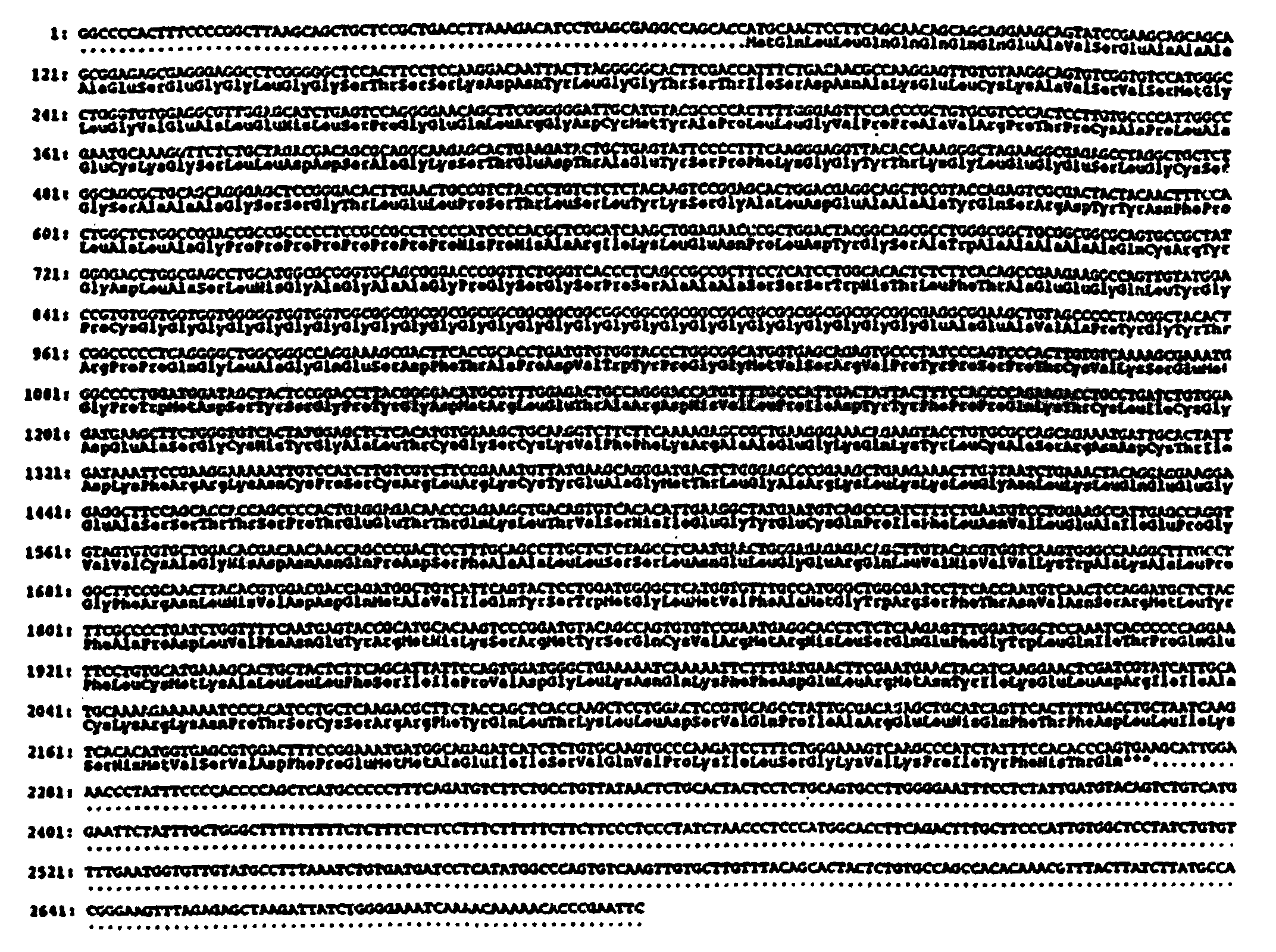
- The cloning vehicle of Claim 3 wherein the DNA molecule encodes the human androgen receptor protein whose amino acid sequence is shown in Figure 4.
- A process for producing an androgen receptor protein, the process comprising transfecting or transforming a host organism with the cloning vehicle of Claim 3, and purifying androgen receptor protein produced by translation of the DNA sequence encoding the protein.
- A process for producing a human androgen receptor protein, the process comprising transfecting or transforming a host organism with the cloning vehicle of Claim 4, and purifying human androgen receptor protein produced by translation of the DNA sequence encoding the protein.
- Use of a probe comprising complementary DNA sequences derived from the deduced sequences encoding androgen receptor as shown in Figure 4 in the manufacture of a reagent to detect the presence of androgen receptor mRNA in tumour cells or to detect abnormalities in the androgen receptor gene or in its mRNA provided that the probe (i) does not hybridise under stringent conditions to a DNA molecule which codes for the polypeptideand (ii) does not hybridise under stringent conditions to DNA molecule which codes for the polypeptide
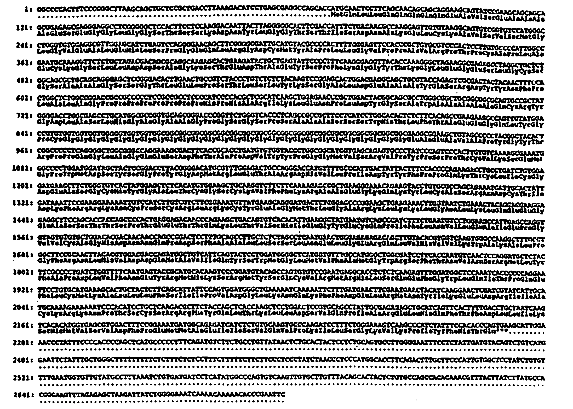 and (iii) is not degenerate with either of the said DNA molecules and (iv) is not either of the DNA molecules shown above.
- A recombinant DNA molecule comprising a DNA sequence having the structural gene which encodes for human androgen receptor protein whose amino acid sequence is shown in Figure 4.
- A cloning vehicle comprising a DNA molecule which upon expression in a host produces human androgen receptor protein whose amino acid sequence is shown in Figure 4.
- A process for producing a human androgen receptor protein, the process comprising transfecting or transforming a host organism with the cloning vehicle of Claim 9, and purifying human androgen receptor protein produced by translation of the DNA sequence encoding the protein.
Applications Claiming Priority (3)
| Application Number | Priority Date | Filing Date | Title |
|---|---|---|---|
| US182646 | 1988-04-14 | ||
| US07/182,646 US6307030B1 (en) | 1988-04-15 | 1988-04-15 | Androgen receptor proteins, recombinant DNA molecules coding for such, and use of such compositions |
| PCT/US1989/001548 WO1989009791A1 (en) | 1988-04-14 | 1989-04-13 | Dna encoding androgen receptor protein |
Publications (3)
| Publication Number | Publication Date |
|---|---|
| EP0365657A1 EP0365657A1 (en) | 1990-05-02 |
| EP0365657A4 EP0365657A4 (en) | 1991-08-21 |
| EP0365657B1 true EP0365657B1 (en) | 1999-08-04 |
Family
ID=22669419
Family Applications (1)
| Application Number | Title | Priority Date | Filing Date |
|---|---|---|---|
| EP89905317A Expired - Lifetime EP0365657B1 (en) | 1988-04-14 | 1989-04-13 | Dna encoding androgen receptor protein |
Country Status (7)
| Country | Link |
|---|---|
| US (3) | US6307030B1 (en) |
| EP (1) | EP0365657B1 (en) |
| JP (1) | JP3385380B2 (en) |
| AT (1) | ATE182901T1 (en) |
| CA (1) | CA1341347C (en) |
| DE (1) | DE68929043T2 (en) |
| WO (1) | WO1989009791A1 (en) |
Cited By (1)
| Publication number | Priority date | Publication date | Assignee | Title |
|---|---|---|---|---|
| US6821767B1 (en) | 1988-04-15 | 2004-11-23 | The University Of North Carolina At Chapel Hill | Androgen receptor proteins, recombinant DNA molecules coding for such, and use of such compositions |
Families Citing this family (35)
| Publication number | Priority date | Publication date | Assignee | Title |
|---|---|---|---|---|
| US5614620A (en) * | 1988-03-30 | 1997-03-25 | Arch Development Corporation | DNA binding proteins including androgen receptor |
| JPH05506141A (en) * | 1989-11-17 | 1993-09-16 | アーチ ディベラップメント コーポレイション | DNA-binding proteins containing androgen receptors |
| CA2239976A1 (en) * | 1995-09-20 | 1997-03-27 | Paul A. Zamecnik | Antisense oligonucleotide chemotherapy for benign hyperplasia or cancer of the prostate |
| AU726158B2 (en) * | 1996-09-11 | 2000-11-02 | Icagen, Inc. | Small and intermediate conductance, calcium-activated potassium channels and uses thereof |
| CA2302169A1 (en) * | 1998-06-30 | 2000-01-13 | The University Of British Columbia | Inhibitors of androgen-independent activation of androgen receptor |
| DE19834180A1 (en) | 1998-07-29 | 2000-02-03 | Benckiser Nv | Composition for use in a dishwasher |
| JP2004530427A (en) | 2001-05-08 | 2004-10-07 | メルク エンド カムパニー インコーポレーテッド | DNA molecule encoding rhesus monkey androgen receptor |
| EP1423695A4 (en) * | 2001-07-27 | 2005-09-28 | Univ Rochester | TRANSGENIC ANIMALS WITH INACTIVATION OF THE ANDROGEN RECEPTOR |
| AU2002318168B2 (en) | 2002-02-25 | 2007-12-13 | Vaxiion Therapeutics, Llc | Minicell compositions and methods |
| WO2004057025A2 (en) * | 2002-12-20 | 2004-07-08 | Biodetection Systems B.V. | Method for determining the presence of one or more ligands in a sample |
| US7483554B2 (en) * | 2003-11-17 | 2009-01-27 | Aureon Laboratories, Inc. | Pathological tissue mapping |
| US7505948B2 (en) * | 2003-11-18 | 2009-03-17 | Aureon Laboratories, Inc. | Support vector regression for censored data |
| US7467119B2 (en) * | 2003-07-21 | 2008-12-16 | Aureon Laboratories, Inc. | Systems and methods for treating, diagnosing and predicting the occurrence of a medical condition |
| EP1709193B1 (en) * | 2003-12-19 | 2011-03-16 | The Regents of The University of California | Methods and materials for assessing prostate cancer therapies |
| WO2005099693A2 (en) | 2004-02-24 | 2005-10-27 | The Regents Of The University Of California | Methods and materials for assessing prostate cancer therapies and compounds |
| EP1728210A2 (en) * | 2004-02-27 | 2006-12-06 | Aureon Laboratories, Inc. | Methods and systems for predicting occurrence of an event |
| EP1789923A1 (en) * | 2004-08-11 | 2007-05-30 | Aureon Laboratories, Inc. | Systems and methods for automated diagnosis and grading of tissue images |
| US7709517B2 (en) | 2005-05-13 | 2010-05-04 | The Regents Of The University Of California | Diarylhydantoin compounds |
| KR100777082B1 (en) | 2005-08-30 | 2007-11-28 | 주식회사 엘지생활건강 | Sebum secretion cosmetic composition containing oligonucleotide |
| ES2689292T3 (en) | 2006-03-27 | 2018-11-13 | The Regents Of The University Of California | Androgen receptor modulator for the treatment of prostate cancer and diseases associated with the androgen receptor |
| NZ572374A (en) | 2006-03-29 | 2011-12-22 | Univ California | Diarylthiohydantoin compounds |
| WO2007136837A2 (en) * | 2006-05-19 | 2007-11-29 | University Of Rochester | Prostate epithelial androgen receptor suppresses prostate growth and tumor invasion |
| US7910565B2 (en) * | 2006-09-01 | 2011-03-22 | Wisconsin Alumni Research Foundation | Prostate cancer vaccine |
| JP2010537623A (en) * | 2007-03-27 | 2010-12-09 | クリストファー ホーヴェンス | Methods and compositions for treating prostate cancer |
| EP2220050A2 (en) | 2007-10-26 | 2010-08-25 | The Regents Of The University Of California | Diarylhydantoin compounds as androgen receptor modulators |
| CA2733506A1 (en) * | 2008-08-08 | 2010-02-11 | Christopher Hovens | Biological applications of steroid binding domains |
| US8133724B2 (en) * | 2008-09-17 | 2012-03-13 | University Of Maryland, Baltimore | Human androgen receptor alternative splice variants as biomarkers and therapeutic targets |
| US8841422B2 (en) | 2008-09-17 | 2014-09-23 | University Of Maryland, Baltimore | Human androgen receptor alternative splice variants |
| GB0921873D0 (en) | 2009-12-15 | 2010-01-27 | Cytosystems Ltd | Assay |
| SG10201907588XA (en) | 2010-02-16 | 2019-09-27 | Aragon Pharmaceuticals Inc | Androgen Receptor Modulators And Uses Thereof |
| CN115737643A (en) | 2012-09-26 | 2023-03-07 | 阿拉贡药品公司 | Antiandrogens for the treatment of non-metastatic castration resistant prostate cancer |
| DK2945628T3 (en) | 2013-01-15 | 2020-05-04 | Aragon Pharmaceuticals Inc | ANDROGEN RECEPTOR MODULATOR IN COMBINATION WITH ABIRATERON ACETATE AND PREDNISON FOR TREATMENT OF PROSTATACANCES |
| AU2015252960A1 (en) | 2014-04-30 | 2016-10-27 | Expression Pathology, Inc. | SRM/MRM assay for the Androgen receptor (AR) protein |
| TWI726969B (en) | 2016-01-11 | 2021-05-11 | 比利時商健生藥品公司 | Substituted thiohydantoin derivatives as androgen receptor antagonists |
| MX2020003845A (en) | 2017-10-16 | 2020-11-06 | Aragon Pharmaceuticals Inc | ANTIANDROGENS FOR THE TREATMENT OF NON-METASTATIC PROSTATE CANCER RESISTANT TO CASTRATION. |
Family Cites Families (5)
| Publication number | Priority date | Publication date | Assignee | Title |
|---|---|---|---|---|
| US4423208A (en) | 1982-02-24 | 1983-12-27 | Peter Grandics | Affinity gel-adsorbent |
| AU7084787A (en) | 1986-02-20 | 1987-09-09 | Karo Bio Ab | Eucaryotic expression of steroid receptor proteins |
| US5614620A (en) * | 1988-03-30 | 1997-03-25 | Arch Development Corporation | DNA binding proteins including androgen receptor |
| JPH03504438A (en) | 1988-03-30 | 1991-10-03 | アーチ デベロプメント コーポレイション | DNA-binding proteins including androgen receptors |
| US6307030B1 (en) | 1988-04-15 | 2001-10-23 | The University Of North Carolina At Chapel Hill | Androgen receptor proteins, recombinant DNA molecules coding for such, and use of such compositions |
-
1988
- 1988-04-15 US US07/182,646 patent/US6307030B1/en not_active Expired - Fee Related
-
1989
- 1989-04-13 DE DE68929043T patent/DE68929043T2/en not_active Expired - Lifetime
- 1989-04-13 JP JP50503989A patent/JP3385380B2/en not_active Expired - Lifetime
- 1989-04-13 EP EP89905317A patent/EP0365657B1/en not_active Expired - Lifetime
- 1989-04-13 WO PCT/US1989/001548 patent/WO1989009791A1/en not_active Ceased
- 1989-04-13 AT AT89905317T patent/ATE182901T1/en not_active IP Right Cessation
- 1989-04-14 CA CA000596763A patent/CA1341347C/en not_active Expired - Fee Related
-
2000
- 2000-02-03 US US09/497,822 patent/US6821767B1/en not_active Expired - Fee Related
-
2004
- 2004-07-07 US US10/886,384 patent/US7129078B2/en not_active Expired - Fee Related
Cited By (2)
| Publication number | Priority date | Publication date | Assignee | Title |
|---|---|---|---|---|
| US6821767B1 (en) | 1988-04-15 | 2004-11-23 | The University Of North Carolina At Chapel Hill | Androgen receptor proteins, recombinant DNA molecules coding for such, and use of such compositions |
| US7129078B2 (en) | 1988-04-15 | 2006-10-31 | University Of North Carolina At Chapel Hill | DNA encoding androgen receptor fragment |
Also Published As
| Publication number | Publication date |
|---|---|
| JP3385380B2 (en) | 2003-03-10 |
| EP0365657A4 (en) | 1991-08-21 |
| US6307030B1 (en) | 2001-10-23 |
| US6821767B1 (en) | 2004-11-23 |
| CA1341347C (en) | 2002-03-12 |
| US7129078B2 (en) | 2006-10-31 |
| JPH03500605A (en) | 1991-02-14 |
| ATE182901T1 (en) | 1999-08-15 |
| WO1989009791A1 (en) | 1989-10-19 |
| DE68929043T2 (en) | 2000-04-13 |
| US20050079576A1 (en) | 2005-04-14 |
| DE68929043D1 (en) | 1999-09-09 |
| EP0365657A1 (en) | 1990-05-02 |
Similar Documents
| Publication | Publication Date | Title |
|---|---|---|
| US6307030B1 (en) | Androgen receptor proteins, recombinant DNA molecules coding for such, and use of such compositions | |
| JP3435011B2 (en) | Hormone receptor compositions and methods | |
| Sakurai et al. | Expression of three forms of thyroid hormone receptor in human tissues | |
| Arriza et al. | Cloning of human mineralocorticoid receptor complementary DNA: structural and functional kinship with the glucocorticoid receptor | |
| US6268221B1 (en) | Melanocyte stimulating hormone receptor and uses | |
| Schipani et al. | Pseudohypoparathyroidism type Ib is not caused by mutations in the coding exons of the human parathyroid hormone (PTH)/PTH-related peptide receptor gene | |
| Zoppi et al. | Complete testicular feminization caused by an amino-terminal truncation of the androgen receptor with downstream initiation. | |
| De Bellis et al. | Characterization of mutant androgen receptors causing partial androgen insensitivity syndrome | |
| US6261838B1 (en) | Rat melanocortin receptor MC3-R | |
| EP0628054B1 (en) | Car receptors and related molecules and methods | |
| Choong et al. | A novel missense mutation in the amino-terminal domain of the human androgen receptor gene in a family with partial androgen insensitivity syndrome causes reduced efficiency of protein translation. | |
| US20030207380A1 (en) | Mutant era and test systems for transactivation | |
| US6392027B1 (en) | Mammalian adrenocorticotropic hormone receptor nucleic acids | |
| US20070020266A1 (en) | Gpr54 receptor agonist and antagonist useful for the treatment of gonadotropin related diseases | |
| JP3099896B2 (en) | Identification of genetic mutations associated with congenital lipid adrenal hyperplasia | |
| Watson et al. | Genetic regulation of androgen-induced accumulation of mouse renal β-glucuronidase messenger ribonucleic acid | |
| EP1017716A1 (en) | Car receptors and related molecules and methods | |
| EP0950711A2 (en) | Gonadotropin receptor | |
| WO1991007423A1 (en) | Dna binding proteins including androgen receptor | |
| US6989242B1 (en) | Car receptors and related molecules and methods | |
| AU718702B2 (en) | Mammalian melanocyte stimulating hormone receptors and uses | |
| JP2000125884A (en) | Novel gonadotropin receptor | |
| ARRiZA et al. | Kinship with the Glucocorticoid Receptor | |
| Sullivan | Androgen binding protein gene expression in the rat fetal liver and adult testis |
Legal Events
| Date | Code | Title | Description |
|---|---|---|---|
| PUAI | Public reference made under article 153(3) epc to a published international application that has entered the european phase |
Free format text: ORIGINAL CODE: 0009012 |
|
| 17P | Request for examination filed |
Effective date: 19891221 |
|
| AK | Designated contracting states |
Kind code of ref document: A1 Designated state(s): AT BE CH DE FR GB IT LI LU NL SE |
|
| A4 | Supplementary search report drawn up and despatched |
Effective date: 19910628 |
|
| AK | Designated contracting states |
Kind code of ref document: A4 Designated state(s): AT BE CH DE FR GB IT LI LU NL SE |
|
| 17Q | First examination report despatched |
Effective date: 19930813 |
|
| GRAG | Despatch of communication of intention to grant |
Free format text: ORIGINAL CODE: EPIDOS AGRA |
|
| GRAG | Despatch of communication of intention to grant |
Free format text: ORIGINAL CODE: EPIDOS AGRA |
|
| GRAG | Despatch of communication of intention to grant |
Free format text: ORIGINAL CODE: EPIDOS AGRA |
|
| GRAH | Despatch of communication of intention to grant a patent |
Free format text: ORIGINAL CODE: EPIDOS IGRA |
|
| GRAH | Despatch of communication of intention to grant a patent |
Free format text: ORIGINAL CODE: EPIDOS IGRA |
|
| GRAA | (expected) grant |
Free format text: ORIGINAL CODE: 0009210 |
|
| AK | Designated contracting states |
Kind code of ref document: B1 Designated state(s): AT BE CH DE FR GB IT LI LU NL SE |
|
| PG25 | Lapsed in a contracting state [announced via postgrant information from national office to epo] |
Ref country code: BE Free format text: LAPSE BECAUSE OF FAILURE TO SUBMIT A TRANSLATION OF THE DESCRIPTION OR TO PAY THE FEE WITHIN THE PRESCRIBED TIME-LIMIT Effective date: 19990804 Ref country code: AT Free format text: LAPSE BECAUSE OF FAILURE TO SUBMIT A TRANSLATION OF THE DESCRIPTION OR TO PAY THE FEE WITHIN THE PRESCRIBED TIME-LIMIT Effective date: 19990804 |
|
| REF | Corresponds to: |
Ref document number: 182901 Country of ref document: AT Date of ref document: 19990815 Kind code of ref document: T |
|
| REG | Reference to a national code |
Ref country code: CH Ref legal event code: EP |
|
| REF | Corresponds to: |
Ref document number: 68929043 Country of ref document: DE Date of ref document: 19990909 |
|
| ITF | It: translation for a ep patent filed | ||
| ET | Fr: translation filed | ||
| REG | Reference to a national code |
Ref country code: CH Ref legal event code: NV Representative=s name: R. A. EGLI & CO. PATENTANWAELTE |
|
| PG25 | Lapsed in a contracting state [announced via postgrant information from national office to epo] |
Ref country code: LU Free format text: LAPSE BECAUSE OF NON-PAYMENT OF DUE FEES Effective date: 20000413 |
|
| PLBE | No opposition filed within time limit |
Free format text: ORIGINAL CODE: 0009261 |
|
| STAA | Information on the status of an ep patent application or granted ep patent |
Free format text: STATUS: NO OPPOSITION FILED WITHIN TIME LIMIT |
|
| 26N | No opposition filed | ||
| REG | Reference to a national code |
Ref country code: GB Ref legal event code: IF02 |
|
| PGFP | Annual fee paid to national office [announced via postgrant information from national office to epo] |
Ref country code: DE Payment date: 20080602 Year of fee payment: 20 Ref country code: CH Payment date: 20080430 Year of fee payment: 20 |
|
| PGFP | Annual fee paid to national office [announced via postgrant information from national office to epo] |
Ref country code: IT Payment date: 20080429 Year of fee payment: 20 |
|
| PGFP | Annual fee paid to national office [announced via postgrant information from national office to epo] |
Ref country code: SE Payment date: 20080429 Year of fee payment: 20 Ref country code: NL Payment date: 20080424 Year of fee payment: 20 |
|
| PGFP | Annual fee paid to national office [announced via postgrant information from national office to epo] |
Ref country code: FR Payment date: 20080417 Year of fee payment: 20 |
|
| PGFP | Annual fee paid to national office [announced via postgrant information from national office to epo] |
Ref country code: GB Payment date: 20080429 Year of fee payment: 20 |
|
| REG | Reference to a national code |
Ref country code: CH Ref legal event code: PL |
|
| REG | Reference to a national code |
Ref country code: GB Ref legal event code: PE20 Expiry date: 20090412 |
|
| PG25 | Lapsed in a contracting state [announced via postgrant information from national office to epo] |
Ref country code: NL Free format text: LAPSE BECAUSE OF EXPIRATION OF PROTECTION Effective date: 20090413 |
|
| NLV7 | Nl: ceased due to reaching the maximum lifetime of a patent |
Effective date: 20090413 |
|
| EUG | Se: european patent has lapsed | ||
| PG25 | Lapsed in a contracting state [announced via postgrant information from national office to epo] |
Ref country code: GB Free format text: LAPSE BECAUSE OF EXPIRATION OF PROTECTION Effective date: 20090412 |