EP0354923B1 - Method for labelling antibodies with a metal ion - Google Patents
Method for labelling antibodies with a metal ion Download PDFInfo
- Publication number
- EP0354923B1 EP0354923B1 EP88904758A EP88904758A EP0354923B1 EP 0354923 B1 EP0354923 B1 EP 0354923B1 EP 88904758 A EP88904758 A EP 88904758A EP 88904758 A EP88904758 A EP 88904758A EP 0354923 B1 EP0354923 B1 EP 0354923B1
- Authority
- EP
- European Patent Office
- Prior art keywords
- antibody
- fragment
- fab
- vial
- technetium
- Prior art date
- Legal status (The legal status is an assumption and is not a legal conclusion. Google has not performed a legal analysis and makes no representation as to the accuracy of the status listed.)
- Expired - Lifetime
Links
Images


Classifications
-
- A—HUMAN NECESSITIES
- A61—MEDICAL OR VETERINARY SCIENCE; HYGIENE
- A61K—PREPARATIONS FOR MEDICAL, DENTAL OR TOILETRY PURPOSES
- A61K51/00—Preparations containing radioactive substances for use in therapy or testing in vivo
- A61K51/02—Preparations containing radioactive substances for use in therapy or testing in vivo characterised by the carrier, i.e. characterised by the agent or material covalently linked or complexing the radioactive nucleus
- A61K51/04—Organic compounds
- A61K51/08—Peptides, e.g. proteins, carriers being peptides, polyamino acids, proteins
- A61K51/10—Antibodies or immunoglobulins; Fragments thereof, the carrier being an antibody, an immunoglobulin or a fragment thereof, e.g. a camelised human single domain antibody or the Fc fragment of an antibody
- A61K51/1018—Antibodies or immunoglobulins; Fragments thereof, the carrier being an antibody, an immunoglobulin or a fragment thereof, e.g. a camelised human single domain antibody or the Fc fragment of an antibody against material from animals or humans
-
- A—HUMAN NECESSITIES
- A61—MEDICAL OR VETERINARY SCIENCE; HYGIENE
- A61K—PREPARATIONS FOR MEDICAL, DENTAL OR TOILETRY PURPOSES
- A61K2123/00—Preparations for testing in vivo
Definitions
- Radiometals Proteins have been labeled with various radiometals and other radioisotopic elements for use in immunodiagnostic and immunotherapeutic procedures. Some radiometals have superior properties for use in these techniques.
- Technetium-99m is an ideal radionuclide for scintigraphic imaging because of its nuclear properties. It has a single photon energy of 140keV, a half-life of about 6 hours, and it is readily available from a 99Mo- 99m Tc generator.
- Rhenium radioisotopes are beta-emitters which can kill target cells and thus are useful in therapy.
- Rhenium-186 and -188 also have gamma emission which, as an added feature, allows it to be imaged by scintigraphic techniques.
- the first is the direct labeling method by which the radiometal is bound to the protein molecule itself.
- the second is the indirect labeling method in which a chelating agent is coupled to the protein and the radiometal is attached to the protein via the chelating agent.
- a chelating agent such as diethylenetriaminepentaacetic acid (DTPA) is conjugated onto the protein and then the metal ion is labeled onto the chelating agent attached to the protein molecule.
- DTPA diethylenetriaminepentaacetic acid
- Khaw et al ., Science 209 : 295-297 (1980) discloses antibodies to cardiac myosin labeled with indium-111 via DTPA and use of the labeled antibodies to image for myocardial infarction. See also, Krejcarek et al ., Biochem . Biophys . Res . Commun . 77 : 581-585 (1977); Childs, R.L.
- This invention pertains to a simple, rapid and efficient method of labeling sulfhydryl-containing antibodies or antibody fragments with the radiometals technetium-99m rhenium-186, rhenium-188, rhenium-189 and rhenium-191.
- the method comprises:
- EP-A-0 237 150 was published on 16.09.87 and claims a priority date of 12.03.86, and therefore is acknowledged pursuant to Art. 54(3) EPC. It discloses a method and kit for preparing a radionuclide-labeled protein from a protein precursor having at least one di-sulfide linkage. The method involves the use of a disulfide reducing agent, and the labeling species is e.g. a labile, soluble complex of reduced Tc-99m, complexed by use of a weak chelating agent. Specific examples of the chelating agents disclosed in EP-A-0 237 150 are gluconate, glucoheptonate, tartrate and malonate species. There is no teaching of the ligands saccharic acid, galactaric acid, arabonic acid and salts thereof, the use of which the present invention claims.
- the technetium-99m labeled antibodies or antibody fragments of the present invention are useful for radioimminodiagnostic purposes such as immunoscintigraphy.
- the rhenium labeled antibodies or antibody fragments can be used for therapy.
- the especially preferred ligand is saccharic acid, (D-glucaric acid). Saccharic acid quickly and stably complexes with technetium-99m in its reduced state and without the formation of significant technetium colloids. When contacted with a sulfhydryl-containing antibody, Technetium-99mm is preferentially transferred to the antibody to form a stable labeled antibody.
- the preferred reducing agents for use in the method are stannous reducing agents such as stannous chloride. These reagents effectively reduce technetium and are pharmacologically acceptable.
- the method of this invention can be used to label whole antibodies (e.g., IgG) or antibody fragments (e.g., Fab').
- Whole antibodies can be reduced with the reducing agent dithiothreitol (DTT) for example, to produce sulfhydryl containing antibodies.
- DTT dithiothreitol
- Fab' fragments are especially suited for labeling by the procedure. Under nonoxidizing conditions, these fragments contain free sulfhydryl groups (as they are produced by reduction of disulfide bridges present in F(ab)'2 fragments.
- antibody fragments such as Fab' fragments are preferred and thus, the labeling procedure of this invention is particularly suited for preparing radiopharmaceuticals for these techniques.
- kits can be provided with the reagents in a form ready for use on site by the clinician.
- a kit can include a first vial containing a reducing agent (e.g. stannous ions) and the water soluble ligand (e.g. saccharic acid or a salt thereof) and a second vial containing a Fab' fragment suited for the particular diagnostic or therapeutic procedure.
- the reactions are preferably carried out in an aqueous medium although the reagents may be supplied in lyophilized, frozen or aqueous form.
- technetium-99m labeled fragments For the preparation of technetium-99m labeled fragments, technetium-99m (generally in the form of pertechnetate) is added to the first vial and then the contents of the first and second vial are mixed and incubated for a time sufficient to effect a quantitative transfer of the technetium-99m to the Fab' fragment. The composition can then be injected into the patient without purification.
- technetium-99m generally in the form of pertechnetate
- rhenium isotopes in the form of a perrhenate
- the rhenium labeled Fab' fragment is also suitable for injection without purification.
- the radiolabeling can be performed in a single vial.
- a kit can include a single vial containing an antibody mixture comprised of a sulfhydryl containing antibody or antibody fragment, a reducing agent (e.g. stannous ions), and preferably a water soluble ligand (e.g. D-glucaric acid or a salt thereof).
- the antibody mixture is preferably supplied in lyophilized form although frozen or aqueous forms are also suitable.
- Technetium-99m (generally in the form of 99m Tc pertechnetate) is added to the vial and the resulting mixture is incubated for a time sufficient to effect a quantitative transfer of the technetium-99m to the antibody or antibody fragment.
- the technetium-99m-labeled antibodies and antibody fragments prepared by the method of this invention can be used for diagnostic purposes such as immunoscintigraphy of tumor, myocardial infarction, thromboses or bacterial abscess.
- Rhenium-labeled antibodies can be used to selectively deliver a rhenium radioisotope in vivo for therapy.
- the method of this invention has several important advantages.
- the ligands employed are capable of complexing technetium-99m quantitatively in stable form as a complex without the formation of a significant amount of technetium colloid.
- the complexed technetium-99m is transferred substantially quantitatively to sulfhydryl-containing antibodies so that radiodiagnostic composition can be prepared with very high specific activity.
- the antibody or antibody fragments labeled by the method retain their original immunoreactivity and consequently their target specificity.
- the radiolabeled antibody is stable in solution and in serum.
- the labeling method can be performed rapidly (it can be completed in less than one hour) and the method can be performed at room temperature and at pH 5-9.
- the labeled product does not require purification before use.
- Figure 1 shows gamma scintigrams of a dog at various times after injection of technetium-99m labeled myosin-specific Fab' fragment.
- Figure 2 shows gamma scintigrams of the same dog taken from different views.
- FIG 3 shows the results of mouse biodistribution studies in blood conducted with antifibrin T2G1S prepared in single vial and double vial labeling kits.
- FIG 4 shows the results of mouse biodistribution studies in liver conducted with antifibrin T2G1S prepared in single vial and double vial labeling kits.
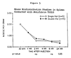
- Figure 5 shows the results of mouse biodistribution studies in spleen conducted with antifibrin T2G1S prepared in single vial and double vial labeling kits.
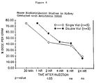
- Figure 6 shows the results of mouse biodistribution studies in kidney conducted with antifibrin T2G1S prepared in single vial and double vial labeling kits.
- Figure 7 shows the results of mouse biodistribution studies in large intestine conducted with antifibrin T2G1S prepared in single vial and double vial labeling kits.
- the method of this invention is performed by reacting Technetium-99m (in an oxidized state) with a water-soluble ligand in the presence of a reducing agent to form a stable complex between technetium-99m in a reduced state (e.g., IV or V valence state) and the ligand and then reacting the complex with an antibody or antibody fragment which contains one or more sulfhydryl groups.
- a reducing agent e.g., a reduced state
- an antibody or antibody fragment which contains one or more sulfhydryl groups.
- aqueous sodium 99m-pertechnetate is mixed with a aqueous solution of a stannous reducing agent and saccharic acid (or a salt thereof) to form a 99m Tc-saccharate complex.
- the complex is then contacted with an Fab' fragment and incubated for a period of time and under conditions which allow an exchange of technetium-99m from the complex to the Fab' fragment to form a technetium-labeled Fab' fragment.
- the entire procedure can be conducted in less than one hour at room temperature and at a pH of about 5-9. Under these conditions an essentially complete transfer of technetium-99m (from the 99m-Tc-saccharate complex to the antibody protein) can be attained without significant loss of antibody immunoreactivity.
- This invention provides a method of radiolabeling protein in a single vial.
- the reduction of the oxidized form of technetium-99m and the radiolabeling reaction i.e., the coupling of the radioisotope to protein
- vial refers to any type of reaction vessel and is not intended to be limiting in any way.
- the method is simple, efficient, and reproducible and it minimizes the safety hazards to persons performing the radiolabeling.
- the method of this invention is particularly suited for labeling antibodies (polyclonal and monoclonal) for diagnosis. Antibodies can be labeled by this method to a high specific activity with minimal loss of immunoreactivity.
- Advantages with the present one vial method over methods using two vials and other known methods for labeling with technetium-99m include: (1) Rapid labeling at ambient conditions. Labeling yields greater than 90% can be achieved in 5-15 minutes at ambient temperature without heating. The clinical advantages of near instantaneous preparation of a diagnostic agent can be substantial. (2) Stability of the lyophilized formulation of the single vial method is superior to the comparable formulation employed in a two vial method. (3) Biodistribution studies of the product resulting from the one vial method show statistically significant differences in key major organs. Uptake in kidney and liver is lower with product produced by this method. Blood clearance is significantly faster.
- Plasma stability of the product is greater. This provides more viable intact product to serve as the diagnostic agent in vivo.
- an antibody mixture comprised sylfhydryl containing antibody or antibody fragment, a reducing agent and a water soluble ligand are added to a vial.
- sealable reaction vial is used which has means for the introduction and withdrawal of reagent preferably under sterile or semi-sterile conditions.
- a vial which contains a port for syringe injection is preferred. All reagents can be injected and withdrawn from the reaction vial by syringe, thereby reducing the risk of exposure to radio- or biohazardous reagents.
- the mixture is lyophilized and the vial is presealed and supplied for use in that form.
- technetium-99m in an oxidized state is contacted with the antibody mixture.
- the radiolabelling reaction is then allowed to proceed.
- the duration and condition of incubation are not critical. Preferably, incubation is conducted for a period from about one minute to about 60 minutes, and most preferably from about 5 minutes to about 30 minutes.
- the labeled antibody or antibody fragment is withdrawn from the vial. No separation or purification is required.
- the entire procedure can be conducted in less than 15 minutes at ambient temperature and at a pH of about 5-9. Under these conditions an essentially complete labeling of the antibody or antibody fragment with technetium-99m can be attained without significant loss of antibody immunoreactivity.
- the ligands useful in the method of this invention are water-soluble (or can be made water soluble) chelators which are capable of complexing technetium-99m or any of the rhenium radioisotopes in their reduced state to form a stable metal ion/ligand complex.
- the complex is capable of exchanging the technetium-99 with a sulfhydryl containing antibody or antibody fragment.
- the ligands used in this invention are polyhydroxydicarboxylic acids having a molecular weight of less than about 10,000 daltons, and are selected from saccharic acid, galactaric acid, arabonic acid, and salts thereof.
- the particularly preferred ligand for use in this method is saccharic acid.
- saccharic acid complexes with technetium-99m quickly to form a stable technetium-99m-saccharate complex.
- a sulfhydryl-containing antibody or antibody fragment substantially quantitative transfer of technetium-99m from the complex to the protein is achieved rapidly and under mild conditions.
- the technetium-99m is preferentially transfered to favored binding sites on the protein molecules. This preferential transfer results in a labeled antibody or fragment which is immunoreactive and exceptionally stable in vivo .
- Reducing agents for use in the method are physiologically acceptable for reducing technetium-99m from its oxidized state to the IV or IV oxidization state or for reducing rhenium from its oxidized state.
- reducing agents which can be used in the method are stannous chloride, stannous fluoride, stannous tartarate, and sodium dithionite; the preferred agents are stannous reducing agents especially stannous chloride.
- the source of Technetium-99m should preferably be water soluble. Preferred sources are alkali and alkaline earth metal pertechnetate (TcO4 ⁇ ).
- the technetium-99m is most preferably obtained in the form of fresh sodium pertechnetate from a sterile technetium-99m generator (e.g., from a conventional 99Mo/99mTc generator). Any other source of physiologically acceptable technetium-99m, however, may be used.
- Rhenium radioisotopes (the isotopes 186, 188, 189 and 191) in the form of perrhenate salts can be produced by suitable reactor technology or made by a suitable generator.
- the perrhenate salts are stable, soluble salts and behave similarly to pertechnetate.
- Perrhenate requires a slightly greater reduction potential to reduce, and tends to return to perrhenate in the presence of oxygen more readily than pertechnetate. For this reason, different conditions may be required to reduce and stabilize rhenium in its reduced state. These can be ascertained empirically by a person of ordinary skill in the art.
- the sulfhydryl containing whole antibodies or lower molecular weight antibody fragments can be labeled by the method of this invention. It is believed that sulfhydryl groups constitute at least a part of favored binding sites which exist on molecules and that by the method of this invention, the radiometals are preferentially exchanged from the radiometal-ligand complex to these favored sites on the molecules. The preferential labeling of these sites on the antibodies molecules results in labeled antibodies of exceptional stability.
- Whole antibodies e.g. IgG
- a reducing agent such as dithiothreitol DTT.
- DTT dithiothreitol
- antibody fragments are preferred reagents.
- Antibody fragments have a number of advantages over whole antibodies for imaging procedures including, in general, more rapid distribution and accumulation at target site and less immunogenicity.
- Fab' fragments are monovalent antibody binding which contain free sulfhydryl groups (when maintained under non-oxidizing conditions). These fragments can be labelled efficiently by the method of this invention.
- Fab' fragments can be prepared from whole antibodies as follows: An antibody molecule is first treated with an endopeptidase such as pepsin to remove the Fc portion of the antibody molecule. The resultant F(ab)'2 fragment is treated with a reducing agent such as DTT or cysteine to break disulfide bonds present on the F(ab)'2 fragment resulting in exposed the sulfhydryl groups present on the molecules and thereby producing two Fab' molecules for each antibody molecule.
- an endopeptidase such as pepsin
- a reducing agent such as DTT or cysteine
- the amount of reducing agent is the amount necessary to reduce the technetium to provide for the binding to the ligand in a reduced state.
- stannous chloride (SnCl2) is the reducing agent and can range from 1-1,000 ug/ml preferably about 30-500 ug/ml.
- the amount of saccharic acid (as potassium saccharate) can range from about 0.5 mg/ml up to the amount maximally soluble in the medium. Preferred amounts of saccharic acid range from 30-15 ug/ml.
- the amount of antibody (or fragment) can range from 0.01 to about 30 mg/ml preverably about .17 to about 1.5 mg/ml.
- technetium-99m in the form of pertechnetate can be in amounts used up to about 500 uCi/ml preferably about 1-50 mCi/ml.
- the amount of mCi per mg of antibody is preferably about 3-150.
- the reaction between the and the metal ion-transfer ligand complex is preferably carried out in an aqueous solution at a pH at which the protein is stable.
- stable it is meant that the protein remains soluble and retains its biological activity.
- the pH for the reaction will be a pH from about 5 to 9, the preferred pH being about 6-8.
- the metal ion-transfer chelate complex and the antibody are incubated, preferably at a temperature from about 20°C to about 60°C, most preferably from about 20°C to about 37°C, for a sufficient amount of time to allow transfer of the metal ion from the ligand complex to the antibody. Generally, less than one hour is sufficient to complete the transfer reaction under these conditions.
- kits for convenient performance of the method in the clinic can consist of a one component a vial (sealed and sterile) containing a reducing agent (preferably stannous ions) and saccharic acid or a salt thereof. These kits can be used when the antibody or antibody fragment is provided by the user.
- a kit for radiolabeling antibody or antibody fragments with the radiometals can consist of a one component a vial (sealed and sterile) containing a reducing agent (preferably stannous ions) and saccharic acid or a salt thereof.
- Kits may also include a second vial containing the sulfhydryl-containing antibody or antibody fragment to be labeled.
- Two component kits would include:
- Kits can be designed to contain the appropriate antibody or antibody fragment(s) for any particular immunodiagnostic or immunotherapeutic procedure (some of which are discussed below).
- the reagents in the kit can be provided in aqueous, lyophilized or from form. Lyophilized preparations can be diluted with aqueous medium upon use. The amount of reagents in each vial can vary according to the chosen parameters of the method (see above under Reaction Conditions).
- the labeling procedure can be performed simply as a two vial technique.
- Technetium-99m for example, in the form of aqueous sodium pertechnetate
- the contents of the two vials are then mixed and incubated for a time sufficient to effect labeling of the antibody or antibody fragment.
- the radiolabeled antibody or antibody fragment can then be used immediately without purification.
- the kit contains one vial (sealed and sterile) containing a sulfhydryl containing antibody or antibody fragment, a reducing agent (preferably stannous ions) and a water soluble ligand (preferably D-glucaric acid or a salt thereof).
- a sulfhydryl containing antibody or antibody fragment preferably stannous ions
- a water soluble ligand preferably D-glucaric acid or a salt thereof.
- kits can be used when technetium-99m is provided by the user.
- the kits are designed to contain the appropriate antibody or antibody fragment(s) for any particular immunodiagnostic or immunotherapeutic procedure (some of which are discussed below).
- the reagents in the kit can be provided in aqueous, frozen or lyophilized form. Lyophilized preparations can be diluted with aqueous medium upon use.
- the amount of reagents in each vial can vary according to the chosen parameters of the method (see above under Reaction Conditions).
- the labeling procedure can be performed simply by adding technetium-99m (for example, in the form of aqueous sodium pertechnetate) to the vial containing the antibody or antibody fragment, reducing agent and, in a preferred embodiment, water soluble ligand. The contents of the vial are then mixed and incubated for a time sufficient to effect labeling of the antibody or antibody fragment. The radiolabeled antibody or antibody fragment can then be used immediately without purification.
- Technetium-99m labeled antibodies or antibody fragments can be used in immunoscintigraphy. One important use is in the imaging of tumors. As mentioned, antibody fragments are preferred for most immunoscintigraphic techniques. Labeled Fab' fragments of tumor specific antibodies can be prepared and used to image primary or secondary tumors. In general, the technetium-99m labeled antibody fragment is prepared by forming an aqueous mixture of (i) 99m Tc; and (ii) a reducing agent and a water-soluble ligand; and contacting the mixture with an Fab' fragment specific for the tumor.
- the labeled Fab' fragment can then be injected parenterally (preferably intraveneously) into a subject. After injection, sufficient time is allowed for the labeled Fab' fragment to accumulate at the site of the tumor. The subject is then scanned with a gamma camera to detect the gamma emission of the technetium-99m and to thereby to obtain an image of the tumor. In this way the tumor can be localized and its size can be determined.
- Tumor-specific antibody fragments for use in these procedures can be derived from anticolorectal cancer antibody, antilung cancer antibody anti-ovarian cancer antibody, antibreast cancer antibody, and antiprostate cancer antibody.
- tumor specific antibodies which can be labeled by the method of this invention and used to image tumors are the monoclonal antibodies 17-1A and 19-9 (gastrointestinal), CA 125 (ovarian) and 103D2 (breast).
- Antibodies labeled by the method of this invention can be used to label myocardial infarcts.
- the imaging of myocardial infarcts to determine their size and location is described by Haber, U.S. Patent No. 4,421,735.
- an image of a mycocardial infarct in a subject can be obtained by first preparing a Tc-99m labeled myosin specific Fab' fragment by first forming an aqueous mixture of (i) 99m Tc and (ii) a reducing agent and a water soluble ligand for 99m Tc; and then contacting the mixture with a myosin specific Fab' fragment.
- the labeled myosin specific fragment is then intraveneously injected into a subject (for example, after coronary occlusion).
- the labeled fragment is allowed to localize at the site of the infarct and an image of the infarct is obtained by scanning the area of the heart with a gamma camera.
- a preferred antibody for production of labeled myosin-specific Fab' fragments is the monoclonal antibody R11D10.
- fibrin-specific Fab' fragments can be labelled by the procedure of this invention to provide reagents for imaging blood clots.
- a Tc-99m labeled fibrin-specific fragment is prepared by forming an aqueous mixture of (i) 99m Tc and (ii) a reducing agent and a water soluble ligand for 99m Tc and contacting the mixture with a fibrin specific Fab' fragment.
- the 99m Tc-labeled fibrin specific fragment is injected into the subject. After allowing the fragment to localize at the site of the blood clot, the subject is scanned to obtain an image of the clot.
- Fibrin-specific antibodies which are not cross-reactive with fibrinogen are the preferred antibodies for this imaging technique.
- Antibody fragments specific for bacteria can be used in immunoscintigraphic techniques for obtaining an image of a bacterial abscess in a subject.
- anti-bacterial or anti-macrophage antibody fragments are employed.
- Antibodies against a common determinant of gram-negative bacteria e.g., anti-lipid A antibody
- the antibody is labeled with technetium-99m as described above injected into the subject and allowed to localize at the abscess.
- the subject is then scanned with the photoscanning equipment to obtain an image of the abscess.
- Rhenium-labeled antibody or antibody fragments can be used to selectively deliver rhenium radioisotopes to target cells in vivo .
- rhenium labeled antibodies can selectively seek out and destroy cancer cells.
- tumor specific antibodies such as those described above, can be labeled by the method of this invention and the resulting labeled antibody can be injected parenterally into a subjected afflicted with the tumor.
- the invention is further illustration by the following exemplification.
- Monopotassium saccharate 25 mg was dissolved in 0.2M bicarbonate (1.0 ml) at pH 8.0.
- To 500 ul of saccharate solution was added 40 ul of stannous chloride (2.5 mg/ml) in 0.1M acetic acid followed by 500 ul of Tc-99m generator eluate (_60mCi/mg protein).
- the resulting solution was allowed to stand for 5 minutes at room temperature and then analyzed for radiochemical purity by paper chromatography (Whatman 3MM, 60% CH3CN:40%H2O).
- Antimyosin monoclonal antibody R11D10 F(ab')2 5mg/ml in 40mM TRIS pH 7.0 was reduced with 10mM DTT for 60 minutes at room temperature and then passed through a Sephadex G-25 column to remove the reducing agent.
- the resulting solution contained _80% Fab' fragment by gel-filtration HPLC.
- Antimyosin antibody R11D10 Fab' 500 ul of a 1 mg/ml solution in 50mM phosphate, 0.35 mM ZnCl2, pH 6.5 was mixed with 500 ul of 99m Tc-saccharate solution and allowed to stand at room temperature for 5-60 minutes.
- the resulting 99m Tc-labeled protein was analyzed for radiochemical purity by paper chromatography (Whatman 3MM; 60% CH3CN: 40%H2O) and gel-filtration HPLC, and for immunoreactivity using a myosin affinity column.
- the 99m Tc-labeled R11D10 Fab' was nearly 80% immunoreactive after 3 hours and 70% immunoreactive after 20 hours. The latter corresponded to 80% retention of immunoreactivity found immediately after labeling.
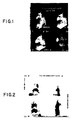
- FIG. 1 shows the gamma scintigrams of a dog after 35 min. (upper left), 1.5 hours (upper right), 2.5 hours (lower left) and 5 hours (lower right) of antibody injection.
- Figure 2 shows the gamma images of the same dog as shown in Figure 1, right lateral (upper left), posterior anterior (lower right) views. Clear myocardial infarct images were observed in all views except the posterior position. More importantly, this figure shows no significant liver uptake 3 hours after injection of Tc-R11D10-Fab'.
- mice Biodistribution studies were carried out in Balb/c mice.
- the mice (4 mice per group) were injected I.V. with either 150 uCi of technetium-99m labeled R11D10 Fab' (4 uCi/ug) or 10 uCi of indium-111 labeled R11D10 Fab-DTPA (4 uCi/ug).
- mice were sacrificed at 1,4 and 8 hours after receiving the injections and organs removed, weighed and counted. Table VII summarizes the percent injected dose per gram obtained for each preparation.
- the 99m Tc-R11D10 Fab' cleared rapidly from both the blood and liver.
- the percent of injected dose for Tc-R11D10 Fab' in the blood at 1 hour was 13.6% and dropped to 2.0% after eight hours.
- a similar drop in radioactivity was observed in the liver at the latter time point (6.4% in 1 hour and 2.4% in eight hours).
- the indium-111 labeled preparation showed much higher radioactivity in both liver (10.8%) and blood (5.1%) at the eighth hour after injection.
- Monopotassium arabonate (20 mg) was dissolved in 0.1M Na2CO3 (1.0 ml) at pH 10.0.
- To 500 ul of arabonate solution was added 500 ul of Tc-99m generator eluate (approx. 60mCi/mg protein) followed by 40 ul of stannous chloride (2.5 mg/ml) in 0.1M acetic acid.
- the resulting solution was allowed to stand at room temperature for 30 minutes and then adjusted to pH 7 using 1.0M hydrochloric acid.
- the sample was analyzed for radiochemical purity by paper chromatography (Whatman 3MM, 60% CH3CN:40% H2O).
- Antimyosin antibody R11D10 Fab' 500 ul of a 1 mg/ml solution in 50mM phosphate, 0.35 mM ZnCl2, pH 6.5 was mixed with 500 ul of 99m Tc-arabonate solution and allowed to stand at room temperature for 60 minutes. The resulting 99m Tc-labeled protein was analyzed for radiochemical purity as previously noted in Example 1. The results showed quantitative transfer of 99m Tc to the protein under these conditions.
- T2G1s F(ab')2 antibody fragment (162 mg) in tris buffer (15.5 ml, 0.05M, pH 8.0) with sodium chloride (0.1M) was treated with DTT (1mM) for 1-2 hours at ambient temperature.
- the resulting mixture was purified by diafiltration under argon by exchange with 20 volumes of sodium phosphate buffer (0.05M, pH 6.4) containing sodium chloride (0.1M) and EDTA (0.001M) to yield a solution containing T2G1s Fab' (135 mg, concentration 1 mg/mL).
- a vial was prepared to contain T2G1s Fab' (0.5 mg), prepared substantially as described above in subsection A.1 above, in a buffer solution (1.0 mL) of potassium phosphate (0.05M, pH 6.4), sodium chloride (0.1M) and EDTA (0.001M). The vial was sealed with a rubber vial closure.
- a vial was prepared to contain T2G1s Fab' (0.5 mg), prepared substantially as described above in subsection A.1 above, in a buffer solution (1.0 mL) of potassium phosphate (0.05M, pH 6.4), sodium chloride (0.05M), lactose (0.05M) and EDTA (0.0005M). The contents of the vial were lyophilized and then sealed with a rubber vial closure.
- vials were prepared to contain a solution (1.0 ml) of monopotassium D-glucaric acid (12.5 mg, 0.05 mmol), stannous chloride (150 ug, 0.79 umol) and sodium bicarbonate (16.8 mg, 0.2 mmol, pH 7.6).
- the contents of vial were lyophilized and then sealed with a rubber vial closure.
- Immunoreactivity of the labeled antibody was tested by applying an aliquot of the antibody reaction mixtures to an affinity column (the first seven amino acids of the amino terminus of the beta chain of human fibrin, coupled to CNBr-Sepharose® 6B). The volume of the packed bed was 1 mL. The column was eluted with 10 mL of 1% BSA in 0.01M sodium phosphate, 0.145M NaCl, pH 7.0, followed by elution with 10 mL of 0.1M glycine, pH 2.5. During these elutions, 1 milliliter fractions were collected and counted in a NaI(Tl) well counter. The percent immunoreactivity was computed as: The results are shown in Tables VIII and IX.
- Table 1 also shows rates of labeling for the one vial and two vial kits as determined by % protein incorporation according to the paper chromatography technique described in subsection A.3 above. The results show that the one vial kit produces labeled product faster than the two vial kits.
- Table VIII Labeling Ratio of One Vial and Two Vial Kits Kit Storage Temp (°C) Age When Tested (Days) % Protein Incorporation Immunoreactivity (%) 5' 15' 30' 60' Two Vial Solution 4° 7 NA 65 78 87 78 Two Vial Lyophilized 4° 7 61 86 89 93 93 One Vial 4° 6 93 94 95 95 98
- the stability of the one vial and two vial kits were determined by % protein incorporation according to the paper chromatography described in subsection A.3 above at both 4° and 37°. The results are shown in Table 2. The results demonstrate that the one vial kit maintains superior labeling efficiency in the 12-17 day period when stored at 37°.
- Labeled antibody was prepared as described in the above examples and comparative experiment (solution formulation).
- the 99m Tc labeled T2G1s Fab' fragments (100 uL) were added to citrated plasma (50 uL).
- Table XI compares the plasma stability as determined by % protein incorporation at 37° of the products from the one vial and two vial kits versus control (no plasma added). The results shorn that the plasma stability of the one vial kit is better than the two vial kit.
Abstract
Description
- Proteins have been labeled with various radiometals and other radioisotopic elements for use in immunodiagnostic and immunotherapeutic procedures. Some radiometals have superior properties for use in these techniques. Technetium-99m is an ideal radionuclide for scintigraphic imaging because of its nuclear properties. It has a single photon energy of 140keV, a half-life of about 6 hours, and it is readily available from a ⁹⁹Mo-99mTc generator. Rhenium radioisotopes are beta-emitters which can kill target cells and thus are useful in therapy. Rhenium-186 and -188 also have gamma emission which, as an added feature, allows it to be imaged by scintigraphic techniques.
- Two general approaches have been taken to label proteins such as antibodies with radiometals. The first is the direct labeling method by which the radiometal is bound to the protein molecule itself. The second is the indirect labeling method in which a chelating agent is coupled to the protein and the radiometal is attached to the protein via the chelating agent.
- Rhodes discloses a method of direct labeling of protein with technetium-99m which involves ligand solid phase exchange. See U.S. Patent 4,305,922. According to the method of Rhodes, pertechnetate is reduced to technetium IV and then applied onto a SephadexR column. The reduced technetium-99m binds to the SephadexR material. A solution of the protein to be labeled is poured onto the top of the Sephadex column where it is allowed to remain so that ligand exchange occurs. As a result, the technetium-99m is transferred preferentially from the Sephadex material to the protein. The protein may be pretreated with a stannous chloride (a procedure called "pretinning") to enhance transfer of the radiometal to the protein. See U.S. Patent No. 4,424,200.
- Various attempts have been made to label proteins with radiometals by the indirect approach. In one such approach, a chelating agent such a diethylenetriaminepentaacetic acid (DTPA) is conjugated onto the protein and then the metal ion is labeled onto the chelating agent attached to the protein molecule. For example, Khaw et al., Science 209: 295-297 (1980) discloses antibodies to cardiac myosin labeled with indium-111 via DTPA and use of the labeled antibodies to image for myocardial infarction. See also, Krejcarek et al., Biochem. Biophys. Res. Commun. 77: 581-585 (1977); Childs, R.L. and Hnatowich, D.J., J. Nucl. Med. 26: 293 (1985); U.S. patent 4,652,440 Paik et al., (1987). In a more recent approach, Fritzberg et al. describe the use particular diaminodithiol and diamidodithiol groups, as a chelating agents. Fritzberg et al, J. Nucl. Med. 27:957 (1986); European Patent Application 86100360.6. In U.S. patent 4,027,005, Adler et al. discloses technetium-labeled polyhydroxycarboxylic acids for use as radionuclide diagnostic agents.
- Various degrees of success have been achieved with both the direct and indirect methods of labeling proteins with radiometals. However, the labeled product is often unstable in vivo. Further, techniques for purifying the labeled product before use are often required. A need exists for improved methods for stably labeling proteins for radioimmunodiagnostic and radioimmunotherapeutic procedures.
- This invention pertains to a simple, rapid and efficient method of labeling sulfhydryl-containing antibodies or antibody fragments with the radiometals technetium-99m rhenium-186, rhenium-188, rhenium-189 and rhenium-191. In general, the method comprises:
- a. forming an aqueouos mixture of
- (i) the radiometal in an oxidized form; and
- (ii) a reducing agent and a water-soluble polyhydroxycarboxylic ligand selected from saccharic acid, galactaric acid, arabonic acid and salts thereof which is capable of forming a stable complex with the radiometal in its reduced state and quantitatively exchanging the radiometal with a sulfhydryl-containing antibody; and
- b. contacting the mixture with a sulfhydryl-containing antibody or antibody fragment to produce a radiometal-labeled antibody or antibody fragment.
- EP-A-0 237 150 was published on 16.09.87 and claims a priority date of 12.03.86, and therefore is acknowledged pursuant to Art. 54(3) EPC. It discloses a method and kit for preparing a radionuclide-labeled protein from a protein precursor having at least one di-sulfide linkage. The method involves the use of a disulfide reducing agent, and the labeling species is e.g. a labile, soluble complex of reduced Tc-99m, complexed by use of a weak chelating agent. Specific examples of the chelating agents disclosed in EP-A-0 237 150 are gluconate, glucoheptonate, tartrate and malonate species. There is no teaching of the ligands saccharic acid, galactaric acid, arabonic acid and salts thereof, the use of which the present invention claims.
- The technetium-99m labeled antibodies or antibody fragments of the present invention are useful for radioimminodiagnostic purposes such as immunoscintigraphy. The rhenium labeled antibodies or antibody fragments can be used for therapy.
- The especially preferred ligand is saccharic acid, (D-glucaric acid). Saccharic acid quickly and stably complexes with technetium-99m in its reduced state and without the formation of significant technetium colloids. When contacted with a sulfhydryl-containing antibody, Technetium-99mm is preferentially transferred to the antibody to form a stable labeled antibody.
- The preferred reducing agents for use in the method are stannous reducing agents such as stannous chloride. These reagents effectively reduce technetium and are pharmacologically acceptable.
- The method of this invention can be used to label whole antibodies (e.g., IgG) or antibody fragments (e.g., Fab'). Whole antibodies can be reduced with the reducing agent dithiothreitol (DTT) for example, to produce sulfhydryl containing antibodies. Fab' fragments are especially suited for labeling by the procedure. Under nonoxidizing conditions, these fragments contain free sulfhydryl groups (as they are produced by reduction of disulfide bridges present in F(ab)'₂ fragments. For most radioimmunodiagnostic techniques, antibody fragments such as Fab' fragments are preferred and thus, the labeling procedure of this invention is particularly suited for preparing radiopharmaceuticals for these techniques.
- The method of radiolabeling antibody or antibody fragments with the designated radiometals can be performed as a simple two-vial procedure. For this purpose, kits can be provided with the reagents in a form ready for use on site by the clinician. For example, such a kit can include a first vial containing a reducing agent (e.g. stannous ions) and the water soluble ligand (e.g. saccharic acid or a salt thereof) and a second vial containing a Fab' fragment suited for the particular diagnostic or therapeutic procedure. The reactions are preferably carried out in an aqueous medium although the reagents may be supplied in lyophilized, frozen or aqueous form. For the preparation of technetium-99m labeled fragments, technetium-99m (generally in the form of pertechnetate) is added to the first vial and then the contents of the first and second vial are mixed and incubated for a time sufficient to effect a quantitative transfer of the technetium-99m to the Fab' fragment. The composition can then be injected into the patient without purification. For radiolabeling with rhenium, rhenium isotopes (in the form of a perrhenate) are used in place of the technetium. The rhenium labeled Fab' fragment is also suitable for injection without purification.
- In a preferred embodiment, the radiolabeling can be performed in a single vial. A kit can include a single vial containing an antibody mixture comprised of a sulfhydryl containing antibody or antibody fragment, a reducing agent (e.g. stannous ions), and preferably a water soluble ligand (e.g. D-glucaric acid or a salt thereof). The antibody mixture is preferably supplied in lyophilized form although frozen or aqueous forms are also suitable. Technetium-99m (generally in the form of 99mTc pertechnetate) is added to the vial and the resulting mixture is incubated for a time sufficient to effect a quantitative transfer of the technetium-99m to the antibody or antibody fragment. This composition can then be injected into the patient without purification. The technetium-99m-labeled antibodies and antibody fragments prepared by the method of this invention can be used for diagnostic purposes such as immunoscintigraphy of tumor, myocardial infarction, thromboses or bacterial abscess. Rhenium-labeled antibodies can be used to selectively deliver a rhenium radioisotope in vivo for therapy.
- The method of this invention has several important advantages. As mentioned, the ligands employed are capable of complexing technetium-99m quantitatively in stable form as a complex without the formation of a significant amount of technetium colloid. Upon contact with a sulfhydryl containing antibody under appropriate conditions, the complexed technetium-99m is transferred substantially quantitatively to sulfhydryl-containing antibodies so that radiodiagnostic composition can be prepared with very high specific activity. The antibody or antibody fragments labeled by the method retain their original immunoreactivity and consequently their target specificity. The radiolabeled antibody is stable in solution and in serum. When Fab' fragments labeled by the method are administered in vivo very little label accumulates in the liver which indicates that the labeled antibody is stable in vivo. In addition, the labeling method can be performed rapidly (it can be completed in less than one hour) and the method can be performed at room temperature and at pH 5-9. The labeled product does not require purification before use.
- Figure 1 shows gamma scintigrams of a dog at various times after injection of technetium-99m labeled myosin-specific Fab' fragment.
- Figure 2 shows gamma scintigrams of the same dog taken from different views.
- Figure 3 shows the results of mouse biodistribution studies in blood conducted with antifibrin T2G1S prepared in single vial and double vial labeling kits.
- Figure 4 shows the results of mouse biodistribution studies in liver conducted with antifibrin T2G1S prepared in single vial and double vial labeling kits.
- Figure 5 shows the results of mouse biodistribution studies in spleen conducted with antifibrin T2G1S prepared in single vial and double vial labeling kits.
- Figure 6 shows the results of mouse biodistribution studies in kidney conducted with antifibrin T2G1S prepared in single vial and double vial labeling kits.
- Figure 7 shows the results of mouse biodistribution studies in large intestine conducted with antifibrin T2G1S prepared in single vial and double vial labeling kits.
- In one embodiment, the method of this invention is performed by reacting Technetium-99m (in an oxidized state) with a water-soluble ligand in the presence of a reducing agent to form a stable complex between technetium-99m in a reduced state (e.g., IV or V valence state) and the ligand and then reacting the complex with an antibody or antibody fragment which contains one or more sulfhydryl groups. In the preferred embodiment for labeling a sulfhydryl-containing antibody with technetium-99m, aqueous sodium 99m-pertechnetate is mixed with a aqueous solution of a stannous reducing agent and saccharic acid (or a salt thereof) to form a 99mTc-saccharate complex. The complex is then contacted with an Fab' fragment and incubated for a period of time and under conditions which allow an exchange of technetium-99m from the complex to the Fab' fragment to form a technetium-labeled Fab' fragment. The entire procedure can be conducted in less than one hour at room temperature and at a pH of about 5-9. Under these conditions an essentially complete transfer of technetium-99m (from the 99m-Tc-saccharate complex to the antibody protein) can be attained without significant loss of antibody immunoreactivity.
- This invention provides a method of radiolabeling protein in a single vial. The reduction of the oxidized form of technetium-99m and the radiolabeling reaction (i.e., the coupling of the radioisotope to protein) are achieved in the same vial. As used herein, the term "vial" refers to any type of reaction vessel and is not intended to be limiting in any way. The method is simple, efficient, and reproducible and it minimizes the safety hazards to persons performing the radiolabeling. The method of this invention is particularly suited for labeling antibodies (polyclonal and monoclonal) for diagnosis. Antibodies can be labeled by this method to a high specific activity with minimal loss of immunoreactivity.
- Advantages with the present one vial method over methods using two vials and other known methods for labeling with technetium-99m include: (1) Rapid labeling at ambient conditions. Labeling yields greater than 90% can be achieved in 5-15 minutes at ambient temperature without heating. The clinical advantages of near instantaneous preparation of a diagnostic agent can be substantial. (2) Stability of the lyophilized formulation of the single vial method is superior to the comparable formulation employed in a two vial method. (3) Biodistribution studies of the product resulting from the one vial method show statistically significant differences in key major organs. Uptake in kidney and liver is lower with product produced by this method. Blood clearance is significantly faster. These types of differences would indicate that product from this method would produce lower background, lower absorbed dose to critical organs and faster blood clearance resulting in faster ability to image areas of interest. All these would be substantial clinical advantages. (4) Plasma stability of the product is greater. This provides more viable intact product to serve as the diagnostic agent in vivo.
- In a preferred embodiment, an antibody mixture comprised sylfhydryl containing antibody or antibody fragment, a reducing agent and a water soluble ligand are added to a vial. Preferably, sealable reaction vial is used which has means for the introduction and withdrawal of reagent preferably under sterile or semi-sterile conditions. A vial which contains a port for syringe injection is preferred. All reagents can be injected and withdrawn from the reaction vial by syringe, thereby reducing the risk of exposure to radio- or biohazardous reagents. In a most preferred embodiment, the mixture is lyophilized and the vial is presealed and supplied for use in that form. In order to label the antibody or antibody fragment, technetium-99m in an oxidized state is contacted with the antibody mixture. The radiolabelling reaction is then allowed to proceed. The duration and condition of incubation are not critical. Preferably, incubation is conducted for a period from about one minute to about 60 minutes, and most preferably from about 5 minutes to about 30 minutes.
- After completion of the labeling reaction, the labeled antibody or antibody fragment is withdrawn from the vial. No separation or purification is required. The entire procedure can be conducted in less than 15 minutes at ambient temperature and at a pH of about 5-9. Under these conditions an essentially complete labeling of the antibody or antibody fragment with technetium-99m can be attained without significant loss of antibody immunoreactivity.
- The various reagents used in the method and the parameters of the method are discussed in detail below.
- In general, the ligands useful in the method of this invention are water-soluble (or can be made water soluble) chelators which are capable of complexing technetium-99m or any of the rhenium radioisotopes in their reduced state to form a stable metal ion/ligand complex. The complex is capable of exchanging the technetium-99 with a sulfhydryl containing antibody or antibody fragment.
- The ligands used in this invention are polyhydroxydicarboxylic acids having a molecular weight of less than about 10,000 daltons, and are selected from saccharic acid, galactaric acid, arabonic acid, and salts thereof.
- The particularly preferred ligand for use in this method is saccharic acid. As mentioned, saccharic acid complexes with technetium-99m quickly to form a stable technetium-99m-saccharate complex. Upon contact with a sulfhydryl-containing antibody or antibody fragment, substantially quantitative transfer of technetium-99m from the complex to the protein is achieved rapidly and under mild conditions. As described below, it is believed that the technetium-99m is preferentially transfered to favored binding sites on the protein molecules. This preferential transfer results in a labeled antibody or fragment which is immunoreactive and exceptionally stable in vivo.
- Reducing agents for use in the method are physiologically acceptable for reducing technetium-99m from its oxidized state to the IV or IV oxidization state or for reducing rhenium from its oxidized state. Examples of reducing agents which can be used in the method are stannous chloride, stannous fluoride, stannous tartarate, and sodium dithionite; the preferred agents are stannous reducing agents especially stannous chloride.
- The source of Technetium-99m should preferably be water soluble. Preferred sources are alkali and alkaline earth metal pertechnetate (TcO₄⁻). The technetium-99m is most preferably obtained in the form of fresh sodium pertechnetate from a sterile technetium-99m generator (e.g., from a conventional 99Mo/99mTc generator). Any other source of physiologically acceptable technetium-99m, however, may be used.
- Rhenium radioisotopes (the isotopes 186, 188, 189 and 191) in the form of perrhenate salts can be produced by suitable reactor technology or made by a suitable generator. The perrhenate salts are stable, soluble salts and behave similarly to pertechnetate. Perrhenate requires a slightly greater reduction potential to reduce, and tends to return to perrhenate in the presence of oxygen more readily than pertechnetate. For this reason, different conditions may be required to reduce and stabilize rhenium in its reduced state. These can be ascertained empirically by a person of ordinary skill in the art.
- The sulfhydryl containing whole antibodies or lower molecular weight antibody fragments can be labeled by the method of this invention. It is believed that sulfhydryl groups constitute at least a part of favored binding sites which exist on molecules and that by the method of this invention, the radiometals are preferentially exchanged from the radiometal-ligand complex to these favored sites on the molecules. The preferential labeling of these sites on the antibodies molecules results in labeled antibodies of exceptional stability.
- Whole antibodies (e.g. IgG) can be provided with sulfhydryl groups by reducing the antibodies with a reducing agent such as dithiothreitol DTT. Treatment with DTT exposes the sulfhydryl groups by reducing disulfide bridges.
- For most immunodiagnostic procedures, antibody fragments are preferred reagents. Antibody fragments have a number of advantages over whole antibodies for imaging procedures including, in general, more rapid distribution and accumulation at target site and less immunogenicity. Fab' fragments are monovalent antibody binding which contain free sulfhydryl groups (when maintained under non-oxidizing conditions). These fragments can be labelled efficiently by the method of this invention.
- Fab' fragments can be prepared from whole antibodies as follows: An antibody molecule is first treated with an endopeptidase such as pepsin to remove the Fc portion of the antibody molecule. The resultant F(ab)'₂ fragment is treated with a reducing agent such as DTT or cysteine to break disulfide bonds present on the F(ab)'₂ fragment resulting in exposed the sulfhydryl groups present on the molecules and thereby producing two Fab' molecules for each antibody molecule.
- The amount of reducing agent is the amount necessary to reduce the technetium to provide for the binding to the ligand in a reduced state. In a preferred mode, stannous chloride (SnCl₂) is the reducing agent and can range from 1-1,000 ug/ml preferably about 30-500 ug/ml. The amount of saccharic acid (as potassium saccharate) can range from about 0.5 mg/ml up to the amount maximally soluble in the medium. Preferred amounts of saccharic acid range from 30-15 ug/ml. The amount of antibody (or fragment) can range from 0.01 to about 30 mg/ml preverably about .17 to about 1.5 mg/ml. Finally, technetium-99m in the form of pertechnetate can be in amounts used up to about 500 uCi/ml preferably about 1-50 mCi/ml. The amount of mCi per mg of antibody is preferably about 3-150.
- The reaction between the and the metal ion-transfer ligand complex is preferably carried out in an aqueous solution at a pH at which the protein is stable. By "stable", it is meant that the protein remains soluble and retains its biological activity. Normally, the pH for the reaction will be a pH from about 5 to 9, the preferred pH being about 6-8. The metal ion-transfer chelate complex and the antibody are incubated, preferably at a temperature from about 20°C to about 60°C, most preferably from about 20°C to about 37°C, for a sufficient amount of time to allow transfer of the metal ion from the ligand complex to the antibody. Generally, less than one hour is sufficient to complete the transfer reaction under these conditions.
- The reagent for performing the labeling method can be assembled in kits for convenient performance of the method in the clinic. At minimum, a kit for radiolabeling antibody or antibody fragments with the radiometals can consist of a one component a vial (sealed and sterile) containing a reducing agent (preferably stannous ions) and saccharic acid or a salt thereof. These kits can be used when the antibody or antibody fragment is provided by the user.
- Kits may also include a second vial containing the sulfhydryl-containing antibody or antibody fragment to be labeled. Two component kits would include:
- a. a vial containing a reducing agent and a water-soluble transfer ligand; and
- b. a sulfhydryl-containing antibody or antibody fragment under non-oxiding conditions.
- Kits can be designed to contain the appropriate antibody or antibody fragment(s) for any particular immunodiagnostic or immunotherapeutic procedure (some of which are discussed below).
- The reagents in the kit can be provided in aqueous, lyophilized or from form. Lyophilized preparations can be diluted with aqueous medium upon use. The amount of reagents in each vial can vary according to the chosen parameters of the method (see above under Reaction Conditions).
- When reagents are provided as a two component kit, as described, the labeling procedure can be performed simply as a two vial technique. Technetium-99m (for example, in the form of aqueous sodium pertechnetate) is added to the vial containing the reducing agent and the water-soluble ligand in aqueous solution. The contents of the two vials are then mixed and incubated for a time sufficient to effect labeling of the antibody or antibody fragment. The radiolabeled antibody or antibody fragment can then be used immediately without purification.
- The reagents for performing the present labeling method are assemlbled in single vial kit for convenient performance in the clinic. In one embodiment, the kit contains one vial (sealed and sterile) containing a sulfhydryl containing antibody or antibody fragment, a reducing agent (preferably stannous ions) and a water soluble ligand (preferably D-glucaric acid or a salt thereof). These kits can be used when technetium-99m is provided by the user. The kits are designed to contain the appropriate antibody or antibody fragment(s) for any particular immunodiagnostic or immunotherapeutic procedure (some of which are discussed below).
- The reagents in the kit can be provided in aqueous, frozen or lyophilized form. Lyophilized preparations can be diluted with aqueous medium upon use. The amount of reagents in each vial can vary according to the chosen parameters of the method (see above under Reaction Conditions). The labeling procedure can be performed simply by adding technetium-99m (for example, in the form of aqueous sodium pertechnetate) to the vial containing the antibody or antibody fragment, reducing agent and, in a preferred embodiment, water soluble ligand. The contents of the vial are then mixed and incubated for a time sufficient to effect labeling of the antibody or antibody fragment. The radiolabeled antibody or antibody fragment can then be used immediately without purification.
- Technetium-99m labeled antibodies or antibody fragments can be used in immunoscintigraphy. One important use is in the imaging of tumors. As mentioned, antibody fragments are preferred for most immunoscintigraphic techniques. Labeled Fab' fragments of tumor specific antibodies can be prepared and used to image primary or secondary tumors. In general, the technetium-99m labeled antibody fragment is prepared by forming an aqueous mixture of (i) 99m Tc; and (ii) a reducing agent and a water-soluble ligand; and contacting the mixture with an Fab' fragment specific for the tumor.
- The labeled Fab' fragment can then be injected parenterally (preferably intraveneously) into a subject. After injection, sufficient time is allowed for the labeled Fab' fragment to accumulate at the site of the tumor. The subject is then scanned with a gamma camera to detect the gamma emission of the technetium-99m and to thereby to obtain an image of the tumor. In this way the tumor can be localized and its size can be determined.
- Tumor-specific antibody fragments for use in these procedures can be derived from anticolorectal cancer antibody, antilung cancer antibody anti-ovarian cancer antibody, antibreast cancer antibody, and antiprostate cancer antibody. Some specific examples of tumor specific antibodies which can be labeled by the method of this invention and used to image tumors are the monoclonal antibodies 17-1A and 19-9 (gastrointestinal), CA 125 (ovarian) and 103D2 (breast).
- Antibodies labeled by the method of this invention can be used to label myocardial infarcts. The imaging of myocardial infarcts to determine their size and location is described by Haber, U.S. Patent No. 4,421,735. In brief, employing the labelling method of this invention, an image of a mycocardial infarct in a subject can be obtained by first preparing a Tc-99m labeled myosin specific Fab' fragment by first forming an aqueous mixture of (i) 99mTc and (ii) a reducing agent and a water soluble ligand for 99mTc; and then contacting the mixture with a myosin specific Fab' fragment. The labeled myosin specific fragment is then intraveneously injected into a subject (for example, after coronary occlusion). The labeled fragment is allowed to localize at the site of the infarct and an image of the infarct is obtained by scanning the area of the heart with a gamma camera.
- A preferred antibody for production of labeled myosin-specific Fab' fragments is the monoclonal antibody R11D10.
- In addition, fibrin-specific Fab' fragments can be labelled by the procedure of this invention to provide reagents for imaging blood clots. A Tc-99m labeled fibrin-specific fragment is prepared by forming an aqueous mixture of (i) 99mTc and (ii) a reducing agent and a water soluble ligand for 99mTc and contacting the mixture with a fibrin specific Fab' fragment. The 99mTc-labeled fibrin specific fragment is injected into the subject. After allowing the fragment to localize at the site of the blood clot, the subject is scanned to obtain an image of the clot. Fibrin-specific antibodies which are not cross-reactive with fibrinogen are the preferred antibodies for this imaging technique.
- Antibody fragments specific for bacteria can be used in immunoscintigraphic techniques for obtaining an image of a bacterial abscess in a subject. For this purpose, anti-bacterial or anti-macrophage antibody fragments are employed. Antibodies against a common determinant of gram-negative bacteria (e.g., anti-lipid A antibody) can be used to image an abscess caused by a gram-negative microorganism. The antibody is labeled with technetium-99m as described above injected into the subject and allowed to localize at the abscess. The subject is then scanned with the photoscanning equipment to obtain an image of the abscess.
- Rhenium-labeled antibody or antibody fragments can be used to selectively deliver rhenium radioisotopes to target cells in vivo. For example, rhenium labeled antibodies can selectively seek out and destroy cancer cells. For this purpose, tumor specific antibodies, such as those described above, can be labeled by the method of this invention and the resulting labeled antibody can be injected parenterally into a subjected afflicted with the tumor.
- The invention is further illustration by the following exemplification.
- Monopotassium saccharate (25 mg) was dissolved in 0.2M bicarbonate (1.0 ml) at pH 8.0. To 500 ul of saccharate solution was added 40 ul of stannous chloride (2.5 mg/ml) in 0.1M acetic acid followed by 500 ul of Tc-99m generator eluate (_60mCi/mg protein). The resulting solution was allowed to stand for 5 minutes at room temperature and then analyzed for radiochemical purity by paper chromatography (Whatman 3MM, 60% CH₃CN:40%H₂O).
- Antimyosin monoclonal antibody R11D10 F(ab')₂ 5mg/ml in 40mM TRIS pH 7.0 was reduced with 10mM DTT for 60 minutes at room temperature and then passed through a Sephadex G-25 column to remove the reducing agent. The resulting solution contained _80% Fab' fragment by gel-filtration HPLC.
- Antimyosin antibody R11D10 Fab' (500 ul of a 1 mg/ml solution) in 50mM phosphate, 0.35 mM ZnCl₂, pH 6.5 was mixed with 500 ul of 99mTc-saccharate solution and allowed to stand at room temperature for 5-60 minutes. The resulting 99mTc-labeled protein was analyzed for radiochemical purity by paper chromatography (Whatman 3MM; 60% CH₃CN: 40%H₂O) and gel-filtration HPLC, and for immunoreactivity using a myosin affinity column.
- 99mTc-Saccharate was prepared as described in Example 1 using different concentrations of potassium saccharate (0.09-12.25 mg/ml). The products were analyzed by paper chromatography (Whatman 3MM, 60% CH₃CN/40% H₂O; 99mTcO₄⁻ Rf = 1.0, 99mTc-saccharate, Rf = 0.4; 99mTcO₂.x H₂O, Rf = 0). The data in Table I show that a concentration of 6 mg/ml potassium saccharate in 0.2M bicarbonate is sufficient to completely stabilize the reduced technetium.
- Samples of 99mTc-saccharate prepared from 6 and 12 mg/ml potassium saccharate were analyzed over a period of 7 hours. The results (Table II) indicated that the preparation from 12 mg/ml saccharate was more stable and was stable for a period of about 2 hours.
- 99mTc-labeled R11D10 Fab' was prepared as described in Example 1 using various protein concentrations up to 1250 ug/ml. After 1 hour, the reaction mixtures were analyzed by paper chromatography and HPLC. The results (Table III) showed that the radiochemical yield was dependent upon the concentration of the protein and that quantitative labeling could be obtained in 1 hour using at least 340 ug/ml.
- 100 ul of whole antibody (2 mg/ml), F(ab')₂ (2mg/ml), Fab' (1mg/ml) of antimyosin antibody R11D10, antipancreatic antibody 19-9 and anticolorectal antibody 17-1A were incubated with 100 ul 99mTc-saccharate solution at room temperatures for 1 and 3 hours. The resulting products were analyzed by paper chromatography. The results (Table IV) showed that the labeling of non-reduced antibody/fragments was less than 5% versus quantitative labeling of the Fab' fragments.
- Both antibody fragments were prepared and labeled as described in Example 1. Gel filtration HPLC analysis of the products after three hours at room temperature shows that for the 17-1A 35% of the protein was in the form of F(ab')₂ and 65% Fab' whereas for R11D10 23% was in the form of F(ab')₂ and 77% as Fab'. However, radioactive detection showed that 80% of the radioactivity was associated with the Fab' peak for both antibodies. These results shows that the 99mTc-saccharate preferentially labels the Fab' fragments.
- 99mTc-labeled 17-1A Fab' and R11D10 Fab' were incubated at 37°C for 1 hour in the presence and absence of human plasma. The results (Table V) showed that 80% of the technetium remained bound to the antibody for over 20 hours even in the presence of plasma.
- Immunoreactivity of 99mTc-R11D10 Fab' preparation was determined using a myosin affinity column. 99mTc-17-1A was used as a control to estimate non-specific binding. Each labeled protein was incubated at 37°C in the Presence of human plasma.
- As shown in Table VI, the 99mTc-labeled R11D10 Fab' was nearly 80% immunoreactive after 3 hours and 70% immunoreactive after 20 hours. The latter corresponded to 80% retention of immunoreactivity found immediately after labeling.
- Mongrel dogs (n=6) were anesthetized with I.V. pentobarbitol (30 mg/kg), and respiration maintained on a Harvard respirator. Left thoracotomy was performed, the heart suspended in a pericardial cradle and a segment of the left anterior descending coronary artery approximately two thirds the distance from the apex to the base was dissected free. The LAD was then occluded with a silk ligature. After three hours of LAD occlusion, the occlusive ligature was removed and reperfusion was established. At 15 minutes of reperfusion, 200 uCi of indium-111 labeled R11D10 Fab-DTPA was injected and 30 seconds later, 10mCi of technetium labeled R11D10 Fab' was injected. Serial imaging with a gamma camera was initiated immediately upon tracer administration. Figure 1 shows the gamma scintigrams of a dog after 35 min. (upper left), 1.5 hours (upper right), 2.5 hours (lower left) and 5 hours (lower right) of antibody injection. Figure 2 shows the gamma images of the same dog as shown in Figure 1, right lateral (upper left), posterior anterior (lower right) views. Clear myocardial infarct images were observed in all views except the posterior position. More importantly, this figure shows no significant liver uptake 3 hours after injection of Tc-R11D10-Fab'.
- Biodistribution studies were carried out in Balb/c mice. The mice (4 mice per group) were injected I.V. with either 150 uCi of technetium-99m labeled R11D10 Fab' (4 uCi/ug) or 10 uCi of indium-111 labeled R11D10 Fab-DTPA (4 uCi/ug).
- Groups of mice were sacrificed at 1,4 and 8 hours after receiving the injections and organs removed, weighed and counted. Table VII summarizes the percent injected dose per gram obtained for each preparation.
- The 99mTc-R11D10 Fab' cleared rapidly from both the blood and liver. The percent of injected dose for Tc-R11D10 Fab' in the blood at 1 hour was 13.6% and dropped to 2.0% after eight hours. A similar drop in radioactivity was observed in the liver at the latter time point (6.4% in 1 hour and 2.4% in eight hours). However, the indium-111 labeled preparation showed much higher radioactivity in both liver (10.8%) and blood (5.1%) at the eighth hour after injection.
- Monopotassium arabonate (20 mg) was dissolved in 0.1M Na₂CO₃ (1.0 ml) at pH 10.0. To 500 ul of arabonate solution was added 500 ul of Tc-99m generator eluate (approx. 60mCi/mg protein) followed by 40 ul of stannous chloride (2.5 mg/ml) in 0.1M acetic acid. The resulting solution was allowed to stand at room temperature for 30 minutes and then adjusted to pH 7 using 1.0M hydrochloric acid.
- The sample was analyzed for radiochemical purity by paper chromatography (Whatman 3MM, 60% CH₃CN:40% H₂O).
- The same procedure as outlined in Example 1 was employed for preparation of R11D10 Fab'.
- Antimyosin antibody R11D10 Fab' (500 ul of a 1 mg/ml solution) in 50mM phosphate, 0.35 mM ZnCl₂, pH 6.5 was mixed with 500 ul of 99mTc-arabonate solution and allowed to stand at room temperature for 60 minutes. The resulting 99mTc-labeled protein was analyzed for radiochemical purity as previously noted in Example 1. The results showed quantitative transfer of 99mTc to the protein under these conditions.
-
Table 1 Percent of 99mTcO₂ and 99mTc-Saccharate After Incubation at Room Temperature for 1 Hr. at Various Concentrations of Saccharic Acid as Analyzed by Paper Chromatography. Saccharic Acid (mg/ml) % 99mTcO2 % 99mTc-Saccharate 12.25 .0 100.0 6.12 .0 100.0 3.06 11.5 88.5 1.53 19.5 80.5 0.76 24.4 75.6 0.38 30.0 70.0 0.19 41.0 59.0 0.09 57.0 43.0 TableII Stability of 99mTc-Saccharate at room temperature Time Hours 6.12 mg/ml 12.24 mg/ml % Tc-SACC %Tc04⁻ % Tc-SACC %Tc04⁻ 1 95 5 95 5 3 76 24 82 18 5 45 55 62 38 7 36 64 60 40 Table III Percent of 99mTc-Labeled R11D10 Fab' After Labeling with 99mTc-Saccharate at Different Protein Concentration as Analyzed by Paper and HPLC Gel Filtration Chromatography. Protein Concentration (ug/ml) %99mTc-Labeled Ab %99mTc-Saccharate 1250 100.0 0 340 100.0 0 165 72.0 28.0 133 66.2 33.8 100 67.0 33.0 33 53.0 47.0 0 0.0 100.0 Table IV Evaluation of the transfer of 99mtechnetium as 99mTc-Saccharate to reduced vs. non-reduced antibody/fragments. Ab % Labeling at (1HR) % Labeling at (3HR) R11D10 IgG 3.6 3.6 R11D10 F(ab')₂ 1.6 1.0 R11D10 Fab-DTPA 4.4 3.2 R11D10 Fab' 100.0 100.0 19-9 IgG 4.0 4.1 19-9 F(ab)₂ 4.3 2.4 19-9 Fab' 100.0 100.0 17-1A IgG 2.7 1.5 17-1A F(ab')₂ 3.0 1.4 17-1A Fab' 100.0 100.0 Table V Stability of 99mTc-labeled 17-1A Fab' and 99mTc-labeled R11D10 Fab' in the Absence and Presence of Human Plasma % Tc-labeled Ab 3 hours 20 hours Absence H. Plasma Presence H. Plasma Absence H. Plasma Presence H. Plasma 17-1A Fab' 82 85 93 84 R11D10 Fab' 82 83 86 86 Table VI Immunoreactivity of Tc-labeled R11D10 Fab' % of Binding 0 hours 3 hours 20 hours Tc-17-1A Fab' 1.2 2.2 3.3 R11D10 Fab' 81 79 70 
- T2G1s F(ab')₂ antibody fragment (162 mg) in tris buffer (15.5 ml, 0.05M, pH 8.0) with sodium chloride (0.1M) was treated with DTT (1mM) for 1-2 hours at ambient temperature. The resulting mixture was purified by diafiltration under argon by exchange with 20 volumes of sodium phosphate buffer (0.05M, pH 6.4) containing sodium chloride (0.1M) and EDTA (0.001M) to yield a solution containing T2G1s Fab' (135 mg,
concentration 1 mg/mL). - To a degassed solution (5 mL) of monopotassium D-glucaric acid (12.5 mg/mL, 0.05M) in potassium phosphate buffer (0.05M, pH 6.4) with EDTA (0.0005M), and sodium chloride (0.16M) was added stannous chloride solution (7.5 uL, 0.1 mg/mL in 1N HCl). To this solution (4.7 mL) was added a solution of murine monoclonal antibody Fab' fragment derived from cell line T2G1s (0.312 mL, 8 mg/mL in 0.05M potassium phosphate buffer pH 6.4 containing 0.1M sodium chloride and 0.001M EDTA) prepared as described in subsection A.1 above. After thorough mixing, portions (1.0 mL) were dispensed into serum vials, lyophilized then sealed with a rubber vial closure.
- In a one vial procedure sodium (99mTc) pertechnetate (1.0 mL, 20 mCi) was added to the vial of T2G1s Fab' described in subsection A.2 above. The solution was allowed to stand at ambient temperature and the mixture was analyzed at intervals using chromatography on Whatman™ 3MM paper eluting with acetonitrile:water (7:3). In this system, product remained at the origin while (99mTc) pertechnetate and reduced complexed technetium-99m migrated off the origin. Completeness of reaction was determined by the percent of radiolabeled product at the origin. Further dilutions were made, if required, using saline (0.9%). The product of this one vial method was further analyzed as described in Section C below.
- A vial was prepared to contain T2G1s Fab' (0.5 mg), prepared substantially as described above in subsection A.1 above, in a buffer solution (1.0 mL) of potassium phosphate (0.05M, pH 6.4), sodium chloride (0.1M) and EDTA (0.001M). The vial was sealed with a rubber vial closure.
- A vial was prepared to contain T2G1s Fab' (0.5 mg), prepared substantially as described above in subsection A.1 above, in a buffer solution (1.0 mL) of potassium phosphate (0.05M, pH 6.4), sodium chloride (0.05M), lactose (0.05M) and EDTA (0.0005M). The contents of the vial were lyophilized and then sealed with a rubber vial closure.
- Using anaerobic conditions, vials were prepared to contain a solution (1.0 ml) of monopotassium D-glucaric acid (12.5 mg, 0.05 mmol), stannous chloride (150 ug, 0.79 umol) and sodium bicarbonate (16.8 mg, 0.2 mmol, pH 7.6). The contents of vial were lyophilized and then sealed with a rubber vial closure.
- In the two vial procedures sodium (99mTc) pertechnetate (1.0 mL, 20 mCi) was added to the stannous composition described above. After 10 minutes 0.5 mL of this solution was added to the solution and lyophlized formulations of the T2G1s Fab' described in subsections a) and b) above. The product was analyzed as described in Section C.
- Immunoreactivity of the labeled antibody was tested by applying an aliquot of the antibody reaction mixtures to an affinity column (the first seven amino acids of the amino terminus of the beta chain of human fibrin, coupled to CNBr-Sepharose® 6B). The volume of the packed bed was 1 mL. The column was eluted with 10 mL of 1% BSA in 0.01M sodium phosphate, 0.145M NaCl, pH 7.0, followed by elution with 10 mL of 0.1M glycine, pH 2.5. During these elutions, 1 milliliter fractions were collected and counted in a NaI(Tl) well counter. The percent immunoreactivity was computed as:

The results are shown in Tables VIII and IX. - Table 1 also shows rates of labeling for the one vial and two vial kits as determined by % protein incorporation according to the paper chromatography technique described in subsection A.3 above. The results show that the one vial kit produces labeled product faster than the two vial kits.
Table VIII Labeling Ratio of One Vial and Two Vial Kits Kit Storage Temp (°C) Age When Tested (Days) % Protein Incorporation Immunoreactivity (%) 5' 15' 30' 60' Two Vial Solution 4° 7 NA 65 78 87 78 Two Vial Lyophilized 4° 7 61 86 89 93 93 One Vial 4° 6 93 94 95 95 98 - The stability of the one vial and two vial kits were determined by % protein incorporation according to the paper chromatography described in subsection A.3 above at both 4° and 37°. The results are shown in Table 2. The results demonstrate that the one vial kit maintains superior labeling efficiency in the 12-17 day period when stored at 37°.
Table IX Stability of One Vial and Two Vial Kits Kit Storage Temp (°C) Age When Tested (Days) % Protein Incorporation Immunoreactivity (%) 5' 15' 30' 60' Two Vial 4° 14 NA 75 84 92 82 Solution 37° 14 NA 35 45 59 40 Two Vial 4° 17 91 85 94 96 98 Lyophilized 37° 17 52 80 92 96 98 One Vial 37° 12 87 96 97 98 96 - Mouse biodistribution of labeled antibody fragments prepared according to the one vial kit and two vial solution kit described above in Sections A and B was examined by injecting mice I.V. with the labeled fragments and determining the relative amounts of radiolabel accumulated in different tissues. The results are shown in Table X and Figures 3-7. The results show statistically significant differences favoring the one vial kit in every organ system evaluated.
Table X ANTIFIBRIN T2G1S MOUSE BIODISTRIBUTION STUDY COMPARISON OF SINGLE VIAL AND DOUBLE VIAL PREPARATIONS MEAN PERCENT DOSE PER GRAM IN FIVE ANIMALS Organ System Time After Injection (Hours) Single Vial (Mean +/- SD) Double Vial (Mean +/- SD) p-value Blood 2.0 3.39 ± 0.17 5.64 ± 0.53 <0.05 6.0 1.88 ± 0.25 2.21 ± 0.28 ns 24.0 0.48 ± 0.07 0.52 ± 0.08 ns Heart 2.0 0.90 ± 0.11 1.34 ± 0.23 <0.05 6.0 0.56 ± 0.06 0.58 ± 0.23 ns 24.0 0.45 ± 0.04 0.25 ± 0.03 <0.05 Lungs 2.0 1.36 ± 0.23 3.17 ± 0.31 <0.05 6.0 0.88 ± 0.14 1.49 ± 0.42 <0.05 24.0 0.41 ± 0.05 0.44 ± 0.06 ns Liver 2.0 1.48 ± 0.17 2.73 ± 0.41 <0.05 6.0 1.11 ± 0.16 1.75 ± 0.23 <0.05 24.0 0.64 ± 0.03 0.65 ± 0.07 ns Spleen 2.0 0.54 ± 0.15 0.73 ± 0.09 ns 6.0 0.51 ± 0.12 0.45 ± 0.15 ns 24.0 0.51 ± 0.09 0.33 ± 0.02 <0.05 Kidneys 2.0 42.98 ± 7.65 58.00 ± 6.63 <0.05 6.0 37.70 ± 6.54 41.59 ± 9.13 ns 24.0 19.85 ± 1.83 16.60 ± 2.90 ns Stomach 2.0 0.34 ± 0.07 0.94 ± 0.31 <0.05 6.0 0.85 ± 0.83 1.05 ± 0.52 ns 24.0 0.35 ± 0.10 0.29 ± 0.12 ns Small Intestine 2.0 1.40 ± 0.44 3.63 ± 0.84 <0.05 6.0 0.75 ± 0.22 1.38 ± 0.34 <0.05 24.0 0.28 ± 0.04 0.24 ± 0.05 ns Large Intestine 2.0 3.13 ± 0.33 2.39 ± 0.89 ns 6.0 3.30 ± 1.31 7.67 ± 1.96 <0.05 24.0 0.73 ± 0.19 0.40 ± 0.11 <0.05 Muscle 2.0 0.19 ± 0.03 0.46 ± 0.20 <0.05 6.0 0.15 ± 0.06 0.18 ± 0.03 ns 24.0 0.29 ± 0.03 0.11 ± 0.03 <0.05 Gonads 2.0 0.36 ± 0.09 1.35 ± 0.39 <0.05 6.0 0.29 ± 0.07 0.64 ± 0.14 <0.05 24.0 0.33 ± 0.06 0.18 ± 0.03 <0.05 - Labeled antibody was prepared as described in the above examples and comparative experiment (solution formulation). The 99mTc labeled T2G1s Fab' fragments (100 uL) were added to citrated plasma (50 uL). Table XI compares the plasma stability as determined by % protein incorporation at 37° of the products from the one vial and two vial kits versus control (no plasma added). The results shorn that the plasma stability of the one vial kit is better than the two vial kit.
Table XI Plasma Stability of One Vial and Two Vial Kits Kit No Plasma Added (Control) % Incorporation Plasma Added % Incorporation 15' 3 hr 6 hr 24 hr 1 hr 2 hr 6 hr 24 hr Two Vial Solution 92 69 90 78 88 78 73 69 One Vial 88 90 84 89 90 88 86 83
Claims (26)
- A method of directly labeling a sulfhydryl-containing antibody or antibody fragment with a radiometal selected from the group consisting of technetium-99m, rhenium-186, rhenium-188, rhenium-189 and rhenium-191, comprising the steps of:a. forming a mixture of:i) the radiometal; andii) a reducing agent and a water-soluble polyhydroxycarboxylic ligand selected from saccharic acid, galactaric acid, arabonic acid and salts thereof, the ligand being capable of complexing the radiometal to form a soluble radiometal-ligand complex;b. contacting the mixture with a sulfhydryl-containing antibody or antibody fragment under conditions which permit transfer of the radiometal to the antibody or fragment to form a radiometal-labeled antibody or antibody fragment.
- A method of Claim 1, wherein the sulfhydryl containing antibody is a reduced IgG.
- A method of Claim 1, wherein the antibody fragment is a Fab' fragment.
- A method of Claim 1, wherein Fab' is produced by reducing an F(ab')₂ with DTT.
- A method of claim 1, wherein the reducing agent is a stannous reducing agent.
- A method of claim 3, wherein the Fab' fragment is derived from anti-myosin, anti-fibrin or anti-platelet antibody.
- A method of claim 3, wherein the Fab' fragment is derived from an anti-tumor antibody.
- A method of claim 7, whereing the anti-tumor antibody is an anti-colorectal cancer antibody, anti-ovarian cancer antibody, anti-lung cancer antibody, anti-breast cancer antibody or anti-prostate cancer antibody.
- A method of claim 3, wherein the Fab' fragment is derived from an antibacterial or anti-macrophage antibody.
- A method of claim 1 or claim 5, wherein the antibody fragment is a Fab' fragment, the radiometal is technetium-99m in aqueous solution and the water-soluble ligand comprises a saccharic acid or a salt thereof.
- A method of claim 10 as dependent of claim 5, wherein the mixture of technetium-99m, saccharic acid or salt thereof and stannous reducing agent is formed in a first vial, and the contends of said first vial is thereafter mixed with the contents of a second vial which contains the Fab' fragment in aqueous solution under non-oxidizing conditions.
- A composition for directly radiolabeling sulfhydryl-containing antibodies or antibody fragments with a radiometal selected from the group consisting of technetium-99m, rhenium-186, rhenium-188, rhenium-189 and rhenium-191, comprising a mixture of the radiometal, a reducing agent and saccharic acid or a salt thereof in an aqueous solution having a pH of from 5 to 9.
- A composition of claim 12, wherein the reducing agent is a stannous reducing agent.
- A composition of claim 13 wherein the stannous reducing agent is stannous chloride.
- A kit for radiolabeling a sulfhydryl-containing antibody or antibody fragment with a radiometal selected from the group consisting of technetium-99m, rhenium-186, rhenium-188, rhenium-189 and rhenium-191 comprising:a. a vial containing a mixture of a reducing agent and a water soluble polyhydroxycarboxylic ligand selected from saccharic acid, galactaric acid, arabonic acid and salts thereof; andb. a vial containing Fab' fragment under non-oxidizing conditions.
- A kit of claim 15, wherein the antibody fragment is a Fab' fragment, the radiometal is technetium-99m and the water soluble ligand is a salt of saccharic acid.
- A kit of claim 15 or claim 16, wherein the reducing agent is a stannous reducing agent.
- A kit of claim, wherein the Fab' fragment is derived from an antimyosin, anti-fibrin or anti-platelet antibody.
- A kit of claim 16, wherein the Fab' fragment is derived from an anti-tumor antibody.
- A kit of claim 19, wherein the anti-tumor antibody is an anti-colorectal cancer antibody, anti-ovarian cancer antibody, anti-breast cancer antibody or anti-prostate cancer antibody.
- A kit of claim 16, wherein the Fab' fragment is derived from an antibacterial or anti-macrophage antibody.
- A method for labeling a sulfhydryl-containing Fab' antibody fragment with technetium-99m comprising:a. providing a vial containing a lyophilized mixture of:i. the Fab' antibody fragment;ii. a stannous reducing agent; andiii. D-glucaric acid; andb. adding to the vial an aqueous solution of sodium (99mTC) pertechnetate.
- A one vial kit for labeling a sulfhydryl containing antibody or antibody fragment with technetium-99m comprising an antibody mixture comprised of a sulfhydryl containing antibody or antibody fragment, a reducing agent and a water-soluble polyhydroxycarboxylic ligand selected from saccharic acid, galactaric acid, arabonic acid and salts thereof, whereby the antibody mixture is contained with technetium-99m in an oxidized state to provide labeled antibody or antibody fragment.
- The one vial kit of claim 23, wherein the antibody mixture is supplied in a lyophilized form.
- A method of claim 1, wherein the conditions which permit transfer of the radiometal are: a temperature of from about 20°C to about 60°C, in an aqueous solution having a pH of from about 5 to about 9, for about 1 hour.
- A method of claim 10, wherein the conditions under which step (b) is performed are: a temperature of from about 20°C to about 60°C, in an aqueous solution having a pH of from about 5 to about 9, for about 1 hour.
Applications Claiming Priority (5)
| Application Number | Priority Date | Filing Date | Title |
|---|---|---|---|
| US07/034,003 US5053493A (en) | 1987-04-02 | 1987-04-02 | Method for labeling antibodies with a metal ion |
| US12832887A | 1987-12-03 | 1987-12-03 | |
| US128328 | 1987-12-03 | ||
| PCT/US1988/001048 WO1988007382A2 (en) | 1987-04-02 | 1988-04-01 | Method for labelling antibodies with a metal ion |
| US34003 | 2001-12-28 |
Publications (2)
| Publication Number | Publication Date |
|---|---|
| EP0354923A1 EP0354923A1 (en) | 1990-02-21 |
| EP0354923B1 true EP0354923B1 (en) | 1994-06-29 |
Family
ID=26710430
Family Applications (1)
| Application Number | Title | Priority Date | Filing Date |
|---|---|---|---|
| EP88904758A Expired - Lifetime EP0354923B1 (en) | 1987-04-02 | 1988-04-01 | Method for labelling antibodies with a metal ion |
Country Status (6)
| Country | Link |
|---|---|
| EP (1) | EP0354923B1 (en) |
| JP (1) | JPH02504387A (en) |
| AT (1) | ATE107862T1 (en) |
| CA (1) | CA1317544C (en) |
| DE (1) | DE3850497T2 (en) |
| WO (1) | WO1988007382A2 (en) |
Families Citing this family (32)
| Publication number | Priority date | Publication date | Assignee | Title |
|---|---|---|---|---|
| US5128119A (en) * | 1989-06-12 | 1992-07-07 | Immunomedics, Inc. | Methods for technetium/rhenium labeling of f(ab1)2 fragments |
| US5328679A (en) * | 1988-04-01 | 1994-07-12 | Immunomedics, Inc. | Methods for technetium/rhenium labeling of proteins |
| US5061641A (en) * | 1988-04-01 | 1991-10-29 | Immunomedics, Inc. | Method for radiolabeling proteins |
| US5024829A (en) * | 1988-11-21 | 1991-06-18 | Centocor, Inc. | Method of imaging coronary thrombi |
| IT1232864B (en) * | 1989-06-28 | 1992-03-05 | Sorin Biomedica Spa | DEVICE AND PROCEDURE FOR CARRYING OUT A PLURALITY OF SEQUENTIAL TRANSFORMATIONS OF A SUBSTRATE |
| US5443816A (en) * | 1990-08-08 | 1995-08-22 | Rhomed Incorporated | Peptide-metal ion pharmaceutical preparation and method |
| US5078985A (en) * | 1989-08-09 | 1992-01-07 | Rhomed, Incorporated | Radiolabeling antibodies and other proteins with technetium or rhenium by regulated reduction |
| US5346687A (en) * | 1989-08-09 | 1994-09-13 | Rhomed Incorporated | Direct radiolabeling of antibody against stage specific embryonic antigen for diagnostic imaging |
| ATE172879T1 (en) * | 1989-08-09 | 1998-11-15 | Rhomed Inc | DIRECT RADIO LABELING OF ANTIBODIES AND OTHER PROTEINS USING TECHNETIUM OR RHENIUM |
| US5460785A (en) * | 1989-08-09 | 1995-10-24 | Rhomed Incorporated | Direct labeling of antibodies and other protein with metal ions |
| CA2065362A1 (en) * | 1989-08-24 | 1991-02-25 | Fook-Thean Lee | Radio-labelled antibodies for imaging |
| CA1340250C (en) * | 1989-09-18 | 1998-12-15 | Hans J. Hansen | Method for rapidly radiolabeling monovalent antibody fragments with technetium |
| US5277892A (en) * | 1990-08-08 | 1994-01-11 | Rhomed Incorporated | In vivo lymphocyte tagging |
| ATE147637T1 (en) * | 1991-02-05 | 1997-02-15 | Cis Bio Int | METHOD FOR PRODUCING AN ORGAN-SPECIFIC SUBSTANCE LABELED WITH TECHNETIUM-99M |
| US5849261A (en) * | 1991-02-08 | 1998-12-15 | Diatide, Inc. | Radiolabeled vasoactive intestinal peptides for diagnosis and therapy |
| US5443815A (en) * | 1991-11-27 | 1995-08-22 | Diatech, Inc. | Technetium-99m labeled peptides for imaging |
| US7238340B1 (en) | 1991-11-27 | 2007-07-03 | Cis Bio International | Monoamine, diamide, thiol-containing metal chelating agents |
| KR100238558B1 (en) * | 1991-02-27 | 2000-02-01 | 에프.쥐.엠.헤르만스 | Technetium-99m labeling of proteins |
| US5317091A (en) * | 1991-02-27 | 1994-05-31 | Akzo N.V. | Technetium-99m labeling of proteins |
| US5225180A (en) * | 1991-09-10 | 1993-07-06 | Diatech, Inc. | Technetium-99m labeled somatostatin-derived peptides for imaging |
| US5783170A (en) * | 1991-11-27 | 1998-07-21 | Diatide, Inc. | Peptide-metal chelate conjugates |
| EP0629133B1 (en) * | 1992-01-03 | 2000-11-29 | Rhomed, Incorporated | Peptide-metal ion pharmaceutical applications |
| US5643549A (en) * | 1992-02-20 | 1997-07-01 | Rhomed Incorporated | Leukostimulatory agent for in vivo leukocyte tagging |
| US5326856A (en) * | 1992-04-09 | 1994-07-05 | Cytogen Corporation | Bifunctional isothiocyanate derived thiocarbonyls as ligands for metal binding |
| US6017512A (en) * | 1992-06-23 | 2000-01-25 | Diatide, Inc. | Radiolabeled peptides |
| US5716596A (en) * | 1992-06-23 | 1998-02-10 | Diatide, Inc. | Radioactively labeled somatostatin-derived peptides for imaging and therapeutic uses |
| US5871711A (en) * | 1992-06-23 | 1999-02-16 | Diatide, Inc. | Radioactively-labeled somatostatin-derived peptides for imaging and therapeutic uses |
| US5620675A (en) | 1992-06-23 | 1997-04-15 | Diatech, Inc. | Radioactive peptides |
| US6051206A (en) * | 1994-06-03 | 2000-04-18 | Diatide, Inc | Radiolabeled somatostatin-derived peptides for imaging and therapeutic uses |
| CN1062566C (en) * | 1996-04-23 | 2001-02-28 | 中国科学院上海细胞生物学研究所 | Nuclide marked immunoglobulin M monoantibody segment |
| US6080384A (en) * | 1997-03-25 | 2000-06-27 | American Biogenetic Sciences, Inc. | Methods for radionuclide-labeling of biomolecules and kits utilizing the same |
| GB0416062D0 (en) * | 2004-07-19 | 2004-08-18 | Amersham Plc | Improved N4 chelator conjugates |
Family Cites Families (7)
| Publication number | Priority date | Publication date | Assignee | Title |
|---|---|---|---|---|
| SE7504915L (en) * | 1974-04-30 | 1975-10-31 | Solco Basel Ag | Means FOR DIAGNOSTICLY VISIBLE NEOPLASTIC TISSUE. |
| US4027005A (en) * | 1974-06-07 | 1977-05-31 | New England Nuclear Corporation | Diagnostic agents |
| US4418052A (en) * | 1980-08-12 | 1983-11-29 | Wong Dennis W | Diagnostic compositions and method for radiologic imaging of fibrinogen deposition in the body |
| US4673562A (en) * | 1983-08-19 | 1987-06-16 | The Children's Medical Center Corporation | Bisamide bisthiol compounds useful for making technetium radiodiagnostic renal agents |
| US4652440A (en) * | 1984-05-03 | 1987-03-24 | Paik Chang H | Method of stably radiolabeling antibodies with technetium and rhenium |
| EP0188256B1 (en) * | 1985-01-14 | 1991-08-21 | NeoRx | Metal radionuclide labeled proteins for diagnosis and therapy |
| US4877868A (en) * | 1986-03-12 | 1989-10-31 | Neorx Corporation | Radionuclide antibody coupling |
-
1988
- 1988-04-01 JP JP88504358A patent/JPH02504387A/en active Pending
- 1988-04-01 AT AT88904758T patent/ATE107862T1/en not_active IP Right Cessation
- 1988-04-01 EP EP88904758A patent/EP0354923B1/en not_active Expired - Lifetime
- 1988-04-01 DE DE3850497T patent/DE3850497T2/en not_active Expired - Fee Related
- 1988-04-01 WO PCT/US1988/001048 patent/WO1988007382A2/en active IP Right Grant
- 1988-04-05 CA CA000563273A patent/CA1317544C/en not_active Expired - Fee Related
Also Published As
| Publication number | Publication date |
|---|---|
| WO1988007382A2 (en) | 1988-10-06 |
| DE3850497T2 (en) | 1995-02-23 |
| CA1317544C (en) | 1993-05-11 |
| WO1988007382A3 (en) | 1988-11-17 |
| EP0354923A1 (en) | 1990-02-21 |
| DE3850497D1 (en) | 1994-08-04 |
| JPH02504387A (en) | 1990-12-13 |
| ATE107862T1 (en) | 1994-07-15 |
Similar Documents
| Publication | Publication Date | Title |
|---|---|---|
| EP0354923B1 (en) | Method for labelling antibodies with a metal ion | |
| US5648471A (en) | One vial method for labeling antibodies with Technetium-99m | |
| US5053493A (en) | Method for labeling antibodies with a metal ion | |
| US5128119A (en) | Methods for technetium/rhenium labeling of f(ab1)2 fragments | |
| JP3070763B2 (en) | Direct radiolabeling of antibodies or other proteins with technetium or rhenium | |
| US4424200A (en) | Method for radiolabeling proteins with technetium-99m | |
| US5328679A (en) | Methods for technetium/rhenium labeling of proteins | |
| US5011676A (en) | Method to directly radiolabel antibodies for diagnostic imaging and therapy | |
| US5116596A (en) | Process for the preparation of an organ-specific substance labeled with technetium-99m | |
| US5130118A (en) | Methods and materials for the preparation of metal labelled antibody solutions | |
| JPS58126817A (en) | Manufacture of radioactive nuclide mark protein, particularly, antibody or antibody piece | |
| US5746996A (en) | Thiolation of peptides for radionuclide-based radiodetection and radiotherapy | |
| IL95723A (en) | Method for producing a solution of a technetium-labeled antibody fragment and a kit for radiolabeling an antibody fragment | |
| Mardirossian et al. | The stability of 99Tcm directly labelled to an Fab'antibody via stannous ion and mercaptoethanol reduction | |
| Gansow et al. | Chelates and antibodies: current methods and new directions | |
| US5723102A (en) | Process for the preparation of an organ-specific substance labeled with technetium-99m | |
| US5177192A (en) | Method for labeling antibodies with a metal ion | |
| AU709219B2 (en) | Thiolation of peptides for radionuclide-based radiodetection and radiotherapy | |
| EP0569531B1 (en) | Stabilized antibody fragments | |
| AU658403B2 (en) | Technetium-99m labeling of proteins | |
| USH735H (en) | Lead-203 as a label for radioimaging | |
| Griffiths et al. | Preparation of a pure 99mTc-F (ab′) 2 radioimmunoconjugate by direct labeling methods | |
| RHODES | Tumor Imaging with 99mTc-Labelled Antibody Fragmentst | |
| IL113168A (en) | Method and kit for radiolabeling a protein using a radioisotope of rhenium |
Legal Events
| Date | Code | Title | Description |
|---|---|---|---|
| PUAI | Public reference made under article 153(3) epc to a published international application that has entered the european phase |
Free format text: ORIGINAL CODE: 0009012 |
|
| 17P | Request for examination filed |
Effective date: 19890929 |
|
| AK | Designated contracting states |
Kind code of ref document: A1 Designated state(s): AT BE CH DE FR GB IT LI LU NL SE |
|
| 17Q | First examination report despatched |
Effective date: 19910709 |
|
| GRAA | (expected) grant |
Free format text: ORIGINAL CODE: 0009210 |
|
| RAP1 | Party data changed (applicant data changed or rights of an application transferred) |
Owner name: CENTOCOR, INC. |
|
| AK | Designated contracting states |
Kind code of ref document: B1 Designated state(s): AT BE CH DE FR GB IT LI LU NL SE |
|
| PG25 | Lapsed in a contracting state [announced via postgrant information from national office to epo] |
Ref country code: IT Free format text: LAPSE BECAUSE OF FAILURE TO SUBMIT A TRANSLATION OF THE DESCRIPTION OR TO PAY THE FEE WITHIN THE PRE;WARNING: LAPSES OF ITALIAN PATENTS WITH EFFECTIVE DATE BEFORE 2007 MAY HAVE OCCURRED AT ANY TIME BEFORE 2007. THE CORRECT EFFECTIVE DATE MAY BE DIFFERENT FROM THE ONE RECORDED.SCRIBED TIME-LIMIT Effective date: 19940629 Ref country code: NL Effective date: 19940629 Ref country code: BE Effective date: 19940629 Ref country code: AT Effective date: 19940629 |
|
| REF | Corresponds to: |
Ref document number: 107862 Country of ref document: AT Date of ref document: 19940715 Kind code of ref document: T |
|
| REF | Corresponds to: |
Ref document number: 3850497 Country of ref document: DE Date of ref document: 19940804 |
|
| PG25 | Lapsed in a contracting state [announced via postgrant information from national office to epo] |
Ref country code: SE Effective date: 19940929 |
|
| ET | Fr: translation filed | ||
| NLV1 | Nl: lapsed or annulled due to failure to fulfill the requirements of art. 29p and 29m of the patents act | ||
| PLBE | No opposition filed within time limit |
Free format text: ORIGINAL CODE: 0009261 |
|
| STAA | Information on the status of an ep patent application or granted ep patent |
Free format text: STATUS: NO OPPOSITION FILED WITHIN TIME LIMIT |
|
| PG25 | Lapsed in a contracting state [announced via postgrant information from national office to epo] |
Ref country code: LI Effective date: 19950430 Ref country code: LU Free format text: LAPSE BECAUSE OF NON-PAYMENT OF DUE FEES Effective date: 19950430 Ref country code: CH Effective date: 19950430 |
|
| 26N | No opposition filed | ||
| REG | Reference to a national code |
Ref country code: CH Ref legal event code: PL |
|
| REG | Reference to a national code |
Ref country code: GB Ref legal event code: IF02 |
|
| PGFP | Annual fee paid to national office [announced via postgrant information from national office to epo] |
Ref country code: GB Payment date: 20060329 Year of fee payment: 19 |
|
| PGFP | Annual fee paid to national office [announced via postgrant information from national office to epo] |
Ref country code: DE Payment date: 20060330 Year of fee payment: 19 |
|
| PGFP | Annual fee paid to national office [announced via postgrant information from national office to epo] |
Ref country code: FR Payment date: 20060410 Year of fee payment: 19 |
|
| GBPC | Gb: european patent ceased through non-payment of renewal fee |
Effective date: 20070401 |
|
| PG25 | Lapsed in a contracting state [announced via postgrant information from national office to epo] |
Ref country code: DE Free format text: LAPSE BECAUSE OF NON-PAYMENT OF DUE FEES Effective date: 20071101 |
|
| PG25 | Lapsed in a contracting state [announced via postgrant information from national office to epo] |
Ref country code: GB Free format text: LAPSE BECAUSE OF NON-PAYMENT OF DUE FEES Effective date: 20070401 |
|
| PG25 | Lapsed in a contracting state [announced via postgrant information from national office to epo] |
Ref country code: FR Free format text: LAPSE BECAUSE OF NON-PAYMENT OF DUE FEES Effective date: 20070430 |