EP0354045A2 - X-ray diffraction inspection system and method - Google Patents
X-ray diffraction inspection system and method Download PDFInfo
- Publication number
- EP0354045A2 EP0354045A2 EP89307921A EP89307921A EP0354045A2 EP 0354045 A2 EP0354045 A2 EP 0354045A2 EP 89307921 A EP89307921 A EP 89307921A EP 89307921 A EP89307921 A EP 89307921A EP 0354045 A2 EP0354045 A2 EP 0354045A2
- Authority
- EP
- European Patent Office
- Prior art keywords
- radiation
- detector
- spectra
- scattered
- source
- Prior art date
- Legal status (The legal status is an assumption and is not a legal conclusion. Google has not performed a legal analysis and makes no representation as to the accuracy of the status listed.)
- Withdrawn
Links
- 238000007689 inspection Methods 0.000 title claims abstract description 13
- 238000000034 method Methods 0.000 title claims abstract description 12
- 238000002441 X-ray diffraction Methods 0.000 title description 2
- 238000001228 spectrum Methods 0.000 claims abstract description 45
- 230000005855 radiation Effects 0.000 claims abstract description 29
- 239000002360 explosive Substances 0.000 claims abstract description 27
- 239000000463 material Substances 0.000 claims abstract description 18
- 239000002178 crystalline material Substances 0.000 claims abstract description 16
- 238000001514 detection method Methods 0.000 claims description 13
- 230000003533 narcotic effect Effects 0.000 claims description 2
- 239000003814 drug Substances 0.000 abstract description 3
- 229940079593 drug Drugs 0.000 abstract description 3
- 230000035945 sensitivity Effects 0.000 description 7
- 229910052732 germanium Inorganic materials 0.000 description 5
- GNPVGFCGXDBREM-UHFFFAOYSA-N germanium atom Chemical compound [Ge] GNPVGFCGXDBREM-UHFFFAOYSA-N 0.000 description 5
- 230000001427 coherent effect Effects 0.000 description 4
- 229910052751 metal Inorganic materials 0.000 description 4
- 239000002184 metal Substances 0.000 description 4
- 239000013078 crystal Substances 0.000 description 3
- 239000000843 powder Substances 0.000 description 3
- 230000003595 spectral effect Effects 0.000 description 3
- 239000000126 substance Substances 0.000 description 3
- 230000004907 flux Effects 0.000 description 2
- 150000002739 metals Chemical class 0.000 description 2
- RYGMFSIKBFXOCR-UHFFFAOYSA-N Copper Chemical compound [Cu] RYGMFSIKBFXOCR-UHFFFAOYSA-N 0.000 description 1
- SNIOPGDIGTZGOP-UHFFFAOYSA-N Nitroglycerin Chemical compound [O-][N+](=O)OCC(O[N+]([O-])=O)CO[N+]([O-])=O SNIOPGDIGTZGOP-UHFFFAOYSA-N 0.000 description 1
- XUIMIQQOPSSXEZ-UHFFFAOYSA-N Silicon Chemical compound [Si] XUIMIQQOPSSXEZ-UHFFFAOYSA-N 0.000 description 1
- 229910000831 Steel Inorganic materials 0.000 description 1
- 238000000333 X-ray scattering Methods 0.000 description 1
- 229910052782 aluminium Inorganic materials 0.000 description 1
- XAGFODPZIPBFFR-UHFFFAOYSA-N aluminium Chemical compound [Al] XAGFODPZIPBFFR-UHFFFAOYSA-N 0.000 description 1
- 238000003491 array Methods 0.000 description 1
- 239000011230 binding agent Substances 0.000 description 1
- 229910052802 copper Inorganic materials 0.000 description 1
- 239000010949 copper Substances 0.000 description 1
- 238000010586 diagram Methods 0.000 description 1
- 239000003574 free electron Substances 0.000 description 1
- 230000014509 gene expression Effects 0.000 description 1
- 229960003711 glyceryl trinitrate Drugs 0.000 description 1
- 239000000380 hallucinogen Substances 0.000 description 1
- 238000009434 installation Methods 0.000 description 1
- 239000000203 mixture Substances 0.000 description 1
- 239000004065 semiconductor Substances 0.000 description 1
- 229910052710 silicon Inorganic materials 0.000 description 1
- 239000010703 silicon Substances 0.000 description 1
- 239000007787 solid Substances 0.000 description 1
- 239000010959 steel Substances 0.000 description 1
Images


Classifications
-
- G01V5/222—
-
- G—PHYSICS
- G01—MEASURING; TESTING
- G01N—INVESTIGATING OR ANALYSING MATERIALS BY DETERMINING THEIR CHEMICAL OR PHYSICAL PROPERTIES
- G01N23/00—Investigating or analysing materials by the use of wave or particle radiation, e.g. X-rays or neutrons, not covered by groups G01N3/00 – G01N17/00, G01N21/00 or G01N22/00
- G01N23/20—Investigating or analysing materials by the use of wave or particle radiation, e.g. X-rays or neutrons, not covered by groups G01N3/00 – G01N17/00, G01N21/00 or G01N22/00 by using diffraction of the radiation by the materials, e.g. for investigating crystal structure; by using scattering of the radiation by the materials, e.g. for investigating non-crystalline materials; by using reflection of the radiation by the materials
- G01N23/201—Investigating or analysing materials by the use of wave or particle radiation, e.g. X-rays or neutrons, not covered by groups G01N3/00 – G01N17/00, G01N21/00 or G01N22/00 by using diffraction of the radiation by the materials, e.g. for investigating crystal structure; by using scattering of the radiation by the materials, e.g. for investigating non-crystalline materials; by using reflection of the radiation by the materials by measuring small-angle scattering
-
- G—PHYSICS
- G01—MEASURING; TESTING
- G01N—INVESTIGATING OR ANALYSING MATERIALS BY DETERMINING THEIR CHEMICAL OR PHYSICAL PROPERTIES
- G01N23/00—Investigating or analysing materials by the use of wave or particle radiation, e.g. X-rays or neutrons, not covered by groups G01N3/00 – G01N17/00, G01N21/00 or G01N22/00
- G01N23/20—Investigating or analysing materials by the use of wave or particle radiation, e.g. X-rays or neutrons, not covered by groups G01N3/00 – G01N17/00, G01N21/00 or G01N22/00 by using diffraction of the radiation by the materials, e.g. for investigating crystal structure; by using scattering of the radiation by the materials, e.g. for investigating non-crystalline materials; by using reflection of the radiation by the materials
- G01N23/207—Diffractometry using detectors, e.g. using a probe in a central position and one or more displaceable detectors in circumferential positions
Definitions
- the invention relates generally to the field of radiographic detection systems, and more particularly, although not exclusively, to coherent x-ray scattering systems used to inspect objects to detect the presence of explosive materials.
- An inspection device of the present invention utilizes an x-ray radiation source to scan objects to determine whether certain crystalline substances are contained therein. Most explosives and many other dangerous or illegally transported substances have a crystalline structure.
- a detector positioned at the appropriate angle relative to the source and scanned object is fitted with a collimator that permits only those x-rays diffracted at that angle to reach the detector.
- the detector is preferably comprised of a photon spectrometer.
- Such spectrometers provide both intensity and spectral composition of the detected radiation and are typically either solid state germanium or silicon planar arrays operated at cryogenic temperatures.
- Each scattered photon is detected individually and a wavelength spectrum of intensities, displayed as an energy dispersive spectrum, is generated by a signal processing circuit.
- a peak comparator algorithm incorporated into a data processing system is applied to the spectral output to determine the presence of sharp peaks within the spectrum. By comparing the peaks from the processed spectrum with the spectrum of crystalline substances sought to be detected the presence of those materials within the object is determined with great accuracy.
- the present invention also provides a method of inspecting parcels to detect the presence of selected crystalline materials in the presence of other crystalline and noncrystalline materials comprising generating x-ray radiation from a source; conveying an object to be inspected past the source to irradiate the object with the radiation; and detecting radiation scattered by the object at a predetermined angle to detect the presence of a selected crystalline material on or within the object.
- the inspection device is comprised of energy dispersive X-ray detectors arranged to measure the coherent elastic scattering of x-ray photons from the lattices of crystalline materials and in particular of crystalline explosives and of narcotic or hallucinogenic drugs.
- the explosives of interest comprise crystalline powders.
- the plastic explosives are manufactured from crystalline powders of cyclotrimethyline-trinitramine (RDX), cyclotetramethyline-tetranitramine (HMX) and pentaerithritol-tentranitrate (PETN), and are compounded into a putty with minor amounts of organic binders.
- Each of the explosives which are to be detected provides a unique diffraction pattern when irradiated with x-rays such that each may be rapidly identified.
- the only notable exceptions are the nitro-glycerine-based dynamites. Fortunately these explosives are easy to detect by their vapor emissions and the two detection systems can be combined into a single instrument.
- the device has sufficient speed of response to detect explosives in bags which are conveyed through the detection zone in a few seconds.
- X-rays from source 10 are arranged in a beam 12 having a fan pattern to irradiate bags 14 which are conveyed along conveyor 16 through the beam 12.
- the beam 12 comprises an x-ray continuum whose range of photon energies is sufficient to penetrate large checked bags.
- the beam 12 is produced by collimation of the single x-ray source 10 of constant potential with slit collimator 20.
- scattered rays comprise a continuum from non-crystalline materials and photons which are coherently scattered from the various 'd' spacings of the crystals.
- the x-rays scattered in a beam of fixed angle defined by the detector collimator 34 comprises a continuum with superimposed intensity peaks at wavelengths ⁇ 1...n .
- the detection system 30 measures the intensity of scattered light in intervals of wavelengths over a wide range of photon energies but at a fixed angle 2 ⁇ of scatter. This provides a unique fingerprint for each type of explosive.
- An array of individual detectors 32 is arranged across the full width of a conveyor system irradiated by an x-ray fan beam 12. This permits scanning of the whole volume of a bag 14.
- the detector 32 employed in the system is a planar germanium photon spectrometer. These detectors operate as photon counters by detecting the liberation of free electrons by an x-ray photon in a germanium semiconductor. The total charge liberated by each x-ray photon is indicative of the photon energy thus allowing an intensity spectrum to be generated over a range of x-ray wavelengths.
- Typical resolution (full-width-half-maximum) provided by commercial detectors is 500eV at 120 KeV photon energy.
- the detector for this embodiment has a 100% detection efficiency over a range of photon energy from 5 KeV to 120 KeV.
- the system is based on the detection of presence of two or three peaks in the spectrum. It is unlikely that the detection of these peaks will be interfered with by other crystalline materials.
- the only crystalline material normally encountered in detectable quantities in 99% of airline bags are the metals aluminum, steel, and copper.
- the sensitivity of the inspection system is proportional to the total amount of crystalline material in a bag, so for small amounts of these metals only a low intensity of scatter is expected and this information is in parts of the spectrum which are distinct from the explosives or drugs of interest. Large amounts of metal can possibly stop 120 KeV photons and this condition can be detected by the attenuation of the transmitted beam in a similar way to standard airport baggage x-rays, but no image would be generated. This condition is not expected to exist in more than a fraction of a percent of airline bags, and of course attempts to conceal the explosive by this method results in a rejection because of too high a metallic content.
- the principle of detection is to send a "white" x-ray beam 12, containing a range of wavelengths, through the specimen and look for diffracted radiation at a fixed angle 2 ⁇ , with a detector system 30 that simultaneously measures intensity and wavelength.
- the advantages of energy dispersive x-ray diffraction over the more common monochromatic powder technique are two fold: the large sensitive volume (and therefore freedom from the necessity of aligning the sample precisely), and the fact that an entire spectrum may be collected in parallel.
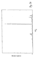
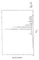
- FIGS. 3a, 3b and 3c show examples of spectra of three particular explosives, namely trinitrotolumine (TNT), so called “C4" plastic explosive, and “Flex-x", respectively. These spectra show peaks which distinctly identify these explosives.
- FIG. 4 illustrates the processing system in greater detail.
- the collimated germanium detector system 30 is used with an x-ray generator 24 capable of operating at up to 160 KV.
- a narrow beam 12 of x-rays is generated which irradiates a container 14 holding an explosive.
- the photons scattered through a fixed angle of 2 ⁇ were detected and all other scatter angles were precluded by a narrow aperture collimator 34 (Fig. 2).
- a narrow aperture collimator 34 Fig. 2
- spectrum of x-rays emerges from the source 10, but only those scattered at or near an angle of 2 ⁇ are seen by the detector.
- a detector supply 56 provides the high voltage and temperature control system necessary to operate the germanium detector array 30. For each x-ray photon incident upon the detector, an electrical pulse is produced proportional to the energy, and therefore inversely proportional to the wavelength.
- a photon energy detector 40 operates as a charge integrator which produces a digitized signal which is then processed by an energy dispersive spectrum generator 42.
- the generator 42 actually counts the number of photons within each of a number of energy intervals for each detector 32. For example, there are 140 separate 500eV intervals in an energy range between 50 KeV and 120 KeV. Thus each detector 32 generates a spectrum displaying the number of photons counted for an inspected item as a function of photon energy.
- the processing sequence is then split into two paths.
- the outputs from each individual spectrum generator 42 are processed by a peak detector algorithms 44 along one of these paths.
- Each algorithm 44 identifies those peaks within the spectrum from spectrum generator 42 which can be distinguished from the background spectra.
- the identified peaks are then isolated and quantified and output as a peak spectrum.
- the spectrum generated by the algorithm 44 for the individual detectors is then compared using the explosives spectrum comparator 46 with a number of predetermined spectra similar to those shown in Figures 3a, b and c. A sufficiently close match between the peak spectrum and one or more of the stored spectra will then identify the materials detected and display the result 54.
- a second processing path that can be performed simultaneously with the above processing sequence takes the output from each spectrum generator 42 and sums all of the spectra at 48.
- the summed spectra is then analyzed with the peak detector algorithm 50 and compared to the stored spectra in the explosives spectrum comparator 52 to determine an aggregate result for the entire parcel being inspected.
- the summing of all the spectra will increase the ratio for materials distributed throughout the parcel sufficiently to properly identify the contents.
- the sensitivity of the diffraction technique depends upon the total signal collected, which, in turn, is a product of the x-ray flux density F, the illuminated volume V i , the time that the sample is in the beam, and the detector aperture seen from the sample.
- V i (b)(c)(z)
- the total radiation dose received by the inspected bag is equal to the product of the x-ray flux density and the time that one point is within the beam.
- the radiation dose is proportional to (F)(z)/s. Note that the x-ray beam properties and the bag speed appear identical in both expressions for the sensitivity and dose. This implies that, for a given acceptable radiation dose, the sensitivity is independent of inspection speed. One can increase the x-ray power and bag speed proportionally without affecting either dose or sensitivity.
- the collimator slit 22 adjacent to the generator 10 provides a variable fan beam 12 which is scattered and detected with a number of detectors fitted with a second set of collimators 34.
- the detector collimators 34 will be arranged to pass x-rays which are scattered through a narrow angle, 2 ⁇ , from samples mounted in the beam or held in a suitcase in the beam as shown.
- the angular aperture of the collimators 34 is on the order of 0.03°. Thus only those rays scattered though an angle of 2 ⁇ .03° are allowed into the detectors 32.
- the detector 32 has a detection area which is capable of detecting scattered rays from a cross section of baggage for the full depth of a large suitcase and a strip of about 20 to 30mm wide across the bag. Several such detectors 32 are arranged across the conveyor having identical scatter angle and detect scattered rays from all of the bag volume.
Abstract
An inspection system and method for detecting the presence of selected crystalline materials, such as explosives or drugs, utilizing an x-ray source (10) and a collimated array of detectors (32) to sense radiation scattered by suitcases (14) (Fig. 1) being inspected. A signal processing system (Fig. 4) comprising a photon energy detector (40), an energy dispersive spectrum generator (42), a peak detector algorithm (44) and an e.g. explosives spectrum comparator (46) compares the measured signal with selected spectra (Fig. 3a, b or c) to determine whether specific materials are present within the inspected suitcase.
Description
- The invention relates generally to the field of radiographic detection systems, and more particularly, although not exclusively, to coherent x-ray scattering systems used to inspect objects to detect the presence of explosive materials.
- Numerous systems have been developed for the inspection of bags, suitcases and briefcases etc., hereinafter more generally referred to as "parcels", that are used to screen travellers at airports or to secure other types of installations. Of particular concern in the development of such systems has been the detection of concealed weapons, explosives or drugs whose transport is restricted. Typically, standard x-ray equipment has been used to scan for metal objects where the outline of the displayed items is visually inspected to determine the presence of the objects of concern. These systems use a conveyor to transport the item to be inspected into and out of a chamber positioned between an x-ray source and a fluroscopic or scintillation screen that detects the transmitted radiation. These inspection systems must be safe for the operators, and must not damage items such as photographic film that are often contained in inspected baggage.
- Unfortunately, many kinds of materials that may pose a danger may not conform to any easily identifiable shape and are not visually detectable in the systems currently in use. In particular, many types of explosive materials in use can be molded into any shape and are not detectable by standard x-ray equipment. Thus, there is a need for a more accurate and dependable system for detecting these materials.
- An inspection device of the present invention utilizes an x-ray radiation source to scan objects to determine whether certain crystalline substances are contained therein. Most explosives and many other dangerous or illegally transported substances have a crystalline structure.
- Crystalline materials are known to scatter radiation when illuminated by an x-ray source of suitable energy and spectral content. Coherent elastic scattering of radiation occurs from lattices of spacing "d" within the crystalline material which satisfy the equation λ = 2d sinϑ where λ is the wavelength and 2ϑ is the angle of diffraction of the scattered radiation away from an axis through the source and the area being scanned. Thus, elastic scattered will occur at a few select wavelengths whereas inelastic scattering by the atoms of the material will occur continuously across a spectrum of wavelengths.
- A detector positioned at the appropriate angle relative to the source and scanned object is fitted with a collimator that permits only those x-rays diffracted at that angle to reach the detector. The detector is preferably comprised of a photon spectrometer. Such spectrometers provide both intensity and spectral composition of the detected radiation and are typically either solid state germanium or silicon planar arrays operated at cryogenic temperatures.
- Each scattered photon is detected individually and a wavelength spectrum of intensities, displayed as an energy dispersive spectrum, is generated by a signal processing circuit.
- A peak comparator algorithm incorporated into a data processing system is applied to the spectral output to determine the presence of sharp peaks within the spectrum. By comparing the peaks from the processed spectrum with the spectrum of crystalline substances sought to be detected the presence of those materials within the object is determined with great accuracy.
- The present invention also provides a method of inspecting parcels to detect the presence of selected crystalline materials in the presence of other crystalline and noncrystalline materials comprising generating x-ray radiation from a source; conveying an object to be inspected past the source to irradiate the object with the radiation; and detecting radiation scattered by the object at a predetermined angle to detect the presence of a selected crystalline material on or within the object.
- Some ways of carrying out the present invention will now be described in detail by way of example with reference to drawings which show one specific embodiment.
- In the drawings:
- FIG. 1 illustrates a front schematic view, of a parcel inspection device of the present invention;
- FIG. 2 shows a side view of the device shown in Figure 1;
- FIGS. 3a, 3b and 3c are illustrative graphical representations of diffraction spectra of particular explosive materials; and
- FIG. 4 is a schematic diagram of the processing system of the device.
- With reference now to the accompanying drawings, the inspection device is comprised of energy dispersive X-ray detectors arranged to measure the coherent elastic scattering of x-ray photons from the lattices of crystalline materials and in particular of crystalline explosives and of narcotic or hallucinogenic drugs. Nearly all of the explosives of interest comprise crystalline powders. For example, the plastic explosives are manufactured from crystalline powders of cyclotrimethyline-trinitramine (RDX), cyclotetramethyline-tetranitramine (HMX) and pentaerithritol-tentranitrate (PETN), and are compounded into a putty with minor amounts of organic binders. Each of the explosives which are to be detected provides a unique diffraction pattern when irradiated with x-rays such that each may be rapidly identified. The only notable exceptions are the nitro-glycerine-based dynamites. Fortunately these explosives are easy to detect by their vapor emissions and the two detection systems can be combined into a single instrument.
- The device has sufficient speed of response to detect explosives in bags which are conveyed through the detection zone in a few seconds. X-rays from
source 10 are arranged in abeam 12 having a fan pattern to irradiatebags 14 which are conveyed alongconveyor 16 through thebeam 12. Thebeam 12 comprises an x-ray continuum whose range of photon energies is sufficient to penetrate large checked bags. Thebeam 12 is produced by collimation of thesingle x-ray source 10 of constant potential withslit collimator 20. - Photons scattered elastically from the crystalline lattice of explosives interferes coherently depending on the lattice structure, the frequency and angle of scatter. At a fixed angle of scatter, scattered rays comprise a continuum from non-crystalline materials and photons which are coherently scattered from the various 'd' spacings of the crystals. In a typical two dimensional lattice there are three major 'd' spacings which allow coherent diffraction. There are other lattice spacings in the third dimension so that for randomly oriented crystals there are sufficient 'd' spacings to provide a unique set, but not so many that they cause an overly cluttered diffraction spectrum.
- The x-rays scattered in a beam of fixed angle defined by the
detector collimator 34 comprises a continuum with superimposed intensity peaks at wavelengths λ1...n. - The
detection system 30 measures the intensity of scattered light in intervals of wavelengths over a wide range of photon energies but at a fixed angle 2ϑ of scatter. This provides a unique fingerprint for each type of explosive. - An array of
individual detectors 32 is arranged across the full width of a conveyor system irradiated by anx-ray fan beam 12. This permits scanning of the whole volume of abag 14. - The
detector 32 employed in the system is a planar germanium photon spectrometer. These detectors operate as photon counters by detecting the liberation of free electrons by an x-ray photon in a germanium semiconductor. The total charge liberated by each x-ray photon is indicative of the photon energy thus allowing an intensity spectrum to be generated over a range of x-ray wavelengths. Typical resolution (full-width-half-maximum) provided by commercial detectors is 500eV at 120 KeV photon energy. The detector for this embodiment has a 100% detection efficiency over a range of photon energy from 5 KeV to 120 KeV. - The system is based on the detection of presence of two or three peaks in the spectrum. It is unlikely that the detection of these peaks will be interfered with by other crystalline materials. For example, the only crystalline material normally encountered in detectable quantities in 99% of airline bags are the metals aluminum, steel, and copper. The sensitivity of the inspection system is proportional to the total amount of crystalline material in a bag, so for small amounts of these metals only a low intensity of scatter is expected and this information is in parts of the spectrum which are distinct from the explosives or drugs of interest. Large amounts of metal can possibly stop 120 KeV photons and this condition can be detected by the attenuation of the transmitted beam in a similar way to standard airport baggage x-rays, but no image would be generated. This condition is not expected to exist in more than a fraction of a percent of airline bags, and of course attempts to conceal the explosive by this method results in a rejection because of too high a metallic content.
- The principle of detection is to send a "white"
x-ray beam 12, containing a range of wavelengths, through the specimen and look for diffracted radiation at a fixed angle 2ϑ, with adetector system 30 that simultaneously measures intensity and wavelength. The advantages of energy dispersive x-ray diffraction over the more common monochromatic powder technique are two fold: the large sensitive volume (and therefore freedom from the necessity of aligning the sample precisely), and the fact that an entire spectrum may be collected in parallel. - In a crystal, there are regular planes of atoms separated by well-defined distances d1...n. X-rays of wavelength λ may be scattered by these planes through a total angle 2ϑ if they meet the diffraction condition
λ = 2 d sinϑ - The set of "d" spacings in a particular material, along with the associated intensities of diffraction, provide a fingerprint of the material. The system simply recognizes a stored pattern as an indication that a given material is present. Figures 3a, 3b and 3c show examples of spectra of three particular explosives, namely trinitrotolumine (TNT), so called "C4" plastic explosive, and "Flex-x", respectively. These spectra show peaks which distinctly identify these explosives.
- Figure 4 illustrates the processing system in greater detail. The collimated
germanium detector system 30 is used with anx-ray generator 24 capable of operating at up to 160 KV. Anarrow beam 12 of x-rays is generated which irradiates acontainer 14 holding an explosive. The photons scattered through a fixed angle of 2ϑ were detected and all other scatter angles were precluded by a narrow aperture collimator 34 (Fig. 2). Thus spectrum of x-rays emerges from thesource 10, but only those scattered at or near an angle of 2ϑ are seen by the detector. - A
detector supply 56 provides the high voltage and temperature control system necessary to operate thegermanium detector array 30. For each x-ray photon incident upon the detector, an electrical pulse is produced proportional to the energy, and therefore inversely proportional to the wavelength. - A
photon energy detector 40 operates as a charge integrator which produces a digitized signal which is then processed by an energydispersive spectrum generator 42. Thegenerator 42 actually counts the number of photons within each of a number of energy intervals for eachdetector 32. For example, there are 140 separate 500eV intervals in an energy range between 50 KeV and 120 KeV. Thus eachdetector 32 generates a spectrum displaying the number of photons counted for an inspected item as a function of photon energy. - The processing sequence is then split into two paths. The outputs from each
individual spectrum generator 42 are processed by apeak detector algorithms 44 along one of these paths. Eachalgorithm 44 identifies those peaks within the spectrum fromspectrum generator 42 which can be distinguished from the background spectra. The identified peaks are then isolated and quantified and output as a peak spectrum. The spectrum generated by thealgorithm 44 for the individual detectors is then compared using theexplosives spectrum comparator 46 with a number of predetermined spectra similar to those shown in Figures 3a, b and c. A sufficiently close match between the peak spectrum and one or more of the stored spectra will then identify the materials detected and display theresult 54. - A second processing path that can be performed simultaneously with the above processing sequence takes the output from each
spectrum generator 42 and sums all of the spectra at 48. The summed spectra is then analyzed with thepeak detector algorithm 50 and compared to the stored spectra in theexplosives spectrum comparator 52 to determine an aggregate result for the entire parcel being inspected. Thus in situations where the signal to noise ratio is too small for the individual detectors to properly identify the contents of the parcel, the summing of all the spectra will increase the ratio for materials distributed throughout the parcel sufficiently to properly identify the contents. - It is also possible to gate the detection and processing system at predetermined time intervals during the scanning of each particular bag to further increase the sensitivity of the system.
- The sensitivity of the diffraction technique depends upon the total signal collected, which, in turn, is a product of the x-ray flux density F, the illuminated volume Vi, the time that the sample is in the beam, and the detector aperture seen from the sample. Consider the case of a bag containing a total volume Ve of explosive material, moving at a certain speed through the x-ray fan beam of width z. For simplicity assume the explosive is a rectangular block of dimensions (a)(b)(c), moving along the "a" direction.
- Then, while it is in the beam the illuminated volume is:
Vi = (b)(c)(z),
and the time that it is in the beam is a / s. Consequently,
Vi t = Ve z/s.
and the overall detection sensitivity from the summed spectra for the whole bag is proportional to
F Ve z/s. - It is important to emphasize that this result is independent of the shape and orientation of the explosive material within the bag.
- The total radiation dose received by the inspected bag is equal to the product of the x-ray flux density and the time that one point is within the beam. The radiation dose is proportional to (F)(z)/s. Note that the x-ray beam properties and the bag speed appear identical in both expressions for the sensitivity and dose. This implies that, for a given acceptable radiation dose, the sensitivity is independent of inspection speed. One can increase the x-ray power and bag speed proportionally without affecting either dose or sensitivity.
- The collimator slit 22 adjacent to the
generator 10 provides avariable fan beam 12 which is scattered and detected with a number of detectors fitted with a second set ofcollimators 34. Thedetector collimators 34 will be arranged to pass x-rays which are scattered through a narrow angle, 2ϑ, from samples mounted in the beam or held in a suitcase in the beam as shown. The angular aperture of thecollimators 34 is on the order of 0.03°. Thus only those rays scattered though an angle of 2 ±.03° are allowed into thedetectors 32. - The
detector 32 has a detection area which is capable of detecting scattered rays from a cross section of baggage for the full depth of a large suitcase and a strip of about 20 to 30mm wide across the bag. Severalsuch detectors 32 are arranged across the conveyor having identical scatter angle and detect scattered rays from all of the bag volume.
Claims (10)
1. A parcel inspection device for the detection of selected crystalline materials in the presence of other crystalline and noncrystalline materials comprising:
an x-ray radiation source to irradiate an object to be inspected such that the radiation is projected along an axis through the source and object;
a conveyor for transporting objects through the projected radiation;
a detector positioned to receive radiation scattered by the object at an angle relative to the source-object axis; and
an analyzer to determine the presence of a predetermined wavelength of radiation within the detected radiation.
2. An inspection device as claimed in claim 1 further comprising a collimator positioned between the source and the object to define a beam of radiation directed at the object along said axis.
3. An inspection device as claimed in claim 1 or 2 further comprising a receiving collimator to define an aperture through which scattered radiation is received by the detector.
4. An inspection device as claimed in any preceding claim wherein said detector is comprised of a plurality of sensors configured to form an array such that each sensor measures the intensity for a spectrum of wavelengths of radiation incident upon the sensor.
5. A method of inspecting parcels to detect the presence of selected crystalline materials in the presence of other crystalline and noncrystalline materials comprising:
generated x-ray radiation from a source; conveying an object to be inspected past the source to irradiate the object with the radiation;
detecting radiation scattered by the object at a predetermined angle to detect the presence of a selected crystalline material on or within the object.
generated x-ray radiation from a source; conveying an object to be inspected past the source to irradiate the object with the radiation;
detecting radiation scattered by the object at a predetermined angle to detect the presence of a selected crystalline material on or within the object.
6. A method as claimed in claim 5 wherein the selected crystalline material is an explosive or narcotic.
7. A method as claimed in claim 5 or 6 wherein said detecting step comprises measuring the scattered radiation with a plurality of detectors such that each detector measures the radiation scattered by a portion of the object.
8. A method as claimed in claim 7 further comprising:
generating a plurality of signals from the radiation measured by each detector;
forming a spectrum from each of the signals; and
summing the formed spectra to generate a spectra for the object being scanned.
generating a plurality of signals from the radiation measured by each detector;
forming a spectrum from each of the signals; and
summing the formed spectra to generate a spectra for the object being scanned.
9. A method as claimed in claim 8 further comprising comparing the spectra with a predetermined spectra to determine the presence of selected crystalline materials.
10. A method as claimed in claim 9 wherein the formed spectra from each signal are compared with the predetermined spectra to determine the presence of selected crystalline materials in each portion of the object.
Applications Claiming Priority (2)
| Application Number | Priority Date | Filing Date | Title |
|---|---|---|---|
| US227722 | 1988-08-03 | ||
| US07/227,722 US5007072A (en) | 1988-08-03 | 1988-08-03 | X-ray diffraction inspection system |
Publications (2)
| Publication Number | Publication Date |
|---|---|
| EP0354045A2 true EP0354045A2 (en) | 1990-02-07 |
| EP0354045A3 EP0354045A3 (en) | 1991-10-02 |
Family
ID=22854200
Family Applications (1)
| Application Number | Title | Priority Date | Filing Date |
|---|---|---|---|
| EP19890307921 Withdrawn EP0354045A3 (en) | 1988-08-03 | 1989-08-03 | X-ray diffraction inspection system and method |
Country Status (2)
| Country | Link |
|---|---|
| US (1) | US5007072A (en) |
| EP (1) | EP0354045A3 (en) |
Cited By (20)
| Publication number | Priority date | Publication date | Assignee | Title |
|---|---|---|---|---|
| EP0462658A2 (en) * | 1990-06-20 | 1991-12-27 | Philips Patentverwaltung GmbH | Arrangement for the measurement of the pulse transfer spectrum of X-ray quanta |
| EP0412189B1 (en) * | 1989-08-09 | 1992-10-28 | Heimann Systems GmbH & Co. KG | Device for transmitting fan-shaped radiation through objects |
| GB2297835A (en) * | 1995-02-08 | 1996-08-14 | Secr Defence | Three dimensional detection of contraband using x rays |
| GB2299251A (en) * | 1995-03-21 | 1996-09-25 | Heimann Systems Gmbh & Co | Detecting crystalline material using X-ray diffraction |
| WO1997012234A1 (en) * | 1995-09-27 | 1997-04-03 | Schlumberger Limited | Material analysis |
| GB2312507A (en) * | 1995-02-08 | 1997-10-29 | Secr Defence | X-ray inspection system |
| EP0871866A1 (en) * | 1995-05-31 | 1998-10-21 | Quanta Vision, Inc. | X-ray and neutron diffractometric imaging of the internal structure of objects |
| WO1999066317A1 (en) * | 1998-06-18 | 1999-12-23 | American Science And Engineering, Inc. | Coherent scattering for material identification |
| FR2801103A1 (en) * | 1999-11-13 | 2001-05-18 | Heimann Systems Gmbh & Co | X-ray baggage scanner compares diffracted and primary rays improves accuracy of material identification |
| DE19954662A1 (en) * | 1999-11-13 | 2001-06-07 | Heimann Systems Gmbh & Co | Device and method for detecting illegal baggage items |
| GB2359718A (en) * | 1999-11-13 | 2001-08-29 | Heimann Systems Gmbh & Co | Determination of crystalline and polycrystalline materials in an article |
| US6542578B2 (en) | 1999-11-13 | 2003-04-01 | Heimann Systems Gmbh | Apparatus for determining the crystalline and polycrystalline materials of an item |
| US6621888B2 (en) | 1998-06-18 | 2003-09-16 | American Science And Engineering, Inc. | X-ray inspection by coherent-scattering from variably disposed scatterers identified as suspect objects |
| WO2005043145A1 (en) * | 2003-11-03 | 2005-05-12 | Xcounter Ab | Coherent scatter imaging |
| US6907107B1 (en) | 1999-03-16 | 2005-06-14 | Qinetiq Limited | Method and apparatus for the analysis of material composition |
| US7065175B2 (en) | 2003-03-03 | 2006-06-20 | Varian Medical Systems Technologies, Inc. | X-ray diffraction-based scanning system |
| DE102004060611A1 (en) * | 2004-12-16 | 2006-06-29 | Yxlon International Security Gmbh | Arrangement for measuring the pulse transmission spectrum of elastically scattered X-ray quanta |
| EP1508800B1 (en) * | 2003-08-19 | 2008-02-27 | Obshchestvo s ogranichennoj otvetstvennostyu "Institut Rentgenovskoi Optiki" | Detecting unit for x-ray diffraction measurements |
| US7844028B2 (en) | 2002-01-25 | 2010-11-30 | Isis Innovation Limited | X-ray diffraction method |
| CN101968454A (en) * | 2010-09-17 | 2011-02-09 | 上海交通大学 | X-ray energy dispersion and diffraction based hazardous article detection method |
Families Citing this family (62)
| Publication number | Priority date | Publication date | Assignee | Title |
|---|---|---|---|---|
| DE4101544A1 (en) * | 1991-01-19 | 1992-07-23 | Philips Patentverwaltung | ROENTGENGERAET |
| US5263075A (en) * | 1992-01-13 | 1993-11-16 | Ion Track Instruments, Inc. | High angular resolution x-ray collimator |
| EP0556887B1 (en) * | 1992-02-06 | 1998-07-08 | Philips Patentverwaltung GmbH | Device for the measurement of the pulse transfer spectrum of X-ray quantor |
| US5600303A (en) * | 1993-01-15 | 1997-02-04 | Technology International Incorporated | Detection of concealed explosives and contraband |
| US5600700A (en) * | 1995-09-25 | 1997-02-04 | Vivid Technologies, Inc. | Detecting explosives or other contraband by employing transmitted and scattered X-rays |
| US5642393A (en) * | 1995-09-26 | 1997-06-24 | Vivid Technologies, Inc. | Detecting contraband by employing interactive multiprobe tomography |
| US5974111A (en) * | 1996-09-24 | 1999-10-26 | Vivid Technologies, Inc. | Identifying explosives or other contraband by employing transmitted or scattered X-rays |
| AU6039798A (en) * | 1997-01-24 | 1998-08-18 | Quanta Vision, Inc. | Device for determining composition and structure of objects |
| US6118850A (en) * | 1997-02-28 | 2000-09-12 | Rutgers, The State University | Analysis methods for energy dispersive X-ray diffraction patterns |
| US6054712A (en) * | 1998-01-23 | 2000-04-25 | Quanta Vision, Inc. | Inspection equipment using small-angle topography in determining an object's internal structure and composition |
| US6094472A (en) * | 1998-04-14 | 2000-07-25 | Rapiscan Security Products, Inc. | X-ray backscatter imaging system including moving body tracking assembly |
| US6442233B1 (en) * | 1998-06-18 | 2002-08-27 | American Science And Engineering, Inc. | Coherent x-ray scatter inspection system with sidescatter and energy-resolved detection |
| US6567496B1 (en) * | 1999-10-14 | 2003-05-20 | Sychev Boris S | Cargo inspection apparatus and process |
| US20050117683A1 (en) * | 2000-02-10 | 2005-06-02 | Andrey Mishin | Multiple energy x-ray source for security applications |
| US20080211431A1 (en) * | 2000-02-10 | 2008-09-04 | American Science And Engineering, Inc. | Pulse-to-Pulse-Switchable Multiple-Energy Linear Accelerators Based on Fast RF Power Switching |
| US7010094B2 (en) * | 2000-02-10 | 2006-03-07 | American Science And Engineering, Inc. | X-ray inspection using spatially and spectrally tailored beams |
| US7538325B2 (en) * | 2000-02-10 | 2009-05-26 | American Science And Engineering, Inc. | Single-pulse-switched multiple energy X-ray source applications |
| US20060104414A1 (en) * | 2002-01-30 | 2006-05-18 | Mayo William E | Combinatorial contraband detection using energy dispersive x-ray diffraction |
| EP1599745A1 (en) * | 2003-02-24 | 2005-11-30 | Philips Intellectual Property & Standards GmbH | Automatic material discrimination by using computer tomography |
| US8243876B2 (en) | 2003-04-25 | 2012-08-14 | Rapiscan Systems, Inc. | X-ray scanners |
| US8451974B2 (en) | 2003-04-25 | 2013-05-28 | Rapiscan Systems, Inc. | X-ray tomographic inspection system for the identification of specific target items |
| GB0525593D0 (en) | 2005-12-16 | 2006-01-25 | Cxr Ltd | X-ray tomography inspection systems |
| US9113839B2 (en) | 2003-04-25 | 2015-08-25 | Rapiscon Systems, Inc. | X-ray inspection system and method |
| US8223919B2 (en) | 2003-04-25 | 2012-07-17 | Rapiscan Systems, Inc. | X-ray tomographic inspection systems for the identification of specific target items |
| US20050058242A1 (en) | 2003-09-15 | 2005-03-17 | Peschmann Kristian R. | Methods and systems for the rapid detection of concealed objects |
| US7949101B2 (en) | 2005-12-16 | 2011-05-24 | Rapiscan Systems, Inc. | X-ray scanners and X-ray sources therefor |
| US8837669B2 (en) | 2003-04-25 | 2014-09-16 | Rapiscan Systems, Inc. | X-ray scanning system |
| US7092485B2 (en) * | 2003-05-27 | 2006-08-15 | Control Screening, Llc | X-ray inspection system for detecting explosives and other contraband |
| US7856081B2 (en) | 2003-09-15 | 2010-12-21 | Rapiscan Systems, Inc. | Methods and systems for rapid detection of concealed objects using fluorescence |
| US7366282B2 (en) | 2003-09-15 | 2008-04-29 | Rapiscan Security Products, Inc. | Methods and systems for rapid detection of concealed objects using fluorescence |
| US7813540B1 (en) * | 2005-01-13 | 2010-10-12 | Oro Grande Technologies Llc | System and method for detecting nuclear material in shipping containers |
| US7221732B1 (en) * | 2005-04-04 | 2007-05-22 | Martin Annis | Method and apparatus for producing laminography images using a fixed x-ray source |
| KR100751023B1 (en) * | 2006-03-30 | 2007-08-22 | 박성근 | X-ray inspection device using diffraction |
| US8576982B2 (en) | 2008-02-01 | 2013-11-05 | Rapiscan Systems, Inc. | Personnel screening system |
| US7796733B2 (en) * | 2007-02-01 | 2010-09-14 | Rapiscan Systems, Inc. | Personnel security screening system with enhanced privacy |
| US8638904B2 (en) | 2010-03-14 | 2014-01-28 | Rapiscan Systems, Inc. | Personnel screening system |
| US8995619B2 (en) | 2010-03-14 | 2015-03-31 | Rapiscan Systems, Inc. | Personnel screening system |
| WO2008133765A2 (en) * | 2007-02-13 | 2008-11-06 | Sentinel Scanning Corporation | Ct scanning and contraband detection |
| JP5503535B2 (en) | 2007-06-21 | 2014-05-28 | ラピスカン システムズ、インコーポレイテッド | System and method for improving controlled physical examination |
| US7869566B2 (en) * | 2007-06-29 | 2011-01-11 | Morpho Detection, Inc. | Integrated multi-sensor systems for and methods of explosives detection |
| US8003949B2 (en) | 2007-11-01 | 2011-08-23 | Rapiscan Systems, Inc. | Multiple screen detection systems |
| AU2008340164A1 (en) | 2007-12-25 | 2009-07-02 | Rapiscan Systems, Inc. | Improved security system for screening people |
| US9310323B2 (en) | 2009-05-16 | 2016-04-12 | Rapiscan Systems, Inc. | Systems and methods for high-Z threat alarm resolution |
| WO2010141101A1 (en) * | 2009-06-05 | 2010-12-09 | Sentinel Scanning Corporation | Transportation container inspection system and method |
| US8654922B2 (en) * | 2009-11-18 | 2014-02-18 | Rapiscan Systems, Inc. | X-ray-based system and methods for inspecting a person's shoes for aviation security threats |
| MX2012010646A (en) | 2010-03-14 | 2012-12-05 | Rapiscan Systems Inc | Multiple screen detection systems. |
| WO2013098520A1 (en) * | 2011-12-28 | 2013-07-04 | Commissariat A L'energie Atomique Et Aux Energies Alternatives | Method of analysing a sample of material by diffractometry and associated diffractometer |
| FR2995688B1 (en) * | 2012-09-20 | 2014-10-10 | Commissariat Energie Atomique | DIFFRACTOMETRY ANALYSIS METHOD AND DIFFRACTOMETER THEREOF, PARTICULARLY ADAPTED TO SAMPLES COMPRISING SEVERAL LAYERS OF MATERIALS |
| FR2985312B1 (en) * | 2011-12-28 | 2017-03-24 | Commissariat Energie Atomique | METHOD OF ANALYZING A MATERIAL SAMPLE BY DIFFRACTOMETRY AND DIFFRACTOMETER |
| AU2013215064B2 (en) | 2012-02-03 | 2015-10-22 | Rapiscan Systems, Inc. | Combined scatter and transmission multi-view imaging system |
| GB2523520B (en) | 2013-01-07 | 2018-05-23 | Rapiscan Systems Inc | X-ray scanner with partial energy discriminating detector array |
| US9791590B2 (en) | 2013-01-31 | 2017-10-17 | Rapiscan Systems, Inc. | Portable security inspection system |
| CN105612416B (en) * | 2013-07-25 | 2019-01-01 | 模拟技术公司 | The generation of the diffractive features of article in object |
| US9557427B2 (en) | 2014-01-08 | 2017-01-31 | Rapiscan Systems, Inc. | Thin gap chamber neutron detectors |
| US11280898B2 (en) | 2014-03-07 | 2022-03-22 | Rapiscan Systems, Inc. | Radar-based baggage and parcel inspection systems |
| KR20160130482A (en) | 2014-03-07 | 2016-11-11 | 라피스캔 시스템스, 인코포레이티드 | Ultra wide band detectors |
| US9405990B2 (en) * | 2014-08-19 | 2016-08-02 | Morpho Detection, Llc | X-ray diffraction imaging system with signal aggregation across voxels containing objects and method of operating the same |
| EP3224797A4 (en) | 2014-11-25 | 2018-07-18 | Rapiscan Systems, Inc. | Intelligent security management system |
| US10345479B2 (en) | 2015-09-16 | 2019-07-09 | Rapiscan Systems, Inc. | Portable X-ray scanner |
| US9939393B2 (en) * | 2015-09-28 | 2018-04-10 | United Technologies Corporation | Detection of crystallographic properties in aerospace components |
| US10720300B2 (en) | 2016-09-30 | 2020-07-21 | American Science And Engineering, Inc. | X-ray source for 2D scanning beam imaging |
| DE102017220600A1 (en) | 2017-11-17 | 2019-05-23 | Fraunhofer-Gesellschaft zur Förderung der angewandten Forschung e.V. | PLANT WITH A CONVEYOR BELT AND AN X-RAY SYSTEM |
Citations (6)
| Publication number | Priority date | Publication date | Assignee | Title |
|---|---|---|---|---|
| GB1081683A (en) * | 1963-10-26 | 1967-08-31 | Bayer Ag | Process and apparatus for the simultaneous qualitative and quantitative determination of at least two crystalline substances |
| GB1329695A (en) * | 1970-09-09 | 1973-09-12 | Nutter J C | Diffractometry |
| GB1426102A (en) * | 1973-01-29 | 1976-02-25 | Aquitaine Petrole | Apparatus for x-ray crystallography |
| EP0209952A2 (en) * | 1985-07-20 | 1987-01-28 | Philips Patentverwaltung GmbH | Method for the measurement of the spatial distribution of scattering cross-sections in elastically scattered X-radiation, and arrangement for carrying out such a method |
| EP0271723A1 (en) * | 1986-11-26 | 1988-06-22 | Heimann GmbH | X-ray scanning system |
| EP0311177A2 (en) * | 1987-10-05 | 1989-04-12 | Philips Patentverwaltung GmbH | System for examining a body with a radiance source |
Family Cites Families (23)
| Publication number | Priority date | Publication date | Assignee | Title |
|---|---|---|---|---|
| US28544A (en) * | 1860-06-05 | Ditching-machine | ||
| US3440419A (en) * | 1966-02-03 | 1969-04-22 | California Inst Res Found | Dual purpose optical instrument capable of simultaneously acting as spectrometer and diffractometer |
| US3428802A (en) * | 1966-04-11 | 1969-02-18 | Ibm | External standard method of x-ray diffraction analysis for determining the percentage of compounds in cement clinker |
| GB1255644A (en) * | 1968-05-10 | 1971-12-01 | Rolls Royce | Method of determining the value of a mechanical property or properties of a fibre |
| US3759383A (en) * | 1971-08-02 | 1973-09-18 | K Inoue | Apparatus for making abrasive articles |
| US4020346A (en) * | 1973-03-21 | 1977-04-26 | Dennis Donald A | X-ray inspection device and method |
| US3895232A (en) * | 1973-12-13 | 1975-07-15 | Westinghouse Electric Corp | X-ray contrast detection system |
| US3980889A (en) * | 1974-04-08 | 1976-09-14 | North American Philips Corporation | Article transfer and inspection apparatus |
| US4031545A (en) * | 1975-09-08 | 1977-06-21 | American Science & Engineering, Inc. | Radiant energy alarm system |
| NL7513716A (en) * | 1975-11-25 | 1977-05-27 | Philips Nv | ROENTGEN FLUORESCENCE SPECTROMETER. |
| US4260898A (en) * | 1978-09-28 | 1981-04-07 | American Science And Engineering, Inc. | X-ray imaging variable resolution |
| US4228357A (en) * | 1978-12-04 | 1980-10-14 | American Science And Engineering, Inc. | Detector on wheel system (flying spot) |
| US4303860A (en) * | 1979-07-30 | 1981-12-01 | American Science And Engineering, Inc. | High resolution radiation detector |
| US4357535A (en) * | 1980-04-30 | 1982-11-02 | North American Philips Corporation | Apparatus for inspecting hand-held articles and persons carrying same |
| JPS5756740A (en) * | 1980-09-22 | 1982-04-05 | Mitsubishi Electric Corp | Object inspecting device |
| US4342914A (en) * | 1980-09-29 | 1982-08-03 | American Science And Engineering, Inc. | Flying spot scanner having arbitrarily shaped field size |
| US4389729A (en) * | 1981-12-15 | 1983-06-21 | American Science And Engineering, Inc. | High resolution digital radiography system |
| US4454605A (en) * | 1982-01-25 | 1984-06-12 | Delucia Victor E | Modular X-ray inspection apparatus |
| US4511799A (en) * | 1982-12-10 | 1985-04-16 | American Science And Engineering, Inc. | Dual energy imaging |
| US4691332A (en) * | 1983-03-14 | 1987-09-01 | American Science And Engineering, Inc. | High energy computed tomography |
| DE3406905A1 (en) * | 1984-02-25 | 1985-09-05 | Philips Patentverwaltung Gmbh, 2000 Hamburg | ROENTGENGERAET |
| US4715053A (en) * | 1985-01-25 | 1987-12-22 | Westinghouse Electric Corp. | Method for monitoring the crystallographic texture of metallic tubes by use of X-ray diffraction |
| US4799247A (en) * | 1986-06-20 | 1989-01-17 | American Science And Engineering, Inc. | X-ray imaging particularly adapted for low Z materials |
-
1988
- 1988-08-03 US US07/227,722 patent/US5007072A/en not_active Expired - Fee Related
-
1989
- 1989-08-03 EP EP19890307921 patent/EP0354045A3/en not_active Withdrawn
Patent Citations (6)
| Publication number | Priority date | Publication date | Assignee | Title |
|---|---|---|---|---|
| GB1081683A (en) * | 1963-10-26 | 1967-08-31 | Bayer Ag | Process and apparatus for the simultaneous qualitative and quantitative determination of at least two crystalline substances |
| GB1329695A (en) * | 1970-09-09 | 1973-09-12 | Nutter J C | Diffractometry |
| GB1426102A (en) * | 1973-01-29 | 1976-02-25 | Aquitaine Petrole | Apparatus for x-ray crystallography |
| EP0209952A2 (en) * | 1985-07-20 | 1987-01-28 | Philips Patentverwaltung GmbH | Method for the measurement of the spatial distribution of scattering cross-sections in elastically scattered X-radiation, and arrangement for carrying out such a method |
| EP0271723A1 (en) * | 1986-11-26 | 1988-06-22 | Heimann GmbH | X-ray scanning system |
| EP0311177A2 (en) * | 1987-10-05 | 1989-04-12 | Philips Patentverwaltung GmbH | System for examining a body with a radiance source |
Non-Patent Citations (1)
| Title |
|---|
| ANALYTICAL INSTRUMENTATION, vol. 16, no. 2, 1987, pages 281-294, New York, US; M.K. SANYAL et al.: "A microprocessor based energy dispersive x-ray diffractometer" * |
Cited By (37)
| Publication number | Priority date | Publication date | Assignee | Title |
|---|---|---|---|---|
| EP0412189B1 (en) * | 1989-08-09 | 1992-10-28 | Heimann Systems GmbH & Co. KG | Device for transmitting fan-shaped radiation through objects |
| EP0462658A2 (en) * | 1990-06-20 | 1991-12-27 | Philips Patentverwaltung GmbH | Arrangement for the measurement of the pulse transfer spectrum of X-ray quanta |
| EP0462658A3 (en) * | 1990-06-20 | 1992-09-09 | Philips Patentverwaltung Gmbh | Arrangement for the measurement of the pulse transfer spectrum of x-ray quanta |
| GB2312507B (en) * | 1995-02-08 | 1999-08-25 | Secr Defence | X-Ray Inspection System |
| GB2297835A (en) * | 1995-02-08 | 1996-08-14 | Secr Defence | Three dimensional detection of contraband using x rays |
| GB2312507A (en) * | 1995-02-08 | 1997-10-29 | Secr Defence | X-ray inspection system |
| US6122344A (en) * | 1995-02-08 | 2000-09-19 | The Secretary Of State For Defence In Her Brittanic Majesty's Government Of The United Kingdom Of Great Britain And Northern Ireland | X-ray inspection system |
| WO1996024863A1 (en) * | 1995-02-08 | 1996-08-15 | The Secretary Of State For Defence | X-ray inspection system |
| GB2299251B (en) * | 1995-03-21 | 1998-12-09 | Heimann Systems Gmbh & Co | Method and apparatus for detecting crystalline and polycrystalline substances |
| GB2299251A (en) * | 1995-03-21 | 1996-09-25 | Heimann Systems Gmbh & Co | Detecting crystalline material using X-ray diffraction |
| US5787145A (en) * | 1995-03-21 | 1998-07-28 | Heimann Systems Gmbh | Method and arrangement for identifying crystalline and polycrystalline materials |
| EP0871866A4 (en) * | 1995-05-31 | 1998-11-25 | ||
| EP0871866A1 (en) * | 1995-05-31 | 1998-10-21 | Quanta Vision, Inc. | X-ray and neutron diffractometric imaging of the internal structure of objects |
| US6072853A (en) * | 1995-09-27 | 2000-06-06 | Schlumberger Technology Corporation | Material analysis |
| WO1997012234A1 (en) * | 1995-09-27 | 1997-04-03 | Schlumberger Limited | Material analysis |
| US6621888B2 (en) | 1998-06-18 | 2003-09-16 | American Science And Engineering, Inc. | X-ray inspection by coherent-scattering from variably disposed scatterers identified as suspect objects |
| WO1999066317A1 (en) * | 1998-06-18 | 1999-12-23 | American Science And Engineering, Inc. | Coherent scattering for material identification |
| US6907107B1 (en) | 1999-03-16 | 2005-06-14 | Qinetiq Limited | Method and apparatus for the analysis of material composition |
| GB2359718A (en) * | 1999-11-13 | 2001-08-29 | Heimann Systems Gmbh & Co | Determination of crystalline and polycrystalline materials in an article |
| US6532276B1 (en) | 1999-11-13 | 2003-03-11 | Heimann Systems Gmbh | Method and apparatus for determining a material of a detected item |
| US6542578B2 (en) | 1999-11-13 | 2003-04-01 | Heimann Systems Gmbh | Apparatus for determining the crystalline and polycrystalline materials of an item |
| DE19954662A1 (en) * | 1999-11-13 | 2001-06-07 | Heimann Systems Gmbh & Co | Device and method for detecting illegal baggage items |
| GB2359718B (en) * | 1999-11-13 | 2003-12-31 | Heimann Systems Gmbh & Co | Apparatus for determining crystalline and polycrystalline materials in an article |
| DE19954662B4 (en) * | 1999-11-13 | 2004-06-03 | Smiths Heimann Gmbh | Apparatus and method for detecting unauthorized luggage items |
| NL1016471C2 (en) * | 1999-11-13 | 2004-08-03 | Heimann Systems Gmbh & Co | Method and device for determining a material of a detected object. |
| US6839406B2 (en) | 1999-11-13 | 2005-01-04 | Smiths Heimann Gmbh | Apparatus and method for detecting items in objects |
| FR2801103A1 (en) * | 1999-11-13 | 2001-05-18 | Heimann Systems Gmbh & Co | X-ray baggage scanner compares diffracted and primary rays improves accuracy of material identification |
| DE19954663B4 (en) * | 1999-11-13 | 2006-06-08 | Smiths Heimann Gmbh | Method and device for determining a material of a detected object |
| DE19954664B4 (en) * | 1999-11-13 | 2006-06-08 | Smiths Heimann Gmbh | Device for the determination of crystalline and polycrystalline materials of an article |
| US7844028B2 (en) | 2002-01-25 | 2010-11-30 | Isis Innovation Limited | X-ray diffraction method |
| US7065175B2 (en) | 2003-03-03 | 2006-06-20 | Varian Medical Systems Technologies, Inc. | X-ray diffraction-based scanning system |
| EP1508800B1 (en) * | 2003-08-19 | 2008-02-27 | Obshchestvo s ogranichennoj otvetstvennostyu "Institut Rentgenovskoi Optiki" | Detecting unit for x-ray diffraction measurements |
| US7099436B2 (en) | 2003-11-03 | 2006-08-29 | Xcounterab | Coherent scatter imaging |
| WO2005043145A1 (en) * | 2003-11-03 | 2005-05-12 | Xcounter Ab | Coherent scatter imaging |
| DE102004060611A1 (en) * | 2004-12-16 | 2006-06-29 | Yxlon International Security Gmbh | Arrangement for measuring the pulse transmission spectrum of elastically scattered X-ray quanta |
| DE102004060611B4 (en) * | 2004-12-16 | 2007-02-22 | Yxlon International Security Gmbh | Arrangement for measuring the pulse transmission spectrum of elastically scattered X-ray quanta |
| CN101968454A (en) * | 2010-09-17 | 2011-02-09 | 上海交通大学 | X-ray energy dispersion and diffraction based hazardous article detection method |
Also Published As
| Publication number | Publication date |
|---|---|
| EP0354045A3 (en) | 1991-10-02 |
| US5007072A (en) | 1991-04-09 |
Similar Documents
| Publication | Publication Date | Title |
|---|---|---|
| US5007072A (en) | X-ray diffraction inspection system | |
| US6122344A (en) | X-ray inspection system | |
| US7092485B2 (en) | X-ray inspection system for detecting explosives and other contraband | |
| US6320933B1 (en) | Multiple scatter system for threat identification | |
| US5479023A (en) | Method and apparatus for detecting concealed substances | |
| US6621888B2 (en) | X-ray inspection by coherent-scattering from variably disposed scatterers identified as suspect objects | |
| EP0396618B1 (en) | Neutron scatter method and apparatus for the noninvasive interrogation of objects | |
| JPH04504310A (en) | Apparatus and method for detecting contraband using fast neutron activation | |
| Drakos et al. | Multivariate analysis of energy dispersive X-ray diffraction data for the detection of illicit drugs in border control | |
| EP2677304B1 (en) | Method and device for identifying unknown substances in an object | |
| CA1314633C (en) | Detection system for explosives | |
| CA2109311A1 (en) | Energy discriminating resonant, neutron detector | |
| Madden et al. | An explosives detection system for airline security using coherent x-ray scattering technology | |
| EP0423763B1 (en) | X-ray fluorescence imaging of elements | |
| NL2009984C2 (en) | Method and device for screening objects for the presence of foreign substances. | |
| O'Flynn et al. | Pixelated diffraction signatures for explosive detection | |
| AU775264B2 (en) | Apparatus for fast detection of X-rays | |
| RU2166749C1 (en) | Roentgenographic device | |
| US11971371B2 (en) | Sample inspection system comprising a beam former to project a polygonal shell beam | |
| Miller et al. | Contraband identification in sealed containers using neutron transmission | |
| Creagh | Technology for border security | |
| NL2026256B1 (en) | A detection system and method for investigating a content of an item | |
| US20220381710A1 (en) | A sample inspection system | |
| Poullain et al. | Research for the detection of explosives at CEA: towards operational use | |
| Barmakov et al. | Application of tagged neutron technology in research, industry and homeland security |
Legal Events
| Date | Code | Title | Description |
|---|---|---|---|
| PUAI | Public reference made under article 153(3) epc to a published international application that has entered the european phase |
Free format text: ORIGINAL CODE: 0009012 |
|
| AK | Designated contracting states |
Kind code of ref document: A2 Designated state(s): BE CH DE FR GB IT LI NL |
|
| PUAL | Search report despatched |
Free format text: ORIGINAL CODE: 0009013 |
|
| AK | Designated contracting states |
Kind code of ref document: A3 Designated state(s): BE CH DE FR GB IT LI NL |
|
| 17P | Request for examination filed |
Effective date: 19920330 |
|
| STAA | Information on the status of an ep patent application or granted ep patent |
Free format text: STATUS: THE APPLICATION IS DEEMED TO BE WITHDRAWN |
|
| 18D | Application deemed to be withdrawn |
Effective date: 19930302 |