EP0349342B1 - Polypeptides with cell-spreading activity - Google Patents
Polypeptides with cell-spreading activity Download PDFInfo
- Publication number
- EP0349342B1 EP0349342B1 EP89306676A EP89306676A EP0349342B1 EP 0349342 B1 EP0349342 B1 EP 0349342B1 EP 89306676 A EP89306676 A EP 89306676A EP 89306676 A EP89306676 A EP 89306676A EP 0349342 B1 EP0349342 B1 EP 0349342B1
- Authority
- EP
- European Patent Office
- Prior art keywords
- cell
- sequence
- dna
- peptide
- minutes
- Prior art date
- Legal status (The legal status is an assumption and is not a legal conclusion. Google has not performed a legal analysis and makes no representation as to the accuracy of the status listed.)
- Expired - Lifetime
Links
Images


Classifications
-
- C—CHEMISTRY; METALLURGY
- C07—ORGANIC CHEMISTRY
- C07K—PEPTIDES
- C07K14/00—Peptides having more than 20 amino acids; Gastrins; Somatostatins; Melanotropins; Derivatives thereof
- C07K14/435—Peptides having more than 20 amino acids; Gastrins; Somatostatins; Melanotropins; Derivatives thereof from animals; from humans
- C07K14/78—Connective tissue peptides, e.g. collagen, elastin, laminin, fibronectin, vitronectin, cold insoluble globulin [CIG]
-
- A—HUMAN NECESSITIES
- A61—MEDICAL OR VETERINARY SCIENCE; HYGIENE
- A61K—PREPARATIONS FOR MEDICAL, DENTAL OR TOILETRY PURPOSES
- A61K8/00—Cosmetics or similar toiletry preparations
- A61K8/18—Cosmetics or similar toiletry preparations characterised by the composition
- A61K8/30—Cosmetics or similar toiletry preparations characterised by the composition containing organic compounds
- A61K8/64—Proteins; Peptides; Derivatives or degradation products thereof
-
- A—HUMAN NECESSITIES
- A61—MEDICAL OR VETERINARY SCIENCE; HYGIENE
- A61P—SPECIFIC THERAPEUTIC ACTIVITY OF CHEMICAL COMPOUNDS OR MEDICINAL PREPARATIONS
- A61P17/00—Drugs for dermatological disorders
-
- A—HUMAN NECESSITIES
- A61—MEDICAL OR VETERINARY SCIENCE; HYGIENE
- A61P—SPECIFIC THERAPEUTIC ACTIVITY OF CHEMICAL COMPOUNDS OR MEDICINAL PREPARATIONS
- A61P27/00—Drugs for disorders of the senses
- A61P27/02—Ophthalmic agents
-
- A—HUMAN NECESSITIES
- A61—MEDICAL OR VETERINARY SCIENCE; HYGIENE
- A61P—SPECIFIC THERAPEUTIC ACTIVITY OF CHEMICAL COMPOUNDS OR MEDICINAL PREPARATIONS
- A61P35/00—Antineoplastic agents
-
- A—HUMAN NECESSITIES
- A61—MEDICAL OR VETERINARY SCIENCE; HYGIENE
- A61P—SPECIFIC THERAPEUTIC ACTIVITY OF CHEMICAL COMPOUNDS OR MEDICINAL PREPARATIONS
- A61P43/00—Drugs for specific purposes, not provided for in groups A61P1/00-A61P41/00
-
- A—HUMAN NECESSITIES
- A61—MEDICAL OR VETERINARY SCIENCE; HYGIENE
- A61Q—SPECIFIC USE OF COSMETICS OR SIMILAR TOILETRY PREPARATIONS
- A61Q19/00—Preparations for care of the skin
-
- A—HUMAN NECESSITIES
- A61—MEDICAL OR VETERINARY SCIENCE; HYGIENE
- A61K—PREPARATIONS FOR MEDICAL, DENTAL OR TOILETRY PURPOSES
- A61K2800/00—Properties of cosmetic compositions or active ingredients thereof or formulation aids used therein and process related aspects
- A61K2800/80—Process related aspects concerning the preparation of the cosmetic composition or the storage or application thereof
- A61K2800/86—Products or compounds obtained by genetic engineering
-
- A—HUMAN NECESSITIES
- A61—MEDICAL OR VETERINARY SCIENCE; HYGIENE
- A61K—PREPARATIONS FOR MEDICAL, DENTAL OR TOILETRY PURPOSES
- A61K38/00—Medicinal preparations containing peptides
-
- A—HUMAN NECESSITIES
- A61—MEDICAL OR VETERINARY SCIENCE; HYGIENE
- A61Q—SPECIFIC USE OF COSMETICS OR SIMILAR TOILETRY PREPARATIONS
- A61Q11/00—Preparations for care of the teeth, of the oral cavity or of dentures; Dentifrices, e.g. toothpastes; Mouth rinses
Definitions
- This invention relates to a protein which has cell-spreading activity like that of fibronectin. More particularly, the invention relates to a polypeptide which has the cell-spreading activity of fibronectin of human origin; and also to a method for the preparation of said polypeptide.
- Fibronectin is a multifunctional glycoprotein which is widely distributed in a variety of animal tissues and body fluids and also on the surface of cultured cells and elsewhere. This compound has various physiological effects, such as causing attachment, spreading, migration, differentiation, proliferation, and phagocytosis by cells, and participates in such activities as tissue reconstruction, tissue construction, and protection from infection.
- Fibronectin is a polypeptide with a molecular weight of about 250,000 and is a dimer with an S-S bond in the vicinity of the C-terminus.
- the amino acid sequence of this molecule contains 3 different types of internal repeat, and can be classified as types I, II and III.
- domain structures which have various functions, with the effect of cell attachment and spreading and the ability to bind to collagen, heparin, fibrin, etc.
- industrial applications of the biological activity related to the cell attachment and spreading domain have been considered; for example, in the preparation of a coating agent for a culture substrate, it is possible to use this function in the preparation of a substrate to which cells will bind.
- this function can be used to an accelerator of cell binding in such preparations as collyrium, lotions, and agents for the healing of wounds.
- Cell spreading is a phenomenon that follows after cell attachment. For cells to proliferate, with some exceptions, it is necessary for the phenomenon of spreading to take place, not cell attachment alone.
- the basic structure which is the minimum essential structure for the cell-attachment domain of fibronectin is the sequence Arg-Gly-Asp-Ser ( Nature , 309 1984, 30-33).
- Japanese Laid-Open Patent (Tokuhyo) 84-501548 discloses a peptide with cell-attachment activity, that is a polypeptide of a molecular weight of 11,500 and that contains this sequence among its sequence of 108 amino acid residues.
- this polypeptide with a molecular weight of 11,500 is much weaker than that of fibronectin of natural origin, and it is not necessarily possible to make use of it in the practical applications mentioned above. This difficulty is discussed, for example, in J . Biol . Chem ., 260 (1985). 13256-13260.
- this polypeptide of 11,500 molecular weight (MW) by genetic engineering, and compared its cell-spreading activity to that of fibronectin of natural origin with the use of normal rat kidney (NRK) cells. The results were that, whereas fibronectin gave noticeable activity at a dose of 0.1-1 ⁇ g/well, a dose of 50 ⁇ g/well of the 11,500 MW polypeptide did not have any such activity.
- This invention identifies the amino acid sequence that has substantial cell-spreading activity as the peptide of the cell-spreading domain of fibronectin and provides a method for producing the same.
- the present invention provides polypeptides with cell-spreading activity, which have an amino acid sequence represented by the following general formula [I]:
- This invention also provides recombinant plasmids which contain DNA which codes for polypeptides (I), and transformants which carry these recombinant plasmids.
- the present invention further provides a method for the preparation of the polypeptides (I) by the cultivation of these transformants and the collection of the polypeptides from the culture medium.
- Preparation by genetic engineering of a plasmid that codes for the peptide with a sequence of 279 amino acids(Pro1239-Met1517) can be as disclosed in USSN 291894 and EP 89301440.7.
- RNA fraction containing poly(A) from the liver is prepared, and a cDNA library can be prepared by such method as the Okayama-Bergmethod or the Gubler-Hoffmann method.
- cDNA libraries are commercially available. Thus it is possible to obtain them from, for example, the Clontech Laboratories, Inc.
- the DNA which corresponds to the amino acid sequence can be used as a probe with the techniques of colony hybridization or plaque hybridization.
- the clones which hybridize with the probe are selected, and DNA is extracted from the cell pellet or a phage lytic solution and cleaved with restriction enzymes. Insertions are confirmed by the use of electrophoresis.
- Southern blotting is used to check the insert being hybridized with the probe.
- the identity of the desired clone can be checked.
- the vector which carried the cDNA coding for the cell-spreading domain of fibronectin can be amplified in the host cells, purified, and cleaved with restriction enzymes so that the cDNA portion can be removed; electrophoresis can be used to purify the cDNA fragments.
- These cDNA fragments are joined to expression vectors by inframe ligation using DNA ligase, and by their introduction to host cells it is possible to express the cDNA.
- Any of the known vectors can be used as expression vectors for Escherichia coli as the host, but for preference, a host-vector system which can have its expression induced and which has strong promoter activity is used.
- vectors there are, for example, a vector which carries the ⁇ PL, promoter, a vector which carries the lac promoter, a vector which carries the trp promoter, a vector which carries the pst promoter, a vector which carries both the lac and the trp promoters, a vector which carries the lpp promoter, a vector which carries both the lpp and lac promoters, and other such known vectors (refer to Yoshiyuki Sakai, "Vector DNA", Kodansha Scientific, 1986).
- a peptide When a peptide is expressed by the gene for the desired peptide by the use of an expression vector which can be joined downstream from a gene for a signal peptide or by the use of an expression vector directly, by the joining of the vector, a certain length of the amino acid sequence of vector origin is sometimes connected to the N-terminus of the desired peptide.
- the addition of this sequence causes no essential change in the cell-spreading activity of the polypeptide, and it is possible to use the polypeptide as is.
- an appropriate restriction enzyme is used to cleave one site slightly upstream of the initiation codon of pTF301 that codes for the sequence Ala1235-Met1517 of FN, and then exonuclease is used, by which means it is possible to remove the 5'-end of the sequence.
- exonuclease is used, by which means it is possible to remove the 5'-end of the sequence.
- an appropriate restriction enzyme is used to cleave a site slightly downstream from the termination codon of the coding region of these plasmids, and the DNA which has been cleaved is separated by gel electrophoresis, by which it is possible to obtain fragments of cDNA from which various portions of the 5'-terminal strand have been removed.
- an appropriate expression vector By the insertion of these cDNA fragments into an appropriate expression vector, it is possible to express peptides of various lengths wherein portions of the N-terminal region of the sequence of Ala1235-Met1517 (283 amino acid residues) have been deleted.
- any of the well-known vectors can be used. We have obtained satisfactory results with direct expression by the use of the pUC-type vectors in which the distance between the ribosome-binding site and the initiation codon has been made optimum.
- Selection of the recombinants which express the peptide with cell-spreading activity can be done conveniently with immunoscreening. That is, expression vectors to which the cDNA fragments of different lengths have been joined are inserted into cells of Escherichia coli by the usual methods, and the transformants obtained are raised on nitrocellulose filters, after which they are lysed, and the protein from the cells is fixed on the filters. After the filters are blocked with bovine serum albumin or the like, a monoclonal antibody which recognizes the domain of cell spreading of FN is caused to act. The monoclonal antibody bound to the filter is detected by labelling with a second antibody. In this way, it is possible to select recombinants that express the peptide with the domain for cell spreading.
- the recombinants so selected are cultured under conditions suitable for expression, and expression of the peptide with the domain for cell spreading is induced.
- immunoblotting can be used.
- the whole-cell protein of the cultured cells is lysed by heat treatment in a buffer containing SDS, and separation is conducted by SDS-polyacrylamide electrophoresis, and the electrophoretic pattern is transferred to a nitrocellulose or nylon membrane.
- Purification of the peptide with the domain for cell spreading from the recombinants can be done, for example, as follows.
- the cell pellet is suspended in a buffer, and the soluble fraction and insoluble fraction are separated by ultrasonification.
- the insoluble fraction is solubilized in a buffer which contains 7 M urea.
- the soluble fractions are pooled, put on a Sepharose 4B column bound with the antibody used in immunoblotting, and affinity purification is carried out. For elution there is used a buffer in the pH region of 2.3.
- affinity purification is carried out.
- a buffer in the pH region of 2.3 By collection of the desired fractions by immunoblotting, it is possible to collect the peptide with the domain for cell spreading.
- further purification by FPLC and HPLC can be done.
- the peptide with the cell-spreading domain thus obtained may be measured for its cell-spreading activity toward NRK (normal rat kidney) cells.
- the sample is dissolved in a buffer, and used to coat microtiter plate wells, after which NRK cells are added, and the plate is incubated for a fixed time at 37°C.
- the spreading of the cells is observed under a microscope, and the minimum dose of sample per well that gives rise to the expression of cell-spreading activity is compared to the dose needed of FN of natural origin. In this way, the strength of the cell-spreading activity can be expressed.
- a plasmid, pLF5 (reported in Biochemistry , 25 [1986], 4936-4941), which is of 4.1 kilo-bases and which contains the cDNA sequence which codes for the cell-spreading domain of fibronectin, was put into 200 ⁇ l of a reaction mixture containing a buffer for use with the restriction enzyme Bal I and 100 units of Bal I, and the mixture was incubated for 2 hours at 37°C.
- reaction mixture was then put on an HPLC column of DEAE-4000 (6 x 125 mm; Nucleogen) and eluted with a concentration gradient of KCl in a 30 mM potassium phosphate buffer (pH 6.5) which contains 5 M urea, and precipitated with isopropyl alcohol, which gave 7.5 ⁇ g of 0.75 kb fragments.
- these fragments were treated with 30 units of Fok I for 1 hour at 37°C, and by the same purification method, 60 ng of 92-bp fragments and 140 ng of 203-bp fragments were obtained.
- 5'-end and 3'-end linkers were chemically synthesized by the following method.
- the base sequence is shown in Fig. 1.
- the upper strand of the 5'-end linker (chain length, 11), labelled 5 U, the lower strand of the 5'-end linker (chain length, 7), labelled 5 L, the upper strand of the 3'-end linker (chain length, 30), labelled 3 U, and the lower strand of the 3'-end linker (chain length, 30), labelled 3 L, were synthesized by use of the DNA synthesizer of Applied Biosystems, Inc.
- the two kinds of linker prepared in section (1-2) above and the two kinds of DNA fragment (with 92 bp and with 203 bp) prepared in section (1-1) above were used, and ligated together.
- 0.5 pmol of each kind of cDNA fragment and 5 pmol of each kind of linker were put into 20 ⁇ l of a solution containing a ligation buffer [66 mM Tris-HCl, pH 7.6, and 6.6 mM MgCl2] and also 0.5 mM ATP, 10 mM DTT, and 2.8 units of T4 DNA ligase, at 16°C; the mixture was incubated overnight.
- the reaction mixture was heated for 10 minutes at 65°C, and then two volumes of cold ethanol was added, and the whole left at -70°C for 30 minutes. After this, the precipitated DNA was collected by centrifugation.
- This DNA was put into 20 ⁇ l of a reaction mixture made of buffer for use with Eco RI and 7.5 units of Eco RI, and incubated for 30 minutes at 37°C. Then the mixture was subjected to polyacrylamide electrophoresis, Whereby 336-bp fragments were obtained. These were extracted for 10 hours at 37°C in a gel elution buffer (0.5 M ammonium acetate, 0.01 M magnesium acetate, and 1 mM EDTA), after which the fragments were precipitated with ethanol, and the DNA was collected. This DNA was dissolved in 20 ⁇ l of TE buffer (10 mM Tris-HCl and 1 mM EDTA, pH 8.0).
- This DNA was cleaved with Eco RI, and then 0.1 ⁇ g of dephosphorylated pIN-III-ompA-l vector was put into a mixture with 0.1 ⁇ g of the DNA fragments obtained in section (1-3) above together with 10 ⁇ l of buffer for use with T4 DNA ligase, 0.5 mM ATP, 10 mM DTT, and 1.8 units of T4 DNA ligase, in a total volume for the reaction mixture of 30 ⁇ l. This mixture was incubated overnight at 16°C. Then 15 ⁇ l of this reaction mixture was used in the transformation of Escherichia coli cells.
- reaction mixture was separated by agarose electrophoresis, and the expected bands for DNA (at 0.33 kb) were checked by ethidium bromide staining. The results showed that nine of the clones gave the desired band.
- the plasmids extracted from these nine clones were doubly digested with the use of Pvu II and Hin dIII. Then the orientation of the inserted fragments was identified. It was found that eight of the clones had the inserts with the correct orientation.
- the plasmids from five clones were doubly digested with the use of Xba I and Hin dIII, and the DNA that contained the inserted fragments was removed, and subcloned into the phages M13mp18 and mp19.
- the base sequences were analyzed by the dideoxy method. Three of the clones were found to contain the desired sequence.
- the recombinant plasmids obtained in this way were named pTF101.
- the pTF101 obtained as described above in section (1-6) was used to transform Escherichia coli cells (SB221/pTF 101) and such transformed cells were cultured in 5 ml of L-broth overnight with agitation at 37°C.
- the culture was transferred to 500 ml of the same culture broth and culture was continued until the absorbance at 660 nm reached 0.6, at which time 170 ⁇ g/ml chloramphenicol was added, and cultured was continued once more for 10-12 hours.
- the culture broth was centrifuged for 10 minutes at 5000 rpm, and the cell pellet was washed in STE buffer (10 mM Tris-HCl, pH 8, 100 mM NaCl, and 1 mM EDTA), and centrifuged again in the same way in order to obtain the cell pellet.
- the pellet was suspended in 10 ml of the solution A, and kept at room temperature for 5 minutes before 20 ml of the solution B was added with rapid stirring, and the suspension was left for 5 minutes at 0°C. To this suspension, 15 ml of 3 M potassium acetate was added with rapid stirring, and the suspension was left at 0°C for 10 minutes.
- the mixture was centrifuged for 60 minutes at 8000 rpm and the supernatant was obtained, to which 27 ml (0.6 volume) of isopropyl alcohol was added, and the mixture was left at room temperature for 15 minutes. This mixture was centrifuged and the precipitate was collected and washed in 80% ethanol. The ethanol and water in the resulting mixture was removed under reduced pressure and the residue was dried. This residue was dissolved in 4 ml of TE buffer and to this solution 4.75 g of cesium chloride was added, followed by the addition of 5 mg/ml ethidium bromide in the volume of 420 ⁇ l; the mixture was left for 30 minutes at 0°C.
- the mixture was then centrifuged for 10 minutes at 8000 rpm, and the supernatant was put into a Bechman Quick-seal tube and ultracentrifuged at 55000 rpm overnight. Then, under ultraviolet light, plasmid bands were removed by the use of a syringe. The resulting mixture was extracted seven times in equal volumes of isopropyl alcohol and the remaining ethidium bromide was removed, after which ethanol precipitation was done twice, and then the remaining mixture was washed in 80% ethanol, dried under reduced pressures, and dissolved in a small amount of TE buffer (yield, 760 ⁇ g).
- plasmid pLF5 50 ⁇ g of the plasmid pLF5 was put into 200 ⁇ l of a reaction mixture containing 200 units of Eco RI methylase in a buffer for use with Eco RI methylase (100 mM Tris-HCl, pH 8.0, 2 mM DTT, 10 mM EDTA, and 80 ⁇ M S-adenosylmethionine), and the mixture was incubated for 60 minutes at 37°C to bring about protection of the Eco RI sites. Then the reaction was stopped by heating at 65°C for 20 minutes, and 96 units of Pvu II in buffer for use with Pvu II was added; 300 ⁇ l of the reaction mixture was incubated for 60 minutes at 37°C.
- Eco RI methylase 100 mM Tris-HCl, pH 8.0, 2 mM DTT, 10 mM EDTA, and 80 ⁇ M S-adenosylmethionine
- This mixture was separated by agarose electrophoresis, and the slice of gel containing 0.60 kb band was cut out. This slice was put into a dialysis tube that contained elution buffer (5 mM Tris-acetate buffer, pH 8.0, and 1 mM EDTA), and eluted by electrophoresis in the same buffer, by which means the DNA was eluted.
- the elution fluid was extracted twice with phenol, and a 1/10 volume of 3 M sodium acetate was added, followed by the addition of two volumes of ethanol, and ethanol precipitation was conducted twice. Then the resulting mixture was washed in 80% ethanol, dried, and dissolved in a small amount of TE buffer (yield, 1.2 ug).
- Example 2 140 ⁇ g of the plasmid pTF101 obtained in Example 1 was put into 200 ⁇ l of a reaction solution containing 144 units of Pvu II, and the mixture was incubated for 60 minutes at 37°C. Then 150 units of Eco RI was added, and the reaction solution brought to 240 ⁇ l, and incubated for 60 minutes at 37°C. The result was separated by polyacrylamide electrophoresis, and the bands at 0.25 kb were cut out. The DNA was extracted from these bands of gel as described above, and the DNA was collected by ethanol precipitation (yield, 0.4 ⁇ g).
- the reaction was stopped by heating at 65°C for 10 minutes, and the buffer was modified to be suitable for Eco RI; 30 units of Eco RI was added, and reaction was allowed to take place for 60 minutes at 37°C. Then the reaction mixture was separated by agarose electrophoresis, and the 0.86-kb bands were cut out. The DNA in the bands was obtained by the method described above (yield, about 25 ng).
- the DNA obtained was put into 200 ⁇ l of a reaction mixture of 7 mM Tris-HCl, pH 7.5, which contained 0.1 mM EDTA, 10 mM NaCl, 7 mM MgCl2, 0.1 mM dATP, dGTP, dCTP, and dTTP, and 2 units of Klenow fragment.
- the mixture was incubated for 20 minutes at 37°C.
- the reaction was stopped by treatment at 65°C for 10 minutes, and the reaction mixture was adjusted to the composition of buffer for use with Hin dIII.
- 60 units for Hin dIII was added, and the mixture was incubated for 1 hour at 37°C. This mixture was separated by agarose gel electrophoresis, and the fragments that correspond to the size of 0.5-0.8 kb were cut out; 1 ⁇ g was obtained.
- pUC119N was obtained by creation of a Nco I site surrounding the translation initiation codon of the commercially available vector pUC119 (Takara Shuzo Co., Ltd.); the distance between the ribosome-binding site and initiation codon was also changed from 7 to 8 bases.
- the transformants obtained in section (3-2) above were transferred to a nitrocellulose filter (BA85; S & S) on L-agar medium containing 50 ⁇ g/ml ampicillin, and cultured overnight at 37°C.
- the colonies that grew were brought into chloroform vapor for 15 minutes, and the nitrocellulose filter was put into a solution of 50 mM Tris-HCl, pH 7.5, containing 150 mM NaCl, 5 mM MgCl2, 3% bovine serum albumin, 1 ⁇ g/ml DNase, and 40 ⁇ g/ml lysozyme; the mixture was incubated overnight at room temperature.
- the filter was treated with an anti-FN monoclonal antibody, FN-10 (Tarara Shuzo Co., Ltd.) which specifically recognizes the cell-spreading domain of FN, and a second antibody labelled with peroxidase was applied, and the hydrogen peroxide gave rise to color in the presence of 4-chloro-1-naphthol, by which the transformants which had appeared were found.
- FN-10 Tarara Shuzo Co., Ltd.
- a second antibody labelled with peroxidase was applied, and the hydrogen peroxide gave rise to color in the presence of 4-chloro-1-naphthol, by which the transformants which had appeared were found.
- the whole-cell protein of the cells obtained was separated on SDS-polyacrylamide gel electrophoresis (SDS-PAGE), and immunoblotting was applied, in which the anti-FN monoclonal antibody FN-10 was allowed to react. It was found that polypeptides of the molecular masses of 22 kDa-32 kDa had been produced. For 11 clones, the base sequence of the 5'-end of the inserted fragments was identified; they were found to code amino acid sequences which were respectively 279, 258, 219, 213, 207, 206, 198, 195, 190, 186, and 178 long counting from Met1517 as the C-terminus.
- the plasmid which expressed the peptide with the sequence 279 amino acids long was designated pTFD707.
- GCT sequence attached that corresponded to alanine of vector origin. This sequence was removed by site-specific mutagenesis (a procedure used, for example, in Example 4 of USSN 291,894).
- the Hin dIII- Sal I fragment with a lpp terminator sequence was removed from the secretion-expression vector pIN-III-ompA-I, and joined with the Hin dIII- Sal I site of pTFD707, by which means plasmids designated pTF7021 were obtained.
- pTFD707 was introduced into Escherichia coli BW313, and the transformants were cultured with agitation at 37°C on 100 ml of 2X YT medium (1.6% Bactotrypton, 1% yeast extract, and 0.5% NaCl) that contained 50 ⁇ g/ml ampicillin.
- 2X YT medium (1.6% Bactotrypton, 1% yeast extract, and 0.5% NaCl
- the absorbance at 660 nm was 0.3
- 1 ml of M13K07 phage suspension containing 1010 pfu was added to the culture, and cultivation was continued at 37°C for 16 hours.
- the culture medium was centrifuged and the supernatant collected; to this supernatant, 25 ml of a mixture of 2.5 M NaCl and 20% polyethlene glycol 6000 was added, and the resulting mixture was left for 10 minutes at room temperature. It was then centrifuged, and 5 ml of TE buffer was added to the precipitate. This mixture was treated with a mixture of phenol and chloroform, and then with chloroform alone, before ethanol precipitation was used to give single-stranded DNA.
- the 30 ng of single-stranded DNA obtained was dissolved in 1 ⁇ l of annealing buffer(20 mM Tris-HCl, pH 8.0, 10 mM MgCl2, 50 mM NaCl, and 1 mM DTT), and added to 1 ⁇ l of a solution that contained 1 pmol of phosphorylated oligonucleotide d[pGGATGGTTAGTCAATTTC], and the mixture was treated by heating at 65°C for 15 minutes and at 37°C for 15 minutes.
- annealing buffer 20 mM Tris-HCl, pH 8.0, 10 mM MgCl2, 50 mM NaCl, and 1 mM DTT
- extension buffer 50mM Tris-HCl, pH8.0, 60 mM ammonium acetate, 5 mM MgCl2, 5 mM DTT, 1 mM NAD, 0.5 mM dATP, dGTP, dCTP and dTTP
- extension buffer 50mM Tris-HCl, pH8.0, 60 mM ammonium acetate, 5 mM MgCl2, 5 mM DTT, 1 mM NAD, 0.5 mM dATP, dGTP, dCTP and dTTP
- DNA ligase from E. coli and 1 unit of T4 DNA polymerase
- reaction mixture 3 ⁇ l of the reaction mixture was mixed with 30 ⁇ l of competent cells of Escherichia coli BMH71-18mutS, and the mixture was kept at 0° C for 30 minutes, at 42°C for 45 seconds, and at 0°C for 2 minutes. Then 300 ⁇ l of L-broth was added, and the mixture was left for 1 hour at 37°C. Next, 10 ⁇ l of a suspension of M13K07 phages was added, and the mixture was left for 30 minutes at 37°C. Next, the cells were cultured for 16 hours at 37°C with agitation in 1 ml of 2X YT medium that contained 150 ⁇ g/ml ampicillin and 70 ⁇ g/ml kanamycin.
- the culture was centrifuged and the supernatant collected. Then 20 ⁇ l of the supernatant was mixed with 80 ⁇ l of the culture medium of an overnight culture of Escherichia coli JM109. This was left for 10 minutes at 37°C, and one portion was used to inoculate L-agar that contained 50 ⁇ g/ml ampicillin; the agar was left at 37 C° overnight, and of the transformants that were obtained. The base sequences of six of the clones were identified. Of these six clones, five were found to have the desired mutation. The recombinant plasmid obtained was designated pTFD707-45.
- This recombinant plasmid was named pTF7221, and the cells of Escherichia coli JM109 that carried this plasmid were designated Escherichia coli JM109/pTF7221.
- the strain was deposited at the Fermentation Research Institute of the Agency of Industrial Science and Technology, Japan, under FERM BP-1915 on 17th June 1988.
- Plasmid pTF7221 obtained by the joining of an expression vector and the DNA that codes for the Pro1239-Asp1512 sequence of FN was introduced into Escherichia coli JM109, giving Escherichia coli JM109/pTF7221, and these cells were cultured overnight with shaking at 37°C in 5 ml of L-broth containing 50 ⁇ g/ml ampicillin in a test tube. This was used to inoculate a 2-1 Erlenmeyer flask containing 500 ml of the same medium, and culture was continued at an agitation rate of 180 r.p.m.
- IPTG isopropyl- ⁇ -thiogalactoside
- the activity of the peroxidase bound to the second antibody gave rise to color in the presence of 4-chloronaphthol and hydrogen peroxide, and it was found that the desired band in the region of 34 kDa lower molecular band than that of 279 amino acid residues was present.
- the whole-cell pellet eas suspended in a solution of 10 mM Tris-HCl, pH 7.5, containing 5 mM EDTA and 5 mM mercaptoethanol, and ultrasonification was conducted. This suspension was centrifuged and the supernatant was obtained and dialyzed against 20 mM Tris-HCl (pH 7.5).
- the inner dialysis liquid was put on a Sepharose 4B column (8 ml) bound with the monoclonal antibody FN-10.
- the column was washed with washing buffer A (20 mM Tris-HCl, pH 8.0, and 0.15 M KCl), and then with washing buffer B (20 mM Tris-HCl, pH 6.4, and 0.15 M KCl).
- washing buffer A (20 mM Tris-HCl, pH 8.0, and 0.15 M KCl
- washing buffer B 20 mM Tris-HCl, pH 6.4, and 0.15 M KCl
- the eluting buffer 50 mM glycine-HCl, pH 2.3, and 0.2 M KCl
- the cell-spreading activity of the polypeptide of 274 amino acid residues obtained in Example 5 and of FN and the peptides with 108 and 279 amino acids was measured by the method of Ruoslahti et al. ( Methods in Enzymology , 82 , 803-831, 1981).
- the samples were diluted stepwise in physiological saline and distilled water, and 50 ⁇ l aliquots were injected into the wells of a 96-well microtitre plate, which was then incubated overnight at 4°C in order to allow the samples to adhere to the wells.
- phosphate-buffered saline PBS
- PBS phosphate-buffered saline
- 100 ⁇ l of 3% BSA was added to each well, and the plate was incubated for one hour at 37°C.
- the plate was washed twice with PBS, and then normal rat kidney (NRK-49F) cells suspended to a concentration of 106 cells/ml in Eagle's Minimum Essential Medium (MEM) were added in the amount of 100 ⁇ l/well, and the plate was incubated for 2-3 hours at 37°C.
- NRK-49F cells that were used were obtained as a lyophilized strain for storage, and first preincubated and then treated with trypsin before use. The spreading of the cells was observed under a microscope, and the minimum dose needed to have cell-spreading activity was calculated.
- this invention provides a peptide which has cell-spreading activity essentially the same as that of FN, and also provides a method for its preparation by the use of genetic engineering.
- the polypeptides mentioned above can be used in pharmaneutical preparations for such uses as for the healing of wounds, in collyria, for the prevention of metastases from cancer, for the implantation of artificial organs into the body, and the like. It can also be used in cosmetics, toothpaste, and the like.
Description
- This invention relates to a protein which has cell-spreading activity like that of fibronectin. More particularly, the invention relates to a polypeptide which has the cell-spreading activity of fibronectin of human origin; and also to a method for the preparation of said polypeptide.
- Fibronectin is a multifunctional glycoprotein which is widely distributed in a variety of animal tissues and body fluids and also on the surface of cultured cells and elsewhere. This compound has various physiological effects, such as causing attachment, spreading, migration, differentiation, proliferation, and phagocytosis by cells, and participates in such activities as tissue reconstruction, tissue construction, and protection from infection.
- Fibronectin is a polypeptide with a molecular weight of about 250,000 and is a dimer with an S-S bond in the vicinity of the C-terminus. The amino acid sequence of this molecule contains 3 different types of internal repeat, and can be classified as types I, II and III. In addition, there are domain structures which have various functions, with the effect of cell attachment and spreading and the ability to bind to collagen, heparin, fibrin, etc. Of these domains, industrial applications of the biological activity related to the cell attachment and spreading domain have been considered; for example, in the preparation of a coating agent for a culture substrate, it is possible to use this function in the preparation of a substrate to which cells will bind. Also, this function can be used to an accelerator of cell binding in such preparations as collyrium, lotions, and agents for the healing of wounds. Cell spreading is a phenomenon that follows after cell attachment. For cells to proliferate, with some exceptions, it is necessary for the phenomenon of spreading to take place, not cell attachment alone.
- The basic structure which is the minimum essential structure for the cell-attachment domain of fibronectin is the sequence Arg-Gly-Asp-Ser (Nature, 309 1984, 30-33). Japanese Laid-Open Patent (Tokuhyo) 84-501548 discloses a peptide with cell-attachment activity, that is a polypeptide of a molecular weight of 11,500 and that contains this sequence among its sequence of 108 amino acid residues.
- However, the cell-attachment activity of this polypeptide with a molecular weight of 11,500 is much weaker than that of fibronectin of natural origin, and it is not necessarily possible to make use of it in the practical applications mentioned above. This difficulty is discussed, for example, in J. Biol. Chem., 260 (1985). 13256-13260. Also, we have constructed this polypeptide of 11,500 molecular weight (MW) by genetic engineering, and compared its cell-spreading activity to that of fibronectin of natural origin with the use of normal rat kidney (NRK) cells. The results were that, whereas fibronectin gave noticeable activity at a dose of 0.1-1 µg/well, a dose of 50 µg/well of the 11,500 MW polypeptide did not have any such activity.
- This invention identifies the amino acid sequence that has substantial cell-spreading activity as the peptide of the cell-spreading domain of fibronectin and provides a method for producing the same.
- The present invention provides polypeptides with cell-spreading activity, which have an amino acid sequence represented by the following general formula [I]:

- This invention also provides recombinant plasmids which contain DNA which codes for polypeptides (I), and transformants which carry these recombinant plasmids. The present invention further provides a method for the preparation of the polypeptides (I) by the cultivation of these transformants and the collection of the polypeptides from the culture medium.
- We have found that the polypeptide for which the above mentioned JP 84-501548 was filed as having cell attachment activity and which is a 11.5-kDa polypeptide (with a sequence of 108 amino acids) of fibronectin of human origin (termed "FN" below), has almost no cell-spreading activity, but that the peptide with the sequence of 283 amino acides (Ala¹²³⁵-Met¹⁵¹⁷) that extends from the N-terminus of said polypeptide does have about the same level of spreading activity as FN; the latter peptide and its preparation by genetic engineering are described in Japanese Patent Application 148/88 of 5 January, 1988 and corresponding US Application Serial No. (USSN) 291894 of 29 December 1989, and attention is directed to this US document (
e.g. pages 3 to 7 and Examples 1,2 and 4) for background information. A copy of the specification of USSN 291894 as filed is included with the papers initially filed with the present application. - Herein the superscript numerals affixed to the symbols for amino acids show the number of the amino acid residue counted from the N-terminus of the amino acids of FN based on the EMBL Data Bank.
- We have conducted further research work and have prepared by the use of the techniques of genetic engineering polypeptides with cell-spreading activity which have different chain lengths because of the deletion of amino acid or peptide from the N-terminus of the peptide having a sequence of 283 amino acid residues. We have measured the cell-spreading activity of these polypeptides, and have made clear the relationship between the chain length of the peptide and cell-spreading activity. Further, in the course of such research work it has been found that there occurs a remarkable change in the expression of the peptide depending on the amino acid sequence in the region of the N-terminus. As a result it has become possible to identify the sequence of a peptide which can be expressed in large amounts in addition to having good cell-spreading activity (for which EP Patent Application No. 89301440.7 was filed), this including a peptide with a sequence of 279 amino acids(Pro¹²³⁹-Met¹⁵¹⁷).
- Through continued research, we have prepared a peptide with a sequence of 274 amino acids(Pro¹²³⁹-Asp¹⁵¹²) by deletion by genetic engineering techniques of five amino acid residues from the C-terminal side of the latter peptide (Pro¹²³⁹-Met¹⁵¹⁷) and have found this new peptide to have about the same level of spreading activity as FN. This invention is based upon these findings.
- Preparation by genetic engineering of a plasmid that codes for the peptide with a sequence of 279 amino acids(Pro¹²³⁹-Met¹⁵¹⁷) can be as disclosed in USSN 291894 and EP 89301440.7.
- The complete amino acid sequence of fibronectin of human origin and its cDNA sequence were published in the EMBO Journal, 4 (1985), 1755-1759. The steps by which the cDNA corresponding to the amino acid sequence of the cell attachment and spreading domain of fibronectin can be cloned are well known. For example, an RNA fraction containing poly(A) from the liver is prepared, and a cDNA library can be prepared by such method as the Okayama-Bergmethod or the Gubler-Hoffmann method. Alternatively, such cDNA libraries are commercially available. Thus it is possible to obtain them from, for example, the Clontech Laboratories, Inc. As a method to obtain the desired cDNA clone from the cDNA library, the DNA which corresponds to the amino acid sequence can be used as a probe with the techniques of colony hybridization or plaque hybridization. The clones which hybridize with the probe are selected, and DNA is extracted from the cell pellet or a phage lytic solution and cleaved with restriction enzymes. Insertions are confirmed by the use of electrophoresis. When necessary, Southern blotting is used to check the insert being hybridized with the probe. In the final step, by the study of the base sequence of the insert by the dideoxy method or the like, the identity of the desired clone can be checked.
- When necessary, the vector which carried the cDNA coding for the cell-spreading domain of fibronectin can be amplified in the host cells, purified, and cleaved with restriction enzymes so that the cDNA portion can be removed; electrophoresis can be used to purify the cDNA fragments. These cDNA fragments are joined to expression vectors by inframe ligation using DNA ligase, and by their introduction to host cells it is possible to express the cDNA. Any of the known vectors can be used as expression vectors for Escherichia coli as the host, but for preference, a host-vector system which can have its expression induced and which has strong promoter activity is used. As such vectors, there are, for example, a vector which carries the λPL, promoter, a vector which carries the lac promoter, a vector which carries the trp promoter, a vector which carries the pst promoter, a vector which carries both the lac and the trp promoters, a vector which carries the lpp promoter, a vector which carries both the lpp and lac promoters, and other such known vectors (refer to Yoshiyuki Sakai, "Vector DNA", Kodansha Scientific, 1986).
- Also, it is possible to use vectors in which exogenous genes can be inserted downstream from a gene for a signal peptide so that the peptide which has been expressed can be secreted.
- When a peptide is expressed by the gene for the desired peptide by the use of an expression vector which can be joined downstream from a gene for a signal peptide or by the use of an expression vector directly, by the joining of the vector, a certain length of the amino acid sequence of vector origin is sometimes connected to the N-terminus of the desired peptide. The addition of this sequence causes no essential change in the cell-spreading activity of the polypeptide, and it is possible to use the polypeptide as is. However, it is possible, whenever necessary, to remove this sequence by the techniques of gene engineering. This removal involves what is called site-specific mutagenesis, which is a well-established technique.
- Thus, first, an appropriate restriction enzyme is used to cleave one site slightly upstream of the initiation codon of pTF301 that codes for the sequence Ala¹²³⁵-Met¹⁵¹⁷ of FN, and then exonuclease is used, by which means it is possible to remove the 5'-end of the sequence. By changes in the reaction conditions, it is possible to obtain a plasmid from which appropriate portions of the 5'-terminus of the coding region have been deleted. Then an appropriate restriction enzyme is used to cleave a site slightly downstream from the termination codon of the coding region of these plasmids, and the DNA which has been cleaved is separated by gel electrophoresis, by which it is possible to obtain fragments of cDNA from which various portions of the 5'-terminal strand have been removed. By the insertion of these cDNA fragments into an appropriate expression vector, it is possible to express peptides of various lengths wherein portions of the N-terminal region of the sequence of Ala¹²³⁵-Met¹⁵¹⁷ (283 amino acid residues) have been deleted.
- As the expression vector, any of the well-known vectors can be used. We have obtained satisfactory results with direct expression by the use of the pUC-type vectors in which the distance between the ribosome-binding site and the initiation codon has been made optimum.
- Also, by joining with a transcription-termination signal downstream from the termination (stop) codon of pUC vectors, it is possible to improve the expression level.
- Selection of the recombinants which express the peptide with cell-spreading activity can be done conveniently with immunoscreening. That is, expression vectors to which the cDNA fragments of different lengths have been joined are inserted into cells of Escherichia coli by the usual methods, and the transformants obtained are raised on nitrocellulose filters, after which they are lysed, and the protein from the cells is fixed on the filters. After the filters are blocked with bovine serum albumin or the like, a monoclonal antibody which recognizes the domain of cell spreading of FN is caused to act. The monoclonal antibody bound to the filter is detected by labelling with a second antibody. In this way, it is possible to select recombinants that express the peptide with the domain for cell spreading.
- Next, the recombinants so selected are cultured under conditions suitable for expression, and expression of the peptide with the domain for cell spreading is induced. For verification that expression is taking place, immunoblotting can be used. Thus, the whole-cell protein of the cultured cells is lysed by heat treatment in a buffer containing SDS, and separation is conducted by SDS-polyacrylamide electrophoresis, and the electrophoretic pattern is transferred to a nitrocellulose or nylon membrane. After a monoclonal antibody specific for the cell-spreading domain of FN is incubated with the membrane, an enzyme-labelled second antibody is applied, and the enzyme activity of the bound antibody gives rise to color in a chromogenic material, whereby it is possible to confirm that there is a band of the peptide with the cell-spreading domain.
- Also by analysis of the base sequence of the 5'-end of the insert fragments of the clones that are obtained, it is possible to identify the N-terminus of the peptide that is expressed.
- For the preparation by genetic engineering of the peptide with the sequence of 274 amino acids(Pro¹²³⁹-Asp¹⁵¹²), it is convenient to use the plasmid pTFD707 that codes for the sequence of 279 amino acids(Pro¹²³⁹-Met¹⁵¹⁷). By replacement of the codon AAA for the fifth amino acid ( Lys¹⁵¹³) from the C-terminus of the peptide with a sequence of 279 amino acids with the termination codon TAA, it is possible to prepare a plasmid that codes for the peptide with a sequence of 274 amino acids(Pro¹²³⁹-Asp¹⁵¹²). The replacement of this base can be done by means of site specific mutagenesis.
- Purification of the peptide with the domain for cell spreading from the recombinants can be done, for example, as follows. The cell pellet is suspended in a buffer, and the soluble fraction and insoluble fraction are separated by ultrasonification. The insoluble fraction is solubilized in a buffer which contains 7 M urea. The soluble fractions are pooled, put on a Sepharose 4B column bound with the antibody used in immunoblotting, and affinity purification is carried out. For elution there is used a buffer in the pH region of 2.3. By collection of the desired fractions by immunoblotting, it is possible to collect the peptide with the domain for cell spreading. When necessary, further purification by FPLC and HPLC can be done.
- The peptide with the cell-spreading domain thus obtained may be measured for its cell-spreading activity toward NRK (normal rat kidney) cells. The sample is dissolved in a buffer, and used to coat microtiter plate wells, after which NRK cells are added, and the plate is incubated for a fixed time at 37°C. The spreading of the cells is observed under a microscope, and the minimum dose of sample per well that gives rise to the expression of cell-spreading activity is compared to the dose needed of FN of natural origin. In this way, the strength of the cell-spreading activity can be expressed.
- By the series of experiments described above, it has been found that the peptide with the sequence of 274 amino acids(Pro¹²³⁹-Asp¹⁵¹²) that has the sequence shown in general formula I above has essentially the same cell-spreading activity as FN.
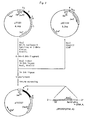
- The invention is illustrated in more detail by the following Examples, which partly refer to the accompanying drawings wherein;
- Fig. 1 is a diagram of the processes involved in the construction of an expression plasmid which carries the DNA sequence which codes for the polypeptide including the Ile¹⁴¹⁰-Met¹⁵¹⁷ sequence of fibronectin;
- Fig. 2 is a diagram of the processes involved in the construction of an expression plasmid which carries the DNA sequence which codes for the polypeptide including the Ala¹²³⁵-Met¹⁵¹⁷ sequence of fibronectin;
- Fig. 3 is a diagram of the processes involved in the construction of an expression plasmid which carries the DNA sequence which codes for the polypeptide including the Pro¹²³⁹-Met¹⁵¹⁷ sequence of fibronectin; and
- Fig. 4 is a diagram of the process involved in the construction of an expression plasmid which carries the DNA sequence which codes for a polypeptide including the Pro¹²³⁹-Asp¹⁵¹² sequence of fibronectin.
-
- First, 100 µg of a plasmid, pLF5 (reported in Biochemistry, 25 [1986], 4936-4941),which is of 4.1 kilo-bases and which contains the cDNA sequence which codes for the cell-spreading domain of fibronectin,was put into 200 µl of a reaction mixture containing a buffer for use with the restriction enzyme BalI and 100 units of BalI, and the mixture was incubated for 2 hours at 37°C. The reaction mixture was then put on an HPLC column of DEAE-4000 (6 x 125 mm; Nucleogen) and eluted with a concentration gradient of KCl in a 30 mM potassium phosphate buffer (pH 6.5) which contains 5 M urea, and precipitated with isopropyl alcohol, which gave 7.5 µg of 0.75 kb fragments. Next, these fragments were treated with 30 units of FokI for 1 hour at 37°C, and by the same purification method, 60 ng of 92-bp fragments and 140 ng of 203-bp fragments were obtained.
- So that the cDNA fragments could be joined to a vector, 5'-end and 3'-end linkers were chemically synthesized by the following method. The base sequence is shown in Fig. 1. The upper strand of the 5'-end linker (chain length, 11), labelled 5 U, the lower strand of the 5'-end linker (chain length, 7), labelled 5 L, the upper strand of the 3'-end linker (chain length, 30), labelled 3 U, and the lower strand of the 3'-end linker (chain length, 30), labelled 3 L, were synthesized by use of the DNA synthesizer of Applied Biosystems, Inc. After deprotection, 5 U and 5 L were purified by ion-exchange HPLC on a TSK gel DEAE-2000 column, and desalted on SepPak (Waters), giving 60 µg and 40 µg, respectively. 3 U and 3 L, were separated by electrophore is using 12% polyacrylamide gel containing 7 M urea, and after removal from the gel, they were desalted in the same way as for the 5 U and 5 L, giving 32 µg and 26 µg, respectively. The base sequences were checked by the solid-phase Maxam-Gilbert method. Each strand was then phosphorylated on its 5' terminus in the following way. First, 25 µg of DNA was dissolved in 10 µl of distilled water, and put into 20 µl of a solution of buffer for use with polynucleotide kinase (PNK) containing 50 mM Tris-HCl, pH 7.6, 10 mM MgCl₂, and 10 mM DTT, 1 mM ATP, and 5 units of PNK; the mixture was incubated for 1 hour at 37°C; then the reaction was stopped by heat treatment at 65°C for 10 minutes. After the phosphorylation, by the annealing of 5 U with 5 L and of 3 U with 3 L, the strands were made double-stranded. That is, equal amounts of 5 U and 5 L were mixed together, and left at 60°C for 10 minutes, after which the mixture was allowed to cool to room temperature gradually, and then cooled further to 15°C. The 3 U and 3 L were treated in the same way.
- The two kinds of linker prepared in section (1-2) above and the two kinds of DNA fragment (with 92 bp and with 203 bp) prepared in section (1-1) above were used, and ligated together. For this, 0.5 pmol of each kind of cDNA fragment and 5 pmol of each kind of linker were put into 20 µl of a solution containing a ligation buffer [66 mM Tris-HCl, pH 7.6, and 6.6 mM MgCl₂] and also 0.5 mM ATP, 10 mM DTT, and 2.8 units of T4 DNA ligase, at 16°C; the mixture was incubated overnight. The reaction mixture was heated for 10 minutes at 65°C, and then two volumes of cold ethanol was added, and the whole left at -70°C for 30 minutes. After this, the precipitated DNA was collected by centrifugation. This DNA was put into 20 µl of a reaction mixture made of buffer for use with EcoRI and 7.5 units of EcoRI, and incubated for 30 minutes at 37°C. Then the mixture was subjected to polyacrylamide electrophoresis, Whereby 336-bp fragments were obtained. These were extracted for 10 hours at 37°C in a gel elution buffer (0.5 M ammonium acetate, 0.01 M magnesium acetate, and 1 mM EDTA), after which the fragments were precipitated with ethanol, and the DNA was collected. This DNA was dissolved in 20 µl of TE buffer (10 mM Tris-HCl and 1 mM EDTA, pH 8.0).
- First, 1 ug of the secretion-expression vector pIN-III-ompA-l (reported in the EMBO Journal, 3 [1984], 2437-2442) was incubated together with 5 units of EcoRI in buffer for use with EcoRI for 1 hour at 37°C; next, 0.5 unit of alkaline phosphatase was added and incubation continued for another hour. The reaction mixture was treated twice with phenol, and ethanol precipitation was used to give 0.5 µg of DNA. This DNA was cleaved with EcoRI, and then 0.1 µg of dephosphorylated pIN-III-ompA-l vector was put into a mixture with 0.1 µg of the DNA fragments obtained in section (1-3) above together with 10 µl of buffer for use with T4 DNA ligase, 0.5 mM ATP, 10 mM DTT, and 1.8 units of T4 DNA ligase, in a total volume for the reaction mixture of 30 µl. This mixture was incubated overnight at 16°C. Then 15 µl of this reaction mixture was used in the transformation of Escherichia coli cells.
- Cells of Escherichia coli SB221 (The EMBO Journal, 3 [1984], 2437-2442) were put into a 20 ml test tube containing 5 ml of L-broth (Bactotrypton, 10 g/l; yeast extract, 5 g/l; NaCl, 5 g/l; pH 7.2) and cultured overnight with agitation at 37°C. Then 0.05 ml of this culture broth was used to inoculate 5 ml of fresh L-broth, and culture was continued with agitation until the absorbance at 600 nm reached 0.6. This culture broth was cooled over ice and centrifuged for 5 minutes at 5000 rpm at 4°C. The supernatant was removed. This was mixed with 2.5 ml of an ice-cold 0.1 M MgCl₂ solution, after which it was centrifuged in the same way and the supernatant removed. This was mixed with 1.25 ml of ice-cold 0.1 M CaCl₂, immediately mixed with ice-water, and left for 30 minutes. The mixture was centrifuged and the supernatant removed; then it was mixed with 0.1 ml of ice-cold 0.1 M CaCl₂ to suspend it rapidly. The suspension was mixed with 10 µl of the reaction mixture obtained in section (1-4) above, and left for 30 minutes in an ice bath. Next, it was heated to 42°C for 2 minutes. Then, 1 ml of L-broth heated to 37°C was added, and the whole was incubated for 30 minutes at 37°C. After this, the mixture was cultured for 1 hour at 37°C with agitation. The culture broth was spread on L agar medium that contained 50 µg/ml ampicillin, and colonies which grew after overnight culture at 37°C were isolated. About 200 colonies were obtained, and fourteen of these were analyzed for plasmids.
- After 14 transformants were cultured with agitation overnight in 1.5 ml of L-broth containing 50 µg/ml ampicillin, the cultures were centrifuged and the cell pellets were obtained. The cell pellets were suspended in 100 µl of solution A (50 mM glucose, 10 mM EDTA, 25 mM Tris-HCl, pH 8.0, and 2 mg/ml lysozyme), and left at room temperature for 5 minutes. Then 200 µl of solution B (0.2 N NaOH and 1 % SDS) was added to the suspension, and the container was rapidly inverted five times and then left at 0°C for 10 minutes. To the containiner, 150 µl of 5 M potassium acetate (pH 5.0) was added, followed by the addition of two volumes of cold ethanol, and the mixture was left at -70°C for 30 minutes, after which it was centrifuged and the DNA was obtained. The DNA was washed with 80% ethanol, and the ethanol and water were removed under reduced pressure. The residue was dissolved in a small amount of TE buffer. One portion of the solution (0.1 µg) was allowed to react for 1 hour at 37°C in 10 µl of a reaction mixture which contains 5 units of EcoRI in buffer for use with EcoRI. Then the reaction mixture was separated by agarose electrophoresis, and the expected bands for DNA (at 0.33 kb) were checked by ethidium bromide staining. The results showed that nine of the clones gave the desired band. The plasmids extracted from these nine clones were doubly digested with the use of PvuII and HindIII. Then the orientation of the inserted fragments was identified. It was found that eight of the clones had the inserts with the correct orientation. Of these eight clones, the plasmids from five clones were doubly digested with the use of XbaI and HindIII, and the DNA that contained the inserted fragments was removed, and subcloned into the phages M13mp18 and mp19. The base sequences were analyzed by the dideoxy method. Three of the clones were found to contain the desired sequence. The recombinant plasmids obtained in this way were named pTF101.
- The pTF101 obtained as described above in section (1-6) was used to transform Escherichia coli cells (SB221/pTF 101) and such transformed cells were cultured in 5 ml of L-broth overnight with agitation at 37°C. The culture was transferred to 500 ml of the same culture broth and culture was continued until the absorbance at 660 nm reached 0.6, at which time 170 µg/ml chloramphenicol was added, and cultured was continued once more for 10-12 hours. The culture broth was centrifuged for 10 minutes at 5000 rpm, and the cell pellet was washed in STE buffer (10 mM Tris-HCl, pH 8, 100 mM NaCl, and 1 mM EDTA), and centrifuged again in the same way in order to obtain the cell pellet. The pellet was suspended in 10 ml of the solution A, and kept at room temperature for 5 minutes before 20 ml of the solution B was added with rapid stirring, and the suspension was left for 5 minutes at 0°C. To this suspension, 15 ml of 3 M potassium acetate was added with rapid stirring, and the suspension was left at 0°C for 10 minutes. The mixture was centrifuged for 60 minutes at 8000 rpm and the supernatant was obtained, to which 27 ml (0.6 volume) of isopropyl alcohol was added, and the mixture was left at room temperature for 15 minutes. This mixture was centrifuged and the precipitate was collected and washed in 80% ethanol. The ethanol and water in the resulting mixture was removed under reduced pressure and the residue was dried. This residue was dissolved in 4 ml of TE buffer and to this solution 4.75 g of cesium chloride was added, followed by the addition of 5 mg/ml ethidium bromide in the volume of 420 µl; the mixture was left for 30 minutes at 0°C. The mixture was then centrifuged for 10 minutes at 8000 rpm, and the supernatant was put into a Bechman Quick-seal tube and ultracentrifuged at 55000 rpm overnight. Then, under ultraviolet light, plasmid bands were removed by the use of a syringe. The resulting mixture was extracted seven times in equal volumes of isopropyl alcohol and the remaining ethidium bromide was removed, after which ethanol precipitation was done twice, and then the remaining mixture was washed in 80% ethanol, dried under reduced pressures, and dissolved in a small amount of TE buffer (yield, 760 µg).
- First, 50 µg of the plasmid pLF5 was put into 200 µl of a reaction mixture containing 200 units of EcoRI methylase in a buffer for use with EcoRI methylase (100 mM Tris-HCl, pH 8.0, 2 mM DTT, 10 mM EDTA, and 80 µM S-adenosylmethionine), and the mixture was incubated for 60 minutes at 37°C to bring about protection of the EcoRI sites. Then the reaction was stopped by heating at 65°C for 20 minutes, and 96 units of PvuII in buffer for use with PvuII was added; 300 µl of the reaction mixture was incubated for 60 minutes at 37°C. This mixture was separated by agarose electrophoresis, and the slice of gel containing 0.60 kb band was cut out. This slice was put into a dialysis tube that contained elution buffer (5 mM Tris-acetate buffer, pH 8.0, and 1 mM EDTA), and eluted by electrophoresis in the same buffer, by which means the DNA was eluted. The elution fluid was extracted twice with phenol, and a 1/10 volume of 3 M sodium acetate was added, followed by the addition of two volumes of ethanol, and ethanol precipitation was conducted twice. Then the resulting mixture was washed in 80% ethanol, dried, and dissolved in a small amount of TE buffer (yield, 1.2 ug).
- Also, 140 µg of the plasmid pTF101 obtained in Example 1 was put into 200 µl of a reaction solution containing 144 units of PvuII, and the mixture was incubated for 60 minutes at 37°C. Then 150 units of EcoRI was added, and the reaction solution brought to 240 µl, and incubated for 60 minutes at 37°C. The result was separated by polyacrylamide electrophoresis, and the bands at 0.25 kb were cut out. The DNA was extracted from these bands of gel as described above, and the DNA was collected by ethanol precipitation (yield, 0.4 µg). Then 1.25 µg of the 0.6-kb fragments obtained here and 0.4 ug of the 0.25-kb fragments were put into a ligation buffer which contains 2.8 units of T4 DNA ligase, 0.5 mM ATP, and 10 mM DTT in 50 µl, and the mixture was incubated for 30 minutes at 16°C. Then 92 pmol of phosphorylated linker (pCCGAATTGG) and 1.4 units of T4 DNA ligase were added to the reaction mixture, which was brought to 70 µl and incubated overnight at 10°C. The reaction was stopped by heating at 65°C for 10 minutes, and the buffer was modified to be suitable for EcoRI; 30 units of EcoRI was added, and reaction was allowed to take place for 60 minutes at 37°C. Then the reaction mixture was separated by agarose electrophoresis, and the 0.86-kb bands were cut out. The DNA in the bands was obtained by the method described above (yield, about 25 ng). Then 20 ng of the 0.86-kb fragments were cleaved with EcoRI, and put into a ligation buffer which contains 0.1 µg of dephosphorylated vector pIN-III-OMPA-l, 2.8 units of T4DNA ligase, 0.5 mM ATP, and 10 mM DTT in a total volume of 20 µl; the mixture was incubated for 10 hours at 16°C and half of the reaction was used to transform Escherichia coli SB 221 cells by the method described above. Of the transformants obtained, 48 of the clones were analysed for plasmids by the methods described above, and it was found that the plasmids included the desired fragments in six of the clones. These six plasmids were digested with BamHI, and the fragments which were produced were analysed. Five of the clones contained the desired fragments inserted in the correct orientation. Three of these five clones were studied for their base sequence. One of the clones were found to have the correct base sequence. This recombinant plasmid was named pTF301, and the cells of Escherichia coli SB221 that carry this plasmid were designated SB221/pTF301.
- First, 100 µg of the plasmid pTF301 which codes for peptide 283AA with cell-spreading activity was mixed with a buffer for use with the restriction enzyme XbaI and with 120 units of XbaI in a total volume of 300 µl, and incubated at 37°C for 2 hours. Then, the DNA was collected by ethanol precipitation. Half of the amount obtained was put into 375 µl of a reaction mixture of buffer for use with BAL 31 nuclease which contained 36 units of BAL 31 nuclease-S, and the mixture was incubated at 30°C; at 2 minutes of incubation to 8 minutes, 28 µl of the reaction mixture was sampled every 30 seconds, and each sample was added to 20 µl of 0.5 M EDTA to stop the reaction. The samples were pooled, and DNA was obtained by phenol treatment and then ethanol precipitation. The DNA obtained was put into 200 µl of a reaction mixture of 7 mM Tris-HCl, pH 7.5, which contained 0.1 mM EDTA, 10 mM NaCl, 7 mM MgCl₂, 0.1 mM dATP, dGTP, dCTP, and dTTP, and 2 units of Klenow fragment. The mixture was incubated for 20 minutes at 37°C. The reaction was stopped by treatment at 65°C for 10 minutes, and the reaction mixture was adjusted to the composition of buffer for use with HindIII. Then, to 250 µl of this reaction mixture, 60 units for HindIII was added, and the mixture was incubated for 1 hour at 37°C. This mixture was separated by agarose gel electrophoresis, and the fragments that correspond to the size of 0.5-0.8 kb were cut out; 1 µg was obtained.
- First, 0.65 µg of the DNA fragments obtained in section (3 - 1) above was put into 30 µl of a ligase buffer which contained 0.5 µg of phosphorylated NcoI linker (d[pAGCCATGGCT]), 2.8 units of T4 DNA ligase, 0.5 mM ATP, and 10 mM DTT; and the mixture was incubated overnight at 10°C. After the reaction was stopped by being heated at 65°C for 10 minutes, 24 units of NcoI and 12 units of HindIII were added to this reaction mixture and the mixture was incubated in a total volume of 50 µl for 1 hour at 37°C. This was put on a 1-ml column of Sepharose CL-4B; the free linker was eluted with STE buffer.
- Then 300 µl of DNA fraction was obtained and concentrated to 55 µl. Of this, 5.5 µl was added to 1 µl of a solution that contained 0.2 µg of the plasmid pUC119 that had been dephosphorylated by treatment with NcoI and HindIII. To this, 65 µl of solution A and 6.5 µl of solution B from a DNA ligation kit (Takara Shuzo Co., Ltd.) were added, and the mixture was incubated overnight at 16°C. Then 20 µl of the reaction mixture was used to transform cells of Escherichia coli
HB 101. - In addition, pUC119N was obtained by creation of a NcoI site surrounding the translation initiation codon of the commercially available vector pUC119 (Takara Shuzo Co., Ltd.); the distance between the ribosome-binding site and initiation codon was also changed from 7 to 8 bases.
- The transformants obtained in section (3-2) above were transferred to a nitrocellulose filter (BA85; S & S) on L-agar medium containing 50 µg/ml ampicillin, and cultured overnight at 37°C. The colonies that grew were brought into chloroform vapor for 15 minutes, and the nitrocellulose filter was put into a solution of 50 mM Tris-HCl, pH 7.5, containing 150 mM NaCl, 5 mM MgCl₂, 3% bovine serum albumin, 1 µg/ml DNase, and 40 µg/ml lysozyme; the mixture was incubated overnight at room temperature. The filter was treated with an anti-FN monoclonal antibody, FN-10 (Tarara Shuzo Co., Ltd.) which specifically recognizes the cell-spreading domain of FN, and a second antibody labelled with peroxidase was applied, and the hydrogen peroxide gave rise to color in the presence of 4-chloro-1-naphthol, by which the transformants which had appeared were found. By this first screening, 52 clones were selected from 1300 clones, and each was cultured overnight with agitation at 37°C on 5 ml of L-broth containing 50 µg/ml ampicillin. The whole-cell protein of the cells obtained was separated on SDS-polyacrylamide gel electrophoresis (SDS-PAGE), and immunoblotting was applied, in which the anti-FN monoclonal antibody FN-10 was allowed to react. It was found that polypeptides of the molecular masses of 22 kDa-32 kDa had been produced. For 11 clones, the base sequence of the 5'-end of the inserted fragments was identified; they were found to code amino acid sequences which were respectively 279, 258, 219, 213, 207, 206, 198, 195, 190, 186, and 178 long counting from Met¹⁵¹⁷ as the C-terminus.
- The amount of these peptides which were expressed were compared on SDS-PAGE; the peptide which was expressed in the greatest amount was a peptide with a sequence 279 amino acids long (accounting for about 20% of the total whole-cell protein); next were peptides with sequence 206 and 258 amino acids long.
- The plasmid which expressed the peptide with the sequence 279 amino acids long was designated pTFD707. At the N-terminus of the peptide expressed by pTFD707, there was a sequence (GCT) attached that corresponded to alanine of vector origin. This sequence was removed by site-specific mutagenesis (a procedure used, for example, in Example 4 of USSN 291,894). Also, in order to raise the level of expression, the HindIII-SalI fragment with a lpp terminator sequence was removed from the secretion-expression vector pIN-III-ompA-I, and joined with the HindIII-SalI site of pTFD707, by which means plasmids designated pTF7021 were obtained.
- Introduction of mutation into pTFD707 was done by the method of Kunkel et al. (Proceedings of the National Academy of Sciences, U.S.A., 82, 488-492, 1985; Methods in Enzymology, 154, 367-382, 1987). For this purpose, the site-directed mutagenesis system, Mutan-K(Takara Shuzo) was used.
- pTFD707 was introduced into Escherichia coli BW313, and the transformants were cultured with agitation at 37°C on 100 ml of 2X YT medium (1.6% Bactotrypton, 1% yeast extract, and 0.5% NaCl) that contained 50 µg/ml ampicillin. When the absorbance at 660 nm was 0.3, 1 ml of M13K07 phage suspension containing 10¹⁰ pfu was added to the culture, and cultivation was continued at 37°C for 16 hours. The culture medium was centrifuged and the supernatant collected; to this supernatant, 25 ml of a mixture of 2.5 M NaCl and 20% polyethlene glycol 6000 was added, and the resulting mixture was left for 10 minutes at room temperature. It was then centrifuged, and 5 ml of TE buffer was added to the precipitate. This mixture was treated with a mixture of phenol and chloroform, and then with chloroform alone, before ethanol precipitation was used to give single-stranded DNA. The 30 ng of single-stranded DNA obtained was dissolved in 1 µl of annealing buffer(20 mM Tris-HCl, pH 8.0, 10 mM MgCl₂, 50 mM NaCl, and 1 mM DTT), and added to 1 µl of a solution that contained 1 pmol of phosphorylated oligonucleotide d[pGGATGGTTAGTCAATTTC], and the mixture was treated by heating at 65°C for 15 minutes and at 37°C for 15 minutes. The resulting mixture was added to 25 µl of extension buffer(50mM Tris-HCl, pH8.0, 60 mM ammonium acetate, 5 mM MgCl₂, 5 mM DTT, 1 mM NAD, 0.5 mM dATP, dGTP, dCTP and dTTP), with 60 units of DNA ligase from E. coli and 1 unit of T4 DNA polymerase, and the reaction mixture was left at 25° C for 2 hours, before 3 µl of a 0.2 M solution of EDTA, pH 8.0, was added, and the reaction mixture was heated at 65° C for 5 minutes. Then, 3 µl of the reaction mixture was mixed with 30 µl of competent cells of Escherichia coli BMH71-18mutS, and the mixture was kept at 0° C for 30 minutes, at 42°C for 45 seconds, and at 0°C for 2 minutes. Then 300 µl of L-broth was added, and the mixture was left for 1 hour at 37°C. Next, 10 µl of a suspension of M13K07 phages was added, and the mixture was left for 30 minutes at 37°C. Next, the cells were cultured for 16 hours at 37°C with agitation in 1 ml of 2X YT medium that contained 150 µg/ml ampicillin and 70 µg/ml kanamycin. The culture was centrifuged and the supernatant collected. Then 20 µl of the supernatant was mixed with 80 µl of the culture medium of an overnight culture of Escherichia coli JM109. This was left for 10 minutes at 37°C, and one portion was used to inoculate L-agar that contained 50 µg/ml ampicillin; the agar was left at 37 C° overnight, and of the transformants that were obtained. The base sequences of six of the clones were identified. Of these six clones, five were found to have the desired mutation. The recombinant plasmid obtained was designated pTFD707-45.
- Then, 2 µg of pTFD707-45 was digested with BamHI and HindIII, the resulting digest was separated by agarose gel electrophoresis, and the 0.5-kb fragment was collected. Separately, 2 µg of pTF7021 was digested with BamHI and ScaI, and the digest separated by agarose gel electrophoresis. The 2.1-kb fragment was obtained. Next, 2 µg of pTF7021 was digested with HindIII and ScaI, and the digest separated by agarose gel electrophoresis, and the 2.4-kb fragment collected. To 3 µl of a solution that contained 5 ng of the 0.5-kb fragment, 20 ng of the 2.1-kb fragment, and 20 ng of the 2.4-kb fragment, 12 µl of solution A and 3 µl of solution B from a DNA ligation kit (Takara Shuzo) were added, and incubation was done at 16°C for 30 minutes. Then 10 µl of the reaction mixture was used to transform cells of Escherichia coli JM109. A plasmid was obtained that coded for the Pro¹²³⁹-Asp¹⁵¹² sequence (274 amino acids long)of FN, and that also had the lpp terminator sequence. This recombinant plasmid was named pTF7221, and the cells of Escherichia coli JM109 that carried this plasmid were designated Escherichia coli JM109/pTF7221. The strain was deposited at the Fermentation Research Institute of the Agency of Industrial Science and Technology, Japan, under FERM BP-1915 on 17th June 1988.
- Cells of JM109/pTF7221 were cultured and it was checked whether they expressed a polypeptide with cell-spreading activity; it was found that this protein accounted for at least 30% of the whole-cell protein.
- Plasmid pTF7221 obtained by the joining of an expression vector and the DNA that codes for the Pro¹²³⁹-Asp¹⁵¹² sequence of FN (a sequence of 274 amino acid residues) was introduced into Escherichia coli JM109, giving Escherichia coli JM109/pTF7221, and these cells were cultured overnight with shaking at 37°C in 5 ml of L-broth containing 50 µg/ml ampicillin in a test tube. This was used to inoculate a 2-1 Erlenmeyer flask containing 500 ml of the same medium, and culture was continued at an agitation rate of 180 r.p.m. When absorbance at 660 nm had reached 0.3, 2 mM IPTG (isopropyl-β-thiogalactoside) was added, and the cells were harvested 20 hours later. One portion of the cells obtained was used in immunoblotting. The whole-cell protein was isolated by SDS-PAGE, and the electrophoretic pattern was transferred to a nitrocellulose membrane. Then a monoclonal antibody (FN-10, Takara Shuzo Co., Ltd.) that specifically recognizes the cell-spreading domain of FN was applied, after which a second antibody labelled with peroxidase was applied. The activity of the peroxidase bound to the second antibody gave rise to color in the presence of 4-chloronaphthol and hydrogen peroxide, and it was found that the desired band in the region of 34 kDa lower molecular band than that of 279 amino acid residues was present. Next, the whole-cell pellet eas suspended in a solution of 10 mM Tris-HCl, pH 7.5, containing 5 mM EDTA and 5 mM mercaptoethanol, and ultrasonification was conducted. This suspension was centrifuged and the supernatant was obtained and dialyzed against 20 mM Tris-HCl (pH 7.5). The inner dialysis liquid was put on a Sepharose 4B column (8 ml) bound with the monoclonal antibody FN-10. The column was washed with washing buffer A (20 mM Tris-HCl, pH 8.0, and 0.15 M KCl), and then with washing buffer B (20 mM Tris-HCl, pH 6.4, and 0.15 M KCl). Finally, the eluting buffer (50 mM glycine-HCl, pH 2.3, and 0.2 M KCl) was used, and fractions were obtained. The desired fractions were obtained by immunoblotting, desalted, and lyophilized, and about 5 mg of almost pure peptide was obtained by electrophoresis. Next, said peptide was treated with aminopeptidase P (Enzyme Handbook, p. 534 (1983), Asakura Publishers), and the N-terminal methionine was removed, after which the peptide was purified again by the same method as described above. The 10 or so amino acid residues in the amino acid sequence starting from the N-terminus were studied, and it was found that the sequence was Pro-Thr-Asp-Leu-Arg-Phe-Thr-Asn-Ile-Gly, which agreed with the N-terminal sequence of the desired peptide.
- The cell-spreading activity of the polypeptide of 274 amino acid residues obtained in Example 5 and of FN and the peptides with 108 and 279 amino acids was measured by the method of Ruoslahti et al. (Methods in Enzymology, 82, 803-831, 1981). The samples were diluted stepwise in physiological saline and distilled water, and 50 µl aliquots were injected into the wells of a 96-well microtitre plate, which was then incubated overnight at 4°C in order to allow the samples to adhere to the wells. Then, phosphate-buffered saline (PBS) was used to wash the plate twice, 100 µl of 3% BSA was added to each well, and the plate was incubated for one hour at 37°C. The plate was washed twice with PBS, and then normal rat kidney (NRK-49F) cells suspended to a concentration of 10⁶ cells/ml in Eagle's Minimum Essential Medium (MEM) were added in the amount of 100 µl/well, and the plate was incubated for 2-3 hours at 37°C. The NRK-49F cells that were used were obtained as a lyophilized strain for storage, and first preincubated and then treated with trypsin before use. The spreading of the cells was observed under a microscope, and the minimum dose needed to have cell-spreading activity was calculated. These results are shown in Table 1.

- As explained above in detail, this invention provides a peptide which has cell-spreading activity essentially the same as that of FN, and also provides a method for its preparation by the use of genetic engineering. The polypeptides mentioned above can be used in pharmaneutical preparations for such uses as for the healing of wounds, in collyria, for the prevention of metastases from cancer, for the implantation of artificial organs into the body, and the like. It can also be used in cosmetics, toothpaste, and the like.
Claims (4)
- A polypeptide with cell-spreading activity which has an amino acid sequence represented by the general formula [I]:

- A recombinant plasmid which contains DNA coding for polypeptide according to claim 1.
- A transformant which carries a recombinant plasmid according to claim 2.
- A method for the preparation of a polypeptide which comprises cultivating a transformant according to claim 3 and recovering a polypeptide according to claim 1 from the culture medium.
Applications Claiming Priority (2)
| Application Number | Priority Date | Filing Date | Title |
|---|---|---|---|
| JP63160949A JP2561131B2 (en) | 1988-06-30 | 1988-06-30 | Cell adhesion activity polypeptide |
| JP160949/88 | 1988-06-30 |
Publications (3)
| Publication Number | Publication Date |
|---|---|
| EP0349342A2 EP0349342A2 (en) | 1990-01-03 |
| EP0349342A3 EP0349342A3 (en) | 1991-08-07 |
| EP0349342B1 true EP0349342B1 (en) | 1993-08-25 |
Family
ID=15725688
Family Applications (1)
| Application Number | Title | Priority Date | Filing Date |
|---|---|---|---|
| EP89306676A Expired - Lifetime EP0349342B1 (en) | 1988-06-30 | 1989-06-30 | Polypeptides with cell-spreading activity |
Country Status (4)
| Country | Link |
|---|---|
| US (1) | US5102988A (en) |
| EP (1) | EP0349342B1 (en) |
| JP (1) | JP2561131B2 (en) |
| DE (1) | DE68908633T2 (en) |
Families Citing this family (20)
| Publication number | Priority date | Publication date | Assignee | Title |
|---|---|---|---|---|
| US5270030A (en) * | 1988-12-29 | 1993-12-14 | Bio-Technology General Corp. | Fibrin binding domain polypeptide and method of producing |
| US5679320A (en) * | 1988-12-29 | 1997-10-21 | Bio-Technology General Corp. | Fibrin binding domain polypeptides and uses and methods of producing same |
| DE69012950T2 (en) * | 1989-10-13 | 1995-03-16 | Takara Shuzo Co | Anti-cancer drugs. |
| JP2829405B2 (en) * | 1991-10-14 | 1998-11-25 | 寳酒造株式会社 | Functional polypeptide |
| US7083979B1 (en) | 1994-03-25 | 2006-08-01 | Indiana University Foundation | Methods for enhanced retroviral-mediated gene transfer |
| WO1996017073A1 (en) * | 1994-11-29 | 1996-06-06 | Takara Shuzo Co., Ltd. | Process for producing transformed cell |
| US5877149A (en) | 1995-06-07 | 1999-03-02 | Beaulieu; Andre | Deepithelialized skin diffusion cell system |
| US7112320B1 (en) | 1995-06-07 | 2006-09-26 | Andre Beaulieu | Solid wound healing formulations containing fibronectin |
| US6528483B2 (en) | 1995-06-07 | 2003-03-04 | André Beaulieu | Method of producing concentrated non-buffered solutions of fibronectin |
| US5641483A (en) * | 1995-06-07 | 1997-06-24 | Beaulieu; Andre | Wound healing formulations containing human plasma fibronectin |
| WO1997011604A1 (en) | 1995-09-29 | 1997-04-03 | Indiana University Foundation | Methods for enhanced virus-mediated dna transfer using molecules with virus- and cell-binding domains |
| CN1222612C (en) * | 2000-08-16 | 2005-10-12 | 宝生物工程株式会社 | Method of extensive culture of antigen-specific cytotoxic T cells |
| CN101633907B (en) * | 2001-08-15 | 2012-09-05 | 宝生物工程株式会社 | Amplification culture method for antigen specific cytotoxic t lymphocytes |
| US8728811B2 (en) | 2002-03-25 | 2014-05-20 | Takara Bio Inc. | Process for producing cytotoxic lymphocyte |
| CA2536492C (en) | 2003-08-22 | 2013-10-08 | Takara Bio Inc. | Process for producing cytotoxic lymphocytes |
| CN101243187B (en) | 2005-08-17 | 2012-07-11 | 宝生物工程株式会社 | Method of producing lymphocytes |
| US20100068192A1 (en) * | 2005-09-30 | 2010-03-18 | Takara Bio Inc. | Method for Production of T Cell Population |
| WO2008111430A1 (en) * | 2007-03-09 | 2008-09-18 | Takara Bio Inc. | METHOD FOR PRODUCING γδT CELL POPULATION |
| US8921516B2 (en) * | 2010-12-08 | 2014-12-30 | Corning Incorporated | Synthetic, defined fibronectin mimetic peptides and surfaces modified with the same |
| CN111454350B (en) * | 2020-06-01 | 2020-12-29 | 广东丸美生物技术股份有限公司 | Recombinant fibronectin mutant and application thereof |
Family Cites Families (4)
| Publication number | Priority date | Publication date | Assignee | Title |
|---|---|---|---|---|
| US4589881A (en) * | 1982-08-04 | 1986-05-20 | La Jolla Cancer Research Foundation | Polypeptide |
| US4614517A (en) * | 1982-08-04 | 1986-09-30 | La Jolla Cancer Research Foundation | Tetrapeptide |
| US4517686A (en) * | 1982-08-04 | 1985-05-21 | La Jolla Cancer Research Foundation | Polypeptide |
| GB8516421D0 (en) * | 1985-06-28 | 1985-07-31 | Biotechnology Interface Ltd | Fibronectins |
-
1988
- 1988-06-30 JP JP63160949A patent/JP2561131B2/en not_active Expired - Lifetime
-
1989
- 1989-06-08 US US07/362,983 patent/US5102988A/en not_active Expired - Lifetime
- 1989-06-30 DE DE89306676T patent/DE68908633T2/en not_active Expired - Fee Related
- 1989-06-30 EP EP89306676A patent/EP0349342B1/en not_active Expired - Lifetime
Also Published As
| Publication number | Publication date |
|---|---|
| DE68908633D1 (en) | 1993-09-30 |
| DE68908633T2 (en) | 1994-03-10 |
| US5102988A (en) | 1992-04-07 |
| EP0349342A3 (en) | 1991-08-07 |
| JPH0297397A (en) | 1990-04-09 |
| EP0349342A2 (en) | 1990-01-03 |
| JP2561131B2 (en) | 1996-12-04 |
Similar Documents
| Publication | Publication Date | Title |
|---|---|---|
| EP0349342B1 (en) | Polypeptides with cell-spreading activity | |
| Ernst et al. | Cloning and sequencing of complementary DNAs encoding the alpha-subunit of translational initiation factor eIF-2. Characterization of the protein and its messenger RNA. | |
| JP3122127B2 (en) | Anticoagulant protein | |
| CA2065409C (en) | Anticoagulant polypeptides | |
| NZ211666A (en) | Pharmaceutical compositions comprising tumour necrosis factor and its production via recombinant techniques | |
| US20070287660A1 (en) | Novel clot-specific steptokinase proteins possessing altered plasminogen activation-characteristics and a process for the preparation of said proteins | |
| NZ234499A (en) | Tumour necrosis factor inhibitor; its preparation; and genes encoding for it | |
| Mahuran et al. | Proteolytic processing of pro-alpha and pro-beta precursors from human beta-hexosaminidase. Generation of the mature alpha and beta a beta b subunits. | |
| EP0329413B1 (en) | Polypeptides with cell-spreading activity | |
| JP2829405B2 (en) | Functional polypeptide | |
| WO1993016709A1 (en) | Therapeutic domains of von willebrand factor | |
| JPH025869A (en) | Dna sequence, recombinant dna molecule and lipocortins iii, iv, v, vi | |
| EP0376491B1 (en) | Polypeptide with cell-spreading activity | |
| US5049658A (en) | Polypeptide with cell-spreading activity | |
| JP3011274B2 (en) | Recombinantly produced 3-des-hydroxy-cystatin C polypeptide or modified product thereof | |
| Hofsteenge et al. | Primary structure of a ribonuclease from porcine liver, a new member of the ribonuclease superfamily | |
| KR940003690B1 (en) | Genes for biological active proteins | |
| EP0352089B1 (en) | A modified human PSTI | |
| JP2829397B2 (en) | Fibrin binding active polypeptide | |
| EP0326013A2 (en) | Novel proteins derived from human alpha2-plasmin inhibitor or proteins similar thereto | |
| JP2562951B2 (en) | Cell adhesion activity polypeptide | |
| JP2516048B2 (en) | Cosmetics | |
| JP2777986B2 (en) | Functional polypeptide | |
| JPH0975082A (en) | Human 26s proteasome-constituting component protein | |
| JPH04234988A (en) | Human prolactin-protein a fused protein-expressing gene |
Legal Events
| Date | Code | Title | Description |
|---|---|---|---|
| PUAI | Public reference made under article 153(3) epc to a published international application that has entered the european phase |
Free format text: ORIGINAL CODE: 0009012 |
|
| AK | Designated contracting states |
Kind code of ref document: A2 Designated state(s): DE FR GB IT |
|
| 17P | Request for examination filed |
Effective date: 19901127 |
|
| PUAL | Search report despatched |
Free format text: ORIGINAL CODE: 0009013 |
|
| AK | Designated contracting states |
Kind code of ref document: A3 Designated state(s): DE FR GB IT |
|
| RHK1 | Main classification (correction) |
Ipc: C07K 15/00 |
|
| 17Q | First examination report despatched |
Effective date: 19921102 |
|
| GRAA | (expected) grant |
Free format text: ORIGINAL CODE: 0009210 |
|
| AK | Designated contracting states |
Kind code of ref document: B1 Designated state(s): DE FR GB IT |
|
| ITF | It: translation for a ep patent filed |
Owner name: JACOBACCI CASETTA & PERANI S.P.A. |
|
| REF | Corresponds to: |
Ref document number: 68908633 Country of ref document: DE Date of ref document: 19930930 |
|
| ET | Fr: translation filed | ||
| PLBE | No opposition filed within time limit |
Free format text: ORIGINAL CODE: 0009261 |
|
| STAA | Information on the status of an ep patent application or granted ep patent |
Free format text: STATUS: NO OPPOSITION FILED WITHIN TIME LIMIT |
|
| 26N | No opposition filed | ||
| REG | Reference to a national code |
Ref country code: GB Ref legal event code: IF02 |
|
| REG | Reference to a national code |
Ref country code: GB Ref legal event code: 732E |
|
| REG | Reference to a national code |
Ref country code: FR Ref legal event code: TP |
|
| PGFP | Annual fee paid to national office [announced via postgrant information from national office to epo] |
Ref country code: FR Payment date: 20060608 Year of fee payment: 18 |
|
| PGFP | Annual fee paid to national office [announced via postgrant information from national office to epo] |
Ref country code: DE Payment date: 20060622 Year of fee payment: 18 |
|
| PGFP | Annual fee paid to national office [announced via postgrant information from national office to epo] |
Ref country code: GB Payment date: 20060628 Year of fee payment: 18 |
|
| PGFP | Annual fee paid to national office [announced via postgrant information from national office to epo] |
Ref country code: IT Payment date: 20060630 Year of fee payment: 18 |
|
| GBPC | Gb: european patent ceased through non-payment of renewal fee |
Effective date: 20070630 |
|
| REG | Reference to a national code |
Ref country code: FR Ref legal event code: ST Effective date: 20080229 |
|
| PG25 | Lapsed in a contracting state [announced via postgrant information from national office to epo] |
Ref country code: DE Free format text: LAPSE BECAUSE OF NON-PAYMENT OF DUE FEES Effective date: 20080101 |
|
| PG25 | Lapsed in a contracting state [announced via postgrant information from national office to epo] |
Ref country code: GB Free format text: LAPSE BECAUSE OF NON-PAYMENT OF DUE FEES Effective date: 20070630 |
|
| PG25 | Lapsed in a contracting state [announced via postgrant information from national office to epo] |
Ref country code: FR Free format text: LAPSE BECAUSE OF NON-PAYMENT OF DUE FEES Effective date: 20070702 |
|
| PG25 | Lapsed in a contracting state [announced via postgrant information from national office to epo] |
Ref country code: IT Free format text: LAPSE BECAUSE OF NON-PAYMENT OF DUE FEES Effective date: 20070630 |