EP0348429B1 - Facteur lactogenique du foie - Google Patents
Facteur lactogenique du foie Download PDFInfo
- Publication number
- EP0348429B1 EP0348429B1 EP88902741A EP88902741A EP0348429B1 EP 0348429 B1 EP0348429 B1 EP 0348429B1 EP 88902741 A EP88902741 A EP 88902741A EP 88902741 A EP88902741 A EP 88902741A EP 0348429 B1 EP0348429 B1 EP 0348429B1
- Authority
- EP
- European Patent Office
- Prior art keywords
- lactogenic
- liver
- factor
- prolactin
- casein
- Prior art date
- Legal status (The legal status is an assumption and is not a legal conclusion. Google has not performed a legal analysis and makes no representation as to the accuracy of the status listed.)
- Expired - Lifetime
Links
- 230000001983 lactogenic effect Effects 0.000 title claims abstract description 128
- 210000004185 liver Anatomy 0.000 title claims abstract description 100
- 239000005018 casein Substances 0.000 claims abstract description 48
- BECPQYXYKAMYBN-UHFFFAOYSA-N casein, tech. Chemical compound NCCCCC(C(O)=O)N=C(O)C(CC(O)=O)N=C(O)C(CCC(O)=N)N=C(O)C(CC(C)C)N=C(O)C(CCC(O)=O)N=C(O)C(CC(O)=O)N=C(O)C(CCC(O)=O)N=C(O)C(C(C)O)N=C(O)C(CCC(O)=N)N=C(O)C(CCC(O)=N)N=C(O)C(CCC(O)=N)N=C(O)C(CCC(O)=O)N=C(O)C(CCC(O)=O)N=C(O)C(COP(O)(O)=O)N=C(O)C(CCC(O)=N)N=C(O)C(N)CC1=CC=CC=C1 BECPQYXYKAMYBN-UHFFFAOYSA-N 0.000 claims abstract description 48
- 235000021240 caseins Nutrition 0.000 claims abstract description 48
- 238000004519 manufacturing process Methods 0.000 claims abstract description 29
- 241000124008 Mammalia Species 0.000 claims abstract description 14
- 235000013336 milk Nutrition 0.000 claims description 35
- 239000008267 milk Substances 0.000 claims description 35
- 210000004080 milk Anatomy 0.000 claims description 35
- 238000000034 method Methods 0.000 claims description 25
- 108090000765 processed proteins & peptides Proteins 0.000 claims description 17
- 239000000203 mixture Substances 0.000 claims description 16
- 102000004196 processed proteins & peptides Human genes 0.000 claims description 16
- 229920001184 polypeptide Polymers 0.000 claims description 15
- 230000001965 increasing effect Effects 0.000 claims description 13
- 235000018102 proteins Nutrition 0.000 claims description 5
- 102000004169 proteins and genes Human genes 0.000 claims description 5
- 108090000623 proteins and genes Proteins 0.000 claims description 5
- 102000007079 Peptide Fragments Human genes 0.000 claims description 3
- 108010033276 Peptide Fragments Proteins 0.000 claims description 3
- 239000002253 acid Substances 0.000 claims description 3
- 230000009467 reduction Effects 0.000 claims description 3
- 108091005804 Peptidases Proteins 0.000 claims description 2
- 239000004365 Protease Substances 0.000 claims description 2
- 102100037486 Reverse transcriptase/ribonuclease H Human genes 0.000 claims description 2
- 230000029936 alkylation Effects 0.000 claims description 2
- 238000005804 alkylation reaction Methods 0.000 claims description 2
- 238000004166 bioassay Methods 0.000 abstract description 15
- 238000000746 purification Methods 0.000 abstract description 5
- 238000012512 characterization method Methods 0.000 abstract description 2
- 238000002955 isolation Methods 0.000 abstract 1
- 108010057464 Prolactin Proteins 0.000 description 77
- 102100024819 Prolactin Human genes 0.000 description 77
- 229940097325 prolactin Drugs 0.000 description 77
- 102000011632 Caseins Human genes 0.000 description 46
- 108010076119 Caseins Proteins 0.000 description 46
- 238000003556 assay Methods 0.000 description 35
- 241000700159 Rattus Species 0.000 description 33
- 210000004027 cell Anatomy 0.000 description 33
- 230000000694 effects Effects 0.000 description 33
- 239000002609 medium Substances 0.000 description 26
- CSCPPACGZOOCGX-UHFFFAOYSA-N Acetone Chemical compound CC(C)=O CSCPPACGZOOCGX-UHFFFAOYSA-N 0.000 description 24
- 229940088597 hormone Drugs 0.000 description 19
- 239000005556 hormone Substances 0.000 description 19
- 210000004216 mammary stem cell Anatomy 0.000 description 18
- 101000687509 Rattus norvegicus Prolactin Proteins 0.000 description 16
- 238000011534 incubation Methods 0.000 description 16
- 210000002966 serum Anatomy 0.000 description 16
- 230000001817 pituitary effect Effects 0.000 description 15
- 210000001519 tissue Anatomy 0.000 description 15
- OZVBMTJYIDMWIL-AYFBDAFISA-N bromocriptine Chemical compound C1=CC(C=2[C@H](N(C)C[C@@H](C=2)C(=O)N[C@]2(C(=O)N3[C@H](C(N4CCC[C@H]4[C@]3(O)O2)=O)CC(C)C)C(C)C)C2)=C3C2=C(Br)NC3=C1 OZVBMTJYIDMWIL-AYFBDAFISA-N 0.000 description 13
- 229960002802 bromocriptine Drugs 0.000 description 13
- 238000011282 treatment Methods 0.000 description 13
- 230000006651 lactation Effects 0.000 description 12
- 210000005228 liver tissue Anatomy 0.000 description 12
- 238000002474 experimental method Methods 0.000 description 11
- 102100021809 Chorionic somatomammotropin hormone 1 Human genes 0.000 description 8
- 230000009471 action Effects 0.000 description 8
- 238000011161 development Methods 0.000 description 8
- 230000018109 developmental process Effects 0.000 description 8
- 230000002441 reversible effect Effects 0.000 description 8
- 230000028327 secretion Effects 0.000 description 8
- 230000003190 augmentative effect Effects 0.000 description 7
- 239000003981 vehicle Substances 0.000 description 7
- QTBSBXVTEAMEQO-UHFFFAOYSA-N Acetic acid Chemical compound CC(O)=O QTBSBXVTEAMEQO-UHFFFAOYSA-N 0.000 description 6
- 101000895818 Homo sapiens Chorionic somatomammotropin hormone 1 Proteins 0.000 description 6
- 101000956228 Homo sapiens Chorionic somatomammotropin hormone 2 Proteins 0.000 description 6
- 150000001413 amino acids Chemical class 0.000 description 6
- 238000000605 extraction Methods 0.000 description 6
- 239000001963 growth medium Substances 0.000 description 6
- 230000002949 hemolytic effect Effects 0.000 description 6
- 238000000338 in vitro Methods 0.000 description 6
- NOESYZHRGYRDHS-UHFFFAOYSA-N insulin Chemical compound N1C(=O)C(NC(=O)C(CCC(N)=O)NC(=O)C(CCC(O)=O)NC(=O)C(C(C)C)NC(=O)C(NC(=O)CN)C(C)CC)CSSCC(C(NC(CO)C(=O)NC(CC(C)C)C(=O)NC(CC=2C=CC(O)=CC=2)C(=O)NC(CCC(N)=O)C(=O)NC(CC(C)C)C(=O)NC(CCC(O)=O)C(=O)NC(CC(N)=O)C(=O)NC(CC=2C=CC(O)=CC=2)C(=O)NC(CSSCC(NC(=O)C(C(C)C)NC(=O)C(CC(C)C)NC(=O)C(CC=2C=CC(O)=CC=2)NC(=O)C(CC(C)C)NC(=O)C(C)NC(=O)C(CCC(O)=O)NC(=O)C(C(C)C)NC(=O)C(CC(C)C)NC(=O)C(CC=2NC=NC=2)NC(=O)C(CO)NC(=O)CNC2=O)C(=O)NCC(=O)NC(CCC(O)=O)C(=O)NC(CCCNC(N)=N)C(=O)NCC(=O)NC(CC=3C=CC=CC=3)C(=O)NC(CC=3C=CC=CC=3)C(=O)NC(CC=3C=CC(O)=CC=3)C(=O)NC(C(C)O)C(=O)N3C(CCC3)C(=O)NC(CCCCN)C(=O)NC(C)C(O)=O)C(=O)NC(CC(N)=O)C(O)=O)=O)NC(=O)C(C(C)CC)NC(=O)C(CO)NC(=O)C(C(C)O)NC(=O)C1CSSCC2NC(=O)C(CC(C)C)NC(=O)C(NC(=O)C(CCC(N)=O)NC(=O)C(CC(N)=O)NC(=O)C(NC(=O)C(N)CC=1C=CC=CC=1)C(C)C)CC1=CN=CN1 NOESYZHRGYRDHS-UHFFFAOYSA-N 0.000 description 6
- 235000001014 amino acid Nutrition 0.000 description 5
- 230000015572 biosynthetic process Effects 0.000 description 5
- 230000004069 differentiation Effects 0.000 description 5
- 238000010790 dilution Methods 0.000 description 5
- 239000012895 dilution Substances 0.000 description 5
- 210000003743 erythrocyte Anatomy 0.000 description 5
- 239000012634 fragment Substances 0.000 description 5
- 238000004128 high performance liquid chromatography Methods 0.000 description 5
- 210000005075 mammary gland Anatomy 0.000 description 5
- 238000005259 measurement Methods 0.000 description 5
- 230000007505 plaque formation Effects 0.000 description 5
- 238000002360 preparation method Methods 0.000 description 5
- 230000004936 stimulating effect Effects 0.000 description 5
- 238000003786 synthesis reaction Methods 0.000 description 5
- CURLTUGMZLYLDI-UHFFFAOYSA-N Carbon dioxide Chemical compound O=C=O CURLTUGMZLYLDI-UHFFFAOYSA-N 0.000 description 4
- 241000700199 Cavia porcellus Species 0.000 description 4
- 239000006144 Dulbecco’s modified Eagle's medium Substances 0.000 description 4
- 102100037852 Insulin-like growth factor I Human genes 0.000 description 4
- RJKFOVLPORLFTN-LEKSSAKUSA-N Progesterone Chemical compound C1CC2=CC(=O)CC[C@]2(C)[C@@H]2[C@@H]1[C@@H]1CC[C@H](C(=O)C)[C@@]1(C)CC2 RJKFOVLPORLFTN-LEKSSAKUSA-N 0.000 description 4
- FAPWRFPIFSIZLT-UHFFFAOYSA-M Sodium chloride Chemical compound [Na+].[Cl-] FAPWRFPIFSIZLT-UHFFFAOYSA-M 0.000 description 4
- 230000033228 biological regulation Effects 0.000 description 4
- 210000004369 blood Anatomy 0.000 description 4
- 239000008280 blood Substances 0.000 description 4
- 238000005119 centrifugation Methods 0.000 description 4
- 230000000295 complement effect Effects 0.000 description 4
- VYFYYTLLBUKUHU-UHFFFAOYSA-N dopamine Chemical compound NCCC1=CC=C(O)C(O)=C1 VYFYYTLLBUKUHU-UHFFFAOYSA-N 0.000 description 4
- 230000006870 function Effects 0.000 description 4
- 230000003054 hormonal effect Effects 0.000 description 4
- 239000000047 product Substances 0.000 description 4
- 230000001105 regulatory effect Effects 0.000 description 4
- 230000004044 response Effects 0.000 description 4
- 230000035945 sensitivity Effects 0.000 description 4
- WEVYAHXRMPXWCK-UHFFFAOYSA-N Acetonitrile Chemical compound CC#N WEVYAHXRMPXWCK-UHFFFAOYSA-N 0.000 description 3
- 102000029816 Collagenase Human genes 0.000 description 3
- 108060005980 Collagenase Proteins 0.000 description 3
- 101000599951 Homo sapiens Insulin-like growth factor I Proteins 0.000 description 3
- 102000004877 Insulin Human genes 0.000 description 3
- 108090001061 Insulin Proteins 0.000 description 3
- GUBGYTABKSRVRQ-QKKXKWKRSA-N Lactose Natural products OC[C@H]1O[C@@H](O[C@H]2[C@H](O)[C@@H](O)C(O)O[C@@H]2CO)[C@H](O)[C@@H](O)[C@H]1O GUBGYTABKSRVRQ-QKKXKWKRSA-N 0.000 description 3
- 241000283973 Oryctolagus cuniculus Species 0.000 description 3
- JGSARLDLIJGVTE-MBNYWOFBSA-N Penicillin G Chemical compound N([C@H]1[C@H]2SC([C@@H](N2C1=O)C(O)=O)(C)C)C(=O)CC1=CC=CC=C1 JGSARLDLIJGVTE-MBNYWOFBSA-N 0.000 description 3
- 229920005654 Sephadex Polymers 0.000 description 3
- 239000012507 Sephadex™ Substances 0.000 description 3
- HEMHJVSKTPXQMS-UHFFFAOYSA-M Sodium hydroxide Chemical compound [OH-].[Na+] HEMHJVSKTPXQMS-UHFFFAOYSA-M 0.000 description 3
- -1 amine cations Chemical class 0.000 description 3
- 125000000539 amino acid group Chemical group 0.000 description 3
- 238000004458 analytical method Methods 0.000 description 3
- 239000006143 cell culture medium Substances 0.000 description 3
- 229960002424 collagenase Drugs 0.000 description 3
- 230000002440 hepatic effect Effects 0.000 description 3
- 238000002347 injection Methods 0.000 description 3
- 239000007924 injection Substances 0.000 description 3
- 229940125396 insulin Drugs 0.000 description 3
- 239000008101 lactose Substances 0.000 description 3
- 239000003094 microcapsule Substances 0.000 description 3
- 235000016709 nutrition Nutrition 0.000 description 3
- 239000008188 pellet Substances 0.000 description 3
- 230000003389 potentiating effect Effects 0.000 description 3
- 238000011533 pre-incubation Methods 0.000 description 3
- 150000003180 prostaglandins Chemical class 0.000 description 3
- 150000003839 salts Chemical class 0.000 description 3
- 239000002904 solvent Substances 0.000 description 3
- 238000012360 testing method Methods 0.000 description 3
- 229950003937 tolonium Drugs 0.000 description 3
- HNONEKILPDHFOL-UHFFFAOYSA-M tolonium chloride Chemical compound [Cl-].C1=C(C)C(N)=CC2=[S+]C3=CC(N(C)C)=CC=C3N=C21 HNONEKILPDHFOL-UHFFFAOYSA-M 0.000 description 3
- XLYOFNOQVPJJNP-UHFFFAOYSA-N water Chemical compound O XLYOFNOQVPJJNP-UHFFFAOYSA-N 0.000 description 3
- 102000009027 Albumins Human genes 0.000 description 2
- 108010088751 Albumins Proteins 0.000 description 2
- 108091003079 Bovine Serum Albumin Proteins 0.000 description 2
- 229920002134 Carboxymethyl cellulose Polymers 0.000 description 2
- 241000272201 Columbiformes Species 0.000 description 2
- 108010051696 Growth Hormone Proteins 0.000 description 2
- 108090000723 Insulin-Like Growth Factor I Proteins 0.000 description 2
- 102000004407 Lactalbumin Human genes 0.000 description 2
- 108090000942 Lactalbumin Proteins 0.000 description 2
- 241001465754 Metazoa Species 0.000 description 2
- 108010003044 Placental Lactogen Proteins 0.000 description 2
- 239000000381 Placental Lactogen Substances 0.000 description 2
- 206010035148 Plague Diseases 0.000 description 2
- 102100038803 Somatotropin Human genes 0.000 description 2
- 238000000692 Student's t-test Methods 0.000 description 2
- 241000607479 Yersinia pestis Species 0.000 description 2
- 239000000654 additive Substances 0.000 description 2
- 230000000996 additive effect Effects 0.000 description 2
- 239000003242 anti bacterial agent Substances 0.000 description 2
- 229940088710 antibiotic agent Drugs 0.000 description 2
- 238000003287 bathing Methods 0.000 description 2
- 229940098773 bovine serum albumin Drugs 0.000 description 2
- 239000001569 carbon dioxide Substances 0.000 description 2
- 229910002092 carbon dioxide Inorganic materials 0.000 description 2
- 239000001768 carboxy methyl cellulose Substances 0.000 description 2
- 235000010948 carboxy methyl cellulose Nutrition 0.000 description 2
- 239000008112 carboxymethyl-cellulose Substances 0.000 description 2
- 230000003915 cell function Effects 0.000 description 2
- 230000001413 cellular effect Effects 0.000 description 2
- 239000001913 cellulose Substances 0.000 description 2
- 229920002678 cellulose Polymers 0.000 description 2
- 230000002301 combined effect Effects 0.000 description 2
- 235000013365 dairy product Nutrition 0.000 description 2
- 230000006735 deficit Effects 0.000 description 2
- 239000003405 delayed action preparation Substances 0.000 description 2
- 229960003638 dopamine Drugs 0.000 description 2
- 238000001962 electrophoresis Methods 0.000 description 2
- 238000010828 elution Methods 0.000 description 2
- 239000000284 extract Substances 0.000 description 2
- 239000000706 filtrate Substances 0.000 description 2
- 239000012737 fresh medium Substances 0.000 description 2
- 238000002523 gelfiltration Methods 0.000 description 2
- 239000011521 glass Substances 0.000 description 2
- 239000000122 growth hormone Substances 0.000 description 2
- 238000010874 in vitro model Methods 0.000 description 2
- 238000001727 in vivo Methods 0.000 description 2
- 230000003993 interaction Effects 0.000 description 2
- YWXYYJSYQOXTPL-SLPGGIOYSA-N isosorbide mononitrate Chemical compound [O-][N+](=O)O[C@@H]1CO[C@@H]2[C@@H](O)CO[C@@H]21 YWXYYJSYQOXTPL-SLPGGIOYSA-N 0.000 description 2
- 210000005229 liver cell Anatomy 0.000 description 2
- 229920002521 macromolecule Polymers 0.000 description 2
- 239000000463 material Substances 0.000 description 2
- 108020004999 messenger RNA Proteins 0.000 description 2
- 230000003278 mimic effect Effects 0.000 description 2
- 230000035764 nutrition Effects 0.000 description 2
- 229940094443 oxytocics prostaglandins Drugs 0.000 description 2
- 239000002245 particle Substances 0.000 description 2
- 230000035479 physiological effects, processes and functions Effects 0.000 description 2
- 229920001308 poly(aminoacid) Polymers 0.000 description 2
- 229920001200 poly(ethylene-vinyl acetate) Polymers 0.000 description 2
- 229920000728 polyester Polymers 0.000 description 2
- 239000002244 precipitate Substances 0.000 description 2
- 230000008569 process Effects 0.000 description 2
- 239000000186 progesterone Substances 0.000 description 2
- 229960003387 progesterone Drugs 0.000 description 2
- 238000011002 quantification Methods 0.000 description 2
- 210000005084 renal tissue Anatomy 0.000 description 2
- 238000012216 screening Methods 0.000 description 2
- 229910052708 sodium Inorganic materials 0.000 description 2
- 239000011734 sodium Substances 0.000 description 2
- 239000011780 sodium chloride Substances 0.000 description 2
- 239000000243 solution Substances 0.000 description 2
- 230000000638 stimulation Effects 0.000 description 2
- 238000003756 stirring Methods 0.000 description 2
- UCSJYZPVAKXKNQ-HZYVHMACSA-N streptomycin Chemical compound CN[C@H]1[C@H](O)[C@@H](O)[C@H](CO)O[C@H]1O[C@@H]1[C@](C=O)(O)[C@H](C)O[C@H]1O[C@@H]1[C@@H](NC(N)=N)[C@H](O)[C@@H](NC(N)=N)[C@H](O)[C@H]1O UCSJYZPVAKXKNQ-HZYVHMACSA-N 0.000 description 2
- 239000000126 substance Substances 0.000 description 2
- 230000001629 suppression Effects 0.000 description 2
- 239000000725 suspension Substances 0.000 description 2
- 235000021241 α-lactalbumin Nutrition 0.000 description 2
- LNAZSHAWQACDHT-XIYTZBAFSA-N (2r,3r,4s,5r,6s)-4,5-dimethoxy-2-(methoxymethyl)-3-[(2s,3r,4s,5r,6r)-3,4,5-trimethoxy-6-(methoxymethyl)oxan-2-yl]oxy-6-[(2r,3r,4s,5r,6r)-4,5,6-trimethoxy-2-(methoxymethyl)oxan-3-yl]oxyoxane Chemical compound CO[C@@H]1[C@@H](OC)[C@H](OC)[C@@H](COC)O[C@H]1O[C@H]1[C@H](OC)[C@@H](OC)[C@H](O[C@H]2[C@@H]([C@@H](OC)[C@H](OC)O[C@@H]2COC)OC)O[C@@H]1COC LNAZSHAWQACDHT-XIYTZBAFSA-N 0.000 description 1
- WEEMDRWIKYCTQM-UHFFFAOYSA-N 2,6-dimethoxybenzenecarbothioamide Chemical compound COC1=CC=CC(OC)=C1C(N)=S WEEMDRWIKYCTQM-UHFFFAOYSA-N 0.000 description 1
- 102000014914 Carrier Proteins Human genes 0.000 description 1
- 108010078791 Carrier Proteins Proteins 0.000 description 1
- SNRUBQQJIBEYMU-UHFFFAOYSA-N Dodecane Natural products CCCCCCCCCCCC SNRUBQQJIBEYMU-UHFFFAOYSA-N 0.000 description 1
- 102000012673 Follicle Stimulating Hormone Human genes 0.000 description 1
- 108010079345 Follicle Stimulating Hormone Proteins 0.000 description 1
- WQZGKKKJIJFFOK-GASJEMHNSA-N Glucose Natural products OC[C@H]1OC(O)[C@H](O)[C@@H](O)[C@@H]1O WQZGKKKJIJFFOK-GASJEMHNSA-N 0.000 description 1
- 108090000144 Human Proteins Proteins 0.000 description 1
- 102000003839 Human Proteins Human genes 0.000 description 1
- 108091006905 Human Serum Albumin Proteins 0.000 description 1
- 102000008100 Human Serum Albumin Human genes 0.000 description 1
- 238000012695 Interfacial polymerization Methods 0.000 description 1
- WTDRDQBEARUVNC-LURJTMIESA-N L-DOPA Chemical compound OC(=O)[C@@H](N)CC1=CC=C(O)C(O)=C1 WTDRDQBEARUVNC-LURJTMIESA-N 0.000 description 1
- WTDRDQBEARUVNC-UHFFFAOYSA-N L-Dopa Natural products OC(=O)C(N)CC1=CC=C(O)C(O)=C1 WTDRDQBEARUVNC-UHFFFAOYSA-N 0.000 description 1
- QIVBCDIJIAJPQS-VIFPVBQESA-N L-tryptophane Chemical compound C1=CC=C2C(C[C@H](N)C(O)=O)=CNC2=C1 QIVBCDIJIAJPQS-VIFPVBQESA-N 0.000 description 1
- 108010070158 Lactose synthase Proteins 0.000 description 1
- 239000004907 Macro-emulsion Substances 0.000 description 1
- 239000004677 Nylon Substances 0.000 description 1
- 241001494479 Pecora Species 0.000 description 1
- 229930182555 Penicillin Natural products 0.000 description 1
- 102000057297 Pepsin A Human genes 0.000 description 1
- 108090000284 Pepsin A Proteins 0.000 description 1
- 102000006877 Pituitary Hormones Human genes 0.000 description 1
- 108010047386 Pituitary Hormones Proteins 0.000 description 1
- 102000007327 Protamines Human genes 0.000 description 1
- 108010007568 Protamines Proteins 0.000 description 1
- 101000868151 Rattus norvegicus Somatotropin Proteins 0.000 description 1
- VMHLLURERBWHNL-UHFFFAOYSA-M Sodium acetate Chemical compound [Na+].CC([O-])=O VMHLLURERBWHNL-UHFFFAOYSA-M 0.000 description 1
- 102000013275 Somatomedins Human genes 0.000 description 1
- 102000011923 Thyrotropin Human genes 0.000 description 1
- 108010061174 Thyrotropin Proteins 0.000 description 1
- QIVBCDIJIAJPQS-UHFFFAOYSA-N Tryptophan Natural products C1=CC=C2C(CC(N)C(O)=O)=CNC2=C1 QIVBCDIJIAJPQS-UHFFFAOYSA-N 0.000 description 1
- 239000006035 Tryptophane Substances 0.000 description 1
- 238000010521 absorption reaction Methods 0.000 description 1
- 239000002390 adhesive tape Substances 0.000 description 1
- 238000001042 affinity chromatography Methods 0.000 description 1
- 239000003513 alkali Substances 0.000 description 1
- 125000000217 alkyl group Chemical group 0.000 description 1
- 150000001412 amines Chemical class 0.000 description 1
- 210000004102 animal cell Anatomy 0.000 description 1
- 239000012911 assay medium Substances 0.000 description 1
- 230000002238 attenuated effect Effects 0.000 description 1
- 238000011888 autopsy Methods 0.000 description 1
- 230000001580 bacterial effect Effects 0.000 description 1
- 230000000975 bioactive effect Effects 0.000 description 1
- 230000003115 biocidal effect Effects 0.000 description 1
- 230000004071 biological effect Effects 0.000 description 1
- DQXBYHZEEUGOBF-UHFFFAOYSA-N but-3-enoic acid;ethene Chemical compound C=C.OC(=O)CC=C DQXBYHZEEUGOBF-UHFFFAOYSA-N 0.000 description 1
- 125000003178 carboxy group Chemical group [H]OC(*)=O 0.000 description 1
- 125000002091 cationic group Chemical group 0.000 description 1
- 150000001768 cations Chemical class 0.000 description 1
- 230000011712 cell development Effects 0.000 description 1
- 235000013351 cheese Nutrition 0.000 description 1
- 238000004587 chromatography analysis Methods 0.000 description 1
- 238000003776 cleavage reaction Methods 0.000 description 1
- 238000005354 coacervation Methods 0.000 description 1
- 150000001875 compounds Chemical class 0.000 description 1
- 239000003636 conditioned culture medium Substances 0.000 description 1
- 230000001143 conditioned effect Effects 0.000 description 1
- 238000011109 contamination Methods 0.000 description 1
- 230000008878 coupling Effects 0.000 description 1
- 238000010168 coupling process Methods 0.000 description 1
- 238000005859 coupling reaction Methods 0.000 description 1
- 230000001186 cumulative effect Effects 0.000 description 1
- 235000018417 cysteine Nutrition 0.000 description 1
- XUJNEKJLAYXESH-UHFFFAOYSA-N cysteine Natural products SCC(N)C(O)=O XUJNEKJLAYXESH-UHFFFAOYSA-N 0.000 description 1
- 238000000326 densiometry Methods 0.000 description 1
- 230000001419 dependent effect Effects 0.000 description 1
- 238000000586 desensitisation Methods 0.000 description 1
- 238000001514 detection method Methods 0.000 description 1
- 206010012601 diabetes mellitus Diseases 0.000 description 1
- 230000009699 differential effect Effects 0.000 description 1
- 230000003292 diminished effect Effects 0.000 description 1
- 239000006185 dispersion Substances 0.000 description 1
- 239000012153 distilled water Substances 0.000 description 1
- 239000003136 dopamine receptor stimulating agent Substances 0.000 description 1
- 231100000673 dose–response relationship Toxicity 0.000 description 1
- 239000012154 double-distilled water Substances 0.000 description 1
- 230000003828 downregulation Effects 0.000 description 1
- 229940079593 drug Drugs 0.000 description 1
- 239000003814 drug Substances 0.000 description 1
- 239000003937 drug carrier Substances 0.000 description 1
- 238000012377 drug delivery Methods 0.000 description 1
- 230000002124 endocrine Effects 0.000 description 1
- 238000005516 engineering process Methods 0.000 description 1
- 230000002708 enhancing effect Effects 0.000 description 1
- 210000002919 epithelial cell Anatomy 0.000 description 1
- 229940011871 estrogen Drugs 0.000 description 1
- 239000000262 estrogen Substances 0.000 description 1
- 239000005038 ethylene vinyl acetate Substances 0.000 description 1
- 238000011156 evaluation Methods 0.000 description 1
- 230000000763 evoking effect Effects 0.000 description 1
- 238000001914 filtration Methods 0.000 description 1
- 229940028334 follicle stimulating hormone Drugs 0.000 description 1
- 238000009472 formulation Methods 0.000 description 1
- 238000004108 freeze drying Methods 0.000 description 1
- 238000007710 freezing Methods 0.000 description 1
- 230000008014 freezing Effects 0.000 description 1
- 239000000499 gel Substances 0.000 description 1
- 239000003862 glucocorticoid Substances 0.000 description 1
- 239000008103 glucose Substances 0.000 description 1
- 238000000265 homogenisation Methods 0.000 description 1
- 239000000017 hydrogel Substances 0.000 description 1
- 239000000413 hydrolysate Substances 0.000 description 1
- 239000000960 hypophysis hormone Substances 0.000 description 1
- 210000003016 hypothalamus Anatomy 0.000 description 1
- 238000000099 in vitro assay Methods 0.000 description 1
- 238000010348 incorporation Methods 0.000 description 1
- 230000001939 inductive effect Effects 0.000 description 1
- 230000002401 inhibitory effect Effects 0.000 description 1
- 230000002608 insulinlike Effects 0.000 description 1
- 238000004255 ion exchange chromatography Methods 0.000 description 1
- 239000010410 layer Substances 0.000 description 1
- 230000000670 limiting effect Effects 0.000 description 1
- 239000002502 liposome Substances 0.000 description 1
- 238000012792 lyophilization process Methods 0.000 description 1
- 239000003550 marker Substances 0.000 description 1
- 230000007246 mechanism Effects 0.000 description 1
- 150000004667 medium chain fatty acids Chemical class 0.000 description 1
- 230000002503 metabolic effect Effects 0.000 description 1
- 150000001457 metallic cations Chemical class 0.000 description 1
- 229920000609 methyl cellulose Polymers 0.000 description 1
- 239000001923 methylcellulose Substances 0.000 description 1
- 239000004530 micro-emulsion Substances 0.000 description 1
- 239000004005 microsphere Substances 0.000 description 1
- 230000002297 mitogenic effect Effects 0.000 description 1
- 238000002156 mixing Methods 0.000 description 1
- 239000002088 nanocapsule Substances 0.000 description 1
- 239000002105 nanoparticle Substances 0.000 description 1
- 238000006386 neutralization reaction Methods 0.000 description 1
- 230000000474 nursing effect Effects 0.000 description 1
- 229920001778 nylon Polymers 0.000 description 1
- 210000000056 organ Anatomy 0.000 description 1
- 238000007911 parenteral administration Methods 0.000 description 1
- 230000032696 parturition Effects 0.000 description 1
- 229940049954 penicillin Drugs 0.000 description 1
- 229940056360 penicillin g Drugs 0.000 description 1
- 239000008196 pharmacological composition Substances 0.000 description 1
- 210000002826 placenta Anatomy 0.000 description 1
- 229920000729 poly(L-lysine) polymer Polymers 0.000 description 1
- 229920000747 poly(lactic acid) Polymers 0.000 description 1
- 229920003229 poly(methyl methacrylate) Polymers 0.000 description 1
- 229920000642 polymer Polymers 0.000 description 1
- 239000004926 polymethyl methacrylate Substances 0.000 description 1
- 229920000036 polyvinylpyrrolidone Polymers 0.000 description 1
- 239000001267 polyvinylpyrrolidone Substances 0.000 description 1
- 235000013855 polyvinylpyrrolidone Nutrition 0.000 description 1
- 230000035935 pregnancy Effects 0.000 description 1
- 125000002924 primary amino group Chemical group [H]N([H])* 0.000 description 1
- 229950008679 protamine sulfate Drugs 0.000 description 1
- 125000006239 protecting group Chemical group 0.000 description 1
- 238000003127 radioimmunoassay Methods 0.000 description 1
- 238000004007 reversed phase HPLC Methods 0.000 description 1
- 230000007017 scission Effects 0.000 description 1
- 230000003248 secreting effect Effects 0.000 description 1
- 238000000926 separation method Methods 0.000 description 1
- 238000013207 serial dilution Methods 0.000 description 1
- 239000002356 single layer Substances 0.000 description 1
- 239000001632 sodium acetate Substances 0.000 description 1
- 235000017281 sodium acetate Nutrition 0.000 description 1
- 238000010186 staining Methods 0.000 description 1
- 229960005322 streptomycin Drugs 0.000 description 1
- 229960002385 streptomycin sulfate Drugs 0.000 description 1
- 210000002536 stromal cell Anatomy 0.000 description 1
- 239000006228 supernatant Substances 0.000 description 1
- 239000010414 supernatant solution Substances 0.000 description 1
- 230000002195 synergetic effect Effects 0.000 description 1
- 229940037128 systemic glucocorticoids Drugs 0.000 description 1
- 125000005207 tetraalkylammonium group Chemical group 0.000 description 1
- WROMPOXWARCANT-UHFFFAOYSA-N tfa trifluoroacetic acid Chemical compound OC(=O)C(F)(F)F.OC(=O)C(F)(F)F WROMPOXWARCANT-UHFFFAOYSA-N 0.000 description 1
- 239000005495 thyroid hormone Substances 0.000 description 1
- 229940036555 thyroid hormone Drugs 0.000 description 1
- 125000005208 trialkylammonium group Chemical group 0.000 description 1
- 210000002993 trophoblast Anatomy 0.000 description 1
- 229960004799 tryptophan Drugs 0.000 description 1
- 239000011782 vitamin Substances 0.000 description 1
- 229940088594 vitamin Drugs 0.000 description 1
- 235000013343 vitamin Nutrition 0.000 description 1
- 229930003231 vitamin Natural products 0.000 description 1
- 235000008939 whole milk Nutrition 0.000 description 1
Images


Classifications
-
- C—CHEMISTRY; METALLURGY
- C07—ORGANIC CHEMISTRY
- C07K—PEPTIDES
- C07K14/00—Peptides having more than 20 amino acids; Gastrins; Somatostatins; Melanotropins; Derivatives thereof
- C07K14/435—Peptides having more than 20 amino acids; Gastrins; Somatostatins; Melanotropins; Derivatives thereof from animals; from humans
- C07K14/575—Hormones
-
- A—HUMAN NECESSITIES
- A61—MEDICAL OR VETERINARY SCIENCE; HYGIENE
- A61P—SPECIFIC THERAPEUTIC ACTIVITY OF CHEMICAL COMPOUNDS OR MEDICINAL PREPARATIONS
- A61P15/00—Drugs for genital or sexual disorders; Contraceptives
- A61P15/14—Drugs for genital or sexual disorders; Contraceptives for lactation disorders, e.g. galactorrhoea
-
- G—PHYSICS
- G01—MEASURING; TESTING
- G01N—INVESTIGATING OR ANALYSING MATERIALS BY DETERMINING THEIR CHEMICAL OR PHYSICAL PROPERTIES
- G01N33/00—Investigating or analysing materials by specific methods not covered by groups G01N1/00 - G01N31/00
- G01N33/48—Biological material, e.g. blood, urine; Haemocytometers
- G01N33/50—Chemical analysis of biological material, e.g. blood, urine; Testing involving biospecific ligand binding methods; Immunological testing
- G01N33/74—Chemical analysis of biological material, e.g. blood, urine; Testing involving biospecific ligand binding methods; Immunological testing involving hormones or other non-cytokine intercellular protein regulatory factors such as growth factors, including receptors to hormones and growth factors
-
- A—HUMAN NECESSITIES
- A61—MEDICAL OR VETERINARY SCIENCE; HYGIENE
- A61K—PREPARATIONS FOR MEDICAL, DENTAL OR TOILETRY PURPOSES
- A61K38/00—Medicinal preparations containing peptides
-
- Y—GENERAL TAGGING OF NEW TECHNOLOGICAL DEVELOPMENTS; GENERAL TAGGING OF CROSS-SECTIONAL TECHNOLOGIES SPANNING OVER SEVERAL SECTIONS OF THE IPC; TECHNICAL SUBJECTS COVERED BY FORMER USPC CROSS-REFERENCE ART COLLECTIONS [XRACs] AND DIGESTS
- Y10—TECHNICAL SUBJECTS COVERED BY FORMER USPC
- Y10S—TECHNICAL SUBJECTS COVERED BY FORMER USPC CROSS-REFERENCE ART COLLECTIONS [XRACs] AND DIGESTS
- Y10S530/00—Chemistry: natural resins or derivatives; peptides or proteins; lignins or reaction products thereof
- Y10S530/827—Proteins from mammals or birds
- Y10S530/843—Digestive system
- Y10S530/846—Liver
-
- Y—GENERAL TAGGING OF NEW TECHNOLOGICAL DEVELOPMENTS; GENERAL TAGGING OF CROSS-SECTIONAL TECHNOLOGIES SPANNING OVER SEVERAL SECTIONS OF THE IPC; TECHNICAL SUBJECTS COVERED BY FORMER USPC CROSS-REFERENCE ART COLLECTIONS [XRACs] AND DIGESTS
- Y10—TECHNICAL SUBJECTS COVERED BY FORMER USPC
- Y10S—TECHNICAL SUBJECTS COVERED BY FORMER USPC CROSS-REFERENCE ART COLLECTIONS [XRACs] AND DIGESTS
- Y10S977/00—Nanotechnology
- Y10S977/70—Nanostructure
- Y10S977/773—Nanoparticle, i.e. structure having three dimensions of 100 nm or less
-
- Y—GENERAL TAGGING OF NEW TECHNOLOGICAL DEVELOPMENTS; GENERAL TAGGING OF CROSS-SECTIONAL TECHNOLOGIES SPANNING OVER SEVERAL SECTIONS OF THE IPC; TECHNICAL SUBJECTS COVERED BY FORMER USPC CROSS-REFERENCE ART COLLECTIONS [XRACs] AND DIGESTS
- Y10—TECHNICAL SUBJECTS COVERED BY FORMER USPC
- Y10S—TECHNICAL SUBJECTS COVERED BY FORMER USPC CROSS-REFERENCE ART COLLECTIONS [XRACs] AND DIGESTS
- Y10S977/00—Nanotechnology
- Y10S977/70—Nanostructure
- Y10S977/788—Of specified organic or carbon-based composition
- Y10S977/802—Virus-based particle
- Y10S977/803—Containing biological material in its interior
- Y10S977/804—Containing nucleic acid
-
- Y—GENERAL TAGGING OF NEW TECHNOLOGICAL DEVELOPMENTS; GENERAL TAGGING OF CROSS-SECTIONAL TECHNOLOGIES SPANNING OVER SEVERAL SECTIONS OF THE IPC; TECHNICAL SUBJECTS COVERED BY FORMER USPC CROSS-REFERENCE ART COLLECTIONS [XRACs] AND DIGESTS
- Y10—TECHNICAL SUBJECTS COVERED BY FORMER USPC
- Y10S—TECHNICAL SUBJECTS COVERED BY FORMER USPC CROSS-REFERENCE ART COLLECTIONS [XRACs] AND DIGESTS
- Y10S977/00—Nanotechnology
- Y10S977/902—Specified use of nanostructure
- Y10S977/904—Specified use of nanostructure for medical, immunological, body treatment, or diagnosis
Definitions
- the invention pertains to liver lactogenic factor, a polypeptide which is capable of augmenting milk production in mammals, and to compositions containing same.
- the invention further pertains to an assay for identifying other lactogenic factors and to processes for isolating these other factors.
- Lactogenesis may be assayed, in such ex plante cultures, by measuring the amount of alpha-lactalbumin, a component of the lactose synthetase enzyme, a well-known marker of mammary differentiation, and correlating the amount of alpha-lactalbumin with the extent of lactogenesis (Nicholas, K.R., et al. , Biochem. Biophys. Res. Commun. 94 :1424-1431 (1980)). Lactogenesis may also be assayed by measuring any characteristic indicator of milk synthesis such as casein synthesis, casein mRNA and, lactose.
- lactogenic hormones are the somatomammotropic hormones. These hormones are a group of single-chain polypeptides having molecular weights of between 21,000-23,000 that include two hormones of pituitary origin, prolactin and growth hormone, and one hormone synthesized by the syncytiotrophoblast layers of the placenta, placental lactogen.
- prolactin is thought to play the major role in the regulation of mammary function (Neville, M.C., et al ., "Cellular and Molecular Aspects of Hormonal Control," in: Lactation physiology, Nutrition, and Breast-Feeding (Neville, M.C., et al ., eds., Plenum Publishing, New York (1983), pp. 141-177.
- Prolactin is involved in the regulation of the activity of the mammary gland, stimulates milk production, and stimulates mammary growth and differentiation.
- Prolactin also is capable of stimulating the growth of various target organs, such as the crop sac of pigeons (Anderson, T.R., et al. , Gen. Comp, Endocrinol. 54 :236 (1984). It has been found to directly stimulate liver cells to secrete a factor, termed "synlactin,” which acts synergistically with the hormone to promote the growth of pigeon crop sac (Mick, C.C.W., et al. , Endocrinol. 116 :2049-2053 (1985), Nicoll, C.S., et al. , Endocrinol. 116 :1449-1453 (1985)). At present, synlactin has no known role in lactation or lactogenesis.
- Prolactin inhibitory factor which is produced in the hypothalamus (Del Pozo, E., et al ., Horm. Res. 10 :143-172 (1979)) controls prolactin release.
- Dopamine is believed to inhibit prolactin secretion, and drugs such as L-dopa, which is converted to dopamine, and bromocryptine, a dopamine agonist, have also been found to interfere with prolactin release.
- estrogens have been found to increase both pituitary and plasma prolactin levels (Emerman, J.T., et al ., In Vitro 13 :316-328 (1977)).
- Placental lactogen also known as human chrorionic somatomammotropin (hCS)
- hCS human chrorionic somatomammotropin
- Glucocorticoids have been found to enhance both prolactin-induced differentiation of mammary tissue, and casein production (Topper, Y.J., et al ., Physiol. Rev. 60 :1049-1106 (1980)).
- Progesterone has been found to inhibit prolactin-induced effects (Rosen, J.M., et al ., Recent Prog. Horm. Res. 36 :157-193 (1980)).
- progesterone has been found to prevent those changes which lead to terminal differentiation and milk secretion in the mammary gland (Neville, M.C., et al ., 1983).
- the present invention relates to a lactogenic factor found in liver tissue (liver lactogenic factor), and to pharmacological compositions containing same, which are capable of stimulating casein release, and of augmenting milk production.
- the invention further relates to processes for isolating said factor and methods for its use.
- the invention further relates to a method for determining the presence of a lactogenic factor in a sample, the method comprising assaying for the presence of the factor in the sample using a reverse hemolytic plaque bioassay.
- the invention pertains to methods for augmenting the production of milk or of casein in milk which comprises providing to a recipient lactating mammal an effective amount of said liver lactogenic factor.
- Figure 1 shows dose response curves for casein plaque formation by mammary cells treated with rat prolactin (PRL) or rat growth hormone (GH) (NIADDK-B5 and -B9, respectively). Values are mean ⁇ standard error (SE) of triplicate determinations within a single assay and are representative of two additional assays.
- PRL prolactin
- GH rat growth hormone
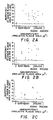
- Figure 2 shows a comparison of bio- and immuno-potency of PRL released from the same pituitary cells.
- PRL prolactin
- Each panel shows the results of a separate experiment and each point reflects a single measurement in the PRL plaque assay and duplicate determinations (60- ⁇ l volumes were used to flush and fill chambers) in the casein plaque bioassay. Values for individual cells that did not form PRL plaques are plotted on the extreme left of the abscissa and the average of these for each experiment is denoted by the dashed line
- Figure 3 shows the differential effects of rat prolactin and liver incubation medium (containing liver lactogenic factor) on casein plaque development.
- Monolayers of rat mammary cells and ovine red blood cells (oRBCs) were first exposed for 17 h to graded doses of rat PRL or serial dilutions of liver incubation medium and then subjected to a casein plaque assay. Both the percent of plaque forming colonies (PFC) and the sizes of plaques that developed were quantified on the same monolayers.
- Rat PRL had a stimulatory influence on each of these variables whereas liver medium augmented only plaque size.
- results are presented as the mean ⁇ SE of triplicate determinations in a single experiment which is representative of at least 3 separate experiments.
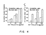
- Figure 4 shows interactions of rat PRL and liver incubation medium containing liver lactogenic factor (1:32, a maximally effective dilution) on casein plaque development.
- Control medium consisted of Dulbecco's Modified Eagle's Medium containing 0.1% BSA and antibiotics. The effects of control medium alone, or liver medium only, are represented as 0 pg/ml rat prolactin.
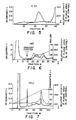
- Figure 5 shows the Sephadex G-50 activity elution profile of liver lactogenic factor.
- Figure 6 shows the carboxymethyl cellulose activity elution profile of liver lactogenic factor.
- Figure 7 shows the purification of liver lactogenic factor on a C18 reverse phase HPLC column.
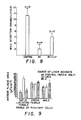
- Figure 8 shows the effect of liver lactogenic factor on milk secretion.
- Figure 9 shows the effect of the source of liver incubate on the amount of casein produced by mammary cells incubated with liver lactogenic factor.
- factor refers to chemical compounds such as hormones, vitamins, peptides, and molecules which may be recognized by cellular receptor molecules.
- a factor is said to be "lactogenic" if it is capable of stimulating a female mammal to produce either (1) enhanced levels of milk, or (2) milk having a higher nutritional value (for example, by increasing the casein content of the milk).
- liver lactogenic factor is intended the naturally occurring polypeptide isolated by the technique described below and polypeptides which are substantially similar thereto, whether synthetic or naturally occurring.
- a factor is said to be “substantially similar” to another factor if it is capable of performing substantially the same function, and has substantially the same chemical structure.
- the term is intended to include those polypeptides synthesized using, for example, a Merrifield synthesis, and those polypeptides produced using recombinant technology.
- the term further includes polypeptides which are substantially the same as the naturally occurring polypeptide but which differ therefrom by one or more amino acids while still having substantially the same biological activity.
- the liver lactogenic factor isolated by the procedure described below is characterized as follows. This factor is believed to be a protein having a molecular weight of approximately 9,400 daltons.
- the liver lactogenic factor may be purified into a substantially pure form by means which are commonly known in the art of peptide purification. Such means include HPLC chromatography, gel filtration, electrophoresis and affinity chromatography, using the casein plaque bioassay as a purification guide.
- the liver lactogenic factor may be obtained from mammalian liver tissue.
- tissue preferably 0.3-0.5 mm thick, approximately 1.0 g
- Any means of preparing such tissue slices may be employed.
- One preferred means employs a Statie-Riggs hand microtome according to the method of Nicoll, C.S., et al. ( Endocrinol. 116 :1449 (1985)).
- the sliced tissue is then incubated in a suitable cell culture medium.
- Dulbecco's Modified Eagle's Medium which has been supplemented with 0.1% bovine serum albumin and antibiotic (such as penicillin, (100 U/ml) streptomycin (100 ⁇ g/ml), etc.) to control against bacterial contamination.
- antibiotic such as penicillin, (100 U/ml) streptomycin (100 ⁇ g/ml), etc.
- suitable cell culture media are described in Lambert, K.J., et al ., Animal Cell Biotechnology , Vol. 1, Academic Press, 1985, pp. 86-122.
- the tissue is, preferably, incubated with constant slow stirring (using an impeller bar which is placed above the slices) for between 1-20 hours, preferably 1.5 hours, at an incubation temperature of approximately 37°C in an atmosphere of 95% air : 5% carbon dioxide.
- the culture medium is filtered, i.e., by filtration through Whatman No. 1 filter paper (Whatman Ltd., Maidstone, England) and the filtrate is analyzed for the presence of a lactogenic factor.
- a variety of different assays may be employed (Falconer, I.R., Aust. J. Biol. Sci. 33 :71-84 (1980)).
- Assays include measurements of the synthesis of medium-chain fatty acids (Strong, C.R., et al ., Biochem. J. 128 :1303-1308 (1972)), measurements of casein concentration (Rillema, J.A., et al ., Endocrinol.
- One aspect of the present invention is the discovery of a novel, in vitro bioassay capable of identifying lactogenic factors.
- This bioassay which measures the amount of casein released from isolated mammary cells, employs a reverse hemolytic plaque assay. This assay was described by Frawley, L.S., et al ., Endocrinol. 119 :2867-2869 (1986).
- This novel assay is sensitive enough to measure the biopotency of prolactin (a lactogenic factor) released from a single pituitary cell, and can be used to evaluate both the biopotency and immunopotency of hormone released from such cells.
- tissue are dissected aseptically from the lower abdomal mammary glands of lactating (days 9-11) rats which have been maintained with their litters until the time of sacrifice.
- the tissue is then diced, as with scalpel blades, into 2-4 mm 3 fragments.
- the tissue fragments are then transferred to a container such as a spinner flask containing a protease (preferably collagenase) (1 mg/ml) in approximately 20 ml of culture medium (Dulbecco's Modified Eagle's Medium containing 0.1% bovine serum albumin, penicillin G and streptomycin sulfate (MEM)) and incubated with continuous stirring for 0.5-2 hours at approximately 37°C.
- a protease preferably collagenase
- the collagenase is preferably type III collagenase (Cooper Biomedical, Malverne, PA).
- the tissue fragments are taken up and expelled through the tip of a 5 ml pipette at the midpoint and conclusion of the incubation.
- the cells and tissue fragments are then pelleted by centrifugation (700 x g for 15 minutes), resuspended in minimal essential medium containing the albumin and antibiotics described above (MEM) and passed successively through two sterile pieces of Nitex nylon screening (75 ⁇ m mesh, Tetko Inc., Elmsford, NY).
- the filtered cells are centrifuged again (500 x g, for 10 minutes), resuspended in MEM, and passed through two successive sterile pieces of Nitex screening (25 um mesh) to yield a suspension in which 70-80% of the cells are monodispersed.
- Rat casein was isolated from rat whole milk by the differential extraction procedure of Kaetzel, C.S., et al . ( J. Dairy Sci. 67 :64 (1984)). The purity of the material was assessed by running it in tandem with molecular weight markers on a sodium dodecyl sulfate-Laemmli gel (15%) followed by scanning densitometry. When subjected to electrophoresis, this casein preparation separated into three distinct bands that corresponded closely to those described as alpha, beta, and gamma caseins in rat milk (Kaetzel, C.S., et al .).
- Antisera for use in plaque assays was prepared by injecting this casein preparation into rabbits according to the multiple site procedure of Vaitukaitis, J., et al . ( J. Clin. Endocrinol. Metab. 33 :988 (1971)).
- the plaque assay for casein release from mammary cells is performed by mixing a suspension of monodispersed mammary cells, preferably at a concentration of between 2 x 10 5 - 5 x 10 5 cells per ml, in cell culture medium with an equal volume of protein A-coated ovine erythrocytes (oRBCs, 12%) and then by infusing this mixture into a chamber.
- the chamber may, for example, be constructed by affixing a glass cover slip to a poly-L-lysine-coated glass microscope slide by two pieces of double-sided adhesive tape (Boockfor, F.R., et al ., Neuroendocrinol. 42 :64 (1986), which reference is hereby incorporated in its entirety).
- the cells are then allowed to attach as a monolayer during a preincubation (30-60 minutes in length), thereby forming an assay chamber.
- the assay chambers are then washed, filled with either medium alone or test samples, and placed overnight in a humidified incubator at 37°C with an atmosphere of 95% air : 5% carbon dioxide. Between 15-20 hours later, the assay chambers are washed with fresh medium and incubated with casein antibody (diluted 1:500) for approximately 2 hours. Plaque development is completed during a subsequent incubation (approximately 50 minutes) with guinea pig complement (diluted 1:60). The monolayers are then fixed and stained with toluidine blue (Boockfor, F.R., et al . (1986)) prior to microscopic quantification of plaque formation.
- the reverse hemolytic plaque assay for lactogenic factors may be performed on cells from any tissue source.
- pituitary cells or liver cells may be assayed for their ability to produce lactogenic factors.
- casein plaque assay may be performed on individual cells in microwells. To accomplish this, approximately 170 ⁇ l of culture medium containing (on average) a single cell is added to each culture well of a microtiter plate and incubated for approximately 14 hours under the same conditions as described above. A 150 ⁇ l volume is then removed and either immediately assayed or stored frozen for subsequent analysis. 50 ⁇ l volume of antisera (appropriately diluted, if necessary) in assay medium containing oRBCs (0.125%) is then added.
- the monolayers may be exposed to guinea pig complement (preferably 50 ⁇ l of a 1:50 dilution) for approximately one hour and then fixed and stained with toluidine blue, as described above, prior to microscopic quantification of plaque formation.
- guinea pig complement preferably 50 ⁇ l of a 1:50 dilution
- lactogenic factor The presence of a lactogenic factor is revealed by determining the percentage of plaque-forming cells, and the average size of the plaques that are formed. Each of these variables increases with increasing lactogenic factor concentration. By calculating the average plaque area per incubation slide, which is simply the product of these two variables, it is possible to quantify the cumulative response of mammary cells to lactogenic factors.
- liver lactogenic factor which are capable of functioning as lactogenic factors. Included as well are the use of additional amino acid residues added to enhance coupling to carrier protein or amino acid residues added to enhance the lactogenic effect.
- the amino acid residues of liver lactogenic factor may be in their protected or unprotected form, using appropriate amino or carboxyl protecting groups as is known in the art.
- the liver lactogenic factor may also be in the presence of cationic salt groups.
- Useful cations are alkali or alkaline earth metallic cations (i.e., Na, K, Li, 1/2Ca and 1/2 Ba) or amine cations (i.e., tetraalkylammonium, trialkylammonium, where alkyl can be C 1 -C12).
- the invention further pertains to polypeptides that, in addition to containing the amino acid sequence of liver lactogen ⁇ c factor, may contain or lack one or more amino acids that may not be present in the naturally-occurring sequence, wherein such polypeptides are functionally similar to the chosen polypeptide.
- polypeptides for the present invention are termed "functional derivatives,” provided that they demonstrate activity which is substantially similar to that of liver lactogenic factor.
- the liver lactogenic factor may be in the form of the free amines (on the N-terminus), or acid-addition salts thereof.
- Common addition salts are hydrohalic acid salts, i.e., HBr, HI, or more preferably, HCl.
- liver lactogenic factor-containing compositions of the present invention can be formulated according to known methods to prepare pharmaceutically useful compositions, whereby liver lactogenic factor or its functional derivatives are combined in admixture with a pharmaceutically acceptable carrier vehicle.
- Suitable vehicles and their formulation, inclusive of other human proteins, e.g., human serum albumin, are described for example in Remington's pharmaceutical Sciences (16th Ed. A. Oslo Ed. Mack, Easton, PA (1980)).
- a pharmaceutically acceptable composition suitable for effective administration such compositions will contain an effective amount of the liver lactogenic factor, or its functional derivatives, together with a suitable amount of carrier vehicle.
- liver lactogenic factor is one which is sufficient to enhance either casein production or milk production in a lactating mammal.
- the effective amount may vary depending upon criteria such as the age, weight, physical condition, past medical history, and sensitivity of the recipient.
- compositions containing liver lactogenic factor or its functional derivatives may be administered orally, intravenously, intramuscularly, subcutaneously, or locally.
- compositions containing liver lactogenic factor may be dissolved in distilled water and the pH-value adjusted to about 6 to 8.
- lactose could be added to the solution.
- the solution is then filter sterilized, introduced into vials, and lyophilized.
- concentration of liver lactogenic factor in these compositions may vary from 10 ⁇ 12M to 10 ⁇ 5M.
- Controlled release preparations may be achieved by the use of polymers to complex or adsorb liver lactogenic factor or its functional derivatives.
- the controlled delivery may be exercised by selecting appropriate macromolecules (for example, polyesters, polyamino acids, polyvinyl pyrrolidone, ethylenevinylacetate, methylcellulose, carboxymethylcellulose, and protamine sulfate) and the concentration of macromolecules as well as the methods of incorporation in order to control release.
- Another possible method to control the duration of action by controlled release preparations is to incorporate liver lactogenic factor into particles of a polymeric material such as polyesters, polyamino acids, hydrogels, poly (lactic acid) or ethylene vinylacetate copolymers.
- liver lactogenic factor instead of incorporating liver lactogenic factor into these polymeric particles, it is possible to entrap liver lactogenic factor in microcapsules prepared, for example, by coacervation techniques or by interfacial polymerization, for example, hydroxylmethylcellulose or gelatin-microcapsules and poly (methylmethacrylate) microcapsules, respectively, or in colloidal drug delivery systems, for example, liposomes, albumin microspheres, microemulsions, nanoparticles, and nanocapsules or in macroemulsions.
- colloidal drug delivery systems for example, liposomes, albumin microspheres, microemulsions, nanoparticles, and nanocapsules or in macroemulsions.
- liver lactogenic factor of the present invention and its functional derivatives, either alone or in pharmacologically acceptable compositions are useful for enhancing lactation in female mammals.
- Administration of liver lactogenic factor results in an increase in milk production and in the concentration of casein present in milk.
- the invention therefore, has utility in augmenting milk, cheese, production in the dairy industry. Additionally, the invention may have utility for nursing women who are in need of higher quantity or quality milk.
- the reverse hemolytic plaque assay was used to measure prolactin from pituitary cells according to the method described above. 170 ⁇ l of culture medium containing (on average) a single pituitary cell was inoculated into microtiter plates and incubated for 14 hours as described previously. A 150 ⁇ l volume was then removed and replaced with a 50 ⁇ l volume of rabbit anti-rat prolactin serum (diluted 1:125) in MEM containing ovine sheep red blood cells. Varying concentrations (from 0 pg/ml - 400 pg/ml) of prolactin were added to the culture medium, and the effect on plaque formation and plaque area was determined. Two hours later, the monolayers were exposed to guinea pig complement (50 ⁇ l of 1:50) for 50 minutes and then fixed as described above. The results of this experiment are shown in Table 1.
- Treatment with graded doses of prolactin during the preincubation period increased (to approximately the same degree) both the percentage of plaque-forming cells and the average size of plaques that formed.
- Each of these variables exhibited a dose-related increase between 6.25 and 100 pg/ml of prolactin and a slight depression in response to higher concentrations. This depression may be due to a desensitization or down-regulation of the response.
- Figure 1 summarizes the results obtained in a single representative experiment and illustrates the specificity and sensitivity of the plaque assay.
- Treatment with rat prolactin increased, in a dose-related manner, the average plaque area per slide, whereas identical concentration of rat gonadotrophin had no effect.
- gonadotrophin, leutinizing hormone, follicle-stimulating hormone, nor thyroidstimulating hormone were capable of inducing plaque formation, even in doses as high as 10 mg/ml.
- the extraordinar sensitivity of the assay is evidenced by the different responses (P ⁇ 0.01, Student's t test) elicited by 0 and 6.25 pg/ml doses of prolactin. When corrected for the volume of incubation chamber (25 ⁇ l), this 6.25 pg/ml translates into 156 fg of prolactin.
- Figure 2 shows the results of three separate experiments in which the biopotency and immunopotency of prolactin released in the same cells are measured.
- Media samples taken from individual pituitary cells that did not form prolactin plaques exhibited biopotencies which were relatively low.
- unequivocal bioactivity was detected in media samples from most pituitary cells that released immunoreacted prolactin.
- a relationship between the bioactivity and immunoreactivity of hormones released from the same cells was not apparent.
- the reverse hemolytic plaque assay was used to measure casein release from individual rat mammary cells in culture.
- Mammary cells were isolated from mammary tissue as described above.
- Addition of 25 or 100 pg/ml rat prolactin (rPRL) at the beginning of the preincubation period increased the percentages of all mammary cells that released casein from a control value of 19.4 to 23.5 and 32.9, respectively.
- addition of medium that had bathed liver slices (for three hours) from day 12 lactaters had no effect.
- Treatment with prolactin, or with medium containing liver lactogenic factor (diluted 1:32) increased the average size of the plaques that formed.
- the results of this experiment are shown in Table 2.
- liver lactogenic factor medium bathing liver slices from lactaters (and thus containing the liver lactogenic factor) was far more potent than rat prolactin in stimulating casein release. Moreover, the combined effects of prolactin and liver lactogenic factor were additive rather than synergistic. Incubations of kidney tissues from lactating rats, liver tissue from male or virgin female rats, and insulin-like Growth Factor-1 (IGF-1) had no activity in the above assay systems. These results demonstrate that liver tissue from lactating rats contained a factor (liver lactogenic factor) which actually mimicked the action of prolactin. Liver lactogenic factor increased the amount of casein released from mammary cells but did not increase the number of cells committed to casein release.
- IGF-1 insulin-like Growth Factor-1
- Silices of the liver tissue were prepared as described above. The slices were incubated in culture medium, as described previously, and the medium was filtered through Whatman No. 1 filter paper.
- plaque development was quantified (with the aid of a microscope) by determining the percentage of cells that formed plaques or the sizes of plaques that developed. Statistical significance of differences between treatment groups was determined by the Student's T test (Steel, R.J.B., et al ., Principles and Procedures of Statistics , McGraw Hill, New York, 1980, page 114.)
- Figure 3 shows a comparison of the quantitative effects of liver incubation medium (which contained liver lactogenic factor) and rat prolactin on two variables of casein plaque development.
- Treatment with rat prolactin evoked dose-related increases in both the percentage of mammary cells that formed plaques and the average size of plaques that developed.
- dilutions of liver lactogenic factor had absolutely no effect on the proportion of casein plague formers that caused graded increases in average plague area.
- maximally effective dilutions of liver lactogenic factor were much more potent than comparable doses of prolactin when only the relative amount of casein released per cell (plaque size) was considered.
- Incubation medium that bathed kidney tissue from lactating rats and liver tissue from male or virgin female rats, as well as insulin-like growth factor-I, were all tested and found to have no activity in this assay system.
- liver lactogenic factor only partially mimicked the action of prolactin; it increased the amount of casein released per mammary cell without affecting the number of cells committed to casein release. Because the presence of the liver lactogenic factor was associated with a physiologic state of elevated prolactin secretion (lactation), one possibility which may be proposed was that liver lactogenic factor served as an extracellular mediator of prolactin's action. The existence of such a liver lactogenic factor might provide a mechanism by which the lactogenic and mammary mitogenic actions of prolactin could be dissociated and differentially regulated.
- liver lactogenic factor was found to retain its activity after freezing or lyophilization. In contrast, the activity of liver lactogenic factor was virtually Polished by treatment with HCl (pH 1.0) for 30 minutes followed by neutralization with NaOH (and centrifugation to remove the precipitate). Activity was also lost after treatment at 80°C for 10 minutes followed by centrifugation of the precipitate. A two-hour incubation with the proteolytic enzyme pepsin was found to destroy liver lactogenic factor activity. The results of these experiments indicated that the factor was a protein or polypeptide.
- liver lactogenic factor (LLF) was associated with states of elevated (lactation) but not basal (virgin female, male) levels of prolactin secretion. This suggested that prolactin may regulate liver lactogenic factor production.
- endogenous prolactin levels were manipulated, and the effect of this manipulation on liver lactogenic factor released was evaluated. Two experimental strategies were used. First, day 10 lactating rats were injected three times at 12-hour intervals with either bromocryptine (BC) (3 x 2 mg/injection) or inert vehicle and then sacrificed 11 hours after the last treatment.
- BC bromocryptine
- liver lactogenic activity rose from undetectable levels (98.9 ⁇ 0.2% of control) in inert vehicle-injected rats to 135.5 ⁇ 12.1% of control after prolactin administration. Since reduction of circulating prolactin diminished hepatic lactogenic activity and injection of prolactin induced such activity, it was concluded that the production of liver lactogenic factor was regulated, in large part, by prolactin.
- LIVER LACTOGENIC FACTOR IS PRESENT IN BLOOD AND CAN MAINTAIN MILK PRODUCTION IN PROLACTIN-SUPPRESSED RATS
- liver lactogenic factor was present in blood
- serum was collected from male and lactating female rats and subjected to an extraction procedure designed to remove prolactin.
- prolactin 5 ⁇ g/ml
- prolactin-free sera were then diluted in medium and tested in the above-described plaque assay for the presence of a lactogenic factor.
- the experiment was complicated by the fact that serum PRL levels in both sexes were adequate to completely overwhelm our bioassay system.
- Prolactin-free serum from lactating female rats contained a factor that significantly increased the average size of casein plaques to 213.3 ⁇ 3.2% of a medium sample extracted concurrently (control).
- extracts of samples of male serum, male serum plus prolactin, and medium plus prolactin produced plaque sizes which were 97.2 ⁇ 3.0, 93.8 ⁇ 3.9, and 79.4 ⁇ 9.6%, respectively, of control values.
- serum from lactating rats possessed lactogenic activity which was completely separate from prolactin.
- liver lactogenic factor significantly augmented milk production in prolactin-suppressed rats.
- liver lactogenic factor was found to mimic the lactogenic component of prolactin's actions in vivo as well as in vitro. Thus, these findings indicate that liver lactogenic factor served as an extracellular mediator of prolactin's actions.
- liver tissue from lactating rats (9-11 days) were suspended in approximately 1000 ml DMEM and homogenized with a polytron (Brinkman). During this procedure, the liver-containing vessel was suspended in an ice bath and, immediately following homogenization, acetone was added to a total concentration of 70%. The extract was maintained at 4°C overnight whereafter the insoluble pellet was removed by slow-speed centrifugation (4,000 rpm, 15 min) Ice-cold acetone was then added to the supernatant solution to a final concentration of 90%. After two days at 4°C, the supernatant acetone was syphoned off and the pellet was collected, redissolved in double distilled water and lyophilized.
- a total of 12 mg of the active preparation was then subjected to ion exchange chromatography on a CM cellulose column (1.5 cm x 20 cm) and eluted with a gradient consisting of 100 ml 0.05 M sodium acetate (pH 5.0) in vessel I and 100 ml 0.5 M sodium chloride in vessel II.
- Two active regions were detected in the effluent of this column as seen in Figure 6 and both were collected and lyophilized separately.
- the final separation on HPLC was done on a C18 reverse phase column, using a gradient of 0.1% trifluoroacetic acid (TFA) in water as solvent A and 0.1% TRA in 80% acetonitrile as solvent B.
- TFA trifluoroacetic acid
- the second activity peak obtained from CM cellulose gave rise to a single peak by HPLC, eluting near the end of the 30-minute linear gradient (from 20% to 50% solvent B). All of the activity was associated with this peak and it was used for subsequent amino acid analyses and physiologic studies.
- liver lactogenic factor differs from both native PRL (which has a much greater MW than the lactogen) and the 8,000 MW PRL cleavage product described by Mittra and coworkers (Mittra et al ., Biochem. Biophys. res. Commun. 95:1750-1767 (1980)).
- the basis for this latter conclusion is that the fraction that precipitated between 70% and 90% acetone, which contained extremely high bioactivity, was completely devoid of PRL immunoreactivity when tested in the rat PRL radioimmunoassay distributed by the National Pituitary Agency.
- This assay employs a polyclonal antibody which most probably recognizes multiple epitopes (including those on the 8,000 MW fragment) of the PRL molecule.
- lactogen is not insulin-like (i.e., a somatomedin). Its activity is not affected by reduction and alkylation which is consistent with the finding that the molecule does not contain cysteine (Table 3). Moreover, IGF-1 is not active in our casein plaque bioassay system.
- liver lactogenic factor (LLF) The physiologic relevance of liver lactogenic factor (LLF) on milk production was examined. If LLF mediates the activity of prolactin, then it should influence milk production. Therefore, it follows that if a deficit in milk production was created by suppressing endogenous PRL with bromocryptine (BC), then treatment with liver lactogenic factor should at least partially reverse this deficit.
- BC bromocryptine
- female lactating rats were treated with either BC alone or BC plus the HPLC purified preparation of liver lactogenic factor (LLF) (a total of 1.5 liver equivalents per animal). The rats were injected at 0, 12, and 24 hours, separated from their litters for 10 hours, and then reunited for 30 minutes.
- Prolactin is viewed as being rather unique among pituitary hormones because its secretion is not controlled via long-loop feedback of an end-organ product.
- PRL-induced substance such as the liver lactogen necessitated a reexamination of this concept.
- PRL plaque assays on pituitary cells from rats in various endocrine states were run to evaluate the effects of medium conditioned by liver tissue from male and lactating female rats. The results of this experiment are illustrated in Figure 9.
- liver medium from lactating females clearly stimulated rather than inhibited PRL plaque development when pituitary cells derived from lactating females were tested.
- Incubates of liver tissue from males had no effect on different batches of pituitary cells from the same dispersion.- Neither type of liver incubate had any influence on PRL plaque development when pituitary cells from male or virgin female rats were employed.
- the sex and physiologic state of an animal appears to influence not only the ability to produce the lactogen but also the capacity of PRL cells to respond to it.
Landscapes
- Health & Medical Sciences (AREA)
- Life Sciences & Earth Sciences (AREA)
- Chemical & Material Sciences (AREA)
- Molecular Biology (AREA)
- Engineering & Computer Science (AREA)
- Medicinal Chemistry (AREA)
- General Health & Medical Sciences (AREA)
- Endocrinology (AREA)
- Organic Chemistry (AREA)
- Hematology (AREA)
- Biochemistry (AREA)
- Urology & Nephrology (AREA)
- Immunology (AREA)
- Biomedical Technology (AREA)
- Food Science & Technology (AREA)
- Analytical Chemistry (AREA)
- Pathology (AREA)
- General Physics & Mathematics (AREA)
- Physics & Mathematics (AREA)
- Toxicology (AREA)
- Zoology (AREA)
- Gastroenterology & Hepatology (AREA)
- Microbiology (AREA)
- Biophysics (AREA)
- Genetics & Genomics (AREA)
- Cell Biology (AREA)
- Proteomics, Peptides & Aminoacids (AREA)
- Biotechnology (AREA)
- General Chemical & Material Sciences (AREA)
- Bioinformatics & Cheminformatics (AREA)
- Gynecology & Obstetrics (AREA)
- Chemical Kinetics & Catalysis (AREA)
- Reproductive Health (AREA)
- Nuclear Medicine, Radiotherapy & Molecular Imaging (AREA)
- Pregnancy & Childbirth (AREA)
- Animal Behavior & Ethology (AREA)
- Pharmacology & Pharmacy (AREA)
- Public Health (AREA)
- Veterinary Medicine (AREA)
- Medicines That Contain Protein Lipid Enzymes And Other Medicines (AREA)
Claims (8)
Priority Applications (1)
| Application Number | Priority Date | Filing Date | Title |
|---|---|---|---|
| AT88902741T ATE81470T1 (de) | 1987-03-06 | 1988-03-07 | Laktogener leber-faktor. |
Applications Claiming Priority (2)
| Application Number | Priority Date | Filing Date | Title |
|---|---|---|---|
| US22893 | 1987-03-06 | ||
| US07/022,893 US4895833A (en) | 1987-03-06 | 1987-03-06 | Liver lactogenic factor |
Publications (3)
| Publication Number | Publication Date |
|---|---|
| EP0348429A1 EP0348429A1 (fr) | 1990-01-03 |
| EP0348429A4 EP0348429A4 (fr) | 1990-04-10 |
| EP0348429B1 true EP0348429B1 (fr) | 1992-10-14 |
Family
ID=21811984
Family Applications (1)
| Application Number | Title | Priority Date | Filing Date |
|---|---|---|---|
| EP88902741A Expired - Lifetime EP0348429B1 (fr) | 1987-03-06 | 1988-03-07 | Facteur lactogenique du foie |
Country Status (7)
| Country | Link |
|---|---|
| US (1) | US4895833A (fr) |
| EP (1) | EP0348429B1 (fr) |
| JP (1) | JPH02502995A (fr) |
| AT (1) | ATE81470T1 (fr) |
| AU (1) | AU622613B2 (fr) |
| DE (1) | DE3875341T2 (fr) |
| WO (1) | WO1988006453A1 (fr) |
Family Cites Families (1)
| Publication number | Priority date | Publication date | Assignee | Title |
|---|---|---|---|---|
| US2370154A (en) * | 1941-01-31 | 1945-02-27 | Schering Corp | Process for the isolation of the lactogenic hormone |
-
1987
- 1987-03-06 US US07/022,893 patent/US4895833A/en not_active Expired - Fee Related
-
1988
- 1988-03-07 EP EP88902741A patent/EP0348429B1/fr not_active Expired - Lifetime
- 1988-03-07 WO PCT/US1988/000673 patent/WO1988006453A1/fr not_active Ceased
- 1988-03-07 JP JP63502680A patent/JPH02502995A/ja active Pending
- 1988-03-07 AU AU14827/88A patent/AU622613B2/en not_active Ceased
- 1988-03-07 AT AT88902741T patent/ATE81470T1/de not_active IP Right Cessation
- 1988-03-07 DE DE8888902741T patent/DE3875341T2/de not_active Expired - Fee Related
Non-Patent Citations (2)
| Title |
|---|
| Endocrinology, vol. 120, no.4, 1987, pages 1679-1681, The Endocrine Society; J.P. HOEFFLER et al.: "Liver tissue produces a potent lactogen that partially mimics the actions of prolactin" * |
| Endocrinology, vol. 123, no. 4, 1988, pages 2014-2018, The Endocrine Society; L.S. FRAWLEY et al.: "Liver tissue from lactating rats produces a factor that stimulates prolactin release and gene expression" * |
Also Published As
| Publication number | Publication date |
|---|---|
| US4895833A (en) | 1990-01-23 |
| AU622613B2 (en) | 1992-04-16 |
| WO1988006453A1 (fr) | 1988-09-07 |
| DE3875341T2 (de) | 1993-04-15 |
| EP0348429A4 (fr) | 1990-04-10 |
| EP0348429A1 (fr) | 1990-01-03 |
| DE3875341D1 (de) | 1992-11-19 |
| JPH02502995A (ja) | 1990-09-20 |
| ATE81470T1 (de) | 1992-10-15 |
| AU1482788A (en) | 1988-09-26 |
Similar Documents
| Publication | Publication Date | Title |
|---|---|---|
| Bryant-Greenwood | Relaxin as a new hormone | |
| Lucy et al. | Placental lactogen and somatotropin: hormone binding to the corpus luteum and effects on the growth and functions of the ovary in heifers | |
| Rosenfeld et al. | Increased somatomedin receptor sites in newborn circulating mononuclear cells | |
| Dogterom et al. | Studies on the presence of vasopressin, oxytocin and vasotocin in the pineal gland, subcommissural organ and fetal pituitary gland: failure to demonstrate vasotocin in mammals | |
| EP0276166A2 (fr) | Composition et méthode pour provoquer une superovulation chez le bétail | |
| Almeida et al. | Effects of different patterns of feed restriction and insulin treatment during the luteal phase on reproductive, metabolic, and endocrine parameters in cyclic gilts | |
| Eppard et al. | Comparison of the galactopoietic response to pituitary-derived and recombinant-derived variants of bovine growth hormone | |
| WO1996009064A1 (fr) | Procede et preparation pharmaceutique pour la prevention et le traitement des lesions cerebrales | |
| AU636195B2 (en) | Method for inhibiting follicular maturation | |
| Rogers et al. | Growth hormone stimulates IGF I gene expression in isolated rat renal collecting duct | |
| Fielder et al. | Expression of a lactogen-dependent insulin-like growth factor-binding protein in cultured mouse mammary epithelial cells | |
| US4895833A (en) | Liver lactogenic factor | |
| Lakshmanan et al. | Epidermal growth factor in mouse ocular tissue: effects of thyroxine and exogenous epidermal growth factor | |
| DE69105114T2 (de) | Methode zur erhöhung der fruchtbarkeit weiblicher säuger. | |
| KISHI et al. | Pituitary factors regulating mouse placental lactogen-II secretion during the last half of pregnancy in mice | |
| Smith et al. | Prolactin in seminal fluid | |
| Ng et al. | Effects of exogenous growth hormone on lipid metabolism in the isolated epididymal fat pad of the growth hormone-deficient little mouse. | |
| Malven et al. | Immunoreactive prolactin in subcellular fractions from bovine mammary tissue | |
| Isozaki et al. | Thyroid-stimulating antibody bioassay using porcine thyroid cells cultured in follicles | |
| Francis et al. | Induction of the synthesis of the pregnancy-specific protein p70 in the endometrium by intramuscular injection of recombinant bovine interferon-αI1 in nonpregnant ewes | |
| Leatherland et al. | Structure of the pars distalis in the adult tammar wallaby (Macropus eugenii) | |
| US5484767A (en) | Substantially pure steroidogenesis inducing protein and uses thereof | |
| Torres et al. | Isolation of two different pools of pituitary prolactin | |
| TEBBS et al. | The role of maternally derived epidermal growth factor and the epidermal growth factor receptor during organogenesis in the rat embryo | |
| Wise et al. | Porcine somatotropic regulation of thymic weight, thymosin β 4, and insulin-like growth factors in lean and obese swine |
Legal Events
| Date | Code | Title | Description |
|---|---|---|---|
| PUAI | Public reference made under article 153(3) epc to a published international application that has entered the european phase |
Free format text: ORIGINAL CODE: 0009012 |
|
| 17P | Request for examination filed |
Effective date: 19890906 |
|
| AK | Designated contracting states |
Kind code of ref document: A1 Designated state(s): AT BE CH DE FR GB IT LI LU NL SE |
|
| A4 | Supplementary search report drawn up and despatched |
Effective date: 19900410 |
|
| 17Q | First examination report despatched |
Effective date: 19910719 |
|
| GRAA | (expected) grant |
Free format text: ORIGINAL CODE: 0009210 |
|
| AK | Designated contracting states |
Kind code of ref document: B1 Designated state(s): AT BE CH DE FR GB IT LI LU NL SE |
|
| PG25 | Lapsed in a contracting state [announced via postgrant information from national office to epo] |
Ref country code: IT Free format text: LAPSE BECAUSE OF FAILURE TO SUBMIT A TRANSLATION OF THE DESCRIPTION OR TO PAY THE FEE WITHIN THE PRE;WARNING: LAPSES OF ITALIAN PATENTS WITH EFFECTIVE DATE BEFORE 2007 MAY HAVE OCCURRED AT ANY TIME BEFORE 2007. THE CORRECT EFFECTIVE DATE MAY BE DIFFERENT FROM THE ONE RECORDED.SCRIBED TIME-LIMIT Effective date: 19921014 Ref country code: AT Effective date: 19921014 Ref country code: SE Effective date: 19921014 Ref country code: NL Effective date: 19921014 Ref country code: BE Effective date: 19921014 |
|
| REF | Corresponds to: |
Ref document number: 81470 Country of ref document: AT Date of ref document: 19921015 Kind code of ref document: T |
|
| REF | Corresponds to: |
Ref document number: 3875341 Country of ref document: DE Date of ref document: 19921119 |
|
| ET | Fr: translation filed | ||
| PGFP | Annual fee paid to national office [announced via postgrant information from national office to epo] |
Ref country code: GB Payment date: 19930219 Year of fee payment: 6 |
|
| PGFP | Annual fee paid to national office [announced via postgrant information from national office to epo] |
Ref country code: FR Payment date: 19930226 Year of fee payment: 6 |
|
| NLV1 | Nl: lapsed or annulled due to failure to fulfill the requirements of art. 29p and 29m of the patents act | ||
| PGFP | Annual fee paid to national office [announced via postgrant information from national office to epo] |
Ref country code: CH Payment date: 19930324 Year of fee payment: 6 |
|
| PG25 | Lapsed in a contracting state [announced via postgrant information from national office to epo] |
Ref country code: LU Free format text: LAPSE BECAUSE OF NON-PAYMENT OF DUE FEES Effective date: 19930331 |
|
| PGFP | Annual fee paid to national office [announced via postgrant information from national office to epo] |
Ref country code: DE Payment date: 19930428 Year of fee payment: 6 |
|
| PLBE | No opposition filed within time limit |
Free format text: ORIGINAL CODE: 0009261 |
|
| STAA | Information on the status of an ep patent application or granted ep patent |
Free format text: STATUS: NO OPPOSITION FILED WITHIN TIME LIMIT |
|
| 26N | No opposition filed | ||
| PG25 | Lapsed in a contracting state [announced via postgrant information from national office to epo] |
Ref country code: GB Effective date: 19940307 |
|
| PG25 | Lapsed in a contracting state [announced via postgrant information from national office to epo] |
Ref country code: CH Effective date: 19940331 Ref country code: LI Effective date: 19940331 |
|
| GBPC | Gb: european patent ceased through non-payment of renewal fee |
Effective date: 19940307 |
|
| PG25 | Lapsed in a contracting state [announced via postgrant information from national office to epo] |
Ref country code: FR Effective date: 19941130 |
|
| REG | Reference to a national code |
Ref country code: CH Ref legal event code: PL |
|
| PG25 | Lapsed in a contracting state [announced via postgrant information from national office to epo] |
Ref country code: DE Effective date: 19941201 |
|
| REG | Reference to a national code |
Ref country code: FR Ref legal event code: ST |