EP0343009B1 - Method for producing the Nde I restriction endonuclease and methylase - Google Patents
Method for producing the Nde I restriction endonuclease and methylase Download PDFInfo
- Publication number
- EP0343009B1 EP0343009B1 EP89305109A EP89305109A EP0343009B1 EP 0343009 B1 EP0343009 B1 EP 0343009B1 EP 89305109 A EP89305109 A EP 89305109A EP 89305109 A EP89305109 A EP 89305109A EP 0343009 B1 EP0343009 B1 EP 0343009B1
- Authority
- EP
- European Patent Office
- Prior art keywords
- nde
- restriction endonuclease
- dna
- restriction
- methylase
- Prior art date
- Legal status (The legal status is an assumption and is not a legal conclusion. Google has not performed a legal analysis and makes no representation as to the accuracy of the status listed.)
- Expired - Lifetime
Links
Images


Classifications
-
- C—CHEMISTRY; METALLURGY
- C12—BIOCHEMISTRY; BEER; SPIRITS; WINE; VINEGAR; MICROBIOLOGY; ENZYMOLOGY; MUTATION OR GENETIC ENGINEERING
- C12N—MICROORGANISMS OR ENZYMES; COMPOSITIONS THEREOF; PROPAGATING, PRESERVING, OR MAINTAINING MICROORGANISMS; MUTATION OR GENETIC ENGINEERING; CULTURE MEDIA
- C12N9/00—Enzymes; Proenzymes; Compositions thereof; Processes for preparing, activating, inhibiting, separating or purifying enzymes
- C12N9/14—Hydrolases (3)
- C12N9/16—Hydrolases (3) acting on ester bonds (3.1)
- C12N9/22—Ribonucleases RNAses, DNAses
-
- C—CHEMISTRY; METALLURGY
- C12—BIOCHEMISTRY; BEER; SPIRITS; WINE; VINEGAR; MICROBIOLOGY; ENZYMOLOGY; MUTATION OR GENETIC ENGINEERING
- C12N—MICROORGANISMS OR ENZYMES; COMPOSITIONS THEREOF; PROPAGATING, PRESERVING, OR MAINTAINING MICROORGANISMS; MUTATION OR GENETIC ENGINEERING; CULTURE MEDIA
- C12N9/00—Enzymes; Proenzymes; Compositions thereof; Processes for preparing, activating, inhibiting, separating or purifying enzymes
- C12N9/10—Transferases (2.)
- C12N9/1003—Transferases (2.) transferring one-carbon groups (2.1)
- C12N9/1007—Methyltransferases (general) (2.1.1.)
Definitions
- the present invention relates to clones for the Nde I restriction endonuclease and modification methylase, and to the production of these enzymes from the clones.
- Restriction endonucleases are a class of enzymes that occur naturally in bacteria. When they are purified away from other contaminating bacterial components, restriction endonucleases can be used in the laboratory to break DNA molecules into precise fragments. This property enables DNA molecules to be uniquely identified and to be fractionated into their constituent genes. Restriction endonucleases have proved to be indispensable tools in modern genetic research. They are the biochemical 'scissors' by means of which genetic engineering and analysis is performed.
- Restriction endonucleases act by recognizing and binding to particular sequences of nucleotides (the 'recognition sequence') along the DNA molecule. Once bound, they cleave the molecule within, or to one side of, the sequence. Different restriction endonucleases have affinity for different recognition sequences. Over one hundred different restriction endonucleases have been identified among many hundreds of bacterial species that have been examined to date.
- Bacteria usually posses only a small number restriction endonucleases per species.
- the endonucleases are named according to the bacteria from which they are derived.
- the species Haemophilus aegyptius for example synthesizes 3 different restriction endonucleases, named HaeI, HaeII and HaeIII.
- These enzymes recognize and cleave the sequences (AT)GGCC(AT), PuGCGCPy and GGCC respectively.
- Escherichia coli RY13 synthesizes only one enzyme, EcoRI, which recognizes the sequence GAATTC.
- restriction endonucleases play a protective role in the welfare of the bacterial cell. They enable bacteria to resist infection by foreign DNA molecules like viruses and plasmids that would otherwise destroy or parasitize them. They impart resistance by binding to infecting DNA molecules and cleaving them each time that the recognition sequence occurs. The disintegration that results inactivates many of the infecting genes and renders the DNA susceptible to further degradation by exonucleases.
- a second component of bacterial protective systems are the modification methylases. These enzymes are complementary to restriction endonucleases and they provide the means by which bacteria are able to protect their own DNA and distinguish it from foreign, infecting DNA. Modification methylases recognize and bind to the same nucleotide recognition sequence as the corresponding restriction endonuclease, but instead of breaking the DNA, they chemically modify one or other of the nucleotides within the sequence by the addition of a methyl group. Following methylation, the recognition sequence is no longer bound or cleaved by the restriction endonuclease.
- the DNA of a bacterial cell is always fully modified, by virtue of the activity of its modification methylase and it is therefore completely insensitive to the presence of the endogenous restriction endonuclease. It is only unmodified, and therefore identifiably foreign, DNA that is insensitive to restriction endonuclease recognition and attack.
- the key to isolating clones of restriction endonuclease genes is to develop a simple and reliable method to identify such clones within complex 'libraries', i.e. populations of clones derived by 'shotgun' procedures, when they occur at frequencies as low as 10 ⁇ 3 to 10 ⁇ 4.
- the method should be selective, such that the unwanted, majority, of clones are destroyed while the desirable, rare, clones survive.
- Type II restriction-modification systems are being cloned with increasing frequency.
- the first cloned systems used bacteriophage infection as a means of identifying or selecting restriction endonuclease clones (HhaII: Mann et al., Gene 3: 97-112, (1978); EcoRII: Kosykh et al., Molec. gen. Genet 178: 717-719, (1980); PstI: Walder et al., Proc. Nat. Acad. Sci. USA 78 1503-1507, (1981)).
- E. coli cloning plasmids E. coli cloning plasmids
- EcoRV Bougueleret et al, Nucleic Acids Res. 12:3659-3676, (1984); PaeR7: Gingeras and Brooks, Proc. Natl. Acad. Sci. USA 80:402-406, (1983); Theriault and Roy, Gene 19:355-359, (1982); PvuII: Blumenthal et al., J. Bacteriol. 164:501-509, (1985)).
- a third approach and one that is being used to clone a growing number of systems, involves selecting for an active methylase gene referring to our Patent application No. 707079 and (BsuRI: Kiss et al., Nucleic Acids Res. 13:6403-6421, (1985)). Since restriction and modification genes tend to be closely linked, clones containing both genes can often be isolated by selecting for just the one gene.
- a potential obstacle to cloning restriction-modification genes lies in trying to introduce the endonuclease gene into a host not already protected by modification. If the methylase gene and endonuclease gene are introduced together as a single clone, the methylase must protectively modify the host DNA before the endonuclease has the opportunity to cleave it. On occasion, therefore, it might only be possible to clone the genes sequentially, methylase first then endonuclease.
- Another obstacle to cloning restriction-modification systems lies in the discovery that some strains of E.coli react adversely to cytosine modification; they possess systems that destroy DNA containing methylated cytosine (Raleigh and Wilson, Proc.
- Cytosine-specific methylase genes cannot be cloned easily into these strains, either on their own, or together with their corresponding endonuclease genes. To avoid this problem it is necessary to use mutant strains of E.coli (McrA ⁇ and McrB ⁇ ) in which these systems are defective.
- a clone containing the genes for the Nde I restriction endonuclease and modification methylase derived from Neisseria denitrificans , as well as related methods for the production of the enzymes. More specifically, this invention relates to clones which express the restriction endonuclease Nde I, an enzyme which recognizes the DNA sequence CA TATG and cleaves as indicated between the first 5' A and T by the arrow. See FEBS Letters . 150: 114-116, (1982), the disclosure of which is hereby incorporated by reference herein.
- the preferred method for cloning this enzyme comprises forming a library containing the DNA from Neisseria denitrificans, isolating those clones which contain DNA coding for the Nde I modification methylase and screening among these to identify those that also contain the Nde I restriction endonuclease gene.
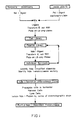
- Figure 1 illustrates the scheme for cloning the Nde I restriction endonuclease.
- FIG. 2 illustrates the scheme for producing the Nde I restriction endonuclease.
- Figure 3 is a restriction map of the 4.0 Kb Nsi I fragment from Neisseria denitrificans that encodes the Nde I restriction endonuclease and modification methylase in pUC19.
- the fragment was cloned into the PstI site of pUC19 (ATCC 37254) to create pNdeIRM6.7-A6 and pNdeIRM6.7-B9.
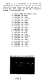
- Figure 4 is a photograph of an agarose gel demonstrating representative sites in the map.
- Figure 5 is a photograph of an agarose gel demonstrating Nde I restriction endonuclease activity in cell extracts of E. coli RR1 (ATCC 31343) carrying pNdeIRM6.7-A6 and pNdeIRM6.7-B9.
- the present invention provides a method for cloning Nde I restriction and modification genes and producing the restriction endonuclease Nde I from clones produced thereby.
- This approach takes advantage of the fact that clones have been selected on the basis of containing expressed Nde I restriction and methylase genes by the use of an endonuclease selection. Such clones are resistant to digestion in vitro by Nde I restriction endonuclease.
- Nde I restriction gene and methylase gene are preferably cloned and expressed.
Landscapes
- Life Sciences & Earth Sciences (AREA)
- Health & Medical Sciences (AREA)
- Chemical & Material Sciences (AREA)
- Genetics & Genomics (AREA)
- Organic Chemistry (AREA)
- Engineering & Computer Science (AREA)
- Bioinformatics & Cheminformatics (AREA)
- Zoology (AREA)
- Wood Science & Technology (AREA)
- Molecular Biology (AREA)
- Microbiology (AREA)
- Biotechnology (AREA)
- Biomedical Technology (AREA)
- Biochemistry (AREA)
- General Engineering & Computer Science (AREA)
- General Health & Medical Sciences (AREA)
- Medicinal Chemistry (AREA)
- Enzymes And Modification Thereof (AREA)
- Micro-Organisms Or Cultivation Processes Thereof (AREA)
Description
- The present invention relates to clones for the Nde I restriction endonuclease and modification methylase, and to the production of these enzymes from the clones.
- Restriction endonucleases are a class of enzymes that occur naturally in bacteria. When they are purified away from other contaminating bacterial components, restriction endonucleases can be used in the laboratory to break DNA molecules into precise fragments. This property enables DNA molecules to be uniquely identified and to be fractionated into their constituent genes. Restriction endonucleases have proved to be indispensable tools in modern genetic research. They are the biochemical 'scissors' by means of which genetic engineering and analysis is performed.
- Restriction endonucleases act by recognizing and binding to particular sequences of nucleotides (the 'recognition sequence') along the DNA molecule. Once bound, they cleave the molecule within, or to one side of, the sequence. Different restriction endonucleases have affinity for different recognition sequences. Over one hundred different restriction endonucleases have been identified among many hundreds of bacterial species that have been examined to date.
- Bacteria usually posses only a small number restriction endonucleases per species. The endonucleases are named according to the bacteria from which they are derived. Thus, the species Haemophilus aegyptius, for example synthesizes 3 different restriction endonucleases, named HaeI, HaeII and HaeIII. These enzymes recognize and cleave the sequences (AT)GGCC(AT), PuGCGCPy and GGCC respectively. Escherichia coli RY13, on the other hand, synthesizes only one enzyme, EcoRI, which recognizes the sequence GAATTC.
- While not wishing to be bound by theory, it is thought that in nature, restriction endonucleases play a protective role in the welfare of the bacterial cell. They enable bacteria to resist infection by foreign DNA molecules like viruses and plasmids that would otherwise destroy or parasitize them. They impart resistance by binding to infecting DNA molecules and cleaving them each time that the recognition sequence occurs. The disintegration that results inactivates many of the infecting genes and renders the DNA susceptible to further degradation by exonucleases.
- A second component of bacterial protective systems are the modification methylases. These enzymes are complementary to restriction endonucleases and they provide the means by which bacteria are able to protect their own DNA and distinguish it from foreign, infecting DNA. Modification methylases recognize and bind to the same nucleotide recognition sequence as the corresponding restriction endonuclease, but instead of breaking the DNA, they chemically modify one or other of the nucleotides within the sequence by the addition of a methyl group. Following methylation, the recognition sequence is no longer bound or cleaved by the restriction endonuclease. The DNA of a bacterial cell is always fully modified, by virtue of the activity of its modification methylase and it is therefore completely insensitive to the presence of the endogenous restriction endonuclease. It is only unmodified, and therefore identifiably foreign, DNA that is insensitive to restriction endonuclease recognition and attack.
- With the advent of genetic engineering technology, it is now possible to clone genes and to produce the proteins and enzymes that they encode in greater quantities than are obtainable by conventional purification techniques. The key to isolating clones of restriction endonuclease genes is to develop a simple and reliable method to identify such clones within complex 'libraries', i.e. populations of clones derived by 'shotgun' procedures, when they occur at frequencies as low as 10⁻³ to 10⁻⁴. Preferably, the method should be selective, such that the unwanted, majority, of clones are destroyed while the desirable, rare, clones survive.
- Type II restriction-modification systems are being cloned with increasing frequency. The first cloned systems used bacteriophage infection as a means of identifying or selecting restriction endonuclease clones (HhaII: Mann et al., Gene 3: 97-112, (1978); EcoRII: Kosykh et al., Molec. gen. Genet 178: 717-719, (1980); PstI: Walder et al., Proc. Nat. Acad. Sci. USA 78 1503-1507, (1981)). Since the presence of restriction-modification systems in bacteria enables them to resist infection by bacteriophages, cells that carry cloned restriction-modification genes can, in principle, be selectively isolated as survivors from libraries that have been exposed to phage. This method has been found, however, to have only limited value. Specifically, it has been found that cloned restriction-modification genes do not always manifest sufficient phage resistance to confer selective survival.
- Another cloning approach involves transferring systems initially characterized as plasmid-borne into E. coli cloning plasmids (EcoRV: Bougueleret et al, Nucleic Acids Res. 12:3659-3676, (1984); PaeR7: Gingeras and Brooks, Proc. Natl. Acad. Sci. USA 80:402-406, (1983); Theriault and Roy, Gene 19:355-359, (1982); PvuII: Blumenthal et al., J. Bacteriol. 164:501-509, (1985)).
- A third approach, and one that is being used to clone a growing number of systems, involves selecting for an active methylase gene referring to our Patent application No. 707079 and (BsuRI: Kiss et al., Nucleic Acids Res. 13:6403-6421, (1985)). Since restriction and modification genes tend to be closely linked, clones containing both genes can often be isolated by selecting for just the one gene. Selection for methylation activity does not always yield a complete restriction-modification system however, but instead sometimes yields only the methylase gene (BspRI: Szomolanyi et al., Gene 10:219-225, (1980); BcnI: Janulaitis et al, Gene 20: 197-204 (1982); BsuRI: Kiss and Baldauf, Gene 21: 111-119, (1983); and MspI: Walder et al., J. Biol. Chem. 258:1235-1241, (1983)).
- A potential obstacle to cloning restriction-modification genes lies in trying to introduce the endonuclease gene into a host not already protected by modification. If the methylase gene and endonuclease gene are introduced together as a single clone, the methylase must protectively modify the host DNA before the endonuclease has the opportunity to cleave it. On occasion, therefore, it might only be possible to clone the genes sequentially, methylase first then endonuclease. Another obstacle to cloning restriction-modification systems lies in the discovery that some strains of E.coli react adversely to cytosine modification; they possess systems that destroy DNA containing methylated cytosine (Raleigh and Wilson, Proc. Natl. Acad. Sci., USA 83:9070-9074, (1986)). Cytosine-specific methylase genes cannot be cloned easily into these strains, either on their own, or together with their corresponding endonuclease genes. To avoid this problem it is necessary to use mutant strains of E.coli (McrA⁻ and McrB⁻) in which these systems are defective.
- Because purified restriction endonucleases, and to a lesser extent, modification methylases, are useful tools for characterizing and rearranging DNA in the laboratory, there is a commercial incentive to obtain strains of bacteria through recombinant DNA techniques that synthesize these enzymes in abundance. Such strains would be useful because they would simplify the task of purification as well as providing the means for production in commercially useful amounts.
- In accordance with the present invention there is provided a clone containing the genes for the Nde I restriction endonuclease and modification methylase derived from Neisseria denitrificans , as well as related methods for the production of the enzymes. More specifically, this invention relates to clones which express the restriction endonuclease Nde I, an enzyme which recognizes the DNA sequence CATATG and cleaves as indicated between the first 5' A and T by the arrow. See FEBS Letters. 150: 114-116, (1982), the disclosure of which is hereby incorporated by reference herein.

- The preferred method for cloning this enzyme comprises forming a library containing the DNA from Neisseria denitrificans, isolating those clones which contain DNA coding for the Nde I modification methylase and screening among these to identify those that also contain the Nde I restriction endonuclease gene.
- Figure 1 illustrates the scheme for cloning the Nde I restriction endonuclease.
- Figure 2 illustrates the scheme for producing the Nde I restriction endonuclease.
- Figure 3 is a restriction map of the 4.0 Kb Nsi I fragment from Neisseria denitrificans that encodes the Nde I restriction endonuclease and modification methylase in pUC19. The fragment was cloned into the PstI site of pUC19 (ATCC 37254) to create pNdeIRM6.7-A6 and pNdeIRM6.7-B9.
- Figure 4 is a photograph of an agarose gel demonstrating representative sites in the map.
- Figure 5 is a photograph of an agarose gel demonstrating Nde I restriction endonuclease activity in cell extracts of E. coli RR1 (ATCC 31343) carrying pNdeIRM6.7-A6 and pNdeIRM6.7-B9.
- The present invention provides a method for cloning Nde I restriction and modification genes and producing the restriction endonuclease Nde I from clones produced thereby. This approach takes advantage of the fact that clones have been selected on the basis of containing expressed Nde I restriction and methylase genes by the use of an endonuclease selection. Such clones are resistant to digestion in vitro by Nde I restriction endonuclease.
- The methods described herein by which Nde I restriction gene and methylase gene are preferably cloned and expressed include the following steps:
- 1. The genomic DNA of Neisseria denitrificans strain is purified.
- 2. The genomic DNA is digested fully with a restriction endonuclease such as Nsi I restriction endonuclease.
- 3. The resulting Nsi I fragments are ligated into the Pst I cloning site of a cloning vector, such as pUC19 or pBR322 or the Nsi I sites of pACYC177 and the mixture is used to transform an appropriate host cell such as E. coli RR1 cells.
- 4. The transformed mixture is plated onto media selective for transformed cells, such as the antibiotic ampicillin. After incubation, the transformed colonies are collected together into a single culture, the cell library.
- 5. The recombinant plasmids are purified in toto from the cell library to make the plasmid library.
- 6. The plasmid library is digested to completion with the Nde I restriction endonuclease, prepared from Neisseria denitrificans by a method similar to that described in Watson et al, supra. Nde I digestion differentially destroys unmodified, non-methylase-containing, clones, increasing the relative frequency of Nde I methylase clones.
- 7. The digested plasmid library is subjected to agarose gel electrophoresis and the undigested supercoiled plasmid DNA is excised and eluted.
- 8. The excised plasmid supercoiled DNA is transformed back into an appropriate host such as E. coli RR1, and transformants are recovered by plating onto selective media. The colonies are picked and their DNA is analyzed for the presence of the Nde I modification gene: the plasmids that they carry are purified and incubated with the Nde I restriction endonuclease to determine whether they are resistant to digestion. Total cellular DNA (chromosomal and plasmid) is also purified and incubated with the Nde I restriction endonuclease. The DNA of clones that carry the Nde I modification gene should be fully modified, and both plasmid DNA and total DNA should be substantially resistant to digestion.
- 9. Clones carrying the Nde I restriction endonuclease are identified by preparing crude extracts of the clones which were determined to carry the Nde I methylase gene, and assaying the crude extract for Nde I restriction endonuclease activity. The level of Nde I activity in the crude cell extract is determined to be approximately 4,500,000 units per gram of cells of the clones containing pNdeIRM6.7-A6 or approximately 500,000 units per gram of cells of the clones containing, pNdeIRM6.7-B9.
- 10. The clone containing the recombinant plasmids pNdeIRM6.7-A6 and pNdeIRM6.7-B6 which is positive for the Nde I restriction endonuclease activity contains a single 4.0 Kb Nsi I DNA fragment inserted into the Pst I cloning site of pUC19.
- 11. A number of restriction endonuclease sites for various restriction endonucleases were mapped on this plasmid and are shown in Figure 3. The positions of the genes have been determined by deletion subcloning.
- 12. The Nde I restriction endonuclease is produced from cells carrying the Nde I restriction and modification genes on the plasmid pNdeIRM6.7-A6. The cells are propogated in a fermenter in a rich medium containing ampicillin.
- 13. The cells are harvested by centrifugation.
- 14. The cells are disrupted by sonication to produce crude cell extract containing the Nde I restriction endonuclease activity.
- 15. The crude cell extract containing the Nde I restriction endonuclease activity is purified by standard ion-exchange and affinity chromatography techniques.
- 16. The endonuclease so purified was found to be homogenous on SDS polyacrylmide gel electrophoresis and to have a molecular weight of 43,000 daltons and a specific activity of 2,000,000 units/mg of protein titered on lambda DNA.
- Although the above-outlined steps represent the preferred mode for practicing the present invention, it will be apparent to those skilled in the art that the above-described approach can vary in accordance with techniques known in the art.
- The following example is given to illustrate embodiments of the present invention as it is presently preferred to practice. It will be understood that this example is illustrative, and that the invention is not to be considered as restricted thereto except as indicated in the appended claims.
-
- 1. Genomic DNA purification: Approximately five grams of Neisseria denitrificans cells (NRCC 31009, NEB strain #321), were thawed and resuspended in 0.1M Tris-HCl, pH 7.1, 0.1M ETDA (25 ml) in a Corning plastic tube (50ml). Samples of the bacterium are available from NRCC under catalogue number 31009 (Strain NEB No. 32l.
A solution of 60mg of lysozyme in 35 ml of the above buffer was divided into two 50ml plastic tubes and equal portions (15ml) of the cell suspension added to each. The solutions were incubated at 37°C for fifteen minutes. SDS was added from a 20% stock solution to adjust the final conc. of SDS to 1%. 200 ul of a Proteinase K (20 mg/ml stock) was added and incubated for one hour at 37°C. The solution appered string and diffuse at this point but was not clear. Added 2 ml of 10% SDS/8% sarcosyl to the tubes (1 ml each) and heated at 55°C for two hours. The sample remained stringy but not totally cleared. The samples were dialyzed against TE (10mM Tris-HCl, pH 7.1, 1 mM EDTA) (2 l) with a single change - total 16 hours. After the dialysis the solution (98 ml) was prepared for CsCl gradients by dilution with an equal vol. of TE pH 8.0, divided into two portions and to each an addition of 98.0 g of CsCl and 1ml of a 5 mg/ml Ethidium bromide was made. The twenty tubes were spun in the Ti70 rotor for 48 hrs at 44,000 rpm. The bands were removed and extracted with water saturated isobutanol. The solution was dialyzied against the same buffer (4 l) as before and then phenol and chloroform extracted (one time each). This solution was dialyzed once again to remove phenol and then subjected to electrophoresis. - 2. Limit digestion: The purified DNA was cut with Nsi I to achieve total digestion as follows: 50 ul of DNA at 100 ug/ml in 10mM Tris pH 7.5, 10mM MgCl₂, 50mM NaCl, 10mM mercaptoethanol buffer was dispensed into three tubes. To the tube was added 10 units of Nsi I. The tubes were incubated at 37°C for one hour, then phenol/chloroform extracted and ethanol precipitated. The pellets were redissolved in 100 ul of 10mM Tris-HCl, 1mM EDTA, pH 8.0 and 10 ul from each analyzed by agarose gel electrophoresis.
- 3. Ligation: The fragmented DNA was ligated to pUC19 or pACYC177 as follows: 1.0 ug of Nsi I digested Neisseria denitrificans DNA (15 ul) was mixed with 0.2 ug of Nsi I- cleaved and dephosphorylated pACYC177 (2.5ul) or with 0.2 ug of Pst I-cleaved and dephosphorylated pUC19 (2.5ul). 2.5 ul of 10X ligation mix (500mM Tris, pH 7.5, 100mM MgCl₂, 100mM DTT, 5mM ATP) was added plus 2.5 ul of sterile distilled water to bring the final volume to 25 ul. 1.0 ul of T4 DNA ligase was added and the mixture allowed to incubate at 16°C for 16 hours. Aliquiots of 2.5 and 5.0 ul were used to transform E. coli strain RR1 as follows: Each aliquot was mixed with 200 ul of ice-cold competent E. coli RR1 cells and placed on ice for thirty minutes. After a 2-minute heat shock at 42°C, the cells were diluted with one ml of Luria-broth (L-broth) and grown for one hour at 37°C.
- 4. Primary Cell Library: The transformed cell cultures were centrifuged, resuspended in 250 ul volumes and plated onto Luria-agar (L-agar) plates containing 100 ug/ml ampicillin or 25 ug/ml tetracycline. After overnight incubation at 37°C, the plates were removed and the approximately 5000 colonies scraped-up into 25 ml of LB with antibiotic. Plasmid DNA was prepared from these cells as follows: the cells were pelleted by centrifugation and three grams of cell paste was resuspended in 14 ml of 25 mM Tris-HCl , 10 mM EDTA pH 8.0 and 50 mM glucose. The suspension was made 1.0 mg/ml in lysozyme and incubated at 25 degrees for 5 minutes. A 27 ml aliquot of 1% sodium dodecyl sulfate and 0.2 N NaOH was added followed by mixing the solution and incubated for 5 minutes at 0 degrees. Genomic DNA was precipated by the addition of 20 ml of ice-cold 3M potassium acetate, pH 4.8, vortexed gently for 10 seconds, left on ice for 5 minutes and centrifuged at 12,000xg for ten minutes. The supernantant was removed and extracted with an equal volume of phenol/chloroform (1:1). The layers were separated by centrifugation at 10,000xg for 5 minutes. The upper layer was removed and the nucleic acids precipitated by the addition of two volumes of ethanol. The precipitate was collected by centrifugation at 12,000xg for ten minutes. The pellet was washed with 70% ethanol once and repelleted as before. The pellet was dried under vacuum and resuspended in 8 ml of 10mM Tris-HCl, 1mM EDTA, pH 8.0 containing 20 ug/ml of RNAase. The DNA solution was incubated at 37 degrees for one hour and then prepared for cesium chloride-ethium bromide equilibrium density centrifugation by the addition of 8.8 grams of cesium chloride and 0.4 ml of a solution of ethidium bromide (5mg/ml) were added. The DNA solution was centrifuged at 44,000 rpm for 48 hours and the resulting plasmid band of DNA was removed with a syringe and 18g needle. The ethidium bromide was removed by extracting with an equal volume of CsCl-water-saturated isopropanol. The cesium chloride was removed by dialysis. The DNA was extracted with an equal volume of phenol/chloroform (1:1), extracted with an equal volume of chloroform, and subjected to dialysis.
- 5. Primary Selection and Selected Library: 1 ug (2.5 ul) of the plasmid library was diluted into 50 ul of restriction endonuclease digestion buffer (10mM Tris pH 7.5, 10mM MgCl₂, 10mM mercaptoethanol, 150mM NaCl and 100ug of bovine serum albumin). 8 units (1 ul) of Nde I restriction endonuclease were added and the tube was incubated at 37°C for 2 hr. This reaction was mixed with 200 ul of ice-cold competent E. coli RR1 cells and transformed, plated and grown overnight as for the primary library. Plasmid DNA was prepared as described before the primary library.
- 6. Secondary Selection: Two identical reactions of plasmid DNA from the one selected primary library were subjected to a second selection as described above. However, after one hour at 37°
C 1 units (1 ul) of Lambda exonuclease was added to one reaction and 100 units (1 ul) of exonuclease III was added to the other reaction and the two reactions were maintained at 37°C for an additional one hour. - 7. Gel Electrophoresis and Transformation: The reactions from above subjected to electrophoresis on a 0.7% agarose gel for two hours and the supercoiled plasmid DNA bands remaining intact from the digestions were excised with a razor blade while visualizing with long wave UV and placed in 1.5 ml microfue tube. 100 ul of TE (10mM Tris-HCl, 1mM EDTA, pH 8.0) was added and vortexed. The tube was frozen in a dry ice ethanol bath for five minutes and then thawed. Using a 200 ul pipetman tip which has been sealed in a flame the gel fragment was squished. The above freeze-thaw-squish steps were repeated twice more. The tubes were then spun in a Eppendorf microfuge at 12,000xg for 10 minutes. Approximately 125 ul of supernantant was removed. Aliquiots of 2.5 and 5.0 ul were used to transform E. coli strain RR1 as follows: Each aliquot was mixed with 200 ul of ice-cold competent E. coli RR1 cells and placed on ice for thirty minutes. After a 2-minute heat shock at 42°C, the cells were diluted with one ml of Luria-broth (L-broth) and grown for one hour at 37°C. This mixture was then subjected to centrifugation and the pelleted cells were spread on LB plates containing
- 8. Analysis of individuals: Thirty-eight colonies obtained from the above transformation were grown up in 10 ml cultures and the plasmids that they carried were prepared by the following miniprep purification procedure, adapted from the method of Birnboim and Doly (Nucleic Acids Res. 7: 1513 (1979)).
Miniprep Procedure: Each culture was processed as follows: The 1.5 ml overnight culture was pelleted at 6,000xg for 2 minutes. The supernatant was poured off and the cell pellet was resuspended in 150 ul of 25mM Tris, 10mM EDTA, 50mM glucose, pH 8.0, containing 1 mg/ml lysozyme. After five minutes at room temperature, 200 ul of 0.2M NaOH, 1% SDS was added and the tube was shaken to lyse the cells, then placed on ice. After five minutes, 150 ul of 3M sodium acetate, pH 4.8, was added and shaken and placed on ice for an additional five minutes. The precipitate that formed was spun down at 12,000xg, 4°C for 10 minutes. The supernatant was removed and extracted with an equal volume of phenol/chloroform (1:1). The layers were separated by centrifugation at 10,000xg for five minutes. The supernatant was poured into a centrifuge tube containing 880 ul of ethanol and mixed. After 10 minutes at room temperature, the tube was spun at 12,000xg for 10 minutes to pellet the precipitated nucleic acids. The supernatant was discarded and the pellet was washed again with one ml of 70% ethanol-water, repelleted and dried at room temperature for 30 minutes under vacuum. Once dry, the pellet was resuspended in 50 ul of 10mM Tris, 1mM EDTA, pH 8.0 containing 20 ug/ml RNase and incubated for 1 hour at 37°C to digest the RNA.
The plasmid minipreps were subsequently analyzed by digestion with Nde I and Hind III. - 9. Methylase Gene Clones: Many of the plasmids that were analyzed were found to carry random Nsi I fragments of DNA and to be sensitive to digestion by Nde I or to be small deletions of pUC19. These plasmids were spurious survivors of no further interest. The remaining plasmids, however, were found to be both resistant to Nde I and to carry Nsi I fragments of approximately 4.0 Kb in length. These plasmids were subsequently shown to carry both the Nde I modification methylase and restriction endonuclease genes.
- 10. Restriction Gene Clones: The clones identified above (section 8) as carrying the Nde I modification methylase gene were also tested for the Nde I restriction endonuclease gene. This was performed as follows:The remaining portion of the overnight culture was used to check for endonuclease activity. This was done as follows:
Endonuclease Assays:
10X restriction endonuclease buffer: 100mM Tris,
pH 7.5, 100mM MgCl₂, 100mM 2-mercaptoethanol, 500mM NaCl.
Cell extracts were prepared as follows: Cells from one ml were pelleted by centrifugation at 4,000 rpm for five minutes. The supernatant was discarded and the pellet was resuspended in one ml of sonication buffer (10mM Tris, pH 7.5, 100mM NaCl, 10mM mercaptoethanol, 1mM EDTA) containing one mg/ml lysozyme. The suspension was swirled and left on ice for thirty minutes. A one ml sample was transferred to an Eppendorf tube and sonicated gently for two 10-second bursts to disrupt the cells. The tube was spun for five minutes in a microfuge and the supernatant was used as the cell extract. The extract, 1ul and 5ul, were incubated with one ug of lambda DNA in 50 ul of 1X restriction endonuclease buffer for five minutes at 37 degrees. Eighteen colonies were found to stably carry the Nde I restriction system.
All methylase positive clones were found to contain endonuclease. These clones were found to synthesize about 4,500,000 units of Nde I restriction endonuclease per gram of wet cell paste in orientaion A and 500,000 units of Nde I restriction endonuclease per gram of wet cell paste in oreintaion B. - 11. The recombinant plasmid pNdeIRM6.7-A6 which carries the genes encoding the Nde I restriction endonuclease and methylase was transferred to E. coli strain RR1 by transformation.
- 12. Nde I endonuclease from E. coli: E. coli RR1 / pNdeIRM6.7 - A6 was propagated in a fermenter at 37 degrees C in L Broth medium consisting of: 10 grams per liter, casein hydrolysate; 5 grams per liter, yeast extract; 10 grams per liter, NaCl; 1 gram per liter, magnesium chloride-hexahydrate; 1 gram per liter, glucose; 100 mg per liter ampicillin. The pH is adjusted to 7.2 with NaOH. The cells are collected by centrifugation and the cell paste is used fresh or stored at -70°C.
- 13. All subsequent steps are carried out at 4°C.
- 14. The cell paste (24 grams) is thawed and the cells are resuspended in 100 mls sonication buffer (25mM Tris-HCl, pH 8.0, 100mM NaCl, 10mM 2-mercaptoethanol and 10mM EDTA.
- 15. The cells are disrupted by sonication (250 watts for two minutes, cooled on ice for five minutes, three times), to achieve release of approximately 50 mg of soluble protein per ml of suspended cells.
- 16. The insoluble cell debris is removed by centrifugation at 21,000 x g for 20 minutes.
- 17. The supernatant fluid applied to phosphocellulose column (5 x 35 cm) (Whatman P-11) equilibrated with 20mM KH2PO4, pH 6.9, 100 mM NaCl, and 10mM 2-mercaptoethanol. The column is washed with two column volumes of the above buffer. The flow-through from the column is collected in a single flask. Nde I endonuclease is retained by the column and elutes between 0.3 and 0.5M NaCl. The most active fractions are pooled and dialyzed against 20mM Tris-HCl, pH 7.4, 50mM NaCl, and 10mM 2-mercaptoethanol.
- 18. The pool from the phosphocellulose column is applied to a Heparin-Sepharose CL-6B column (2.5 x 25 cm) equilibrated with 20mM Tris-HCl, pH 7.4, 50mM NaCl, and 10mM 2-mercaptoethanol and washed with two column volumes of the same buffer. A linear gradient of NaCl from 0.1 M to 1.0 M (total volume 700 ml) is developed and applied to the column. Ten ml fractions are collected. The fractions are assayed for the presence of the Nde I restriction endonuclease activity on lambda DNA. The active fractions are pooled and dialysed against 100 volumes of buffer (50 mM KCl; 20mM Tris-HCl, pH 7.4; 10 mM 2-mercaptoethanol.
- 19. The dialyzed pool (50 ml) of Nde I activity is applied to a 1 ml Mono-Q FPLC column (Pharmacia) and washed with buffer Q (0.020M Tris-HCl, pH 7.4, 50 mM KCl, 10 mM 2-mercaptoethanol) and a 40 ml linear gradient from 50 mM KCl to 0.6 M KCl is developed in Q buffer and applied to the column. One ml fractions are collected and assayed for the presence of Nde I restriction endonuclease activity. The two most active fractions are homogeneous.
Claims (6)
- Isolated DNA segment coding for the NdeI restriction endonuclease, wherein the isolated DNA is obtainable from Neisseria denitrificans (NRCC 31009).
- A recombinant DNA vector comprising a vector into which the isolated DNA segment of claim 1 has been inserted.
- Isolated DNA segment coding for the NdeI restriction endonuclease and methylase, wherein the isolated DNA is obtainable from Neisseria denitrificans (NRCC 31009).
- A cloning vector which comprises the isolated DNA segment of claim 3.
- A host cell transformed by the vector of claims 2 or 4.
- A method of producing an NdeI restriction endonuclease comprising culturing a host cell transformed with the vector of claims 2 or 4 under conditions for expression of said endonuclease.
Applications Claiming Priority (2)
| Application Number | Priority Date | Filing Date | Title |
|---|---|---|---|
| US07/196,028 US5139942A (en) | 1988-05-19 | 1988-05-19 | Method for producing the nde i restriction endonuclease and methylase |
| US196028 | 1988-05-19 |
Publications (3)
| Publication Number | Publication Date |
|---|---|
| EP0343009A2 EP0343009A2 (en) | 1989-11-23 |
| EP0343009A3 EP0343009A3 (en) | 1990-03-21 |
| EP0343009B1 true EP0343009B1 (en) | 1994-08-10 |
Family
ID=22723846
Family Applications (1)
| Application Number | Title | Priority Date | Filing Date |
|---|---|---|---|
| EP89305109A Expired - Lifetime EP0343009B1 (en) | 1988-05-19 | 1989-05-19 | Method for producing the Nde I restriction endonuclease and methylase |
Country Status (4)
| Country | Link |
|---|---|
| US (1) | US5139942A (en) |
| EP (1) | EP0343009B1 (en) |
| JP (1) | JP3162059B2 (en) |
| DE (1) | DE68917372T2 (en) |
Families Citing this family (11)
| Publication number | Priority date | Publication date | Assignee | Title |
|---|---|---|---|---|
| US5288696A (en) * | 1990-09-07 | 1994-02-22 | New England Biolabs, Inc. | Method for producing and cloning SacII restriction endonuclease and methylase |
| JP4580106B2 (en) | 1999-03-02 | 2010-11-10 | ライフ テクノロジーズ コーポレーション | Compositions and methods for use in recombinant cloning of nucleic acids |
| EP2210948A3 (en) | 1999-12-10 | 2010-10-06 | Life Technologies Corporation | Use of multiple recombination sites with unique specificity in recombinational cloning |
| US7198924B2 (en) | 2000-12-11 | 2007-04-03 | Invitrogen Corporation | Methods and compositions for synthesis of nucleic acid molecules using multiple recognition sites |
| JP4210769B2 (en) * | 2001-11-22 | 2009-01-21 | 独立行政法人産業技術総合研究所 | How to make a Gateway entry clone |
| EP1697534B1 (en) | 2003-12-01 | 2010-06-02 | Life Technologies Corporation | Nucleic acid molecules containing recombination sites and methods of using the same |
| WO2005086654A2 (en) * | 2004-02-27 | 2005-09-22 | The Trustees Of Princeton University | Self-cleaving affinity tags and methods of use |
| EP1919937A2 (en) | 2005-08-04 | 2008-05-14 | New England Biolabs, Inc. | Novel restriction endonucleases, dna encoding these endonucleases and methods for identifying new endonucleases with the same or varied specificity |
| US10429869B2 (en) | 2011-02-16 | 2019-10-01 | Kortek Industries Pty Ltd | Wireless power, light and automation control |
| WO2016030501A1 (en) | 2014-08-28 | 2016-03-03 | Centre National De La Recherche Scientifique - Cnrs - | Synthetic alu-retrotransposon vectors for gene therapy |
| CN110438102A (en) * | 2019-09-03 | 2019-11-12 | 莫纳(武汉)生物科技有限公司 | A kind of methylation protection host strain and its construction method and application |
Family Cites Families (2)
| Publication number | Priority date | Publication date | Assignee | Title |
|---|---|---|---|---|
| IN166864B (en) * | 1985-03-01 | 1990-07-28 | New England Biolabs Inc | |
| DE3750708T2 (en) * | 1986-06-06 | 1995-06-29 | New England Biolabs Inc | Process for cloning a restriction modification system. |
-
1988
- 1988-05-19 US US07/196,028 patent/US5139942A/en not_active Expired - Lifetime
-
1989
- 1989-05-18 JP JP12547389A patent/JP3162059B2/en not_active Expired - Lifetime
- 1989-05-19 EP EP89305109A patent/EP0343009B1/en not_active Expired - Lifetime
- 1989-05-19 DE DE68917372T patent/DE68917372T2/en not_active Expired - Lifetime
Also Published As
| Publication number | Publication date |
|---|---|
| DE68917372D1 (en) | 1994-09-15 |
| JP3162059B2 (en) | 2001-04-25 |
| EP0343009A3 (en) | 1990-03-21 |
| DE68917372T2 (en) | 1995-03-09 |
| US5139942A (en) | 1992-08-18 |
| EP0343009A2 (en) | 1989-11-23 |
| JPH0220285A (en) | 1990-01-23 |
Similar Documents
| Publication | Publication Date | Title |
|---|---|---|
| US5002882A (en) | Method for producing the XmaI restriction endonuclease and methylase | |
| EP0321266B1 (en) | Method for producing the FokI restriction endonuclease and methylase | |
| EP0343009B1 (en) | Method for producing the Nde I restriction endonuclease and methylase | |
| EP0323081B1 (en) | Method for producing the XbaI restriction endonuclease and methylase | |
| EP0388212B1 (en) | Method for producing the Hinc II restriction endonuclease and methylase | |
| EP0343010B1 (en) | Method for producing the eag I restriction endonuclease and methylase | |
| US4999293A (en) | Method for producing the HhaI restriction endonuclease and methylase | |
| EP0321269B1 (en) | Method for producing the HinPI restriction endonuclease and methylase | |
| EP0321271B1 (en) | Method for producing the HgiAI restriction endonuclease and methylase | |
| US5053330A (en) | Method for producing the mwoi restriction endonuclease and methylase | |
| EP0332406B1 (en) | Method for producing the Ase I restriction endonuclease and methylase | |
| US4988620A (en) | Method for producing the FnuDI restriction endonuclease and methylase | |
| US5030569A (en) | Method for producing the AFL II restriction endonuclease and methylase | |
| US5075232A (en) | Method for producing the nlavi restriction endonuclease and methylase | |
| US5516678A (en) | Method for producing the SSPI restriction endonuclease and methylase | |
| EP0321267B1 (en) | Method for producing the AccI restriction endonuclease and methylase | |
| EP0422951B1 (en) | Method for producing the Hpa I restriction endonuclease and methylase | |
| EP0321268A2 (en) | Method for producing the BanI restriction endonuclease and methylase | |
| EP0314346B1 (en) | Method for producing the Afl II restriction endonuclease | |
| ENDONUCLEASE | Looney et al.[45] Date of Patent: Mar. 12, 1991 |
Legal Events
| Date | Code | Title | Description |
|---|---|---|---|
| PUAI | Public reference made under article 153(3) epc to a published international application that has entered the european phase |
Free format text: ORIGINAL CODE: 0009012 |
|
| AK | Designated contracting states |
Kind code of ref document: A2 Designated state(s): DE FR GB |
|
| PUAL | Search report despatched |
Free format text: ORIGINAL CODE: 0009013 |
|
| AK | Designated contracting states |
Kind code of ref document: A3 Designated state(s): DE FR GB |
|
| 17P | Request for examination filed |
Effective date: 19900817 |
|
| 17Q | First examination report despatched |
Effective date: 19920708 |
|
| GRAA | (expected) grant |
Free format text: ORIGINAL CODE: 0009210 |
|
| AK | Designated contracting states |
Kind code of ref document: B1 Designated state(s): DE FR GB |
|
| REF | Corresponds to: |
Ref document number: 68917372 Country of ref document: DE Date of ref document: 19940915 |
|
| ET | Fr: translation filed | ||
| PLBE | No opposition filed within time limit |
Free format text: ORIGINAL CODE: 0009261 |
|
| STAA | Information on the status of an ep patent application or granted ep patent |
Free format text: STATUS: NO OPPOSITION FILED WITHIN TIME LIMIT |
|
| 26N | No opposition filed | ||
| REG | Reference to a national code |
Ref country code: GB Ref legal event code: IF02 |
|
| PGFP | Annual fee paid to national office [announced via postgrant information from national office to epo] |
Ref country code: DE Payment date: 20080530 Year of fee payment: 20 |
|
| PGFP | Annual fee paid to national office [announced via postgrant information from national office to epo] |
Ref country code: GB Payment date: 20080407 Year of fee payment: 20 |
|
| REG | Reference to a national code |
Ref country code: GB Ref legal event code: PE20 Expiry date: 20090518 |
|
| PG25 | Lapsed in a contracting state [announced via postgrant information from national office to epo] |
Ref country code: GB Free format text: LAPSE BECAUSE OF EXPIRATION OF PROTECTION Effective date: 20090518 |
|
| PGFP | Annual fee paid to national office [announced via postgrant information from national office to epo] |
Ref country code: FR Payment date: 20080424 Year of fee payment: 20 |