EP0337746A2 - Composition pour augmenter la biodistribution d'anticorps pour localisation dans des lésions - Google Patents
Composition pour augmenter la biodistribution d'anticorps pour localisation dans des lésions Download PDFInfo
- Publication number
- EP0337746A2 EP0337746A2 EP89303591A EP89303591A EP0337746A2 EP 0337746 A2 EP0337746 A2 EP 0337746A2 EP 89303591 A EP89303591 A EP 89303591A EP 89303591 A EP89303591 A EP 89303591A EP 0337746 A2 EP0337746 A2 EP 0337746A2
- Authority
- EP
- European Patent Office
- Prior art keywords
- antibody
- amount
- marker
- tumor
- uptake
- Prior art date
- Legal status (The legal status is an assumption and is not a legal conclusion. Google has not performed a legal analysis and makes no representation as to the accuracy of the status listed.)
- Withdrawn
Links
- 230000003902 lesion Effects 0.000 title claims abstract description 42
- 239000000203 mixture Substances 0.000 title claims abstract description 17
- 230000004807 localization Effects 0.000 title description 8
- 230000002708 enhancing effect Effects 0.000 title description 4
- 239000003550 marker Substances 0.000 claims abstract description 51
- 239000000126 substance Substances 0.000 claims abstract description 42
- 239000003795 chemical substances by application Substances 0.000 claims abstract description 11
- 241000124008 Mammalia Species 0.000 claims abstract description 10
- 241001529936 Murinae Species 0.000 claims abstract description 7
- 238000002560 therapeutic procedure Methods 0.000 claims abstract description 6
- 239000003814 drug Substances 0.000 claims abstract description 4
- 229940124597 therapeutic agent Drugs 0.000 claims abstract description 4
- 238000003745 diagnosis Methods 0.000 claims abstract 3
- 206010028980 Neoplasm Diseases 0.000 claims description 113
- 210000004185 liver Anatomy 0.000 claims description 69
- APFVFJFRJDLVQX-AHCXROLUSA-N indium-111 Chemical group [111In] APFVFJFRJDLVQX-AHCXROLUSA-N 0.000 claims description 20
- 239000000427 antigen Substances 0.000 claims description 19
- 108091007433 antigens Proteins 0.000 claims description 17
- 102000036639 antigens Human genes 0.000 claims description 17
- 229940055742 indium-111 Drugs 0.000 claims description 13
- 210000000952 spleen Anatomy 0.000 claims description 11
- 239000012634 fragment Substances 0.000 claims description 3
- 102000004190 Enzymes Human genes 0.000 claims description 2
- 108090000790 Enzymes Proteins 0.000 claims description 2
- 229940088597 hormone Drugs 0.000 claims description 2
- 239000005556 hormone Substances 0.000 claims description 2
- 210000001519 tissue Anatomy 0.000 description 30
- 241000699670 Mus sp. Species 0.000 description 27
- 239000008280 blood Substances 0.000 description 26
- 210000004369 blood Anatomy 0.000 description 26
- LOKCTEFSRHRXRJ-UHFFFAOYSA-I dipotassium trisodium dihydrogen phosphate hydrogen phosphate dichloride Chemical compound P(=O)(O)(O)[O-].[K+].P(=O)(O)([O-])[O-].[Na+].[Na+].[Cl-].[K+].[Cl-].[Na+] LOKCTEFSRHRXRJ-UHFFFAOYSA-I 0.000 description 25
- 239000002953 phosphate buffered saline Substances 0.000 description 25
- 108010022366 Carcinoembryonic Antigen Proteins 0.000 description 21
- 102100025475 Carcinoembryonic antigen-related cell adhesion molecule 5 Human genes 0.000 description 21
- 230000000694 effects Effects 0.000 description 17
- 241001465754 Metazoa Species 0.000 description 16
- 238000000034 method Methods 0.000 description 16
- 102000008394 Immunoglobulin Fragments Human genes 0.000 description 12
- 108010021625 Immunoglobulin Fragments Proteins 0.000 description 12
- 230000007423 decrease Effects 0.000 description 12
- 238000002347 injection Methods 0.000 description 10
- 239000007924 injection Substances 0.000 description 10
- -1 132CS Proteins 0.000 description 9
- 241000699660 Mus musculus Species 0.000 description 9
- 230000001965 increasing effect Effects 0.000 description 9
- 238000011580 nude mouse model Methods 0.000 description 9
- 230000035508 accumulation Effects 0.000 description 8
- 238000009825 accumulation Methods 0.000 description 8
- 210000004027 cell Anatomy 0.000 description 7
- 230000003247 decreasing effect Effects 0.000 description 7
- 238000001514 detection method Methods 0.000 description 7
- 238000002474 experimental method Methods 0.000 description 7
- 238000003384 imaging method Methods 0.000 description 7
- 102000011022 Chorionic Gonadotropin Human genes 0.000 description 6
- 108010062540 Chorionic Gonadotropin Proteins 0.000 description 6
- 229940084986 human chorionic gonadotropin Drugs 0.000 description 6
- 210000000056 organ Anatomy 0.000 description 6
- 210000004881 tumor cell Anatomy 0.000 description 6
- 238000007920 subcutaneous administration Methods 0.000 description 5
- 230000001225 therapeutic effect Effects 0.000 description 5
- XEEYBQQBJWHFJM-UHFFFAOYSA-N Iron Chemical compound [Fe] XEEYBQQBJWHFJM-UHFFFAOYSA-N 0.000 description 4
- 229910052738 indium Inorganic materials 0.000 description 4
- APFVFJFRJDLVQX-UHFFFAOYSA-N indium atom Chemical compound [In] APFVFJFRJDLVQX-UHFFFAOYSA-N 0.000 description 4
- 239000002245 particle Substances 0.000 description 4
- 230000002829 reductive effect Effects 0.000 description 4
- ZOXJGFHDIHLPTG-BJUDXGSMSA-N Boron-10 Chemical compound [10B] ZOXJGFHDIHLPTG-BJUDXGSMSA-N 0.000 description 3
- 206010009944 Colon cancer Diseases 0.000 description 3
- 230000008859 change Effects 0.000 description 3
- 208000029742 colonic neoplasm Diseases 0.000 description 3
- 238000010353 genetic engineering Methods 0.000 description 3
- 238000002203 pretreatment Methods 0.000 description 3
- 229910052723 transition metal Inorganic materials 0.000 description 3
- 150000003624 transition metals Chemical class 0.000 description 3
- ZCYVEMRRCGMTRW-UHFFFAOYSA-N 7553-56-2 Chemical compound [I] ZCYVEMRRCGMTRW-UHFFFAOYSA-N 0.000 description 2
- 102100023994 Beta-1,3-galactosyltransferase 6 Human genes 0.000 description 2
- 108010008286 DNA nucleotidylexotransferase Proteins 0.000 description 2
- 102100033215 DNA nucleotidylexotransferase Human genes 0.000 description 2
- AOJJSUZBOXZQNB-TZSSRYMLSA-N Doxorubicin Chemical compound O([C@H]1C[C@@](O)(CC=2C(O)=C3C(=O)C=4C=CC=C(C=4C(=O)C3=C(O)C=21)OC)C(=O)CO)[C@H]1C[C@H](N)[C@H](O)[C@H](C)O1 AOJJSUZBOXZQNB-TZSSRYMLSA-N 0.000 description 2
- 229910052688 Gadolinium Inorganic materials 0.000 description 2
- 108010066371 Galactosylxylosylprotein 3-beta-galactosyltransferase Proteins 0.000 description 2
- 102000053171 Glial Fibrillary Acidic Human genes 0.000 description 2
- 101710193519 Glial fibrillary acidic protein Proteins 0.000 description 2
- 241000282412 Homo Species 0.000 description 2
- 108060003951 Immunoglobulin Proteins 0.000 description 2
- ZCYVEMRRCGMTRW-AHCXROLUSA-N Iodine-123 Chemical compound [123I] ZCYVEMRRCGMTRW-AHCXROLUSA-N 0.000 description 2
- FBOZXECLQNJBKD-ZDUSSCGKSA-N L-methotrexate Chemical compound C=1N=C2N=C(N)N=C(N)C2=NC=1CN(C)C1=CC=C(C(=O)N[C@@H](CCC(O)=O)C(O)=O)C=C1 FBOZXECLQNJBKD-ZDUSSCGKSA-N 0.000 description 2
- 102100035703 Prostatic acid phosphatase Human genes 0.000 description 2
- VWQVUPCCIRVNHF-OUBTZVSYSA-N Yttrium-90 Chemical compound [90Y] VWQVUPCCIRVNHF-OUBTZVSYSA-N 0.000 description 2
- RJURFGZVJUQBHK-UHFFFAOYSA-N actinomycin D Natural products CC1OC(=O)C(C(C)C)N(C)C(=O)CN(C)C(=O)C2CCCN2C(=O)C(C(C)C)NC(=O)C1NC(=O)C1=C(N)C(=O)C(C)=C2OC(C(C)=CC=C3C(=O)NC4C(=O)NC(C(N5CCCC5C(=O)N(C)CC(=O)N(C)C(C(C)C)C(=O)OC4C)=O)C(C)C)=C3N=C21 RJURFGZVJUQBHK-UHFFFAOYSA-N 0.000 description 2
- 102000013529 alpha-Fetoproteins Human genes 0.000 description 2
- 108010026331 alpha-Fetoproteins Proteins 0.000 description 2
- 230000002494 anti-cea effect Effects 0.000 description 2
- 239000002872 contrast media Substances 0.000 description 2
- OPTASPLRGRRNAP-UHFFFAOYSA-N cytosine Chemical compound NC=1C=CNC(=O)N=1 OPTASPLRGRRNAP-UHFFFAOYSA-N 0.000 description 2
- 231100000433 cytotoxic Toxicity 0.000 description 2
- 230000001472 cytotoxic effect Effects 0.000 description 2
- 210000003722 extracellular fluid Anatomy 0.000 description 2
- UIWYJDYFSGRHKR-UHFFFAOYSA-N gadolinium atom Chemical compound [Gd] UIWYJDYFSGRHKR-UHFFFAOYSA-N 0.000 description 2
- 210000005046 glial fibrillary acidic protein Anatomy 0.000 description 2
- 102000018358 immunoglobulin Human genes 0.000 description 2
- 238000000338 in vitro Methods 0.000 description 2
- 238000001727 in vivo Methods 0.000 description 2
- 238000007912 intraperitoneal administration Methods 0.000 description 2
- 238000001990 intravenous administration Methods 0.000 description 2
- 239000011630 iodine Substances 0.000 description 2
- 229910052740 iodine Inorganic materials 0.000 description 2
- 229910052742 iron Inorganic materials 0.000 description 2
- 230000014759 maintenance of location Effects 0.000 description 2
- WPBNNNQJVZRUHP-UHFFFAOYSA-L manganese(2+);methyl n-[[2-(methoxycarbonylcarbamothioylamino)phenyl]carbamothioyl]carbamate;n-[2-(sulfidocarbothioylamino)ethyl]carbamodithioate Chemical compound [Mn+2].[S-]C(=S)NCCNC([S-])=S.COC(=O)NC(=S)NC1=CC=CC=C1NC(=S)NC(=O)OC WPBNNNQJVZRUHP-UHFFFAOYSA-L 0.000 description 2
- GLVAUDGFNGKCSF-UHFFFAOYSA-N mercaptopurine Chemical compound S=C1NC=NC2=C1NC=N2 GLVAUDGFNGKCSF-UHFFFAOYSA-N 0.000 description 2
- 229960000485 methotrexate Drugs 0.000 description 2
- 108010043671 prostatic acid phosphatase Proteins 0.000 description 2
- 230000004044 response Effects 0.000 description 2
- FDKXTQMXEQVLRF-ZHACJKMWSA-N (E)-dacarbazine Chemical compound CN(C)\N=N\c1[nH]cnc1C(N)=O FDKXTQMXEQVLRF-ZHACJKMWSA-N 0.000 description 1
- 102100025573 1-alkyl-2-acetylglycerophosphocholine esterase Human genes 0.000 description 1
- VSNHCAURESNICA-NJFSPNSNSA-N 1-oxidanylurea Chemical compound N[14C](=O)NO VSNHCAURESNICA-NJFSPNSNSA-N 0.000 description 1
- PNDPGZBMCMUPRI-HVTJNCQCSA-N 10043-66-0 Chemical compound [131I][131I] PNDPGZBMCMUPRI-HVTJNCQCSA-N 0.000 description 1
- WYWHKKSPHMUBEB-UHFFFAOYSA-N 6-Mercaptoguanine Natural products N1C(N)=NC(=S)C2=C1N=CN2 WYWHKKSPHMUBEB-UHFFFAOYSA-N 0.000 description 1
- STQGQHZAVUOBTE-UHFFFAOYSA-N 7-Cyan-hept-2t-en-4,6-diinsaeure Natural products C1=2C(O)=C3C(=O)C=4C(OC)=CC=CC=4C(=O)C3=C(O)C=2CC(O)(C(C)=O)CC1OC1CC(N)C(O)C(C)O1 STQGQHZAVUOBTE-UHFFFAOYSA-N 0.000 description 1
- 102100024321 Alkaline phosphatase, placental type Human genes 0.000 description 1
- 108010024976 Asparaginase Proteins 0.000 description 1
- 238000011725 BALB/c mouse Methods 0.000 description 1
- 108010006654 Bleomycin Proteins 0.000 description 1
- 206010006187 Breast cancer Diseases 0.000 description 1
- COVZYZSDYWQREU-UHFFFAOYSA-N Busulfan Chemical compound CS(=O)(=O)OCCCCOS(C)(=O)=O COVZYZSDYWQREU-UHFFFAOYSA-N 0.000 description 1
- 102000055006 Calcitonin Human genes 0.000 description 1
- 108060001064 Calcitonin Proteins 0.000 description 1
- OKTJSMMVPCPJKN-NJFSPNSNSA-N Carbon-14 Chemical compound [14C] OKTJSMMVPCPJKN-NJFSPNSNSA-N 0.000 description 1
- DLGOEMSEDOSKAD-UHFFFAOYSA-N Carmustine Chemical compound ClCCNC(=O)N(N=O)CCCl DLGOEMSEDOSKAD-UHFFFAOYSA-N 0.000 description 1
- 206010008342 Cervix carcinoma Diseases 0.000 description 1
- 102100021906 Cyclin-O Human genes 0.000 description 1
- CMSMOCZEIVJLDB-UHFFFAOYSA-N Cyclophosphamide Chemical compound ClCCN(CCCl)P1(=O)NCCCO1 CMSMOCZEIVJLDB-UHFFFAOYSA-N 0.000 description 1
- 108010092160 Dactinomycin Proteins 0.000 description 1
- WEAHRLBPCANXCN-UHFFFAOYSA-N Daunomycin Natural products CCC1(O)CC(OC2CC(N)C(O)C(C)O2)c3cc4C(=O)c5c(OC)cccc5C(=O)c4c(O)c3C1 WEAHRLBPCANXCN-UHFFFAOYSA-N 0.000 description 1
- GHASVSINZRGABV-UHFFFAOYSA-N Fluorouracil Chemical compound FC1=CNC(=O)NC1=O GHASVSINZRGABV-UHFFFAOYSA-N 0.000 description 1
- GYHNNYVSQQEPJS-OIOBTWANSA-N Gallium-67 Chemical compound [67Ga] GYHNNYVSQQEPJS-OIOBTWANSA-N 0.000 description 1
- GYHNNYVSQQEPJS-YPZZEJLDSA-N Gallium-68 Chemical compound [68Ga] GYHNNYVSQQEPJS-YPZZEJLDSA-N 0.000 description 1
- 208000032612 Glial tumor Diseases 0.000 description 1
- 206010018338 Glioma Diseases 0.000 description 1
- 102000003886 Glycoproteins Human genes 0.000 description 1
- 108090000288 Glycoproteins Proteins 0.000 description 1
- 101000897441 Homo sapiens Cyclin-O Proteins 0.000 description 1
- GQYIWUVLTXOXAJ-UHFFFAOYSA-N Lomustine Chemical compound ClCCN(N=O)C(=O)NC1CCCCC1 GQYIWUVLTXOXAJ-UHFFFAOYSA-N 0.000 description 1
- 206010027476 Metastases Diseases 0.000 description 1
- 241000699666 Mus <mouse, genus> Species 0.000 description 1
- QPCDCPDFJACHGM-UHFFFAOYSA-N N,N-bis{2-[bis(carboxymethyl)amino]ethyl}glycine Chemical compound OC(=O)CN(CC(O)=O)CCN(CC(=O)O)CCN(CC(O)=O)CC(O)=O QPCDCPDFJACHGM-UHFFFAOYSA-N 0.000 description 1
- 206010061535 Ovarian neoplasm Diseases 0.000 description 1
- 102000003982 Parathyroid hormone Human genes 0.000 description 1
- 108090000445 Parathyroid hormone Proteins 0.000 description 1
- 241000220317 Rosa Species 0.000 description 1
- GKLVYJBZJHMRIY-OUBTZVSYSA-N Technetium-99 Chemical compound [99Tc] GKLVYJBZJHMRIY-OUBTZVSYSA-N 0.000 description 1
- FOCVUCIESVLUNU-UHFFFAOYSA-N Thiotepa Chemical compound C1CN1P(N1CC1)(=S)N1CC1 FOCVUCIESVLUNU-UHFFFAOYSA-N 0.000 description 1
- 108010079337 Tissue Polypeptide Antigen Proteins 0.000 description 1
- 208000006105 Uterine Cervical Neoplasms Diseases 0.000 description 1
- 208000027418 Wounds and injury Diseases 0.000 description 1
- 230000002159 abnormal effect Effects 0.000 description 1
- KRHYYFGTRYWZRS-BJUDXGSMSA-N ac1l2y5h Chemical compound [18FH] KRHYYFGTRYWZRS-BJUDXGSMSA-N 0.000 description 1
- RJURFGZVJUQBHK-IIXSONLDSA-N actinomycin D Chemical compound C[C@H]1OC(=O)[C@H](C(C)C)N(C)C(=O)CN(C)C(=O)[C@@H]2CCCN2C(=O)[C@@H](C(C)C)NC(=O)[C@H]1NC(=O)C1=C(N)C(=O)C(C)=C2OC(C(C)=CC=C3C(=O)N[C@@H]4C(=O)N[C@@H](C(N5CCC[C@H]5C(=O)N(C)CC(=O)N(C)[C@@H](C(C)C)C(=O)O[C@@H]4C)=O)C(C)C)=C3N=C21 RJURFGZVJUQBHK-IIXSONLDSA-N 0.000 description 1
- 229940009456 adriamycin Drugs 0.000 description 1
- 239000002870 angiogenesis inducing agent Substances 0.000 description 1
- 229910052787 antimony Inorganic materials 0.000 description 1
- WATWJIUSRGPENY-UHFFFAOYSA-N antimony atom Chemical compound [Sb] WATWJIUSRGPENY-UHFFFAOYSA-N 0.000 description 1
- 239000002246 antineoplastic agent Substances 0.000 description 1
- 125000000089 arabinosyl group Chemical group C1([C@@H](O)[C@H](O)[C@H](O)CO1)* 0.000 description 1
- 102000015736 beta 2-Microglobulin Human genes 0.000 description 1
- 108010081355 beta 2-Microglobulin Proteins 0.000 description 1
- 230000015572 biosynthetic process Effects 0.000 description 1
- 229960001561 bleomycin Drugs 0.000 description 1
- OYVAGSVQBOHSSS-UAPAGMARSA-O bleomycin A2 Chemical compound N([C@H](C(=O)N[C@H](C)[C@@H](O)[C@H](C)C(=O)N[C@@H]([C@H](O)C)C(=O)NCCC=1SC=C(N=1)C=1SC=C(N=1)C(=O)NCCC[S+](C)C)[C@@H](O[C@H]1[C@H]([C@@H](O)[C@H](O)[C@H](CO)O1)O[C@@H]1[C@H]([C@@H](OC(N)=O)[C@H](O)[C@@H](CO)O1)O)C=1N=CNC=1)C(=O)C1=NC([C@H](CC(N)=O)NC[C@H](N)C(N)=O)=NC(N)=C1C OYVAGSVQBOHSSS-UAPAGMARSA-O 0.000 description 1
- 230000036765 blood level Effects 0.000 description 1
- 229960002092 busulfan Drugs 0.000 description 1
- 229960004015 calcitonin Drugs 0.000 description 1
- BBBFJLBPOGFECG-VJVYQDLKSA-N calcitonin Chemical compound N([C@H](C(=O)N[C@@H](CC(C)C)C(=O)NCC(=O)N[C@@H](CCCCN)C(=O)N[C@@H](CC(C)C)C(=O)N[C@@H](CO)C(=O)N[C@@H](CCC(N)=O)C(=O)N[C@@H](CCC(O)=O)C(=O)N[C@@H](CC(C)C)C(=O)N[C@@H](CC=1NC=NC=1)C(=O)N[C@@H](CCCCN)C(=O)N[C@@H](CC(C)C)C(=O)N[C@@H](CCC(N)=O)C(=O)N[C@@H]([C@@H](C)O)C(=O)N[C@@H](CC=1C=CC(O)=CC=1)C(=O)N1[C@@H](CCC1)C(=O)N[C@@H](CCCNC(N)=N)C(=O)N[C@@H]([C@@H](C)O)C(=O)N[C@@H](CC(N)=O)C(=O)N[C@@H]([C@@H](C)O)C(=O)NCC(=O)N[C@@H](CO)C(=O)NCC(=O)N[C@@H]([C@@H](C)O)C(=O)N1[C@@H](CCC1)C(N)=O)C(C)C)C(=O)[C@@H]1CSSC[C@H](N)C(=O)N[C@@H](CO)C(=O)N[C@@H](CC(N)=O)C(=O)N[C@@H](CC(C)C)C(=O)N[C@@H](CO)C(=O)N[C@@H]([C@@H](C)O)C(=O)N1 BBBFJLBPOGFECG-VJVYQDLKSA-N 0.000 description 1
- 201000011510 cancer Diseases 0.000 description 1
- 239000002458 cell surface marker Substances 0.000 description 1
- 201000010881 cervical cancer Diseases 0.000 description 1
- 229940044683 chemotherapy drug Drugs 0.000 description 1
- 229960004630 chlorambucil Drugs 0.000 description 1
- JCKYGMPEJWAADB-UHFFFAOYSA-N chlorambucil Chemical compound OC(=O)CCCC1=CC=C(N(CCCl)CCCl)C=C1 JCKYGMPEJWAADB-UHFFFAOYSA-N 0.000 description 1
- 229910017052 cobalt Inorganic materials 0.000 description 1
- 239000010941 cobalt Substances 0.000 description 1
- GUTLYIVDDKVIGB-UHFFFAOYSA-N cobalt atom Chemical compound [Co] GUTLYIVDDKVIGB-UHFFFAOYSA-N 0.000 description 1
- 210000001072 colon Anatomy 0.000 description 1
- 150000001875 compounds Chemical class 0.000 description 1
- RYGMFSIKBFXOCR-AKLPVKDBSA-N copper-67 Chemical compound [67Cu] RYGMFSIKBFXOCR-AKLPVKDBSA-N 0.000 description 1
- 230000001186 cumulative effect Effects 0.000 description 1
- 229960004397 cyclophosphamide Drugs 0.000 description 1
- 229940104302 cytosine Drugs 0.000 description 1
- 230000001086 cytosolic effect Effects 0.000 description 1
- 230000003013 cytotoxicity Effects 0.000 description 1
- 231100000135 cytotoxicity Toxicity 0.000 description 1
- 229960000640 dactinomycin Drugs 0.000 description 1
- 230000006378 damage Effects 0.000 description 1
- STQGQHZAVUOBTE-VGBVRHCVSA-N daunorubicin Chemical compound O([C@H]1C[C@@](O)(CC=2C(O)=C3C(=O)C=4C=CC=C(C=4C(=O)C3=C(O)C=21)OC)C(C)=O)[C@H]1C[C@H](N)[C@H](O)[C@H](C)O1 STQGQHZAVUOBTE-VGBVRHCVSA-N 0.000 description 1
- CFCUWKMKBJTWLW-UHFFFAOYSA-N deoliosyl-3C-alpha-L-digitoxosyl-MTM Natural products CC=1C(O)=C2C(O)=C3C(=O)C(OC4OC(C)C(O)C(OC5OC(C)C(O)C(OC6OC(C)C(O)C(C)(O)C6)C5)C4)C(C(OC)C(=O)C(O)C(C)O)CC3=CC2=CC=1OC(OC(C)C1O)CC1OC1CC(O)C(O)C(C)O1 CFCUWKMKBJTWLW-UHFFFAOYSA-N 0.000 description 1
- 230000001066 destructive effect Effects 0.000 description 1
- 238000010790 dilution Methods 0.000 description 1
- 239000012895 dilution Substances 0.000 description 1
- 230000003292 diminished effect Effects 0.000 description 1
- 230000003467 diminishing effect Effects 0.000 description 1
- 201000010099 disease Diseases 0.000 description 1
- 208000037265 diseases, disorders, signs and symptoms Diseases 0.000 description 1
- 230000001700 effect on tissue Effects 0.000 description 1
- 229940088598 enzyme Drugs 0.000 description 1
- ODKNJVUHOIMIIZ-RRKCRQDMSA-N floxuridine Chemical compound C1[C@H](O)[C@@H](CO)O[C@H]1N1C(=O)NC(=O)C(F)=C1 ODKNJVUHOIMIIZ-RRKCRQDMSA-N 0.000 description 1
- 229960002949 fluorouracil Drugs 0.000 description 1
- 238000005194 fractionation Methods 0.000 description 1
- 229940006110 gallium-67 Drugs 0.000 description 1
- 239000012216 imaging agent Substances 0.000 description 1
- 230000006872 improvement Effects 0.000 description 1
- 230000002401 inhibitory effect Effects 0.000 description 1
- 208000014674 injury Diseases 0.000 description 1
- 230000003834 intracellular effect Effects 0.000 description 1
- XMBWDFGMSWQBCA-BJUDXGSMSA-N iodane Chemical compound [126IH] XMBWDFGMSWQBCA-BJUDXGSMSA-N 0.000 description 1
- XMBWDFGMSWQBCA-LZFNBGRKSA-N iodane Chemical compound [133IH] XMBWDFGMSWQBCA-LZFNBGRKSA-N 0.000 description 1
- 210000003734 kidney Anatomy 0.000 description 1
- 210000005228 liver tissue Anatomy 0.000 description 1
- 210000004072 lung Anatomy 0.000 description 1
- 238000002595 magnetic resonance imaging Methods 0.000 description 1
- 239000000463 material Substances 0.000 description 1
- 230000007246 mechanism Effects 0.000 description 1
- HAWPXGHAZFHHAD-UHFFFAOYSA-N mechlorethamine Chemical class ClCCN(C)CCCl HAWPXGHAZFHHAD-UHFFFAOYSA-N 0.000 description 1
- 229960004961 mechlorethamine Drugs 0.000 description 1
- 201000001441 melanoma Diseases 0.000 description 1
- 229960001924 melphalan Drugs 0.000 description 1
- SGDBTWWWUNNDEQ-LBPRGKRZSA-N melphalan Chemical compound OC(=O)[C@@H](N)CC1=CC=C(N(CCCl)CCCl)C=C1 SGDBTWWWUNNDEQ-LBPRGKRZSA-N 0.000 description 1
- 239000012528 membrane Substances 0.000 description 1
- 206010027191 meningioma Diseases 0.000 description 1
- 229960001428 mercaptopurine Drugs 0.000 description 1
- QSHDDOUJBYECFT-AHCXROLUSA-N mercury-197 Chemical compound [197Hg] QSHDDOUJBYECFT-AHCXROLUSA-N 0.000 description 1
- QSHDDOUJBYECFT-NJFSPNSNSA-N mercury-203 Chemical compound [203Hg] QSHDDOUJBYECFT-NJFSPNSNSA-N 0.000 description 1
- CFCUWKMKBJTWLW-BKHRDMLASA-N mithramycin Chemical compound O([C@@H]1C[C@@H](O[C@H](C)[C@H]1O)OC=1C=C2C=C3C[C@H]([C@@H](C(=O)C3=C(O)C2=C(O)C=1C)O[C@@H]1O[C@H](C)[C@@H](O)[C@H](O[C@@H]2O[C@H](C)[C@H](O)[C@H](O[C@@H]3O[C@H](C)[C@@H](O)[C@@](C)(O)C3)C2)C1)[C@H](OC)C(=O)[C@@H](O)[C@@H](C)O)[C@H]1C[C@@H](O)[C@H](O)[C@@H](C)O1 CFCUWKMKBJTWLW-BKHRDMLASA-N 0.000 description 1
- 201000007426 ovarian cystadenocarcinoma Diseases 0.000 description 1
- 239000000199 parathyroid hormone Substances 0.000 description 1
- 108010031345 placental alkaline phosphatase Proteins 0.000 description 1
- 229910052697 platinum Inorganic materials 0.000 description 1
- BASFCYQUMIYNBI-UHFFFAOYSA-N platinum Substances [Pt] BASFCYQUMIYNBI-UHFFFAOYSA-N 0.000 description 1
- 229960003171 plicamycin Drugs 0.000 description 1
- 230000035935 pregnancy Effects 0.000 description 1
- CPTBDICYNRMXFX-UHFFFAOYSA-N procarbazine Chemical compound CNNCC1=CC=C(C(=O)NC(C)C)C=C1 CPTBDICYNRMXFX-UHFFFAOYSA-N 0.000 description 1
- 229960000624 procarbazine Drugs 0.000 description 1
- 230000002285 radioactive effect Effects 0.000 description 1
- 238000001959 radiotherapy Methods 0.000 description 1
- 230000000717 retained effect Effects 0.000 description 1
- 230000002441 reversible effect Effects 0.000 description 1
- KJTLSVCANCCWHF-NJFSPNSNSA-N ruthenium-103 Chemical compound [103Ru] KJTLSVCANCCWHF-NJFSPNSNSA-N 0.000 description 1
- KJTLSVCANCCWHF-RNFDNDRNSA-N ruthenium-105 Chemical compound [105Ru] KJTLSVCANCCWHF-RNFDNDRNSA-N 0.000 description 1
- KJTLSVCANCCWHF-AHCXROLUSA-N ruthenium-97 Chemical compound [97Ru] KJTLSVCANCCWHF-AHCXROLUSA-N 0.000 description 1
- SIXSYDAISGFNSX-NJFSPNSNSA-N scandium-47 Chemical compound [47Sc] SIXSYDAISGFNSX-NJFSPNSNSA-N 0.000 description 1
- 239000007790 solid phase Substances 0.000 description 1
- 241000894007 species Species 0.000 description 1
- 238000010254 subcutaneous injection Methods 0.000 description 1
- 239000007929 subcutaneous injection Substances 0.000 description 1
- 101150047061 tag-72 gene Proteins 0.000 description 1
- 229940056501 technetium 99m Drugs 0.000 description 1
- BKVIYDNLLOSFOA-YPZZEJLDSA-N thallium-202 Chemical compound [202Tl] BKVIYDNLLOSFOA-YPZZEJLDSA-N 0.000 description 1
- 229960001196 thiotepa Drugs 0.000 description 1
- FRNOGLGSGLTDKL-YPZZEJLDSA-N thulium-167 Chemical compound [167Tm] FRNOGLGSGLTDKL-YPZZEJLDSA-N 0.000 description 1
- 229960003087 tioguanine Drugs 0.000 description 1
- MNRILEROXIRVNJ-UHFFFAOYSA-N tioguanine Chemical compound N1C(N)=NC(=S)C2=NC=N[C]21 MNRILEROXIRVNJ-UHFFFAOYSA-N 0.000 description 1
- 230000036326 tumor accumulation Effects 0.000 description 1
- 230000005747 tumor angiogenesis Effects 0.000 description 1
- 229960004528 vincristine Drugs 0.000 description 1
- OGWKCGZFUXNPDA-XQKSVPLYSA-N vincristine Chemical compound C([N@]1C[C@@H](C[C@]2(C(=O)OC)C=3C(=CC4=C([C@]56[C@H]([C@@]([C@H](OC(C)=O)[C@]7(CC)C=CCN([C@H]67)CC5)(O)C(=O)OC)N4C=O)C=3)OC)C[C@@](C1)(O)CC)CC1=C2NC2=CC=CC=C12 OGWKCGZFUXNPDA-XQKSVPLYSA-N 0.000 description 1
- OGWKCGZFUXNPDA-UHFFFAOYSA-N vincristine Natural products C1C(CC)(O)CC(CC2(C(=O)OC)C=3C(=CC4=C(C56C(C(C(OC(C)=O)C7(CC)C=CCN(C67)CC5)(O)C(=O)OC)N4C=O)C=3)OC)CN1CCC1=C2NC2=CC=CC=C12 OGWKCGZFUXNPDA-UHFFFAOYSA-N 0.000 description 1
- 238000005303 weighing Methods 0.000 description 1
Images


Classifications
-
- A—HUMAN NECESSITIES
- A61—MEDICAL OR VETERINARY SCIENCE; HYGIENE
- A61K—PREPARATIONS FOR MEDICAL, DENTAL OR TOILETRY PURPOSES
- A61K51/00—Preparations containing radioactive substances for use in therapy or testing in vivo
- A61K51/02—Preparations containing radioactive substances for use in therapy or testing in vivo characterised by the carrier, i.e. characterised by the agent or material covalently linked or complexing the radioactive nucleus
- A61K51/04—Organic compounds
- A61K51/08—Peptides, e.g. proteins, carriers being peptides, polyamino acids, proteins
- A61K51/10—Antibodies or immunoglobulins; Fragments thereof, the carrier being an antibody, an immunoglobulin or a fragment thereof, e.g. a camelised human single domain antibody or the Fc fragment of an antibody
- A61K51/1084—Antibodies or immunoglobulins; Fragments thereof, the carrier being an antibody, an immunoglobulin or a fragment thereof, e.g. a camelised human single domain antibody or the Fc fragment of an antibody the antibody being a hybrid immunoglobulin
- A61K51/1087—Antibodies or immunoglobulins; Fragments thereof, the carrier being an antibody, an immunoglobulin or a fragment thereof, e.g. a camelised human single domain antibody or the Fc fragment of an antibody the antibody being a hybrid immunoglobulin the immunoglobulin comprises domains from different animal species, e.g. chimeric immunoglobulins
-
- A—HUMAN NECESSITIES
- A61—MEDICAL OR VETERINARY SCIENCE; HYGIENE
- A61K—PREPARATIONS FOR MEDICAL, DENTAL OR TOILETRY PURPOSES
- A61K47/00—Medicinal preparations characterised by the non-active ingredients used, e.g. carriers or inert additives; Targeting or modifying agents chemically bound to the active ingredient
- A61K47/50—Medicinal preparations characterised by the non-active ingredients used, e.g. carriers or inert additives; Targeting or modifying agents chemically bound to the active ingredient the non-active ingredient being chemically bound to the active ingredient, e.g. polymer-drug conjugates
- A61K47/51—Medicinal preparations characterised by the non-active ingredients used, e.g. carriers or inert additives; Targeting or modifying agents chemically bound to the active ingredient the non-active ingredient being chemically bound to the active ingredient, e.g. polymer-drug conjugates the non-active ingredient being a modifying agent
- A61K47/68—Medicinal preparations characterised by the non-active ingredients used, e.g. carriers or inert additives; Targeting or modifying agents chemically bound to the active ingredient the non-active ingredient being chemically bound to the active ingredient, e.g. polymer-drug conjugates the non-active ingredient being a modifying agent the modifying agent being an antibody, an immunoglobulin or a fragment thereof, e.g. an Fc-fragment
- A61K47/6835—Medicinal preparations characterised by the non-active ingredients used, e.g. carriers or inert additives; Targeting or modifying agents chemically bound to the active ingredient the non-active ingredient being chemically bound to the active ingredient, e.g. polymer-drug conjugates the non-active ingredient being a modifying agent the modifying agent being an antibody, an immunoglobulin or a fragment thereof, e.g. an Fc-fragment the modifying agent being an antibody or an immunoglobulin bearing at least one antigen-binding site
- A61K47/6875—Medicinal preparations characterised by the non-active ingredients used, e.g. carriers or inert additives; Targeting or modifying agents chemically bound to the active ingredient the non-active ingredient being chemically bound to the active ingredient, e.g. polymer-drug conjugates the non-active ingredient being a modifying agent the modifying agent being an antibody, an immunoglobulin or a fragment thereof, e.g. an Fc-fragment the modifying agent being an antibody or an immunoglobulin bearing at least one antigen-binding site the antibody being a hybrid immunoglobulin
- A61K47/6877—Medicinal preparations characterised by the non-active ingredients used, e.g. carriers or inert additives; Targeting or modifying agents chemically bound to the active ingredient the non-active ingredient being chemically bound to the active ingredient, e.g. polymer-drug conjugates the non-active ingredient being a modifying agent the modifying agent being an antibody, an immunoglobulin or a fragment thereof, e.g. an Fc-fragment the modifying agent being an antibody or an immunoglobulin bearing at least one antigen-binding site the antibody being a hybrid immunoglobulin the antibody being an immunoglobulin containing regions, domains or residues from different species
-
- A—HUMAN NECESSITIES
- A61—MEDICAL OR VETERINARY SCIENCE; HYGIENE
- A61K—PREPARATIONS FOR MEDICAL, DENTAL OR TOILETRY PURPOSES
- A61K49/00—Preparations for testing in vivo
- A61K49/06—Nuclear magnetic resonance [NMR] contrast preparations; Magnetic resonance imaging [MRI] contrast preparations
- A61K49/08—Nuclear magnetic resonance [NMR] contrast preparations; Magnetic resonance imaging [MRI] contrast preparations characterised by the carrier
- A61K49/10—Organic compounds
- A61K49/14—Peptides, e.g. proteins
- A61K49/16—Antibodies; Immunoglobulins; Fragments thereof
-
- A—HUMAN NECESSITIES
- A61—MEDICAL OR VETERINARY SCIENCE; HYGIENE
- A61K—PREPARATIONS FOR MEDICAL, DENTAL OR TOILETRY PURPOSES
- A61K51/00—Preparations containing radioactive substances for use in therapy or testing in vivo
- A61K51/02—Preparations containing radioactive substances for use in therapy or testing in vivo characterised by the carrier, i.e. characterised by the agent or material covalently linked or complexing the radioactive nucleus
- A61K51/04—Organic compounds
- A61K51/08—Peptides, e.g. proteins, carriers being peptides, polyamino acids, proteins
- A61K51/10—Antibodies or immunoglobulins; Fragments thereof, the carrier being an antibody, an immunoglobulin or a fragment thereof, e.g. a camelised human single domain antibody or the Fc fragment of an antibody
- A61K51/1027—Antibodies or immunoglobulins; Fragments thereof, the carrier being an antibody, an immunoglobulin or a fragment thereof, e.g. a camelised human single domain antibody or the Fc fragment of an antibody against receptors, cell-surface antigens or cell-surface determinants
-
- A—HUMAN NECESSITIES
- A61—MEDICAL OR VETERINARY SCIENCE; HYGIENE
- A61K—PREPARATIONS FOR MEDICAL, DENTAL OR TOILETRY PURPOSES
- A61K2123/00—Preparations for testing in vivo
Definitions
- This invention relates to methods of enhancing the biodistribution in a mammalian subject. More specifically this invention relates to the use of monoclonal antibodies for localization, detection, and treatment of lesions, including tumors, in humans.
- Carcinoembryonic antigen is an example of an antigen or marker substance produced by tumor cells and associated with tumor cell membrane. Some of the CEA becomes separated from the tumor cell and progresses through the interstitial fluid and eventually reaches the circulation of the host.
- U.S. Patent No. 3,927,193 to Hansen et al. describes a method for determining the site of tumors which produce or are associated with CEA using labeled antibodies to CEA.
- U.S. Patent No. 4,311,688 to Burchiel et al. describes the detection of cells that produce human chorionic gonadotropin (hCG) and compounds similar to the beta-chain of hCG by first administering anti-hCG or anti-hCG-beta and then administering radioactively labeled antibodies to anti-hCG-beta or anti-hCG.
- hCG human chorionic gonadotropin
- a major problem encountered in the use of radiolabeled antibodies to detect or treat tumors is the uptake or accumulation of radioactivity caused by radiolabeled antibodies, antibody fragments, or their metabolities in the blood pool, in interstitial fluids, or other tissues such as the liver and spleen.
- the isotopes most frequently chosen for radioimaging are the iodine isotopes (I-123, I-125, and I-131) and indium-111. While indium-111 has several advantages over the most commonly used iodine isotope, I-131, the use of indium-111 as a label for monoclonal or other antibodies has the disadvantage of a marked uptake in normal liver.
- antibody-antigen complexes formed will be cleared rapidly, primarily by the liver and spleen.
- marker substance such as CEA available in the circulation
- Such uptake of radioactivity in these organs, especially the liver predominates over any uptake by tumor and significantly reduces the resolution of tumor localization using radiolabeled antibodies.
- the amount of labeled antibody in the blood will drop rapidly due primarily to liver uptake and the ratio of uptake in liver as compared to that of the blood (L/B ratio) is very high. The detection or localization of lesions located in or near the liver is consequently particularly difficult because of the increased uptake of radiolabeled substances.
- U.S. Patent No. 4,348,376 to Goldenberg describes the localization of tumors associated with CEA by concurrently administering both a radiolabeled antibody specific to CEA, and a background compensating material, normal immunoglobulin from the same or different species as that used to prepare the antibody, which is radiolabeled with a different radioisotope of the same element used to label the antibody to CEA.
- the level of radioactivity of the labeled normal immunoglobulin is used to determine the distribution of background radioactivity due to non-targeted specific antibody, which background distribution is then substracted from the total activity.
- U.S. Patent No. 4,444,744 to Goldenberg similarly describes a method for detecting other tumor-associated antigens.
- U.S. Patent No. 4,460,561 to Goldenberg describes a method of tumor therapy wherein thermal neutrons excite a boron-10 isotope-containing antibody which has been localized by detection of an attached radioisotope label.
- a further method of diminishing nontumor-associated antibody is described in Goldenberg, PCT Patent No. W09500522.
- a radiolabeled tumor-specific antibody is injected into the subject, and at a time after injection of the antibody sufficient to permit maximum selective uptake thereof by the tumor, a second, non-radiolabeled antibody specific against the first radiolabeled antibody is injected.
- the second antibody binds with an amount of labeled antibody not associated with tumor tissue such that the level of circulating (nontumor-associated) radiolabeled antibody is decreased.
- liver uptake of indium-labeled antibodies is not yet known. Accumulation of indium-111 in liver tissue is seen in both tumor and non-tumor bearing mice. However, tumor bearing mice have significantly higher liver levels of radioactivity, up to 40-50% injected dose/gram (ID/g), than non-tumor bearing mice, 6-10% ID/g. In the nude mice model, the size of the tumor directly affects the liver uptake of indium-111 and inversely affects tumor uptake of indium-111. Williams, L.E., et al., J. Nucl. Med. 29 :103-109 (1988); Hagan, P.L., et al., J. Nucl. Med.
- the term “biodistribution” refers to the distribution of antibody in a subject to which antibody has been administered.
- uptake refers to the quantity of antibody in a given tissue.
- the terms “lesion-associated marker substance”, “marker substance”, or “marker” refer to a substance produced by or associated with a lesion and for which an administered antibody is specific.
- a lesion is an abnormal change in the structure of an organ or part due to injury or disease, e.g., a tumor.
- the invention features a method for enhancing the biodistribution of antibody for localization in lesions comprising initially exposing a mammalian subject containing a lesion-associated marker substance to a first antibody specific for a given epitope of the marker substance, and after a period of time such that the marker substance has associated with the antibody, exposing the mammal to a further amount of the same antibody, or a second antibody specific either for an epitope of the marker substance different from the epitope for which the first antibody is specific or for the same epitope of the marker substance as the first antibody but of a different binding affinity from the first antibody.
- the subsequently administered antibody is labeled with an agent detectable by radioimaging, photoimaging, or other suitable means. Imaging of lesion-associated marker is enhanced due to reduce uptake of the subsequently administered antibody by liver and spleen tissue.
- the subsequently administered antibody is labeled with a therapeutically effective agent.
- the therapeutic effects of this agent with respect to the lesion are enhanced due to reduce uptake of the therapeutic-labeled antibody by non-lesion tissue.
- a mammalian subject containing a market substance produced by or associated with a lesion is pretreated with an unlabeled antibody specific for the marker substance.
- the subject is again exposed to the same antibody or an antibody specific for the same or different epitope of the marker substance. It is also possible to increase biodistribution by combining the pretreatment and treatment steps so as to administer unlabeled and labeled antibody simultaneously. Exposure to the antibody may be accomplished by, for example, intravenous, intraperitoneal, parenteral or subcutaneous injection or oral administration.
- the first exposure of the subject to the antibody causes the subject to recognize the antibody or to change its response to subsequently administered antibody which results in an enhanced biodistribution of that antibody upon the subject's subsequent reexposure to it.
- the first exposure to a given antibody causes a decrease in the uptake of the subsequently administered labeled or unlabeled antibody by tissues such as the liver and spleen. This allows more of the subsequently administered antibody to remain in the blood and eventually reach the lesion sites.
- the decreased uptake of antibody by the liver and spleen which is consequent from the methods of this invention is useful in a variety of contexts, for example, lesion detection and location in humans.
- lesions situated in or near the human liver and spleen are more easily detected and located due to the reduced uptake of labeled antibody by those tissues.
- Imaging tumors in or near the liver is of particular importance with respect to colon cancer since colon cancer metastases are frequently found in or near the liver.
- a human subject in which the presence and/or location of a lesion is to be ascertained by imaging means is first exposed to an antibody specific for a given lesion-associated marker. After one or a number of exposures, and after a period of time such that the subject has come to recognize the antibody such that circulating marker substance complexes with the antibody, the subject is exposed to the same broad antibody which is labeled with a detectable agent.
- This label could be a radionuclide such as indium-111 or technecium-99m suitable for gamma imaging, or could be a magnetic contrast agent such as gadolinium, manganese, or iron for magnetic resonance imaging.
- a human or animal subject having a lesion with which is associated a marker substance is first exposed to an antibody specific for that marker. After a period of time such that the subject has come to recognize that antibody, the subject is exposed to the same antibody which is labeled with a therapeutically effective agent such as yttrium-90 or methotrexate.
- a therapeutically effective agent such as yttrium-90 or methotrexate.
- antibodies used to practice this invention are monoclonal antibodies (MABs) or mammalian origin synthesized by known genetic engineering or synthesized in vitro or in vivo .
- Chimeric antibodies comprising murine variable regions and human constant regions are also useful.
- the following example broadly illustrates the enhanced biodistribution of a given labeled antibody caused by prior exposure of the subjects to the same antibody in unlabeled form to which they are subsequently exposed. Specifically, it illustrates the phenomenon of recognition by which specific organs in a subject, exposed to a given antibody, by some means identify, recognize, or change their response to the antibody upon a later exposure to it. The uptake of the labeled antibody is greatly reduced due to the prior exposure to the unlabeled antibody.
- the example also illustrates, when compared to the further examples set forth, the effect of tumor size on the practice of the present invention.
- mice were injected with approximately 1 x 10⁶ cells of the CEA-producing human colon cancer-derived cell line LS174T subcutaneously (sc) on day zero. Tumors were palpable on day seven.
- mice were divided into five groups. To the first group, a control, phosphate buffered saline (PBS) was administered. Different monoclonal antibody pretreatment regimes were administered to the other four groups of mice. For the initial exposure step (or pretreatment), PBS or one of the three types of MAB were used. All three MABs were specific for CEA.
- PBS phosphate buffered saline
- mice were administered either the PBS control or anti-CEA MABs intraperitoneally on days 7, 8, 9 and 10.
- the total MAB pretreatment dose of 2.5 mg was given as 1.0, 0.5, 0.5 and 0.5 mg on those successive days.
- the MAB pretreatment dose of 0.25 mg was given at one-tenth these doses on those successive days.
- All five groups of mice were injected with 2.5 micrograms T84.66 MAB labeled with 12.5 ⁇ Ci of indium-111 on day 13. On day 15, randomly selected animals from each group were imaged by photoscintigraph. On day 16, the biodistributions of labeled T84.66 were determined.
- T84.66 One of the monoclonal antibodies used for pretreatment, T84.66, is the same, except for the absence of a label, as that to which all five groups of mice were subsequently exposed in the second step.
- T84.66, an IgG1 MAB, and T84.12, an IgG 2a MAB recognize the same epitope on CEA as described by Wagener, C., et al., J. Immunol. 130 :2303-2307 (1983).
- the ZCE025 MAB, an IgG1 MAB is specific for a different epitope on CEA and was obtained from Hybritech, Inc., San Diego, CA.
- the MABs were conjugated to DTPA using the method of Paxton, R.J., et al., Cancer Research 45 5694-5699 (1985) and labeled with indium-111 at a ratio of 5 ⁇ Ci/ ⁇ g antibody.
- the 111In-labeled anti-CEA MAB is also referred to below as "Indacea”.
- Table I shows the results of the pretreatments.
- n denotes the number of mice in each of the five groups.
- TABLE I MURINE BIODISTRIBUTION OF LS174T SUBCUTANEOUS XENOGRAPHS-THE EFFECT OF ANTIBODY EXPOSURE PRIOR TO ADMINISTRATION OF INDIUM-111 LABELED T84.66 (MEAN + SE) Pretreatment Substance Phosphate Buffered Saline T84.12 ZCEO25 T84.66 T84.66
- Pretreatment Dose (mg) -- 2.5 mg 2.5 mg 2.5 mg 0.25 mg n 10 9 8 9 6 Indium Labeled T84.66 2.5 ug 2.5 ug 2.5 ug 2.5 ug (a) % ID/g ⁇ SE* Blood (B) 5.95 ⁇ 1.79 4.03 ⁇ 1.29 2.27 ⁇ 0.75 13.57 ⁇ 1.17 10.28 ⁇ 1.50 Liver (L) 24.69 ⁇ 4.43 24.31 ⁇ 2.81 29.63 ⁇ 2.77
- the first column of Table I shows that the administration of PBS control followed by administration of indium-labeled T84.66 resulted in the greatest uptake in both tumor and liver (%ID/g).
- the scintiscan image had shown localization of intensity primarily in the tumor and the liver.
- the ratios of uptake as measured by the percentage of the injected dose of MAB per gram of tissue for different tissues for the PBS control are 3.6 and 8.2 for tumor to liver (T/L) and tumor to blood (T/B), respectively.
- the liver to blood ratio (L/B) was much higher at 14.5.
- T84.66 used for initial exposure (2.5 mg) resulted in lower liver uptake, but also lower uptake in the tumor. This resulted in all tissue ratios being near unity (1).
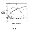
- Figures 1 and 2 show that tumor uptake of Indacea in control mice has an inverse logarithmic relationship with tumor size, whereas liver uptake has a direct logarithmic relationship with tumor size.
- the average tumor uptake of radiolabel with pretreatment was very uniform (23.7 ⁇ 1.04 %ID/g) and independent of tumor size.
- specific antibody pretreatment resulted in an inhibitory effect on tumor uptake of Indacea by tumors smaller than 0.5 g.
- tumors larger than 0.5 g showed significant enhancement of tumor uptake with MAB pretreatment (p ⁇ 0.0005).
- the following example shows the effects of various MAB pretreatment doses on biodistribution.
- Table II shows the effect of changing the dose of T84.66 pretreatment MAB on the tissue biodistribution of 2.0 ⁇ g of (10 ⁇ Ci) T84.66 Indacea in a series of 10-fold dilutions from 2.0 mg to 0.002 mg in mice bearing sc tumors.
- mice were injected sc with 1 x 10⁶ LS174T cells on day 0, and on day 7 received either 2.0, 0.2, 0.02, 0.002, or 0 mg of T84.66 intraperitoneally (ip) over a period of 3 days.
- 1.0 mg MAB was administered on the first day and 0.5 mg was administered on the next 2 days.
- each animal received 2 ⁇ g (10 ⁇ Ci) Indacea (T84.66) ip.
- Nuclear scintiscans were obtained using a Siemens Pho-Gamma V camera 48 hours later.
- the animals were euthanized and tissue biodistributions were performed 48 hours after Indacea injection and the radiolabel uptake expressed as mean percentage injected dose per gram (%ID/g) for each tissue.
- the Table II results are the mean for each experiment ⁇ standard error (SE).
- Mean tumor weight in grams g ⁇ SE is shown as well as the number of mice (n).
- tumors weighing 0.5-1.5 g were selected for these studies.
- Nude mice were inoculated sc with 1 x 10⁶ LS174T tumor cells (day 0) and divided into three groups. Each group was treated ip with 0.2 mg of unlabeled T84.66 given in one dose on day 7, two doses on days 7 and 8 (0.1 mg/day) or three doses on day 7, 8 and 9 (0.1 mg on day 7, 0.05 mg on days 8 and 9). In all cases, the total cumulative pretreatment dose was 0.2 mg T84.66. In each case, 2 ⁇ g (10 ⁇ Ci) Indacea (T84.66) was injected ip on day 14 and tissue biodistributions were performed 48 hours later as described above. Average tumor size was 0.56 ⁇ 0.03 g. PBS controls were matched accordingly.
- the uptake of labeled MAB by the liver in all three pretreated groups was significantly lower than the liver uptake in the PBS control group (p ⁇ 0.0005).
- the blood level of circulating Indacea was significantly increased in all three treated groups over that of the control (p ⁇ 0.0005).
- the tumor uptake with the unlabeled pretreatment given as three fractions was also significantly lower than in the PBS control (p ⁇ 0.05).
- T84.66 0.2 mg was administered ip in one dose between 20 minutes (m) and 7 days (d) prior to Indacea (T84.66).
- a PBS control group received no pretreatment.
- Indacea was injected ip (2 ⁇ g, 10 ⁇ Ci) on day 14.
- Tissue biodistributions were performed 48 hours after Indacea injection and expressed as %ID/g ⁇ SE. Results are in Table IV.
- the following example compares the effects of the route of administration of the pretreatment antibody on biodistribution.
- Nude mice were inoculated sc with 1 x 10⁶ LS174T cells on day 0 and divided into four treatment groups. Two of the groups received single dose injections of T84.66 MAB (0.2 mg) 7 days after administration of tumor cells and thereafter received by Indacea (T84.66) 7 days later. One of these groups received the antibody intravenously (iv) (group A) and the other intraperitoneally (ip) (group C). The other two groups received 0.2 mg T84.66 MAB simultaneously with Indacea on day 14. One of these groups was injected iv (group B) and the other ip (group D). All the animals were euthanized and tissue uptake of the label determined at 48 hours (day 16) after Indacea injection.
- the specificity of the antibody used for pretreatment was determined in the following example.
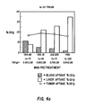
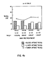
- mice received 1x10⁶ LS174T tumor cells on day 0 and were divided into 4 groups. Each group received 0.2 mg of either T84.66, T84.12, ZCE025 or PBS ip on day 7. On day 14, the mice were given 2 ⁇ g (10 ⁇ Ci) of either T84.66 Indacea (Fig. 4a), ZCE025 Indacea (Fig 4b), or T84.12 Indacea (Fig. 4c). Imaging and tissue biodistributions were done at 48 hours (day 16). All MAB injections were ip.
- pretreatment with ZCE025 and T84.12 produced 25.6% and 36.4% decrease in liver uptake of Indacea respectively, relative to the PBS control.
- pretreatment with T84.66 lowered the level of Indacea in the liver by 76.8%. Concomitant with the decrease in liver radioactivity, was an increased level of radiolabel in the blood.
- the T/L ratio showed a significant increase from 0.46 ⁇ .06 in the nontreated mice to 2.16 ⁇ 0.13 in the mice pretreated with T84.66 MAB (p ⁇ .0005).
- the L/B ratio was lowered from 23.18 ⁇ 3.42 to 0.72 ⁇ .06, with T84.66 pretreatment (p ⁇ 0.0005).
- the tumor uptake (%ID/g) was not affected by the pretreatments.
- T84.66 Due to the differences in liver uptake of the three radiolabeled MABs seen in the untreated control animals in Example 5, the antibody affinity of these three antibodies was measured using a solid phase EIA as described by Beatty, J.D., et al., J. Immunol. Methods 100 :173-179 (1987).
- the affinity constants of T84.66, ZCE025 and T84.12 so measured are 4.67 x 10 ⁇ 10 (L/M), 6.80 x 10 ⁇ 9 (L/M) and 5.65 x 10 ⁇ 9 (L/M), respectively.
- T84.66 showed the highest affinity for CEA and was also the only MAB to successivefully lower the liver uptake for all three MABs tested.
- 111In-labeled T84.66 exhibits the highest liver uptake of the label in the absence of pretreatment.
- MAB affinity for the marker substance to be targeted or treated is to be considered when designing a treatment protocol.
- the above data suggests that a relatively large dose of MAB with a high affinity for a particular marker substance used for pretreatment followed by a labeled lower affinity MAB direction against a different epitope on the marker substance may yield the highest T/L ratios.
- timing of pretreatment in cases where labeled and unlabeled antibody are not simultaneously administered is not limited by the above examples. Preferably timing of pretreatment is from about three weeks to about 20 minutes prior to the administration of a labeled antibody.
- the antibodies used in the method of this invention may be specific for intracellular, cytoplasmic, or cell surface marker substances. Fragments of antibodies may also be used. These antibodies are preferably monoclonal of mammalian origin and may be synthesized by genetic engineering technology well known in the art, or they may be synthesized in vitro or in vivo utilizing genetic engineering technology. Of particular value are chimeric antibodies comprising murine variable region and human constant region or any other type specific for a given marker substance antibody.
- Marker substances may be produced by lesions, or they may simply accumulate in, on, or near lesions.
- Substances suitable as markers for which antibodies used in accordance with this invention may be specific generally include, but are not limited to oncofetal antigens, tumor antigens, receptors, hormones and enzymes.
- marker substances are human chorionic gonadotropin (HCG) and/or its beta subunit, alpha-fetoprotein (AFP), Ca 19-9, CA125, 132CS, tissue polypeptide antigen, colon-specific antigen-p (CSA p ), A 431, TA-4, prostatic acid phosphatase, pancreatic oncofetal antigen, T101, S 100, parathormone, B1.1, TAG72, placental alkaline phosphatase, calcitonin, mammary tumor-associated glycoproteins, galactosyl transferase-II (gt-II), glial fibrillary acidic protein (GFA), tumor angiogenesis factor (TAF), prostatic acid phosphatase, pregnancy beta1-globulin, beta2-microglobulin ovarian cystadenocarcinoma-associated antigen (OCAA), ovarian tumor-specific antigen, common glioma antigen (CGA), cervical cancer antigens, glioe
- Substances which may be used as detectable labels for the antibodies used in the methods of this invention include various isotopes of the transition metals. In general, any transition metal isotope that gives off alpha particles, beta particles, gamma rays, or any other emission that could be used for detection or therapy may be used.
- Magnetic contrast agents such as gadolinium, manganese, and iron, which do not emit particles or rays, possess a property which is identifiable by the agents' ability to resonate at a certain frequency due to magnetic resonance, and as such may also be used as detectable labels
- Substances which may be used as therapeutic labels for the antibodies used in the methods of this invention include various isotopes of the transition metals and chemotherapeutic drugs. These include, but are not limited to yttrium-90, methotrexate, copper-67, indium-194, cobalt, carbon-14, daunomycin, vincristine, 5-fluorouracil, BCNU, nitrogen mustard, vinblasatin, cyclophosphamide, actinomycin D, 6-mercaptopurine, arabinosyl, cytosine, L-asparaginase, adriamycin, cytoxin, busulfan, chlorambucil, thiotepa, melphalan, thioguanine, hydroxyurea, procarbazine, o,p-D,D, FUDR, mithramycin, bleomycin, DTIC, CCNU, and cis-platinum.
- yttrium-90 methotrex
- a radioactive or otherwise detectable level may also be attached to or otherwise associated with the subsequently administered antibody.
- a human subject is first exposed to a given marker-specific antibody. Subsequently, the same antibody, now labeled with a detectable label and boron-10, is administered to the subject. The lesion is located by detecting the site or sites of labeled subsequently administered antibody accumulation. The lesion with which boron-10-labeled antibody is now associated is later irradiated with a well collimated beam of thermal neutrons. Such irradiation gives rise to cytotoxic particles which have a therapeutic effect.
- the prior exposure of the human subject to an antibody the same as that which is subsequently administered diminishes uptake of said subsequently administered antibody by the liver and spleen, and thus allows more of the antibody to become associated with marker substance.
- marker substance is itself associated with the lesion
- antibody accumulation in, on, or near the lesion is increased. In this way, the lesion is not precisely located.
- the therapeutic agent's cytotoxic effects are also directed more precisely to the lesion and undesired cytotoxicity is minimized.
- antibody fragments specific for lesion-associated marker substances may be used in accordance with the present invention.
- the human subject may be initially exposed to antibody fragments specific for the marker substance.
- the same broad antibody fragment in labeled form may then be administered to the subject.
- the prior exposure to the antibody fragment decreases the uptake of the subsequently administered antibody fragment by the subject's liver and spleen.
- Such fragments may be associated with detectable and/or therapeutically effective labels as previously described.
Applications Claiming Priority (2)
| Application Number | Priority Date | Filing Date | Title |
|---|---|---|---|
| US07/180,447 US4921690A (en) | 1986-12-29 | 1988-04-12 | Method of enhancing the biodistribution of antibody for localization in lesions |
| US180447 | 1994-01-12 |
Publications (2)
| Publication Number | Publication Date |
|---|---|
| EP0337746A2 true EP0337746A2 (fr) | 1989-10-18 |
| EP0337746A3 EP0337746A3 (fr) | 1991-10-09 |
Family
ID=22660499
Family Applications (1)
| Application Number | Title | Priority Date | Filing Date |
|---|---|---|---|
| EP19890303591 Withdrawn EP0337746A3 (fr) | 1988-04-12 | 1989-04-12 | Composition pour augmenter la biodistribution d'anticorps pour localisation dans des lésions |
Country Status (2)
| Country | Link |
|---|---|
| US (1) | US4921690A (fr) |
| EP (1) | EP0337746A3 (fr) |
Cited By (2)
| Publication number | Priority date | Publication date | Assignee | Title |
|---|---|---|---|---|
| WO1993025237A1 (fr) * | 1992-06-15 | 1993-12-23 | City Of Hope | Anticorps chimerique anti antigene carcino-embryonnaire |
| US6869606B1 (en) * | 1999-02-22 | 2005-03-22 | Millennium Pharmaceuticals, Inc. | Biotinylated-chemokine antibody complexes |
Families Citing this family (11)
| Publication number | Priority date | Publication date | Assignee | Title |
|---|---|---|---|---|
| USRE38008E1 (en) | 1986-10-09 | 2003-02-25 | Neorx Corporation | Methods for improved targeting of antibody, antibody fragments, hormones and other targeting agents, and conjugates thereof |
| US5034223A (en) * | 1986-10-09 | 1991-07-23 | Neorx Corporation | Methods for improved targeting of antibody, antibody fragments, hormones and other targeting agents, and conjugates thereof |
| US5595721A (en) * | 1993-09-16 | 1997-01-21 | Coulter Pharmaceutical, Inc. | Radioimmunotherapy of lymphoma using anti-CD20 |
| MY133346A (en) * | 1999-03-01 | 2007-11-30 | Biogen Inc | Kit for radiolabeling ligands with yttrium-90 |
| US20020102208A1 (en) * | 1999-03-01 | 2002-08-01 | Paul Chinn | Radiolabeling kit and binding assay |
| WO2005077065A2 (fr) * | 2004-02-09 | 2005-08-25 | The Regents Of The University Of California | Ligands polyvalents selectifs a haute affinite et methodes de fabrication |
| US20100119457A1 (en) * | 2007-03-30 | 2010-05-13 | Helmut Lenz | Composition of labeled and non-labeled monoclonal antibodies |
| WO2008148546A2 (fr) * | 2007-06-06 | 2008-12-11 | F.Hoffmann-La Roche Ag | Composition d'un premier anticorps monoclonal non marqué se liant à un antigène tumoral et d'un second anticorps monoclonal réactif non croisé marqué par un marqueur à fluorescence nir |
| ES2613844T3 (es) | 2008-04-21 | 2017-05-26 | Lawrence Livermore National Security, Llc | Ligandos polidentados de alta afinidad selectivos y métodos para producirlos |
| WO2019084273A1 (fr) * | 2017-10-25 | 2019-05-02 | Actinium Pharmaceuticals, Inc. | Procédés de lymphodéplétion à base d'anti-cd45 et utilisations associées en combinaison avec des thérapies anticancéreuses à base d'act |
| CA3128518A1 (fr) * | 2019-02-01 | 2020-08-06 | Actinium Pharmaceuticals, Inc. | Molecules et leurs derives diriges contre cd45 |
Citations (4)
| Publication number | Priority date | Publication date | Assignee | Title |
|---|---|---|---|---|
| WO1986007088A1 (fr) * | 1985-05-24 | 1986-12-04 | Northwestern University | Hybridome lym-1 de souris et anticorps de diagnostic produit par celui-ci |
| EP0222617A2 (fr) * | 1985-11-13 | 1987-05-20 | Ortho Pharmaceutical Corporation | Thérapie par anticorps monoclonal |
| EP0273754A2 (fr) * | 1986-12-29 | 1988-07-06 | City Of Hope National Medical Center | Méthode pour augmenter la biodistribution d'anticorps pour la localisation dans des lésions |
| WO1988005309A1 (fr) * | 1987-01-27 | 1988-07-28 | Xoma Corporation | Augmentation de la puissance de conjugues cytotoxiques |
Family Cites Families (3)
| Publication number | Priority date | Publication date | Assignee | Title |
|---|---|---|---|---|
| US4565687A (en) * | 1981-11-16 | 1986-01-21 | The Reagents Of The University Of Michigan | Monoclonal antibodies specific for the unbound β subunit of human chorionic gonadotropin |
| US4624846A (en) * | 1983-07-29 | 1986-11-25 | Immunomedics, Inc. | Method for enhancing target specificity of antibody localization and clearance of non-target diagnostic and therapeutic principles |
| US4665897A (en) * | 1984-05-25 | 1987-05-19 | Lemelson Jerome H | Composition and method for detecting and treating cancer |
-
1988
- 1988-04-12 US US07/180,447 patent/US4921690A/en not_active Expired - Lifetime
-
1989
- 1989-04-12 EP EP19890303591 patent/EP0337746A3/fr not_active Withdrawn
Patent Citations (4)
| Publication number | Priority date | Publication date | Assignee | Title |
|---|---|---|---|---|
| WO1986007088A1 (fr) * | 1985-05-24 | 1986-12-04 | Northwestern University | Hybridome lym-1 de souris et anticorps de diagnostic produit par celui-ci |
| EP0222617A2 (fr) * | 1985-11-13 | 1987-05-20 | Ortho Pharmaceutical Corporation | Thérapie par anticorps monoclonal |
| EP0273754A2 (fr) * | 1986-12-29 | 1988-07-06 | City Of Hope National Medical Center | Méthode pour augmenter la biodistribution d'anticorps pour la localisation dans des lésions |
| WO1988005309A1 (fr) * | 1987-01-27 | 1988-07-28 | Xoma Corporation | Augmentation de la puissance de conjugues cytotoxiques |
Non-Patent Citations (1)
| Title |
|---|
| FILE SERVER STN, FILE BIOSIS, accession no. 88:484823, Biological Abstracts, abstract no. 86:116133; M.G. ROSENBLUM et al.: "Interferon-induced changes in pharmacokinetics and tumor uptake of indium-111-labeled antimelanoma antibody 96.5 in melanoma patients"; & J. NATL. CANCER INST. (BETHESDA) 80(3), 6th April 1988, 160-165 * |
Cited By (2)
| Publication number | Priority date | Publication date | Assignee | Title |
|---|---|---|---|---|
| WO1993025237A1 (fr) * | 1992-06-15 | 1993-12-23 | City Of Hope | Anticorps chimerique anti antigene carcino-embryonnaire |
| US6869606B1 (en) * | 1999-02-22 | 2005-03-22 | Millennium Pharmaceuticals, Inc. | Biotinylated-chemokine antibody complexes |
Also Published As
| Publication number | Publication date |
|---|---|
| US4921690A (en) | 1990-05-01 |
| EP0337746A3 (fr) | 1991-10-09 |
Similar Documents
| Publication | Publication Date | Title |
|---|---|---|
| US5194594A (en) | Modified antibodies | |
| US5990286A (en) | Antibodies with reduced net positive charge | |
| JP2519305B2 (ja) | クリアランスを向上させた修飾抗体 | |
| Dykes et al. | Radioimmunotherapy of cancer: clinical studies and limiting factors | |
| US4921690A (en) | Method of enhancing the biodistribution of antibody for localization in lesions | |
| Hird et al. | Tumour localisation with a radioactively labelled reshaped human monoclonal antibody | |
| Vaickus et al. | Biotechnology Update: Overview of Monoclonal Antibodies in the Diagnosis and Therapy of Cancer | |
| JP2012131808A (ja) | 減少した正味の正電荷を有する抗体 | |
| Govindan et al. | Preclinical therapy of breast cancer with a radioiodinated humanized anti-EGP-1 monoclonal antibody: advantage of a residualizing iodine radiolabel | |
| Murray | Radioimmunotherapy of colorectal cancer | |
| WO1997025069A9 (fr) | Anticorps presentant une charge positive nette reduite | |
| Gerretsen et al. | Radioimmunotherapy of human head and neck squamous cell carcinoma xenografts with 131I-labelled monoclonal antibody E48 IgG | |
| DeNardo et al. | Strategies for enhancement of radioimmunotherapy | |
| Molthoff et al. | Influence of dose and schedule on the therapeutic efficacy of 131I‐labelled monoclonal antibody 139H2 in a human ovarian cancer xenograft model | |
| EP0273754A2 (fr) | Méthode pour augmenter la biodistribution d'anticorps pour la localisation dans des lésions | |
| Chiou | Biodistribution and radioimmunoscintigraphy studies of renal cell carcinoma using tumor-preferential monoclonal antibodies and F (ab’) 2 fragments | |
| Saga et al. | Radioimmunotherapy of human glioma xenografts in nude mice by indium-111 labelled internalising monoclonal antibody | |
| Ghosea et al. | Targeting of radionuclides and drugs for the diagnosis and treatment of cancer | |
| Macmillan et al. | Immunoscintigraphy of small-cell lung cancer: a study using technetium and indium labelled anti-carcinoembryonic antigen monoclonal antibody preparations | |
| Desrues et al. | Biodistribution of monoclonal antibody Po66 in a human lung tumour-bearing mouse model: effect of blood exchange on tumour antibody uptake | |
| Bradwell et al. | Antibody targeting: Theoretical considerations | |
| Keenan et al. | The Potential Value of Radiolabelled Monoclonal Antibodies in Cancer Therapy | |
| Goodwin | Antibody-Hapten Complexes for Imaging | |
| Endo et al. | Tumor imaging by monoclonal antibodies labeled with radioactive metal ions | |
| Norrlund et al. | Combinations of nonlabeled, 125I-labeled, and anti-idiotypic antiplacental alkaline phosphatase monoclonal antibodies at experimental radioimmunotargeting |
Legal Events
| Date | Code | Title | Description |
|---|---|---|---|
| PUAI | Public reference made under article 153(3) epc to a published international application that has entered the european phase |
Free format text: ORIGINAL CODE: 0009012 |
|
| AK | Designated contracting states |
Kind code of ref document: A2 Designated state(s): CH DE FR GB IT LI |
|
| PUAL | Search report despatched |
Free format text: ORIGINAL CODE: 0009013 |
|
| AK | Designated contracting states |
Kind code of ref document: A3 Designated state(s): CH DE FR GB IT LI |
|
| RHK1 | Main classification (correction) |
Ipc: A61K 49/02 |
|
| STAA | Information on the status of an ep patent application or granted ep patent |
Free format text: STATUS: THE APPLICATION IS DEEMED TO BE WITHDRAWN |
|
| 18D | Application deemed to be withdrawn |
Effective date: 19920409 |