EP0336562B1 - Improvements relating to antigens - Google Patents
Improvements relating to antigens Download PDFInfo
- Publication number
- EP0336562B1 EP0336562B1 EP89302174A EP89302174A EP0336562B1 EP 0336562 B1 EP0336562 B1 EP 0336562B1 EP 89302174 A EP89302174 A EP 89302174A EP 89302174 A EP89302174 A EP 89302174A EP 0336562 B1 EP0336562 B1 EP 0336562B1
- Authority
- EP
- European Patent Office
- Prior art keywords
- antigen
- antibody
- cells
- kda
- ovarian
- Prior art date
- Legal status (The legal status is an assumption and is not a legal conclusion. Google has not performed a legal analysis and makes no representation as to the accuracy of the status listed.)
- Expired - Lifetime
Links
Images

Classifications
-
- C—CHEMISTRY; METALLURGY
- C07—ORGANIC CHEMISTRY
- C07K—PEPTIDES
- C07K16/00—Immunoglobulins [IG], e.g. monoclonal or polyclonal antibodies
- C07K16/18—Immunoglobulins [IG], e.g. monoclonal or polyclonal antibodies against material from animals or humans
- C07K16/28—Immunoglobulins [IG], e.g. monoclonal or polyclonal antibodies against material from animals or humans against receptors, cell surface antigens or cell surface determinants
-
- C—CHEMISTRY; METALLURGY
- C07—ORGANIC CHEMISTRY
- C07K—PEPTIDES
- C07K14/00—Peptides having more than 20 amino acids; Gastrins; Somatostatins; Melanotropins; Derivatives thereof
- C07K14/435—Peptides having more than 20 amino acids; Gastrins; Somatostatins; Melanotropins; Derivatives thereof from animals; from humans
- C07K14/705—Receptors; Cell surface antigens; Cell surface determinants
-
- C—CHEMISTRY; METALLURGY
- C07—ORGANIC CHEMISTRY
- C07K—PEPTIDES
- C07K16/00—Immunoglobulins [IG], e.g. monoclonal or polyclonal antibodies
- C07K16/18—Immunoglobulins [IG], e.g. monoclonal or polyclonal antibodies against material from animals or humans
- C07K16/28—Immunoglobulins [IG], e.g. monoclonal or polyclonal antibodies against material from animals or humans against receptors, cell surface antigens or cell surface determinants
- C07K16/30—Immunoglobulins [IG], e.g. monoclonal or polyclonal antibodies against material from animals or humans against receptors, cell surface antigens or cell surface determinants from tumour cells
-
- A—HUMAN NECESSITIES
- A61—MEDICAL OR VETERINARY SCIENCE; HYGIENE
- A61K—PREPARATIONS FOR MEDICAL, DENTAL OR TOILETRY PURPOSES
- A61K38/00—Medicinal preparations containing peptides
-
- A—HUMAN NECESSITIES
- A61—MEDICAL OR VETERINARY SCIENCE; HYGIENE
- A61K—PREPARATIONS FOR MEDICAL, DENTAL OR TOILETRY PURPOSES
- A61K39/00—Medicinal preparations containing antigens or antibodies
Definitions
- THIS INVENTION relates to a new antigenic peptide, isolatable from human trophoblast cell membranes, antibodies to the antigenic peptide, and in particular to the use of the antigen and antibody in the diagnosis of colon, stomach, ovarian and cervical cancer.
- Trophoblast demonstrates some functional properties of neoplastic tissue, namely invasiveness of host tissue and escape from immunological surveillance.
- Several monoclonal antibodies to trophoblast membrane proteins have been described. In terms of cancer research, the rationale behind this approach has been to identify 'oncofetal' antigens present on both trophoblast and neoplastic cells (Johnson, 1984). If such antigens were restricted to neoplastic tissues, then these reagents would be potentially useful in diagnosis, tumour localisation and drug targeting. Of those monoclonal antibodies that do identify trophoblast oncofetal antigens, relatively few have been fully characterised.
- PLAP placental alkaline phosphatase
- the present invention provides a diagnostic method for colon, stomach, ovarian or cervical cancer involving targeting 5T4 antigen or fragments thereof which comprises contacting a sample of tissue taken from a host suspected of having colon, stomach, ovarian or cervical cancer respectively with an antibody or fragment thereof which recognises 5T4 antigen and determining from the presence or absence of antigen/antibody complexes, the presence or absence of 5T4 antigen or fragments thereof in the sample; wherein 5T4 antigen is a glycoprotein characterised by the following properties
- the present invention also provides a diagnostic method for colon, stomach, ovarian or cervical cancer involving targeting antibody that recognises 5T4 antigen or fragments thereof which comprises contacting a body sample taken from a host with 5T4 antigen and determining from the presence or absence of antibody/antigen complexes, the presence or absence of antibody that recognises 5T4 antigen or fragments thereof in the sample; wherein 5T4 antigen is as defined above.
- the invention further provides the use of 5T4 antigen, or an antibody or fragment thereof which recognises 5T4 antigen for the manufacture of a medicament for the diagnosis of colon, stomach, ovarian or cervical cancer.
- the antibody or fragment thereof recognises the 42 kDa core of 5T4.
- the 5T4 glycoprotein of the invention can be isolated and purified from human trophoblast cells by recovering the syncytiotrophoblast glycoproteins from human placenta, subjecting these glycoproteins to purification by either antibody affinity chromatography or a combination of other chromatographic methods and isolating 5T4 antigen as described in more detail below.
- the glycoprotein can be prepared by synthetic or semi-synthetic techniques, synthetic techniques involving building up the polypeptide core structure by building up the polypeptide chain by conventional peptide synthesis followed by introducing any appropriate glycosylation by chemical or biological methods.
- the 42 KDa polypeptide core can be produced by recombinant DNA techniques, utilising a synthetic or naturally-occurring DNA encoding the 42 KDa polypeptide core.
- DNA will comprise a first DNA sequence encoding the 42 KDa polypeptide core of 5T4 antigen and a second DNA sequence, not normally found in association with the first sequence, but under whose influence, the first sequence can express the 42KDa core in a suitable host cell.
- Suitable techniques include incorporating the selected DNA in a plasmid, transforming a suitable host cell with this plasmid and expressing the DNA in the host cell.
- the glycosylation of the genetically engineered 42 KD apolypeptide core can then follow by appropriate chemical or biological methods.
- Antibodies may be monoclonal or polyclonal antibodies.
- the antibodies may be prepared by conventional techniques.
- Polyclonal antibodies may be obtained by using the 5T4 glycoprotein of the invention or fragments thereof or the 42 KDa core or fragments thereof as immunogen injected into small or large animals from whose blood the polyclonal antibodies are recovered by conventional methods.
- Monoclonal antibodies can be prepared utilising the 5T4 glycoprotein of the invention or fragments thereof or the 42 KDa core or fragments thereof as immunogen in a host animal, immortalising antibody producing cells of the host animal and recovering monoclonal antibody from the immortalised cells.
- the 5T4 glycoprotein of the invention As an alternative to the use of the 5T4 glycoprotein of the invention, or its 42 KDa core or fragments thereof as immunogen in the raising of antibodies, one can also use a natural product including the 5T4 glycoprotein of the invention, isolatable from trophoblast cells.
- This material is known as syncytiotrophoblast glycoproteins, (StMPM), which can be isolated from human placenta by known methods.
- StMPM syncytiotrophoblast glycoproteins
- the 5T4 glycoprotein of the invention can be isolated from the StMPM by either antibody affinity chromatography or a combination of other chromatographic methods.
- One particular monoclonal antibody that we have isolated and tested is one prepared by hybridoma techniques using StMPM wheat germ agglutin (WGA) glycoprotein as immunogen and which has become known as 5T4.
- WGA wheat germ agglutin glycoprotein
- the antigens (5T4 glycoprotein, fragments thereof, the 42 KDa core and fragments thereof) of this invention and antibodies (that recognise antigens of this invention) are useful as diagnostic tools for colon, stomach, ovarian and cervical cancer.
- the purified 5T4 antigen for example allows the production of a family of related antibodies which recognise different epitopes of 5T4 antigen.
- tumour screening and diagnosis in vitro and/or in vivo may offer significant advantage over antibodies to PLAP since the antigen is not found in pregnancy serum;
- compositions comprising the antigen or antibody of the invention together with a carrier or diluent.
- a carrier or diluent The exact nature of the carrier or diluent will depend upon the ultimate application of the antigen or antibody.
- the carrier may be liquid or solid and solid carriers for the antibody also represent a particularly important aspect of the present invention where the antibody is to be used as a means of purifying the naturally-occurring antigen by techniques of affinity chromatography.
- the antigens and antibodies, immobilised or not, may be linked with radioisotopes or other revealing labels for localisation and/or therapy or conjugated with anti-tumour reagents for therapy.
- the antigen and antibody can be derivatised for use in different forms of assay for antigen concentration.
- the present invention includes a diagnostic test kit containing, as a solid component, an immobilised antigen or antibody of the invention and more specifically can contain, depending upon the specific type of assay to be used, an antigen and an antibody of the invention, one of which bears a revealing label.
- the antigen of the invention can be used in methods of in vitro or in vivo diagnosis targeting antibody while the antibody of the invention may be similarly used to target antigen. Such methods are of particular use in the diagnosis of various types of cancer, particularly for mass screening of cervical smears, and for screening or diagnosing stomach, colon or ovarian cancer.
- 5T4 antigen has a relatively limited tissue distribution. It appears to be a pan-trophoblast marker which is expressed by all types of trophoblast examined as early as 9 weeks of development. It is specific for this tissue type within the placenta except for the amniotic epithelium which is also antigen positive. On the basis of immunoperoxidase staining of frozen sections from normal tissue, 5T4 antigen is also expressed by certain epithelial cell types. It should be noted that several 'trophoblast-characteristic' antigens, such as PLAP, are in fact found in normal tissues at trace concentrations (McLaughlin, 1986).
- 5T4 antigen was found in placental plasma membrane in at least a 1000-fold higher concentration than that found in other normal tissues tested. However, this level of sensitivity would not necessarily detect expression in minor subpopulations of cells within a given tissue.
- tumour cell lines of such apparent diversity of tissue origin are not clear; the normal cell line types tested are all of embryonic origin.
- the lack of reactivity with tumour cell lines derived from lung, bronchus and lymphoid tissue is consistent with the immunohistology of the normal tissue types.
- Other antigen positive tumour cell lines may have been derived from an epithelial component of normal tissue or represent reexpression of embryonic antigen on tumour cells.
- trophoblast antigens have been reported to exhibit a pattern of expression by tumour cell types apparently not detected in the normal cell counterpart (McLaughlin et al., 1982).
- Rettig et al. (1985) a series of six monoclonal antibodies were generated against choriocarcinoma cells, one of which was reactive with neoplastic, but not normal, kidney cells; the other mAbs did not demonstrate such a selective expression.
- 5T4 antigen does however appear to be novel. On the basis of reactivity in dot-blots and other criteria, we have specifically excluded PLAP and transferrin as the 5T4 antigen.
- the 5T4 antigen is carried by glycoprotein molecules of 72kD on syncytiotrophoblast microvillous plasma membranes but appears on molecules of similar molecular weight from several different cell lines including some choriocarcinomas.
- the molecules are sialylated and have approximately 30kD of the apparent molecular weight due to N-linked carbohydrate structures as judged from removal of the latter by N-glycanase endoglycosidase.
- 5T4 appears to exist on the cell surface as a monomeric protein. Firstly, 5T4 antigen elutes with an apparent molecular weight in gel filtration of 120kD, an increase consistent with the addition of a detergent shell, and inferring that 5T4 is not associated non-covalently with any other large molecules. Additionally, reduction with 2-mercaptoethanol does not substantially alter the apparent molecular weight of the 5T4 radio immunoprecipitate, as would be the case if it were disulphide bonded to another protein.
- the pattern of expression of 5T4 is similar to that of the family of mucin type glycoproteins (Swallow et al., 1987), but with clear differences from those defined by the CA or HMFG series of antigens (Wiseman et al., 1984; Burchell et al., 1983). These latter glycoproteins are defined by several monoclonal antibodies which have been shown to be reactive with a wide range of malignant tumour cells but also reactive with certain specialized normal epithelia.
- StMPM was purified from full term human placentae, obtained within one hour post partum, by the method of Smith et al. (1974).
- the StMPM pellet was solubilised in 0.5% DOC in tris-buffered saline (TBS, 0.15M NaCI, 25mM tris, pH 8.0) containing 0.1 mM phenylsulphonylmethyl fluoride (PMSF) and centrifuged at 14,000g for 10 minutes.
- the WGA-reactive glycoproteins were then purified by incubation of the supernatant with WGA-Sepharose (5mg ligand/ml Sepharose) for one hour at room temperature.
- the beads were washed extensively in TBS/0.5%DOC, and the specifically bound glycoproteins eluted in 5ml of 0.2M N-acetyl glucosamine (Sigma) in TBS. The eluted fraction was extensively dialysed against 30mM ammonium bicarbonate (pH 7.9), and lyophilised.
- "Sepharose” is a Registered Trade Marke.
- a male BALB/c mouse was immunised by six intra-peritoneal injections of WGA-purified StMPM glycoproteins (100-200tig/injection). Spleen cells were fused with NS1 murine myeloma cells (Kohler and Milstein, 1975), and the cells plated out in 24-well Linbro plates at 7x10 5 cells/well. After two weeks, wells were assayed for StMPM reactive antibody by immunodotting. Positive clones were picked directly and further subcloned by limiting dilution. The antibody subclass was determined by double radial diffusion using a monoclonal isotype typing kit (Serotec, Bicester, U.K.). Antibody 5T4 was obtained by this technique.
- AV-3 cells Near confluent cell cultures of AV-3 cells were radiolabeled for 15-18 hours with 3 H-glucosamine (20 ⁇ Ci/ml) (Amersham International) in RPMI containing 10% dialysed FCS. Metabolically labeled cells were collected and immunoprecipitated as follows : cells were removed from tissue culture flasks by incubation in 0.1 M EGTA-PBS, washed in PBS (Dulbeccos-A) and then solubilized for 30 minutes at 4° C in 0.5% (v/v) NP40 in tris-buffered saline (TBS, 0.15M NaCI, 25mM Tris, pH 8.0) containing 0.1 mM PMSF. Non- solubilized cellular components were removed by centrifugation at 14,000g and the amount of radioactivity incorporated into protein was determined following precipitation with 10% trichloroacetic acid.
- Cell surface labelling by the lactoperoxidase- 125 1 method together with the techniques of immunoprecipitation and SDS-PAGE were carried out as previously described; high molecular weight standards (Sigma), red blood cell membrane proteins or 14 C-methylated protein mixtures (Amersham International) were used as marker proteins (Thompson et al., 1984; Stern et al., 1984; 1986).
- Tritiated sodium borohydride labelling of cell surface glycoproteins was carried out as described by Axelsson et al. (1978)-.Autoradiography and fluorography were as described in Thompson et al. (1984) using pre-flashed Fuji X-ray film.
- Immunoperoxidase staining of frozen tissue sections was carried out by the method of Bulmer and Sunderland (1983). Tissues were obtained as soon as possible post mortem, always within 12 hours, and processed immediately. Indirect immunofluorescence with cell suspensions was as described previously (Thompson et al., 1984). A monoclonal antibody we have isolated, directed against a widely expressed human antigen (mAb 1 D2),was used as positive control.
- EBSS Earle's buffered saline solution
- bovine serum albumin 0.1% sodium azide
- the suspensions were plated out at 50 ⁇ l (10 5 cells)/well in microtitre plates. 50 ⁇ l mAb/well were added and incubated at room temperature for one hour. The cells were washed and 5x10 5 CPM of 125 1-labelled (Fab') 2 fragments of sheep anti-murine immunoglobulin (Amersham International) added.
- Fab' 125 1-labelled
- Tissues were obtained at post mortem held within 12 hours of death, and processed immediately. 10-20g of tissue was finely chopped, rinsed, and homogenised in 10-20ml of ice-cold phosphate buffered saline containing 5mM MgCl 2 and 0.1 mM PMSF with 20 strokes of a Dounce homogeniser. The homogenate was centrifuged at 10,000g for 20 minutes, the pellet discarded and the supernatant centrifuged at 100,000g for 1 hour. This pellet was solubilised in 0.5% (w/v) DOC/TBS containing 0.1 mM PMSF and unsolubilised material pelleted by centrifugation at 14,000g. The protein concentration of the supernatant was determined by the method of Lowry et al.(1951). Membranes from 12-hour old placentae were prepared identically and acted as positive controls.
- StMPM protein was solubilised in 6.5ml of 1.0% (w/v) DOC/TBS containing 0.1 mM PMSF, centrifuged at 100,000g for 30 minutes, and the supernatant fractionated over S200 Sephacryl (Pharmacia).
- Column size was 90x2cm, running buffer was 0.1% (w/v) NaDOC/TBS containing 0.1 mM PMSF. Flow rate was 17ml/hour. Fraction size was 3.3ml. The column was calibrated with the following proteins; Equine ferritin (Sigma), IgG (Kabi), transferrin (Sigma), Bovine serum albumin (Sigma) and ovalbumin (Sigma). Fractions were assayed for 5T4 antigen in ELISA and immunodot. "Sephacryl" is a Registered Trade Mark.
- Elisa plates were activated by one hour incubation with 100 ⁇ l/well of PBS containing 0.25% gluteraldehyde (BDH), the plates washed with PBS, and 100ul/well of undiluted or 10-fold diluted fractions from gel filtration bound to the plates by overnight incubation at 4° C. Following washing, the plates were incubated with 1% BSA/TBS as blocking agent. ELISA was then carried out as described (Johnson et al., 1981). lmmunodotting on nitrocellulose was carried out using the Bio-Rad Dot-Blot apparatus. Fractions from gel filtration were loaded at 10ul and 100ul/dot.
- BDH gluteraldehyde
- NaDOC solubilised plasma membrane protein was loaded in the range of 50 ⁇ g-12.5ng protein/dot.
- the following antigens were loaded at 1 ⁇ g protein/dot; transferrin (Sigma), PLAP (Gift of Dr.P.J.McLaughlin), human placental lactogen (HPL) (Sigma), calmodulin (Sigma), IgG (Miles Ltd.), albumin (Miles Ltd.) and normal human sera.
- transferrin Sigma
- PLAP Gaift of Dr.P.J.McLaughlin
- HPL human placental lactogen
- HPL human placental lactogen
- calmodulin Sigma
- IgG Meats Ltd.
- albumin Methylactin
- normal human sera normal human sera.
- the nitrocellulose sheet was blocked with 3% (w/v) BSA (Sigma) in TBS and processed as described previously (Webb et al.,1985).
- mAb 1 D2
- StMPM membranes (approx. 1 mg protein) were treated overnight at 37° C with either 2mg trypsin (Boehringer), 2mg pronase (Boehringer), 0.1 U neuraminidase (Behringwerke) in 300 ⁇ l of PBS or 10 units/ml N-glycanase (Genzyme) in buffer containing final concentrations as follows: 0.17% SDS; 0.2M tris-HCI, pH 8.7; 10mM 1,10-phenanthroline hydrate (in methanol); 1.25% NP-40 (Plummer et al., 1984). The treated membranes were solubilised in DOC/TBS and 5T4 residual antigenicity assayed by dot-blot.
- 5T4 immunoprecipitates of detergent solubilised 125 1-radiolabelled StMPM were eluted form protein-A-Sepharose with 0.5% SDS in water and incubated overnight at 37 ° C with or without 10 units/ml N-glycanase in buffer as above. Digests were subjected to reduced SDS-PAGE and autoradiography.
- All procedures were carried out at room temperature. All buffers contained 0.1 mM phenyl methyl sulphonyl fluoride (PMSF).
- PMSF phenyl methyl sulphonyl fluoride
- the microvlllous membranes from one placenta (approx. 1 g weight) were solubilised in 100 ml of 1% nonidet P40 (NP-40) in TBS (20 mM Tris, 150 mM sodium chloride. pH 8.0) for 30 minutes on a multimixer. Unsolubilised material was pelleted at 100,000 g for 30 minutes. This was loaded onto a wheat germ agglutinin (WGA)-agarose column (Pharmacia; 2 mg/ml ligand, 5 ml column) at 1 ml/minute using Pharmacia chromatography package A apparatus.
- WGA wheat germ agglutinin
- the column was washed with 50 ml of 1% NP-40/TBS and the specifically bound glycoproteins eluted with two column volumes of 0.3 M N-acetyl-D-glucosamine in the same buffer. This fraction was loaded onto a mAb 5T4-sepharose affinity column (2 mg/ml ligand, 1 ml column).
- the IgG1 5T4 mAb was purified by high salt protein A affinity chromatography (loading buffer 1.5 M glycine, 3 M NaCI, pH 8.9. Elution buffer 100 mM citrate, pH 6.0) and bound to CNBr- activated Sepharose (Pharmacia).
- the mAb 5T4 affinity column was washed with 5 column volumes of 1 % NP40/TBS and 5 column volumes of TBS.
- the bound 5T4 glycoprotein was eluted with 8 M urea. Fractions were assessed by immunodot (Stern et al, 1986), protein assay (Lowry, Rosebrough, Farr and Randall, 1951) and SDS-PAGE.
- the WGA and 5T4 affinity chromatographic steps give a 10,000 fold purification with approximately 70% yield. Minor contaminants visible in silver stained SDS-PAGE are present at least 100-fold lower protein concentration than 5T4 antigen. Further fractionation by either Superose 12 gel filtration ('Superose' is a registered Trade Mark) or hydrophobic interaction reverse phase chromatography yields 5T4 molecules devoid of contaminants.
- the 5T4 antigen isolated by these procedures had the characterising data set out in paragraphs (a) to (i) above.
- the monoclonal antibody 5T4 is a murine IgG1. All work detailed in this study was carried out using subclone 5T4.B8. The preliminary screen by immunodot showed that the antigen recognised was none of the following major proteins associated with the trophoblast; IgG, transferrin, PLAP, HPL, albumin, calmodulin nor was it detectable in serum.
- FIG. 1 illustrates antigen expression in term villous placenta as assessed by immunohistology of frozen sections. Villous trophoblast is strongly labelled by mAb 5T4, whereas the stroma is negative. There is specific labelling of the amniotic epithelium and extravillous cytotrophoblast of the chorion laeve but not of the amniotic mesenchyme or maternal decidua ( Figure 1 c,d).
- mAb 1 D2 labels all parts of villi (fig 1 a)
- mAb H316 labels trophoblast but is not specific for this tissue type (fig 1 b; Stern et al., 1986); negative controls are unlabelled (Figs 1 e, f).
- Extravillous cytotrophoblast in the placental bed is also labelled by mAb 5T4; no other element of the term placenta is 5T4 antigen-positive. Similar analysis of first trimester villous tissue has shown antigen expression by both syncytiotrophoblast and cytotrophoblast (data not shown).
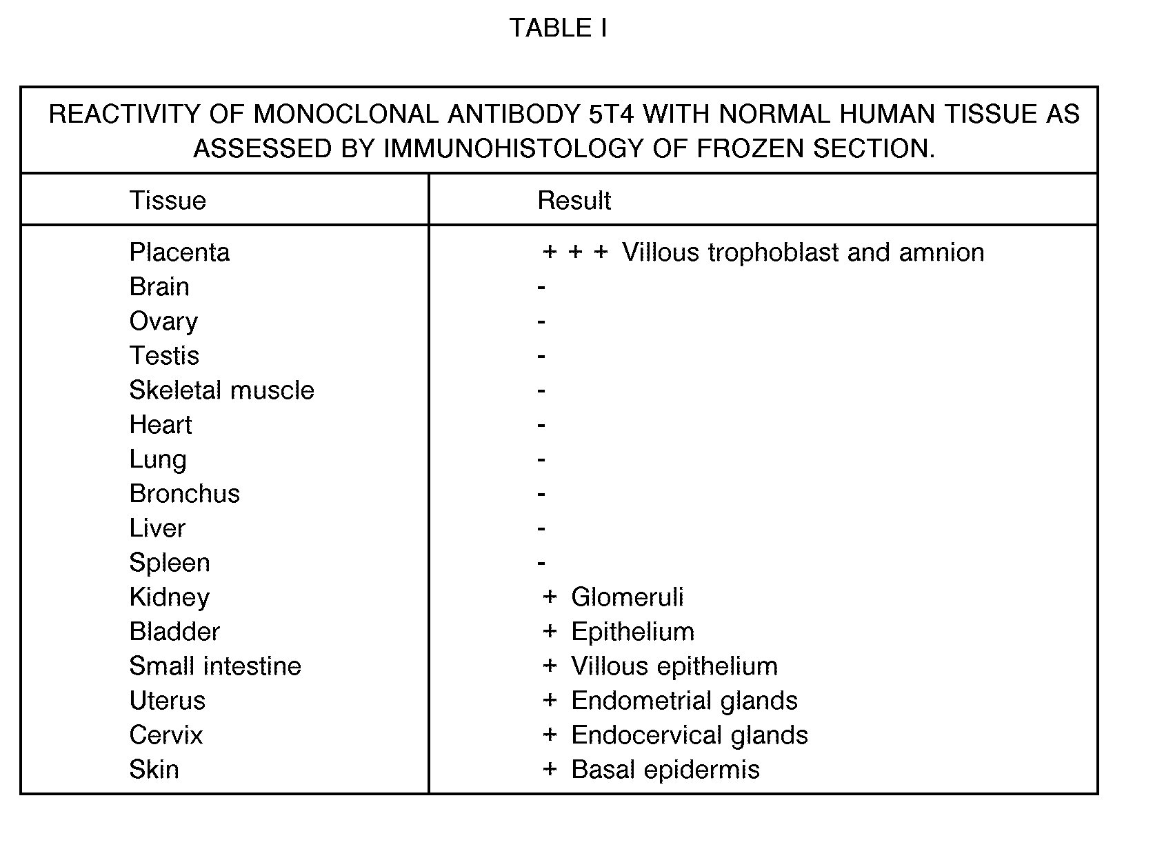
- 5T4 was unreactive with the following non-pregnant tissues examined in immunohistology; spleen, heart, brain, liver, lung, bronchus, skeletal muscle, testis or ovary. Glomeruli in the kidney, villi of the small intestine, bladder epithelium, basal layer of the epidermis, endometrial glands of non-pregnant uterus and endocervical glands showed some specific labelling with mAb 5T4. Some small vessels in various tissues appeared to be weakly stained. Table I summarises 5T4 reactivity assayed by immunohistology of frozen tissue sections.
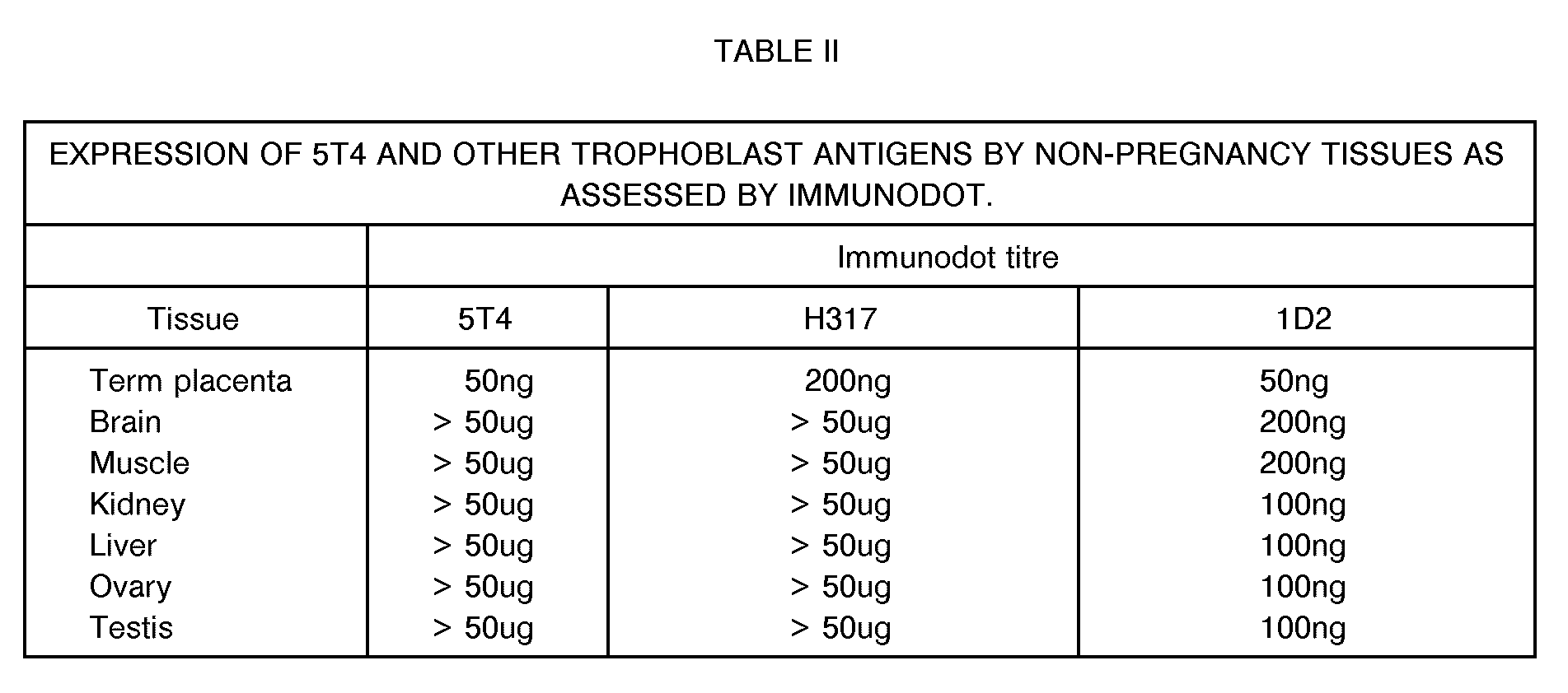
- 5T4 was still reactive with full term placental plasma membrane protein at an antigen concentration of 50ng/dot.
- 5T4 was not specifically reactive with any other tissue tested (ovary, testis, kidney, brain, liver, liver and muscle) at all antigen concentrations used (up to 50tig/dot). From this it was concluded that these normal non-gestational tissues express 5T4 antigen at approximately 1000-fold lower concentration than full-term placenta on a weight of crude membrane protein basis. This relative level of expression is comparable with PLAP as measured using mAb H317 (Table II).
- 5T4 antigen expression by cell lines of normal and neoplastic derivation was assessed by indirect immunofluorescence and a more quantitative radiobinding assay (Table III). By comparison of reactivity with negative control xenogeneic cell lines, radiobinding indices of greater than 1.5 were considered to indicate postive expression of antigen. Trypsinisation was necessary to remove some attached cell lines from the substratum and it was noted where compared that this procedure tended to reduce the binding index compared with EGTA removal (data not shown).
- Normal leukocytes were 5T4 antigen negative and "normal" types represented by cell lines of amnion, embryonic lung fibroblasts and embryonic intestine origin were labelled by 5T4 antibodies.
- Tumour cell lines of myeloid origin were all 5T4 antigen negative; 6/6 tumour cell lines of gestational or developmental origin were positive. 11/15 carcinomas of other histological types and origins were positive, as was one glioma and 1/3 Wilms tumour lines tested.
- 5T4 was unreactive with reduced and unreduced western blots of StMPM.
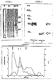
- the molecular species bearing the 5T4 antigen was identified as a 72kD protein by reduced SDS-PAGE analysis of immunoprecipitates from 125 1 lactoperoxidase labelled StMPM ( Figure 3, lane 1).
- the molecules migrate with a molecular weight of 69kd in unreduced SDS-PAGE. It was observed that the relative mobility in SDS-PAGE varies anomalously with the percentage of the acrylamide. This is sometimes indicative of a glycoprotein, which is confirmed by the change in molecular weight following removal of N-linked sugars by digestion with N-glycanase, yielding a molecule of 42kd ( Figure 3, lane 2).
- 5T4 glycoprotein can be labelled by reduction with tritiated sodium borohydride either after periodate oxidation of sugar residues or galactose oxidase/neuraminidase treatment. These latter treatments change the relative mobility in SDS-PAGE as compared with 125 1 labelled 5T4 antigen ( Figure 3).
- AV-3, Tera-2, MRC-5, Hep-2, HN5, HT29 cell lines all express a molecule of similar molecular weight to that on StMPM as judged by SDS-PAGE of immunoprecipitates or surface iodinated cells; the antigen has been immunoprecipitated from AV-3 cells metabolically labelled with tritiated glucosamine (data not shown).
- Isolated StMPM membranes were digested with trypsin, pronase, neuraminidase or N-glycanase, the components solubilised and subjected to immuno-dot assay. Both proteases and N-glycanase destroyed 5T4 antigenicity, whilst neuraminidase did not (Table IV).
- the effects of various fixatives on 5T4 antigenicity as expressed by Tera-2 cells was assessed by solid-phase radiobinding assay. Neither Bouins' fixative, buffered formalin, gluteraldehyde or absolute ethanol were found to significantly affect 5T4 binding index relative to PBS control (data not shown).
- IVS intervillous space
- St syncytiotrophoblast
- VS villous stroma
- AE amniotic epithelium
- AM amniotic mesenchyme
- CL chorion laeve
- DP decidua parietalis.
- 5T4 shows specific labelling of villous trophoblast and extravillous cytotrophoblast of the chorion laeve as well as amniotic epithelium.
- Positive control mAb 1 D2 labels all cell types;
- mAb H316 labels trophoblast of the chorion laeve and amniotic epithelium.
- Normal mouse serum shows no labelling.
- StMPM protein incubated overnight at 37 ° C with appropriate enzymes or PBS (as control for auto- degradation) and dot-blotted onto nitrocellulose. Results expressed as minimum protein dot concentration required to produce a positive result.
- TBS peroxidase was visualised using a freshly prepared and filtered solution of diaminobenzidine tetrahydrochloride (DAB-Sigma) in TBS containing 0.03% hydrogen peroxidase. (6 minutes). Sections were washed in tap water and counter stained in Coles haematoxylin, dehydrated, cleared and mounted (Ralmount-R.A. Lamb). The immunohistochemical results were interpreted with reference to a set of controls run in parallel with each test. These included sections treated with DAB only to show endogenous peroxidase, omission of the primary antibody and replacement of the primary antibody with one of the same class but of unrelated specificity.
- DAB-Sigma diaminobenzidine tetrahydrochloride
- the distribution of positive reactions with MAb 5T4 in normal and non neoplastic tissues is summarised in Table V.
- the villous syncytiotrophoblast from first and third trimester placentae and an ectopic (tubal) pregnancy showed strong membrane positivity.
- Placental site trophoblast displayed both membrane and cytoplasmic reactions.
- the stroma of chorionic villi and foetal blood vessels were negative.
- Table VI summarises the distribution of MAb 5T4 in neoplastic tissues. Many of the malignant epithelial tumours displayed positive reactions in the neoplastic cells. Of note, were carcinomas of breast (5/5), lung (5/5), stomach (6/7) and pancreas including one of the ampulla of Vater (4/4). Also positive, albeit in only a limited number of cases available, were carcinomas of endometrium and cervix.
- Cystadenocarcinomas of the ovary produced variable reactions. In three of the four positive cases the majority of tumour cells were positive, and in the other the majority were negative.
- the cystic epithelium of mature teratomas often displayed a focal weak to moderate reaction.
- Syncytiotrophoblast of choriocarcinomas and a complete hydatidiform mole was strongly positive. Much of the trophoblast of placental site tumours showed moderate or strong labelling on both cell membranes and within the cytoplasm. Single examples of fibrosarcoma and leiomyosarcoma showed that whilst most tumour cells were negative, there were focal and weak reactions in a few cells. Malignant melanomas (2) and malignant lymphomas (3) were negative.
- the stroma of some tumours showed weak and focal reactions. This was also noted in the endothelium lining mainly small blood vessels in many tissues and tumours.
- the cellular location of binding with mAb5T4 in tumours may be either membranous or cytoplasmic or a combination of both.
- Heavy membrane-bound location is a particular feature of syncytiotrophoblast. Cytoplasmic reactivity was predominant in pancreatic carcinomas. In gastric and breast carcinomas both types of pattern were present.
- MAb 5T4 was unreactive with fixed and paraffin wax embedded tissue sections of villous trophoblast of term placentae.
- MAb 5T4 gives reactions in trophoblast which are similar to other antitrophoblast antibodies.
- our detailed immunohistochemical analysis tends to suggest that the antigen recognised is distinct from HCG,HPL,PLAP and those which react with mAb 18A/C4 and 18B/A5 (Loke, University of Cambridge).
- 5T4 will react with some non-HCG producing tumours and gives intense reactions with syncytiotrophoblast of term placenta.
- Antibodies to both HCG and HPL are unreactive with the basal layer of stratified squamous epithelium and normal or non-neoplastic endocervical glands.
- mAb 5T4 The failure of mAb 5T4 to react in fixed and routinely processed paraffin sections is a characteristic of some other reported antibodies directed against membrane-associated antigens, notably anti-PLAP, 18A/C4 and 17.1A antibodies.
- Cervical carcinoma arises from dysplastic precursor lesions in the reserve cells in the basal layer of the stratified metaplastic epithelium. These areas develop from proliferating basal cells which have undergone some form of transformation, and gradually spread throughout the whole epithelium. Cervical carcinomas thus develop from a series of atypical changes which progress in continuum to a stage of carcinoma in situ. This is probably the final premalignant state before the lesion invades the underlying stroma becoming microinvasive.
- the dysplastic variations have been categorised as a series of changes in cervical intraepithelia neoplasia (CIN). They have been graded from 1-3, with CIN 1 representing less than a third of the dysplastic involvement, located in the basal layer. CIN 2 with a third to two thirds involvement, and CIN 3 two thirds to full thickness involvement, equivalent to carcinoma in situ. At any stage, the lesion may regress back to normality. 90% of cervical neoplasms are squamous cell carcinomas and have a contrasting aetiology and of epidemiology to adenocarcinomas (present in nulliparous women) which comprise the remaining 8-10%.
- Placental tissue was washed in PBS (phosphate buffered saline). 1 cm 3 of tissue was then embedded in OCT compound and snap-frozen by immersing the specimen in C0 2 ice with isopentane, within 1 hour post partum.
- Frozen cervical specimens were selected from a store at the Royal Liverpool Women's Hospital (RLWH). These samples, from cone or punch biopsies routinely submitted for histology, were embedded in polycel, snap-frozen and stored at -70 °C. The specimens were cut at 7 ⁇ rn thick using a cryostat and placed on slides (cleaned with ethanol and coated with poly-L-lysine). The pathological assessment was obtained from the records of examination of the specimens subjected to fixation in formal buffered saline, dehydration wax embedding and haemotoxin/eosin staining.
- the first layer test mAb was applied (ascites fluid diluted 1/100 in 1% BSA/PBS) and incubated for 1 hour at room temperature in a moist box. Subsequently, each slide was washed individually x3 in 1% BSA/PBS before having the second layer peroxidase conjugated rabbit anti-mouse (R anti-Mlg) (Dako) diluted 1:50 in 1% BSA/PBS +10% normal human serum (NHS).
- R anti-Mlg second layer peroxidase conjugated rabbit anti-mouse
- NHS normal human serum
- Tissue sections were selected on the basis of routine diagnosis, being placed into the appropriate groups according to the pathology of the epithelium.
- the groups were categorised as follows: normal, metaplastic, HPV infected, CIN 1, CIN 2 or CIN 3, with or without HPV infection, invasive carcinoma and common non-malignant cervical inflammatory disorders.
- Over a 100 biopsy specimens were investigated and each experiment was performed with a positive and negative control (W6/32 and 10.2.16 respectively), read independently by two observers - some material was poorly preserved and not included in the summary. Placental villous sections were included in each experiment to ensure that the procedure was working optimally.
- the degree of labelling was assessed as anything above that shown in the negative control, eliminating the possibility of false positives becoming included into the study. A subjective estimation of the intensity of the labelling was also made. Experiments were repeated at least once on greater than 50% of the specimens.
- Table VII summarises the extent of 5T4 labelling from the basal epithelium to the surface in 66 cone or punch biopsies from the ectocervix.
- the data can be grouped in several categories.
- the sections of "normal" ectocervix, squamous metaplasia and HPV infection without evident dysplasia exhibited an overlapping phenotype in intensity and range of distribution of 5T4 antigen.
- 9/17 showed labelling confined to the basal cells of the epithelium; 6 showed faint labelling throughout the epithelial layers and only 2 demonstrated significant labelling to level C3.
- the above arbitrary grouping shows no obvious differences from the specimens in the CIN 1 category.
- the latter is characterised by the appearance of atypical nuclei located in the lower third of the epithelium. Where the morphology was preserved in the frozen sections, it was frequently noted that the 5T4 labelling was located in the parabasal layers corresponding to the area of dysplasia.
- 5/5 examples of squamous cell carcinoma showed positive intense labelling of the malignant cells and surrounding stroma.
- the final group of miscellaneous conditions includes hyperplasia, chronic inflammation, cervicitus, acanthotic epithelium and radiation induced atypia. These specimens were selected on the basis of their conventional pathology and exhibited a range of labelling.
- the inflammatory infiltration response did not increase 5T4 expression per se; the single example of acanthotic epithelium was clearly labelled as were 2/3 of the hyperplastic epithelia. This arbitrary grouping shows some tendency to higher levels of 5T4 expression in the centre layers but appears different from the CIN 2 and CIN 3 groupings.
- tumour marker specific for cervical cancer may revolutionise current methods for screening, by offering the potential for the tumour specific Ag to be detected in serum and mucous samples and solubilised biopsies.
- the quantitative assessment of 5T4 antigenicity in cervical smear material using radio or other immunoassay with 5T4 monoclonal antibodies may be used as a means of assessing the degree of dysplastic cells in a specimen. This procedure can be highly efficient in mass screening and assigning further investigative procedures.
- Patillo R.A., Ruckert, A., Hussa, R., Bernstein, R. and Delfs, E. (1971) The JAr cell line - continuous human multihormone production and controls. In Vitro, 6, 398.
- tumour-associated epithelial mucins are coded by and expressed hypervariable gene locus PUM. Nature, 328, 82.
Landscapes
- Health & Medical Sciences (AREA)
- Chemical & Material Sciences (AREA)
- Life Sciences & Earth Sciences (AREA)
- Organic Chemistry (AREA)
- Immunology (AREA)
- Medicinal Chemistry (AREA)
- General Health & Medical Sciences (AREA)
- Genetics & Genomics (AREA)
- Biophysics (AREA)
- Molecular Biology (AREA)
- Proteomics, Peptides & Aminoacids (AREA)
- Biochemistry (AREA)
- Cell Biology (AREA)
- Toxicology (AREA)
- Zoology (AREA)
- Gastroenterology & Hepatology (AREA)
- Peptides Or Proteins (AREA)
- Medicines Containing Antibodies Or Antigens For Use As Internal Diagnostic Agents (AREA)
- Preparation Of Compounds By Using Micro-Organisms (AREA)
Abstract
Description
- THIS INVENTION relates to a new antigenic peptide, isolatable from human trophoblast cell membranes, antibodies to the antigenic peptide, and in particular to the use of the antigen and antibody in the diagnosis of colon, stomach, ovarian and cervical cancer.
- Trophoblast demonstrates some functional properties of neoplastic tissue, namely invasiveness of host tissue and escape from immunological surveillance. Several monoclonal antibodies to trophoblast membrane proteins have been described. In terms of cancer research, the rationale behind this approach has been to identify 'oncofetal' antigens present on both trophoblast and neoplastic cells (Johnson, 1984). If such antigens were restricted to neoplastic tissues, then these reagents would be potentially useful in diagnosis, tumour localisation and drug targeting. Of those monoclonal antibodies that do identify trophoblast oncofetal antigens, relatively few have been fully characterised. A variety of monoclonal antibodies have been shown to be reactive with the placental alkaline phosphatase (PLAP), and these have shown the greatest clinical potential (McLaughlin, 1986). The low level of PLAP in normal non-pregnant sera, and restricted tissue distribution has been useful in monitoring some ovarian carcinomas by a serum assay (McDicken et al, 1985) and radio-imaging Epenetos et al, 1985; Critchely et al, 1986). However, PLAP-reactive monoclonal antibodies are not reactive with all ovarian carcinomas. Anderson et al, (J. Reproductive lmmunol. 10, 231-257, (1987)) report the results of a WHO-sponsored workshop held to identify trophoblast-specific antigens that might represent suitable candidates for the development of antifertility vaccines. An antibody designated T05, or L2-5T4, which recognises a 76 kDa reduced antigen, was found to be reactive with first-trimester and term human placental tissues.
- We have now found an antigen, designated 5T4 antigen, which is isolatable from trophoblast and which is also expressed by some tumour cell lines, in particular colon, stomach, ovarian and cervical tumour cell lines. Accordingly, the present invention provides a diagnostic method for colon, stomach, ovarian or cervical cancer involving targeting 5T4 antigen or fragments thereof which comprises contacting a sample of tissue taken from a host suspected of having colon, stomach, ovarian or cervical cancer respectively with an antibody or fragment thereof which recognises 5T4 antigen and determining from the presence or absence of antigen/antibody complexes, the presence or absence of 5T4 antigen or fragments thereof in the sample; wherein 5T4 antigen is a glycoprotein characterised by the following properties
- a. Molecular weight of 72 KDa on SDS-polyacrylamide gel electrophoresis (PAGE) under reduced conditions; 69 KDa under non-reducing conditions;
- b. Monomeric structure in the plasma membrane as judged by gel filtration and two-dimensional SDS-PAGE-IEF (iso-electric focusing); Approximate isoelectric point = 6.9;
- c. Removal of N-linked sugars with N-glycanase reveals a 42KDa core structure;
- d. Native glycoprotein is N-terminus blocked and resistant to digestion with V8 protease, pepsin, chymotrypsin or chemical cleavage with 75% formic acid, hydroxylamine or N-chloro-succinimide;
- e. High sensitivity amino acid analysis reveals most abundant residues as approximately 10% glutamic acid, 12% serine, 16% glycine, 9% threonine and 15% alanine;
- f. The N-linked carbohydrate structures are not susceptible to endo beta-galactosidase digestion;
- g. Following removal of the N-linked sugar the core structure is susceptible to Cleveland peptide mapping yielding major characteristic peptides of 16.5, 14.0 and 10 KDa with chymotrypsin digestion and 22, 13.5 and 11 KDa with V8 protease digestion;
- h. Boiled and reduced native 5T4 antigen is also susceptible to V8 protease yielding major glycoproteins of 24, 12.5 and 10 KDa by Cleveland mapping;
- i. 5T4 antigen purified by reverse phase chromatography shows an unusually high ratio 280:215 nm absorption.
- The present invention also provides a diagnostic method for colon, stomach, ovarian or cervical cancer involving targeting antibody that recognises 5T4 antigen or fragments thereof which comprises contacting a body sample taken from a host with 5T4 antigen and determining from the presence or absence of antibody/antigen complexes, the presence or absence of antibody that recognises 5T4 antigen or fragments thereof in the sample; wherein 5T4 antigen is as defined above.
- The invention further provides the use of 5T4 antigen, or an antibody or fragment thereof which recognises 5T4 antigen for the manufacture of a medicament for the diagnosis of colon, stomach, ovarian or cervical cancer. Desirably, the antibody or fragment thereof recognises the 42 kDa core of 5T4.
- The 5T4 glycoprotein of the invention can be isolated and purified from human trophoblast cells by recovering the syncytiotrophoblast glycoproteins from human placenta, subjecting these glycoproteins to purification by either antibody affinity chromatography or a combination of other chromatographic methods and isolating 5T4 antigen as described in more detail below. Alternatively, the glycoprotein can be prepared by synthetic or semi-synthetic techniques, synthetic techniques involving building up the polypeptide core structure by building up the polypeptide chain by conventional peptide synthesis followed by introducing any appropriate glycosylation by chemical or biological methods. Alternatively the 42 KDa polypeptide core can be produced by recombinant DNA techniques, utilising a synthetic or naturally-occurring DNA encoding the 42 KDa polypeptide core. Such DNA will comprise a first DNA sequence encoding the 42 KDa polypeptide core of 5T4 antigen and a second DNA sequence, not normally found in association with the first sequence, but under whose influence, the first sequence can express the 42KDa core in a suitable host cell. Suitable techniques include incorporating the selected DNA in a plasmid, transforming a suitable host cell with this plasmid and expressing the DNA in the host cell. The glycosylation of the genetically engineered 42 KD apolypeptide core can then follow by appropriate chemical or biological methods.
- Antibodies may be monoclonal or polyclonal antibodies. The antibodies may be prepared by conventional techniques. Polyclonal antibodies may be obtained by using the 5T4 glycoprotein of the invention or fragments thereof or the 42 KDa core or fragments thereof as immunogen injected into small or large animals from whose blood the polyclonal antibodies are recovered by conventional methods. Monoclonal antibodies can be prepared utilising the 5T4 glycoprotein of the invention or fragments thereof or the 42 KDa core or fragments thereof as immunogen in a host animal, immortalising antibody producing cells of the host animal and recovering monoclonal antibody from the immortalised cells.
- As an alternative to the use of the 5T4 glycoprotein of the invention, or its 42 KDa core or fragments thereof as immunogen in the raising of antibodies, one can also use a natural product including the 5T4 glycoprotein of the invention, isolatable from trophoblast cells. This material is known as syncytiotrophoblast glycoproteins, (StMPM), which can be isolated from human placenta by known methods. The 5T4 glycoprotein of the invention can be isolated from the StMPM by either antibody affinity chromatography or a combination of other chromatographic methods.
- One particular monoclonal antibody that we have isolated and tested is one prepared by hybridoma techniques using StMPM wheat germ agglutin (WGA) glycoprotein as immunogen and which has become known as 5T4.
- The antigens (5T4 glycoprotein, fragments thereof, the 42 KDa core and fragments thereof) of this invention and antibodies (that recognise antigens of this invention) are useful as diagnostic tools for colon, stomach, ovarian and cervical cancer. The purified 5T4 antigen for example allows the production of a family of related antibodies which recognise different epitopes of 5T4 antigen.
- In tumour screening and diagnosis in vitro and/or in vivo the present invention may offer significant advantage over antibodies to PLAP since the antigen is not found in pregnancy serum;
- In routine monitoring of the female population with respect to premalignant conditions known as cervical
intraepithelial neoplasia CIN CIN 2 and 3 preneoplastic lesions. The labelling intensity corresponds to the severity of the dysplasia with invasive carcinomas of the cervix strongly labelled. - Accordingly, the present invention includes compositions comprising the antigen or antibody of the invention together with a carrier or diluent. The exact nature of the carrier or diluent will depend upon the ultimate application of the antigen or antibody.
- When the antigen or antibody is to be used for diagnostic purposes, the carrier may be liquid or solid and solid carriers for the antibody also represent a particularly important aspect of the present invention where the antibody is to be used as a means of purifying the naturally-occurring antigen by techniques of affinity chromatography.
- The antigens and antibodies, immobilised or not, may be linked with radioisotopes or other revealing labels for localisation and/or therapy or conjugated with anti-tumour reagents for therapy. The antigen and antibody can be derivatised for use in different forms of assay for antigen concentration.
- Specifically, the present invention includes a diagnostic test kit containing, as a solid component, an immobilised antigen or antibody of the invention and more specifically can contain, depending upon the specific type of assay to be used, an antigen and an antibody of the invention, one of which bears a revealing label. The antigen of the invention can be used in methods of in vitro or in vivo diagnosis targeting antibody while the antibody of the invention may be similarly used to target antigen. Such methods are of particular use in the diagnosis of various types of cancer, particularly for mass screening of cervical smears, and for screening or diagnosing stomach, colon or ovarian cancer.
- 5T4 antigen has a relatively limited tissue distribution. It appears to be a pan-trophoblast marker which is expressed by all types of trophoblast examined as early as 9 weeks of development. It is specific for this tissue type within the placenta except for the amniotic epithelium which is also antigen positive. On the basis of immunoperoxidase staining of frozen sections from normal tissue, 5T4 antigen is also expressed by certain epithelial cell types. It should be noted that several 'trophoblast-characteristic' antigens, such as PLAP, are in fact found in normal tissues at trace concentrations (McLaughlin, 1986). Using a solid phase immunoassay to quantitate the expression of 5T4 relative to normal tissue, 5T4 antigen was found in placental plasma membrane in at least a 1000-fold higher concentration than that found in other normal tissues tested. However, this level of sensitivity would not necessarily detect expression in minor subpopulations of cells within a given tissue.
- Several antibodies have exhibited a similar pattern of reactivity with normal epithelial tissues, for example HMFG1 and 2 (Taylor-Papadimitriou _et al., 1981; Wilkinson et al., 1984), and
CA - Several trophoblast associated antigens have been reported in the literature to be expressed on tumour cell lines. 5T4 antigen does however appear to be novel. On the basis of reactivity in dot-blots and other criteria, we have specifically excluded PLAP and transferrin as the 5T4 antigen. On the basis of molecular weight in reduced gels, we have further excluded transferrin receptor (Trowbridge et al., 1984), Insulin receptor (Ullrich et al., 1985), EGF receptor (Waterfield et al., 1982), HMFG1 and 2 (Burchell et al., 1983), CA (Wiseman et al., 1984), CEA (Krantz et al., 1979), alpha feto-protein (Ruoslahti, 1979) and all of the placental specific proteins reviewed by Bohn et al.(1983). On the basis of molecular weight and cell line reactivity, none of the monoclonal antibodies described by Lipinski et al. (1981), Sunderland et al. (1981), McLaughlin et al. (1982), Loke et al. (1984), Travers and Bodmer (1984), Rettig et al. (1985), Yamashita et al. (1986) or Mueller et a!(1986) appear to recognise this antigen.
- The 5T4 antigen is carried by glycoprotein molecules of 72kD on syncytiotrophoblast microvillous plasma membranes but appears on molecules of similar molecular weight from several different cell lines including some choriocarcinomas. The molecules are sialylated and have approximately 30kD of the apparent molecular weight due to N-linked carbohydrate structures as judged from removal of the latter by N-glycanase endoglycosidase.
- 5T4 appears to exist on the cell surface as a monomeric protein. Firstly, 5T4 antigen elutes with an apparent molecular weight in gel filtration of 120kD, an increase consistent with the addition of a detergent shell, and inferring that 5T4 is not associated non-covalently with any other large molecules. Additionally, reduction with 2-mercaptoethanol does not substantially alter the apparent molecular weight of the 5T4 radio immunoprecipitate, as would be the case if it were disulphide bonded to another protein.
- The pattern of expression of 5T4 is similar to that of the family of mucin type glycoproteins (Swallow et al., 1987), but with clear differences from those defined by the CA or HMFG series of antigens (Wiseman et al., 1984; Burchell et al., 1983). These latter glycoproteins are defined by several monoclonal antibodies which have been shown to be reactive with a wide range of malignant tumour cells but also reactive with certain specialized normal epithelia.
- Various aspects of the present invention invention will now be illustrated by the experimental data given below.
- StMPM was purified from full term human placentae, obtained within one hour post partum, by the method of Smith et al. (1974). The StMPM pellet was solubilised in 0.5% DOC in tris-buffered saline (TBS, 0.15M NaCI, 25mM tris, pH 8.0) containing 0.1 mM phenylsulphonylmethyl fluoride (PMSF) and centrifuged at 14,000g for 10 minutes. The WGA-reactive glycoproteins were then purified by incubation of the supernatant with WGA-Sepharose (5mg ligand/ml Sepharose) for one hour at room temperature. The beads were washed extensively in TBS/0.5%DOC, and the specifically bound glycoproteins eluted in 5ml of 0.2M N-acetyl glucosamine (Sigma) in TBS. The eluted fraction was extensively dialysed against 30mM ammonium bicarbonate (pH 7.9), and lyophilised. "Sepharose" is a Registered Trade Marke.
- A male BALB/c mouse was immunised by six intra-peritoneal injections of WGA-purified StMPM glycoproteins (100-200tig/injection). Spleen cells were fused with NS1 murine myeloma cells (Kohler and Milstein, 1975), and the cells plated out in 24-well Linbro plates at 7x10⁵ cells/well. After two weeks, wells were assayed for StMPM reactive antibody by immunodotting. Positive clones were picked directly and further subcloned by limiting dilution. The antibody subclass was determined by double radial diffusion using a monoclonal isotype typing kit (Serotec, Bicester, U.K.). Antibody 5T4 was obtained by this technique.
- Details of the cell lines described are found in table 3. Standard tissue culture media, alpha Dulbecco's modified Eagles medium (DMEM), DMEM or RPMI supplemented with antibiotics and 10-20% foetal calf serum (Gibco) were used.
- Near confluent cell cultures of AV-3 cells were radiolabeled for 15-18 hours with 3H-glucosamine (20 µCi/ml) (Amersham International) in RPMI containing 10% dialysed FCS. Metabolically labeled cells were collected and immunoprecipitated as follows : cells were removed from tissue culture flasks by incubation in 0.1 M EGTA-PBS, washed in PBS (Dulbeccos-A) and then solubilized for 30 minutes at 4° C in 0.5% (v/v) NP40 in tris-buffered saline (TBS, 0.15M NaCI, 25mM Tris, pH 8.0) containing 0.1 mM PMSF. Non- solubilized cellular components were removed by centrifugation at 14,000g and the amount of radioactivity incorporated into protein was determined following precipitation with 10% trichloroacetic acid.
- Cell surface labelling by the lactoperoxidase-1251 method together with the techniques of immunoprecipitation and SDS-PAGE were carried out as previously described; high molecular weight standards (Sigma), red blood cell membrane proteins or 14C-methylated protein mixtures (Amersham International) were used as marker proteins (Thompson et al., 1984; Stern et al., 1984; 1986). Tritiated sodium borohydride labelling of cell surface glycoproteins was carried out as described by Axelsson et al. (1978)-.Autoradiography and fluorography were as described in Thompson et al. (1984) using pre-flashed Fuji X-ray film.
- Immunoperoxidase staining of frozen tissue sections was carried out by the method of Bulmer and Sunderland (1983). Tissues were obtained as soon as possible post mortem, always within 12 hours, and processed immediately. Indirect immunofluorescence with cell suspensions was as described previously (Thompson et al., 1984). A monoclonal antibody we have isolated, directed against a widely expressed human antigen (
mAb 1 D2),was used as positive control. - Cells were harvested with either EGTA-PBS or EGTA/trypsin, washed and resuspended in Earle's buffered saline solution (EBSS) with 0.5% bovine serum albumin and 0.1% sodium azide at 2x10⁶ cells/ml. The suspensions were plated out at 50µl (105 cells)/well in microtitre plates. 50µl mAb/well were added and incubated at room temperature for one hour. The cells were washed and 5x10⁵ CPM of 1251-labelled (Fab')2 fragments of sheep anti-murine immunoglobulin (Amersham International) added. Following incubation for one hour at room temperature, the cells were washed, harvested, and bound radioactivity determined on a gamma-counter. Assays were carried out in quadruplicate. Results are expressed as a ratio of specifically bound radioactive CPM relative to CPM with negative control antibodies. In some experiments 107 cells were incubated with 1 ml of fixative (Buffered 10% formalin, Bouins' fixative, 0.25% gluteraldehyde, absolute ethanol or PBS control) for 30 minutes at room temperature and washed in EBSS. After incubation in 0.5% BSA in EBSS for 30 minutes, the cells were then processed as described above.
- Tissues were obtained at post mortem held within 12 hours of death, and processed immediately. 10-20g of tissue was finely chopped, rinsed, and homogenised in 10-20ml of ice-cold phosphate buffered saline containing 5mM MgCl2and 0.1 mM PMSF with 20 strokes of a Dounce homogeniser. The homogenate was centrifuged at 10,000g for 20 minutes, the pellet discarded and the supernatant centrifuged at 100,000g for 1 hour. This pellet was solubilised in 0.5% (w/v) DOC/TBS containing 0.1 mM PMSF and unsolubilised material pelleted by centrifugation at 14,000g. The protein concentration of the supernatant was determined by the method of Lowry et al.(1951). Membranes from 12-hour old placentae were prepared identically and acted as positive controls.
- 50mg of StMPM protein was solubilised in 6.5ml of 1.0% (w/v) DOC/TBS containing 0.1 mM PMSF, centrifuged at 100,000g for 30 minutes, and the supernatant fractionated over S200 Sephacryl (Pharmacia). Column size was 90x2cm, running buffer was 0.1% (w/v) NaDOC/TBS containing 0.1 mM PMSF. Flow rate was 17ml/hour. Fraction size was 3.3ml. The column was calibrated with the following proteins; Equine ferritin (Sigma), IgG (Kabi), transferrin (Sigma), Bovine serum albumin (Sigma) and ovalbumin (Sigma). Fractions were assayed for 5T4 antigen in ELISA and immunodot. "Sephacryl" is a Registered Trade Mark.
- Elisa plates (Dynatech) were activated by one hour incubation with 100µl/well of PBS containing 0.25% gluteraldehyde (BDH), the plates washed with PBS, and 100ul/well of undiluted or 10-fold diluted fractions from gel filtration bound to the plates by overnight incubation at 4° C. Following washing, the plates were incubated with 1% BSA/TBS as blocking agent. ELISA was then carried out as described (Johnson et al., 1981). lmmunodotting on nitrocellulose was carried out using the Bio-Rad Dot-Blot apparatus. Fractions from gel filtration were loaded at 10ul and 100ul/dot. NaDOC solubilised plasma membrane protein was loaded in the range of 50µg-12.5ng protein/dot. The following antigens were loaded at 1µg protein/dot; transferrin (Sigma), PLAP (Gift of Dr.P.J.McLaughlin), human placental lactogen (HPL) (Sigma), calmodulin (Sigma), IgG (Miles Ltd.), albumin (Miles Ltd.) and normal human sera. The nitrocellulose sheet was blocked with 3% (w/v) BSA (Sigma) in TBS and processed as described previously (Webb et al.,1985). In both ELISA and immunodot,
mAb 1 D2 was used as positive control. - StMPM membranes (approx. 1 mg protein) were treated overnight at 37° C with either 2mg trypsin (Boehringer), 2mg pronase (Boehringer), 0.1 U neuraminidase (Behringwerke) in 300µl of PBS or 10 units/ml N-glycanase (Genzyme) in buffer containing final concentrations as follows: 0.17% SDS; 0.2M tris-HCI, pH 8.7;
10mM 1,10-phenanthroline hydrate (in methanol); 1.25% NP-40 (Plummer et al., 1984). The treated membranes were solubilised in DOC/TBS and 5T4 residual antigenicity assayed by dot-blot. 5T4 immunoprecipitates of detergent solubilised 1251-radiolabelled StMPM were eluted form protein-A-Sepharose with 0.5% SDS in water and incubated overnight at 37 ° C with or without 10 units/ml N-glycanase in buffer as above. Digests were subjected to reduced SDS-PAGE and autoradiography. - All procedures were carried out at room temperature. All buffers contained 0.1 mM phenyl methyl sulphonyl fluoride (PMSF). The microvlllous membranes from one placenta (approx. 1 g weight) were solubilised in 100 ml of 1% nonidet P40 (NP-40) in TBS (20 mM Tris, 150 mM sodium chloride. pH 8.0) for 30 minutes on a multimixer. Unsolubilised material was pelleted at 100,000 g for 30 minutes. This was loaded onto a wheat germ agglutinin (WGA)-agarose column (Pharmacia; 2 mg/ml ligand, 5 ml column) at 1 ml/minute using Pharmacia chromatography package A apparatus. The column was washed with 50 ml of 1% NP-40/TBS and the specifically bound glycoproteins eluted with two column volumes of 0.3 M N-acetyl-D-glucosamine in the same buffer. This fraction was loaded onto a mAb 5T4-sepharose affinity column (2 mg/ml ligand, 1 ml column). The IgG1 5T4 mAb was purified by high salt protein A affinity chromatography (loading buffer 1.5 M glycine, 3 M NaCI, pH 8.9. Elution buffer 100 mM citrate, pH 6.0) and bound to CNBr- activated Sepharose (Pharmacia). The mAb 5T4 affinity column was washed with 5 column volumes of 1 % NP40/TBS and 5 column volumes of TBS. The bound 5T4 glycoprotein was eluted with 8 M urea. Fractions were assessed by immunodot (Stern et al, 1986), protein assay (Lowry, Rosebrough, Farr and Randall, 1951) and SDS-PAGE.
- The WGA and 5T4 affinity chromatographic steps give a 10,000 fold purification with approximately 70% yield. Minor contaminants visible in silver stained SDS-PAGE are present at least 100-fold lower protein concentration than 5T4 antigen. Further fractionation by either Superose 12 gel filtration ('Superose' is a registered Trade Mark) or hydrophobic interaction reverse phase chromatography yields 5T4 molecules devoid of contaminants. The 5T4 antigen isolated by these procedures had the characterising data set out in paragraphs (a) to (i) above.
- The monoclonal antibody 5T4 is a murine IgG1. All work detailed in this study was carried out using subclone 5T4.B8. The preliminary screen by immunodot showed that the antigen recognised was none of the following major proteins associated with the trophoblast; IgG, transferrin, PLAP, HPL, albumin, calmodulin nor was it detectable in serum.
- 5T4 antigen expression in first trimester and full term placentae was investigated using indirect immunoperoxidase staining of frozen sections. Figure 1 illustrates antigen expression in term villous placenta as assessed by immunohistology of frozen sections. Villous trophoblast is strongly labelled by mAb 5T4, whereas the stroma is negative. There is specific labelling of the amniotic epithelium and extravillous cytotrophoblast of the chorion laeve but not of the amniotic mesenchyme or maternal decidua (Figure 1 c,d). Appropriate positive and negative controls are also shown;
mAb 1 D2 labels all parts of villi (fig 1 a), mAb H316 labels trophoblast but is not specific for this tissue type (fig 1 b; Stern et al., 1986); negative controls are unlabelled (Figs 1 e, f). Extravillous cytotrophoblast in the placental bed is also labelled by mAb 5T4; no other element of the term placenta is 5T4 antigen-positive. Similar analysis of first trimester villous tissue has shown antigen expression by both syncytiotrophoblast and cytotrophoblast (data not shown). The earliest stage examined for 5T4 expression is in a chorionic villous biopsy at 9 weeks gestation which is positive by indirect immunofluorescence (with Dr.Bruce Smith, Jefferson, Philadelphia). This level of analysis suggests that 5T4 antigenic molecules are expressed by representatives of all subpopulations of trophoblastic cells. - 5T4 was unreactive with the following non-pregnant tissues examined in immunohistology; spleen, heart, brain, liver, lung, bronchus, skeletal muscle, testis or ovary. Glomeruli in the kidney, villi of the small intestine, bladder epithelium, basal layer of the epidermis, endometrial glands of non-pregnant uterus and endocervical glands showed some specific labelling with mAb 5T4. Some small vessels in various tissues appeared to be weakly stained. Table I summarises 5T4 reactivity assayed by immunohistology of frozen tissue sections.
- To further examine 5T4 expression, a semi-quantitative assay of 5T4 antigen on isolated membranes of some of the above tissues were assessed using solubilised proteins in an immunodot assay. 5T4 was still reactive with full term placental plasma membrane protein at an antigen concentration of 50ng/dot. In contrast to the widely distributed antigen recognised by
mAb 1 D2, 5T4 was not specifically reactive with any other tissue tested (ovary, testis, kidney, brain, liver, liver and muscle) at all antigen concentrations used (up to 50tig/dot). From this it was concluded that these normal non-gestational tissues express 5T4 antigen at approximately 1000-fold lower concentration than full-term placenta on a weight of crude membrane protein basis. This relative level of expression is comparable with PLAP as measured using mAb H317 (Table II). - 5T4 antigen expression by cell lines of normal and neoplastic derivation was assessed by indirect immunofluorescence and a more quantitative radiobinding assay (Table III). By comparison of reactivity with negative control xenogeneic cell lines, radiobinding indices of greater than 1.5 were considered to indicate postive expression of antigen. Trypsinisation was necessary to remove some attached cell lines from the substratum and it was noted where compared that this procedure tended to reduce the binding index compared with EGTA removal (data not shown). Normal leukocytes were 5T4 antigen negative and "normal" types represented by cell lines of amnion, embryonic lung fibroblasts and embryonic intestine origin were labelled by 5T4 antibodies. Tumour cell lines of myeloid origin were all 5T4 antigen negative; 6/6 tumour cell lines of gestational or developmental origin were positive. 11/15 carcinomas of other histological types and origins were positive, as was one glioma and 1/3 Wilms tumour lines tested.
- 5T4 was unreactive with reduced and unreduced western blots of StMPM. The molecular species bearing the 5T4 antigen was identified as a 72kD protein by reduced SDS-PAGE analysis of immunoprecipitates from 1251 lactoperoxidase labelled StMPM (Figure 3, lane 1). The molecules migrate with a molecular weight of 69kd in unreduced SDS-PAGE. It was observed that the relative mobility in SDS-PAGE varies anomalously with the percentage of the acrylamide. This is sometimes indicative of a glycoprotein, which is confirmed by the change in molecular weight following removal of N-linked sugars by digestion with N-glycanase, yielding a molecule of 42kd (Figure 3, lane 2).
- 5T4 glycoprotein can be labelled by reduction with tritiated sodium borohydride either after periodate oxidation of sugar residues or galactose oxidase/neuraminidase treatment. These latter treatments change the relative mobility in SDS-PAGE as compared with 1251 labelled 5T4 antigen (Figure 3). AV-3, Tera-2, MRC-5, Hep-2, HN5, HT29 cell lines all express a molecule of similar molecular weight to that on StMPM as judged by SDS-PAGE of immunoprecipitates or surface iodinated cells; the antigen has been immunoprecipitated from AV-3 cells metabolically labelled with tritiated glucosamine (data not shown).
- In order to investigate any association of 5T4 antigen with itself or any other protein, DOC solubilised StMPM was subjected to gel filtration over S200 Sephacryl@ run in the presence of detergent, and the fractions assayed for 5T4 reactivity in ELISA. 5T4 antigen eluted with an apparent molecular weight of 120kD, although there was a small peak of reactivity in the void volume (fig.4).
- Isolated StMPM membranes were digested with trypsin, pronase, neuraminidase or N-glycanase, the components solubilised and subjected to immuno-dot assay. Both proteases and N-glycanase destroyed 5T4 antigenicity, whilst neuraminidase did not (Table IV). The effects of various fixatives on 5T4 antigenicity as expressed by Tera-2 cells was assessed by solid-phase radiobinding assay. Neither Bouins' fixative, buffered formalin, gluteraldehyde or absolute ethanol were found to significantly affect 5T4 binding index relative to PBS control (data not shown).
- Expression of 5T4 antigen in placenta. Immunohistology of term chorionic villi (a,c,e) or amnio-chorion (b,d,f) with normal mouse serum (e,f) or
monoclonal antibodies 1 D2 (a) H316 (b) or 5T4 (c,d) followed by rabbit anti-murine immunoglobulin peroxidase conjugate. Sections were counterstained with haemalum (BDH). IVS, intervillous space; St, syncytiotrophoblast; VS, villous stroma; AE amniotic epithelium; AM, amniotic mesenchyme; CL, chorion laeve; DP, decidua parietalis. 5T4 shows specific labelling of villous trophoblast and extravillous cytotrophoblast of the chorion laeve as well as amniotic epithelium.Positive control mAb 1 D2 labels all cell types; mAb H316 labels trophoblast of the chorion laeve and amniotic epithelium. Normal mouse serum shows no labelling. - Immunoprecipitation of 5T4 molecules from StMPM. Autoradiography of SDS-PAGE analysis of 5T4 immunoprecipitates of NP-40 solubilised 1251-lactoperoxidase labelled StMPM (lane 1) and following digestion with N-glycanase (lane 2). 8% gel.
- Fluorography of reduced SDS-PAGE of 5T4 immunoprecipitates from StMPM labelled with NaB3H4 following treatment with either periodate (PI) or galactose oxidase and neuraminidase (GO-N). 10% gel. T is total radiolabelled glycoprotein following periodate treatment.
- Gel filtration of 5T4 antigenic molecules. Solubilised StMPM protein fractionated over S200 Sephacryl@ in the presence of detergent. Fractionated 5T4 antigenicity assessed in ELISA.
- Results of immunodot expressed as minimum antigen concentration required to produce a positive result.


- StMPM protein incubated overnight at 37 ° C with appropriate enzymes or PBS (as control for auto- degradation) and dot-blotted onto nitrocellulose. Results expressed as minimum protein dot concentration required to produce a positive result.
- The following experimental data describes tests on the immunohistological distribution of 5T4 antigen in a range of neoplastic and non-neoplastic tissues.
- lmmunohistochemistry:
- A panel of normal, non-neoplastic and neoplastic tissues were used. Fresh tissue samples were quenched in iso-pentane, cooled in liquid nitrogen for a few minutes until viscous. 6 micrometer thick cryostat sections were cut, air dried for 10 minutes and then fixed in acetone. An avidin-biotin immunoperoxidase technique was employed for the screening of the hybridoma culture supernatant 5T4.
- Specifically sections were washed in two changes of tris buffered saline (TBS) pH 7.6 and then covered with 10% normal horse serum in TBS for 20 minutes. After draining, the slides were incubated with neat culture supernatant for 30 minutes in a moist chamber. Following 3 washes in TBS (5 minutes each) biotinylated anti mouse Ig (Vector Laboratories) diluted 1/250 in TBS containing 10% normal human serum was applied. After 30 minutes incubation in the moist chamber the slides were washed 3 times with TBS. Sections were then covered with avidin-biotin peroxidase complexes reagent (Vector Laboratories) and incubated for 50 minutes. After three washes in TBS peroxidase was visualised using a freshly prepared and filtered solution of diaminobenzidine tetrahydrochloride (DAB-Sigma) in TBS containing 0.03% hydrogen peroxidase. (6 minutes). Sections were washed in tap water and counter stained in Coles haematoxylin, dehydrated, cleared and mounted (Ralmount-R.A. Lamb). The immunohistochemical results were interpreted with reference to a set of controls run in parallel with each test. These included sections treated with DAB only to show endogenous peroxidase, omission of the primary antibody and replacement of the primary antibody with one of the same class but of unrelated specificity. In addition some sections of tumour were incubated with monoclonal antibodies to CEA, HCG and placental alkaline phosphatase for comparison with the antigen distribution recognised by monoclonal antibody (MAb ) 5T4. Reactivity of MAb 5T4 with fixed and paraffin wax embedded material was also assessed by immunoperoxidase.
- The distribution of positive reactions with MAb 5T4 in normal and non neoplastic tissues is summarised in Table V. The villous syncytiotrophoblast from first and third trimester placentae and an ectopic (tubal) pregnancy showed strong membrane positivity. Placental site trophoblast displayed both membrane and cytoplasmic reactions. The stroma of chorionic villi and foetal blood vessels were negative.
- In the non-neoplastic tissues examined weak or moderate reactions were found in the basal layer of stratified squamous epithelium (cervix, oesophagus and skin), glandular epithelium of endocervix and endometrium, mucosal glands of stomach and large intestine and some excretory ductal epithelium of pancreas. Whilst the tissues of the lung were usually negative, focal weak labelling of cuboidal epithelium lining a bronchiole was seen. All components of non-neoplastic or normal ovary, liver and testis were unreactive with MAb 5T4.
- Table VI summarises the distribution of MAb 5T4 in neoplastic tissues. Many of the malignant epithelial tumours displayed positive reactions in the neoplastic cells. Of note, were carcinomas of breast (5/5), lung (5/5), stomach (6/7) and pancreas including one of the ampulla of Vater (4/4). Also positive, albeit in only a limited number of cases available, were carcinomas of endometrium and cervix.
- The majority of colonic adenocarcinomas were negative, positivity in 3/12 was confined to only a few tumour cells and was weak.
- Cystadenocarcinomas of the ovary produced variable reactions. In three of the four positive cases the majority of tumour cells were positive, and in the other the majority were negative.
- In the testes all classical seminomas were negative; only a seminoma with syncytiotrophoblast-like giant cells and admixed with embryonal carcinoma being positive. All anaplastic germ cell tumours of the testis showed variable positive reactions. Where syncytiotrophoblast was present this was strongly positive. Generally embryonal carcinoma and yolk sac structures were only feintly positive. This ranged from the majority of tumour cells being positive (1 case) to a minority (1 case). Undifferentiated mesenchyme may also be positive.
- The cystic epithelium of mature teratomas often displayed a focal weak to moderate reaction.
- Syncytiotrophoblast of choriocarcinomas and a complete hydatidiform mole was strongly positive. Much of the trophoblast of placental site tumours showed moderate or strong labelling on both cell membranes and within the cytoplasm. Single examples of fibrosarcoma and leiomyosarcoma showed that whilst most tumour cells were negative, there were focal and weak reactions in a few cells. Malignant melanomas (2) and malignant lymphomas (3) were negative.
- The stroma of some tumours showed weak and focal reactions. This was also noted in the endothelium lining mainly small blood vessels in many tissues and tumours.
- The cellular location of binding with mAb5T4 in tumours may be either membranous or cytoplasmic or a combination of both. Heavy membrane-bound location is a particular feature of syncytiotrophoblast. Cytoplasmic reactivity was predominant in pancreatic carcinomas. In gastric and breast carcinomas both types of pattern were present. MAb 5T4 was unreactive with fixed and paraffin wax embedded tissue sections of villous trophoblast of term placentae.
- MAb 5T4 gives reactions in trophoblast which are similar to other antitrophoblast antibodies. However our detailed immunohistochemical analysis tends to suggest that the antigen recognised is distinct from HCG,HPL,PLAP and those which react with mAb 18A/C4 and 18B/A5 (Loke, University of Cambridge). Some of the differentiating immunohistochemical features are summarised below.
- Unlike antibodies to HCG, 5T4 will react with some non-HCG producing tumours and gives intense reactions with syncytiotrophoblast of term placenta. Antibodies to both HCG and HPL are unreactive with the basal layer of stratified squamous epithelium and normal or non-neoplastic endocervical glands.
- Seminomas, usually positive with mAbs against the Nagao isozyme of PLAP, (egH17/E2) were almost all negative using 5T4. (The one case that showed some positivity was a seminoma containing syncytiotrophoblast giant cells, admixed with embryonal carcinoma. MAb 5T4 in contrast to mAbs reactive with the Regan isoenzyme of PLAP was usually negative with bronchiolar epithelium.
- The failure of mAb 5T4 to react in fixed and routinely processed paraffin sections is a characteristic of some other reported antibodies directed against membrane-associated antigens, notably anti-PLAP, 18A/C4 and 17.1A antibodies.


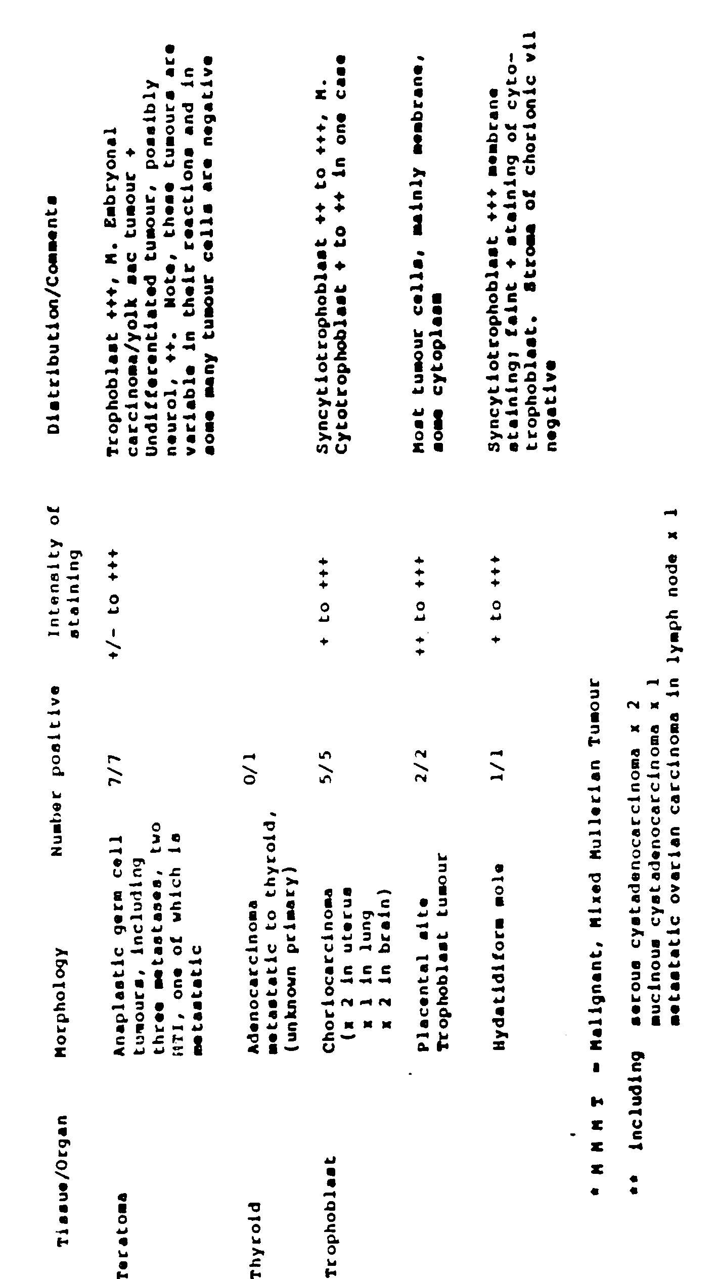
- The reactivity of the 5T4 mAb with invasive carcinoma of the cervix [refer to Table VI above] has prompted a comprehensive investigation of this malignancy and the premalignant changes which can be monitored because of the accessibility to the cervix. Cervical carcinoma arises from dysplastic precursor lesions in the reserve cells in the basal layer of the stratified metaplastic epithelium. These areas develop from proliferating basal cells which have undergone some form of transformation, and gradually spread throughout the whole epithelium. Cervical carcinomas thus develop from a series of atypical changes which progress in continuum to a stage of carcinoma in situ. This is probably the final premalignant state before the lesion invades the underlying stroma becoming microinvasive. The dysplastic variations have been categorised as a series of changes in cervical intraepithelia neoplasia (CIN). They have been graded from 1-3, with
CIN 1 representing less than a third of the dysplastic involvement, located in the basal layer.CIN 2 with a third to two thirds involvement, and CIN 3 two thirds to full thickness involvement, equivalent to carcinoma in situ. At any stage, the lesion may regress back to normality. 90% of cervical neoplasms are squamous cell carcinomas and have a contrasting aetiology and of epidemiology to adenocarcinomas (present in nulliparous women) which comprise the remaining 8-10%. - Placental tissue was washed in PBS (phosphate buffered saline). 1 cm3 of tissue was then embedded in OCT compound and snap-frozen by immersing the specimen in C02 ice with isopentane, within 1 hour post partum. Frozen cervical specimens were selected from a store at the Royal Liverpool Women's Hospital (RLWH). These samples, from cone or punch biopsies routinely submitted for histology, were embedded in polycel, snap-frozen and stored at -70 °C. The specimens were cut at 7αrn thick using a cryostat and placed on slides (cleaned with ethanol and coated with poly-L-lysine). The pathological assessment was obtained from the records of examination of the specimens subjected to fixation in formal buffered saline, dehydration wax embedding and haemotoxin/eosin staining.
- The method described by Bulmer & Sunderland (1983) was used for placental sections but modified for cervical sections. Briefly, the slides were dried at room temperature for 30 minutes before being washed in PBS. Endogenous peroxidase activity was blocked in 3% H202 in ethanol, followed by 3 washes 2.5% sucrose/PBS, and 2
washes 1% Bovine Serum Albumin (BSA)/PBS. The sections were incubated with 10% normal goat serum (NGS) (in 1% BSA/PBS) for 20 minutes before the application of the first layer Ab. All reagents were microfuged at 14,000g for 10 minutes to remove debris before use. The first layer test mAb was applied (ascites fluid diluted 1/100 in 1% BSA/PBS) and incubated for 1 hour at room temperature in a moist box. Subsequently, each slide was washed individually x3 in 1% BSA/PBS before having the second layer peroxidase conjugated rabbit anti-mouse (R anti-Mlg) (Dako) diluted 1:50 in 1% BSA/PBS +10% normal human serum (NHS). After 1 hour incubation (same conditions as before), the slides were washed 2 x 1% NHS, 1% BSA/PBS and developed using 3'3' diaminobenzidine, 5 tig/10 mls PBS + 0.02% H202). The reaction was stopped after 10 minutes by rinsing with tap water. The slides were counterstained using Meyer's Haemalin dehydrated by passing up graded alcohols, fixed in xylene and mounted. All experiments were performed using positive W6/32) and negative (10.2.16) control ascites fluid. - Tissue sections were selected on the basis of routine diagnosis, being placed into the appropriate groups according to the pathology of the epithelium. The groups were categorised as follows: normal, metaplastic, HPV infected,
CIN 1,CIN 2 or CIN 3, with or without HPV infection, invasive carcinoma and common non-malignant cervical inflammatory disorders. Over a 100 biopsy specimens were investigated and each experiment was performed with a positive and negative control (W6/32 and 10.2.16 respectively), read independently by two observers - some material was poorly preserved and not included in the summary. Placental villous sections were included in each experiment to ensure that the procedure was working optimally. The degree of labelling was assessed as anything above that shown in the negative control, eliminating the possibility of false positives becoming included into the study. A subjective estimation of the intensity of the labelling was also made. Experiments were repeated at least once on greater than 50% of the specimens. - Table VII summarises the extent of 5T4 labelling from the basal epithelium to the surface in 66 cone or punch biopsies from the ectocervix. The data can be grouped in several categories. The sections of "normal" ectocervix, squamous metaplasia and HPV infection without evident dysplasia exhibited an overlapping phenotype in intensity and range of distribution of 5T4 antigen. 9/17 showed labelling confined to the basal cells of the epithelium; 6 showed faint labelling throughout the epithelial layers and only 2 demonstrated significant labelling to level C3. There was labelling associated with the stromal elements to the same degree as the basal layer: columnar epithelium and glands when present were labelled. These results are in the range of those described for cervical tissue in a previous immunohistological study of 5T4 expression in normal and neoplastic tissues.
- The above arbitrary grouping shows no obvious differences from the specimens in the
CIN 1 category. The latter is characterised by the appearance of atypical nuclei located in the lower third of the epithelium. Where the morphology was preserved in the frozen sections, it was frequently noted that the 5T4 labelling was located in the parabasal layers corresponding to the area of dysplasia. - From the data on Table VII, it is apparent that there is a progression through
CIN 2 and CIN 3 to a more extensive pattern and intensity of labelling with 5T4 monoclonal antibody. The staining is of higher intensity than in that detected generally in the non-dysplastic orCIN 1 specimens. All the CIN specimens frequently exhibited stromal labelling with an intensity to the basal layers. Where the morphology of dysplastic cells could be assessed, it was evident that from the CIN 2 (HPV) and CIN 3 categories that the specific 5T4 labelling associated with the abnormal cells. 14/15 CIN 3 showed labelling from the basal layer to just below the surface epithelium; 9/15 exhibited labelling along the surface. 5/5 examples of squamous cell carcinoma showed positive intense labelling of the malignant cells and surrounding stroma. The final group of miscellaneous conditions includes hyperplasia, chronic inflammation, cervicitus, acanthotic epithelium and radiation induced atypia. These specimens were selected on the basis of their conventional pathology and exhibited a range of labelling. The inflammatory infiltration response did not increase 5T4 expression per se; the single example of acanthotic epithelium was clearly labelled as were 2/3 of the hyperplastic epithelia. This arbitrary grouping shows some tendency to higher levels of 5T4 expression in the centre layers but appears different from theCIN 2 and CIN 3 groupings. - A new approach using a tumour marker specific for cervical cancer may revolutionise current methods for screening, by offering the potential for the tumour specific Ag to be detected in serum and mucous samples and solubilised biopsies.
- Observing the 5T4 antigenic distribution over a wide range of malignant and premalignant conditions in cervical cancer, a consistent pattern of staining for specific pathological disorders was evident. Normal cervical epithelium, one of the 'specialised' epithelia, showed faint reactivity localised to the reserve cells only. CIN, being the progressive transformation from normal to the malignant state, demonstrated an increased pattern of epithelial labelling corresponding to the severity of the dysplasia. Labelling of the stroma, glands and vessel endothelium also increased as the malignant potential progressed, with no evidence of reactivity with inflammatory cells. Invasive carcinoma showed strong staining of the malignant cells and the intervening stroma. Anaplastic tumours showed patchy variable labelling which may reflect the transformed cells' ability to modulate their morphology and antigenic characteristics. The labelling of the atypical cells is consistent with the theory of the malignant lesion commencing from the basal layers and spreading to the surface.
- The quantitative assessment of 5T4 antigenicity in cervical smear material using radio or other immunoassay with 5T4 monoclonal antibodies may be used as a means of assessing the degree of dysplastic cells in a specimen. This procedure can be highly efficient in mass screening and assigning further investigative procedures.


- Andersson, K., Nilsson, R. and Gahmberg, C.G. (1979) K562- A human erythroleukamia cell line. Int.J.Cancer., 23, 143.
- Andrews, P.W., Damjanov, I., Simon, D. and four others (1984). Pluripotent embryonal carcinoma clones derived from the human teratocarcinoma cell line Tera-2. Lab. Invest., 50, 147.
- Axelsson, B., Kimura, A., Hammarstrom, S., Wigzell, E., Nilsson, K. and Mellstedt, H. (1978) Helix pomatia A haemagglutinin: selectivity of binding to lymphocyte surface glycoproteins on T cells and certain B cells. Eur.J.lmmunol., B, 757.
- Bohn, H., Dati, F. and Luben, G. (1983) Human trophoblast specific products other than hormones. In Biology of trophoblast, Loke, Y.W. and Whyte, A. (eds.), p318, Elsevier: Amsterdam.
- Bramwell, M.E., Ghosh, A.K., Smith, W.D., Wiseman, G., Sprigger, A.I. and Harris, H. (1985). New monoclonal antibodies evaluated as tumour markers in serious effusions. Cancer, 56, 105.
- Bulmer, J.N. and Sunderland, C.A. (1983) Bone-marrow origin of endometrial granulocytes in the early human placental bed. J.Reprod.lmmunol., 5, 383.
- Burchell, J., Durbin, H. and Taylor-Papadimitriou, J. (1983) Complexity of expression of antigenic determinants recognised by monoclonal antibodies HMFG1 and HMFG2 in normal and malignant human epithelial cells. J.lmmunol., 131, 508.
- Critchley, M., McLaughlin, P.J., Brownless, S., Tromans, P.M. Pattern, M. McDicken, I.W. (1986) Radionuclide localisation of epithelial ovarian tumours with 1231-labelled monoclonal antibody (H317). Clin. Radiol., 37, 107 and Johnson PM.
- Daniels, M.R., Hancock, A.M., Walker, C. & Mates G. Interaction of vascular endothelial cells with normal and malignant cells. 3rd Int. Symp. on Biology of Vascular Endothelial Cells p. 57 MIT.
- Easty, D.M., Easty, C.C., Carter, R.C., Monaghan, P. and Butler, C.J. (1981) Ten human carcinoma cell lines derived from squamous carcinomas of the head and neck. Br.J.Cancer, 43, 772.
- Epenetos, A.A., Snook, B., Hooker G. and five others (1985) Indium-111 labelled moonoclonal antibody to PLAP in the detection of neoplasms of testis, ovary and cervix. Lancet ii, 350.
- Fabricant, R.N., DeLarco, I.E. and Todaro, G.J. (1977) Nerve growth factor receptors on human melanoma cells in culture. Proc.Nat.Acad.Sci.USA., 74, 565.
- Fogh, J. and Trempe, G. (1975) in New Human Tumour Cell Lines. Fogh, J. (Ed.), Human Tumour Cells In Vitro., pp115, Plenum Press: New York.
- Jacobs,J.P., Jones, C.M. and Baille, J.P. (1970) Characterisation of a human diploid cell line designated MRC-5. Nature, 227, 168.
- Johnson, P.M. (1984) Immunobiology of the human trophoblast. In Immunological aspects of Reproduction in Mammals. Creighton, D.B. (ed.) p109, Butterworth Press: London.
- Johnson, P.M., Cheng, H.M., Molloy, C.M., Stern, C.M.M. and Slade, M.B. (1981)Human trophoblast-specific surface antigens identified using monoclonal antibodies. Am.J.Reprod.lmmunol., 1, 246.
- Klein, E., Klein, G., Nadkarni, J.S., Nadkarni, J.J., Wigzell, H. and Clifford, P. (1967) Surface IgM specificity on cells derived from a Burkitts lymphoma. Lancet ii, 1068.
- Kohler, G. and Milstein, C. (1975) Derivation of specific antibody-producing tissue culture and tumour cell lines by cell fusion. Eur.J.lmmunol., 6, 511.
- Krantz, M., Ariel, N. and Gold, P. (1979) CEA biology and chemistry: characterisation of partial proteolysis fragments. In: Carcino-embryonic proteins vol. 1. Lehmann, F.-G. (Ed.) p17, Elsevier / North Holland Biomedical Press: Amsterdam.
- Kumar, S., West, D., Daniel, M., Hancock, A. and Carr, T. (1983) Human lung tumour cell lines adapted to grow in serum-free medium secretes angiogenesis factor. Int.J.Cancer, 32, 461.
- Lipinski, M., Parks, D.R., Rouse, R.V., Herzenberg, L.A. (1981)Human trophoblast cell surface antigens defined by monoclonal antibodies. Proc.NatI.Acad. Sci.USA., 78, 5147.
- Loke, Y.W. and Day, S. (1984) Monoclonal antibody to human cytotrophoblast. Am.J. Reprod. Immunol., 5, 106.
- Lowry, O.H., Rosebrough, N.T., Farr, A.L. and Randall, R.J. (1951) Protein measurement with the folin phenol reagent. J.Biol.Chem. 193, 265.
- McDicken, I.W., McLaughlin, P.J., Tromans, P.M., Lenesley,D.M. and Johnson, P.M. (1985) Detection of placental-type alkaline phosphatase in ovarian cancer. Br.J.Cancer, 52, 59.
- McLaughlin, P.J., Cheng, M.H., Slade, M.B. and Johnson, P.M. (1982) Expression on cultured human tumour cells of placental trophoblast membrane antigens and placental alkaline phosphatase defined by monoclonal antibodies. Int.J.Cancer., 30, 21.
- McLaughlin, P.J. (1986) Cancer associated forms of human alkaline phosphatase. In Advances in Clinical Enzymology. Blaton, V. (Ed.) p30, S. Karger AG: Basel.
- Minowada J., Ohnuma, T., and Moore, G.E. (1972) Rosette forming human lymphoid lines. J.Nat.Cancer.lnst., 49, 891.
- Moore, A.E., Sabachewsky, L. and Toolen, H.W. (1955) Culture characteristics of four permanent lines of human cancer cells. Cancer Res., 15, 598.
- Mueller, U.W., Hawes, C.S. and Jones, W.R. (1986) Cell surface antigens of human trophoblast: definition of an apparently unique system with a monoclonal antibody. Immunol., 59, 135.
- O'Toole, C.M., Povey, S., Hepburn, P. and Franks, L.M. (1983). Identity of some human bladder cancer cell lines. Nature, 301, 429.
- Patillo, R.A. and Gey, G.O. (1968) The establishment of human hormone-synthesizing cells in vitro. Cancer Res., 28, 1231.
- Patillo , R.A., Ruckert, A., Hussa, R., Bernstein, R. and Delfs, E. (1971) The JAr cell line - continuous human multihormone production and controls. In Vitro, 6, 398.
- Pateisky, N., Philipp, K., Skodler, W.D., Czerwenka, K., Hamilton, G. and Burchell, J. (1985). Radioim- munodetection in patients with suspected ovarian cancer. J.Nuc.Med. 26, 1369.
- Plummer, T.H., Elder, J.H., Alexander, S., Phelan, A.W. and Tarentino, A.L. (1984). Demonstration of peptide: N-glycosidase F activity in endo-,8-N-acetylglucosaminidase F preparations. J.BioI.Chem., 259, 10700.
- Pulvertaft, R.J.V. (1964) Cytology of Burkitt's tumour (African lymphoma). Lancet i, 238.
- Rettig, W.J., Cordon-Cardo, C., Koulos, J.P., Lewis, J.L., Oettgen, H.F. and Old, L.L. (1985) Cell surface antigens of human trophoblast and choriocarcinoma defined by monoclonal antibodies. Int.J.Cancer, 35, 469. Ruoslahti, E. and Engvall, E. (1978) Alpha feto-protein. Scand.J.lmmunol., 7 Suppl. 6, 1.
- Smith, N.C., Brush, M.G. and Luckett, S. (1974) Preparation of human placental villous surface membrane. Nature, 252, 302.
- Stern, P.L., Gilbert, P., Sternberg, S., Thompson, S. and Chada, K. (1984). A monoclonal antibody which detects 125 kDa glycoprotein on embryonal carcinoma cells and is mitogenic for murine spleen cells. J. Reprod. Immunol., 6, 313.
- Stern, P.L., Berisford, N., Thompson, S., Johnson, P.M., Webb, P.D. and Hole, N. (1986)-.Characterisation of the human trophoblast leukocyte antigenic molecules defined by a monoclonal antibody. J.lmmunol, 137, 1604.
- Sunderland, C.A., Redman, C.W.G. and Stirrat, G.M. (1981) Monoclonal antibodies to human syncytiotrophoblast. Immunol., 43, 541.
- Swallow, D.M., Gendler, S., Griffiths, B, Corney, G., Taylor-Papadimitriou, J. and Bramwell, M.E. (1987) The human tumour-associated epithelial mucins are coded by and expressed hypervariable gene locus PUM. Nature, 328, 82.
- Taylor-Papadimitriou, J., Petersen, J.A., Arklie, J., Burchell, J., Geriani, R.L. and Bodmer, W.F. (1981) Monoclonal antibodies to epithelium specific component of the milk fat globule membrane: production and reaction with cells in culture. Int.J.Cancer, 28, 17.
- Thompson, S., Stern, P.L., Webb, M. and six others (1984). Differentiation of neuron-like cells and other cell types from cloned human teratocarcinoma cells cultured in retionic acid. J.Cell.Sci., 72, 37.
- Travers, P. and Bodmer, W. (1984) Preparation and characterisation of monoclonal antibodies against placental alkaline phosphatase and other human trophoblast associated determinants. Int.J.Cancer. 33, 633.
- Trowbridge, I.S., Newman, R.A., Domingo, D.L. and Sauvage, C. (1984).Transferrin receptor: structure and function. Biochem. Pharmacol., 33, 925.
- Ullrich, A., Boll, J.R., Rhen, E.Y. and eleven others (1985) Human insulin receptor and its relationship to the tyrosine kinase family of oncogenes. Nature, 313, 756.
- Walker, C., Daniels, M., and Mates, G. (1984) Proliferative response of human venous endothelial cells to medium conditioned by human tumour cells. Cell. Biol. Int. Rep., 8, 731.
- Warr, B.G. and Cruickshank, D.J. (1987) Circulating tumour associated antigen detected by the monoclonal antibody HMFG2 in human epithelial ovarian cancer. Int.J.Cancer, 39, 30.
- Waterfield, M.D., Mayes, E.L.V., Stroobant, P. and five others (1982). A monoclonal antibody to the human epidermal growth factor receptor. J.Cell.Biochem., 20, 149.
- Webb, P.D., Evans, P.W., Molloy, C.M. and Johnson, P.M. (1985) Biochemical studies of human microvillous plasma membrane proteins. Am.J.Reprod. lmmunol.Microbiol., 8, 113.
- Wilkinson, M.J.S., Howell, A., Harris, M., Pad, J.T., Swindell, R. and Sellwood, R.A. (1984) The prognostic significance of two epithelial membrane antigens expressed by human mammary carcinoma. Int.J.Cancer, 33, 299.
- Wiseman, G., Bramwell, M.E., Bhavanandan, V.P. and Harris, H. (1984).The structure of the Ca-antigen. Biochem.Soc.Trans., 12, 537.
- Yamashita, K., Nakamura, T., Shimizu, T. and Ohno, H. (1986). Monoclonal antibodies to choriocarcinoma. Am.J.Reprod.immunol.Microbiol., 11, 130.
- Zeuthen, J., Nogaard, J.O.R., Avner, P. and five others (1980) Characterisation of an ovarian teratocarcinoma cell line. Int.J.Cancer, 25, 19.
Claims (7)
Applications Claiming Priority (4)
| Application Number | Priority Date | Filing Date | Title |
|---|---|---|---|
| GB888805240A GB8805240D0 (en) | 1988-03-04 | 1988-03-04 | Improvements relating to antigen |
| GB8805240 | 1988-03-04 | ||
| GB888821078A GB8821078D0 (en) | 1988-09-08 | 1988-09-08 | Improvements relating to antigens |
| GB8821078 | 1988-09-08 |
Publications (2)
| Publication Number | Publication Date |
|---|---|
| EP0336562A1 EP0336562A1 (en) | 1989-10-11 |
| EP0336562B1 true EP0336562B1 (en) | 1995-06-14 |
Family
ID=26293590
Family Applications (2)
| Application Number | Title | Priority Date | Filing Date |
|---|---|---|---|
| EP89302174A Expired - Lifetime EP0336562B1 (en) | 1988-03-04 | 1989-03-03 | Improvements relating to antigens |
| EP89904148A Pending EP0403559A1 (en) | 1988-03-04 | 1989-03-03 | Improvements relating to antigens |
Family Applications After (1)
| Application Number | Title | Priority Date | Filing Date |
|---|---|---|---|
| EP89904148A Pending EP0403559A1 (en) | 1988-03-04 | 1989-03-03 | Improvements relating to antigens |
Country Status (6)
| Country | Link |
|---|---|
| EP (2) | EP0336562B1 (en) |
| AT (1) | ATE123652T1 (en) |
| AU (1) | AU3353989A (en) |
| DE (1) | DE68923027D1 (en) |
| IE (1) | IE890720L (en) |
| WO (1) | WO1989007947A1 (en) |
Families Citing this family (49)
| Publication number | Priority date | Publication date | Assignee | Title |
|---|---|---|---|---|
| SE9601245D0 (en) | 1996-03-29 | 1996-03-29 | Pharmacia Ab | Chimeric superantigens and their use |
| TW517061B (en) | 1996-03-29 | 2003-01-11 | Pharmacia & Amp Upjohn Ab | Modified/chimeric superantigens and their use |
| AU715845B2 (en) * | 1996-12-05 | 2000-02-10 | Macquarie Research Limited | Electrophoresis separation methods |
| AUPO403496A0 (en) * | 1996-12-05 | 1997-01-02 | Macquarie Research Limited | Electrophoresis separation methods |
| AU747602B2 (en) | 1997-06-04 | 2002-05-16 | Oxford Biomedica (Uk) Limited | Vector |
| US7276488B2 (en) | 1997-06-04 | 2007-10-02 | Oxford Biomedica (Uk) Limited | Vector system |
| US7767422B2 (en) | 1998-09-22 | 2010-08-03 | Oncomedx, Inc. | Detection of 5T4 RNA in plasma and serum |
| US7148035B1 (en) | 1998-11-18 | 2006-12-12 | Oxford Biomedica (Uk) Limited | Polypeptide |
| CN1298851C (en) * | 1998-11-18 | 2007-02-07 | 牛津生物医学(英国)有限公司 | polypeptide |
| GB9825303D0 (en) * | 1998-11-18 | 1999-01-13 | Oxford Biomedica Ltd | Vector |
| GB0400443D0 (en) | 2004-01-09 | 2004-02-11 | Oxford Biomedica Ltd | Cascade |
| GB2378704B (en) * | 1998-11-18 | 2003-05-07 | Oxford Biomedica Ltd | Uses of 5t4 antigen |
| GB2370571B (en) * | 1998-11-18 | 2003-05-07 | Oxford Biomedica Ltd | A modified 5t4 antigen |
| GB2371803A (en) * | 1999-11-18 | 2002-08-07 | Oxford Biomedica Ltd | Antibodies |
| CN1423660A (en) * | 1999-11-18 | 2003-06-11 | 牛津生物医学(英国)有限公司 | Antibody |
| AU2001285157A1 (en) * | 2000-08-28 | 2002-03-13 | Oncomedx, Inc. | 5t4 rna in plasma and serum as a marker for neoplastic disease |
| EP1347994A2 (en) * | 2000-11-13 | 2003-10-01 | Oxford Biomedica (UK) Limited | Canine and feline tumour-associated antigen 5t4 |
| GB0126378D0 (en) | 2001-11-02 | 2002-01-02 | Oxford Biomedica Ltd | Antigen |
| GB0203419D0 (en) | 2002-02-13 | 2002-04-03 | Oxford Biomedica Ltd | Peptide |
| GB0203420D0 (en) | 2002-02-13 | 2002-04-03 | Oxford Biomedica Ltd | Peptide |
| US7662625B2 (en) | 2002-07-02 | 2010-02-16 | Cancer Research Technology Limited | Methods for detecting the differentiation status of cells using 5T4 antigen expression |
| GB0215287D0 (en) * | 2002-07-02 | 2002-08-14 | Oxford Biomedica Ltd | 5T4 antigen expression |
| EP1888634A2 (en) | 2005-05-13 | 2008-02-20 | Oxford Biomedica (UK) Limited | Mhc class i and ii peptide antigens derived from tumour antigen 5t4 |
| NZ596295A (en) | 2006-03-10 | 2013-01-25 | Wyeth Corp | Anti-5T4 antibodies and uses thereof |
| MX2009003542A (en) | 2006-10-12 | 2009-04-15 | Wyeth Corp | Modification of ionic strength in antibody-solutions to reduce opalescence/aggregates. |
| GB0822836D0 (en) * | 2008-12-15 | 2009-01-21 | Oxford Biomedica Ltd | Method |
| JP5788863B2 (en) | 2009-03-27 | 2015-10-07 | ワイス・エルエルシー | Tumor initiating cells and methods of use thereof |
| CA2807552A1 (en) | 2010-08-06 | 2012-02-09 | Moderna Therapeutics, Inc. | Engineered nucleic acids and methods of use thereof |
| ES3005233T3 (en) | 2010-10-01 | 2025-03-14 | Modernatx Inc | Modified nucleosides, nucleotides, and nucleic acids, and uses thereof |
| JP2014511687A (en) | 2011-03-31 | 2014-05-19 | モデルナ セラピューティクス インコーポレイテッド | Engineered nucleic acid delivery and formulation |
| US9464124B2 (en) | 2011-09-12 | 2016-10-11 | Moderna Therapeutics, Inc. | Engineered nucleic acids and methods of use thereof |
| EP2763701B1 (en) | 2011-10-03 | 2018-12-19 | Moderna Therapeutics, Inc. | Modified nucleosides, nucleotides, and nucleic acids, and uses thereof |
| JP2015501844A (en) | 2011-12-16 | 2015-01-19 | モデルナ セラピューティクス インコーポレイテッドModerna Therapeutics,Inc. | Modified nucleosides, nucleotides and nucleic acid compositions |
| RU2646498C2 (en) | 2012-01-24 | 2018-03-05 | Пфайзер Инк. | Methods of detecting 5t4-positive circulating tumor cells and methods of diagnostics of 5t4-positive cancer in mammals |
| US9254311B2 (en) | 2012-04-02 | 2016-02-09 | Moderna Therapeutics, Inc. | Modified polynucleotides for the production of proteins |
| WO2013151663A1 (en) | 2012-04-02 | 2013-10-10 | modeRNA Therapeutics | Modified polynucleotides for the production of membrane proteins |
| US9572897B2 (en) | 2012-04-02 | 2017-02-21 | Modernatx, Inc. | Modified polynucleotides for the production of cytoplasmic and cytoskeletal proteins |
| US9283287B2 (en) | 2012-04-02 | 2016-03-15 | Moderna Therapeutics, Inc. | Modified polynucleotides for the production of nuclear proteins |
| ES2921623T3 (en) | 2012-11-26 | 2022-08-30 | Modernatx Inc | terminally modified RNA |
| US8980864B2 (en) | 2013-03-15 | 2015-03-17 | Moderna Therapeutics, Inc. | Compositions and methods of altering cholesterol levels |
| WO2015048744A2 (en) | 2013-09-30 | 2015-04-02 | Moderna Therapeutics, Inc. | Polynucleotides encoding immune modulating polypeptides |
| KR20160067219A (en) | 2013-10-03 | 2016-06-13 | 모더나 세라퓨틱스, 인코포레이티드 | Polynucleotides encoding low density lipoprotein receptor |
| AU2016358854B2 (en) | 2015-11-24 | 2023-02-02 | Byondis B.V. | Anti-5T4 antibodies and antibody-drug conjugates |
| EA201891601A1 (en) | 2016-01-10 | 2019-03-29 | Неоткс Терапьютикс Лтд. | METHODS AND COMPOSITIONS ENHANCED EFFICIENCY BY MEDIATED SUPERANTIGEN IMMUNOTHERAPY OF MALIGNANT TUMORS |
| AU2018235130B2 (en) | 2017-03-15 | 2023-12-07 | Oxford Biomedica (Uk) Limited | Method |
| CN111227039A (en) * | 2018-11-29 | 2020-06-05 | 内蒙古伊利实业集团股份有限公司 | Extraction method of fat globule membrane, yoghourt rich in fat globule membrane and preparation method |
| EA202193121A1 (en) | 2019-05-15 | 2022-02-21 | Неоткс Терапьютикс Лтд. | CANCER TREATMENT |
| AU2021357520A1 (en) | 2020-03-05 | 2022-09-29 | Neotx Therapeutics Ltd. | Methods and compositions for treating cancer with immune cells |
| JP2025535082A (en) * | 2022-10-05 | 2025-10-22 | エムユーエスシー ファウンデーション フォー リサーチ ディベロップメント | Superantigen vaccine conjugates for the treatment of cancer - Patent Application 20070122999 |
Family Cites Families (1)
| Publication number | Priority date | Publication date | Assignee | Title |
|---|---|---|---|---|
| FR2571146B1 (en) * | 1984-10-01 | 1988-02-26 | Centre Nat Rech Scient | CARCINO-FETAL ANTIGENS OF HUMAN PANCREAS AND A PURIFICATION PROCESS, ANTISERUM AGAINST SUCH ANTIGENS AND ITS PREPARATION METHOD AND DIAGNOSTIC COMPOSITIONS CONTAINING THEM |
-
1989
- 1989-03-03 DE DE68923027T patent/DE68923027D1/en not_active Expired - Lifetime
- 1989-03-03 AU AU33539/89A patent/AU3353989A/en not_active Abandoned
- 1989-03-03 EP EP89302174A patent/EP0336562B1/en not_active Expired - Lifetime
- 1989-03-03 AT AT89302174T patent/ATE123652T1/en not_active IP Right Cessation
- 1989-03-03 EP EP89904148A patent/EP0403559A1/en active Pending
- 1989-03-03 WO PCT/GB1989/000207 patent/WO1989007947A1/en not_active Ceased
- 1989-03-06 IE IE890720A patent/IE890720L/en unknown
Non-Patent Citations (6)
| Title |
|---|
| AMERIC. JOURNAL REPROD. IMMUNOL., vol. 1, 1981, Alan R. Liss Inc., US; P.M. JOHNSON et al., pp. 246-254# * |
| BRITISH JOURNAL OF CANCER, vol. 57, 1988, The MacMillan Press Ltd. (GB); N. HOLE et al., pp. 239-246# * |
| INT. JOURNAL OF CANCER, vol. 35, no. 4, 1985, Alan R. Liss Inc. (US); W.J. RETTIG et al., pp. 469-475# * |
| INT. JOURNAL OF CANCER, vol. 45, 1990; pp. 179-184# * |
| JOURNAL OF IMMUNOLOGY, vol. 137, no. 5, 01 September 1986, American Association of Immunologists, US; P.L. STERN et al., pp. 1604-1609# * |
| Journal Reprod. Immunology, 1987. vol. 10pp 231-257. * |
Also Published As
| Publication number | Publication date |
|---|---|
| DE68923027D1 (en) | 1995-07-20 |
| WO1989007947A1 (en) | 1989-09-08 |
| ATE123652T1 (en) | 1995-06-15 |
| EP0403559A1 (en) | 1990-12-27 |
| IE890720L (en) | 1989-09-04 |
| EP0336562A1 (en) | 1989-10-11 |
| AU3353989A (en) | 1989-09-22 |
Similar Documents
| Publication | Publication Date | Title |
|---|---|---|
| EP0336562A1 (en) | Improvements relating to antigens | |
| US5869053A (en) | 5T4 antigen from human trophoblasts | |
| Hole et al. | A 72 kD trophoblast glycoprotein defined by a monoclonal antibody | |
| Marks et al. | Characterization and distribution of an oncofetal antigen (M2A antigen) expressed on testicular germ cell tumours | |
| Flavell et al. | Identification of tissue histiocytes on paraffin sections by a new monoclonal antibody. | |
| Southall et al. | Immunohistological distribution of 5T4 antigen in normal and malignant tissues | |
| CA1303531C (en) | Choriocarcinoma monoclonal antibodies and antibody panels | |
| Sutherland et al. | Acute lymphoblastic leukaemia associated antigen—II. Isolation and partial characterisation | |
| Goddard et al. | Comparison of commercially available cytokeratin antibodies in normal and neoplastic adult epithelial and non-epithelial tissues. | |
| Griffiths et al. | Immunological analysis of mucin molecules expressed by normal and malignant mammary epithelial cells | |
| JP2002502621A (en) | Antibodies specific for mucins with breast tumors, methods for producing and using the same | |
| EP0240341A2 (en) | Antigens, antibodies and methods for the identification of metastatic human tumors, and cell lines for producing said antibodies | |
| HU208546B (en) | Process for producing human antibodies for treating non-small-cell pulmonary-carcinomes, hib ridomes producing them, and antigenes recognised by the antibodies, and diagnostical process and kit | |
| Klein et al. | Binding site of human IgG subclasses and their domains for Fc receptors of activated murine T cells | |
| EP0466818B1 (en) | Monoclonal antibody recognizing a glycoprotein of human milk-fat globule membrane mucin antigen | |
| US5880267A (en) | Trophinin and trophinin-assisting protein agonists, antibodies and antagonists | |
| Hoffman et al. | A haptoglobin-like glycoprotein is produced by implantation-stage rabbit endometrium | |
| HINODA et al. | Detection of circulating adenocarcinoma-associated antigen in the sera of cancer patients with a monoclonal antibody | |
| Bhattacharya et al. | Identification of a human cancer-associated antigen defined with monoclonal antibody | |
| CA2013544C (en) | Retinoblastoma gene product antibodies and uses therefor | |
| US5077220A (en) | Monoclonal antibody specific to a novel glycoprotein antigen on human carcinoma cells | |
| US6242204B1 (en) | Methods of detecting non-small cell lung carcinoma and ovarian cancer | |
| Shuen-Kuei et al. | Difference in cell binding patterns of two monoclonal antibodies recognizing distinct epitopes on a human melanoma-associated oncofetal antigen | |
| Sacco et al. | Use of the zona pellucida as an immunocontraceptive target antigen | |
| Colombatti et al. | Monoclonal antibodies against chick gp 115, a matrix glycoprotein with broad distribution |
Legal Events
| Date | Code | Title | Description |
|---|---|---|---|
| PUAI | Public reference made under article 153(3) epc to a published international application that has entered the european phase |
Free format text: ORIGINAL CODE: 0009012 |
|
| AK | Designated contracting states |
Kind code of ref document: A1 Designated state(s): ES GR |
|
| 17P | Request for examination filed |
Effective date: 19900406 |
|
| XX | Miscellaneous (additional remarks) |
Free format text: VERBUNDEN MIT 89904148.7/0403559 (EUROPAEISCHE ANMELDENUMMER/VEROEFFENTLICHUNGSNUMMER) DURCH ENTSCHEIDUNG VOM 07.02.92. |
|
| 17Q | First examination report despatched |
Effective date: 19920609 |
|
| RAP1 | Party data changed (applicant data changed or rights of an application transferred) |
Owner name: CANCER RESEARCH CAMPAIGN TECHNOLOGY LTD. |
|
| GRAA | (expected) grant |
Free format text: ORIGINAL CODE: 0009210 |
|
| AK | Designated contracting states |
Kind code of ref document: B1 Designated state(s): AT BE CH DE ES FR GB GR IT LI LU NL SE |
|
| PG25 | Lapsed in a contracting state [announced via postgrant information from national office to epo] |
Ref country code: IT Free format text: LAPSE BECAUSE OF FAILURE TO SUBMIT A TRANSLATION OF THE DESCRIPTION OR TO PAY THE FEE WITHIN THE PRE;WARNING: LAPSES OF ITALIAN PATENTS WITH EFFECTIVE DATE BEFORE 2007 MAY HAVE OCCURRED AT ANY TIME BEFORE 2007. THE CORRECT EFFECTIVE DATE MAY BE DIFFERENT FROM THE ONE RECORDED.SCRIBED TIME-LIMIT Effective date: 19950614 Ref country code: CH Effective date: 19950614 Ref country code: ES Free format text: THE PATENT HAS BEEN ANNULLED BY A DECISION OF A NATIONAL AUTHORITY Effective date: 19950614 Ref country code: BE Effective date: 19950614 Ref country code: FR Effective date: 19950614 Ref country code: GR Free format text: LAPSE BECAUSE OF FAILURE TO SUBMIT A TRANSLATION OF THE DESCRIPTION OR TO PAY THE FEE WITHIN THE PRESCRIBED TIME-LIMIT Effective date: 19950614 Ref country code: NL Free format text: LAPSE BECAUSE OF FAILURE TO SUBMIT A TRANSLATION OF THE DESCRIPTION OR TO PAY THE FEE WITHIN THE PRESCRIBED TIME-LIMIT Effective date: 19950614 Ref country code: AT Effective date: 19950614 Ref country code: LI Effective date: 19950614 |
|
| REF | Corresponds to: |
Ref document number: 123652 Country of ref document: AT Date of ref document: 19950615 Kind code of ref document: T |
|
| XX | Miscellaneous (additional remarks) |
Free format text: VERBUNDEN MIT 89904148.7/0403559 (EUROPAEISCHE ANMELDENUMMER/VEROEFFENTLICHUNGSNUMMER) DURCH ENTSCHEIDUNG VOM 07.02.92. |
|
| REF | Corresponds to: |
Ref document number: 68923027 Country of ref document: DE Date of ref document: 19950720 |
|
| PG25 | Lapsed in a contracting state [announced via postgrant information from national office to epo] |
Ref country code: SE Effective date: 19950914 |
|
| PG25 | Lapsed in a contracting state [announced via postgrant information from national office to epo] |
Ref country code: DE Effective date: 19950915 |
|
| REG | Reference to a national code |
Ref country code: CH Ref legal event code: PL |
|
| EN | Fr: translation not filed | ||
| NLV1 | Nl: lapsed or annulled due to failure to fulfill the requirements of art. 29p and 29m of the patents act | ||
| PG25 | Lapsed in a contracting state [announced via postgrant information from national office to epo] |
Ref country code: GB Effective date: 19960303 |
|
| PG25 | Lapsed in a contracting state [announced via postgrant information from national office to epo] |
Ref country code: LU Free format text: LAPSE BECAUSE OF NON-PAYMENT OF DUE FEES Effective date: 19960331 |
|
| PLBE | No opposition filed within time limit |
Free format text: ORIGINAL CODE: 0009261 |
|
| STAA | Information on the status of an ep patent application or granted ep patent |
Free format text: STATUS: NO OPPOSITION FILED WITHIN TIME LIMIT |
|
| 26N | No opposition filed | ||
| GBPC | Gb: european patent ceased through non-payment of renewal fee |
Effective date: 19960303 |