EP0332682B1 - Neutralized perfluoro-3,6-dioxa-4-methyl-7-octene sulphonyl fluoride copolymer surface for attachment and growth of animal cells - Google Patents
Neutralized perfluoro-3,6-dioxa-4-methyl-7-octene sulphonyl fluoride copolymer surface for attachment and growth of animal cells Download PDFInfo
- Publication number
- EP0332682B1 EP0332682B1 EP88908115A EP88908115A EP0332682B1 EP 0332682 B1 EP0332682 B1 EP 0332682B1 EP 88908115 A EP88908115 A EP 88908115A EP 88908115 A EP88908115 A EP 88908115A EP 0332682 B1 EP0332682 B1 EP 0332682B1
- Authority
- EP
- European Patent Office
- Prior art keywords
- nafion
- growth
- cells
- attachment
- animal cells
- Prior art date
- Legal status (The legal status is an assumption and is not a legal conclusion. Google has not performed a legal analysis and makes no representation as to the accuracy of the status listed.)
- Expired - Lifetime
Links
- 230000012010 growth Effects 0.000 title claims abstract description 44
- 229920001577 copolymer Polymers 0.000 title claims abstract description 23
- 210000004102 animal cell Anatomy 0.000 title claims abstract description 18
- BFKJFAAPBSQJPD-UHFFFAOYSA-N tetrafluoroethene Chemical group FC(F)=C(F)F BFKJFAAPBSQJPD-UHFFFAOYSA-N 0.000 claims abstract description 36
- 238000001727 in vivo Methods 0.000 claims abstract description 17
- 238000000338 in vitro Methods 0.000 claims abstract description 14
- GFLUWOXITVIXTN-UHFFFAOYSA-N 1,1,1,2,3,3-hexafluoro-2-(1,1,2,2,2-pentafluoroethoxy)-3-(1,2,2-trifluoroethenoxy)propane sulfuryl difluoride Chemical compound FS(F)(=O)=O.FC(F)=C(F)OC(F)(F)C(F)(C(F)(F)F)OC(F)(F)C(F)(F)F GFLUWOXITVIXTN-UHFFFAOYSA-N 0.000 claims abstract description 13
- 210000004027 cell Anatomy 0.000 claims description 148
- 210000002966 serum Anatomy 0.000 claims description 30
- 238000000034 method Methods 0.000 claims description 29
- 239000000853 adhesive Substances 0.000 claims description 24
- 230000001070 adhesive effect Effects 0.000 claims description 24
- 239000000758 substrate Substances 0.000 claims description 21
- 108010067306 Fibronectins Proteins 0.000 claims description 20
- 102000016359 Fibronectins Human genes 0.000 claims description 20
- 102000004169 proteins and genes Human genes 0.000 claims description 16
- 108090000623 proteins and genes Proteins 0.000 claims description 16
- 239000012528 membrane Substances 0.000 claims description 15
- 230000002792 vascular Effects 0.000 claims description 14
- 239000000178 monomer Substances 0.000 claims description 13
- 230000008569 process Effects 0.000 claims description 13
- 229920000642 polymer Polymers 0.000 claims description 12
- 238000002360 preparation method Methods 0.000 claims description 12
- 108010031318 Vitronectin Proteins 0.000 claims description 11
- 102100035140 Vitronectin Human genes 0.000 claims description 9
- 229920001343 polytetrafluoroethylene Polymers 0.000 claims description 6
- 239000004810 polytetrafluoroethylene Substances 0.000 claims description 6
- 210000002808 connective tissue Anatomy 0.000 claims description 5
- 102000004506 Blood Proteins Human genes 0.000 claims description 4
- 108010017384 Blood Proteins Proteins 0.000 claims description 4
- 239000012634 fragment Substances 0.000 claims description 4
- 239000011521 glass Substances 0.000 claims description 4
- -1 polytetrafluoroethylene Polymers 0.000 claims description 4
- 238000004519 manufacturing process Methods 0.000 claims description 3
- 210000004872 soft tissue Anatomy 0.000 claims description 3
- 102000002938 Thrombospondin Human genes 0.000 claims description 2
- 108060008245 Thrombospondin Proteins 0.000 claims description 2
- 238000004026 adhesive bonding Methods 0.000 claims description 2
- 239000000919 ceramic Substances 0.000 claims description 2
- 229920000295 expanded polytetrafluoroethylene Polymers 0.000 claims description 2
- 229910052751 metal Inorganic materials 0.000 claims description 2
- 239000002184 metal Substances 0.000 claims description 2
- 230000003472 neutralizing effect Effects 0.000 claims description 2
- 229920000728 polyester Polymers 0.000 claims description 2
- 229920002635 polyurethane Polymers 0.000 claims description 2
- 239000004814 polyurethane Substances 0.000 claims description 2
- 230000005855 radiation Effects 0.000 claims description 2
- 229920000557 Nafion® Polymers 0.000 description 143
- 239000004809 Teflon Substances 0.000 description 32
- 229920006362 Teflon® Polymers 0.000 description 32
- 210000001519 tissue Anatomy 0.000 description 30
- 239000004793 Polystyrene Substances 0.000 description 26
- 229920002223 polystyrene Polymers 0.000 description 26
- 239000007943 implant Substances 0.000 description 23
- 230000010261 cell growth Effects 0.000 description 22
- 210000002889 endothelial cell Anatomy 0.000 description 18
- 239000001963 growth medium Substances 0.000 description 18
- 229920000544 Gore-Tex Polymers 0.000 description 16
- 238000010899 nucleation Methods 0.000 description 16
- 239000000463 material Substances 0.000 description 12
- 239000000243 solution Substances 0.000 description 12
- 239000002609 medium Substances 0.000 description 11
- LFQSCWFLJHTTHZ-UHFFFAOYSA-N Ethanol Chemical compound CCO LFQSCWFLJHTTHZ-UHFFFAOYSA-N 0.000 description 9
- FAPWRFPIFSIZLT-UHFFFAOYSA-M Sodium chloride Chemical compound [Na+].[Cl-] FAPWRFPIFSIZLT-UHFFFAOYSA-M 0.000 description 9
- 241000283690 Bos taurus Species 0.000 description 8
- 238000004113 cell culture Methods 0.000 description 7
- LOKCTEFSRHRXRJ-UHFFFAOYSA-I dipotassium trisodium dihydrogen phosphate hydrogen phosphate dichloride Chemical compound P(=O)(O)(O)[O-].[K+].P(=O)(O)([O-])[O-].[Na+].[Na+].[Cl-].[K+].[Cl-].[Na+] LOKCTEFSRHRXRJ-UHFFFAOYSA-I 0.000 description 7
- 238000002474 experimental method Methods 0.000 description 7
- 210000005260 human cell Anatomy 0.000 description 7
- 239000002953 phosphate buffered saline Substances 0.000 description 7
- 239000004033 plastic Substances 0.000 description 7
- 229920003023 plastic Polymers 0.000 description 7
- 210000004618 arterial endothelial cell Anatomy 0.000 description 6
- 210000002950 fibroblast Anatomy 0.000 description 6
- UCSJYZPVAKXKNQ-HZYVHMACSA-N streptomycin Chemical compound CN[C@H]1[C@H](O)[C@@H](O)[C@H](CO)O[C@H]1O[C@@H]1[C@](C=O)(O)[C@H](C)O[C@H]1O[C@@H]1[C@@H](NC(N)=N)[C@H](O)[C@@H](NC(N)=N)[C@H](O)[C@H]1O UCSJYZPVAKXKNQ-HZYVHMACSA-N 0.000 description 6
- PGOHTUIFYSHAQG-LJSDBVFPSA-N (2S)-6-amino-2-[[(2S)-5-amino-2-[[(2S)-2-[[(2S)-2-[[(2S)-2-[[(2S)-4-amino-2-[[(2S)-2-[[(2S)-2-[[(2S)-2-[[(2S)-2-[[(2S)-5-amino-2-[[(2S)-5-amino-2-[[(2S)-2-[[(2S)-2-[[(2S)-2-[[(2S,3R)-2-[[(2S)-5-amino-2-[[(2S)-2-[[(2S)-2-[[(2S,3R)-2-[[(2S)-2-[[(2S)-2-[[(2S)-2-[[(2S)-2-[[(2S)-5-amino-2-[[(2S)-1-[(2S,3R)-2-[[(2S)-2-[[(2S)-2-[[(2R)-2-[[(2S)-2-[[(2S)-2-[[2-[[(2S)-2-[[(2S)-2-[[(2S)-2-[[(2S)-1-[(2S)-2-[[(2S)-2-[[(2S)-2-[[(2S)-2-amino-4-methylsulfanylbutanoyl]amino]-3-(1H-indol-3-yl)propanoyl]amino]-5-carbamimidamidopentanoyl]amino]propanoyl]pyrrolidine-2-carbonyl]amino]-3-methylbutanoyl]amino]-4-methylpentanoyl]amino]-4-methylpentanoyl]amino]acetyl]amino]-3-hydroxypropanoyl]amino]-4-methylpentanoyl]amino]-3-sulfanylpropanoyl]amino]-4-methylsulfanylbutanoyl]amino]-5-carbamimidamidopentanoyl]amino]-3-hydroxybutanoyl]pyrrolidine-2-carbonyl]amino]-5-oxopentanoyl]amino]-3-hydroxypropanoyl]amino]-3-hydroxypropanoyl]amino]-3-(1H-imidazol-5-yl)propanoyl]amino]-4-methylpentanoyl]amino]-3-hydroxybutanoyl]amino]-3-(1H-indol-3-yl)propanoyl]amino]-5-carbamimidamidopentanoyl]amino]-5-oxopentanoyl]amino]-3-hydroxybutanoyl]amino]-3-hydroxypropanoyl]amino]-3-carboxypropanoyl]amino]-3-hydroxypropanoyl]amino]-5-oxopentanoyl]amino]-5-oxopentanoyl]amino]-3-phenylpropanoyl]amino]-5-carbamimidamidopentanoyl]amino]-3-methylbutanoyl]amino]-4-methylpentanoyl]amino]-4-oxobutanoyl]amino]-5-carbamimidamidopentanoyl]amino]-3-(1H-indol-3-yl)propanoyl]amino]-4-carboxybutanoyl]amino]-5-oxopentanoyl]amino]hexanoic acid Chemical compound CSCC[C@H](N)C(=O)N[C@@H](Cc1c[nH]c2ccccc12)C(=O)N[C@@H](CCCNC(N)=N)C(=O)N[C@@H](C)C(=O)N1CCC[C@H]1C(=O)N[C@@H](C(C)C)C(=O)N[C@@H](CC(C)C)C(=O)N[C@@H](CC(C)C)C(=O)NCC(=O)N[C@@H](CO)C(=O)N[C@@H](CC(C)C)C(=O)N[C@@H](CS)C(=O)N[C@@H](CCSC)C(=O)N[C@@H](CCCNC(N)=N)C(=O)N[C@@H]([C@@H](C)O)C(=O)N1CCC[C@H]1C(=O)N[C@@H](CCC(N)=O)C(=O)N[C@@H](CO)C(=O)N[C@@H](CO)C(=O)N[C@@H](Cc1cnc[nH]1)C(=O)N[C@@H](CC(C)C)C(=O)N[C@@H]([C@@H](C)O)C(=O)N[C@@H](Cc1c[nH]c2ccccc12)C(=O)N[C@@H](CCCNC(N)=N)C(=O)N[C@@H](CCC(N)=O)C(=O)N[C@@H]([C@@H](C)O)C(=O)N[C@@H](CO)C(=O)N[C@@H](CC(O)=O)C(=O)N[C@@H](CO)C(=O)N[C@@H](CCC(N)=O)C(=O)N[C@@H](CCC(N)=O)C(=O)N[C@@H](Cc1ccccc1)C(=O)N[C@@H](CCCNC(N)=N)C(=O)N[C@@H](C(C)C)C(=O)N[C@@H](CC(C)C)C(=O)N[C@@H](CC(N)=O)C(=O)N[C@@H](CCCNC(N)=N)C(=O)N[C@@H](Cc1c[nH]c2ccccc12)C(=O)N[C@@H](CCC(O)=O)C(=O)N[C@@H](CCC(N)=O)C(=O)N[C@@H](CCCCN)C(O)=O PGOHTUIFYSHAQG-LJSDBVFPSA-N 0.000 description 5
- 102000002262 Thromboplastin Human genes 0.000 description 5
- 108010000499 Thromboplastin Proteins 0.000 description 5
- 230000001413 cellular effect Effects 0.000 description 5
- JKMHFZQWWAIEOD-UHFFFAOYSA-N 2-[4-(2-hydroxyethyl)piperazin-1-yl]ethanesulfonic acid Chemical compound OCC[NH+]1CCN(CCS([O-])(=O)=O)CC1 JKMHFZQWWAIEOD-UHFFFAOYSA-N 0.000 description 4
- CSCPPACGZOOCGX-UHFFFAOYSA-N Acetone Chemical compound CC(C)=O CSCPPACGZOOCGX-UHFFFAOYSA-N 0.000 description 4
- 108010035532 Collagen Proteins 0.000 description 4
- 102000008186 Collagen Human genes 0.000 description 4
- 230000001464 adherent effect Effects 0.000 description 4
- 238000013459 approach Methods 0.000 description 4
- 229920001436 collagen Polymers 0.000 description 4
- 230000000694 effects Effects 0.000 description 4
- 230000003511 endothelial effect Effects 0.000 description 4
- 239000012909 foetal bovine serum Substances 0.000 description 4
- 238000010348 incorporation Methods 0.000 description 4
- 210000004698 lymphocyte Anatomy 0.000 description 4
- 239000011780 sodium chloride Substances 0.000 description 4
- 230000007480 spreading Effects 0.000 description 4
- 238000003892 spreading Methods 0.000 description 4
- 238000012360 testing method Methods 0.000 description 4
- 239000003104 tissue culture media Substances 0.000 description 4
- 238000011282 treatment Methods 0.000 description 4
- XLYOFNOQVPJJNP-UHFFFAOYSA-N water Substances O XLYOFNOQVPJJNP-UHFFFAOYSA-N 0.000 description 4
- WSFSSNUMVMOOMR-UHFFFAOYSA-N Formaldehyde Chemical compound O=C WSFSSNUMVMOOMR-UHFFFAOYSA-N 0.000 description 3
- 239000007995 HEPES buffer Substances 0.000 description 3
- 241001465754 Metazoa Species 0.000 description 3
- 229930182555 Penicillin Natural products 0.000 description 3
- JGSARLDLIJGVTE-MBNYWOFBSA-N Penicillin G Chemical compound N([C@H]1[C@H]2SC([C@@H](N2C1=O)C(O)=O)(C)C)C(=O)CC1=CC=CC=C1 JGSARLDLIJGVTE-MBNYWOFBSA-N 0.000 description 3
- 108010006886 Vitrogen Proteins 0.000 description 3
- 238000005266 casting Methods 0.000 description 3
- 238000005119 centrifugation Methods 0.000 description 3
- 239000011248 coating agent Substances 0.000 description 3
- 238000000576 coating method Methods 0.000 description 3
- 238000002513 implantation Methods 0.000 description 3
- 239000000203 mixture Substances 0.000 description 3
- 229940049954 penicillin Drugs 0.000 description 3
- 239000002356 single layer Substances 0.000 description 3
- 229960005322 streptomycin Drugs 0.000 description 3
- NWUYHJFMYQTDRP-UHFFFAOYSA-N 1,2-bis(ethenyl)benzene;1-ethenyl-2-ethylbenzene;styrene Chemical compound C=CC1=CC=CC=C1.CCC1=CC=CC=C1C=C.C=CC1=CC=CC=C1C=C NWUYHJFMYQTDRP-UHFFFAOYSA-N 0.000 description 2
- CIWBSHSKHKDKBQ-JLAZNSOCSA-N Ascorbic acid Chemical compound OC[C@H](O)[C@H]1OC(=O)C(O)=C1O CIWBSHSKHKDKBQ-JLAZNSOCSA-N 0.000 description 2
- GHXZTYHSJHQHIJ-UHFFFAOYSA-N Chlorhexidine Chemical compound C=1C=C(Cl)C=CC=1NC(N)=NC(N)=NCCCCCCN=C(N)N=C(N)NC1=CC=C(Cl)C=C1 GHXZTYHSJHQHIJ-UHFFFAOYSA-N 0.000 description 2
- RTZKZFJDLAIYFH-UHFFFAOYSA-N Diethyl ether Chemical compound CCOCC RTZKZFJDLAIYFH-UHFFFAOYSA-N 0.000 description 2
- KCXVZYZYPLLWCC-UHFFFAOYSA-N EDTA Chemical compound OC(=O)CN(CC(O)=O)CCN(CC(O)=O)CC(O)=O KCXVZYZYPLLWCC-UHFFFAOYSA-N 0.000 description 2
- HTTJABKRGRZYRN-UHFFFAOYSA-N Heparin Chemical compound OC1C(NC(=O)C)C(O)OC(COS(O)(=O)=O)C1OC1C(OS(O)(=O)=O)C(O)C(OC2C(C(OS(O)(=O)=O)C(OC3C(C(O)C(O)C(O3)C(O)=O)OS(O)(=O)=O)C(CO)O2)NS(O)(=O)=O)C(C(O)=O)O1 HTTJABKRGRZYRN-UHFFFAOYSA-N 0.000 description 2
- 206010061218 Inflammation Diseases 0.000 description 2
- 229920002684 Sepharose Polymers 0.000 description 2
- 239000007983 Tris buffer Substances 0.000 description 2
- DPKHZNPWBDQZCN-UHFFFAOYSA-N acridine orange free base Chemical compound C1=CC(N(C)C)=CC2=NC3=CC(N(C)C)=CC=C3C=C21 DPKHZNPWBDQZCN-UHFFFAOYSA-N 0.000 description 2
- DZBUGLKDJFMEHC-UHFFFAOYSA-N benzoquinolinylidene Natural products C1=CC=CC2=CC3=CC=CC=C3N=C21 DZBUGLKDJFMEHC-UHFFFAOYSA-N 0.000 description 2
- 210000004369 blood Anatomy 0.000 description 2
- 239000008280 blood Substances 0.000 description 2
- 230000021164 cell adhesion Effects 0.000 description 2
- 230000004956 cell adhesive effect Effects 0.000 description 2
- 239000006143 cell culture medium Substances 0.000 description 2
- 230000035602 clotting Effects 0.000 description 2
- 239000008367 deionised water Substances 0.000 description 2
- 238000013461 design Methods 0.000 description 2
- 238000011161 development Methods 0.000 description 2
- 230000018109 developmental process Effects 0.000 description 2
- 239000012530 fluid Substances 0.000 description 2
- 229960002897 heparin Drugs 0.000 description 2
- 229920000669 heparin Polymers 0.000 description 2
- 239000000017 hydrogel Substances 0.000 description 2
- 238000003018 immunoassay Methods 0.000 description 2
- 230000004054 inflammatory process Effects 0.000 description 2
- 239000003112 inhibitor Substances 0.000 description 2
- NOESYZHRGYRDHS-UHFFFAOYSA-N insulin Chemical compound N1C(=O)C(NC(=O)C(CCC(N)=O)NC(=O)C(CCC(O)=O)NC(=O)C(C(C)C)NC(=O)C(NC(=O)CN)C(C)CC)CSSCC(C(NC(CO)C(=O)NC(CC(C)C)C(=O)NC(CC=2C=CC(O)=CC=2)C(=O)NC(CCC(N)=O)C(=O)NC(CC(C)C)C(=O)NC(CCC(O)=O)C(=O)NC(CC(N)=O)C(=O)NC(CC=2C=CC(O)=CC=2)C(=O)NC(CSSCC(NC(=O)C(C(C)C)NC(=O)C(CC(C)C)NC(=O)C(CC=2C=CC(O)=CC=2)NC(=O)C(CC(C)C)NC(=O)C(C)NC(=O)C(CCC(O)=O)NC(=O)C(C(C)C)NC(=O)C(CC(C)C)NC(=O)C(CC=2NC=NC=2)NC(=O)C(CO)NC(=O)CNC2=O)C(=O)NCC(=O)NC(CCC(O)=O)C(=O)NC(CCCNC(N)=N)C(=O)NCC(=O)NC(CC=3C=CC=CC=3)C(=O)NC(CC=3C=CC=CC=3)C(=O)NC(CC=3C=CC(O)=CC=3)C(=O)NC(C(C)O)C(=O)N3C(CCC3)C(=O)NC(CCCCN)C(=O)NC(C)C(O)=O)C(=O)NC(CC(N)=O)C(O)=O)=O)NC(=O)C(C(C)CC)NC(=O)C(CO)NC(=O)C(C(C)O)NC(=O)C1CSSCC2NC(=O)C(CC(C)C)NC(=O)C(NC(=O)C(CCC(N)=O)NC(=O)C(CC(N)=O)NC(=O)C(NC(=O)C(N)CC=1C=CC=CC=1)C(C)C)CC1=CN=CN1 NOESYZHRGYRDHS-UHFFFAOYSA-N 0.000 description 2
- 230000010118 platelet activation Effects 0.000 description 2
- 230000001737 promoting effect Effects 0.000 description 2
- 210000003491 skin Anatomy 0.000 description 2
- 238000001179 sorption measurement Methods 0.000 description 2
- 239000013589 supplement Substances 0.000 description 2
- 229920002994 synthetic fiber Polymers 0.000 description 2
- 230000002885 thrombogenetic effect Effects 0.000 description 2
- LENZDBCJOHFCAS-UHFFFAOYSA-N tris Chemical compound OCC(N)(CO)CO LENZDBCJOHFCAS-UHFFFAOYSA-N 0.000 description 2
- WZUVPPKBWHMQCE-XJKSGUPXSA-N (+)-haematoxylin Chemical compound C12=CC(O)=C(O)C=C2C[C@]2(O)[C@H]1C1=CC=C(O)C(O)=C1OC2 WZUVPPKBWHMQCE-XJKSGUPXSA-N 0.000 description 1
- 238000011725 BALB/c mouse Methods 0.000 description 1
- 108091003079 Bovine Serum Albumin Proteins 0.000 description 1
- VYZAMTAEIAYCRO-BJUDXGSMSA-N Chromium-51 Chemical compound [51Cr] VYZAMTAEIAYCRO-BJUDXGSMSA-N 0.000 description 1
- 241000699800 Cricetinae Species 0.000 description 1
- 239000006144 Dulbecco’s modified Eagle's medium Substances 0.000 description 1
- 238000002965 ELISA Methods 0.000 description 1
- IAYPIBMASNFSPL-UHFFFAOYSA-N Ethylene oxide Chemical compound C1CO1 IAYPIBMASNFSPL-UHFFFAOYSA-N 0.000 description 1
- 102000018233 Fibroblast Growth Factor Human genes 0.000 description 1
- 108050007372 Fibroblast Growth Factor Proteins 0.000 description 1
- 201000008808 Fibrosarcoma Diseases 0.000 description 1
- SXRSQZLOMIGNAQ-UHFFFAOYSA-N Glutaraldehyde Chemical compound O=CCCCC=O SXRSQZLOMIGNAQ-UHFFFAOYSA-N 0.000 description 1
- 102000003886 Glycoproteins Human genes 0.000 description 1
- 108090000288 Glycoproteins Proteins 0.000 description 1
- WZUVPPKBWHMQCE-UHFFFAOYSA-N Haematoxylin Natural products C12=CC(O)=C(O)C=C2CC2(O)C1C1=CC=C(O)C(O)=C1OC2 WZUVPPKBWHMQCE-UHFFFAOYSA-N 0.000 description 1
- 108010003272 Hyaluronate lyase Proteins 0.000 description 1
- 102000004877 Insulin Human genes 0.000 description 1
- 108090001061 Insulin Proteins 0.000 description 1
- FFEARJCKVFRZRR-BYPYZUCNSA-N L-methionine Chemical compound CSCC[C@H](N)C(O)=O FFEARJCKVFRZRR-BYPYZUCNSA-N 0.000 description 1
- 108010085895 Laminin Proteins 0.000 description 1
- 102000007547 Laminin Human genes 0.000 description 1
- 206010023825 Laryngeal cancer Diseases 0.000 description 1
- CERQOIWHTDAKMF-UHFFFAOYSA-N Methacrylic acid Chemical compound CC(=C)C(O)=O CERQOIWHTDAKMF-UHFFFAOYSA-N 0.000 description 1
- 108010071390 Serum Albumin Proteins 0.000 description 1
- 102000007562 Serum Albumin Human genes 0.000 description 1
- 239000002253 acid Substances 0.000 description 1
- 230000002378 acidificating effect Effects 0.000 description 1
- 230000001154 acute effect Effects 0.000 description 1
- 230000002965 anti-thrombogenic effect Effects 0.000 description 1
- 210000002403 aortic endothelial cell Anatomy 0.000 description 1
- 210000003433 aortic smooth muscle cell Anatomy 0.000 description 1
- 229960005070 ascorbic acid Drugs 0.000 description 1
- 235000010323 ascorbic acid Nutrition 0.000 description 1
- 239000011668 ascorbic acid Substances 0.000 description 1
- 239000012298 atmosphere Substances 0.000 description 1
- 238000001574 biopsy Methods 0.000 description 1
- 210000004204 blood vessel Anatomy 0.000 description 1
- 244000309466 calf Species 0.000 description 1
- 210000001043 capillary endothelial cell Anatomy 0.000 description 1
- 239000002775 capsule Substances 0.000 description 1
- 239000003729 cation exchange resin Substances 0.000 description 1
- 125000002091 cationic group Chemical group 0.000 description 1
- 239000006285 cell suspension Substances 0.000 description 1
- 230000006800 cellular catabolic process Effects 0.000 description 1
- 230000036755 cellular response Effects 0.000 description 1
- 208000019065 cervical carcinoma Diseases 0.000 description 1
- 239000003795 chemical substances by application Substances 0.000 description 1
- 239000000356 contaminant Substances 0.000 description 1
- 238000007334 copolymerization reaction Methods 0.000 description 1
- 210000000399 corneal endothelial cell Anatomy 0.000 description 1
- 239000013078 crystal Substances 0.000 description 1
- 230000009089 cytolysis Effects 0.000 description 1
- 230000007547 defect Effects 0.000 description 1
- 230000001419 dependent effect Effects 0.000 description 1
- 239000000645 desinfectant Substances 0.000 description 1
- 238000010586 diagram Methods 0.000 description 1
- 238000002224 dissection Methods 0.000 description 1
- 239000012153 distilled water Substances 0.000 description 1
- 238000005553 drilling Methods 0.000 description 1
- 229940079593 drug Drugs 0.000 description 1
- 239000003814 drug Substances 0.000 description 1
- 230000002526 effect on cardiovascular system Effects 0.000 description 1
- YQGOJNYOYNNSMM-UHFFFAOYSA-N eosin Chemical compound [Na+].OC(=O)C1=CC=CC=C1C1=C2C=C(Br)C(=O)C(Br)=C2OC2=C(Br)C(O)=C(Br)C=C21 YQGOJNYOYNNSMM-UHFFFAOYSA-N 0.000 description 1
- 238000005530 etching Methods 0.000 description 1
- 239000012894 fetal calf serum Substances 0.000 description 1
- 229940126864 fibroblast growth factor Drugs 0.000 description 1
- 229920002313 fluoropolymer Polymers 0.000 description 1
- UQSQSQZYBQSBJZ-UHFFFAOYSA-N fluorosulfonic acid Chemical class OS(F)(=O)=O UQSQSQZYBQSBJZ-UHFFFAOYSA-N 0.000 description 1
- 239000003365 glass fiber Substances 0.000 description 1
- ZDXPYRJPNDTMRX-UHFFFAOYSA-N glutamine Natural products OC(=O)C(N)CCC(N)=O ZDXPYRJPNDTMRX-UHFFFAOYSA-N 0.000 description 1
- 238000010562 histological examination Methods 0.000 description 1
- 230000003301 hydrolyzing effect Effects 0.000 description 1
- 125000002887 hydroxy group Chemical group [H]O* 0.000 description 1
- 230000006872 improvement Effects 0.000 description 1
- 208000015181 infectious disease Diseases 0.000 description 1
- 230000028709 inflammatory response Effects 0.000 description 1
- 230000005764 inhibitory process Effects 0.000 description 1
- 229940125396 insulin Drugs 0.000 description 1
- 230000003993 interaction Effects 0.000 description 1
- 238000011835 investigation Methods 0.000 description 1
- 238000010884 ion-beam technique Methods 0.000 description 1
- 229920000554 ionomer Polymers 0.000 description 1
- 150000002500 ions Chemical class 0.000 description 1
- 230000002427 irreversible effect Effects 0.000 description 1
- 210000003734 kidney Anatomy 0.000 description 1
- 201000005264 laryngeal carcinoma Diseases 0.000 description 1
- 238000005567 liquid scintillation counting Methods 0.000 description 1
- 230000014759 maintenance of location Effects 0.000 description 1
- 210000004962 mammalian cell Anatomy 0.000 description 1
- 238000005297 material degradation process Methods 0.000 description 1
- 150000002739 metals Chemical class 0.000 description 1
- 229930182817 methionine Natural products 0.000 description 1
- 238000000386 microscopy Methods 0.000 description 1
- 238000002156 mixing Methods 0.000 description 1
- 238000012986 modification Methods 0.000 description 1
- 230000004048 modification Effects 0.000 description 1
- 125000000896 monocarboxylic acid group Chemical group 0.000 description 1
- 230000007935 neutral effect Effects 0.000 description 1
- 238000006386 neutralization reaction Methods 0.000 description 1
- 210000000056 organ Anatomy 0.000 description 1
- 238000012856 packing Methods 0.000 description 1
- 230000002093 peripheral effect Effects 0.000 description 1
- 230000035699 permeability Effects 0.000 description 1
- 238000002135 phase contrast microscopy Methods 0.000 description 1
- 229920000307 polymer substrate Polymers 0.000 description 1
- 230000008092 positive effect Effects 0.000 description 1
- 238000012545 processing Methods 0.000 description 1
- 230000002285 radioactive effect Effects 0.000 description 1
- 230000008439 repair process Effects 0.000 description 1
- 238000011160 research Methods 0.000 description 1
- 230000002207 retinal effect Effects 0.000 description 1
- 238000004626 scanning electron microscopy Methods 0.000 description 1
- 238000005245 sintering Methods 0.000 description 1
- 239000011550 stock solution Substances 0.000 description 1
- 239000000126 substance Substances 0.000 description 1
- 125000001273 sulfonato group Chemical group [O-]S(*)(=O)=O 0.000 description 1
- 230000003319 supportive effect Effects 0.000 description 1
- 238000001356 surgical procedure Methods 0.000 description 1
- 210000001644 umbilical artery Anatomy 0.000 description 1
- 230000000007 visual effect Effects 0.000 description 1
- 238000005406 washing Methods 0.000 description 1
- 238000005303 weighing Methods 0.000 description 1
Images


Classifications
-
- C—CHEMISTRY; METALLURGY
- C12—BIOCHEMISTRY; BEER; SPIRITS; WINE; VINEGAR; MICROBIOLOGY; ENZYMOLOGY; MUTATION OR GENETIC ENGINEERING
- C12N—MICROORGANISMS OR ENZYMES; COMPOSITIONS THEREOF; PROPAGATING, PRESERVING, OR MAINTAINING MICROORGANISMS; MUTATION OR GENETIC ENGINEERING; CULTURE MEDIA
- C12N5/00—Undifferentiated human, animal or plant cells, e.g. cell lines; Tissues; Cultivation or maintenance thereof; Culture media therefor
- C12N5/0068—General culture methods using substrates
-
- C—CHEMISTRY; METALLURGY
- C12—BIOCHEMISTRY; BEER; SPIRITS; WINE; VINEGAR; MICROBIOLOGY; ENZYMOLOGY; MUTATION OR GENETIC ENGINEERING
- C12N—MICROORGANISMS OR ENZYMES; COMPOSITIONS THEREOF; PROPAGATING, PRESERVING, OR MAINTAINING MICROORGANISMS; MUTATION OR GENETIC ENGINEERING; CULTURE MEDIA
- C12N2533/00—Supports or coatings for cell culture, characterised by material
- C12N2533/30—Synthetic polymers
-
- Y—GENERAL TAGGING OF NEW TECHNOLOGICAL DEVELOPMENTS; GENERAL TAGGING OF CROSS-SECTIONAL TECHNOLOGIES SPANNING OVER SEVERAL SECTIONS OF THE IPC; TECHNICAL SUBJECTS COVERED BY FORMER USPC CROSS-REFERENCE ART COLLECTIONS [XRACs] AND DIGESTS
- Y10—TECHNICAL SUBJECTS COVERED BY FORMER USPC
- Y10S—TECHNICAL SUBJECTS COVERED BY FORMER USPC CROSS-REFERENCE ART COLLECTIONS [XRACs] AND DIGESTS
- Y10S623/00—Prosthesis, i.e. artificial body members, parts thereof, or aids and accessories therefor
- Y10S623/915—Method or apparatus for preparing biological material
-
- Y—GENERAL TAGGING OF NEW TECHNOLOGICAL DEVELOPMENTS; GENERAL TAGGING OF CROSS-SECTIONAL TECHNOLOGIES SPANNING OVER SEVERAL SECTIONS OF THE IPC; TECHNICAL SUBJECTS COVERED BY FORMER USPC CROSS-REFERENCE ART COLLECTIONS [XRACs] AND DIGESTS
- Y10—TECHNICAL SUBJECTS COVERED BY FORMER USPC
- Y10S—TECHNICAL SUBJECTS COVERED BY FORMER USPC CROSS-REFERENCE ART COLLECTIONS [XRACs] AND DIGESTS
- Y10S623/00—Prosthesis, i.e. artificial body members, parts thereof, or aids and accessories therefor
- Y10S623/92—Method or apparatus for preparing or treating prosthetic
Definitions
- This invention relates to the use of a copolymer of perfluoro-3,6-dioxa-4-methyl-7-octene sulphonyl fluoride and a monomer as a surface for the attachment and growth of adherent animal cells.
- the invention has particular application to the manufacture and use of prosthetic vascular grafts, connective tissue replacements and soft tissue replacements that incorporate such a copolymer.
- a fluorocarbon polymer with pendant sulphonic groups is the chemically inert, non-crosslinked cation-exchange resin known by the trade mark NAFION.
- NAFION is chemically identified as a copolymer of tetrafluoroethylene and perfluoro-3,6-dioxa-4-methyl-7-octene sulphonyl fluoride. The mechanical and chemical stability of this perfluorosulphonate ionomer and its selective permeability to charged ions had made it useful for industrial electrochemical separating processes.
- PTFE polytetrafluoroethylene
- TEFLON polytetrafluoroethylene
- GORE-TEX expanded PTFE
- any copolymer of perfluoro-3,6-dioxa-4-methyl-7-octene sulphonyl fluoride and a monomer, and particularly NAFION may, when in a neutralised form, be used as a surface for the attachment and growth of adherent animal cells from different tissue sources, including endothelial cells.
- reference to being in a neutralised form means within one pH unit of pH 7.0.
- a surface for the attachment and growth of cells in vivo comprising the neutralized form of a copolymer of perfluoro-3,6-dioxa-4-methyl-7-octene sulphonyl fluoride and a monomer.
- the monomer is tetrafluoroethylene.
- the above surface may be adsorbed or attached to an appropriate substrate that is preferably porous.
- the types of substrate that may be used include polymers, ceramics, metals, glass or preformed membranes.
- the polymer is preferably porous such as polytetrafluoroethylene, expanded polytetrafluoroethylene, knitted or woven polyester and polyurethane.
- the surfaces of the present invention that may be used in vivo are in the form of a sponge or tube and may be adapted for use in a biosensor.
- the surfaces of the present invention that may be used in vivo may also be modified by selective incorporation of platelet binding inhibitors such as serum albumen or heparin or by treating the surfaces with agents that specifically repel or inactivate platelet attachment.
- a surface for the attachment and growth of cells in vitro comprising the neutralised form of a copolymer of perfluoro-3,6-dioxa-4-methyl-7-octene sulphonyl fluoride and a monomer optionally adsorbed or attached to an appropriate substrate as described above, and wherein said surface further includes adsorbed adhesive proteins.
- the preferred adhesive proteins are derived from serum and include fibronectin, vitronectin or adhesive fragments of these proteins. Other preferred adhesive proteins include laminin, collagens and thrombospondin and adhesive fragments of these proteins.
- the above surfaces that may be used in vivo may also have adsorbed thereto adhesive proteins or their adhesive fragments.
- the above surfaces for use both in vivo and in vitro include adhered cells of the type sought to be grown.
- a process for the preparation of a surface for the attachment and growth of cells in vivo comprising applying a copolymer of perfluoro-3,6-dioxa-4-methyl-7-octene sulphonyl fluoride and a monomer to an appropriate substrate.
- the surface so prepared must be brought to neutrality, and this may be done by either neutralizing the resultant surface or by applying the above copolymer in a neutralized form to the appropriate substrate.
- the above copolymer is preferably applied to the substrate by means of radiation grafting or adhesive bonding.
- a process for the preparation of a surface for the attachment and growth of cells in vitro comprising exposing adhesive proteins or adhesive serum proteins to a surface comprising the neutralized form of a copolymer of perfluoro-3,6-dioxa-4-methyl-7-octene sulphonyl fluoride and a monomer optionally adsorbed or attached to an appropriate substrate, whereupon the copolymer adsorbs said proteins to form a copolymer-protein complex.
- the surfaces prepared according to the above processes for use both in vivo and in vitro are further to cells of the type sought to be grown, whereupon the cells adhere to the said surface to further improve its cell attachment and growth properties.
- a method for the attachment and growth of cells to a surface both in vivo and in vitro comprises exposing cells or a medium containing cells and adhesive proteins to a surface of the present invention.
- EXAMPLE 1 Preparation of NAFION N117, NAFION N125, NAFION 22 and NAFION 70 membrane preparations, and PTFE and GORE-TEX coated with NAFION 70.
- NAFION N117 membrane and NAFION N125 membrane were cut into 1cm2 pieces and washed in acetone followed by absolute ethanol. The pieces were then treated with 0.2% EDTA to remove cationic contaminants, washed thoroughly with deionised water and sterilised by autoclaving. Prior to tissue culture studies the pieces were extensively washed in sterile phosphate buffered saline (PBS) pH 7.2.
- PBS phosphate buffered saline
- the EWC of both NAFION N125 and the NAFION 22 and 70 membranes was determined essentially as described by Pedley and Tighe [(1979) Br. Polym.J., 11, 130-135]. In both cases the NAFION was pretreated with 0.2% EDTA and then washed and equilibrated in deionised water for 4 to 6 days before weighing.
- NAFION N-117 and N-125 commercially available preformed sheet NAFION (NAFION N-117 and N-125) and NAFION membrane cast from a 5% solution on either glow discharged or non-glow discharged polystyrene at 22 o C (NAFION 22) were transparent neutral coloured substrates. After casting at 70 o C (NAFION 70), no difference in texture or colour was observed. However this treatment rendered it insoluble in ethanol and acetone. NAFION membranes were cast in a variety of thickness from approximately 10 to 40 microns. By comparison the preformed sheet NAFION used in this study was approximately 100 microns thick. NAFION N125 has an equilibrium water content of 12.0% and NAFION 22 and 70 equilibrium water contents of 32% and 36% respectively. NAFION membranes cast on polystyrene could be peeled off the surface by gently pulling with forceps and the thickness of the membrane determined the fragility of such material.
- a clonal line of normal bovine aortic endothelial (BAE) cells were grown and maintained in M199 cell culture medium supplemented with 20% (v/v) fetal calf serum.
- M199 cell culture medium supplemented with 20% foetal bovine serum containing between 5 x 10⁴ and 2 x 10⁵ BAE cells were added to dishes containing NAFION N125, or coated with either NAFION 22 or NAFION 70, and incubated in a humidified atmosphere of 5% CO2 in air at 37 o C. After 6 hours, cell attachment was estimated by counting cells within 15 randomly chosen 0.931 mm2 areas.
- NAFION is a strong acid and washing the cast polymers (NAFION 22 and NAFION 70) in serum-free tissue culture medium showed that a comparitively large volume of medium was required to neutralise its acidic property. This is therefore, an important aspect of the use of NAFION as a substrate for cell growth which must be taken into account in its preparation. Similarly, NAFION preformed membrane (NAFION N117 and NAFION N125) required neutralisation before use for cell culture.
- bovine aortic endothelial cells BAE
- bovine aortic smooth muscle cells bovine corneal endothelial cells
- bovine retinal capillary endothelial cells bovine retinal capillary endothelial cells
- baby hamster kidney fibroblasts and 3T3 fibroblasts. All cell types displayed their own characteristic morphology and growth characteristics when observed on TCP control dishes. Endothelial cells formed a cobblestone pavement monolayer whilst fibroblasts showed spindle shaped morphology and eventually formed a whorl-like pattern.
- Fig. 1 shows the kinetics of growth of BAE cells on NAFION N-125, 22 and 70 compared to their growth on tissue culture polystyrene after 4 days. Only small differences were seen in the growth rates and final numbers of cells on the NAFION preparations compared with TCP. The growth rate of BAE cells on NAFION preparations observed in these experiments was considerably higher than the level recognised for such cells on the commonly used TEFLON vascular graft material. Cell growth on NAFION coated TEFLON showed a similar improvement, over such cells grown on TEFLON alone.
- BAE cells cultured on TEFLON had a slower growth rate than cells cultured on the other polymers.
- BAE cells seeded on NAFION failed to achieve the characteristic polyhedral morphology and remained fibroblastoid until almost confluent.
- This effect was demonstrated by growing 105 BAE cells/ml in culture for 4 days on untreated TEFLON and NAFION coated TEFLON.
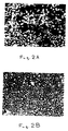
- BAE cells grown on untreated TEFLON displayed a typical patchiness of fibroblast-like morphology in the areas of sparse cover (Fig. 2a). This is characteristic of BAE cells when growing on a less than ideal substrate.
- BAE cells grown on NAFION coated TEFLON achieved the polyhedral morphology characteristic of such cells grown on ideal substrates (Fig. 2b).
- BAE cells could be maintained in culture on NAFION N125 successfully for up to 3 weeks.
- the serum adhesive glycoprotein fibronectin (Fn) was removed from serum prior to use of the serum for cell culture by passage over a gelatin-Sepharose affinity column. Serum treated on a gelatin-Sepharose column was confirmed to be free of Fn by immunoassay of the Fn content.
- the serum adhesive glycoprotein vitronectin (Vn) was removed by passage over an affinity column consisting of immobilized anti-Vn antibody. The sera that were depleted in Vn by this affinity technique were confirmed to have been exhaustively stripped of vitronectin by immunoassay for Vn content.
- Human cell lines HeLa from cervical carcinoma, HeP-2 from carcinoma of Larynx, and HT 1080 from human fibrosarcoma were grown in a growth medium consisting of minimal essential medium supplemented with 10%(v/v) foetal calf serum, 60 microgram/ml penicillin and 100 microgram/ml streptomycin.
- a human umbilical artery endothelial (HUAE) cell culture was established and grown in 75cm2 tissue culture polystyrene (TCP) flasks coated with Fn. Coating with Fn was achieved by incubating the flasks with 5ml solution of 40 ⁇ g/ml Fn in PBS at 37 o C for 1 hour prior to cell seeding. Excess solution was removed before cells were added.
- TCP tissue culture polystyrene
- the cells were routinely maintained in a growth medium consisting of an equal mixture of McCoy 5A (modified) and BM86-Wissler media supplemented with 30%v/v foetal bovine serum, 40 ng/ml fibroblast growth factor, 60 ⁇ g/ml endothelial cell growth supplement, 20 ⁇ g/ml insulin, 60 ⁇ g/ml penicillin and 100 ⁇ g/ml streptomycin.
- the cells were routinely passaged using trypsin-versene, and for experimental work cells were used between passage 15 and passage 20 (inclusive).
- NAFION 70 films cast onto 22mm wells were equilibrated with PBS, and 2ml of growth medium containing 5 x 10⁴ cells was added to each well.
- the cell attachment was determined by counting, in randomly selected fields, the total number of cells and the number of these cells that had spread onto the surface.
- Cell growth in each well was quantitated by counting 5 randomly selected fields per well after successive days of culture, until cell confluence was reached. The mean and standard error of cell number per cm2 for triplicate samples were determined.
- GORE-TEX samples were cut into pieces of approximately 1cm2, coated with approximately 0.3 ml NAFION solution per piece, then immediately treated at 70 o C for 2h.
- the NAFION-coated GORE-TEX was exposed to UV light for 2h and then washed extensively in serum-free tissue culture medium. Some samples were then coated with 40 ⁇ g/ml Fibronectin for 45-60 min at 37 o C prior to seeding with HUAE cells.
- the attachment and growth of human cell lines Hep-2, HeLa and HT 1080 was compared to that on tissue culture polystyrene (TCP). Cell attachment and growth was also determined on NAFION films that had been coated with a solution of 40 ⁇ g/ml Fibronectin.
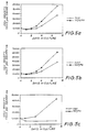
- the human cell lines Hep-2, HeLa and HT 1080 all attached and grew on NAFION films (see Fig. 3 for cell growth curves). It was necessary to use culture medium that contained serum for the cells to attach and to grow on the NAFION surface.
- HUAE cells were grown on NAFION and compared to growth on TCP. It was also necessary to use culture medium that contained serum for the HUAE cells to become attached and to grow. Fn-coated TCP was also included as a control surface, as this surface is known to support good HUAE cell attachment and growth.
- the number of HUAE cells attached to the NAFION surface as viewed after 4 hours fo cell seeding was equivalent to that on TCP, whereas for the Fn-coated NAFION, the number of cells attached was equivaent to that on the Fn-coated TCP.
- the morphology of the HUAE cells attached to the NAFION surface was generally similar to that of the HUAE cells seeded onto TCP, see Fig. 4.
- HUAE cells grew on the NAFION and Fn-coated NAFION surfaces at a rate that was similar to the cell growth on TCP and Fn-coated TCP, respectively (see Fig. 5 for growth curve).
- NAFION was coated onto GORE-TEX, then the NAFION GORE-TEX surface was seeded with HUAE cells. In some samples the NAFION-GORE-TEX surface was precoated with Fibronectin prior to cell seeding. The HUAE cells attached to the NAFION-GORE-TEX surface and grew to produce a surface that was almost completely covered with HUAE cells (See Fig. 6). The HUAE cells grown on each of the NAFION-GORE-TEX surface and the fibronectin-coated NATION-GORE-TEX surface had a well attached and spread morphology as observed in the scanning electron microscope (see Fig. 6).
- EXAMPLE 4 Attachment and Growth of Ovine Endothelial Cells on NAFION tubes.
- OCAE ovine carotid arterial endothelial
- Preequilibrated NAFION tubes (2.9mm internal diameter and 25mm in length) were incubated with a 40 ⁇ g/ml solution of fibronectin (Fn), washed with PBS and individually placed into sterile-cap polystyrene vials, then 9 ml of growth medium containing 2 x 10⁶ cells was added to each vial.
- the cell suspension was gassed with a mixture of 5% CO2 in air and the vial tightly sealed.
- the vials were then placed inside a TCP roller bottle and firmly held in a position by packing.
- the loaded bottle was then rotated at 1 r.p.m. on a roller at 37 o C.
- the culture medium was replenished at 24h and 72h and the tubes removed for subsequent flow testing after 5 days.
- the tubes were cultured for 6hr in culture medium consisting of Dulbecco's modified Eagle's medium containing glutamine, 3 mg/l methionine and 25 ⁇ Ci/ml of 35S-methionine, then further incubated with the normal (McCoy 5A medium with serum and supplements) medium for a further 15h.
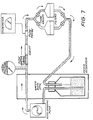
- the tubes containing the metabolically-labelled cells were briefly washed in PBS then inserted into the flow test system as detailed in Figure 7.
- the tubes were subjected to increasing flow rates of a medium consisting of McCoys 5A medium containing 20mM Hepes buffer (pH 7.2) and 20% (v/v) foetal bovine serum at 37 o C for the specified time periods.
- Cells released from the tubes were collected on the downstream glass fibre filters and quantitated by radioactive determination (liquid scintillation counting).
- the tube was removed and bisected then half of the tube was examined for adherent cells by microscopic techniques and the cells on the other half were removed using trypsin-versene and the radioactivity in the released cells was determined
- the NAFION tubes that were used in the flow experiments were precoated with Fn.
- OCAE cells seeded into the Fn-coated NAFION tubes attached to the luminal surface and formed a confluent monolayer of cells during 3 to 5 days of culture.
- the cells attached to the tube were tested for cellular attachment in an in vitro flow system that permitted laminar flow (at flow rates up to 207 ml/min, equivalent to 12.6 dynes/cm2 shear force, and above this flow rate, turbulent flow, see Fig 7 for design).
- the OCEA cells withstood the shear force treatments of up to 20 dynes/cm2, with negligible cell detachment during the flow treatment see Table 1 below.
- the cellular monolayer was examined microscopically after the flow treatment and the cells remained attached and well spread to the luminal surface of the tube, with no evidence of detachment or damage to the cells.
- Human platelets prepared from fresh human plasma were labelled with Chromium 51. Platelets collected by centrifugation were labelled with Cr-51 in 0.25 M HEPES/tris buffer pH 7.0 containing a stock solution of Cr-51 in 0.2 HEPES/tris buffer for 1 h at room temperature. The percentage incorporation of Cr-51 into 10 platelets was checked by counting the amount of radioactivity incorporated in platelets collected by centrifugation and unincorporated radioactivity in the supernatent. Incorporation of the label was then inhibited by the addition of 5% ascorbic acid. Platelets were incubated in the presence of the polymer surface under study in a 96 well ELISA tray for 3 hours at room temperature.
- Partial thromboplastin time was determined by incubating platelet poor plasma (Prepared by centrifugation) in the polymer surface in glass tubes. 0.1 M NaCl was added to the plasma and the time for clot formation measured. This time was taken as the partial thromboplastin time.
- the platelet binding experiments showed that polystyrene, TEFLON and NAFION bound between approximately 12,000 and 18,000 platelets per mm2 whereas polystyrene coated with vitrogen 100 (purified collagen) bound 163,682 platelets per mm2. Actual figures obtained were for polystyrene 15,023; for TEFLON 17,860 and for NAFION 18,070 platelets per mm2. Partial thromboplastin times revealed that NAFION required 210 seconds for clot formation followed by vitrogen 100-coated polystyrene that required 238s; TEFLON 247 s and polystyrene 250 s.
- NAFION and TEFLON have similar partial thromboplastin times and platelet binding properties (low, compared to collagen surfaces) which suggests that NAFION is no more thrombogenic than TEFLON.
- NAFION implants were prepared by mixing a 5% solution of NAFION Equivalent Weight 1100 with NaCl crystals in approximately 10:1 ratio. The mixture was poured into either glass petri dishes or small (20ml) volume beakers and incubated at 60 o C for between four and seven days. After this time the NaCl was dissolved in distilled water.
- implants could have been made by alternative techniques described in the art, e.g. sintering, thermal expansion, laser or ion beam drilling, etc. The material samples were sterilized prior to implantation using an industrial method of ethylene oxide processing. In vivo biocompatibility testing was conducted using males of an inbred strain of BALB/c mice.
- Animals were anaesthetised using ether and their dorsal and flank regions prepared by clipping the fur and swabbing with HIBITANE disinfectant (10% in 70% ethanol, each (v/v)). Implants were inserted subdermally by making a small incision with a pair of scissors and further blunt dissection to prepare a small pocket into which each implant was placed. The skin was closed using two sutures (Mersilk 4.0) and swabbed again with HIBITANE. Each animal received one implant of the material and was caged separately after surgery. Animals were biopsied after one, three, four, six and twelve weeks.
- implants were examined macroscopically for signs of lysis or gross inflammation, excised with the overlying skin and fixed in 10% formaldehyde in normal saline.
- the tissue was dehydrated in ethanol and prepared for routine histology by embedding in Historesin (LKB); 2 mm sections were stained with haematoxylin and eosin and viewed in an Olympus BH-2 microscope.
- LLB Historesin
- Examples 1 to 5 demonstrate the excellent cell response to NAFION in different forms.
- NAFION has good cell supportive characteristics.
- Example 6 demonstrates the biological acceptance of porous NAFION implants indicating that NAFION may be useful for connective tissue or soft tissue prostheses.
- NAFION or indeed any copolymer of perfluoro-3,6-dioxa-4-methyl-7-octene sulphonyl fluoride and a monomer, may be used for the in vitro attachment and growth of animal cells, and may be incorporated into a vascular prosthesis or be a useful alternative to commercially available materials currently used as components of vascular prostheses.
- the copolymer of the present invention may be readily cast into tubes or coated onto pre-existing tubes to serve as an effective vascular graft.
- the effectiveness of the said copolymer as a component of a vascular graft may be further enhanced by the many apparent variations in its preparation as discussed with reference to NAFION herein.
- the use of the said copolymer as a surface for endothelial cell attachment and growth may be of particular value in the new technique of cell seeding of vascular grafts and prostheses prior to implantation.
Landscapes
- Life Sciences & Earth Sciences (AREA)
- Engineering & Computer Science (AREA)
- Health & Medical Sciences (AREA)
- Chemical & Material Sciences (AREA)
- Biomedical Technology (AREA)
- Biotechnology (AREA)
- Organic Chemistry (AREA)
- Bioinformatics & Cheminformatics (AREA)
- Genetics & Genomics (AREA)
- Wood Science & Technology (AREA)
- Zoology (AREA)
- Microbiology (AREA)
- Biochemistry (AREA)
- General Engineering & Computer Science (AREA)
- General Health & Medical Sciences (AREA)
- Cell Biology (AREA)
- Materials For Medical Uses (AREA)
- Micro-Organisms Or Cultivation Processes Thereof (AREA)
- Manufacture Of Porous Articles, And Recovery And Treatment Of Waste Products (AREA)
Abstract
Description
- This invention relates to the use of a copolymer of perfluoro-3,6-dioxa-4-methyl-7-octene sulphonyl fluoride and a monomer as a surface for the attachment and growth of adherent animal cells. The invention has particular application to the manufacture and use of prosthetic vascular grafts, connective tissue replacements and soft tissue replacements that incorporate such a copolymer.
- The design or selection of materials useful in vascular prostheses requires an understanding of the characteristics necessary for irreversible endothelialisation of a surface and for inhibition of undesirable platelet interactions. An approach to the development of vascular prostheses that has been taken has been guided by the object of circumventing the acute problems of platelet activation, adhesion and thrombogenesis. This approach involves designing a blood interface which disallows thrombogenesis by preventing platelet activation directly, and may be achieved either by the selective incorporation or adsorption of platelet binding inhibitors, such as serum albumin or heparin, or by providing a surface which directly repels or inactivates platelets electrostatically. However these modifications might also suppress the attachment and growth of endothelial cells on the luminal surface of the prosthesis. Grafts prepared using this approach may therefore be regarded as unhealed and a physiological and anatomical state comparable to the normal luminal structure is not achieved.
- It is generally known that surfaces which support endothelial cell growth comparable to that seen on glow discharged polystyrene also tend to be thrombogenic. However it is also known that sulphonated polystyrenes have antithrombogenic activity which is reported to be a feature of the negative charge of sulphonate groups. The present invention has been developed by following this line of investigation.
- In a recent study, McAuslan and Johnson [(1987) J. Biomedical Materials Research 21.921-935] showed that the hydroxyl rich surface of poly(hydroxyl ethyl methacrylate) (pHEMA) hydrogel can be converted from a non-cell adhesive to a highly cell adhesive state by either hydrolytic surface etching or by copolymerization with methacrylic acid. Thus cell adhesion appeared to correlate with the introduction of surface COOH groups although this alone was not a sufficient condition. This has raised the question of whether other negatively charged moieties would be just as effective at promoting cell attachment.
- A fluorocarbon polymer with pendant sulphonic groups is the chemically inert, non-crosslinked cation-exchange resin known by the trade mark NAFION. NAFION is chemically identified as a copolymer of tetrafluoroethylene and perfluoro-3,6-dioxa-4-methyl-7-octene sulphonyl fluoride. The mechanical and chemical stability of this perfluorosulphonate ionomer and its selective permeability to charged ions had made it useful for industrial electrochemical separating processes. It can be prepared as films or tubes and is hydrophilic, which is in contrast to polytetrafluoroethylene (PTFE, which is known by the trade mark TEFLON) or expanded PTFE (which is known by the trade mark GORE-TEX), a material which is in wide use as a vascular graft.
- We have now found that any copolymer of perfluoro-3,6-dioxa-4-methyl-7-octene sulphonyl fluoride and a monomer, and particularly NAFION, may, when in a neutralised form, be used as a surface for the attachment and growth of adherent animal cells from different tissue sources, including endothelial cells. In this specification and claims, reference to being in a neutralised form means within one pH unit of pH 7.0.
- It is an object of the present invention to provide a material useful in vascular prostheses and other implantables having improved biocompatibility arising from enhanced endothelial cell attachment properties and anti-thrombogenicity which will substantially overcome the disadvantages of the prior art.
- In accordance with one aspect of the present invention, there is provided a surface for the attachment and growth of cells in vivo, said surface comprising the neutralized form of a copolymer of perfluoro-3,6-dioxa-4-methyl-7-octene sulphonyl fluoride and a monomer. Preferably, the monomer is tetrafluoroethylene. The above surface may be adsorbed or attached to an appropriate substrate that is preferably porous. The types of substrate that may be used include polymers, ceramics, metals, glass or preformed membranes. When a polymer substrate is used, the polymer is preferably porous such as polytetrafluoroethylene, expanded polytetrafluoroethylene, knitted or woven polyester and polyurethane.
- In a preferred form, the surfaces of the present invention that may be used in vivo are in the form of a sponge or tube and may be adapted for use in a biosensor. The surfaces of the present invention that may be used in vivo may also be modified by selective incorporation of platelet binding inhibitors such as serum albumen or heparin or by treating the surfaces with agents that specifically repel or inactivate platelet attachment.
- In accordance with another aspect of the present invention, there is provided a surface for the attachment and growth of cells in vitro, said surface comprising the neutralised form of a copolymer of perfluoro-3,6-dioxa-4-methyl-7-octene sulphonyl fluoride and a monomer optionally adsorbed or attached to an appropriate substrate as described above, and wherein said surface further includes adsorbed adhesive proteins. The preferred adhesive proteins are derived from serum and include fibronectin, vitronectin or adhesive fragments of these proteins. Other preferred adhesive proteins include laminin, collagens and thrombospondin and adhesive fragments of these proteins. The above surfaces that may be used in vivo, may also have adsorbed thereto adhesive proteins or their adhesive fragments.
- In a further preferred embodiment, the above surfaces for use both in vivo and in vitro include adhered cells of the type sought to be grown.
- In accordance with yet another aspect of the present invention, there is provided a process for the preparation of a surface for the attachment and growth of cells in vivo, said process comprising applying a copolymer of perfluoro-3,6-dioxa-4-methyl-7-octene sulphonyl fluoride and a monomer to an appropriate substrate. The surface so prepared must be brought to neutrality, and this may be done by either neutralizing the resultant surface or by applying the above copolymer in a neutralized form to the appropriate substrate.
- The above copolymer is preferably applied to the substrate by means of radiation grafting or adhesive bonding.
- In accordance with a further aspect of the present invention, there is provided a process for the preparation of a surface for the attachment and growth of cells in vitro, said process comprising exposing adhesive proteins or adhesive serum proteins to a surface comprising the neutralized form of a copolymer of perfluoro-3,6-dioxa-4-methyl-7-octene sulphonyl fluoride and a monomer optionally adsorbed or attached to an appropriate substrate, whereupon the copolymer adsorbs said proteins to form a copolymer-protein complex.
- In a further preferred embodiment, the surfaces prepared according to the above processes for use both in vivo and in vitro are further to cells of the type sought to be grown, whereupon the cells adhere to the said surface to further improve its cell attachment and growth properties.
- In accordance with a still further aspect of the present invention, there is provided a method for the attachment and growth of cells to a surface both in vivo and in vitro, which method comprises exposing cells or a medium containing cells and adhesive proteins to a surface of the present invention.
-
- Fig. 1 is a graph of the relative growth rates of bovine aortal endothelial cells on the following surfaces: tissue culture plastic (TCP), NAFION 22, 70 and 125 films prepared as described in Example 1, during the period of 4 days after seeding, and,
- Fig. 2 shows the difference in cell morphology and cell density of bovine aortal endothelial cells after 4 days culture on untreated TEFLON (panel a) or on TEFLON that was coated with NAFION, as described in Example 1, (panel b). Cells were stained with acridine orange and photographed under UV light and the photographs are at 80 times magnification.
- Fig. 3a is a graph of the relative growth rate of the human cell line Hep-2 on the following surfaces: tissue culture plastic, NAFION 70 prepared as described in Examples 1 and 3 and both the above surfaces precoated with fibronectin prior to cell seeding, cultured for a period of 7 days.
- Fig. 3b is a graph of the relative growth rate of the human cell line HeLa on the following surfaces: tissue culture plastic, NAFION 70 prepared as described in Examples 1 and 3 and both the above surfaces precoated with fibronectin prior to cell seeding, cultured for a period of 8 days.
- Fig. 3c is a graph of the relative growth rate of the human cell line HT 1080 on the following surfaces: tissue culture plastic, NAFION 70 prepared as described in Examples 1 and 3 and both the above surfaces precoated with fibronectin prior to cell seeding, cultured for a period of 7 days.
- Fig. 4 shows the difference in morphology of human umbilical arterial endothelial cells cultured on tissue culture plastic (panels a and c) or NAFION 70, prepared as described in Examples 1 and 3 (panels b and d). Some of the surface (panels c and d) were precoated with fibronectin prior to cell seeding. The cells were cultured for 7 days then photographed. The photographs in panels a and b are at 110 times magnification and in panels c and d, at 220 times magnification. Note that the morphology of the cells on NAFION 70 is indistinguishable from that of the cells on the tissue culture plastic, and note also the enhanced cell spreading on each of the surfaces when precoated with fibronectin.
- Fig. 5a is a graph of the relative growth rates of the human umbilical arterial endothelial cells cultured for 7 days on tissue culture plastic (TCP) and TCP precoated with fibronectin prior to cell seeding.
- Fig. 5b is a graph of the relative growth rates of the human umbilical arterial endothelial cells cultured for 7 days on TEFLON that was coated with NAFION (NAF) as described in Example 1, and NAF precoated with fibronectin prior to cell seeding.
- Fig. 5c is a graph of the relative growth rates of the human umbilical arterial endothelial cells cultured for 7 days on untreated TEFLON (TEF), and TEF precoated with fibronectin prior to cell seeding.
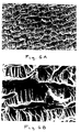
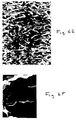
- Fig. 6 shows the difference in morphology of human umbilical arterial endothelial cells cultured on GORE-TEX (panels a and b), or GORE-TEX that was coated with NAFION as described in Example 3 (panels c and d). Another sample of NAFION-coated GORE-TEX (panels e and f) was precoated with fibronectin prior to cell seeding. The cell attachment and morphology was examined by scanning electron microscopy and the magnification of each panel is given as follows: for panels a, c and e, 200 microns = 62 mm on the print, whereas for panels b, d and f, 50 microns = 61 mm on the print. Note the sparce cell attachment to GORE-TEX (panels a and b) but markedly better cell coverage and spreading on the GORE-TEX that was coated with NAFION, giving almost complete cell coverage of the surface (panels c to f). The cells were fixed after 1 day of culture (panels a, b, c, d, f) or 3 days of culture (panel e).
- Fig. 7 is a diagram of the apparatus used to test cellular attachment under conditions of flowing culture medium, as described in Example 4.
- In order that the invention may be more readily understood and put into practical effect, reference will now be made to the following examples that describe the use of NAFION as a preferred embodiment of the copolymer of perfluoro-3,6-dioxa-4-methyl-7-octene sulphonyl fluoride and a monomer.
- Portions of NAFION N117 membrane and NAFION N125 membrane were cut into 1cm² pieces and washed in acetone followed by absolute ethanol. The pieces were then treated with 0.2% EDTA to remove cationic contaminants, washed thoroughly with deionised water and sterilised by autoclaving. Prior to tissue culture studies the pieces were extensively washed in sterile phosphate buffered saline (PBS) pH 7.2.
- A 5% (w/vol) solution of NAFION 1100 Equivalent Weight perfluorinated ion-exchange resin was used for casting the following membranes:
- i) NAFION 22 - prepared by casting 0.5 ml 5% NAFION solution in the lid of a 35 mm diameter tissue culture petri dish at 22oC.
- ii) NAFION 70 - prepared by repeating the procedure for NAFION 22 at 70oC for 2 hours.
Prior to tissue culture studies, the NAFION 22 and NAFION 70 membranes were sterilised under ultravoilet light for 2 hours and then extensively washed in serum free tissue culture medium. - iii) Unfilled virgin TEFLON samples were washed extensively in ethanol and some were coated with NAFION solution and some were left uncoated, for cell culture studies. Approximately 1cm² pieces of material were coated with 50 microliters of 5% NAFION Equivalent Weight 1100 solution and treated at 70oC for 2 hours. Substrates so prepared and tissue culture polystyrene (TCP) were sterilised under UV light for 2 hours and washed extensively in serum free tissue culture medium before being used for cell culture studies.
- The EWC of both NAFION N125 and the NAFION 22 and 70 membranes was determined essentially as described by Pedley and Tighe [(1979) Br. Polym.J., 11, 130-135]. In both cases the NAFION was pretreated with 0.2% EDTA and then washed and equilibrated in deionised water for 4 to 6 days before weighing.
- Commercially available preformed sheet NAFION (NAFION N-117 and N-125) and NAFION membrane cast from a 5% solution on either glow discharged or non-glow discharged polystyrene at 22oC (NAFION 22) were transparent neutral coloured substrates. After casting at 70oC (NAFION 70), no difference in texture or colour was observed. However this treatment rendered it insoluble in ethanol and acetone. NAFION membranes were cast in a variety of thickness from approximately 10 to 40 microns. By comparison the preformed sheet NAFION used in this study was approximately 100 microns thick. NAFION N125 has an equilibrium water content of 12.0% and NAFION 22 and 70 equilibrium water contents of 32% and 36% respectively. NAFION membranes cast on polystyrene could be peeled off the surface by gently pulling with forceps and the thickness of the membrane determined the fragility of such material.
- A clonal line of normal bovine aortic endothelial (BAE) cells were grown and maintained in M199 cell culture medium supplemented with 20% (v/v) fetal calf serum. To determine the increase in the number of cells grown on the different substrates, 2ml of M199 cell culture medium supplemented with 20% foetal bovine serum containing between 5 x 10⁴ and 2 x 10⁵ BAE cells were added to dishes containing NAFION N125, or coated with either NAFION 22 or NAFION 70, and incubated in a humidified atmosphere of 5% CO₂ in air at 37oC. After 6 hours, cell attachment was estimated by counting cells within 15 randomly chosen 0.931 mm² areas. Each polymer sample was the subject of three individual trials and mean cell numbers expressed per cm². After this initial period cell numbers were determined every 24 hours to determine cell growth rate. For comparison the growth of BAE cells on GORE-TEX, TEFLON and glow discharged tissue culture polystyrene was also determined. Cell morphology was investigated by routine light microscopy of cultured surfaces for the NAFION membranes. For the NAFION cast onto TEFLON surfaces, the opacity of the TEFLON precluded the use of phase contrast microscopy for a full visual comparison, so cells were fixed with 2.5% gluteraldehyde, stained with 3mM acridine orange then photographed under UV light.
- It is known that NAFION is a strong acid and washing the cast polymers (NAFION 22 and NAFION 70) in serum-free tissue culture medium showed that a comparitively large volume of medium was required to neutralise its acidic property. This is therefore, an important aspect of the use of NAFION as a substrate for cell growth which must be taken into account in its preparation. Similarly, NAFION preformed membrane (NAFION N117 and NAFION N125) required neutralisation before use for cell culture.
- A wide range of different cell types have been successfully grown on NAFION. These include bovine aortic endothelial cells (BAE), bovine aortic smooth muscle cells, bovine corneal endothelial cells, bovine retinal capillary endothelial cells, baby hamster kidney fibroblasts, and 3T3 fibroblasts. All cell types displayed their own characteristic morphology and growth characteristics when observed on TCP control dishes. Endothelial cells formed a cobblestone pavement monolayer whilst fibroblasts showed spindle shaped morphology and eventually formed a whorl-like pattern.
- Since it was proposed that the material might show potential as a component of a vascular prosthesis the attachment and growth of BAE cells on NAFION compared to TCP and TEFLON was studied in some detail. Cell attachment in vitro was found to be dependent on the presence of serum; and in the absence of serum no cells were attached to any of the NAFION preparations after six hours. The attachment of BAE cells to the different substrates after 6 hours was expressed with respect to cell attachment to tissue culture polystyrene (which was set as 100%). The BAE cell attachment to NAFION 70 and NAFION 22 was 109 ± 3.2% (Mean Standard Deviation) and 97 ± 3.6% respectively; to NAFION N125, 84 ± 3.2% and to TEFLON, 75 ± 6.8%. Of particular interest is the fact that cells appeared to be more evenly distributed on the surface of the NAFION films that on NAFION N-125 or tissue culture polystyrene. Fig. 1 shows the kinetics of growth of BAE cells on NAFION N-125, 22 and 70 compared to their growth on tissue culture polystyrene after 4 days. Only small differences were seen in the growth rates and final numbers of cells on the NAFION preparations compared with TCP. The growth rate of BAE cells on NAFION preparations observed in these experiments was considerably higher than the level recognised for such cells on the commonly used TEFLON vascular graft material. Cell growth on NAFION coated TEFLON showed a similar improvement, over such cells grown on TEFLON alone. By casual appraisal cells cultured on TEFLON had a slower growth rate than cells cultured on the other polymers. In contrast to BAE cells seeded on NAFION, cells seeded on TEFLON failed to achieve the characteristic polyhedral morphology and remained fibroblastoid until almost confluent. This effect was demonstrated by growing 10⁵ BAE cells/ml in culture for 4 days on untreated TEFLON and NAFION coated TEFLON. BAE cells grown on untreated TEFLON displayed a typical patchiness of fibroblast-like morphology in the areas of sparse cover (Fig. 2a). This is characteristic of BAE cells when growing on a less than ideal substrate. In contrast, BAE cells grown on NAFION coated TEFLON achieved the polyhedral morphology characteristic of such cells grown on ideal substrates (Fig. 2b). Further, BAE cells could be maintained in culture on NAFION N125 successfully for up to 3 weeks.
- The growth of BHK fibroblasts on non-glow discharged polystyrene was facilitated by coating the surface with NAFION solution. In accordance with the findings for BAE cells, no difference between the behaviour of BHK fibroblasts to NAFION 22 compared to glow discharged polystyrene was seen whereas cells failed to attach and spread properly or non-glow discharged polystyrene (results not shown). Attachment of BAE cells to TEFLON was less strong than to polystyrene or NAFION as seen when only gentle pipetting of medium or PBS with a Pasteur pipette was sufficient to detach the cells from the TEFLON surface. Such physically weak attachment to tissue culture polystyrene and NAFION was not seen.
- In some experiments of this Example, the serum adhesive glycoprotein fibronectin (Fn) was removed from serum prior to use of the serum for cell culture by passage over a gelatin-Sepharose affinity column. Serum treated on a gelatin-Sepharose column was confirmed to be free of Fn by immunoassay of the Fn content. In other experiments, the serum adhesive glycoprotein vitronectin (Vn) was removed by passage over an affinity column consisting of immobilized anti-Vn antibody. The sera that were depleted in Vn by this affinity technique were confirmed to have been exhaustively stripped of vitronectin by immunoassay for Vn content.
- Human cell lines HeLa from cervical carcinoma, HeP-2 from carcinoma of Larynx, and HT 1080 from human fibrosarcoma were grown in a growth medium consisting of minimal essential medium supplemented with 10%(v/v) foetal calf serum, 60 microgram/ml penicillin and 100 microgram/ml streptomycin.
- A human umbilical artery endothelial (HUAE) cell culture was established and grown in 75cm² tissue culture polystyrene (TCP) flasks coated with Fn. Coating with Fn was achieved by incubating the flasks with 5ml solution of 40µg/ml Fn in PBS at 37oC for 1 hour prior to cell seeding. Excess solution was removed before cells were added. The cells were routinely maintained in a growth medium consisting of an equal mixture of McCoy 5A (modified) and BM86-Wissler media supplemented with 30%v/v foetal bovine serum, 40 ng/ml fibroblast growth factor, 60 µg/ml endothelial cell growth supplement, 20 µg/ml insulin, 60 µg/ml penicillin and 100 µg/ml streptomycin. The cells were routinely passaged using trypsin-versene, and for experimental work cells were used between passage 15 and passage 20 (inclusive).
- For attachment and cell growth studies, NAFION 70 films cast onto 22mm wells were equilibrated with PBS, and 2ml of growth medium containing 5 x 10⁴ cells was added to each well. The cell attachment was determined by counting, in randomly selected fields, the total number of cells and the number of these cells that had spread onto the surface. Cell growth in each well was quantitated by counting 5 randomly selected fields per well after successive days of culture, until cell confluence was reached. The mean and standard error of cell number per cm² for triplicate samples were determined.
- GORE-TEX samples were cut into pieces of approximately 1cm², coated with approximately 0.3 ml NAFION solution per piece, then immediately treated at 70oC for 2h. The NAFION-coated GORE-TEX was exposed to UV light for 2h and then washed extensively in serum-free tissue culture medium. Some samples were then coated with 40 µg/ml Fibronectin for 45-60 min at 37oC prior to seeding with HUAE cells.
- The attachment and growth of human cell lines Hep-2, HeLa and HT 1080 was compared to that on tissue culture polystyrene (TCP). Cell attachment and growth was also determined on NAFION films that had been coated with a solution of 40 µg/ml Fibronectin. The human cell lines Hep-2, HeLa and HT 1080 all attached and grew on NAFION films (see Fig. 3 for cell growth curves). It was necessary to use culture medium that contained serum for the cells to attach and to grow on the NAFION surface. In the case of cell lines Hep-2 and HeLa, the rate of cell growth was increased where the NAFION film had been precoated with Fn (see Fig 3 for comparison of Fn coated NAFION and NAFION that had not been coated with Fn) whereas in the case of HT 1080 cells the Fn coating of the NAFION had no positive effect on the cell growth rate.
- HUAE cells were grown on NAFION and compared to growth on TCP. It was also necessary to use culture medium that contained serum for the HUAE cells to become attached and to grow. Fn-coated TCP was also included as a control surface, as this surface is known to support good HUAE cell attachment and growth. The number of HUAE cells attached to the NAFION surface as viewed after 4 hours fo cell seeding was equivalent to that on TCP, whereas for the Fn-coated NAFION, the number of cells attached was equivaent to that on the Fn-coated TCP. The morphology of the HUAE cells attached to the NAFION surface was generally similar to that of the HUAE cells seeded onto TCP, see Fig. 4. This morphology indicated that although the HUAE cells had attached to the NAFION surface when seeded in the presence of serum, the cells had not formed the well spread morphology that is typical of HUAE cells that have been seeded onto Fn-coated TCP. However the morphology of the HUAE cells that attached to the Fn-coated NAFION films was well spread and the cell morphology was similar to HUAE cells growing of Fn-coated TCP, see Fig. 4.
- HUAE cells grew on the NAFION and Fn-coated NAFION surfaces at a rate that was similar to the cell growth on TCP and Fn-coated TCP, respectively (see Fig. 5 for growth curve).
- The role that Fn from the serum and Vitronectin from the serum may play in the attachment of the HUAE cells to the NAFION and Fn-coated NAFION surfaces was determined by selective removal of these components from the serum used in the culture medium in which the cells were seeded. Selective removal of Vn from the culture medium completely abolished the attachment of HUAE cells to the NAFION surface. The importance of Vitronectin (which is also known as serum spreading factor, epibolin or 70K spreading factor) has been previously reported for other polymer surfaces such as tissue culture polystyrene, see Grinnell [(1976) Exp. Cell Res., 97, 265-274 and (1977) Exp. Cell Res., 110, 175-190]. Attachment of HUAE cells to Fn-coated NAFION over a 4 hour period when seeded in culture medium containing Vn-depleted serum was equivalent to that of HUAE cells seeded in intact medium onto the Fn-coated surface.
- The selective removal of Fn from the seeding culture medium did not abolish the attachment of HUAE cells to the NAFION surface. As a consequence of removal of serum Fn, the rate of cell attachment was somewhat reduced over the first 4 hours as compared to cell attachment to NAFION where the culture medium contained intact serum. However after 24 hours the HUAE cell coverage of the NAFION surface with the Fn-depleted culture medium was identical to that seen on the NAFION surface with the medium containing intact serum. The use of culture medium containing vitronectin-depleted serum for seeding of HUAE cells onto Fn-coated NAFION surface did not effect the rate and extent of HUAE cell attachment and cell morphology, as compared to that where the HUAE cells were seeded onto Fn-coated NAFION using culture medium containing intact serum.
- These results indicate that the serum-dependence of the attachment, cell spreading and growth of HUAE cells on a NAFION surface that has not been precoated with purified Fn or other adhesive proteins involves as an essential component the serum adhesive glycoprotein Vitronectin. Vitronectin from serum or culture medium containing serum is known from previous work to adsorb readily onto other culture surfaces such as tissue culture polystyrene. The results also indicate that the NAFION surface is similar to other surfaces used for the attachment of cells in that the adhesive glycoprotein Fn may be purified from serum and then coated onto the NAFION surface to give a substratum that supports good HUAE cell attachment, with consequential effects on cell growth. Taken together, these results indicate that the adsorption of adhesive serum proteins such as vitronectin or fibronectin onto a NAFION surface produces a substratum for promoting cell adhesion and growth.
- NAFION was coated onto GORE-TEX, then the NAFION GORE-TEX surface was seeded with HUAE cells. In some samples the NAFION-GORE-TEX surface was precoated with Fibronectin prior to cell seeding. The HUAE cells attached to the NAFION-GORE-TEX surface and grew to produce a surface that was almost completely covered with HUAE cells (See Fig. 6). The HUAE cells grown on each of the NAFION-GORE-TEX surface and the fibronectin-coated NATION-GORE-TEX surface had a well attached and spread morphology as observed in the scanning electron microscope (see Fig. 6).
- An ovine carotid arterial endothelial (OCAE) cell culture was established after the methodology of Jaffe ((ed) Biology of Endothelial Cells (Developments in Cardiovascular Medicine) 1984, Martinus Nijhoff Publishers, Boston), and routinely maintained in McCoy 5A (modified) medium supplemented with 20% foetal bovine serum, 60µg/ml penicillin and 100 µg/ml streptomycin and passaged using trypsin-versene. For experimental work, cells were used between
passage 6 and passage 12 (inclusive). Preequilibrated NAFION tubes (2.9mm internal diameter and 25mm in length) were incubated with a 40 µg/ml solution of fibronectin (Fn), washed with PBS and individually placed into sterile-cap polystyrene vials, then 9 ml of growth medium containing 2 x 10⁶ cells was added to each vial. The cell suspension was gassed with a mixture of 5% CO₂ in air and the vial tightly sealed. The vials were then placed inside a TCP roller bottle and firmly held in a position by packing. The loaded bottle was then rotated at 1 r.p.m. on a roller at 37oC. The culture medium was replenished at 24h and 72h and the tubes removed for subsequent flow testing after 5 days. Cell growth could be observed through the NAFION tube using a phase contrast microscope. Having observed that the tubes support cell attachment and growth over 5 days, the tubes were cultured for 6hr in culture medium consisting of Dulbecco's modified Eagle's medium containing glutamine, 3 mg/l methionine and 25 µCi/ml of 35S-methionine, then further incubated with the normal (McCoy 5A medium with serum and supplements) medium for a further 15h. The tubes containing the metabolically-labelled cells were briefly washed in PBS then inserted into the flow test system as detailed in Figure 7. The tubes were subjected to increasing flow rates of a medium consisting of McCoys 5A medium containing 20mM Hepes buffer (pH 7.2) and 20% (v/v) foetal bovine serum at 37oC for the specified time periods. Cells released from the tubes were collected on the downstream glass fibre filters and quantitated by radioactive determination (liquid scintillation counting). Following the flow studies, the tube was removed and bisected then half of the tube was examined for adherent cells by microscopic techniques and the cells on the other half were removed using trypsin-versene and the radioactivity in the released cells was determined - In view of the results with HUAE cells where enhanced cell spreading, attachment and growth was produced by precoating the NAFION surface with Fibronectin (Example 3 above), the NAFION tubes that were used in the flow experiments were precoated with Fn. OCAE cells seeded into the Fn-coated NAFION tubes attached to the luminal surface and formed a confluent monolayer of cells during 3 to 5 days of culture. The cells attached to the tube were tested for cellular attachment in an in vitro flow system that permitted laminar flow (at flow rates up to 207 ml/min, equivalent to 12.6 dynes/cm² shear force, and above this flow rate, turbulent flow, see Fig 7 for design). The OCEA cells withstood the shear force treatments of up to 20 dynes/cm², with negligible cell detachment during the flow treatment (see Table 1 below). The cellular monolayer was examined microscopically after the flow treatment and the cells remained attached and well spread to the luminal surface of the tube, with no evidence of detachment or damage to the cells. These experiments show that the endothelial cells form strong attachment to the Fn-coated NAFION tube surface and can withstand shear forces that are equivalent to those that would be encountered in vivo.
Table 1 Retention of OCAE cells on NAFION tubes under fluid flow conditions Experiment % cells remaining attached to tube % cells detached and recovered on filters 1 99.4 0.6 2 99.1 0.9 3 99.0 1.0 4 98.2 1.8 5 97.8 2.2
The cells were cultured on Fibronectin-coated NAFION tubes as described in the text above, then the cells attached to the tubes were subjected to the following flow protocol: 10 min at a flowrate of 66 ml/min corresponding to a shear force of 4 dynes/cm² followed by 10 min at 132 ml/min equivalent to 8 dynes/cm², then 10 min at 198 ml/min equivalent to 12 dynes/cm², then 10 min at 264 ml/min equivalent to 16 dynes/cm², then 10 min at 330 ml/min equivalent to 20 dynes/cm². It should be noted that the increase in flow corresponding to the step going from 12 to 16 dynes/cm² necessitated going from laminar to turbulent flow. - In vitro thrombogenesis of TEFLON, NAFION, tissue culture polystyrene and vitrogen coated polystyrene surfaces, was studied in the following manner:
- Human platelets prepared from fresh human plasma were labelled with Chromium 51. Platelets collected by centrifugation were labelled with Cr-51 in 0.25 M HEPES/tris buffer pH 7.0 containing a stock solution of Cr-51 in 0.2 HEPES/tris buffer for 1 h at room temperature. The percentage incorporation of Cr-51 into 10 platelets was checked by counting the amount of radioactivity incorporated in platelets collected by centrifugation and unincorporated radioactivity in the supernatent. Incorporation of the label was then inhibited by the addition of 5% ascorbic acid. Platelets were incubated in the presence of the polymer surface under study in a 96 well ELISA tray for 3 hours at room temperature. The polymers were the removed and washed thoroughly in 0.14 M NaCl/0.02 HEPES buffer pH 7.0 and bound radioactivity determined by counting the polymers in a gamma counter. Results were expressed as numbers of platelets bound to the polymer surface per mm²
- Partial thromboplastin time was determined by incubating platelet poor plasma (Prepared by centrifugation) in the polymer surface in glass tubes. 0.1 M NaCl was added to the plasma and the time for clot formation measured. This time was taken as the partial thromboplastin time.
- The platelet binding experiments showed that polystyrene, TEFLON and NAFION bound between approximately 12,000 and 18,000 platelets per mm² whereas polystyrene coated with vitrogen 100 (purified collagen) bound 163,682 platelets per mm². Actual figures obtained were for polystyrene 15,023; for TEFLON 17,860 and for NAFION 18,070 platelets per mm². Partial thromboplastin times revealed that NAFION required 210 seconds for clot formation followed by vitrogen 100-coated polystyrene that required 238s; TEFLON 247 s and polystyrene 250 s.
- These data indicate that NAFION and TEFLON have similar partial thromboplastin times and platelet binding properties (low, compared to collagen surfaces) which suggests that NAFION is no more thrombogenic than TEFLON.
- The use of autograft material is still the most desirable method used to replace diseased or damaged tissues or organs. However, due to anatomical or other considerations, such as those of infection or rejection, this approach may not be feasible. Of particular challenge is the repair of connective tissue defects. Both naturally derived and synthetic materials have been used in this regard, for example, injectable solubilized collagen and polymeric hydrogels. There is increasing interest in the provision of synthetic materials as components of prosthetic devices.
- NAFION implants were prepared by mixing a 5% solution of NAFION Equivalent Weight 1100 with NaCl crystals in approximately 10:1 ratio. The mixture was poured into either glass petri dishes or small (20ml) volume beakers and incubated at 60oC for between four and seven days. After this time the NaCl was dissolved in distilled water. However, implants could have been made by alternative techniques described in the art, e.g. sintering, thermal expansion, laser or ion beam drilling, etc. The material samples were sterilized prior to implantation using an industrial method of ethylene oxide processing. In vivo biocompatibility testing was conducted using males of an inbred strain of BALB/c mice. Animals were anaesthetised using ether and their dorsal and flank regions prepared by clipping the fur and swabbing with HIBITANE disinfectant (10% in 70% ethanol, each (v/v)). Implants were inserted subdermally by making a small incision with a pair of scissors and further blunt dissection to prepare a small pocket into which each implant was placed. The skin was closed using two sutures (Mersilk 4.0) and swabbed again with HIBITANE. Each animal received one implant of the material and was caged separately after surgery. Animals were biopsied after one, three, four, six and twelve weeks. At biopsy, implants were examined macroscopically for signs of lysis or gross inflammation, excised with the overlying skin and fixed in 10% formaldehyde in normal saline. The tissue was dehydrated in ethanol and prepared for routine histology by embedding in Historesin (LKB); 2 mm sections were stained with haematoxylin and eosin and viewed in an Olympus BH-2 microscope.
- All implants were recovered progressively up to 12 weeks. Macroscopic examination revealed that no overt tissue inflammation was present. An indication of the resistance of the implant to cellular degradation was that the original angular contours of the cut edges of the implants were still visible even after 12 weeks in vivo. Implants appeared to maintain their original size throughout the 12-week study period. Histological examination showed that after one week, implants were infiltrated with lymphocytes and a highly cellular connective tissue capsule had formed around the periphery. Lymphocytes had also migrated into the centre of the implants. Three and four week implants appeared similar. Peripheral aspects of the implants were well embedded in the fibrous tissue surrounding the implant. Some fibrous tissue ingrowth was seen particularly after four weeks, as was the presence of large capillaries. At this stage multinuclear giant cells were seen in close association with the implants. After six weeks cellular and fibrous tissue ingrowth was well developed. The lymphocyte inflammatory response was reduced by this time however more multinuclear giant cells were found around the edges of the implants. Twelve weeks after implantation, isolated fingers of fibrous tissue had extended into the centre of most of the implants. These were populated by fibroblasts and lymphocytes and contained small blood capillaries. All implants were well embedded in host tissue and larger blood vessels were observed growing across the surface of the implants. The implants showed no sign of material degradation.
- In summary Examples 1 to 5 demonstrate the excellent cell response to NAFION in different forms. By successfully growing a number of mammalian cell types, notably human arterial endothelial cells, on a variety of NAFION substrates we have shown that NAFION has good cell supportive characteristics. In particular, we have demonstrated the efficacy of NAFION coated TEFLON for support of endothelial cell growth, and that endothelial cells grown on NAFION tubes by the procedures contained in this invention resist shear forces due to fluid flow. It is generally understood that a covering of only approximately 20% of the luminal surface of a vascular graft by metabolising endothelial cells is required to avoid thrombogenesis and we have shown that a morphologically normal endothelial surface is achieved quickly on NAFION. This is in contrast to attempts to culture endothelial cells on TEFLON where cells remain fibroblastoid until reaching confluence and are poorly attached to the surface. Example 6 demonstrates the biological acceptance of porous NAFION implants indicating that NAFION may be useful for connective tissue or soft tissue prostheses.
- These results suggest that NAFION or indeed any copolymer of perfluoro-3,6-dioxa-4-methyl-7-octene sulphonyl fluoride and a monomer, may be used for the in vitro attachment and growth of animal cells, and may be incorporated into a vascular prosthesis or be a useful alternative to commercially available materials currently used as components of vascular prostheses. Preferably, the copolymer of the present invention may be readily cast into tubes or coated onto pre-existing tubes to serve as an effective vascular graft. The effectiveness of the said copolymer as a component of a vascular graft may be further enhanced by the many apparent variations in its preparation as discussed with reference to NAFION herein. The use of the said copolymer as a surface for endothelial cell attachment and growth may be of particular value in the new technique of cell seeding of vascular grafts and prostheses prior to implantation.
Claims (17)
- A surface for the attachment and growth of animal cells in vivo, said surface comprising the neutralised form of a copolymer of perfluoro-3,6-dioxa-4-methyl-7-octene sulphonyl fluoride and a monomer, or in vitro said surface additionally including adsorbed adhesive proteins.
- A surface according to Claim 1, wherein the monomer is tetrafluoroethylene.
- A surface for the attachment and growth of animal cells in vivo, said surface comprising the surface of Claim 1 or Claim 2 adsorbed or attached to a biocompatible synthetic substrate.
- A surface according to Claim 3, wherein said substrate is selected from the group consisting of a polymer, ceramic, metal, glass, preformed membrane, polytetrafluoroethylene, expanded polytetrafluoroethylene, knitted or woven polyester, or polyurethane.
- A surface according to Claim 3 or 4, wherein said substrate is porous.
- A surface according to any preceding claim for the attachment and growth of animal cells in vivo, said surface additionally including adsorbed adhesive proteins.
- A surface according to any one of Claims 1 to 6 adapted for use in a biosensor.
- A surface according to any one of Claims 1 to 6 in the form of a sponge, or a tube.
- A surface according to any preceding claim, wherein said adhesive proteins are derived from serum.
- A surface according to Claim 9 wherein said adhesive serum proteins are selected from the group consisting of fibronectin, vitronectin, thrombospondin or adhesive fragments of any of these proteins.
- A surface according to any one of Claims 1 to 10 further including adhered animal cells.
- A process far the preparation of a surface for the attachment and growth of animal cells in vivo, said process comprising applying a copolymer of perfluoro-3, 6-dioxa-4-methyl-7-octene sulphonyl fluoride and a monomer to a biocompatible synthetic substrate and neutralising the resultant surface, or said process comprising applying the neutralised form of a copolymer of perfluoro-3,6-dioxa-4-methyl-7-octene sulphonyl fluoride and a monomer to a biocompatible synthetic substrate.
- A process according to Claim 12 wherein the application of said copolymer to said biocompatible synthetic substrate is by means of radiation grafting, or adhesive bonding.
- A process for the preparation of a surface for the attachment and growth of animal cells in vitro, said process comprising exposing adhesive proteins or adhesive serum proteins to the surface of any one of Claims 1 to 5 or Claim 8, whereupon said copolymer adsorbs said proteins to form a copolymer-protein complex for the attachment and growth of animal cells.
- A process according to any one of Claims 12 to 14, further including the step of exposing said surface to animal cells, whereupon said cells adhere to the said surface to form an adhesive surface for the further attachment and growth of animal cells.
- A method for the attachment and growth of animal cells to a surface in vitro, which method comprises exposing animal cells to a surface of any one of Claim 1 to 5 and 7 to 11.
- Use of the surface according to any one of Claims 1 to 11 in the manufacture of prosthetic vascular grafts, connective tissue replacements or soft tissue replacements for the purpose of the attachment and growth of animal cells on said surface in vivo.
Applications Claiming Priority (3)
| Application Number | Priority Date | Filing Date | Title |
|---|---|---|---|
| AUPI440387 | 1987-09-17 | ||
| AU4403/87 | 1987-09-17 | ||
| PCT/AU1988/000368 WO1989002457A1 (en) | 1987-09-17 | 1988-09-19 | Neutralized perfluoro-3,6-dioxa-4-methyl-7-octene sulphonyl fluoride copolymer surface for attachment and growth of animal cells |
Publications (3)
| Publication Number | Publication Date |
|---|---|
| EP0332682A1 EP0332682A1 (en) | 1989-09-20 |
| EP0332682A4 EP0332682A4 (en) | 1990-02-20 |
| EP0332682B1 true EP0332682B1 (en) | 1994-08-03 |
Family
ID=3772454
Family Applications (1)
| Application Number | Title | Priority Date | Filing Date |
|---|---|---|---|
| EP88908115A Expired - Lifetime EP0332682B1 (en) | 1987-09-17 | 1988-09-19 | Neutralized perfluoro-3,6-dioxa-4-methyl-7-octene sulphonyl fluoride copolymer surface for attachment and growth of animal cells |
Country Status (6)
| Country | Link |
|---|---|
| US (1) | US5077215A (en) |
| EP (1) | EP0332682B1 (en) |
| JP (1) | JPH02502246A (en) |
| AU (1) | AU608913B2 (en) |
| DE (1) | DE3850957T2 (en) |
| WO (1) | WO1989002457A1 (en) |
Families Citing this family (20)
| Publication number | Priority date | Publication date | Assignee | Title |
|---|---|---|---|---|
| US5037656A (en) * | 1986-12-04 | 1991-08-06 | Millipore Corporation | Porous membrane having hydrophilic and cell growth promotions surface and process |
| US5721131A (en) * | 1987-03-06 | 1998-02-24 | United States Of America As Represented By The Secretary Of The Navy | Surface modification of polymers with self-assembled monolayers that promote adhesion, outgrowth and differentiation of biological cells |
| US5079600A (en) * | 1987-03-06 | 1992-01-07 | Schnur Joel M | High resolution patterning on solid substrates |
| US5713888A (en) * | 1990-10-31 | 1998-02-03 | Baxter International, Inc. | Tissue implant systems |
| US5733336A (en) * | 1990-10-31 | 1998-03-31 | Baxter International, Inc. | Ported tissue implant systems and methods of using same |
| US5314471A (en) * | 1991-07-24 | 1994-05-24 | Baxter International Inc. | Tissue inplant systems and methods for sustaining viable high cell densities within a host |
| EP0507933B1 (en) * | 1990-10-31 | 1996-05-22 | Baxter International Inc. | Close vascularization implant material |
| US5344454A (en) * | 1991-07-24 | 1994-09-06 | Baxter International Inc. | Closed porous chambers for implanting tissue in a host |
| FR2675038B3 (en) * | 1991-04-10 | 1993-07-16 | France Chirurgie Instrumentation | MESOPROSTHESIS SUPPORT. |
| US5453278A (en) * | 1991-07-24 | 1995-09-26 | Baxter International Inc. | Laminated barriers for tissue implants |
| US6773458B1 (en) | 1991-07-24 | 2004-08-10 | Baxter International Inc. | Angiogenic tissue implant systems and methods |
| CA2148354A1 (en) * | 1993-09-24 | 1995-03-30 | Laura A. Martinson | Methods for enhancing vascularization of implant devices |
| US5693085A (en) | 1994-04-29 | 1997-12-02 | Scimed Life Systems, Inc. | Stent with collagen |
| US6156305A (en) * | 1994-07-08 | 2000-12-05 | Baxter International Inc. | Implanted tumor cells for the prevention and treatment of cancer |
| AU1504895A (en) | 1994-07-11 | 1996-01-25 | Meadox Medicals, Inc. | Expanded PTFE implantable prosthesis with improved blood and tissue compatibility and superior potency |
| US5702949A (en) * | 1995-06-22 | 1997-12-30 | University Of Utah Research Foundation | Culture method for multilayer growth of anchorage-dependent cells |
| US5874165A (en) * | 1996-06-03 | 1999-02-23 | Gore Enterprise Holdings, Inc. | Materials and method for the immobilization of bioactive species onto polymeric subtrates |
| US5914182A (en) * | 1996-06-03 | 1999-06-22 | Gore Hybrid Technologies, Inc. | Materials and methods for the immobilization of bioactive species onto polymeric substrates |
| US7299805B2 (en) | 2002-06-07 | 2007-11-27 | Marctec, Llc | Scaffold and method for implanting cells |
| CN112823030A (en) * | 2018-08-31 | 2021-05-18 | 威斯康星校友研究基金会 | Generation of vascular grafts for arterial endothelial cell implantation |
Family Cites Families (15)
| Publication number | Priority date | Publication date | Assignee | Title |
|---|---|---|---|---|
| US3635926A (en) * | 1969-10-27 | 1972-01-18 | Du Pont | Aqueous process for making improved tetrafluoroethylene / fluoroalkyl perfluorovinyl ether copolymers |
| AU464313B2 (en) * | 1971-12-27 | 1975-08-21 | Hooker Chemicals And Plastics Coup | Electrolysis method and apparatus |
| AU535734B2 (en) * | 1977-04-20 | 1984-04-05 | E.I. Du Pont De Nemours And Company | Process for preparing fluorinated polymer containing carbonyl fluoride and/or carboxylic groups |
| AU9184082A (en) * | 1977-04-20 | 1983-04-21 | E.I. Du Pont De Nemours And Company | Fluorinated ion-exchange polymers |
| DE2724200C2 (en) * | 1977-05-27 | 1985-06-20 | E.I. Du Pont De Nemours And Co., Wilmington, Del. | Coagulated tetrafluoroethylene copolymer powder and process for its production |
| JPS6045653B2 (en) * | 1979-05-31 | 1985-10-11 | 旭化成株式会社 | Novel fluorinated cation exchange membrane and its manufacturing method |
| US4329435A (en) * | 1979-05-31 | 1982-05-11 | Asahi Kasei Kogyo Kabushiki Kaisha | Novel fluorinated copolymer with tridihydro fluorosulfonyl fluoride pendant groups and preparation thereof |
| US4329434A (en) * | 1979-05-31 | 1982-05-11 | Asahi Kasei Kogyo Kabushiki Kaisha | Novel fluorinated cation exchange membrane and process for producing the same |
| CA1201400A (en) * | 1982-04-16 | 1986-03-04 | Joel L. Williams | Chemically specific surfaces for influencing cell activity during culture |
| US4578079A (en) * | 1982-08-04 | 1986-03-25 | La Jolla Cancer Research Foundation | Tetrapeptide |
| US4883057A (en) * | 1984-05-09 | 1989-11-28 | Research Foundation, The City University Of New York | Cathodic electrochemical current arrangement with telemetric application |
| SE452110B (en) * | 1984-11-08 | 1987-11-16 | Medinvent Sa | MULTILAYER PROTEST MATERIAL AND PROCEDURE FOR ITS MANUFACTURING |
| DE3521684A1 (en) * | 1985-06-18 | 1986-12-18 | Dr. Müller-Lierheim KG, Biologische Laboratorien, 8033 Planegg | METHOD FOR COATING POLYMERS |
| US4795475A (en) * | 1985-08-09 | 1989-01-03 | Walker Michael M | Prosthesis utilizing salt-forming oxyacids for bone fixation |
| JPS62288614A (en) * | 1986-06-06 | 1987-12-15 | Asahi Glass Co Ltd | Production of perfluorocarbon polymer having sulfonic acid type functional group |
-
1988
- 1988-09-19 DE DE3850957T patent/DE3850957T2/en not_active Expired - Fee Related
- 1988-09-19 JP JP63507825A patent/JPH02502246A/en active Pending
- 1988-09-19 AU AU25243/88A patent/AU608913B2/en not_active Ceased
- 1988-09-19 US US07/360,887 patent/US5077215A/en not_active Expired - Fee Related
- 1988-09-19 WO PCT/AU1988/000368 patent/WO1989002457A1/en not_active Ceased
- 1988-09-19 EP EP88908115A patent/EP0332682B1/en not_active Expired - Lifetime
Non-Patent Citations (1)
| Title |
|---|
| NATURE, vol. 309, no. 5963, 03 May 1984; M.D. PIERSCHBACHER et al., pp. 30-33# * |
Also Published As
| Publication number | Publication date |
|---|---|
| DE3850957D1 (en) | 1994-09-08 |
| DE3850957T2 (en) | 1995-03-30 |
| JPH02502246A (en) | 1990-07-26 |
| AU2524388A (en) | 1989-04-17 |
| EP0332682A1 (en) | 1989-09-20 |
| WO1989002457A1 (en) | 1989-03-23 |
| EP0332682A4 (en) | 1990-02-20 |
| AU608913B2 (en) | 1991-04-18 |
| US5077215A (en) | 1991-12-31 |
Similar Documents
| Publication | Publication Date | Title |
|---|---|---|
| EP0332682B1 (en) | Neutralized perfluoro-3,6-dioxa-4-methyl-7-octene sulphonyl fluoride copolymer surface for attachment and growth of animal cells | |
| Meyle et al. | Fibroblast anchorage to microtextured surfaces | |
| US5258041A (en) | Method of biomolecule attachment to hydrophobic surfaces | |
| EP0494216B1 (en) | Surfaces having desirable cell adhesive effects | |
| JP2741378B2 (en) | Improved biocompatibility of solid surfaces | |
| Ertel et al. | In vitro study of the intrinsic toxicity of synthetic surfaces to cells | |
| Bordenave et al. | Endothelial cell compatibility testing of three different Pellethanes | |
| AU685576B2 (en) | Soluble factor stimulation of attachment and hemidesmosome assembly in epithelial cells | |
| Hsiue et al. | Surface characterization and biological properties study of silicone rubber membrane grafted with phospholipid as biomaterial via plasma induced graft copolymerization | |
| EP1326655A2 (en) | Biocompatible materials | |
| JPH09504706A (en) | Corneal surface graft | |
| Sipehia et al. | Enhanced attachment and growth of human endothelial cells derived from umbilical veins on ammonia plasma modified surfaces of PTFE and ePTFE synthetic vascular graft biomaterials | |
| EP0407390A4 (en) | Biocompatible coatings | |
| Brubert et al. | Hemocompatibility of styrenic block copolymers for use in prosthetic heart valves | |
| JP7058397B2 (en) | Tissue regeneration cultured cell sheet, manufacturing method and its utilization method | |
| US5106743A (en) | Hydrogels capable of supporting cell growth | |
| US5010009A (en) | Material for cell attachment and growth | |
| McAuslan et al. | Cell responses to biomaterials II: Endothelial cell adhesion and growth on perfluorosulfonic acid | |
| LEE et al. | Endothelial cell seeding onto the extracellular matrix of fibroblasts for the development of a small diameter polyurethane vessel | |
| Kobayashi et al. | An artificial blood vessel with an endothelial-cell monolayer | |
| JPH05244938A (en) | Cell and culture method | |
| Vladkova | Surface modification of silicone rubber with poly (ethylene glycol) hydrogel coatings | |
| JPH02181628A (en) | Immobilization of biologically active part on substrate, immobilization of living cell on substrate and cultivation of cell | |
| Hegyeli et al. | Further studies on the interaction of blood and tissue cells with artificial heart implant candidate materials in vitro | |
| Johnsson-Hegyeli et al. | Direct microscopic visualization of blood-implant interaction in vitro and in vivo |
Legal Events
| Date | Code | Title | Description |
|---|---|---|---|
| PUAI | Public reference made under article 153(3) epc to a published international application that has entered the european phase |
Free format text: ORIGINAL CODE: 0009012 |
|
| 17P | Request for examination filed |
Effective date: 19890605 |
|
| AK | Designated contracting states |
Kind code of ref document: A1 Designated state(s): DE FR GB IT |
|
| A4 | Supplementary search report drawn up and despatched |
Effective date: 19900220 |
|
| 17Q | First examination report despatched |
Effective date: 19930111 |
|
| GRAA | (expected) grant |
Free format text: ORIGINAL CODE: 0009210 |
|
| AK | Designated contracting states |
Kind code of ref document: B1 Designated state(s): DE FR GB IT |
|
| PG25 | Lapsed in a contracting state [announced via postgrant information from national office to epo] |
Ref country code: IT Free format text: LAPSE BECAUSE OF FAILURE TO SUBMIT A TRANSLATION OF THE DESCRIPTION OR TO PAY THE FEE WITHIN THE PRE;WARNING: LAPSES OF ITALIAN PATENTS WITH EFFECTIVE DATE BEFORE 2007 MAY HAVE OCCURRED AT ANY TIME BEFORE 2007. THE CORRECT EFFECTIVE DATE MAY BE DIFFERENT FROM THE ONE RECORDED.SCRIBED TIME-LIMIT Effective date: 19940803 |
|
| REF | Corresponds to: |
Ref document number: 3850957 Country of ref document: DE Date of ref document: 19940908 |
|
| ET | Fr: translation filed | ||
| PLBE | No opposition filed within time limit |
Free format text: ORIGINAL CODE: 0009261 |
|
| STAA | Information on the status of an ep patent application or granted ep patent |
Free format text: STATUS: NO OPPOSITION FILED WITHIN TIME LIMIT |
|
| 26N | No opposition filed | ||
| PGFP | Annual fee paid to national office [announced via postgrant information from national office to epo] |
Ref country code: FR Payment date: 19990909 Year of fee payment: 12 |
|
| PGFP | Annual fee paid to national office [announced via postgrant information from national office to epo] |
Ref country code: GB Payment date: 19990915 Year of fee payment: 12 |
|
| PGFP | Annual fee paid to national office [announced via postgrant information from national office to epo] |
Ref country code: DE Payment date: 19990927 Year of fee payment: 12 |
|
| PG25 | Lapsed in a contracting state [announced via postgrant information from national office to epo] |
Ref country code: GB Free format text: LAPSE BECAUSE OF NON-PAYMENT OF DUE FEES Effective date: 20000919 |
|
| GBPC | Gb: european patent ceased through non-payment of renewal fee |
Effective date: 20000919 |
|
| PG25 | Lapsed in a contracting state [announced via postgrant information from national office to epo] |
Ref country code: FR Free format text: LAPSE BECAUSE OF NON-PAYMENT OF DUE FEES Effective date: 20010531 |
|
| PG25 | Lapsed in a contracting state [announced via postgrant information from national office to epo] |
Ref country code: DE Free format text: LAPSE BECAUSE OF NON-PAYMENT OF DUE FEES Effective date: 20010601 |
|
| REG | Reference to a national code |
Ref country code: FR Ref legal event code: ST |