EP0330080A2 - Method and means for the determination of kidney damage - Google Patents
Method and means for the determination of kidney damage Download PDFInfo
- Publication number
- EP0330080A2 EP0330080A2 EP89102714A EP89102714A EP0330080A2 EP 0330080 A2 EP0330080 A2 EP 0330080A2 EP 89102714 A EP89102714 A EP 89102714A EP 89102714 A EP89102714 A EP 89102714A EP 0330080 A2 EP0330080 A2 EP 0330080A2
- Authority
- EP
- European Patent Office
- Prior art keywords
- antibodies
- antibody
- villin
- bound
- fimbrin
- Prior art date
- Legal status (The legal status is an assumption and is not a legal conclusion. Google has not performed a legal analysis and makes no representation as to the accuracy of the status listed.)
- Withdrawn
Links
Images

Classifications
-
- A—HUMAN NECESSITIES
- A61—MEDICAL OR VETERINARY SCIENCE; HYGIENE
- A61B—DIAGNOSIS; SURGERY; IDENTIFICATION
- A61B5/00—Measuring for diagnostic purposes; Identification of persons
- A61B5/20—Measuring for diagnostic purposes; Identification of persons for measuring urological functions restricted to the evaluation of the urinary system
- A61B5/201—Assessing renal or kidney functions
-
- G—PHYSICS
- G01—MEASURING; TESTING
- G01N—INVESTIGATING OR ANALYSING MATERIALS BY DETERMINING THEIR CHEMICAL OR PHYSICAL PROPERTIES
- G01N33/00—Investigating or analysing materials by specific methods not covered by groups G01N1/00 - G01N31/00
- G01N33/48—Biological material, e.g. blood, urine; Haemocytometers
- G01N33/50—Chemical analysis of biological material, e.g. blood, urine; Testing involving biospecific ligand binding methods; Immunological testing
- G01N33/53—Immunoassay; Biospecific binding assay; Materials therefor
- G01N33/574—Immunoassay; Biospecific binding assay; Materials therefor for cancer
- G01N33/57407—Specifically defined cancers
- G01N33/57438—Specifically defined cancers of liver, pancreas or kidney
-
- G—PHYSICS
- G01—MEASURING; TESTING
- G01N—INVESTIGATING OR ANALYSING MATERIALS BY DETERMINING THEIR CHEMICAL OR PHYSICAL PROPERTIES
- G01N33/00—Investigating or analysing materials by specific methods not covered by groups G01N1/00 - G01N31/00
- G01N33/48—Biological material, e.g. blood, urine; Haemocytometers
- G01N33/50—Chemical analysis of biological material, e.g. blood, urine; Testing involving biospecific ligand binding methods; Immunological testing
- G01N33/53—Immunoassay; Biospecific binding assay; Materials therefor
- G01N33/574—Immunoassay; Biospecific binding assay; Materials therefor for cancer
- G01N33/57407—Specifically defined cancers
- G01N33/57446—Specifically defined cancers of stomach or intestine
-
- G—PHYSICS
- G01—MEASURING; TESTING
- G01N—INVESTIGATING OR ANALYSING MATERIALS BY DETERMINING THEIR CHEMICAL OR PHYSICAL PROPERTIES
- G01N33/00—Investigating or analysing materials by specific methods not covered by groups G01N1/00 - G01N31/00
- G01N33/48—Biological material, e.g. blood, urine; Haemocytometers
- G01N33/50—Chemical analysis of biological material, e.g. blood, urine; Testing involving biospecific ligand binding methods; Immunological testing
- G01N33/68—Chemical analysis of biological material, e.g. blood, urine; Testing involving biospecific ligand binding methods; Immunological testing involving proteins, peptides or amino acids
- G01N33/6893—Chemical analysis of biological material, e.g. blood, urine; Testing involving biospecific ligand binding methods; Immunological testing involving proteins, peptides or amino acids related to diseases not provided for elsewhere
-
- G—PHYSICS
- G01—MEASURING; TESTING
- G01N—INVESTIGATING OR ANALYSING MATERIALS BY DETERMINING THEIR CHEMICAL OR PHYSICAL PROPERTIES
- G01N2800/00—Detection or diagnosis of diseases
- G01N2800/34—Genitourinary disorders
- G01N2800/347—Renal failures; Glomerular diseases; Tubulointerstitial diseases, e.g. nephritic syndrome, glomerulonephritis; Renovascular diseases, e.g. renal artery occlusion, nephropathy
-
- Y—GENERAL TAGGING OF NEW TECHNOLOGICAL DEVELOPMENTS; GENERAL TAGGING OF CROSS-SECTIONAL TECHNOLOGIES SPANNING OVER SEVERAL SECTIONS OF THE IPC; TECHNICAL SUBJECTS COVERED BY FORMER USPC CROSS-REFERENCE ART COLLECTIONS [XRACs] AND DIGESTS
- Y10—TECHNICAL SUBJECTS COVERED BY FORMER USPC
- Y10S—TECHNICAL SUBJECTS COVERED BY FORMER USPC CROSS-REFERENCE ART COLLECTIONS [XRACs] AND DIGESTS
- Y10S436/00—Chemistry: analytical and immunological testing
- Y10S436/811—Test for named disease, body condition or organ function
Definitions
- the invention relates to a method for determining kidney damage, in which poly- or monoclonal antibodies are brought into contact with proteins occurring in the kidney and the reaction product obtained is then subjected to an immunological detection method, and to an agent with which this method can be carried out.
- Kidney damage is usually first noticeable through damage to the parenchyma, which occurs, for example, when taking kidney-damaging medications, such as phenacetin, in a healthy kidney or when an immunosuppressant is not taken in a transplanted kidney.
- kidney-damaging medications such as phenacetin
- an immunosuppressant is not taken in a transplanted kidney.
- the early detection of parenchymal damage to the kidney is an important medical problem.
- the invention is therefore based on the object of improving the method of the type mentioned at the outset in such a way that even minor or swelling kidney damage can be practically quantified.
- Another object of the invention is to provide a means for carrying out the method.
- the problem is solved by using antibodies which are specific for the proteins fimbrin or villin as antibodies.
- proteins villin and fimbrin which are two molecularly defined proteins of the proximal renal tubular epithelium, can be used in a practically quantitative manner as a yardstick for kidney damage. These proteins can be determined quantitatively in the urine. Both proteins are namely increased in Damage to the kidney that affects or involves the proximal tubule.
- a non-invasive, diagnostic method is available with which damage to the kidney, for example caused by circulatory disorders, impending acute kidney failure or impending transplant rejection, can be quantified at an early stage.
- the proteins villin and fimbrin can easily be detected in the urine with which they are excreted. Usually only about 0.1 ml of urine is excreted by the patient. In this respect, the detection method has no harmful or toxic effects on the patient, since the examination is carried out in the excreted urine in vitro.
- the proteins Villin and Fimbrin can be represented in pure form. In this respect, an exact determination of the measured values is possible, ie the proteins Fimbrin and Villin can be determined exactly quantitatively in the urine, so that this is a parameter for the severity of the Damage to the renal tubules is available.
- a calibration of the reagent used is advantageously carried out in order to quantitatively determine the content of the proteins villin and / or fimbrin in the urine.
- a typical, simple method with which the proteins villin and fimbrin can be quantitatively detected in urine consists of the following features:
- the proteins villin and fimbrin are isolated in a manner known per se from the intestinal epithelium of the chicken or the pig. This method is from A. Bretscher and K. Weber in Proc. Natl. Acad. Sci. USA 76 (1979), pp. 2321-2325 or J. Cell. Biol. 86 (1980) pages 335-340 or D. Drenckhahn et al. in Cell. Tiss. Res. 228 (1983). Reference is expressly made to the insulation processes explained therein.
- these proteins can be isolated from the intestinal epithelium of surgically removed pieces of human intestine.
- Antibodies against fimbrin and / or villin can be produced according to the literature references given above or else according to generally known methods. Such methods are described, for example, in the monograph by R.J. Mayer and J.H. Walker, entitled “Immunochemical methods in the biological sciences: Enzymes and proteins", appeared in Academic Press, London (1980).
- the isolated proteins in amounts of 50-100 ⁇ g with and without Freund's complete or incomplete adjuvant become rabbits or guinea pigs injected under the dorsal skin or at the level of the abdomen. The same amount of proteins is re-injected at three-week intervals. After the third or later injection, good titres of polyclonal antibodies are obtained, which are affinity-cleaned and tested for their specificity according to standard methods. Such a method has been described in the above publications by Drenckhahn et al., 1983, or Bretscher and Weber, 1979, 1980 or by Talian et al. in J. Cell. Biol. 97 (1983), pp. 1277-1282, to which reference is made.
- Villin monoclonal antibodies against Villin are already commercially available from Dianova, Hamburg, for the histological diagnosis of tumors of the intestine, in which Villin is also released.
- the poly- or monoclonal antibodies against the proteins villin and / or fimbrin are bound to the surface of reaction carriers.
- Binding is understood to mean either a physical adsorption of the antibodies to a substrate or a chemical covalent binding of these antibodies to the substrate.
- Filter materials materials from reaction vessels such as microtiter plates, column chromatography materials such as Sepharose and the like.
- the antibodies are brought into contact with the substrate material in an aqueous solution, advantageously in an aqueous buffer solution, until an adsorption equilibrium has formed.
- This equilibrium setting is temperature-dependent and takes about 3 hours at about 21 ° C and about 12 hours at 4 ° C.
- a plastic material in particular polystyrene, is preferred as the substrate material, which is usually used for the production of microtiter plates.
- the adsorbed antibodies are treated with protein solutions, for example albumin solutions, gelatin solutions or skimmed milk to saturate free protein binding sites, optionally with the addition of sodium chloride and / or detergent.
- protein solutions for example albumin solutions, gelatin solutions or skimmed milk to saturate free protein binding sites, optionally with the addition of sodium chloride and / or detergent.
- the antibody coatings treated in this way are then washed several times, after which treatment with a urine sample can take place.
- the incubation with urine is carried out until a reaction equilibrium has been established between the antibodies and the villin and / or fimbrin proteins.
- the incubation process advantageously takes about 30-90 minutes.
- the urine is then removed and, if microtiter plates are used, the holes are filled again with the washing solution specified above, which is changed several times at certain intervals.
- the antibody-protein binding complex thus produced is then subjected to an immunological quantification reaction in a second process step.
- the antibody-protein binding complex adhering to the substrate is again treated with antibodies against villin or fimbrin.
- a polyclonal antibody was used in the first process step, either the same polyclonal antibody or a monoclonal antibody against villin or fimbrin is used in the second process step.
- a monoclonal antibody is used in the first process step, then either a polyclonal antibody or a second monoclonal antibody different from the first antibody, which is directed against another binding site of the protein molecule, can be used as the antibody.
- the antibody used in this second method step is referred to below as the "second antibody”.
- the holes in the microtiter plates are also filled with a solution containing this second antibody for about 30-90 minutes and incubated.
- the second antibody solution is then removed, whereupon the holes are rinsed with a washing solution.
- a complex remains on the substrate, consisting of the first antibody, the bound proteins villin or fimbrin and the second antibody, hereinafter referred to as "quantification complex".
- This quantification complex can be subjected to a quantification reaction in several ways:
- the second antibody is subjected to covalent coupling with enzymes, for example peroxidase, alkaline phosphatase, glucose oxidase or beta-galactosidase.
- enzymes for example peroxidase, alkaline phosphatase, glucose oxidase or beta-galactosidase.
- the amount of the enzyme-labeled second antibodies bound in the quantification complex can then be determined quantitatively by an enzyme color reaction.
- Such quantification reactions are known per se, with usable enzymes and typical enzyme color reactions from L.A. Sternberger in "Immunocytochemistry", third edition, published by John Wiley & Sons, New York (1986) or from W.D. Kuhlmann in the monograph "Immunoenzyme techniques in cytochemistry", Verlag Chemie, Weinheim (1984) have been described. This is referred to.
- the second antibodies can be radioactively labeled using methods known per se, for example with iodine 125.
- radioactive labeling kits and substances are used which are supplied, for example, by the company NEN, Dreieich.
- the amount of bound second antibodies can be directly determined quantitatively by a particle counter or radiation counter.
- the complex can be treated with a third antibody which is specific for the bound second antibody.
- Antibodies against mouse immunoglobulins or Rabbit immunoglobulins are used. Such antibodies are commercially available in labeled form with enzymes or radioisotopes and are, for example, from the companies Dianova, Hamburg; Janssen, Belgium or NEN, Dreieich. This is followed by the quantification step already mentioned, either evaluating the color reaction quantitatively or determining the radioactive radiation.
- third antibodies can also serve in unlabelled form as bridge antibodies, which form enzyme-anti-enzyme immune complexes, which may be introduced in a fourth incubation step.
- enzyme-anti-enzyme immune complexes are also commercially available and are sold, for example, by Dianova, Hamburg or Janssen, Belgium.
- the quantification of villin or fimbrin in the urine is carried out by covalently coupling the second antibodies to biotin.
- Biotin is coupled to the second antibody by methods known per se, which have been described, for example, by NH Heggeness et al. in J. Cell. Biol. 73 (1977), pp. 783-788, which is incorporated by reference.
- biotin-N-hydroxysuccinimide ester is used, which is manufactured by Calbiochem. Hoechst is distributed.
- biotinylated second antibodies bound in the quantification complex are quantified by the biotin binding protein streptavidin.
- Streptavidin is covalently bound to the second antibody with the enzyme peroxidase or the other enzymes mentioned above.
- Enzyme streptavidin solutions of this type are commercially available and are marketed, for example, by Calbiochem-Hoechst.
- the streptavidin-antibody-complex obtained is then, if the enzyme is a peroxidase, mixed with 1,2-phenylenediamine and H2O2 solution, the colorless phenylenediamine being converted into a yellow dye by the reaction of peroxidase with hydrogen peroxide.
- the resulting dye can be determined by its absorbance at 492 nm compared to its reference wavelength of 620 nm in a UV photometer. The absorbance correlates with the amount of bound villin and / or fimbrin molecules, as can be shown below.
- villin and fimbrin can also be detected in the urine by a displacement assay.
- reaction carriers are coated with purified villin and / or fimbrin.
- a mixture of a urine sample to be examined and antibodies specific to villin or fimbrin is then added.
- the amount of bound antibodies can then be determined by direct labeling of the antibodies with enzymes or radioisotopes or by the other described Ver driving be quantified.
- the agent consists of mono- or polyclonal antibodies against fimbrin and / or villin.
- Antibodies which are already bound to a reaction carrier are preferably brought onto the market, the introduction of antibodies into microtiter plates being preferred.
- a trading unit can thus consist of a microtiter plate with a multiplicity of holes, each hole being provided with a specific amount of antibody against villin and / or fimbrin.
- the agent according to the invention advantageously has the above-mentioned second antibody for producing the quantitation complex.
- Biotin groups are advantageously covalently bound to this second antibody.
- Such a commercial unit can also - if not available in the laboratory - contain concentrates of the above-mentioned washing solutions and the quantification reagents streptavidin peroxidase, H2O2, phenylenediamine, which are used to quantify the quantification complex.
- the isolated purified proteins fimbrin and / or villin are advantageously supplied with each substrate unit for calibration of the measured values, advantageously in an amount of approximately 4 ⁇ g. These are distributed in lyophilized form in a container which is filled up with a certain amount of water or control urine by the consumer.
- the agent in addition to the antibodies against fimbrin and / or villin mentioned above under variant A, consists of the isolated proteins villin or fimbrin.
- the proteins are brought onto the market bound to reaction carriers.
- a commercial unit can consist of a microtiter plate, each hole being provided with a certain amount (for example 0.5-1 ⁇ g) of the isolated proteins villin and / or fimbrin.
- the second agent has the abovementioned mono- or polyclonal antibodies against villin or fimbrin, to which biotin groups are advantageously covalently bound.
- each unit of this embodiment advantageously also contains the isolated proteins villin and / or fimbrin for calibration purposes, as also described for variant A.
- this detection kit can also contain the above-mentioned washing solutions and the quantification reagents.
- Polyclonal antibodies to Fimbrin and / or Villin are made according to the method of Bretscher and Weber 1979, 1980 and Drenckhahn et al., 1983. Monoclonal antibodies are produced according to the method of Peter et al., 1985 or a monoclonal antibody against Villin from Dianova, Hamburg, is used.
- the isolated antibodies are dissolved in an immunoglobulin concentration of 5 ⁇ g / ml in 0.2 M carbonate buffer with a pH of 10.6.
- Immune serum which has not been purified is expediently diluted in a ratio of 1: 100 in the carbonate buffer.
- antibody is used as a general term for isolated immunoglobulins and immune serum.
- the diluted antibody solution For sufficient binding of the antibodies to the wall holes in the microtiter plates, 100 ⁇ l of the diluted antibody solution are left in the holes of the microtiter plates for about three hours at 21 ° C. or twelve hours at 4 ° C. The antibody solution is then removed from the holes, and the holes are then filled with about 400 ⁇ l of a 1% strength by weight gelatin solution in the carbonate buffer indicated above in order to saturate free binding sites.
- the gelatin solution is removed again and the holes are filled with approximately 400 ⁇ l washing buffer.
- This washing buffer consists of a saline solution buffered with phosphate (25 mM phosphate buffer, 100mM NaCl with a pH of 7.4), which is called PPK below. 0.05% by weight of the detergent Tween 20, which can be obtained from Serva, Heidelberg, is added to this buffer solution.
- the wash buffer is changed 3 times in 5-minute intervals.
- microtiter plate prepared in this way can either be exposed directly to the reaction with a urine sample to be examined or else dried and then packaged for use.
- the holes in the microtiter plate thus produced are each filled with 100 ⁇ l of undiluted urine to be examined, the urine then being left in the holes for about 1.5 hours. Then the urine is removed and the holes are again filled with 400 ⁇ l of the above-mentioned washing buffer, which is changed 3 times at 5-minute intervals.
- the holes are again incubated with 100 ⁇ l of the polyclonal antibodies against villin and / or fimbrin given for the coating of the plates.
- Biotin is first covalently coupled to these second antibodies using the method of Heggeness, 1977 (see above).
- the biotinylated antibodies are dissolved in an immunoglobulin concentration of 10 ⁇ g / ml in a buffer which consists of 150 mM NaCl, 20 mM Tris-HCl, 0.5 mM urea and 1% gelatin and has a pH of 7.4.
- This antibody solution is left in the holes for about 90 minutes. After removing the second antibody solution from the holes, the holes are each filled with 400 ml of PPK with 0.05% by weight of Tween 20, which is changed three times at 5-minute intervals.
- the quantification reaction is then carried out in the following manner.
- the holes are filled with 100 ⁇ l of the above. phosphate-buffered saline (PPK, pH 7.4) filled.
- PPK phosphate-buffered saline
- This solution contains the biotin-binding protein streptavidin, which is covalently coupled to the enzyme peroxidase.
- the peroxidase streptavidin is obtained as a stock solution from Calbiochem-Hoechst and used in a dilution of 1: 5,000 in PPK, pH 7.4. This streptavidin solution is left in the holes at 21 ° C. for about 1 hour and then removed.
- the holes are then filled with about 400 ⁇ l of the above-mentioned washing buffer (PPk, pH 7.4, 0.05% by weight Tween 20). This buffer solution is changed three times in 5-minute intervals.
- PPk pH 7.4, 0.05% by weight Tween 20
- the holes are each filled with 100 ul of a phosphate citrate buffer (0.2 M disodium hydrogen phosphate, 0.1 M citric acid monohydrate with a pH of 5), each containing 1 ml of buffer 1.5 mg 1,2-phenylenediamine (based on the Merck) and 1 ⁇ l of a 30% H2O2 solution (sold under the name "Perhydrol” by Merck).
- a phosphate citrate buffer 0.2 M disodium hydrogen phosphate, 0.1 M citric acid monohydrate with a pH of 5
- the phenylenediamine and H2O2 are added to the phosphate citrate buffer immediately before use. The holes are then incubated with this solution for 30 minutes.
- the reaction is terminated by adding 50 ⁇ l of 0.5 N sulfuric acid / hole.
- a yellow dye is formed by oxidation of the colorless phenylenediamine, the absorbance of which is measured at 492 nm compared to the reference wavelength of 620 nm in a UV photometer.
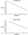
- This calibration curve was created in that defined amounts of villin (a) or fimbrin (b) were given to the urine of a person were added to the normal person and quantified using the above-mentioned immunological method.
- the microtiter well contains 1: 10 ⁇ g / ml, 2: 5 ⁇ g / ml, 3: 2.5 ⁇ g / ml, 4: 1.25 ⁇ g / ml, 5: 0.625 ⁇ g / ml and 6: 0.312 ⁇ g / ml protein .
- FIG. 2 graphically depicts a case study on a patient with a transplanted kidney over 30 days (abscissa), the villine content in the patient's urine samples being determined for each day.
- Fig. 2 From Fig. 2 it can be seen that the patient has a villin concentration with a baseline absorbance of 0.05.
- the absolute concentrations of villin in the patient's urine can be read from the calibration curve according to FIG. 1.
- the arrows drawn in FIG. 2 indicate rejection crises that have been clinically verified by increased creatinine values and clinical symptoms. Therapy for these rejection crises consisted of increased doses of immunosuppressive drugs, which resulted in a decrease in urine villi excretion, i.e. reflected in the decline in the risk of damage to the kidney.
Abstract
Description
Die Erfindung betrifft ein Verfahren zur Bestimmung von Nierenschäden, bei dem poly- oder monoklonale Antikörper mit in der Niere auftretenden Proteinen in Berührung gebracht werden und das erhaltene Reaktionsprodukt anschließend einem immunologischen Nachweisverfahren unterzogen wird, sowie ein Mittel, mit dem dieses Verfahren durchgeführt werden kann.The invention relates to a method for determining kidney damage, in which poly- or monoclonal antibodies are brought into contact with proteins occurring in the kidney and the reaction product obtained is then subjected to an immunological detection method, and to an agent with which this method can be carried out.
Nierenschäden machen sich üblicherweise zuerst durch Schädigungen des Parenschyms bemerkbar, die beispielsweise bei fortlaufender Einnahme von nierenschädigenden Medikamenten, wie Phenacetin, bei einer gesunden Niere oder bei Nichteinnahme eines Immunosuppresivums bei einer transplantierten Niere auftreten. Insofern ist die Früherkennung von Parenschymschäden der Niere ein wichtiges medizinisches Problem.Kidney damage is usually first noticeable through damage to the parenchyma, which occurs, for example, when taking kidney-damaging medications, such as phenacetin, in a healthy kidney or when an immunosuppressant is not taken in a transplanted kidney. In this respect, the early detection of parenchymal damage to the kidney is an important medical problem.
Derzeit werden im wesentlichen drei Untersuchungsverfahren angewandt, um Parenchymschäden der Niere zu diagnostizieren :
- a. Das klassische Verfahren besteht darin, daß durch Biopsie Gewebsproben aus der Niere entnommen werden. Dieses invasive Verfahren ist jedoch mit Komplikationen behaftet und kommt nur in Ausnahmefällen zum Einsatz.
- b. Es wird die Aktivität von Gewebsenzymen im Urin bestimmt. Dieser klinische Test ist jedoch in seiner Aussagefähigkeit stark eingeschränkt, weil durch ihn keine verläßlich Lokalisation des Schädigungsortes in der Niere erfolgen kann und die Aktivität der Enzyme im Urin durch zahlreiche äußere Faktoren beeinflußt wird. Dieser Test ist bereits in qualitativer Hinsicht zu unspezifisch.
- c. Desweiteren wurde versucht, auf immunologische Weise Schädigungen der Niere nachzuweisen. Dabei wurden monoklonale Antikörper gegen unbekannte oder unzureichend charakterisierte Nierenproteine eingesetzt, die in erhöhten Mengen im Urin ausgeschieden werden. Diese Methode kann allenfalls als grobes qualitatives Verfahren zur Bestimmung von Nierenschädigungen angesehen werden, da die Proteine, gegen die die Antikörper gerichtet sind, nicht isoliert und deshalb nicht zur quantitativen Kalibrierung der Meßwerte herangezogen werden. Insofern sind mit dieser Methods keinesfalls quantitative Aussagen möglich, die zur Abschätzung des Schweregrades einer Nierenschädigung aber erforderlich sind.
- a. The classic method is to take tissue samples from the kidney by biopsy. However, this invasive procedure is fraught with complications and is only used in exceptional cases.
- b. The activity of tissue enzymes in the urine is determined. This clinical test, however, is severely limited because it cannot reliably locate the site of damage in the kidney and the activity of the enzymes in the urine is influenced by numerous external factors. This test is already too unspecific in terms of quality.
- c. Furthermore, attempts have been made to demonstrate kidney damage in an immunological manner. Monoclonal antibodies against unknown or insufficiently characterized kidney proteins were used, which are excreted in the urine in increased amounts. This method can at best be regarded as a rough qualitative method for the determination of kidney damage, since the proteins against which the antibodies are directed are not isolated and are therefore not used for the quantitative calibration of the measured values. In this respect, quantitative statements are by no means possible with these methods, but they are necessary to estimate the severity of kidney damage.
Der Erfindung liegt daher die Aufgabe zugrunde, das Verfahren der eingangs erwähnten Art so zu verbessern, daß auch geringe bzw. im Anschwellen befindliche Nierenschädigungen praktisch quantitativ erfaßt werden können.The invention is therefore based on the object of improving the method of the type mentioned at the outset in such a way that even minor or swelling kidney damage can be practically quantified.
Der Erfindung liegt weiterhin die Aufgabe zugrunde, ein Mittel zur Durchführung des Verfahrens zur Verfügung zu stellen.Another object of the invention is to provide a means for carrying out the method.
Die Lösung der Aufgabe erfolgt dadurch, daß man als Antikörper solche einsetzt, die gegen die Proteine Fimbrin oder Villin spezifisch sind.The problem is solved by using antibodies which are specific for the proteins fimbrin or villin as antibodies.
Überraschenderweise wurde nunmehr festgestellt, daß die Proteine Villin und Fimbrin, die zwei molekulardefinierte Proteine des proximalen Nierentubulusepithels sind, in praktisch quantitativer Weise als Maßstab für eine Nierenschädigung herangezogen werden können. Dabei können diese Proteine im Urin quantitative ermittelt werden. Beide Proteine werden nämlich vermehrt bei Schädigung der Niere ausgeschieden, die den proximalen Tubulus betreffen oder ihn einbeziehen. Somit steht erfindungsgemäß ein nichtinvasives, diagnostisches Verfahren zur Verfügung, mit dem Schädigungen der Niere, beispielsweise verursacht durch Durchblutungsstörungen, drohendes akutes Nierenversagen oder drohende Tranplantatabstoßung, frühzeitig quantitativ erfaßt werden können.Surprisingly, it has now been found that the proteins villin and fimbrin, which are two molecularly defined proteins of the proximal renal tubular epithelium, can be used in a practically quantitative manner as a yardstick for kidney damage. These proteins can be determined quantitatively in the urine. Both proteins are namely increased in Damage to the kidney that affects or involves the proximal tubule. Thus, according to the invention, a non-invasive, diagnostic method is available with which damage to the kidney, for example caused by circulatory disorders, impending acute kidney failure or impending transplant rejection, can be quantified at an early stage.
Mit demn erfindungsgemäßen Verfahren können Aussagen zu folgenden klinischen Fragestellungen gemacht werden, nämlich zur
- a. Lokalisation einer Nierenschädigung im proximalen Tubulus,
- b. Beurteilung des Schweregrades der Zellschädigung,
- c. Ansprechbarkeit der Nierenschädigung auf therapeutische Maßnahmen und
- d. Abschätzung des Risikos der Behandlung von Nierenkranken und nierentransplantierten Patienten mit Medikamenten, die nierenschädigende Nebenwirkungen haben.
- a. Localization of kidney damage in the proximal tubule,
- b. Assessment of the severity of cell damage,
- c. Responsiveness of kidney damage to therapeutic measures and
- d. Assessment of the risk of treating kidney patients and kidney transplant patients with medication that has kidney-damaging side effects.
Die Proteine Villin und Fimbrin lassen sich einfach im Urin nachweisen, mit dem sie ausgeschieden werden. Üblicherweise sind nur etwa 0,1 ml Urin notwendig, das der Patient ausscheidet. Insofern hat das Nachweisverfahren keine schädigenden oder toxischen Wirkungen auf den Patienten, da die Untersuchung im ausgeschiedenen Urin in vitro erfolgt.The proteins villin and fimbrin can easily be detected in the urine with which they are excreted. Usually only about 0.1 ml of urine is excreted by the patient. In this respect, the detection method has no harmful or toxic effects on the patient, since the examination is carried out in the excreted urine in vitro.
Die Proteine Villin und Fimbrin lassen sich in reiner Form darstellen. Insofern ist eine exakte Festlegung der Meßwerte möglich, d.h. die Proteine Fimbrin und Villin lassen sich exakt quantitativ im Urin bestimmen, so daß hierdurch ein Parameter für den Schweregrad der Schädigung der Nierentubuli Verfügung steht. Bei dem erfindungsgemäßen Verfahren wird vorteilhaft eine Kalibrierung des eingesetzten Reagenz durchgeführt, um den Gehalt der Proteine Villin und/oder Fimbrin im Urin quantitativ festzulegen.The proteins Villin and Fimbrin can be represented in pure form. In this respect, an exact determination of the measured values is possible, ie the proteins Fimbrin and Villin can be determined exactly quantitatively in the urine, so that this is a parameter for the severity of the Damage to the renal tubules is available. In the method according to the invention, a calibration of the reagent used is advantageously carried out in order to quantitatively determine the content of the proteins villin and / or fimbrin in the urine.
Ein typisches, einfaches Verfahren, mit dem die Proteine Villin und Fimbrin im Urin quantitative nachgewiesen werden können, besteht aus folgenden Merkmalen : Die Proteine Villin und Fimbrin werden auf an sich bekannte Weise aus dem Darmepithel des Huhns oder des Schweins isoliert. Dieses Verfahren ist von A. Bretscher und K. Weber in Proc. Natl. Acad. Sci. USA 76 (1979), S. 2321-2325 oder J. Cell. Biol. 86 (1980) Seite 335-340 oder D. Drenckhahn et al. in Cell. Tiss. Res. 228 (1983) beschrieben. Auf die darin erläuterten Isolierungsverfahren wird ausdrücklich Bezug genommen.A typical, simple method with which the proteins villin and fimbrin can be quantitatively detected in urine consists of the following features: The proteins villin and fimbrin are isolated in a manner known per se from the intestinal epithelium of the chicken or the pig. This method is from A. Bretscher and K. Weber in Proc. Natl. Acad. Sci. USA 76 (1979), pp. 2321-2325 or J. Cell. Biol. 86 (1980) pages 335-340 or D. Drenckhahn et al. in Cell. Tiss. Res. 228 (1983). Reference is expressly made to the insulation processes explained therein.
Auf gleiche Weise können diese Proteine aus dem Darmepithel von chirurgisch entfernten Darmstücken des Menschen isoliert werden.In the same way, these proteins can be isolated from the intestinal epithelium of surgically removed pieces of human intestine.
Antikörper gegen Fimbrin und/oder Villin können nach den vorstehend angegebenen Literaturstellen oder aber auch nach allgemein bekannten Methoden hergestellt werden. Solche Methoden sind beispielsweise in der Monographie von R.J. Mayer und J.H. Walker mit dem Titel "Immunochemical methods in the biological sciences: Enzymes und proteins" erschienen in Academic Press, London (1980), beschrieben.Antibodies against fimbrin and / or villin can be produced according to the literature references given above or else according to generally known methods. Such methods are described, for example, in the monograph by R.J. Mayer and J.H. Walker, entitled "Immunochemical methods in the biological sciences: Enzymes and proteins", appeared in Academic Press, London (1980).
So werden z.B. die isolierten Proteine in Mengen von 50-100 µg mit und ohne Freund'schem kompletten oder inkompletten Adjuvanz Kaninchen oder Meerschweinchen unter die Rückenhaut oder in die Bauchhöhe injiziert. In dreiwöchigen Abständen wird die gleiche Menge an Proteinen wieder injiziert. Nach der dritten oder späteren Injektion werden gute Titer polyklonaler Antikörper erhalten, die nach üblichen Standardverfahren affinitätsgereinigt und auf ihre Spezifität getestet werden. Ein solches Verfahren wurde in den o.a. Publikationen von Drenckhahn et al., 1983, oder Bretscher und Weber, 1979, 1980 oder von Talian et al. in J. Cell. Biol. 97 (1983), S. 1277-1282 beschrieben, worauf Bezug genommen wird.For example, the isolated proteins in amounts of 50-100 µg with and without Freund's complete or incomplete adjuvant become rabbits or guinea pigs injected under the dorsal skin or at the level of the abdomen. The same amount of proteins is re-injected at three-week intervals. After the third or later injection, good titres of polyclonal antibodies are obtained, which are affinity-cleaned and tested for their specificity according to standard methods. Such a method has been described in the above publications by Drenckhahn et al., 1983, or Bretscher and Weber, 1979, 1980 or by Talian et al. in J. Cell. Biol. 97 (1983), pp. 1277-1282, to which reference is made.
Neben polyklonalen Antikörpern lassen sich auch monoklonale Antikörper einsetzen, die in Mäusen und Ratten nach ebenfalls allgemein bekannten Verfahren hergestellt werden (vgl. die Publikation von J.H. Peters et al., "Monoklonale Antikörper: Herstellung und Charakterisierung", erschienen in Springer-Verlag, Berlin (1985).In addition to polyclonal antibodies, it is also possible to use monoclonal antibodies which are also produced in mice and rats by methods which are also generally known (cf. the publication by JH Peters et al., "Monoclonal Antibodies: Production and Characterization", published by Springer-Verlag, Berlin (1985).
Derartige monoklonale Antikörper gegen Villin werden bereits zur histologischen Diagnose von Tumoren des Darms, in dem ebenfalls Villin freigesetzt wird, kommerziell von der Firma Dianova, Hamburg, angeboten.Such monoclonal antibodies against Villin are already commercially available from Dianova, Hamburg, for the histological diagnosis of tumors of the intestine, in which Villin is also released.
Erfindungsgemäß werden die poly- oder monoklonalen Antikörper gegen die Proteine Villin und/oder Fimbrin an der Oberfläche von Reaktionsträgern gebunden. Unter "Bindung" ist dabei entweder eine physikalische Adsorption der Antikörper an ein Substrat oder aber eine chemische kovalente Bindung dieser Antikörper an das Substrat zu verstehen.According to the invention, the poly- or monoclonal antibodies against the proteins villin and / or fimbrin are bound to the surface of reaction carriers. "Binding" is understood to mean either a physical adsorption of the antibodies to a substrate or a chemical covalent binding of these antibodies to the substrate.
Als Reaktionsträger können Filtermaterialien, Materialien von Reaktionsgefäßen, wie Mikrotiterplatten, Säulenchromatographiematerialien, wie Sepharose und dergl. in Frage kommen. Die Antikörper werden in wässeriger Lösung, vorteilhafterweise in einer wässerigen Pufferlösung mit dem Substratmaterial so lange in Berührung gebracht, bis sich ein Adsorptionsgleichgewicht gebildet hat. Diese Gleichgewichtseinstellung ist temperaturabhängig und dauert bei etwa 21 °C ca. 3 Stunden und bei 4°C ca. 12 Stunden.Filter materials, materials from reaction vessels such as microtiter plates, column chromatography materials such as Sepharose and the like. The antibodies are brought into contact with the substrate material in an aqueous solution, advantageously in an aqueous buffer solution, until an adsorption equilibrium has formed. This equilibrium setting is temperature-dependent and takes about 3 hours at about 21 ° C and about 12 hours at 4 ° C.
Als Substratmaterial ist ein Kunststoffmaterial, insbesondere Polystyrol, bevorzugt, das üblicherweise zur Herstellung von Mikrotiterplatten eingesetzt wird.A plastic material, in particular polystyrene, is preferred as the substrate material, which is usually used for the production of microtiter plates.
Nach Abschluß der Bindungsreaktion werden die adsorbierten Antikörper zur Absättigung freier Proteinbindungsstellen mit Proteinlösungen, beispielsweise Albuminlösungen, Gelatinelösungen oder Magermilch ggf. unter Zusatz von Kochsalz und/oder Detergens behandelt.After completion of the binding reaction, the adsorbed antibodies are treated with protein solutions, for example albumin solutions, gelatin solutions or skimmed milk to saturate free protein binding sites, optionally with the addition of sodium chloride and / or detergent.
Anschließend werden die so behandelten Antikörper-Beschichtungen mehrfach gewaschen, woraufhin die Behandlung mit einer Urinprobe erfolgen kann.The antibody coatings treated in this way are then washed several times, after which treatment with a urine sample can take place.
Die Inkubation mit Urin wird so lange durchgeführt, bis sich ein Reaktionsgleichgewicht zwischen den Antikörpern und den Proteinen Villin und/oder Fimbrin eingestellt hat. Vorteilhafterweise dauert der Inkubationsvorgang etwa 30-90 Minuten. Anschließend entfernt man den Urin und füllt, sofern Mikrotiterplatten eingesetzt werden, erneut die Löcher mit der vorstehend angegebenen Waschlösung, die in bestimmten Zeitabständen mehrfach gewechselt wird.The incubation with urine is carried out until a reaction equilibrium has been established between the antibodies and the villin and / or fimbrin proteins. The incubation process advantageously takes about 30-90 minutes. The urine is then removed and, if microtiter plates are used, the holes are filled again with the washing solution specified above, which is changed several times at certain intervals.
Der so erzeugte Antikörper-Protein-Bindungskomplex wird anschließend einer immunologischen Quantifizierungsreaktion in einem zweiten Verfahrensschritt unterzogen.The antibody-protein binding complex thus produced is then subjected to an immunological quantification reaction in a second process step.
Zur Durchführung dieser im zweiten Verfahrensschritt stattfindenden Quantifizierungsreaktion wird der am Substrat haftende Antikörper-Protein-Bindungskomplex erneut mit Antikörpern gegen Villin oder Fimbrin behandelt.To carry out this quantification reaction, which takes place in the second process step, the antibody-protein binding complex adhering to the substrate is again treated with antibodies against villin or fimbrin.
Sofern im ersten Verfahrensschritt ein polyklonaler Antikörper verwendet wurde, wird im zweiten Verfahrensschritt entweder der gleiche polyklonale Antikörper oder aber ein monoklonaler Antikörper gegen Villin oder Fimbrin eingesetzt.If a polyclonal antibody was used in the first process step, either the same polyclonal antibody or a monoclonal antibody against villin or fimbrin is used in the second process step.
Wird jedoch gemäß einer zweiten Ausführungsform ein monoklonaler Antikörper im ersten Verfahrensschritt eingesetzt, dann kann als Antikörper im zweiten Verfahrensschritt entweder ein polyklonaler oder ein vom ersten Antikörper unterschiedlicher zweiter monoklonaler Antikörper verwendet werden, der gegen eine andere Bindungsstelle des Proteinmoleküls gerichtet ist.If, however, according to a second embodiment, a monoclonal antibody is used in the first process step, then either a polyclonal antibody or a second monoclonal antibody different from the first antibody, which is directed against another binding site of the protein molecule, can be used as the antibody.
Nachstehend wird der in diesem zweiten Verfahrensschritt eingesetzte Antikörper als "zweiter Antikörper" bezeichnet.The antibody used in this second method step is referred to below as the "second antibody".
Erfindungsgemäß werden die Löcher der Mikrotiterplatten mit einer diesen zweiten Antikörper enthaltenden Lösung ebenfalls ca. 30-90 Minuten gefüllt und inkubiert. Anschließend wird die zweite Antikörperlösung entfernt, woraufhin die Löcher mit einer Waschlösung gespült werden. Es bleibt auf dem Substrat zurück ein Komplex, bestehend aus dem ersten Antikörper, den gebundenen Proteinen Villin oder Fimbrin und dem zweiten Antikörper, nachstehend als "Quantifizierungskomplex" bezeichnet.According to the invention, the holes in the microtiter plates are also filled with a solution containing this second antibody for about 30-90 minutes and incubated. The second antibody solution is then removed, whereupon the holes are rinsed with a washing solution. A complex remains on the substrate, consisting of the first antibody, the bound proteins villin or fimbrin and the second antibody, hereinafter referred to as "quantification complex".
Dieser Quantifizierungskomplex kann auf mehrfache Weise einer Quantifizierungsreaktion unterzogen werden :This quantification complex can be subjected to a quantification reaction in several ways:
Gemäß einer ersten Ausführungsform wird der zweite Antikörper einer kovalenten Kopplung mit Enzymen, beispielsweise Peroxidase, alkalischer Phosphatase, Glukoseoxidase oder Betagalaktosidase unterworfen. Die Menge der im Quantifizierungskomplex gebundenen enzymmarkierten zweiten Antikörper kann anschließend durch eine Enzymfarbreaktion quantitativ bestimmt werden. Derartige Quantifizierungsreaktionen sind an sich bekannt, wobei einsetzbare Enzyme und typische Enzymfarbreaktionen von L.A. Sternberger in "Immunocytochemistry", Dritte Ausgabe, Verlag John Wiley & Sons, New York (1986) oder von W.D. Kuhlmann in der Monographie "Immunoenzyme techniques in cytochemistry", Verlag Chemie, Weinheim (1984) beschrieben worden sind. Hierauf wird Bezug genommen.According to a first embodiment, the second antibody is subjected to covalent coupling with enzymes, for example peroxidase, alkaline phosphatase, glucose oxidase or beta-galactosidase. The amount of the enzyme-labeled second antibodies bound in the quantification complex can then be determined quantitatively by an enzyme color reaction. Such quantification reactions are known per se, with usable enzymes and typical enzyme color reactions from L.A. Sternberger in "Immunocytochemistry", third edition, published by John Wiley & Sons, New York (1986) or from W.D. Kuhlmann in the monograph "Immunoenzyme techniques in cytochemistry", Verlag Chemie, Weinheim (1984) have been described. This is referred to.
Gemäß einer zweiten Ausführungsform können die zweiten Antikörper nach ansich bekannten Verfahren radioaktiv markiert werden, beispielsweise mit Jod 125. Für diese Zwecke werden radioaktive Markierungskits und Substanzen eingesetzt, die beispielsweise von der Firma NEN, Dreieich, geliefert werden.According to a second embodiment, the second antibodies can be radioactively labeled using methods known per se, for example with iodine 125. For this purpose, radioactive labeling kits and substances are used which are supplied, for example, by the company NEN, Dreieich.
Die Menge der gebundenen zweiten Antikörper kann dabei durch einen Teilchenzähler bzw. Strahlenzähler direkt quantitativ ermittelt werden.The amount of bound second antibodies can be directly determined quantitatively by a particle counter or radiation counter.
In einer dritten Ausführungsform der Quantifizierungsreaktion kann der Komplex mit einem dritten Antikörper behandelt werden, der gegen den gebundenen zweiten Antikörper spezifisch ist. Für diese Zwecke können beispielsweise Antikörper gegen Mäuseimmunoglobuline oder Kaninchenimmunoglobuline eingesetzt werden. Solche Antikörper sind in markierter Form mit Enzymen oder Radioisotopen kommerziell erhältlich und werden beispielsweise von den Firmen Dianova, Hamburg; Janssen, Belgien oder NEN, Dreieich, vertrieben. Anschließend erfolgt der bereits oben erwähnte Quantifizierungsschritt, wobei entweder die Farbreaktion quantitativ ausgewertet oder aber die radioaktive Strahlung bestimmt werden.In a third embodiment of the quantification reaction, the complex can be treated with a third antibody which is specific for the bound second antibody. Antibodies against mouse immunoglobulins or Rabbit immunoglobulins are used. Such antibodies are commercially available in labeled form with enzymes or radioisotopes and are, for example, from the companies Dianova, Hamburg; Janssen, Belgium or NEN, Dreieich. This is followed by the quantification step already mentioned, either evaluating the color reaction quantitatively or determining the radioactive radiation.
Diese dritten Antikörper können auch in nichtmarkierter Form als Brückenantikörper dienen, welche Enzym-Anti-Enzymimmunkomplexe bilden, die ggf. in einem vierten Inkubationsschritt eingebracht werden. Diese indirekten Immunglobulinnachweisverfahren sind ebenfalls aus der Literatur bekannt, beispielsweise in der vorstehend erwähnten Monographie von Sternberger, worauf Bezug genommen wird. Im übrigen sind Enzym-Anti-Enzymimmunkomplexe ebenfalls kommerziell erhältlich und werden beispielsweise von Firmen Dianova, Hamburg oder Janssen, Belgien, vertrieben.These third antibodies can also serve in unlabelled form as bridge antibodies, which form enzyme-anti-enzyme immune complexes, which may be introduced in a fourth incubation step. These indirect immunoglobulin detection methods are also known from the literature, for example in the above-mentioned monograph by Sternberger, to which reference is made. In addition, enzyme-anti-enzyme immune complexes are also commercially available and are sold, for example, by Dianova, Hamburg or Janssen, Belgium.
In einer vierten Quantifizierungsreaktion, die nachstehend detailliert beschrieben ist und insofern bevorzugt ist, wird die Quantifizierung von Villin oder Fimbrin im Urin dadurch durchgeführt, daß die zweiten Antikörper mit Biotin kovalent gekoppelt werden. Die Kopplung von Biotin erfolgt an den zweiten Antikörper nach ansich bekannten Verfahren, die beispielsweise von N.H. Heggeness et al. in J. Cell. Biol. 73 (1977), S. 783-788 beschrieben ist, worauf Bezug genommen wird. Hierzu wird zum Beispiel Biotin-N-hydroxysuccinimid-ester eingesetzt, das von der Firma Calbiochem. Hoechst vertrieben wird.In a fourth quantification reaction, which is described in detail below and is preferred in this respect, the quantification of villin or fimbrin in the urine is carried out by covalently coupling the second antibodies to biotin. Biotin is coupled to the second antibody by methods known per se, which have been described, for example, by NH Heggeness et al. in J. Cell. Biol. 73 (1977), pp. 783-788, which is incorporated by reference. For this purpose, for example, biotin-N-hydroxysuccinimide ester is used, which is manufactured by Calbiochem. Hoechst is distributed.
Die im Quantifizierungskomplex gebundenen biotinilierten zweiten Antikörper werden durch das biotinbindende Protein Streptavidin quantifiziert. Streptavidin ist mit dem Enzym Peroxidase oder den anderen oben erwähnten Enzymen kovalent an den zweiten Antikörper gebunden. Derartige Enzym-Streptavidin-Lösungen sind kommerziell erhältlich und werden beispielsweise von der Firma Calbiochem-Hoechst vertrieben.The biotinylated second antibodies bound in the quantification complex are quantified by the biotin binding protein streptavidin. Streptavidin is covalently bound to the second antibody with the enzyme peroxidase or the other enzymes mentioned above. Enzyme streptavidin solutions of this type are commercially available and are marketed, for example, by Calbiochem-Hoechst.
Der erhaltene Streptavidin-Enzym-Antikörperkomplex wird anschließend, sofern das Enzym eine Peroxidase ist, mit 1,2-Phenylendiamin und H₂O₂- Lösung versetzt, wobei durch die Reaktion von Peroxidase mit Wasserstoffperoxid das farblose Phenylendiamin in einen gelben Farbstoff umgewandelt wird. Der entstandene Farbstoff kann durch seine Extinktion bei 492 nm gegenüber seiner Referenzwellenlänge von 620 nm im UV-Photometer bestimmt werden. Dabei korreliert die Extinktion mit der Menge der gebundenen Villin- und/oder Fimbrin-Moleküle, wie dies nachstehend gezeigt werden kann.The streptavidin-antibody-complex obtained is then, if the enzyme is a peroxidase, mixed with 1,2-phenylenediamine and H₂O₂ solution, the colorless phenylenediamine being converted into a yellow dye by the reaction of peroxidase with hydrogen peroxide. The resulting dye can be determined by its absorbance at 492 nm compared to its reference wavelength of 620 nm in a UV photometer. The absorbance correlates with the amount of bound villin and / or fimbrin molecules, as can be shown below.
Gemäß einer fünften Ausführungsform können Villin und Fimbrin im Urin auch durch einen Verdrängungsassay nachgewiesen werden. Bei diesem Testverfahren werden Reaktionsträger mit gereinigtem Villin und/oder Fimbrin beschichtet. Anschließend wird ein Gemisch von einer zu untersuchenden Urinprobe und von Antikörpern beigefügt, die gegen Villin oder Fimbrin spezifisch sind. Je höher die Menge von Villin oder Fimbrin im Urin ist, desto geringer wird die Menge an Antikörpern sein, die an diejenigen Villin- oder Fimbrinmoleküle binden, die bereits am Reaktionsträger adsorbiert worden waren. Die Menge der gebundenen Antikörper kann anschließend durch direkte Markierung der Antikörper mit Enzymen oder Radioisotopen oder durch die übrigen beschriebenen Ver fahren quantifiziert werden.According to a fifth embodiment, villin and fimbrin can also be detected in the urine by a displacement assay. In this test procedure, reaction carriers are coated with purified villin and / or fimbrin. A mixture of a urine sample to be examined and antibodies specific to villin or fimbrin is then added. The higher the amount of villin or fimbrin in the urine, the lower the amount of antibodies that will bind to those villin or fimbrin molecules that have already been adsorbed on the reaction support. The amount of bound antibodies can then be determined by direct labeling of the antibodies with enzymes or radioisotopes or by the other described Ver driving be quantified.
Es sei nochmals erwähnt, daß die Quantifizierungsreaktionen, mit denen der Quantifizie rungskomplex behandelt wird, ansich bekannt sind. Insofern sind auch weitere Abwandlungen der Quantifizierungsreaktionen denkbar, sofern die gebundenen Villin oder Fimbrin-Moleküle hierdurch quantitativ erfaßt werden können.It should be mentioned again that the quantification reactions with which the quantification complex is treated are known per se. In this respect, further modifications of the quantification reactions are also conceivable, provided that the bound villin or fimbrin molecules can be detected quantitatively.
Erfindungsgemäß wird auch ein Mittel zur Durchführung des vorstehend beschriebenen Verfahrens zur Verfügung gestellt.According to the invention, a means for carrying out the method described above is also made available.
Gemäß einer ersten Variante A besteht das Mittel aus mono- oder polyklonalen Antikörpern gegen Fimbrin und/oder Villin.According to a first variant A , the agent consists of mono- or polyclonal antibodies against fimbrin and / or villin.
Vorzugsweise werden bereits an einen Reaktionsträger gebundene Antikörper in den Handel gebracht, wobei das Einbringen von Antikörpern in Mikrotiterplatten bevorzugt ist. So kann eine Handelseinheit aus einer Mikrotiterplatte mit einer Vielzahl von Löchern bestehen, wobei jedes Loch mit einer bestimmten Menge Antikörper gegen Villin und/oder Fimbrin versehen ist.Antibodies which are already bound to a reaction carrier are preferably brought onto the market, the introduction of antibodies into microtiter plates being preferred. A trading unit can thus consist of a microtiter plate with a multiplicity of holes, each hole being provided with a specific amount of antibody against villin and / or fimbrin.
Zusätzlich zu dieser Substrateinheit weist das erfindungsgemäße Mittel vorteilhaft den vorstehend erwähnten zweiten Antikörper zur Herstellung des Quanitfizierungskomplexes auf. An diesen zweiten Antikörper sind vorteilhafterweise Biotingruppen kovalent gebunden. Diese biotinilierten Antikörper können entweder bereits als in fertiger gepufferter Gebrauchslösung in den Handel gebracht werden oder aber zusammen mit der Puffersubstanz in lyophilisierter Form vertrieben werden, wobei das die lyophilisierten Produkte enthaltene Gefäß für den Einsatz lediglich mit einer bestimmten Menge Wasser gefüllt werden muß.In addition to this substrate unit, the agent according to the invention advantageously has the above-mentioned second antibody for producing the quantitation complex. Biotin groups are advantageously covalently bound to this second antibody. These biotinylated antibodies can either already be put on the market as a ready-to-use buffered solution, or else they can be sold together with the buffer substance in lyophilized form, the vessel containing the lyophilized products only needs to be filled with a certain amount of water for use.
Eine derartige Handelseinheit kann weiterhin - sofern nicht im Labor vorhanden - Konzentrate der vorstehend erwähnten Waschlösungen sowie die Quantifizierungsreagenzien Streptavidin-Peroxidase, H₂O₂, Phenylendiamin enthalten, die zur Quantifizierung des Quantifizierungskomplexes eingesetzt werden.Such a commercial unit can also - if not available in the laboratory - contain concentrates of the above-mentioned washing solutions and the quantification reagents streptavidin peroxidase, H₂O₂, phenylenediamine, which are used to quantify the quantification complex.
Außerdem werden vorteilhafterweise zur Eichung der Meßwerte mit jeder Substrateinheit die isolierten gereinigten proteine Fimbrin und/oder Villin geliefert, vorteilhafterweise in einer Menge von etwa 4 µg. Diese werden in lyophilisierter Form in einem Gefäß vertrieben, das mit einer bestimmten Menge Wasser oder Kontrollurin vom Verbraucher aufgefüllt wird.In addition, the isolated purified proteins fimbrin and / or villin are advantageously supplied with each substrate unit for calibration of the measured values, advantageously in an amount of approximately 4 μg. These are distributed in lyophilized form in a container which is filled up with a certain amount of water or control urine by the consumer.
Gemäß einer zweiten Varianten B, die auf dem vorstehend beschriebenen Verdrängungsassay beruht, besteht das Mittel neben den vorstehend unter Variante A genannten Antikörpern gegen Fimbrin und/oder Villin aus den isolierten Proteinen Villin oder Fimbrin.According to a second variant B , which is based on the displacement assay described above, the agent, in addition to the antibodies against fimbrin and / or villin mentioned above under variant A, consists of the isolated proteins villin or fimbrin.
Die Proteine werden an Reaktionsträger gebunden in den Handel gebracht. Eine Handelseinheit kann aus einer Mikrotiterplatte bestehen, wobei jedes Loch mit einer bestimmten Menge (beispielsweise 0,5 - 1 µg) der isolierten Proteine Villin und/oder Fimbrin versehen ist.The proteins are brought onto the market bound to reaction carriers. A commercial unit can consist of a microtiter plate, each hole being provided with a certain amount (for example 0.5-1 μg) of the isolated proteins villin and / or fimbrin.
Zusätzlich zu dieser Substrateinheit weist das zweite Mittel die vorstehend erwähnten mono- oder polyklonalen Antikörper gegen Villin oder Fimbrin auf, an die vorteilhafterweise Biotingruppen kovalent gebunden sind.In addition to this substrate unit, the second agent has the abovementioned mono- or polyclonal antibodies against villin or fimbrin, to which biotin groups are advantageously covalently bound.
Diese Antikörper werden entweder als fertige Gebrauchslösung oder in lyophilisierter Form vertrieben, wie in der ersten Ausführungsform beschrieben.These antibodies are sold either as a ready-to-use solution or in lyophilized form as described in the first embodiment.
Außerdem enthält vorteilhafterweise jede Einheit dieser Ausführungsform zu Eichzwecken noch die isolierten Proteine Villin und/oder Fimbrin, wie ebenfalls zur Variante A beschrieben.In addition, each unit of this embodiment advantageously also contains the isolated proteins villin and / or fimbrin for calibration purposes, as also described for variant A.
Dieser Nachweiskit kann weiterhin, wie in der Variante A beschrieben, die vorstehend erwähnten Waschlösungen sowie die Quantifizierungsreagenzien enthalten.As described in variant A, this detection kit can also contain the above-mentioned washing solutions and the quantification reagents.
Das nachfolgende Beispiel erläutert die Erfindung :The following example explains the invention:
Polyklonale Antikörper gegen Fimbrin und/oder Villin werden gemäß dem Verfahren von Bretscher und Weber 1979, 1980 und Drenckhahn et al.,1983 hergestellt. Monoklonale Antikörper werden entsprechend dem Verfahren von Peter et al., 1985 hergestellt oder es wird ein monoklonaler Antikörper gegen Villin von der Firma Dianova, Hamburg, eingesetzt.Polyclonal antibodies to Fimbrin and / or Villin are made according to the method of Bretscher and Weber 1979, 1980 and Drenckhahn et al., 1983. Monoclonal antibodies are produced according to the method of Peter et al., 1985 or a monoclonal antibody against Villin from Dianova, Hamburg, is used.
Diese Antikörper werden an Polystyrol-Mikrotiterplatten, bezogen von der Firma Nunc, Wiesbaden, entsprechend dem Verfahren von Peter et al., 1985, gebunden.These antibodies are bound to polystyrene microtiter plates, obtained from Nunc, Wiesbaden, in accordance with the method of Peter et al., 1985.
Dazu werden die isolierten Antikörper in einer Immunoglobulin-Konzentration von 5 µg/ml in 0,2 M- Carbonatpuffer mit einem pH-Wert von 10,6 gelöst. Nicht gereinigtes Immunserum wird zweckmäßgerweise in einem Verhältnis von 1:100 im Carbonatpuffer verdünnt. Nachstehend wird der Begriff "Antikörper" als übergreifender Begriff für isolierte Immunglobuline und Immunserum verwendet.For this purpose, the isolated antibodies are dissolved in an immunoglobulin concentration of 5 µg / ml in 0.2 M carbonate buffer with a pH of 10.6. Immune serum which has not been purified is expediently diluted in a ratio of 1: 100 in the carbonate buffer. In the following, the term "antibody" is used as a general term for isolated immunoglobulins and immune serum.
Zur ausreichenden Bindung der Antikörper an die Wand Löcher der Mikrotiterplatten werden jeweils 100 µl der verdünnten Antikörper-Lösung etwa drei Stunden bei 21°C bzw. zwölf Stunden bei 4°C in den Löchern der Mikrotiterplatten belassen. Danach wird die Antikörperlösung aus den Löchern entfernt, wobei anschließend die Löcher zur Absättigung freier Bindungsstellen mit etwa 400 µl einer 1 gew.-%igen Gelatinelösung in dem vorstehend angegebenen Karbonatpuffer gefüllt werden.For sufficient binding of the antibodies to the wall holes in the microtiter plates, 100 μl of the diluted antibody solution are left in the holes of the microtiter plates for about three hours at 21 ° C. or twelve hours at 4 ° C. The antibody solution is then removed from the holes, and the holes are then filled with about 400 μl of a 1% strength by weight gelatin solution in the carbonate buffer indicated above in order to saturate free binding sites.
Nach einer Stunde Inkubationszeit wird die Gelatinelösung wieder entfernt und die Löcher werden mit etwa 400 ul Waschpuffer gefüllt.After an hour of incubation, the gelatin solution is removed again and the holes are filled with approximately 400 μl washing buffer.
Dieser Waschpuffer besteht aus einer mit Phosphat gepufferten Kochsalzlösung (25 mM Phosphatpuffer, 100mM NaCl mit einem pH-Wert von 7,4), der nachstehend PPK genannt wird. Dieser Pufferlösung werden 0,05 Gew.-% des Detergens Tween 20 zugesetzt, das von der Firma Serva, Heidelberg, bezogen werden kann.This washing buffer consists of a saline solution buffered with phosphate (25 mM phosphate buffer, 100mM NaCl with a pH of 7.4), which is called PPK below. 0.05% by weight of the detergent Tween 20, which can be obtained from Serva, Heidelberg, is added to this buffer solution.
Der Waschpuffer wird in 5-minütigen Abständen 3 mal gewechselt.The wash buffer is changed 3 times in 5-minute intervals.
Die so präparierte Mikrotiterplatte kann entweder unmittelbar der Reaktion mit einer zu untersuchenden Urinprobe ausgesetzt werden oder aber getrocknet und anschließend zum Einsatz verpackt werden.The microtiter plate prepared in this way can either be exposed directly to the reaction with a urine sample to be examined or else dried and then packaged for use.
Die Löcher der so hergestellten Mikrotiterplatte werden jeweils mit 100 µl unverdünntem zu untersuchenden Urin gefüllt, wobei der Urin anschließend etwa 1,5 Stunden in den Löchern belassen wird. Danach wird der Urin entfernt und die Löcher werden erneut mit 400 µl des vorstehend angegebenen Waschpuffers inkubiert, der in 5-minütigen Abständen 3 mal gewechselt wird.The holes in the microtiter plate thus produced are each filled with 100 μl of undiluted urine to be examined, the urine then being left in the holes for about 1.5 hours. Then the urine is removed and the holes are again filled with 400 µl of the above-mentioned washing buffer, which is changed 3 times at 5-minute intervals.
Nach Entfernung des Waschpuffers werden die Löcher wiederum jeweils mit 100 µl der für die Beschichtung der Platten angegebenen polyklonalen Antikörper gegen Villin und/oder Fimbrin inkubiert. An diese zweiten Antikörper wird zunächst nach der Methode von Heggeness, 1977 (siehe oben) Biotin kovalent gekoppelt. Die biotinilierten Antikörper werden in einer Immunglobulinkonzentration von 10 µg/ml in einem Puffer gelöst, der aus 150 mM NaCl, 20 mM Tris-HCl, 0,5 mM Harnstoff und 1% Gelatine besteht und einen pH-Wert von 7,4 aufweist.After removing the washing buffer, the holes are again incubated with 100 μl of the polyclonal antibodies against villin and / or fimbrin given for the coating of the plates. Biotin is first covalently coupled to these second antibodies using the method of Heggeness, 1977 (see above). The biotinylated antibodies are dissolved in an immunoglobulin concentration of 10 μg / ml in a buffer which consists of 150 mM NaCl, 20 mM Tris-HCl, 0.5 mM urea and 1% gelatin and has a pH of 7.4.
Diese Antikörperlösung wird wiederum etwa 90 Minuten in den Löchern belassen. Nach Entfernung der zweiten Antikörperlösung aus den Löchern werden die Löcher jeweils mit 400 ml PPK mit 0,05 Gew.-% Tween 20 gefüllt, das in 5-minütigen Abständen drei mal gewechselt wird.This antibody solution is left in the holes for about 90 minutes. After removing the second antibody solution from the holes, the holes are each filled with 400 ml of PPK with 0.05% by weight of Tween 20, which is changed three times at 5-minute intervals.
Hierauf wird die Quantifizierungsreaktion auf folgende Weise durchgeführt.The quantification reaction is then carried out in the following manner.
Nach Entfernung des Waschpuffers werden die Löcher mit 100 µl der o.a. phosphat-gepufferten Kochsalzlösung (PPK, pH 7,4) gefüllt. Diese Lösung enthält das biotinbindende Protein Streptavidin, das mit dem Enzym Peroxidase kovalent gekoppelt ist.After removing the washing buffer, the holes are filled with 100 µl of the above. phosphate-buffered saline (PPK, pH 7.4) filled. This solution contains the biotin-binding protein streptavidin, which is covalently coupled to the enzyme peroxidase.
Das Peroxidase-Streptavidin wird als Stammlösung von der Firma Calbiochem-Hoechst bezogen und in einer Verdünnung mit 1:5.000 in PPK, pH 7,4 eingesetzt. Man beläßt diese Streptavidin-Lösung etwa 1 Stunde bei 21°C in den Löchern und entfernt diese anschließend.The peroxidase streptavidin is obtained as a stock solution from Calbiochem-Hoechst and used in a dilution of 1: 5,000 in PPK, pH 7.4. This streptavidin solution is left in the holes at 21 ° C. for about 1 hour and then removed.
Danach werden die Löcher mit etwa 400 µl des vorstehend angegebenen Waschpuffers (PPk, pH 7,4, 0,05 Gew.-% Tween 20) gefüllt. Diese Pufferlösung wird in 5-minütigen Abständen drei mal gewechselt.The holes are then filled with about 400 μl of the above-mentioned washing buffer (PPk, pH 7.4, 0.05% by weight Tween 20). This buffer solution is changed three times in 5-minute intervals.
Anschließend werden die Löcher jeweils mit 100 µl eines Phosphat-Zitratpuffers (0,2 M Dinatriumhydrogenphosphat, 0,1 M Zitronensäuremonohydrat mit einem pH von 5) gefüllt, der je 1 ml Puffer 1,5 mg 1,2-Phenylendiamin (bezogen von der Firma Merck) und 1 µl einer 30%igen H₂O₂-Lösung (unter der Bezeichnung "Perhydrol" von der Firma Merck vertrieben) enthält.Then the holes are each filled with 100 ul of a phosphate citrate buffer (0.2 M disodium hydrogen phosphate, 0.1 M citric acid monohydrate with a pH of 5), each containing 1 ml of buffer 1.5
Das Phenylendiamin und H₂O₂ werden unmittelbar vor Gebrauch dem Phosphat-Zitratpuffer zugegeben. Anschließend werden mit dieser Lösung die Löcher 30 Minuten inkubiert.The phenylenediamine and H₂O₂ are added to the phosphate citrate buffer immediately before use. The holes are then incubated with this solution for 30 minutes.
Durch Zusatz von 50 µl 0,5 N Schwefelsäure/Loch wird die Reaktion beendet.The reaction is terminated by adding 50 μl of 0.5 N sulfuric acid / hole.
Proportional zur Menge der gebundenen biotinilierten Antikörper und der an die Biotingruppen gebundenen, mit Peroxidase markierten Streptavidinmoleküle entsteht durch Oxidation des farblosen Phenylendiamins ein gelber Farbstoff, dessen Extinktion bei 492 nm gegenüber der Referenzwellenlänge von 620 nm im UV-Photometer gemessen wird.Proportional to the amount of bound biotinylated antibodies and the streptavidin molecules labeled with peroxidase and bound to the biotin groups, a yellow dye is formed by oxidation of the colorless phenylenediamine, the absorbance of which is measured at 492 nm compared to the reference wavelength of 620 nm in a UV photometer.
In Fig. 1 sind zwei Standardeichkurven von Villin und von Fimbrin gezeigt, aus denen die Korrelation zwischen der Extinktion einerseits und der Konzentration des Proteins andererseits grafisch dargestellt ist.1 shows two standard calibration curves of Villin and of Fimbrin, from which the correlation between the absorbance on the one hand and the concentration of the protein on the other hand is shown graphically.
Diese Eichkurve wurde dadurch erstellt, daß definierte Mengen Villin (a) oder Fimbrin(b) dem Urin einer gesun den Normalperson zugegeben und mit Hilfe des vorstehend erwähnten immunologischen Verfahrens quantifiziert wurden. Dabei enthält das Mikrotiterloch 1 : 10 µg/ml, 2 : 5 µg/ml, 3 : 2,5 µg/ml, 4 : 1,25 µg/ml, 5 : 0,625 µg/ml und 6 : 0,312 µg/ml Protein.This calibration curve was created in that defined amounts of villin (a) or fimbrin (b) were given to the urine of a person were added to the normal person and quantified using the above-mentioned immunological method. The microtiter well contains 1: 10 µg / ml, 2: 5 µg / ml, 3: 2.5 µg / ml, 4: 1.25 µg / ml, 5: 0.625 µg / ml and 6: 0.312 µg / ml protein .
Aus diesen Standardkurven ist ersichtlich, daß sowohl Villin als auch Fimbrin quantitativ durch die vorstehende UV-photometrische Bestimmung im Urin ermittelt werden können.From these standard curves it can be seen that both villin and fimbrin can be determined quantitatively in the urine by the above UV photometric determination.
In Fig. 2 ist grafisch eine Fallstudie an einem Patienten mit transplantierter Niere über 30 Tage (Abszisse) festgehalten, wobei je Tag der Villingehalt in den Urinproben des Patienten bestimmt wurde.2 graphically depicts a case study on a patient with a transplanted kidney over 30 days (abscissa), the villine content in the patient's urine samples being determined for each day.
Aus Fig. 2 ist ersichtlich, daß der Patient eine Villinkonzentration mit einer Basislinie der Extinktion von 0,05 aufweist. Dabei können die absoluten Konzentrationen von Villin im Urin des Patienten aus der Eichkruve gemäß Fig. 1 abgelesen werden.From Fig. 2 it can be seen that the patient has a villin concentration with a baseline absorbance of 0.05. The absolute concentrations of villin in the patient's urine can be read from the calibration curve according to FIG. 1.
Die in Fig. 2 eingezeichneten Pfeile weisen auf Abstoßungskrisen hin, die klinisch durch erhöhte Kreatininwerte und klinische Symtomatik verifiziert wurden. Die Therapie dieser Abstoßungskrisen bestand in erhöhten Dosen von Immunsuppressiva, was sich in einem Abfall der Villinausscheidungen im Urin, d.h. im Zurückgehen der Schädigungsgefahr der Niere widerspiegelte.The arrows drawn in FIG. 2 indicate rejection crises that have been clinically verified by increased creatinine values and clinical symptoms. Therapy for these rejection crises consisted of increased doses of immunosuppressive drugs, which resulted in a decrease in urine villi excretion, i.e. reflected in the decline in the risk of damage to the kidney.
Claims (14)
Applications Claiming Priority (2)
| Application Number | Priority Date | Filing Date | Title |
|---|---|---|---|
| DE3805447 | 1988-02-22 | ||
| DE3805447A DE3805447A1 (en) | 1988-02-22 | 1988-02-22 | METHOD AND MEANS FOR DETERMINING KIDNEY DAMAGE |
Publications (2)
| Publication Number | Publication Date |
|---|---|
| EP0330080A2 true EP0330080A2 (en) | 1989-08-30 |
| EP0330080A3 EP0330080A3 (en) | 1989-12-27 |
Family
ID=6347881
Family Applications (1)
| Application Number | Title | Priority Date | Filing Date |
|---|---|---|---|
| EP89102714A Withdrawn EP0330080A3 (en) | 1988-02-22 | 1989-02-17 | Method and means for the determination of kidney damage |
Country Status (7)
| Country | Link |
|---|---|
| US (1) | US5141876A (en) |
| EP (1) | EP0330080A3 (en) |
| JP (1) | JPH01308236A (en) |
| KR (1) | KR890012615A (en) |
| AU (1) | AU624113B2 (en) |
| BR (1) | BR8900796A (en) |
| DE (1) | DE3805447A1 (en) |
Cited By (2)
| Publication number | Priority date | Publication date | Assignee | Title |
|---|---|---|---|---|
| WO1993003381A1 (en) * | 1991-07-26 | 1993-02-18 | Board Of Regents, The University Of Texas System | Assay for free secretory component and methods for monitoring organ rejection |
| WO2002010755A2 (en) * | 2000-07-28 | 2002-02-07 | Marlene Lydia Rose | Organ transplant rejection and associated conditions |
Citations (1)
| Publication number | Priority date | Publication date | Assignee | Title |
|---|---|---|---|---|
| WO1986006494A2 (en) * | 1985-05-02 | 1986-11-06 | Pasteur Institut | Means for in vitro diagnostic of malignant cells originating from the digestive tube |
Family Cites Families (2)
| Publication number | Priority date | Publication date | Assignee | Title |
|---|---|---|---|---|
| US4465776A (en) * | 1982-09-27 | 1984-08-14 | Research Corporation | Monoclonal antibodies to vitamin B6 and immunoassay method |
| US4731326A (en) * | 1984-06-04 | 1988-03-15 | Ortho Diagnostic Systems Inc. | Disease diagnosis by detection of shed normal tissue antigens |
-
1988
- 1988-02-22 DE DE3805447A patent/DE3805447A1/en not_active Withdrawn
-
1989
- 1989-02-17 EP EP89102714A patent/EP0330080A3/en not_active Withdrawn
- 1989-02-21 US US07/312,488 patent/US5141876A/en not_active Expired - Fee Related
- 1989-02-21 AU AU30170/89A patent/AU624113B2/en not_active Ceased
- 1989-02-22 KR KR1019890002079A patent/KR890012615A/en not_active Application Discontinuation
- 1989-02-22 BR BR898900796A patent/BR8900796A/en not_active Application Discontinuation
- 1989-02-22 JP JP1042901A patent/JPH01308236A/en active Pending
Patent Citations (1)
| Publication number | Priority date | Publication date | Assignee | Title |
|---|---|---|---|---|
| WO1986006494A2 (en) * | 1985-05-02 | 1986-11-06 | Pasteur Institut | Means for in vitro diagnostic of malignant cells originating from the digestive tube |
Non-Patent Citations (5)
| Title |
|---|
| CEL AND TISSUE RESEARCH, Band 228, 1983, Seiten 409-414, Springer-Verlag, Berlin, DE; D. DRENCKHAHN et al.: "Evidence for the association of villin with core filaments and rootlets of intestinal epithelial microvilli" * |
| MOLAC. ASPECTS MED., Band 10, Nr. 3, Mai/Juni 1988, Seiten 257-272, Pergamon Press Plc, Oxford, GB; M. ARPIN et al.: "Villin, a specific marker for some epithelia specialized in transport, to study the differentiation of intestinal and kidney cells in vivo and in a human colon adenocarcinoma line HT29 in culture" * |
| PROC. NATL. ACAD. SCI. USA, Band 76, Nr. 5, Mai 1979, Seiten 2321-2325, Washington, US; A. BRETSCHER et al.: "Villin: The major microfilament-associated protein of the intestinal microvillus" * |
| PROC. NATL. ACAD. SCI. USA, Band 82, Nr. 24, Dezember 1985, Seiten 8488-8492, Washington, US; S. ROBINE et al.: "Can villin be used to identify malignant and undifferentiated normal digestive epithelial cells?" * |
| THE JOURNAL OF CELL BIOLOGY, Band 86, Juli 1980, Seiten 335-340, The Rockefeller University Press, New York, US; A. BRETSCHER et al.: "Fimbrin, a new microfilament-associated protein present in microvilli and other cell surface structures" * |
Cited By (5)
| Publication number | Priority date | Publication date | Assignee | Title |
|---|---|---|---|---|
| WO1993003381A1 (en) * | 1991-07-26 | 1993-02-18 | Board Of Regents, The University Of Texas System | Assay for free secretory component and methods for monitoring organ rejection |
| US5340721A (en) * | 1991-07-26 | 1994-08-23 | Board Of Regents, The University Of Texas System | Assay for free secretory component and methods for monitoring organ rejection |
| WO2002010755A2 (en) * | 2000-07-28 | 2002-02-07 | Marlene Lydia Rose | Organ transplant rejection and associated conditions |
| WO2002010755A3 (en) * | 2000-07-28 | 2003-10-09 | Marlene Lydia Rose | Organ transplant rejection and associated conditions |
| US7192716B2 (en) | 2000-07-28 | 2007-03-20 | Marlene Lydia Rose | Organ transplant rejection and associated conditions |
Also Published As
| Publication number | Publication date |
|---|---|
| BR8900796A (en) | 1989-10-17 |
| JPH01308236A (en) | 1989-12-12 |
| US5141876A (en) | 1992-08-25 |
| AU624113B2 (en) | 1992-06-04 |
| DE3805447A1 (en) | 1989-08-31 |
| EP0330080A3 (en) | 1989-12-27 |
| AU3017089A (en) | 1989-08-24 |
| KR890012615A (en) | 1989-09-18 |
Similar Documents
| Publication | Publication Date | Title |
|---|---|---|
| DE69633780T2 (en) | METHOD FOR THE ASSAY OF TROPONIN I AND T AND COMPLEXES OF TROPONIN I AND T AND SELECTION OF ANTIBODIES FOR USE IN IMMUNOASSAYS | |
| DE69733488T2 (en) | PROCESS FOR IMPROVING THE RECOVERY TROPONIN I AND T | |
| DE69837015T2 (en) | Method for stabilizing and measuring troponin complexes | |
| DE60120487T2 (en) | METHOD FOR DETECTING VITAMIN D METABOLITES | |
| DE2656155C2 (en) | ||
| DE2548963A1 (en) | METHOD AND MEANS FOR DETERMINING THE ACTIVITY OF CREATINKINASE-MB | |
| DE3136579A1 (en) | ANALYSIS FOR THYROXIN AND 3, 5, 3'-TRIYODTHYRONINE | |
| EP0032204B1 (en) | Process and reagent for the immunological determination of enzymes | |
| DE69730479T2 (en) | Marker and immunological reagent for dialysis-related amyloidosis, diabetes mellitus and diabetes mellitus complications | |
| DE2258822A1 (en) | METHODS AND MEANS OF ANALYSIS OF ISOENZYME PATTERNS | |
| EP0001223A2 (en) | Latex coated with a polyhydroxy compound, process for the preparation of this latex, immunological reagent containing this latex, process for the preparation of this reagent, application of this reagent, testing procedure utilising this reagent and reagent kit containing this reagent | |
| DE3205301C2 (en) | ||
| DE19920704C1 (en) | Use of an anti-urinary trypsin inhibitor antibody for the diagnosis of the onset of AIDS | |
| WO2003048778A1 (en) | Method for diagnosis of sepsis by determination of s100b | |
| DE60108316T2 (en) | Medical kit and method for the determination of a drug | |
| DE60035267T2 (en) | METHOD FOR ANALYZING THE AMOUNT OF INTRAABDOMINAL FAT TISSUE | |
| DE69628713T2 (en) | Marker and reagent for diabetes mellitus and diabetes mellitus complications | |
| EP0330080A2 (en) | Method and means for the determination of kidney damage | |
| DE69736893T2 (en) | OXYDATIVE METABOLISM IN SMOOTH MUSCLE CELLS: METHODS RELATED TO THEM | |
| DE69531059T2 (en) | ANTIBODY REAGENT TO DETECT AND USE THE ANEURYSMA DISSECAN OF THE AORTA | |
| DE3743402A1 (en) | Monoclonal antibodies and their use | |
| DE3915344A1 (en) | SERUM DIAGNOSIS METHOD | |
| WO1997019355A1 (en) | Detection of the end-position sialic acid groups of the human transferrin molecule | |
| DE3838718A1 (en) | METHOD FOR CHARACTERIZING A DISEASE RELATED TO A METABOLIC CHARACTERISTICITY OF L-FUCOSE | |
| DE3024270A1 (en) | DIMENSIONS FOR DETERMINING HUMAN ERYTHROPOIETIN AND THEIR USE |
Legal Events
| Date | Code | Title | Description |
|---|---|---|---|
| PUAI | Public reference made under article 153(3) epc to a published international application that has entered the european phase |
Free format text: ORIGINAL CODE: 0009012 |
|
| AK | Designated contracting states |
Kind code of ref document: A2 Designated state(s): AT BE CH DE ES FR GB GR IT LI LU NL SE |
|
| PUAL | Search report despatched |
Free format text: ORIGINAL CODE: 0009013 |
|
| AK | Designated contracting states |
Kind code of ref document: A3 Designated state(s): AT BE CH DE ES FR GB GR IT LI LU NL SE |
|
| 17P | Request for examination filed |
Effective date: 19900627 |
|
| 17Q | First examination report despatched |
Effective date: 19920605 |
|
| STAA | Information on the status of an ep patent application or granted ep patent |
Free format text: STATUS: THE APPLICATION IS DEEMED TO BE WITHDRAWN |
|
| 18D | Application deemed to be withdrawn |
Effective date: 19960117 |