EP0328404A1 - Modified antibodies - Google Patents
Modified antibodies Download PDFInfo
- Publication number
- EP0328404A1 EP0328404A1 EP89301291A EP89301291A EP0328404A1 EP 0328404 A1 EP0328404 A1 EP 0328404A1 EP 89301291 A EP89301291 A EP 89301291A EP 89301291 A EP89301291 A EP 89301291A EP 0328404 A1 EP0328404 A1 EP 0328404A1
- Authority
- EP
- European Patent Office
- Prior art keywords
- antibody
- light chain
- cdrs
- heavy chain
- heavy
- Prior art date
- Legal status (The legal status is an assumption and is not a legal conclusion. Google has not performed a legal analysis and makes no representation as to the accuracy of the status listed.)
- Granted
Links
Images


Classifications
-
- C—CHEMISTRY; METALLURGY
- C07—ORGANIC CHEMISTRY
- C07K—PEPTIDES
- C07K16/00—Immunoglobulins [IGs], e.g. monoclonal or polyclonal antibodies
- C07K16/46—Hybrid immunoglobulins
- C07K16/461—Igs containing Ig-regions, -domains or -residues form different species
- C07K16/464—Igs containing CDR-residues from one specie grafted between FR-residues from another
- C07K16/465—Igs containing CDR-residues from one specie grafted between FR-residues from another with additional modified FR-residues
-
- A—HUMAN NECESSITIES
- A61—MEDICAL OR VETERINARY SCIENCE; HYGIENE
- A61P—SPECIFIC THERAPEUTIC ACTIVITY OF CHEMICAL COMPOUNDS OR MEDICINAL PREPARATIONS
- A61P35/00—Antineoplastic agents
-
- A—HUMAN NECESSITIES
- A61—MEDICAL OR VETERINARY SCIENCE; HYGIENE
- A61P—SPECIFIC THERAPEUTIC ACTIVITY OF CHEMICAL COMPOUNDS OR MEDICINAL PREPARATIONS
- A61P35/00—Antineoplastic agents
- A61P35/02—Antineoplastic agents specific for leukemia
-
- A—HUMAN NECESSITIES
- A61—MEDICAL OR VETERINARY SCIENCE; HYGIENE
- A61P—SPECIFIC THERAPEUTIC ACTIVITY OF CHEMICAL COMPOUNDS OR MEDICINAL PREPARATIONS
- A61P37/00—Drugs for immunological or allergic disorders
-
- A—HUMAN NECESSITIES
- A61—MEDICAL OR VETERINARY SCIENCE; HYGIENE
- A61P—SPECIFIC THERAPEUTIC ACTIVITY OF CHEMICAL COMPOUNDS OR MEDICINAL PREPARATIONS
- A61P37/00—Drugs for immunological or allergic disorders
- A61P37/02—Immunomodulators
- A61P37/06—Immunosuppressants, e.g. drugs for graft rejection
-
- C—CHEMISTRY; METALLURGY
- C07—ORGANIC CHEMISTRY
- C07K—PEPTIDES
- C07K16/00—Immunoglobulins [IGs], e.g. monoclonal or polyclonal antibodies
- C07K16/18—Immunoglobulins [IGs], e.g. monoclonal or polyclonal antibodies against material from animals or humans
- C07K16/28—Immunoglobulins [IGs], e.g. monoclonal or polyclonal antibodies against material from animals or humans against receptors, cell surface antigens or cell surface determinants
- C07K16/2893—Immunoglobulins [IGs], e.g. monoclonal or polyclonal antibodies against material from animals or humans against receptors, cell surface antigens or cell surface determinants against CD52
-
- A—HUMAN NECESSITIES
- A61—MEDICAL OR VETERINARY SCIENCE; HYGIENE
- A61K—PREPARATIONS FOR MEDICAL, DENTAL OR TOILETRY PURPOSES
- A61K38/00—Medicinal preparations containing peptides
-
- C—CHEMISTRY; METALLURGY
- C07—ORGANIC CHEMISTRY
- C07K—PEPTIDES
- C07K2317/00—Immunoglobulins specific features
- C07K2317/20—Immunoglobulins specific features characterized by taxonomic origin
- C07K2317/24—Immunoglobulins specific features characterized by taxonomic origin containing regions, domains or residues from different species, e.g. chimeric, humanized or veneered
-
- C—CHEMISTRY; METALLURGY
- C07—ORGANIC CHEMISTRY
- C07K—PEPTIDES
- C07K2317/00—Immunoglobulins specific features
- C07K2317/70—Immunoglobulins specific features characterized by effect upon binding to a cell or to an antigen
- C07K2317/73—Inducing cell death, e.g. apoptosis, necrosis or inhibition of cell proliferation
- C07K2317/732—Antibody-dependent cellular cytotoxicity [ADCC]
-
- C—CHEMISTRY; METALLURGY
- C07—ORGANIC CHEMISTRY
- C07K—PEPTIDES
- C07K2317/00—Immunoglobulins specific features
- C07K2317/70—Immunoglobulins specific features characterized by effect upon binding to a cell or to an antigen
- C07K2317/73—Inducing cell death, e.g. apoptosis, necrosis or inhibition of cell proliferation
- C07K2317/734—Complement-dependent cytotoxicity [CDC]
-
- C—CHEMISTRY; METALLURGY
- C07—ORGANIC CHEMISTRY
- C07K—PEPTIDES
- C07K2319/00—Fusion polypeptide
- C07K2319/01—Fusion polypeptide containing a localisation/targetting motif
- C07K2319/02—Fusion polypeptide containing a localisation/targetting motif containing a signal sequence
Definitions
- This invention relates to antibodies.
- Antibodies, or immunoglobulins comprise two heavy chains linked together by disulphide bonds and two light chains, each light chain being linked to a respective heavy chain by disulphide bonds.
- the general structure of an antibody of class IgG i.e. an immunoglobulin (Ig) of class gamma (G) is shown schematically in Figure 1 of the accompanying drawings.
- Each heavy chain has at one end a variable domain followed by a number of constant domains.
- Each light chain has a variable domain at one end and a constant domain at its other end, the light chain variable domain being aligned with the variable domain of the heavy chain and the light chain constant domain being aligned with the first constant domain of the heavy chain.
- the constant domains in the light and heavy chains are not involved directly in binding the antibody to antigen.
- variable domains of each pair of light and heavy chains form the antigen binding site.
- the domains on the light and heavy chains have the same general structure and each domain comprises four framework regions, whose sequences are relatively conserved, connected by three complementarity determining regions (CDRs) (see reference 11).
- CDRs complementarity determining regions
- the four framework regions largely adopt a beta-sheet conformation and the CDRs form loops connecting, and in some cases forming part of, the beta-sheet structure.
- the CDRs are held in close proximity by the framework regions and, with the CDRs from the other domain, contribute to the formation of the antigen binding site.
- an antibody having at least one CDR which is foreign with respect to the constant region of the antibody, said at least one foreign CDR being selected from CDRs substantially as identified in Figure 2, namely residues 31 to 35, 50 to 65 and 95 to 102 of the heavy chain and residues 24 to 34, 50 to 56 and 89 to 97 of the light chain, the antibody being capable of binding effectively to the antigen Campath-1.
- amino acid residues are identified by the conventionally used one letter symbols, as follows: Amino Acid One-letter symbol Alanine A Arginine R Asparagine N Aspartic acid D Asparagine or aspartic acid B Cysteine C Glutamine Q Glutamic acid E Glutamine or glutamic acid Z Glycine G Histidine H Isoleucine I Leucine L Lysine K Methionine M Phenylalanine F Proline P Serine S Threonine T Tryptophan W Tyrosine Y Valine V
- a standard procedure for the extraction of glycolipids can be applied to the extraction of the Campath-1 antigen from human spleens.
- This standard extraction procedure involves the treatment of 1 volume of homogenised human spleen tissue with 3 volumes of water, 11 volumes of methanol and 5.4 volumes of chloroform. After mixing precipitated material is discarded and a further 3.5 volumes of water are added, followed by further mixing. The mixture is then allowed to separate into two phases, the lower chloroform-containing phase is discarded and the upper aqueous phase is concentrated to provide a crude extract of the Campath-1 antigen, which can if desired be purified further by affinity chromatography, for example using the YTH66.9 antibody referred to hereinafter.
- the antibody of the present invention desirably has a light chain with at least one CDR selected from CDRs substantially as identified in Figure 2 and a heavy chain with at least one CDR selected from CDRs substantially as identified in Figure 2.
- the antibody of the present invention preferably has three heavy chain CDRs substantially as identified in Figure 2, or three light chain CDRs substantially as identified in Figure 2. More preferably, the antibody has all six heavy and light chain CDRs substantially as identified in Figure 2.
- the present invention provides an antibody having heavy and light chain CDRs which are foreign with respect to the constant region of the antibody, said CDRs being substantially as identified in Figure 2, namely residues 31 to 35, 50 to 65 and 95 to 102 of the heavy chain and residues 24 to 34, 50 to 56 and 89 to 97 of the light chain, the antibody being capable of binding effectively to the antigen Campath-1.
- the CDRs identified in Figure 2 are of rat origin and may be combined with a range of different variable domain framework regions, as desired, including, eg, framework regions of rat or human origin.
- the present invention provides an antibody having heavy and light chain variable domains as identified in the lower lines of sequence information in Figure 2, namely residues 1 to 113 of the heavy chain and residues 1 to 108 of the light chain, the CDRs and constant region of the antibody being foreign with respect to one another, the antibody being capable of binding effectively to the antigen Campath-1.
- Such an antibody comprises CDRs and framework regions of rat origin.
- the invention also provides an antibody having heavy and light chain variable domains as identified in the upper lines of sequence information in Figure 2, namely residues 1 to 113 of the heavy chain and residues 1 to 108 of the light chain, and that will bind effectively to the antigen Campath-1.
- Such an antibody comprises CDRs of rat origin in framework regions of human origin.
- Such an antibody may be modified by having a phenylalanine group at residue 27 of the heavy chain in place of serine, and possibly also by having a threonine group at residue 30 of the heavy chain in place of serine.
- a Ser(27) to Phe mutation is found to increase antibody-antigen binding significantly.
- the mutation of Ser (30) to Thr in the human framework with the Ser (27) to Phe mutation) has little effect on binding affinity. This illustrates that point mutations in the antibody may have a major effect or little effect on the affinity of the antibody for the antigen.
- the two changes Ser (27) to Phe and Ser (30) to Thr are located within the framework region as defined in reference 11, they lie within the hypervariable loop H1 as defined in reference 18.
- CDRs substantially as identified in Figure 2 are accordingly intended to include within their scope not only CDRs identical to those identified in Figure 2 but also variants of such CDRs, subject to the requirement of the antibody binding effectively to Campath-1.
- the antibody is preferably in biologically pure form, desirably being at least 95% (by wt) free of other biological materials.
- the remainder of the antibody namely the heavy and light chain constant domains and possibly also variable domain framework regions and one or more CDRs, can be based on antibodies of various different types as desired including, eg, rat and human antibodies of different classes.
- the constant domains can be selected to have desired effector functions appropriate to the intended use of the antibody.
- human IgG1 and rat IgG2b are currently favoured isotypes.
- IgG1 and IgG3 appear to be the most effective for complement and cell mediated lysis, and therefore for killing tumour cells.
- isotypes may be favoured, eg, rat IgM, IgG1, IgG2a, IgG2c, human IgG2, IgG4 etc.
- human therapy it is particularly desirable to use human isotypes, to minimise antiglobulin responses during therapy.
- the Campath-1 antigen is strongly expressed on virtually all human lymphocytes and monocytes, but is absent from other blood cells including the hemopoietic stem cells, the antigen being described by Hale et al in Blood, 1983, 62 , 873-882 (reference 6). That paper describes the antibody YTH66.9 which is specific for the Campath-1 antigen, this antibody being available from Serotec of 22 Bankside, Station Approach, Kidlington, Oxford, England, under the designation YTH 66.9 HL.
- a further series of antibodies to Campath-1 have been produced, including rat monoclonal antibodies of IgM, IgG2a, and IgG2c isotypes (reference 7) and more recently IgG1 and IgG2b isotypes have been isolated as class switch variants from the IgG2a secreting cell line YTH 34.5HL (reference 8). All of these antibodies with the exception of the rat IgG2c isotype are able to efficiently lyse human lymphocytes with human complement.
- the IgG2b antibody YTH 34.5HL-G2b is effective in antibody dependent cell mediated cytotoxicity (ADCC) with human effector cells (reference 8).
- ADCC antibody dependent cell mediated cytotoxicity
- These rat monoclonal antibodies have found important application in the context of immunosuppression, for control of graft-versus-host disease in bone marrow transplantation (reference 6); the management of organ rejection (reference 9); the prevention of marrow rejection and in the treatment of various lymphoid malignancies (reference 10).
- the IgG2b antibody YTH 34.5HL-G2b seems to be the most effective at depleting lymphocytes, but the use of any of the antibodies in this group is limited by the antiglobulin response which can occur within two weeks of the initiation of treatment (reference 10).
- Antibodies in accordance with the invention thus have good therapeutic potential.
- the availability of a reshaped human antibody with specificity for the Campath-1 antigen should permit a full analysis of the in vivo potency and immunogenicity of an anti-lymphocyte antibody with wide therapeutic potential.
- Such reshaped antibodies have been used in the treatment of patients with non-Hodgkin lymphoma, as well as in the treatment of some cases of autoimmune disease. Further trials with organ graft patients, particularly kidney graft patients, are proposed. Even if anti-idiotypic responses are eventually observed, considerable therapeutic benefit could be derived by an extended course of treatment.
- an antiglobulin response restricted to idiotype should be circumvented by using a series of antibodies with different idiotype (reference 20).
- the idiotype of the reshaped Campath-1 could be changed by altering the hypervariable regins or the framework regions: evidence from a reshaped antibody specific for the hapten nitrophenyl acetate suggests that the recognition by anti-idiotypic antisera and anti-idiotypic monoclonal antibodies is influenced by residues in the framework region (reference 5).
- recycling the hypervariable regions on different human framework regions should change the idiotype, although ultimately it could focuss the response directly onto the binding site for Campath-1 antigen. Although such focussing would be undesirable for Campath-1 antibodies, it could be an advantage for the development of anti-idiotypic vaccines.
- the invention thus provides a method of treating patients, particularly humans, with cancers, particularly lymphomas, or for immunosuppression purposes, comprising administering antibodies in accordance with the invention.
- Antibodies in accordance with the present invention may be formulated for administration to patients by mixing antibody purified in conventional manner with a physiologically acceptable diluent or carrier, possibly in admixture with other therapeutic agents such as other antibodies.
- purified antibody was reconstituted in a commercially available human plasma protein solution (minus gamma globulin). The formulation was administered by intravenous infusion at the rate of 5 mg antibody per day for at least 10 days.
- Antibodies in accordance with the invention can be produced in various different ways, as will be described in greater detail in the Examples following.
- Heavy and light chain variable domains are conveniently produced separately and assembled with the remainder of an antibody of desired origin, eg desired human isotype.
- Genes encoding the variable domains of an antibody of desired structure may be produced, and attached to genes encoding the constant domains of an antibody of desired isotype.
- Genes encoding variable domains can be derived from hybridoma cDNA or from the chromosome. Genes encoding the variable domains are also conveniently constructed by gene synthesis techniques or by site directed mutagenesis using long synthetic oligonucleotides. Expression is conveniently effected by transforming a cell line, eg an immortalised mammalian cell line such as a myeloma cell line, with expression vectors including DNA coding for the variable domains and for the remainder of the antibody and culturing the transfomed cell line to produce the desired antibody.
- a cell line eg an immortalised mammalian cell line such as a myeloma cell line
- the invention provides a process for the preparation of an antibody having at least one CDR (complementarity determining region) which is foreign with respect of the constant region of the antibody, said at least one foreign CDR being selected from CDRs substantially as identified in Figure 2, that is amino acid residues 31 to 35, 50 to 65 and 95 to 102 of the heavy chain as shown in Figure 2a, and amino acid residues 24 to 34, 50 to 56 and 89 to 97 of the light chain as shown in Figure 2b, the antibody being capable of binding effectively to the antigen Campath-1, which process comprises culturing a cell capable of expressing the antibody in order to effect expression thereof.
- CDR complementarity determining region
- the antibody may be used in a form which retains the CDRs but lacks other parts of the whole molecule not essential to its binding efficacy, in particular a F(ab′)2 fragment, and the word antibody is used herein to include such materials.
- Pro(41) and Leu(45) are highly conserved: Pro(41) helps turn a loop distant from the antigen binding site and Leu(45) is in the beta bulge which forms part of the conserved packing between heavy and light chain variable domains (reference 12). In YTH 34.5HL these residues are replaced by Ala(41) and Pro(45), and presumably this could have some effect on the packing of the heavy and light chain variable domains.
- the hypervariable regions of YTH 34.5HL were mounted on human heavy or light chain framework regions taken from the crystallographically solved proteins NEW for the heavy chain (reference 13) and from a protein based closely on the human myeloma protein REI for the light chain (reference 14).
- the NEW light chain was not used because there is a deletion at the beginning of the third framework region of the NEW light chain.
- the resulting reshaped heavy chain variable domain HuVHCAMP is based on the HuVHNP gene (references 1, 5) with the framework regions of human NEW alternating with the hypervariable regions of rat YTH 34.5HL.
- the reshaped light chain variable domain HuVLCAMP is a similar construct, except with essentially the framework regions of the human myeloma protein REI, with the C-terminal and the 3′ non-coding sequence taken from a human J k -region sequence (reference 22). Sequence information for the variable domain of the reshaped antibody is given in the upper lines in Figure 2. The sequences of oligonucleotide primers are given and their locations on the genes are also marked in Figure 2.
- mRNA was purified (reference 23) from the hybridoma clone YTH 34.5HL (gamma 2a, k b ), and first strand cDNA made by priming with oligonucleotides complementary to the 5′ end of the CH1 (oligonucleotide I) and the Ck exons (oligonucleotide II).
- cDNA was cloned and sequenced as described in references 24 and 25.
- rat heavy chain variable domain RaVHCAMP For expression of the rat heavy chain variable domain RaVHCAMP, two restriction sites (XbaI and SalI) were introduced at each end of the cDNA clone in M13 using mutagenic oligonucleotides III and V respectively, and the XbaI-SalI fragment excised. Simultaneously, the corresponding sites were introduced into the M13-HuVHNP gene using oligonucleotides IV and VI, and the region between the sites exchanged. The sequence at the junctions was corrected with oligonucleotides VII and VIII, and an internal BamHI site removed using the oligonucleotide IX, to create the M13-RaVHCAMP gene. The encoded sequence of the mature domain is thus identical to that of YTH 34.5HL.
- the reshaped heavy chain variable domain (HuVHCAMP) was constructed in an M13 vector by priming with three long oligonucleotides simultaneously on the single strand containing the M13-HuVHNP gene (references 1, 5).
- the mutagenesis techniques used were similar to those described in reference 33, using the host 71-18 mutL and without imposing strand selection.
- Each oligonucleotide (X, XI and XII) was designed to replace each of the hypervariable regions with the corresponding region from the heavy chain of the YTH 34.5HL antibody.
- Colony blots were probed initially with the oligonucleotide X and hybridisation positives were sequenced: the overall yield of the triple mutant was 5%. Ser27 to Phe and Ser27 to Phe, Ser30 to Thr mutants (to be described below) of M13mp8-HuVHCAMP were made with the mixed oligonucleotide XIII.
- the reshaped light chain variable domain (HuVLCAMP) was constructed in an M13 vector from a gene with framework regions based on human REI. As above, three long oligonucleotides (XIV, XV, and XVI) were used to introduce the hypervariable regions of the YTH 34.5HL light chain.
- Humanised light chain variable domain (HuVLCAMP) was constructed in three stages, utilising a “humanised” light chain variable domain (HuVLLYS) which had been constructed for other purposes.
- HuVLLYS° gene For the synthesis of the HuVLLYS° gene, three blocks of oligonucleotides (PK1-5, KK1-5 and KE1-8 in the table in paragraph 3 below were cloned separately into M13 vectors, and sequenced. Each cloned block was excised and ligated together into M13mp19 to create the HuVLLYS° gene.
- Oligonucleotides listed below were produced on an Applied Biosystems 380B synthesizer. Each oligonucleotide was size-purified, 10 nmol being subjected to electrophoresis on a 20 x 40 cm 12% polyacrylamide, 7M urea gel, eluted from the gel by dialysis against water, and lyophilized.
- oligonucleotide For gene synthesis or mutagenesis, a 50 pmol aliquot of each purified oligonucleotide was phosphorylated in a 20 ul reaction mixture with 50mM Tris-Cl (pH 8.0), 10mM MgCl2, 5mM dithiothreitol, 1 mM ATP, and 5 units of polynucleotide kinase, incubated at 37° for 30 minutes. When used as hybridization probes, gel-purified oligonucleotides were phosphorylated in a similar fashion, except on a 15 pmol scale with an excess of 32P labeled ATP.
- the three cloned blocks were excised from 10ug double-stranded replicative form of the thee M13 vectors, by digestion with PstI/KpnI (block PK1-5), KpnI (block KKI-5) and KpnI/EcoRI (block KE1-8).
- the inserts were separated from the vector by electrophoresis on a 20 x 20 cm 12% polyacrylamide gel, eluted from the gel slices with 0.5 M NH4OAc, 10 mM Mg (OAc)2, 0.1 mM EDTA, 0.1% SDS, and purified by phenol extraction and ethanol precipitation. All three fragments were ligated to PstI/EcoRI cut M13- mp19.
- M13-HuVLLYS* gene to join the signal sequence of MOVHNP correctly to the 5′ end of the HuVLLYS° gene (at the PstI site), single stranded DNA was prepared and altered by oligonucleotide directed mutagenesis with the primer PKO- see paragraph (3) for sequence.
- the mutant clone was designated M13-HuVLLYS.
- the illustrated procedure permits a step-wise check on the reshaping of the heavy chain variable domain (stage 1), the selection of the human isotype (stage 2), and the reshaping of the light chain variable domain and assembly of human antibody (stage 3).
- the vector constructions were genomic, with the variable domains excised from the M13 vectors and cloned as HindIII-BamHI fragments and the constant domains as BamHI-BamHI fragments in either pSVgpt (heavy chain) (reference 15) or pSVneo (light chain) (reference 16) vectors.
- the heavy chain enhancer was included to the 5′ side of the variable domain,and expression of both light and heavy chains was driven from heavy chain promoter and the heavy chain signal sequence.
- the human gamma 1 (reference 26), gamma 2 (reference 27), gamma 3 (reference 28), gamma 4 (reference 21) and K (reference 22) constant domains, and the rat gamma 2b (reference 29) constant domains were introduced as BamHI-BamHI fragments.
- the following plasmids were constructed and transfected into lymphoid cell lines by electroporation (reference 30).
- stage 1 the pSVgpt vectors HuVHCAMP-RaIgG2B, and also two mutants for reasons to be explained below, HuVHCAMP(Ser27 to Phe)-RaIgG2B, HuVHCAMP(Ser27 to Phe, Ser30 to Thr)-RaIgG2B) were introduced into the heavy chain loss variant of YTH34.5HL.
- stage 2 the pSVgpt vectors RaVHCAMP-RaIgG2B, RaVHCAMP-HuIgG1, RaVHCAMP-HuIgG2, RaVHCAMP-HuIgG3, RaVHCAMP-HuIgG4 were transfected as described above.
- stage 3 the pSVgpt vector Hu(Ser27-Phe, Ser30-Thr)VHCAMP-HuIgG1 was cotransfected with the pSV-neo vector HuVLCAMP-HuIgK into the rat myeloma cell line Y0 (Y B2/3.0 Ag 20) (ref. 17).
- clones resistant to mycophenolic acid were selected and screened for antibody production by ELISA assays. Clones secreting antibody were subcloned by limiting dilution (for Y0) or the soft agar method (for the loss variant) and assayed again before 1 litre growth in roller bottles.
- stage 1 the reshaped heavy chain variable domain (HuVHCAMP) was attached to constant domains of the rat isotype IgG2b and transfected into a heavy chain loss variant of the YTH34.5 hybridoma.
- the loss variant carries two light chains, one derived from the Y3 fusion partner (reference 17).
- the cloned rat heavy chain variable domain (RaVHCAMP) was also expressed as above.
- Antibodies were harvested at stationary phase and concentrated by precipitation with ammonium sulphate, followed by ion exchange chromatography on a Pharmacia MonoQ column. The yields of antibody were measured by an ELISA assay directed against the rat IgG2b isotype, and each adjusted to the same concentration (reference 21).
- the HuVHCAMP and RaVHCAMP antibodies - all of the rat IgG2b isotype - were compared in a direct binding assay to the Campath-1 antigen (obtained from a glycolipid extract from human spleen), and also in complement lysis of human lymphocytes.
- the partially purified Campath-1 antigen was coated onto microtitre wells. Bound antibody was detected via a biotin labelled anti-rat IgG2b monoclonal antibody (reference 21), developed with a streptavidin-peroxidase conjugate (Amersham plc).
- Complement lysis of human lymphocytes with human serum as the complement source was as described in reference 7.
- the titres for the antibodies were determined by fitting the data to a sigmoid curve by a least squares iterative procedure (reference 7), and the concentration of antibody giving 50% maximal binding or lysis was noted.
- Table 1 Reshaping the heavy chain variable domain heavy chain variable domain Concentration of antibody in ug/ml at 50% binding or lysis antigen binding complement lysis RaVHCAMP 0.7 2.1 HuVHCAMP 27.3 (*) HuVHCAMP (Ser27 to Phe) 1.8 16.3 HuVHCAMP (Ser27 to Phe,Ser30 to Thr) 2.0 17.6 (*) Complement lysis with the HuVHCAMP variable domain was too weak for the estimation of lysis titre.
- the antibody with the reshaped heavy chain domain HuVHCAMP bound poorly to the Campath-1 antigen and was weakly lytic. This suggested an error in the design of the reshaped domain.
- hypervariable loops There are several assumptions underlying the transfer of hypervariable loops from one antibody to another, and in particular that the antigen binds mainly to the hypervariable regions. These are defined as regions of sequence (reference 11) or structural (reference 18) hypervariability, and the locations of hypervariable regions are similar by either criterion, except for the first hypervariable loop of the heavy chain.
- sequence the first hypervariable loop extends from residues 31 to 35 (reference 11) and by structure from residues 26 to 32 (reference 18).
- FIG. 6 illustrates loop Phe27 to Tyr35 in the heavy chain variable domain of the human myeloma protein KOL which is crystallographically solved (reference 31).
- the backbone of the hypervariable region according to Kabat (reference 11) is highlighted, and a 200% van der Waal surface is thrown around Phe27 to show the interactions with Tyr32 and Met34 of the Kabat hypervariable region.
- these three side chains are conserved, but in HuVHCAMP, Phe27 is replaced by Ser: this is because, unlike most human heavy chains, in NEW the phenylalanine is replaced by serine, which would be unable to pack in the same way as phenylalanine.
- a Ser(27) to Phe mutation was made in HuVHCAMP, and also a double mutation Ser(27) to Phe, Ser(30) to Thr (as mentioned above).
- the two mutants showed a significant increase in binding to CAMPATH-1 antigen and lysed human lymphocytes with human complement. See the results given in Table 1.
- the affinity of the reshaped antibody could be restored by altering the packing between the hypervariable regions and the framework by a single Ser(27) to Phe mutation. This suggests that alterations in the "Kabat" framework region can enhance the affinity of the affinity of the antibody, and extends previous work in which an engineered change in the hypervariable region yielded an antibody with increased affinity (reference 19).
- stage 2 (Figure 5), the rat heavy chain variable domain was attached to constant domains of the human isotypes IgG1, 2, 3, and 4, and transfected into the heavy chain loss variant of the YTH34.5 hybridoma.
- Antibody was harvested from cells in stationary phase, concentrated by precipitation with ammonium sulphate and desalted into phosphate buffered saline (PBS). Antibodies bound to the Campath-1 antigen coated on microtitre plates, were assayed in ELISA directed against the rat k light chain (reference 21), and adjusted to the same concentration. The antibodies were assayed in complement lysis (as described above) and ADCC with activated human peripheral blood mononuclear cells (references 21, 32). Briefly, 5 x 10⁴ human peripheral blood cells were labelled with 51Cr and incubated for 30 minutes at room temperature with different concentrations of antibody. Excess antibody was removed and a 20 fold excess of activated cells added as effectors. After 4 hours at 37°C death was estimated by 51Cr release.
- PBS phosphate buffered saline
- the human IgG1 isotype proved similar to the YTH34.5HL-G2b, with the human IgG3 isotype less effective.
- the human IgG2 isotype was only weakly lytic and the IgG4 isotype non-lytic.
- the human IgG1 was more lytic than the YTH34.5HL-G2b antibody.
- the decrease in lysis at higher concentration of the rat IgG2b and the human IgG1 antibody is due to an excess of antibody, which causes the lysis of effector cells.
- the human IgG3 antibody was weakly lytic, and the IgG2 and IgG4 isotypes were non-lytic.
- the human IgG1 isotype was therefore suitable for a reshaped antibody for therapeutic use. Other recent work also suggests the IgG1 isotype as favoured for therapeutic application.
- the effector functions of human isotypes were compared using a set of chimaeric antibodies with an anti-hapten variable domain, the IgG1 isotype appeared superior to the IgG3 in both complement and cell mediated lysis (reference 4).
- the IgG1 or IgG3 isotypes only the IgG1 isotype mediated complement lysis (references 2, 3).
- stage 3 the reshaped heavy chain was completed, by attaching the reshaped HuVHCAMP (Ser27 to Phe, Ser30 to Thr) domain to the human IgG1 isotype.
- the reshaped light chain domain HuVHCAMP was attached to the human Ck domain.
- the two vectors were cotransfected into the non-secreting rat Y0 myeloma line.
- HuVHCAMP (Ser27 to Phe, Thr30 to Ser)-HuIgG1
- HuVLCAMP-HuIGK was purified from supernatants of cells in stationary phase by affinity chromatography on protein A Sepharose. The antibody was at least 95% (by wt) pure. The yield (about l0mg/l) was measured spectrophotometrically. Complement and ADCC assays were performed as described in connection with Figure 7.
- the purified antibody proved almost identical to the YTH34.5HL-G2b antibody in complement lysis ( Figure 8a).
- the reshaped human antibody was more reactive than the rat antibody ( Figure 8b). Similar results to the ones in Figure 8b were obtained with three different donors of target and effector cells (data not shown).
- the antibody was as effective as YTH34.5HL-G2b in killing leukaemic cells from three patients with B Cell lymphocytic leukaemia by complement mediated lysis with human serum.
- the rat antibody and fully humanised antibody were compared in a direct binding assay to Campath-1 antigen.
- Antibody concentrations were determined as described in Figures 7 and 8.
- the amount of rat antibody bound to partially purified Campath-1 antigen was determined as described in connection with Table 1.
- the amount of human antibody bound was determined by an ELISA assay using a biotinylated sheep anti-human IgG antibody (Amersham).
- Table 2 Reshaping the heavy and light chain variable domains simultaneously antibody Concentration of antibody in ug/ml at 50% binding antigen binding RaVHCAMP Ra1GG2B RaVHCAMP RaKappa 1.01 HuVHCAMP (Ser27 to Phe, Ser30 to Thr) Hu1GG1 HuVLCAMP HuKappa 1.11
- the antibody can be reshaped for therapeutic application.
- the strategy illustrated in Figure 5 is stepwise assembly to allow any problems to be detected at each stage (reshaping of heavy chain variable domain, selection of constant domain and reshaping of light chain variable domain). It is quite possible to build the reshaped antibody in a single step assembly, i.e. constructing the two reshaped variable domains, attaching to appropriate constant domains and cotransfecting into e.g. YO.
- CAMPATH-1H Antibody HuVHCAMP (Ser 27 to Phe, Thr 30 to Ser) - HuIgG1, HuVLCAMP - HuIGK, hereinafter referred to as CAMPATH-1H, was prepared as described in Example 1.
- the CAMPATH-1H antibodies were obtained from culture supernatant of cells growing in a hollow fibre bioreactor ('Acusyst - Jr', Endotronics) and purified by affinity chromatography on protein-A-'sepharose'. They were dissolved in phosphate-buffered saline, sterile filtered, and tested for pyrogen and sterility. Patients were prehydrated over night and antibody, diluted in 500 ml saline, was infused over 2-4 hours.
- Campath-1 expression on tumour cells was measured by flow cytometry and complement-mediated lysis (references 6, 35). Serum concentrations of CAMPATH-1H were measured by immunofluorescence with normal lymphocytes. Southern blot analysis with an immunoglobulin J H probe was used to detect residual tumour cells in DNA extracted from mononuclear fractions of bone marrow. Antiglobulin responses were sought by two techniques. The first was a solid-phase enzyme-linked assay using microlitre plates coated with CAMPATH-1H. After incubation with patients' serum samples, the assay was developed with biotin-labelled CAMPATH-1H followed by streptavidin-peroxidase.
- a mixture of monoclonal mouse antibodies against human IgG was used as a positive control and 500 ng/ml of this mixture would be detected.
- patients serum samples were mixed with red cells coupled with CAMPATH-1H (reference 10). Agglutination by 5 ng/ml of the control mixture would be detected.
- Immunoglobulin allotypes were determined by means of standard reagents and techniques from the Central Laboratory of the Netherlands Red Cross Blood Transfusion Service.
- the starting dose was 1 mg daily and, since it was well tolerated, the dose was increased to a maximum of 20 mg/day, though the usual was 4 mg/day owing to the small amount available.
- the response was prompt; in 6 days night sweats had abated, by day 10 there was pronounced reduction in splenomegaly and recovery of blood neutrophils, and by day 18 lymphoma cells were cleared from the blood (Figure 9).
- lymphocytes which appeared morphologically normal, slowly reappeared in the blood (up to 0.2 x 10⁹/l). They did not show the characteristic rearranged immunogloublin fragments, and both CD3-positive and CD19-positive cells were present (see Table 3). Serum immunoglobulin levels, which had been very low since presentation, have risen towards normal (Table 3). A marrow aspirate and trephine taken 50 days after the end of treatment were again hypocellular but had no lymphomatous infiltration. This marrow sample contained 4% CAMPATH-1-positive cells and showed some oligoclonal rearrangements of immunoglobulin genes.
- lymphoma cells were again detected in the blood and the spleen size had started to increase.
- a second course of 12 days' therapy with CAMPATH-1H was completed with similar therapeutic benefit to the first and no adverse effects. Since the main resevoir of disease in the patient appeared to be the spleen, splenectomy was carried out at the end of this second course of treatment. At that time no tumour cells could be detected in blood or marrow. The patient is now well 37 days after the splenectomy. The lymphocyte count is low but she has normal neutrophil, platelet, and red-cell counts.
- lymphoma cells expressed the CAMPATH-1 antigen in amounts comparable with normal lymphocytes but were more resistant to complement-mediated lysis. Stage IVA grade I lymphoplasmacytoid non-Hodgkin lymphoma was diagnosed.
- the patient was offered CAMPATH-1H as primary therapy and received a total of 85 mg over 43 days. This resulted in clearance of the lymphoma cells and normal lymphocytes from blood ( Figure 12) and marrow (Figure 10D), resolution of splenomegaly, and improvement in the lymphadenopathy.
- a computed tomography scan taken 8 days after the end of antibody treatment was normal ( Figure 11D).
- a bone marrow aspirate taken at the same time showed active haemopoiesis but no lymphoma cells, and no CAMPATH-1-positive cells could be detected by flow cytometry. DNA from the mononuclear fraction of this marrow showed only germline configuration when probed with the immunoglobulin J H probe.
- day morphologically normal blood lymphocytes began to reappear and none of the vacuolated cells could be seen. The patient remains well and off therapy.
- CAMPATH-1H Some toxic effects of CAMPATH-1H were observed.
- the first dose was stopped after 3 mg had been given because of hypotension, possibly caused by tumour lysis. This problem was subsequently avoided by giving smaller doses at a slower rate and when lymphoma cells had been cleared from the blood, the dose was increased to a maximun of 8 mg over 4 h without any effect on blood pressure. Nevertheless, all doses induced fever (up to 38.5°C), and malaise for up to 36 h, but these were not severe enough to stop antibody treatment which, after the first week, was given on an outpatient basis. Treatment was stopped after 43 days because of the development of an urticarial rash after two successive antibody infusions.
- CAMPATH-1H concentration of CAMPATH-1H was measured in serum samples taken before and after antibody infusions and at other times throughout treatment.
- a dose of 4-6 mg corresponds to about 1 ug/ml in the plasma.
- free antibody could not be detected until day 4-6, about 5-20% after 24 hours.
- the allotype of CAMPATH-1H is Glm(1,2,17),Km(3).
- Patient 1 was Glm(1,3,17),Km(3) and patient 2 was Glm(3),Km(3), so both could theoretically have made an anti-allotype response as well as a response to the hypervariable regions.
- the rate of clearance of CAMPATH-1H did not change and free antibody could be detected for up to 8 days after the last dose had been given. It is possible that the reactions experienced at the end of the course of treatment could have been provoked by contaminating non-human proteins in the antibody preparation.
Abstract
Description
- This invention relates to antibodies.
- Antibodies, or immunoglobulins, comprise two heavy chains linked together by disulphide bonds and two light chains, each light chain being linked to a respective heavy chain by disulphide bonds. The general structure of an antibody of class IgG (i.e. an immunoglobulin (Ig) of class gamma (G)) is shown schematically in Figure 1 of the accompanying drawings.
- Each heavy chain has at one end a variable domain followed by a number of constant domains. Each light chain has a variable domain at one end and a constant domain at its other end, the light chain variable domain being aligned with the variable domain of the heavy chain and the light chain constant domain being aligned with the first constant domain of the heavy chain. The constant domains in the light and heavy chains are not involved directly in binding the antibody to antigen.
- The variable domains of each pair of light and heavy chains form the antigen binding site. The domains on the light and heavy chains have the same general structure and each domain comprises four framework regions, whose sequences are relatively conserved, connected by three complementarity determining regions (CDRs) (see reference 11). The four framework regions largely adopt a beta-sheet conformation and the CDRs form loops connecting, and in some cases forming part of, the beta-sheet structure.
- The CDRs are held in close proximity by the framework regions and, with the CDRs from the other domain, contribute to the formation of the antigen binding site.
- According to one aspect of the present invention there is provided an antibody having at least one CDR which is foreign with respect to the constant region of the antibody, said at least one foreign CDR being selected from CDRs substantially as identified in Figure 2, namely residues 31 to 35, 50 to 65 and 95 to 102 of the heavy chain and residues 24 to 34, 50 to 56 and 89 to 97 of the light chain, the antibody being capable of binding effectively to the antigen Campath-1.
- The term "foreign" is used in relation to the CDR(s) and constant region to mean of different origin.
- In Figure 2 and elsewhere in the specification amino acid residues are identified by the conventionally used one letter symbols, as follows:
Amino Acid One-letter symbol Alanine A Arginine R Asparagine N Aspartic acid D Asparagine or aspartic acid B Cysteine C Glutamine Q Glutamic acid E Glutamine or glutamic acid Z Glycine G Histidine H Isoleucine I Leucine L Lysine K Methionine M Phenylalanine F Proline P Serine S Threonine T Tryptophan W Tyrosine Y Valine V - In this specification, effective antibody-antigen binding is used to mean that antibody effects 50% binding to antigen at antibody concentrations of less than or equal to 70 ug/ml, preferably at concentrations of less than or equal to 7 ug/ml. Binding affinity may be tested by assay procedures such as are described in the Example herein, eg using Campath-1 antigen obtained from a glycolipid extract from human spleen. (ug = microgram)
- Thus, a standard procedure for the extraction of glycolipids can be applied to the extraction of the Campath-1 antigen from human spleens. This standard extraction procedure involves the treatment of 1 volume of homogenised human spleen tissue with 3 volumes of water, 11 volumes of methanol and 5.4 volumes of chloroform. After mixing precipitated material is discarded and a further 3.5 volumes of water are added, followed by further mixing. The mixture is then allowed to separate into two phases, the lower chloroform-containing phase is discarded and the upper aqueous phase is concentrated to provide a crude extract of the Campath-1 antigen, which can if desired be purified further by affinity chromatography, for example using the YTH66.9 antibody referred to hereinafter.
- The antibody of the present invention desirably has a light chain with at least one CDR selected from CDRs substantially as identified in Figure 2 and a heavy chain with at least one CDR selected from CDRs substantially as identified in Figure 2.
- As a further possibility, the antibody of the present invention preferably has three heavy chain CDRs substantially as identified in Figure 2, or three light chain CDRs substantially as identified in Figure 2. More preferably, the antibody has all six heavy and light chain CDRs substantially as identified in Figure 2.
- Hence, in a preferred aspect the present invention provides an antibody having heavy and light chain CDRs which are foreign with respect to the constant region of the antibody, said CDRs being substantially as identified in Figure 2, namely residues 31 to 35, 50 to 65 and 95 to 102 of the heavy chain and residues 24 to 34, 50 to 56 and 89 to 97 of the light chain, the antibody being capable of binding effectively to the antigen Campath-1.
- The CDRs identified in Figure 2 are of rat origin and may be combined with a range of different variable domain framework regions, as desired, including, eg, framework regions of rat or human origin.
- In a further aspect the present invention provides an antibody having heavy and light chain variable domains as identified in the lower lines of sequence information in Figure 2, namely
residues 1 to 113 of the heavy chain andresidues 1 to 108 of the light chain, the CDRs and constant region of the antibody being foreign with respect to one another, the antibody being capable of binding effectively to the antigen Campath-1. - Such an antibody comprises CDRs and framework regions of rat origin.
- The invention also provides an antibody having heavy and light chain variable domains as identified in the upper lines of sequence information in Figure 2, namely
residues 1 to 113 of the heavy chain andresidues 1 to 108 of the light chain, and that will bind effectively to the antigen Campath-1. - Such an antibody comprises CDRs of rat origin in framework regions of human origin.
- Such an antibody may be modified by having a phenylalanine group at
residue 27 of the heavy chain in place of serine, and possibly also by having a threonine group atresidue 30 of the heavy chain in place of serine. A Ser(27) to Phe mutation is found to increase antibody-antigen binding significantly. However, the mutation of Ser (30) to Thr (in the human framework with the Ser (27) to Phe mutation) has little effect on binding affinity. This illustrates that point mutations in the antibody may have a major effect or little effect on the affinity of the antibody for the antigen. Although the two changes Ser (27) to Phe and Ser (30) to Thr are located within the framework region as defined in reference 11, they lie within the hypervariable loop H1 as defined in reference 18. It is accordingly believed that some changes in the CDRs may similarly be made without necessarily having an adverse effect on antibody-antigen affinity. References to CDRs substantially as identified in Figure 2 are accordingly intended to include within their scope not only CDRs identical to those identified in Figure 2 but also variants of such CDRs, subject to the requirement of the antibody binding effectively to Campath-1. - The antibody is preferably in biologically pure form, desirably being at least 95% (by wt) free of other biological materials.
- The remainder of the antibody, namely the heavy and light chain constant domains and possibly also variable domain framework regions and one or more CDRs, can be based on antibodies of various different types as desired including, eg, rat and human antibodies of different classes. Thus, the constant domains can be selected to have desired effector functions appropriate to the intended use of the antibody. For example, for therapeutic purposes, human IgG1 and rat IgG2b are currently favoured isotypes. Further, of the human IgG isotypes, IgG1 and IgG3 appear to be the most effective for complement and cell mediated lysis, and therefore for killing tumour cells. For other purposes other isotypes may be favoured, eg, rat IgM, IgG1, IgG2a, IgG2c, human IgG2, IgG4 etc. For human therapy it is particularly desirable to use human isotypes, to minimise antiglobulin responses during therapy.
- The Campath-1 antigen is strongly expressed on virtually all human lymphocytes and monocytes, but is absent from other blood cells including the hemopoietic stem cells, the antigen being described by Hale et al in Blood, 1983, 62, 873-882 (reference 6). That paper describes the antibody YTH66.9 which is specific for the Campath-1 antigen, this antibody being available from Serotec of 22 Bankside, Station Approach, Kidlington, Oxford, England, under the designation YTH 66.9 HL. A further series of antibodies to Campath-1 have been produced, including rat monoclonal antibodies of IgM, IgG2a, and IgG2c isotypes (reference 7) and more recently IgG1 and IgG2b isotypes have been isolated as class switch variants from the IgG2a secreting cell line YTH 34.5HL (reference 8). All of these antibodies with the exception of the rat IgG2c isotype are able to efficiently lyse human lymphocytes with human complement.
- In addition, the IgG2b antibody YTH 34.5HL-G2b, but not the other isotypes, is effective in antibody dependent cell mediated cytotoxicity (ADCC) with human effector cells (reference 8). These rat monoclonal antibodies have found important application in the context of immunosuppression, for control of graft-versus-host disease in bone marrow transplantation (reference 6); the management of organ rejection (reference 9); the prevention of marrow rejection and in the treatment of various lymphoid malignancies (reference 10). For in-vivo use, the IgG2b antibody YTH 34.5HL-G2b seems to be the most effective at depleting lymphocytes, but the use of any of the antibodies in this group is limited by the antiglobulin response which can occur within two weeks of the initiation of treatment (reference 10).
- Antibodies in accordance with the invention, particularly those based on human isotypes, thus have good therapeutic potential. In particular, the availability of a reshaped human antibody with specificity for the Campath-1 antigen should permit a full analysis of the in vivo potency and immunogenicity of an anti-lymphocyte antibody with wide therapeutic potential. Such reshaped antibodies have been used in the treatment of patients with non-Hodgkin lymphoma, as well as in the treatment of some cases of autoimmune disease. Further trials with organ graft patients, particularly kidney graft patients, are proposed. Even if anti-idiotypic responses are eventually observed, considerable therapeutic benefit could be derived by an extended course of treatment. In addition an antiglobulin response restricted to idiotype should be circumvented by using a series of antibodies with different idiotype (reference 20). In principle, the idiotype of the reshaped Campath-1 could be changed by altering the hypervariable regins or the framework regions: evidence from a reshaped antibody specific for the hapten nitrophenyl acetate suggests that the recognition by anti-idiotypic antisera and anti-idiotypic monoclonal antibodies is influenced by residues in the framework region (reference 5). Thus recycling the hypervariable regions on different human framework regions should change the idiotype, although ultimately it could focuss the response directly onto the binding site for Campath-1 antigen. Although such focussing would be undesirable for Campath-1 antibodies, it could be an advantage for the development of anti-idiotypic vaccines.
- In further aspect, the invention thus provides a method of treating patients, particularly humans, with cancers, particularly lymphomas, or for immunosuppression purposes, comprising administering antibodies in accordance with the invention.
- Antibodies in accordance with the present invention may be formulated for administration to patients by mixing antibody purified in conventional manner with a physiologically acceptable diluent or carrier, possibly in admixture with other therapeutic agents such as other antibodies. In one example, purified antibody was reconstituted in a commercially available human plasma protein solution (minus gamma globulin). The formulation was administered by intravenous infusion at the rate of 5 mg antibody per day for at least 10 days.
- Antibodies in accordance with the invention can be produced in various different ways, as will be described in greater detail in the Examples following.
- Heavy and light chain variable domains are conveniently produced separately and assembled with the remainder of an antibody of desired origin, eg desired human isotype.
- Genes encoding the variable domains of an antibody of desired structure may be produced, and attached to genes encoding the constant domains of an antibody of desired isotype. Genes encoding variable domains can be derived from hybridoma cDNA or from the chromosome. Genes encoding the variable domains are also conveniently constructed by gene synthesis techniques or by site directed mutagenesis using long synthetic oligonucleotides. Expression is conveniently effected by transforming a cell line, eg an immortalised mammalian cell line such as a myeloma cell line, with expression vectors including DNA coding for the variable domains and for the remainder of the antibody and culturing the transfomed cell line to produce the desired antibody.
- In another aspect the invention provides a process for the preparation of an antibody having at least one CDR (complementarity determining region) which is foreign with respect of the constant region of the antibody, said at least one foreign CDR being selected from CDRs substantially as identified in Figure 2, that is amino acid residues 31 to 35, 50 to 65 and 95 to 102 of the heavy chain as shown in Figure 2a, and amino acid residues 24 to 34, 50 to 56 and 89 to 97 of the light chain as shown in Figure 2b, the antibody being capable of binding effectively to the antigen Campath-1, which process comprises culturing a cell capable of expressing the antibody in order to effect expression thereof.
- It will be appreciated that the antibody may be used in a form which retains the CDRs but lacks other parts of the whole molecule not essential to its binding efficacy, in particular a F(ab′)₂ fragment, and the word antibody is used herein to include such materials.
- The invention will be further described, by way of illustration, in the following Examples which refer to the accompanying drawings, in which:
- Figure 1 is a schematic diagram illustrating the structre of an IgG molecule:
- Figure 2 illustrates nucleic acid and amino acid sequences of the variable domains of antibodies in accordance with the invention, with Figure 2a representing the heavy chain and Figure 2b representng the light chain. The upper line of the Figure gives sequence information for reshaped antibody, with the lower line giving sequence information for rat YTH 34.5HL antibody;
- Figure 3 illustrates the sequence of the HuVLLYS° gene and derived amino acid sequence;
- Figure 4 illustrates the sequence of the HuVLLYS gene and derived amino acid sequence;
- Figure 5 illustrates a strategy for producing a reshaped human antibody having rat CDRs;
- Figure 6 illustrate
loop Phe 27 toTyr 35 in the heavy chain variable domain of the human myeloma protein KOL; - Figure 7 illustrates the results of complement lysis and ADCC for various antibodies;
- Figure 8 illustrates the results of complement lysis and ADCC of various further antibodies;
- Figure 9 shows the effect of CAMPATH-1H on blood counts in a patient (patient 1), with solid triangles showing results for lymphocytes and empty triangles results for neutrophils;
- Figure 10 shows the cytology of bone marrow cells from two (
patients 1 and 2) patients treated with CAMPATH-1H:- A =
patient 1 trephine before treatment with CAMPATH-1H - B =
patient 1 trephine on day 43 (ie 16 days after treatment) - C =
patient 2 aspirate before treatment with CAMPATH-1H - D =
patient 2 aspirate on day 78 (ie 35 days after treatment);
- A =
- Figure 11 shows computed tomography scans from
patients - A =
patient 1 before treatment with CAMPATH-1H - B =
patient 1 on day 57 - C =
patient 2 before treatment with CAMPATH-!H (retrocrural node arrowed) - D =
patient 2 on day 51; and
- A =
- Figure 12 shows the effect of CAMPATH-!H on blood counts in
patient 2, with solid triangles showing results for lymphocytes and empty triangles results for neutrophils. - The sequences of the heavy and light chain variable domains of rat IgG2a Campath-1 antibody YTH 34.5HL were determined by cloning the cDNA (Figure 2), and the hypervariable regions were identified according to Kabat (see reference 11). Sequence information is given in the lower lines of Figure 2, with the CDRs identified in boxes.
- In the heavy chain variable domain there is an unusual feature in the framework region. In most know heavy chan sequences Pro(41) and Leu(45) are highly conserved: Pro(41) helps turn a loop distant from the antigen binding site and Leu(45) is in the beta bulge which forms part of the conserved packing between heavy and light chain variable domains (reference 12). In YTH 34.5HL these residues are replaced by Ala(41) and Pro(45), and presumably this could have some effect on the packing of the heavy and light chain variable domains.
- Working at the level of the gene and using three large mutagenic oligonucleotides for each variable domain, in a single step the hypervariable regions of YTH 34.5HL were mounted on human heavy or light chain framework regions taken from the crystallographically solved proteins NEW for the heavy chain (reference 13) and from a protein based closely on the human myeloma protein REI for the light chain (reference 14). The NEW light chain was not used because there is a deletion at the beginning of the third framework region of the NEW light chain. The resulting reshaped heavy chain variable domain HuVHCAMP is based on the HuVHNP gene (
references 1, 5) with the framework regions of human NEW alternating with the hypervariable regions of rat YTH 34.5HL. There are discrepancies involving the first framework region and the first hypervariable loop of the NEW heavy chain between the published sequence used here and the sequence deposited in the Brookhaven data base (in parentheses): Ser27 (to Thr), Thr28 (to Ser) and Ser30 (to Asp). Neither version is definitive and the discrepancies do not affect present considerations. The reshaped light chain variable domain HuVLCAMP is a similar construct, except with essentially the framework regions of the human myeloma protein REI, with the C-terminal and the 3′ non-coding sequence taken from a human Jk-region sequence (reference 22). Sequence information for the variable domain of the reshaped antibody is given in the upper lines in Figure 2. The sequences of oligonucleotide primers are given and their locations on the genes are also marked in Figure 2. - Considering the above in further detail, mRNA was purified (reference 23) from the hybridoma clone YTH 34.5HL (gamma 2a, kb), and first strand cDNA made by priming with oligonucleotides complementary to the 5′ end of the CH1 (oligonucleotide I) and the Ck exons (oligonucleotide II). cDNA was cloned and sequenced as described in
references 24 and 25. - For expression of the rat heavy chain variable domain RaVHCAMP, two restriction sites (XbaI and SalI) were introduced at each end of the cDNA clone in M13 using mutagenic oligonucleotides III and V respectively, and the XbaI-SalI fragment excised. Simultaneously, the corresponding sites were introduced into the M13-HuVHNP gene using oligonucleotides IV and VI, and the region between the sites exchanged. The sequence at the junctions was corrected with oligonucleotides VII and VIII, and an internal BamHI site removed using the oligonucleotide IX, to create the M13-RaVHCAMP gene. The encoded sequence of the mature domain is thus identical to that of YTH 34.5HL.
- The reshaped heavy chain variable domain (HuVHCAMP) was constructed in an M13 vector by priming with three long oligonucleotides simultaneously on the single strand containing the M13-HuVHNP gene (
references 1, 5). The mutagenesis techniques used were similar to those described in reference 33, using the host 71-18 mutL and without imposing strand selection. Each oligonucleotide (X, XI and XII) was designed to replace each of the hypervariable regions with the corresponding region from the heavy chain of the YTH 34.5HL antibody. - Colony blots were probed initially with the oligonucleotide X and hybridisation positives were sequenced: the overall yield of the triple mutant was 5%. Ser27 to Phe and Ser27 to Phe, Ser30 to Thr mutants (to be described below) of M13mp8-HuVHCAMP were made with the mixed oligonucleotide XIII.
- The reshaped light chain variable domain (HuVLCAMP) was constructed in an M13 vector from a gene with framework regions based on human REI. As above, three long oligonucleotides (XIV, XV, and XVI) were used to introduce the hypervariable regions of the YTH 34.5HL light chain.
- Construction of the humanised light chain variable domain is described in greater detail in the following seven paragraphs.
- (1) The "humanised" light chain variable domain (HuVLCAMP) was constructed in three stages, utilising a "humanised" light chain variable domain (HuVLLYS) which had been constructed for other purposes.
- (a) The first stage involved the gene synthesis of a "humanised" light chain variable domain gene (HuVLLYS°). The HuVLLYS° gene incorporates human framework regions identical to the most common residue in each position in the Kabat alignment of the human kappa subgroup I, except for residues 97-108, which were identical to those in the human J1 fragment described in
reference 34. The sequences of the framework regions are very similar to the crystallographically solved light chain structure REI. The CDRs in HuVLLY°s were identical to those in the mouse antilysozyme antibody (D1.3) light chain (unpublished). A 30 bp sequence, identical to the sequence following the genomic JI segment, was introduced to the 3′ side ofresidue 108. BamH1 and EcoRI restriction sites were introduced at the 3′ end of the synthetic gene, and a PstI site at the 5′ end. The gene synthesis of HuVLLYS° is described in paragraphs (2) to (5) below, and the sequence of the gene and the derived amino acid sequence is given in Figure 3. - (b) The second stage involved the introduction of the HuVLLYS° gene in place of the heavy chain variable domain in the vector M13-MOVHNP and this is described in pargraphs 6 and 7 below. Thus the light chain variable domain utilises the promoter and signal sequence of a heavy chain variable domain: at the 3′ end of the gene the sequence is derived from the human light chain J1 segment as described in paragraph (1a). The sequence of the HuVLLYS gene and the derived amino acid sequence is given in Figure 4.
- (c) The third stage involved the conversion of HuVLLYS to a "humanised" light chain variable domain with the CDRs of Campath-1 specifity.
- 2. For the synthesis of the HuVLLYS° gene, three blocks of oligonucleotides (PK1-5, KK1-5 and KE1-8 in the table in
paragraph 3 below were cloned separately into M13 vectors, and sequenced. Each cloned block was excised and ligated together into M13mp19 to create the HuVLLYS° gene. - 3. Oligonucleotides listed below were produced on an Applied Biosystems 380B synthesizer. Each oligonucleotide was size-purified, 10 nmol being subjected to electrophoresis on a 20 x 40 cm 12% polyacrylamide, 7M urea gel, eluted from the gel by dialysis against water, and lyophilized. For gene synthesis or mutagenesis, a 50 pmol aliquot of each purified oligonucleotide was phosphorylated in a 20 ul reaction mixture with 50mM Tris-Cl (pH 8.0), 10mM MgCl₂, 5mM dithiothreitol, 1 mM ATP, and 5 units of polynucleotide kinase, incubated at 37° for 30 minutes. When used as hybridization probes, gel-purified oligonucleotides were phosphorylated in a similar fashion, except on a 15 pmol scale with an excess of ³²P labeled ATP.

- 4. The construction of individual blocks is described for the PK1-5 block, but KK1-5 and KE1-8 blocks were cloned as KpnI-KpnI and KpnI-EcoRI fragments respectively in a similar way. 4ul portions of each oligonucleotide PK1, PK2, PK3, PK4 and PK5, kinased as in
paragraph 3, were combined and annealed at 80°C for 5 minutes, 67°C for 30 minutes, and allowed to cool to room temperature over the span of 30 minutes, 0.1ul of this annealing mix was ligated with 20 ng of PstI/KpnI digested M13-mp19, in 10ul 50mM Tris-Cl (pH7.5), 10mM MgCl₂, 10mM dithiothreitol, 1 mM ATP, 120 units T4 DNA ligase (Biolabs), and incubated 12 hours at 15°C. The ligation mix was used to transfect competent E. coli strain BMH 71-18, plated with BCIG and IPTG, and a clone containing the complete PstI-KpnI insert was identified. - 5. The three cloned blocks were excised from 10ug double-stranded replicative form of the thee M13 vectors, by digestion with PstI/KpnI (block PK1-5), KpnI (block KKI-5) and KpnI/EcoRI (block KE1-8). The inserts were separated from the vector by electrophoresis on a 20 x 20 cm 12% polyacrylamide gel, eluted from the gel slices with 0.5 M NH₄OAc, 10 mM Mg (OAc)₂, 0.1 mM EDTA, 0.1% SDS, and purified by phenol extraction and ethanol precipitation. All three fragments were ligated to PstI/EcoRI cut M13- mp19. 200 white plaques were transferred by toothpick to a fresh 2xTY plate, and grown as a grid of infected colonies. The plate was blotted with nitrocellulose filters, which were then treated with 0.5 M NaOH, followed by 1M Tris-Cl (pH7.5), and baked 1 hr at 80°C under vacuum. The filters were washed at 67°C in 3x Denhardt's solution, 2xSSC, 0.07% SDS, followed by 6xSSC at room temperature. Filters were then probed with the radiolabeled oligonucleotides KK3 or KK4 in 3ml of 6xSSC at 37°. Following hybridization with both olignucleotides, positive colonies were picked for DNA sequencing. A phage clone containing correctly assembled blocks was designated M13-HuVLLYS°.
- 6. To introduce the HuVLLYS° gene in place of the heavy chain variable domain in the vector M13-MOVHNP (described in reference 5) as MVNP with HindII site at the 3′ end of the reading frame), double-stranded replicative form DNA of phages M13-HuVLLYS° and M13-MOVHNP were prepared and digested with PstI and BamHI. The insert of M13-HuVLLYS was isolated on a polyacrylamide gel, and the vector portion of M13-MOVHNP was isolated on an agarose gel. The purified fragments were ligated and transfected into E. coli strain BMH71-18, and the resulting plaques probed with oligonucleotide KK3 to identify the insert. The clone was designated M13-HuVLLYS*.
- 7. In the M13-HuVLLYS* gene, to join the signal sequence of MOVHNP correctly to the 5′ end of the HuVLLYS° gene (at the PstI site), single stranded DNA was prepared and altered by oligonucleotide directed mutagenesis with the primer PKO- see paragraph (3) for sequence. The mutant clone was designated M13-HuVLLYS.
- The reshaped human heavy and light chain variable domains were then assembled with constant domains in three stages as illustrated in Figure 5. In Figure 5 sequences of rat origin are marked in black, and those of human origin in white. The recombinant heavy and light chains are also marked using a systematic nomenclature.
- The illustrated procedure permits a step-wise check on the reshaping of the heavy chain variable domain (stage 1), the selection of the human isotype (stage 2), and the reshaping of the light chain variable domain and assembly of human antibody (stage 3). The vector constructions were genomic, with the variable domains excised from the M13 vectors and cloned as HindIII-BamHI fragments and the constant domains as BamHI-BamHI fragments in either pSVgpt (heavy chain) (reference 15) or pSVneo (light chain) (reference 16) vectors. The heavy chain enhancer was included to the 5′ side of the variable domain,and expression of both light and heavy chains was driven from heavy chain promoter and the heavy chain signal sequence.
- The human gamma 1 (reference 26), gamma 2 (reference 27), gamma 3 (reference 28), gamma 4 (reference 21) and K (reference 22) constant domains, and the rat gamma 2b (reference 29) constant domains were introduced as BamHI-BamHI fragments. The following plasmids were constructed and transfected into lymphoid cell lines by electroporation (reference 30). In
stage 1, the pSVgpt vectors HuVHCAMP-RaIgG2B, and also two mutants for reasons to be explained below, HuVHCAMP(Ser27 to Phe)-RaIgG2B, HuVHCAMP(Ser27 to Phe, Ser30 to Thr)-RaIgG2B) were introduced into the heavy chain loss variant of YTH34.5HL. Instage 2, the pSVgpt vectors RaVHCAMP-RaIgG2B, RaVHCAMP-HuIgG1, RaVHCAMP-HuIgG2, RaVHCAMP-HuIgG3, RaVHCAMP-HuIgG4 were transfected as described above. Instage 3, the pSVgpt vector Hu(Ser27-Phe, Ser30-Thr)VHCAMP-HuIgG1 was cotransfected with the pSV-neo vector HuVLCAMP-HuIgK into the rat myeloma cell line Y0 (Y B2/3.0 Ag 20) (ref. 17). In each of the three stages, clones resistant to mycophenolic acid were selected and screened for antibody production by ELISA assays. Clones secreting antibody were subcloned by limiting dilution (for Y0) or the soft agar method (for the loss variant) and assayed again before 1 litre growth in roller bottles. - In
stage 1, the reshaped heavy chain variable domain (HuVHCAMP) was attached to constant domains of the rat isotype IgG2b and transfected into a heavy chain loss variant of the YTH34.5 hybridoma. The loss variant carries two light chains, one derived from the Y3 fusion partner (reference 17). The cloned rat heavy chain variable domain (RaVHCAMP) was also expressed as above. - Antibodies were harvested at stationary phase and concentrated by precipitation with ammonium sulphate, followed by ion exchange chromatography on a Pharmacia MonoQ column. The yields of antibody were measured by an ELISA assay directed against the rat IgG2b isotype, and each adjusted to the same concentration (reference 21).
- The HuVHCAMP and RaVHCAMP antibodies - all of the rat IgG2b isotype - were compared in a direct binding assay to the Campath-1 antigen (obtained from a glycolipid extract from human spleen), and also in complement lysis of human lymphocytes. For measuring the binding to antigen, the partially purified Campath-1 antigen was coated onto microtitre wells. Bound antibody was detected via a biotin labelled anti-rat IgG2b monoclonal antibody (reference 21), developed with a streptavidin-peroxidase conjugate (Amersham plc). Complement lysis of human lymphocytes with human serum as the complement source was as described in reference 7. For both binding and complement assays, the titres for the antibodies were determined by fitting the data to a sigmoid curve by a least squares iterative procedure (reference 7), and the concentration of antibody giving 50% maximal binding or lysis was noted.
- The results are given in Table 1.
Table 1 Reshaping the heavy chain variable domain heavy chain variable domain Concentration of antibody in ug/ml at 50% binding or lysis antigen binding complement lysis RaVHCAMP 0.7 2.1 HuVHCAMP 27.3 (*) HuVHCAMP (Ser27 to Phe) 1.8 16.3 HuVHCAMP (Ser27 to Phe,Ser30 to Thr) 2.0 17.6 (*) Complement lysis with the HuVHCAMP variable domain was too weak for the estimation of lysis titre. - Compared with the original rat antibody, or the engineered equivalent, the antibody with the reshaped heavy chain domain HuVHCAMP bound poorly to the Campath-1 antigen and was weakly lytic. This suggested an error in the design of the reshaped domain.
- There are several assumptions underlying the transfer of hypervariable loops from one antibody to another, and in particular that the antigen binds mainly to the hypervariable regions. These are defined as regions of sequence (reference 11) or structural (reference 18) hypervariability, and the locations of hypervariable regions are similar by either criterion, except for the first hypervariable loop of the heavy chain. By sequence the first hypervariable loop extends from residues 31 to 35 (reference 11) and by structure from residues 26 to 32 (reference 18).
Residues residue 27 which is phenylalanine or tyrosine in most sequences including YTH34.5HL, helps pack againstresidues - By way of illustration, see Figure 6 which illustrates loop Phe27 to Tyr35 in the heavy chain variable domain of the human myeloma protein KOL which is crystallographically solved (reference 31). The backbone of the hypervariable region according to Kabat (reference 11) is highlighted, and a 200% van der Waal surface is thrown around Phe27 to show the interactions with Tyr32 and Met34 of the Kabat hypervariable region. In the rat YTH34.5HL heavy chain, these three side chains are conserved, but in HuVHCAMP, Phe27 is replaced by Ser: this is because, unlike most human heavy chains, in NEW the phenylalanine is replaced by serine, which would be unable to pack in the same way as phenylalanine. To restore the packing of the loop, a Ser(27) to Phe mutation was made in HuVHCAMP, and also a double mutation Ser(27) to Phe, Ser(30) to Thr (as mentioned above).
- The two mutants showed a significant increase in binding to CAMPATH-1 antigen and lysed human lymphocytes with human complement. See the results given in Table 1. Thus the affinity of the reshaped antibody could be restored by altering the packing between the hypervariable regions and the framework by a single Ser(27) to Phe mutation. This suggests that alterations in the "Kabat" framework region can enhance the affinity of the affinity of the antibody, and extends previous work in which an engineered change in the hypervariable region yielded an antibody with increased affinity (reference 19).
- In stage 2 (Figure 5), the rat heavy chain variable domain was attached to constant domains of the human isotypes IgG1, 2, 3, and 4, and transfected into the heavy chain loss variant of the YTH34.5 hybridoma.
- Antibody was harvested from cells in stationary phase, concentrated by precipitation with ammonium sulphate and desalted into phosphate buffered saline (PBS). Antibodies bound to the Campath-1 antigen coated on microtitre plates, were assayed in ELISA directed against the rat k light chain (reference 21), and adjusted to the same concentration. The antibodies were assayed in complement lysis (as described above) and ADCC with activated human peripheral blood mononuclear cells (references 21, 32). Briefly, 5 x 10⁴ human peripheral blood cells were labelled with ⁵¹Cr and incubated for 30 minutes at room temperature with different concentrations of antibody. Excess antibody was removed and a 20 fold excess of activated cells added as effectors. After 4 hours at 37°C death was estimated by ⁵¹Cr release.
- The results are shown in Figure 7, in which the results for rat heavy chain variable domain attached to different human isotypes are represented as follows:
IgG1 empty squares IgG2 empty circles IgG3 solid squares IgG4 empty triangles - Results of lysis with the antibody YTH34.5HL are represented by solid circles.
- In complement lysis (Figure 7a), the human IgG1 isotype proved similar to the YTH34.5HL-G2b, with the human IgG3 isotype less effective. The human IgG2 isotype was only weakly lytic and the IgG4 isotype non-lytic. In ADCC (Figure 7b) the human IgG1 was more lytic than the YTH34.5HL-G2b antibody. The decrease in lysis at higher concentration of the rat IgG2b and the human IgG1 antibody is due to an excess of antibody, which causes the lysis of effector cells. The human IgG3 antibody was weakly lytic, and the IgG2 and IgG4 isotypes were non-lytic.
- The human IgG1 isotype was therefore suitable for a reshaped antibody for therapeutic use. Other recent work also suggests the IgG1 isotype as favoured for therapeutic application. When the effector functions of human isotypes were compared using a set of chimaeric antibodies with an anti-hapten variable domain, the IgG1 isotype appeared superior to the IgG3 in both complement and cell mediated lysis (reference 4). Furthermore, of two mouse chimaeric antibodies directed against cell surface antigens as tumour cell markers, with human IgG1 or IgG3 isotypes, only the IgG1 isotype mediated complement lysis (references 2, 3).
- In stage 3 (Figure 5), the reshaped heavy chain was completed, by attaching the reshaped HuVHCAMP (Ser27 to Phe, Ser30 to Thr) domain to the human IgG1 isotype. The reshaped light chain domain HuVHCAMP was attached to the human Ck domain. The two vectors were cotransfected into the non-secreting rat Y0 myeloma line.
- Antibody HuVHCAMP (Ser27 to Phe, Thr30 to Ser)-HuIgG1, HuVLCAMP-HuIGK was purified from supernatants of cells in stationary phase by affinity chromatography on protein A Sepharose. The antibody was at least 95% (by wt) pure. The yield (about l0mg/l) was measured spectrophotometrically. Complement and ADCC assays were performed as described in connection with Figure 7.
- The results are shown in Figure 8, in which the results are reshaped human antibodies are represented by squares and those for rat YTH34.5HL antibodies are represented by solid circles.
- The purified antibody proved almost identical to the YTH34.5HL-G2b antibody in complement lysis (Figure 8a). In cell mediated lysis the reshaped human antibody was more reactive than the rat antibody (Figure 8b). Similar results to the ones in Figure 8b were obtained with three different donors of target and effector cells (data not shown). Furthermore the antibody was as effective as YTH34.5HL-G2b in killing leukaemic cells from three patients with B Cell lymphocytic leukaemia by complement mediated lysis with human serum.
- The rat antibody and fully humanised antibody were compared in a direct binding assay to Campath-1 antigen. Antibody concentrations were determined as described in Figures 7 and 8. The amount of rat antibody bound to partially purified Campath-1 antigen was determined as described in connection with Table 1. The amount of human antibody bound was determined by an ELISA assay using a biotinylated sheep anti-human IgG antibody (Amersham).
Table 2 Reshaping the heavy and light chain variable domains simultaneously antibody Concentration of antibody in ug/ml at 50% binding antigen binding RaVHCAMP Ra1GG2B RaVHCAMP RaKappa 1.01 HuVHCAMP (Ser27 to Phe, Ser30 to Thr) Hu1GG1 HuVLCAMP HuKappa 1.11 - Thus by transplanting the hypervariable regions from a rodent to a human antibody of the IgG1 subtype, the antibody can be reshaped for therapeutic application.
- The strategy illustrated in Figure 5 is stepwise assembly to allow any problems to be detected at each stage (reshaping of heavy chain variable domain, selection of constant domain and reshaping of light chain variable domain). It is quite possible to build the reshaped antibody in a single step assembly, i.e. constructing the two reshaped variable domains, attaching to appropriate constant domains and cotransfecting into e.g. YO.
- Antibody HuVHCAMP (
Ser 27 to Phe,Thr 30 to Ser) - HuIgG1, HuVLCAMP - HuIGK, hereinafter referred to as CAMPATH-1H, was prepared as described in Example 1. The CAMPATH-1H antibodies were obtained from culture supernatant of cells growing in a hollow fibre bioreactor ('Acusyst - Jr', Endotronics) and purified by affinity chromatography on protein-A-'sepharose'. They were dissolved in phosphate-buffered saline, sterile filtered, and tested for pyrogen and sterility. Patients were prehydrated over night and antibody, diluted in 500 ml saline, was infused over 2-4 hours. - Campath-1 expression on tumour cells was measured by flow cytometry and complement-mediated lysis (references 6, 35). Serum concentrations of CAMPATH-1H were measured by immunofluorescence with normal lymphocytes. Southern blot analysis with an immunoglobulin JH probe was used to detect residual tumour cells in DNA extracted from mononuclear fractions of bone marrow. Antiglobulin responses were sought by two techniques. The first was a solid-phase enzyme-linked assay using microlitre plates coated with CAMPATH-1H. After incubation with patients' serum samples, the assay was developed with biotin-labelled CAMPATH-1H followed by streptavidin-peroxidase. A mixture of monoclonal mouse antibodies against human IgG was used as a positive control and 500 ng/ml of this mixture would be detected. In the second assay, patients serum samples were mixed with red cells coupled with CAMPATH-1H (reference 10). Agglutination by 5 ng/ml of the control mixture would be detected. Immunoglobulin allotypes were determined by means of standard reagents and techniques from the Central Laboratory of the Netherlands Red Cross Blood Transfusion Service.
- A 69-year-old woman presented in 1983, and
grade 1, stage IVA non-Hodgkin lymphoma in leukaemic phase was diagnosed. Between 1983 and 1988 the patient received various types of treatment, including chemotherapy and radiotherapy and rat antibody to Campath-1. She was then given treatment with CAMPATH-1H. - The starting dose was 1 mg daily and, since it was well tolerated, the dose was increased to a maximum of 20 mg/day, though the usual was 4 mg/day owing to the small amount available. In all the patient received 126 mg over 30 days. The response was prompt; in 6 days night sweats had abated, by
day 10 there was pronounced reduction in splenomegaly and recovery of blood neutrophils, and by day 18 lymphoma cells were cleared from the blood (Figure 9). - On day 28 a bone marrow aspirate and trephine were hypocellular but showed active myelopoiesis and erythropoiesis and no lymphoid cells (Figure 10B). No CAMPATH-1 positive cells could be detected by flow cytometry. DNA from the mononuclear marrow cells was germline when probed with an immunogloublin JH probe under conditions where clonal rearrangements could be detected in 0.2% of cells. Thus, it is concluded that lymphoma cells were cleared from the marrow. The spleen volume was reduced about eight-fold (Figures 11A, B), although it was still slightly larger than normal.
- Other than fever occurring about 1 hour after the end of antibody infusions there were no adverse effects of antibody treatment until the 5th week, when severe rigors occurred after each infusion. No antiglobulin response could be detected and the rate of clearance of antibody from the serum was unchanged. For the next 3 weeks the patient continued to experience occasional fever and rigors. She was given oral cotrimoxazole because of her lymphopenia, but no infective cause of these symptoms could be found.
- In the next 4 months lymphocytes, which appeared morphologically normal, slowly reappeared in the blood (up to 0.2 x 10⁹/l). They did not show the characteristic rearranged immunogloublin fragments, and both CD3-positive and CD19-positive cells were present (see Table 3). Serum immunoglobulin levels, which had been very low since presentation, have risen towards normal (Table 3). A marrow aspirate and trephine taken 50 days after the end of treatment were again hypocellular but had no lymphomatous infiltration. This marrow sample contained 4% CAMPATH-1-positive cells and showed some oligoclonal rearrangements of immunoglobulin genes. However, by
day 100, lymphoma cells were again detected in the blood and the spleen size had started to increase. A second course of 12 days' therapy with CAMPATH-1H was completed with similar therapeutic benefit to the first and no adverse effects. Since the main resevoir of disease in the patient appeared to be the spleen, splenectomy was carried out at the end of this second course of treatment. At that time no tumour cells could be detected in blood or marrow. The patient is now well 37 days after the splenectomy. The lymphocyte count is low but she has normal neutrophil, platelet, and red-cell counts. - A 67-year old man presented in April 1988 with splenic pain; there was 12 cm splenomegaly, and computed tomography scan of thorax and abdomen revealed retrocrural and para-aortic lymphadenopathy, the largest node measuring 3 cm in diameter (Figure 11C). A blood count revealed 36.6 x 10⁹ lymphocytes/ml, the majority being lymphoplasmacytoid cells which expressed surface IgG-kappa and were characterised by large cytoplasmic periodic-acid-Schiff-positive vacuoles which could be intensely stained by anti-IgG. A marrow aspirate contained 72% lymphomatous cells (Figure 10C). DNA from blood monomuclear cells showed biallelic rearrangement of immunoglobulin JH genes but was germline with various T-cell receptor and oncogene probes. The lymphoma cells expressed the CAMPATH-1 antigen in amounts comparable with normal lymphocytes but were more resistant to complement-mediated lysis. Stage IVA grade I lymphoplasmacytoid non-Hodgkin lymphoma was diagnosed.
- The patient was offered CAMPATH-1H as primary therapy and received a total of 85 mg over 43 days. This resulted in clearance of the lymphoma cells and normal lymphocytes from blood (Figure 12) and marrow (Figure 10D), resolution of splenomegaly, and improvement in the lymphadenopathy. A computed tomography scan taken 8 days after the end of antibody treatment was normal (Figure 11D). A bone marrow aspirate taken at the same time showed active haemopoiesis but no lymphoma cells, and no CAMPATH-1-positive cells could be detected by flow cytometry. DNA from the mononuclear fraction of this marrow showed only germline configuration when probed with the immunoglobulin JH probe. By day 78 morphologically normal blood lymphocytes began to reappear and none of the vacuolated cells could be seen. The patient remains well and off therapy.
- Some toxic effects of CAMPATH-1H were observed. The first dose was stopped after 3 mg had been given because of hypotension, possibly caused by tumour lysis. This problem was subsequently avoided by giving smaller doses at a slower rate and when lymphoma cells had been cleared from the blood, the dose was increased to a maximun of 8 mg over 4 h without any effect on blood pressure. Nevertheless, all doses induced fever (up to 38.5°C), and malaise for up to 36 h, but these were not severe enough to stop antibody treatment which, after the first week, was given on an outpatient basis. Treatment was stopped after 43 days because of the development of an urticarial rash after two successive antibody infusions.
- The concentration of CAMPATH-1H was measured in serum samples taken before and after antibody infusions and at other times throughout treatment. In theory, a dose of 4-6 mg corresponds to about 1 ug/ml in the plasma. In fact free antibody could not be detected until day 4-6, about 5-20% after 24 hours.
- The allotype of CAMPATH-1H is Glm(1,2,17),Km(3).
Patient 1 was Glm(1,3,17),Km(3) andpatient 2 was Glm(3),Km(3), so both could theoretically have made an anti-allotype response as well as a response to the hypervariable regions. However, we failed to detect any antiglobulin to CAMPATH-1H either by the solid-phase enzyme-linked assay or by the more sensitive haemagglutination assay. In addition, the rate of clearance of CAMPATH-1H did not change and free antibody could be detected for up to 8 days after the last dose had been given. It is possible that the reactions experienced at the end of the course of treatment could have been provoked by contaminating non-human proteins in the antibody preparation. - The remissions achieved in these two patients show that it is possible to clear large numbers of tumour cells with small amounts of an unmodified monocclonal antibody. The effects in the first patient were far superior to the results of the previous chemotherapy and radiotherapy. The selective lysis of lymphoma cells with recovery of normal haemopoiesis during the course of treatment was an important advantage, which allowed treatment to be given largely on an outpatient basis. It is believed the choice of antibody-specificity and isotype is important; indeed, it may be why these tests had more success than previous efforts with other monoclonal antibodies. (References 36-38.) The CAMPATH-1 antigen seems to be a good target because it is widely distributed and abundant, and does not suffer from antigenic modulation. (
References 6,35.)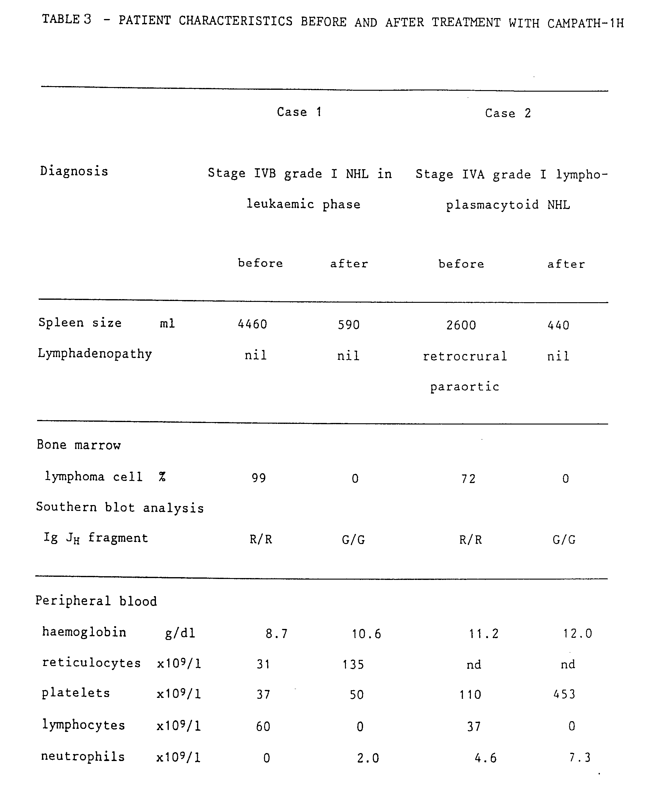

-
- 1. Neuberger, M.S., Williams, G.T., Mitchell, E.B., Jouhal, S.S., Flanagan, J.G. & Rabbits, T.H. Nature 314,268-270 (1985).
- 2. Liu, A.Y., Robinson, R.R., Hellstrom, K.E., Murray, E.D. Jr., Cheng, C.P. & Hellstrom, I. Proc.natl.Acad.Sci USA 84,3439-3443 (1987).
- 3. Shaw, D.R., Khazaeli, M.B., Sun, L.K., Ghraeyeb, J., Daddona, P.E., McKinney, S. & Lopuglio, A.F. J, Immunol. 138,4534-4538 (1987).
- 4. Bruggemann, M., Williams, G.T., Bindon, C., Clark, M.R., Walker, M.R., Jefferis, R., Waldmann, H. & Neuberger, M.S. J.Exp.Med. 166, 1351-1361 (1987).
- 5. Jones, P.T., Dear, P.H., Foote, J., Neuberger, M.S. & Winter, G. Nature 321,522-525 (1986).
- 6. Hale, G., Bright, S., Chumbley, G., Hoang, T., Metcalf, D., Munro, A.J. & Waldmann, H. Blood 62,873-882 (1983).
- 7. Hale, G., Hoang, T., Prospero, T., Watts, S.M. & Waldmann, H. Mol.Biol.Med. 1,305-319 (1983).
- 8. Hale, G., Cobbold, S.P., Waldmann, H., Easter, G., Matejtschuk, P. & Coombs, R.R.A. J. Immunol.Meth. 103,59-67 (1987).
- 9. Hale, G., Waldmann, H., Friend, P. & Calne, R. Transplantation 42,308-311 (1986).
- 10. Hale, G., Swirsky, D.M., Hayhoe, F.G.J. & Waldmann, H. Mol.Biol.Med. 1,321-334 (1983).
- 11. Kabat, E.A., Wu, T.T., Reid-Miller, M., Perry, H.M. & Gottesman, K.S. in Sequences of Proteins of Immunological Interest (US Dept. of Health and Human Services, 1987).
- 12. Chothia, C., Novotny, J., Bruccoleri, R. & Karplus, M. J. Mol.Biol. 186,651-663 (1985).
- 13. Saul, F.A., Amzel, M. & Poljak, R.J. J.Biol.Chem. 253,585-597 (1978).
- 14. EPP, O., Colman, P., Fehlhammer, H., Bode, W., Schiffer, M. & Huber, R. Eur.J.Biochem. 45,513-524 (1974).
- 15. Mulligan, R.C. & Berg, P. Proc.natl.Acad.Sci USA 78,2072-2076 (1981).
- 16. Southern, P.J. & Berg, P.J. Mol.Appl.Genetics 1,327-341 (1981).
- 17. Galfre, G. & Milstein, C. Meth.Enzymol. 73,1-46 (1981).
- 18. Chothia, C. & Lesk, A. J.Mol.Biol. 196,901-917 (1987).
- 19. Roberts, S., Cheetham, J.C. & Rees, A.R. Nature 328,731-734 (1987).
- 20. Jonker, M. & den Brok, J.H.A.M. Eur.J. Immunol. 17, 1547-1552 (1987).
- 21. Clark, M. & Waldmann, H. J.N.C.I. 79, 1393-1398 (1987).
- 22. Hieter, P.A., Max, E.E., Seidmann, J.G., Maizel, J.V.Jr & Leder, P. Cell 22,197-207 (1980).
- 23. Kaartinen, M., Griffiths, G.M., Hamlyn, P.H., Markham, A.F., Karjalainen, K., Pelkonen J.L.T., Makela, O. & Milstein, C.J. Immunol. 130,320-324 (1983).
- 24. Gubler, U. & Hoffmann, B.J. Gene 25,263-269 (1983).
- 25. Sanger, F., Nicklen, S.A. & Coulson, A.R. Proc.natl.Acad.Sci USA 74,5463-5467 (1977).
- 26. Takahashi, N., Ueda, N.S., Obata, M., Nikaido, T. & Honjo, T. Cell 29,671-679 (1982).
- 27. Flanagan, J.G. & Rabbits, T.H. Nature 300,709-713 (1982).
- 28. Huck, S., Fort, P., Crawford, D.H., Lefranc, M.-P. & Lefranc, G. Nucl.Acid Res. 14,1779-1789 (1986).
- 29. Bruggemann, M., Free, J., Diamond, A., Howard, J., Cobbold, S. & Waldmann, H. Proc.natl.
Acad.Sci. USA 83,6075-6079 (1986). - 30. Potter, H., Weir, L. & Leder, P. Proc.natl.Acad.Sci. USA 81,7161-7163 (1984).
- 31. Marquardt, M., Deisenhofer, J.,Huber, R. & Palm, W. J.Mol.Biol. 141,368-391 (1980).
- 32. Hale, G., Clark, M. & Waldmann, H. J. Immunol. 134,3056-3061 (1985).
- 33. Carter, P., Bedouelle, H. & Winter, G. Nucleic Acids Res. 13,4431-4443 (1985).
- 34. Heiter, P., Maizel, J & Leder, P. J. Biol. Chem. 257, 1516-1522 (1982).
- 35. Hale, G., Swirsky, D., Waldman, H., & Chan, L.C., Br. J. Haematol. 60, 41-48, (1985).
- 36. Ritz. J., Schlossman, S.F., Blood, 59, 1-11 (1982).
- 37. Levy. R., Miller R.A., Annu. Rev. Med., 34, 107-16 (1983).
- 38. Stevenson, G.T., Glennie, M.J., Cancer Surv. 4, 213-44, (1985).
Claims (11)
Priority Applications (1)
| Application Number | Priority Date | Filing Date | Title |
|---|---|---|---|
| AT89301291T ATE95068T1 (en) | 1988-02-12 | 1989-02-10 | MODIFIED ANTIBODIES. |
Applications Claiming Priority (4)
| Application Number | Priority Date | Filing Date | Title |
|---|---|---|---|
| GB888803228A GB8803228D0 (en) | 1988-02-12 | 1988-02-12 | Improvements in/relating to antibodies |
| GB8803228 | 1988-02-12 | ||
| GB888804464A GB8804464D0 (en) | 1988-02-25 | 1988-02-25 | Improvements in/relating to antibodies |
| GB8804464 | 1988-02-25 |
Publications (2)
| Publication Number | Publication Date |
|---|---|
| EP0328404A1 true EP0328404A1 (en) | 1989-08-16 |
| EP0328404B1 EP0328404B1 (en) | 1993-09-29 |
Family
ID=26293475
Family Applications (1)
| Application Number | Title | Priority Date | Filing Date |
|---|---|---|---|
| EP89301291A Expired - Lifetime EP0328404B1 (en) | 1988-02-12 | 1989-02-10 | Modified antibodies |
Country Status (13)
| Country | Link |
|---|---|
| EP (1) | EP0328404B1 (en) |
| JP (2) | JP3121823B2 (en) |
| AT (1) | ATE95068T1 (en) |
| AU (1) | AU618989B2 (en) |
| CA (1) | CA1339198C (en) |
| DE (2) | DE68909441T2 (en) |
| DK (1) | DK174854B1 (en) |
| ES (1) | ES2059719T3 (en) |
| GB (1) | GB2216126B (en) |
| LU (1) | LU90842I2 (en) |
| NL (1) | NL300067I1 (en) |
| NZ (1) | NZ227968A (en) |
| WO (1) | WO1989007452A1 (en) |
Cited By (28)
| Publication number | Priority date | Publication date | Assignee | Title |
|---|---|---|---|---|
| WO1991004336A1 (en) * | 1989-09-19 | 1991-04-04 | Centocor, Inc. | Method for improving human monoclonal antibody production |
| WO1991009966A1 (en) * | 1989-12-21 | 1991-07-11 | Ortho Pharmaceutical Corporation | Cd4 specific recombinant antibody |
| WO1992005274A1 (en) * | 1990-09-17 | 1992-04-02 | Scott David Gorman | Framework mutated antibodies and their preparation |
| EP0481791A3 (en) * | 1990-10-17 | 1992-07-08 | The Wellcome Foundation Limited | Culture medium for cho-cells and adapted cho-cells |
| WO1992016562A1 (en) * | 1991-03-12 | 1992-10-01 | Lynxvale Limited | Humanised antibodies having modified allotypic determinants |
| WO1992018530A1 (en) * | 1991-04-16 | 1992-10-29 | Geoffrey Hale | SYNTHETIC CDw52(CAMPATH-1) PEPTIDE ANTIGEN |
| WO1993001296A1 (en) * | 1991-07-04 | 1993-01-21 | The Wellcome Foundation Limited | Antibody production in vaccinia virus infected cells |
| WO1993007899A1 (en) * | 1991-10-15 | 1993-04-29 | The Wellcome Foundation Limited | CDw52 - SPECIFIC ANTIBODY FOR TREATMENT OF T-CELL MEDIATED INFLAMMATION OF THE JOINTS |
| WO1993010817A1 (en) * | 1991-12-04 | 1993-06-10 | Geoffrey Hale | CDw52-SPECIFIC ANTIBODY FOR TREATMENT OF MULTIPLE SCLEROSIS |
| EP0585570A1 (en) * | 1992-08-05 | 1994-03-09 | BEHRINGWERKE Aktiengesellschaft | Antigranulocyte antibody construct, preparation and use thereof |
| JPH0690752A (en) * | 1990-10-17 | 1994-04-05 | Wellcome Found Ltd:The | Production of antibody |
| EP0590058A1 (en) | 1991-06-14 | 1994-04-06 | Genentech, Inc. | Method for making humanized antibodies |
| EP0682040A1 (en) * | 1988-12-28 | 1995-11-15 | Protein Design Labs, Inc. | Humanized immunoglobulins and their production and use |
| EP0699755A2 (en) | 1994-06-30 | 1996-03-06 | Centro de Inmunologia Molecular | Method for obtaining modified immunoglobulins with reduced immunogenicity of murine antibody variable domains, compositions containing them |
| US5558864A (en) * | 1991-03-06 | 1996-09-24 | Merck Patent Gesellschaft Mit Beschrankter Haftung | Humanized and chimeric anti-epidermal growth factor receptor monoclonal antibodies |
| US5644036A (en) * | 1990-10-17 | 1997-07-01 | Burroughs Wellcome Company | Purified immunoglobulin |
| WO1997031024A1 (en) * | 1996-02-20 | 1997-08-28 | Isis Innovation Limited | Antibody variants |
| US5693761A (en) * | 1988-12-28 | 1997-12-02 | Protein Design Labs, Inc. | Polynucleotides encoding improved humanized immunoglobulins |
| US5859205A (en) * | 1989-12-21 | 1999-01-12 | Celltech Limited | Humanised antibodies |
| US5861155A (en) * | 1993-12-08 | 1999-01-19 | Astra Ab | Humanized antibodies and uses thereof |
| US5886152A (en) * | 1991-12-06 | 1999-03-23 | Sumitomo Pharmaceuticals Company, Limited | Humanized B-B10 |
| US5985279A (en) * | 1991-07-16 | 1999-11-16 | Waldmann; Herman | Humanized antibody against CD18 |
| US6010861A (en) * | 1994-08-03 | 2000-01-04 | Dgi Biotechnologies, Llc | Target specific screens and their use for discovering small organic molecular pharmacophores |
| US6054297A (en) * | 1991-06-14 | 2000-04-25 | Genentech, Inc. | Humanized antibodies and methods for making them |
| US6358710B1 (en) | 1996-06-07 | 2002-03-19 | Neorx Corporation | Humanized antibodies that bind to the antigen bound by antibody NR-LU-13 |
| US6750325B1 (en) | 1989-12-21 | 2004-06-15 | Celltech R&D Limited | CD3 specific recombinant antibody |
| US6797492B2 (en) | 1991-05-17 | 2004-09-28 | Merck & Co., Inc. | Method for reducing the immunogenicity of antibody variable domains |
| US6800738B1 (en) | 1991-06-14 | 2004-10-05 | Genentech, Inc. | Method for making humanized antibodies |
Families Citing this family (11)
| Publication number | Priority date | Publication date | Assignee | Title |
|---|---|---|---|---|
| US7192584B2 (en) | 1991-03-18 | 2007-03-20 | Centocor, Inc. | Methods of treating psoriasis with anti-TNF antibodies |
| US6764681B2 (en) | 1991-10-07 | 2004-07-20 | Biogen, Inc. | Method of prophylaxis or treatment of antigen presenting cell driven skin conditions using inhibitors of the CD2/LFA-3 interaction |
| GB9206422D0 (en) * | 1992-03-24 | 1992-05-06 | Bolt Sarah L | Antibody preparation |
| US5683891A (en) * | 1992-11-28 | 1997-11-04 | Juridical Foundation The Chemo-Sero Therapeutic Research Institute | Method for producing exogenous protein |
| EP1419236A4 (en) | 2001-07-24 | 2005-08-03 | Biogen Idec Inc | Methods for treating or preventing sclerotic disorders using cd2-binding agents |
| EP1618891A1 (en) * | 2003-03-31 | 2006-01-25 | Kirin Beer Kabushiki Kaisha | Method of inducing differentiation and proliferating regulatory t cell by anti-cd52 antibody and medicinal composition therefor |
| WO2005115436A1 (en) | 2004-05-07 | 2005-12-08 | Astellas Us Llc | Soluble lfa-3 polypeptide for treating viral disorders |
| KR20160103160A (en) | 2009-05-13 | 2016-08-31 | 젠자임 코포레이션 | Anti-human cd52 immunoglobulins |
| GB201109238D0 (en) * | 2011-06-01 | 2011-07-13 | Antitope Ltd | Antibodies |
| US20140359902A1 (en) | 2011-12-16 | 2014-12-04 | Synthon Biopharmaceuticals B.V. | EXPRESSION OF SECRETORY IgA ANTIBODIES IN DUCKWEED |
| WO2018174274A1 (en) | 2017-03-24 | 2018-09-27 | 全薬工業株式会社 | ANTI-IgM/B CELL SURFACE ANTIGEN BISPECIFIC ANTIBODY |
Citations (1)
| Publication number | Priority date | Publication date | Assignee | Title |
|---|---|---|---|---|
| EP0239400A2 (en) * | 1986-03-27 | 1987-09-30 | Medical Research Council | Recombinant antibodies and methods for their production |
-
1989
- 1989-02-10 DE DE89301291T patent/DE68909441T2/en not_active Expired - Lifetime
- 1989-02-10 DE DE2001199051 patent/DE10199051I2/en active Active
- 1989-02-10 ES ES89301291T patent/ES2059719T3/en not_active Expired - Lifetime
- 1989-02-10 WO PCT/GB1989/000113 patent/WO1989007452A1/en unknown
- 1989-02-10 AT AT89301291T patent/ATE95068T1/en active
- 1989-02-10 GB GB8902978A patent/GB2216126B/en not_active Revoked
- 1989-02-10 EP EP89301291A patent/EP0328404B1/en not_active Expired - Lifetime
- 1989-02-10 CA CA000590704A patent/CA1339198C/en not_active Expired - Fee Related
- 1989-02-10 AU AU30626/89A patent/AU618989B2/en not_active Expired
- 1989-02-10 JP JP01502205A patent/JP3121823B2/en not_active Expired - Lifetime
- 1989-02-13 NZ NZ227968A patent/NZ227968A/en unknown
- 1989-10-10 DK DK198905021A patent/DK174854B1/en not_active IP Right Cessation
-
1998
- 1998-11-19 JP JP10329954A patent/JPH11228600A/en not_active Withdrawn
-
2001
- 2001-09-26 LU LU90842C patent/LU90842I2/en unknown
- 2001-10-10 NL NL300067C patent/NL300067I1/en unknown
Patent Citations (1)
| Publication number | Priority date | Publication date | Assignee | Title |
|---|---|---|---|---|
| EP0239400A2 (en) * | 1986-03-27 | 1987-09-30 | Medical Research Council | Recombinant antibodies and methods for their production |
Non-Patent Citations (3)
| Title |
|---|
| JOURNAL OF IMMUNOLOGICAL METHODS, vol. 103, 1987, pages 59-67, Elsevier Science Publishers B.V. (Biomedical Division); G. HALE et al.: "Isolation of low-frequency class-switch variants from rat hybrid myelomas" * |
| NATURE, vol. 321, 29th May 1986, pages 522-525; P.T. JONES et al.: "Replacing the complementarity-determining regions in a human antibody with those from a mouse" * |
| NATURE, vol. 332, 24th March 1988, pages 323-327; L. RIECHMANN et al.: "Reshaping human antibodies for therapy" * |
Cited By (77)
| Publication number | Priority date | Publication date | Assignee | Title |
|---|---|---|---|---|
| EP2341080A1 (en) * | 1988-12-28 | 2011-07-06 | PDL BioPharma, Inc. | Humanized immunoglobulins and their production and use |
| US7022500B1 (en) | 1988-12-28 | 2006-04-04 | Protein Design Labs, Inc. | Humanized immunoglobulins |
| EP1491556A1 (en) * | 1988-12-28 | 2004-12-29 | Protein Design Labs, Inc. | Humanized immunoglobulins and their production and use |
| EP1477497A1 (en) * | 1988-12-28 | 2004-11-17 | Protein Design Labs, Inc. | Humanized immunoglobulins and their production and use |
| EP0939127A3 (en) * | 1988-12-28 | 2004-07-21 | Protein Design Labs, Inc. | Humanized immunoglobulins and their production and use |
| US6180370B1 (en) | 1988-12-28 | 2001-01-30 | Protein Design Labs, Inc. | Humanized immunoglobulins and methods of making the same |
| US5693762A (en) * | 1988-12-28 | 1997-12-02 | Protein Design Labs, Inc. | Humanized immunoglobulins |
| US5693761A (en) * | 1988-12-28 | 1997-12-02 | Protein Design Labs, Inc. | Polynucleotides encoding improved humanized immunoglobulins |
| EP0682040A1 (en) * | 1988-12-28 | 1995-11-15 | Protein Design Labs, Inc. | Humanized immunoglobulins and their production and use |
| WO1991004336A1 (en) * | 1989-09-19 | 1991-04-04 | Centocor, Inc. | Method for improving human monoclonal antibody production |
| EP0626390A1 (en) * | 1989-12-21 | 1994-11-30 | Celltech Therapeutics Limited | Humanised antibodies |
| AU664801B2 (en) * | 1989-12-21 | 1995-11-30 | Celltech Limited | Humanised antibodies |
| USRE48787E1 (en) | 1989-12-21 | 2021-10-26 | UCB Biopharma SRL | Humanised antibodies |
| US5929212A (en) * | 1989-12-21 | 1999-07-27 | Celltech Therapeutics Limited | CD3 specific recombinant antibody |
| US6632927B2 (en) | 1989-12-21 | 2003-10-14 | Celltech Therapeutics Limited | Humanized antibodies |
| WO1991009966A1 (en) * | 1989-12-21 | 1991-07-11 | Ortho Pharmaceutical Corporation | Cd4 specific recombinant antibody |
| GB2246781B (en) * | 1989-12-21 | 1994-05-11 | Celltech Ltd | CD3 specific recombinant antibody |
| GB2246570B (en) * | 1989-12-21 | 1994-05-11 | Celltech Ltd | Antibody molecules having a variable domain comprising acceptor framework residues and donor antigen binding residues |
| US7566771B1 (en) | 1989-12-21 | 2009-07-28 | Celltech R&D Limited | Humanised antibodies |
| US7262050B2 (en) | 1989-12-21 | 2007-08-28 | Celltech R&D Limited | Humanised antibodies |
| US7244832B2 (en) | 1989-12-21 | 2007-07-17 | Celltech R&D Limited | Humanised antibodies |
| US5859205A (en) * | 1989-12-21 | 1999-01-12 | Celltech Limited | Humanised antibodies |
| US7244615B2 (en) | 1989-12-21 | 2007-07-17 | Celltech R&D Limited | Humanized antibodies |
| US7241877B2 (en) | 1989-12-21 | 2007-07-10 | Celltech R&D Limited | Humanised antibodies |
| US6750325B1 (en) | 1989-12-21 | 2004-06-15 | Celltech R&D Limited | CD3 specific recombinant antibody |
| WO1991009967A1 (en) * | 1989-12-21 | 1991-07-11 | Celltech Limited | Humanised antibodies |
| WO1991009968A1 (en) * | 1989-12-21 | 1991-07-11 | Celltech Limited | Cd3 specific recombinant antibody |
| GB2246570A (en) * | 1989-12-21 | 1992-02-05 | Celltech Ltd | Humanised antibodies |
| GB2246781A (en) * | 1989-12-21 | 1992-02-12 | Celltech Ltd | CD3 specific recombinant antibody |
| US6767996B1 (en) | 1990-09-17 | 2004-07-27 | Glaxo Wellcome Inc. | Altered antibodies and their preparation |
| US7098006B1 (en) | 1990-09-17 | 2006-08-29 | Burroughs Wellcome Co. | Chimeric antibody, pharmaceutical composition and process of its production |
| USRE43898E1 (en) | 1990-09-17 | 2013-01-01 | Glaxo Wellcome Inc. Research | Altered antibodies and their preparation |
| WO1992005274A1 (en) * | 1990-09-17 | 1992-04-02 | Scott David Gorman | Framework mutated antibodies and their preparation |
| USRE46877E1 (en) | 1990-09-17 | 2018-05-29 | Btg International Limited | Altered antibodies and their preparation |
| US5644036A (en) * | 1990-10-17 | 1997-07-01 | Burroughs Wellcome Company | Purified immunoglobulin |
| EP1849862A2 (en) * | 1990-10-17 | 2007-10-31 | The Wellcome Foundation Limited | Media |
| JPH0690752A (en) * | 1990-10-17 | 1994-04-05 | Wellcome Found Ltd:The | Production of antibody |
| USRE41974E1 (en) | 1990-10-17 | 2010-11-30 | Glaxosmithkline Llc | Method for culturing Chinese hamster ovary cells |
| US5316938A (en) * | 1990-10-17 | 1994-05-31 | Burroughs Wellcome Co. | Defined media for serum-free tissue culture |
| EP1950301A2 (en) | 1990-10-17 | 2008-07-30 | The Wellcome Foundation Limited | Antibody for use in Therapy |
| EP1849862A3 (en) * | 1990-10-17 | 2008-02-13 | The Wellcome Foundation Limited | Media |
| US5633162A (en) * | 1990-10-17 | 1997-05-27 | Glaxo Wellcome Inc. | Method for culturing Chinese hamster ovary cells |
| USRE39792E1 (en) | 1990-10-17 | 2007-08-21 | Smithkline Beecham Corporation | Method for culturing Chinese hamster ovary cells |
| US5545404A (en) * | 1990-10-17 | 1996-08-13 | Burroughs Wellcome Co. | Method for treating a mammal suffering from a T-cell medicated disorder with a CHO-Glycosylated antibody |
| US5545405A (en) * | 1990-10-17 | 1996-08-13 | Burroughs Wellcome Co. | Method for treating a mammal suffering from cancer with a cho-glycosylated antibody |
| EP0481791A3 (en) * | 1990-10-17 | 1992-07-08 | The Wellcome Foundation Limited | Culture medium for cho-cells and adapted cho-cells |
| US5545403A (en) * | 1990-10-17 | 1996-08-13 | Burroughs Wellcome Co. | Method for treating a mammal by administering a CHO-glycosylated antibody |
| US5558864A (en) * | 1991-03-06 | 1996-09-24 | Merck Patent Gesellschaft Mit Beschrankter Haftung | Humanized and chimeric anti-epidermal growth factor receptor monoclonal antibodies |
| WO1992016562A1 (en) * | 1991-03-12 | 1992-10-01 | Lynxvale Limited | Humanised antibodies having modified allotypic determinants |
| WO1992018530A1 (en) * | 1991-04-16 | 1992-10-29 | Geoffrey Hale | SYNTHETIC CDw52(CAMPATH-1) PEPTIDE ANTIGEN |
| US6797492B2 (en) | 1991-05-17 | 2004-09-28 | Merck & Co., Inc. | Method for reducing the immunogenicity of antibody variable domains |
| US8075890B2 (en) | 1991-06-14 | 2011-12-13 | Genentech, Inc. | Method for making humanized antibodies |
| EP0590058A1 (en) | 1991-06-14 | 1994-04-06 | Genentech, Inc. | Method for making humanized antibodies |
| US5821337A (en) * | 1991-06-14 | 1998-10-13 | Genentech, Inc. | Immunoglobulin variants |
| US6719971B1 (en) | 1991-06-14 | 2004-04-13 | Genentech, Inc. | Method for making humanized antibodies |
| US6800738B1 (en) | 1991-06-14 | 2004-10-05 | Genentech, Inc. | Method for making humanized antibodies |
| US6054297A (en) * | 1991-06-14 | 2000-04-25 | Genentech, Inc. | Humanized antibodies and methods for making them |
| US6639055B1 (en) | 1991-06-14 | 2003-10-28 | Genentech, Inc. | Method for making humanized antibodies |
| US6407213B1 (en) | 1991-06-14 | 2002-06-18 | Genentech, Inc. | Method for making humanized antibodies |
| WO1993001296A1 (en) * | 1991-07-04 | 1993-01-21 | The Wellcome Foundation Limited | Antibody production in vaccinia virus infected cells |
| US5985279A (en) * | 1991-07-16 | 1999-11-16 | Waldmann; Herman | Humanized antibody against CD18 |
| US6689869B2 (en) | 1991-07-16 | 2004-02-10 | Cambridge University Technical Services Limited | Labeled humanized anti-CD18 antibodies and fragments and kits comprising same |
| US5997867A (en) * | 1991-07-16 | 1999-12-07 | Waldmann; Herman | Method of using humanized antibody against CD18 |
| KR100264218B1 (en) * | 1991-10-15 | 2000-08-16 | 그레이엄 브레레톤 | Cdw52-specific antibody for treatment of t-cell mediated inflammation of the joints |
| WO1993007899A1 (en) * | 1991-10-15 | 1993-04-29 | The Wellcome Foundation Limited | CDw52 - SPECIFIC ANTIBODY FOR TREATMENT OF T-CELL MEDIATED INFLAMMATION OF THE JOINTS |
| WO1993010817A1 (en) * | 1991-12-04 | 1993-06-10 | Geoffrey Hale | CDw52-SPECIFIC ANTIBODY FOR TREATMENT OF MULTIPLE SCLEROSIS |
| US6120766A (en) * | 1991-12-04 | 2000-09-19 | Hale; Geoffrey | CDW52-specific antibody for treatment of multiple sclerosis |
| KR100271333B1 (en) * | 1991-12-04 | 2000-11-01 | 헤일 지오프리 | Cdw-52 specific antibody for treatment of multiple sclerosis |
| US5886152A (en) * | 1991-12-06 | 1999-03-23 | Sumitomo Pharmaceuticals Company, Limited | Humanized B-B10 |
| US5645817A (en) * | 1992-08-05 | 1997-07-08 | Behringwerke Aktiengesellschaft | Granulocyte-binding antibody constructs, their preparation and use |
| EP0585570A1 (en) * | 1992-08-05 | 1994-03-09 | BEHRINGWERKE Aktiengesellschaft | Antigranulocyte antibody construct, preparation and use thereof |
| US5861155A (en) * | 1993-12-08 | 1999-01-19 | Astra Ab | Humanized antibodies and uses thereof |
| EP0699755A2 (en) | 1994-06-30 | 1996-03-06 | Centro de Inmunologia Molecular | Method for obtaining modified immunoglobulins with reduced immunogenicity of murine antibody variable domains, compositions containing them |
| US6010861A (en) * | 1994-08-03 | 2000-01-04 | Dgi Biotechnologies, Llc | Target specific screens and their use for discovering small organic molecular pharmacophores |
| WO1997031024A1 (en) * | 1996-02-20 | 1997-08-28 | Isis Innovation Limited | Antibody variants |
| US8440190B2 (en) | 1996-02-20 | 2013-05-14 | Isis Innovation Limited | Antibody variants |
| US6358710B1 (en) | 1996-06-07 | 2002-03-19 | Neorx Corporation | Humanized antibodies that bind to the antigen bound by antibody NR-LU-13 |
Also Published As
| Publication number | Publication date |
|---|---|
| GB2216126B (en) | 1992-06-03 |
| NL300067I1 (en) | 2001-12-01 |
| JPH11228600A (en) | 1999-08-24 |
| DK174854B1 (en) | 2003-12-22 |
| DK502189D0 (en) | 1989-10-10 |
| GB8902978D0 (en) | 1989-03-30 |
| DE68909441T2 (en) | 1994-02-10 |
| DE68909441D1 (en) | 1993-11-04 |
| ES2059719T3 (en) | 1994-11-16 |
| CA1339198C (en) | 1997-08-05 |
| NZ227968A (en) | 1992-01-29 |
| LU90842I2 (en) | 2001-11-26 |
| EP0328404B1 (en) | 1993-09-29 |
| JP3121823B2 (en) | 2001-01-09 |
| DK502189A (en) | 1989-10-12 |
| DE10199051I2 (en) | 2005-05-04 |
| JPH02503514A (en) | 1990-10-25 |
| ATE95068T1 (en) | 1993-10-15 |
| WO1989007452A1 (en) | 1989-08-24 |
| DE10199051I1 (en) | 2002-01-10 |
| AU3062689A (en) | 1989-09-06 |
| AU618989B2 (en) | 1992-01-16 |
| GB2216126A (en) | 1989-10-04 |
Similar Documents
| Publication | Publication Date | Title |
|---|---|---|
| EP0328404B1 (en) | Modified antibodies | |
| US5846534A (en) | Antibodies to the antigen campath-1 | |
| US6548640B1 (en) | Altered antibodies | |
| Riechmann et al. | Reshaping human antibodies for therapy | |
| JP3854306B2 (en) | Humanized and chimeric monoclonal antibodies | |
| FI103131B (en) | A method for preparing a CD25 binding molecule | |
| EP0794966B1 (en) | Humanized antibodies to cd38 | |
| AU606320B2 (en) | Modular assembly of antibody genes, antibodies prepared thereby and use | |
| EP0812333B1 (en) | Bispecific antibody effective to treat b-cell lymphoma and cell line | |
| EP0504350B1 (en) | Antibodies directed against cd3 | |
| EP0403156B1 (en) | Improved monoclonal antibodies against the human alpha/beta t-cell receptor, their production and use | |
| US8440190B2 (en) | Antibody variants | |
| US20020164788A1 (en) | Humanized antibodies to CD38 | |
| EP0388151A1 (en) | Modified antibodies | |
| AU3544589A (en) | Il-2 receptor-specific chimeric antibodies | |
| JPH08507680A (en) | Recombinant anti-VLA4 antibody molecule | |
| CA2106719C (en) | Reshaped monoclonal antibodies against an immunoglobulin isotype | |
| KR970007783B1 (en) | Specific binding agents | |
| EP0511308B1 (en) | Chimeric immunoglobulin for cd4 receptors | |
| BOND | NEH | |
| US7037496B2 (en) | Chimeric immunoglobulin for CD4 receptors |
Legal Events
| Date | Code | Title | Description |
|---|---|---|---|
| PUAI | Public reference made under article 153(3) epc to a published international application that has entered the european phase |
Free format text: ORIGINAL CODE: 0009012 |
|
| AK | Designated contracting states |
Kind code of ref document: A1 Designated state(s): AT BE CH DE ES FR GB GR IT LI LU NL SE |
|
| 17P | Request for examination filed |
Effective date: 19900209 |
|
| RAP1 | Party data changed (applicant data changed or rights of an application transferred) |
Owner name: NATIONAL RESEARCH DEVELOPMENT CORPORATION |
|
| 17Q | First examination report despatched |
Effective date: 19911108 |
|
| RAP1 | Party data changed (applicant data changed or rights of an application transferred) |
Owner name: BRITISH TECHNOLOGY GROUP LTD |
|
| GRAA | (expected) grant |
Free format text: ORIGINAL CODE: 0009210 |
|
| AK | Designated contracting states |
Kind code of ref document: B1 Designated state(s): AT BE CH DE ES FR GB GR IT LI LU NL SE |
|
| REF | Corresponds to: |
Ref document number: 95068 Country of ref document: AT Date of ref document: 19931015 Kind code of ref document: T |
|
| ET | Fr: translation filed | ||
| ITF | It: translation for a ep patent filed |
Owner name: JACOBACCI CASETTA & PERANI S.P.A. |
|
| REF | Corresponds to: |
Ref document number: 68909441 Country of ref document: DE Date of ref document: 19931104 |
|
| REG | Reference to a national code |
Ref country code: GR Ref legal event code: FG4A Free format text: 3010121 |
|
| EPTA | Lu: last paid annual fee | ||
| REG | Reference to a national code |
Ref country code: AT Ref legal event code: UEP Ref document number: 89301291 Country of ref document: AT Kind code of ref document: T |
|
| PLBE | No opposition filed within time limit |
Free format text: ORIGINAL CODE: 0009261 |
|
| STAA | Information on the status of an ep patent application or granted ep patent |
Free format text: STATUS: NO OPPOSITION FILED WITHIN TIME LIMIT |
|
| 26N | No opposition filed | ||
| REG | Reference to a national code |
Ref country code: DE Ref legal event code: 8364 Ref document number: 68909441 Country of ref document: DE Kind code of ref document: T |
|
| REG | Reference to a national code |
Ref country code: ES Ref legal event code: FG2A Ref document number: 2059719 Country of ref document: ES Kind code of ref document: T3 |
|
| EAL | Se: european patent in force in sweden |
Ref document number: 89301291.4 |
|
| REG | Reference to a national code |
Ref country code: DE Ref legal event code: V448 Ref document number: 68909441 Country of ref document: DE Free format text: PRODUCT NAME: MABCAMPATH-ALEMTUZUMAB; REGISTRATION NO/DATE: EU/1/01/193/001, 20010706 Spc suppl protection certif: 101 99 051 Filing date: 20010917 |
|
| REG | Reference to a national code |
Ref country code: FR Ref legal event code: CP Free format text: PRODUCT NAME: ALEMTUZUMAB; REGISTRATION NO/DATE: EU/1/01/193/001 20010706 Spc suppl protection certif: 01C0041 Filing date: 20010926 |
|
| REG | Reference to a national code |
Ref country code: SE Ref legal event code: SPCF Free format text: 0190040-6, 20010706 |
|
| REG | Reference to a national code |
Ref country code: GB Ref legal event code: CTFF Free format text: GBCTFFSPC/GB2001/046: 20010928 |
|
| REG | Reference to a national code |
Ref country code: LU Ref legal event code: CCP Free format text: LUCCP 90842, EXPIRES: 20140210 |
|
| REG | Reference to a national code |
Ref country code: NL Ref legal event code: AC1 Free format text: NLAC1 300067, 20011010 |
|
| REG | Reference to a national code |
Ref country code: AT Ref legal event code: ESZA Ref document number: 89301291 Country of ref document: AT Kind code of ref document: T Free format text: PRODUCT NAME: ALEMTUZUMAB Spc suppl protection certif: SZ 32/2001 Filing date: 20011008 |
|
| REG | Reference to a national code |
Ref country code: GB Ref legal event code: IF02 |
|
| CCPV | Be: grant of a complementary protection certificate |
Free format text: 2001C/039, EXPIRES: 20140210 |
|
| REG | Reference to a national code |
Ref country code: ES Ref legal event code: PC2A |
|
| NLT1 | Nl: modifications of names registered in virtue of documents presented to the patent office pursuant to art. 16 a, paragraph 1 |
Owner name: BTG INTERNATIONAL LTD. |
|
| REG | Reference to a national code |
Ref country code: FR Ref legal event code: CR Free format text: FRCR 2001C0041, 20010926 |
|
| REG | Reference to a national code |
Ref country code: GB Ref legal event code: CTFG Free format text: GBCTFGSPC/GB01/046: 20020502, EXPIRES: 20140209 |
|
| REG | Reference to a national code |
Ref country code: FR Ref legal event code: CY Free format text: FRCY 01C0041, 20010926, EXPIRES: 20140209 |
|
| REG | Reference to a national code |
Ref country code: AT Ref legal event code: EEZF Ref document number: 95068 Country of ref document: AT Kind code of ref document: T Free format text: PRODUCT NAME: ALEMTUZUMAB Spc suppl protection certif: SZ 32/2001 Filing date: 20011008 Extension date: 20140210 Effective date: 20031105 |
|
| REG | Reference to a national code |
Ref country code: SE Ref legal event code: SPCG Spc suppl protection certif: 01900406.L Ref country code: SE Ref legal event code: SPCG Free format text: 0190040 Spc suppl protection certif: 0190040 |
|
| REG | Reference to a national code |
Ref country code: DE Ref legal event code: V484 Ref document number: 68909441 Country of ref document: DE Free format text: PRODUCT NAME: MABCAMPATH-ALEMTUZUMAB; REGISTRATION NO/DATE: EU/1/01/193/001, 20010706 Spc suppl protection certif: 101 99 051 Filing date: 20010917 |
|
| REG | Reference to a national code |
Ref country code: DE Ref legal event code: V457 Ref document number: 68909441 Country of ref document: DE Free format text: PRODUCT NAME: MABCAMPATH-ALEMTUZUMAB; REGISTRATION NO/DATE: EU/1/01/193/001, 20010706 Spc suppl protection certif: 101 99 051 Filing date: 20010917 |
|
| REG | Reference to a national code |
Ref country code: DE Ref legal event code: 8327 Ref document number: 68909441 Country of ref document: DE Owner name: BTG INTERNATIONAL LTD., LONDON, GB |
|
| PGFP | Annual fee paid to national office [announced via postgrant information from national office to epo] |
Ref country code: CH Payment date: 20080129 Year of fee payment: 20 Ref country code: ES Payment date: 20080324 Year of fee payment: 20 |
|
| PGFP | Annual fee paid to national office [announced via postgrant information from national office to epo] |
Ref country code: LU Payment date: 20080226 Year of fee payment: 20 Ref country code: DE Payment date: 20080207 Year of fee payment: 20 Ref country code: IT Payment date: 20080226 Year of fee payment: 20 Ref country code: NL Payment date: 20080103 Year of fee payment: 20 Ref country code: GB Payment date: 20080206 Year of fee payment: 20 Ref country code: SE Payment date: 20080219 Year of fee payment: 20 |
|
| PGFP | Annual fee paid to national office [announced via postgrant information from national office to epo] |
Ref country code: AT Payment date: 20080213 Year of fee payment: 20 |
|
| PGFP | Annual fee paid to national office [announced via postgrant information from national office to epo] |
Ref country code: FR Payment date: 20080208 Year of fee payment: 20 |
|
| PGFP | Annual fee paid to national office [announced via postgrant information from national office to epo] |
Ref country code: BE Payment date: 20080417 Year of fee payment: 20 |
|
| PGFP | Annual fee paid to national office [announced via postgrant information from national office to epo] |
Ref country code: GR Payment date: 20080116 Year of fee payment: 20 |
|
| REG | Reference to a national code |
Ref country code: CH Ref legal event code: PL |
|
| REG | Reference to a national code |
Ref country code: GB Ref legal event code: CTFE Free format text: PRODUCT NAME: MABCAMPATH-ALEMTUZUMAB; REGISTERED: UK EU/1/01/193/001 20010706 Spc suppl protection certif: SPC/GB01/046 Filing date: 20010928 Expiry date: 20090210 Extension date: 20140209 Ref country code: GB Ref legal event code: PE20 Expiry date: 20090209 |
|
| NLV7 | Nl: ceased due to reaching the maximum lifetime of a patent |
Effective date: 20090210 |
|
| EUG | Se: european patent has lapsed | ||
| REG | Reference to a national code |
Ref country code: AT Ref legal event code: EELA Ref document number: 95068 Country of ref document: AT Kind code of ref document: T |
|
| PG25 | Lapsed in a contracting state [announced via postgrant information from national office to epo] |
Ref country code: NL Free format text: LAPSE BECAUSE OF EXPIRATION OF PROTECTION Effective date: 20090210 |
|
| PG25 | Lapsed in a contracting state [announced via postgrant information from national office to epo] |
Ref country code: GB Free format text: LAPSE BECAUSE OF EXPIRATION OF PROTECTION Effective date: 20090209 |
|
| REG | Reference to a national code |
Ref country code: ES Ref legal event code: FD2A Effective date: 20130801 |
|
| PG25 | Lapsed in a contracting state [announced via postgrant information from national office to epo] |
Ref country code: ES Free format text: LAPSE BECAUSE OF EXPIRATION OF PROTECTION Effective date: 20090211 |
|
| REG | Reference to a national code |
Ref country code: NL Ref legal event code: SPCX Free format text: PRODUCT NAME: ALEMTUZUMAB; REGISTRATION NO/DATE: EU/1/01/193/001 20010706 Spc suppl protection certif: 300067 Filing date: 20011010 Expiry date: 20090210 Extension date: 20140209 |
|
| REG | Reference to a national code |
Ref country code: BE Ref legal event code: CCRE Owner name: *BTG INTERNATIONAL LTD Free format text: PRODUCT NAME: ALEMTUZUMAB; REGISTRATION NO/DATE: EU/1/01/193/001 20010709 Spc suppl protection certif: 2001C/039 Filing date: 20020205 Expiry date: 20090210 Extension date: 20140210 Effective date: 20140210 |
|
| REG | Reference to a national code |
Ref country code: GB Ref legal event code: SPCE Free format text: PRODUCT NAME: MABCAMPATH-ALEMTUZUMAB; REGISTERED: UK EU/1/01/193/001 20010706 Spc suppl protection certif: SPC/GB01/046 Filing date: 20010928 Expiry date: 20090210 Extension date: 20140209 |
|
| REG | Reference to a national code |
Ref country code: DE Ref legal event code: R071 Ref document number: 68909441 Country of ref document: DE Free format text: PRODUCT NAME: MABCAMPATH-ALEMTUZUMAB IN ALLEN SEINEN DEM SCHUTZ DES GRUNDPATENTS UNTERLIEGENDEN FORMEN; REGISTRATION NO/DATE: EU/1/01/193/001 20010706 Spc suppl protection certif: 10199051 Filing date: 20010917 Expiry date: 20090211 Extension date: 20140210 Effective date: 20140211 |
|
| REG | Reference to a national code |
Ref country code: AT Ref legal event code: SPCX Ref document number: 95068 Country of ref document: AT Kind code of ref document: T Free format text: PRODUCT NAME: ALEMTUZUMAB; REGISTRATION NO/DATE: EU/1/01/193/001 20010706 Spc suppl protection certif: 32/2001 Filing date: 20011008 Expiry date: 20090210 Extension date: 20140210 Effective date: 20140210 |