EP0324789B1 - Methods of regulating metabolic stability of proteins - Google Patents
Methods of regulating metabolic stability of proteins Download PDFInfo
- Publication number
- EP0324789B1 EP0324789B1 EP87907061A EP87907061A EP0324789B1 EP 0324789 B1 EP0324789 B1 EP 0324789B1 EP 87907061 A EP87907061 A EP 87907061A EP 87907061 A EP87907061 A EP 87907061A EP 0324789 B1 EP0324789 B1 EP 0324789B1
- Authority
- EP
- European Patent Office
- Prior art keywords
- protein
- amino
- ubiquitin
- proteins
- βgal
- Prior art date
- Legal status (The legal status is an assumption and is not a legal conclusion. Google has not performed a legal analysis and makes no representation as to the accuracy of the status listed.)
- Expired - Lifetime
Links
- 108090000623 proteins and genes Proteins 0.000 title claims abstract description 290
- 102000004169 proteins and genes Human genes 0.000 title claims abstract description 248
- 238000000034 method Methods 0.000 title abstract description 32
- 230000002503 metabolic effect Effects 0.000 title abstract description 15
- 230000001105 regulatory effect Effects 0.000 title description 5
- 125000003275 alpha amino acid group Chemical group 0.000 claims abstract description 11
- 125000002924 primary amino group Chemical group [H]N([H])* 0.000 claims abstract description 5
- 108090000848 Ubiquitin Proteins 0.000 claims description 76
- 102000044159 Ubiquitin Human genes 0.000 claims description 74
- 108020001507 fusion proteins Proteins 0.000 claims description 31
- 102000037865 fusion proteins Human genes 0.000 claims description 31
- 238000003776 cleavage reaction Methods 0.000 claims description 29
- 230000007017 scission Effects 0.000 claims description 29
- 125000000539 amino acid group Chemical group 0.000 claims description 25
- 125000000729 N-terminal amino-acid group Chemical group 0.000 claims description 24
- 241000588724 Escherichia coli Species 0.000 claims description 11
- 108090000765 processed proteins & peptides Proteins 0.000 claims description 11
- 229920001184 polypeptide Polymers 0.000 claims description 10
- 102000004196 processed proteins & peptides Human genes 0.000 claims description 10
- 101710118538 Protease Proteins 0.000 claims description 6
- 108091028043 Nucleic acid sequence Proteins 0.000 claims description 3
- 230000001580 bacterial effect Effects 0.000 claims description 2
- 238000001727 in vivo Methods 0.000 abstract description 60
- 230000004927 fusion Effects 0.000 abstract description 25
- 238000000338 in vitro Methods 0.000 abstract description 16
- 230000002068 genetic effect Effects 0.000 abstract description 7
- 235000018102 proteins Nutrition 0.000 description 226
- 108010005774 beta-Galactosidase Proteins 0.000 description 86
- WQZGKKKJIJFFOK-FPRJBGLDSA-N beta-D-galactose Chemical class OC[C@H]1O[C@@H](O)[C@H](O)[C@@H](O)[C@H]1O WQZGKKKJIJFFOK-FPRJBGLDSA-N 0.000 description 57
- 235000001014 amino acid Nutrition 0.000 description 53
- 229940024606 amino acid Drugs 0.000 description 53
- 150000001413 amino acids Chemical class 0.000 description 49
- 210000004027 cell Anatomy 0.000 description 48
- 230000000368 destabilizing effect Effects 0.000 description 35
- 238000006731 degradation reaction Methods 0.000 description 25
- 230000003834 intracellular effect Effects 0.000 description 25
- 230000000087 stabilizing effect Effects 0.000 description 25
- 230000015556 catabolic process Effects 0.000 description 24
- 230000037361 pathway Effects 0.000 description 22
- 125000001360 methionine group Chemical group N[C@@H](CCSC)C(=O)* 0.000 description 20
- MTCFGRXMJLQNBG-REOHCLBHSA-N (2S)-2-Amino-3-hydroxypropansäure Chemical compound OC[C@H](N)C(O)=O MTCFGRXMJLQNBG-REOHCLBHSA-N 0.000 description 19
- 108020004705 Codon Proteins 0.000 description 16
- FFEARJCKVFRZRR-BYPYZUCNSA-N L-methionine Chemical compound CSCC[C@H](N)C(O)=O FFEARJCKVFRZRR-BYPYZUCNSA-N 0.000 description 16
- 239000000047 product Substances 0.000 description 15
- 241000894007 species Species 0.000 description 15
- 240000004808 Saccharomyces cerevisiae Species 0.000 description 14
- CKLJMWTZIZZHCS-REOHCLBHSA-N L-aspartic acid Chemical compound OC(=O)[C@@H](N)CC(O)=O CKLJMWTZIZZHCS-REOHCLBHSA-N 0.000 description 13
- 235000014680 Saccharomyces cerevisiae Nutrition 0.000 description 13
- 239000013612 plasmid Substances 0.000 description 13
- 230000001323 posttranslational effect Effects 0.000 description 13
- 239000000758 substrate Substances 0.000 description 13
- 108091005804 Peptidases Proteins 0.000 description 12
- 102000004190 Enzymes Human genes 0.000 description 11
- 108090000790 Enzymes Proteins 0.000 description 11
- 102000035195 Peptidases Human genes 0.000 description 11
- 238000004458 analytical method Methods 0.000 description 11
- 230000009504 deubiquitination Effects 0.000 description 11
- 229940088598 enzyme Drugs 0.000 description 11
- 230000006870 function Effects 0.000 description 11
- DHMQDGOQFOQNFH-UHFFFAOYSA-N Glycine Chemical compound NCC(O)=O DHMQDGOQFOQNFH-UHFFFAOYSA-N 0.000 description 10
- 239000004365 Protease Substances 0.000 description 10
- 230000000694 effects Effects 0.000 description 10
- KDXKERNSBIXSRK-YFKPBYRVSA-N L-lysine Chemical compound NCCCC[C@H](N)C(O)=O KDXKERNSBIXSRK-YFKPBYRVSA-N 0.000 description 9
- KDXKERNSBIXSRK-UHFFFAOYSA-N Lysine Natural products NCCCCC(N)C(O)=O KDXKERNSBIXSRK-UHFFFAOYSA-N 0.000 description 9
- DBMJMQXJHONAFJ-UHFFFAOYSA-M Sodium laurylsulphate Chemical compound [Na+].CCCCCCCCCCCCOS([O-])(=O)=O DBMJMQXJHONAFJ-UHFFFAOYSA-M 0.000 description 9
- 239000000872 buffer Substances 0.000 description 9
- AYFVYJQAPQTCCC-GBXIJSLDSA-N L-threonine Chemical compound C[C@@H](O)[C@H](N)C(O)=O AYFVYJQAPQTCCC-GBXIJSLDSA-N 0.000 description 8
- KZSNJWFQEVHDMF-UHFFFAOYSA-N Valine Chemical compound CC(C)C(N)C(O)=O KZSNJWFQEVHDMF-UHFFFAOYSA-N 0.000 description 8
- 238000010276 construction Methods 0.000 description 8
- YPHMISFOHDHNIV-FSZOTQKASA-N cycloheximide Chemical compound C1[C@@H](C)C[C@H](C)C(=O)[C@@H]1[C@H](O)CC1CC(=O)NC(=O)C1 YPHMISFOHDHNIV-FSZOTQKASA-N 0.000 description 8
- 238000002474 experimental method Methods 0.000 description 8
- 239000000284 extract Substances 0.000 description 8
- 239000000499 gel Substances 0.000 description 8
- 239000003550 marker Substances 0.000 description 8
- 235000019419 proteases Nutrition 0.000 description 8
- 230000034512 ubiquitination Effects 0.000 description 8
- 238000010798 ubiquitination Methods 0.000 description 8
- 241000894006 Bacteria Species 0.000 description 7
- 241000206602 Eukaryota Species 0.000 description 7
- DCXYFEDJOCDNAF-REOHCLBHSA-N L-asparagine Chemical compound OC(=O)[C@@H](N)CC(N)=O DCXYFEDJOCDNAF-REOHCLBHSA-N 0.000 description 7
- 125000000174 L-prolyl group Chemical group [H]N1C([H])([H])C([H])([H])C([H])([H])[C@@]1([H])C(*)=O 0.000 description 7
- 102000018390 Ubiquitin-Specific Proteases Human genes 0.000 description 7
- 108010066496 Ubiquitin-Specific Proteases Proteins 0.000 description 7
- 238000001114 immunoprecipitation Methods 0.000 description 7
- 230000002797 proteolythic effect Effects 0.000 description 7
- ROHFNLRQFUQHCH-YFKPBYRVSA-N L-leucine Chemical compound CC(C)C[C@H](N)C(O)=O ROHFNLRQFUQHCH-YFKPBYRVSA-N 0.000 description 6
- YBYRMVIVWMBXKQ-UHFFFAOYSA-N phenylmethanesulfonyl fluoride Chemical compound FS(=O)(=O)CC1=CC=CC=C1 YBYRMVIVWMBXKQ-UHFFFAOYSA-N 0.000 description 6
- 230000008684 selective degradation Effects 0.000 description 6
- COLNVLDHVKWLRT-QMMMGPOBSA-N L-phenylalanine Chemical compound OC(=O)[C@@H](N)CC1=CC=CC=C1 COLNVLDHVKWLRT-QMMMGPOBSA-N 0.000 description 5
- 238000013461 design Methods 0.000 description 5
- 230000000873 masking effect Effects 0.000 description 5
- 239000002609 medium Substances 0.000 description 5
- 229930182817 methionine Natural products 0.000 description 5
- 230000017854 proteolysis Effects 0.000 description 5
- 239000006228 supernatant Substances 0.000 description 5
- 108010016264 ubiquitin-Nalpha-protein hydrolase Proteins 0.000 description 5
- 229920002684 Sepharose Polymers 0.000 description 4
- 238000013459 approach Methods 0.000 description 4
- 230000015572 biosynthetic process Effects 0.000 description 4
- 230000021615 conjugation Effects 0.000 description 4
- 230000001419 dependent effect Effects 0.000 description 4
- 238000001962 electrophoresis Methods 0.000 description 4
- 230000003241 endoproteolytic effect Effects 0.000 description 4
- 229930182830 galactose Natural products 0.000 description 4
- 230000014509 gene expression Effects 0.000 description 4
- 125000003588 lysine group Chemical group [H]N([H])C([H])([H])C([H])([H])C([H])([H])C([H])([H])C([H])(N([H])[H])C(*)=O 0.000 description 4
- 238000012545 processing Methods 0.000 description 4
- 239000000523 sample Substances 0.000 description 4
- 238000002741 site-directed mutagenesis Methods 0.000 description 4
- 239000000126 substance Substances 0.000 description 4
- VHJLVAABSRFDPM-UHFFFAOYSA-N 1,4-dithiothreitol Chemical compound SCC(O)C(O)CS VHJLVAABSRFDPM-UHFFFAOYSA-N 0.000 description 3
- 102000004400 Aminopeptidases Human genes 0.000 description 3
- 108090000915 Aminopeptidases Proteins 0.000 description 3
- 239000004471 Glycine Substances 0.000 description 3
- 239000007995 HEPES buffer Substances 0.000 description 3
- XUJNEKJLAYXESH-REOHCLBHSA-N L-Cysteine Chemical compound SC[C@H](N)C(O)=O XUJNEKJLAYXESH-REOHCLBHSA-N 0.000 description 3
- 238000001042 affinity chromatography Methods 0.000 description 3
- 230000004075 alteration Effects 0.000 description 3
- 210000003567 ascitic fluid Anatomy 0.000 description 3
- 230000006652 catabolic pathway Effects 0.000 description 3
- 238000006243 chemical reaction Methods 0.000 description 3
- 230000000295 complement effect Effects 0.000 description 3
- 238000000605 extraction Methods 0.000 description 3
- 238000001914 filtration Methods 0.000 description 3
- 125000003630 glycyl group Chemical group [H]N([H])C([H])([H])C(*)=O 0.000 description 3
- 238000003780 insertion Methods 0.000 description 3
- 230000037431 insertion Effects 0.000 description 3
- 230000037041 intracellular level Effects 0.000 description 3
- 238000002372 labelling Methods 0.000 description 3
- 238000004519 manufacturing process Methods 0.000 description 3
- 230000001404 mediated effect Effects 0.000 description 3
- 230000004048 modification Effects 0.000 description 3
- 238000012986 modification Methods 0.000 description 3
- 230000035772 mutation Effects 0.000 description 3
- 230000035790 physiological processes and functions Effects 0.000 description 3
- 238000012163 sequencing technique Methods 0.000 description 3
- 238000006467 substitution reaction Methods 0.000 description 3
- 238000003786 synthesis reaction Methods 0.000 description 3
- 239000003053 toxin Substances 0.000 description 3
- 231100000765 toxin Toxicity 0.000 description 3
- 108700012359 toxins Proteins 0.000 description 3
- 238000013519 translation Methods 0.000 description 3
- 230000007306 turnover Effects 0.000 description 3
- YBJHBAHKTGYVGT-ZKWXMUAHSA-N (+)-Biotin Chemical compound N1C(=O)N[C@@H]2[C@H](CCCCC(=O)O)SC[C@@H]21 YBJHBAHKTGYVGT-ZKWXMUAHSA-N 0.000 description 2
- MRXDGVXSWIXTQL-HYHFHBMOSA-N (2s)-2-[[(1s)-1-(2-amino-1,4,5,6-tetrahydropyrimidin-6-yl)-2-[[(2s)-4-methyl-1-oxo-1-[[(2s)-1-oxo-3-phenylpropan-2-yl]amino]pentan-2-yl]amino]-2-oxoethyl]carbamoylamino]-3-phenylpropanoic acid Chemical compound C([C@H](NC(=O)N[C@H](C(=O)N[C@@H](CC(C)C)C(=O)N[C@@H](CC=1C=CC=CC=1)C=O)C1NC(N)=NCC1)C(O)=O)C1=CC=CC=C1 MRXDGVXSWIXTQL-HYHFHBMOSA-N 0.000 description 2
- 108010087765 Antipain Proteins 0.000 description 2
- 108010039627 Aprotinin Proteins 0.000 description 2
- 239000004475 Arginine Substances 0.000 description 2
- IJGRMHOSHXDMSA-UHFFFAOYSA-N Atomic nitrogen Chemical compound N#N IJGRMHOSHXDMSA-UHFFFAOYSA-N 0.000 description 2
- OLVPQBGMUGIKIW-UHFFFAOYSA-N Chymostatin Natural products C=1C=CC=CC=1CC(C=O)NC(=O)C(C(C)CC)NC(=O)C(C1NC(N)=NCC1)NC(=O)NC(C(O)=O)CC1=CC=CC=C1 OLVPQBGMUGIKIW-UHFFFAOYSA-N 0.000 description 2
- 241000701959 Escherichia virus Lambda Species 0.000 description 2
- WQZGKKKJIJFFOK-GASJEMHNSA-N Glucose Natural products OC[C@H]1OC(O)[C@H](O)[C@@H](O)[C@@H]1O WQZGKKKJIJFFOK-GASJEMHNSA-N 0.000 description 2
- PFMUCCYYAAFKTH-YFKPBYRVSA-N Gly-Met Chemical compound CSCC[C@@H](C(O)=O)NC(=O)CN PFMUCCYYAAFKTH-YFKPBYRVSA-N 0.000 description 2
- QNAYBMKLOCPYGJ-REOHCLBHSA-N L-alanine Chemical compound C[C@H](N)C(O)=O QNAYBMKLOCPYGJ-REOHCLBHSA-N 0.000 description 2
- 125000000998 L-alanino group Chemical group [H]N([*])[C@](C([H])([H])[H])([H])C(=O)O[H] 0.000 description 2
- ODKSFYDXXFIFQN-BYPYZUCNSA-N L-arginine Chemical compound OC(=O)[C@@H](N)CCCN=C(N)N ODKSFYDXXFIFQN-BYPYZUCNSA-N 0.000 description 2
- KZSNJWFQEVHDMF-BYPYZUCNSA-N L-valine Chemical compound CC(C)[C@H](N)C(O)=O KZSNJWFQEVHDMF-BYPYZUCNSA-N 0.000 description 2
- GDBQQVLCIARPGH-UHFFFAOYSA-N Leupeptin Natural products CC(C)CC(NC(C)=O)C(=O)NC(CC(C)C)C(=O)NC(C=O)CCCN=C(N)N GDBQQVLCIARPGH-UHFFFAOYSA-N 0.000 description 2
- 239000004472 Lysine Substances 0.000 description 2
- 239000000020 Nitrocellulose Substances 0.000 description 2
- 108010068086 Polyubiquitin Proteins 0.000 description 2
- 102100037935 Polyubiquitin-C Human genes 0.000 description 2
- MTCFGRXMJLQNBG-UHFFFAOYSA-N Serine Natural products OCC(N)C(O)=O MTCFGRXMJLQNBG-UHFFFAOYSA-N 0.000 description 2
- FAPWRFPIFSIZLT-UHFFFAOYSA-M Sodium chloride Chemical compound [Na+].[Cl-] FAPWRFPIFSIZLT-UHFFFAOYSA-M 0.000 description 2
- 108010090804 Streptavidin Proteins 0.000 description 2
- AYFVYJQAPQTCCC-UHFFFAOYSA-N Threonine Natural products CC(O)C(N)C(O)=O AYFVYJQAPQTCCC-UHFFFAOYSA-N 0.000 description 2
- 239000004473 Threonine Substances 0.000 description 2
- 108020004566 Transfer RNA Proteins 0.000 description 2
- 229920004890 Triton X-100 Polymers 0.000 description 2
- 239000013504 Triton X-100 Substances 0.000 description 2
- ISAKRJDGNUQOIC-UHFFFAOYSA-N Uracil Chemical compound O=C1C=CNC(=O)N1 ISAKRJDGNUQOIC-UHFFFAOYSA-N 0.000 description 2
- FJWGYAHXMCUOOM-QHOUIDNNSA-N [(2s,3r,4s,5r,6r)-2-[(2r,3r,4s,5r,6s)-4,5-dinitrooxy-2-(nitrooxymethyl)-6-[(2r,3r,4s,5r,6s)-4,5,6-trinitrooxy-2-(nitrooxymethyl)oxan-3-yl]oxyoxan-3-yl]oxy-3,5-dinitrooxy-6-(nitrooxymethyl)oxan-4-yl] nitrate Chemical compound O([C@@H]1O[C@@H]([C@H]([C@H](O[N+]([O-])=O)[C@H]1O[N+]([O-])=O)O[C@H]1[C@@H]([C@@H](O[N+]([O-])=O)[C@H](O[N+]([O-])=O)[C@@H](CO[N+]([O-])=O)O1)O[N+]([O-])=O)CO[N+](=O)[O-])[C@@H]1[C@@H](CO[N+]([O-])=O)O[C@@H](O[N+]([O-])=O)[C@H](O[N+]([O-])=O)[C@H]1O[N+]([O-])=O FJWGYAHXMCUOOM-QHOUIDNNSA-N 0.000 description 2
- 235000004279 alanine Nutrition 0.000 description 2
- SDNYTAYICBFYFH-TUFLPTIASA-N antipain Chemical compound NC(N)=NCCC[C@@H](C=O)NC(=O)[C@H](C(C)C)NC(=O)[C@H](CCCN=C(N)N)NC(=O)N[C@H](C(O)=O)CC1=CC=CC=C1 SDNYTAYICBFYFH-TUFLPTIASA-N 0.000 description 2
- 229960004405 aprotinin Drugs 0.000 description 2
- ODKSFYDXXFIFQN-UHFFFAOYSA-N arginine Natural products OC(=O)C(N)CCCNC(N)=N ODKSFYDXXFIFQN-UHFFFAOYSA-N 0.000 description 2
- 239000011324 bead Substances 0.000 description 2
- 230000000903 blocking effect Effects 0.000 description 2
- 238000009835 boiling Methods 0.000 description 2
- 238000005119 centrifugation Methods 0.000 description 2
- 230000008859 change Effects 0.000 description 2
- 108010086192 chymostatin Proteins 0.000 description 2
- 238000004132 cross linking Methods 0.000 description 2
- 239000000287 crude extract Substances 0.000 description 2
- XUJNEKJLAYXESH-UHFFFAOYSA-N cysteine Natural products SCC(N)C(O)=O XUJNEKJLAYXESH-UHFFFAOYSA-N 0.000 description 2
- 235000018417 cysteine Nutrition 0.000 description 2
- 231100000599 cytotoxic agent Toxicity 0.000 description 2
- 239000002619 cytotoxin Substances 0.000 description 2
- 230000006378 damage Effects 0.000 description 2
- 230000003413 degradative effect Effects 0.000 description 2
- 230000000593 degrading effect Effects 0.000 description 2
- 230000001687 destabilization Effects 0.000 description 2
- 238000010586 diagram Methods 0.000 description 2
- 230000004069 differentiation Effects 0.000 description 2
- 229940042399 direct acting antivirals protease inhibitors Drugs 0.000 description 2
- 230000002255 enzymatic effect Effects 0.000 description 2
- 210000003527 eukaryotic cell Anatomy 0.000 description 2
- 239000012634 fragment Substances 0.000 description 2
- 239000011521 glass Substances 0.000 description 2
- 239000008103 glucose Substances 0.000 description 2
- 230000001965 increasing effect Effects 0.000 description 2
- 238000011534 incubation Methods 0.000 description 2
- ZPNFWUPYTFPOJU-LPYSRVMUSA-N iniprol Chemical compound C([C@H]1C(=O)NCC(=O)NCC(=O)N[C@H]2CSSC[C@H]3C(=O)N[C@@H](CCCCN)C(=O)N[C@@H](C)C(=O)N[C@@H](CCCNC(N)=N)C(=O)N[C@H](C(N[C@H](C(=O)N[C@@H](CCCNC(N)=N)C(=O)N[C@@H](CC=4C=CC(O)=CC=4)C(=O)N[C@@H](CC=4C=CC=CC=4)C(=O)N[C@@H](CC=4C=CC(O)=CC=4)C(=O)N[C@@H](CC(N)=O)C(=O)N[C@@H](C)C(=O)N[C@@H](CCCCN)C(=O)N[C@@H](C)C(=O)NCC(=O)N[C@@H](CC(C)C)C(=O)N[C@@H](CSSC[C@H](NC(=O)[C@H](CC(O)=O)NC(=O)[C@H](CCC(O)=O)NC(=O)[C@H](C)NC(=O)[C@H](CO)NC(=O)[C@H](CCCCN)NC(=O)[C@H](CC=4C=CC=CC=4)NC(=O)[C@H](CC(N)=O)NC(=O)[C@H](CC(N)=O)NC(=O)[C@H](CCCNC(N)=N)NC(=O)[C@H](CCCCN)NC(=O)[C@H](C)NC(=O)[C@H](CCCNC(N)=N)NC2=O)C(=O)N[C@@H](CCSC)C(=O)N[C@@H](CCCNC(N)=N)C(=O)N[C@@H]([C@@H](C)O)C(=O)N[C@@H](CSSC[C@H](NC(=O)[C@H](CC=2C=CC=CC=2)NC(=O)[C@H](CC(O)=O)NC(=O)[C@H]2N(CCC2)C(=O)[C@@H](N)CCCNC(N)=N)C(=O)N[C@@H](CC(C)C)C(=O)N[C@@H](CCC(O)=O)C(=O)N2[C@@H](CCC2)C(=O)N2[C@@H](CCC2)C(=O)N[C@@H](CC=2C=CC(O)=CC=2)C(=O)N[C@@H]([C@@H](C)O)C(=O)NCC(=O)N2[C@@H](CCC2)C(=O)N3)C(=O)NCC(=O)NCC(=O)N[C@@H](C)C(O)=O)C(=O)N[C@@H](CCC(N)=O)C(=O)N[C@H](C(=O)N[C@@H](CC=2C=CC=CC=2)C(=O)N[C@H](C(=O)N1)C(C)C)[C@@H](C)O)[C@@H](C)CC)=O)[C@@H](C)CC)C1=CC=C(O)C=C1 ZPNFWUPYTFPOJU-LPYSRVMUSA-N 0.000 description 2
- 238000002955 isolation Methods 0.000 description 2
- 238000005304 joining Methods 0.000 description 2
- GDBQQVLCIARPGH-ULQDDVLXSA-N leupeptin Chemical compound CC(C)C[C@H](NC(C)=O)C(=O)N[C@@H](CC(C)C)C(=O)N[C@H](C=O)CCCN=C(N)N GDBQQVLCIARPGH-ULQDDVLXSA-N 0.000 description 2
- 108010052968 leupeptin Proteins 0.000 description 2
- -1 lysine Chemical class 0.000 description 2
- 239000000203 mixture Substances 0.000 description 2
- 238000002703 mutagenesis Methods 0.000 description 2
- 231100000350 mutagenesis Toxicity 0.000 description 2
- 229920001220 nitrocellulos Polymers 0.000 description 2
- 229940079938 nitrocellulose Drugs 0.000 description 2
- 239000002773 nucleotide Substances 0.000 description 2
- 125000003729 nucleotide group Chemical group 0.000 description 2
- 239000008188 pellet Substances 0.000 description 2
- 108010091212 pepstatin Proteins 0.000 description 2
- FAXGPCHRFPCXOO-LXTPJMTPSA-N pepstatin A Chemical compound OC(=O)C[C@H](O)[C@H](CC(C)C)NC(=O)[C@H](C)NC(=O)C[C@H](O)[C@H](CC(C)C)NC(=O)[C@H](C(C)C)NC(=O)[C@H](C(C)C)NC(=O)CC(C)C FAXGPCHRFPCXOO-LXTPJMTPSA-N 0.000 description 2
- 239000000137 peptide hydrolase inhibitor Substances 0.000 description 2
- 230000006337 proteolytic cleavage Effects 0.000 description 2
- 210000001995 reticulocyte Anatomy 0.000 description 2
- 231100000331 toxic Toxicity 0.000 description 2
- 230000002588 toxic effect Effects 0.000 description 2
- 238000012546 transfer Methods 0.000 description 2
- 239000004474 valine Substances 0.000 description 2
- PGOHTUIFYSHAQG-LJSDBVFPSA-N (2S)-6-amino-2-[[(2S)-5-amino-2-[[(2S)-2-[[(2S)-2-[[(2S)-2-[[(2S)-4-amino-2-[[(2S)-2-[[(2S)-2-[[(2S)-2-[[(2S)-2-[[(2S)-5-amino-2-[[(2S)-5-amino-2-[[(2S)-2-[[(2S)-2-[[(2S)-2-[[(2S,3R)-2-[[(2S)-5-amino-2-[[(2S)-2-[[(2S)-2-[[(2S,3R)-2-[[(2S)-2-[[(2S)-2-[[(2S)-2-[[(2S)-2-[[(2S)-5-amino-2-[[(2S)-1-[(2S,3R)-2-[[(2S)-2-[[(2S)-2-[[(2R)-2-[[(2S)-2-[[(2S)-2-[[2-[[(2S)-2-[[(2S)-2-[[(2S)-2-[[(2S)-1-[(2S)-2-[[(2S)-2-[[(2S)-2-[[(2S)-2-amino-4-methylsulfanylbutanoyl]amino]-3-(1H-indol-3-yl)propanoyl]amino]-5-carbamimidamidopentanoyl]amino]propanoyl]pyrrolidine-2-carbonyl]amino]-3-methylbutanoyl]amino]-4-methylpentanoyl]amino]-4-methylpentanoyl]amino]acetyl]amino]-3-hydroxypropanoyl]amino]-4-methylpentanoyl]amino]-3-sulfanylpropanoyl]amino]-4-methylsulfanylbutanoyl]amino]-5-carbamimidamidopentanoyl]amino]-3-hydroxybutanoyl]pyrrolidine-2-carbonyl]amino]-5-oxopentanoyl]amino]-3-hydroxypropanoyl]amino]-3-hydroxypropanoyl]amino]-3-(1H-imidazol-5-yl)propanoyl]amino]-4-methylpentanoyl]amino]-3-hydroxybutanoyl]amino]-3-(1H-indol-3-yl)propanoyl]amino]-5-carbamimidamidopentanoyl]amino]-5-oxopentanoyl]amino]-3-hydroxybutanoyl]amino]-3-hydroxypropanoyl]amino]-3-carboxypropanoyl]amino]-3-hydroxypropanoyl]amino]-5-oxopentanoyl]amino]-5-oxopentanoyl]amino]-3-phenylpropanoyl]amino]-5-carbamimidamidopentanoyl]amino]-3-methylbutanoyl]amino]-4-methylpentanoyl]amino]-4-oxobutanoyl]amino]-5-carbamimidamidopentanoyl]amino]-3-(1H-indol-3-yl)propanoyl]amino]-4-carboxybutanoyl]amino]-5-oxopentanoyl]amino]hexanoic acid Chemical compound CSCC[C@H](N)C(=O)N[C@@H](Cc1c[nH]c2ccccc12)C(=O)N[C@@H](CCCNC(N)=N)C(=O)N[C@@H](C)C(=O)N1CCC[C@H]1C(=O)N[C@@H](C(C)C)C(=O)N[C@@H](CC(C)C)C(=O)N[C@@H](CC(C)C)C(=O)NCC(=O)N[C@@H](CO)C(=O)N[C@@H](CC(C)C)C(=O)N[C@@H](CS)C(=O)N[C@@H](CCSC)C(=O)N[C@@H](CCCNC(N)=N)C(=O)N[C@@H]([C@@H](C)O)C(=O)N1CCC[C@H]1C(=O)N[C@@H](CCC(N)=O)C(=O)N[C@@H](CO)C(=O)N[C@@H](CO)C(=O)N[C@@H](Cc1cnc[nH]1)C(=O)N[C@@H](CC(C)C)C(=O)N[C@@H]([C@@H](C)O)C(=O)N[C@@H](Cc1c[nH]c2ccccc12)C(=O)N[C@@H](CCCNC(N)=N)C(=O)N[C@@H](CCC(N)=O)C(=O)N[C@@H]([C@@H](C)O)C(=O)N[C@@H](CO)C(=O)N[C@@H](CC(O)=O)C(=O)N[C@@H](CO)C(=O)N[C@@H](CCC(N)=O)C(=O)N[C@@H](CCC(N)=O)C(=O)N[C@@H](Cc1ccccc1)C(=O)N[C@@H](CCCNC(N)=N)C(=O)N[C@@H](C(C)C)C(=O)N[C@@H](CC(C)C)C(=O)N[C@@H](CC(N)=O)C(=O)N[C@@H](CCCNC(N)=N)C(=O)N[C@@H](Cc1c[nH]c2ccccc12)C(=O)N[C@@H](CCC(O)=O)C(=O)N[C@@H](CCC(N)=O)C(=O)N[C@@H](CCCCN)C(O)=O PGOHTUIFYSHAQG-LJSDBVFPSA-N 0.000 description 1
- IVLXQGJVBGMLRR-UHFFFAOYSA-N 2-aminoacetic acid;hydron;chloride Chemical compound Cl.NCC(O)=O IVLXQGJVBGMLRR-UHFFFAOYSA-N 0.000 description 1
- OPIFSICVWOWJMJ-AEOCFKNESA-N 5-bromo-4-chloro-3-indolyl beta-D-galactoside Chemical compound O[C@@H]1[C@@H](O)[C@@H](O)[C@@H](CO)O[C@H]1OC1=CNC2=CC=C(Br)C(Cl)=C12 OPIFSICVWOWJMJ-AEOCFKNESA-N 0.000 description 1
- 229930024421 Adenine Natural products 0.000 description 1
- GFFGJBXGBJISGV-UHFFFAOYSA-N Adenine Chemical compound NC1=NC=NC2=C1N=CN2 GFFGJBXGBJISGV-UHFFFAOYSA-N 0.000 description 1
- DCXYFEDJOCDNAF-UHFFFAOYSA-N Asparagine Natural products OC(=O)C(N)CC(N)=O DCXYFEDJOCDNAF-UHFFFAOYSA-N 0.000 description 1
- 101000909256 Caldicellulosiruptor bescii (strain ATCC BAA-1888 / DSM 6725 / Z-1320) DNA polymerase I Proteins 0.000 description 1
- GUBGYTABKSRVRQ-WFVLMXAXSA-N DEAE-cellulose Chemical compound OC1C(O)C(O)C(CO)O[C@H]1O[C@@H]1C(CO)OC(O)C(O)C1O GUBGYTABKSRVRQ-WFVLMXAXSA-N 0.000 description 1
- 108010017826 DNA Polymerase I Proteins 0.000 description 1
- 102000004594 DNA Polymerase I Human genes 0.000 description 1
- 108010058643 Fungal Proteins Proteins 0.000 description 1
- 206010064571 Gene mutation Diseases 0.000 description 1
- 108091005904 Hemoglobin subunit beta Proteins 0.000 description 1
- 102100021519 Hemoglobin subunit beta Human genes 0.000 description 1
- XMAUFHMAAVTODF-STQMWFEESA-N His-Phe Chemical compound C([C@H](N)C(=O)N[C@@H](CC=1C=CC=CC=1)C(O)=O)C1=CN=CN1 XMAUFHMAAVTODF-STQMWFEESA-N 0.000 description 1
- 101000899111 Homo sapiens Hemoglobin subunit beta Proteins 0.000 description 1
- 102000012745 Immunoglobulin Subunits Human genes 0.000 description 1
- 108010079585 Immunoglobulin Subunits Proteins 0.000 description 1
- 238000012404 In vitro experiment Methods 0.000 description 1
- OUYCCCASQSFEME-QMMMGPOBSA-N L-tyrosine Chemical compound OC(=O)[C@@H](N)CC1=CC=C(O)C=C1 OUYCCCASQSFEME-QMMMGPOBSA-N 0.000 description 1
- ROHFNLRQFUQHCH-UHFFFAOYSA-N Leucine Natural products CC(C)CC(N)C(O)=O ROHFNLRQFUQHCH-UHFFFAOYSA-N 0.000 description 1
- 102000003960 Ligases Human genes 0.000 description 1
- 108090000364 Ligases Proteins 0.000 description 1
- 241000124008 Mammalia Species 0.000 description 1
- 102000016943 Muramidase Human genes 0.000 description 1
- 108010014251 Muramidase Proteins 0.000 description 1
- 108010062010 N-Acetylmuramoyl-L-alanine Amidase Proteins 0.000 description 1
- GHAZCVNUKKZTLG-UHFFFAOYSA-N N-ethyl-succinimide Natural products CCN1C(=O)CCC1=O GHAZCVNUKKZTLG-UHFFFAOYSA-N 0.000 description 1
- HDFGOPSGAURCEO-UHFFFAOYSA-N N-ethylmaleimide Chemical compound CCN1C(=O)C=CC1=O HDFGOPSGAURCEO-UHFFFAOYSA-N 0.000 description 1
- 101710163270 Nuclease Proteins 0.000 description 1
- 108091034117 Oligonucleotide Proteins 0.000 description 1
- 108020005187 Oligonucleotide Probes Proteins 0.000 description 1
- 108700026244 Open Reading Frames Proteins 0.000 description 1
- 101710132611 Protein E3 Proteins 0.000 description 1
- 101000902592 Pyrococcus furiosus (strain ATCC 43587 / DSM 3638 / JCM 8422 / Vc1) DNA polymerase Proteins 0.000 description 1
- 108020005091 Replication Origin Proteins 0.000 description 1
- 101001025539 Saccharomyces cerevisiae (strain ATCC 204508 / S288c) Homothallic switching endonuclease Proteins 0.000 description 1
- 108700005078 Synthetic Genes Proteins 0.000 description 1
- 101150003725 TK gene Proteins 0.000 description 1
- 102000006601 Thymidine Kinase Human genes 0.000 description 1
- 108020004440 Thymidine kinase Proteins 0.000 description 1
- 102000004357 Transferases Human genes 0.000 description 1
- 108090000992 Transferases Proteins 0.000 description 1
- 102000003431 Ubiquitin-Conjugating Enzyme Human genes 0.000 description 1
- 108060008747 Ubiquitin-Conjugating Enzyme Proteins 0.000 description 1
- 208000027418 Wounds and injury Diseases 0.000 description 1
- 230000002159 abnormal effect Effects 0.000 description 1
- 230000005856 abnormality Effects 0.000 description 1
- 125000002777 acetyl group Chemical group [H]C([H])([H])C(*)=O 0.000 description 1
- 238000006640 acetylation reaction Methods 0.000 description 1
- 239000002253 acid Substances 0.000 description 1
- 150000007513 acids Chemical class 0.000 description 1
- 230000009471 action Effects 0.000 description 1
- 229960000643 adenine Drugs 0.000 description 1
- BFNBIHQBYMNNAN-UHFFFAOYSA-N ammonium sulfate Chemical class N.N.OS(O)(=O)=O BFNBIHQBYMNNAN-UHFFFAOYSA-N 0.000 description 1
- 239000012491 analyte Substances 0.000 description 1
- 235000009582 asparagine Nutrition 0.000 description 1
- 229960001230 asparagine Drugs 0.000 description 1
- 238000000376 autoradiography Methods 0.000 description 1
- 210000003050 axon Anatomy 0.000 description 1
- 102000005936 beta-Galactosidase Human genes 0.000 description 1
- 230000033228 biological regulation Effects 0.000 description 1
- 229960002685 biotin Drugs 0.000 description 1
- 235000020958 biotin Nutrition 0.000 description 1
- 239000011616 biotin Substances 0.000 description 1
- 210000004899 c-terminal region Anatomy 0.000 description 1
- 230000022131 cell cycle Effects 0.000 description 1
- 230000001413 cellular effect Effects 0.000 description 1
- 238000012512 characterization method Methods 0.000 description 1
- 238000007385 chemical modification Methods 0.000 description 1
- 238000004587 chromatography analysis Methods 0.000 description 1
- 238000005352 clarification Methods 0.000 description 1
- 230000000052 comparative effect Effects 0.000 description 1
- 230000001276 controlling effect Effects 0.000 description 1
- 238000007796 conventional method Methods 0.000 description 1
- 230000001086 cytosolic effect Effects 0.000 description 1
- 230000007812 deficiency Effects 0.000 description 1
- 230000002950 deficient Effects 0.000 description 1
- 239000007857 degradation product Substances 0.000 description 1
- 238000004925 denaturation Methods 0.000 description 1
- 230000036425 denaturation Effects 0.000 description 1
- 238000011161 development Methods 0.000 description 1
- 238000009826 distribution Methods 0.000 description 1
- 210000002472 endoplasmic reticulum Anatomy 0.000 description 1
- 238000011067 equilibration Methods 0.000 description 1
- 230000000925 erythroid effect Effects 0.000 description 1
- 239000013604 expression vector Substances 0.000 description 1
- 210000001723 extracellular space Anatomy 0.000 description 1
- 239000012467 final product Substances 0.000 description 1
- 238000001415 gene therapy Methods 0.000 description 1
- 102000034356 gene-regulatory proteins Human genes 0.000 description 1
- 108091006104 gene-regulatory proteins Proteins 0.000 description 1
- 108060003196 globin Proteins 0.000 description 1
- 102000018146 globin Human genes 0.000 description 1
- 239000006481 glucose medium Substances 0.000 description 1
- 230000012010 growth Effects 0.000 description 1
- 239000001963 growth medium Substances 0.000 description 1
- 230000009931 harmful effect Effects 0.000 description 1
- 108010092114 histidylphenylalanine Proteins 0.000 description 1
- 238000009396 hybridization Methods 0.000 description 1
- 230000002209 hydrophobic effect Effects 0.000 description 1
- 230000001900 immune effect Effects 0.000 description 1
- 238000003119 immunoblot Methods 0.000 description 1
- 238000012606 in vitro cell culture Methods 0.000 description 1
- 238000010348 incorporation Methods 0.000 description 1
- 230000006698 induction Effects 0.000 description 1
- 230000001939 inductive effect Effects 0.000 description 1
- 208000014674 injury Diseases 0.000 description 1
- 230000010039 intracellular degradation Effects 0.000 description 1
- 210000003093 intracellular space Anatomy 0.000 description 1
- 101150066555 lacZ gene Proteins 0.000 description 1
- 230000003902 lesion Effects 0.000 description 1
- 229960000274 lysozyme Drugs 0.000 description 1
- 239000004325 lysozyme Substances 0.000 description 1
- 235000010335 lysozyme Nutrition 0.000 description 1
- 210000004962 mammalian cell Anatomy 0.000 description 1
- 238000005259 measurement Methods 0.000 description 1
- 238000001466 metabolic labeling Methods 0.000 description 1
- 210000002569 neuron Anatomy 0.000 description 1
- 229910052757 nitrogen Inorganic materials 0.000 description 1
- 229940124276 oligodeoxyribonucleotide Drugs 0.000 description 1
- 239000002751 oligonucleotide probe Substances 0.000 description 1
- 238000007500 overflow downdraw method Methods 0.000 description 1
- COLNVLDHVKWLRT-UHFFFAOYSA-N phenylalanine Natural products OC(=O)C(N)CC1=CC=CC=C1 COLNVLDHVKWLRT-UHFFFAOYSA-N 0.000 description 1
- 229940080469 phosphocellulose Drugs 0.000 description 1
- 229920002401 polyacrylamide Polymers 0.000 description 1
- 108010054442 polyalanine Proteins 0.000 description 1
- 239000008057 potassium phosphate buffer Substances 0.000 description 1
- 239000002243 precursor Substances 0.000 description 1
- 238000002360 preparation method Methods 0.000 description 1
- 230000002265 prevention Effects 0.000 description 1
- 230000008569 process Effects 0.000 description 1
- 125000001500 prolyl group Chemical group [H]N1C([H])(C(=O)[*])C([H])([H])C([H])([H])C1([H])[H] 0.000 description 1
- 235000019833 protease Nutrition 0.000 description 1
- 230000009145 protein modification Effects 0.000 description 1
- 230000020978 protein processing Effects 0.000 description 1
- 230000007026 protein scission Effects 0.000 description 1
- 238000000734 protein sequencing Methods 0.000 description 1
- 230000004844 protein turnover Effects 0.000 description 1
- 238000000746 purification Methods 0.000 description 1
- 238000009790 rate-determining step (RDS) Methods 0.000 description 1
- 230000009467 reduction Effects 0.000 description 1
- 230000001172 regenerating effect Effects 0.000 description 1
- 230000003362 replicative effect Effects 0.000 description 1
- 238000011160 research Methods 0.000 description 1
- 230000004044 response Effects 0.000 description 1
- 239000012723 sample buffer Substances 0.000 description 1
- 230000003248 secreting effect Effects 0.000 description 1
- 238000004904 shortening Methods 0.000 description 1
- 238000012868 site-directed mutagenesis technique Methods 0.000 description 1
- 239000011780 sodium chloride Substances 0.000 description 1
- 239000002689 soil Substances 0.000 description 1
- 230000021595 spermatogenesis Effects 0.000 description 1
- 238000010186 staining Methods 0.000 description 1
- 239000000725 suspension Substances 0.000 description 1
- 238000002560 therapeutic procedure Methods 0.000 description 1
- 230000036962 time dependent Effects 0.000 description 1
- 210000001519 tissue Anatomy 0.000 description 1
- 238000013518 transcription Methods 0.000 description 1
- 230000035897 transcription Effects 0.000 description 1
- 230000009466 transformation Effects 0.000 description 1
- 238000000108 ultra-filtration Methods 0.000 description 1
- 229940035893 uracil Drugs 0.000 description 1
- 210000003934 vacuole Anatomy 0.000 description 1
- 238000003260 vortexing Methods 0.000 description 1
- 238000005406 washing Methods 0.000 description 1
- 239000002023 wood Substances 0.000 description 1
- 239000012138 yeast extract Substances 0.000 description 1
Images



Classifications
-
- C—CHEMISTRY; METALLURGY
- C12—BIOCHEMISTRY; BEER; SPIRITS; WINE; VINEGAR; MICROBIOLOGY; ENZYMOLOGY; MUTATION OR GENETIC ENGINEERING
- C12N—MICROORGANISMS OR ENZYMES; COMPOSITIONS THEREOF; PROPAGATING, PRESERVING, OR MAINTAINING MICROORGANISMS; MUTATION OR GENETIC ENGINEERING; CULTURE MEDIA
- C12N15/00—Mutation or genetic engineering; DNA or RNA concerning genetic engineering, vectors, e.g. plasmids, or their isolation, preparation or purification; Use of hosts therefor
- C12N15/09—Recombinant DNA-technology
- C12N15/11—DNA or RNA fragments; Modified forms thereof; Non-coding nucleic acids having a biological activity
-
- C—CHEMISTRY; METALLURGY
- C07—ORGANIC CHEMISTRY
- C07K—PEPTIDES
- C07K1/00—General methods for the preparation of peptides, i.e. processes for the organic chemical preparation of peptides or proteins of any length
- C07K1/107—General methods for the preparation of peptides, i.e. processes for the organic chemical preparation of peptides or proteins of any length by chemical modification of precursor peptides
- C07K1/1072—General methods for the preparation of peptides, i.e. processes for the organic chemical preparation of peptides or proteins of any length by chemical modification of precursor peptides by covalent attachment of residues or functional groups
- C07K1/1075—General methods for the preparation of peptides, i.e. processes for the organic chemical preparation of peptides or proteins of any length by chemical modification of precursor peptides by covalent attachment of residues or functional groups by covalent attachment of amino acids or peptide residues
-
- C—CHEMISTRY; METALLURGY
- C07—ORGANIC CHEMISTRY
- C07K—PEPTIDES
- C07K14/00—Peptides having more than 20 amino acids; Gastrins; Somatostatins; Melanotropins; Derivatives thereof
-
- C—CHEMISTRY; METALLURGY
- C12—BIOCHEMISTRY; BEER; SPIRITS; WINE; VINEGAR; MICROBIOLOGY; ENZYMOLOGY; MUTATION OR GENETIC ENGINEERING
- C12N—MICROORGANISMS OR ENZYMES; COMPOSITIONS THEREOF; PROPAGATING, PRESERVING, OR MAINTAINING MICROORGANISMS; MUTATION OR GENETIC ENGINEERING; CULTURE MEDIA
- C12N15/00—Mutation or genetic engineering; DNA or RNA concerning genetic engineering, vectors, e.g. plasmids, or their isolation, preparation or purification; Use of hosts therefor
- C12N15/09—Recombinant DNA-technology
- C12N15/11—DNA or RNA fragments; Modified forms thereof; Non-coding nucleic acids having a biological activity
- C12N15/62—DNA sequences coding for fusion proteins
-
- C—CHEMISTRY; METALLURGY
- C12—BIOCHEMISTRY; BEER; SPIRITS; WINE; VINEGAR; MICROBIOLOGY; ENZYMOLOGY; MUTATION OR GENETIC ENGINEERING
- C12N—MICROORGANISMS OR ENZYMES; COMPOSITIONS THEREOF; PROPAGATING, PRESERVING, OR MAINTAINING MICROORGANISMS; MUTATION OR GENETIC ENGINEERING; CULTURE MEDIA
- C12N15/00—Mutation or genetic engineering; DNA or RNA concerning genetic engineering, vectors, e.g. plasmids, or their isolation, preparation or purification; Use of hosts therefor
- C12N15/09—Recombinant DNA-technology
- C12N15/63—Introduction of foreign genetic material using vectors; Vectors; Use of hosts therefor; Regulation of expression
- C12N15/67—General methods for enhancing the expression
-
- C—CHEMISTRY; METALLURGY
- C12—BIOCHEMISTRY; BEER; SPIRITS; WINE; VINEGAR; MICROBIOLOGY; ENZYMOLOGY; MUTATION OR GENETIC ENGINEERING
- C12N—MICROORGANISMS OR ENZYMES; COMPOSITIONS THEREOF; PROPAGATING, PRESERVING, OR MAINTAINING MICROORGANISMS; MUTATION OR GENETIC ENGINEERING; CULTURE MEDIA
- C12N9/00—Enzymes; Proenzymes; Compositions thereof; Processes for preparing, activating, inhibiting, separating or purifying enzymes
- C12N9/14—Hydrolases (3)
- C12N9/48—Hydrolases (3) acting on peptide bonds (3.4)
- C12N9/50—Proteinases, e.g. Endopeptidases (3.4.21-3.4.25)
- C12N9/58—Proteinases, e.g. Endopeptidases (3.4.21-3.4.25) derived from fungi
- C12N9/60—Proteinases, e.g. Endopeptidases (3.4.21-3.4.25) derived from fungi from yeast
-
- C—CHEMISTRY; METALLURGY
- C07—ORGANIC CHEMISTRY
- C07K—PEPTIDES
- C07K2319/00—Fusion polypeptide
- C07K2319/50—Fusion polypeptide containing protease site
-
- C—CHEMISTRY; METALLURGY
- C07—ORGANIC CHEMISTRY
- C07K—PEPTIDES
- C07K2319/00—Fusion polypeptide
- C07K2319/61—Fusion polypeptide containing an enzyme fusion for detection (lacZ, luciferase)
-
- C—CHEMISTRY; METALLURGY
- C07—ORGANIC CHEMISTRY
- C07K—PEPTIDES
- C07K2319/00—Fusion polypeptide
- C07K2319/95—Fusion polypeptide containing a motif/fusion for degradation (ubiquitin fusions, PEST sequence)
Definitions
- Nagai and Thogersen, Nature, Vol. 309, pp 810-812 (1984) disclose insertion of the sequence Ile-Glu-Gly-Arg between the 31 amino-terminal residues of ⁇ cII protein and Val 1 of human ⁇ -globin, and the production of the hybrid in high yield in E. coli . They then cleaved the hybrid specifically at the single arginine, using blood coagulation factor X a and thus liberated the authentic ⁇ -globin chain.
- factor X a is specific for the tetrapeptide Ile-Glu-Gly-Arg 7 , which is rare in protein sequences, their expression/cleavage system is applicable to the efficient production of eukaryotic proteins.
- N ⁇ -acetylation of cellular proteins is to prevent their degradation by the ubiquitin system.
- ⁇ -NH 2 -blocked proteins can have their activity as substrates for degradation increased by incorporation of ⁇ -NH 2 groups through the introduction of polyalanine side chains. Proteins in which most ⁇ -NH 2 groups are blocked but the ⁇ -NH 2 group is free are degraded by the ubiquitin system, but at a reduced rate.
- the authors suggest that the exposure of a free NH 2 terminus of proteins is required for degradation and may initiate the formation of ubiquitin conjugates committed for degradation.
- This invention pertains to methods of engineering the amino terminus of proteins thereby controlling the metabolic stability and other properties of a protein. Further, this invention provides a method for either in vivo or in vitro production of proteins with any of the amino acid residues as recited in claim 1 at the protein's amino-terminus. The invention is based in part upon the striking discovery that the in vivo half-life of an intracellular protein is a function of its amino-terminal amino acid residue and upon a novel (and more generally applicable) technique that allows one to generate proteins with specified amino-termini in vivo or in vitro.
- the invention also pertains to a newly identified protease, ubiquitin-specific processing protease, which has properties that allow one to expose, either in vitro or in vivo , any desired amino acid residue at the amino-terminus of a protein of interest.
- the nature of the amino acid exposed at the amino-terminus of an intracellular protein was shown to be one crucial determinant that specifies whether a protein will be long- or short-lived in vivo .
- Individual amino acids can be categorized as either stabilizing or destabilizing amino acids with respect to the half-life that they confer upon a protein when exposed at the protein's amino-terminus.
- Destabilizing amino acid residues confer short half-lives, down to a few minutes for some of the destabilizing amino acids.
- Stabilizing amino acid residues confer long half-lives of many hours. This striking and newly discovered dependency of a protein's half-life on its amino-terminal residue is referred to herein as the N-end rule.
- the amino-terminus of a protein can thus be designed or altered to change the intracellular half-life of the protein and in this way the lifetime and/or activity of the protein in vivo can be regulated.
- This capability can be exploited for rational protein design in many different contexts.
- Natural or wild type proteins can be modified to render them more or less resistant to degradation in vivo.
- the design or alteration of the protein can be done at the protein level or at the genetic (DNA) level.
- proteins can be modified by chemically altering or engineering the amino-terminus to provide for exposure at the amino-terminus of an amino acid residue of the stabilizing or destabilizing class.
- genes encoding proteins can be made to encode an amino acid of the desired class at the amino-terminus so that the expressed protein exhibits a predetermined amino-terminal structure which renders it either metabolically stable or unstable with respect to the N-end rule pathway of proteolytic degradation.
- proteins can be expressed fused to a "masking" protein sequence which masks the engineered amino-terminus so that when unmasked the protein will exhibit the desired stability or other properties that depend on the nature of the protein's amino-terminal residue.
- the junction between the two protein sequences can be designed to be cleaved specifically, for instance, by an endoprotease.
- Endoproteolytic cleavage of the fused sequence unmasks the specifically engineered amino-terminus of the protein of interest and subjects the protein to degradation governed by the N-end rule.
- One specific and new way to engineer the protein's amino-terminus is provided in this invention by the identification of ubiquitin-specific processing protease and determination of its substrate specificity. Using this protease, fusions of ubiquitin with other proteins can be specifically processed either in vitro or in vivo to generate proteins with desired amino-terminal residues.
- ubiquitin-protein fusions such as ubiquitin-Pro- ⁇ -galactosidase
- ubiquitin-Pro- ⁇ -galactosidase that cannot be efficiently deubiquitinated
- variant cells can be developed which contain putative mutations in the "N-end" degrading protease(s) which either conditionally or nonconditionally stop degrading short-lived proteins. These cells can be used to overproduce proteins that ordinarily would be short-lived within the cell.
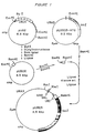
- Figure 1 shows the construction of ubiquitin- lacZ gene fusions.
- Figure 2 shows experiments in which the half-lives of engineered ⁇ -gal proteins are directly measured.
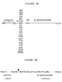
- Figure 3 shows the changing of amino acid residues at the ubiquitin- ⁇ gal junction (A) using the newly discovered properties of ubiquitin-specific processing protease and the amino acid sequence in the vicinity of the junction (B).
- Figure 4 shows the presence of multiple ubiquitin moieties in metabolically unstable ⁇ -gal proteins.
- Figure 5 shows a series of ⁇ -gal species containing ubiquitin in metabolically unstable ⁇ -gal proteins.
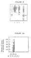
- Figure 6 shows that both prokaryotic and eukaryotic long-lived intracellular proteins have stabilizing amino acid residues at their amino termini whereas secreted proteins exhibit a complementary bias.
- this cleavage takes place regardless of the nature of the amino acid residue of ⁇ gal at the ubiquitin- ⁇ gal junction, thereby making it possible to expose selectively different residues at the amino-termini of otherwise identical ⁇ gal proteins.
- the ⁇ gal proteins so designed exhibited strikingly different half-lives in vivo, ranging from more than 20 hours to less than 3 minutes, depending upon the nature of the amino acid at the amino-terminus of ⁇ gal.
- Amino acids can be thus ordered according to the the half lives they confer on ⁇ gal when present at its amino-terminus. For example, the amino acids methionine, serine, alanine, threonine, valine, glycine and cysteine confer a half-life of more than 20 hours.
- Phenylalanine, leucine, asparagine, and lysine yield half lives of about three minutes.
- Arginine the most destabilizing amino acid, confers a half life of about two minutes. (See Table 1 below for complete list of amino acids and the corresponding half-lives)
- the appropriate amino-terminal amino acid appears to be an essential (though not necessarily a sufficient) requirement for the metabolic stability of a noncompartmentalized, intracellular protein.
- a stabilizing amino acid should be present at the amino-terminus.
- the presence of a destabilizing residue at the amino-terminus of a protein is often, though not always, sufficient for its metabolic destabilization in vivo .
- further analysis shows either an insufficient accessibility of the amino-terminus or a lack of "permissible" sequence environment in the vicinity of the amino-terminus such as a lack of segmental mobility in the protein's amino-terminal region.
- a stabilizing amino acid at the amino-terminus at least in some cases (for instance, as observed for ⁇ -gal) will confer stability upon the protein.
- a stabilizing amino acid at the amino-terminus may not always confer a long half-life because other degradative pathways may be involved in determining the ultimate fate of protein.
- endoproteolytic cleavages cleavages outside of terminal regions of the protein
- a destabilizing amino acid at the amino-terminus of a resulting product of the cleavage which is then rapidly degraded via the N-end rule pathway.
- Appropriate circumstances for use of a stabilizing amino acid can be ascertained empirically.
- the N-end rule may be only one component (albeit a central one) of a more complex "half-life rule" which embraces other aspects of selective protein degradation in vivo
- the N-end rule provides a rational, practicable approach for designing or changing protein structure in order to produce proteins which are more or less resistant to degradation by the N-end rule pathway than natural, unmodified protein.
- Proteins can be designed or modified at the protein or gene level to provide a desired amino acid of either the stabilizing or destabilizing class at their amino-terminus. The ability to regulate the half-life of a protein will allow one to modulate the intracellular activity of the protein.
- a straightforward approach to modifying a protein to increase or decrease its metabolic stability is to directly engineer the amino-terminus of the protein at the protein level.
- the amino-terminus of the protein of interest can be chemically altered, for example, by adding an amino acid of the stabilizing or destabilizing class to the amino-terminus of a protein or polypeptide, employing an appropriate chemistry.
- an unstable protein can be rendered more stable by adding a stabilizing amino acid residue (e.g. methionine serine, alanine, threonine, valine glycine or cysteine) to the amino-terminus of the protein.
- a stabilizing amino acid residue e.g. methionine serine, alanine, threonine, valine glycine or cysteine
- a stable protein can be destabilized by adding a destabilizing amino acid to the amino-terminus.
- One distinct way to modify the amino-terminus of a protein would be to employ specific enzymes, amino acid-protein ligases, which catalyze posttranslational addition of a single amino acid to the protein amino-terminus.
- Other methods for nongenetic alterations of the same type can readily be ascertained by those skilled in the art.
- the amino-terminal end is obscured as a result of the protein's conformation (i.e., its tertiary or quaternary structure). In these cases, more extensive alteration of the amino-terminus may be necessary to make the protein subject to the N-end rule pathway. For example, where simple addition or replacement of the single amino-terminal residue is insufficient because of an inaccessible amino-terminus, several amino acids (including lysine, the site of ubiquitin joining to substrate proteins) may be added to the initial amino-terminus to increase the accessibility and/or segmental mobility of the engineered amino-terminus.
- Modification or design of the amino-terminus of a protein can also be accomplished at the genetic level.
- Conventional techniques of site directed mutagenesis for addition or substitution of appropriate codons to the 5' end of an isolated or synthesized gene can be employed to provide a desired amino-terminal structure for the encoded protein.
- the appropriate codon for a stabilizing amino acid can be inserted or built into the amino-terminus of the protein-encoding sequence.
- expressed proteins are often naturally modified within a cell after translation.
- This can include modification of the amino-terminus.
- the amino-terminus can be acted on by an aminopeptidase which cleaves one or several amino acids from the amino-terminus.
- Amino acids may also be added to the amino-terminus by post-translational processing.
- This invention provides a way to "by-pass" still undefined rules of amino-terminal protein processing to expose exactly and specifically the desired amino acid residues at the amino-terminus of a mature processed protein species.
- specific fusion proteins can be designed wherein the amino-terminus of a protein of interest (designed to have the desired stabilizing or destabilizing structure) is preceded by a "masking" protein sequence fused to the amino-terminus.
- the fusion proteins are designed so that the protein sequence fused to the amino-terminus of the masking protein of interest is susceptible to specific cleavage at the junction between the two. Removal of the protein sequence thus unmasks the amino-terminus of the protein of interest and the half-life of the released protein is thus governed by the predesigned amino-terminus.
- the fusion protein can be designed for specific cleavage in vivo, for example, by a host cell endoprotease or for specific cleavage in a in vitro system where it can be cleaved after isolation from a producer cell (which lacks the capability to cleave the fusion protein).
- Ubiquitin is a broadly useful fusion partner for construction of a fused protein with a protein of interest: the discovery that artifical ubiquitin-protein fusions can be cleaved precisely by a cytoplasmic eukaryotic protease with little or no dependence on the protein to which ubiquitin is fused can be applied both in vivo and in vitro in protein engineering strategies, and is a major aspect of this invention.
- the ubiquitin-protein fusion method can be used to artifically generate authentic amino-termini in proteins produced through artifical means.
- amino-termini characteristic of natural eukaryotic or prokaryotic proteins can be generated by in vitro cleavage of ubiquitin-protein fusions produced in a prokaryotic host.
- ubiquitin fusion proteins are expressed by a chimeric gene construct comprising, in 5' to 3' orientation, a ubiquitin gene linked to a gene encoding the protein of interest.
- the codon for the amino-terminal amino acid of the protein of interest is located immediately adjacent the 3' end of the ubiquitin gene.
- the fused gene product is cleaved endoproteolytically either in vivo or in vitro (using either pure or partially purified ubiquitin-specific protease identified in the present invention) at the junction between ubiquitin and the protein of interest to generate the protein of interest having the desired amino acid at its amino-terminus.
- This method of generating the desired amino acid residue at the amino-terminus of a protein of interest involves two novel components: one, the use of ubiquitin-protein fusions, and the other, the use of ubiquitin-specific processing protease that has been identified, and whose striking substrate requirements were discovered, in this work.
- the enzyme is also relatively stable and active in vitro (in extracts), and can readily be purified to homogeneity by techniques known to those skilled in the art.
- the substrate specificity of the ubiquitin-specific processing protease is highly conserved in evolution, being the same in yeast and mammals.
- the enzyme can be purified chromatographically from a crude extract by sequential chromatography on phosphocellulose, DEAE cellulose, and SH Sepharose among other methods known to those skilled in the art.
- the gene for this protease can be cloned by those skilled in the art.
- the cloned protease gene can be used either in vivo, or, alternatively, the gene can be overexpressed in a suitable host, the overexpressed ubiquitin-specific protease purified and used for the same or similar purposes in vitro.
- the discovery of this enzyme, and detailed characterization of its substrate specificity herein provides for the in vitro and in vivo use of this enzyme.
- ubiquitin-protein fusions to allow the generation of a desired amino acid residue at the amino-terminus of a protein of interest can be extended to facilitate the purification of such proteins from producer cells.
- a gene can be readily constructed that encodes a convenient marker protein, such as streptavidin, linked to a ubiquitin-protein fusion construct described above.
- the resulting (marker protein)-ubiquitin-protein fusion can be simply isolated from producer cells by using the preselected property of the marker protein, for instance, the known ability of streptavidin to be isolatable by affinity chromatography on a biotin column.
- purified (marker protein)-ubiquitin-protein fusion can then be specifically cleaved by the ubiquitin-specific protease described in this invention to generate the final product, a protein of interest with the desired amino acid residue at its amino-terminus.
- the codon for the amino-terminal amino acid of desired amino acid by, for example, site-directed mutagenesis techniques currently standard in the field. If the gene encoding the protein of interest is a synthetic gene the appropriate 5' codon can be built-in during the synthetic process. Alternatively, nucleotides for a specific codon can be added to the 5' end of an isolated or synthesized gene by ligation of an appropriate DNA sequence to the 5' (amino-terminus encoding) end of the gene.
- Ubiquitin-like fusion partners capable of being cleaved by the ubiquitin-specific protease can also be used.
- fusion partners other then ubiquitin for masking the amino-terminus of a protein of interest can be used.
- the fusion proteins can be designed to contain a proteolytic cleavage site for a restriction endoprotease which has sufficiently narrow specificity so that only one target site is cleaved in a fusion protein.
- a crucial property of such a protease must be a sufficiently relaxed requirement for the nature of the amino acid residue(s) abutting the carboxy-terminal side of the cleavage site.
- the target site for cleavage is the junction between the fusion partner and the amino-terminus of the protein of interest and thus the recognition site for the endoprotease is located to provide for cleavage at this location.
- the commerically available protease, complement factor X a exhibits these properties and thus can be used to directly generate proteins with predetermined amino acid residues in the ultimate position of their amino-termini (see, K. Nogai and H.C. Thogersen Nature 309 :810 (1984)).
- the recognition site for the endoprotease can be engineered into the junction between the masking protein sequence and the 3' region encoding the amino-terminus of the protein of interest.
- ubiquitin-protein fusions such as ubiquitin-Pro- ⁇ -galactosidase fusion (Table 1)
- ubiquitin-Pro- ⁇ -galactosidase fusion Table 1
- Prevention of the efficient deubiquitination of a ubiquitin-protein fusion can be achieved in several ways, for instance, by using a proline residue at the ubiquitin-protein junction as shown in Table 1, or by changing the amino acid sequence of ubiquitin near its carboxyl-terminus in such as way that the ubiquitin moiety is no longer recognized by the ubiquitin-specific processing protease but can still be recognized by the rest of the degradative pathway.
- These and other ways to reduce the rate of deubiquitination of a ubiquitin-protein fusion can be readily ascertained by those skilled in the art.
- the methods of this invention can be employed, inter alia , for regulating the half-life of a protein intracellularly. There are many instances where this capability is useful. For example, when a gene is introduced into a cell for expression therein, the expressed product can be designed for a long or short half-life depending upon the particular need.
- destabilized proteins which have short half-lives are more amenable to regulation of intracellular levels of the protein.
- the ability to finely regulate the intracellular levels and activity of a protein can be useful in therapy or in the work with in vitro cell cultures.
- gene therapy for example, a gene may be introduced into a cell to compensate for a genetic deficiency or abnormality.
- the gene can be inserted under control of an inducible promoter. Induction results in enhanced expression of the gene product and consequently, higher levels of the product within the cell. If the gene is designed to encode an unstable protein, the intracellular concentration of the expressed protein will be more quickly responsive to a later reduction in the rate of its synthesis because it does not persist within the cell. In this way, the intracellular level and/or activity of the protein encoded by the inserted gene can be more finely regulated.
- the method of this invention can also be used to expand the uses of selectable markers by shortening the time necessary for a phenotype related to the marker to become manifest.
- a product encoded by a marker gene can be destabilized by altering its amino-terminus according to the N-end rule. In this way selection for the negative phenotype can be facilitated because the product of the marker gene will be more quickly extinguished after function of the gene encoding the marker is abolished.
- An example is the thymidine kinase (tk) gene
- the tk gene can be engineered to encode a less stable enzyme by introducing an appropriate destabilizing amino acid at the amino-terminus.
- Gene mutation resulting in tk - phenotype will be more quickly manifested by cells because residual tk will be more quickly degraded. This can be especially useful in slow growing cells where more time is required to "dilute out" tk synthesized prior to transformation to the tk - type.
- Proteinaceous cytotoxins can be designed as unstable proteins degradable by the N-end rule pathway so that they do not persist after their toxic action has been exerted on a target cell. Reducing the lifetime of the toxin reduces the likelihood of killing nontargeted cells.
- N-end rule pathway of degradation allows development of mutant cells having mutations in genes encoding essential components of the N-end rule pathway. For example, cells can be produced that either permanently or conditionally are unable to efficiently degrade otherwise short-lived proteins. These cells can be used to produce desired proteins that ordinarily would be unstable within a cell.
- pUB23 (figure 1) was treated sequentially with Acc I, the Klenow fragment of pol I, and Bam HI. A fragment containing the Xho I site was purified and inserted between a filled-in Hind III site and a BAM HI site of the M13mp9 phage DNA. (J. Messing and J. Vieira, Gene 19, 263 (1982)). Site-directed mutagenesis (M. Smith, Annu . Rev. Genet. 19, 423 (1985)) was carried out as described by Kramer, W. et al . Nucl . Acids Res.
- cells from a 5 ml culture were harvested by filtration through the well of a Millipore microtiter filtration plate, washed several times on the filter with the same medium lacking methionine and resuspended in 0.3 ml of 1 percent galactose, 50 mM potassium phosphate buffer (pH 7.4).
- [ 35 S]methionine 50 to 100 ⁇ Ci was then added for 5 minutes at 30°C; the cells were collected by filtration and resuspended on 0.4 ml of the growth medium containing cycloheximide at 0.5 mg/ml.
- Samples (0.1 ml) were withdrawn at indicated times, and added to 0.75 ml of cold buffer A (see below for buffer composition) containing leupeptin, pepstatin A, antipain, aprotinin and chymostatin (Sigma), (each at 20 ⁇ g/ml) in addition to 0.4 ml of glass beads.
- the cells were disrupted by vortexing for approximately 3 minutes at 4°C; the extracts were centrifuged at 12,000g for 3 minutes and the radioactivity of acid-insoluble 35 S in the supernatants was determined. Aliquots of the supernatants containing equal amounts of the total acid-insoluble 35 S were processed for immunoprecipitation with a monoclonal antibody to gal.
- Ascitic fluid containing a molar excess of the antibody (at least tenfold) was added to each aliquot, with subsequent incubation at 4°C for 2 hours; protein A-Sepharose (Pharmacia) was then added, the suspension was incubated with rocking at 4°C for 30 minutes and centrifuged at 12,000g for 1 minute.
- the protein A-Sepharose pellets were washed three times in buffer A (see below) containing 0.1 percent sodium dodecyl sulfate (SDS), resuspended in an SDS, dithiotreitol (DTT)-containing electrophoretic sample buffer (U.K.
- Plasmid pUB23 (Figs. 1 and 3) was introduced into DS410, a minicell-producing E . coli strain.
- Minicells were prepared and labeled for 60 minutes at 36°C with [ 35 S]methionine ( 600 Ci/mmole, Amersham) as described by N. Stoker et al., supra.
- Labeled minicells were centrifuged, resuspended in 2 percent SDS, 10 mM DTT, 10 mM Na-HEPES (ph 7.5) and heated at 100°C for 3 minutes. After centrifugation at 12,000g for 1 minute the supernatant was diluted 20-fold with buffer A (1 percent Triton X-100, 0.15 M NaCl, 5 mM Na-EDTA, 50 mM Na-HEPES, ph 7.5), followed by the addition of phenylmethylsulfonyl fluoride (PMSF) and N-ethylmaleimide to 0.5 mM and 10mM, respectively. After 4 hours at 4°C, the sample was dialyzed against buffer A containing 0.5 mM PMSF overnight at 4°C, and processed for immunoprecipitation (as described above).
- buffer A 1 percent Triton X-100, 0.15 M NaCl, 5 mM Na-EDTA, 50 mM Na-HE
- S . cerevisiae cells carrying plasmids of interest were grown in 800 ml of a uracil-deficient medium, then harvested and disrupted with glass beads in buffer A containing leupeptin, pepstatin A, antipain, aprotinin and chymostatin (each at 3 ⁇ g/ml).
- the extract was centrifuged at 12,000g for 3 minutes.
- Saturated ammonium sulfate was added to the supernatant to a final concentration of 57 percent. After overnight incubation at 4°C, the precipitated protein was collected by centrifugation at 23,000g for 30 minutes. The pellet was redissolved in buffer A containing protease inhibitors.
- the sample was passed through an affinity column which had been prepared by crosslinking an IgG fraction from an ascitic fluid (containing a monoclonal antibody to gal to Affi-Gel 10 (Bio-Rad).
- the IgG fraction used for crosslinking had been purified from the ascitic fluid by affinity chromatography on protein A-Sepharose. After washing with buffer A lacking Triton X-100, the antibody-bound proteins were eluted with 0.25 M glycine-HCl (pH 2.6). The eluate was immediately adjusted to pH 7.5 with 1 M Na-HEPES (pH 8.5), and thereafter made 0.1 percent in SDS.
- the sample was concentrated by ultrafiltration in Centricon 30 (Amicon), and subjected to electrophoresis in a 7 percent discontinuous polyacrylamide-SDS gel (U.K. Laemmli, Nature (London) 227, 680 (1970)). Electroblotting of proteins to nitrocellulose, and immunoblot analysis with a peptide-mediated antibody to ubiquitin were performed as described by P.S. Swerdlow, D. Finley and A. Varshavsky, Analyt . Biochem. 156, 147 (1986). The same results were obtained with a different antibody to ubiquitin obtained from A. Haas (Univ of Milwaukee Med. School).
- Figure 1 shows construction of a ubiquitin- lacZ gene fusion.
- pUB2 a pBR322-based genomic DNA clone (E. Ozkaynak, et al . Nature 312, 663 (1984) contains six repeats of the yeast ubiquitin-coding sequence (open boxes) together with the flanking regions (jagged lines).
- pUB2 was modified as shown in the diagram by placing a Bam HI site six bases downstream from the first ubiquitin repeat. This allowed the construction of an in-frame fusion (confirmed by nucleotide sequencing) between a single ubiquitin repeat and the lacZ gene of the expression vector pLGSD5-ATG (called G2 in L. Guarente, Methods Enzymol .
- 2 ⁇ m denotes a region of the pLGSD-ATG that contains the replication origin and flanking sequences of the yeast plasmid called 2 ⁇ m circle (See L. Guarente, supra).
- Figure 3B shows the amino acid sequence of the fusion protein in the vicinity of the ubiquitin- ⁇ gal junction.
- Figure 2 shows that the in vivo half-life of gal is a function of its amino-terminal residue.
- (lane a) Minicells isolated from an E . coli strain carrying pUB23, the initial ub- lacZ fusion (Figs. 1 and 3B), were labeled with [ 35 S]methionine for 60 minutes at 36°C, with subsequent analysis of ⁇ gal as described. The same result was obtained when the labeled minicell SDS extract was combined with an unlabeled yeast SDS extract before immunoprecipitation of gal.
- Lane b S . cerevisiae cells carrying pUB23 (Fig. 1), which encodes ub-Met- ⁇ gal (Fig.
- Figure 3 shows the changing amino acid residues of gal at the ubiquitin- gal junction.
- the initial plasmid, pUB23 (Fig.1) which encodes ub-Met- ⁇ gal, was mutagenized as described above to convert the original Met codon ATG at the ub- ⁇ gal junction into codons specifying 19 amino acids other than Met.
- the original round of mutagenesis shown in Figure 3, produced 15 out of 19 possible substitutions. The remaining four substitutions were produced later (see Table 1)).
- the arrowhead indicates the site of the deubiquitinating in vivo cleavage in the nascent fusion protein that occurs with all of the fusion proteins except ub-Pro- gal (see text). All of the constructions shown encode His as the second gal residue.
- A Ala
- C Cys
- D Asp
- E Glu
- F Phe
- G Gly
- H His
- I Ile
- K Lys
- L Leu
- M Met
- N Asn
- P Pro
- Q Gln
- R Arg
- S Ser
- T Thr
- V Val
- W Trp
- Y Tyr.
- FIG. 4 shows that ubiquitin- gal is short-lived if not deubiquitinated.
- Lanes a to g S . cerevisiae cells carrying plasmids encoding ub-X- ⁇ gal fusion proteins in which X is the residue indicated at the top of each lane, were labeled for 5 minutes at 30°C with [ 35 S]methionine, followed by extraction, immunoprecipitation and analysis of ⁇ gal. Fluorographic exposures for these lanes were several times longer than those for similar patterns in Figure 2 to reveal the multiple ubiquitination of short-lived ⁇ gal proteins.
- Lanes h, i Fluorographic overexposure of lanes n, o in Fig.
- cerevisiae cells carrying a plasmid encoding ub-Pro- ⁇ gal were labeled for 5 minutes at 30°C with [ 35 S]methionine (lane m) followed by a chase in the presence of cycloheximide for 10, 30, and 60 minutes (lanes n to p).
- the upper small arrow to the right of lane p denotes ub-Pro- ⁇ gal, a small proportion of which is still present after 1 hour chase.
- the lower small arrow indicates an apparently deubiquitinated Pro- ⁇ gal that slowly accumulates during chase and is metabolically stable.
- the dot to the left of lane m denotes an endogeneous yeast protein that is precipitated in some experiments by the antibody used.
- Square brackets denote the multiply ubiquitinated ⁇ -gal species (see Fig. 5). Other designation are as in Figure 2.
- FIG. 5 shows the "ladder" gal species containing ubiquitin.
- (lane a) S . cerevisiae cells carrying a plasmid which encodes ub-Gln- ⁇ gal, were grown and disrupted, and the extracts processed for isolation of ⁇ gal proteins by affinity chromatography on a column with immobilized antibody to ⁇ gal. The ⁇ gal proteins thus obtained were electrophoresed in a polyacrylamide-SDS gel, transferred to nitro-cellulose, and probed with an antibody to ubiquitin.
- (lane b) Same as lane a, but with ub-Pro- ⁇ gal.
- (lane c) Same as b but a longer autoradiographic exposure.
- (lane d) S .
- cerevisiae cells carrying a plasmid which encodes ub-Leu- ⁇ gal were labeled with [ 35 S]methionine for 5 minutes, with subsequent extraction, immunoprecipitation and electrophoresis of ⁇ gal (the same sample as in Figure 4, lane f).
- Square brackets denote the multiply ubiquitinated Gln- ⁇ gal species detected with antibody to ubiquitin.
- the arrow indicates the band of ub-Pro- ⁇ gal, the initial fusion protein seen in lanes b and c.
- the arrowheads indicate the position of the band of deubiquitinated ⁇ gal (detectable by either Coomassie staining or metabolic labeling, but not with antibody to ubiquitin) derived from the ub-Gln- ⁇ gal fusion protein.
- Figure 6 shows both prokaryotic and eukaryotic long-lived intracellular proteins have stabilizing amino acid residues at their amino-termini, whereas secreted proteins exhibit a complementary bias.
- Branched ubiquitin conjugates in which the carboxyl-terminal glycine of ubiquitin moieties is joined via an isopeptide bond to the ⁇ -amino groups of internal lysine residues in proteins apparently comprise the bulk of ubiquitin conjugates in eukaryotic cells. Joining of ubiquitin to the amino-terminal ⁇ -amino groups of target proteins, to yield linear ubiquitin conjugates, may also be chemically feasible. See A. Hershko, et al ., PNAS USA 81 : 7021 (1984).
- the resulting ⁇ gal-containing protein When this gene is expressed in E . coli , the resulting ⁇ gal-containing protein has an apparent molecular mass which is approximately 6 kD greater that that of the control ⁇ gal, a value consistent with the presence of ubiquitin in the protein encoded by the chimeric gene. In contrast, when the same gene is expressed in yeast, the corresponding ⁇ gal protein is electrophoretically indistinguishable from the control ⁇ gal. This result is independent of the length of the [ 35 S]methionine labeling period (between 1 and 30 minutes).
- the ubiquitin- ⁇ -gal junction encoded by the chimeric gene, Gly-Met ( Figures 1 and 3B), is identical to the junctions between adjacent repeats in the polyubiquitin precursor protein, which is efficiently processed into mature ubiquitin.
- the same protease as yet uncharacterized biochemically, is responsible both for the conversion of polyubiquitin into mature ubiquitin and for the deubiquitination of the nascent ubiquitin- ⁇ gal protein.
- the set of individual amino acids can be ordered with respect to the half-lives that they confer on gal when exposed at its amino-terminus.
- the resulting rule (Table 1) is referred to as the "N-end rule”.
- the N-end rule Residue X in ub-X- ⁇ gal Radius of gyration of X(A) In vivo deubiquitination of nascent ub-X- ⁇ gal t 1/2 of X- ⁇ gal Met 1.80 + Ser 1.08 + Ala 0.77 + Thr 1.24 + 20 hours Val 1.29 + Gly 0 + Cys + Ile 1.56 + Glu 1.77 + 30 minutes Tyr 2.13 + Gln 1.75 + 10 minutes His Phe 1.90 + Leu 1.54 + 3 minutes Trp + Asp 1.43 + Asn + Lys 2.08 + Arg 2.38 + 2 minutes Pro 1.25 -* 7 minutes *The rate of in vivo deubiquitination of ub-Pro- ⁇ gal is extremely low.
- the t 1/2 shown is that
- Site-directed mutagenesis was employed to insert a codon specifying a "stabilizing" amino acid (in this experiment, the Met residue) before the first codon of ⁇ gal at the ubiquitin- ⁇ gal junction (Table 2). Insertion of a stabilizing residue (Met) before either another stabilizing residue (Thr) or a variety of destabilizing residues (Gln, Lys, and Arg) at the ubiquitin- ⁇ gal junction invariably results in a long-lived deubiquitinated ⁇ gal (Table 2).
- a long-lived cleavage product of gal is formed during decay of short-lived ⁇ gal proteins.
- the electrophoretic patterns of short-lived (but not of long-lived) ⁇ gal proteins invariably contain a specific, about 90 kD cleavage product of ⁇ gal ( Figure 2, lanes n to u) which, unlike the parental ⁇ gal species, accumulates during the postlabeling (chase) period ( Figure 4, lanes m-p).
- the 90 KD ⁇ gal fragment constitutes a relatively small proportion of the initial amount of the pulse-labeled ⁇ gal. Nonetheless, its existence implies that an in vivo endoproteolytic cleavage can rescue a protein fragment from the metabolic fate of its short-lived parental protein. It remains to be seen whether the resulting possibility of multiple half-lives within a single protein species is exploited in the design of naturally short-lived proteins.
- Ubiquitin- ⁇ gal is short-lived when not deubiquitinated.
- Ubiquitin-Pro- ⁇ gal the only ubiquitin- ⁇ gal fusion that is not deubiquitinated in vivo (Fig. 4, lanes j to p), has a half-life of approximately 7 minutes (Table 1) which is less than 1 percent of the half-life of metabolically stable ⁇ gal proteins (Table 1).
- Table 1 One interpretation of this result is that a metabolically stable ubiquitin attachment to protein amino-termini is sufficient to signal degradation of acceptor proteins. This interpretation is consistent with earlier biochemical and genetic evidence that ubiquitination of short-lived proteins in a mammalian cell is essential for their degradation.
- Short-lived ⁇ gal proteins are multiply ubiquitinated in vivo.
- nascent proteins apparently lack ubiquitin moieties.
- the in vivo amino-terminal processing of nascent noncompartmentalized proteins generates their mature amino-termini via the action of amino-terminal peptidases whose substrate specificities have been partially characterized. (See Tsunasawa, S. et al . J . Biol . Chem . 260 5382 (1985); Boissel, J.P. et al . PNAS USA 82 , 8448 (1985)). We suggest that the amino-termini thus generated are recognized by an "N-end-reading" enzyme.
- One specific model is that a commitment to degrade a protein molecule is made as a result of the recognition of its amino-terminal residue by a stochastically operating enzyme whose probability of "clamping" at the target's amino-terminus is determined by the N-end rule (Table 1).
- the commitment is made, it is followed by a highly processive ubiquitination of the target protein which in the case of ⁇ gal is conjugated to more than 15 ubiquitin moieties per molecule of gal (Fig. 4, lanes c to g, and Fig. 5).
- the multiply ubiquitinated target protein is then degraded by a "down stream" enzyme (1) for which the ubiquitin moieties of the target serve as either recognition signals or denaturation (unfolding) devices, or both.
- the ubiquitin-containing "ladder” ⁇ gal species consist of apparently branched ubiquitin moeities joined to the ⁇ -amino groups of internal lysine residues in ⁇ gal.
- the "ladder” ⁇ gal species derived from ubiquitin-Pro- ⁇ gal are electrophoretically indistinguishable from the analogous species of ⁇ gal whose amino-terminal ubiquitin is cleaved off the nascent fusion protein (Fig. 4, lanes j to l, and Fig. 5).
- the unblocked amino-terminal residues in metabolically stable, noncompartmentalized proteins from both prokaryotes and eukaryotes are exclusively (Fig. 6A) of the stabilizing class (Met, Ser, Ala, Gly, Thr, Val), that is, the class that confers long in vivo half-lives on gal (Table 1).
- the one short-lived intracellular protein for which the mature amino-terminus is known is the cII protein of phage lambda, the central component of a trigger that determines whether ⁇ grows lytically or lysogenizes an infected cell.
- destabilizing amino acids can be either hydrophobic, uncharged hydrophilic or charged, they share the property of having larger radii of gyration than any of the stabilizing amino acids except Met (Table 1).
- Amino-terminal residues in compartmentalized proteins are largely of the destabilizing class.
- Figure 6 illustrates a striking difference between the choice of amino-terminal residues in long-lived, noncompartmentalized intracellular proteins (A) and in compartmentalized proteins, such as secreted proteins (B), many of which are also long-lived in their respective extracellular compartments.
- A noncompartmentalized intracellular proteins
- B secreted proteins
- secreted eukaryotic toxins contain strongly destabilizing residues (Arg, Lys, Leu, Phe, Asp) at their amino-termini more often than the general population of secreted proteins (Fig. 6, panels B to D).
- the methods of the present invention should also be useful for manipulating the metabolic stability and other properties of compartmentalized proteins, including secreted ones.
- Long-lived intracellular proteins with destabilizing penultimate residues generally retain their initial amino-terminal methionine residue.
- the amino-terminal residues in long-lived intracellular proteins that do undergo amino-terminal processing are invariably of the stabilizing class (Table 1).
- An interesting possibility that would involve the N-end rule pathway in the turnover of long-lived proteins is that the rate-limiting step in the in vivo degradation of long-lived proteins may be a slow aminopeptidase cleavage that exposes a destabilizing residue, followed by rapid degradation via the N-end rule pathway. Note that fine-tuning of the rate of degradation may in this case be a function of the rate of aminopeptidase cleavage exposing a destabilizing residue rather than a function of the residue's destabilizing capacity according to the N-end rule.
- the N-end rule and selective degradation of short-lived and damaged proteins are defined by The N-end rule and selective degradation of short-lived and damaged proteins.
- the preferential exposure of destabilizing residues at the amino-termini of products of the initial protein cleavages may be due either to intrinsic specificities of the proteases involved or simply to the fact that a majority of the amino acids belong to the destabilizing class (Table 1).
- initial cleavages of a protein would be expected to destabilize aspects of its original conformation, thus increasing the probability of further internal cuts.
- Whether the initial cleavage products of a protein would be degraded exclusively via the N-end rule pathway or would have to be processed further by additional internal cleavages should depend on several factors, such as the exposure of destabilizing residues at the amino-termini of initial cleavage products, and the relative rates of introduction of internal cuts.
- the N-end rule pathway should be essential for degradation of most of the metabolically unstable proteins, from chemically damaged, prematurely terminated, improperly folded and miscompartmentalized ones to those that cannot assemble into native multisubunit aggregates, and finally to otherwise normal proteins that are short-lived in vivo.
- the metabolic instability of a protein may be mediated not only by the exposure of a destabilizing residue at its amino-terminus, but also by local conformational and chemical features of its polypeptide chain that result in proteolytic cleavages exposing destabilizing residues at the amino-termini of cleavage products.
- any given protein a variety of factors in addition to the N-end rule may combine to modulate its half-life in vivo. Among such factors may be the flexibility and accessibility of the protein's amino-terminus (Thornton, J.M. and Sibanda, B.L., J . Mol . Bio. 167 443 (1983)), the presence of chemically blocking amino-terminal groups such as the acetyl group, the distribution of ubiquitinatable lysine residues near the amino-terminus, and other variables, such as the structure of the carboxy-terminus. Since amino-terminal regions of multisubunit proteins are commonly involved in the interfaces between subunits (Thornton, J.M. and Sibanda, B.L., J . Mol.
- quarternary structure of proteins is yet another parameter that is expected to modulate the impact of the N-end rule pathway on protein half-lives in vivo.
- the N-end rule pathway may also be essential for the degradation of proteins whose initial recognition as targets for degradation is independent of the structures at their amino-termini.
- the posttranslational addition of amino acids to proteins in vivo dramatically accelerates in a stressed or regenerating tissue, for example, after physical injury to axons of nerve cells (S. Shyne-Athwal, R.V. Riccio, G. Chakraborty, N.A. Ingolia, Science 231 , 603 (1986); N.A. Ingolia et al ., J. Neurosci 3 , 2463 (1983)).
- the N-end rule provides an explanation for this phenomenon.
- Physiological states in which addition of destabilizing amino acids to proteins could be expected to occur include entry to and exit from the cell cycle, responses to chemical or physical stress, and specific differentiation events, such as erythroid differentiation and spermatogenesis, in which a proportion of preexisting, otherwise long-lived intracellular proteins is selectively degraded.
Landscapes
- Health & Medical Sciences (AREA)
- Life Sciences & Earth Sciences (AREA)
- Chemical & Material Sciences (AREA)
- Genetics & Genomics (AREA)
- Engineering & Computer Science (AREA)
- Organic Chemistry (AREA)
- Biomedical Technology (AREA)
- Molecular Biology (AREA)
- Bioinformatics & Cheminformatics (AREA)
- Wood Science & Technology (AREA)
- Zoology (AREA)
- Biotechnology (AREA)
- General Engineering & Computer Science (AREA)
- General Health & Medical Sciences (AREA)
- Biochemistry (AREA)
- Biophysics (AREA)
- Microbiology (AREA)
- Medicinal Chemistry (AREA)
- Plant Pathology (AREA)
- Physics & Mathematics (AREA)
- Mycology (AREA)
- Proteomics, Peptides & Aminoacids (AREA)
- Analytical Chemistry (AREA)
- General Chemical & Material Sciences (AREA)
- Gastroenterology & Hepatology (AREA)
- Chemical Kinetics & Catalysis (AREA)
- Peptides Or Proteins (AREA)
- Preparation Of Compounds By Using Micro-Organisms (AREA)
- Micro-Organisms Or Cultivation Processes Thereof (AREA)
- Saccharide Compounds (AREA)
Abstract
Description
- In both bacterial and eukaryotic cells, relatively long-lived proteins, whose half-lives are close to or exceed the cell generation time, coexist with proteins whose half-lives can be less than one percent of the cell generation time. Rates of intracellular protein degradation are a function of the cell's physiological state, and appear to be controlled differentially for individual proteins. In particular, damaged and otherwise abnormal proteins are metabolically unstable in vivo. Although the specific functions of selective protein degradation are in most cases still unknown, it is clear that many regulatory proteins are extremely short-lived in vivo. Metabolic instability of such proteins allows for rapid adjustment of their intracellular concentrations through regulated changes in rates of their synthesis or degradation. The few instances in which the metabolic instability of an intracellular protein has been shown to be essential for its function include the cII protein of bacteriophage lambda and the HO endonuclease of the yeast Saccharomyces cerevisiae.
- Most of the selective turnover of intracellular proteins under normal metabolic conditions is ATP-dependent and (in eukaryotes) nonlysosomal. Recent biochemical and genetic evidence indicates that, in eukaryotes, covalent conjugation of ubiquitin to short-lived intracellular proteins is essential for their selective degradation. The rules which determine whether a given protein is metabolically stable or unstable in vivo were previously unknown.
- Nagai and Thogersen, Nature, Vol. 309, pp 810-812 (1984) disclose insertion of the sequence Ile-Glu-Gly-Arg between the 31 amino-terminal residues of λ cII protein and Val 1 of human β-globin, and the production of the hybrid in high yield in E.coli. They then cleaved the hybrid specifically at the single arginine, using blood coagulation factor Xa and thus liberated the authentic β-globin chain. As factor Xa is specific for the tetrapeptide Ile-Glu-Gly-Arg7, which is rare in protein sequences, their expression/cleavage system is applicable to the efficient production of eukaryotic proteins.
- Hershko et al, Proc. Natl. Acad. Sci. USA, vol. 81, pp 7021-7025 (1984) disclose that the selective modification of NH2-terminal α-NH2 groups of globin and lysozyme prevents their degradation by the ubiquitin proteolytic system from reticulocytes. The conjugation by ubiquitin of ε-NH2 groups of lysine residues, usually seen in multiples, was also inhibited in α-NH2-blocked proteins. Naturally occurring Nα-acetylated proteins are not degraded by the ubiquitin system at a significant rate, while their nonacetylated counterparts from other species are good substrates. The authors suggest that one function of Nα-acetylation of cellular proteins is to prevent their degradation by the ubiquitin system. α-NH2-blocked proteins can have their activity as substrates for degradation increased by incorporation of α-NH2 groups through the introduction of polyalanine side chains. Proteins in which most ε-NH2 groups are blocked but the α-NH2 group is free are degraded by the ubiquitin system, but at a reduced rate. The authors suggest that the exposure of a free NH2 terminus of proteins is required for degradation and may initiate the formation of ubiquitin conjugates committed for degradation.
- The invention is as claimed in the claims. Gene constructs and/or fusion proteins which differ from those defined in the claims only are mentioned for comparative purpose in present specification. They are not within the scope of present invention.
- This invention pertains to methods of engineering the amino terminus of proteins thereby controlling the metabolic stability and other properties of a protein. Further, this invention provides a method for either in vivo or in vitro production of proteins with any of the amino acid residues as recited in claim 1 at the protein's amino-terminus. The invention is based in part upon the striking discovery that the in vivo half-life of an intracellular protein is a function of its amino-terminal amino acid residue and upon a novel (and more generally applicable) technique that allows one to generate proteins with specified amino-termini in vivo or in vitro. The invention also pertains to a newly identified protease, ubiquitin-specific processing protease, which has properties that allow one to expose, either in vitro or in vivo, any desired amino acid residue at the amino-terminus of a protein of interest.
- The nature of the amino acid exposed at the amino-terminus of an intracellular protein was shown to be one crucial determinant that specifies whether a protein will be long- or short-lived in vivo. Individual amino acids can be categorized as either stabilizing or destabilizing amino acids with respect to the half-life that they confer upon a protein when exposed at the protein's amino-terminus. Destabilizing amino acid residues confer short half-lives, down to a few minutes for some of the destabilizing amino acids. Stabilizing amino acid residues confer long half-lives of many hours. This striking and newly discovered dependency of a protein's half-life on its amino-terminal residue is referred to herein as the N-end rule.
- Based upon the N-end rule, the amino-terminus of a protein can thus be designed or altered to change the intracellular half-life of the protein and in this way the lifetime and/or activity of the protein in vivo can be regulated. This capability can be exploited for rational protein design in many different contexts. Natural or wild type proteins can be modified to render them more or less resistant to degradation in vivo. The design or alteration of the protein can be done at the protein level or at the genetic (DNA) level. For example, proteins can be modified by chemically altering or engineering the amino-terminus to provide for exposure at the amino-terminus of an amino acid residue of the stabilizing or destabilizing class. At the genetic level, genes encoding proteins can be made to encode an amino acid of the desired class at the amino-terminus so that the expressed protein exhibits a predetermined amino-terminal structure which renders it either metabolically stable or unstable with respect to the N-end rule pathway of proteolytic degradation. Furthermore, proteins can be expressed fused to a "masking" protein sequence which masks the engineered amino-terminus so that when unmasked the protein will exhibit the desired stability or other properties that depend on the nature of the protein's amino-terminal residue. In such constructs, for example, the junction between the two protein sequences can be designed to be cleaved specifically, for instance, by an endoprotease. Endoproteolytic cleavage of the fused sequence unmasks the specifically engineered amino-terminus of the protein of interest and subjects the protein to degradation governed by the N-end rule. One specific and new way to engineer the protein's amino-terminus is provided in this invention by the identification of ubiquitin-specific processing protease and determination of its substrate specificity. Using this protease, fusions of ubiquitin with other proteins can be specifically processed either in vitro or in vivo to generate proteins with desired amino-terminal residues.
- A different, and also new way to specifically engineer short-lived proteins is provided in this invention by the discovery that ubiquitin-protein fusions, such as ubiquitin-Pro-β-galactosidase, that cannot be efficiently deubiquitinated, are metabolically unstable. Thus, by attaching the amino-terminal ubiquitin moiety to a protein in a way that makes its removal either impossible or inefficient, one can destabilize proteins by a distinct technique that is not directly based on the N-end rule.
- In addition, variant cells can be developed which contain putative mutations in the "N-end" degrading protease(s) which either conditionally or nonconditionally stop degrading short-lived proteins. These cells can be used to overproduce proteins that ordinarily would be short-lived within the cell.
- Figure 1 shows the construction of ubiquitin-lacZ gene fusions.
- Figure 2 shows experiments in which the half-lives of engineered β-gal proteins are directly measured.
- Figure 3 shows the changing of amino acid residues at the ubiquitin-βgal junction (A) using the newly discovered properties of ubiquitin-specific processing protease and the amino acid sequence in the vicinity of the junction (B).
- Figure 4 shows the presence of multiple ubiquitin moieties in metabolically unstable β-gal proteins.
- Figure 5 shows a series of β-gal species containing ubiquitin in metabolically unstable β-gal proteins.
- Figure 6 shows that both prokaryotic and eukaryotic long-lived intracellular proteins have stabilizing amino acid residues at their amino termini whereas secreted proteins exhibit a complementary bias.
- The elucidation of the N-end rule is described in detail below. Briefly, this rule governing protein degradation was revealed by examining the in vivo half-lives of the enzyme β-galactosidase having various amino acid residues at its amino-terminus and produced as a fusion protein with ubiquitin. When a chimeric gene encoding a ubiquitin-β-galactosidase fusion protein is expressed in the yeast S. cerevisiae, ubiquitin is cleaved off the nascent fusion protein, yielding a deubiquitinated-β-galactosidase (βgal). With one exception, this cleavage takes place regardless of the nature of the amino acid residue of βgal at the ubiquitin-βgal junction, thereby making it possible to expose selectively different residues at the amino-termini of otherwise identical βgal proteins. The βgal proteins so designed exhibited strikingly different half-lives in vivo, ranging from more than 20 hours to less than 3 minutes, depending upon the nature of the amino acid at the amino-terminus of βgal. Amino acids can be thus ordered according to the the half lives they confer on βgal when present at its amino-terminus. For example, the amino acids methionine, serine, alanine, threonine, valine, glycine and cysteine confer a half-life of more than 20 hours. Phenylalanine, leucine, asparagine, and lysine yield half lives of about three minutes. Arginine, the most destabilizing amino acid, confers a half life of about two minutes. (See Table 1 below for complete list of amino acids and the corresponding half-lives)
- Currently known amino-terminal residues in long-lived, noncompartmentalized intracellular proteins from both prokaryotes and eukaryotes belong virtually exclusively to the stabilizing class of amino acids, exactly as predicted by the N-end rule. This result stongly implicates the N-end rule in the selective degradation of intracellular proteins in general.
- The appropriate amino-terminal amino acid appears to be an essential (though not necessarily a sufficient) requirement for the metabolic stability of a noncompartmentalized, intracellular protein. Thus, in order for a protein to be relatively stable intracellularly, a stabilizing amino acid should be present at the amino-terminus. The presence of a destabilizing residue at the amino-terminus of a protein is often, though not always, sufficient for its metabolic destabilization in vivo. When such destabilization occurs to a relatively small extent, further analysis shows either an insufficient accessibility of the amino-terminus or a lack of "permissible" sequence environment in the vicinity of the amino-terminus such as a lack of segmental mobility in the protein's amino-terminal region. The presence of a stabilizing amino acid at the amino-terminus at least in some cases (for instance, as observed for β-gal) will confer stability upon the protein. However, a stabilizing amino acid at the amino-terminus may not always confer a long half-life because other degradative pathways may be involved in determining the ultimate fate of protein. For example, endoproteolytic cleavages (cleavages outside of terminal regions of the protein) may result in exposure of a destabilizing amino acid at the amino-terminus of a resulting product of the cleavage which is then rapidly degraded via the N-end rule pathway. Appropriate circumstances for use of a stabilizing amino acid can be ascertained empirically.
- Although the N-end rule may be only one component (albeit a central one) of a more complex "half-life rule" which embraces other aspects of selective protein degradation in vivo, the N-end rule provides a rational, practicable approach for designing or changing protein structure in order to produce proteins which are more or less resistant to degradation by the N-end rule pathway than natural, unmodified protein. Proteins can be designed or modified at the protein or gene level to provide a desired amino acid of either the stabilizing or destabilizing class at their amino-terminus. The ability to regulate the half-life of a protein will allow one to modulate the intracellular activity of the protein.
- A straightforward approach to modifying a protein to increase or decrease its metabolic stability is to directly engineer the amino-terminus of the protein at the protein level. To provide a desired amino-terminal amino acid, the amino-terminus of the protein of interest can be chemically altered, for example, by adding an amino acid of the stabilizing or destabilizing class to the amino-terminus of a protein or polypeptide, employing an appropriate chemistry. Thus, for example, an unstable protein can be rendered more stable by adding a stabilizing amino acid residue (e.g. methionine serine, alanine, threonine, valine glycine or cysteine) to the amino-terminus of the protein. Conversely, a stable protein can be destabilized by adding a destabilizing amino acid to the amino-terminus. One distinct way to modify the amino-terminus of a protein would be to employ specific enzymes, amino acid-protein ligases, which catalyze posttranslational addition of a single amino acid to the protein amino-terminus. Other methods for nongenetic alterations of the same type can readily be ascertained by those skilled in the art.
- In some proteins, the amino-terminal end is obscured as a result of the protein's conformation (i.e., its tertiary or quaternary structure). In these cases, more extensive alteration of the amino-terminus may be necessary to make the protein subject to the N-end rule pathway. For example, where simple addition or replacement of the single amino-terminal residue is insufficient because of an inaccessible amino-terminus, several amino acids (including lysine, the site of ubiquitin joining to substrate proteins) may be added to the initial amino-terminus to increase the accessibility and/or segmental mobility of the engineered amino-terminus.
- Modification or design of the amino-terminus of a protein can also be accomplished at the genetic level. Conventional techniques of site directed mutagenesis for addition or substitution of appropriate codons to the 5' end of an isolated or synthesized gene can be employed to provide a desired amino-terminal structure for the encoded protein. For example, so that the protein expressed has the desired amino acid at its amino-terminus the appropriate codon for a stabilizing amino acid can be inserted or built into the amino-terminus of the protein-encoding sequence.
- At the same time, expressed proteins are often naturally modified within a cell after translation. This can include modification of the amino-terminus. For example, the amino-terminus can be acted on by an aminopeptidase which cleaves one or several amino acids from the amino-terminus. Amino acids may also be added to the amino-terminus by post-translational processing. This invention provides a way to "by-pass" still undefined rules of amino-terminal protein processing to expose exactly and specifically the desired amino acid residues at the amino-terminus of a mature processed protein species. To minimize the impact of such posttranslational events on the ultimate structure of the amino-terminus of a protein of interest, specific fusion proteins can be designed wherein the amino-terminus of a protein of interest (designed to have the desired stabilizing or destabilizing structure) is preceded by a "masking" protein sequence fused to the amino-terminus. The fusion proteins are designed so that the protein sequence fused to the amino-terminus of the masking protein of interest is susceptible to specific cleavage at the junction between the two. Removal of the protein sequence thus unmasks the amino-terminus of the protein of interest and the half-life of the released protein is thus governed by the predesigned amino-terminus. The fusion protein can be designed for specific cleavage in vivo, for example, by a host cell endoprotease or for specific cleavage in a in vitro system where it can be cleaved after isolation from a producer cell (which lacks the capability to cleave the fusion protein).
- Ubiquitin is a broadly useful fusion partner for construction of a fused protein with a protein of interest: the discovery that artifical ubiquitin-protein fusions can be cleaved precisely by a cytoplasmic eukaryotic protease with little or no dependence on the protein to which ubiquitin is fused can be applied both in vivo and in vitro in protein engineering strategies, and is a major aspect of this invention. For example, the ubiquitin-protein fusion method can be used to artifically generate authentic amino-termini in proteins produced through artifical means. Thus, amino-termini characteristic of natural eukaryotic or prokaryotic proteins can be generated by in vitro cleavage of ubiquitin-protein fusions produced in a prokaryotic host.
- A specific methodology for producing ubiquitin-β-galactosidase fusion proteins is described in detail below. Genes encoding any other proteins can be substituted for LacZ (the β-gal gene) in this methodology.
- In general, ubiquitin fusion proteins are expressed by a chimeric gene construct comprising, in 5' to 3' orientation, a ubiquitin gene linked to a gene encoding the protein of interest. The codon for the amino-terminal amino acid of the protein of interest is located immediately adjacent the 3' end of the ubiquitin gene. The fused gene product is cleaved endoproteolytically either in vivo or in vitro (using either pure or partially purified ubiquitin-specific protease identified in the present invention) at the junction between ubiquitin and the protein of interest to generate the protein of interest having the desired amino acid at its amino-terminus. There are a number of specific uses for the described ability to specifically engineer the protein's amino-terminus. One such use is established by the fact that the intracellular half-life of the released protein is governed by the principles of the N-end rule. other applications of the specific method for engineering the protein amino-terminus described herein range from adjusting the desired functional properties of a protein of interest, to modulating its antigenicity, and again, to other uses that can readily be ascertained by those skilled in the art.
- This method of generating the desired amino acid residue at the amino-terminus of a protein of interest involves two novel components: one, the use of ubiquitin-protein fusions, and the other, the use of ubiquitin-specific processing protease that has been identified, and whose striking substrate requirements were discovered, in this work. Although the initial identification of the ubiquitin-specific protease has been made in vivo, the enzyme is also relatively stable and active in vitro (in extracts), and can readily be purified to homogeneity by techniques known to those skilled in the art. Furthermore, the substrate specificity of the ubiquitin-specific processing protease is highly conserved in evolution, being the same in yeast and mammals. The enzyme can be purified chromatographically from a crude extract by sequential chromatography on phosphocellulose, DEAE cellulose, and SH Sepharose among other methods known to those skilled in the art. Alternatively, the gene for this protease can be cloned by those skilled in the art. The cloned protease gene can be used either in vivo, or, alternatively, the gene can be overexpressed in a suitable host, the overexpressed ubiquitin-specific protease purified and used for the same or similar purposes in vitro. The discovery of this enzyme, and detailed characterization of its substrate specificity herein provides for the in vitro and in vivo use of this enzyme.
- The use of ubiquitin-protein fusions to allow the generation of a desired amino acid residue at the amino-terminus of a protein of interest can be extended to facilitate the purification of such proteins from producer cells. A gene can be readily constructed that encodes a convenient marker protein, such as streptavidin, linked to a ubiquitin-protein fusion construct described above. The resulting (marker protein)-ubiquitin-protein fusion can be simply isolated from producer cells by using the preselected property of the marker protein, for instance, the known ability of streptavidin to be isolatable by affinity chromatography on a biotin column. Thus, purified (marker protein)-ubiquitin-protein fusion can then be specifically cleaved by the ubiquitin-specific protease described in this invention to generate the final product, a protein of interest with the desired amino acid residue at its amino-terminus.
- The codon for the amino-terminal amino acid of desired amino acid by, for example, site-directed mutagenesis techniques currently standard in the field. If the gene encoding the protein of interest is a synthetic gene the appropriate 5' codon can be built-in during the synthetic process. Alternatively, nucleotides for a specific codon can be added to the 5' end of an isolated or synthesized gene by ligation of an appropriate DNA sequence to the 5' (amino-terminus encoding) end of the gene.
- Ubiquitin-like fusion partners capable of being cleaved by the ubiquitin-specific protease can also be used. In addition, fusion partners other then ubiquitin for masking the amino-terminus of a protein of interest can be used. In appropriate cases, the fusion proteins can be designed to contain a proteolytic cleavage site for a restriction endoprotease which has sufficiently narrow specificity so that only one target site is cleaved in a fusion protein. A crucial property of such a protease must be a sufficiently relaxed requirement for the nature of the amino acid residue(s) abutting the carboxy-terminal side of the cleavage site. The target site for cleavage is the junction between the fusion partner and the amino-terminus of the protein of interest and thus the recognition site for the endoprotease is located to provide for cleavage at this location. The commerically available protease, complement factor Xa, exhibits these properties and thus can be used to directly generate proteins with predetermined amino acid residues in the ultimate position of their amino-termini (see, K. Nogai and H.C. Thogersen Nature 309:810 (1984)). The recognition site for the endoprotease can be engineered into the junction between the masking protein sequence and the 3' region encoding the amino-terminus of the protein of interest.
- A different and distinct method for engineering short-lived proteins is provided in this invention by the discovery that ubiquitin-protein fusions, such as ubiquitin-Pro-β-galactosidase fusion (Table 1), that cannot be efficiently deubiquitinated are metabolically unstable. Thus, by attaching the amino-terminal ubiquitin moiety to a protein in a way that makes its removal either impossible or inefficient, one can destabilize a protein by a distinct technique which is qualitatively different from the method of generating the desired amino-terminus of a protein according to the requirements of the N-end rule. Prevention of the efficient deubiquitination of a ubiquitin-protein fusion can be achieved in several ways, for instance, by using a proline residue at the ubiquitin-protein junction as shown in Table 1, or by changing the amino acid sequence of ubiquitin near its carboxyl-terminus in such as way that the ubiquitin moiety is no longer recognized by the ubiquitin-specific processing protease but can still be recognized by the rest of the degradative pathway. These and other ways to reduce the rate of deubiquitination of a ubiquitin-protein fusion can be readily ascertained by those skilled in the art.
- The methods of this invention can be employed, inter alia, for regulating the half-life of a protein intracellularly. There are many instances where this capability is useful. For example, when a gene is introduced into a cell for expression therein, the expressed product can be designed for a long or short half-life depending upon the particular need.
- In general, destabilized proteins which have short half-lives are more amenable to regulation of intracellular levels of the protein. The ability to finely regulate the intracellular levels and activity of a protein can be useful in therapy or in the work with in vitro cell cultures. In gene therapy, for example, a gene may be introduced into a cell to compensate for a genetic deficiency or abnormality. The gene can be inserted under control of an inducible promoter. Induction results in enhanced expression of the gene product and consequently, higher levels of the product within the cell. If the gene is designed to encode an unstable protein, the intracellular concentration of the expressed protein will be more quickly responsive to a later reduction in the rate of its synthesis because it does not persist within the cell. In this way, the intracellular level and/or activity of the protein encoded by the inserted gene can be more finely regulated.
- The method of this invention can also be used to expand the uses of selectable markers by shortening the time necessary for a phenotype related to the marker to become manifest. Toward this end, a product encoded by a marker gene can be destabilized by altering its amino-terminus according to the N-end rule. In this way selection for the negative phenotype can be facilitated because the product of the marker gene will be more quickly extinguished after function of the gene encoding the marker is abolished. An example is the thymidine kinase (tk) gene The tk gene can be engineered to encode a less stable enzyme by introducing an appropriate destabilizing amino acid at the amino-terminus. Gene mutation resulting in tk- phenotype will be more quickly manifested by cells because residual tk will be more quickly degraded. This can be especially useful in slow growing cells where more time is required to "dilute out" tk synthesized prior to transformation to the tk- type.
- The principles of protein modification based upon the N-end rule may also be employed in the design of cytotoxins. Proteinaceous cytotoxins can be designed as unstable proteins degradable by the N-end rule pathway so that they do not persist after their toxic action has been exerted on a target cell. Reducing the lifetime of the toxin reduces the likelihood of killing nontargeted cells.
- Discovery of the N-end rule pathway of degradation allows development of mutant cells having mutations in genes encoding essential components of the N-end rule pathway. For example, cells can be produced that either permanently or conditionally are unable to efficiently degrade otherwise short-lived proteins. These cells can be used to produce desired proteins that ordinarily would be unstable within a cell.
- The invention is illustrated further by the following detailed description of the elucidation of the N-end rule.
- S. cerevisiae cells carrying pUB23 (Fig. 1), which encodes ub-Met-βgal (Fig. 3A), were labeled with [35S] methionine, followed by extract preparation, immunoprecipitation of βgal and electrophoresis as described below. The wet polyacrylamide gel was subjected to autoradiography, the band of βgal was excised, and the electroeluted βgal was subjected to six cycles of radiochemical sequencing by Edman degradation. The sequencing was carried out by W. Lane at the MicroChem Facility of Harvard University.
- pUB23 (figure 1) was treated sequentially with Acc I, the Klenow fragment of pol I, and Bam HI. A fragment containing the Xho I site was purified and inserted between a filled-in Hind III site and a BAM HI site of the M13mp9 phage DNA. (J. Messing and J. Vieira, Gene 19, 263 (1982)). Site-directed mutagenesis (M. Smith, Annu. Rev. Genet. 19, 423 (1985)) was carried out as described by Kramer, W. et al. Nucl. Acids Res. 12, 9441 (1984) using a synthetic 25-residue oligodeoxyribonucleotide, containing ten bases on the 5' side and twelve bases on the 3' side of the Met codon of βgal. All four bases were allowed to occur at the original Met codon positions during synthesis. Primary phage plaques were screened by hybridization (Wood, N.I. et al. PNAS 82, 1585 (1985)), with the use of a 12-residue oligonucleotide probe spanning the region of codon changes and hybridizing to the original sequence. Nonhybridizing plaques containing inserts of the expected size were sequenced by the chain termination method. (Sanger, F. et al., PNAS 71 5463 (1977)). To transfer the desired constructs into the pUB23 background, replicative form DNA of mutant phages was digested with Xho I and Bam HI, and added to the same digest of the plasmid pLGSD5-ATG (see Fig. 1 and L. Guarente, Methods Enzymol., 101 181 (1983)). The ligated mixture was used to transform the E. coli strain MC1061. (M.J. Casadaban and S.N. Cohen, J. Mol. Biol., 138 179 (1980)). Colonies containing plasmids of interest (in which the open reading frame of βgal had been restored) were recognized by their light blue color on X-gal plates.
- S. cerevisiae cells of the strain BWG-9a-1 (MAT his4 ura3 ade6), transformed (F. Sherman et al. Methods in Yeast Genetics Cold Spring Harbor Laboratory, N.Y., 1981)) with plasmids of interest were grown at 30°C to A600 of approximately 5 in a medium of 2 percent galactose, 0.67 percent Yeast Nitrogen Base without amino acids (DIFCO), adenine (10 µg/ml) and amino acids including methionine (Sherman, F. et al., supra). Typically, cells from a 5 ml culture were harvested by filtration through the well of a Millipore microtiter filtration plate, washed several times on the filter with the same medium lacking methionine and resuspended in 0.3 ml of 1 percent galactose, 50 mM potassium phosphate buffer (pH 7.4). [35S]methionine (50 to 100 µCi) was then added for 5 minutes at 30°C; the cells were collected by filtration and resuspended on 0.4 ml of the growth medium containing cycloheximide at 0.5 mg/ml. Samples (0.1 ml) were withdrawn at indicated times, and added to 0.75 ml of cold buffer A (see below for buffer composition) containing leupeptin, pepstatin A, antipain, aprotinin and chymostatin (Sigma), (each at 20µg/ml) in addition to 0.4 ml of glass beads. Immediately thereafter, the cells were disrupted by vortexing for approximately 3 minutes at 4°C; the extracts were centrifuged at 12,000g for 3 minutes and the radioactivity of acid-insoluble 35S in the supernatants was determined. Aliquots of the supernatants containing equal amounts of the total acid-insoluble 35S were processed for immunoprecipitation with a monoclonal antibody to gal. Ascitic fluid containing a molar excess of the antibody (at least tenfold) was added to each aliquot, with subsequent incubation at 4°C for 2 hours; protein A-Sepharose (Pharmacia) was then added, the suspension was incubated with rocking at 4°C for 30 minutes and centrifuged at 12,000g for 1 minute. The protein A-Sepharose pellets were washed three times in buffer A (see below) containing 0.1 percent sodium dodecyl sulfate (SDS), resuspended in an SDS, dithiotreitol (DTT)-containing electrophoretic sample buffer (U.K. Laemmli, Nature 227 680 (1970)), heated at 100°C for 3 minutes, and centrifuged at 12,000g for 1 minute. Equal aliquots of the supernatants were subjected to electrophoresis in a 7 percent discontinuous polyacrylamide-SDS gel (15 by 15 by 0.15 cm) with subsequent flourography. In some experiments, the above protocol was not used, but the extracts were prepared by boiling cells directly in the presence of SDS, with essentially the same results.
- Plasmid pUB23 (Figs. 1 and 3) was introduced into DS410, a minicell-producing E. coli strain. (N. Stoker, et al., in Transcription and Translation: A practical Approach B. D. Harnes and S. J. Higgins, Eds., IRL press, Oxford, 1984, p.153). Minicells were prepared and labeled for 60 minutes at 36°C with [35S]methionine ( 600 Ci/mmole, Amersham) as described by N. Stoker et al., supra.
- Labeled minicells were centrifuged, resuspended in 2 percent SDS, 10 mM DTT, 10 mM Na-HEPES (ph 7.5) and heated at 100°C for 3 minutes. After centrifugation at 12,000g for 1 minute the supernatant was diluted 20-fold with buffer A (1 percent Triton X-100, 0.15 M NaCl, 5 mM Na-EDTA, 50 mM Na-HEPES, ph 7.5), followed by the addition of phenylmethylsulfonyl fluoride (PMSF) and N-ethylmaleimide to 0.5 mM and 10mM, respectively. After 4 hours at 4°C, the sample was dialyzed against buffer A containing 0.5 mM PMSF overnight at 4°C, and processed for immunoprecipitation (as described above).
- S. cerevisiae cells carrying plasmids of interest were grown in 800 ml of a uracil-deficient medium, then harvested and disrupted with glass beads in buffer A containing leupeptin, pepstatin A, antipain, aprotinin and chymostatin (each at 3 µg/ml). The extract was centrifuged at 12,000g for 3 minutes. Saturated ammonium sulfate was added to the supernatant to a final concentration of 57 percent. After overnight incubation at 4°C, the precipitated protein was collected by centrifugation at 23,000g for 30 minutes. The pellet was redissolved in buffer A containing protease inhibitors. After clarification at 12,000g for 3 minutes, the sample was passed through an affinity column which had been prepared by crosslinking an IgG fraction from an ascitic fluid (containing a monoclonal antibody to gal to Affi-Gel 10 (Bio-Rad). The IgG fraction used for crosslinking had been purified from the ascitic fluid by affinity chromatography on protein A-Sepharose. After washing with buffer A lacking Triton X-100, the antibody-bound proteins were eluted with 0.25 M glycine-HCl (pH 2.6). The eluate was immediately adjusted to pH 7.5 with 1 M Na-HEPES (pH 8.5), and thereafter made 0.1 percent in SDS. The sample was concentrated by ultrafiltration in Centricon 30 (Amicon), and subjected to electrophoresis in a 7 percent discontinuous polyacrylamide-SDS gel (U.K. Laemmli, Nature (London) 227, 680 (1970)). Electroblotting of proteins to nitrocellulose, and immunoblot analysis with a peptide-mediated antibody to ubiquitin were performed as described by P.S. Swerdlow, D. Finley and A. Varshavsky, Analyt. Biochem. 156, 147 (1986). The same results were obtained with a different antibody to ubiquitin obtained from A. Haas (Univ of Milwaukee Med. School).
- Figure 1 shows construction of a ubiquitin-lacZ gene fusion. pUB2, a pBR322-based genomic DNA clone (E. Ozkaynak, et al. Nature 312, 663 (1984) contains six repeats of the yeast ubiquitin-coding sequence (open boxes) together with the flanking regions (jagged lines). pUB2 was modified as shown in the diagram by placing a Bam HI site six bases downstream from the first ubiquitin repeat. This allowed the construction of an in-frame fusion (confirmed by nucleotide sequencing) between a single ubiquitin repeat and the lacZ gene of the expression vector pLGSD5-ATG (called G2 in L. Guarente, Methods Enzymol. 101 181 (1983)). The term "2 µm" denotes a region of the pLGSD-ATG that contains the replication origin and flanking sequences of the yeast plasmid called 2µm circle (See L. Guarente, supra). Figure 3B shows the amino acid sequence of the fusion protein in the vicinity of the ubiquitin-βgal junction.
- Figure 2 shows that the in vivo half-life of gal is a function of its amino-terminal residue. (lane a) Minicells isolated from an E. coli strain carrying pUB23, the initial ub-lacZ fusion (Figs. 1 and 3B), were labeled with [35S]methionine for 60 minutes at 36°C, with subsequent analysis of βgal as described. The same result was obtained when the labeled minicell SDS extract was combined with an unlabeled yeast SDS extract before immunoprecipitation of gal. (lane b) S. cerevisiae cells carrying pUB23 (Fig. 1), which encodes ub-Met-βgal (Fig. 3B), were labeled with [35S]methionine for 5 minutes at 30°C, with subsequent analysis of βgal. The same result was obtained with the lengths of the [35S]methionine labeling periods from 1 to 30 minutes, and with yeast extracts produced either by mechanical disruption of cells in the presence of protease inhibitors or by boiling the cells directly in an SDS-containing buffer. (lane c) Same as lane a but with E. coli cells carrying the control plasmid pLGSD5 (called G1 in L. Guarente, supra.) which encodes gal. (lanes d to g) S. cerevisiae cells carrying pUB23 (Fig. 1), which encodes ub-Met-βgal (Fig. 3A), were labeled with [35S]methionine for 5 minutes at 30°C (lane d) followed by a chase in the presence of cycloheximide for 10, 30, and 60 minutes (lanes e to g), extraction, immunoprecipitation, and analysis of βgal. (lanes h to j) Same as lanes d to f, but with ub-Ile-βgal (see Fig. 3A). (lanes k to m) Same as lanes h to j, but with ub-Gln-βgal. (lanes n to q) Same as lanes d to g, but with ub-Leu-βgal. (lanes r to u) Same as lanes d to g, but with ub-Arg-βgal. Designations: ori; origin of the separating gel; ub, ubiquitin; βgal, an electrophoretic band of the βgal protein containing a specified amino-terminal residue; in this terminology, the Met-βgal portion of ub-Met-βgal is designated as βgal. Arrowheads denote a metabolically stable, about 90kD degradation product of βgal which is formed apparently as the result of an in vivo endoproteolytic cleavage of a proportion of short-lived βgal proteins such as Leu-βgal and Arg-βgal (lanes n to u).
- Figure 3 shows the changing amino acid residues of gal at the ubiquitin- gal junction. (A) The initial plasmid, pUB23 (Fig.1), which encodes ub-Met-βgal, was mutagenized as described above to convert the original Met codon ATG at the ub-βgal junction into codons specifying 19 amino acids other than Met. (The original round of mutagenesis shown in Figure 3, produced 15 out of 19 possible substitutions. The remaining four substitutions were produced later (see Table 1)). The arrowhead indicates the site of the deubiquitinating in vivo cleavage in the nascent fusion protein that occurs with all of the fusion proteins except ub-Pro- gal (see text). All of the constructions shown encode His as the second gal residue. In addition, in some of the constructions (ub-Met-His-Gly-βgal, ub-Met-Gln-Gly-βgal, and ub-Met-Gln-His-Gly-βgal, the last one produced by an insertion mutation, see Table 2), either His or Gln were following Met at the ubiquitin-βgal junction, with indistinguishable consequences for the metabolic stabilities of the corresponding gal proteins. (B) The amino acid sequence (in single-letter abbreviations) of ub-Met-βgal, the initial fusion protein (Fig. 1), in the vicinity of the ub-βgal junction. Single-letter amino acid abbreviations: A, Ala; C, Cys; D, Asp; E, Glu; F, Phe; G, Gly; H, His; I, Ile; K, Lys; L, Leu; M, Met; N, Asn; P, Pro; Q, Gln; R, Arg; S, Ser; T, Thr; V, Val; W, Trp; Y, Tyr.
- Figure 4 shows that ubiquitin- gal is short-lived if not deubiquitinated. (lanes a to g) S. cerevisiae cells carrying plasmids encoding ub-X-βgal fusion proteins in which X is the residue indicated at the top of each lane, were labeled for 5 minutes at 30°C with [35S]methionine, followed by extraction, immunoprecipitation and analysis of βgal. Fluorographic exposures for these lanes were several times longer than those for similar patterns in Figure 2 to reveal the multiple ubiquitination of short-lived βgal proteins. (lanes h, i) Fluorographic overexposure of lanes n, o in Fig. 2 to reveal the "ladder" of multiply ubiquitinated Leu-βgal proteins in a pulse-chase experiment (zero and 10 minutes chase, respectively). (lane j) Same as lanes a to g, but with ub-Pro-βgal. (lane k) Same as lane j, but with ub-Gln-βgal. (lane l) Same as lane j. (lanes m to p) S. cerevisiae cells carrying a plasmid encoding ub-Pro-βgal were labeled for 5 minutes at 30°C with [35S]methionine (lane m) followed by a chase in the presence of cycloheximide for 10, 30, and 60 minutes (lanes n to p). The upper small arrow to the right of lane p denotes ub-Pro-βgal, a small proportion of which is still present after 1 hour chase. The lower small arrow indicates an apparently deubiquitinated Pro-βgal that slowly accumulates during chase and is metabolically stable. The dot to the left of lane m denotes an endogeneous yeast protein that is precipitated in some experiments by the antibody used. Square brackets denote the multiply ubiquitinated β-gal species (see Fig. 5). Other designation are as in Figure 2.
- Figure 5 shows the "ladder" gal species containing ubiquitin. (lane a) S. cerevisiae cells carrying a plasmid which encodes ub-Gln-βgal, were grown and disrupted, and the extracts processed for isolation of βgal proteins by affinity chromatography on a column with immobilized antibody to βgal. The βgal proteins thus obtained were electrophoresed in a polyacrylamide-SDS gel, transferred to nitro-cellulose, and probed with an antibody to ubiquitin. (lane b) Same as lane a, but with ub-Pro-βgal. (lane c) Same as b but a longer autoradiographic exposure. (lane d) S. cerevisiae cells carrying a plasmid which encodes ub-Leu-βgal were labeled with [35S]methionine for 5 minutes, with subsequent extraction, immunoprecipitation and electrophoresis of βgal (the same sample as in Figure 4, lane f). Square brackets denote the multiply ubiquitinated Gln-βgal species detected with antibody to ubiquitin. The arrow indicates the band of ub-Pro-βgal, the initial fusion protein seen in lanes b and c. The arrowheads indicate the position of the band of deubiquitinated βgal (detectable by either Coomassie staining or metabolic labeling, but not with antibody to ubiquitin) derived from the ub-Gln-βgal fusion protein.
- Figure 6 shows both prokaryotic and eukaryotic long-lived intracellular proteins have stabilizing amino acid residues at their amino-termini, whereas secreted proteins exhibit a complementary bias.
- (A) 208 long-lived, directly sequenced, intracellular (noncompartmentalized) proteins with unblocked amino-termini from both prokaryotes (77 proteins) and eukaryotes (131 proteins) were distributed into three groups according to the nature of their amino-terminal residues as defined by the N-end rule (Table 1). All of the long-lived intracellular proteins examined bear exclusively stabilizing residues at their amino-termini. In panels B to D, analogous diagrams are presented for 243 secreted eukaryotic proteins (B), for 37 light and heavy immunoglobulin chains (C), and for 94 secreted eukaryotic toxins (D). Entries in C and D are subsets of entries in B. For proteins in B to D, the amino-termini compiled correspond, whenever the assignment is possible, to the most processed form of a protein that is still located within a secreting cell. The data in A to D were manually compiled from the entire set of complete protein sequences available before 1981. The same conclusions have been recently reached after a more detailed and extensive, computer-assisted tabulation of protein amino-termini using the current National Biomedical Research Foundation database. The amino-terminal residues of Asn, Cys, His, and Trp were excluded from the compilation because in vivo half-lives of the corresponding gal proteins are still unknown (see, however, the legend to Table 1). Inclusion of the residues (Table 1) into a recently compilation of the same type did not change the original conclusion. Although the amino-terminal Pro was also excluded from the compilation, Pro appears to be a stabilizing residue for βgal (Table 1), consistent with the frequent presence of Pro at the amino-termini of long-lived noncompartmentalized proteins.
- Branched ubiquitin conjugates in which the carboxyl-terminal glycine of ubiquitin moieties is joined via an isopeptide bond to the ε-amino groups of internal lysine residues in proteins apparently comprise the bulk of ubiquitin conjugates in eukaryotic cells. Joining of ubiquitin to the amino-terminal α-amino groups of target proteins, to yield linear ubiquitin conjugates, may also be chemically feasible. See A. Hershko, et al., PNAS USA 81: 7021 (1984). Whether or not linear ubiquitin-protein fusions are actually synthesized in vivo through posttranslational enzymatic conjugation of ubiquitin to protein amino-termini, such proteins can also be produced by constructing appropriate chimeric genes and expressing them in vivo. Construction of one such gene, which encodes yeast ubiquitin linked to gal of Escherichia coli, is shown in Figure 1.
- When this gene is expressed in E. coli, the resulting βgal-containing protein has an apparent molecular mass which is approximately 6 kD greater that that of the control βgal, a value consistent with the presence of ubiquitin in the protein encoded by the chimeric gene. In contrast, when the same gene is expressed in yeast, the corresponding βgal protein is electrophoretically indistinguishable from the control βgal. This result is independent of the length of the [35S]methionine labeling period (between 1 and 30 minutes). Furthermore, determination of the amino-terminal residue in the putative Met-βgal (half-life,
t 1/2 20 hours) by Edman degradation of the in vivo-labeled, gel -purified β-gal (Figure 2, lane d) directly confirmed the presence of the expected Met residue (Figure 3A and Table 1) at its amino-terminus. Independent evidence that ubiquitin cleavage off the fusion protein occurs immediately after the last Gly residue of ubiquitin is presented below. We conclude that in yeast, ubiquitin is efficiently cleaved off the nascent ubiquitin- fusion protein, yielding a deubiquitinated βgal. [The absence of the deubiquitination reaction in E. coli is consistent with other lines of evidence indicating that prokaryotes lack both ubiquitin and ubiquitin-specific enzymes]. - The ubiquitin-β-gal junction encoded by the chimeric gene, Gly-Met (Figures 1 and 3B), is identical to the junctions between adjacent repeats in the polyubiquitin precursor protein, which is efficiently processed into mature ubiquitin. Thus it is likely that the same protease, as yet uncharacterized biochemically, is responsible both for the conversion of polyubiquitin into mature ubiquitin and for the deubiquitination of the nascent ubiquitin-βgal protein. If so, one potential way to inhibit the in vivo deubiquitination of the ubiquitin-βgal (and thereby to allow analysis of metabolic consequences of a stable ubiquitin attachment to β-gal) would be to convert the Met residue of β-gal at the ubiquitin-βgal junction (Figure 3B) into other amino acid residues (Figure 3A). The unexpected results of such an approach are described below.
- The in vivo half-life of βgal is a function of its amino-terminal residue. The ATG codon which specifies the original Met residue of gal at the ubiquitin junction (Figure 3B) was converted by sited directed mutagenesis into codons specifying 19 other amino acids (See Figure 3A and Table 1). These constructions differ exclusively in the first codon of βgal at the ubiquitin- junction (Figure 3A). After each of the 16 plasmids thus designed was introduced into yeast, analysis of the corresponding gal proteins pulse-labeled in vivo led to the following results (Figures 2, 4, and Table 1):
- 1) With one exception (see below), the efficient deubiquitination of the nascent ubiquitin-βgal occurs irrespective of the nature of the amino acid residue of βgal at the ubiquitin-βgal junction. Thus, the apparently ubiquitin-specific protease that cleaves the original ubiquitin-βgal protein at the Gly-Met junction is generally insensitive to the nature of the first residue of βgal at the junction (Figure 3A and Table 1). This result, in effect, makes it possible to expose different amino acid residues at the amino-termini of the otherwise identical βgal proteins produced in vivo.
- 2) The in vivo half-lives of the βgal proteins thus designed vary from more than 20 hours to less than 3 minutes, depending on the nature of the amino acid residue exposed at the amino-terminus of βgal (Figures 2, 4, and Table 1). Specifically, deubiquitinated βgal proteins with either Met, Ser, Ala, Thr, Val, Cys or Gly at the amino-terminus have relatively long in vivo half-lives of 20 hours or more (figure 2, lanes d to g, and Table 1), similar to the half-life of a control βgal whose gene had not been fused to that of ubiquitin. In striking contrast, the βgal proteins with either Arg, Lys, Phe, Leu, Asp or Trp at the amino-terminus have very short half-lives, between approximately 2 minutes for Arg-βgal and approximately 3 minutes for Lys-βgal, Phe-βgal, Leu-βgal, Asp-βgal, Asn-βgal and Trp-βgal (Figure 2, lanes n to u, and Table 1). The half-life of βgal proteins with amino-terminal residues of either Gln, His or Tyr is approximately 10 minutes (Figure 2, lanes k to m, and Table 1), while an amino-terminal Ile or Glu confers on βgal a half-life of approximately 30 minutes (Figure 2, lanes h to j, and Table 1). Both pulse-chase and continuous labeling techniques were used in these experiments and yielded similar results.
-
- The set of individual amino acids can be ordered with respect to the half-lives that they confer on gal when exposed at its amino-terminus. The resulting rule (Table 1) is referred to as the "N-end rule".
The N-end rule Residue X in ub-X-βgal Radius of gyration of X(A) In vivo deubiquitination of nascent ub-X-βgal t1/2 of X-βgal Met 1.80 + Ser 1.08 + Ala 0.77 + Thr 1.24 + 20 hours Val 1.29 + Gly 0 + Cys + Ile 1.56 + Glu 1.77 + 30 minutes Tyr 2.13 + Gln 1.75 + 10 minutes His Phe 1.90 + Leu 1.54 + 3 minutes Trp + Asp 1.43 + Asn + Lys 2.08 + Arg 2.38 + 2 minutes Pro 1.25 -* 7 minutes *The rate of in vivo deubiquitination of ub-Pro-βgal is extremely low. The t1/2 shown is that of the initial ub-Pro-βgal fusion protein (see Fig. 4, lanes j to p). - The N-end rule. In vivo half-lives of βgal proteins in the yeast S. cerevisiae were determined either by the pulse-chase technique (for short-lived gal's; see below) or by measuring the enzymatic activity of gal in crude extracts. For the measurements of βgal activity, cells growing in a galactose-containing medium were transferred to an otherwise identical medium lacking galactose and containing 10 percent glucose. After further growth for at least 5 hours at 30°C, the ratio of βgal activities per cell before and after shift to glucose was determined for each of the βgal proteins. [GAL promoter-driven expression of the fusion genes (Figs. 1 and 3) is repressed in glucose medium]. For shorter-lived βgal proteins (t1/2 < 1 hour), the pulse-chase technique was used as well (Figs. 2 and 4). Electrophoretic bands of βgal proteins labeled with [25S]methionine in pulse-chase experiments were cut out from scintillant-impregnated dried gels similar to those of Figs. 2 and 4, and 35S in the bands was determined. The in vivo decay of short-lived gal proteins deviated from first-order kinetics in that the rate of degradation was lower when measured at later (1 hour) time points of the chase, the lower rate reflecting either a time-dependent toxic effect of cycloheximide or intrinsic characteristics of the in vivo degradation process. [Arrest of translation is required for an efficient short-term chase in S. cerevisiae because of the amino acid pool equilibration problems related to the presence of vacuoles in this organism]. The half-life values listed below were determined for the first 10 minutes of chase. Several lines of evidence (see description of Figs. 4 and 6) suggest that Pro is a stabilizing residue. The listed radii of gyration of amino acids are from. M. Levitt, J. Mol. Biol. 104:59 (1976).
- Site-directed mutagenesis was employed to insert a codon specifying a "stabilizing" amino acid (in this experiment, the Met residue) before the first codon of βgal at the ubiquitin-βgal junction (Table 2). Insertion of a stabilizing residue (Met) before either another stabilizing residue (Thr) or a variety of destabilizing residues (Gln, Lys, and Arg) at the ubiquitin-βgal junction invariably results in a long-lived deubiquitinated βgal (Table 2). Furthermore, in contrast to ubiquitin-Pro-βgal which is not only short-lived but also resistant to deubiquitination (Figure 4, lanes j to p, and Table 1), ubiquitin-Met-Pro-βgal is efficiently deubiquitinated in vivo to yield a long-lived Met-Pro-βgal (Table 2). These results show that both the identity of amino acid residue and its amino-terminal location (presumably the presence of a free a-amino group) are essential for its effect on βgal halflife. In addition, these results (Table 2) further support the expectation that ubiquitin cleavage off the fusion protein occurs immediately after the last Gly residue of ubiquitin (Figure 3A).

- The electrophoretic patterns of short-lived (but not of long-lived) βgal proteins invariably contain a specific, about 90 kD cleavage product of βgal (Figure 2, lanes n to u) which, unlike the parental βgal species, accumulates during the postlabeling (chase) period (Figure 4, lanes m-p). The 90 KD βgal fragment constitutes a relatively small proportion of the initial amount of the pulse-labeled βgal. Nonetheless, its existence implies that an in vivo endoproteolytic cleavage can rescue a protein fragment from the metabolic fate of its short-lived parental protein. It remains to be seen whether the resulting possibility of multiple half-lives within a single protein species is exploited in the design of naturally short-lived proteins.
- Ubiquitin-Pro-βgal, the only ubiquitin-βgal fusion that is not deubiquitinated in vivo (Fig. 4, lanes j to p), has a half-life of approximately 7 minutes (Table 1) which is less than 1 percent of the half-life of metabolically stable β gal proteins (Table 1). One interpretation of this result is that a metabolically stable ubiquitin attachment to protein amino-termini is sufficient to signal degradation of acceptor proteins. This interpretation is consistent with earlier biochemical and genetic evidence that ubiquitination of short-lived proteins in a mammalian cell is essential for their degradation. At the same time, all ubiquitin-βgal fusion proteins other than ubiquitin-Pro-βgal are rapidly deubiquitinated in vivo (Table 1). Thus, the posttranslational amino-termal ubiquitination of proteins may not be involved in an initial recognition or commitment step that designates proteins for degration in vivo. Whether posttranslational amino-terminal ubiquitination (if it actually occurs in vivo) is essential for later stages of the degradation pathway remains to be determined. Earlier in vitro experiments indicated that preferential chemical modification of amino-termini of proteolytic substrates inhibits their degradation in an in vitro ubiquitin-dependent proteolytic system. Based on these data, it was proposed that amino-terminal ubiquitination of proteins is essential for their degradation. An alternative interpretation of the same results is that chemical blocking of proteins' amino-termini prevents the recognition of their amino-terminal residues by the "N-end rule" pathway whose initial stages are not necessarily ubiquitin-dependent.
- Over exposures of the pulse-chase fluorograms (Fig. 2) reveal that the major band of a deubiquitinated, short-lived βgal protein coexists with a "ladder" of larger molecular mass, βgal-containing bands irregularly spaced at 4 to 7 kD intervals (Fig. 4, lanes c to g). No such larger species appear when the fluorograms of long-lived βgal proteins are similarly overexposed (Fig. 4, lanes a and b). Immunological analysis with both antibodies to βgal and antibodies to ubiquitin demonstrates that the "ladder" βgal species contain ubiquitin (Fig. 5).
- With the exception of natural or engineered ubiquitin fusion proteins (Fig. 1 and Table 1), nascent proteins apparently lack ubiquitin moieties. The in vivo amino-terminal processing of nascent noncompartmentalized proteins generates their mature amino-termini via the action of amino-terminal peptidases whose substrate specificities have been partially characterized. (See Tsunasawa, S. et al. J. Biol. Chem. 260 5382 (1985); Boissel, J.P. et al. PNAS USA 82, 8448 (1985)). We suggest that the amino-termini thus generated are recognized by an "N-end-reading" enzyme. One specific model is that a commitment to degrade a protein molecule is made as a result of the recognition of its amino-terminal residue by a stochastically operating enzyme whose probability of "clamping" at the target's amino-terminus is determined by the N-end rule (Table 1). Once the commitment is made, it is followed by a highly processive ubiquitination of the target protein which in the case of βgal is conjugated to more than 15 ubiquitin moieties per molecule of gal (Fig. 4, lanes c to g, and Fig. 5). The multiply ubiquitinated target protein is then degraded by a "down stream" enzyme (1) for which the ubiquitin moieties of the target serve as either recognition signals or denaturation (unfolding) devices, or both.
- The ubiquitin-containing "ladder" βgal species (Fig. 4, lanes c to 1, and Fig. 5) consist of apparently branched ubiquitin moeities joined to the ε-amino groups of internal lysine residues in βgal. Surprisingly, the "ladder" βgal species derived from ubiquitin-Pro-βgal are electrophoretically indistinguishable from the analogous species of βgal whose amino-terminal ubiquitin is cleaved off the nascent fusion protein (Fig. 4, lanes j to l, and Fig. 5). If the electrophoretically indistinguishable ubiquitinated βgal species are indeed structurally homologous, these results would be compatible with two alternative models in which, immediately after the first ubiquitins are branch-conjugated to βgal, either a branch-ubiquitinated ubiquitin-Pro-βgal undergoes amino-terminal deubiquitination or, alternatively, and analogous βgal species lacking the amino-terminal ubiquitin moiety reacquires it. Experimental resolution of this ambiguity may establish whether the posttranslational amino-terminal ubiquitination of proteins (if it occurs in vivo) plays a role in the selective protein turnover.
- Although both prokaryotic and eukaryotic proteins appear to follow the N-end rule (see below), bacteria apparently lack the ubiquitin system. Thus it is possible that the hypothetical N-end-recognizing protein is more strongly conserved between prokaryotes and eukaryotes than is the rest of the selective degradation pathway. Interestingly, the properties of a mammalian protein E3 whose presence is required for ubiquitination of proteolytic substrates by ubiquitin-conjugating enzymes in vitro are consistent with it being a component of the N-end-recognizing protein.
- The unblocked amino-terminal residues in metabolically stable, noncompartmentalized proteins from both prokaryotes and eukaryotes are exclusively (Fig. 6A) of the stabilizing class (Met, Ser, Ala, Gly, Thr, Val), that is, the class that confers long in vivo half-lives on gal (Table 1). The one short-lived intracellular protein for which the mature amino-terminus is known is the cII protein of phage lambda, the central component of a trigger that determines whether λ grows lytically or lysogenizes an infected cell. (Y.S. Ho, D. Wulff, M. Rosenberg, in Regulation of Gene Expression, I. Booth and C. Higgins, Eds. (Cambridge Univ. Press, London, 1986), p. 79; F. Banuett, M.A. Hoyt, L. McFarlane, H. Echols, I. Herskowitz, J. Mol. Biol. 187, 213 (1986); M.A. Hoyt, D.M. Knight, A. Das, H.I. Miller, H. Echols, Cell 31, 565 (1982); K. Nasmyth, Nature (london) 320, 670 (1983)). The half-life of cII in lambda-infected E. coli is less than 3 minutes. Strikingly, the mature amino-terminus of cII starts with Arg (Ho, Y.W. et al., J. Biol. Chem. 257, 9128 (1982)), the most destablizing residue in the N-end rule (Table 1).
- While the destabilizing amino acids can be either hydrophobic, uncharged hydrophilic or charged, they share the property of having larger radii of gyration than any of the stabilizing amino acids except Met (Table 1).
- Figure 6 illustrates a striking difference between the choice of amino-terminal residues in long-lived, noncompartmentalized intracellular proteins (A) and in compartmentalized proteins, such as secreted proteins (B), many of which are also long-lived in their respective extracellular compartments. One implication of this finding is that a single intracellular degradation pathway operating according to the N-end rule could be responsible both for the diversity of in vivo half-lives of intracellular proteins and for the selective destruction of compartmentalized proteins that are aberrantly introduced into the intracellular space. Some miscompartmentalized proteins may be more harmful to the cell than others. It is therefore of interest that secreted eukaryotic toxins contain strongly destabilizing residues (Arg, Lys, Leu, Phe, Asp) at their amino-termini more often than the general population of secreted proteins (Fig. 6, panels B to D).
- The above consideration also suggest that, if the topological outside of a cell, such as lumens of the endoplasmic reticulum and Golgi, and the extracellular space, were to have degradation pathways analogous to the N-end rule pathway, they could be based on "inverted" versions of the N-end rule in which the amino-terminal residues that are destabilizing inside the cell are now the stabilizing ones and vice versa. Thus, the methods of the present invention should also be useful for manipulating the metabolic stability and other properties of compartmentalized proteins, including secreted ones.
- Long-lived intracellular proteins with destabilizing (Table 1) penultimate residues generally retain their initial amino-terminal methionine residue. The amino-terminal residues in long-lived intracellular proteins that do undergo amino-terminal processing are invariably of the stabilizing class (Table 1). An interesting possibility that would involve the N-end rule pathway in the turnover of long-lived proteins is that the rate-limiting step in the in vivo degradation of long-lived proteins may be a slow aminopeptidase cleavage that exposes a destabilizing residue, followed by rapid degradation via the N-end rule pathway. Note that fine-tuning of the rate of degradation may in this case be a function of the rate of aminopeptidase cleavage exposing a destabilizing residue rather than a function of the residue's destabilizing capacity according to the N-end rule.
- The recognition of polypeptide chain folding patterns or of local chemical features that target an otherwise long-lived but damaged protein for selective degradation in vivo is unlikely to be mediated directly by the N-end rule pathway. Instead, we suggest that specific proteases (analogous in function to nucleases that recognize specific lesions in DNA) cleave a targeted protein so as to expose a destabilizing residue at the amino-terminus of one of the two products of a cut. One testable prediction of this model is that the initial cleavage products of the degradation pathway should bear destabilizing residues at their N-ter-mini. The preferential exposure of destabilizing residues at the amino-termini of products of the initial protein cleavages may be due either to intrinsic specificities of the proteases involved or simply to the fact that a majority of the amino acids belong to the destabilizing class (Table 1). Furthermore, initial cleavages of a protein would be expected to destabilize aspects of its original conformation, thus increasing the probability of further internal cuts. Whether the initial cleavage products of a protein would be degraded exclusively via the N-end rule pathway or would have to be processed further by additional internal cleavages should depend on several factors, such as the exposure of destabilizing residues at the amino-termini of initial cleavage products, and the relative rates of introduction of internal cuts. In this model, the N-end rule pathway should be essential for degradation of most of the metabolically unstable proteins, from chemically damaged, prematurely terminated, improperly folded and miscompartmentalized ones to those that cannot assemble into native multisubunit aggregates, and finally to otherwise normal proteins that are short-lived in vivo. Thus, the metabolic instability of a protein may be mediated not only by the exposure of a destabilizing residue at its amino-terminus, but also by local conformational and chemical features of its polypeptide chain that result in proteolytic cleavages exposing destabilizing residues at the amino-termini of cleavage products.
- For any given protein, a variety of factors in addition to the N-end rule may combine to modulate its half-life in vivo. Among such factors may be the flexibility and accessibility of the protein's amino-terminus (Thornton, J.M. and Sibanda, B.L., J. Mol. Bio. 167 443 (1983)), the presence of chemically blocking amino-terminal groups such as the acetyl group, the distribution of ubiquitinatable lysine residues near the amino-terminus, and other variables, such as the structure of the carboxy-terminus. Since amino-terminal regions of multisubunit proteins are commonly involved in the interfaces between subunits (Thornton, J.M. and Sibanda, B.L., J. Mol. Bio. 167 443 (1983)), quarternary structure of proteins is yet another parameter that is expected to modulate the impact of the N-end rule pathway on protein half-lives in vivo. Finally as suggested above, the N-end rule pathway may also be essential for the degradation of proteins whose initial recognition as targets for degradation is independent of the structures at their amino-termini.
- It has been known for many years that in both bacteria and eukaryotes there exists an unusual class of enzymes, aminoacyl-transfer RNA-protein transferases, which catalyze posttranslational conjugation of specific amino acids to the mature amino-termini of acceptor proteins in vitro (R.L. Soffer, in Transfer RNA:Biological Aspects, D. Soil, J.N. Abelson, P.R. Schimmel, Eds. (Cold Spring Harbor Laboratory, Cold Spring Harbor, NY 1980), p493;C. Deutch, Methods Enzymol. 106, 198(1984): A. Kaji, H. Kaji, G.D. Novelli, J. Biol. Chem. 240, 1185 (1965)). The posttranslational addition of amino acids to proteins in vivo dramatically accelerates in a stressed or regenerating tissue, for example, after physical injury to axons of nerve cells (S. Shyne-Athwal, R.V. Riccio, G. Chakraborty, N.A. Ingolia, Science 231, 603 (1986); N.A. Ingolia et al., J. Neurosci 3, 2463 (1983)). The N-end rule provides an explanation for this phenomenon. We suggest that selective changes in metabolic stability of otherwise undamaged, longlived proteins that may be required by a changed physiological state of the cell are brought about by post-translational addition of destabilizing amino acids to the amino-termini of target proteins in vivo. Strikingly, the known reactions of posttranslational addition of amino acids to proteins (R.L. Soffer, in Transfer RNA:Biological Aspects, D. Soll, J.N. Abelson, P.R. Schimmel, Eds. (Cold Spring Harbor Laboratory, Cold Spring Harbor, NY 1980), p493;C. Deutch, Methods Enzymol. 106, 198(1984); A. Kaji, H. Kaji, G.D. Novelli, J. Biol. Chem. 240, 1185 (1965); S. Shyne-Athwal, R.V. Riccio, G. Chakraborty, N.A. Ingolia, Science 231, 603 (1986); N.A. Ingolia et al., J. Neurosci 3, 2463 (1983)) involve largely those amino acids (Arg, Lys, Leu, Phe, and Tyr) that are destabilizing according to the N-end rule (Table 1). Physiological states in which addition of destabilizing amino acids to proteins could be expected to occur include entry to and exit from the cell cycle, responses to chemical or physical stress, and specific differentiation events, such as erythroid differentiation and spermatogenesis, in which a proportion of preexisting, otherwise long-lived intracellular proteins is selectively degraded.
- The in vitro degradation of some proteolytic substrates in a ubiquitin-dependent system from mammalian reticulocytes has recently been shown to depend on the presence of certain aminoacyl-tRNAs (Ferber, S. and Crechanover, A., J. Biol. Chem. 261 3128 (1986)). We suggest that this phenomenon also reflects a requirement for posttranslational addition of specific destabilizing amino acids to the amino-termini of proteolytic substrates. The initial proteolytic substrates in question have amino-terminal residues of Asp or Glu, both of which are destabilizing according to the N-end rule (Table 1). This raises an interesting and testable possibility that certain amino-terminal residues in proteins may not be directly destabilizing as such but only through their ability to be conjugated to other destabilizing residues.
Claims (7)
- A gene construct encoding a ubiquitin fusion protein, the gene construct comprising a DNA sequence encoding ubiquitin joined to a DNA sequence encoding a protein or polypeptide of interest, the protein or polypeptide of interest having an amino-terminal amino acid selected from Ser, Ala, Thr, Val, Gly, Cys, Met, Ile, Glu, Tyr, Gln, His, Phe, Leu, Trp, Asp, Asn, Lys, Arg and wherein the protein or polypeptide of interest is not ubiquitin when the amino-terminal amino acid of the protein or polypeptide of interest to Met the ubiguitin fusion protein being specifically proteolytically cleavable by a ubiquitin-specific endoprotease between the carboxy-terminal residue of ubiquitin and the adjacent amino-terminal residue of the protein or polypeptide of interest.
- A fusion protein encoded by the gene construct of claim 1, the gene construct comprising a protein or polypeptide of interest having a predetermined amino acid residue at its amino terminus, the ubiquitin being proteolytically cleavable such that cleavage results in the exposure of the predetermined amino-terminal residue of the protein or polypeptide of interest.
- A host cell containing the gene construct of claim 1, or the fusion protein of claim 2.
- A host cell of claim 3 which is eukaryotic.
- A host cell of claim 3 which is prokaryotic.
- A host cell of claim 5 which is bacterial.
- A host cell of claim 6 which is E. coli.
Applications Claiming Priority (3)
| Application Number | Priority Date | Filing Date | Title |
|---|---|---|---|
| US91515186A | 1986-10-02 | 1986-10-02 | |
| US915151 | 1986-10-02 | ||
| PCT/US1987/002522 WO1988002406A2 (en) | 1986-10-02 | 1987-10-01 | Methods of regulating metabolic stability of proteins |
Publications (2)
| Publication Number | Publication Date |
|---|---|
| EP0324789A1 EP0324789A1 (en) | 1989-07-26 |
| EP0324789B1 true EP0324789B1 (en) | 2003-08-20 |
Family
ID=25435312
Family Applications (1)
| Application Number | Title | Priority Date | Filing Date |
|---|---|---|---|
| EP87907061A Expired - Lifetime EP0324789B1 (en) | 1986-10-02 | 1987-10-01 | Methods of regulating metabolic stability of proteins |
Country Status (6)
| Country | Link |
|---|---|
| EP (1) | EP0324789B1 (en) |
| JP (4) | JP2612874B2 (en) |
| AT (1) | ATE247708T1 (en) |
| CA (1) | CA1313831C (en) |
| DE (1) | DE3752372T2 (en) |
| WO (1) | WO1988002406A2 (en) |
Families Citing this family (46)
| Publication number | Priority date | Publication date | Assignee | Title |
|---|---|---|---|---|
| US5196321A (en) * | 1986-10-02 | 1993-03-23 | Massachusetts Institute Of Technology | Methods for in vitro cleavage of ubiquitin fusion proteins |
| US5132213A (en) * | 1986-10-02 | 1992-07-21 | Massachusetts Institute Of Technology | Method for producing proteins and polypeptides using ubiquitin fusions |
| US5156968A (en) * | 1988-06-24 | 1992-10-20 | Genentech, Inc. | Purified yeast ubiquitin hydrolase |
| US5108919A (en) * | 1988-06-24 | 1992-04-28 | Genentech, Inc. | Dna sequences encoding yeast ubiquitin hydrolase |
| US5262322A (en) * | 1988-06-24 | 1993-11-16 | Genentech, Inc. | Host transformed with yeast gene and ubiquitin/polypeptide fusions |
| CA2063400C (en) * | 1989-06-30 | 1997-07-15 | David K. Gonda | Inhibition of the n-end rule pathway in living cells |
| US6068994A (en) * | 1989-08-07 | 2000-05-30 | Chiron Corporation | Ubiquitin expression system |
| US5620923A (en) * | 1989-10-12 | 1997-04-15 | The University Of Utah | Synthesis of peptides as cloned ubiquitin extensions |
| US5212058A (en) * | 1990-05-09 | 1993-05-18 | Massachusetts Institute Of Technology | Nucleic acid encoding ubiquitin-specific proteases |
| ATE121454T1 (en) * | 1990-05-09 | 1995-05-15 | Massachusetts Inst Technology | UBIQUITIN-SPECIFIC PROTEASE. |
| EP0785273A1 (en) | 1990-11-26 | 1997-07-23 | Genetics Institute, Inc. | Paired basic amino acid converting enzyme and DNA sequence encoding it |
| BR9206131A (en) * | 1991-06-10 | 1995-05-02 | Lucky Ltd | Hepatitis C: diagnosis and vaccines |
| US5656456A (en) * | 1992-07-13 | 1997-08-12 | Bionebraska, Inc. | Chemical method for selective modification of the N- and/or C-terminal amino acid α-carbon reactive group of a recombinant polypeptide or a portion thereof |
| DE69332377T2 (en) * | 1992-07-13 | 2003-07-03 | Bionebraska, Inc. | METHOD FOR MODIFYING RECOMBINANT POLYPEPTIDES |
| CA2146973C (en) * | 1992-10-29 | 2008-09-02 | Patricia R. Segarini | Uses of tgf-.beta. receptor fragment as a therapeutic agent |
| US5459051A (en) * | 1993-03-26 | 1995-10-17 | Celtrix Pharmaceuticals, Inc. | Methods and vectors for over-expression of ubiquitin fusion proteins in host cells |
| US5563046A (en) * | 1993-08-02 | 1996-10-08 | Celtrix Pharmaceuticals, Inc. | Fusion polypeptides and proteins |
| US5914254A (en) * | 1993-08-02 | 1999-06-22 | Celtrix Pharmaceuticals, Inc. | Expression of fusion polypeptides transported out of the cytoplasm without leader sequences |
| AU4415796A (en) * | 1994-12-07 | 1996-06-26 | Bionebraska, Inc. | Production of c-terminal amidated peptides from recombinant protein constructs |
| US6576469B1 (en) | 1997-09-10 | 2003-06-10 | President And Fellows Of Harvard College | Inducible methods for repressing gene function |
| EP1900818A3 (en) | 1997-11-06 | 2008-06-11 | Novartis Vaccines and Diagnostics S.r.l. | Neisserial antigens |
| WO1999036544A2 (en) | 1998-01-14 | 1999-07-22 | Chiron S.P.A. | Neisseria meningitidis antigens |
| GB9808932D0 (en) | 1998-04-27 | 1998-06-24 | Chiron Spa | Polyepitope carrier protein |
| EP2261347A3 (en) | 1998-05-01 | 2012-01-11 | Novartis Vaccines and Diagnostics, Inc. | Neisseria meningitidis antigens and compositions |
| WO1999064583A2 (en) * | 1998-06-12 | 1999-12-16 | The Government Of The United States Of America, Represented By The Secretary, Department Of Healt H And Human Services | Compositions and methods for modulating proteolytic degradation of intracellular targets |
| DE69935857T2 (en) * | 1998-07-10 | 2008-01-10 | Calgene Llc, Davis | EXPRESSION OF EUCAROTIC PEPTIDES IN PLANT PLASTIDES |
| WO2000042185A1 (en) * | 1999-01-11 | 2000-07-20 | Mindset Biopharmaceuticals (Usa), Inc. | Methods for regulating the stability of recombinant proteins and products useful therein |
| CA2372235A1 (en) | 1999-04-30 | 2000-11-09 | Rino Rappuoli | Conserved neisserial antigens |
| GB9911683D0 (en) | 1999-05-19 | 1999-07-21 | Chiron Spa | Antigenic peptides |
| GB9916529D0 (en) | 1999-07-14 | 1999-09-15 | Chiron Spa | Antigenic peptides |
| EP1226253A2 (en) | 1999-10-29 | 2002-07-31 | Chiron S.P.A. | Neisserial antigenic peptides |
| CA2871789C (en) | 2000-01-17 | 2017-04-04 | Novartis Vaccines And Diagnostics S.R.L. | Outer membrane vesicle (omv) vaccine comprising n. meningitidis serogroup b outer membrane proteins |
| US7262005B1 (en) | 2000-02-04 | 2007-08-28 | Aurora Biosciences Corporation | Methods of protein destabilization and uses thereof |
| PL366134A1 (en) * | 2000-06-15 | 2005-01-24 | Smithkline Beecham Corporation | Method for preparing a physiologically active il-18 polypeptide |
| ATE439372T1 (en) | 2000-10-27 | 2009-08-15 | Novartis Vaccines & Diagnostic | NUCLEIC ACIDS AND PROTEINS OF GROUP A AND B STREPTOCOCICS |
| GB0107658D0 (en) | 2001-03-27 | 2001-05-16 | Chiron Spa | Streptococcus pneumoniae |
| GB0107661D0 (en) | 2001-03-27 | 2001-05-16 | Chiron Spa | Staphylococcus aureus |
| WO2002083907A1 (en) * | 2001-04-10 | 2002-10-24 | Shionogi & Co., Ltd. | Process for producing target polypeptide by using uspa |
| EP2168596B1 (en) | 2001-12-12 | 2012-05-23 | Novartis Vaccines and Diagnostics S.r.l. | Immunisation against chlamydia trachomatis |
| WO2004046177A2 (en) | 2002-11-15 | 2004-06-03 | Chiron Srl | Unexpected surface proteins in neisseria meningitidis |
| GB0308198D0 (en) | 2003-04-09 | 2003-05-14 | Chiron Srl | ADP-ribosylating bacterial toxin |
| ATE522541T1 (en) | 2006-06-09 | 2011-09-15 | Novartis Ag | BACTERIAL ADHESIN CONFORMERS |
| RU2708160C2 (en) | 2015-06-11 | 2019-12-04 | Дженексин, Инк. | Modified interleukin-7 protein and methods for use thereof |
| KR102386735B1 (en) | 2015-11-06 | 2022-04-14 | 주식회사 제넥신 | Formulation for modified interleukin-7 fusion protein |
| KR102604595B1 (en) | 2015-12-04 | 2023-11-21 | 주식회사 제넥신 | PHARMACEUTICAL COMPOSITION FOR THE PREVENTION OR TREATMENT OF INFLUENZA VIRUS INFECTION COMPRISING IMMUNOGLOBULIN Fc-FUSED INTERLEUKIN-7 |
| KR20170066265A (en) | 2015-12-04 | 2017-06-14 | 주식회사 제넥신 | PHARMACEUTICAL COMPOSITION FOR THE PREVENTION OR TREATMENT OF HUMAN PAPILLOMAVIRUS-RELATED DISEASES COMPRISING IMMUNOGLOBULIN Fc-FUSED INTERLEUKIN-7 |
Family Cites Families (4)
| Publication number | Priority date | Publication date | Assignee | Title |
|---|---|---|---|---|
| WO1985000831A1 (en) * | 1983-08-10 | 1985-02-28 | Amgen | Microbial expression of insulin-like growth factor |
| EP0318765A1 (en) * | 1984-03-16 | 1989-06-07 | Biogen, Inc. | A modified gamma interferon dna sequences encoding it and process for producing it |
| EP0163573B1 (en) * | 1984-05-24 | 1989-09-06 | Merck & Co. Inc. | Site-specific proteolysis by renin |
| JPS62135500A (en) * | 1985-06-20 | 1987-06-18 | モンサント コンパニ− | Release of peptide from polypeptide |
-
1987
- 1987-10-01 AT AT87907061T patent/ATE247708T1/en not_active IP Right Cessation
- 1987-10-01 JP JP62506416A patent/JP2612874B2/en not_active Expired - Fee Related
- 1987-10-01 DE DE3752372T patent/DE3752372T2/en not_active Expired - Fee Related
- 1987-10-01 WO PCT/US1987/002522 patent/WO1988002406A2/en not_active Ceased
- 1987-10-01 EP EP87907061A patent/EP0324789B1/en not_active Expired - Lifetime
- 1987-10-02 CA CA000548512A patent/CA1313831C/en not_active Expired - Fee Related
-
1996
- 1996-02-08 JP JP8045655A patent/JP2776414B2/en not_active Expired - Lifetime
-
1997
- 1997-05-26 JP JP9149916A patent/JP2883594B2/en not_active Expired - Lifetime
-
1998
- 1998-10-07 JP JP10299072A patent/JP3038193B2/en not_active Expired - Lifetime
Also Published As
| Publication number | Publication date |
|---|---|
| JPH08252096A (en) | 1996-10-01 |
| WO1988002406A3 (en) | 1988-07-14 |
| JPH1057061A (en) | 1998-03-03 |
| JPH02501618A (en) | 1990-06-07 |
| JP2612874B2 (en) | 1997-05-21 |
| ATE247708T1 (en) | 2003-09-15 |
| JPH11187875A (en) | 1999-07-13 |
| JP2883594B2 (en) | 1999-04-19 |
| JP3038193B2 (en) | 2000-05-08 |
| JP2776414B2 (en) | 1998-07-16 |
| CA1313831C (en) | 1993-02-23 |
| WO1988002406A2 (en) | 1988-04-07 |
| DE3752372D1 (en) | 2003-09-25 |
| DE3752372T2 (en) | 2004-06-24 |
| EP0324789A1 (en) | 1989-07-26 |
Similar Documents
| Publication | Publication Date | Title |
|---|---|---|
| US5132213A (en) | Method for producing proteins and polypeptides using ubiquitin fusions | |
| CA1313831C (en) | Method of generating desired aminoterminal residues in proteins and methods of regulating metabolic stability of proteins | |
| US5496721A (en) | Methods of generating desired amino-terminal residues in proteins | |
| US5093242A (en) | Methods of generating desired amino-terminal residues in proteins | |
| Bachmair et al. | In vivo half-life of a protein is a function of its amino-terminal residue | |
| CLEAVER et al. | The DNA damage-recognition problem in human and other eukaryotic cells: the XPA damage binding protein | |
| Hochstrasser et al. | In vivo degradation of a transcriptional regulator: the yeast α2 repressor | |
| EP0746611B1 (en) | Method for increasing production of disulfide bonded recombinant proteins by (saccharomyces cerevisiae) | |
| Heinemeyer et al. | Proteinase yscE, the yeast proteasome/multicatalytic‐multifunctional proteinase: mutants unravel its function in stress induced proteolysis and uncover its necessity for cell survival. | |
| Müller et al. | A nuclear mutation that post-transcriptionally blocks accumulation of a yeast mitochondrial gene product can be suppressed by a mitochondrial gene rearrangement | |
| US5391490A (en) | Ubiquitin-specific protease | |
| Rosenthal et al. | A multifunctional Urechis caupo protein, PAPS synthetase, has both ATP sulfurylase and APS kinase activities | |
| WO1993009235A2 (en) | Ubiquitin-specific proteases | |
| US6303369B1 (en) | Protein expression system | |
| KR20010020482A (en) | Improved protein expression strains | |
| Wiebauer et al. | A 78-kilobase region of mouse chromosome 3 contains salivary and pancreatic amylase genes and a pseudogene. | |
| WO1982000158A1 (en) | System for amplification of eukaryotic genes | |
| Ladds et al. | Sxa2 is a serine carboxypeptidase that degrades extracellular P‐factor in the fission yeast Schizosaccharomyces pombe | |
| US5389526A (en) | Plasmid vectors for cellular slime moulds of the genus dictyostelium | |
| EP0271003A2 (en) | Expression vectors | |
| JP3638657B2 (en) | DNA repair enzyme | |
| Varshavsky et al. | The N-end rule of selective protein turnover: Mechanistic aspects and functional implications | |
| Varshavsky et al. | Mechanistic Aspects and Functional Implications | |
| Fidel | Molecular analysis of the actin gene and the actin gene products of Aspergillus nidulans | |
| Van Nocker | Biomolecular characterization of multiubiquitin chains and multiubiquitin chain-forming enzymes |
Legal Events
| Date | Code | Title | Description |
|---|---|---|---|
| PUAI | Public reference made under article 153(3) epc to a published international application that has entered the european phase |
Free format text: ORIGINAL CODE: 0009012 |
|
| 17P | Request for examination filed |
Effective date: 19890411 |
|
| AK | Designated contracting states |
Kind code of ref document: A1 Designated state(s): AT BE CH DE FR GB IT LI LU NL SE |
|
| 17Q | First examination report despatched |
Effective date: 19950502 |
|
| GRAG | Despatch of communication of intention to grant |
Free format text: ORIGINAL CODE: EPIDOS AGRA |
|
| GRAG | Despatch of communication of intention to grant |
Free format text: ORIGINAL CODE: EPIDOS AGRA |
|
| GRAG | Despatch of communication of intention to grant |
Free format text: ORIGINAL CODE: EPIDOS AGRA |
|
| GRAH | Despatch of communication of intention to grant a patent |
Free format text: ORIGINAL CODE: EPIDOS IGRA |
|
| GRAH | Despatch of communication of intention to grant a patent |
Free format text: ORIGINAL CODE: EPIDOS IGRA |
|
| GRAA | (expected) grant |
Free format text: ORIGINAL CODE: 0009210 |
|
| AK | Designated contracting states |
Designated state(s): AT BE CH DE FR GB IT LI LU NL SE |
|
| REG | Reference to a national code |
Ref country code: GB Ref legal event code: FG4D |
|
| REG | Reference to a national code |
Ref country code: CH Ref legal event code: EP |
|
| REG | Reference to a national code |
Ref country code: CH Ref legal event code: NV Representative=s name: PATENTANWAELTE FELDMANN & PARTNER AG |
|
| REF | Corresponds to: |
Ref document number: 3752372 Country of ref document: DE Date of ref document: 20030925 Kind code of ref document: P |
|
| PG25 | Lapsed in a contracting state [announced via postgrant information from national office to epo] |
Ref country code: CH Free format text: LAPSE BECAUSE OF NON-PAYMENT OF DUE FEES Effective date: 20031031 Ref country code: LI Free format text: LAPSE BECAUSE OF NON-PAYMENT OF DUE FEES Effective date: 20031031 |
|
| REG | Reference to a national code |
Ref country code: SE Ref legal event code: TRGR |
|
| ET | Fr: translation filed | ||
| REG | Reference to a national code |
Ref country code: CH Ref legal event code: PL |
|
| PLBE | No opposition filed within time limit |
Free format text: ORIGINAL CODE: 0009261 |
|
| STAA | Information on the status of an ep patent application or granted ep patent |
Free format text: STATUS: NO OPPOSITION FILED WITHIN TIME LIMIT |
|
| 26N | No opposition filed |
Effective date: 20040524 |
|
| PGFP | Annual fee paid to national office [announced via postgrant information from national office to epo] |
Ref country code: NL Payment date: 20050916 Year of fee payment: 19 |
|
| PGFP | Annual fee paid to national office [announced via postgrant information from national office to epo] |
Ref country code: AT Payment date: 20050921 Year of fee payment: 19 |
|
| PGFP | Annual fee paid to national office [announced via postgrant information from national office to epo] |
Ref country code: GB Payment date: 20050928 Year of fee payment: 19 |
|
| APAH | Appeal reference modified |
Free format text: ORIGINAL CODE: EPIDOSCREFNO |
|
| PGFP | Annual fee paid to national office [announced via postgrant information from national office to epo] |
Ref country code: FR Payment date: 20051017 Year of fee payment: 19 |
|
| PGFP | Annual fee paid to national office [announced via postgrant information from national office to epo] |
Ref country code: SE Payment date: 20051027 Year of fee payment: 19 |
|
| PGFP | Annual fee paid to national office [announced via postgrant information from national office to epo] |
Ref country code: LU Payment date: 20051104 Year of fee payment: 19 |
|
| PGFP | Annual fee paid to national office [announced via postgrant information from national office to epo] |
Ref country code: BE Payment date: 20051122 Year of fee payment: 19 |
|
| PGFP | Annual fee paid to national office [announced via postgrant information from national office to epo] |
Ref country code: DE Payment date: 20051130 Year of fee payment: 19 |
|
| PG25 | Lapsed in a contracting state [announced via postgrant information from national office to epo] |
Ref country code: AT Free format text: LAPSE BECAUSE OF NON-PAYMENT OF DUE FEES Effective date: 20061001 |
|
| PG25 | Lapsed in a contracting state [announced via postgrant information from national office to epo] |
Ref country code: SE Free format text: LAPSE BECAUSE OF NON-PAYMENT OF DUE FEES Effective date: 20061002 |
|
| PGFP | Annual fee paid to national office [announced via postgrant information from national office to epo] |
Ref country code: IT Payment date: 20061031 Year of fee payment: 20 |
|
| PG25 | Lapsed in a contracting state [announced via postgrant information from national office to epo] |
Ref country code: DE Free format text: LAPSE BECAUSE OF NON-PAYMENT OF DUE FEES Effective date: 20070501 Ref country code: NL Free format text: LAPSE BECAUSE OF NON-PAYMENT OF DUE FEES Effective date: 20070501 |
|
| EUG | Se: european patent has lapsed | ||
| GBPC | Gb: european patent ceased through non-payment of renewal fee |
Effective date: 20061001 |
|
| NLV4 | Nl: lapsed or anulled due to non-payment of the annual fee |
Effective date: 20070501 |
|
| REG | Reference to a national code |
Ref country code: FR Ref legal event code: ST Effective date: 20070629 |
|
| PG25 | Lapsed in a contracting state [announced via postgrant information from national office to epo] |
Ref country code: GB Free format text: LAPSE BECAUSE OF NON-PAYMENT OF DUE FEES Effective date: 20061001 |
|
| BERE | Be: lapsed |
Owner name: MASSACHUSETTS *INSTITUTE OF TECHNOLOGY Effective date: 20061031 |
|
| PG25 | Lapsed in a contracting state [announced via postgrant information from national office to epo] |
Ref country code: FR Free format text: LAPSE BECAUSE OF NON-PAYMENT OF DUE FEES Effective date: 20061031 |
|
| PG25 | Lapsed in a contracting state [announced via postgrant information from national office to epo] |
Ref country code: LU Free format text: LAPSE BECAUSE OF NON-PAYMENT OF DUE FEES Effective date: 20061001 |
|
| PG25 | Lapsed in a contracting state [announced via postgrant information from national office to epo] |
Ref country code: BE Free format text: LAPSE BECAUSE OF FAILURE TO SUBMIT A TRANSLATION OF THE DESCRIPTION OR TO PAY THE FEE WITHIN THE PRESCRIBED TIME-LIMIT Effective date: 20061031 |