EP0324474A1 - Method for the detection of nucleic acids - Google Patents
Method for the detection of nucleic acids Download PDFInfo
- Publication number
- EP0324474A1 EP0324474A1 EP89100482A EP89100482A EP0324474A1 EP 0324474 A1 EP0324474 A1 EP 0324474A1 EP 89100482 A EP89100482 A EP 89100482A EP 89100482 A EP89100482 A EP 89100482A EP 0324474 A1 EP0324474 A1 EP 0324474A1
- Authority
- EP
- European Patent Office
- Prior art keywords
- hapten
- nucleic acid
- dna
- digoxigenin
- hybridization
- Prior art date
- Legal status (The legal status is an assumption and is not a legal conclusion. Google has not performed a legal analysis and makes no representation as to the accuracy of the status listed.)
- Granted
Links
- 150000007523 nucleic acids Chemical class 0.000 title claims abstract description 43
- 102000039446 nucleic acids Human genes 0.000 title claims abstract description 41
- 108020004707 nucleic acids Proteins 0.000 title claims abstract description 41
- 238000000034 method Methods 0.000 title claims description 62
- 238000001514 detection method Methods 0.000 title claims description 60
- 239000000523 sample Substances 0.000 claims abstract description 56
- 238000009396 hybridization Methods 0.000 claims abstract description 48
- 108020004711 Nucleic Acid Probes Proteins 0.000 claims abstract description 27
- 239000002853 nucleic acid probe Substances 0.000 claims abstract description 27
- 150000003431 steroids Chemical class 0.000 claims abstract description 26
- 230000000295 complement effect Effects 0.000 claims abstract description 23
- 229910052739 hydrogen Inorganic materials 0.000 claims abstract description 15
- 230000015572 biosynthetic process Effects 0.000 claims abstract description 9
- 239000001257 hydrogen Substances 0.000 claims abstract description 7
- SHIBSTMRCDJXLN-UHFFFAOYSA-N Digoxigenin Natural products C1CC(C2C(C3(C)CCC(O)CC3CC2)CC2O)(O)C2(C)C1C1=CC(=O)OC1 SHIBSTMRCDJXLN-UHFFFAOYSA-N 0.000 claims description 26
- QONQRTHLHBTMGP-UHFFFAOYSA-N digitoxigenin Natural products CC12CCC(C3(CCC(O)CC3CC3)C)C3C11OC1CC2C1=CC(=O)OC1 QONQRTHLHBTMGP-UHFFFAOYSA-N 0.000 claims description 26
- SHIBSTMRCDJXLN-KCZCNTNESA-N digoxigenin Chemical compound C1([C@@H]2[C@@]3([C@@](CC2)(O)[C@H]2[C@@H]([C@@]4(C)CC[C@H](O)C[C@H]4CC2)C[C@H]3O)C)=CC(=O)OC1 SHIBSTMRCDJXLN-KCZCNTNESA-N 0.000 claims description 26
- 239000001226 triphosphate Substances 0.000 claims description 23
- 235000011178 triphosphate Nutrition 0.000 claims description 23
- 102000004190 Enzymes Human genes 0.000 claims description 16
- 108090000790 Enzymes Proteins 0.000 claims description 16
- 230000002285 radioactive effect Effects 0.000 claims description 16
- UNXRWKVEANCORM-UHFFFAOYSA-N triphosphoric acid Chemical compound OP(O)(=O)OP(O)(=O)OP(O)(O)=O UNXRWKVEANCORM-UHFFFAOYSA-N 0.000 claims description 16
- 239000000758 substrate Substances 0.000 claims description 13
- 229940124276 oligodeoxyribonucleotide Drugs 0.000 claims description 12
- 108091034117 Oligonucleotide Proteins 0.000 claims description 11
- 150000001875 compounds Chemical class 0.000 claims description 10
- LTMHDMANZUZIPE-AMTYYWEZSA-N Digoxin Natural products O([C@H]1[C@H](C)O[C@H](O[C@@H]2C[C@@H]3[C@@](C)([C@@H]4[C@H]([C@]5(O)[C@](C)([C@H](O)C4)[C@H](C4=CC(=O)OC4)CC5)CC3)CC2)C[C@@H]1O)[C@H]1O[C@H](C)[C@@H](O[C@H]2O[C@@H](C)[C@H](O)[C@@H](O)C2)[C@@H](O)C1 LTMHDMANZUZIPE-AMTYYWEZSA-N 0.000 claims description 8
- LTMHDMANZUZIPE-PUGKRICDSA-N digoxin Chemical compound C1[C@H](O)[C@H](O)[C@@H](C)O[C@H]1O[C@@H]1[C@@H](C)O[C@@H](O[C@@H]2[C@H](O[C@@H](O[C@@H]3C[C@@H]4[C@]([C@@H]5[C@H]([C@]6(CC[C@@H]([C@@]6(C)[C@H](O)C5)C=5COC(=O)C=5)O)CC4)(C)CC3)C[C@@H]2O)C)C[C@@H]1O LTMHDMANZUZIPE-PUGKRICDSA-N 0.000 claims description 8
- 229960005156 digoxin Drugs 0.000 claims description 8
- LTMHDMANZUZIPE-UHFFFAOYSA-N digoxine Natural products C1C(O)C(O)C(C)OC1OC1C(C)OC(OC2C(OC(OC3CC4C(C5C(C6(CCC(C6(C)C(O)C5)C=5COC(=O)C=5)O)CC4)(C)CC3)CC2O)C)CC1O LTMHDMANZUZIPE-UHFFFAOYSA-N 0.000 claims description 8
- 238000007901 in situ hybridization Methods 0.000 claims description 8
- 108010014303 DNA-directed DNA polymerase Proteins 0.000 claims description 7
- 102000016928 DNA-directed DNA polymerase Human genes 0.000 claims description 7
- 238000003786 synthesis reaction Methods 0.000 claims description 7
- 108010008286 DNA nucleotidylexotransferase Proteins 0.000 claims description 5
- 102100029764 DNA-directed DNA/RNA polymerase mu Human genes 0.000 claims description 5
- 150000002148 esters Chemical class 0.000 claims description 5
- 239000002777 nucleoside Substances 0.000 claims description 5
- 239000002342 ribonucleoside Substances 0.000 claims description 5
- 108090000626 DNA-directed RNA polymerases Proteins 0.000 claims description 4
- 102000004163 DNA-directed RNA polymerases Human genes 0.000 claims description 4
- NYHBQMYGNKIUIF-UUOKFMHZSA-N Guanosine Chemical compound C1=NC=2C(=O)NC(N)=NC=2N1[C@@H]1O[C@H](CO)[C@@H](O)[C@H]1O NYHBQMYGNKIUIF-UUOKFMHZSA-N 0.000 claims description 4
- 102100034343 Integrase Human genes 0.000 claims description 4
- 108010092799 RNA-directed DNA polymerase Proteins 0.000 claims description 4
- ISAKRJDGNUQOIC-UHFFFAOYSA-N Uracil Chemical group O=C1C=CNC(=O)N1 ISAKRJDGNUQOIC-UHFFFAOYSA-N 0.000 claims description 4
- OPTASPLRGRRNAP-UHFFFAOYSA-N cytosine Chemical group NC=1C=CNC(=O)N=1 OPTASPLRGRRNAP-UHFFFAOYSA-N 0.000 claims description 4
- UYTPUPDQBNUYGX-UHFFFAOYSA-N guanine Chemical group O=C1NC(N)=NC2=C1N=CN2 UYTPUPDQBNUYGX-UHFFFAOYSA-N 0.000 claims description 4
- 125000002924 primary amino group Chemical group [H]N([H])* 0.000 claims description 4
- PYMYPHUHKUWMLA-LMVFSUKVSA-N Ribose Natural products OC[C@@H](O)[C@@H](O)[C@@H](O)C=O PYMYPHUHKUWMLA-LMVFSUKVSA-N 0.000 claims description 3
- HMFHBZSHGGEWLO-UHFFFAOYSA-N alpha-D-Furanose-Ribose Natural products OCC1OC(O)C(O)C1O HMFHBZSHGGEWLO-UHFFFAOYSA-N 0.000 claims description 3
- 150000001408 amides Chemical class 0.000 claims description 3
- 125000006239 protecting group Chemical group 0.000 claims description 3
- 229930024421 Adenine Natural products 0.000 claims description 2
- 229960000643 adenine Drugs 0.000 claims description 2
- GFFGJBXGBJISGV-UHFFFAOYSA-N adenyl group Chemical group N1=CN=C2N=CNC2=C1N GFFGJBXGBJISGV-UHFFFAOYSA-N 0.000 claims description 2
- 229940104302 cytosine Drugs 0.000 claims description 2
- 150000002170 ethers Chemical class 0.000 claims description 2
- 239000007850 fluorescent dye Substances 0.000 claims description 2
- 150000003833 nucleoside derivatives Chemical class 0.000 claims description 2
- 125000000548 ribosyl group Chemical group C1([C@H](O)[C@H](O)[C@H](O1)CO)* 0.000 claims description 2
- 229940035893 uracil Drugs 0.000 claims description 2
- 125000004429 atom Chemical group 0.000 abstract description 9
- 239000000126 substance Substances 0.000 abstract description 7
- 108020004414 DNA Proteins 0.000 description 86
- YBJHBAHKTGYVGT-ZKWXMUAHSA-N (+)-Biotin Chemical compound N1C(=O)N[C@@H]2[C@H](CCCCC(=O)O)SC[C@@H]21 YBJHBAHKTGYVGT-ZKWXMUAHSA-N 0.000 description 66
- ZMXDDKWLCZADIW-UHFFFAOYSA-N N,N-Dimethylformamide Chemical compound CN(C)C=O ZMXDDKWLCZADIW-UHFFFAOYSA-N 0.000 description 51
- XLYOFNOQVPJJNP-UHFFFAOYSA-N water Chemical compound O XLYOFNOQVPJJNP-UHFFFAOYSA-N 0.000 description 35
- 229960002685 biotin Drugs 0.000 description 33
- 235000020958 biotin Nutrition 0.000 description 33
- 239000011616 biotin Substances 0.000 description 33
- 238000006243 chemical reaction Methods 0.000 description 31
- 239000000243 solution Substances 0.000 description 31
- 210000004027 cell Anatomy 0.000 description 29
- 239000000203 mixture Substances 0.000 description 27
- 238000002372 labelling Methods 0.000 description 23
- 239000012634 fragment Substances 0.000 description 22
- LFQSCWFLJHTTHZ-UHFFFAOYSA-N Ethanol Chemical compound CCO LFQSCWFLJHTTHZ-UHFFFAOYSA-N 0.000 description 19
- 108091032973 (ribonucleotides)n+m Proteins 0.000 description 18
- 239000012528 membrane Substances 0.000 description 18
- 230000035945 sensitivity Effects 0.000 description 17
- FAPWRFPIFSIZLT-UHFFFAOYSA-M Sodium chloride Chemical compound [Na+].[Cl-] FAPWRFPIFSIZLT-UHFFFAOYSA-M 0.000 description 16
- 229940088598 enzyme Drugs 0.000 description 15
- QKNYBSVHEMOAJP-UHFFFAOYSA-N 2-amino-2-(hydroxymethyl)propane-1,3-diol;hydron;chloride Chemical compound Cl.OCC(N)(CO)CO QKNYBSVHEMOAJP-UHFFFAOYSA-N 0.000 description 13
- 102000053602 DNA Human genes 0.000 description 12
- XEKOWRVHYACXOJ-UHFFFAOYSA-N Ethyl acetate Chemical compound CCOC(C)=O XEKOWRVHYACXOJ-UHFFFAOYSA-N 0.000 description 12
- TWRXJAOTZQYOKJ-UHFFFAOYSA-L Magnesium chloride Chemical compound [Mg+2].[Cl-].[Cl-] TWRXJAOTZQYOKJ-UHFFFAOYSA-L 0.000 description 12
- LRHPLDYGYMQRHN-UHFFFAOYSA-N N-Butanol Chemical compound CCCCO LRHPLDYGYMQRHN-UHFFFAOYSA-N 0.000 description 12
- 239000000047 product Substances 0.000 description 12
- 102000002260 Alkaline Phosphatase Human genes 0.000 description 11
- 108020004774 Alkaline Phosphatase Proteins 0.000 description 11
- 108090001008 Avidin Proteins 0.000 description 11
- RTZKZFJDLAIYFH-UHFFFAOYSA-N ether Chemical group CCOCC RTZKZFJDLAIYFH-UHFFFAOYSA-N 0.000 description 11
- 238000005406 washing Methods 0.000 description 11
- -1 nucleoside triphosphate Chemical class 0.000 description 10
- OKKJLVBELUTLKV-UHFFFAOYSA-N Methanol Chemical compound OC OKKJLVBELUTLKV-UHFFFAOYSA-N 0.000 description 9
- 239000000020 Nitrocellulose Substances 0.000 description 9
- ZMANZCXQSJIPKH-UHFFFAOYSA-N Triethylamine Chemical compound CCN(CC)CC ZMANZCXQSJIPKH-UHFFFAOYSA-N 0.000 description 9
- 239000002253 acid Substances 0.000 description 9
- 239000000872 buffer Substances 0.000 description 9
- 239000005549 deoxyribonucleoside Substances 0.000 description 9
- 239000005547 deoxyribonucleotide Substances 0.000 description 9
- 125000002637 deoxyribonucleotide group Chemical group 0.000 description 9
- 229920001220 nitrocellulos Polymers 0.000 description 9
- 108090000623 proteins and genes Proteins 0.000 description 9
- 239000011541 reaction mixture Substances 0.000 description 9
- 150000003839 salts Chemical class 0.000 description 9
- KCXVZYZYPLLWCC-UHFFFAOYSA-N EDTA Chemical compound OC(=O)CN(CC(O)=O)CCN(CC(O)=O)CC(O)=O KCXVZYZYPLLWCC-UHFFFAOYSA-N 0.000 description 8
- 229910052757 nitrogen Inorganic materials 0.000 description 8
- 239000002773 nucleotide Substances 0.000 description 8
- 125000003729 nucleotide group Chemical group 0.000 description 8
- 229910052698 phosphorus Inorganic materials 0.000 description 8
- 239000011780 sodium chloride Substances 0.000 description 8
- 229910019142 PO4 Inorganic materials 0.000 description 7
- 238000009739 binding Methods 0.000 description 7
- 230000008878 coupling Effects 0.000 description 7
- 238000010168 coupling process Methods 0.000 description 7
- 238000005859 coupling reaction Methods 0.000 description 7
- 238000004925 denaturation Methods 0.000 description 7
- 230000036425 denaturation Effects 0.000 description 7
- 239000010452 phosphate Substances 0.000 description 7
- 210000002966 serum Anatomy 0.000 description 7
- 108010085238 Actins Proteins 0.000 description 6
- 230000027455 binding Effects 0.000 description 6
- KRKNYBCHXYNGOX-UHFFFAOYSA-N citric acid Chemical compound OC(=O)CC(O)(C(O)=O)CC(O)=O KRKNYBCHXYNGOX-UHFFFAOYSA-N 0.000 description 6
- 239000002299 complementary DNA Substances 0.000 description 6
- KWGKDLIKAYFUFQ-UHFFFAOYSA-M lithium chloride Chemical compound [Li+].[Cl-] KWGKDLIKAYFUFQ-UHFFFAOYSA-M 0.000 description 6
- 229910001629 magnesium chloride Inorganic materials 0.000 description 6
- 238000006116 polymerization reaction Methods 0.000 description 6
- 238000002360 preparation method Methods 0.000 description 6
- 229920002477 rna polymer Polymers 0.000 description 6
- 239000007790 solid phase Substances 0.000 description 6
- 239000002904 solvent Substances 0.000 description 6
- 238000013518 transcription Methods 0.000 description 6
- 230000035897 transcription Effects 0.000 description 6
- 108091003079 Bovine Serum Albumin Proteins 0.000 description 5
- 239000004677 Nylon Substances 0.000 description 5
- VYPSYNLAJGMNEJ-UHFFFAOYSA-N Silicium dioxide Chemical compound O=[Si]=O VYPSYNLAJGMNEJ-UHFFFAOYSA-N 0.000 description 5
- 238000002105 Southern blotting Methods 0.000 description 5
- 102000003990 Urokinase-type plasminogen activator Human genes 0.000 description 5
- 108090000435 Urokinase-type plasminogen activator Proteins 0.000 description 5
- 238000004458 analytical method Methods 0.000 description 5
- 229910052799 carbon Inorganic materials 0.000 description 5
- 238000000921 elemental analysis Methods 0.000 description 5
- 238000011534 incubation Methods 0.000 description 5
- 230000003993 interaction Effects 0.000 description 5
- JPXMTWWFLBLUCD-UHFFFAOYSA-N nitro blue tetrazolium(2+) Chemical compound COC1=CC(C=2C=C(OC)C(=CC=2)[N+]=2N(N=C(N=2)C=2C=CC=CC=2)C=2C=CC(=CC=2)[N+]([O-])=O)=CC=C1[N+]1=NC(C=2C=CC=CC=2)=NN1C1=CC=C([N+]([O-])=O)C=C1 JPXMTWWFLBLUCD-UHFFFAOYSA-N 0.000 description 5
- 229920001778 nylon Polymers 0.000 description 5
- 230000036961 partial effect Effects 0.000 description 5
- 239000003208 petroleum Substances 0.000 description 5
- 239000008363 phosphate buffer Substances 0.000 description 5
- 239000011734 sodium Substances 0.000 description 5
- 238000010186 staining Methods 0.000 description 5
- 238000002211 ultraviolet spectrum Methods 0.000 description 5
- 229950010342 uridine triphosphate Drugs 0.000 description 5
- 229960005356 urokinase Drugs 0.000 description 5
- QTBSBXVTEAMEQO-UHFFFAOYSA-N Acetic acid Chemical compound CC(O)=O QTBSBXVTEAMEQO-UHFFFAOYSA-N 0.000 description 4
- 102000007469 Actins Human genes 0.000 description 4
- 102000008394 Immunoglobulin Fragments Human genes 0.000 description 4
- 108010021625 Immunoglobulin Fragments Proteins 0.000 description 4
- NQTADLQHYWFPDB-UHFFFAOYSA-N N-Hydroxysuccinimide Chemical class ON1C(=O)CCC1=O NQTADLQHYWFPDB-UHFFFAOYSA-N 0.000 description 4
- 108091028043 Nucleic acid sequence Proteins 0.000 description 4
- 240000004808 Saccharomyces cerevisiae Species 0.000 description 4
- 235000014680 Saccharomyces cerevisiae Nutrition 0.000 description 4
- 108010090804 Streptavidin Proteins 0.000 description 4
- 229940098773 bovine serum albumin Drugs 0.000 description 4
- BTANRVKWQNVYAZ-UHFFFAOYSA-N butan-2-ol Chemical compound CCC(C)O BTANRVKWQNVYAZ-UHFFFAOYSA-N 0.000 description 4
- 238000005119 centrifugation Methods 0.000 description 4
- 238000010790 dilution Methods 0.000 description 4
- 239000012895 dilution Substances 0.000 description 4
- LOKCTEFSRHRXRJ-UHFFFAOYSA-I dipotassium trisodium dihydrogen phosphate hydrogen phosphate dichloride Chemical compound P(=O)(O)(O)[O-].[K+].P(=O)(O)([O-])[O-].[Na+].[Na+].[Cl-].[K+].[Cl-].[Na+] LOKCTEFSRHRXRJ-UHFFFAOYSA-I 0.000 description 4
- 239000003480 eluent Substances 0.000 description 4
- 238000000605 extraction Methods 0.000 description 4
- 238000010438 heat treatment Methods 0.000 description 4
- 238000004519 manufacturing process Methods 0.000 description 4
- 239000003550 marker Substances 0.000 description 4
- 239000002609 medium Substances 0.000 description 4
- ZRSNZINYAWTAHE-UHFFFAOYSA-N p-methoxybenzaldehyde Chemical compound COC1=CC=C(C=O)C=C1 ZRSNZINYAWTAHE-UHFFFAOYSA-N 0.000 description 4
- 239000012071 phase Substances 0.000 description 4
- 239000002953 phosphate buffered saline Substances 0.000 description 4
- 239000013612 plasmid Substances 0.000 description 4
- 238000003752 polymerase chain reaction Methods 0.000 description 4
- 239000007787 solid Substances 0.000 description 4
- SLXKOJJOQWFEFD-UHFFFAOYSA-N 6-aminohexanoic acid Chemical compound NCCCCCC(O)=O SLXKOJJOQWFEFD-UHFFFAOYSA-N 0.000 description 3
- CSCPPACGZOOCGX-UHFFFAOYSA-N Acetone Chemical compound CC(C)=O CSCPPACGZOOCGX-UHFFFAOYSA-N 0.000 description 3
- NLXLAEXVIDQMFP-UHFFFAOYSA-N Ammonium chloride Substances [NH4+].[Cl-] NLXLAEXVIDQMFP-UHFFFAOYSA-N 0.000 description 3
- VHUUQVKOLVNVRT-UHFFFAOYSA-N Ammonium hydroxide Chemical compound [NH4+].[OH-] VHUUQVKOLVNVRT-UHFFFAOYSA-N 0.000 description 3
- KRKNYBCHXYNGOX-UHFFFAOYSA-K Citrate Chemical compound [O-]C(=O)CC(O)(CC([O-])=O)C([O-])=O KRKNYBCHXYNGOX-UHFFFAOYSA-K 0.000 description 3
- 102000004594 DNA Polymerase I Human genes 0.000 description 3
- 108010017826 DNA Polymerase I Proteins 0.000 description 3
- 230000006820 DNA synthesis Effects 0.000 description 3
- AHCYMLUZIRLXAA-SHYZEUOFSA-N Deoxyuridine 5'-triphosphate Chemical group O1[C@H](COP(O)(=O)OP(O)(=O)OP(O)(O)=O)[C@@H](O)C[C@@H]1N1C(=O)NC(=O)C=C1 AHCYMLUZIRLXAA-SHYZEUOFSA-N 0.000 description 3
- QOSSAOTZNIDXMA-UHFFFAOYSA-N Dicylcohexylcarbodiimide Chemical compound C1CCCCC1N=C=NC1CCCCC1 QOSSAOTZNIDXMA-UHFFFAOYSA-N 0.000 description 3
- 241000588724 Escherichia coli Species 0.000 description 3
- 241000700721 Hepatitis B virus Species 0.000 description 3
- 102000004160 Phosphoric Monoester Hydrolases Human genes 0.000 description 3
- 108090000608 Phosphoric Monoester Hydrolases Proteins 0.000 description 3
- HEMHJVSKTPXQMS-UHFFFAOYSA-M Sodium hydroxide Chemical compound [OH-].[Na+] HEMHJVSKTPXQMS-UHFFFAOYSA-M 0.000 description 3
- DBMJMQXJHONAFJ-UHFFFAOYSA-M Sodium laurylsulphate Chemical compound [Na+].CCCCCCCCCCCCOS([O-])(=O)=O DBMJMQXJHONAFJ-UHFFFAOYSA-M 0.000 description 3
- 101710137500 T7 RNA polymerase Proteins 0.000 description 3
- YXFVVABEGXRONW-UHFFFAOYSA-N Toluene Chemical compound CC1=CC=CC=C1 YXFVVABEGXRONW-UHFFFAOYSA-N 0.000 description 3
- JLCPHMBAVCMARE-UHFFFAOYSA-N [3-[[3-[[3-[[3-[[3-[[3-[[3-[[3-[[3-[[3-[[3-[[5-(2-amino-6-oxo-1H-purin-9-yl)-3-[[3-[[3-[[3-[[3-[[3-[[5-(2-amino-6-oxo-1H-purin-9-yl)-3-[[5-(2-amino-6-oxo-1H-purin-9-yl)-3-hydroxyoxolan-2-yl]methoxy-hydroxyphosphoryl]oxyoxolan-2-yl]methoxy-hydroxyphosphoryl]oxy-5-(5-methyl-2,4-dioxopyrimidin-1-yl)oxolan-2-yl]methoxy-hydroxyphosphoryl]oxy-5-(6-aminopurin-9-yl)oxolan-2-yl]methoxy-hydroxyphosphoryl]oxy-5-(6-aminopurin-9-yl)oxolan-2-yl]methoxy-hydroxyphosphoryl]oxy-5-(6-aminopurin-9-yl)oxolan-2-yl]methoxy-hydroxyphosphoryl]oxy-5-(6-aminopurin-9-yl)oxolan-2-yl]methoxy-hydroxyphosphoryl]oxyoxolan-2-yl]methoxy-hydroxyphosphoryl]oxy-5-(5-methyl-2,4-dioxopyrimidin-1-yl)oxolan-2-yl]methoxy-hydroxyphosphoryl]oxy-5-(4-amino-2-oxopyrimidin-1-yl)oxolan-2-yl]methoxy-hydroxyphosphoryl]oxy-5-(5-methyl-2,4-dioxopyrimidin-1-yl)oxolan-2-yl]methoxy-hydroxyphosphoryl]oxy-5-(5-methyl-2,4-dioxopyrimidin-1-yl)oxolan-2-yl]methoxy-hydroxyphosphoryl]oxy-5-(6-aminopurin-9-yl)oxolan-2-yl]methoxy-hydroxyphosphoryl]oxy-5-(6-aminopurin-9-yl)oxolan-2-yl]methoxy-hydroxyphosphoryl]oxy-5-(4-amino-2-oxopyrimidin-1-yl)oxolan-2-yl]methoxy-hydroxyphosphoryl]oxy-5-(4-amino-2-oxopyrimidin-1-yl)oxolan-2-yl]methoxy-hydroxyphosphoryl]oxy-5-(4-amino-2-oxopyrimidin-1-yl)oxolan-2-yl]methoxy-hydroxyphosphoryl]oxy-5-(6-aminopurin-9-yl)oxolan-2-yl]methoxy-hydroxyphosphoryl]oxy-5-(4-amino-2-oxopyrimidin-1-yl)oxolan-2-yl]methyl [5-(6-aminopurin-9-yl)-2-(hydroxymethyl)oxolan-3-yl] hydrogen phosphate Polymers Cc1cn(C2CC(OP(O)(=O)OCC3OC(CC3OP(O)(=O)OCC3OC(CC3O)n3cnc4c3nc(N)[nH]c4=O)n3cnc4c3nc(N)[nH]c4=O)C(COP(O)(=O)OC3CC(OC3COP(O)(=O)OC3CC(OC3COP(O)(=O)OC3CC(OC3COP(O)(=O)OC3CC(OC3COP(O)(=O)OC3CC(OC3COP(O)(=O)OC3CC(OC3COP(O)(=O)OC3CC(OC3COP(O)(=O)OC3CC(OC3COP(O)(=O)OC3CC(OC3COP(O)(=O)OC3CC(OC3COP(O)(=O)OC3CC(OC3COP(O)(=O)OC3CC(OC3COP(O)(=O)OC3CC(OC3COP(O)(=O)OC3CC(OC3COP(O)(=O)OC3CC(OC3COP(O)(=O)OC3CC(OC3COP(O)(=O)OC3CC(OC3CO)n3cnc4c(N)ncnc34)n3ccc(N)nc3=O)n3cnc4c(N)ncnc34)n3ccc(N)nc3=O)n3ccc(N)nc3=O)n3ccc(N)nc3=O)n3cnc4c(N)ncnc34)n3cnc4c(N)ncnc34)n3cc(C)c(=O)[nH]c3=O)n3cc(C)c(=O)[nH]c3=O)n3ccc(N)nc3=O)n3cc(C)c(=O)[nH]c3=O)n3cnc4c3nc(N)[nH]c4=O)n3cnc4c(N)ncnc34)n3cnc4c(N)ncnc34)n3cnc4c(N)ncnc34)n3cnc4c(N)ncnc34)O2)c(=O)[nH]c1=O JLCPHMBAVCMARE-UHFFFAOYSA-N 0.000 description 3
- 239000011543 agarose gel Substances 0.000 description 3
- 229960002684 aminocaproic acid Drugs 0.000 description 3
- 235000011114 ammonium hydroxide Nutrition 0.000 description 3
- 238000000376 autoradiography Methods 0.000 description 3
- 230000008901 benefit Effects 0.000 description 3
- 230000000903 blocking effect Effects 0.000 description 3
- 229910021538 borax Inorganic materials 0.000 description 3
- SUYVUBYJARFZHO-RRKCRQDMSA-N dATP Chemical compound C1=NC=2C(N)=NC=NC=2N1[C@H]1C[C@H](O)[C@@H](COP(O)(=O)OP(O)(=O)OP(O)(O)=O)O1 SUYVUBYJARFZHO-RRKCRQDMSA-N 0.000 description 3
- RGWHQCVHVJXOKC-SHYZEUOFSA-N dCTP Chemical compound O=C1N=C(N)C=CN1[C@@H]1O[C@H](CO[P@](O)(=O)O[P@](O)(=O)OP(O)(O)=O)[C@@H](O)C1 RGWHQCVHVJXOKC-SHYZEUOFSA-N 0.000 description 3
- HAAZLUGHYHWQIW-KVQBGUIXSA-N dGTP Chemical compound C1=NC=2C(=O)NC(N)=NC=2N1[C@H]1C[C@H](O)[C@@H](COP(O)(=O)OP(O)(=O)OP(O)(O)=O)O1 HAAZLUGHYHWQIW-KVQBGUIXSA-N 0.000 description 3
- 238000001962 electrophoresis Methods 0.000 description 3
- 230000002255 enzymatic effect Effects 0.000 description 3
- 238000001704 evaporation Methods 0.000 description 3
- 230000008020 evaporation Effects 0.000 description 3
- 108020004999 messenger RNA Proteins 0.000 description 3
- 229920001467 poly(styrenesulfonates) Polymers 0.000 description 3
- 238000000746 purification Methods 0.000 description 3
- 239000000741 silica gel Substances 0.000 description 3
- 229910002027 silica gel Inorganic materials 0.000 description 3
- 230000005783 single-strand break Effects 0.000 description 3
- 235000010339 sodium tetraborate Nutrition 0.000 description 3
- 239000006228 supernatant Substances 0.000 description 3
- 125000003831 tetrazolyl group Chemical group 0.000 description 3
- 238000004809 thin layer chromatography Methods 0.000 description 3
- 210000001519 tissue Anatomy 0.000 description 3
- 125000002264 triphosphate group Chemical class [H]OP(=O)(O[H])OP(=O)(O[H])OP(=O)(O[H])O* 0.000 description 3
- BSVBQGMMJUBVOD-UHFFFAOYSA-N trisodium borate Chemical compound [Na+].[Na+].[Na+].[O-]B([O-])[O-] BSVBQGMMJUBVOD-UHFFFAOYSA-N 0.000 description 3
- QRXMUCSWCMTJGU-UHFFFAOYSA-L (5-bromo-4-chloro-1h-indol-3-yl) phosphate Chemical compound C1=C(Br)C(Cl)=C2C(OP([O-])(=O)[O-])=CNC2=C1 QRXMUCSWCMTJGU-UHFFFAOYSA-L 0.000 description 2
- RYHBNJHYFVUHQT-UHFFFAOYSA-N 1,4-Dioxane Chemical compound C1COCCO1 RYHBNJHYFVUHQT-UHFFFAOYSA-N 0.000 description 2
- KDCGOANMDULRCW-UHFFFAOYSA-N 7H-purine Chemical compound N1=CNC2=NC=NC2=C1 KDCGOANMDULRCW-UHFFFAOYSA-N 0.000 description 2
- 241000894006 Bacteria Species 0.000 description 2
- HEDRZPFGACZZDS-UHFFFAOYSA-N Chloroform Chemical compound ClC(Cl)Cl HEDRZPFGACZZDS-UHFFFAOYSA-N 0.000 description 2
- 239000003298 DNA probe Substances 0.000 description 2
- 102000007260 Deoxyribonuclease I Human genes 0.000 description 2
- 108010008532 Deoxyribonuclease I Proteins 0.000 description 2
- 102000016911 Deoxyribonucleases Human genes 0.000 description 2
- 108010053770 Deoxyribonucleases Proteins 0.000 description 2
- 241000701867 Enterobacteria phage T7 Species 0.000 description 2
- 241001131785 Escherichia coli HB101 Species 0.000 description 2
- 238000000636 Northern blotting Methods 0.000 description 2
- ISWSIDIOOBJBQZ-UHFFFAOYSA-N Phenol Chemical compound OC1=CC=CC=C1 ISWSIDIOOBJBQZ-UHFFFAOYSA-N 0.000 description 2
- 239000013614 RNA sample Substances 0.000 description 2
- 230000006819 RNA synthesis Effects 0.000 description 2
- 229920005654 Sephadex Polymers 0.000 description 2
- 239000012507 Sephadex™ Substances 0.000 description 2
- 241000589500 Thermus aquaticus Species 0.000 description 2
- 108090000373 Tissue Plasminogen Activator Proteins 0.000 description 2
- PGAVKCOVUIYSFO-XVFCMESISA-N UTP Chemical compound O[C@@H]1[C@H](O)[C@@H](COP(O)(=O)OP(O)(=O)OP(O)(O)=O)O[C@H]1N1C(=O)NC(=O)C=C1 PGAVKCOVUIYSFO-XVFCMESISA-N 0.000 description 2
- 241000700605 Viruses Species 0.000 description 2
- 229960000583 acetic acid Drugs 0.000 description 2
- 230000009471 action Effects 0.000 description 2
- 239000008346 aqueous phase Substances 0.000 description 2
- 238000006664 bond formation reaction Methods 0.000 description 2
- 239000005018 casein Substances 0.000 description 2
- BECPQYXYKAMYBN-UHFFFAOYSA-N casein, tech. Chemical compound NCCCCC(C(O)=O)N=C(O)C(CC(O)=O)N=C(O)C(CCC(O)=N)N=C(O)C(CC(C)C)N=C(O)C(CCC(O)=O)N=C(O)C(CC(O)=O)N=C(O)C(CCC(O)=O)N=C(O)C(C(C)O)N=C(O)C(CCC(O)=N)N=C(O)C(CCC(O)=N)N=C(O)C(CCC(O)=N)N=C(O)C(CCC(O)=O)N=C(O)C(CCC(O)=O)N=C(O)C(COP(O)(O)=O)N=C(O)C(CCC(O)=N)N=C(O)C(N)CC1=CC=CC=C1 BECPQYXYKAMYBN-UHFFFAOYSA-N 0.000 description 2
- 235000021240 caseins Nutrition 0.000 description 2
- 238000005341 cation exchange Methods 0.000 description 2
- 239000003153 chemical reaction reagent Substances 0.000 description 2
- 238000004587 chromatography analysis Methods 0.000 description 2
- 210000000349 chromosome Anatomy 0.000 description 2
- 239000007979 citrate buffer Substances 0.000 description 2
- 210000000172 cytosol Anatomy 0.000 description 2
- 238000001212 derivatisation Methods 0.000 description 2
- 230000029087 digestion Effects 0.000 description 2
- MOTZDAYCYVMXPC-UHFFFAOYSA-N dodecyl hydrogen sulfate Chemical compound CCCCCCCCCCCCOS(O)(=O)=O MOTZDAYCYVMXPC-UHFFFAOYSA-N 0.000 description 2
- 229940043264 dodecyl sulfate Drugs 0.000 description 2
- 239000012154 double-distilled water Substances 0.000 description 2
- 239000000975 dye Substances 0.000 description 2
- 230000000694 effects Effects 0.000 description 2
- 230000008030 elimination Effects 0.000 description 2
- 238000003379 elimination reaction Methods 0.000 description 2
- 238000012869 ethanol precipitation Methods 0.000 description 2
- 238000004108 freeze drying Methods 0.000 description 2
- BTCSSZJGUNDROE-UHFFFAOYSA-N gamma-aminobutyric acid Chemical compound NCCCC(O)=O BTCSSZJGUNDROE-UHFFFAOYSA-N 0.000 description 2
- 239000012362 glacial acetic acid Substances 0.000 description 2
- 239000012456 homogeneous solution Substances 0.000 description 2
- 229940127121 immunoconjugate Drugs 0.000 description 2
- 230000001771 impaired effect Effects 0.000 description 2
- 238000005342 ion exchange Methods 0.000 description 2
- KQNPFQTWMSNSAP-UHFFFAOYSA-N isobutyric acid Chemical compound CC(C)C(O)=O KQNPFQTWMSNSAP-UHFFFAOYSA-N 0.000 description 2
- 239000007791 liquid phase Substances 0.000 description 2
- 230000004048 modification Effects 0.000 description 2
- 238000012986 modification Methods 0.000 description 2
- 238000010369 molecular cloning Methods 0.000 description 2
- 230000009871 nonspecific binding Effects 0.000 description 2
- 238000011022 operating instruction Methods 0.000 description 2
- 229910052760 oxygen Inorganic materials 0.000 description 2
- 210000002826 placenta Anatomy 0.000 description 2
- 235000018102 proteins Nutrition 0.000 description 2
- 102000004169 proteins and genes Human genes 0.000 description 2
- 238000000163 radioactive labelling Methods 0.000 description 2
- 230000002829 reductive effect Effects 0.000 description 2
- 108091008146 restriction endonucleases Proteins 0.000 description 2
- 238000000926 separation method Methods 0.000 description 2
- 241000894007 species Species 0.000 description 2
- ATHGHQPFGPMSJY-UHFFFAOYSA-N spermidine Chemical compound NCCCCNCCCN ATHGHQPFGPMSJY-UHFFFAOYSA-N 0.000 description 2
- 238000003756 stirring Methods 0.000 description 2
- IQFYYKKMVGJFEH-CSMHCCOUSA-N telbivudine Chemical compound O=C1NC(=O)C(C)=CN1[C@H]1O[C@@H](CO)[C@H](O)C1 IQFYYKKMVGJFEH-CSMHCCOUSA-N 0.000 description 2
- HWCKGOZZJDHMNC-UHFFFAOYSA-M tetraethylammonium bromide Chemical compound [Br-].CC[N+](CC)(CC)CC HWCKGOZZJDHMNC-UHFFFAOYSA-M 0.000 description 2
- 238000013519 translation Methods 0.000 description 2
- 230000014616 translation Effects 0.000 description 2
- PGAVKCOVUIYSFO-UHFFFAOYSA-N uridine-triphosphate Natural products OC1C(O)C(COP(O)(=O)OP(O)(=O)OP(O)(O)=O)OC1N1C(=O)NC(=O)C=C1 PGAVKCOVUIYSFO-UHFFFAOYSA-N 0.000 description 2
- ADFXKUOMJKEIND-UHFFFAOYSA-N 1,3-dicyclohexylurea Chemical compound C1CCCCC1NC(=O)NC1CCCCC1 ADFXKUOMJKEIND-UHFFFAOYSA-N 0.000 description 1
- MXHRCPNRJAMMIM-SHYZEUOFSA-N 2'-deoxyuridine Chemical class C1[C@H](O)[C@@H](CO)O[C@H]1N1C(=O)NC(=O)C=C1 MXHRCPNRJAMMIM-SHYZEUOFSA-N 0.000 description 1
- BMYCCWYAFNPAQC-UHFFFAOYSA-N 2-[dodecyl(methyl)azaniumyl]acetate Chemical compound CCCCCCCCCCCCN(C)CC(O)=O BMYCCWYAFNPAQC-UHFFFAOYSA-N 0.000 description 1
- HNLFMDRUSCMHMA-UHFFFAOYSA-N 2-azidobenzoic acid 1-hydroxypyrrolidine-2,5-dione Chemical compound ON1C(=O)CCC1=O.OC(=O)C1=CC=CC=C1N=[N+]=[N-] HNLFMDRUSCMHMA-UHFFFAOYSA-N 0.000 description 1
- XUNXIMNGYARPAO-UHFFFAOYSA-N 3-[2-(2-aminoethoxy)ethoxy]-2-(2-diazohydrazinyl)-1-phenylpropan-1-one Chemical compound NCCOCCOCC(NN=[N+]=[N-])C(=O)C1=CC=CC=C1 XUNXIMNGYARPAO-UHFFFAOYSA-N 0.000 description 1
- ZKHQWZAMYRWXGA-KQYNXXCUSA-J ATP(4-) Chemical compound C1=NC=2C(N)=NC=NC=2N1[C@@H]1O[C@H](COP([O-])(=O)OP([O-])(=O)OP([O-])([O-])=O)[C@@H](O)[C@H]1O ZKHQWZAMYRWXGA-KQYNXXCUSA-J 0.000 description 1
- ZKHQWZAMYRWXGA-UHFFFAOYSA-N Adenosine triphosphate Natural products C1=NC=2C(N)=NC=NC=2N1C1OC(COP(O)(=O)OP(O)(=O)OP(O)(O)=O)C(O)C1O ZKHQWZAMYRWXGA-UHFFFAOYSA-N 0.000 description 1
- 229920001817 Agar Polymers 0.000 description 1
- 241001076973 Aroma Species 0.000 description 1
- IJGRMHOSHXDMSA-UHFFFAOYSA-N Atomic nitrogen Chemical compound N#N IJGRMHOSHXDMSA-UHFFFAOYSA-N 0.000 description 1
- 208000035143 Bacterial infection Diseases 0.000 description 1
- LSNNMFCWUKXFEE-UHFFFAOYSA-M Bisulfite Chemical compound OS([O-])=O LSNNMFCWUKXFEE-UHFFFAOYSA-M 0.000 description 1
- 101710132601 Capsid protein Proteins 0.000 description 1
- 108020004638 Circular DNA Proteins 0.000 description 1
- 229910021580 Cobalt(II) chloride Inorganic materials 0.000 description 1
- PCDQPRRSZKQHHS-CCXZUQQUSA-N Cytarabine Triphosphate Chemical compound O=C1N=C(N)C=CN1[C@H]1[C@@H](O)[C@H](O)[C@@H](COP(O)(=O)OP(O)(=O)OP(O)(O)=O)O1 PCDQPRRSZKQHHS-CCXZUQQUSA-N 0.000 description 1
- HMFHBZSHGGEWLO-SOOFDHNKSA-N D-ribofuranose Chemical compound OC[C@H]1OC(O)[C@H](O)[C@@H]1O HMFHBZSHGGEWLO-SOOFDHNKSA-N 0.000 description 1
- 108020003215 DNA Probes Proteins 0.000 description 1
- ZAFNJMIOTHYJRJ-UHFFFAOYSA-N Diisopropyl ether Chemical compound CC(C)OC(C)C ZAFNJMIOTHYJRJ-UHFFFAOYSA-N 0.000 description 1
- 241000701832 Enterobacteria phage T3 Species 0.000 description 1
- 241000701959 Escherichia virus Lambda Species 0.000 description 1
- 241000701533 Escherichia virus T4 Species 0.000 description 1
- 108700024394 Exon Proteins 0.000 description 1
- XKMLYUALXHKNFT-UUOKFMHZSA-N Guanosine-5'-triphosphate Chemical compound C1=2NC(N)=NC(=O)C=2N=CN1[C@@H]1O[C@H](COP(O)(=O)OP(O)(=O)OP(O)(O)=O)[C@@H](O)[C@H]1O XKMLYUALXHKNFT-UUOKFMHZSA-N 0.000 description 1
- 108091027974 Mature messenger RNA Proteins 0.000 description 1
- 238000003820 Medium-pressure liquid chromatography Methods 0.000 description 1
- 229930040373 Paraformaldehyde Natural products 0.000 description 1
- 108020004518 RNA Probes Proteins 0.000 description 1
- 108010065868 RNA polymerase SP6 Proteins 0.000 description 1
- 239000003391 RNA probe Substances 0.000 description 1
- 241001468001 Salmonella virus SP6 Species 0.000 description 1
- 108020004682 Single-Stranded DNA Proteins 0.000 description 1
- VMHLLURERBWHNL-UHFFFAOYSA-M Sodium acetate Chemical compound [Na+].CC([O-])=O VMHLLURERBWHNL-UHFFFAOYSA-M 0.000 description 1
- QAOWNCQODCNURD-UHFFFAOYSA-N Sulfuric acid Chemical compound OS(O)(=O)=O QAOWNCQODCNURD-UHFFFAOYSA-N 0.000 description 1
- 102000003978 Tissue Plasminogen Activator Human genes 0.000 description 1
- 239000007984 Tris EDTA buffer Substances 0.000 description 1
- AZRNEVJSOSKAOC-VPHBQDTQSA-N [[(2r,3s,5r)-5-[5-[(e)-3-[6-[5-[(3as,4s,6ar)-2-oxo-1,3,3a,4,6,6a-hexahydrothieno[3,4-d]imidazol-4-yl]pentanoylamino]hexanoylamino]prop-1-enyl]-2,4-dioxopyrimidin-1-yl]-3-hydroxyoxolan-2-yl]methoxy-hydroxyphosphoryl] phosphono hydrogen phosphate Chemical compound O1[C@H](COP(O)(=O)OP(O)(=O)OP(O)(O)=O)[C@@H](O)C[C@@H]1N1C(=O)NC(=O)C(\C=C\CNC(=O)CCCCCNC(=O)CCCC[C@H]2[C@H]3NC(=O)N[C@H]3CS2)=C1 AZRNEVJSOSKAOC-VPHBQDTQSA-N 0.000 description 1
- 230000001133 acceleration Effects 0.000 description 1
- 230000002378 acidificating effect Effects 0.000 description 1
- 150000007513 acids Chemical class 0.000 description 1
- 239000008272 agar Substances 0.000 description 1
- 125000000217 alkyl group Chemical group 0.000 description 1
- XAGFODPZIPBFFR-UHFFFAOYSA-N aluminium Chemical compound [Al] XAGFODPZIPBFFR-UHFFFAOYSA-N 0.000 description 1
- 229910052782 aluminium Inorganic materials 0.000 description 1
- BFNBIHQBYMNNAN-UHFFFAOYSA-N ammonium sulfate Chemical compound N.N.OS(O)(=O)=O BFNBIHQBYMNNAN-UHFFFAOYSA-N 0.000 description 1
- 229910052921 ammonium sulfate Inorganic materials 0.000 description 1
- 235000011130 ammonium sulphate Nutrition 0.000 description 1
- 239000000427 antigen Substances 0.000 description 1
- 102000036639 antigens Human genes 0.000 description 1
- 108091007433 antigens Proteins 0.000 description 1
- QVGXLLKOCUKJST-UHFFFAOYSA-N atomic oxygen Chemical compound [O] QVGXLLKOCUKJST-UHFFFAOYSA-N 0.000 description 1
- 208000022362 bacterial infectious disease Diseases 0.000 description 1
- 108010028263 bacteriophage T3 RNA polymerase Proteins 0.000 description 1
- 102000005936 beta-Galactosidase Human genes 0.000 description 1
- 108010005774 beta-Galactosidase Proteins 0.000 description 1
- 239000012620 biological material Substances 0.000 description 1
- 238000009835 boiling Methods 0.000 description 1
- 239000007853 buffer solution Substances 0.000 description 1
- 125000000484 butyl group Chemical group [H]C([*])([H])C([H])([H])C([H])([H])C([H])([H])[H] 0.000 description 1
- 150000001732 carboxylic acid derivatives Chemical class 0.000 description 1
- 239000000969 carrier Substances 0.000 description 1
- 239000012876 carrier material Substances 0.000 description 1
- 239000003593 chromogenic compound Substances 0.000 description 1
- 238000003776 cleavage reaction Methods 0.000 description 1
- 239000012141 concentrate Substances 0.000 description 1
- 238000010411 cooking Methods 0.000 description 1
- 238000001816 cooling Methods 0.000 description 1
- 239000006059 cover glass Substances 0.000 description 1
- 238000004132 cross linking Methods 0.000 description 1
- SUYVUBYJARFZHO-UHFFFAOYSA-N dATP Natural products C1=NC=2C(N)=NC=NC=2N1C1CC(O)C(COP(O)(=O)OP(O)(=O)OP(O)(O)=O)O1 SUYVUBYJARFZHO-UHFFFAOYSA-N 0.000 description 1
- NHVNXKFIZYSCEB-XLPZGREQSA-N dTTP Chemical compound O=C1NC(=O)C(C)=CN1[C@@H]1O[C@H](COP(O)(=O)OP(O)(=O)OP(O)(O)=O)[C@@H](O)C1 NHVNXKFIZYSCEB-XLPZGREQSA-N 0.000 description 1
- 230000001419 dependent effect Effects 0.000 description 1
- 230000010460 detection of virus Effects 0.000 description 1
- IJKVHSBPTUYDLN-UHFFFAOYSA-N dihydroxy(oxo)silane Chemical compound O[Si](O)=O IJKVHSBPTUYDLN-UHFFFAOYSA-N 0.000 description 1
- 239000000539 dimer Substances 0.000 description 1
- 239000012153 distilled water Substances 0.000 description 1
- VHJLVAABSRFDPM-QWWZWVQMSA-N dithiothreitol Chemical compound SC[C@@H](O)[C@H](O)CS VHJLVAABSRFDPM-QWWZWVQMSA-N 0.000 description 1
- 238000001035 drying Methods 0.000 description 1
- 229960001484 edetic acid Drugs 0.000 description 1
- 238000006911 enzymatic reaction Methods 0.000 description 1
- XTLNYNMNUCLWEZ-UHFFFAOYSA-N ethanol;propan-2-one Chemical compound CCO.CC(C)=O XTLNYNMNUCLWEZ-UHFFFAOYSA-N 0.000 description 1
- 125000001495 ethyl group Chemical group [H]C([H])([H])C([H])([H])* 0.000 description 1
- DNJIEGIFACGWOD-UHFFFAOYSA-N ethyl mercaptane Natural products CCS DNJIEGIFACGWOD-UHFFFAOYSA-N 0.000 description 1
- IWBOPFCKHIJFMS-UHFFFAOYSA-N ethylene glycol bis(2-aminoethyl) ether Chemical compound NCCOCCOCCN IWBOPFCKHIJFMS-UHFFFAOYSA-N 0.000 description 1
- 230000007717 exclusion Effects 0.000 description 1
- 239000012894 fetal calf serum Substances 0.000 description 1
- 238000001215 fluorescent labelling Methods 0.000 description 1
- 239000011888 foil Substances 0.000 description 1
- 125000000524 functional group Chemical group 0.000 description 1
- 229960003692 gamma aminobutyric acid Drugs 0.000 description 1
- 230000002068 genetic effect Effects 0.000 description 1
- 239000011521 glass Substances 0.000 description 1
- 239000001963 growth medium Substances 0.000 description 1
- FUZZWVXGSFPDMH-UHFFFAOYSA-N hexanoic acid Chemical class CCCCCC(O)=O FUZZWVXGSFPDMH-UHFFFAOYSA-N 0.000 description 1
- AFQIYTIJXGTIEY-UHFFFAOYSA-N hydrogen carbonate;triethylazanium Chemical compound OC(O)=O.CCN(CC)CC AFQIYTIJXGTIEY-UHFFFAOYSA-N 0.000 description 1
- 230000002209 hydrophobic effect Effects 0.000 description 1
- 230000001900 immune effect Effects 0.000 description 1
- 238000010348 incorporation Methods 0.000 description 1
- 238000009434 installation Methods 0.000 description 1
- 238000004255 ion exchange chromatography Methods 0.000 description 1
- 230000007257 malfunction Effects 0.000 description 1
- 239000000463 material Substances 0.000 description 1
- 238000005259 measurement Methods 0.000 description 1
- 238000002844 melting Methods 0.000 description 1
- 230000008018 melting Effects 0.000 description 1
- 230000031864 metaphase Effects 0.000 description 1
- MYWUZJCMWCOHBA-VIFPVBQESA-N methamphetamine Chemical compound CN[C@@H](C)CC1=CC=CC=C1 MYWUZJCMWCOHBA-VIFPVBQESA-N 0.000 description 1
- GBMDVOWEEQVZKZ-UHFFFAOYSA-N methanol;hydrate Chemical compound O.OC GBMDVOWEEQVZKZ-UHFFFAOYSA-N 0.000 description 1
- 238000002156 mixing Methods 0.000 description 1
- VMGAPWLDMVPYIA-HIDZBRGKSA-N n'-amino-n-iminomethanimidamide Chemical compound N\N=C\N=N VMGAPWLDMVPYIA-HIDZBRGKSA-N 0.000 description 1
- 125000004433 nitrogen atom Chemical group N* 0.000 description 1
- 235000015097 nutrients Nutrition 0.000 description 1
- 239000012074 organic phase Substances 0.000 description 1
- 239000007800 oxidant agent Substances 0.000 description 1
- 230000003647 oxidation Effects 0.000 description 1
- 238000007254 oxidation reaction Methods 0.000 description 1
- 239000001301 oxygen Substances 0.000 description 1
- 229920002866 paraformaldehyde Polymers 0.000 description 1
- 239000008188 pellet Substances 0.000 description 1
- 102000013415 peroxidase activity proteins Human genes 0.000 description 1
- 108040007629 peroxidase activity proteins Proteins 0.000 description 1
- NMHMNPHRMNGLLB-UHFFFAOYSA-N phloretic acid Chemical compound OC(=O)CCC1=CC=C(O)C=C1 NMHMNPHRMNGLLB-UHFFFAOYSA-N 0.000 description 1
- NBIIXXVUZAFLBC-UHFFFAOYSA-K phosphate Chemical compound [O-]P([O-])([O-])=O NBIIXXVUZAFLBC-UHFFFAOYSA-K 0.000 description 1
- 125000002467 phosphate group Chemical group [H]OP(=O)(O[H])O[*] 0.000 description 1
- 239000004033 plastic Substances 0.000 description 1
- 229920003023 plastic Polymers 0.000 description 1
- 230000008488 polyadenylation Effects 0.000 description 1
- 239000011148 porous material Substances 0.000 description 1
- HJRIWDYVYNNCFY-UHFFFAOYSA-M potassium;dimethylarsinate Chemical compound [K+].C[As](C)([O-])=O HJRIWDYVYNNCFY-UHFFFAOYSA-M 0.000 description 1
- 239000000843 powder Substances 0.000 description 1
- 239000002244 precipitate Substances 0.000 description 1
- 238000001556 precipitation Methods 0.000 description 1
- 239000002243 precursor Substances 0.000 description 1
- 238000000039 preparative column chromatography Methods 0.000 description 1
- 230000008569 process Effects 0.000 description 1
- 238000012545 processing Methods 0.000 description 1
- 230000006920 protein precipitation Effects 0.000 description 1
- 125000000714 pyrimidinyl group Chemical group 0.000 description 1
- 238000010791 quenching Methods 0.000 description 1
- 230000000171 quenching effect Effects 0.000 description 1
- 150000003254 radicals Chemical class 0.000 description 1
- 239000012857 radioactive material Substances 0.000 description 1
- 238000006479 redox reaction Methods 0.000 description 1
- 230000009467 reduction Effects 0.000 description 1
- BOLDJAUMGUJJKM-LSDHHAIUSA-N renifolin D Natural products CC(=C)[C@@H]1Cc2c(O)c(O)ccc2[C@H]1CC(=O)c3ccc(O)cc3O BOLDJAUMGUJJKM-LSDHHAIUSA-N 0.000 description 1
- 230000000717 retained effect Effects 0.000 description 1
- 238000010839 reverse transcription Methods 0.000 description 1
- 230000007017 scission Effects 0.000 description 1
- 238000011896 sensitive detection Methods 0.000 description 1
- 239000000377 silicon dioxide Substances 0.000 description 1
- 239000001632 sodium acetate Substances 0.000 description 1
- 235000017281 sodium acetate Nutrition 0.000 description 1
- 238000011895 specific detection Methods 0.000 description 1
- 229940063673 spermidine Drugs 0.000 description 1
- 239000007921 spray Substances 0.000 description 1
- 238000005507 spraying Methods 0.000 description 1
- 239000008223 sterile water Substances 0.000 description 1
- 229910052717 sulfur Inorganic materials 0.000 description 1
- 235000011149 sulphuric acid Nutrition 0.000 description 1
- 239000000725 suspension Substances 0.000 description 1
- 229960000187 tissue plasminogen activator Drugs 0.000 description 1
- 238000012546 transfer Methods 0.000 description 1
- 210000001215 vagina Anatomy 0.000 description 1
- 229940088594 vitamin Drugs 0.000 description 1
- 229930003231 vitamin Natural products 0.000 description 1
- 235000013343 vitamin Nutrition 0.000 description 1
- 239000011782 vitamin Substances 0.000 description 1
- 150000003722 vitamin derivatives Chemical class 0.000 description 1
- 238000003466 welding Methods 0.000 description 1
- 238000010626 work up procedure Methods 0.000 description 1
- DGVVWUTYPXICAM-UHFFFAOYSA-N β‐Mercaptoethanol Chemical compound OCCS DGVVWUTYPXICAM-UHFFFAOYSA-N 0.000 description 1
Images


Classifications
-
- C—CHEMISTRY; METALLURGY
- C12—BIOCHEMISTRY; BEER; SPIRITS; WINE; VINEGAR; MICROBIOLOGY; ENZYMOLOGY; MUTATION OR GENETIC ENGINEERING
- C12Q—MEASURING OR TESTING PROCESSES INVOLVING ENZYMES, NUCLEIC ACIDS OR MICROORGANISMS; COMPOSITIONS OR TEST PAPERS THEREFOR; PROCESSES OF PREPARING SUCH COMPOSITIONS; CONDITION-RESPONSIVE CONTROL IN MICROBIOLOGICAL OR ENZYMOLOGICAL PROCESSES
- C12Q1/00—Measuring or testing processes involving enzymes, nucleic acids or microorganisms; Compositions therefor; Processes of preparing such compositions
- C12Q1/68—Measuring or testing processes involving enzymes, nucleic acids or microorganisms; Compositions therefor; Processes of preparing such compositions involving nucleic acids
- C12Q1/6813—Hybridisation assays
- C12Q1/6816—Hybridisation assays characterised by the detection means
-
- C—CHEMISTRY; METALLURGY
- C07—ORGANIC CHEMISTRY
- C07H—SUGARS; DERIVATIVES THEREOF; NUCLEOSIDES; NUCLEOTIDES; NUCLEIC ACIDS
- C07H21/00—Compounds containing two or more mononucleotide units having separate phosphate or polyphosphate groups linked by saccharide radicals of nucleoside groups, e.g. nucleic acids
-
- C—CHEMISTRY; METALLURGY
- C12—BIOCHEMISTRY; BEER; SPIRITS; WINE; VINEGAR; MICROBIOLOGY; ENZYMOLOGY; MUTATION OR GENETIC ENGINEERING
- C12Q—MEASURING OR TESTING PROCESSES INVOLVING ENZYMES, NUCLEIC ACIDS OR MICROORGANISMS; COMPOSITIONS OR TEST PAPERS THEREFOR; PROCESSES OF PREPARING SUCH COMPOSITIONS; CONDITION-RESPONSIVE CONTROL IN MICROBIOLOGICAL OR ENZYMOLOGICAL PROCESSES
- C12Q1/00—Measuring or testing processes involving enzymes, nucleic acids or microorganisms; Compositions therefor; Processes of preparing such compositions
- C12Q1/68—Measuring or testing processes involving enzymes, nucleic acids or microorganisms; Compositions therefor; Processes of preparing such compositions involving nucleic acids
- C12Q1/6804—Nucleic acid analysis using immunogens
-
- C—CHEMISTRY; METALLURGY
- C12—BIOCHEMISTRY; BEER; SPIRITS; WINE; VINEGAR; MICROBIOLOGY; ENZYMOLOGY; MUTATION OR GENETIC ENGINEERING
- C12Q—MEASURING OR TESTING PROCESSES INVOLVING ENZYMES, NUCLEIC ACIDS OR MICROORGANISMS; COMPOSITIONS OR TEST PAPERS THEREFOR; PROCESSES OF PREPARING SUCH COMPOSITIONS; CONDITION-RESPONSIVE CONTROL IN MICROBIOLOGICAL OR ENZYMOLOGICAL PROCESSES
- C12Q1/00—Measuring or testing processes involving enzymes, nucleic acids or microorganisms; Compositions therefor; Processes of preparing such compositions
- C12Q1/68—Measuring or testing processes involving enzymes, nucleic acids or microorganisms; Compositions therefor; Processes of preparing such compositions involving nucleic acids
- C12Q1/6844—Nucleic acid amplification reactions
- C12Q1/686—Polymerase chain reaction [PCR]
Definitions
- the invention relates to a method for the detection of nucleic acids of defined sequence by hybridization with a complementary, labeled nucleic acid probe.
- DNA / DNA, RNA / RNA or RNA / DNA hybridization for the detection of homologous nucleic acid sequences.
- a nucleic acid (DNA or RNA) used as a probe is marked and brought into contact with a nucleic acid (DNA or RNA), which is usually fixed on a filter, under hybridization conditions. If there is homology between the nucleic acids used as the probe and the nucleic acid to be detected, the complementary single-stranded nucleic acids then hybridize to form a hybrid double-strand. The hybrids are then detected. So far, the probe has mostly been labeled by incorporating radioactively derivatized deoxyribonucleoside triphosphates.
- the hybrid was then detected by autoradiography.
- Such conventional, radioactively labeled DNA probes are very effective and sensitive, but there are problems with handling radioactivity. Specially trained personnel are required to handle the radioactive compounds, since improper handling poses a risk to laboratory safety. The disposal of radioactive compounds is another problem.
- the radioactively labeled samples can only be used for a certain period after production. In the detection of small amounts of DNA to be detected, the required exposure time of the autoradiography can be very long, namely days to weeks.
- nucleic acid samples used with biotin molecules US Pat. No. 2,915,082, EP-A 0063879) digoxin / T3 / T4 molecules (EP -AO 173251) or with alkyl / butyl / ethyl / sulfonic acid / nitrosomolecules EP-A 128018) are modified.
- biotin molecules US Pat. No. 2,915,082, EP-A 0063879
- digoxin / T3 / T4 molecules EP -AO 173251
- alkyl / butyl / ethyl / sulfonic acid / nitrosomolecules EP-A 128018 alkyl / butyl / ethyl / sulfonic acid / nitrosomolecules EP-A 128018
- the incorporation of these low molecular weight detection molecules into the complementary nucleic acid probe takes place chemically, photochemically or enzymatically.
- the detection of the hybrids is then carried out by binding the low-molecular molecule by means of a (strept) -avidin-marker enzyme conjugate in the case of biotin, antidigoxin / -T3 - / - T4-antibody-marker enzyme conjugates in the case of digoxin / T3- / T4 molecules or via anti-alkyl - / - butyl - / - ethyl - / - sulfonic acid - / - nitroso-antibody-marker enzyme conjugates.
- the hybridization product is detected by determining the enzymatic activity of the marker enzyme via coupled dye systems.
- the digoxin / T3 / T4 molecules are bound to an N atom of one or more bases of the nucleic acid probe that is involved in hydrogen bonding, which results in hybridization - especially when the probe is modified several times - May be impaired with the nucleic acid to be detected.
- the sensitivity of the currently known, non-radioactive systems is, however, reduced by at least a factor of 10 to 100 in comparison with radioactive systems.
- K 1015 mol ⁇ 1
- the maximum attainable sensitivity of the biotin / (strept) avidin system is in the detection of 1 pg to 0.1 pg DNA in the dot blot, and in the detection of "single-copy" genes, that is, of genes occurring only once in the genome, in the genomic blot when using 10 to 1 ⁇ g genomic DNA fragments.
- the object of the invention was therefore to provide a method for the detection of nucleic acids which allows the use of a non-radioactive label and is less susceptible to interference than the biotin / (strept) avidin interaction, but on the other hand the hitherto uniquely high detection sensitivity of the radioactive label or the biotin / (strept) avidin label reached.
- This object is achieved according to the invention by a method for the detection of nucleic acids of a defined sequence by hybridization with a complementary nucleic acid probe which contains at least one hapten bound as a label via a chemical compound, which is characterized in that a steroid is used as the hapten which is at least a position of the nucleic acid which is not involved in the hydrogen bond formation, is bound via a bridge of at least 4 atoms in length and detects the hybridized sample via an anti-hapten antibody which in turn is labeled.
- Digoxigenin or digoxin are preferably used as steroids.
- the method according to the invention enables the detection of 0.5 pg to 0.05 pg homologous DNA in a dot blot and of "single copy" genes in 5 ⁇ g to 0.5 ⁇ g genomic DNA fragments.
- Another surprising technical advantage is that when digoxigenin or digoxin and the associated antibodies are used, there is much less non-specific binding (background) to the filter on which the nucleic acid to be detected is fixed than when using biotin / (strept) - Avidin.
- DNA and RNA nucleic acids
- the hapten can be incorporated enzymatically, chemically or photochemically
- double-stranded deoxyribonucleic acid probes are first denatured, ie the two strands are separated by heating and thereby converted into single nucleic acid strands. Denaturation is not required for single-stranded deoxyribonucleic acid probes.
- random primers that is, any short oligo-deoxyribonucleotides with different sequences that hybridize with complementary sequence sections of the single-strand DNA, are bound to the deoxyribonucleic acid single strands.
- the strand complementary to the single strand is synthesized.
- the four deoxyribonucleoside triphosphate types offered as substrates are incorporated. Up to four of these triphosphate types are partially or completely derivatized by coupling a steroid via a bridge, so that this steroid is incorporated in the deoxyribonucleic acid synthesis.
- a preferred, steroid-derivatized nucleoside triphosphate is shown in FIG. 1 with two different preferred bridge chain lengths.
- the DNA strand complementary to the RNA single strand is synthesized by the action of the enzyme reverse transcriptase.
- the enzyme reverse transcriptase for example, virus AMV or Mo-MLV-encoded enzymes are used as reverse transcriptases.
- the four deoxyribonucleoside triphosphate types offered as substrates are incorporated into the DNA strand synthesis. Up to four of these triphosphate types are partially or wholly derivatized by coupling a steroid via a bridge, so that this steroid is incorporated in the DNA synthesis.
- double-stranded deoxyribonucleic acid probes with 5'-overhanging single-strand ends are derivatized.
- the 3'-OH ends of the non-protruding single strand are extended to complement the 5'-overhanging single strand sequence using the enzyme Klenow polymerase or other DNA polymerases (e.g. phage T4- or T7- coded).
- Klenow polymerase or other DNA polymerases e.g. phage T4- or T7- coded.
- up to four of the offered deoxyribonucleoside triphosphate species are incorporated. Up to four of these triphosphate types are in turn partially or wholly derivatized by coupling a steroid via a bridge, so that this steroid is incorporated when the nucleotides are incorporated.
- double-stranded deoxyribonucleic acid probes are used simultaneously with the enzyme E. coli DNA polymerase I and a small amount of the enzyme DNase I in the presence of those serving as the substrate four deoxyribonucleoside triphosphates incubated.
- the DNase I enzyme produces single-strand breaks, the so-called "nicks". This creates 5'- and 3'-ends within the broken single strand.
- the 5'-terminal deoxyribonucleosides are then broken down at the internal single-strand breaks by the enzyme E.
- At least one of these nucleotides is attached to 3′-OH ends of double- or single-stranded deoxyribo- or ribonucleic acid probes with the enzyme terminal transferase in the presence of at least one type of deoxyribonucleoside triphosphate, dideoxyribonucleoside triphosphate or ribonucleoside triphosphate.
- the type of triphosphates used is in turn partially or wholly derivatized by coupling a steroid via a bridge, so that this steroid is incorporated when the nucleotides are incorporated.
- phage RNA polymerase-catalyzed "transcription" method J. Mol. Biol. 166 (1983) 477
- the ribonucleic acid formed is derivatized during the deoxyribonucleic acid-dependent ribonucleic acid synthesis.
- phage-encoded RNA polymerases for example, phage SP6, T7 or T3-encoded enzymes are used.
- double-stranded deoxyribonucleic acids are used, which contain, for example, SP6, T7 or T3 promoters.
- the ribonucleic acid strand which is complementary to the codogenic deoxyribonucleic acid, the transcript, is formed.
- the ribonucleoside triphosphate species offered as substrates are incorporated. Up to four of these triphosphate types are partially or wholly derivatized by coupling a steroid via a bridge, so that this steroid is incorporated in the ribonucleic acid synthesis.
- the nucleic acid probe is irradiated with visible light with a UV component in the presence of photo-digoxigenin (FIG. 2). With the elimination of nitrogen (N2), a nitrene radical is formed which binds covalently to the nucleic acid.
- oligo-deoxyribonucleotides which can be labeled with substitutable amino functions and which can be labeled with suitable haptens result.
- suitable haptens are steroids, preferably digoxigenin, digoxin, i.e. the labeling is done by reacting the oligo-deoxyribonucleotide with the corresponding activated esters, amides or ethers of the haptens, preferably their N-hydroxysuccinimide esters.
- a nucleic acid probe is used, the hapten of which was incorporated into the nucleic acid probe enzymatically with the aid of DNA polymerases, RNA polymerases, reverse transcriptases or terminal transferases and corresponding hapten-modified deoxy or ribonucleoside triphosphate substrates .
- a nucleic acid probe is used, the hapten of which has been photochemically incorporated into the nucleic acid probe with the aid of photo-hapten
- a Nucleic acid probe used the hapten chemically incorporated into the nucleic acid probe in the course of the oligo-deoxyribonucleotide synthesis by incorporating protected nucleoside phosphoamidites modified with substitutable amino functions and - after removal of the protective groups - by reacting the modified oligo-deoxyribonucleotide with activated esters of the haptens has been.
- the nucleic acid probe derivatized with one of the methods described and derivatized with a steroid is brought into contact with a denatured DNA or RNA bound to a carrier and the temperature, ionic strengths, pH value and other buffer conditions - depending on the length of the nucleic acid sample and the resulting melting temperature of the hybrid to be expected - selected so that the labeled DNA or RNA can bind to homologous DNA or RNA (hybridization) (J. Mol. Biol. 98 (1975) 503; Proc. Natl. Acad. Sci. USA 76 (1979) 3683).
- Suitable carriers are membranes or carrier materials based on nitrocellulose (e.g.
- Hybridized DNA or RNA is then detected by incubating the support with an antibody or antibody fragment after thorough washing and saturation to prevent unspecific binding.
- the antibody or antibody fragment is directed against the steroid hapten incorporated into the nucleic acid probe during derivatization. It carries a mark conjugated. After the antibody incubation, washing is carried out again in order to detect only specifically bound antibody conjugates. The determination is then carried out by labeling the antibody or antibody fragment using methods known per se.
- the length of the bridge over which the hapten is bound to the nucleic acid sample can be between 4 and 32 atoms.
- the bridge is made up of molecules that contain the atoms C, O, S or / and N. A longer chain length is possible, but no longer sensible, since a loss of sensitivity can be expected.
- the hapten is bound to the sample via a bridge of 11 to 16 atoms in length.
- the bridge preferably also contains hydrophilic groups.
- the bridge is linear.
- the bridge consists of a branched chain and carries a hapten molecule on at least one of the chain ends. The presence of several hapten molecules at the chain ends of a branched-chain bridge can further increase detection sensitivity.
- connection of the steroid hapten to the bridge is preferably an ester, amide or ether bond.
- the binding of the steroid hapten via the bridge to the nucleic acid sample is via a terminal or non-terminal phosphate group as well as via one Sugar residue or a base of the nucleic acid sample possible.
- the hapten must be bound via the bridge in such a way that the hydrogen bond formation between the two complementary nucleic acid strands is not impaired.
- the hapten is preferably linked via the bridge to a base or the ribose portion of the nucleic acid sample, the hapten being particularly preferably linked via the bridge to the C5 position of uracil or cytosine, the C8 position of adenine or guanine or to the 2'-position of the ribose is bound.
- the anti-hapten antibody or the antibody fragment is labeled in a manner known per se. Are suitable for.
- an enzyme label with enzymes such as alkaline phosphatase, peroxidase or ⁇ -galactosidase is preferably used.
- Alkaline phosphatase is particularly preferably used as the labeling enzyme.
- the determination of the alkaline phosphatase is carried out using leuco systems, in particular using indigoid systems as oxidizable compounds (see EP 228 663). Tetrazolium salts serve as the oxidizing agent.
- X-phosphate / nitro blue tetrazolium is preferably used as the redox system for the labeling enzyme alkaline phosphatase (FP Altmann, Tetrazolium Salts and Formazans, Progr. Histochem. Cytochem. Vol. 913 (1976), Gustav Fischer Verlag, Stuttgart, p. 1).
- X-phosphate is a common name for 5-bromo-4-chloro-3-indolyl phosphate, and nitro blue tetrazolium stands for the compound 3,3 '- (3,3'-dimethoxy-4,4'-biphenylene) -bis -5-phenyl-2- (4-nitrophenyl) tetrazolium chloride (FP Altmann, tetrazolium Salts and Formazans, Progr. Histochem. Cytochem. Vol. 913 (1976), Gustav Fischer Verlag, Stuttgart, pp. 1).
- Alkaline phosphatase cleaves the chromogenic substrate, in this case X-phosphate, which forms a blue, poorly soluble dimer due to the elimination of the phosphate and oxidation, while the tetrazolium compound is simultaneously reduced to a blue, poorly soluble formazan.
- This redox reaction is shown in Fig. 3.
- the method according to the invention is shown schematically in FIG. 4 using digoxigenin bound to dUTP-bound as a hapten via a bridge of 11 atoms.
- the method according to the invention can be used particularly advantageously for: in situ hybridization with fixed whole cells, with fixed tissue smears and isolated chromosomes (metaphase chromosomes) Colony hybridization (cells) and plaque hybridization (phages and viruses) Northern hybridization (RNA detection) Serum analysis (detection of virus and bacterial infections in serum, cell type analysis of cells in the serum; eg by slot blot analysis).
- the sample is amplified as described in EP-A 0200362 before the determination method according to the invention is carried out.
- Another object of the invention is a method for the detection of nucleic acids defined sequence (sample), which is characterized in that the sample, after denaturation, with at least two oligonucleotides (primer), of which a first in its sequence complementary to a partial sequence one strand of the nucleic acid to be detected and another is identical to another partial sequence of the same strand of the nucleic acid to be detected, treated (hybridization), with polymerase, preferably tag DNA polymerase, with deoxyribonucleotides and with at least one deoxyribonucleotide which contains a steroid chemically bound, which is bound to a position of the deoxyribonucleotide which is not involved in the hydrogen bond, is bridged via a bridge of at least 4 atoms in length (polymerization) and then repeats the cycle consisting of denaturation, hybridization and polymerization at least once, where a nucleic acid complementary to the sample is formed, which is labeled and detects the
- a nucleic acid which is complementary to the sample and immobilized on a solid phase is added after the cycles have ended, a hybridization reaction is carried out and after Separation of solid and liquid phase determines the marking in the solid phase.
- Another object of the invention is a method for the detection of nucleic acids defined sequence (sample), which is characterized in that the sample after denaturing with at least two oligonucleotides (primer), of which a first in its sequence complementary to a partial sequence of a strand the sample and another is identical to another partial sequence of the same strand of the nucleic acid to be detected, treated (hybridization), treated with deoxyribonucleotides, with polymerase, preferably tag DNA polymerase, (polymerization), then the cycle consisting of denaturation, hybridization and Polymerization is repeated at least once and finally one cycle in the presence of at least one deoxyribonucleotide which contains chemically bound a steroid which is at a position of the deoxyribonucleotide which is not involved in hydrogen bonding, via a bridge of at least 4 atoms in length g is bound, whereby a nucleic acid complementary to the sample is formed, which is labeled and detects the
- a nucleic acid complementary to the sample, immobilized on a solid phase is added, a hybridization reaction is carried out and, after separation of the solid and liquid phase, the labeling in the solid phase is determined.
- paper electrophoresis (0.05 m citrate buffer, pH 5.0) is observed under UV light in addition to unreacted 5-allylamino-dUTP, a somewhat deeper stain of the desired product.
- DMF is completely distilled off in a high vacuum and the remaining oil is dissolved in 50 ml of H2O with the addition of conc. Ammonia solution.
- the "free" digoxigenin amidocaproic acid is then separated off by adding 225 ml of aqueous citric acid solution (100 g of citric acid / l).
- reaction mixture is evaporated in an oil pump vacuum to a solid residue, taken up in 200 ml of H2O and placed on an ion exchange column (DEAE-Sephadex A25, HCO3 ⁇ form, column dimension 1.5 x 30 cm). After mounting, it is washed briefly with water, then eluted with a gradient of 1 1 H2O to 0.4 m TEAB (triethylammonium bicarbonate), pH 8. The fractions containing pure product are combined in the Concentrated vacuum and freed of excess TEAB by repeated evaporation with methanol (no smell of free triethylamine!).
- TEAB triethylammonium bicarbonate
- the flask contents are taken up in a few ml of water, the solution is passed through a short DOWEX 50 WS8 cation exchange column (1 x 10 cm) in Na+ form, the column is washed until the washing water is ODE-free (measurement in UV at 240 nm ) and evaporate in vacuo to about 20 ml. After lyophilization, 200 mg (45% of th.) Dig-11-dUTP-Na4 are obtained as a white powder.
- the compound is prepared by reacting 3 g of digoxigenin-O-succinyl-N-hydroxysuccinimide ester (5.1 mmol) with 0.63 g of 4-aminobutyric acid (6.1 mmol) as described in Example 1 for the caproic acid derivative. After the reaction has taken place, the reaction mixture is evaporated in vacuo, the residue is dissolved in H2O-methanol (20%) and passed through a cation exchange column (DOWEX 50 WX8) given in the H+ form. The eluate and wash water (pH approx. 4) are evaporated, the remaining greasy, viscous residue is dissolved in n-butanol and extracted three times with water.
- the butanol phase contains the desired product and, after the butanol has been stripped off and coded three times with anhydrous DMF (removal of residual water), is used directly for further processing to give the corresponding N-hydroxysuccinimide ester (Example 6). Yield: 2.94 g (oil)
- the mixture is evaporated to a solid residue in an oil pump vacuum, dissolved in about 200 ml of H2O and placed on an ion exchange column (DEAE-Sephadex A-25, Cl ⁇ -form, column size 1.5 x 30 cm). After washing with H2O is eluted with a linear gradient of 2 1 H2O to 2 1 0.3 m LiCl. The fractions containing the pure product are combined, concentrated in vacuo until no more H2O passes over and the concentrate is then precipitated in an acetone-ethanol mixture (3: 1) by stirring. It is centrifuged from the supernatant, washed with ethanol until freedom from Cl ⁇ and dried in vacuo over P2O5 / KOH.
- the compound is prepared by reacting 520 mg of digoxigenin-O-succinyl- ⁇ -amidocaproic acid-N-hydroxysuccinimide ester (0.74 mmol) with 416.5 mg of 5-allylamino-UTP tetralithium salt (0.74 mmol) analogously to Example 4.
- H2O determination 7.2% Elemental analysis (taking into account the H2O value): C calc .: 44.45% H calc .: 5.67% N calc .: 5.3% P calc .: 7.0% C found: 44.3% H found: 5.5% N calc .: 5.3% P found: 7.1% UV spectrum (phosphate buffer pH 7.0): corresponds to Dig-16-dUTP
- the organic phase is freed from the solvent in vacuo and the product is purified by preparative column chromatography on silica gel (column 80 ⁇ 10 cm, eluent: chloroform / methanol / concentrated ammonia solution 65: 30: 5) and, after evaporation of the solvent, dried in a high vacuum. Yield: 3.2 g (55%); colorless, viscous oil.
- the mixture is stirred at room temperature for 20 hours, the solvent is removed in vacuo and the product formed is separated by preparative medium-pressure liquid chromatography from a column volume of 1640 ml, labochrome reversed-phase silica HD-SIL-18-30-60, eluent methanol / Water 7: 3 + 1% glacial acetic acid). After collecting the appropriate fractions, the solvent is removed in vacuo and the oily residue is dissolved in dioxane.
- 1 ⁇ g of the linear DNA to be labeled was denatured in a volume of 10 ⁇ l by boiling for 5 minutes and then quenching on ice. Thereafter, 1 .mu.l of a 2 mmol / 1 solution of deoxyadenosine triphosphate, deoxycytidine triphosphate and deoxyguanosine triphosphate and the denatured DNA were combined in a reaction vessel. For this purpose, 1 ul of a solution containing 1.4 mmol / l deoxythymidine triphosphate and 0.6 mmol / l, as in Example 4 digoxigenin-11-dUTP, was added. Then 2 ul of a so-called reaction mixture was added.
- Random hexanucleotide are chemically synthesized oligonucleotides of 6 bases in length, with all 4 nucleotides (A, C, G and T) being offered in the synthesis in each step, thus creating a mixture of all possible oligonucleotide sequences.
- the chemical composition of the reaction mixture was: Tris-HCl, 0.5 mol / l; MgCl2, 0.1 mol / l; Dithioeritrite, 1 mmol / l; Bovine serum albumin, molecular biological quality, 2 mg / ml; Random hexanucleotide, 3.125 mg / ml; pH 7.2 (20 ° C); Finally, 1 ul Klenow polymerase, corresponding to 2 units, was added and the mixture was brought to 20 ul final volume with sterile water and incubated for 1 hour at 37 ° C.
- the reaction was then stopped by adding 2 ⁇ l of 0.2 mol / l EDTA (ethylenedinitrilotetraacetic acid), pH 8.0, and unincorporated deoxyribonucleoside trisphosphates were separated off by ethanol precipitation.
- 2 ⁇ l of LiCl, 4 mol / l, and 60 ⁇ l of absolute ethanol precooled to -20 ° C. were added to the incubation mixture, and the precipitation mixture was incubated at -70 ° C. for 30 minutes or at -20 ° C. overnight.
- the mixture was then centrifuged at 12000 ⁇ g (gravitational acceleration) for 10 minutes, the supernatant was decanted off, the precipitate was briefly washed with cold 70% ethanol and finally dried in vacuo.
- the labeled, purified DNA was finally resuspended in 50 ⁇ l TE buffer (Tris-HCl, 10 mmol / l; EDTA, 1 mmol / l; pH 8.0).
- 1 ⁇ g of the DNA to be labeled was mixed with 1 ⁇ l of a 0.4 mmol / l solution of deoxyadenosine triphosphate, deoxycytidine triphosphate and deoxyguanosine triphosphate.
- the reaction was stopped with EDTA as in Example 13 and the labeled DNA was purified and dissolved as described above.
- a digoxigenin-11-dideoxyuridine triphosphate was used for the 3′-end labeling with the aid of the enzyme terminal transferase. This was synthesized as described in Example 4, with the exception that 5-allylamino-2 ', 3'-dideoxy-uridine-5'-triphosphate tetralithium salt was used instead of the 2'-deoxyuridine salt as the starting substance.
- the labeling reaction was carried out as follows: The following were mixed in a reaction vessel: 25 ⁇ l labeling buffer (potassium cacodylate, 200 mmol / l; Tris-HCl, 50 mmol / l; bovine serum albumin, 0.4 mg / ml; pH 7.2), 5 ⁇ l 25 mmol / l CoCl2 solution, 1.5 ⁇ g HaeII-digested pBR322 DNA and 5 ⁇ l corresponding to 25 units of Terminal Transferase. The volume is brought to 48 ⁇ l with sterile, double-distilled water and finally 2 ⁇ l of a 0.1 mmol / l solution of digoxigenin-11-ddUTP is added.
- labeling buffer potassium cacodylate, 200 mmol / l
- Tris-HCl 50 mmol / l
- bovine serum albumin 0.4 mg / ml
- pH 7.2 pH 7.2
- reaction was incubated at 37 ° C for one hour.
- the principle of the reaction is shown in FIG. 5.
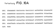
- the DNA used for labeling was inserted into the transcription vector pSPT18 (FIGS. 6 and 10).
- the vector contains a promoter for SP6 and a promoter for T7 RNA polymerase.
- the DNA was linearized at a location outside the inserted DNA sequence and the promoters, as well as the sequences connecting the promoters and the DNA sequence.
- 1 ⁇ g of the linearized substrate DNA was added in a reaction vessel to 1 ⁇ l of a 10 mmol / l solution of adenosine triphosphate, cytidine triphosphate and guanosine triphosphate.
- 1 ⁇ l of a solution containing 6.5 mmol / l uridine triphosphate and 3.5 mmol / l digoxigenin-11-UTP was added.
- Digoxigenin-11-UTP is prepared in Example 10, similarly to Example 4 digoxigenin-11-dUTP, the starting substance used is only the allylamino-uridine salt instead of the allylamino-deoxyuridine salt.
- RNA polymerase SP6 or T7
- the mixture was incubated at 37 ° C. for one hour.
- the substrate DNA was then degraded by adding 1 ⁇ l of DNAseI, RNAse-free, corresponding to 10 units, at 37 ° C. for 15 minutes.
- the reaction was stopped by adding 2 ul 0.2 mol / l EDTA, pH 8.0.
- the digoxigenin-labeled RNA sample obtained was purified by extraction with 1 volume of phenol and subsequent ethanol precipitation of proteins and nucleotides and finally in sterile buffer (Tris-HCl, 10 mmol / l; EDTA, 1 mmol / l; pH 8 , 0) solved.
- the purified DNA solution (no organic buffers), preferably in a concentration of 0.5 to 1.0 ⁇ g / ⁇ l, was mixed in an Eppendorf reaction vessel in the dark with the same volume of a 1 mg / ml photodigoxigenin solution (Example 13).
- the mixture was irradiated with ice cooling with a Philips HPLR 400 W lamp at a distance of 10 cm from above through the vessel opening for 10 to 30 minutes.
- the reaction mixture had to remain cold.
- the mixture was made up to 100 ⁇ l with 0.1 M Tris-HCl, pH 9.0, 1.0 mM EDTA.
- carrier carrier
- DNA only for small amounts of DNA
- 5 ⁇ l of 3 M sodium acetate or a corresponding salt e.g. 4 M LiCl
- 100 ⁇ l of ethanol were precipitated overnight at -20 ° C.
- a nitrocellulose filter (Schleicher & Schüll, BA 85) with DNA fixed on it was used for hybridization.
- the filter was first run in a solution consisting of NaCl, 0.75 mol / l Na citrate, 75 mmol / 1; Casein, 0.5% (w / v); Lauryl sarcosine, 0.1% (v / v); Sodium dodecyl sulfate, 0.02% (w / v); pH 7.0 (20 ° C) incubated at 65 ° C.
- non-specifically bound DNA was removed by two washing steps, first with a solution of NaCl, 0.3 mol / l; Na citrate, 30 mmol / 1; Na dodecyl sulfate, 0.1% (w / v); pH 7.0 for 2 x 5 minutes at room temperature and then with a solution of NaCl, 15 mmol / l; Na citrate, 1.5 mmol / l; Na dodecyl sulfate, 0.1% (w / v); pH 7.0 for 2 x 15 minutes at 65 ° C.
- Hybridization and subsequent washes were carried out exactly as described in Example 19 using the DNA sample.
- a freshly denatured RNA sample labeled as in Example 17 above was used.
- the concentration of the digoxigenin-labeled RNA in the hybridization solution was chosen so that the transcripts of 100 ng substrate DNA were used per ml hybridization solution.
- the hybridized filter from Example 19 or 20 was first in Tris-HCl, 0.1 mol / l, pH 7.5; NaCl, 0.15 mol / l washed. To block non-specific binding sites on the membrane, 0.5% (w / v) casein in Tris-HCl 0.1 mol / l, pH 7.5; NaCl, 0.15 mol / l incubated for 40 minutes with gentle swirling.
- the membrane was coated with anti-digoxigenin / alkaline phosphatase conjugate, 150 mU / ml in Tris-HCl, 0.1 mol / l; pH 7.5; NaCl, 0.15 mol / l incubated for 20 minutes. Unspecifically bound protein was then washed 2 x 15 minutes with the same buffer away. The filter was then placed in Tris-HCl, 0.1 mol / l, pH 9.5; NaCl 0.1 mol / l; MgCl2, 50 mmol / 1 reequilibrated for 5 minutes to pH 9.5, because the alkaline phosphatase has its optimum activity at higher pH values.
- Bound antibody conjugate and thus the hybridized labeled DNA was detected by a color reaction catalyzed by the alkaline phosphatase.
- the filter in Tris-HCl, 0.1 mol / l, pH 9.5; NaCl, 0.1 mol / l; MgCl2, 50 mmol / 1, with 45 ⁇ l nitro blue tetrazolium solution (75 mg / ml in dimethylformamide, 70% (v / v)) and 35 ⁇ l of an X-phosphate solution (50 mg / ml 5-bromo -4-chloro-3-indolyl phosphate in dimethylformamide) were added, incubated.
- the color reaction was carried out with the substantial exclusion of oxygen by welding the membrane with this solution into a film (normal cooking or freezer bags) and storing it in the dark.
- the color reaction started very quickly, but could increase in intensity over several days. Documentation of the result was obtained by photographing or photocopying the moist filter. The filter was then dried and stored. The color remained, but faded somewhat. It could be intensified again by moistening the filter.
- the DNA fragment was labeled with digoxigenin as in Example 14, hybridized with 100 ng / ml of labeled DNA as in Example 19 and the hybrids were detected as described in Example 21.
- the fragment was labeled with biotin using the analog method. Biotin 16-dUTP (Brigati et al., Virology 126 (1983) 32-50) was used for this.
- the DNA on the membrane was hybridized with 50 ⁇ g / ml labeled DNA. Otherwise, the procedure was as in Example 19. Detection was as described in Example 21, except that a streptavidin-alkaline phosphatase conjugate was used.
- pBR328 DNA was digested separately with the restriction enzymes BglI and HinfI. The fragments obtained were mixed in equal parts. The mixture then contains 15 pBR328 fragments of size: 2176, 1766, 1230, 1033, 653, 517, 453, 394, 298 (2x), 234 (2x), 220 and 154 (2x) bp.
- pBR328 DNA was cleaved with NciI. There are 10 fragments of size: 810, 724, 696, 597, 522, 507, 364, 351, 244 and 92 bp.
- different amounts of DNA were electrophoresed and then transferred to a nitrocellulose membrane according to Southern. The membrane was then hybridized as described in Example 19. However, 200 ng / ml as in Examples 1 and 2 of EPA 0173251 labeled pBR322 DNA were used for hybridization.
- pBR328 DNA differs from the pBR322 DNA used as a probe only by the 810 bp NciI fragment, which contains pBR328, whereas pBR322 does not.
- the in situ hybridization with carrier-fixed cells and cell swabs can be carried out.
- MEM minimal essential medium
- fetal calf serum was used as the culture medium (MEM, PBS, SSC see Science 130 (1959) 432 ff).
- the slides were removed from the culture dishes and washed several times with PBS.
- the hybridization took place under siliconized cover glasses.
- the DNA of the HPV sample in hybridization solution (non-radioactive sample: 20 ng / hybridization area, isotope-labeled sample: 3 ng with five times 105 cpm / hybridization area) was placed on the cell lawn.
- the pre-hybridization was carried out as described in Example 19).
- the DNA in the cells and the DNA sample were then denatured by placing the slide on a heating oven for 3 to 5 minutes. A temperature of 90 - 92 ° C was measured on the slide using a temperature sensor.
- the slides are then placed briefly on ice and then incubated overnight in a moist chamber (2 x SSC).
- Example 21 In the case of the non-radioactive systems, the procedure was then as in Example 21 or in the biotin system (cf. operating instructions for the BRL kit: blocking with 3% bovine serum albumin, addition of a conjugate of streptavidin and alkaline phosphatase and substrate mixture). The procedure in Example 21 was repeated in the digoxigenin system. The enzyme reaction was observed up to 48 hours.
- the situation is different when using biotin-labeled samples.
- the biotin system leads to a weak staining of HeLa cells with SiHa cells, but the cytosol is mainly stained unspecifically. The core area is only slightly stained.
- the unspecificity of the biotin detection system is also evident in the fact that the same staining of the cytosol is also without Biotin-labeled sample, but after adding only a streptavidin-alkaline phosphatase conjugate and the corresponding dye system.
- the method according to the invention has the advantage of saving time over the method using radioactively labeled samples compared to the method using biotin-labeled probes, the advantage of higher sensitivity and significantly higher specificity.
- the system according to the invention can also be used to detect HPV in tissue smears, e.g. in HPV-infected tissue from the vagina area.
- a 750 bp BamHI fragment of SUP 6 tRNA (Science 209 (1978), 1396-1460) from Saccaromyces cerevisiae was cloned into pUC19 (Gene 133 (1985), 103-119) and into E. coli HB 101 (DSM 1607) transformed.
- the pUC19 vector was transformed into E. coli HB 101 alone.
- insert-containing bacteria were alternately coated on nitrocellulose in addition to control transformants. After incubation for 5 hours at 37 ° C. on agar plates with nutrient medium, the E. coli cells were treated with alkaline, washed and the released DNA the membrane is fixed by cross-linking at 354 nm.
- the insert-containing bacteria each give clear signals, the control transformants are not detected. Longer detection times amplify the positive signals, but the control transformants remain background-free even with longer detection.
- Lambda gt10 phages (Mol.Gen.Genet. 150 (1977), 53) with a complete cDNA of human urokinase as an insert were incubated on indicator plates, so that about 500 individual plaques were visible on one plate.
- Two nitrocellulose pads were made from all plates as described in Science 196 (1977), 180, and hybridized.
- the sample was prepared by digesting 12 ⁇ g of a plasmid containing human urokinase cDNA with PstI. This creates a 1.1 Kb part fragment of the cDNA, which was eluted by electrophoresis (yield about 2 ⁇ g).
- the partial fragment is marked as in Example 14.
- the prehybridization and hybridization of the filters is carried out as in Example 19.
- the further detection steps are carried out as in Example 21.
- the system according to the invention is also suitable for the sensitive and specific detection of RNA in Northern blots.
- actin mRNA was detected in total yeast RNA (Saccharomyces cerevisiae).
- the actin gene (PNAS 77 (1980), 3912-3916) consists of two exons and one intron.
- precursor mRNA (1679 bases) is first generated.
- splicing and polyadenylation procedure as described in Meth. In Enzymol. 65 (1980) 380-391 and PNAS USA 77 1980) 5201-5205
- mature mRNA with a length of approx. 1400 bases (1370 bases + polyA) is produced. .
- HBV hepatitis B virus
- the hepatitis B virus has an approximately 32 kb circular genome, which is composed of two linear complementary strands. The 3'-end of one strand is variable. The 5'-ends of the two complementary strands are mutually offset.
- a digoxigenin-labeled 1.8 kb BamHI fragment (preparation according to Example 14) was used as the sample, which covers the double-stranded area core antigen and parts of the Prae-S1, Prae-S2 and contains X antigen (position 1.4-3.2 kb).
- biotin see example 24
- 32 P-labeled samples labeling analogous to the working instructions of the BRL kit, see example 24
- the examined human sera are classified as positive or negative on the basis of the immunological markers HB s Ag, HB c Ag, Anti-HB s and Anti-HB c .
- Biodyne nylon membrane (1.2 ⁇ m pore size) (Pall Bio Support Div. Glen Cove, NY) is cut to chamber size and prepared as follows: Soaked in distilled water for 5 min 15 min in 10 x SSC Dried for 5 min on a Whatman R paper under a heat lamp. Place on 10 x SSC-soaked Whatman R paper and insert in Minifold R apparatus.
- the sample collection plate is then placed on the filter, clamped, and then the sera are spotted.
- the filters are removed from the Minifold R device and placed on 10 x SSC, 5 x SSC and 1 x SSC for 5 min each. Then it is dried under a heat lamp and baked for 2 hours at 80 ° C in a vacuum.
- the pre-hybridization and hybridization are carried out analogously to Example 19.
- the radioactive sample is prepared as described in the operating instructions for the Sp6 / T7 transcription kit from BOEHRINGER MANNHEIM GMBH, order no. 999644.
- HPV detection using the method according to the invention is very sensitive.
- HPV sequences up to a serum dilution of 10 ⁇ 5 to 10 ⁇ 5 can be detected. This corresponds to the sensitivity in the radioactive system.
- the corresponding negative sera in the non-radioactive system give no signal when diluted.
- a clear background can also be seen in the dilution series in the biotin system.
- Digoxigenin-labeled dUTP is used in the polymerase chain reaction (PCR, analogous to EP-A 0200362) to label amplified DNA.
- the sample used is: plasmid pBR 322 Primer 1: 5′-GCTCCCTCGTGCGCTCTCCTGT-3 ′ Primer 2: 5′-CCGCCTACATACCTCGCTCTGC-3 ′
- Thermus aquaticus DNA polymerase is added and incubated for 3 min at 65 ° C. (polymerization).
- the volume of the reaction is 50 microliters, the concentration information relates to this reaction volume).
- a total of 25 cycles (denaturation, hybridization, polymerization) were carried out.
Landscapes
- Chemical & Material Sciences (AREA)
- Life Sciences & Earth Sciences (AREA)
- Organic Chemistry (AREA)
- Health & Medical Sciences (AREA)
- Engineering & Computer Science (AREA)
- Zoology (AREA)
- Wood Science & Technology (AREA)
- Proteomics, Peptides & Aminoacids (AREA)
- Biochemistry (AREA)
- Molecular Biology (AREA)
- Immunology (AREA)
- Biotechnology (AREA)
- Analytical Chemistry (AREA)
- General Health & Medical Sciences (AREA)
- Genetics & Genomics (AREA)
- Microbiology (AREA)
- Biophysics (AREA)
- Physics & Mathematics (AREA)
- Bioinformatics & Cheminformatics (AREA)
- General Engineering & Computer Science (AREA)
- Chemical Kinetics & Catalysis (AREA)
- Pathology (AREA)
- Measuring Or Testing Involving Enzymes Or Micro-Organisms (AREA)
Abstract
Description
Die Erfindung betrifft ein Verfahren zum Nachweis von Nukleinsäuren definierter Sequenz durch Hybridisierung mit einer komplementären, markierten Nukleinsäure-Sonde.The invention relates to a method for the detection of nucleic acids of defined sequence by hybridization with a complementary, labeled nucleic acid probe.
Eine der meist angewandten molekular-biologischen Techniken ist die DNA/DNA-, RNA/RNA- bzw. RNA/DNA-Hybridisierung zum Nachweis homologer Nukleinsäure-Sequenzen. Dabei wird eine als Sonde verwendete Nukleinsäure (DNA oder RNA) markiert und mit einer meist auf einem Filter fixierten, zu untersuchenden Nukleinsäure (DNA oder RNA) unter Hybridisierungsbedingungen in Kontakt gebracht. Bei vorhandener Homologie zwischen der als Sonde verwendeten Nukleinsäuren und der nachzuweisenden Nukleinsäure kommt es sodann zur Hybridisierung der jeweils komplementären Nukleinsäure-Einzelstränge unter Ausbildung eines Hybrid-Doppelstranges. Anschließend werden die Hybride nachgewiesen. Bisher erfolgte die Markierung der Sonde meist durch Einbau radioaktiy derivatisierter Desoxyribonukleosidtriphosphate. Der Nachweis der Hybride erfolgte dann durch Autoradiographie. Solche konventionellen, radioaktiy markierten DNA-Sonden sind sehr effektiv und sensitiv, es ergeben sich jedoch durch den Umgang mit der Radioaktivität Probleme. So wird zum Umgang mit den radioaktiven Verbindungen speziell ausgebildetes Personal benötigt, da bei unsachgemäßer Handhabung eine Gefährdung der Laborsicherheit besteht. Außerdem bildet die Entsorgung der radioaktiven Verbindungen ein weiteres Problem. Zusätzlich sind die radioaktiy markierten Proben aufgrund der Halbwertzeit der verwendeten radioaktiven Materialien nur über einen gewissen Zeitraum nach der Herstellung verwendbar. Auch kann bei der Detektion von geringen Mengen von nachzuweisender DNA die nötige Belichtungszeit der Autoradiographie sehr lange, nämlich Tage bis Wochen sein.One of the most commonly used molecular-biological techniques is DNA / DNA, RNA / RNA or RNA / DNA hybridization for the detection of homologous nucleic acid sequences. Here, a nucleic acid (DNA or RNA) used as a probe is marked and brought into contact with a nucleic acid (DNA or RNA), which is usually fixed on a filter, under hybridization conditions. If there is homology between the nucleic acids used as the probe and the nucleic acid to be detected, the complementary single-stranded nucleic acids then hybridize to form a hybrid double-strand. The hybrids are then detected. So far, the probe has mostly been labeled by incorporating radioactively derivatized deoxyribonucleoside triphosphates. The hybrid was then detected by autoradiography. Such conventional, radioactively labeled DNA probes are very effective and sensitive, but there are problems with handling radioactivity. Specially trained personnel are required to handle the radioactive compounds, since improper handling poses a risk to laboratory safety. The disposal of radioactive compounds is another problem. In addition, due to the half-life of the radioactive materials used, the radioactively labeled samples can only be used for a certain period after production. In the detection of small amounts of DNA to be detected, the required exposure time of the autoradiography can be very long, namely days to weeks.
Neben den radioaktiy markierten Systemen zum Nachweis von Nukleinsäuren sind nicht-radioaktive Methoden bekannt, wobei die verwendeten Nukleinsäure-Proben mit Biotin-Molekülen (US-PS 2 915 082, EP-A 0063879) Digoxin-/T3-/T4-Molekülen (EP-A-O 173251) oder mit Alkyl-/Butyl-/Ethyl-/Sulfonsäure-/Nitrosomolekülen EP-A 128018) modifiziert sind. Der Einbau dieser niedermolekularen Nachweismoleküle in die komplementäre Nukleinsäure-Sonde erfolgt dabei chemisch, photochemisch oder enzymatisch. Sodann erfolgt die Hybridisierung mit der nachzuweisenden Nukleinsäuresequenz. Der Nachweis der Hybride erfolgt dann über Bindung des niedermolekularen Moleküls durch ein (Strept)-Avidin-Markerenzym-Konjugat im Fall von Biotin, Antidigoxin/-T3-/-T4-Antikörper-Markerenzym-Konjugate im Fall von Digoxin-/T3-/T4-Molekülen oder über Anti-Alkyl-/-Butyl-/-Ethyl-/-Sulfonsäure-/-Nitroso-Antikörper-Markerenzym-Konjugate. Der Nachweis des Hybridisierungsproduktes erfolgt durch die Bestimmung der enzymatischen Aktivität des Markerenzyms über gekoppelte Farbstoffsysteme. Bei der Methode der EP-A-0 173 251 erfolgt jedoch die Bindung der Digoxin/T3-/T4-Moleküle an ein an der Wasserstoffbrückenbindung beteiligtes N-Atom einer oder mehrerer Basen der Nukleinsäuresonde, wodurch die Hybridisierung - vor allem bei Mehrfachmodifizierung der Sonde - mit der nachzuweisenden Nukleinsäure beeinträchtigt sein kann. Mit Ausnahme des Biotin/(Strept)-Avidin-System ist die Sensitivität der derzeit bekannten, nicht-radioaktiven Systeme, im Vergleich mit radioaktiven Systemen, jedoch um mindestens den Faktor 10 bis 100 vermindert. Die bisher einmalig hohe Sensitivität des nicht-radioaktiven Nachweises auf Biotin/(Strept)-Avidin-Basis ist auf die hohe Bindungskonstante (K = 10¹⁵ Mol⁻¹) speziell des Biotin/(Strept)- Avidin-Systems zurückzuführen (Lit. Kinow; Proc. Natl. Acad. Sci. USA 80 (1983) 4045). Die maximal erreichbare Sensitivität des Biotin/(Strept)-Avidin-Systems liegt wie auch bei radioaktiver Markierung im Nachweis von 1 pg bis 0,1 pg DNA im Dot-Blot, und im Nachweis von "single-copy" Genen, das heißt von nur einmal im Genom vorkommenden Genen, im genomischen Blot bei Verwendung von 10 bis 1 µg genomischer DNA-Fragmente. Die Ausnützung der Biotin/(Strept-Avidin-Wechselwirkung hat jedoch den entscheidenden Nachteil, daß sie sehr störanfällig ist, da das Vitamin Biotin in nahezu allen biologischen Materialien vorkommt (Biochem. Biophys. Acta 29 (1985) 225; Biochem. Biophys. Acta 41 (1960) 122).In addition to the radioactively labeled systems for the detection of nucleic acids, non-radioactive methods are known, the nucleic acid samples used with biotin molecules (US Pat. No. 2,915,082, EP-A 0063879) digoxin / T3 / T4 molecules (EP -AO 173251) or with alkyl / butyl / ethyl / sulfonic acid / nitrosomolecules EP-A 128018) are modified. The incorporation of these low molecular weight detection molecules into the complementary nucleic acid probe takes place chemically, photochemically or enzymatically. The hybridization then takes place with the nucleic acid sequence to be detected. The detection of the hybrids is then carried out by binding the low-molecular molecule by means of a (strept) -avidin-marker enzyme conjugate in the case of biotin, antidigoxin / -T3 - / - T4-antibody-marker enzyme conjugates in the case of digoxin / T3- / T4 molecules or via anti-alkyl - / - butyl - / - ethyl - / - sulfonic acid - / - nitroso-antibody-marker enzyme conjugates. The hybridization product is detected by determining the enzymatic activity of the marker enzyme via coupled dye systems. In the method of EP-A-0 173 251, however, the digoxin / T3 / T4 molecules are bound to an N atom of one or more bases of the nucleic acid probe that is involved in hydrogen bonding, which results in hybridization - especially when the probe is modified several times - May be impaired with the nucleic acid to be detected. With the exception of the biotin / (strept) avidin system, the sensitivity of the currently known, non-radioactive systems is, however, reduced by at least a factor of 10 to 100 in comparison with radioactive systems. The unprecedentedly high sensitivity of non-radioactive detection based on biotin / (strept) avidin is due to the high binding constant (K = 10¹⁵ mol⁻¹) especially of biotin / (strept) - Avidin system attributed (Lit. Kinow; Proc. Natl. Acad. Sci. USA 80 (1983) 4045). As with radioactive labeling, the maximum attainable sensitivity of the biotin / (strept) avidin system is in the detection of 1 pg to 0.1 pg DNA in the dot blot, and in the detection of "single-copy" genes, that is, of genes occurring only once in the genome, in the genomic blot when using 10 to 1 µg genomic DNA fragments. However, the use of the biotin / (strept-avidin interaction has the decisive disadvantage that it is very susceptible to malfunction, since the vitamin biotin occurs in almost all biological materials (Biochem. Biophys. Acta 29 (1985) 225; Biochem. Biophys. Acta 41 (1960) 122).
Die Aufgabe der Erfindung bestand daher darin, ein Verfahren zum Nachweis von Nukleinsäuren bereitzustellen, das die Verwendung einer nicht radioaktiven Markierung erlaubt und weniger störanfällig als die Biotin/(Strept)-Avidin-Wechselwirkung ist, andererseits jedoch die bisher einzigartig hohe Nachweis-Sensitivität der radioaktiven Markierung bzw. der Biotin/(Strept)-Avidin-Markierung erreicht.The object of the invention was therefore to provide a method for the detection of nucleic acids which allows the use of a non-radioactive label and is less susceptible to interference than the biotin / (strept) avidin interaction, but on the other hand the hitherto uniquely high detection sensitivity of the radioactive label or the biotin / (strept) avidin label reached.
Diese Aufgabe wird erfindungsgemäß gelöst durch ein Verfahren zum Nachweis von Nukleinsäuren definierter Sequenz durch Hybridisierung mit einer komplementären Nukleinsäuresonde, die über eine chemische Verbindung mindestens ein Hapten als Markierung gebunden enthält, welches dadurch gekennzeichnet ist, daß man als Hapten ein Steroid verwendet, das an mindestens eine Position der Nukleinsäure, die nicht an der Wasserstoffbrückenbildung beteiligt ist, über eine Brücke von mindestens 4 Atomen Länge gebunden ist und die hybridisierte Probe über einen seinerseits markierten Anti-Hapten-Antikörper nachweist.This object is achieved according to the invention by a method for the detection of nucleic acids of a defined sequence by hybridization with a complementary nucleic acid probe which contains at least one hapten bound as a label via a chemical compound, which is characterized in that a steroid is used as the hapten which is at least a position of the nucleic acid which is not involved in the hydrogen bond formation, is bound via a bridge of at least 4 atoms in length and detects the hybridized sample via an anti-hapten antibody which in turn is labeled.
Bevorzugt werden hierbei als Steroid Digoxigenin oder Digoxin verwendet.Digoxigenin or digoxin are preferably used as steroids.
Das erfindungsgemäße Verfahren ermöglicht den Nachweis von 0,5 pg bis 0,05 pg homologer DNA im Dot-Blot und von "single copy"-Genen in 5 µg bis 0,5 µg genomischen DNA-Fragmenten. In beiden Nachweisarten ist die Nachweissensitivität mindestens analog der Nachweissensitivität des Biotin/(Strept)-Avidin-Systems. Dies ist insofern überraschend, da die Bindungskonstante der Biotin/(Strept)-Avidin-Wechselwirkung (K = 10¹⁵ Mol⁻¹, Green, N.M. (1975) Adv. Protein Chem. 29, 85-133; Chaiet, L., Wolf, F.J. (1964) Arch. Biochem. Biophys. 106, 1-5) um mindestens den Faktor 10⁵ höher ist als die Wechselwirkung von Digoxigenin oder Digoxin mit dem entsprechenden Antikörper (K=2x10⁸ Mol⁻¹ - 7x10⁹ Mol⁻¹, Hunter et al., J. Immunol. Vol. 129, Nr. 3 (1982) 1165), und zusätzlich die Biotin/(Strept)-Avidin-Wechselwirkung durch das Vorhandensein von 4 Biotin-Bindungsstellen auf (Strept)-Avidin begünstigt ist.The method according to the invention enables the detection of 0.5 pg to 0.05 pg homologous DNA in a dot blot and of "single copy" genes in 5 µg to 0.5 µg genomic DNA fragments. In both types of detection, the detection sensitivity is at least analogous to the detection sensitivity of the biotin / (strept) avidin system. This is surprising in that the binding constant of the biotin / (strept) avidin interaction (K = 10¹⁵ Mol⁵¹, Green, NM (1975) Adv. Protein Chem. 29, 85-133; Chaiet, L., Wolf, FJ (1964) Arch. Biochem. Biophys. 106, 1-5) is at least a factor 10⁵ higher than the interaction of digoxigenin or digoxin with the corresponding antibody (K = 2x10⁸ mol⁻¹ - 7x10⁹ mol⁻¹, Hunter et al ., J. Immunol. Vol. 129, No. 3 (1982) 1165), and in addition the biotin / (strept) avidin interaction is favored by the presence of 4 biotin binding sites on (strept) avidin.
Ein weiterer überraschender technischer Vorteil ist, daß bei Verwendung von Digoxigenin oder Digoxin und den dazugehörigen Antikörpern sehr viel weniger unspezifische Bindung (background) an den Filter, auf welchem die nachzuweisende Nukleinsäure fixiert ist, entsteht, als bei Verwendung von Biotin/(Strept)-Avidin.Another surprising technical advantage is that when digoxigenin or digoxin and the associated antibodies are used, there is much less non-specific binding (background) to the filter on which the nucleic acid to be detected is fixed than when using biotin / (strept) - Avidin.
Die Durchführung des erfindungsgemäßen Verfahrens zum Nachweis von Nukleinsäuren läßt sich in drei Schritte aufgliedern.
- 1. Die Derivatisierung der als Nachweisreagenz dienenden Nukleinsäure-Probe mit dem Hapten,
- 2. die Hybridisierung, und
- 3. die Detektion der Hybride.
- 1. The derivatization of the nucleic acid sample serving as detection reagent with the hapten,
- 2. the hybridization, and
- 3. the detection of the hybrid.
Zur Derivatisierung von Nukleinsäuren (DNA und RNA) können verschiedene Methoden angewendet werden (Molecular Cloning, Maniatis et al., (1982) Cold Spring Harbor Laboratory, Cold Spring Harbor, New York). Der Einbau des Haptens kann hierbei enzymatisch, chemisch oder photochemisch erfolgenVarious methods can be used to derivatize nucleic acids (DNA and RNA) (Molecular Cloning, Maniatis et al., (1982) Cold Spring Harbor Laboratory, Cold Spring Harbor, New York). The hapten can be incorporated enzymatically, chemically or photochemically
In der "Random-primed" Methode (Anal. Biochem. 132 (1983) 6) werden doppelsträngige Desoxyribonukleinsäure-Sonden zunächst denaturiert, d.h. die beiden Stränge werden durch Erhitzen getrennt und dadurch in Nukleinsäure-Einzelstränge überführt. Bei einzelsträngigen Desoxyribonukleinsäuresonden entfällt die Denaturierung. An die Desoxyribonukleinsäure-Einzelstränge werden sogenannte "Random Primer", das sind beliebige kurze Oligo-Desoxyribonukleotide mit unterschiedlichen Sequenzen, die mit komplementären Sequenzabschnitten der Einzelstrang-DNA hybridisieren, gebunden. Anschließend wird ausgehend von den 3′-OH-Enden der "Random Primern" durch Einwirkung des Enzyms Klenow Polymerase oder anderer DNA-Polymerasen (z.B. Phage T4 oder T7-kodiert oder aus Thermus aquaticus) der zum Einzelstrang komplementäre Strang synthetisiert. Dabei werden die als Substrat angebotenen vier Desoxyribonukleosidtriphosphat-Arten eingebaut. Bis zu vier dieser Triphosphat-Arten sind teilweise oder gänzlich durch Kopplung eines Steroids über eine Brücke derivatisiert, so daß bei der Desoxyribonukleinsäure-Synthese dieses Steroid miteingebaut wird. Ein bevorzugtes, Steroid-derivatisiertes Nukleosidtriphosphat ist in der Fig. 1 mit zwei verschiedenen bevorzugten Brückenkettenlängen dargestellt.In the "random primed" method (Anal. Biochem. 132 (1983) 6), double-stranded deoxyribonucleic acid probes are first denatured, ie the two strands are separated by heating and thereby converted into single nucleic acid strands. Denaturation is not required for single-stranded deoxyribonucleic acid probes. So-called “random primers”, that is, any short oligo-deoxyribonucleotides with different sequences that hybridize with complementary sequence sections of the single-strand DNA, are bound to the deoxyribonucleic acid single strands. Subsequently, starting from the 3'-OH ends of the "random primers" by the action of the enzyme Klenow polymerase or other DNA polymerases (eg Phage T4 or T7-coded or from Thermus aquaticus) the strand complementary to the single strand is synthesized. The four deoxyribonucleoside triphosphate types offered as substrates are incorporated. Up to four of these triphosphate types are partially or completely derivatized by coupling a steroid via a bridge, so that this steroid is incorporated in the deoxyribonucleic acid synthesis. A preferred, steroid-derivatized nucleoside triphosphate is shown in FIG. 1 with two different preferred bridge chain lengths.
In der "Specific-primed" Methode werden anstelle von "Random Primern", also kurzen Oligo-Desoxyribonukleotiden mit unterschiedlichsten Sequenzen, "Specific Primer", also Oligo-Desoxyribonukleotide mit spezifischen Sequenzen, verwendet. Diese spezifischen Primer binden einheitlich nur an den komplementären Sequenzabschnitt der Einzelstrang-DNA. Die Synthese des komplementären Stranges wird im Gegensatz zur "Random-primer"-Methode nur von diesem bestimmten Sequenzabschnitt gestartet. Es werden die als Substrat angebotenen vier Desoxyribonukleosidtriphosphat-Arten eingebaut. Bis zu vier dieser Triphosphat-Arten sind teilweise oder gänzlich durch Kopplung eines Steroids über eine Brücke derivatisiert, so daß bei der Desoxyribonukleinsäure-Synthese dieses Steroid mit eingebaut wird.In the "specific-primed" method, instead of "random primers", ie short oligo-deoxyribonucleotides with a wide variety of sequences, "specific primers", ie oligo-deoxyribonucleotides with specific sequences, are used. These specific primers bind uniformly only to the complementary sequence section of the single-stranded DNA. In contrast to the "random primer" method, the synthesis of the complementary strand is only started from this specific sequence section. The four types of deoxyribonucleoside triphosphate offered as substrates are installed. Up to four of these triphosphate types are partially or wholly derivatized by coupling a steroid via a bridge, so that this steroid is incorporated in the deoxyribonucleic acid synthesis.
Bei der "Reversen Transkriptions" Methode (Efstratiadis, A.F.C., Villa-Komaroff, L. (1979) Genetic Engeneering (Stelow, J.K. and Hollaender, A., eds.) Plenum Press, New York and London, Vol. 1, pp. 1) werden einzelsträngige Ribonukleinsäure-Sonden oder doppelsträngige Ribonukleinsäure-Sonden nach Denaturierung, d.h. Überführung in Einzelstränge, retrotranskribiert, d.h. die entsprechende Desoxyribonukleinsäure gebildet. Hierzu werden an die einzelsträngigen Ribonukleinsäure-Sonden Oligo- Desoxyribonukleotide, die "Primer", an den komplementären Sequenzabschnitt gebunden. Anschließend wird, ausgehend von dem 3′-OH-Ende des gebundenen "Primers" durch Einwirkung des Enzyms Reverse Transkriptase der zum RNA-Einzelstrang komplementäre DNA-Strang synthetisiert. Als Reverse Transkriptasen werden z.B. Virus AMV oder Mo-MLV kodierte Enzyme verwendet. Bei der DNA-Strangsynthese werden die als Substrat angebotenen vier Desoxyribonukleosidtriphosphat-Arten eingebaut. Bis zu vier dieser Triphosphat-Arten sind teilweise oder gänzlich durch Kopplung eines Steroids über eine Brücke derivatisiert, so daß bei der DNA-Synthese dieses Steroid mit eingebaut wird.With the "reverse transcription" method (Efstratiadis, AFC, Villa-Komaroff, L. (1979) Genetic Engeneering (Stelow, JK and Hollaender, A., eds.) Plenum Press, New York and London, Vol. 1, pp. 1) single-stranded ribonucleic acid probes or double-stranded ribonucleic acid probes are retrotranscribed after denaturation, ie conversion into single strands, ie the corresponding deoxyribonucleic acid is formed. For this purpose, oligo-deoxyribonucleotides, the “primers”, are bound to the complementary sequence section on the single-stranded ribonucleic acid probes. Subsequently, starting from the 3'-OH end of the bound "primer", the DNA strand complementary to the RNA single strand is synthesized by the action of the enzyme reverse transcriptase. For example, virus AMV or Mo-MLV-encoded enzymes are used as reverse transcriptases. The four deoxyribonucleoside triphosphate types offered as substrates are incorporated into the DNA strand synthesis. Up to four of these triphosphate types are partially or wholly derivatized by coupling a steroid via a bridge, so that this steroid is incorporated in the DNA synthesis.
In der "Fill-in" Methode werden doppelsträngige Desoxyribonukleinsäure-Sonden mit 5′-überhängenden Einzelstrang-Enden derivatisiert. Dazu werden mit dem Enzym Klenow Polymerase oder anderer DNA-Polymerasen (z.B. Phage T4- oder T7- codiert) die 3′-OH Enden des nicht-überstehenden Einzelstrangs komplementär zu der 5′-überhängenden Einzelstrang-Sequenz verlängert. Dabei werden, je nach Sequenz des 5′-überhängenden Einzelstrang-Bereichs, bis zu vier der angebotenen Desoxyribonukleosidtriphosphat-Arten eingebaut. Bis zu vier dieser Triphosphat-Arten sind wiederum teilweise oder gänzlich durch Kopplung eines Steroids über eine Brücke derivatisiert, so daß bei dem Einbau der Nukleotide dieses Steroid mit eingebaut wird.In the "fill-in" method, double-stranded deoxyribonucleic acid probes with 5'-overhanging single-strand ends are derivatized. For this, the 3'-OH ends of the non-protruding single strand are extended to complement the 5'-overhanging single strand sequence using the enzyme Klenow polymerase or other DNA polymerases (e.g. phage T4- or T7- coded). Depending on the sequence of the 5'-overhanging single-strand region, up to four of the offered deoxyribonucleoside triphosphate species are incorporated. Up to four of these triphosphate types are in turn partially or wholly derivatized by coupling a steroid via a bridge, so that this steroid is incorporated when the nucleotides are incorporated.
In der "Nick-Translation" Methode (J. Mol. Biol. 113 (1977) 237) werden doppelsträngige Desoxyribonukleinsäure-Sonden gleichzeitig mit dem Enzym E. coli DNA Polymerase I sowie einer kleinen Menge des Enzyms DNase I in Anwesenheit der als Substrat dienenden vier Desoxyribonukleosidtriphosphate inkubiert. Durch das Enzym DNase I werden Einzelstrang-Brüche, die sogenannten "nicks", erzeugt. Dadurch entstehen 5′- und 3′-Enden innerhalb des gebrochenen Einzelstranges. Durch das Enzym E. coli DNA Polymerase I werden anschließend an den internen Einzelstrang-Brüchen die 5′-endständigen Desoxyribonukleoside abgebaut, gleichzeitig jedoch an das benachbarte freie 3′-OH-Ende eines der als Substrat angebotenen Desoxyribonukleotid-Arten eingebaut. Durch wiederholtes Ablaufen des gleichzeitigen Ab- und Neu-Einbaus eines Nukleotids wandert der Einzelstrang-Bruch in Richtung 3′-Ende. Bis zu vier der als Substrat angebotenen Triphosphat-Arten sind wiederum teilweise oder gänzlich durch Kopplung eines Steroids über eine Brücke derivatisiert, so daß beim Neueinbau der Nukleotide dieses Steroid mit eingebaut wird.In the "nick translation" method (J. Mol. Biol. 113 (1977) 237) double-stranded deoxyribonucleic acid probes are used simultaneously with the enzyme E. coli DNA polymerase I and a small amount of the enzyme DNase I in the presence of those serving as the substrate four deoxyribonucleoside triphosphates incubated. The DNase I enzyme produces single-strand breaks, the so-called "nicks". This creates 5'- and 3'-ends within the broken single strand. The 5'-terminal deoxyribonucleosides are then broken down at the internal single-strand breaks by the enzyme E. coli DNA polymerase I, but at the same time incorporated into the adjacent free 3'-OH end of one of the deoxyribonucleotide types offered as a substrate. By repeating the simultaneous removal and reinstallation of a nucleotide, the single-strand break moves towards the 3'-end. Up to four of the triphosphate types offered as substrates are in turn partially or completely derivatized by coupling a steroid via a bridge, so that this steroid is also incorporated when the nucleotides are reinstalled.
In der "Tailing" Methode wird an 3′-OH-Enden von doppel- oder einzelsträngigen Desoxyribo- oder Ribonukleinsäure-Sonden mit dem Enzym Terminale Transferase in Anwesenheit mindestens einer Art der als Substrat dienenden Desoxyribonukleosidtriphosphate, Dideoxyribonukleosidtriphosphate oder Ribonukleosidtriphosphate mindestens eines dieser Nukleotide angehängt. Die verwendete Art der Triphosphate ist wiederum teilweise oder gänzlich durch Kopplung eines Steroids über eine Brücke derivatisiert, so daß bei dem Einbau der Nukleotide dieses Steroid mit eingebaut wird.In the "tailing" method, at least one of these nucleotides is attached to 3′-OH ends of double- or single-stranded deoxyribo- or ribonucleic acid probes with the enzyme terminal transferase in the presence of at least one type of deoxyribonucleoside triphosphate, dideoxyribonucleoside triphosphate or ribonucleoside triphosphate. The type of triphosphates used is in turn partially or wholly derivatized by coupling a steroid via a bridge, so that this steroid is incorporated when the nucleotides are incorporated.
Bei der Phagen-RNA-Polymerase-katalysierten "Transkriptions"-Methode (J. Mol. Biol. 166 (1983) 477) wird während der Desoxyribonukleinsäure-abhängigen Ribonukleinsäure-Synthese die gebildete Ribonukleinsäure derivatisiert. Als Phagen-codierte RNA-Polymerasen werden z.B. Phage SP6-, T7- oder T3-codierte Enzyme verwendet. Für diese Methode werden doppelsträngige Desoxyribonukleinsäuren verwendet, die z.B. SP6-, T7- oder T3-Promotoren enthalten. Durch Zugabe von z.B. SP6-, T7- oder T3-RNA Polymerase und aller vier Arten der Ribonukleosid-Triphosphate wird, ausgehend vom homologen Promotor, der zur codogenen Desoxyribonukleinsäure komplementäre Ribonukleinsäure-Strang, das Transkript, gebildet. Dabei werden die als Substrat angebotenen Ribonukleosidtriphosphat-Arten eingebaut. Bis zu vier dieser Triphosphat-Arten sind teilweise oder gänzlich durch Kopplung eines Steroids über eine Brücke derivatisiert, so daß bei der Ribonukleinsäure-Synthese dieses Steroid mit eingebaut wird.In the phage RNA polymerase-catalyzed "transcription" method (J. Mol. Biol. 166 (1983) 477), the ribonucleic acid formed is derivatized during the deoxyribonucleic acid-dependent ribonucleic acid synthesis. As phage-encoded RNA polymerases, for example, phage SP6, T7 or T3-encoded enzymes are used. For this method double-stranded deoxyribonucleic acids are used, which contain, for example, SP6, T7 or T3 promoters. By adding, for example, SP6, T7 or T3 RNA polymerase and all four types of ribonucleoside triphosphates, starting from the homologous promoter, the ribonucleic acid strand which is complementary to the codogenic deoxyribonucleic acid, the transcript, is formed. The ribonucleoside triphosphate species offered as substrates are incorporated. Up to four of these triphosphate types are partially or wholly derivatized by coupling a steroid via a bridge, so that this steroid is incorporated in the ribonucleic acid synthesis.
In der "photochemischen" Methode (Nucl. Acids Res. 13 (1985) 745-761) wird die Nukleinsäure-Sonde in Gegenwart von Photo-Digoxigenin (Fig. 2) mit sichtbarem Licht mit UV-Anteil bestrahlt. Unter Abspaltung von Stickstoff (N₂) entsteht ein Nitrenradikal, das kovalent an die Nukleinsäure bindet.In the "photochemical" method (Nucl. Acids Res. 13 (1985) 745-761) the nucleic acid probe is irradiated with visible light with a UV component in the presence of photo-digoxigenin (FIG. 2). With the elimination of nitrogen (N₂), a nitrene radical is formed which binds covalently to the nucleic acid.
Für die "chemische" Methode werden im Rahmen der Oligo-Desoxyribonukleotidsynthese nach der Phosphit-Triester-Methode neben den geschützten Nukleosid-phosphoramiditen (dA/dG/dC/dT) geschützte, mit substituierbaren Aminofunktionen modifizierte Nukleosid-phosphoramidite (dA/dG/dC/dU) zum gezielten Einbau in den Oligo-Desoxyribonukleotid-Einzelstrang verwendet. Die Modifizierung von dC/dU geschieht bevorzugt in Position 5 des Pyrimidin-Ringes, die von dA/dG bevorzugt an Position 8 des Purinmoleküls.For the "chemical" method, as part of the oligo-deoxyribonucleotide synthesis according to the phosphite-triester method, in addition to the protected nucleoside phosphoramidites (dA / dG / dC / dT), protected nucleoside phosphoramidites (dA / dG / dC) modified with substitutable amino functions are used / dU) used for targeted installation in the oligo-deoxyribonucleotide single strand. The modification of dC / dU occurs preferably in
Nach Abschluß der Synthesezyklen und Entfernung der Schutzgruppen resultieren einzelsträngige, an den Nukleobasen mit substituierbaren Aminofunktionen modifizierte Oligo-Desoxyribonukleotide, die mit geeigneten Haptenen markierbar sind. Solche Haptene sind Steroide, bevorzugt Digoxigenin, Digoxin, d.h. die Markierung geschieht durch Umsetzung des Oligo-Desoxyribonukleotides mit den entsprechenden aktivierten Estern, Amiden oder Ethern der Haptene, vorzugsweise ihren N-hydroxysuccinimidestern.After completion of the synthesis cycles and removal of the protective groups, single-stranded oligo-deoxyribonucleotides which can be labeled with substitutable amino functions and which can be labeled with suitable haptens result. Such haptens are steroids, preferably digoxigenin, digoxin, i.e. the labeling is done by reacting the oligo-deoxyribonucleotide with the corresponding activated esters, amides or ethers of the haptens, preferably their N-hydroxysuccinimide esters.
In einer bevorzugten Ausführungsform der Erfindung wird eine Nukleinsäure-Sonde verwendet, deren Hapten in die Nukleinsäure-Sonde enzymatisch mit Hilfe von DNA-Polymerasen, RNA-Polymerasen, Reversen Transkriptasen oder Terminalen Transferasen und entsprechenden Hapten-modifizierten Desoxy- oder Ribonukleosidtriphosphat-Substraten eingebaut wurde.In a preferred embodiment of the invention, a nucleic acid probe is used, the hapten of which was incorporated into the nucleic acid probe enzymatically with the aid of DNA polymerases, RNA polymerases, reverse transcriptases or terminal transferases and corresponding hapten-modified deoxy or ribonucleoside triphosphate substrates .
In einer weiteren bevorzugten Ausführungsform der Erfindung wird eine Nukleinsäure-Sonde verwendet, deren Hapten in die Nukleinsäure-Sonde photochemisch mit Hilfe von Photo-Hapten eingebaut wurde und in einer dritten bevorzugten Ausführungsform wird eine Nukleinsäure-Sonde verwendet, deren Hapten in die Nukleinsäure-Sonde chemisch im Rahmen der Oligo-Desoxyribonukleotid-Synthese durch Einbau geschützter, mit substituierbaren Aminofunktionen modifizierter Nukleosidphosphoamidite und - nach Entfernung der Schutzgruppen - durch Umsetzung des modifizierten Oligo-Desoxyribonukleotids mit aktivierten Estern der Haptene eingebaut wurde.In a further preferred embodiment of the invention, a nucleic acid probe is used, the hapten of which has been photochemically incorporated into the nucleic acid probe with the aid of photo-hapten, and in a third preferred embodiment, a Nucleic acid probe used, the hapten chemically incorporated into the nucleic acid probe in the course of the oligo-deoxyribonucleotide synthesis by incorporating protected nucleoside phosphoamidites modified with substitutable amino functions and - after removal of the protective groups - by reacting the modified oligo-deoxyribonucleotide with activated esters of the haptens has been.
Die über eine der beschriebenen Methoden hergestellte, mit einem Steroid derivatisierte Nukleinsäure-Sonde wird mit einer an einen Träger gebundenen denaturierten DNA oder RNA in Kontakt gebracht und hierbei die Temperatur, Ionenstärken, pH-Wert und sonstige Pufferbedingungen - abhängig von der Länge der Nukleinsäureprobe und der daraus resultierenden Schmelztemperatur des zu erwartenden Hybrids - so gewählt, daß die markierte DNA oder RNA an homologe DNA oder RNA binden kann (Hybridisierung) (J. Mol. Biol. 98 (1975) 503; Proc. Natl. Acad. Sci. USA 76 (1979) 3683). Als Träger geeignet sind Membranen oder Trägermaterialien auf Basis Nitrozellulose (z.B. Schleicher und Schüll BA85; Amersham Hybond C), verstärkte oder gebundene pulverförmige Nitrocellulose oder mit verschiedenen funktionellen Gruppen (z.B. Nitro-) derivatisierte Nylonmembranen (z.B. Schleicher und Schüll Nytran, NEN Gene Screen, Amersham Hybond N, Pall Biodyne).The nucleic acid probe derivatized with one of the methods described and derivatized with a steroid is brought into contact with a denatured DNA or RNA bound to a carrier and the temperature, ionic strengths, pH value and other buffer conditions - depending on the length of the nucleic acid sample and the resulting melting temperature of the hybrid to be expected - selected so that the labeled DNA or RNA can bind to homologous DNA or RNA (hybridization) (J. Mol. Biol. 98 (1975) 503; Proc. Natl. Acad. Sci. USA 76 (1979) 3683). Suitable carriers are membranes or carrier materials based on nitrocellulose (e.g. Schleicher and Schüll BA85; Amersham Hybond C), reinforced or bound powdery nitrocellulose or nylon membranes derivatized with various functional groups (e.g. nitro-) (e.g. Schleicher and Schüll Nytran, NEN Gene Screen, Amersham Hybond N, Pall Biodyne).
Die Detektion von hybridisierter DNA oder RNA erfolgt dann dadurch, daß der Träger nach gründlichem Waschen und Absättigung zur Verhinderung unspezifischer Bindungen mit einem Antikörper oder Antikörperfragment inkubiert wird. Der Antikörper oder das Antikörperfragment ist gegen das in die Nukleinsäure-Sonde bei der Derivatisierung inkorporierte Steroidhapten gerichtet. Er trägt konjugiert eine Markierung. Nach der Antikörper-Inkubation wird nochmals gewaschen, um nur spezifisch gebundene Antikörper-Konjugate nachzuweisen. Die Bestimmung erfolgt sodann über die Markierung des Antikörpers oder Antikörperfragments nach an sich bekannten Methoden.Hybridized DNA or RNA is then detected by incubating the support with an antibody or antibody fragment after thorough washing and saturation to prevent unspecific binding. The antibody or antibody fragment is directed against the steroid hapten incorporated into the nucleic acid probe during derivatization. It carries a mark conjugated. After the antibody incubation, washing is carried out again in order to detect only specifically bound antibody conjugates. The determination is then carried out by labeling the antibody or antibody fragment using methods known per se.
Die Länge der Brücke, über die das Hapten an die Nukleinsäure-Probe gebunden ist, kann zwischen 4 und 32 Atome betragen. Die Brücke ist hierbei aus Molekülen aufgebaut, die die Atome C, O, S oder/und N enthalten. Eine größere Kettenlänge ist zwar möglich, aber nicht mehr sinnvoll, da mit einem Sensitivitätsverlust zu rechnen ist.The length of the bridge over which the hapten is bound to the nucleic acid sample can be between 4 and 32 atoms. The bridge is made up of molecules that contain the atoms C, O, S or / and N. A longer chain length is possible, but no longer sensible, since a loss of sensitivity can be expected.
In einer bevorzugten Ausführungsform der Erfindung ist das Hapten über eine Brücke von 11 bis 16 Atomen Länge an die Probe gebunden. Bevorzugt enthält die Brücke neben hydrophoben auch hydrophile Gruppierungen.In a preferred embodiment of the invention, the hapten is bound to the sample via a bridge of 11 to 16 atoms in length. In addition to hydrophobic, the bridge preferably also contains hydrophilic groups.
In einer bevorzugten Ausführungsform ist die Brücke linear. In einer weiteren Ausführungsform besteht die Brücke aus einer verzweigten Kette und trägt an mindestens einem der Kettenenden ein Haptenmolekül. Durch das Vorhandensein mehrerer Haptenmoleküle an den Kettenenden einer verzweigtkettigen Brücke kann die Nachweissensitivität zusätzlich verstärkt werden.In a preferred embodiment, the bridge is linear. In a further embodiment, the bridge consists of a branched chain and carries a hapten molecule on at least one of the chain ends. The presence of several hapten molecules at the chain ends of a branched-chain bridge can further increase detection sensitivity.
Die Verbindung des Steroid-Haptens mit der Brücke ist vorzugsweise eine Ester-, Amid- oder Etherbindung.The connection of the steroid hapten to the bridge is preferably an ester, amide or ether bond.
Die Bindung des Steroid-Haptens über die Brücke an die Nukleinsäureprobe ist sowohl über eine endständige oder nicht endständige Phosphatgruppe, als auch über einen Zuckerrest oder eine Base der Nukleinsäure-Probe möglich. Die Bindung des Haptens über die Brücke muß jedoch so erfolgen, daß nicht die Wasserstoffbrückenbildung zwischen den beiden komplementären Nukleinsäuresträngen beeinträchtigt wird.The binding of the steroid hapten via the bridge to the nucleic acid sample is via a terminal or non-terminal phosphate group as well as via one Sugar residue or a base of the nucleic acid sample possible. However, the hapten must be bound via the bridge in such a way that the hydrogen bond formation between the two complementary nucleic acid strands is not impaired.
Bevorzugt ist das Hapten über die Brücke an eine Base oder den Ribose-Anteil der Nukleinsäure-Probe gebunden, wobei das Hapten besonders bevorzugt über die Brücke an die C₅-Position von Uracil oder Cytosin, die C₈-Position von Adenin oder Guanin oder an die 2′-Position der Ribose gebunden ist.The hapten is preferably linked via the bridge to a base or the ribose portion of the nucleic acid sample, the hapten being particularly preferably linked via the bridge to the C₅ position of uracil or cytosine, the C₈ position of adenine or guanine or to the 2'-position of the ribose is bound.
Die Markierung des Anti-Hapten-Antikörpers oder des Antikörperfragments erfolgt in an sich bekannter Weise. Geeignet sind z. B. Enzymmarkierung, radioaktive Markierung, (Bio-)Luminiszensmarkierung oder Fluoreszenzmarkierung. Bevorzugt wird jedoch eine Enzymmarkierung mit Enzymen wie Alkalische Phosphatase, Peroxidase oder β-Galactosidase verwendet. Besonders bevorzugt wird als Markierungsenzym Alkalische Phosphatase verwendet. Die Bestimmung der Alkalischen Phosphatase erfolgt über Leukosysteme, insbesondere über Indigoide Systems als oxydierbare Verbindungen (siehe EP 228 663). Als Oxydationsmittel dienen Tetrazoliumsalze. Bei dem Markierungsenzym Alkalische Phosphatase wird als Redoxsystem bevorzugt X-Phosphat/Nitroblau-Tetrazolium eingesetzt (F.P. Altmann, Tetrazolium Salts and Formazans, Progr. Histochem. Cytochem. Vol. 913 (1976), Gustav Fischer Verlag, Stuttgart, S. 1). X-Phosphat ist hierbei ein Trivialname für 5-Brom-4-Chlor-3-Indolylphosphat, und Nitroblau-Tetrazolium steht für die Verbindung 3,3′-(3,3′-Dimethoxy-4,4′-Biphenylen)-bis-5-Phenyl-2-(4-Nitrophenyl)-Tetrazoliumchlorid (F.P. Altmann, Tetrazolium Salts and Formazans, Progr. Histochem. Cytochem. Vol. 913 (1976), Gustav Fischer Verlag, Stuttgart, pp. 1). Alkalische Phosphatase spaltet das chromogene Substrat, in diesem Fall X-Phosphat, das durch die Abspaltung des Phosphats und Oxidation ein blaues, schwerlösliches Dimer bildet, wobei gleichzeitig die Tetrazolium-Verbindung zu einem ebenfalls blauen, schwerlöslichen Formazan reduziert wird. Diese Redoxreaktion ist in Fig. 3 dargestellt.The anti-hapten antibody or the antibody fragment is labeled in a manner known per se. Are suitable for. B. enzyme labeling, radioactive labeling, (bio) luminisc labeling or fluorescent labeling. However, an enzyme label with enzymes such as alkaline phosphatase, peroxidase or β-galactosidase is preferably used. Alkaline phosphatase is particularly preferably used as the labeling enzyme. The determination of the alkaline phosphatase is carried out using leuco systems, in particular using indigoid systems as oxidizable compounds (see EP 228 663). Tetrazolium salts serve as the oxidizing agent. X-phosphate / nitro blue tetrazolium is preferably used as the redox system for the labeling enzyme alkaline phosphatase (FP Altmann, Tetrazolium Salts and Formazans, Progr. Histochem. Cytochem. Vol. 913 (1976), Gustav Fischer Verlag, Stuttgart, p. 1). X-phosphate is a common name for 5-bromo-4-chloro-3-indolyl phosphate, and nitro blue tetrazolium stands for the
Der Nachweis der anderen geeigneten Markierungssysteme ((Bio)-Luminiszensmarkierung, Fluoreszenzmarkierung, radioaktive Markierung) wird nach an sich bekannten Methoden durchgeführt.The detection of the other suitable labeling systems ((bio) -luminiscent, fluorescent, radioactive) is carried out according to methods known per se.
Das erfindungsgemäße Verfahren ist zur weiteren Veranschaulichung in Fig. 4 schematisch unter Verwendung von über eine Brücke von 11 Atomen an dUTP-gebundenem Digoxigenin als Hapten dargestellt.For further illustration, the method according to the invention is shown schematically in FIG. 4 using digoxigenin bound to dUTP-bound as a hapten via a bridge of 11 atoms.
Das erfindungsgemäße Verfahren kann besonders vorteilhaft angewendet werden zur:
in situ-Hybridisierung mit fixierten ganzen Zellen, mit fixierten Gewebsabstrichen und isolierten Chromosomen (Metaphasen-Chromosomen)
Colony-Hybridisierung (Zellen) und plaque-Hybridisierung (Phagen und Viren)
Northern-Hybridisierung (RNA-Nachweis)
Serum-Analytik (Nachweis von Virus- und bakteriellen Infektionen in Serum, Zelltyp-Analyse von Zellen im Serum; z.B. durch slot-blot-Analyse).The method according to the invention can be used particularly advantageously for:
in situ hybridization with fixed whole cells, with fixed tissue smears and isolated chromosomes (metaphase chromosomes)
Colony hybridization (cells) and plaque hybridization (phages and viruses)
Northern hybridization (RNA detection)
Serum analysis (detection of virus and bacterial infections in serum, cell type analysis of cells in the serum; eg by slot blot analysis).
In einer weiteren bevorzugten Ausführungsform wird vor der Durchführung des erfindungsgemäßen Bestimmungsverfahrens eine Amplifikation der sample, wie in EP-A 0200362 beschrieben, durchgeführt.In a further preferred embodiment, the sample is amplified as described in EP-A 0200362 before the determination method according to the invention is carried out.
Ein weiterer Gegenstand der Erfindung ist ein Verfahren zum Nachweis von Nucleinsäuren definierter Sequenz (sample), welches dadurch gekennzeichnet ist, daß man die sample, nach Denaturierung, mit mindestens zwei Oligonucleotiden (primer), von welchen ein erstes in seiner Sequenz komplementär zu einer Teilsequenz eines Stranges der nachzuweisenden Nucleinsäure und ein weiteres identisch mit einer anderen Teilsequenz desselben Stranges der nachzuweisenden Nucleinsäure ist, behandelt (Hybridisierung), mit Polymerase, vorzugsweise tag-DNA-Polymerase, mit Desoxyribonucleotiden und mit mindestens einem Desoxyribonucleotid, das chemisch gebunden ein Steroid enthält, welches an eine Position des Desoxyribonucleotids, die nicht an der Wasserstoffbrückenbindung beteiligt ist, über eine Brücke von mindestens 4 Atomen Länge gebunden ist, behandelt (Polymerisierung) und anschließend den Zyklus, bestehend aus Denaturierung, Hybridisierung und Polymerisierung mindestens einmal wiederholt, wobei eine zur sample komplementäre Nucleinsäure entsteht, welche markiert ist und die so entstandene markierte Nucleinsäure über einen seinerseits markierten Anti-Hapten-Antikörper nachweist. In einer bevorzugten Ausführungsform werden die Desoxyribonucleotide, die markierten Desoxyribonucleotide und die primer bereits vor der Denaturierungsreaktion zugesetzt.Another object of the invention is a method for the detection of nucleic acids defined sequence (sample), which is characterized in that the sample, after denaturation, with at least two oligonucleotides (primer), of which a first in its sequence complementary to a partial sequence one strand of the nucleic acid to be detected and another is identical to another partial sequence of the same strand of the nucleic acid to be detected, treated (hybridization), with polymerase, preferably tag DNA polymerase, with deoxyribonucleotides and with at least one deoxyribonucleotide which contains a steroid chemically bound, which is bound to a position of the deoxyribonucleotide which is not involved in the hydrogen bond, is bridged via a bridge of at least 4 atoms in length (polymerization) and then repeats the cycle consisting of denaturation, hybridization and polymerization at least once, where a nucleic acid complementary to the sample is formed, which is labeled and detects the resulting labeled nucleic acid via an anti-hapten antibody labeled for its part. In a preferred embodiment, the deoxyribonucleotides, the labeled deoxyribonucleotides and the primers are added before the denaturation reaction.
In einer weiteren bevorzugten Ausführungsform wird nach Ablauf der Zyklen eine zur sample komplementäre, an eine Festphase immobilisierte Nucleinsäure zugegeben, eine Hybridisierungsreaktion durchgeführt und nach Trennung von fester und flüssiger Phase die Markierung in der Festphase bestimmt.In a further preferred embodiment, a nucleic acid which is complementary to the sample and immobilized on a solid phase is added after the cycles have ended, a hybridization reaction is carried out and after Separation of solid and liquid phase determines the marking in the solid phase.
Ein weiterer Gegenstand der Erfindung ist ein Verfahren zum Nachweis von Nucleinsäuren definierter Sequenz (sample), welches dadurch gekennzeichnet ist, daß man die sample nach Denaturierung mit mindestens zwei Oligonucleotiden (primer), von welchen ein erstes in seiner Sequenz komplementär zu einer Teilsequenz eines Stranges der sample und ein weiteres identisch mit einer anderen Teilsequenz desselben Stranges der nachzuweisenden Nucleinsäure ist, behandelt (Hybridisierung), mit Desoxyribonucleotiden, mit Polymerase, vorzugsweise tag-DNA-Polymerase, behandelt (Polymerisierung), anschließend den Zyklus, bestehend aus Denaturierung, Hybridisierung und Polymerisierung mindestens einmal wiederholt und abschließend einen Zyklus in Gegenwart von mindestens einem Desoxyribonucleotid, welches chemisch gebunden ein Steroid enthält, welches an eine Position des Desoxyribonucleotids, die nicht an der Wasserstoffbrückenbindung beteiligt ist, über eine Brücke von mindestens 4 Atomen Länge gebunden ist, wobei eine zur sample komplementäre Nucleinsäure entsteht, die markiert ist und die so entstandene markierte Nucleinsäure über einen seinerseits markierten Anti-Hapten-Antikörper nachweist.Another object of the invention is a method for the detection of nucleic acids defined sequence (sample), which is characterized in that the sample after denaturing with at least two oligonucleotides (primer), of which a first in its sequence complementary to a partial sequence of a strand the sample and another is identical to another partial sequence of the same strand of the nucleic acid to be detected, treated (hybridization), treated with deoxyribonucleotides, with polymerase, preferably tag DNA polymerase, (polymerization), then the cycle consisting of denaturation, hybridization and Polymerization is repeated at least once and finally one cycle in the presence of at least one deoxyribonucleotide which contains chemically bound a steroid which is at a position of the deoxyribonucleotide which is not involved in hydrogen bonding, via a bridge of at least 4 atoms in length g is bound, whereby a nucleic acid complementary to the sample is formed, which is labeled and detects the resulting labeled nucleic acid via an anti-hapten antibody labeled for its part.
In einer weiteren bevorzugten Ausführungsform wird nach Ablauf der Zyklen eine zur sample komplementäre, an eine Festphase immobilisierte Nucleinsäure zugegeben, eine Hybridisierungsreaktion durchgeführt und nach Trennung von fester und flüssiger Phase die Markierung in der Festphase bestimmt.In a further preferred embodiment, after the cycles have ended, a nucleic acid complementary to the sample, immobilized on a solid phase, is added, a hybridization reaction is carried out and, after separation of the solid and liquid phase, the labeling in the solid phase is determined.
Die weiteren Bedingungen für diese Verfahren sind beispielsweise in der EP-A 0200362 beschrieben und unter dem Namen Polymerase chain reaction (PCR) bekannt.The further conditions for these processes are described, for example, in EP-A 0200362 and known under the name Polymerase chain reaction (PCR).
Die folgenden Beispiele erläutern in Verbindung mit den Abbildungen die Erfindung näher.
- Fig. 1 zeigt ein über eine
Brücke von 11 Atomen Länge mit einem Digoxigenin-Molekül markiertes Desoxyuridintriphosphat; - Fig. 2 zeigt Photodigoxigenin;
- Fig. 3 zeigt die Farbreaktionen beim Nachweis des Enzyms Alkalische Phosphatase über das Redoxsystem X-Phosphat/Nitroblau Tetrazolium;
- Fig. 4 zeigt schematisch eine bevorzugte Ausführungsform des erfindungsgemäßen DNA-Nachweises;
- Fig. 5 deutet die Herstellung eines RNA-Transkripts nach der SP6- bzw. T7-RNA-Polymerase katalysierten Transkriptionsmethode mit den Plasmiden pSPT18 an;
- Fig. 6 zeigt die unterschiedlichen DNA-Bereiche der Plasmide pSPT18;
- Fig. 7 zeigt ein Syntheseschema für die Herstellung von Photodigoxigenin;
- Fig. 8 zeigt einen Vergleich der Sensitivitäten des erfindungsgemäßen Nachweises mit Digoxigenin als Hapten gegenüber dem Nachweis über Biotin/Streptavidin im Nachweis von Plazenta-DNA nach Southern-Blotting und Hybridisierung;
- Fig. 9 zeigt einen Vergleich der Sensitivitäten einer erfindungsgemäßen enzymatischen Markierung (A) mit einer bekannten chemischen Markierung (B) im DNA-Nachweis; und
- Fig. 10 zeigt die DNA-Sequenz (A) und Restriktionskante (B) von pSPT18.
- 1 shows a deoxyuridine triphosphate labeled with a digoxigenin molecule over a bridge of 11 atoms in length;
- Fig. 2 shows photodigoxigenin;
- 3 shows the color reactions in the detection of the enzyme alkaline phosphatase via the redox system X-phosphate / nitro blue tetrazolium;
- 4 schematically shows a preferred embodiment of the DNA detection according to the invention;
- 5 indicates the production of an RNA transcript by the SP6 or T7 RNA polymerase-catalyzed transcription method with the plasmids pSPT18;
- 6 shows the different DNA regions of the plasmids pSPT18;
- Fig. 7 shows a synthetic scheme for the production of photodigoxigenin;
- 8 shows a comparison of the sensitivities of the detection according to the invention with digoxigenin as hapten compared to the detection via biotin / streptavidin in the detection of placenta DNA after Southern blotting and hybridization;
- 9 shows a comparison of the sensitivities of an enzymatic label (A) according to the invention with a known chemical label (B) in DNA detection; and
- Figure 10 shows the DNA sequence (A) and restriction edge (B) of pSPT18.
200 mg Digoxigenin-O-succinyl-N-hydroxysuccinimidester 0,34 mMol) werden in 7 ml Dimethylformamid gelöst und einer Lösung von 186 mg 5-Allylamino-2′-desoxy-uridin5′-triphosphat-Tetralithiumsalz (0,34 mMol) in 6 ml H₂O zugefügt. Man gibt zu dem Reaktionsgemisch 62 ml 0,1 m Na-boratpuffer, pH 8,5 und rührt ca. 15 Stunden bei Raumtemperatur über Nacht.200 mg digoxigenin-O-succinyl-N-hydroxysuccinimide ester 0.34 mmol) are dissolved in 7 ml dimethylformamide and a solution of 186 mg 5-allylamino-2'-deoxy-uridine 5'-triphosphate tetralithium salt (0.34 mmol) in 6 ml of H₂O added. 62 ml of 0.1 M sodium borate buffer, pH 8.5, are added to the reaction mixture and the mixture is stirred for about 15 hours at room temperature overnight.
Nach dieser Zeit beobachtet man papierelektrophoretisch (0,05 m-Citratpuffer, pH 5,0) unter UV-Licht neben unumgesetztem 5-Allylamino-dUTP einen etwas tiefer laufenderen Fleck des gewünschten Produktes.After this time, paper electrophoresis (0.05 m citrate buffer, pH 5.0) is observed under UV light in addition to unreacted 5-allylamino-dUTP, a somewhat deeper stain of the desired product.
Die Aufreinigung geschieht wie unter Beispiel 9 beschrieben.
Ausbeute: 130 mg = 37% der Th.
UV-Spektrum (Phosphatpuffer pH 7,0): Maxima: 220 nm, 290 nmThe purification is done as described in Example 9.
Yield: 130 mg = 37% of th.
UV spectrum (phosphate buffer pH 7.0): maxima: 220 nm, 290 nm
In einem 250 ml-Rundkolben werden 5 g Digoxigenin-O-succinyl-N-hydroxysuccinimidester (8,5 mMol) in 150 ml Dimethylformamid (DMF) gelöst und dazu eine Suspension von 1,12 g 6-Aminocapronsäure (8,5 mMol) und 1,2 ml Triethylamin in 20 ml DMF gegeben. Man rührt über Nacht bei Raumtemperatur magnetisch, wobei allmählich eine homogene Lösung entsteht. Nach dieser Zeit ist die Umsetzung laut Dünnschichtchromatographie (Kieselgel; Essigsäureethylester/Petrolether/Ethanol 1:1:1,5 g of digoxigenin-O-succinyl-N-hydroxysuccinimide ester (8.5 mmol) are dissolved in 150 ml of dimethylformamide (DMF) in a 250 ml round-bottomed flask, and a suspension of 1.12 g of 6-aminocaproic acid (8.5 mmol) is added and 1.2 ml of triethylamine in 20 ml of DMF. The mixture is stirred magnetically at room temperature overnight, gradually forming a homogeneous solution. After this time, the reaction according to thin layer chromatography (silica gel; ethyl acetate / petroleum ether / ethanol 1: 1: 1,
Detektion: Besprühen mit einer Mischung von 10 ml Eisessig + 0,2 ml konz. H₂SO₄ + 0,1 ml Anisaldehyd und Erhitzen auf 120°C bis zum Erscheinen blauschwarzer Flecke; Rf ca. 0,7; Rf Digoxigenin-OSu-ester ca. 0,85) praktisch vollständig.Detection: spray with a mixture of 10 ml glacial acetic acid + 0.2 ml conc. H₂SO₄ + 0.1 ml anisaldehyde and heating to 120 ° C until blue-black spots appear; R f about 0.7; R f Digoxigenin-OSu-ester approx. 0.85) practically complete.
Man destilliert DMF im Hochvakuum restlos ab und löst das verbleibende Öl in 50 ml H₂O unter Zugabe von konz. Ammoniaklösung. Dann wird durch Zufügen von 225 ml wäßriger Zitronensäurelösung (100 g Zitronensäure/l) die "freie" Digoxigeninamidocapronsäure abgeschieden. Die harzig-zähe Masse wird durch Anreiben mit Wasser fest; man saugt ab, wäscht mehrfach mit H₂O nach und trocknet letztlich über P₂O₅ im Ölpumpenvakuum.
Ausbeute: 3,45 g = 68% der Th. DMF is completely distilled off in a high vacuum and the remaining oil is dissolved in 50 ml of H₂O with the addition of conc. Ammonia solution. The "free" digoxigenin amidocaproic acid is then separated off by adding 225 ml of aqueous citric acid solution (100 g of citric acid / l). The resinous and viscous mass is solidified by rubbing with water; it is suctioned off, washed several times with H₂O and finally dried over P₂O₅ in an oil pump vacuum.
Yield: 3.45 g = 68% of th.
In einem 100 ml-Rundkolben werden 3,45 g Digoxigenin-O-succinyl-ε-amidocapronsäure (5,7 mMol) in 20 ml wasserfreiem Dimethylformamid (DMF) gelöst, und nacheinander mit 0,7 g N-Hydroxysuccinimid (6 mMol), sowie 1,3 g Dicyclohexylcarbodiimid (6,3 mMol) versetzt. Man rührt über Nacht bei Raumtemperatur, saugt anderntags vom abgeschiedenen Dicyclohexylharnstoff ab und zieht das DMF im Ölpumpenvakuum ab. Das zurückbleibende Öl wird in 20 ml Essigsäureethylester aufgenommen und in ca. 150 ml eiskalten (-20°C) Petrolether eingerührt. Das ausgefallene, anfangs noch harzig-zähe Produkt reibt man mehrfach mit eiskaltem trockenem Petrolether bis zum Festwerden durch. Nach Trocknung über P₂O₅ im Vakuum erhält man
3,35 g = 84 % der Th. 3.45 g of digoxigenin-O-succinyl-ε-amidocaproic acid (5.7 mmol) are dissolved in 20 ml of anhydrous dimethylformamide (DMF) in a 100 ml round-bottomed flask, and successively with 0.7 g of N-hydroxysuccinimide (6 mmol) , and 1.3 g of dicyclohexylcarbodiimide (6.3 mmol) were added. The mixture is stirred overnight at room temperature, the next day is suctioned off from the separated dicyclohexylurea and the DMF is removed in an oil pump vacuum. The remaining oil is taken up in 20 ml of ethyl acetate and stirred into about 150 ml of ice-cold (-20 ° C.) petroleum ether. The unusual, initially resinous and viscous product is rubbed through several times with ice-cold, dry petroleum ether until solid. After drying over P₂O₅ in a vacuum
3.35 g = 84% of th.
260 mg Digoxigenin-O-succinyl-ε-amidocapronsäure-N-hydroxysuccinimidester (0,37 mMol) werden in 7 ml DMF gelöst und zu einer Lösung von 200 mg 5-Allylamino-2′-desoxy-uridin-5′-triphosphat-Tetralithiumsalz (0,37 mMol) in 6 ml H₂O gegeben. Man fügt dem Gemisch 62 ml 0,1 m-Natriumboratpuffer, pH 8,5 zu und rührt bei Raumtemperatur über Nacht (ca. 15 Stunden).260 mg of digoxigenin-O-succinyl-ε-amidocaproic acid-N-hydroxysuccinimide ester (0.37 mmol) are dissolved in 7 ml of DMF and to a solution of 200 mg of 5-allylamino-2'-deoxy-uridine-5'-triphosphate. Tetralithium salt (0.37 mmol) placed in 6 ml H₂O. 62 ml of 0.1 M sodium borate buffer, pH 8.5 are added to the mixture and the mixture is stirred at room temperature overnight (about 15 hours).
In der Papierelektrophorese (0,05 m-Citratpuffer, pH 5,0) beobachtet man im UV-Licht nach dieser Zeit neben etwas unumgesetztem Allylamino-dUTP einen etwas tiefer laufenderen Fleck der gewünschten Verbindung (alternativ: Dünnschichtchromatographie (DC) auf Kieselgel, Fließmittel Isobuttersäure/konz. Ammoniaklösung/H₂O = 66:1:33, Detektion im UV oder Besprühen mit Anisaldehyd-Reagenz - siehe Beispiel 2 -; Rf-Werte: 5-Allylamino-dUTP 0,2; Dig-amidocapronsäure-OSu-ester 0,7; Dig-11-dUTP 0,45).In paper electrophoresis (0.05 m citrate buffer, pH 5.0), in addition to a little unreacted allylamino dUTP, a slightly deeper spot of the desired compound is observed in UV light after this time (alternatively: thin layer chromatography (TLC) on silica gel, eluent) Isobutyric acid / concentrated ammonia solution / H₂O = 66: 1: 33, detection in the UV or spraying with anisaldehyde reagent - see Example 2 -; R f values: 5-allylamino-dUTP 0.2; dig-amidocaproic acid OSu ester 0.7; Dig-11-dUTP 0.45).
Zur Aufreinigung wird das Reaktionsgemisch im Ölpumpenvakuum bis zum festen Rückstand eingedampft, in 200 ml H₂O aufgenommen und auf eine Ionenaustauschersäule (DEAE-Sephadex A25, HCO₃⁻-Form, Säulendimension 1,5 x 30 cm) gegeben. Nach Aufziehen wird kurz mit Wasser gewaschen, dann mit einem Gradient von je 1 1 H₂O auf 0,4 m TEAB (Triethylammoniumbicarbonat), pH 8 eluiert. Die reines Produkt enthaltenden Fraktionen werden vereinigt, im Vakuum konzentriert und durch mehrfaches Eindampfen mit Methanol von überschüssigem TEAB befreit (kein Geruch von freiem Triethylamin mehr!). Man nimmt den Kolbeninhalt in wenigen ml Wasser auf, passiert die Lösung über eine kurze Kationenaustauschersäule DOWEX 50 WS8 (1 x 10 cm) in der Na⁺-Form, wäscht die Säule bis zur ODE-Freiheit des Waschwassers (Messung im UV bei 240 nm) und dampft im Vakuum bis auf ca. 20 ml ein. Nach Lyophilisation werden 200 mg (45% der Th.) Dig-11-dUTP-Na₄ als weißes Pulver erhalten.
Analytik: H₂O-Bestimmung: 7,9%
Elementaranalyse: (unter Berücksichtigung des H₂O-Gehaltes):
Analytics: H₂O determination: 7.9%
Elemental analysis: (taking into account the H₂O content):
Die Verbindung wird durch Umsetzung von 3 g Digoxigenin-O-succinyl-N-hydroxysuccinimidester (5,1 mMol) mit 0,63 g 4-Aminobuttersäure (6,1 mMol) wie in Beispiel 1 für das Capronsäurederivat beschrieben, hergestellt. Nach erfolgter Umsetzung wird das Reaktionsgemisch im Vakuum eingedampft, der Rückstand in H₂O-Methanol (20%) gelöst und über eine Kationenaustauschersäule (DOWEX 50 WX8) in der H⁺-Form gegeben. Eluat und Waschwasser (pH ca. 4) werden eingedampft, der zurückbleibende schmierig-zähe Rückstand in n-Butanol gelöst und dreimal mit Wasser ausgeschüttelt. Die Butanol-Phase enthält das gewünschte Produkt und wird nach Abziehen des Butanols und dreimaliger Codestillation mit wasserfreiem DMF (Entfernung restlichen Wassers) direkt zur Weiterverarbeitung zum entsprechenden N-Hydroxy-succinimidester (Beispiel 6) eingesetzt.
Ausbeute: 2,94 g (Öl)The compound is prepared by reacting 3 g of digoxigenin-O-succinyl-N-hydroxysuccinimide ester (5.1 mmol) with 0.63 g of 4-aminobutyric acid (6.1 mmol) as described in Example 1 for the caproic acid derivative. After the reaction has taken place, the reaction mixture is evaporated in vacuo, the residue is dissolved in H₂O-methanol (20%) and passed through a cation exchange column (DOWEX 50 WX8) given in the H⁺ form. The eluate and wash water (pH approx. 4) are evaporated, the remaining greasy, viscous residue is dissolved in n-butanol and extracted three times with water. The butanol phase contains the desired product and, after the butanol has been stripped off and coded three times with anhydrous DMF (removal of residual water), is used directly for further processing to give the corresponding N-hydroxysuccinimide ester (Example 6).
Yield: 2.94 g (oil)
2,94 g Digoxigenin-O-succinyl-γ-amidobuttersäure (ca. 5,1 mMol) werden als Öl aus Beispiel 5 mit 0,62 g N-Hydroxysuccinimid (5,4 mMol) und 1,16 g Dicyclohexylcarbodiimid (5,6 mMol) in 20 ml wasserfreiem DMF wie unter Beispiel 3 beschrieben, umgesetzt und aufgearbeitet. Der resultierende Hydroxysuccinimidester wird als Öl - wie in Beispiel 7 beschrieben - mit ε-Aminocapronsäure umgesetzt.
Ausbeute: 3,46 g (Öl)2.94 g of digoxigenin-O-succinyl-γ-amidobutyric acid (about 5.1 mmol) are obtained as an oil from Example 5 with 0.62 g of N-hydroxysuccinimide (5.4 mmol) and 1.16 g of dicyclohexylcarbodiimide (5, 6 mmol) in 20 ml of anhydrous DMF as described in Example 3, reacted and worked up. The resulting hydroxysuccinimide ester is reacted as an oil - as described in Example 7 - with ε-aminocaproic acid.
Yield: 3.46 g (oil)
In einem 250 ml-Rundkolben werden 0,8 g ε-Aminocapronsäure (6,2 mMol) und 0,75 ml Triethylamin in 12 ml DMF suspendiert und dazu eine Lösung von 3,46 g Digoxigenin-O-succinyl-γ-amidobuttersäure-N-hydroxysuccinimidester (5,1 mMol, Öl aus Beispiel 6) in 90 ml DMF gegeben. Man läßt über Nacht ca. 15 Stunden bei Raumtemperatur rühren, wonach eine nahezu homogene Lösung entstanden ist. Laut DC (Bedingungen siehe unter Beispiel 2) ist die Umsetzung fast quantitativ.0.8 g of ε-aminocaproic acid (6.2 mmol) and 0.75 ml of triethylamine are suspended in 12 ml of DMF in a 250 ml round-bottomed flask, and a solution of 3.46 g of digoxigenin-O-succinyl-γ-amidobutyric acid- N-hydroxysuccinimide ester (5.1 mmol, oil from Example 6) added to 90 ml of DMF. The mixture is stirred overnight at room temperature for about 15 hours, after which an almost homogeneous solution has formed. According to DC (conditions see under example 2) the implementation is almost quantitative.
Die Aufarbeitung geschieht wie unter Beispiel 5 beschrieben (Überführung in die "freie" Carbonsäure durch DOWEX 50-Chromatographie, Extraktion mit n-Butanol). Die Butanol-Phase enthält neben dem gesuchten Produkt noch etwas polareres und unpolareres Material und wird deswegen durch Chromatographie an Kieselgel 60 (Säule 40x3cm, Elutionsmittel Essigsäurethylester/Petrolether 50/75/Ethanol 1:1:1) gereinigt. Nach Vereinigen der sauren Fraktionen und Eindampfen erhält man als Öl
1,48 g = 42% der Th. The work-up is carried out as described in Example 5 (conversion into the "free" carboxylic acid by DOWEX 50 chromatography, extraction with n-butanol). In addition to the product you are looking for, the butanol phase contains somewhat more polar and non-polar material and is therefore purified by chromatography on silica gel 60 (column 40x3cm, eluent ethyl acetate / petroleum ether 50/75 / ethanol 1: 1: 1). After combining the acidic fractions and evaporation, the oil is obtained
1.48 g = 42% of th.
0,2 g Digoxigenin-O-succinyl-γ-amidobutyryl-ε-amidocapronsäure (Öl aus Beispiel 7, ca. 0,29 mMol) werden mit 0,034 g N-hydroxysuccinimid (0,3 mMol) und 66 mg Dicyclohexylcarbodiimid (0,32 mMol) in 8 ml wasserfreiem DMF wie unter Beispiel 3 beschrieben, umgesetzt und aufgearbeitet. Der erhaltene ölige Rückstand ist auch durch mehrfaches Durchreiben mit kaltem Petrolether nicht festzubekommen und wurde daher nach Abziehen der Lösungsmittel direkt - wie in Beispiel 9 beschrieben - mit 5-Aminoallyl-dUTP zur Reaktion gebracht.
Ausbeute: 0,25 g (Öl)0.2 g of digoxigenin-O-succinyl-γ-amidobutyryl-ε-amidocaproic acid (oil from Example 7, approx. 0.29 mmol) are mixed with 0.034 g of N-hydroxysuccinimide (0.3 mmol) and 66 mg of dicyclohexylcarbodiimide (0, 32 mmol) in 8 ml of anhydrous DMF as described in Example 3, reacted and worked up. The resulting oily residue cannot be fixed even by repeated rubbing with cold petroleum ether and was therefore reacted directly with 5-aminoallyl-dUTP after removal of the solvents, as described in Example 9.
Yield: 0.25 g (oil)
250 mg Digoxigenin-O-succinyl-γ-amidobutyryl-ε-amidocapronsäure-N-hydroxy-succinimid-ester (Öl aus Beispiel 8, ca. 0,3 mMol) werden in 7 ml DMF gelöst und zu einer Lösung von 210 mg 5-Allylamino-2′-desoxy-uridin-5′-triphosphat-Tetralithiumsalz (0,38 mMol) in 6 ml H₂O gegeben. Man gibt dem Reaktionsgemisch 62 ml 0,2 m Natriumboratpuffer, pH 8,5 zu und rührt ca. 15 Stunden über Nacht bei Raumtemperatur. Der Reaktionsablauf wird wie unter Beispiel 4 beschrieben, verfolgt.250 mg of digoxigenin-O-succinyl-γ-amidobutyryl-ε-amidocaproic acid-N-hydroxy-succinimide ester (oil from Example 8, approx. 0.3 mmol) are dissolved in 7 ml of DMF and to a solution of 210 mg 5 -Allylamino-2'-deoxy-uridine-5'-triphosphate tetralithium salt (0.38 mmol) in 6 ml of H₂O. 62 ml of 0.2 M sodium borate buffer, pH 8.5 are added to the reaction mixture and the mixture is stirred at room temperature for about 15 hours overnight. The course of the reaction is followed as described in Example 4.
Zur Aufreinigung wird der Ansatz im Ölpumpenvakuum bis zum festen Rückstand eingedampft, in ca. 200 ml H₂O gelöst und auf eine Ionenaustauschersäule (DEAE-Sephadex A-25, Cl⁻-Form, Säulenabmessung 1,5 x 30 cm) gegeben. Nach Waschen mit H₂O wird mit einem linearen Gradienten von 2 1 H₂O auf 2 1 0,3 m-LiCl eluiert. Die das reine Produkt enthaltenden Fraktionen werden vereinigt, im Vakuum so weit konzentriert, bis kein H₂O mehr übergeht und das Konzentrat anschließend in einem Aceton-Ethanol-Gemisch (3:1) durch Einrühren gefällt. Man zentrifugiert vom Überstand ab, wäscht bis zur Cl⁻-Freiheit mit Ethanol und trocknet mit Vakuum über P₂O₅/KOH.
Ausbeute: 250 mg = 68 % der Th.
Analytik: H₂O-Bestimmung: 6,3%.
Elementaranalyse (unter Berücksichtigung des H₂O-Gehaltes):
Maxima: 220 nm (Schulter)
289 nmFor the purification, the mixture is evaporated to a solid residue in an oil pump vacuum, dissolved in about 200 ml of H₂O and placed on an ion exchange column (DEAE-Sephadex A-25, Cl⁻-form, column size 1.5 x 30 cm). After washing with H₂O is eluted with a linear gradient of 2 1 H₂O to 2 1 0.3 m LiCl. The fractions containing the pure product are combined, concentrated in vacuo until no more H₂O passes over and the concentrate is then precipitated in an acetone-ethanol mixture (3: 1) by stirring. It is centrifuged from the supernatant, washed with ethanol until freedom from Cl⁻ and dried in vacuo over P₂O₅ / KOH.
Yield: 250 mg = 68% of th.
Analysis: H₂O determination: 6.3%.
Elemental analysis (taking into account the H₂O content):
Maxima: 220 nm (shoulder)
289 nm
Die Verbindung wird durch Umsetzung von 520 mg Digoxigenin-O-succinyl-ε-amidocapronsäure-N-hydroxysuccinimidester (0,74 mMol) mit 416,5 mg 5-Allylamino-UTP-Tetralithiumsalz (0,74 mMol) analog Beispiel 4 hergestellt. Die Ionenaustauscherchromatographie erfolgt jedoch in Abwandlung nach Beispiel 9 an DEAE-Sephadex-A-25 in der Cl⁻-Form.
Ausbeute: 560 mg = 66 % d. Th.
Analytik: H₂O-Bestimmung: 8,1%
Elementaranalyse (unter Berücksichtigung des H₂O-Gehaltes):
entspricht Dig-11-dUTPThe compound is prepared by reacting 520 mg of digoxigenin-O-succinyl-ε-amidocaproic acid-N-hydroxysuccinimide ester (0.74 mmol) with 416.5 mg of 5-allylamino-UTP tetralithium salt (0.74 mmol) analogously to Example 4. However, the ion exchange chromatography is carried out in a modification according to Example 9 on DEAE-Sephadex-A-25 in the Cl⁻ form.
Yield: 560 mg = 66% of theory. Th.
Analytics: H₂O determination: 8.1%
Elemental analysis (taking into account the H₂O content):
corresponds to Dig-11-dUTP
Die Verbindung wird durch Umsetzung von 250 mg Digoxigenin-O-succinyl-γ-amidobutyryl-ε-amidocapronsäure-N-hydroxysuccinimidester (0,3 mMol, nach Beispiel 8 erhalten) mit 214 mg 5-Allylamino-UTP-Li₄ (0,38 mMol) analog Beispiel 9 hergestellt.
Ausbeute: 218 mg = 59 % d. Th.
Analytik: H₂O-Bestimmung = 7,2%
Elementaranalyse (unter Berücksichtigung des H₂O-Wertes):
entspricht Dig-16-dUTPThe compound is obtained by reacting 250 mg of digoxigenin-O-succinyl-γ-amidobutyryl-ε-amidocaproic acid-N-hydroxysuccinimide ester (0.3 mmol, obtained according to Example 8) with 214 mg of 5-allylamino-UTP-Li₄ (0.38 mmol) prepared analogously to Example 9.
Yield: 218 mg = 59% of theory. Th.
Analytics: H₂O determination = 7.2%
Elemental analysis (taking into account the H₂O value):
corresponds to Dig-16-dUTP
5,20 g (20 mmol) Azidobenzoesäure-N-hydroxysuccinimidester (Fa. Pierce, D-6054 Rodgau 1) werden in wasserfreiem Essigester gelöst und mit 29,3 ml (200 mmol) 1,8-Diamino-3,6-dioxaoctan versetzt. Die Reaktionsmischung läßt man 20 Stunden bei 20°C im Dunkeln rühren. Das Lösungsmittel wird im Vakuum entfernt und der ölige Rückstand in 300 ml Wasser gelöst. Das Produkt wird mit 2 1 Toluol in einem Perforator aus der wäßrigen Phase extrahiert, wobei man die Apparatur mit Aluminiumfolie umwickelt. Die Extraktion ist nach ca. 16 Stunden beendet. Die organische Phase wird am Vakuum vom Lösungsmittel befreit und das Produkt durch präparative Säulenchromatographie an Kieselgel (Säule 80 x 10 cm, Eluent: Chloroform/Methanol/konz. Ammoniaklösung 65:30:5) aufgereinigt und nach Abdampfen des Lösungsmittels im Hochvakuum getrocknet.
Ausbeute: 3,2 g (55%); farbloses, zähes Öl.5.20 g (20 mmol) of N-hydroxysuccinimide azidobenzoic acid (Pierce, D-6054 Rodgau 1) are dissolved in anhydrous ethyl acetate and mixed with 29.3 ml (200 mmol) of 1,8-diamino-3,6-dioxaoctane transferred. The reaction mixture is left to stir at 20 ° C. in the dark for 20 hours. The solvent is removed in vacuo and the oily residue is dissolved in 300 ml of water. The product is extracted from the aqueous phase with 2 l of toluene in a perforator, the apparatus being wrapped with aluminum foil. The extraction is finished after about 16 hours. The organic phase is freed from the solvent in vacuo and the product is purified by preparative column chromatography on silica gel (column 80 × 10 cm, eluent: chloroform / methanol / concentrated ammonia solution 65: 30: 5) and, after evaporation of the solvent, dried in a high vacuum.
Yield: 3.2 g (55%); colorless, viscous oil.
2,93 g (10 mmol) des Produktes aus Beispiel 12 werden in 200 ml wasserfreiem Dioxan gelöst und mit 5,87 g (10 mmol) Digoxigenin-3-hemisuccinat-N-hydroxysuccinimidester (Herstellung analog: G. C. Oliver, Jr. Brent, M. Parker, D.L. Brasfield und Ch.W. Parker; J. clin. Invest. 47 (1986), 1035) versetzt. Man läßt 20 Stunden bei Raumtemperatur rühren, entfernt das Lösungsmittel im Vakuum und trennt das entstandene Produkt durch präparative Mitteldruck-Flüssig-Chromatographie ab Säulenvolumen: 1640 ml, Labochrom Reversed-Phase-Silica HD-SIL-18-30-60, Eluent Methanol/Wasser 7:3 + 1 % Eisessig). Nach Sammlung der entsprechenden Fraktionen wird das Lösungsmittel im Vakuum entfernt und der ölige Rückstand in Dioxan Gelöst. Nach Lyophilisation und Waschen mit 100 ml Diisopropylether wird das Produkt Digoxigenin-3-hemisuccinat[N′-(4-azidobenzoyl)]-8-amino-3,6-dioxaoctylamid als farbloser, leicht klebriger Feststoff erhalten, der im Hochvakuum getrocknet wird.
Ausbeute: 4,9 g (64%).
IR (Aceton): = 2150, 1728, 1639 cm⁻¹.2.93 g (10 mmol) of the product from Example 12 are dissolved in 200 ml of anhydrous dioxane and mixed with 5.87 g (10 mmol) of digoxigenin-3-hemisuccinate-N-hydroxysuccinimide ester (preparation analogously: GC Oliver, Jr. Brent, M. Parker, DL Brasfield and Ch.W. Parker; J. clin. Invest. 47 (1986), 1035). The mixture is stirred at room temperature for 20 hours, the solvent is removed in vacuo and the product formed is separated by preparative medium-pressure liquid chromatography from a column volume of 1640 ml, labochrome reversed-phase silica HD-SIL-18-30-60, eluent methanol / Water 7: 3 + 1% glacial acetic acid). After collecting the appropriate fractions, the solvent is removed in vacuo and the oily residue is dissolved in dioxane. After lyophilization and washing with 100 ml of diisopropyl ether, the product digoxigenin-3-hemisuccinate [N '- (4-azidobenzoyl)] - 8-amino-3,6-dioxaoctylamide is obtained as a colorless, slightly sticky solid which is dried under high vacuum.
Yield: 4.9 g (64%).
IR (acetone): = 2150, 1728, 1639 cm -1.
1 µg der zu markierenden linearen DNA wurde in einem Volumen von 10 µl durch 5minütiges Kochen und anschliessendes Abschrecken auf Eis denaturiert. Danach wurden in einem Reaktionsgefäß je 1 µl einer 2 mmol/1 Lösung von Desoxyadenosintriphosphat, Desoxycytidintriphosphat und Desoxyguanosintriphosphat und die denaturierte DNA vereinigt. Dazu wurde 1 µl einer Lösung, die 1,4 mmol/l Desoxythymidintriphosphat und 0,6 mmol/l, wie in Beispiel 4 hergestelltes Digoxigenin-11-dUTP enthält zugesetzt. Danach wurden 2 µl eines sogenannten Reaktionsgemisches zugegeben. Das Reaktionsgemisch enthielt als wichtigste Komponente das sogenannte "Random-Hexanucleotid". Es handelt sich dabei um chemisch synthetisierte Oligonukleotide von 6 Basen Länge, wobei bei der Synthese in jedem Schritt alle 4 Nukleotide (A, C, G und T) angeboten werden und Somit ein Gemisch aller möglichen Oligonukleotid-Sequenzen entsteht. Die chemische Zusammensetzung des Reaktionsgemisches war: Tris-HCl, 0,5 mol/l; MgCl₂, 0,1 mol/l; Dithioeritrit, 1 mmol/l; Rinderserum-Albumin, molekularbiologische Qualität, 2 mg/ml; Random Hexanucleotid, 3,125 mg/ml; pH 7,2 (20°C); zuletzt wurde 1 µl Klenow Polymerase, entsprechend 2 Einheiten, zugegeben und die Mischung mit sterilem Wasser auf 20 µl Endvolumen gebracht und 1 Stunde bei 37°C inkubiert.1 µg of the linear DNA to be labeled was denatured in a volume of 10 µl by boiling for 5 minutes and then quenching on ice. Thereafter, 1 .mu.l of a 2 mmol / 1 solution of deoxyadenosine triphosphate, deoxycytidine triphosphate and deoxyguanosine triphosphate and the denatured DNA were combined in a reaction vessel. For this purpose, 1 ul of a solution containing 1.4 mmol / l deoxythymidine triphosphate and 0.6 mmol / l, as in Example 4 digoxigenin-11-dUTP, was added. Then 2 ul of a so-called reaction mixture was added. The most important component of the reaction mixture was the so-called "random hexanucleotide". These are chemically synthesized oligonucleotides of 6 bases in length, with all 4 nucleotides (A, C, G and T) being offered in the synthesis in each step, thus creating a mixture of all possible oligonucleotide sequences. The chemical composition of the reaction mixture was: Tris-HCl, 0.5 mol / l; MgCl₂, 0.1 mol / l; Dithioeritrite, 1 mmol / l; Bovine serum albumin, molecular biological quality, 2 mg / ml; Random hexanucleotide, 3.125 mg / ml; pH 7.2 (20 ° C); Finally, 1 ul Klenow polymerase, corresponding to 2 units, was added and the mixture was brought to 20 ul final volume with sterile water and incubated for 1 hour at 37 ° C.
Danach wurde die Reaktion durch Zugabe von 2 µl 0,2 mol/l EDTA (Ethylendinitrilo-Tetraessigsäure), pH 8,0 gestoppt und uneingebaute Desoxyribonukleosidtrisphosphate wurden durch eine Ethanolfällung abgetrennt. Hierzu wurden dem Inkubationsansatz 2 µl LiCl, 4 mol/l, und 60 µl auf -20°C vorgekühltes absolutes Ethanol zugegeben, gemischt und der Fällungsansatz bei -70°C 30 Minuten lang oder bei -20°C über Nacht inkubiert. Anschließend wurde bei 12000 x g (Erdbeschleunigung) 10 Minuten lang zentrifugiert, der Überstand abdekantiert, der Niederschlag kurz mitkaltem 70 %igem Ethanol gewaschen und zuletzt im Vakuum getrocknet. Die markierte gereinigte DNA wurde schließlich in 50 µl TE-Puffer (Tris-HCl, 10 mmol/l; EDTA, 1 mmol/l; pH 8,0) resuspendiert.The reaction was then stopped by adding 2 μl of 0.2 mol / l EDTA (ethylenedinitrilotetraacetic acid), pH 8.0, and unincorporated deoxyribonucleoside trisphosphates were separated off by ethanol precipitation. For this purpose, 2 μl of LiCl, 4 mol / l, and 60 μl of absolute ethanol precooled to -20 ° C. were added to the incubation mixture, and the precipitation mixture was incubated at -70 ° C. for 30 minutes or at -20 ° C. overnight. The mixture was then centrifuged at 12000 × g (gravitational acceleration) for 10 minutes, the supernatant was decanted off, the precipitate was briefly washed with cold 70% ethanol and finally dried in vacuo. The labeled, purified DNA was finally resuspended in 50 μl TE buffer (Tris-HCl, 10 mmol / l; EDTA, 1 mmol / l; pH 8.0).
1 µg der zu markierenden DNA wurde gemischt mit je 1 µl einer 0,4 mmol/l Lösung von Desoxyadenosintriphosphat, Desoxycytidintriphosphat und Desoxyguanosintriphosphat.1 µg of the DNA to be labeled was mixed with 1 µl of a 0.4 mmol / l solution of deoxyadenosine triphosphate, deoxycytidine triphosphate and deoxyguanosine triphosphate.
Es wurde dann 1 µl einer Lösung, die 0,35 mmol/l Desoxythymidintriphosphat und 0,15 mmol/l wie in Beispiel 4 hergestelltes Dig-11-dUTP enthielt, zugesetzt. Desweiteren wurden 2 µl einer 10-fach konzentrierten Puffer-Lösung (Tris-HCl, 0,5 mol/1; MgCl₂, 0,1 mol/l; Dithioerytrit, 1 mmol/1; pH 7,5) sowie 2 µl einer Enzym-Lösung, enthaltend 2 Einheiten DNA Polymerase I und 1.6 mEinheiten DNAseI zugesetzt, mit sterilem zweifach destilliertem Wasser auf ein Endvolumen von 20 µl aufgefüllt und der Ansatz 60 Minuten bei 14°C inkubiert.1 .mu.l of a solution containing 0.35 mmol / l deoxythymidine triphosphate and 0.15 mmol / l Dig-11-dUTP prepared as in Example 4 was then added. Furthermore, 2 ul of a 10-fold concentrated buffer solution (Tris-HCl, 0.5 mol / 1; MgCl₂, 0.1 mol / l; dithioerytrite, 1 mmol / 1; pH 7.5) and 2 ul of an enzyme Solution, containing 2 units of DNA polymerase I and 1.6 munits of DNAseI, added to sterile double-distilled water to a final volume of 20 μl and the mixture incubated at 14 ° C. for 60 minutes.
Die Reaktion wurde wie im Beispiel 13 mit EDTA gestoppt und die markierte DNA wie oben beschrieben gereinigt und gelöst.The reaction was stopped with EDTA as in Example 13 and the labeled DNA was purified and dissolved as described above.
Bei der 3′-Endmarkierung mit Hilfe des Enzyms Terminal Transferase wurde ein Digoxigenin-11-Didesoxyuridintriphosphat eingesetzt. Dieses wurde, wie im Beispiel 4 beschrieben, synthetisiert, mit der Ausnahme, daß als Ausgangssubstanz 5-Allylamino-2′,3′-didesoxy-uridin-5′-triphosphat-Tetralithiumsalz anstelle des 2′-Desoxyuridinsalzes eingesetzt wurde. Die Markierungsreaktion wurde wie folgt durchgeführt:
In einem Reaktionsgefäß wurden gemischt:
25 µl Markierungspuffer (Kalium-Kakodylat, 200 mmol/l; Tris-HCl, 50 mmol/l; Rinderserumalbumin, 0,4 mg/ml; pH 7,2), 5 µl 25 mmol/l CoCl₂-Lösung, 1,5 µg HaeII-gespaltene pBR322-DNA und 5 µl entsprechend 25 Einheiten Terminal Transferase. Das Volumen wirde mit sterilem zweifach destilliertem Wasser auf 48 µl gebracht und schließlich 2 µl einer 0,1 mmol/l Lösung von Digoxigenin-11-ddUTP zugegeben.A digoxigenin-11-dideoxyuridine triphosphate was used for the 3′-end labeling with the aid of the enzyme terminal transferase. This was synthesized as described in Example 4, with the exception that 5-allylamino-2 ', 3'-dideoxy-uridine-5'-triphosphate tetralithium salt was used instead of the 2'-deoxyuridine salt as the starting substance. The labeling reaction was carried out as follows:
The following were mixed in a reaction vessel:
25 µl labeling buffer (potassium cacodylate, 200 mmol / l; Tris-HCl, 50 mmol / l; bovine serum albumin, 0.4 mg / ml; pH 7.2), 5
Nach Mischen und Zentrifugation wurde die Reaktion bei 37°C für eine Stunde inkubiert.After mixing and centrifugation, the reaction was incubated at 37 ° C for one hour.
Das Abstoppen der Reaktion und die Abtrennung uneingebauter Nukleotide erfolgte wie oben im Beispiel 14 beschrieben.The reaction was stopped and unincorporated nucleotides were separated off as described in Example 14 above.
Das Prinzip der Reaktion ist der Fig. 5 zu entnehmen. Die zur Markierung verwendete DNA wurde in den Transkriptionsvektor pSPT18 (Fig. 6 und Fig. 10) insertiert. Der Vektor enthält einen Promotor für die SP6 und einen Promotor für die T7 RNA Polymerase. Die DNA wurde vor der Markierungsreaktion an einer Stelle außerhalb der insertierten DNA-Sequenz und der Promotoren, sowie den die Promotoren und die DNA-Sequenz verbindenden Sequenzen linearisiert.The principle of the reaction is shown in FIG. 5. The DNA used for labeling was inserted into the transcription vector pSPT18 (FIGS. 6 and 10). The vector contains a promoter for SP6 and a promoter for T7 RNA polymerase. Before the labeling reaction, the DNA was linearized at a location outside the inserted DNA sequence and the promoters, as well as the sequences connecting the promoters and the DNA sequence.
1 µg der linearisierten Substrat-DNA wurde in einem Reaktionsgefäß je 1 µl einer 10 mmol/l Lösung von Adenosintriphosphat, Cytidintriphosphat und Guanosintriphosphat zugesetzt. Dazu wurde 1 µl einer Lösung, die 6,5 mmol/l Uridintriphosphat und 3,5 mmol/l Digoxigenin-11-UTP enthält, zugegeben. Digoxigenin-11-UTP wird im Beispiel 10, ähnlich wie im Beispiel 4 Digoxigenin-11-dUTP hergestellt, als Ausgangssubstanz wird lediglich anstelle des Allylamino-Desoxyuridinsalzes das Allylamino-Uridinsalz eingesetzt.1 µg of the linearized substrate DNA was added in a reaction vessel to 1 µl of a 10 mmol / l solution of adenosine triphosphate, cytidine triphosphate and guanosine triphosphate. 1 µl of a solution containing 6.5 mmol / l uridine triphosphate and 3.5 mmol / l digoxigenin-11-UTP was added. Digoxigenin-11-UTP is prepared in Example 10, similarly to Example 4 digoxigenin-11-dUTP, the starting substance used is only the allylamino-uridine salt instead of the allylamino-deoxyuridine salt.
Weiterhin wurden dem Ansatz 2 µl eines 10fach konzentrierten Puffers (Tris-HCl, 0,4 mol/l; MgCl₂, 60 mmol/l; Dithiothreitol, 50 mmol/l; Spermidin, 40 mmol/l; pH 7,2) zugegeben, das Volumen auf 19 µl mit sterilem bidestilliertem Wasser aufgefüllt und schließlich die Reaktion durch Zugabe von 1 µl, entsprechend 10 Einheiten der RNA Polymerase (SP6 bzw. T7) gestartet.Furthermore, 2 μl of a 10-fold buffer (Tris-HCl, 0.4 mol / l; MgCl₂, 60 mmol / l; dithiothreitol, 50 mmol / l; spermidine, 40 mmol / l; pH 7.2) were added to the batch, the volume was made up to 19 µl with sterile bidistilled water and finally the reaction was started by adding 1 µl, corresponding to 10 units of the RNA polymerase (SP6 or T7).
Nach kurzer Zentrifugation wurde der Ansatz bei 37°C für eine Stunde inkubiert.After a brief centrifugation, the mixture was incubated at 37 ° C. for one hour.
Die Substrat-DNA wurde anschließend durch Zusatz von 1 µl DNAseI, RNAse-frei, entsprechend 10 Einheiten, 15 Minuten bei 37°C abgebaut.The substrate DNA was then degraded by adding 1 μl of DNAseI, RNAse-free, corresponding to 10 units, at 37 ° C. for 15 minutes.
Die Reaktion wurde durch Zugabe von 2 µl 0,2 mol/l EDTA, pH 8,0 gestoppt. Die erhaltene Digoxigenin-markierte RNA-Probe wurde durch Extraktion mit 1 Volumen Phenol und durch anschließende Ethanol-Präzipitation von Proteinen und Nukleotiden gereinigt und schließlich in sterilem Puffer (Tris-HCl, 10 mmol/l; EDTA, 1 mmol/l; pH 8,0) gelöst.The reaction was stopped by adding 2 ul 0.2 mol / l EDTA, pH 8.0. The digoxigenin-labeled RNA sample obtained was purified by extraction with 1 volume of phenol and subsequent ethanol precipitation of proteins and nucleotides and finally in sterile buffer (Tris-HCl, 10 mmol / l; EDTA, 1 mmol / l; pH 8 , 0) solved.
Die gereinigte DNA-Lösung (keine organischen Puffer) vorzugsweise in einer Konzentration von 0,5 bis 1,0 µg/µl wurde in einem Eppendorf Reaktionsgefäß im Halbdunkeln mit demselben Volumen einer 1 mg/ml Photodigoxigenin-Lösung (Beispiel 13) vermischt. Das Gemisch wurde unter Eiskühlung mit einer Philips HPLR 400 W Lampe in 10 cm Abstand 10 bis 30 Minuten lang von oben durch die Gefäßöffnung bestrahlt. Das Reaktionsgemisch mußte dabei kalt bleiben.The purified DNA solution (no organic buffers), preferably in a concentration of 0.5 to 1.0 μg / μl, was mixed in an Eppendorf reaction vessel in the dark with the same volume of a 1 mg / ml photodigoxigenin solution (Example 13). The mixture was irradiated with ice cooling with a Philips HPLR 400 W lamp at a distance of 10 cm from above through the vessel opening for 10 to 30 minutes. The reaction mixture had to remain cold.
Nach der Reaktion wurde mit 0,1 M Tris-HCl, pH 9,0, 1,0 mM EDTA auf 100 µl aufgefüllt.After the reaction, the mixture was made up to 100 μl with 0.1 M Tris-HCl, pH 9.0, 1.0 mM EDTA.
Die Lösung wurde mit 100 µl Butan-2-ol ausgeschüttelt, kurz zentrifugiert und die obere Butanolphase entfernt.The solution was shaken out with 100 μl butan-2-ol, centrifuged briefly and the upper butanol phase removed.
Die Butan-2-ol Extraktion wurde einmal wiederholt, die wäßrige Phase sollte jetzt auf 30 bis 40 µl konzentriert sein.The butan-2-ol extraction was repeated once, the aqueous phase should now be concentrated to 30 to 40 ul.
Nach Zugabe von Träger (Carrier) DNA (nur bei kleinen DNA Mengen) wurde durch Zugabe von 5 µl 3 M Natriumacetat oder einem entsprechenden Salz (z.B. 4 M LiCl) und 100 µl Ethanol bei -20°C über Nacht gefällt.After adding carrier (carrier) DNA (only for small amounts of DNA), 5 µl of 3 M sodium acetate or a corresponding salt (e.g. 4 M LiCl) and 100 µl of ethanol were precipitated overnight at -20 ° C.
Nach Zentrifugation bei 4°C für 15 Minuten in einer Eppendorfzentrifuge wurde das Pellet mit kaltem 70% Ethanol gewaschen, getrocknet und in 0,1 mM EDTA gelöst.After centrifugation at 4 ° C for 15 minutes in an Eppendorf centrifuge, the pellet was washed with cold 70% ethanol, dried and dissolved in 0.1 mM EDTA.
Zur Hybridisierung wurde ein Nitrocellulosefilter (Schleicher & Schüll, BA 85) mit darauf fixierter DNA verwendet. Zur Vorhybridisierung wurde der Filter zunächst 1 Stunde in einer Lösung, bestehend aus NaCl, 0,75 mol/l Na-Citrat, 75 mmol/1; Casein, 0,5 % (w/v); Laurylsarcosin, 0,1 % (v/v); Natriumdodecylsulfat, 0,02 % (w/v); pH 7,0 (20°C) bei 65°C inkubiert. Anschließend wurde die Lösung durch eine frische Lösung der selben Zusammensetzung ersetzt, der zusätzlich 100 ng/ml der wie in Beispiel 14 bis 16 oder 18 hergestellten markierten, frisch denaturierten DNA zugegeben wurden. Es erfolgte eine erneute Inkubation bei 65°C für 14 Stunden. Danach wurde unspezifisch gebundene DNA durch zwei Waschschritte entfernt und zwar zuerst mit einer Lösung von NaCl, 0,3 mol/l; Na-Citrat, 30 mmol/1; Na-Dodecylsulfat, 0,1 % (w/v); pH 7,0 für 2 x 5 Minuten bei Raumtemperatur und anschließend mit einer Lösung von NaCl, 15 mmol/l; Na-Citrat, 1,5 mmol/l; Na-Dodecylsulfat, 0,1 % (w/v); pH 7,0 für 2 x 15 Minuten bei 65°C.A nitrocellulose filter (Schleicher & Schüll, BA 85) with DNA fixed on it was used for hybridization. For prehybridization, the filter was first run in a solution consisting of NaCl, 0.75 mol / l Na citrate, 75 mmol / 1; Casein, 0.5% (w / v); Lauryl sarcosine, 0.1% (v / v); Sodium dodecyl sulfate, 0.02% (w / v); pH 7.0 (20 ° C) incubated at 65 ° C. The solution was then replaced by a fresh solution of the same composition, to which an additional 100 ng / ml of the labeled, freshly denatured DNA prepared as in Examples 14 to 16 or 18 were added. There was a new incubation at 65 ° C for 14 hours. Then non-specifically bound DNA was removed by two washing steps, first with a solution of NaCl, 0.3 mol / l; Na citrate, 30 mmol / 1; Na dodecyl sulfate, 0.1% (w / v); pH 7.0 for 2 x 5 minutes at room temperature and then with a solution of NaCl, 15 mmol / l; Na citrate, 1.5 mmol / l; Na dodecyl sulfate, 0.1% (w / v); pH 7.0 for 2 x 15 minutes at 65 ° C.
Hybridisierung und anschließende Waschungen wurden exakt wie im Beispiel 19, anhand der DNA-Probe geschildert, durchgeführt. Es wurde lediglich anstelle der im Beispiel geschilderten denaturierten markierten DNA-Probe eine wie im obigen Beispiel 17 markierte, frisch denaturierte RNA-Probe verwendet.Hybridization and subsequent washes were carried out exactly as described in Example 19 using the DNA sample. Instead of the denatured labeled DNA sample described in the example, a freshly denatured RNA sample labeled as in Example 17 above was used.
Die Konzentration der Digoxigenin-markierten RNA in der Hybridisierungslösung wurde so gewählt, daß pro ml Hybridisierungslösung die Transkripte von 100 ng Substrat-DNA eingesetzt wurden.The concentration of the digoxigenin-labeled RNA in the hybridization solution was chosen so that the transcripts of 100 ng substrate DNA were used per ml hybridization solution.
Der hybridisierte Filter aus Beispiel 19 oder 20 wurde zunächst 1 Minute in Tris-HCl, 0,1 mol/l, pH 7,5; NaCl, 0,15 mol/l gewaschen. Zur Blockierung unspezifischer Bindungsstellen der Membran wurde anschließend mit 0,5% (w/v) Casein in Tris-HCl 0,1 mol/l, pH 7,5; NaCl, 0,15 mol/l 40 Minuten lang unter leichtem Schwenken inkubiert.The hybridized filter from Example 19 or 20 was first in Tris-HCl, 0.1 mol / l, pH 7.5; NaCl, 0.15 mol / l washed. To block non-specific binding sites on the membrane, 0.5% (w / v) casein in Tris-HCl 0.1 mol / l, pH 7.5; NaCl, 0.15 mol / l incubated for 40 minutes with gentle swirling.
Danach wurde die Antikörperbindungsreaktion durchgeführt. Dazu wurde die Membran mit Anti-Digoxigenin/Alkalische Phosphatase-Konjugat, 150 mEinheiten/ml in Tris-HCl, 0,1 mol/l; pH 7,5; NaCl, 0,15 mol/l 20 Minuten lang inkubiert. Unspezifisch gebundenes Protein wurde dann durch 2 x 15 minütiges Waschen mit dem selben Puffer entfernt. Danach wurde der Filter in Tris-HCl, 0,1 mol/l, pH 9,5; NaCl 0,1 mol/l; MgCl₂, 50 mmol/1 5 Minuten lang auf pH 9,5 umequilibriert, die Alkalische Phosphatase hat nämlich bei höheren pH-Werten ihr Aktivitätsoptimum. Der Nachweis gebundenen Antikörper-Konjugats und damit der hybridisierten markierten DNA erfolgte durch eine von der Alkalischen Phosphatase katalysierten Farbreaktion. Dazu wurde der Filter in Tris-HCl, 0,1 mol/l, pH 9,5; NaCl, 0,1 mol/l; MgCl₂, 50 mmol/1, wobei pro 10 ml Lösung 45 µl Nitroblau-Tetrazolium-Lösung (75 mg/ml in Dimethylformamid, 70 % (v/v)) und 35 µl einer X-Phosphatlösung (50 mg/ml 5-Bromo-4-Chloro-3-Indolylphosphat in Dimethylformamid) zugesetzt wurden, inkubiert.Then the antibody binding reaction was carried out. For this purpose, the membrane was coated with anti-digoxigenin / alkaline phosphatase conjugate, 150 mU / ml in Tris-HCl, 0.1 mol / l; pH 7.5; NaCl, 0.15 mol / l incubated for 20 minutes. Unspecifically bound protein was then washed 2 x 15 minutes with the same buffer away. The filter was then placed in Tris-HCl, 0.1 mol / l, pH 9.5; NaCl 0.1 mol / l; MgCl₂, 50 mmol / 1 reequilibrated for 5 minutes to pH 9.5, because the alkaline phosphatase has its optimum activity at higher pH values. Bound antibody conjugate and thus the hybridized labeled DNA was detected by a color reaction catalyzed by the alkaline phosphatase. For this purpose, the filter in Tris-HCl, 0.1 mol / l, pH 9.5; NaCl, 0.1 mol / l; MgCl₂, 50 mmol / 1, with 45 µl nitro blue tetrazolium solution (75 mg / ml in dimethylformamide, 70% (v / v)) and 35 µl of an X-phosphate solution (50 mg / ml 5-bromo -4-chloro-3-indolyl phosphate in dimethylformamide) were added, incubated.
Die Farbreaktion erfolgte unter weitgehendem Sauerstoffausschluß, indem man die Membran mit dieser Lösung in eine Folie (normale Koch- oder Gefrierbeutel) einschweißte und im Dunkeln aufbewahrte. Die Farbreaktion setzte sehr schnell ein, konnte aber an Intensität noch über mehrere Tage zunehmen. Eine Dokumentation des Ergebnisses wurde durch Photographie bzw. Photokopieren des feuchten Filters erhalten. Sodann wurde der Filter getrocknet und so aufbewahrt. Die Färbung blieb erhalten, verblaßte jedoch etwas. Sie ließ sich durch Anfeuchten des Filters wieder intensivieren.The color reaction was carried out with the substantial exclusion of oxygen by welding the membrane with this solution into a film (normal cooking or freezer bags) and storing it in the dark. The color reaction started very quickly, but could increase in intensity over several days. Documentation of the result was obtained by photographing or photocopying the moist filter. The filter was then dried and stored. The color remained, but faded somewhat. It could be intensified again by moistening the filter.
I. Humane Plazenta-DNA wurde mit dem Restriktionsenzym EcoRI gespalten. Unterschiedliche Mengen davon wurden in einem Agarosegel elektrophoretisch aufgetrennt und anschließend nach der Methode von Southern (J. Mol. Biol. 98 (1975) 503) auf eine Nitrocellulose-Membran transferiert. Die DNA auf der Membran wurde durch 2 Stunden backen bei 80°C im Vakuumschrank fixiert. Die Membranen wurden mit einem markierten DNA-Fragment, das der cDNA des humanen Gewebs-Plasminogenaktivators entspricht (Pennica D. et al, Nature 301 (1983) 214) hybridisiert. Das humane Gewebs-Plasminogenaktivatorgen ist im menschlichen Genom einmal vorhanden. Die cDNA dieses Gens hybridisiert mit den entsprechenden Banden des Genoms. Zum einen wurde das DNA-Fragment wie in Beispiel 14 mit Digoxigenin markiert, mit 100 ng/ml markierter DNA wie in Beispiel 19 hybridisiert und wie in Beispiel 21 beschrieben die Hybride detektiert. Zum anderen wurde das Fragment im analogen Verfahren mit Biotin markiert. Dabei wurde Biotin 16-dUTP (Brigati et al., Virology 126 (1983) 32-50) eingesetzt. Die DNA auf der Membran wurde mit 50 µg/ml markierter DNA hybridisiert. Ansonsten wurde nach Beispiel 19 verfahren. Die Detektion erfolgte wie in Beispiel 21 beschrieben, mit der Ausnahme, daß ein Streptavidin-Alkalische Phosphatase-Konjugat verwendet wurde. Im Vergleich zu in der Literatur beschriebenen Verfahren zum Biotinsystem zeigte das hier beschriebene System zur Biotinmarkierung ("Random primed"-Markierung, Mischung Biotin 16-dUTP/dTTP, Prähybridisierung mit der im Beispiel 19 angegebenen Lösung, Hybridisierung mit der angegebenen Konzentration an DNA, Blockierung mit der im Beispiel 21 angegebenen Lösung) geringeren Hinter grund und höhere Sensitivität. Dennoch ist bei der beschriebenen Verwendung von Digoxigenin anstelle von Biotin im optimierten System eine deutliche weitere Reduktion des Hintergrundes gegeben, wobei die Sensitivität erhalten bleibt. (Fig. 8)I. Human placenta DNA was digested with the restriction enzyme EcoRI. Different amounts thereof were separated electrophoretically in an agarose gel and then transferred to a nitrocellulose membrane by the method of Southern (J. Mol. Biol. 98 (1975) 503). The DNA on the membrane was fixed by baking at 80 ° C. in a vacuum cabinet for 2 hours. The membranes were hybridized with a labeled DNA fragment which corresponds to the cDNA of the human tissue plasminogen activator (Pennica D. et al, Nature 301 (1983) 214). The human tissue plasminogen activator gene is unique to the human genome. The cDNA of this gene hybridizes with the corresponding bands of the genome. On the one hand, the DNA fragment was labeled with digoxigenin as in Example 14, hybridized with 100 ng / ml of labeled DNA as in Example 19 and the hybrids were detected as described in Example 21. On the other hand, the fragment was labeled with biotin using the analog method. Biotin 16-dUTP (Brigati et al., Virology 126 (1983) 32-50) was used for this. The DNA on the membrane was hybridized with 50 µg / ml labeled DNA. Otherwise, the procedure was as in Example 19. Detection was as described in Example 21, except that a streptavidin-alkaline phosphatase conjugate was used. In comparison to the biotin system described in the literature, the system described here for biotin labeling ("random primed" labeling, mixture of biotin 16-dUTP / dTTP, prehybridization with the solution given in Example 19, hybridization with the stated concentration of DNA, Blocking with the solution given in Example 21) lower back reason and higher sensitivity. Nevertheless, with the described use of digoxigenin instead of biotin in the optimized system there is a clear further reduction of the background, whereby the sensitivity is retained. (Fig. 8)
Vergleich der Sensitivitäten im DNA-Nachweis (Southern-Blot) bei Verwendung von a) nach Beispiel 14 mit Digoxigenin enzymatisch und b) nach Beispiel 1 und 2 der europäischen Patentanmeldung 0173251 mit Digoxigenin chemisch markierter DNA.Comparison of the sensitivities in DNA detection (Southern blot) when using a ) enzymatically according to Example 14 with digoxigenin and b ) according to Examples 1 and 2 of European patent application 0173251 with digoxigenin chemically labeled DNA.
pBR328 DNA wurde separat mit den Restriktionsenzymen BglI und HinfI gespalten. Die erhaltenen Fragmente wurden zu gleichen Teilen gemischt. DIe Mischung enthält danach 15 pBR328-Fragmente der Größe: 2176, 1766, 1230, 1033, 653, 517, 453, 394, 298 (2x), 234 (2x), 220 und 154 (2x) bp.pBR328 DNA was digested separately with the restriction enzymes BglI and HinfI. The fragments obtained were mixed in equal parts. The mixture then contains 15 pBR328 fragments of size: 2176, 1766, 1230, 1033, 653, 517, 453, 394, 298 (2x), 234 (2x), 220 and 154 (2x) bp.
Unterschiedliche Mengen der Fragmente wurden dann in einem Agarosegel elektrophoretisch aufgetrennt und anschließend nach der Methode von Southern (J. Mol. Biol. 98 (1975) 503) auf eine Nitrocellulosemembran transferiert. Nach Fixieren der DNA-Fragmente durch Erhitzen der Membran im Vakuum auf 80°C wurde diese mit 100 ng/ml nach Beispiel 14 mit Digoxigenin markierter DNA wie im Beispiel 19 beschrieben hybridisiert. Die Detektion der Hybride erfolgte exakt wie im Beispiel 21 beschrieben. Wie Fig. 9A in Verbindung mit Tabelle I zeigt, können Fragmente bis hinab zu einer Menge von 1 bis 0,1 pg nachgewiesen werden.Different amounts of the fragments were then electrophoresed in an agarose gel and then transferred to a nitrocellulose membrane by the method of Southern (J. Mol. Biol. 98 (1975) 503). After fixing the DNA fragments by heating the membrane in vacuo to 80 ° C., it was hybridized with 100 ng / ml of DNA labeled with digoxigenin according to Example 14 as described in Example 19. The detection of the hybrids was carried out exactly as described in Example 21. As shown in FIG. 9A in connection with Table I, fragments down to an amount of 1 to 0.1 pg can be detected.
Als Vergleich wurde pBR328 DNA mit NciI gespalten. Es entstehen 10 Fragmente der Größe: 810, 724, 696, 597, 522, 507, 364, 351, 244 und 92 bp. Auch hier wurden unterschiedliche DNA-Mengen elektrophoresiert und anschließend nach Southern auf eine Nitrocellulosemembran transferiert. Die Membran wurde dann wie in Beispiel 19 beschrieben, hybridisiert. Es wurden allerdings hier 200 ng/ml wie in Beispiel 1 und 2 der EPA 0173251 markierte pBR322 DNA zur Hybridisierung eingesetzt. pBR328 DNA unterscheidet sich von der als Sonde eingesetzten pBR322 DNA lediglich durch das 810 bp große NciI-Fragment, welches pBR328 enthält, pBR322 dagegen nicht. Dieses wird also in der Hybridisierung nicht detektiert werden. Nach der Hybridisierung wurden die Hybride genauso wie im Beispiel 21 beschrieben detektiert. Wie Fig. 9B in Verbindung mit Tabelle II zeigt, können nach dieser Methode lediglich DNA-Fragmente bis zu einer Menge von 25 bis 2,5 pg nachgewiesen werden. Durch die enzymatische Markierung wird also im Vergleich zur chemischen Markierung eine um das Vielfache (Faktor 25) höhere Sensitivität der DNA-Detektion erreicht.

Nachweis von HPV-Sequenzen in fixierten HeLa-und SiHa-Zellinien durch in situ-Hybridisierung.Detection of HPV sequences in fixed HeLa and SiHa cell lines by in situ hybridization.
Mit dem erfindungsgemäßen Verfahren kann die in situ-Hybridisierung mit Träger-fixierten Zellen und Zellabstrichen durchgeführt werden.With the method according to the invention, the in situ hybridization with carrier-fixed cells and cell swabs can be carried out.
Für den nichtradioaktiven HPV-Nachweis wurden zwei Zellinien verwendet:
SiHa-Zellen (2 - 3 Kopien HPV/Zelle) ATCC-Nr. HTB 35 (Biology 159 (1987) 389 - 398)
HeLa-Zellen (100 - 200 Kopien HPV/Zelle) ATCC-Nr. CCL 2 (Human Genetics 77 (1987) 251 - 255)
DNA-Probe:
HPV-DNA (Sequenz vgl. Progress med. Virol. 32 (1985) 15 ff.)
Markierungen:
Biotin-11-dUTP (Blugene nonradioaktive DNA detection kit, Best.-Nr. 8279SA der Fa. Gibco/BRL)
Digoxigenin-dUTP (hergestellt und markiert nach Beispiel 4 und 14).
³⁵S-dATP (Amersham-Buchler, Best. Nr. SJ1304), spezifische Aktivität 1200 Curie/mmol
Hybridisierungs- und Waschbedingungen gemäß J. mol. Biol. 3 (1961) 585 ff.Two cell lines were used for non-radioactive HPV detection:
SiHa cells (2-3 copies HPV / cell) ATCC no. HTB 35 (Biology 159 (1987) 389 - 398)
HeLa cells (100-200 copies HPV / cell) ATCC no. CCL 2 (Human Genetics 77 (1987) 251-255)
DNA sample:
HPV-DNA (sequence see Progress med. Virol. 32 (1985) 15 ff.)
Markings:
Biotin-11-dUTP (Blugene nonradioactive DNA detection kit, Order No. 8279SA from Gibco / BRL)
Digoxigenin-dUTP (produced and labeled according to Examples 4 and 14).
³⁵S-dATP (Amersham-Buchler, Order No. SJ1304), specific activity 1200 Curie / mmol
Hybridization and washing conditions according to J. mol. Biol. 3 (1961) 585 ff.
Zellen der genannten Zellinien werden auf HCl gereinigten (0,1 N HCl, 10 min bei 100°C in phosphate buffered saline (PBS)) gewaschen und anschließend auf UV-bestrahlten Objektträgern in Plastikkulturschalen ausgesät. Als Kulturmedium wurde hierzu minimal essential medium (MEM) mit 5 % an fötalem Kälberserum verwendet (MEM, PBS, SSC vgl. Science 130 (1959) 432 ff).Cells of the cell lines mentioned are washed on HCl (0.1 N HCl, 10 min at 100 ° C. in phosphate buffered saline (PBS)) and then sown on UV-irradiated slides in plastic culture dishes. Minimal essential medium (MEM) with 5% fetal calf serum was used as the culture medium (MEM, PBS, SSC see Science 130 (1959) 432 ff).
Nach Ausbildung eines halbkonfluenten Zellrasens wurden die Objektträger den Kulturschalen entnommen und mehrfach mit PBS gewaschen.After the formation of a semi-confluent cell lawn, the slides were removed from the culture dishes and washed several times with PBS.
Dann schloß sich die Fixierung der Zellen auf der Glasoberfläche an. Hierzu wurde 4 % Paraformaldehyd verwendet (5 min bei Raumtemperatur, dann Waschen in 2 x SSC).Then the cells were fixed on the glass surface. For this, 4% paraformaldehyde was used (5 min at room temperature, then washing in 2 x SSC).
Die Hybridisierung erfolgte unter silikonisierten Deckgläsern. Hierzu wurde die DNA der HPV-Probe in Hybridisierungslösung (nicht-radioaktive Probe: 20 ng/Hybridisierungsbereich, Isotop-markierte Probe: 3 ng mit fünfmal 10⁵ cpM/Hybridisierungsbereich) auf den Zellrasen gegeben. (Die Vorhybridisierung erfolgte wie in Beispiel 19 beschrieben). Nach Auflegen des Deckgläschens wurde dieses versiegelt (z.B. mit FixogumR von Marabu). Die Denaturierung der DNA in den Zellen und der DNA-Probe erfolgte dann durch Auflegen des Objektträgers auf einen Heizofen für 3 bis 5 Minuten. Hierbei wurde eine Temperatur von 90 - 92°C auf dem Objektträger mittels eines Tempraturfühlers gemessen. Die Objektträger werden dann kurz auf Eis gelegt und anschließend über Nacht in einer feuchten Kammer (2 x SSC) inkubiert.The hybridization took place under siliconized cover glasses. For this, the DNA of the HPV sample in hybridization solution (non-radioactive sample: 20 ng / hybridization area, isotope-labeled sample: 3 ng with five times 10⁵ cpm / hybridization area) was placed on the cell lawn. (The pre-hybridization was carried out as described in Example 19). After placing the cover slip on, it was sealed (eg with Fixogum R from Marabu). The DNA in the cells and the DNA sample were then denatured by placing the slide on a heating oven for 3 to 5 minutes. A temperature of 90 - 92 ° C was measured on the slide using a temperature sensor. The slides are then placed briefly on ice and then incubated overnight in a moist chamber (2 x SSC).
Nach Ablösen der Deckgläser wurde mit 2 x SSC, 0,1 % SDS, 2 x 15 min bei Raumtemperatur und 0,2 x SSC, 0,1 % SDS, 2 x 15 min bei 52°C gewaschen.After the cover slips had been removed, washing was carried out with 2 x SSC, 0.1% SDS, 2 x 15 min at room temperature and 0.2 x SSC, 0.1% SDS, 2 x 15 min at 52 ° C.
Im Falle der nicht-radioaktiven Systeme wurde dann nach Beispiel 21 bzw. im Biotinsystem (vgl. Arbeitsanleitung des BRL-Kits: Blocken mit 3 % Rinderserumalbumin, Zugabe eines Konjugats aus Streptavidin und alkalischer Phosphatase sowie Substratmischung) gearbeitet. Im Digoxigenin-System wurde entsprechend Beispiel 21 verfahren. Die Enzymreaktion wurde bis zu 48 Stunden beobachtet.In the case of the non-radioactive systems, the procedure was then as in Example 21 or in the biotin system (cf. operating instructions for the BRL kit: blocking with 3% bovine serum albumin, addition of a conjugate of streptavidin and alkaline phosphatase and substrate mixture). The procedure in Example 21 was repeated in the digoxigenin system. The enzyme reaction was observed up to 48 hours.
Als Probe wurde Digoxigenin-markiertes Subfragment des Genoms von HPV TYP 18 (PNAS 80 (1983) 3812-3815, New Engl. J. of Med. 310 (1984) 880-883) verwendet. Es zeigt sich, daß mit dem Digoxigeninsystem in HeLa-Zellen deutlich die Anfärbung des Kernbereichs zu erkennen ist. Nach DNase-Verdau vor Hybridisierung bleibt nur ein geringer unspezifischer Hintergrund. Das Weglassen der markierten Probe führt zu keiner Anfärbung. Auch bei SiHa-Zellen, die lediglich zwei bis drei Kopien HPV pro Zelle ins Genom integriert haben, gelingt der HPV-Nachweis über Nacht. Auch bei diesen Zellen wird vorwiegend der Kernbereich angefärbt. DNase-Verdau führt zu geringem Hintergrund. Das Weglassen der markierten Probe führt zu ungefärbten Zellen.Digoxigenin-labeled subfragment of the
Anders ist die Situation bei Verwendung von Biotin-markierten Proben. Das Biotin-System führt zwar zu einer schwachen Anfärbung von HeLa-Zellen mit SiHa-Zellen, wird jedoch vorwiegend unspezifisch das Cytosol angefärbt. Der Kernbereich ist nur wenig angefärbt. Die Unspezifität des Biotin-Nachweissystems zeigt sich auch darin, daß die gleiche Anfärbung des Cytosols auch ohne Biotin-markierter Probe, jedoch nach Zugabe allein eines Streptavidin-alkalische-Phosphatase-Konjugats und des entsprechenden Farbstoff-Systems erfolgt.The situation is different when using biotin-labeled samples. The biotin system leads to a weak staining of HeLa cells with SiHa cells, but the cytosol is mainly stained unspecifically. The core area is only slightly stained. The unspecificity of the biotin detection system is also evident in the fact that the same staining of the cytosol is also without Biotin-labeled sample, but after adding only a streptavidin-alkaline phosphatase conjugate and the corresponding dye system.
Mit radioaktiy markierten ³⁵S-Proben ist zwar der HPV-Nachweis in HeLa- und SiHa-Zellen möglich, jedoch ist dazu eine Autoradiographie von 2 - 3 Wochen notwendig.With radioactively labeled ³⁵S samples, HPV detection in HeLa and SiHa cells is possible, but autoradiography of 2-3 weeks is necessary.
Bei in situ-Hybridisierung an fixierten Zellen hat das erfindungsgemäße Verfahren gegenüber dem Verfahren unter Verwendung von radioaktiy markierten Proben den Vorteil der Zeitersparnis gegenüber dem Verfahren unter Verwendung von Biotin-markierten probes den Vorteil der höheren Sensitivität und deutlich höherer Spezifität.In the case of in situ hybridization on fixed cells, the method according to the invention has the advantage of saving time over the method using radioactively labeled samples compared to the method using biotin-labeled probes, the advantage of higher sensitivity and significantly higher specificity.
Neben in situ-Hybridisierung in fixierten Zellen ist mit dem erfindungsgemäßen System ebenfalls ein HPV-Nachweis in Gewebsabstrichen, z.B. in HPV-infiziertem Gewebe aus dem Vagina-Bereich, möglich.In addition to in situ hybridization in fixed cells, the system according to the invention can also be used to detect HPV in tissue smears, e.g. in HPV-infected tissue from the vagina area.
Ein 750 bp BamHI-Fragment von SUP 6 tRNA (Science 209 (1978), 1396-1460) aus Saccaromyces cerevisiae wurde in pUC19 (Gene 133 (1985), 103-119) kloniert und in E.coli HB 101 (DSM 1607) transformiert. Als Kontrolle wurde der pUC19-Vektor allein in E.coli HB 101 transformiert. Danach wurden abwechselnd Insert-haltige Bakterien neben Kontroll-Transformanten auf Nitrocellulose aufgestrichen. Nach Inkubation für 5 Stunden bei 37°C auf Agarplatten mit Nährmedium wurden die E.coli-Zellen alkalisch behandelt, gewaschen und die freigesetzte DNA durch cross-linking bei 354 nm die Membran fixiert. Anschließend erfolgte wiederum Waschen für 3 Stunden bei 50°C, Vorhybridisierung und Hybridisierung analog Beispiel 19 mit Digoxigenin-markiertem SUP tRNA-BamHI-Fragmenten (Herstellung analog Beispiel 14). Für die anschließende Detektion ist eine Stunde ausreichend.A 750 bp BamHI fragment of
Die Insert-haltigen Bakterien geben jeweils eindeutige Signale, die Kontrolltransformanten werden nicht detektiert. Längere Detektionszeiten verstärken die positiven Signale, die Kontrolltransformanten bleiben jedoch auch bei längerer Detektion hintergrundfrei.The insert-containing bacteria each give clear signals, the control transformants are not detected. Longer detection times amplify the positive signals, but the control transformants remain background-free even with longer detection.
Lambda gt10-Phagen (Mol.Gen.Genet. 150 (1977), 53) mit einer vollständigen cDNA von humaner Urokinase als Insert wurden auf Indikatorplatten inkubiert, so daß auf einer Platte ca. 500 einzelne Plaques sichtbar waren. Auf eine weitere Platte wurde eine Mischung aus Urokinase-Rekombinaten und einer gleichen Phagenmenge, bestehend aus einer heterogenen Population einer Genbank ausplattiert. Diese Platte diente als Kontrolle, denn nur die Urokinase-Rekombinanten sollten mit einer Urokinase cDNA als Hybridisierungsprobe ein positives Signal geben. Von allen Platten wurden zwei Nitrocellulose-Abklatsche, wie in Science 196 (1977), 180 beschrieben, hergestellt und hybridisiert.Lambda gt10 phages (Mol.Gen.Genet. 150 (1977), 53) with a complete cDNA of human urokinase as an insert were incubated on indicator plates, so that about 500 individual plaques were visible on one plate. A mixture of urokinase recombinates and an equal amount of phage, consisting of a heterogeneous population from a gene bank, was plated onto another plate. This plate served as a control, because only the urokinase recombinants should give a positive signal with a urokinase cDNA as hybridization sample. Two nitrocellulose pads were made from all plates as described in Science 196 (1977), 180, and hybridized.
Die Probe wurde dadurch hergestellt, daß 12 ug eines Plasmids, welches die humane Urokinase cDNA enthält, mit PstI verdaut wurde. Dabei entsteht ein 1,1 Kb-Teil fragment der cDNA, welches durch Elektrophorese eluiert wurde (Ausbeute etwas 2 ug).The sample was prepared by digesting 12 µg of a plasmid containing human urokinase cDNA with PstI. This creates a 1.1 Kb part fragment of the cDNA, which was eluted by electrophoresis (yield about 2 µg).
Die Markierung des Teilfragments erfolgt analog Beispiel 14. Die Vorhybridisierung und Hybridisierung der Filter erfolgt analog Beispiel 19. Die weiteren Detektionsschritte erfolgen analog Beispiel 21.The partial fragment is marked as in Example 14. The prehybridization and hybridization of the filters is carried out as in Example 19. The further detection steps are carried out as in Example 21.
Es zeigte sich, daß die Filter der Platten, die nur Urokinase-rekombinante Phagen enthielten, ein positives Signal für alle Plaques gaben, die auch im zweiten Abklatsch gut sichtbar waren. Im Falle der Mischung waren lediglich so viele positive Signale zu sehen, wie rekombinante Phagen zugesetzt wurden. Die Lambda-Phagen mit anderen Inserts gaben kein positives Signal.It was found that the filters of the plates which contained only urokinase-recombinant phages gave a positive signal for all plaques which were also clearly visible in the second contact. In the case of the mixture, only as many positive signals could be seen as recombinant phages were added. The lambda phages with other inserts did not give a positive signal.
Das erfindungsgemäße System ist auch zum sensitiven und spezifischen Nachweis von RNA in Northern-blots geeignet. Um dies zu demonstrieren, wurde Actin-mRNA in Gesamt-Hefe-RNA (Saccharomyces cerevisiae) nachgewiesen. Das Actin-Gen (PNAS 77 (1980), 3912-3916) besteht aus zwei Exons und einem Intron. Bei der Transkription ensteht zunächst Vorläufer-mRNA (1679 Basen). Nach Capping, Splicing und Polyadenylierung (Durchführung wie in Meth. in Enzymol. 65 (1980) 380-391 und PNAS USA 77 1980) 5201-5205 beschrieben) entsteht gereifte mRNA mit einer Länge von ca. 1400 Basen (1370 Basen + PolyA).The system according to the invention is also suitable for the sensitive and specific detection of RNA in Northern blots. To demonstrate this, actin mRNA was detected in total yeast RNA (Saccharomyces cerevisiae). The actin gene (PNAS 77 (1980), 3912-3916) consists of two exons and one intron. During the transcription, precursor mRNA (1679 bases) is first generated. After capping, splicing and polyadenylation (procedure as described in Meth. In Enzymol. 65 (1980) 380-391 and PNAS USA 77 1980) 5201-5205), mature mRNA with a length of approx. 1400 bases (1370 bases + polyA) is produced. .
Danach erfolgt Transfer auf Nitrocellulose wie in T. Maniatis et al. "Molecular cloning" Cold Spring Harbor Laboratory, Cold Spring Harbor, New York (1982) beschrieben. Die Vor- und Haupthybridisierung und Detektion (18 Stunden) erfolgt wie in Beispiel 19 beschrieben. Zur Detektion wurde eine Digoxigenin-markierte Actin-spezifische Probe, welche aus einem 2,3 kB-Actin-EcoRI/HindIII-Fragment des Actin-Gens bestand, verwendet.This is followed by transfer to nitrocellulose as in T. Maniatis et al. "Molecular cloning" Cold Spring Harbor Laboratory, Cold Spring Harbor, New York (1982). The pre- and main hybridization and detection (18 hours) is carried out as described in Example 19. A digoxigenin-labeled actin-specific sample, which consisted of a 2.3 kB actin EcoRI / HindIII fragment of the actin gene, was used for the detection.
Das Hepatitis B-Virus hat ein ca. 32 kb großes zirkuläres Genom, das aus zwei linearen komplementären Strängen aufgebaut ist. Das 3′-Ende des einen Strangs ist variabel. Die 5′-Enden der beiden komplementären Stränge sind gegeneinander versetzt. Für den HBV-Nachweis auf DNA-Basis wurde ein Digoxigenin-markiertes 1,8 kb BamHI-Fragment (Herstellung nach Beispiel 14) als Probe verwendet, das den doppelsträngigen Bereich Core-Antigen sowie Teile des Prae-S1-, Prae-S2- und X-Antigens enthält (Position 1,4 - 3,2 kb). Parallel zum Digoxigeninnachweis wurden Biotin - (vgl. Beispiel 24) und ³²P-markierte Proben (Markierung analog Arbeitsanweisung des BRL-Kits, vgl. Beispiel 24) verwendet.The hepatitis B virus has an approximately 32 kb circular genome, which is composed of two linear complementary strands. The 3'-end of one strand is variable. The 5'-ends of the two complementary strands are mutually offset. For the HBV detection based on DNA, a digoxigenin-labeled 1.8 kb BamHI fragment (preparation according to Example 14) was used as the sample, which covers the double-stranded area core antigen and parts of the Prae-S1, Prae-S2 and contains X antigen (position 1.4-3.2 kb). In parallel to the digoxigenin detection, biotin (see example 24) and 32 P-labeled samples (labeling analogous to the working instructions of the BRL kit, see example 24) were used.
Die untersuchten Humanseren sind auf Basis der immunologischen Marker HBsAg, HBcAg, Anti-HBs und Anti-HBc als positiv oder negativ eingestuft.The examined human sera are classified as positive or negative on the basis of the immunological markers HB s Ag, HB c Ag, Anti-HB s and Anti-HB c .
Für eine Doppelbestimmung werden 100 µl zentrifugiertes Serum mit 300 µl 0,5 M NaOH versetzt. Nach 5 minütiger Inkubation bei Raumtemperatur wird erneut 5 min zentrifugiert. Jeweils 200 µl des Überstands werden in einen Kammschlitz des MinifoltR-Geräts pipettiert und dann Vakuum angelegt.For a double determination, 100 µl of centrifuged serum are mixed with 300 µl of 0.5 M NaOH. After 5 minutes of incubation at room temperature, centrifugation is continued for 5 minutes. 200 µl of the supernatant are pipetted into a comb slot of the Minifolt R device and vacuum is then applied.
Biodyne Nylon-Membran (1,2 µm Porengröße) (Pall Bio Support Div. Glen Cove, N.Y.) wird auf Kammergröße zugeschnitten und folgendermaßen vorbereitet:
5 min in destilliertem Wasser eingelegt
15 min in 10 x SSC
5 min unter Wärmelampe auf WhatmanR-Papier getrocknet.
Auflegen auf 10 x SSC-getränktes WhatmanR-Papier und Einlegen in MinifoldR-Apparatur.Biodyne nylon membrane (1.2 µm pore size) (Pall Bio Support Div. Glen Cove, NY) is cut to chamber size and prepared as follows:
Soaked in distilled water for 5 min
15 min in 10 x SSC
Dried for 5 min on a Whatman R paper under a heat lamp.
Place on 10 x SSC-soaked Whatman R paper and insert in Minifold R apparatus.
Die Probensammelplatte wird dann auf das Filter gelegt, festgeklemmt, und anschließend werden die Seren gespottet.The sample collection plate is then placed on the filter, clamped, and then the sera are spotted.
Die Filter werden aus dem MinifoldR-Gerät entnommen und für jeweils 5 min auf 10 x SSC, 5 x SSC und 1 x SSC aufgelegt. Anschließend wird unter einer Wärmelampe getrocknet und für 2 Stunden bei 80°C im Vakuum gebacken.The filters are removed from the Minifold R device and placed on 10 x SSC, 5 x SSC and 1 x SSC for 5 min each. Then it is dried under a heat lamp and baked for 2 hours at 80 ° C in a vacuum.
Die Vorhybridisierung und Hybridisierung erfolgt analog Beispiel 19.The pre-hybridization and hybridization are carried out analogously to Example 19.
Die Herstellung der radioaktiven Probe erfolgt wie in der Arbeitsanleitung des Sp6/T7 Transcription kit von BOEHRINGER MANNHEIM GMBH, Best.-Nr. 999644, beschrieben.The radioactive sample is prepared as described in the operating instructions for the Sp6 / T7 transcription kit from BOEHRINGER MANNHEIM GMBH, order no. 999644.
Praehybridisierung, Hybridisierung, Waschen und Entwikkeln für die ³²p-Probe erfolgt wie in Maniatis (supra) beschrieben.Pre-hybridization, hybridization, washing and developing for the 32 p-sample is carried out as described in Maniatis (supra).
Es zeigt sich, daß mit dem erfindungsgemäßen Verfahren Signale im Slot-blot nur mit den positiven, nicht jedoch mit den negativen Seren erhalten werden. Die Kontrollreaktionen mit immunisiertem Serum sind ebenfalls negativ. Der Vergleich mit entsprechendem raktioaktiven Nachweis zeigt analoge Spezifität. Im Biotin-System dagegen ist deutlich erhöhter Hintergrund zu sehen.It is shown that with the method according to the invention, signals in slot blot are obtained only with the positive, but not with the negative sera. The control reactions with immunized serum are also negative. The comparison with the corresponding raktioactive detection shows analog specificity. In the biotin system, on the other hand, the background is clearly increased.
Der HPV-Nachweis mit dem erfindungsgemäßen Verfahren ist sehr empfindlich. In einer Verdünnungsreihe können HPV-Sequenzen bis zu einer Serumverdünnung von 10⁻⁴ bis 10⁻⁵ nachgewiesen werden. Dies entspricht der Sensitivität im radioaktiven System. DIe entsprechenden Negativ-Seren ergeben im nicht-radioaktiven System bei keiner Verdünnung ein Signal. Auch in der Verdünnungsreihe ist im Biotinsystem ein deutlicher Hintergrund zu sehen.HPV detection using the method according to the invention is very sensitive. In a dilution series, HPV sequences up to a serum dilution of 10⁻⁵ to 10⁻⁵ can be detected. This corresponds to the sensitivity in the radioactive system. The corresponding negative sera in the non-radioactive system give no signal when diluted. A clear background can also be seen in the dilution series in the biotin system.
Digoxigenin-markiertes dUTP wird in der polymerase chain reaction (PCR, analog EP-A 0200362) zur Markierung amplifizierter DNA eingesetzt.Digoxigenin-labeled dUTP is used in the polymerase chain reaction (PCR, analogous to EP-A 0200362) to label amplified DNA.
Als sample wird verwendet: Plasmid pBR 322
Primer 1: 5′-GCTCCCTCGTGCGCTCTCCTGT-3′
Primer 2: 5′-CCGCCTACATACCTCGCTCTGC-3′The sample used is: plasmid pBR 322
Primer 1: 5′-GCTCCCTCGTGCGCTCTCCTGT-3 ′
Primer 2: 5′-CCGCCTACATACCTCGCTCTGC-3 ′
10 pg sample, 300 ng primer 1, 300 ng primer 2, je 1 mmol/l dATP, dCTP, dGTP, dTTp, 0,1 mmol/l Digoxigenin-dUTP in 67 mmol/l Tris-HCl pH 8,8, 6,7 mmol/l MgCl₂, 16,6 mmol/l Ammoniumsulfat, 1 mmol/l Mercaptoethanol und 0,17 mg/ml Rinderserumalbumin werden 2 min bei 95°C denaturiert und 3 min bei 40°C hybridisiert. Danach werden 6 U Thermus aquaticus DNA-Polymerase zugegeben und 3 min bei 65°C inkubiert (Polymerisierung). (Das Volumen der Reaktion beträgt 50 Mikroliter, die Konzentrationsangaben beziehen sich auf dieses Reaktionsvolumen). Insgesamt wurden 25 Zyklen (Denaturierung, Hybridisierung, Polymerisierung) durchgeführt.10 pg sample, 300
Anschließend werden je 1/5 der Reaktionsansätze in 0,8 % Agarose-Gel aufgetrennt und nach der Methode von Southern (J.Mol.Biol. 98 (1975) 503) auf Nylonmembranen (z.B. Hybond-N, Amersham) transferiert. Alternatiy werden Verdünnungen von 1:10 bis 1:10⁵ auf Nylonmembranen aufgetropft und fixiert. Blockierung der Filter, Konjugatreaktionen und Färbung werden analog Beispiel 21 durchgeführt.Then 1/5 each of the reaction batches are separated into 0.8% agarose gel and transferred to nylon membranes (e.g. Hybond-N, Amersham) by the method of Southern (J. Mol.Biol. 98 (1975) 503). Alternatively, dilutions of 1:10 to 1:10⁵ are dropped on nylon membranes and fixed. Blocking of the filters, conjugate reactions and staining are carried out analogously to Example 21.
Claims (10)
dadurch gekennzeichnet,
daß man als Steroid Digoxigenin oder Digoxin verwendet.2. The method according to any one of claims 1,
characterized,
that one uses digoxigenin or digoxin as steroid.
dadurch gekennzeichnet,
daß das Hapten über die Brücke an die C₅-Position von Uracil oder Cytosin oder an die C₈-Position von Adenin oder Guanin gebunden ist.3. The method according to claim 1 or 2,
characterized by
that the hapten is bound via the bridge to the C₅ position of uracil or cytosine or to the C₈ position of adenine or guanine.
dadurch gekennzeichnet,
daß das Hapten über die Brücke an die 2′-Position der Ribose gebunden ist.4. The method according to claim 1 or 2,
characterized,
that the hapten is bound over the bridge to the 2'-position of the ribose.
dadurch gekennzeichnet,
daß eine Nukleinsäure-Sonde verwendet wird, deren Hapten in die Nukleinsäure-Sonde enzymatisch mit Hilfe von DNA-Polymerasen, RNA-Polymerasen, Reversen Transkriptasen oder Terminalen Transferasen und entsprechenden Hapten-modifizierten Desoxy- oder Ribonukleosidtriphosphat-Substraten eingebaut wurde.5. The method according to any one of claims 1 to 4,
characterized,
that a nucleic acid probe is used, the Hapten was incorporated into the nucleic acid probe enzymatically with the aid of DNA polymerases, RNA polymerases, reverse transcriptases or terminal transferases and corresponding hapten-modified deoxy or ribonucleoside triphosphate substrates.
dadurch gekennzeichnet,
daß eine Nukleinsäure-Sonde verwendet wird, deren Hapten in die Nukleinsäure-Sonde photochemisch mit Hilfe von Photo-Hapten eingebaut wurde.6. The method according to any one of claims 1 to 4,
characterized,
that a nucleic acid probe is used, the hapten of which has been photochemically incorporated into the nucleic acid probe with the aid of photo-hapten.
dadurch gekennzeichnet,
daß eine Nukleinsäure-Sonde verwendet wird, deren Hapten in die Nukleinsäure-Sonde chemisch im Rahmen der Oligo-Desoxyribonukleotid-Synthese durch Einbau geschützter, mit substituierbaren Aminofunktionen modifizierter Nukleosidphosphoamidite und - nach Entfernung der Schutzgruppen - durch Umsetzung des modifizierten Oligo-Desoxyribonukleotids mit aktivierten Estern, Amiden oder Ethern des Haptens eingebaut wurde.7. The method according to any one of claims 1 to 4,
characterized,
that a nucleic acid probe is used, the hapten in the nucleic acid probe chemically in the context of oligo-deoxyribonucleotide synthesis by incorporating protected nucleoside phosphoamidites modified with substitutable amino functions and - after removal of the protective groups - by reacting the modified oligo-deoxyribonucleotide with activated esters , Amides or ethers of hapten was installed.
dadurch gekennzeichnet,
daß als Markierung des Anti-Hapten-Antikörpers eine Enzymmarkierung, radioaktive Markierung, Fluoreszenzmarkierung oder (Bio-)Luminiszensmarkierung verwendet wird.8. The method according to any one of claims 1 to 7,
characterized,
that an enzyme label, radioactive label, fluorescent label or (bio-) luminiscent label is used as the label of the anti-hapten antibody.
dadurch gekennzeichnet,
daß die nachzuweisenden Nukleinsäuren durch in situ-Hybridisierung in auf Objektträgern fixierten Zellen nachgewiesen werden.9. The method according to any one of the preceding claims,
characterized,
that the nucleic acids to be detected by in situ hybridization can be detected in cells fixed on slides.
dadurch gekennzeichnet,
daß die nachzuweisenden Nukleinsäuren durch in situ-Hybridisierung in Gewebsabstrichen nachgewiesen werden.10. The method according to any one of claims 1 to 8,
characterized,
that the nucleic acids to be detected are detected by in situ hybridization in tissue swabs.
Priority Applications (1)
| Application Number | Priority Date | Filing Date | Title |
|---|---|---|---|
| AT89100482T ATE87665T1 (en) | 1988-01-12 | 1989-01-12 | METHODS FOR THE DETECTION OF NUCLEIC ACIDS. |
Applications Claiming Priority (4)
| Application Number | Priority Date | Filing Date | Title |
|---|---|---|---|
| DE3800642 | 1988-01-12 | ||
| DE3800642 | 1988-01-12 | ||
| DE3813278 | 1988-04-20 | ||
| DE3813278A DE3813278A1 (en) | 1988-01-12 | 1988-04-20 | METHOD FOR DETECTING NUCLEIC ACIDS |
Publications (2)
| Publication Number | Publication Date |
|---|---|
| EP0324474A1 true EP0324474A1 (en) | 1989-07-19 |
| EP0324474B1 EP0324474B1 (en) | 1993-03-31 |
Family
ID=25863884
Family Applications (1)
| Application Number | Title | Priority Date | Filing Date |
|---|---|---|---|
| EP89100482A Expired - Lifetime EP0324474B1 (en) | 1988-01-12 | 1989-01-12 | Method for the detection of nucleic acids |
Country Status (7)
| Country | Link |
|---|---|
| US (2) | US5344757A (en) |
| EP (1) | EP0324474B1 (en) |
| JP (1) | JPH0731194B2 (en) |
| DE (2) | DE3813278A1 (en) |
| ES (1) | ES2054883T3 (en) |
| HK (1) | HK116996A (en) |
| WO (1) | WO1989006698A1 (en) |
Cited By (9)
| Publication number | Priority date | Publication date | Assignee | Title |
|---|---|---|---|---|
| WO1994008046A1 (en) * | 1992-10-02 | 1994-04-14 | University Of Leicester | HISTONE mRNA OLIGONUCLEOTIDE PROBES |
| WO1994012663A1 (en) * | 1992-11-30 | 1994-06-09 | Medical Research Council | Identification and purification of serine esterase enzymes |
| WO1994016103A3 (en) * | 1993-01-08 | 1994-09-01 | Hybridon Inc | Detection of synthetic oligonucleotides extracted from body fluids or tissues |
| EP0687502A2 (en) | 1994-06-15 | 1995-12-20 | Roche Diagnostics GmbH | Device for treating nucleic acids from a sample |
| WO1997013874A1 (en) * | 1995-10-12 | 1997-04-17 | Boehringer Mannheim Gmbh | Method of detecting a substance to be analyzed |
| EP0798388A1 (en) * | 1996-03-14 | 1997-10-01 | Toa Medical Electronics Co., Ltd. | Method for detecting gene |
| WO2000060126A3 (en) * | 1999-04-08 | 2002-06-20 | Sir Mortimer B Davis Jewish Ge | Quantitative assay for expression of genes in microarray |
| US6790623B2 (en) | 1998-06-04 | 2004-09-14 | Roche Diagnostics Gmbh | DNA detection by means of a strand reassociation complex |
| US7109314B2 (en) | 2000-04-10 | 2006-09-19 | Fuji Photo Film Co., Ltd. | Fluorescent nucleotides |
Families Citing this family (68)
| Publication number | Priority date | Publication date | Assignee | Title |
|---|---|---|---|---|
| DE3813278A1 (en) * | 1988-01-12 | 1989-07-20 | Boehringer Mannheim Gmbh | METHOD FOR DETECTING NUCLEIC ACIDS |
| ATE193019T1 (en) * | 1989-10-13 | 2000-06-15 | Zeneca Ltd | PROBE |
| DE4001154A1 (en) * | 1990-01-17 | 1991-07-18 | Boehringer Mannheim Gmbh | New nucleic acids contg. detectable immobilising gps. - formed as amplification prod. in new nucleic acid assay, useful for clinical diagnosis |
| DE4222254A1 (en) * | 1992-07-07 | 1994-01-13 | Bayer Ag | Photochemical labeling of nucleic acids with digoxigenin reagents and their use in gene probe test systems |
| DE4302459A1 (en) * | 1993-01-29 | 1994-08-04 | Bayer Ag | Nucleotides containing sulfocoumarin and their use in detection methods for nucleic acids |
| US5646126A (en) * | 1994-02-28 | 1997-07-08 | Epoch Pharmaceuticals | Sterol modified oligonucleotide duplexes having anticancer activity |
| EP0752474A1 (en) | 1995-07-07 | 1997-01-08 | Boehringer Mannheim Gmbh | Nucleic acid coding for CMP-N-acetyl-neuraminic acid hydroxylase and its use for the production of modified glycoproteins |
| US5989813A (en) * | 1995-07-13 | 1999-11-23 | Molecular Innovations, Inc. | Detection of amplified nucleic acid sequences using bifunctional haptenization and dyed microparticles |
| US5955351A (en) * | 1995-07-13 | 1999-09-21 | Gerdes; John C. | Self-contained device integrating nucleic acid extraction amplification and detection |
| WO1997007242A1 (en) * | 1995-08-16 | 1997-02-27 | Steven Lehrer | Method for detecting circulating breast cancer cells |
| DE19608687A1 (en) * | 1996-03-06 | 1997-09-11 | Retro Tech Gmbh | Method and test kit for the non-radioactive, enzymatic detection of reverse transcriptase |
| EP0934330A1 (en) * | 1996-07-24 | 1999-08-11 | Michelle M. Hanna | Base-protected nucleotide analogs with protected thiol groups |
| AU726045B2 (en) * | 1996-08-28 | 2000-10-26 | Johns Hopkins University School Of Medicine, The | Method for detecting cell proliferative disorders |
| US6218114B1 (en) * | 1998-03-27 | 2001-04-17 | Academia Sinica | Methods for detecting differentially expressed genes |
| EP1006184A1 (en) | 1998-12-03 | 2000-06-07 | F. Hoffmann-La Roche Ag | IGF-1 receptor interacting proteins (IIPs) genes coding therefor and uses thereof |
| US20020146728A1 (en) | 1998-12-03 | 2002-10-10 | Tanja Ligensa | IGF-1 receptor interacting proteins |
| AU3274500A (en) | 1999-03-18 | 2000-10-09 | Exiqon A/S | Detection of mutations in genes by specific lna primers |
| DE19926068C1 (en) * | 1999-06-08 | 2001-01-11 | Arne Skerra | Muteins of the bilin binding protein |
| CA2384407C (en) | 1999-08-30 | 2009-10-20 | Roche Diagnostics Gmbh | 2-azapurine compounds and their use |
| EP1099757B1 (en) | 1999-11-11 | 2010-02-24 | F. Hoffmann-La Roche Ag | Process for the determination of CTp11 and for determining whether a tumor sample has metatastic potential |
| US6503736B1 (en) | 1999-11-12 | 2003-01-07 | BIOMéRIEUX, INC. | Antibodies to crosslinkers and methods for using the same |
| US20030077636A1 (en) * | 2000-03-31 | 2003-04-24 | Andreas Bosio | Use of particles which have signal-emitting characteristics and at least one group with an affinity for maked nucleic acids |
| EP1287129A1 (en) | 2000-05-19 | 2003-03-05 | F.Hoffmann-La Roche Ag | A process for determing the tumoricidal potential of a sample by the use of a nucleic acid which is downregulated in human tumor cells |
| EP1307469B1 (en) | 2000-08-03 | 2008-01-23 | Boehringer Mannheim Gmbh | Nucleic acid binding compounds containing pyrazolo¬3,4-d pyrimidine analogues of purin-2,6-diamine and their uses |
| EP1236804A1 (en) | 2001-03-02 | 2002-09-04 | Boehringer Mannheim Gmbh | A method for determination of a nucleic acid using a control |
| GB0114852D0 (en) * | 2001-06-18 | 2001-08-08 | Medical Res Council | Happiar mapping |
| EP1647592B1 (en) * | 2003-06-30 | 2009-03-11 | Panasonic Corporation | Method of modifying nucleotide chain |
| US20100291637A1 (en) * | 2003-06-30 | 2010-11-18 | Panasonic Corporation | Method for modifying nucleotide chain |
| DE602006014106D1 (en) | 2005-03-14 | 2010-06-17 | Link Genomics Inc | METHOD FOR DIAGNOSIS OF PROSTATE CANCER |
| CA2611671C (en) | 2005-06-15 | 2013-10-08 | Callida Genomics, Inc. | Single molecule arrays for genetic and chemical analysis |
| WO2007035527A2 (en) | 2005-09-15 | 2007-03-29 | Duke University | Non-fouling polymeric surface modification and signal amplification method for biomolecular detection |
| EP1951900B1 (en) | 2005-10-07 | 2016-06-15 | Callida Genomics, Inc. | Self-assembled single molecule arrays and uses thereof |
| SG170028A1 (en) * | 2006-02-24 | 2011-04-29 | Callida Genomics Inc | High throughput genome sequencing on dna arrays |
| EP1994180A4 (en) | 2006-02-24 | 2009-11-25 | Callida Genomics Inc | High throughput genome sequencing on dna arrays |
| US7910354B2 (en) | 2006-10-27 | 2011-03-22 | Complete Genomics, Inc. | Efficient arrays of amplified polynucleotides |
| US20090111706A1 (en) | 2006-11-09 | 2009-04-30 | Complete Genomics, Inc. | Selection of dna adaptor orientation by amplification |
| WO2009142214A1 (en) | 2008-05-19 | 2009-11-26 | 株式会社セリッシュエフディー | Rna in situ hybridization |
| ES2536996T3 (en) * | 2009-07-06 | 2015-06-01 | F. Hoffmann-La Roche Ag | Bispecific Antibodies to Digoxigenin Binding |
| US9121054B2 (en) * | 2009-12-08 | 2015-09-01 | Biohelix Corporation | Detection of nucleic acid amplification products in the presence of an internal control sequence on an immunochromatographic strip |
| US8865404B2 (en) | 2010-11-05 | 2014-10-21 | President And Fellows Of Harvard College | Methods for sequencing nucleic acid molecules |
| EP3604555A1 (en) | 2011-10-14 | 2020-02-05 | President and Fellows of Harvard College | Sequencing by structure assembly |
| US9914967B2 (en) | 2012-06-05 | 2018-03-13 | President And Fellows Of Harvard College | Spatial sequencing of nucleic acids using DNA origami probes |
| US9476089B2 (en) | 2012-10-18 | 2016-10-25 | President And Fellows Of Harvard College | Methods of making oligonucleotide probes |
| US9540685B2 (en) | 2013-03-15 | 2017-01-10 | President And Fellows Of Harvard College | Methods of identifying homologous genes using FISH |
| EP3183358B1 (en) | 2014-08-19 | 2020-10-07 | President and Fellows of Harvard College | Rna-guided systems for probing and mapping of nucleic acids |
| EP4045887A4 (en) | 2019-10-18 | 2023-11-15 | The Board of Trustees of the Leland Stanford Junior University | Clinical- and industrial-scale intact-tissue sequencing |
| US12071667B2 (en) | 2020-11-04 | 2024-08-27 | 10X Genomics, Inc. | Sequence analysis using meta-stable nucleic acid molecules |
| US12060603B2 (en) | 2021-01-19 | 2024-08-13 | 10X Genomics, Inc. | Methods for internally controlled in situ assays using padlock probes |
| WO2022256324A1 (en) | 2021-06-01 | 2022-12-08 | 10X Genomics, Inc. | Methods and compositions for analyte detection and probe resolution |
| EP4347880A1 (en) | 2021-06-02 | 2024-04-10 | 10X Genomics, Inc. | Sample analysis using asymmetric circularizable probes |
| US20230026886A1 (en) | 2021-07-13 | 2023-01-26 | 10X Genomics, Inc. | Methods for preparing polymerized matrix with controllable thickness |
| CA3224093A1 (en) | 2021-07-30 | 2023-02-02 | Jorge Ivan Hernandez Neuta | Methods and compositions for synchronizing reactions in situ |
| US20230057571A1 (en) | 2021-08-03 | 2023-02-23 | 10X Genomics, Inc. | Nucleic acid concatemers and methods for stabilizing and/or compacting the same |
| US20230061542A1 (en) | 2021-08-16 | 2023-03-02 | 10X Genomics, Inc. | Probes comprising a split barcode region and methods of use |
| EP4423296A2 (en) | 2021-12-27 | 2024-09-04 | 10X Genomics, Inc. | Methods and compositions for rolling circle amplification |
| US20230279475A1 (en) | 2022-01-21 | 2023-09-07 | 10X Genomics, Inc. | Multiple readout signals for analyzing a sample |
| WO2023164570A1 (en) | 2022-02-23 | 2023-08-31 | Insitro, Inc. | Pooled optical screening and transcriptional measurements of cells comprising barcoded genetic perturbations |
| US20240026427A1 (en) | 2022-05-06 | 2024-01-25 | 10X Genomics, Inc. | Methods and compositions for in situ analysis of v(d)j sequences |
| WO2023215612A1 (en) | 2022-05-06 | 2023-11-09 | 10X Genomics, Inc. | Analysis of antigen and antigen receptor interactions |
| US20240035071A1 (en) | 2022-06-17 | 2024-02-01 | 10X Genomics, Inc. | Catalytic de-crosslinking of samples for in situ analysis |
| US20240101978A1 (en) | 2022-08-12 | 2024-03-28 | 10X Genomics, Inc. | Puma1 polymerases and uses thereof |
| US12116626B2 (en) | 2022-08-16 | 2024-10-15 | 10X Genomics, Inc. | AP50 polymerases and uses thereof |
| WO2024081869A1 (en) | 2022-10-14 | 2024-04-18 | 10X Genomics, Inc. | Methods for analysis of biological samples |
| US20240167081A1 (en) | 2022-11-08 | 2024-05-23 | 10X Genomics,Inc. | Immobilization methods and compositions for in situ detection |
| US20240158852A1 (en) | 2022-11-16 | 2024-05-16 | 10X Genomics, Inc. | Methods and compositions for assessing performance of in situ assays |
| US20240218437A1 (en) | 2022-12-16 | 2024-07-04 | 10X Genomics, Inc. | Methods and compositions for assessing performance |
| US20240263219A1 (en) | 2023-01-06 | 2024-08-08 | 10X Genomics, Inc. | Methods and compositions for in situ analysis of variant sequences |
| US20240263220A1 (en) | 2023-02-03 | 2024-08-08 | 10X Genomics, Inc. | In situ analysis of variant sequences in biological samples |
Citations (2)
| Publication number | Priority date | Publication date | Assignee | Title |
|---|---|---|---|---|
| EP0063879A2 (en) * | 1981-04-17 | 1982-11-03 | Yale University | Modified nucleotides and methods of preparing and using same |
| EP0173251A2 (en) * | 1984-08-28 | 1986-03-05 | Roche Diagnostics GmbH | Nucleic-acids sequence derivatives, process for their preparation and their use in the determination of nucleic acids |
Family Cites Families (2)
| Publication number | Priority date | Publication date | Assignee | Title |
|---|---|---|---|---|
| DE3813278A1 (en) * | 1988-01-12 | 1989-07-20 | Boehringer Mannheim Gmbh | METHOD FOR DETECTING NUCLEIC ACIDS |
| DE3836656A1 (en) * | 1988-10-27 | 1990-05-03 | Boehringer Mannheim Gmbh | NEW DIGOXIGENINE DERIVATIVES AND THEIR USE |
-
1988
- 1988-04-20 DE DE3813278A patent/DE3813278A1/en not_active Withdrawn
-
1989
- 1989-01-12 DE DE8989100482T patent/DE58903898D1/en not_active Expired - Lifetime
- 1989-01-12 JP JP1501143A patent/JPH0731194B2/en not_active Expired - Lifetime
- 1989-01-12 EP EP89100482A patent/EP0324474B1/en not_active Expired - Lifetime
- 1989-01-12 WO PCT/EP1989/000026 patent/WO1989006698A1/en unknown
- 1989-01-12 US US07/415,307 patent/US5344757A/en not_active Expired - Lifetime
- 1989-01-12 ES ES89100482T patent/ES2054883T3/en not_active Expired - Lifetime
-
1995
- 1995-01-11 US US08/371,320 patent/US5702888A/en not_active Expired - Lifetime
-
1996
- 1996-07-04 HK HK116996A patent/HK116996A/en not_active IP Right Cessation
Patent Citations (2)
| Publication number | Priority date | Publication date | Assignee | Title |
|---|---|---|---|---|
| EP0063879A2 (en) * | 1981-04-17 | 1982-11-03 | Yale University | Modified nucleotides and methods of preparing and using same |
| EP0173251A2 (en) * | 1984-08-28 | 1986-03-05 | Roche Diagnostics GmbH | Nucleic-acids sequence derivatives, process for their preparation and their use in the determination of nucleic acids |
Non-Patent Citations (5)
| Title |
|---|
| BIOLOGICAL ABSTRACTS * |
| BIOLOGICAL ABSTRACTS RRM * |
| CHEMICAL ABSTRACTS * |
| NUCLEIC ACIDS RESEARCH * |
| VIROLOGY * |
Cited By (9)
| Publication number | Priority date | Publication date | Assignee | Title |
|---|---|---|---|---|
| WO1994008046A1 (en) * | 1992-10-02 | 1994-04-14 | University Of Leicester | HISTONE mRNA OLIGONUCLEOTIDE PROBES |
| WO1994012663A1 (en) * | 1992-11-30 | 1994-06-09 | Medical Research Council | Identification and purification of serine esterase enzymes |
| WO1994016103A3 (en) * | 1993-01-08 | 1994-09-01 | Hybridon Inc | Detection of synthetic oligonucleotides extracted from body fluids or tissues |
| EP0687502A2 (en) | 1994-06-15 | 1995-12-20 | Roche Diagnostics GmbH | Device for treating nucleic acids from a sample |
| WO1997013874A1 (en) * | 1995-10-12 | 1997-04-17 | Boehringer Mannheim Gmbh | Method of detecting a substance to be analyzed |
| EP0798388A1 (en) * | 1996-03-14 | 1997-10-01 | Toa Medical Electronics Co., Ltd. | Method for detecting gene |
| US6790623B2 (en) | 1998-06-04 | 2004-09-14 | Roche Diagnostics Gmbh | DNA detection by means of a strand reassociation complex |
| WO2000060126A3 (en) * | 1999-04-08 | 2002-06-20 | Sir Mortimer B Davis Jewish Ge | Quantitative assay for expression of genes in microarray |
| US7109314B2 (en) | 2000-04-10 | 2006-09-19 | Fuji Photo Film Co., Ltd. | Fluorescent nucleotides |
Also Published As
| Publication number | Publication date |
|---|---|
| HK116996A (en) | 1996-07-12 |
| EP0324474B1 (en) | 1993-03-31 |
| JPH0731194B2 (en) | 1995-04-10 |
| JPH01503647A (en) | 1989-12-07 |
| US5702888A (en) | 1997-12-30 |
| ES2054883T3 (en) | 1994-08-16 |
| US5344757A (en) | 1994-09-06 |
| WO1989006698A1 (en) | 1989-07-27 |
| DE3813278A1 (en) | 1989-07-20 |
| DE58903898D1 (en) | 1993-05-06 |
Similar Documents
| Publication | Publication Date | Title |
|---|---|---|
| EP0324474B1 (en) | Method for the detection of nucleic acids | |
| DE69432709T2 (en) | Nucleic acid sandwich assay in solution phase with reduced background signals | |
| DE69130501T2 (en) | POLYNUCLEOTIDE CAPTURE ASSAY USING IN VITRO AMPLIFICATION | |
| DE69524232T2 (en) | POLYNUCLEOTIDE REAGENTS WITH NON-NUCLEOTIDIC GROUPS, THEIR PRODUCTION AND USE | |
| DE3854056T2 (en) | Method for the detection of a nucleic acid sequence in a sample. | |
| DE68911648T2 (en) | CATALYTIC HYBRIDIZING SYSTEMS FOR DETECTING NUCLEIC ACID SEQUENCES BASED ON THEIR ACTIVITY AS COFACTORS IN CATALYTIC REACTIONS IN WHICH A COMPLEMENTARY, MARKED NUCLEIC ACID SAMPLE IS SPLIT. | |
| EP0628568A2 (en) | Method for the immobilisation of nucleic acids | |
| DE60029961T2 (en) | ANCHORED STRAND EXTENSIONAL AMPLIFICATION ON AN ELECTRONICALLY ADDRESSABLE MICROCHIP | |
| DE69232567T2 (en) | METHOD FOR EXPONENTALLY APPLICATING NUCLEIC ACID BY A SINGLE UNPAIRED PRIMER | |
| DE3856224T2 (en) | Detection of a polynucleotide by etching a chain on a capture probe | |
| EP1029084A2 (en) | Specific and sensitive method for detecting nucleic acids | |
| EP0829542A1 (en) | Method for amplification of nucleic acids | |
| EP0437774B1 (en) | Process for the preparation of modified nucleic acids | |
| DE3910151A1 (en) | 5'-Bromo-2'-deoxyuridine-labelled DNA or RNA probes | |
| EP0523557A1 (en) | Immobilisation of nucleic acids | |
| DE68907305T2 (en) | Method of analyzing a specific sequence of DNA or RNA, reagents and kits therefor. | |
| EP0566050B2 (en) | Method for the detection of nucleic acids | |
| EP0324468A2 (en) | A highly specific method for detecting nucleic acids in solution | |
| EP0286958A2 (en) | Process and means for the determination of nucleic acids | |
| EP0552203B1 (en) | Nucleic acid sensitive detection process | |
| EP0303155A2 (en) | Method to identify nucleic-acid sequences | |
| WO2003068981A2 (en) | Method for reducing background contamination after marking reactions | |
| EP0996631A1 (en) | Modified nucleic acid analogues | |
| DE19814828A1 (en) | Nucleic acid amplification assay for detecting viral, bacterial, cellular, yeast or fungal nucleic acids | |
| DE4041608A1 (en) | Sensitive and selective nucleic acid detection |
Legal Events
| Date | Code | Title | Description |
|---|---|---|---|
| PUAI | Public reference made under article 153(3) epc to a published international application that has entered the european phase |
Free format text: ORIGINAL CODE: 0009012 |
|
| 17P | Request for examination filed |
Effective date: 19890112 |
|
| AK | Designated contracting states |
Kind code of ref document: A1 Designated state(s): AT BE CH DE ES FR GB GR IT LI LU NL SE |
|
| 17Q | First examination report despatched |
Effective date: 19910729 |
|
| GRAA | (expected) grant |
Free format text: ORIGINAL CODE: 0009210 |
|
| AK | Designated contracting states |
Kind code of ref document: B1 Designated state(s): AT BE CH DE ES FR GB GR IT LI LU NL SE |
|
| REF | Corresponds to: |
Ref document number: 87665 Country of ref document: AT Date of ref document: 19930415 Kind code of ref document: T |
|
| REF | Corresponds to: |
Ref document number: 58903898 Country of ref document: DE Date of ref document: 19930506 |
|
| ITF | It: translation for a ep patent filed | ||
| ET | Fr: translation filed | ||
| GBT | Gb: translation of ep patent filed (gb section 77(6)(a)/1977) |
Effective date: 19930618 |
|
| REG | Reference to a national code |
Ref country code: GR Ref legal event code: FG4A Free format text: 3008113 |
|
| PLBE | No opposition filed within time limit |
Free format text: ORIGINAL CODE: 0009261 |
|
| STAA | Information on the status of an ep patent application or granted ep patent |
Free format text: STATUS: NO OPPOSITION FILED WITHIN TIME LIMIT |
|
| EPTA | Lu: last paid annual fee | ||
| 26N | No opposition filed | ||
| REG | Reference to a national code |
Ref country code: ES Ref legal event code: FG2A Ref document number: 2054883 Country of ref document: ES Kind code of ref document: T3 |
|
| EAL | Se: european patent in force in sweden |
Ref document number: 89100482.2 |
|
| REG | Reference to a national code |
Ref country code: GB Ref legal event code: IF02 |
|
| PGFP | Annual fee paid to national office [announced via postgrant information from national office to epo] |
Ref country code: LU Payment date: 20071227 Year of fee payment: 20 |
|
| PGFP | Annual fee paid to national office [announced via postgrant information from national office to epo] |
Ref country code: GB Payment date: 20071212 Year of fee payment: 20 Ref country code: CH Payment date: 20080108 Year of fee payment: 20 Ref country code: ES Payment date: 20080117 Year of fee payment: 20 |
|
| PGFP | Annual fee paid to national office [announced via postgrant information from national office to epo] |
Ref country code: IT Payment date: 20080115 Year of fee payment: 20 Ref country code: DE Payment date: 20080131 Year of fee payment: 20 Ref country code: NL Payment date: 20071219 Year of fee payment: 20 Ref country code: SE Payment date: 20080107 Year of fee payment: 20 |
|
| REG | Reference to a national code |
Ref country code: CH Ref legal event code: PFA Owner name: BOEHRINGER MANNHEIM GMBH Free format text: BOEHRINGER MANNHEIM GMBH#SANDHOFERSTRASSE 112-132 POSTFACH 31 01 20#D-68298 MANNHEIM (DE) -TRANSFER TO- BOEHRINGER MANNHEIM GMBH#SANDHOFERSTRASSE 112-132 POSTFACH 31 01 20#D-68298 MANNHEIM (DE) |
|
| PGFP | Annual fee paid to national office [announced via postgrant information from national office to epo] |
Ref country code: AT Payment date: 20071212 Year of fee payment: 20 |
|
| PGFP | Annual fee paid to national office [announced via postgrant information from national office to epo] |
Ref country code: FR Payment date: 20080107 Year of fee payment: 20 |
|
| PGFP | Annual fee paid to national office [announced via postgrant information from national office to epo] |
Ref country code: BE Payment date: 20080201 Year of fee payment: 20 |
|
| REG | Reference to a national code |
Ref country code: CH Ref legal event code: PL |
|
| REG | Reference to a national code |
Ref country code: GB Ref legal event code: PE20 Expiry date: 20090111 |
|
| PGFP | Annual fee paid to national office [announced via postgrant information from national office to epo] |
Ref country code: GR Payment date: 20080116 Year of fee payment: 20 |
|
| NLV7 | Nl: ceased due to reaching the maximum lifetime of a patent |
Effective date: 20090112 |
|
| EUG | Se: european patent has lapsed | ||
| REG | Reference to a national code |
Ref country code: ES Ref legal event code: FD2A Effective date: 20090113 |
|
| PG25 | Lapsed in a contracting state [announced via postgrant information from national office to epo] |
Ref country code: NL Free format text: LAPSE BECAUSE OF EXPIRATION OF PROTECTION Effective date: 20090112 |
|
| PG25 | Lapsed in a contracting state [announced via postgrant information from national office to epo] |
Ref country code: GB Free format text: LAPSE BECAUSE OF EXPIRATION OF PROTECTION Effective date: 20090111 |
|
| PG25 | Lapsed in a contracting state [announced via postgrant information from national office to epo] |
Ref country code: ES Free format text: LAPSE BECAUSE OF EXPIRATION OF PROTECTION Effective date: 20090113 |