EP0311469A2 - Transformed lactic acid bacteria - Google Patents
Transformed lactic acid bacteria Download PDFInfo
- Publication number
- EP0311469A2 EP0311469A2 EP88402204A EP88402204A EP0311469A2 EP 0311469 A2 EP0311469 A2 EP 0311469A2 EP 88402204 A EP88402204 A EP 88402204A EP 88402204 A EP88402204 A EP 88402204A EP 0311469 A2 EP0311469 A2 EP 0311469A2
- Authority
- EP
- European Patent Office
- Prior art keywords
- dna
- plasmid
- lactic acid
- amylase
- transformed
- Prior art date
- Legal status (The legal status is an assumption and is not a legal conclusion. Google has not performed a legal analysis and makes no representation as to the accuracy of the status listed.)
- Withdrawn
Links
- JVTAAEKCZFNVCJ-UHFFFAOYSA-N lactic acid Chemical compound CC(O)C(O)=O JVTAAEKCZFNVCJ-UHFFFAOYSA-N 0.000 title claims abstract description 194
- 241000894006 Bacteria Species 0.000 title claims abstract description 132
- 239000004310 lactic acid Substances 0.000 title claims abstract description 97
- 235000014655 lactic acid Nutrition 0.000 title claims abstract description 97
- 239000013612 plasmid Substances 0.000 claims abstract description 221
- 239000004382 Amylase Substances 0.000 claims abstract description 90
- 108020004414 DNA Proteins 0.000 claims abstract description 79
- 108010065511 Amylases Proteins 0.000 claims abstract description 57
- 239000012634 fragment Substances 0.000 claims abstract description 57
- 102000004190 Enzymes Human genes 0.000 claims abstract description 52
- 108090000790 Enzymes Proteins 0.000 claims abstract description 52
- 238000000034 method Methods 0.000 claims abstract description 50
- 239000004460 silage Substances 0.000 claims abstract description 42
- 102000013142 Amylases Human genes 0.000 claims abstract description 41
- 235000019418 amylase Nutrition 0.000 claims abstract description 41
- 150000001720 carbohydrates Chemical class 0.000 claims abstract description 32
- 108091028043 Nucleic acid sequence Proteins 0.000 claims abstract description 26
- 150000004676 glycans Chemical class 0.000 claims abstract description 25
- 229920001542 oligosaccharide Polymers 0.000 claims abstract description 25
- 150000002482 oligosaccharides Chemical class 0.000 claims abstract description 25
- 229920001282 polysaccharide Polymers 0.000 claims abstract description 25
- 239000005017 polysaccharide Substances 0.000 claims abstract description 25
- 239000006041 probiotic Substances 0.000 claims abstract description 22
- 235000018291 probiotics Nutrition 0.000 claims abstract description 22
- 230000001131 transforming effect Effects 0.000 claims abstract description 17
- 238000004520 electroporation Methods 0.000 claims abstract description 16
- 230000000529 probiotic effect Effects 0.000 claims abstract description 11
- 239000002054 inoculum Substances 0.000 claims abstract description 9
- 108091029865 Exogenous DNA Proteins 0.000 claims description 56
- 229940088598 enzyme Drugs 0.000 claims description 51
- 108010059892 Cellulase Proteins 0.000 claims description 43
- 241000194032 Enterococcus faecalis Species 0.000 claims description 30
- 241000186660 Lactobacillus Species 0.000 claims description 22
- 241001465754 Metazoa Species 0.000 claims description 18
- 229940039696 lactobacillus Drugs 0.000 claims description 18
- 244000005700 microbiome Species 0.000 claims description 16
- 240000006024 Lactobacillus plantarum Species 0.000 claims description 15
- 241000194017 Streptococcus Species 0.000 claims description 14
- 229940106157 cellulase Drugs 0.000 claims description 13
- 239000002773 nucleotide Substances 0.000 claims description 13
- 125000003729 nucleotide group Chemical group 0.000 claims description 13
- 241000194033 Enterococcus Species 0.000 claims description 12
- 239000000872 buffer Substances 0.000 claims description 12
- 101000666833 Autographa californica nuclear polyhedrosis virus Uncharacterized 20.8 kDa protein in FGF-VUBI intergenic region Proteins 0.000 claims description 9
- 101000977027 Azospirillum brasilense Uncharacterized protein in nodG 5'region Proteins 0.000 claims description 9
- 101000962005 Bacillus thuringiensis Uncharacterized 23.6 kDa protein Proteins 0.000 claims description 9
- 101000785191 Drosophila melanogaster Uncharacterized 50 kDa protein in type I retrotransposable element R1DM Proteins 0.000 claims description 9
- 101000747704 Enterobacteria phage N4 Uncharacterized protein Gp1 Proteins 0.000 claims description 9
- 101000861206 Enterococcus faecalis (strain ATCC 700802 / V583) Uncharacterized protein EF_A0048 Proteins 0.000 claims description 9
- 101000769180 Escherichia coli Uncharacterized 11.1 kDa protein Proteins 0.000 claims description 9
- 101000976301 Leptospira interrogans Uncharacterized 35 kDa protein in sph 3'region Proteins 0.000 claims description 9
- 101000658690 Neisseria meningitidis serogroup B Transposase for insertion sequence element IS1106 Proteins 0.000 claims description 9
- 101000748660 Pseudomonas savastanoi Uncharacterized 21 kDa protein in iaaL 5'region Proteins 0.000 claims description 9
- 101000584469 Rice tungro bacilliform virus (isolate Philippines) Protein P1 Proteins 0.000 claims description 9
- 101000818096 Spirochaeta aurantia Uncharacterized 15.5 kDa protein in trpE 3'region Proteins 0.000 claims description 9
- 101000766081 Streptomyces ambofaciens Uncharacterized HTH-type transcriptional regulator in unstable DNA locus Proteins 0.000 claims description 9
- 101000804403 Synechococcus elongatus (strain PCC 7942 / FACHB-805) Uncharacterized HIT-like protein Synpcc7942_1390 Proteins 0.000 claims description 9
- 101000750910 Synechococcus elongatus (strain PCC 7942 / FACHB-805) Uncharacterized HTH-type transcriptional regulator Synpcc7942_2319 Proteins 0.000 claims description 9
- 101000644897 Synechococcus sp. (strain ATCC 27264 / PCC 7002 / PR-6) Uncharacterized protein SYNPCC7002_B0001 Proteins 0.000 claims description 9
- 101000916336 Xenopus laevis Transposon TX1 uncharacterized 82 kDa protein Proteins 0.000 claims description 9
- 101001000760 Zea mays Putative Pol polyprotein from transposon element Bs1 Proteins 0.000 claims description 9
- 101000678262 Zymomonas mobilis subsp. mobilis (strain ATCC 10988 / DSM 424 / LMG 404 / NCIMB 8938 / NRRL B-806 / ZM1) 65 kDa protein Proteins 0.000 claims description 9
- 229920001277 pectin Polymers 0.000 claims description 9
- 101000977023 Azospirillum brasilense Uncharacterized 17.8 kDa protein in nodG 5'region Proteins 0.000 claims description 7
- 101000961984 Bacillus thuringiensis Uncharacterized 30.3 kDa protein Proteins 0.000 claims description 7
- 101000644901 Drosophila melanogaster Putative 115 kDa protein in type-1 retrotransposable element R1DM Proteins 0.000 claims description 7
- 101000747702 Enterobacteria phage N4 Uncharacterized protein Gp2 Proteins 0.000 claims description 7
- 101000758599 Escherichia coli Uncharacterized 14.7 kDa protein Proteins 0.000 claims description 7
- 241000194036 Lactococcus Species 0.000 claims description 7
- 101000768930 Lactococcus lactis subsp. cremoris Uncharacterized protein in pepC 5'region Proteins 0.000 claims description 7
- 101000976302 Leptospira interrogans Uncharacterized protein in sph 3'region Proteins 0.000 claims description 7
- 101000778886 Leptospira interrogans serogroup Icterohaemorrhagiae serovar Lai (strain 56601) Uncharacterized protein LA_2151 Proteins 0.000 claims description 7
- 241000192001 Pediococcus Species 0.000 claims description 7
- 101001121571 Rice tungro bacilliform virus (isolate Philippines) Protein P2 Proteins 0.000 claims description 7
- 101000818098 Spirochaeta aurantia Uncharacterized protein in trpE 3'region Proteins 0.000 claims description 7
- 101001026590 Streptomyces cinnamonensis Putative polyketide beta-ketoacyl synthase 2 Proteins 0.000 claims description 7
- 101000750896 Synechococcus elongatus (strain PCC 7942 / FACHB-805) Uncharacterized protein Synpcc7942_2318 Proteins 0.000 claims description 7
- 101000916321 Xenopus laevis Transposon TX1 uncharacterized 149 kDa protein Proteins 0.000 claims description 7
- 101000760088 Zymomonas mobilis subsp. mobilis (strain ATCC 10988 / DSM 424 / LMG 404 / NCIMB 8938 / NRRL B-806 / ZM1) 20.9 kDa protein Proteins 0.000 claims description 7
- 235000013965 Lactobacillus plantarum Nutrition 0.000 claims description 6
- 241000192132 Leuconostoc Species 0.000 claims description 6
- -1 arabinanase Proteins 0.000 claims description 6
- 230000000968 intestinal effect Effects 0.000 claims description 6
- 229940072205 lactobacillus plantarum Drugs 0.000 claims description 6
- 101710112457 Exoglucanase Proteins 0.000 claims description 4
- 108010059820 Polygalacturonase Proteins 0.000 claims description 4
- 108010093305 exopolygalacturonase Proteins 0.000 claims description 4
- 108010005131 levanase Proteins 0.000 claims description 4
- 101710121765 Endo-1,4-beta-xylanase Proteins 0.000 claims description 3
- 108010002430 hemicellulase Proteins 0.000 claims description 3
- 102100032487 Beta-mannosidase Human genes 0.000 claims description 2
- 108090000371 Esterases Proteins 0.000 claims description 2
- 102000005744 Glycoside Hydrolases Human genes 0.000 claims description 2
- 108010031186 Glycoside Hydrolases Proteins 0.000 claims description 2
- 108010028688 Isoamylase Proteins 0.000 claims description 2
- 102000004317 Lyases Human genes 0.000 claims description 2
- 108090000856 Lyases Proteins 0.000 claims description 2
- 108090000637 alpha-Amylases Proteins 0.000 claims description 2
- 108010047754 beta-Glucosidase Proteins 0.000 claims description 2
- 102000006995 beta-Glucosidase Human genes 0.000 claims description 2
- 108010055059 beta-Mannosidase Proteins 0.000 claims description 2
- 229940059442 hemicellulase Drugs 0.000 claims description 2
- 108010090785 inulinase Proteins 0.000 claims description 2
- 125000002496 methyl group Chemical group [H]C([H])([H])* 0.000 claims description 2
- 125000003275 alpha amino acid group Chemical group 0.000 claims 1
- 108090000623 proteins and genes Proteins 0.000 abstract description 112
- 239000013598 vector Substances 0.000 abstract description 42
- 150000002772 monosaccharides Chemical class 0.000 abstract description 6
- 150000002016 disaccharides Chemical class 0.000 abstract description 5
- ULGZDMOVFRHVEP-RWJQBGPGSA-N Erythromycin Chemical compound O([C@@H]1[C@@H](C)C(=O)O[C@@H]([C@@]([C@H](O)[C@@H](C)C(=O)[C@H](C)C[C@@](C)(O)[C@H](O[C@H]2[C@@H]([C@H](C[C@@H](C)O2)N(C)C)O)[C@H]1C)(C)O)CC)[C@H]1C[C@@](C)(OC)[C@@H](O)[C@H](C)O1 ULGZDMOVFRHVEP-RWJQBGPGSA-N 0.000 description 150
- 229960003276 erythromycin Drugs 0.000 description 75
- 241000212468 Lactobacillus plantarum 80 Species 0.000 description 61
- 210000004027 cell Anatomy 0.000 description 50
- 241000588724 Escherichia coli Species 0.000 description 39
- 239000000203 mixture Substances 0.000 description 33
- 229920002472 Starch Polymers 0.000 description 32
- 239000008107 starch Substances 0.000 description 32
- 235000019698 starch Nutrition 0.000 description 32
- 235000014633 carbohydrates Nutrition 0.000 description 28
- 230000012010 growth Effects 0.000 description 26
- 210000000349 chromosome Anatomy 0.000 description 24
- 229920001817 Agar Polymers 0.000 description 23
- 239000008272 agar Substances 0.000 description 23
- 230000000694 effects Effects 0.000 description 19
- 230000009466 transformation Effects 0.000 description 19
- 238000002474 experimental method Methods 0.000 description 18
- 125000001475 halogen functional group Chemical group 0.000 description 18
- 239000002609 medium Substances 0.000 description 18
- 210000001938 protoplast Anatomy 0.000 description 18
- 229930101283 tetracycline Natural products 0.000 description 18
- 239000004098 Tetracycline Substances 0.000 description 17
- 238000010276 construction Methods 0.000 description 17
- 229960002180 tetracycline Drugs 0.000 description 17
- 235000019364 tetracycline Nutrition 0.000 description 17
- 150000003522 tetracyclines Chemical class 0.000 description 17
- 230000002759 chromosomal effect Effects 0.000 description 15
- 230000014509 gene expression Effects 0.000 description 15
- 239000000523 sample Substances 0.000 description 15
- 229920002678 cellulose Polymers 0.000 description 14
- 239000001913 cellulose Substances 0.000 description 14
- 230000010354 integration Effects 0.000 description 14
- 102000004169 proteins and genes Human genes 0.000 description 14
- ZCYVEMRRCGMTRW-UHFFFAOYSA-N 7553-56-2 Chemical compound [I] ZCYVEMRRCGMTRW-UHFFFAOYSA-N 0.000 description 13
- 239000006142 Luria-Bertani Agar Substances 0.000 description 13
- 229960000723 ampicillin Drugs 0.000 description 13
- AVKUERGKIZMTKX-NJBDSQKTSA-N ampicillin Chemical compound C1([C@@H](N)C(=O)N[C@H]2[C@H]3SC([C@@H](N3C2=O)C(O)=O)(C)C)=CC=CC=C1 AVKUERGKIZMTKX-NJBDSQKTSA-N 0.000 description 13
- 229910052740 iodine Inorganic materials 0.000 description 13
- 239000011630 iodine Substances 0.000 description 13
- 239000003550 marker Substances 0.000 description 13
- WQZGKKKJIJFFOK-GASJEMHNSA-N Glucose Natural products OC[C@H]1OC(O)[C@H](O)[C@@H](O)[C@@H]1O WQZGKKKJIJFFOK-GASJEMHNSA-N 0.000 description 12
- 239000013611 chromosomal DNA Substances 0.000 description 12
- XLYOFNOQVPJJNP-UHFFFAOYSA-N water Substances O XLYOFNOQVPJJNP-UHFFFAOYSA-N 0.000 description 12
- 241000645061 Enterococcus faecalis OG1X Species 0.000 description 11
- 150000001413 amino acids Chemical group 0.000 description 11
- 238000004458 analytical method Methods 0.000 description 11
- 239000008103 glucose Substances 0.000 description 11
- 238000003780 insertion Methods 0.000 description 11
- 230000037431 insertion Effects 0.000 description 11
- 230000006798 recombination Effects 0.000 description 11
- 238000005215 recombination Methods 0.000 description 11
- 230000010076 replication Effects 0.000 description 11
- 244000025254 Cannabis sativa Species 0.000 description 10
- 229920002134 Carboxymethyl cellulose Polymers 0.000 description 10
- 108010084185 Cellulases Proteins 0.000 description 10
- 102000005575 Cellulases Human genes 0.000 description 10
- 241000193385 Geobacillus stearothermophilus Species 0.000 description 10
- OJMMVQQUTAEWLP-UHFFFAOYSA-N Lincomycin Natural products CN1CC(CCC)CC1C(=O)NC(C(C)O)C1C(O)C(O)C(O)C(SC)O1 OJMMVQQUTAEWLP-UHFFFAOYSA-N 0.000 description 10
- ISWSIDIOOBJBQZ-UHFFFAOYSA-N Phenol Chemical compound OC1=CC=CC=C1 ISWSIDIOOBJBQZ-UHFFFAOYSA-N 0.000 description 10
- 229960005287 lincomycin Drugs 0.000 description 10
- OJMMVQQUTAEWLP-KIDUDLJLSA-N lincomycin Chemical compound CN1C[C@H](CCC)C[C@H]1C(=O)N[C@H]([C@@H](C)O)[C@@H]1[C@H](O)[C@H](O)[C@@H](O)[C@@H](SC)O1 OJMMVQQUTAEWLP-KIDUDLJLSA-N 0.000 description 10
- 229940024606 amino acid Drugs 0.000 description 9
- 239000001768 carboxy methyl cellulose Substances 0.000 description 9
- 235000010948 carboxy methyl cellulose Nutrition 0.000 description 9
- 239000008112 carboxymethyl-cellulose Substances 0.000 description 9
- 229940105329 carboxymethylcellulose Drugs 0.000 description 9
- 235000013305 food Nutrition 0.000 description 9
- 238000009396 hybridization Methods 0.000 description 9
- 238000011534 incubation Methods 0.000 description 9
- 239000000243 solution Substances 0.000 description 9
- 239000000758 substrate Substances 0.000 description 9
- 235000014469 Bacillus subtilis Nutrition 0.000 description 8
- 239000000654 additive Substances 0.000 description 8
- 210000003578 bacterial chromosome Anatomy 0.000 description 8
- 108091008146 restriction endonucleases Proteins 0.000 description 8
- 230000000717 retained effect Effects 0.000 description 8
- 241000894007 species Species 0.000 description 8
- 241000193388 Bacillus thuringiensis Species 0.000 description 7
- 241000196324 Embryophyta Species 0.000 description 7
- 241000192125 Firmicutes Species 0.000 description 7
- 238000002105 Southern blotting Methods 0.000 description 7
- 238000005119 centrifugation Methods 0.000 description 7
- 229960005091 chloramphenicol Drugs 0.000 description 7
- WIIZWVCIJKGZOK-RKDXNWHRSA-N chloramphenicol Chemical compound ClC(Cl)C(=O)N[C@H](CO)[C@H](O)C1=CC=C([N+]([O-])=O)C=C1 WIIZWVCIJKGZOK-RKDXNWHRSA-N 0.000 description 7
- 238000010367 cloning Methods 0.000 description 7
- 230000007423 decrease Effects 0.000 description 7
- 239000006872 mrs medium Substances 0.000 description 7
- 230000008569 process Effects 0.000 description 7
- 239000011543 agarose gel Substances 0.000 description 6
- 230000002255 enzymatic effect Effects 0.000 description 6
- 238000000855 fermentation Methods 0.000 description 6
- 239000007788 liquid Substances 0.000 description 6
- 238000002360 preparation method Methods 0.000 description 6
- 238000000746 purification Methods 0.000 description 6
- 238000012360 testing method Methods 0.000 description 6
- 241000193830 Bacillus <bacterium> Species 0.000 description 5
- 241000186685 Lactobacillus hilgardii Species 0.000 description 5
- 108010052160 Site-specific recombinase Proteins 0.000 description 5
- 240000008042 Zea mays Species 0.000 description 5
- 235000002017 Zea mays subsp mays Nutrition 0.000 description 5
- 230000001580 bacterial effect Effects 0.000 description 5
- 244000309466 calf Species 0.000 description 5
- 230000015556 catabolic process Effects 0.000 description 5
- 230000000593 degrading effect Effects 0.000 description 5
- 230000001747 exhibiting effect Effects 0.000 description 5
- 230000004151 fermentation Effects 0.000 description 5
- 238000011049 filling Methods 0.000 description 5
- 239000004461 grass silage Substances 0.000 description 5
- 230000006801 homologous recombination Effects 0.000 description 5
- 238000002744 homologous recombination Methods 0.000 description 5
- 238000004519 manufacturing process Methods 0.000 description 5
- 238000007747 plating Methods 0.000 description 5
- 229920001223 polyethylene glycol Polymers 0.000 description 5
- 238000011084 recovery Methods 0.000 description 5
- 235000000346 sugar Nutrition 0.000 description 5
- 241000194108 Bacillus licheniformis Species 0.000 description 4
- HEDRZPFGACZZDS-UHFFFAOYSA-N Chloroform Chemical compound ClC(Cl)Cl HEDRZPFGACZZDS-UHFFFAOYSA-N 0.000 description 4
- 241000193403 Clostridium Species 0.000 description 4
- KCXVZYZYPLLWCC-UHFFFAOYSA-N EDTA Chemical compound OC(=O)CN(CC(O)=O)CCN(CC(O)=O)CC(O)=O KCXVZYZYPLLWCC-UHFFFAOYSA-N 0.000 description 4
- 241000233866 Fungi Species 0.000 description 4
- 241000282414 Homo sapiens Species 0.000 description 4
- 108700026244 Open Reading Frames Proteins 0.000 description 4
- 101100173636 Rattus norvegicus Fhl2 gene Proteins 0.000 description 4
- 241000193448 Ruminiclostridium thermocellum Species 0.000 description 4
- 241000187747 Streptomyces Species 0.000 description 4
- 235000005824 Zea mays ssp. parviglumis Nutrition 0.000 description 4
- 241000193453 [Clostridium] cellulolyticum Species 0.000 description 4
- 229940025131 amylases Drugs 0.000 description 4
- 238000003556 assay Methods 0.000 description 4
- 230000003115 biocidal effect Effects 0.000 description 4
- 108091092356 cellular DNA Proteins 0.000 description 4
- 235000013339 cereals Nutrition 0.000 description 4
- 235000005822 corn Nutrition 0.000 description 4
- 239000000463 material Substances 0.000 description 4
- 238000010186 staining Methods 0.000 description 4
- 150000008163 sugars Chemical class 0.000 description 4
- 238000013518 transcription Methods 0.000 description 4
- 230000035897 transcription Effects 0.000 description 4
- 230000017105 transposition Effects 0.000 description 4
- QTBSBXVTEAMEQO-UHFFFAOYSA-N Acetic acid Chemical compound CC(O)=O QTBSBXVTEAMEQO-UHFFFAOYSA-N 0.000 description 3
- 244000063299 Bacillus subtilis Species 0.000 description 3
- 241000908115 Bolivar Species 0.000 description 3
- 241000222120 Candida <Saccharomycetales> Species 0.000 description 3
- 241001478240 Coccus Species 0.000 description 3
- GUBGYTABKSRVRQ-CUHNMECISA-N D-Cellobiose Chemical compound O[C@@H]1[C@@H](O)[C@H](O)[C@@H](CO)O[C@H]1O[C@@H]1[C@@H](CO)OC(O)[C@H](O)[C@H]1O GUBGYTABKSRVRQ-CUHNMECISA-N 0.000 description 3
- 241001131785 Escherichia coli HB101 Species 0.000 description 3
- LFQSCWFLJHTTHZ-UHFFFAOYSA-N Ethanol Chemical compound CCO LFQSCWFLJHTTHZ-UHFFFAOYSA-N 0.000 description 3
- 241000287828 Gallus gallus Species 0.000 description 3
- PEDCQBHIVMGVHV-UHFFFAOYSA-N Glycerine Chemical compound OCC(O)CO PEDCQBHIVMGVHV-UHFFFAOYSA-N 0.000 description 3
- 229920002488 Hemicellulose Polymers 0.000 description 3
- KFZMGEQAYNKOFK-UHFFFAOYSA-N Isopropanol Chemical compound CC(C)O KFZMGEQAYNKOFK-UHFFFAOYSA-N 0.000 description 3
- 244000199866 Lactobacillus casei Species 0.000 description 3
- YJQPYGGHQPGBLI-UHFFFAOYSA-N Novobiocin Natural products O1C(C)(C)C(OC)C(OC(N)=O)C(O)C1OC1=CC=C(C(O)=C(NC(=O)C=2C=C(CC=C(C)C)C(O)=CC=2)C(=O)O2)C2=C1C YJQPYGGHQPGBLI-UHFFFAOYSA-N 0.000 description 3
- 239000002202 Polyethylene glycol Substances 0.000 description 3
- 101710150114 Protein rep Proteins 0.000 description 3
- 101710152114 Replication protein Proteins 0.000 description 3
- DBMJMQXJHONAFJ-UHFFFAOYSA-M Sodium laurylsulphate Chemical compound [Na+].CCCCCCCCCCCCOS([O-])(=O)=O DBMJMQXJHONAFJ-UHFFFAOYSA-M 0.000 description 3
- 244000057717 Streptococcus lactis Species 0.000 description 3
- 230000009471 action Effects 0.000 description 3
- 238000005273 aeration Methods 0.000 description 3
- 210000002421 cell wall Anatomy 0.000 description 3
- 238000002144 chemical decomposition reaction Methods 0.000 description 3
- 235000013330 chicken meat Nutrition 0.000 description 3
- 150000001875 compounds Chemical class 0.000 description 3
- 238000006731 degradation reaction Methods 0.000 description 3
- 238000000502 dialysis Methods 0.000 description 3
- 238000010790 dilution Methods 0.000 description 3
- 239000012895 dilution Substances 0.000 description 3
- 238000001962 electrophoresis Methods 0.000 description 3
- 238000009472 formulation Methods 0.000 description 3
- 210000001035 gastrointestinal tract Anatomy 0.000 description 3
- 230000003834 intracellular effect Effects 0.000 description 3
- 229930027917 kanamycin Natural products 0.000 description 3
- 229960000318 kanamycin Drugs 0.000 description 3
- SBUJHOSQTJFQJX-NOAMYHISSA-N kanamycin Chemical compound O[C@@H]1[C@@H](O)[C@H](O)[C@@H](CN)O[C@@H]1O[C@H]1[C@H](O)[C@@H](O[C@@H]2[C@@H]([C@@H](N)[C@H](O)[C@@H](CO)O2)O)[C@H](N)C[C@@H]1N SBUJHOSQTJFQJX-NOAMYHISSA-N 0.000 description 3
- 229930182823 kanamycin A Natural products 0.000 description 3
- 238000009630 liquid culture Methods 0.000 description 3
- 239000012528 membrane Substances 0.000 description 3
- MHWLWQUZZRMNGJ-UHFFFAOYSA-N nalidixic acid Chemical compound C1=C(C)N=C2N(CC)C=C(C(O)=O)C(=O)C2=C1 MHWLWQUZZRMNGJ-UHFFFAOYSA-N 0.000 description 3
- 229960000210 nalidixic acid Drugs 0.000 description 3
- YJQPYGGHQPGBLI-KGSXXDOSSA-N novobiocin Chemical compound O1C(C)(C)[C@H](OC)[C@@H](OC(N)=O)[C@@H](O)[C@@H]1OC1=CC=C(C(O)=C(NC(=O)C=2C=C(CC=C(C)C)C(O)=CC=2)C(=O)O2)C2=C1C YJQPYGGHQPGBLI-KGSXXDOSSA-N 0.000 description 3
- 229960002950 novobiocin Drugs 0.000 description 3
- 108020004707 nucleic acids Proteins 0.000 description 3
- 102000039446 nucleic acids Human genes 0.000 description 3
- 150000007523 nucleic acids Chemical class 0.000 description 3
- 239000001814 pectin Substances 0.000 description 3
- 235000010987 pectin Nutrition 0.000 description 3
- 229920001184 polypeptide Polymers 0.000 description 3
- 108090000765 processed proteins & peptides Proteins 0.000 description 3
- 102000004196 processed proteins & peptides Human genes 0.000 description 3
- 235000021251 pulses Nutrition 0.000 description 3
- 230000002829 reductive effect Effects 0.000 description 3
- 230000001105 regulatory effect Effects 0.000 description 3
- 230000028327 secretion Effects 0.000 description 3
- 239000011734 sodium Substances 0.000 description 3
- 238000002415 sodium dodecyl sulfate polyacrylamide gel electrophoresis Methods 0.000 description 3
- KDYFGRWQOYBRFD-UHFFFAOYSA-L succinate(2-) Chemical compound [O-]C(=O)CCC([O-])=O KDYFGRWQOYBRFD-UHFFFAOYSA-L 0.000 description 3
- 230000004083 survival effect Effects 0.000 description 3
- 150000004043 trisaccharides Chemical class 0.000 description 3
- OSBLTNPMIGYQGY-UHFFFAOYSA-N 2-amino-2-(hydroxymethyl)propane-1,3-diol;2-[2-[bis(carboxymethyl)amino]ethyl-(carboxymethyl)amino]acetic acid;boric acid Chemical compound OB(O)O.OCC(N)(CO)CO.OC(=O)CN(CC(O)=O)CCN(CC(O)=O)CC(O)=O OSBLTNPMIGYQGY-UHFFFAOYSA-N 0.000 description 2
- 108020004705 Codon Proteins 0.000 description 2
- 229920002261 Corn starch Polymers 0.000 description 2
- 241000605056 Cytophaga Species 0.000 description 2
- WQZGKKKJIJFFOK-QTVWNMPRSA-N D-mannopyranose Chemical compound OC[C@H]1OC(O)[C@@H](O)[C@@H](O)[C@@H]1O WQZGKKKJIJFFOK-QTVWNMPRSA-N 0.000 description 2
- SRBFZHDQGSBBOR-IOVATXLUSA-N D-xylopyranose Chemical compound O[C@@H]1COC(O)[C@H](O)[C@H]1O SRBFZHDQGSBBOR-IOVATXLUSA-N 0.000 description 2
- 102000004594 DNA Polymerase I Human genes 0.000 description 2
- 108010017826 DNA Polymerase I Proteins 0.000 description 2
- 206010012735 Diarrhoea Diseases 0.000 description 2
- 108090000204 Dipeptidase 1 Proteins 0.000 description 2
- 241000588698 Erwinia Species 0.000 description 2
- 241000588722 Escherichia Species 0.000 description 2
- 241000701959 Escherichia virus Lambda Species 0.000 description 2
- 108700039691 Genetic Promoter Regions Proteins 0.000 description 2
- DHMQDGOQFOQNFH-UHFFFAOYSA-N Glycine Chemical compound NCC(O)=O DHMQDGOQFOQNFH-UHFFFAOYSA-N 0.000 description 2
- 241000282412 Homo Species 0.000 description 2
- 235000013958 Lactobacillus casei Nutrition 0.000 description 2
- 241000186840 Lactobacillus fermentum Species 0.000 description 2
- 241001149698 Lipomyces Species 0.000 description 2
- 239000004677 Nylon Substances 0.000 description 2
- 241000228143 Penicillium Species 0.000 description 2
- 108020004511 Recombinant DNA Proteins 0.000 description 2
- 108020005091 Replication Origin Proteins 0.000 description 2
- 241000235527 Rhizopus Species 0.000 description 2
- 240000004808 Saccharomyces cerevisiae Species 0.000 description 2
- 235000014680 Saccharomyces cerevisiae Nutrition 0.000 description 2
- 241000311088 Schwanniomyces Species 0.000 description 2
- FAPWRFPIFSIZLT-UHFFFAOYSA-M Sodium chloride Chemical compound [Na+].[Cl-] FAPWRFPIFSIZLT-UHFFFAOYSA-M 0.000 description 2
- 240000006394 Sorghum bicolor Species 0.000 description 2
- 235000014897 Streptococcus lactis Nutrition 0.000 description 2
- 239000008051 TBE buffer Substances 0.000 description 2
- 241000589634 Xanthomonas Species 0.000 description 2
- 239000002253 acid Substances 0.000 description 2
- 239000008186 active pharmaceutical agent Substances 0.000 description 2
- PYMYPHUHKUWMLA-UHFFFAOYSA-N arabinose Natural products OCC(O)C(O)C(O)C=O PYMYPHUHKUWMLA-UHFFFAOYSA-N 0.000 description 2
- 229940097012 bacillus thuringiensis Drugs 0.000 description 2
- 230000009286 beneficial effect Effects 0.000 description 2
- SRBFZHDQGSBBOR-UHFFFAOYSA-N beta-D-Pyranose-Lyxose Natural products OC1COC(O)C(O)C1O SRBFZHDQGSBBOR-UHFFFAOYSA-N 0.000 description 2
- 238000009835 boiling Methods 0.000 description 2
- 239000013592 cell lysate Substances 0.000 description 2
- 230000001332 colony forming effect Effects 0.000 description 2
- 230000002860 competitive effect Effects 0.000 description 2
- 239000000470 constituent Substances 0.000 description 2
- 239000008120 corn starch Substances 0.000 description 2
- 239000012228 culture supernatant Substances 0.000 description 2
- 235000013365 dairy product Nutrition 0.000 description 2
- 238000012217 deletion Methods 0.000 description 2
- 230000037430 deletion Effects 0.000 description 2
- 239000012153 distilled water Substances 0.000 description 2
- 229940032049 enterococcus faecalis Drugs 0.000 description 2
- 239000013604 expression vector Substances 0.000 description 2
- 230000004927 fusion Effects 0.000 description 2
- 150000002402 hexoses Chemical class 0.000 description 2
- 230000001965 increasing effect Effects 0.000 description 2
- 238000011081 inoculation Methods 0.000 description 2
- 239000000543 intermediate Substances 0.000 description 2
- 210000000936 intestine Anatomy 0.000 description 2
- 229940017800 lactobacillus casei Drugs 0.000 description 2
- 238000012986 modification Methods 0.000 description 2
- 230000004048 modification Effects 0.000 description 2
- 229920001778 nylon Polymers 0.000 description 2
- 230000020477 pH reduction Effects 0.000 description 2
- 230000001717 pathogenic effect Effects 0.000 description 2
- 239000000047 product Substances 0.000 description 2
- 230000035755 proliferation Effects 0.000 description 2
- 230000003362 replicative effect Effects 0.000 description 2
- 238000012216 screening Methods 0.000 description 2
- 239000007858 starting material Substances 0.000 description 2
- 230000003068 static effect Effects 0.000 description 2
- 239000000126 substance Substances 0.000 description 2
- 239000006228 supernatant Substances 0.000 description 2
- 239000000725 suspension Substances 0.000 description 2
- 238000000844 transformation Methods 0.000 description 2
- 238000011144 upstream manufacturing Methods 0.000 description 2
- 238000012800 visualization Methods 0.000 description 2
- HDTRYLNUVZCQOY-UHFFFAOYSA-N α-D-glucopyranosyl-α-D-glucopyranoside Natural products OC1C(O)C(O)C(CO)OC1OC1C(O)C(O)C(O)C(CO)O1 HDTRYLNUVZCQOY-UHFFFAOYSA-N 0.000 description 1
- NWXMGUDVXFXRIG-WESIUVDSSA-N (4s,4as,5as,6s,12ar)-4-(dimethylamino)-1,6,10,11,12a-pentahydroxy-6-methyl-3,12-dioxo-4,4a,5,5a-tetrahydrotetracene-2-carboxamide Chemical compound C1=CC=C2[C@](O)(C)[C@H]3C[C@H]4[C@H](N(C)C)C(=O)C(C(N)=O)=C(O)[C@@]4(O)C(=O)C3=C(O)C2=C1O NWXMGUDVXFXRIG-WESIUVDSSA-N 0.000 description 1
- OWEGMIWEEQEYGQ-UHFFFAOYSA-N 100676-05-9 Natural products OC1C(O)C(O)C(CO)OC1OCC1C(O)C(O)C(O)C(OC2C(OC(O)C(O)C2O)CO)O1 OWEGMIWEEQEYGQ-UHFFFAOYSA-N 0.000 description 1
- UHPMCKVQTMMPCG-UHFFFAOYSA-N 5,8-dihydroxy-2-methoxy-6-methyl-7-(2-oxopropyl)naphthalene-1,4-dione Chemical compound CC1=C(CC(C)=O)C(O)=C2C(=O)C(OC)=CC(=O)C2=C1O UHPMCKVQTMMPCG-UHFFFAOYSA-N 0.000 description 1
- WFPZSXYXPSUOPY-ROYWQJLOSA-N ADP alpha-D-glucoside Chemical compound C([C@H]1O[C@H]([C@@H]([C@@H]1O)O)N1C=2N=CN=C(C=2N=C1)N)OP(O)(=O)OP(O)(=O)O[C@H]1O[C@H](CO)[C@@H](O)[C@H](O)[C@H]1O WFPZSXYXPSUOPY-ROYWQJLOSA-N 0.000 description 1
- 241001495178 Acetivibrio Species 0.000 description 1
- 241000589291 Acinetobacter Species 0.000 description 1
- 241000186361 Actinobacteria <class> Species 0.000 description 1
- 241000607534 Aeromonas Species 0.000 description 1
- 241000222518 Agaricus Species 0.000 description 1
- 108020004774 Alkaline Phosphatase Proteins 0.000 description 1
- 102000002260 Alkaline Phosphatase Human genes 0.000 description 1
- GUBGYTABKSRVRQ-XLOQQCSPSA-N Alpha-Lactose Chemical compound O[C@@H]1[C@@H](O)[C@@H](O)[C@@H](CO)O[C@H]1O[C@@H]1[C@@H](CO)O[C@H](O)[C@H](O)[C@H]1O GUBGYTABKSRVRQ-XLOQQCSPSA-N 0.000 description 1
- 241000223600 Alternaria Species 0.000 description 1
- 244000153158 Ammi visnaga Species 0.000 description 1
- 235000010585 Ammi visnaga Nutrition 0.000 description 1
- 241000228212 Aspergillus Species 0.000 description 1
- 241000228245 Aspergillus niger Species 0.000 description 1
- 244000075850 Avena orientalis Species 0.000 description 1
- 235000007319 Avena orientalis Nutrition 0.000 description 1
- 235000007558 Avena sp Nutrition 0.000 description 1
- 101150076489 B gene Proteins 0.000 description 1
- 241000193744 Bacillus amyloliquefaciens Species 0.000 description 1
- 241000193755 Bacillus cereus Species 0.000 description 1
- 101100326958 Bacillus pumilus cat86 gene Proteins 0.000 description 1
- 108010062877 Bacteriocins Proteins 0.000 description 1
- 241000606125 Bacteroides Species 0.000 description 1
- 101710130006 Beta-glucanase Proteins 0.000 description 1
- 108091003079 Bovine Serum Albumin Proteins 0.000 description 1
- 101000713368 Bovine immunodeficiency virus (strain R29) Protein Tat Proteins 0.000 description 1
- 241000605902 Butyrivibrio Species 0.000 description 1
- 241000605900 Butyrivibrio fibrisolvens Species 0.000 description 1
- 241000178334 Caldicellulosiruptor Species 0.000 description 1
- 241000186321 Cellulomonas Species 0.000 description 1
- 108010008885 Cellulose 1,4-beta-Cellobiosidase Proteins 0.000 description 1
- 229920000324 Cellulosome Polymers 0.000 description 1
- 241000863387 Cellvibrio Species 0.000 description 1
- 241000221955 Chaetomium Species 0.000 description 1
- 108010035563 Chloramphenicol O-acetyltransferase Proteins 0.000 description 1
- 241001112696 Clostridia Species 0.000 description 1
- 108091026890 Coding region Proteins 0.000 description 1
- 208000003322 Coinfection Diseases 0.000 description 1
- 108020004635 Complementary DNA Proteins 0.000 description 1
- 108091028732 Concatemer Proteins 0.000 description 1
- 241001312183 Coniothyrium Species 0.000 description 1
- 241001529717 Corticium <basidiomycota> Species 0.000 description 1
- FBPFZTCFMRRESA-FSIIMWSLSA-N D-Glucitol Natural products OC[C@H](O)[C@H](O)[C@@H](O)[C@H](O)CO FBPFZTCFMRRESA-FSIIMWSLSA-N 0.000 description 1
- FBPFZTCFMRRESA-KVTDHHQDSA-N D-Mannitol Chemical compound OC[C@@H](O)[C@@H](O)[C@H](O)[C@H](O)CO FBPFZTCFMRRESA-KVTDHHQDSA-N 0.000 description 1
- FBPFZTCFMRRESA-JGWLITMVSA-N D-glucitol Chemical compound OC[C@H](O)[C@@H](O)[C@H](O)[C@H](O)CO FBPFZTCFMRRESA-JGWLITMVSA-N 0.000 description 1
- RGHNJXZEOKUKBD-SQOUGZDYSA-M D-gluconate Chemical compound OC[C@@H](O)[C@@H](O)[C@H](O)[C@@H](O)C([O-])=O RGHNJXZEOKUKBD-SQOUGZDYSA-M 0.000 description 1
- SHZGCJCMOBCMKK-UHFFFAOYSA-N D-mannomethylose Natural products CC1OC(O)C(O)C(O)C1O SHZGCJCMOBCMKK-UHFFFAOYSA-N 0.000 description 1
- HMFHBZSHGGEWLO-SOOFDHNKSA-N D-ribofuranose Chemical compound OC[C@H]1OC(O)[C@H](O)[C@@H]1O HMFHBZSHGGEWLO-SOOFDHNKSA-N 0.000 description 1
- 102000053602 DNA Human genes 0.000 description 1
- 230000004543 DNA replication Effects 0.000 description 1
- 108010014303 DNA-directed DNA polymerase Proteins 0.000 description 1
- 102000016928 DNA-directed DNA polymerase Human genes 0.000 description 1
- 101100001677 Emericella variicolor andL gene Proteins 0.000 description 1
- 108010067770 Endopeptidase K Proteins 0.000 description 1
- 241000194031 Enterococcus faecium Species 0.000 description 1
- 241000124107 Entodinium Species 0.000 description 1
- 241000856707 Epidinium ecaudatum Species 0.000 description 1
- 241001121592 Eremoplastron Species 0.000 description 1
- 241000186394 Eubacterium Species 0.000 description 1
- 108060002716 Exonuclease Proteins 0.000 description 1
- 241000589565 Flavobacterium Species 0.000 description 1
- 229930091371 Fructose Natural products 0.000 description 1
- 239000005715 Fructose Substances 0.000 description 1
- RFSUNEUAIZKAJO-ARQDHWQXSA-N Fructose Chemical compound OC[C@H]1O[C@](O)(CO)[C@@H](O)[C@@H]1O RFSUNEUAIZKAJO-ARQDHWQXSA-N 0.000 description 1
- 241000223218 Fusarium Species 0.000 description 1
- 230000005526 G1 to G0 transition Effects 0.000 description 1
- 229920001503 Glucan Polymers 0.000 description 1
- 108010073178 Glucan 1,4-alpha-Glucosidase Proteins 0.000 description 1
- 102100022624 Glucoamylase Human genes 0.000 description 1
- 239000004471 Glycine Substances 0.000 description 1
- 240000005979 Hordeum vulgare Species 0.000 description 1
- 235000007340 Hordeum vulgare Nutrition 0.000 description 1
- 241000223198 Humicola Species 0.000 description 1
- 241000588748 Klebsiella Species 0.000 description 1
- 241000235649 Kluyveromyces Species 0.000 description 1
- SHZGCJCMOBCMKK-JFNONXLTSA-N L-rhamnopyranose Chemical compound C[C@@H]1OC(O)[C@H](O)[C@H](O)[C@H]1O SHZGCJCMOBCMKK-JFNONXLTSA-N 0.000 description 1
- PNNNRSAQSRJVSB-UHFFFAOYSA-N L-rhamnose Natural products CC(O)C(O)C(O)C(O)C=O PNNNRSAQSRJVSB-UHFFFAOYSA-N 0.000 description 1
- 240000001046 Lactobacillus acidophilus Species 0.000 description 1
- 235000013956 Lactobacillus acidophilus Nutrition 0.000 description 1
- 241000186714 Lactobacillus amylophilus Species 0.000 description 1
- 241000186713 Lactobacillus amylovorus Species 0.000 description 1
- 240000001929 Lactobacillus brevis Species 0.000 description 1
- 235000013957 Lactobacillus brevis Nutrition 0.000 description 1
- 241000186679 Lactobacillus buchneri Species 0.000 description 1
- 241000186842 Lactobacillus coryniformis Species 0.000 description 1
- 241001134659 Lactobacillus curvatus Species 0.000 description 1
- GUBGYTABKSRVRQ-QKKXKWKRSA-N Lactose Natural products OC[C@H]1O[C@@H](O[C@H]2[C@H](O)[C@@H](O)C(O)O[C@@H]2CO)[C@H](O)[C@@H](O)[C@H]1O GUBGYTABKSRVRQ-QKKXKWKRSA-N 0.000 description 1
- 244000172809 Leuconostoc cremoris Species 0.000 description 1
- 235000017632 Leuconostoc cremoris Nutrition 0.000 description 1
- 241000192130 Leuconostoc mesenteroides Species 0.000 description 1
- 241001468194 Leuconostoc mesenteroides subsp. dextranicum Species 0.000 description 1
- 229920002097 Lichenin Polymers 0.000 description 1
- 240000004296 Lolium perenne Species 0.000 description 1
- 241001495424 Macrophomina Species 0.000 description 1
- GUBGYTABKSRVRQ-PICCSMPSSA-N Maltose Natural products O[C@@H]1[C@@H](O)[C@H](O)[C@@H](CO)O[C@@H]1O[C@@H]1[C@@H](CO)OC(O)[C@H](O)[C@H]1O GUBGYTABKSRVRQ-PICCSMPSSA-N 0.000 description 1
- 229930195725 Mannitol Natural products 0.000 description 1
- 241000203578 Microbispora Species 0.000 description 1
- 241000192041 Micrococcus Species 0.000 description 1
- 241001363490 Monilia Species 0.000 description 1
- 241000235395 Mucor Species 0.000 description 1
- 108010014251 Muramidase Proteins 0.000 description 1
- 102000016943 Muramidase Human genes 0.000 description 1
- 241000226677 Myceliophthora Species 0.000 description 1
- 241000223251 Myrothecium Species 0.000 description 1
- 241001544324 Myxobacter Species 0.000 description 1
- 108010062010 N-Acetylmuramoyl-L-alanine Amidase Proteins 0.000 description 1
- 241000221960 Neurospora Species 0.000 description 1
- 108010053775 Nisin Proteins 0.000 description 1
- NVNLLIYOARQCIX-MSHCCFNRSA-N Nisin Chemical compound N1C(=O)[C@@H](CC(C)C)NC(=O)C(=C)NC(=O)[C@@H]([C@H](C)CC)NC(=O)[C@@H](NC(=O)C(=C/C)/NC(=O)[C@H](N)[C@H](C)CC)CSC[C@@H]1C(=O)N[C@@H]1C(=O)N2CCC[C@@H]2C(=O)NCC(=O)N[C@@H](C(=O)N[C@H](CCCCN)C(=O)N[C@@H]2C(NCC(=O)N[C@H](C)C(=O)N[C@H](CC(C)C)C(=O)N[C@H](CCSC)C(=O)NCC(=O)N[C@H](CS[C@@H]2C)C(=O)N[C@H](CC(N)=O)C(=O)N[C@H](CCSC)C(=O)N[C@H](CCCCN)C(=O)N[C@@H]2C(N[C@H](C)C(=O)N[C@@H]3C(=O)N[C@@H](C(N[C@H](CC=4NC=NC=4)C(=O)N[C@H](CS[C@@H]3C)C(=O)N[C@H](CO)C(=O)N[C@H]([C@H](C)CC)C(=O)N[C@H](CC=3NC=NC=3)C(=O)N[C@H](C(C)C)C(=O)NC(=C)C(=O)N[C@H](CCCCN)C(O)=O)=O)CS[C@@H]2C)=O)=O)CS[C@@H]1C NVNLLIYOARQCIX-MSHCCFNRSA-N 0.000 description 1
- 241001236817 Paecilomyces <Clavicipitaceae> Species 0.000 description 1
- 241000191998 Pediococcus acidilactici Species 0.000 description 1
- 241000191996 Pediococcus pentosaceus Species 0.000 description 1
- 241001523629 Pestalotiopsis Species 0.000 description 1
- 241001619461 Poria <basidiomycete fungus> Species 0.000 description 1
- 241000589516 Pseudomonas Species 0.000 description 1
- 241000394642 Pseudomonas marginalis pv. marginalis Species 0.000 description 1
- 244000184734 Pyrus japonica Species 0.000 description 1
- MUPFEKGTMRGPLJ-RMMQSMQOSA-N Raffinose Natural products O(C[C@H]1[C@@H](O)[C@H](O)[C@@H](O)[C@@H](O[C@@]2(CO)[C@H](O)[C@@H](O)[C@@H](CO)O2)O1)[C@@H]1[C@H](O)[C@@H](O)[C@@H](O)[C@@H](CO)O1 MUPFEKGTMRGPLJ-RMMQSMQOSA-N 0.000 description 1
- 108091036333 Rapid DNA Proteins 0.000 description 1
- 108010091086 Recombinases Proteins 0.000 description 1
- 108091081062 Repeated sequence (DNA) Proteins 0.000 description 1
- 241001361634 Rhizoctonia Species 0.000 description 1
- PYMYPHUHKUWMLA-LMVFSUKVSA-N Ribose Natural products OC[C@@H](O)[C@@H](O)[C@@H](O)C=O PYMYPHUHKUWMLA-LMVFSUKVSA-N 0.000 description 1
- 241000192031 Ruminococcus Species 0.000 description 1
- 241000192029 Ruminococcus albus Species 0.000 description 1
- 241000192026 Ruminococcus flavefaciens Species 0.000 description 1
- 241000235070 Saccharomyces Species 0.000 description 1
- 241000607142 Salmonella Species 0.000 description 1
- 241000235346 Schizosaccharomyces Species 0.000 description 1
- 241000605036 Selenomonas Species 0.000 description 1
- 108020004682 Single-Stranded DNA Proteins 0.000 description 1
- VMHLLURERBWHNL-UHFFFAOYSA-M Sodium acetate Chemical compound [Na+].CC([O-])=O VMHLLURERBWHNL-UHFFFAOYSA-M 0.000 description 1
- 235000011684 Sorghum saccharatum Nutrition 0.000 description 1
- 241001660858 Sporocytophaga myxococcoides Species 0.000 description 1
- 241001085826 Sporotrichum Species 0.000 description 1
- 241000191967 Staphylococcus aureus Species 0.000 description 1
- 241000194023 Streptococcus sanguinis Species 0.000 description 1
- 229930006000 Sucrose Natural products 0.000 description 1
- CZMRCDWAGMRECN-UGDNZRGBSA-N Sucrose Chemical compound O[C@H]1[C@H](O)[C@@H](CO)O[C@@]1(CO)O[C@@H]1[C@H](O)[C@@H](O)[C@H](O)[C@@H](CO)O1 CZMRCDWAGMRECN-UGDNZRGBSA-N 0.000 description 1
- 241000228341 Talaromyces Species 0.000 description 1
- 241000228178 Thermoascus Species 0.000 description 1
- 241000203640 Thermomonospora Species 0.000 description 1
- 235000009430 Thespesia populnea Nutrition 0.000 description 1
- 241001494489 Thielavia Species 0.000 description 1
- 241000739701 Thiocystis chemoclinalis Species 0.000 description 1
- 239000004473 Threonine Substances 0.000 description 1
- 241000006364 Torula Species 0.000 description 1
- 108090000992 Transferases Proteins 0.000 description 1
- HDTRYLNUVZCQOY-WSWWMNSNSA-N Trehalose Natural products O[C@@H]1[C@@H](O)[C@@H](O)[C@@H](CO)O[C@@H]1O[C@@H]1[C@H](O)[C@@H](O)[C@@H](O)[C@@H](CO)O1 HDTRYLNUVZCQOY-WSWWMNSNSA-N 0.000 description 1
- 241000223259 Trichoderma Species 0.000 description 1
- 241000209140 Triticum Species 0.000 description 1
- 235000021307 Triticum Nutrition 0.000 description 1
- MUPFEKGTMRGPLJ-UHFFFAOYSA-N UNPD196149 Natural products OC1C(O)C(CO)OC1(CO)OC1C(O)C(O)C(O)C(COC2C(C(O)C(O)C(CO)O2)O)O1 MUPFEKGTMRGPLJ-UHFFFAOYSA-N 0.000 description 1
- 241000186882 Weissella viridescens Species 0.000 description 1
- 239000005862 Whey Substances 0.000 description 1
- 102000007544 Whey Proteins Human genes 0.000 description 1
- 108010046377 Whey Proteins Proteins 0.000 description 1
- 235000016383 Zea mays subsp huehuetenangensis Nutrition 0.000 description 1
- 238000000246 agarose gel electrophoresis Methods 0.000 description 1
- HDTRYLNUVZCQOY-LIZSDCNHSA-N alpha,alpha-trehalose Chemical compound O[C@@H]1[C@@H](O)[C@H](O)[C@@H](CO)O[C@@H]1O[C@@H]1[C@H](O)[C@@H](O)[C@H](O)[C@@H](CO)O1 HDTRYLNUVZCQOY-LIZSDCNHSA-N 0.000 description 1
- HMFHBZSHGGEWLO-UHFFFAOYSA-N alpha-D-Furanose-Ribose Natural products OCC1OC(O)C(O)C1O HMFHBZSHGGEWLO-UHFFFAOYSA-N 0.000 description 1
- WQZGKKKJIJFFOK-PHYPRBDBSA-N alpha-D-galactose Chemical compound OC[C@H]1O[C@H](O)[C@H](O)[C@@H](O)[C@H]1O WQZGKKKJIJFFOK-PHYPRBDBSA-N 0.000 description 1
- AEMOLEFTQBMNLQ-BKBMJHBISA-N alpha-D-galacturonic acid Chemical compound O[C@H]1O[C@H](C(O)=O)[C@H](O)[C@H](O)[C@H]1O AEMOLEFTQBMNLQ-BKBMJHBISA-N 0.000 description 1
- 230000003698 anagen phase Effects 0.000 description 1
- 230000000433 anti-nutritional effect Effects 0.000 description 1
- PYMYPHUHKUWMLA-WDCZJNDASA-N arabinose Chemical compound OC[C@@H](O)[C@@H](O)[C@H](O)C=O PYMYPHUHKUWMLA-WDCZJNDASA-N 0.000 description 1
- 238000000211 autoradiogram Methods 0.000 description 1
- 244000052616 bacterial pathogen Species 0.000 description 1
- 230000008901 benefit Effects 0.000 description 1
- WQZGKKKJIJFFOK-VFUOTHLCSA-N beta-D-glucose Chemical compound OC[C@H]1O[C@@H](O)[C@H](O)[C@@H](O)[C@@H]1O WQZGKKKJIJFFOK-VFUOTHLCSA-N 0.000 description 1
- 102000006635 beta-lactamase Human genes 0.000 description 1
- GUBGYTABKSRVRQ-QUYVBRFLSA-N beta-maltose Chemical compound OC[C@H]1O[C@H](O[C@H]2[C@H](O)[C@@H](O)[C@H](O)O[C@@H]2CO)[C@H](O)[C@@H](O)[C@@H]1O GUBGYTABKSRVRQ-QUYVBRFLSA-N 0.000 description 1
- DLRVVLDZNNYCBX-ZZFZYMBESA-N beta-melibiose Chemical compound O[C@@H]1[C@@H](O)[C@@H](O)[C@@H](CO)O[C@@H]1OC[C@@H]1[C@@H](O)[C@H](O)[C@@H](O)[C@H](O)O1 DLRVVLDZNNYCBX-ZZFZYMBESA-N 0.000 description 1
- 230000033228 biological regulation Effects 0.000 description 1
- 230000015572 biosynthetic process Effects 0.000 description 1
- 229940098773 bovine serum albumin Drugs 0.000 description 1
- 239000006143 cell culture medium Substances 0.000 description 1
- 230000032823 cell division Effects 0.000 description 1
- 239000006285 cell suspension Substances 0.000 description 1
- 230000001413 cellular effect Effects 0.000 description 1
- 210000000166 cellulosome Anatomy 0.000 description 1
- 230000008859 change Effects 0.000 description 1
- 238000012512 characterization method Methods 0.000 description 1
- 238000006243 chemical reaction Methods 0.000 description 1
- 239000007795 chemical reaction product Substances 0.000 description 1
- BKHZIBWEHPHYAI-UHFFFAOYSA-N chloroform;3-methylbutan-1-ol Chemical compound ClC(Cl)Cl.CC(C)CCO BKHZIBWEHPHYAI-UHFFFAOYSA-N 0.000 description 1
- 238000003776 cleavage reaction Methods 0.000 description 1
- 239000013599 cloning vector Substances 0.000 description 1
- IQFVPQOLBLOTPF-HKXUKFGYSA-L congo red Chemical compound [Na+].[Na+].C1=CC=CC2=C(N)C(/N=N/C3=CC=C(C=C3)C3=CC=C(C=C3)/N=N/C3=C(C4=CC=CC=C4C(=C3)S([O-])(=O)=O)N)=CC(S([O-])(=O)=O)=C21 IQFVPQOLBLOTPF-HKXUKFGYSA-L 0.000 description 1
- 230000001276 controlling effect Effects 0.000 description 1
- 238000012258 culturing Methods 0.000 description 1
- 230000034994 death Effects 0.000 description 1
- 230000001419 dependent effect Effects 0.000 description 1
- 239000008121 dextrose Substances 0.000 description 1
- 235000005911 diet Nutrition 0.000 description 1
- 230000037213 diet Effects 0.000 description 1
- 235000019621 digestibility Nutrition 0.000 description 1
- 229940079919 digestives enzyme preparation Drugs 0.000 description 1
- 238000007599 discharging Methods 0.000 description 1
- 238000011037 discontinuous sequential dilution Methods 0.000 description 1
- 230000005684 electric field Effects 0.000 description 1
- 238000005516 engineering process Methods 0.000 description 1
- XCGSFFUVFURLIX-VFGNJEKYSA-N ergotamine Chemical compound C([C@H]1C(=O)N2CCC[C@H]2[C@]2(O)O[C@@](C(N21)=O)(C)NC(=O)[C@H]1CN([C@H]2C(C=3C=CC=C4NC=C(C=34)C2)=C1)C)C1=CC=CC=C1 XCGSFFUVFURLIX-VFGNJEKYSA-N 0.000 description 1
- 229940071106 ethylenediaminetetraacetate Drugs 0.000 description 1
- 238000011156 evaluation Methods 0.000 description 1
- 102000013165 exonuclease Human genes 0.000 description 1
- 239000006052 feed supplement Substances 0.000 description 1
- 239000003337 fertilizer Substances 0.000 description 1
- 235000013373 food additive Nutrition 0.000 description 1
- 239000002778 food additive Substances 0.000 description 1
- 238000004108 freeze drying Methods 0.000 description 1
- 239000012520 frozen sample Substances 0.000 description 1
- 244000000004 fungal plant pathogen Species 0.000 description 1
- 229930182830 galactose Natural products 0.000 description 1
- 239000000499 gel Substances 0.000 description 1
- 230000002068 genetic effect Effects 0.000 description 1
- 229940050410 gluconate Drugs 0.000 description 1
- 125000002791 glucosyl group Chemical group C1([C@H](O)[C@@H](O)[C@H](O)[C@H](O1)CO)* 0.000 description 1
- 244000000058 gram-negative pathogen Species 0.000 description 1
- 239000001963 growth medium Substances 0.000 description 1
- 238000003306 harvesting Methods 0.000 description 1
- 230000036541 health Effects 0.000 description 1
- 230000007062 hydrolysis Effects 0.000 description 1
- 238000006460 hydrolysis reaction Methods 0.000 description 1
- 230000001939 inductive effect Effects 0.000 description 1
- 208000015181 infectious disease Diseases 0.000 description 1
- 238000002955 isolation Methods 0.000 description 1
- 230000002147 killing effect Effects 0.000 description 1
- 229940039695 lactobacillus acidophilus Drugs 0.000 description 1
- 229940012969 lactobacillus fermentum Drugs 0.000 description 1
- 239000008101 lactose Substances 0.000 description 1
- 235000021374 legumes Nutrition 0.000 description 1
- 229920005610 lignin Polymers 0.000 description 1
- 229960000274 lysozyme Drugs 0.000 description 1
- 239000004325 lysozyme Substances 0.000 description 1
- 235000010335 lysozyme Nutrition 0.000 description 1
- 239000003120 macrolide antibiotic agent Substances 0.000 description 1
- 238000012423 maintenance Methods 0.000 description 1
- 235000009973 maize Nutrition 0.000 description 1
- 239000000594 mannitol Substances 0.000 description 1
- 235000010355 mannitol Nutrition 0.000 description 1
- 238000013507 mapping Methods 0.000 description 1
- 238000005259 measurement Methods 0.000 description 1
- QWIZNVHXZXRPDR-WSCXOGSTSA-N melezitose Chemical compound O([C@@]1(O[C@@H]([C@H]([C@@H]1O[C@@H]1[C@@H]([C@@H](O)[C@H](O)[C@@H](CO)O1)O)O)CO)CO)[C@H]1O[C@H](CO)[C@@H](O)[C@H](O)[C@H]1O QWIZNVHXZXRPDR-WSCXOGSTSA-N 0.000 description 1
- 108020004999 messenger RNA Proteins 0.000 description 1
- 238000002156 mixing Methods 0.000 description 1
- 235000013379 molasses Nutrition 0.000 description 1
- 238000010369 molecular cloning Methods 0.000 description 1
- 239000013642 negative control Substances 0.000 description 1
- 239000004309 nisin Substances 0.000 description 1
- 235000010297 nisin Nutrition 0.000 description 1
- 235000016709 nutrition Nutrition 0.000 description 1
- 230000003287 optical effect Effects 0.000 description 1
- 238000005457 optimization Methods 0.000 description 1
- 239000008188 pellet Substances 0.000 description 1
- 230000002688 persistence Effects 0.000 description 1
- 150000002989 phenols Chemical class 0.000 description 1
- 238000013492 plasmid preparation Methods 0.000 description 1
- 239000013600 plasmid vector Substances 0.000 description 1
- 229920002401 polyacrylamide Polymers 0.000 description 1
- 150000004804 polysaccharides Polymers 0.000 description 1
- 239000013641 positive control Substances 0.000 description 1
- 230000000644 propagated effect Effects 0.000 description 1
- MUPFEKGTMRGPLJ-ZQSKZDJDSA-N raffinose Chemical compound O[C@H]1[C@H](O)[C@@H](CO)O[C@@]1(CO)O[C@@H]1[C@H](O)[C@@H](O)[C@H](O)[C@@H](CO[C@@H]2[C@@H]([C@@H](O)[C@@H](O)[C@@H](CO)O2)O)O1 MUPFEKGTMRGPLJ-ZQSKZDJDSA-N 0.000 description 1
- 230000008707 rearrangement Effects 0.000 description 1
- 230000008521 reorganization Effects 0.000 description 1
- 210000003296 saliva Anatomy 0.000 description 1
- 229920006395 saturated elastomer Polymers 0.000 description 1
- 230000007017 scission Effects 0.000 description 1
- 238000005204 segregation Methods 0.000 description 1
- 238000013207 serial dilution Methods 0.000 description 1
- 230000000405 serological effect Effects 0.000 description 1
- 239000013605 shuttle vector Substances 0.000 description 1
- 239000001632 sodium acetate Substances 0.000 description 1
- 235000017281 sodium acetate Nutrition 0.000 description 1
- 239000011780 sodium chloride Substances 0.000 description 1
- 235000021055 solid food Nutrition 0.000 description 1
- 239000000600 sorbitol Substances 0.000 description 1
- 238000001228 spectrum Methods 0.000 description 1
- 230000002269 spontaneous effect Effects 0.000 description 1
- 230000007480 spreading Effects 0.000 description 1
- 238000003892 spreading Methods 0.000 description 1
- 239000008223 sterile water Substances 0.000 description 1
- 238000003860 storage Methods 0.000 description 1
- 239000008362 succinate buffer Substances 0.000 description 1
- 239000005720 sucrose Substances 0.000 description 1
- 239000013589 supplement Substances 0.000 description 1
- 230000001502 supplementing effect Effects 0.000 description 1
- 238000003786 synthesis reaction Methods 0.000 description 1
- 230000009897 systematic effect Effects 0.000 description 1
- 229960002898 threonine Drugs 0.000 description 1
- 230000007704 transition Effects 0.000 description 1
- 230000014621 translational initiation Effects 0.000 description 1
- 238000000539 two dimensional gel electrophoresis Methods 0.000 description 1
- 238000010396 two-hybrid screening Methods 0.000 description 1
- 239000011782 vitamin Substances 0.000 description 1
- 229940088594 vitamin Drugs 0.000 description 1
- 229930003231 vitamin Natural products 0.000 description 1
- 235000013343 vitamin Nutrition 0.000 description 1
- 238000005406 washing Methods 0.000 description 1
Images


Classifications
-
- C—CHEMISTRY; METALLURGY
- C12—BIOCHEMISTRY; BEER; SPIRITS; WINE; VINEGAR; MICROBIOLOGY; ENZYMOLOGY; MUTATION OR GENETIC ENGINEERING
- C12N—MICROORGANISMS OR ENZYMES; COMPOSITIONS THEREOF; PROPAGATING, PRESERVING, OR MAINTAINING MICROORGANISMS; MUTATION OR GENETIC ENGINEERING; CULTURE MEDIA
- C12N9/00—Enzymes; Proenzymes; Compositions thereof; Processes for preparing, activating, inhibiting, separating or purifying enzymes
- C12N9/14—Hydrolases (3)
- C12N9/24—Hydrolases (3) acting on glycosyl compounds (3.2)
- C12N9/2402—Hydrolases (3) acting on glycosyl compounds (3.2) hydrolysing O- and S- glycosyl compounds (3.2.1)
- C12N9/2405—Glucanases
- C12N9/2408—Glucanases acting on alpha -1,4-glucosidic bonds
- C12N9/2411—Amylases
- C12N9/2414—Alpha-amylase (3.2.1.1.)
- C12N9/2417—Alpha-amylase (3.2.1.1.) from microbiological source
-
- A—HUMAN NECESSITIES
- A23—FOODS OR FOODSTUFFS; TREATMENT THEREOF, NOT COVERED BY OTHER CLASSES
- A23K—FODDER
- A23K10/00—Animal feeding-stuffs
- A23K10/10—Animal feeding-stuffs obtained by microbiological or biochemical processes
- A23K10/16—Addition of microorganisms or extracts thereof, e.g. single-cell proteins, to feeding-stuff compositions
- A23K10/18—Addition of microorganisms or extracts thereof, e.g. single-cell proteins, to feeding-stuff compositions of live microorganisms
-
- A—HUMAN NECESSITIES
- A23—FOODS OR FOODSTUFFS; TREATMENT THEREOF, NOT COVERED BY OTHER CLASSES
- A23K—FODDER
- A23K30/00—Processes specially adapted for preservation of materials in order to produce animal feeding-stuffs
- A23K30/10—Processes specially adapted for preservation of materials in order to produce animal feeding-stuffs of green fodder
- A23K30/15—Processes specially adapted for preservation of materials in order to produce animal feeding-stuffs of green fodder using chemicals or microorganisms for ensilaging
- A23K30/18—Processes specially adapted for preservation of materials in order to produce animal feeding-stuffs of green fodder using chemicals or microorganisms for ensilaging using microorganisms or enzymes
-
- A—HUMAN NECESSITIES
- A61—MEDICAL OR VETERINARY SCIENCE; HYGIENE
- A61K—PREPARATIONS FOR MEDICAL, DENTAL OR TOILETRY PURPOSES
- A61K35/00—Medicinal preparations containing materials or reaction products thereof with undetermined constitution
- A61K35/66—Microorganisms or materials therefrom
- A61K35/74—Bacteria
- A61K35/741—Probiotics
- A61K35/744—Lactic acid bacteria, e.g. enterococci, pediococci, lactococci, streptococci or leuconostocs
- A61K35/747—Lactobacilli, e.g. L. acidophilus or L. brevis
-
- C—CHEMISTRY; METALLURGY
- C12—BIOCHEMISTRY; BEER; SPIRITS; WINE; VINEGAR; MICROBIOLOGY; ENZYMOLOGY; MUTATION OR GENETIC ENGINEERING
- C12N—MICROORGANISMS OR ENZYMES; COMPOSITIONS THEREOF; PROPAGATING, PRESERVING, OR MAINTAINING MICROORGANISMS; MUTATION OR GENETIC ENGINEERING; CULTURE MEDIA
- C12N15/00—Mutation or genetic engineering; DNA or RNA concerning genetic engineering, vectors, e.g. plasmids, or their isolation, preparation or purification; Use of hosts therefor
- C12N15/09—Recombinant DNA-technology
- C12N15/63—Introduction of foreign genetic material using vectors; Vectors; Use of hosts therefor; Regulation of expression
- C12N15/74—Vectors or expression systems specially adapted for prokaryotic hosts other than E. coli, e.g. Lactobacillus, Micromonospora
- C12N15/746—Vectors or expression systems specially adapted for prokaryotic hosts other than E. coli, e.g. Lactobacillus, Micromonospora for lactic acid bacteria (Streptococcus; Lactococcus; Lactobacillus; Pediococcus; Enterococcus; Leuconostoc; Propionibacterium; Bifidobacterium; Sporolactobacillus)
-
- C—CHEMISTRY; METALLURGY
- C12—BIOCHEMISTRY; BEER; SPIRITS; WINE; VINEGAR; MICROBIOLOGY; ENZYMOLOGY; MUTATION OR GENETIC ENGINEERING
- C12N—MICROORGANISMS OR ENZYMES; COMPOSITIONS THEREOF; PROPAGATING, PRESERVING, OR MAINTAINING MICROORGANISMS; MUTATION OR GENETIC ENGINEERING; CULTURE MEDIA
- C12N15/00—Mutation or genetic engineering; DNA or RNA concerning genetic engineering, vectors, e.g. plasmids, or their isolation, preparation or purification; Use of hosts therefor
- C12N15/09—Recombinant DNA-technology
- C12N15/87—Introduction of foreign genetic material using processes not otherwise provided for, e.g. co-transformation
- C12N15/90—Stable introduction of foreign DNA into chromosome
-
- C—CHEMISTRY; METALLURGY
- C12—BIOCHEMISTRY; BEER; SPIRITS; WINE; VINEGAR; MICROBIOLOGY; ENZYMOLOGY; MUTATION OR GENETIC ENGINEERING
- C12N—MICROORGANISMS OR ENZYMES; COMPOSITIONS THEREOF; PROPAGATING, PRESERVING, OR MAINTAINING MICROORGANISMS; MUTATION OR GENETIC ENGINEERING; CULTURE MEDIA
- C12N9/00—Enzymes; Proenzymes; Compositions thereof; Processes for preparing, activating, inhibiting, separating or purifying enzymes
- C12N9/14—Hydrolases (3)
- C12N9/24—Hydrolases (3) acting on glycosyl compounds (3.2)
- C12N9/2402—Hydrolases (3) acting on glycosyl compounds (3.2) hydrolysing O- and S- glycosyl compounds (3.2.1)
- C12N9/2405—Glucanases
- C12N9/2434—Glucanases acting on beta-1,4-glucosidic bonds
- C12N9/2437—Cellulases (3.2.1.4; 3.2.1.74; 3.2.1.91; 3.2.1.150)
-
- C—CHEMISTRY; METALLURGY
- C12—BIOCHEMISTRY; BEER; SPIRITS; WINE; VINEGAR; MICROBIOLOGY; ENZYMOLOGY; MUTATION OR GENETIC ENGINEERING
- C12Y—ENZYMES
- C12Y302/00—Hydrolases acting on glycosyl compounds, i.e. glycosylases (3.2)
- C12Y302/01—Glycosidases, i.e. enzymes hydrolysing O- and S-glycosyl compounds (3.2.1)
- C12Y302/01001—Alpha-amylase (3.2.1.1)
-
- C—CHEMISTRY; METALLURGY
- C12—BIOCHEMISTRY; BEER; SPIRITS; WINE; VINEGAR; MICROBIOLOGY; ENZYMOLOGY; MUTATION OR GENETIC ENGINEERING
- C12Y—ENZYMES
- C12Y302/00—Hydrolases acting on glycosyl compounds, i.e. glycosylases (3.2)
- C12Y302/01—Glycosidases, i.e. enzymes hydrolysing O- and S-glycosyl compounds (3.2.1)
- C12Y302/01004—Cellulase (3.2.1.4), i.e. endo-1,4-beta-glucanase
Definitions
- This invention relates to bacterial microorganisms, particularly Streptococcus, Lactobacillus, Enterococcus, Lactococcus, Pediococcus and Leuconostoc species, which carry out lactic acid fermentation.
- This invention particularly relates to the use of such lactic acid bacteria as inocula in silage and as probiotics.
- This invention more particularly relates to the use in silage and as probiotics of lactic acid bacteria which have been transformed with foreign genes or DNA fragments which code for enzymes that degrade oligosaccharides and/or polysaccharides into monosaccharides, and disaccharides and other carbohydrates fermentable into lactic acid.
- Lactic acid bacteria are a non-taxonomic group of Gram-positive non-sporeforming bacteria which ferment simple water soluble carbohydrates ("WSC") to produce lactic acid as the predominant end product. Such bacteria are widely used in the feed, food and dairy industries. Traditionally, they have mainly included species from the genera Lactobacillus, Streptococcus, Leuconostoc and Pediococcus. The taxonomy of these microorganisms has been described in "Bergey's Manual of Systematic Bacteriology” (Sneath et al., 1986). However, the taxonomy of Streptococcus has recently been revised extensively (Schleifer and Kilpper-Bälz, 1987).
- Streptococcus On the basis of many biochemical and serological studies, the genus Streptococcus has been divided into three new genera: 1) Streptococcus sensu stricto that contains the majority of known streptococcus species, many of which can be pathogenic; 2) Enterococcus that contains the enteric streptococci; and 3) Lactococcus that contains all lactic streptococci. Of these three genera, it is the Lactococcus and Enterococcus which are extensively used in the feed, food and dairy industries.
- Silage is a process of controlled anaerobic fermentation of a crop (e.g., grass, cereals or legumes) in order to preserve it for use as a feed for animals (McDonald, 1981).
- Proper ensilage depends upon the presence of suitable lactic acid bacteria in order to produce enough lactic acid to cause a rapid and significant pH decline in the fermentation medium. The pH decline stops the growth of undesirable microorganisms such as Clostridia (Woolford & Sawczyk, 1984) in the silage and tends to inhibit post-harvest changes in the silage, particularly in its protein fraction (Woolford, 1984).
- the lactic acid bacteria commonly used in ensilage can be divided into two major categories: 1) the homofermentative lactic acid bacteria which ferment predominantly hexoses to lactic acid; and 2) the heterofermentative lactic acid bacteria which ferment hexoses to lactic acid and other products, such as ethanol and acetic acid (Beck, 1978). Both categories of bacteria can be further subdivided into rod and coccus categories (Beck, 1978).
- the most important homofermentative rod bacteria are: Lactobacillus casei, Lactobacillus coryniformis, subsp. coryniformis, Lactobacillus curvatus, Lactobacillus salvarius, Lactobacillus acidophilus and Lactobacillus plantarum.
- the most important homofermentative coccus bacteria are: Pediococcus acidilactici, Pediococcus cerevisae, Pediococcus pentosaceus, Enterococcus faecalis, Lactococcus lactis and Enterococcus faecium.
- the most important heterofermentative rod bacteria are: Lactobacillus brevis, Lactobacillus buchneri, Lactobacillus fermentum and Lactobacillus viridescens.
- the most important heterofermentative coccus bacteria are: Leuconostoc cremoris, Leuconostoc citrovorum, Leuconostoc dextranicum and Leuconostoc mesenteroides.
- Lactic acid bacteria in a silo normally come from the ensiled vegetation itself or from natural inoculation by farm equipment (McDonald, 1981).
- cultured lactic acid bacteria such as one or more of the aforementioned Lactobacillus, Enterococcus, Lactococcus, Pediococcus and/or Leuconostoc species, are often added to a crop prior to ensilage (Seale, 1986).
- lactic acid bacteria can ferment only simple water soluble carbohydrates, such as the monosaccharides, disaccharides and trisaccharides (e.g., glucose, arabinose, maltose, cellobiose, fructose, galactose, lactose, mannose, melizitose, melibiose, raffinose, rhamnose, ribose, sucrose, trehalose and xylose) and related compounds such as mannitol, sorbitol, galacturonate and gluconate, which are part of the WSC of the crop.
- simple water soluble carbohydrates such as the monosaccharides, disaccharides and trisaccharides (e.g., glucose, arabinose, maltose, cellobiose, fructose, galactose, lactose, mannose, melizitose, melibiose, raffinose, rhamnose, rib
- the WSC content of a crop is variable and depends upon the species of crop, its stage of growth, and the weather and fertilizer to which it has been exposed (Woolford, 1984).
- carbohydrates fermentable to lactic acid by lactic acid bacteria are often added to a crop (McDonald, 1981), either in the form of: WSC-rich vegetation such as maize, barley, oat, wheat or milo; other WSC-rich material such as molasses or whey; or directly in the form of more or less pure fermentable monosaccharides, disaccharides and trisaccharides as mentioned above.
- These fermentable carbohydrates can be added to the crop either alone or in combination.
- silage has been improved by adding enzymes to the crop to break down polysaccharides and oligosaccharides, whether naturally present in the crop or added to the crop prior to ensilage, which could not otherwise be fermented by the lactic acid bacteria.
- exogenous enzymes such as amylases, pectinases, cellulases and/or hemicellulases, which can degrade polysaccharides (e.g., starch and cellulose) and/or oligosaccharides into monosaccharides, disaccharides, trisaccharides and/or other fermentable carbohydrates, have been added to crops prior to ensilage.
- non-fermentable carbohydrates, such as starch have also been added to a crop, together with a suitable enzyme, such as an amylase, to break down the starch.
- lactic acid bacteria inocula has been found to provide better control of the ensiling process, with rapid fermentation to produce lactic acid, with minimal losses of silage during storage and with maximal feed quality of the stored product (Bolson, 1985).
- cellulases together with lactic acid bacteria
- cellulases have been particularly useful (Seale, 1987).
- Such cellulase additives have been important in breaking down rigid plant cell walls, which consist of complexes of cellulose, hemicellulose, pectin and lignin, and derivatives thereof, and liberating from the cell walls fermentable carbohydrates that would otherwise not be part of the WSC content of the crop.
- cellulase additives are likely to have locally degraded, and thereby perforated, the cell walls so that intracellular WSC of the plant cells has become available for the lactic acid bacteria.
- Cellulases have classically been divided into three major classes: 1) the endoglucanases which break the beta-1,4-linkage in the amorphous region of cellulose; 2) the exoglucanases or cellobiohydrolases which liberate cellobiose to glucose units from the ends of the glucose-chains; and 3) the beta-glucosidases which degrade cellobiose to glucose. Genes coding for these different cellulases are known (Knowles et al, 1987).
- the term "enzyme that degrades a polysaccharide and/or an oligosaccharide” means an enzyme in the classic sense (e.g., a cellulase) which degrades a polysaccharide and/or an oligosaccharide, as well as the non-enzymatic proteins which promote the degradation of polysaccharides and/or the oligosaccharide by the enzyme.
- Lactic acid bacteria particularly Enterococcus and Lactobacillus
- the intestinal tracts of animals constitute a very specific ecological system colonized by a mixed population of microorganisms whose balance plays an essential role in the animals' health and growth (Raibaud and Ducluzeau, 1984).
- the spectrum of species and genera of microorganisms in such systems varies greatly from one animal species to another.
- Potentially harmful germs such as Escherichia and Salmonella, cohabit with beneficial bacteria such as Enterococcus and Lactobacillus (Vrignaud, 1982). Their equilibrium is controlled by both external factors (mainly food quality and hygiene) and internal parameters such as stress.
- Probiotics are food additives which help to establish and maintain an optimal intestinal flora in man and other animals.
- Lactic acid bacteria are considered to be preferred bacterial probiotics (Fernandes et al, 1987).
- the beneficial effects of lactic acid bacteria is primarily due to their positive role in natural intestinal flora. By producing lactic acid, such bacteria lower the pH in the intestine, thereby setting up unfavorable conditions for the proliferation of most gram-negative pathogens.
- lactic acid bacteria secrete compounds, known as bacteriocins (e.g., nisin produced by many strains of L. lactis), which exhibit strong antibiotic properties. Lactic acid bacteria also produce vitamins (mainly groups B and K) and lower redox potential (Klaenhammer, 1982; Aumaitre, 1988). The use of lactic acid-bacteria probiotics as human food and animal feed supplements has become popular in recent years (Wolter and Henry, 1982; Aumaitre 1988).
- amylases to the feed of young calves has also been found to facilitate the transition from weaning to solid food (Gilliland, 1988). Not only do such enzymes increase the nutritional value of the diet by improving digestibility (Broz, 1987), but they also decrease the antinutritional value of some feedstuffs (such as rye) by degrading viscous polysaccharide components (e.g., pentosanes) known to interfere with the proper functioning of the digestive tract in chickens (Fengler and Marquardt, 1986).
- viscous polysaccharide components e.g., pentosanes
- an inoculum for silage which comprises lactic acid bacteria transformed with at least one exogenous gene or DNA fragment of the gene (the "exogenous DNA”) coding for an enzyme which breaks down an oligosaccharide and/or a polysaccharide into a fermentable carbohydrate.
- the transformed lactic acid bacteria of this invention can breakdown polysaccharides and oligosaccharides in the crop itself to provide a complete source of the fermentable carbohydrates that the lactic acid bacteria needs for producing lactic acid.
- a probiotic which comprises lactic acid bacteria transformed with at least one of the exogenous DNAs; provided that when the transformed lactic acid bacteria is a Streptococcus, it is transformed with at least two of the exogenous DNAs which code for different enzymes that can degrade a polysaccharide and/or an oligosaccharide into a fermentable carbohydrate.
- This probiotic can be used in an otherwise conventional manner in a method for establishing and maintaining optimal intestinal flora in animals, including humans.
- lactic acid bacteria transformed with at least one, preferably at least two, of the exogenous DNAs; provided that when the transformed lactic acid bacteria are Streptococcus, they are transformed with at least two of the exogenous DNAs which code for different enzymes that can degrade a polysaccharide and/or oligosaccharide into a fermentable carbohydrate.
- an electroporation method is provided for transforming lactic acid bacteria, including the use of new plasmids, vectors and DNA sequences.
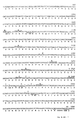
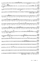
- a new amylase having an amino acid sequence as shown in Fig. 2.
- a DNA sequence, encoding the new amylase is provided as also shown in Fig. 2, particularly from nucleotide 184 to nucleotide 2131.
- any lactic acid bacteria can be used in accordance with this invention.
- the lactic acid bacteria is a Streptococcus that is to be used as a probiotic
- the Streptococcus is transformed with at least two of the exogenous DNAs
- the lactic acid bacteria is a Lactobacillus transformed with an exogenous DNA coding for an amylase
- the Lactobacillus is preferably transformed also with at least one other exogenous DNA coding for a different enzyme, preferably a cellulase.
- the preferred species of lactic acid bacteria for silage are from the genera Lactobacillus, Enterococcus, Lactococcus, Pediococcus and Leuconostoc, all of which have been used previously in silage.
- the particularly preferred species for silage are from the genera Enterococcus, Lactobacillus and Pediococcus, such as the aforementioned species of such genera.
- the preferred species for probiotics are from the genera Enterococcus and Lactobacillus.
- the lactic acid bacteria can be transformed with any foreign gene or DNA fragment thereof which codes for the expression and secretion of an enzyme that can degrade a polysaccharide and/or an oligosaccharide to a fermentable carbohydrate.
- the foreign gene or DNA fragment can code for: an enzyme that can degrade starch, i.e., an amylase such as an a-amylase, a-amylase, glucoamylase, isoamylase or pullulanase; an enzyme that can degrade cellulose, i.e., a cellulase such as an endoglucanase, exoglucanase or beta-glucosidase; an enzyme that can degrade pectin, i.e., a pectinase such as a pectic lyase, pectic glycosidase or a pectic methyl esterase; or an enzyme that can degrade hemicellulose, i.e.,
- the lactic acid bacteria is preferably transformed with 2 or more (e.g., up to 5) of these exogenous DNAs coding for different enzymes.
- Each of the different enzymes, encoded by the plurality of exogenous DNAs, can degrade a polysaccharide or oligosaccharide into a fermentable carbohydrate.
- one or more of the different enzymes can degrade a polysaccharide or oligosaccharide into a simpler saccharide, and one or more of the other different enzymes can degrade the simpler saccharide into a fermentable carbohydrate, so long as the overall action of the different enzymes is to degrade a polysaccharide or oligosaccharide into a fermentable carbohydrate.
- a plurality of DNAs will preferably be selected to provide the transformed lactic acid bacteria with a suitable combination of enzymes for degrading the intended substrate (e.g., the crop or the animal feed) to obtain an increase in fermentable carbohydrates.
- Lactic acid bacteria for silage are preferably transformed with exogenous DNAs coding for at least one cellulase, especially two or more cellulases (e.g., an endoglucanase and an exoglucanase).
- Lactic acid bacteria for a probiotic are preferably transformed with at least one exogenous DNA coding for an amylase.
- the enzyme encoded by the exogenous DNA can serve as a marker for transformed microorganisms.
- a method for detecting transformed microorganisms of this invention which involves detecting the enzyme produced by the transformed microorganisms.
- Suitable exogenous DNAs of this invention coding for enzymes that degrade cellulose, can be obtained from prokaryotic or eukaryotic microorganisms such as:
- Suitable exogenous DNAs, coding for enzymes that degrade starch can be obtained from a large variety of prokaryotes such as Bacillus, Streptomyces, Clostridium Klebsiella, Streptococcus and lactic acid bacteria such as Lactobacillus amylophilus, Lactobacillus amylovorus, Lactobacillus cellobiosus and Enterococcus, from fungi such as Candida japonica, Endomycopsis, Lipomyces, Schwanniomyces, Aspergillus niger and Rhizopus and from most higher plants and animals.
- prokaryotes such as Bacillus, Streptomyces, Clostridium Klebsiella, Streptococcus and lactic acid bacteria such as Lactobacillus amylophilus, Lactobacillus amylovorus, Lactobacillus cellobiosus and Enterococcus, from fungi such as Candida japonica, Endomycopsis
- Suitable exogenous DNAs coding for enzymes that degrade pectins, can be obtained from prokaryots such as Pseudomonas marginalis, Bacillus, Clostridium, Erwinia, Cytophaga, Xanthomonas, Aeromonas, Flavobacterium and Streptomyces, from various plant pathogenic fungi and saprophytic fungi, and from most higher plants.
- prokaryots such as Pseudomonas marginalis, Bacillus, Clostridium, Erwinia, Cytophaga, Xanthomonas, Aeromonas, Flavobacterium and Streptomyces
- Suitable exogenous DNAs coding for enzymes that degrade hemicellulose, can be obtained from bacteria such as Streptomyces, Bacillus, Sporocytophaga myxococcoides, Clostridium, Butyrivibrio fibrisolvens, Ruminococcus albus and R. flavefaciens, from protozoa such as Epidinium ecaudatum, Entodinium and Eremoplastron bovis, and from a wide variety of plant pathogenic and saprophytic fungi.
- bacteria such as Streptomyces, Bacillus, Sporocytophaga myxococcoides, Clostridium, Butyrivibrio fibrisolvens, Ruminococcus albus and R. flavefaciens
- protozoa such as Epidinium ecaudatum, Entodinium and Eremoplastron bovis, and from a wide variety of plant pathogenic and saprophytic fungi.
- microorganisms can provide suitable exogenous DNAs for transforming lactic acid bacteria in accordance with this invention.
- exogenous DNA obtained by a combination of such methods can also be used.
- terms such as “gene”, “foreign DNA fragment thereof” and “exogenous DNA”, as used herein, are not limited to naturally occurring genes and DNA fragments thereof which encode natural enzymes that degrade polysaccharides and/or oligosaccharides.
- each exogenous DNA used for the transformation of lactic acid bacteria must cause the bacteria to express and secrete the enzyme encoded by the exogenous DNA.
- the exogeneous DNA has to contain regulatory sequences such as a suitable promoter, secretion signal, translation initiation signal and termination signal operable in the lactic acid bacteria.
- regulatory sequences may be homologous to the enzyme encoding sequences, i.e., naturally associated with such a coding sequence.
- this invention also extends to so called chimeric exogenous DNAs, i.e., DNAs in which one or more regulatory sequences are not normally associated with the enzyme encoding sequence.
- the "PRLB" promoter of Fig. 7a is a preferred heterologous promoter for controlling an exogenous DNA of this invention in lactic acid bacteria.
- each exogenous DNA can be provided in an episomal vector, e.g., a plasmid.
- an episomal vector e.g., a plasmid.
- Well known process techniques can then be used to carry out such a transformation with the vector to achieve high levels of expression of the exogeneous DNA of this invention.
- Cultivating cultures of such transformed lactic acid bacteria also can be carried out in a conventional manner.
- segregation of vectors during cell division occurs at random, and cells will appear which do not carry a vector with one or more of the exogenous DNAs of this invention cloned on them.
- cells without such a vector could eventually predominate in cultures of the bacteria in which there is no selective pressure for carrying cells with the vector.
- each exogenous DNA of this invention be inserted in the bacterial chromosome.
- This provides more stable transformants and can be suitably accomplished as follows
- Each exogenous DNA can be first inserted in a bacterial transposon.
- the transposon also carries a selectable or screenable marker gene and is cloned into a plasmid which cannot replicate in the lactic acid bacteria or whose replication can be inhibited in the bacteria.
- the plasmid is then introduced in the lactic acid bacteria, and the transformants are screened for the presence of the marker gene so that only bacteria, in which the transposon has moved to the chromosome, will be selected.
- a plasmid can be constructed which itself contains a marker gene and a part of the bacterial chromosome and which also cannot replicate in the lactic acid bacteria. After introduction of the exogenous DNA in the plasmid, transformation of the lactic acid bacteria and screening for the marker gene, only those bacteria are selected in which the plasmid has been inserted in the bacterial chromosome through a single recombination event between the homologous DNA regions of the plasmid and the bacterial chromosome.
- the plasmid can also be contructed so that, after transformation of the lactic acid bacteria, the exogenous DNA is inserted in the bacterial chromosome through a double homologous recombination event.
- the choice of a suitable region of the bacterial chromosome, which is to be cloned in the plasmid must be such that the later integration of the exogenous DNA in the chromosome does not hinder the bacteria in their growth or other essential activities.
- the transformed lactic acid bacteria of this invention can be used in silage in a conventional manner and in conventional amounts.
- about 10 5 to 10 6 viable bacteria per gram of silage will generally be a minimum, and about 10 7 bacteria per gram of silage will generally be a maximum.
- the expression of the foreign genes incorporated in the lactic acid bacteria will: 1) increase the amount of fermentable carbohydrates to about 10 to 35 grams per kilogram of silage, preferably to an amount of 15 to 25 grams per kilogram of silage; and 2) lower the pH in the silage to a value of about 3 to 5, preferably to a value of 3.5 to 4.5, within a period of about 24 to 120 hours, preferably 24 to 72 hours.
- the lactic acid bacteria is preferably transformed with an exogenous DNA fragment that encodes an amylase.
- starch is included in most formulations of food for humans and industrial feed for farm animals and constitutes the major part of some natural aliments (e.g., grain for chicken).
- an amylase produced by a lactic acid bacteria probiotic in the intestinal tract of the human or animal, will hydrolyse residual starch remaining undigested after the action of amylases from the human's or animal's saliva, thereby increasing the energy value of most food and feed formulations and/or allowing the use of less expensive formulations.
- the lactic acid bacteria preferably is derived from a bacteria found in the natural intestinal flora of animals, such as Enterococcus and Lactobacillus species (Vrignaud, 1982). Such bacteria can be transformed in a conventional manner with an exogenous DNA which directs the expression and secretion of an enzyme, such as one of the aforementioned enzymes for degrading oligosaccharides and/or polysaccharides into fermentable carbohydrates.
- an enzyme such as one of the aforementioned enzymes for degrading oligosaccharides and/or polysaccharides into fermentable carbohydrates.
- the transformed lactic acid bacteria of this invention can be used as a probiotic in a conventional manner and in conventional amounts.
- about 10 6 cells per liter (or kg) of food stuff will generally be a minimum, and about 10 10 to 10 12 cells per liter of food stuff will generally be a maximum.
- the invention further relates to the plasmid designated hereinafter as "pLH1 " or "pLAB1000", the nucleotide sequence of which appears in Fig. 3a, and to cloning vectors for lactic acid bacteria which are derived from pHL1 and contain at least the regions of this plasmid necessary for replication in lactic acid bacteria. While this plasmid and its derivatives find preferred use for the production of lactic acid bacteria which produce enzymes which degrade polysaccharides and/or oligosaccharides into fermentable carbohydrates, they can also be used as vectors for the production of transformed lactic acid bacteria which, in cell culture media, produce other proteins. In this regard, the invention concerns plasmids derived from pLH1 in which other nucleotide sequences, preferably coding for determined polypeptides that are to be produced in lactic acid bacteria, are cloned.
- the invention also extends to modified plasmids derived from pLH1 and containing a nucleotide sequence foreign to that plasmid and coding for a determined polypeptide inserted either within an exogenous DNA of the present invention, preferably an amylase, or in another part of the plasmid under the control of its own regulation sequences.
- a sequence encoding amylase can then be used as a screenable transformation marker.
- bacteria transformed with the so-modified plasmid and expressing the foreign nucleotide sequence can be detected by their amylase producing capability.
- the invention also relates to plasmids derived from pLH1 which comprise a promoter operable in lactic acid bacteria, such as the PRLB promoter of Lactobacillus hilgardii, the DNA sequence of which is shown in Fig. 7a (Platteeuw, 1986).
- DNA fragments which are normally not expressible in lactic acid bacteria due to the absence of a suitable promoter, can be easily cloned in these pLH1-derived expression vectors to achieve expression of the DNA fragments in these bacteria.
- the invention still further relates to a process for transforming lactic acid bacteria with one or more exogenous DNAs by electroporation.
- electroporation is carried out with rapidly growing, exponential phase, bacterial cells, particularly as measured at an O.D. 600 equal to 0.2 to 0.8, preferably 0.4 to 0.6.
- the cells be washed, immediately before electroporation, with water or conventional buffers of low ionic strength.
- the electric field be applied to a concentrated suspension of the cells of 10 10- 10 11 Colony Forming Units ("CFU")/mi in a low ionic strength buffer. This process avoids the need for forming protoplasts of the lactic acid bacteria before transformation.
- CFU Colony Forming Units
- Lactobacilli used in the Examples, have been deposited in the Deutsche Sammlung Von Mikroorganismen (the "DSM"), Gottingen, Federal Republic of Germany, under the provisions of the Budapest Treaty:
- DSM Deutsche Sammlung Von Mikroorganismen
- Gottingen Federal Republic of Germany
- the other microorganisms in the Examples are also readily available or can readily be reproduced from readily available microorganisms and genetic material without performing any inventive work.
- Example 1 Cloning an a-amylase gene from Bacillus stearothermophilus
- the a-amylase gene from B. stearothermophilus strain CU21 has been characterized and its DNA sequence determined (Aiba et al, 1983; Nakajima et al, 1985). Total DNA was prepared from B. stearothermophilus CU21 as described by Aiba et al. (1983), digested with Pvull and Hindlll, and size fractionated on an agarose gel. The 3 kilobase (“kb”) pair size fraction was recovered, and DNA was purified from this size fraction and ligated to plasmid pUC9 (Messing, 1980) which was digested with Hindlll and Smal.
- kb 3 kilobase
- the ligation mixture was transformed into Escherichia coli strain JM83 (Yanish-Perron et ai, 1985). Transformed cells were plated on LB agar with 100 mg/I ampicillin and 1% (by weight) soluble starch (Difco Laboratories, Detroit, Michigan, U.S.A.) and were incubated overnight at 37°C. The agar plates were then stained with iodine vapor. A halo of non-staining agar around certain colonies was indicative of amylase activity. These colonies were picked and colony purified, and plasmid DNA was prepared from them.
- Example 2 Cloning an amylase gene from Bacillus thuringiensis (Figs. 1 and 2)
- Total DNA was prepared (Aiba et al, 1983) from B. thuringiensis strain H1.1, serotype H1, var. thuringiensis, obtained from Dr. Huguette de Barjac, Institut Pasteur, Paris. It was digested with restriction enzyme Hindlll an ligated to bacteriophage lambda cloning vector 590 (Murray et al, 1977) cleaved with Hindlll. The ligation was packaged, infected on E. coli and plated on LB agar supplemented with 1% soluble s tarch . Phage containing the B. thuringiensis amylase gene were identified by the halo they form after staining in iodine vapor. The existence of these halos proved the expression of the amylase gene in E. coli.
- FIG. 1 A restriction map of this fragment, including the amylase gene, is given in Fig. 1.
- the DNA sequence of most of the fragment insert was defined by the Maxam and Gilbert chemical degradation method and is given in Fig. 2.
- ORF open reading frame
- Downstream of the B. thuringiensis amylase gene is a sequence that is highly homologous on the amino acid level to the E. coli ADP glucose transferase gene (Baecker et al, 1983).
- Lactobacillus hilgardii 67 was isolated from grass silage by Radar N.V. (Deinze, Belgium). The strain was grown in static culture at 37°C in MRS medium (made by Oxoid Ltd., Basingstoke, Hampshire, England) supplemented with 20 mM D-L-threonine. Cells were pelleted from a late log phase culture, resuspended in 10 mM Tris-HCI pH 7.0, pelleted, resuspended in the same buffer and pelleted again.
- MRS medium made by Oxoid Ltd., Basingstoke, Hampshire, England
- 0.2 gram of cells was resuspended in 2 ml of 20 mM Tris-HCI pH 7.0, 2ml of a 24% solution of polyethylene glycol in water were added, and 34 enzyme units of mutanolysine (Sigma Chemical Company, St. Louis, MO, USA) in water were added. The solution was incubated at 62° C for 1 hour. 0.4 mg of Proteinase K (Sigma) was added, and the cells were incubated 60 minutes at 37° C to form protoplasts.
- the protoplasts were collected from the mixture by centrifugation and resuspended in 5 ml of 20 mM Tris-HCI and 10 mM ethylene diamine tetraacetate ("EDTA") at pH 8.0. SDS was added to 3% and mixed with the protoplasts which were then left at room temperature (25°C) for 30 minutes. The resulting cell lysate was centrifuged to remove unlysed cells, and the supernatant was extracted with water saturated phenol and then with chloroform-isoamyl alcohol (24/1 by volume). The nucleic acids were precipitated from the aequous phase by adding sodium acetate to 0.3 M and 1 volume of 2-propanol, followed by centrifugation.
- EDTA ethylene diamine tetraacetate
- the pellet was resuspended in a mixture of 10 mM Tris-HC1/1 mM EDTA at pH 8.0.
- the DNA was tested for plasmid content by two dimensional gel electrophoresis (Hintermann et al, 1981).
- pLH1 One of the plasmids found was named "pLH1" -- later renamed "pLAB1000” -- and characterized as described below.
- the pLH1 plasmid was cleaved with Xbal (Xbal has 1 recognition site in pLH1) and ligated to E. coli vector pUC18 (Yanisch-Perron et al, 1985) cleaved with Xbal.
- the ligation mixture was transformed into E. coli strain K514 (Zabeau and Stanley, 1982), and the resulting fusion plasmid was named "pUH1.3" (Fig. 4).
- a restriction map of the pLH1 plasmid was determined, and the entire pLH1 plasmid was sequenced by the Maxam and Gilbert chemical degradation method. The sequence is 3331 basepairs long and has 3 ORFs reading in the same direction. A fourth ORF (87 amino acids), reading in the opposite direction, is found within ORF 1.
- the DNA sequence of the pLH1 plasmid is shown in Fig. 3a, and a physical map of the pLH1 plasmid, with restriction sites cleaving once or twice in pLH1, is shown in Fig. 3b.
- ORF 1 in pLH1 encodes 361 amino acids. It exhibits amino acid homology with an ORF on the pE194 plasmid from Staphylococcus aureus (Horinouchi and Weisblum, 1982A), with an ORF from the pT181 plasmid of S. aureus (Khan and Novick, 1983), and with the prot B gene of pUB110 (McKenzie et al, 1987).
- Some of the ORFs that are homologous to the ORF 1 of pLH1 are known to encode site-specific recombinases (Gennaro et al, 1987) that act upon a target site located in the promotor of the recombinase gene.
- the promotor region of pLH1's ORF 1 contains significant homology with the target site of these site-specific recombinases. It is believed therefore that pLH1's ORF 1 is a site-specific recombinase, catalyzing low frequency recombination at the RS A , site in the promotor region of ORF 1 ( Figure 3c-B). ORF 1 and the RS A site are located on a 1819 basepair Aval-Sacl fragment of pLH1 in Fig. 3a.
- ORF 2 in pLH1 shows amino acid homology with the ORF coding for protein A in plasmid pC194 from S. aureus (Horinouchi and Weisblum, 1982B; Alonso et al, 1987; McKenzie et al, 1987), with the prot A gene of plasmid pUB110 from S. aureus, and with an ORF of plasmid pFTB14 of Bacillus amyloliquefaciens (Murai et al, 1987) as shown in Figure 3d-A.
- These ORFs of other genes are known to encode replication proteins that initiate plasmid replication by nicking plasmids at specific sites.
- a nicking site was defined in pLH1 by the striking homology (Figure 3d-B) between a pLH1 DNA sequence and the nicking sites of the plasmids exhibiting homology with pLH1'S ORF 2. Moreover, as found in the other plasmids, this nicking site is located upstream of the gene encoding the replication protein in a cluster of inverted repeated sequences. ORF 2 and the nicking site are located on a 1512 basepair Aval-Sacl fragment of pLH1 between nucleotides 2221 and 710 in Fig. 3a.
- ORF 2 and the nicking site are both required for DNA replication and plasmid stability of pLH1 in a variety of Gram-positive bacteria (See Example 6). From this and from the direct demonstration of single stranded DNA in plasmid preparations of pLH1, it is believed that pLH1 replicates by a single strand intermediate as is the case for several other Gram-positive plasmids (Ter Riele et al, 1986).
- Lactobacillus plantarum strains 183, 195 and 30 isolated from grass silage by Radar N.V. cross-hybridized to pLH1 in Southern blots of restriction-digested DNA, but the cross-hybridizing plasmids had a different restriction pattern and different size from pLH1.
- Example 4 Constructing Lactobacillus vectors based on the pLH1 replicon (Fig. 4)
- Plasmid pVA838 (Macrina et al, 1982) carries the macrolide lincosamide streptogrammin B ("MLS") resistance gene, encoding resistance to erythromycin, of pAMB1 (Macrina et al, 1982).
- MLS macrolide lincosamide streptogrammin B
- the DNA sequence of the pVA838 erythromycin resistance gene was published by Martin et al (1987) and found to be very homologous to the MLS resistance gene from Tn917 (Shaw and Clewell, 1985).
- pVA838 was cleaved with Hindlll and Clal and ligated to pBR322 (Bolivar and Backmann, 1979) which was cleaved with Hindlll and Clal.
- the ligation mixture was transformed into E. coli, which was plated on LB agar supplement ed wit h 100 ug/m l ampicillin.
- a plasmid was recovered carry ing the ⁇ -lactamase and tetracycline resistance genes and the origin of replication from pBR322 joined to the erythromycin resistance gene from pVA838, and the plasmid was named "pERM1".
- pERM1 was cleaved with EcoRV and Pvull and was religated in low DNA concentration.
- the ligation mixture was transformed in E. coli and was plated on LB agar supplemented with 100 mg/I ampicillin and 200 mg/I erythromycin.
- pERM1 derived plasmid was obtained in which the pBR322 tetracycline resistance gene was deleted, and the plasmid was named "pERM2".
- pERM2 was cleaved with Hindlll, and ligated to pLH1 from Example 3 cleaved with Hindlll. This produced plasmids "pERM3.1” and "pERM3.2” which differ in the orientation of the pLH1 fragment.
- pERM3.2 was cleaved with EcoRl and Ddel, the cohesive ends were made blunt by filling in with Klenow DNA polymerase, and ligated to plasmid pLK58 (Botterman and Zabeau, 1987), cleaved with EcoRV.
- a plasmid was selected containing the erythromycin resistance gene of pERM3.2 inserted in pLK58, and this was named "pG1401".
- the P L promotor from pLK58 was removed from pG1401 by cleaving pG1401 with Sail and Stul, filling in the sticky ends with Klenow polymerase, and religating the mixture in dilute conditions, yielding plasmid "pG14010".
- pG14010 was cleaved with BamHl, filled in with Klenow polymerase, then cleaved with Xbal, and ligated to plasmid pUH1.3 from Example 3, cleaved with Sac!, rendered blunt by the exonuclease activity of Klenow polymerase, and then cleaved with Xbal.
- a plasmid containing pG14010 as well as the pLH1 Xbal - Sacl fragment was selected after transformation and plating on LB plates containing 100 mg/I ampicillin and 200 mg/I erythromycin, and was named "pG14011".
- the BamHl site was regenerated at the BamHl-Sacl fusion.
- Example 5 Introducing plasmids from Example 4 into strains of Lactobacillus and other Gram-positive bacteria
- Lactobacillus plantarum 80 isolated from grass silage by Radar N.V., is noted for its rapid acid production, high acid resistance, high growth rate, homofermentative growth in silage and survival after lyophilization.
- Several methods can be used to transform this strain with pLH1 derived plasmids. Electroporation, as described below, is particularly efficient.
- An overnight saturated culture of L. plantarum 80 was diluted 100 fold in MRS medium prewarmed at 37° C and grown in an aerobic shaking culture in MRS medium at 37° C.
- O.D. optical density
- its cells were centrifuged for 10 minutes at 5,000 g, resuspended in 1/20 of the original culture volume of sterile distilled water and centrifuged for 10 minutes at 10,000g. They were then resuspended in 1/100 culture volume of an autoclaved solution of 30% (weight by volume) polyethylene glycol 100 (Merck Schuchardt, Kunststoff, Federal Republic of Germany) in distilled water. All centrifugations and washings were performed at room temperature (25°C). After these manipulations were completed, the cells were put on ice.
- the cells were left on ice for 30 minutes in the original electroporation cuvette. Then, 10 ml of MRS were added, and the cells were incubated 1 to 2 hours at 28°C. 100 ⁇ l of cells (approximately 10 8 to 10 9 cells having received 0.05 ⁇ g of DNA) were plated on MRS medium supplemented with the appropriate antibiotic: either 10 mg/I chloramphenicol or 10 mg/I of erythromycin. The cells were incubated 48 hours at 37°C.
- lincomycin 100 mg/I of lincomycin (Sigma) was also added to the MRS medium before incubation because most erythromycin resistance genes used in this Example, especially the erythromycin resistance gene from pVA838, also induce resistance to lincomycin. Moreover, the addition of lincomycin reduced the frequency of spontaneous erythromycin-resistant mutants of L. plantarum 80 from 10- 7 down to less than 10- 9 . By this procedure, 10 6 transformant colonies of L. plantarum 80 per microgram of input DNA were obtained. Using this procedure, high efficiency transformation of L.
- plantarum 50 another bacterium isolated from silage by RADAR N.V., and noted for its qualities as a silage starter strain
- Lactobacillus casei strain DSM20020 on deposit at the DSM under accession no. 20020.
- Enterococcus faecalis strain OG1X (Ike et al, 1983)
- Bacillus subtilis strains 1A40 and 1A42 from Bacillus Genetic Stock Center, Columbus, Ohio, USA
- B thuringiensis strains kurstaki DIPEL from Abbott Laboratories, Abbott Park, North Chicago, III, U.S.A) entomicides (received from Dr. D. Lereclus, Inst.
- Example 6 Studying replication properties of pLH1 in several Gram-positive bacteria
- plasmids e.g., pGI4011 from Example 4
- pGI4011 from Example 4
- nicking site see Example 3
- L plantarum 50 and 80 L. casei DSM20020
- E. faecalis strain OG1X E. faecalis strain OG1X
- B. subtilis strain 1A42 There was no evidence of autonomous replication of pLH1 in various strains of E. coli.
- L. plantarum 80 Several plasmids originating from different Gram-positive bacteria were introduced in L. plantarum 80 by electroporation following the procedures of Example 5.
- the comparison plasmids were: pVA838 (Macrina et al, 1982), pSA3 (Dao and Feretti, 1985), pTG402 (Zukowski et al, 1983), pTV8 (Youngman, 1987), pDB64 (Gryczan et al, 1980) and pGKV2 (Kok et al, 1985).
- L. plantarum 80 cells transformed with the comparison plasmids and with pERM3.1 were grown for 13 generations on MRS medium (Oxoid Manual, 1982) without antibiotic pressure.
- Plasmids replicating via single strand intermediates like pLH1 are prone to reorganizations and deletions when used in recombinant DNA constructions (Noirot et al, 1987). No instability was observed in any of the cloning experiments with pLH1. In this regard, during the plasmid transformations in Examples 3, 4, 7 and 8, it was verified by restriction digests of DNA from plasmid minipreparations of transformed bacteria (Anderson & McKay, 1983) that the native configuration of the pLH1 plasmid did not change.
- Example 7 Expressing an amylase gene and an endoglucanase gene in L. plantarum 80 using vectors based on pLH1 (Figs. 5 and 6)
- a-amylase and endoglucanase genes were inserted in separate vectors replicating in L. plantarum 80 and assayed for expression as follows.
- Plasmid pSTUC (Example 1) was cleaved with SnaBi and Dral, and ligated to pERM3.2 cleaved with Spel and made blunt end by filling with Klenow polymerase.
- Two recombinant plasmids carrying the pSTUC fragment with the amylase gene inserted in both orientations in the pERM3.2 replicon were selected by their amylase production (as evidenced by the assay described in Example 1) and erythromycin 200 mg/I resistance in E. coli. They were named "pRAM11", and "pRAM12", respectively ( Figure 5).
- both pRAM11 and pRAM12 replicate stably under erythromycin selective pressure, express amylase and secrete the enzyme into the medium (as evidenced by staining the MRS agar plates containing 1 0 / 0 soluble starch with iodine vapor). Since both pRAM11 and pRAM12 produce similar amounts of amylase activity, the expression of the amylase gene must have been directed from the native (B. stearothermophilus) promotor. The amylase expression appears not to have been repressed by presence of glucose in the medium, and not to have been dependent on the growth phase of the strain.
- Plasmid pWS11 described by Schwarz et al (1986) and obtained from these authors, carries the Clostridium thermocellum endoglucanase cel A gene and surrounding sequences including the promotor on a 3.2 Kb Hindlll fragment. The sequence of this gene is described in Beguin et al (1985). This gene has an endoglucanase activity towards carboxymethyl cellulose ("CMC”) and towards lichenan which is a R-1-3-glucan (Hazlewood et al, 1988).
- CMC carboxymethyl cellulose
- lichenan which is a R-1-3-glucan
- pWS11 was cleaved with Hindlll, the cohesive ends were filled in by Klenow polymerase, and ligated to plasmid pSA3 (Dao and Feretti, 1985) cleaved with EcoRV. Plasmids having the cel A gene inserted in the pSA3 EcoRV site of pSA3 were identified among transformants in E.
- the stability of the plasmids pST11, pST12, pRAM11, pRAM12 was good with selective antibiotic (10 mg/I erythromycin) pressure. Without such selective pressure, the plasmids were gradually lost in growing cultures as described in Example 26.
- Example 8 Constructing a pLH1 derived expression vector (Fig. 7)
- a promotor sequence was isolated from L. hilgardii 67 by digesting L. hilgardii 67 total cellular DNA (prepared according to Example 3) with Sau3AI and ligating the fragments in vector pPGV5 cleaved with BamHl (Fig. 7b).
- pPGV5 was derived from pPGV2 (D'Haese et al, 1984) by modifying the polylinker sequence in front of its promotorless CAT86 gene (Cm R ) (Fig. 7b).
- the ligation mixture was transformed in Bacillus subtilis strain 1A40, and the recombinants were selected for chloramphenicol resistance.
- the DNA sequence of one particular Sau3AI fragment, inducing very high levels of chloramphenicol acetyl transferase, is shown in Figure 7a.
- the fragment was named the "PRLB promoter”.
- the pPGV5 vector with this Sau3AI fragment was named pPGVlLH (Platteeuw, 1986) and could also transform L. plantarum 80 to provide chloramphenicol resistance.
- Plasmid pC1 obtained from Dr. Julio Polaina (Instituto de Agroquimica y Technologica deAlimento, Jaime Roig 11, Valencia, Spain) is described in Polaina et al (1988) and its restriction map is shown in Figure 7c.
- E coli transformed with this plasmid, express the Clostridium cellulolyticum endoglucanase as an intracellular enzyme.
- This endoglucanase can be demonstrated on LB agar plates containing 1 g/I CMC after the colonies have been lysed with chloroform vapor.
- the endoglucanase gene from Clostridium cellulolyticum was excised as a Hindlll - Sail restriction fragment, made blunt end with Klenow, and was ligated to plasmid pG14011 (from Example 4) which was cleaved with Sail and made blunt end with Klenow.
- the resulting plasmids were named "pGC11" and "pGC12".
- pGC11 and pGC12 produced small amounts of non-secreted endoglucanase in E. coli but no detectable intracellular or extracellular endoglucanase activity when introduced in L. plantarum 80 by the electroporation procedure of Example 5.
- the expression of the gene can be induced in L. plantarum 80.
- endoglucanase activity can be detected by the methods described in Example 7.
- Example 9 Transforming L. plantarum 80 by integration of DNA sequences in the chromosome (Fig. 8)
- heterologous DNA was integrated in the chromosome of L. plantarum 80 by a single recombination event in a region of homology between a plasmid and the chromosome.
- Total cellular DNA was prepared from L. plantarum 80 as described for L. hilgardii in Example 3. This DNA was cleaved partially with Sau3AI, so as to produce restriction fragments with an average size of 3 kilobase pairs (verified on agarose gel). These fragments were ligated in plasmid vector pG14010 (from Example 4), which was cleaved with BamHl, and the ligation mixture was transformed in E. coli. Minipreparation plasmid DNA was prepared from 48 transformants, and the presence of the chromosomal DNA insert was verified by restriction digests. The plasmid DNAs were combined in 8 pools of 6 plasmids each and were introduced in L.
- the hybridization showed the absence of pG14010 in the plasmid fraction of the LpGl transformants and in L. plantarum 80 and the presence of pG14010-related material in the digested chromosomal DNA in 19 out of 20 transformants.
- the LpG14010 sequences had integrated in the indigeneous plasmid of L. plantarum 80.
- the intensity of hybridization to the chromosomal DNA was higher than expected from a single copy insertion and varied in a way unlinked to the amount of chromosomal DNA loaded on the gel. Moreover, the pattern was more complex than expected for a single copy insertion. Therefore, the LpGl strains may carry multiple insertions of pG14010 in the chromosome.
- An EcoRl digest was made from total cellular DNA of LpGl2, LpGl4 and LpGl7 and from wild type L. plantarum 80. This DNA was diluted to low concentration and ligated, and transformed in E coli. No ampicillin-resistant transformants were obtained with the ligation mixture from L. plantarum 80. Ligations of LpGl1 to LpGl20, however, gave rise to ampicillin-resistant E. coli colonies. Plas mid DNA from these colonies showed the restriction pattern of the large EcoRl - BamHl fragment of pG14010, linked at the BamHl site to a variable fragment of chromosomal L. plantarum 80 DNA. This shows that at least part of the integrated copies of pG14010 show no major rearrangements.
- LpGl1 to LpG120 were mixed with wild type L. plantarum 80 cells at a ratio of 1 LpGl cell per 9 L. plantarum 80 cells, and the mixture was allowed to grow for 13 gener ations in MRS liquid medium without any selective pressure.
- Appropriate dilutions of the resulting culture were plated on MRS agar and on MRS agar supplemented with 10mg/1 erythromycin and 100mg/l lincomycin. It was found that 17 out of 20 LpGl strains have the same growth rate as the wild type L. plantarum 80 in this assay, and three strains, LpGI5, LpG110, and LpG120, do not grow as fast as L. plantarum 80.
- LpGl2, LpGl4, and LpGl5 were mixed with L. plantarum 80 (wild type) at a ratio 1:10, and the mixture was inoculated at 10 6 CFU per gram in autoclaved Italian Rye grass.
- LpGl2 and LpGl4 had the same growth rate as L. plantarum 80, whereas LpGI5 grew slower.
- the ratio of 1/10 was maintained for over 5 days, indicating that the survival of LpGl2 and LpGl4 in grass was not affected.
- LpGl2, LpGl4, and LpGI5 were grown for 78 generations in MRS liquid medium without any selective pressure for maintenance of the pGl4010 sequences. After growth for 13, 26, 39, 52, 65 and 78 generations, aliquots of the cultures were plated on MRS agar and on MRS agar supplemented with 10 mg/I erythromycin and 100 mg/l lincomycin. 100 colonies, that appeared on the MRS agar plate, were replicated on MRS agar supplemented with 10mg/l erythromycin and 100mg/i lincomycin. In this assay, more than 95% of the cells of LpGl2 and LpGl4 maintained their erythromycin resistance over 78 generations.
- This experiment demonstrated the feasability of stably integrating exogenous DNA in the chromosome of L. plantarum 80 without detectably modifying the phenotype of the strain.
- this experiment showed that strain LpGl4 is generated by integration of plasmid pH421 in the chromosome of L. plantarum 80 and that LpGl4 has the same phenotype as L. plantarum 80 and has a stable integration of pG14010 sequences. pH421 is therefore used in the following examples as a vehicle for inserting an amylase gene and an endoglucanase gene in the chromosome of L. plantarum 80.
- Example 10 Transforming L. plantarum 80 by integrating amylase and endoglucanase genes in the chromosome (Fig. 9)
- Plasmid pSTUC (from Example 1) was cleaved with SnaBI and Dral and ligated with plasmid pJB66 (Botterman and Zabeau, 1987) cleaved with EcoRV.
- Two hybrid plasmids carrying the 1.9 Kbp pSTUC fragment with the amylase gene as an insertion in opposed orientations in pJB66 were selected by screening for amylase activity and restriction mapping of plasmid minipreps from candidate transformants.
- the plasmids were called "pSATE101" and "pSATE102" (Fig. 9).
- Plasmid pWS11 (Schwarz et al, 1986) was cleaved with Hindlll, the cohesive ends were made blunt by filling in with Klenow polymerase, and the digest was ligated to pSATE101 cleaved with Smal.
- a plasmid was selected that had the 3.2 Kbp Hindlll fragment with the cel A gene of pWS11 inserted in the Smal site of pSATE101. This plasmid was named "pSATE123”.
- An analogous construction was made for pSATE102 and was named "pSATE125". The direction of transcription of the cel A gene and the amylase gene was parallel in pSATE123 and antiparallel in pSATE125 ( Figure 9).
- pSATE123 was cleaved with Bglll, and the cohesive ends were made blunt ended by filling in with Klenow polymerase. The resulting fragments were ligated to plasmid pH421 (from Example 9) which was cleaved with Smal. Two plasmids were selected which had the amylase - endoglucanase cassette integrated in the Smal site of pH421. They were named "pSATE253" and "pSATE256", and the orientations of the respective genes are shown in Figure 9.
- Plasmid DNA of pSATE253 and pSATE256 was introduced in L. plantarum 80 by electroporation as described in Example 5, and selected on MRS agar containing 10mg/i erythomycin and 100mg/llincomycin. About 3 transformants per ⁇ g were obtained with each plasmid.
- pERM3.2 was introduced in L. plantarum 80 at a frequency of 5x10 6 transformants per ⁇ g plasmid DNA.
- L. plantarum 80 with pSATE253 8 transformants of L. plantarum 80 with pSATE253 were named "LpGA3.1” - “LpGA3.10” (LpGA3.7 and LpGA3.8 were not allocated) and selected for further study.
- 11 transformants of L. plantarum 80 with pSATE256 were named “LpGA6.1” - “LpGA6.11” and selected for further study. All of these LpGA strains (except LpGA6.2) expressed both amylase and endoglucanase activity as determined by the methods described in Examples 1 and 7.
- Example 11 Constructing a Lactobacillus transposon vector ("pTnAMYL”) carrying an a-amylase gene (Fig. 10)
- Plasmid pJH1 from E. faecalis (Banai and Le Blanc,1983) was cleaved partially with restriction enzyme Oral and ligated to plasmid pLK58 (Botterman, 1986) cleaved with Smal.
- the ligation mixture was transformed into E. coli strain K12 ⁇ H ⁇ 1 trp (Bernard et al, 1979), and th e tran sformed cells were plated on LB agar supplemented with 10 ⁇ g/ml tetracycline.
- a transformant was recovered that had the pJH1 tetracycline resistance gene inserted in the pLK58 Smal site, and the transformant was named "pLKTJ".
- the DNA sequence of the pJH1 tetracycline resistance gene was determined and found to be very homologous to the sequence of the tetracycline resistance gene of plasmid pTHT15 from Bacillus stearothermophilus (Hoshino et al, 1985).
- pLKTJ was cleaved with SnaBI and EcoRl and was ligated to pERM2 (from Example 4) cleaved with Hindlll filled in with Klenow polymerase and EcoRl. The ligation mixture was transformed in E.
- plasmid was recovered that carries the pJH1 tetracycline resistance gene linked to the pERM2 replication origin and ⁇ -lactamase gene, and the plasmid was named "pTET1".
- pTET1 is cleaved with restriction enzymes EcoRl and Pstl, is ligated to plasmid pTV8 (Youngman, 1987) cleaved with EcoRl and Pstl, and is transfor med into E. coli strain HB101.
- Cells are plated on LB agar supplemented with 10 ⁇ g/ ml tetracyciine.
- a plasmid carrying the Tn917 element from PTV8 and the tetracycline resistance gene and gram negative origin of replication from pTET1 is recovered, and the plasmid is named "pTn917.1".
- Plasmid pLH1 (from Example 3) is linearized by cleavage with Hindlll, the cohesive Hindlll ends are filled in with the Kienow fragment of E. coli DNA polymerase I, and the linearized plasmid is then ligated to pTn917.1 cleaved with Fspl.
- the ligatio n mi xture is transformed into E. coli and is plated on LB agar supplemented with 10 ⁇ g/ml tetracycline.
- a plasmid is recovered in which both parental plasmids are joined, and the plasmid is named "pTn917.4".
- pTn917.4 is cleaved with restriction endonuclease Smal and is ligated to pSTUC (from Example 1) cleaved with SnaBl and Dral.
- Th e lig ation m ixt ure is transformed into E. coli and plated on LB agar supplemented with 10 ⁇ g/ml tetracycline. Transformants are replicated on LB plates, on which 10 ⁇ g/ml tetracycline and 1% soluble starch were added, and were tested for secreted amylase activity by incubating them briefly with iodine vapor.
- a plasmid is recovered carrying the a-amylase gene inserted into the Smal site of the Tn917 counterpart of pTn917.4 and exhibiting amylase activity.
- the a-amylase gene is oriented with its direction of transcription pointing from the inverted repeat of Tn917 towards the erythromycin gene. This plasmid is named "pTnAMYL".
- Example 12 Introducing the pTnAMYL Transposon Vector carrying an a-amylase gene into two strains of Lactobacillus plantarum
- L. plantarum 50 and 80 are transformed with pTnAMYL using the procedure described in Example 5.
- Example 13 Transforming L. plantarum with an a-amylase gene by integrating the pTnAMYL Transposon Vector in the chromosome of L. plantarum
- each culture is diluted to 20 ml with MRS, nalidixic acid is added to 1.0 ⁇ g/ml, novobiocin is added to 0.1 ⁇ g/ml and erythromycin is added to 10 ⁇ g/ml (Novobiocin and nalidixic acid in the above concentrations cure lactobacilli from pLH1).
- Aval restriction digest of this DNA is size separated on agarose gels and is then transferred to nylon membranes by Southern blotting.
- the membranes are hybridized to 32 P radioactively labeled Tn917 DNA to verify insertion of the unmodified pTnAMYL construct in multiple sites of the L. plantarum genome.
- the erythromycin resistant, tetracycline insertion mutants exhibit a-amylase activity on starch plates.
- Example 14 Transforming L. plantarum with the amylase gene from B. thuringiensis
- pJAM23 (from Example 2) is cleaved with restriction endonucleases Pvull and Hindlll, and the cohesive ends are filled in with DNA polymerase I Klenow fragment. The resulting fragments are ligated to pTn917.4 cleaved with Smal. After transformation into E. coli strain HB101, a tetracycline resistant colony exhibiting amylase activity is selected, and its plasmid is named "pTnAMYL2". As with pTnAMYL of Example 11, the direction of transcription of pTnAMYL2 points from the inverted repeat of Tn917 towards the erythromycin gene.
- pTnAMYL2 is introduced into L. plantarum strains 50 and 80 following the procedure set forth in Examples 5 and 12 and is integrated into their chromosomes as described in Example 13.
- Example 15 Constructing a shuttle plasmid ("pVa") containing an a-amylase gene from B. stearothermophilus (Fig. 11)
- the vector pVA838 (ATCC catalog number 37160) is able to replicate in E. coli and Streptococcus sanguis (Macrina et al, 1982) and in E. faecalis (Wirth et al, 1986).
- pVA838 was prepared from the E. coli strain DB11 obtained from Dr. Clewell (Clewell, USA) using the rapid LiCI "mini prep" procedure (Wilimzig, 1985).
- pVA838 was digested with Pvull, and pSTUC (from Example 1) was digested with Snabl and Dral. The restriction digests were phenol extracted and ligated. The ligation mixture was used to transform E.
- Example 16 Transforming E. faecalis protoplasts with the pVa shuttle plasmid containing an a-amylase gene
- the E. faecalis OG1X strain (Ike et al, 1983), obtained from Dr. Clewell, was used to prepare protoplasts according to the method of Wirth et al (1986).
- the protoplasts were collected by centrifugation (10 min. at 3,000 g), were washed once in 10 ml of TSCM buffer containing 100 ⁇ g/ml BSA, and were resuspended by gentle shaking in 1 ml of the same buffer.
- a 100 ⁇ l sample of the protoplasts (approximately 10 8 cells) was mixed with about 100 ⁇ l (about 1 ⁇ g) of the pVa shuttle plasmid (from Example 15) in 2 X SSC (1 X SSC is 0.15 M NaCl plus 0.015 M Na-citrate). The mixture was added to 1.8 ml of filter sterilized 40% polyethyleneglycol with a molecular weight of 1000 ("PEG"; Sigma) in TSCM buffer.
- PEG polyethyleneglycol with a molecular weight of 1000
- erythromycin resistant colonies were obtained at a frequency of about 10 3 / ⁇ g DNA. They were replicated onto agar THB plates containing 20 ⁇ g/ml erythromycin plus 0.2 0 /o soluble starch. Erythromycin resistant colonies displayed a clear halo upon exposure to iodine vapor. Their plasmids were further identified by restriction analysis of mini preparation plasmid DNA (Ish-Horowicz and Burke and 1981). As expected, erythromycin-resistant, a-amylase positive colonies harbored the pVa recombinant plasmid. The halo surrounding the colonies on starch plates exposed to iodine vapor indicate that the B. stearothermophilus a-amylase gene was actively expressed in E. faecalis and that the a-amylase was secreted into the medium.
- Example 17 Constructing another shuttle plasmid ("pVAa”) from B. licheniformis (Fig. 12)
- the B. licheniformis a-amylase had been cloned from different strains, and its sequence had been determined by several groups (Ortlepp et al, 1983; Sibakov and Palva, 1984; Piggott et al, 1984; Joyet et al, 1984; Stephens et al, 1984; Yuuki et al, 1985; Gray et al, 1986). Plasmid pRPI bearing the a-amylase gene from the B. licheniformis RP01 as a Bell fragment (Piggott et al, 1984) was received from Dr. D.J. McConnell, Dep artment of Gene tics, Trinity Col litis, Univ. Dublin, Republic of Ireland.
- This plasmid was restricted with Bell, and the 2.5 kb Bell fragment was recovered from 0.80/0 agarose gel in TBE buffer after electrophoresis for 2 hours at 75V in a horizontal submarine apparatus filled with TBE buffer. Recovery was performed according to Maxam and Gilbert (1980).
- This 2.5 kb Bcll fragment containing the complete transcription unit for the a-amylase (Piggott et al, 1984) was ligated into pUC18 (Yanisch-Perron et al, 1985) previously cut with BamHl. The ligation mixture was transformed into E. coli strain JM101 (Messing et al, 1981).
- Colonies harboring recombinant plasmids comprising the pUC18 backbone plus the 2.5 kb a-amylase insert were selected on agar plates (LB supplemented with 50 ⁇ g ampicillin/ml and 0.2% starch). Candidates were identified by the presence of a clear halo surrounding the colony after a brief exposure to iodine vapor. The identity of the recombinant plasmids present in such transformants was checked by restriction analysis performed on plasmid DNA prepared according to the LiCI rapid procedure (Wilimzig, 1985). One transformant colony harboring the expected recombinant plasmid was retained. After single colony purification, it was used for large scale purification of the recombinant plasmid by the alkaline lysis-CsCI method. This plasmid was named "p ⁇ 21".
- Plasmid pVA838 was prepared as before and digested with EcoRl and Sphl. In parallel, the plasmid pa21 was digested with the same enzymes. The restriction digests were phenol-extracted and ligated. The ligation mixture was used to transform E. coli strain tgl (Amersham). Spreading on LB plates containing 80 ⁇ g/ml of erythromycin plus 0.2% starch allowed the selection and visualization of transformants harboring recombinant PVA838 plasmids whose EcoRl-Sphl insert coming from pa21 and containing the a-amylase gene. Single transformant colonies were checked for the presence of the expected recombinant plasmid by restriction analysis of rapid DNA preparations as described before.
- Example 18 Constructing other shuttle plasmids ("pTVa") containing the a-amylase gene from B. stearothermophilus (Fig. 13)
- the vector pTV1 can be propagated in B. subtilis (Youngman, 1987) or E. faecalis (Weaver and Clewell, 1987) as an autoreplicative plasmid but only at low temperatures (e.g., 32°C). At non-permissive temperatures (e.g., 48° C), its origin of replication is no longer functional, and the plasmid is lost in a few generations. However, the erythromycin resistance phenotype can be retained in some of the cells exposed to non-permissive conditions due to the transposition of Tn917 into the chromosome.
- pTV1 and its derivatives can be used either as an autoreplicative vector or as an integration vector as used below.
- a shuttle plasmid was made in E. coli.
- pSTUC from Example 1
- pBR322 were each digested with Pvull, phenol extracted and were ligated to yield a "pBRa1" " plasmid consisting of a pBR322 backbone plus an a-amylase gene inserted at the Pvull site in the orientation shown in Fig. 13.
- the size of the pBRa1 plasmid was reduced by deletion of the Hindlll fragment bearing the tetracycline resistance gene, yielding a "pBRa2" plasmid.
- the pBRa2 plasmid was restricted with Pvull, and the plasmid pTV8 was cut with Smal. These digests were phenol extracted and ligated to yield pBRa2-pTV8 concatemers which were named "pTVa1" and "pTVa2" according to the orientation of the insert as shown in Fig. 13.
- Example 19 Transforming E. faecalis protoplasts with the pTVa autoreplicative shuttle plasmids containing an a-amylase gene
- the two shuttle plasmids pTVa1 and pTVa2 were purified from E. coli by the LiCI rapid minipreparation method (Wilimzig, 1985) and were used to transform E. faecalis strain OGIX protoplasts as described in Example 16. Control experiments with intact pTV8 showed that the origin of replication of this plasmid was functional in the E faecalis strain at the permissive temperature (32° C). Transformation with the pTVa plasmids yielded erythromycin-resistant colonies upon selection at 32° C on THB plates supplemented with 5 gg/ml erythromycin.
- Example 20 Integrating the pTVa borne a-amylase gene into E.faecalis chromosome by Tn917 transposition
- Clones of E. faecalis strain OG1X with the autoreplicative pTVa1 and pTVa2 plasmids were subjected to the procedure of Youngman (1987).
- a culture was prepared in liquid THB medium supplemented with 50 ⁇ g/ml erythromycin. This culture was grown at 32° C with moderate aeration up to stationary phase. The culture was then diluted 1:50 with THB broth containing 0.5 ⁇ g/ml erythromycin. This culture was grown overnight at 45° C with moderate aeration. Then, the culture was diluted again 1:50 with the same medium and grown at 45°C with moderate aeration.
- each clone was subjected to the plasmid minipreparation procedure of Ish-Horowicz and Burke (1981). Each preparation was analyzed by agarose gel electrophoresis, on the one hand, and used to back-transform E. coli, on the other hand. 13 clones from the pTVaAl series, and 12 clones from the pTVa2 series were negative by both criteria. These were further characterized as follows.
- a-amylase gene had been stably incorporated into the chromosome of E faecalis and expressed as an active enzyme released into the medium, as evidenced by the presence of a halo surrounding the colonies.
- Example 21 Constructing a set of single recombination prone integration vectors ("PG18 ⁇ ") containing the a-amylase gene from B. stearothermophilus (Fig. 14)
- a plasmid bearing a segment of chromosomal DNA from E. faecalis but lacking an origin of replication for that host behaves as an integration vector as a result of a single homologous recombination event taking place between duplicated chromosomal sequences.
- a-amylase and a phenotypic marker which will be expressed in E. faecalis, are inserted in the vector.
- a similar construction has recently been shown to function as an integration vector in B. subtilis (O'Kane et al, 1986).
- the erythromycin marker from pVA838 was excised as a Hindlll-Aval fragment and inserted into pUC18 (Yanisch-Perron et al, 1985) cleaved with Hindlll and Sall. Transformation into E. coli allows the recovery of clones harboring plasmids consisting of the pUC18 backbone plus the erythromycin resistance marker. These clones are selected on LB agar plates containing 50 ⁇ g ampicillin and 80 ⁇ g erythromycin per ml. After single colony purification, they are checked for the presence of the desired recombinant plasmid as described above by restriction analysis of minipreparation plasmid DNA (Wilimzig, 1985). One such clone is retained for further work and is named "pG18".
- pG18 is restricted with Smal
- pSTUC from Example 1
- Restriction digests are phenol extracted and ligated as described above. This ligation mixture is used to transform E. coli which allows the recovery of clones harboring the plasmid named "pG18a". In addition to being erythromycin and ampicillin resistant, these clones display a clear halo on starch plates exposed to iodine vapor.
- Example 22 Integrating the pG18 ⁇ borne a-amylase gene into an E. faecalis chromosome by single homologous recombination
- Minipreparation plasmid is prepared from individual cultures of clones harboring plasmids pG18 ⁇ 1 to 24 (from Example 21). DNA from each preparation is used as before to transform E. faecalis OG1X protoplasts. In some cases, transformants are obtained which have integrated a giv en pG18 ⁇ ⁇ plasmid into their chromosome as a result of a single recombination event between the region of the plasmid and its chromosomal counterpart. These are identified among erythromycin-resistant halo-positive colonies by Southern blotting experiments as described below. Pstl digested total DNA is hybridized with ( 32 P)-labeled pG18a as a probe.
- Control experiments are performed in parallel with total DNA from the corresponding pG18 ⁇ transformed E. coli.
- Each control displays only one band upon hybridization with the probe, corresponding in size to the linear form of the pG18 ⁇ plasmid-harbored by that clone (Fig. 14).
- Each integrative transformant displays two bands when hybridized with ( 32 P)-labeled PG18a. The sizes of those two bands are identical for all transformants of a given homology region, but different among different homology regions. The same band hybridizes with pUC18 as a probe.
- faecalis chromosome depending on the nature of the region present in each clone. Again, the presence of a halo surrounding the colonies on starch plates exposed to iodine vapor indicates that the a-amylase gene is expressed.
- Example 23 Constructing a set of double recombination prone integration vectors ("pG18 ⁇ ") containing the a-amylase gene from B. stearothermphilus (Fig. 15)
- a plasmid bearing a segment of chromosomal DNA from E. faecalis, into which the a-amylase gene has been inserted, and lacking an origin of replication for that host, behaves as an integration vector.
- a double recombination event can occur, one on each side of the a-amylase gene.
- a marker gene should be present next to the a-amylase gene inside the chromosomal fragment. Constructions of this kind have been shown to function as integration vectors in B. subtilis (Youngman, 1987).
- Random fragments of the E. faecalis chromosome are inserted into the pUC18 vector as follows. Chromosomal DNA from E. faecalis is prepared (Dale and Greenaway, 1984) and digested with Nhel. In parallel, pUC18 is digested with Xbal. Restriction digests are phenol extracted and are ligated. Transformation into E. coli allows the recovery of plasmids consisting of the pUC18 backbone plus a fragment of E. faecalis chromosomal DNA. After single colony purification, they are checked for the presence of the desired recombinant plasmid by restriction analysis of minipreparation plasmid DNA (Wilimzig, 1985).
- the plasmids present in these clones are given the generic name "pUC18 ⁇ ".
- pUC18 ⁇ a subset of 24 pUC18 ⁇ plasmids are retained having no Xbal site while comprising at least one site for the following blunt-end restriction enzymes: Ball, EcoRV, Hpal, Nael, Nrul, and Stul (labeled "R” in Fig. 15).
- Ball, EcoRV, Hpal, Nael, Nrul, and Stul (labeled "R” in Fig. 15).
- These plasmids are numbered pUC18(p 1 to 24.
- each of the plasmids is cut with its cognate blunt-end restriction enzyme as listed above and ligated to Pvull cut pG18a (from Example 21). Transformation into E. coli yields a set of recombinant plasmids, each consisting of a given pUC18(p backbone plus an insert comprising the a-amylase/erythromycin resistance region of pG18a. The corresponding clones are selected individually on LB plates supplemented with 50 ⁇ g ampicillin/ml plus 80 ⁇ g erythromycin/ml and 0.2% soluble starch.
- Halo-positive colonies are analyzed by restriction analysis of minipreparation plasmid DNA to establish the presence of recombinant plasmids of the expected structure. These plasmids are given the generic name "pG18 ⁇ " and numbered pG18 ⁇ 1 to 24.
- Example 24 Integrating the pG18 ⁇ -borne a-amylase gene into an E. faecalis chromosome by double homologous recombination
- Minipreparation plasmid DNA is prepared as described above from individual cultures of clones harboring plasmids pG18 ⁇ 1 to 24 (from Example 23). DNA from each preparation is used as before to transform E. faecalis protoplasts.
- Plating on THB plates containing 5ng erythromycin/ml allows, in some cases, the recovery of transformants which have integrated the erythromycin resistance gene and the a-amylase gene into their chromosomes as a result of double recombination events which have occured between each of the ⁇ regions of pG18 ⁇ and their chromosomal counterparts.
- These transformants are erythromycin-resistant and halo-positive and display a specific pattern in Southern blotting experiments as described below. Total DNA is extracted from candidate colonies, is digested with Xbal and is analyzed by Southern blotting using ( 32 P)-labeled pG18a as a probe.
- Control experiments are p erfor med in parallel with total DNA from the corresponding pG18 ⁇ transformed E coli. Each control displays one band upon hybridization with the probe, corresponding in size to the linear form of t he pG18 ⁇ plasmid harbored by that clone (Fig. 15).
- the expected E. faecalis integrative transformants display two bands with the probe with band sizes that vary from one transformant to another.
- the transformants showing two bands with the probe display an identical pattern in Southern blotting experiments using ( 32 P)-labeled pVa1 as a probe, but they do not show any hybridizable band with ( 32p )-labeled pUC18 as a probe, unlike the integrative transformants resulting from the single recombination event described above.
- the total DNA of transformants showing two bands with the probe, display a single major band on the autoradiogram. This band corresponds in size with the Hindlll fragment from pG18a spanning the erm and a regions (See Fig. 15).
- Example 25 Evaluation of the performance of transformed L. plantarum 80 and E. faecalis OG1X in grass silage
- the strains consisted of:
- the substrates were:
- the substrates were dialyzed (cutoff 12,000 dalton) overnight for the first time then twice dialyzed for 2 hours against 5 mM CaCI 2 , 50 mM Na-Succinate buffer pH 5.8 at 4°C. This removes most of the indigeneous low molecular weight carbohydrates in e.g. corn starch.
- strains were cultured overnight in MRS (for Lp80) or TH (for E. faecalis). For transformed strain, 10 ⁇ g/ml erythromycin and 100 ⁇ g/ml lincomycin were added.
- the measurement of the OD was roughly linear to the concentration of glucose in a series of controls and 1 OD equalled approximately 0.14 g/I.
- Table 1 shows that the mixture of Lp80 transformed with pRAMl1 and Lp80 transformed with pST11 and LpGA 6.6 performed better than the controls for all subtrates while EfOGIX transformed with pVAa performed better than its control for soluble starch and milled corn grains.
- the final concentration of the cells was 10 6 cells per ml.
- the substrates were not sterilized to avoid modification of the physicochemical structure. However, the probability of secondary infection was reduced by the addition of kanamycin which does not affect Lp80 and erythromycin (10 ⁇ g/ml) which does not affect the transformed strains.
- the 100 ml bottles as prepared above were incubated at 37°C with shaking.
- the pH was measured after 0, 2 and 5 days.
- Table 2 shows that pRAM11 and the LPGA mixture induced a more pronounced pH decrease than the other strains on soluble starch and milled corn. Table 2 also shows that pST11 and the LpGA mixture induced a more pronounced pH decrease than the other strains on CMC. Some variability of the results is probably due to growth of contaminating bacteria.
- Example 26 Stability and competitive abilities of engineered Lactobacillus plantarum strains in various conditions
- CFU colony forming units
- Table 4 The results are presented in Table 4.
- the numbers in Table 4 refer to 10 6 CFU per gram of ensiled grass. It is obvious from Table 4 that LPGI2 and LPGI4 have the same growth rate and survive equally well in grass silage as wild type L. plantarum 80. As in liquid culture (Example 9), LpGl5 has a lower growth rate although its survival is comparable to wild type L. plantarum 80. This is probably due to the fact that insertion of the Em gene occurred in a vital place of the bacterial chromosome, proving the importance of an adequate choice of a chromosomal segment as a recombination target.
- Table 5 shows that LpGA 3.9, 6.3 and 6.6 have growth rates comparable to L. plantarum 80 as was expected since strain LpG14 has the same growth rate as wild type L.plantarum 80. Again the engineered strains perform very well.
- DNA sequences other than those shown in Figs. 2 and 3 for the novel amylase gene and plasmid, respectively, of this invention can be used for transforming lactic acid bacteria.
- such DNA sequences can be modified: 1) by replacing some codons with others that code either for the same amino acids or for other amino acids; and/or 2) by deleting or adding some codons; provided that such modifications do not substantially alter the properties of such DNA sequences.
- other DNA recombinants containing the aforementioned DNA sequences in association with other foreign DNA particularly the DNA of vectors suitable for transforming microorganisms other than lactic acid bacteria, such as E. coli, B. subtilis and even eucaryotic organisms (e.g., yeasts), are encompassed by this invention.
- exogenous DNAs encoding enzymes can be used for transforming lactic acid bacteria in accordance with this invention.
- exogenous DNAs encoding other proteins which promote the degradation of polysaccharides and/or oligosaccharides to fermentable carbohydrates, can be used.
- thermostable a-amylase gene Cloning and expression of thermostable a-amylase gene from Bacillus stearothermophilus in Bacillus stearothermophilus and Bacillus subtilis. Appl. Envir. Microbiol. 46,1059 (1983).
- Alonso, J., and Tailor, H. Initiation of plasmid pC194 replication and its control in Bacillus subtilis. Mol. Gen. Genet. 210, 476-484 (1987).
- Aumaitre Additifs years et efficacite zootechnique sacrifice le pore Biofutur 69, 91-93 (1988) Baecker P.A.. C.E. Furlong & J. Preiss: Biosynthesis of bacterial glycogen. J. Biol. Chem. 258, 5084 (1983). Banai, B., and Leblanc, D.J. : Genetic, molecular, and functional analysis of Streptococcus faecalis R plasmid pJH1. J. Bacteriol. 155, 1094 (1983).
- Botterman, J., and Zabeau, M. A standarized vector system for manipulation and enhanced expression of genes in Escherichia coli. DNA, 6, 583 (1987).
- Chesson, A. Supplementary enzymes to improve the utilization of pig and poultry diets in : HARESIGN, W. and COLE, D.J.A. (eds) Recent Advances in animal nutrition, Butterworths, London UK, pp 71-89 (1987). Clewell. D. Department of Oral Biology and Microbiology / Immunology School of Dentistry and Medicine and the Dental Research Institute, The University of Michigan, Ann Harbor, Michigan USA).
- Fernandes C.F., Shahani K.M. and Amer M.A. Therapeutic role of dietary lactobacilli and lactobaccillic fermented dairy products. FEMS Microbiol. Rev. 46, 343-356 (1987).
- Zabeau, M., and Stanley, K Enhanced expression of cro-B-galactosidase fusion proteins under control of the P R promotor of bacteriophage lambda. EMBO J. 1, 1217 (1982).
Abstract
Description
- This invention relates to bacterial microorganisms, particularly Streptococcus, Lactobacillus, Enterococcus, Lactococcus, Pediococcus and Leuconostoc species, which carry out lactic acid fermentation. This invention particularly relates to the use of such lactic acid bacteria as inocula in silage and as probiotics. This invention more particularly relates to the use in silage and as probiotics of lactic acid bacteria which have been transformed with foreign genes or DNA fragments which code for enzymes that degrade oligosaccharides and/or polysaccharides into monosaccharides, and disaccharides and other carbohydrates fermentable into lactic acid.
- Lactic acid bacteria are a non-taxonomic group of Gram-positive non-sporeforming bacteria which ferment simple water soluble carbohydrates ("WSC") to produce lactic acid as the predominant end product. Such bacteria are widely used in the feed, food and dairy industries. Traditionally, they have mainly included species from the genera Lactobacillus, Streptococcus, Leuconostoc and Pediococcus. The taxonomy of these microorganisms has been described in "Bergey's Manual of Systematic Bacteriology" (Sneath et al., 1986). However, the taxonomy of Streptococcus has recently been revised extensively (Schleifer and Kilpper-Bälz, 1987). On the basis of many biochemical and serological studies, the genus Streptococcus has been divided into three new genera: 1) Streptococcus sensu stricto that contains the majority of known streptococcus species, many of which can be pathogenic; 2) Enterococcus that contains the enteric streptococci; and 3) Lactococcus that contains all lactic streptococci. Of these three genera, it is the Lactococcus and Enterococcus which are extensively used in the feed, food and dairy industries.
- Silage is a process of controlled anaerobic fermentation of a crop (e.g., grass, cereals or legumes) in order to preserve it for use as a feed for animals (McDonald, 1981). Proper ensilage depends upon the presence of suitable lactic acid bacteria in order to produce enough lactic acid to cause a rapid and significant pH decline in the fermentation medium. The pH decline stops the growth of undesirable microorganisms such as Clostridia (Woolford & Sawczyk, 1984) in the silage and tends to inhibit post-harvest changes in the silage, particularly in its protein fraction (Woolford, 1984).
- The lactic acid bacteria commonly used in ensilage can be divided into two major categories: 1) the homofermentative lactic acid bacteria which ferment predominantly hexoses to lactic acid; and 2) the heterofermentative lactic acid bacteria which ferment hexoses to lactic acid and other products, such as ethanol and acetic acid (Beck, 1978). Both categories of bacteria can be further subdivided into rod and coccus categories (Beck, 1978). The most important homofermentative rod bacteria are: Lactobacillus casei, Lactobacillus coryniformis, subsp. coryniformis, Lactobacillus curvatus, Lactobacillus salvarius, Lactobacillus acidophilus and Lactobacillus plantarum. The most important homofermentative coccus bacteria are: Pediococcus acidilactici, Pediococcus cerevisae, Pediococcus pentosaceus, Enterococcus faecalis, Lactococcus lactis and Enterococcus faecium. The most important heterofermentative rod bacteria are: Lactobacillus brevis, Lactobacillus buchneri, Lactobacillus fermentum and Lactobacillus viridescens. The most important heterofermentative coccus bacteria are: Leuconostoc cremoris, Leuconostoc citrovorum, Leuconostoc dextranicum and Leuconostoc mesenteroides.
- Lactic acid bacteria in a silo normally come from the ensiled vegetation itself or from natural inoculation by farm equipment (McDonald, 1981). To ensure a rapid and reproducible pH decline in silage, cultured lactic acid bacteria, such as one or more of the aforementioned Lactobacillus, Enterococcus, Lactococcus, Pediococcus and/or Leuconostoc species, are often added to a crop prior to ensilage (Seale, 1986).
- The majority of lactic acid bacteria can ferment only simple water soluble carbohydrates, such as the monosaccharides, disaccharides and trisaccharides (e.g., glucose, arabinose, maltose, cellobiose, fructose, galactose, lactose, mannose, melizitose, melibiose, raffinose, rhamnose, ribose, sucrose, trehalose and xylose) and related compounds such as mannitol, sorbitol, galacturonate and gluconate, which are part of the WSC of the crop.
- However, the WSC content of a crop is variable and depends upon the species of crop, its stage of growth, and the weather and fertilizer to which it has been exposed (Woolford, 1984). In order to increase the WSC content and composition of silage, carbohydrates fermentable to lactic acid by lactic acid bacteria (the "fermentable carbohydrates") are often added to a crop (McDonald, 1981), either in the form of: WSC-rich vegetation such as maize, barley, oat, wheat or milo; other WSC-rich material such as molasses or whey; or directly in the form of more or less pure fermentable monosaccharides, disaccharides and trisaccharides as mentioned above. These fermentable carbohydrates can be added to the crop either alone or in combination. Besides adding fermentable carbohydrates to a crop to increase its WSC content, silage has been improved by adding enzymes to the crop to break down polysaccharides and oligosaccharides, whether naturally present in the crop or added to the crop prior to ensilage, which could not otherwise be fermented by the lactic acid bacteria. In this regard, exogenous enzymes such as amylases, pectinases, cellulases and/or hemicellulases, which can degrade polysaccharides (e.g., starch and cellulose) and/or oligosaccharides into monosaccharides, disaccharides, trisaccharides and/or other fermentable carbohydrates, have been added to crops prior to ensilage. Sometimes, non-fermentable carbohydrates, such as starch, have also been added to a crop, together with a suitable enzyme, such as an amylase, to break down the starch.
- The addition of lactic acid bacteria inocula, enzymes and/or sources of fermentable carbohydrates, as described above, has been found to provide better control of the ensiling process, with rapid fermentation to produce lactic acid, with minimal losses of silage during storage and with maximal feed quality of the stored product (Bolson, 1985).
- The addition of so-called "cellulases", together with lactic acid bacteria, has been particularly useful (Seale, 1987). Such cellulase additives have been important in breaking down rigid plant cell walls, which consist of complexes of cellulose, hemicellulose, pectin and lignin, and derivatives thereof, and liberating from the cell walls fermentable carbohydrates that would otherwise not be part of the WSC content of the crop. In addition, such cellulase additives are likely to have locally degraded, and thereby perforated, the cell walls so that intracellular WSC of the plant cells has become available for the lactic acid bacteria.
- However, these cellulase additives have contained more than just cellulases. Cellulases have classically been divided into three major classes: 1) the endoglucanases which break the beta-1,4-linkage in the amorphous region of cellulose; 2) the exoglucanases or cellobiohydrolases which liberate cellobiose to glucose units from the ends of the glucose-chains; and 3) the beta-glucosidases which degrade cellobiose to glucose. Genes coding for these different cellulases are known (Knowles et al, 1987). It is now believed that many of these cellulase additives have contained, besides cellulases, proteins with no known enzymatic activity (i.e., non-enzymatic proteins) which have promoted the action of the cellulases in the additives in degrading cellulose in the silage. In this regard, it has long been known that although cellulose is a chemically simple compound, it is a physically complex beta-1,4-linked glucose polysaccharide which consists of crystalline cellulose regions linked by amorphous cellulose regions (Coughlan, 1985). For this reason, natural organisms which degrade cellulose have generally been found to, possess a multiprotein system, including different enzymatic and non-enzymatic proteins that act synergistically and, at least in some bacteria, are physically aggregated in so-called cellulosomes. Recent studies also have demonstrated that, at least in some cases, only two cellulases are required to obtain a substantial breakdown of cellulose (Wong et al 1988) and the use of only one cellulase, plus a non-enzymatic protein which promotes the degradation of cellulose by the cellulase, has been found to break down substantially crystalline cellulose (Wu et al, 1988). Thus as used herein, the term "enzyme that degrades a polysaccharide and/or an oligosaccharide" means an enzyme in the classic sense (e.g., a cellulase) which degrades a polysaccharide and/or an oligosaccharide, as well as the non-enzymatic proteins which promote the degradation of polysaccharides and/or the oligosaccharide by the enzyme.
- Lactic acid bacteria, particularly Enterococcus and Lactobacillus, also serve a key role in establishing and maintaining optimal intestinal flora in animals. The intestinal tracts of animals constitute a very specific ecological system colonized by a mixed population of microorganisms whose balance plays an essential role in the animals' health and growth (Raibaud and Ducluzeau, 1984). The spectrum of species and genera of microorganisms in such systems varies greatly from one animal species to another. Potentially harmful germs, such as Escherichia and Salmonella, cohabit with beneficial bacteria such as Enterococcus and Lactobacillus (Vrignaud, 1982). Their equilibrium is controlled by both external factors (mainly food quality and hygiene) and internal parameters such as stress. Severe disruption of this equilibrium can lead to diarrhea which is a significant cause of death among young farm animals such as piglets and calves (Vrignaud, 1982). Probiotics are food additives which help to establish and maintain an optimal intestinal flora in man and other animals. Lactic acid bacteria are considered to be preferred bacterial probiotics (Fernandes et al, 1987). The beneficial effects of lactic acid bacteria is primarily due to their positive role in natural intestinal flora. By producing lactic acid, such bacteria lower the pH in the intestine, thereby setting up unfavorable conditions for the proliferation of most gram-negative pathogens. In addition, many species of lactic acid bacteria secrete compounds, known as bacteriocins (e.g., nisin produced by many strains of L. lactis), which exhibit strong antibiotic properties. Lactic acid bacteria also produce vitamins (mainly groups B and K) and lower redox potential (Klaenhammer, 1982; Aumaitre, 1988). The use of lactic acid-bacteria probiotics as human food and animal feed supplements has become popular in recent years (Wolter and Henry, 1982; Aumaitre 1988).
- It has been demonstrated that the addition of 3-4% of dried lactic acid bacteria to the powdered aliments, fed to piglets and calves, reduces their incidence of diarrhea significantly, lowers their consumption index, and increases their growth (Vrignaud, 1982). It has also been demonstrated that the growth performance of farm animals, such as piglets, calves and chickens, can be significantly enhanced by supplementing their food with enzyme preparations (Lyons, 1987; Chesson, 1987). Enzymes, such as a-amylase, β-glucanase, xylanase and pectinase, have been found to be most useful in this respect (Marquardt et al, 1987). The addition of amylases to the feed of young calves has also been found to facilitate the transition from weaning to solid food (Gilliland, 1988). Not only do such enzymes increase the nutritional value of the diet by improving digestibility (Broz, 1987), but they also decrease the antinutritional value of some feedstuffs (such as rye) by degrading viscous polysaccharide components (e.g., pentosanes) known to interfere with the proper functioning of the digestive tract in chickens (Fengler and Marquardt, 1986).
- In accordance with this invention, an inoculum for silage is provided, which comprises lactic acid bacteria transformed with at least one exogenous gene or DNA fragment of the gene (the "exogenous DNA") coding for an enzyme which breaks down an oligosaccharide and/or a polysaccharide into a fermentable carbohydrate. By the use of this inocula, an ensilage method is provided in which it is not necessary to add exogenous enzymes to the crop and it may not even be necessary to add fermentable carbohydrates to the silage while carrying out the ensilage process in an otherwise conventional manner. In this regard, the transformed lactic acid bacteria of this invention can breakdown polysaccharides and oligosaccharides in the crop itself to provide a complete source of the fermentable carbohydrates that the lactic acid bacteria needs for producing lactic acid.
- Also in accordance with this invention, a probiotic is provided which comprises lactic acid bacteria transformed with at least one of the exogenous DNAs; provided that when the transformed lactic acid bacteria is a Streptococcus, it is transformed with at least two of the exogenous DNAs which code for different enzymes that can degrade a polysaccharide and/or an oligosaccharide into a fermentable carbohydrate. This probiotic can be used in an otherwise conventional manner in a method for establishing and maintaining optimal intestinal flora in animals, including humans.
- Further in accordance with this invention are provided lactic acid bacteria transformed with at least one, preferably at least two, of the exogenous DNAs; provided that when the transformed lactic acid bacteria are Streptococcus, they are transformed with at least two of the exogenous DNAs which code for different enzymes that can degrade a polysaccharide and/or oligosaccharide into a fermentable carbohydrate. Still further in accordance with this invention, an electroporation method is provided for transforming lactic acid bacteria, including the use of new plasmids, vectors and DNA sequences.
- Yet further in accordance with this invention, a new amylase is provided, having an amino acid sequence as shown in Fig. 2. In addition, a DNA sequence, encoding the new amylase, is provided as also shown in Fig. 2, particularly from nucleotide 184 to nucleotide 2131.
- In transforming lactic acid bacteria for use as an inoculum for silage or as a probiotic, particularly for silage, any lactic acid bacteria can be used in accordance with this invention. However, when the lactic acid bacteria is a Streptococcus that is to be used as a probiotic, the Streptococcus is transformed with at least two of the exogenous DNAs, and when the lactic acid bacteria is a Lactobacillus transformed with an exogenous DNA coding for an amylase, the Lactobacillus is preferably transformed also with at least one other exogenous DNA coding for a different enzyme, preferably a cellulase. The preferred species of lactic acid bacteria for silage are from the genera Lactobacillus, Enterococcus, Lactococcus, Pediococcus and Leuconostoc, all of which have been used previously in silage. The particularly preferred species for silage are from the genera Enterococcus, Lactobacillus and Pediococcus, such as the aforementioned species of such genera. The preferred species for probiotics are from the genera Enterococcus and Lactobacillus.
- The lactic acid bacteria can be transformed with any foreign gene or DNA fragment thereof which codes for the expression and secretion of an enzyme that can degrade a polysaccharide and/or an oligosaccharide to a fermentable carbohydrate. In this regard, the foreign gene or DNA fragment can code for: an enzyme that can degrade starch, i.e., an amylase such as an a-amylase, a-amylase, glucoamylase, isoamylase or pullulanase; an enzyme that can degrade cellulose, i.e., a cellulase such as an endoglucanase, exoglucanase or beta-glucosidase; an enzyme that can degrade pectin, i.e., a pectinase such as a pectic lyase, pectic glycosidase or a pectic methyl esterase; or an enzyme that can degrade hemicellulose, i.e., a hemicellulase such as a xylanase, mannanase, arabinanase, inulinase, levanase or fructanase.
- For many uses as an inoculum for silage or as a probiotic, the lactic acid bacteria is preferably transformed with 2 or more (e.g., up to 5) of these exogenous DNAs coding for different enzymes. Each of the different enzymes, encoded by the plurality of exogenous DNAs, can degrade a polysaccharide or oligosaccharide into a fermentable carbohydrate. Alternatively, one or more of the different enzymes can degrade a polysaccharide or oligosaccharide into a simpler saccharide, and one or more of the other different enzymes can degrade the simpler saccharide into a fermentable carbohydrate, so long as the overall action of the different enzymes is to degrade a polysaccharide or oligosaccharide into a fermentable carbohydrate. In any event, a plurality of DNAs will preferably be selected to provide the transformed lactic acid bacteria with a suitable combination of enzymes for degrading the intended substrate (e.g., the crop or the animal feed) to obtain an increase in fermentable carbohydrates. Lactic acid bacteria for silage are preferably transformed with exogenous DNAs coding for at least one cellulase, especially two or more cellulases (e.g., an endoglucanase and an exoglucanase). Lactic acid bacteria for a probiotic are preferably transformed with at least one exogenous DNA coding for an amylase.
- For any lactic acid bacteria, as well as any other microorganism, transformed with two or more exogenous DNAs, one of which codes for an enzyme, particularly an amylase, in accordance with this invention, the enzyme encoded by the exogenous DNA can serve as a marker for transformed microorganisms. Thereby, a method for detecting transformed microorganisms of this invention is provided, which involves detecting the enzyme produced by the transformed microorganisms.
- Suitable exogenous DNAs of this invention, coding for enzymes that degrade cellulose, can be obtained from prokaryotic or eukaryotic microorganisms such as:
- the eukaryotic fungi: Trichoderma, Aspergillus, Agaricus, Botryodiplodia, Chaetomium, Eupenicillium, Fusarium, Humicola, Macrophomina, Myrothecium, Myceliophthora, Pellicularia, Penicillium, Pestalotiopsis, Poluporus, Poria, Sporotrichum, Talaromyces, Thermoascus, Thielavia, Tranetes, Trichosporum, Mucor, Torula, Neurospora, Paecilomyces, Lipomyces, Schwanniomyces, Torulopsis, Endomycopsis, Rhizoctonia, Rhizopus, Coniothyrium, Corticium, Verticillum and Monilia;
- the prokaryotic bacteria: Acetivibrio, Bacteroides, Bacillus, Cellulomonas, Cellvibrio, Clostridium, Microbis- pora, Plectridium, Erwinia, Pseudomonas, Selenomonas, Butyrivibrio, Eubacterium, Aerobacter, Micrococcus, Acinetobacter, Cytophaga, Escherichia, Myxobacter, Xanthomonas, Alternaria and Ruminococcus;
- the prokaryotic actinomycetes: Streptomyces, Thermoactinomycete Thermomonospora and Caldocellum; and
- the eukaryotic yeasts: Kluyveromyces, Candida, Saccharomyces and Schizosaccharomyces.
- Suitable exogenous DNAs, coding for enzymes that degrade starch, can be obtained from a large variety of prokaryotes such as Bacillus, Streptomyces, Clostridium Klebsiella, Streptococcus and lactic acid bacteria such as Lactobacillus amylophilus, Lactobacillus amylovorus, Lactobacillus cellobiosus and Enterococcus, from fungi such as Candida japonica, Endomycopsis, Lipomyces, Schwanniomyces, Aspergillus niger and Rhizopus and from most higher plants and animals.
- Suitable exogenous DNAs, coding for enzymes that degrade pectins, can be obtained from prokaryots such as Pseudomonas marginalis, Bacillus, Clostridium, Erwinia, Cytophaga, Xanthomonas, Aeromonas, Flavobacterium and Streptomyces, from various plant pathogenic fungi and saprophytic fungi, and from most higher plants.
- Suitable exogenous DNAs, coding for enzymes that degrade hemicellulose, can be obtained from bacteria such as Streptomyces, Bacillus, Sporocytophaga myxococcoides, Clostridium, Butyrivibrio fibrisolvens, Ruminococcus albus and R. flavefaciens, from protozoa such as Epidinium ecaudatum, Entodinium and Eremoplastron bovis, and from a wide variety of plant pathogenic and saprophytic fungi.
- Needless to say, other microorganisms can provide suitable exogenous DNAs for transforming lactic acid bacteria in accordance with this invention. Furthermore, one can transform lactic acid bacteria: with an exogenous DNA produced from a messenger RNA corresponding to a naturally produced gene or DNA fragment thereof; or when the sequence of a desired exogenous DNA has been established, with a synthetically produced exogenous DNA. Of course, exogenous DNA obtained by a combination of such methods can also be used. Thus, terms such as "gene", "foreign DNA fragment thereof" and "exogenous DNA", as used herein, are not limited to naturally occurring genes and DNA fragments thereof which encode natural enzymes that degrade polysaccharides and/or oligosaccharides. These terms also cover other nucleic acids, such as cDNAs, chimeric genes and synthetic nucleic acids, encoding the same or similar enzymes. Further, it goes without saying that each exogenous DNA used for the transformation of lactic acid bacteria must cause the bacteria to express and secrete the enzyme encoded by the exogenous DNA. This means that the exogeneous DNA has to contain regulatory sequences such as a suitable promoter, secretion signal, translation initiation signal and termination signal operable in the lactic acid bacteria. These regulatory sequences may be homologous to the enzyme encoding sequences, i.e., naturally associated with such a coding sequence. However, this invention also extends to so called chimeric exogenous DNAs, i.e., DNAs in which one or more regulatory sequences are not normally associated with the enzyme encoding sequence. In this regard, the "PRLB" promoter of Fig. 7a is a preferred heterologous promoter for controlling an exogenous DNA of this invention in lactic acid bacteria.
- For transforming lactic acid bacteria, each exogenous DNA can be provided in an episomal vector, e.g., a plasmid. Well known process techniques can then be used to carry out such a transformation with the vector to achieve high levels of expression of the exogeneous DNA of this invention. Cultivating cultures of such transformed lactic acid bacteria also can be carried out in a conventional manner. Of course, segregation of vectors during cell division occurs at random, and cells will appear which do not carry a vector with one or more of the exogenous DNAs of this invention cloned on them. As a result, cells without such a vector could eventually predominate in cultures of the bacteria in which there is no selective pressure for carrying cells with the vector. Nevertheless, a population of bacteria in which some bacteria carry the vector will be at an advantage under silage conditions. Indeed, since the growth of bacteria is limited by the supply of WSC in silage, bacteria carrying the vector, enabling them to degrade plant oligosaccharides and/or polysaccharides, will be assured of an increased supply of WSC. Thus, vectors carrying exogenous DNAs for degrading plant polysaccharides and/or oligosaccharides may be maintained in a silage environment, even though outside the silage environment, such vectors would probably be lost at a high rate.
- However, it is preferred that, after transformation of the lactic acid bacteria, each exogenous DNA of this invention be inserted in the bacterial chromosome. This provides more stable transformants and can be suitably accomplished as follows Each exogenous DNA can be first inserted in a bacterial transposon. The transposon also carries a selectable or screenable marker gene and is cloned into a plasmid which cannot replicate in the lactic acid bacteria or whose replication can be inhibited in the bacteria. The plasmid is then introduced in the lactic acid bacteria, and the transformants are screened for the presence of the marker gene so that only bacteria, in which the transposon has moved to the chromosome, will be selected. Alternatively, a plasmid can be constructed which itself contains a marker gene and a part of the bacterial chromosome and which also cannot replicate in the lactic acid bacteria. After introduction of the exogenous DNA in the plasmid, transformation of the lactic acid bacteria and screening for the marker gene, only those bacteria are selected in which the plasmid has been inserted in the bacterial chromosome through a single recombination event between the homologous DNA regions of the plasmid and the bacterial chromosome. If the exogenous DNA and the marker gene are introduced within the bacterial chromosomal region of the vector, the plasmid can also be contructed so that, after transformation of the lactic acid bacteria, the exogenous DNA is inserted in the bacterial chromosome through a double homologous recombination event. Of course, the choice of a suitable region of the bacterial chromosome, which is to be cloned in the plasmid, must be such that the later integration of the exogenous DNA in the chromosome does not hinder the bacteria in their growth or other essential activities.
- The transformed lactic acid bacteria of this invention can be used in silage in a conventional manner and in conventional amounts. For example, about 105 to 106 viable bacteria per gram of silage will generally be a minimum, and about 107 bacteria per gram of silage will generally be a maximum. Preferably, the expression of the foreign genes incorporated in the lactic acid bacteria will: 1) increase the amount of fermentable carbohydrates to about 10 to 35 grams per kilogram of silage, preferably to an amount of 15 to 25 grams per kilogram of silage; and 2) lower the pH in the silage to a value of about 3 to 5, preferably to a value of 3.5 to 4.5, within a period of about 24 to 120 hours, preferably 24 to 72 hours.
- For probiotics of this invention, the lactic acid bacteria is preferably transformed with an exogenous DNA fragment that encodes an amylase. This is because starch is included in most formulations of food for humans and industrial feed for farm animals and constitutes the major part of some natural aliments (e.g., grain for chicken). Thus, an amylase, produced by a lactic acid bacteria probiotic in the intestinal tract of the human or animal, will hydrolyse residual starch remaining undigested after the action of amylases from the human's or animal's saliva, thereby increasing the energy value of most food and feed formulations and/or allowing the use of less expensive formulations.
- Also in probiotics, the lactic acid bacteria preferably is derived from a bacteria found in the natural intestinal flora of animals, such as Enterococcus and Lactobacillus species (Vrignaud, 1982). Such bacteria can be transformed in a conventional manner with an exogenous DNA which directs the expression and secretion of an enzyme, such as one of the aforementioned enzymes for degrading oligosaccharides and/or polysaccharides into fermentable carbohydrates.
- The transformed lactic acid bacteria of this invention can be used as a probiotic in a conventional manner and in conventional amounts. For example, about 106 cells per liter (or kg) of food stuff will generally be a minimum, and about 1010 to 1012 cells per liter of food stuff will generally be a maximum.
- The invention further relates to the plasmid designated hereinafter as "pLH1 " or "pLAB1000", the nucleotide sequence of which appears in Fig. 3a, and to cloning vectors for lactic acid bacteria which are derived from pHL1 and contain at least the regions of this plasmid necessary for replication in lactic acid bacteria. While this plasmid and its derivatives find preferred use for the production of lactic acid bacteria which produce enzymes which degrade polysaccharides and/or oligosaccharides into fermentable carbohydrates, they can also be used as vectors for the production of transformed lactic acid bacteria which, in cell culture media, produce other proteins. In this regard, the invention concerns plasmids derived from pLH1 in which other nucleotide sequences, preferably coding for determined polypeptides that are to be produced in lactic acid bacteria, are cloned.
- The invention also extends to modified plasmids derived from pLH1 and containing a nucleotide sequence foreign to that plasmid and coding for a determined polypeptide inserted either within an exogenous DNA of the present invention, preferably an amylase, or in another part of the plasmid under the control of its own regulation sequences. In the latter case, a sequence encoding amylase can then be used as a screenable transformation marker. In other words, bacteria transformed with the so-modified plasmid and expressing the foreign nucleotide sequence can be detected by their amylase producing capability.
- The invention also relates to plasmids derived from pLH1 which comprise a promoter operable in lactic acid bacteria, such as the PRLB promoter of Lactobacillus hilgardii, the DNA sequence of which is shown in Fig. 7a (Platteeuw, 1986). DNA fragments, which are normally not expressible in lactic acid bacteria due to the absence of a suitable promoter, can be easily cloned in these pLH1-derived expression vectors to achieve expression of the DNA fragments in these bacteria.
- The invention still further relates to a process for transforming lactic acid bacteria with one or more exogenous DNAs by electroporation. Preferably, electroporation is carried out with rapidly growing, exponential phase, bacterial cells, particularly as measured at an O.D.600 equal to 0.2 to 0.8, preferably 0.4 to 0.6. It is also preferred that the cells be washed, immediately before electroporation, with water or conventional buffers of low ionic strength. It is further preferred that the electric field be applied to a concentrated suspension of the cells of 1010-1011 Colony Forming Units ("CFU")/mi in a low ionic strength buffer. This process avoids the need for forming protoplasts of the lactic acid bacteria before transformation.
- The processes for transforming lactic acid bacteria (e.g., Lactobacillus plantarum and Enterococcus faecalis) with one or more exogenous DNAs by inserting such exogenous DNAs in the bacterial chromosomes by means of transposon vectors or by means of homologous recombination will be further described in the Examples which follow and which illustrate the invention. The figures, referred to in the Examples, are as follows:
- Figure 1 Restriction map of the Bacillus thuringiensis amylase gene.
- Figure 2 DNA sequence of the B. thuringiensis amylase gene and the amino acid sequence of the amylase encoded by the DNA sequence.
- Figure 3 a. Nucleotide sequence of the pLH1 or pLAB1000 plasmid.
- b. Physical map of the pLAB1000 plasmid (with different numbering from Fig. 3a) .
- c.
- A. homology of
ORF 1 with other site-specific recombinases. - B. homology of RSA with target size of other site-specific recombinases.
- d.
- A. homology of
ORF 2 with replication proteins of other organisms. - B. homology of nicking site with those of other organisms.
- Figure 4 Construction of pG14010, pG14011 and pERM 3.2 derived from pLAB1000.
- Figure 5 Construction of pRAM11 and pRAM12.
- Figure 6 Construction of pST11 and pST12.
- Figure 7 a. Sequence of PRLB promoter.
- b. Construction of pPGV1 LH.
- c. Construction of pGC11 and pGC12.
- Figure 8 Construction of pH421.
- Figure 9 Construction of pSATE253 and pSATE256.
- Figure 10 Construction of pTnAMYL.
- Figure 11 Construction of pVa.
- Figure 12 Construction of pVAa
- Figure 13 Construction of pTVa.
- Figure 14 Construction of pG18αϕ.
- Figure 15 Construction of PG18ϕα. Abbreviations used in the figures and throughout the examples are:
- - Em, EmR, ERM, ERY, Erythro : erythromycin resistance gene
- - Amp, AmpR, BLA : Ampicillin resistance gene
- - Cm, CmR, Cam, Cml : Chloramphenicol resistance gene
- - KmR, Kan : kanamycin resistance gene
- - Tc, tet", Tet : tetracyclin resistance gene
- -① : polylinker
- - ori : replication origin (ori- : gram negative bacteria; ori+ : gram positive bacteria)
- - amy, a : a-amylase gene from Bacillus stearothermophilus
- - celA : endoglucanase gene from Clostridium thermocellum
- - lacpo : lac promoter
- - cceg : DNA from Clostridium cellulolyticum containing an endoglucanase gene
- - Lp80 DNA : chromosomal DNA from Lactobacillus plantarum 80
- - PL : PL promoter from phage lambda
- - LB : pLH1 sequence
- Unless otherwise stated in the Examples, all procedures for making and manipulating recombinant DNA were carried out by the standardized procedures described in Maniatis et al, Molecular Cloning - A Laboratory Manual, Cold Spring Harbor Laboratory (1982). Unless otherwise stated, the Escherichia coli used for transformations was E. coli strain MC 1061 (Casadaban and Cohen, 1980).
- The following Lactobacilli, used in the Examples, have been deposited in the Deutsche Sammlung Von Mikroorganismen (the "DSM"), Gottingen, Federal Republic of Germany, under the provisions of the Budapest Treaty:

- The a-amylase gene from B. stearothermophilus strain CU21 has been characterized and its DNA sequence determined (Aiba et al, 1983; Nakajima et al, 1985). Total DNA was prepared from B. stearothermophilus CU21 as described by Aiba et al. (1983), digested with Pvull and Hindlll, and size fractionated on an agarose gel. The 3 kilobase ("kb") pair size fraction was recovered, and DNA was purified from this size fraction and ligated to plasmid pUC9 (Messing, 1980) which was digested with Hindlll and Smal. The ligation mixture was transformed into Escherichia coli strain JM83 (Yanish-Perron et ai, 1985). Transformed cells were plated on LB agar with 100 mg/I ampicillin and 1% (by weight) soluble starch (Difco Laboratories, Detroit, Michigan, U.S.A.) and were incubated overnight at 37°C. The agar plates were then stained with iodine vapor. A halo of non-staining agar around certain colonies was indicative of amylase activity. These colonies were picked and colony purified, and plasmid DNA was prepared from them.
- The DNA sequence of the insert in one of these plasmids named "pSTUC" (Fig. 5), was determined by the Maxam and Gilbert chemical degradation method (Maxam and Gilbert, 1980) and was found to be identical to the DNA sequence published previously for the a-amylase gene from this strain (Nakajima et ai, 1985).
- Total DNA was prepared (Aiba et al, 1983) from B. thuringiensis strain H1.1, serotype H1, var. thuringiensis, obtained from Dr. Huguette de Barjac, Institut Pasteur, Paris. It was digested with restriction enzyme Hindlll an ligated to bacteriophage lambda cloning vector 590 (Murray et al, 1977) cleaved with Hindlll. The ligation was packaged, infected on E. coli and plated on LB agar supplemented with 1% soluble s
tarch . Phage containing the B. thuringiensis amylase gene were identified by the halo they form after staining in iodine vapor. The existence of these halos proved the expression of the amylase gene in E. coli. - From one such phage, having a 5.6 kb pair insert, a restriction map was determined, and the amylase gene was localized more precisely by subcloning restriction fragments of its insert in plasmid pBR322 (Bolivar and Backmann, 1979) and testing amylase expression in E. coli HB101 (Bolivar and Backmann, 1979). A 2.55 kilobase pair Pvull-Hindlll restriction fragment of the 5.6 kb pair insert, cloned in pBR322, exhibited strong amylase activity on agar plates supplemented with 1% soluble starch. This pBR322 done was named "pJAM23". The DNA sequence of this fragment insert in pJAM23 was shown by sodium dodecyl sulfate ("SDS") polyacrylamide electrophoresis of E. coli cell lysates to encode a 76,000 dalton protein.
- A restriction map of this fragment, including the amylase gene, is given in Fig. 1. The DNA sequence of most of the fragment insert was defined by the Maxam and Gilbert chemical degradation method and is given in Fig. 2. There is only one big open reading frame ("ORF"), oriented from the Hindlll site towards the Pvull site. This encodes a 76,059 dalton polypeptide consisting of 648 amino acids. No homology was found to other a- or amylases such as those described in Nakajima et al (1986), Nishizawa et al (1987) and Kawazu et al (1987). Downstream of the B. thuringiensis amylase gene is a sequence that is highly homologous on the amino acid level to the E. coli ADP glucose transferase gene (Baecker et al, 1983).
- Lactobacillus hilgardii 67 was isolated from grass silage by Radar N.V. (Deinze, Belgium). The strain was grown in static culture at 37°C in MRS medium (made by Oxoid Ltd., Basingstoke, Hampshire, England) supplemented with 20 mM D-L-threonine. Cells were pelleted from a late log phase culture, resuspended in 10 mM Tris-HCI pH 7.0, pelleted, resuspended in the same buffer and pelleted again. 0.2 gram of cells was resuspended in 2 ml of 20 mM Tris-HCI pH 7.0, 2ml of a 24% solution of polyethylene glycol in water were added, and 34 enzyme units of mutanolysine (Sigma Chemical Company, St. Louis, MO, USA) in water were added. The solution was incubated at 62° C for 1 hour. 0.4 mg of Proteinase K (Sigma) was added, and the cells were incubated 60 minutes at 37° C to form protoplasts.
- The protoplasts were collected from the mixture by centrifugation and resuspended in 5 ml of 20 mM Tris-HCI and 10 mM ethylene diamine tetraacetate ("EDTA") at pH 8.0. SDS was added to 3% and mixed with the protoplasts which were then left at room temperature (25°C) for 30 minutes. The resulting cell lysate was centrifuged to remove unlysed cells, and the supernatant was extracted with water saturated phenol and then with chloroform-isoamyl alcohol (24/1 by volume). The nucleic acids were precipitated from the aequous phase by adding sodium acetate to 0.3 M and 1 volume of 2-propanol, followed by centrifugation. The pellet was resuspended in a mixture of 10 mM Tris-HC1/1 mM EDTA at pH 8.0. The DNA was tested for plasmid content by two dimensional gel electrophoresis (Hintermann et al, 1981).
- One of the plasmids found was named "pLH1" -- later renamed "pLAB1000" -- and characterized as described below. The pLH1 plasmid was cleaved with Xbal (Xbal has 1 recognition site in pLH1) and ligated to E. coli vector pUC18 (Yanisch-Perron et al, 1985) cleaved with Xbal. The ligation mixture was transformed into E. coli strain K514 (Zabeau and Stanley, 1982), and the resulting fusion plasmid was named "pUH1.3" (Fig. 4). A restriction map of the pLH1 plasmid was determined, and the entire pLH1 plasmid was sequenced by the Maxam and Gilbert chemical degradation method. The sequence is 3331 basepairs long and has 3 ORFs reading in the same direction. A fourth ORF (87 amino acids), reading in the opposite direction, is found within
ORF 1. The DNA sequence of the pLH1 plasmid is shown in Fig. 3a, and a physical map of the pLH1 plasmid, with restriction sites cleaving once or twice in pLH1, is shown in Fig. 3b. -
ORF 1 in pLH1 encodes 361 amino acids. It exhibits amino acid homology with an ORF on the pE194 plasmid from Staphylococcus aureus (Horinouchi and Weisblum, 1982A), with an ORF from the pT181 plasmid of S. aureus (Khan and Novick, 1983), and with the prot B gene of pUB110 (McKenzie et al, 1987). Some of the ORFs that are homologous to theORF 1 of pLH1 are known to encode site-specific recombinases (Gennaro et al, 1987) that act upon a target site located in the promotor of the recombinase gene. As shown in figure 3c-A, the promotor region of pLH1'sORF 1 contains significant homology with the target site of these site-specific recombinases. It is believed therefore that pLH1'sORF 1 is a site-specific recombinase, catalyzing low frequency recombination at the RSA, site in the promotor region of ORF 1 (Figure 3c-B).ORF 1 and the RSA site are located on a 1819 basepair Aval-Sacl fragment of pLH1 in Fig. 3a. -
ORF 2 in pLH1 shows amino acid homology with the ORF coding for protein A in plasmid pC194 from S. aureus (Horinouchi and Weisblum, 1982B; Alonso et al, 1987; McKenzie et al, 1987), with the prot A gene of plasmid pUB110 from S. aureus, and with an ORF of plasmid pFTB14 of Bacillus amyloliquefaciens (Murai et al, 1987) as shown in Figure 3d-A. These ORFs of other genes are known to encode replication proteins that initiate plasmid replication by nicking plasmids at specific sites. A nicking site was defined in pLH1 by the striking homology (Figure 3d-B) between a pLH1 DNA sequence and the nicking sites of the plasmids exhibiting homology with pLH1'SORF 2. Moreover, as found in the other plasmids, this nicking site is located upstream of the gene encoding the replication protein in a cluster of inverted repeated sequences.ORF 2 and the nicking site are located on a 1512 basepair Aval-Sacl fragment of pLH1 between nucleotides 2221 and 710 in Fig. 3a.ORF 2 and the nicking site are both required for DNA replication and plasmid stability of pLH1 in a variety of Gram-positive bacteria (See Example 6). From this and from the direct demonstration of single stranded DNA in plasmid preparations of pLH1, it is believed that pLH1 replicates by a single strand intermediate as is the case for several other Gram-positive plasmids (Ter Riele et al, 1986). - In 66 Lactobacillus strains, sequences cross-hybridizing to pLH1 were searched by colony hybridization. Lactobacillus plantarum strains 183, 195 and 30 (isolated from grass silage by Radar N.V.) cross-hybridized to pLH1 in Southern blots of restriction-digested DNA, but the cross-hybridizing plasmids had a different restriction pattern and different size from pLH1.
- Plasmid pVA838 (Macrina et al, 1982) carries the macrolide lincosamide streptogrammin B ("MLS") resistance gene, encoding resistance to erythromycin, of pAMB1 (Macrina et al, 1982). The DNA sequence of the pVA838 erythromycin resistance gene was published by Martin et al (1987) and found to be very homologous to the MLS resistance gene from Tn917 (Shaw and Clewell, 1985).
- pVA838 was cleaved with Hindlll and Clal and ligated to pBR322 (Bolivar and Backmann, 1979) which was cleaved with Hindlll and Clal. The ligation mixture was transformed into E. coli, which was plated on LB agar supplement
ed wit h 100ug/m l ampicillin. A plasmid was recovered carrying the β-lactamase and tetracycline resistance genes and the origin of replication from pBR322 joined to the erythromycin resistance gene from pVA838, and the plasmid was named "pERM1". - pERM1 was cleaved with EcoRV and Pvull and was religated in low DNA concentration. The ligation mixture was transformed in E. coli and was plated on LB agar supplemented with 100 mg/I ampicillin and 200 mg/I erythromycin. In this way, a pERM1 derived plasmid was obtained in which the pBR322 tetracycline resistance gene was deleted, and the plasmid was named "pERM2".
- pERM2 was cleaved with Hindlll, and ligated to pLH1 from Example 3 cleaved with Hindlll. This produced plasmids "pERM3.1" and "pERM3.2" which differ in the orientation of the pLH1 fragment.
- pERM3.2 was cleaved with EcoRl and Ddel, the cohesive ends were made blunt by filling in with Klenow DNA polymerase, and ligated to plasmid pLK58 (Botterman and Zabeau, 1987), cleaved with EcoRV. A plasmid was selected containing the erythromycin resistance gene of pERM3.2 inserted in pLK58, and this was named "pG1401". The PL promotor from pLK58 was removed from pG1401 by cleaving pG1401 with Sail and Stul, filling in the sticky ends with Klenow polymerase, and religating the mixture in dilute conditions, yielding plasmid "pG14010".
- pG14010 was cleaved with BamHl, filled in with Klenow polymerase, then cleaved with Xbal, and ligated to plasmid pUH1.3 from Example 3, cleaved with Sac!, rendered blunt by the exonuclease activity of Klenow polymerase, and then cleaved with Xbal. A plasmid containing pG14010 as well as the pLH1 Xbal - Sacl fragment was selected after transformation and plating on LB plates containing 100 mg/I ampicillin and 200 mg/I erythromycin, and was named "pG14011". The BamHl site was regenerated at the BamHl-Sacl fusion.
- Lactobacillus plantarum 80, isolated from grass silage by Radar N.V., is noted for its rapid acid production, high acid resistance, high growth rate, homofermentative growth in silage and survival after lyophilization. Several methods can be used to transform this strain with pLH1 derived plasmids. Electroporation, as described below, is particularly efficient.
- An overnight saturated culture of L. plantarum 80 was diluted 100 fold in MRS medium prewarmed at 37° C and grown in an aerobic shaking culture in MRS medium at 37° C. When the exponential culture reached an optical density ("O.D.") of 0.2-0.8 at 600 nm, its cells were centrifuged for 10 minutes at 5,000 g, resuspended in 1/20 of the original culture volume of sterile distilled water and centrifuged for 10 minutes at 10,000g. They were then resuspended in 1/100 culture volume of an autoclaved solution of 30% (weight by volume) polyethylene glycol 100 (Merck Schuchardt, Munich, Federal Republic of Germany) in distilled water. All centrifugations and washings were performed at room temperature (25°C). After these manipulations were completed, the cells were put on ice.
- For each transformation of L. plantarum 80 cells by electroporation with a plasmid from Example 4, 0.8 ml of cell suspension was mixed wi
th 4.0 µg of the vector as a 0.3 µg/ml solution in 10 mM Tris-HCI/I mM EDTA at pH 8.0, and placed in a 4 mm cuvette in a BioRad Gene Pulser (Bio Rad Chemical Division, 1414 Harbour Way, South Richmond, California 94804, U.S.A.) Plasmid DNA was introduced in L. plantarum 80 cells by discharging a single pulse of 1700 volts, capacity of 25 µF, over the cuvette. Typically, time constants of 15 - 20 milliseconds were observed (Time constant is the decay time from peak voltage to 1 /e X peak voltage). - After the pulse, the cells were left on ice for 30 minutes in the original electroporation cuvette. Then, 10 ml of MRS were added, and the cells were incubated 1 to 2 hours at 28°C. 100 µl of cells (approximately 108 to 109 cells having received 0.05 µg of DNA) were plated on MRS medium supplemented with the appropriate antibiotic: either 10 mg/I chloramphenicol or 10 mg/I of erythromycin. The cells were incubated 48 hours at 37°C. 100 mg/I of lincomycin (Sigma) was also added to the MRS medium before incubation because most erythromycin resistance genes used in this Example, especially the erythromycin resistance gene from pVA838, also induce resistance to lincomycin. Moreover, the addition of lincomycin reduced the frequency of spontaneous erythromycin-resistant mutants of L. plantarum 80 from 10-7 down to less than 10-9. By this procedure, 106 transformant colonies of L. plantarum 80 per microgram of input DNA were obtained. Using this procedure, high efficiency transformation of L. plantarum 50 (another bacterium isolated from silage by RADAR N.V., and noted for its qualities as a silage starter strain), Lactobacillus casei strain DSM20020 (on deposit at the DSM under accession no. 20020). Enterococcus faecalis strain OG1X (Ike et al, 1983), Bacillus subtilis strains 1A40 and 1A42 (from Bacillus Genetic Stock Center, Columbus, Ohio, USA), B thuringiensis strains kurstaki DIPEL (from Abbott Laboratories, Abbott Park, North Chicago, III, U.S.A) entomicides (received from Dr. D. Lereclus, Inst. Pasteur, Paris France) and an isolate of berliner 1715 (received from Prof. A. Klier, Inst. Pasteur, Paris, France), Bacillus cereus BCT4 (received from B. Carlton, Ecogen, Langhorne, PA, U.S.A.), Lactococcus lactis NIZO 22186 (from Nederlands Instituut voor Zuivelonderzoek, Ede, The Nederiands), could be obtained. Optimizations of the electroporation procedure for specific strains were done as described in Chassy and Flickinger (1987) and in the BioRad manual accompanying the Pulse Controller.
- It was found that plasmids (e.g., pGI4011 from Example 4), containing the pLH1's
ORF 2 and nicking site (see Example 3) in a 1652 basepair Xbal - Sacl fragment of pLH1, can replicate autonomously in Gram-positive bacteria. This was demonstrated for L plantarum 50 and 80, L. casei DSM20020, E. faecalis strain OG1X, and B. subtilis strain 1A42. There was no evidence of autonomous replication of pLH1 in various strains of E. coli.Several pL H1-based replicons, which did not contain the intact 1652 basepair Xbal- Sacl fragment of pLH1 (with theORF 2 and the nicking site), were unable to replicate in Gram-positive bacteria. - Several plasmids originating from different Gram-positive bacteria were introduced in L. plantarum 80 by electroporation following the procedures of Example 5. The comparison plasmids were: pVA838 (Macrina et al, 1982), pSA3 (Dao and Feretti, 1985), pTG402 (Zukowski et al, 1983), pTV8 (Youngman, 1987), pDB64 (Gryczan et al, 1980) and pGKV2 (Kok et al, 1985). L. plantarum 80 cells transformed with the comparison plasmids and with pERM3.1 (from Example 4) were grown for 13 generations on MRS medium (Oxoid Manual, 1982) without antibiotic pressure. The persistence of the transformed plasmid DNA in such non-selective growth conditions was verified by plating cultures on MRS agar (Oxoid Manual, 1982) supplemented with 10 mg/I erythromycin or chloramphenicol (as appropriate) before and after the non-selective growth for 13 generations. Only pLH1 was maintained in more than 30% of the cells after the nonselective growth for 13 generations. The other plasmids were lost in more than 90% of the cells after non-selective growth for 13 generations. This demonstrated the superiority of pLH1 as a cloning vehicle in Lactobacillus.
- Plasmids replicating via single strand intermediates like pLH1 (from Example 3) are prone to reorganizations and deletions when used in recombinant DNA constructions (Noirot et al, 1987). No instability was observed in any of the cloning experiments with pLH1. In this regard, during the plasmid transformations in Examples 3, 4, 7 and 8, it was verified by restriction digests of DNA from plasmid minipreparations of transformed bacteria (Anderson & McKay, 1983) that the native configuration of the pLH1 plasmid did not change.
- a-amylase and endoglucanase genes were inserted in separate vectors replicating in L. plantarum 80 and assayed for expression as follows.
- Plasmid pSTUC (Example 1) was cleaved with SnaBi and Dral, and ligated to pERM3.2 cleaved with Spel and made blunt end by filling with Klenow polymerase. Two recombinant plasmids carrying the pSTUC fragment with the amylase gene inserted in both orientations in the pERM3.2 replicon were selected by their amylase production (as evidenced by the assay described in Example 1) and erythromycin 200 mg/I resistance in E. coli. They were named "pRAM11", and "pRAM12", respectively (Figure 5). Upon introduction in L. plantarum 80, both pRAM11 and pRAM12 replicate stably under erythromycin selective pressure, express amylase and secrete the enzyme into the medium (as evidenced by staining the MRS agar plates containing 10/0 soluble starch with iodine vapor). Since both pRAM11 and pRAM12 produce similar amounts of amylase activity, the expression of the amylase gene must have been directed from the native (B. stearothermophilus) promotor. The amylase expression appears not to have been repressed by presence of glucose in the medium, and not to have been dependent on the growth phase of the strain.
- Plasmid pWS11, described by Schwarz et al (1986) and obtained from these authors, carries the Clostridium thermocellum endoglucanase cel A gene and surrounding sequences including the promotor on a 3.2 Kb Hindlll fragment. The sequence of this gene is described in Beguin et al (1985). This gene has an endoglucanase activity towards carboxymethyl cellulose ("CMC") and towards lichenan which is a R-1-3-glucan (Hazlewood et al, 1988).
- pWS11 was cleaved with Hindlll, the cohesive ends were filled in by Klenow polymerase, and ligated to plasmid pSA3 (Dao and Feretti, 1985) cleaved with EcoRV. Plasmids having the cel A gene inserted in the pSA3 EcoRV site of pSA3 were identified among transformants in E. coli, plated on LB agar supplemented with chloramphenicol 25 mg/I, by: replica plating several colonies on LB agar containing chloramphenicol 25 mg/I and 0.5 g/I CMC (Sigma, low viscosity); allowing these replica plates to grow overnight at 37°C; and then flooding them with a solution of 10 g/I Congo Red (Merck, Darmstadt, Federal Republic of Germany) in water for 15 min at room temperature. The Congo red solution was then poured off, and the plates were washed with 1 M NaCI. Colonies producing endoglucanase were surrounded by a yellow halo on a red background (Cornet et al, 1983A). Two plasmids carrying the endoglucanase in a different orientation were identified, and named "pST11" and "pST12" (Fig. 6). Upon introduction in L. plantarum 80, both pST11 and pST12 replicated stably under erythromycin (10 mg/I) selective pressure, and secreted endoglucanase enzyme into the medium. Expression was not repressed by glucose in the culture medium. Endoglucanase activity could also be demonstrated by: incubating supernatants of liquid cultures of the transformed L. plantarum 80 containing pST11 or pST12 in wells of agar plates containing 1g/l CMC; measuring the viscosity decrease of aqueous CMC solutions upon incubation with said culture supernatants as described by Cornet et al (1983B); or growing the L. plantarum 80 containing pST11 or pST12 in MRS medium from which the normal glucose constituent was omitted, incubating the culture supernatant with a solution of CMC, and measuring the reducing sugars by the method of Somogyi-Nelson (Nelson, 1952). Since both pST11 and pST12 produced similar amounts of endoglucanase activity, the expression of the endoglucanase gene must have been directed from the native C. thermocellum promotor.
- The stability of the plasmids pST11, pST12, pRAM11, pRAM12 was good with selective antibiotic (10 mg/I erythromycin) pressure. Without such selective pressure, the plasmids were gradually lost in growing cultures as described in Example 26.
- A promotor sequence was isolated from L. hilgardii 67 by digesting L. hilgardii 67 total cellular DNA (prepared according to Example 3) with Sau3AI and ligating the fragments in vector pPGV5 cleaved with BamHl (Fig. 7b). pPGV5 was derived from pPGV2 (D'Haese et al, 1984) by modifying the polylinker sequence in front of its promotorless CAT86 gene (CmR) (Fig. 7b). The ligation mixture was transformed in Bacillus subtilis strain 1A40, and the recombinants were selected for chloramphenicol resistance. The DNA sequence of one particular Sau3AI fragment, inducing very high levels of chloramphenicol acetyl transferase, is shown in Figure 7a. The fragment was named the "PRLB promoter". The pPGV5 vector with this Sau3AI fragment was named pPGVlLH (Platteeuw, 1986) and could also transform L. plantarum 80 to provide chloramphenicol resistance. Plasmid pC1, obtained from Dr. Julio Polaina (Instituto de Agroquimica y Technologica deAlimento,
Jaime Roig 11, Valencia, Spain) is described in Polaina et al (1988) and its restriction map is shown in Figure 7c. E coli, transformed with this plasmid, express the Clostridium cellulolyticum endoglucanase as an intracellular enzyme. This endoglucanase can be demonstrated on LB agar plates containing 1 g/I CMC after the colonies have been lysed with chloroform vapor. The endoglucanase gene from Clostridium cellulolyticum was excised as a Hindlll - Sail restriction fragment, made blunt end with Klenow, and was ligated to plasmid pG14011 (from Example 4) which was cleaved with Sail and made blunt end with Klenow. The resulting plasmids were named "pGC11" and "pGC12". The structure of pGCl1 and pGC12 was identical except for the orientation of the endoglucanase gene (Figure 7c). pGC11 and pGC12 produced small amounts of non-secreted endoglucanase in E. coli but no detectable intracellular or extracellular endoglucanase activity when introduced in L. plantarum 80 by the electroporation procedure of Example 5. - By inserting the PRLB promoter in pGC11 and pGC12, upstream from the C. cellulolyticum endoglucanase gene, the expression of the gene can be induced in L. plantarum 80. In this regard, endoglucanase activity can be detected by the methods described in Example 7.
- Using the procedure described for Bacillus subtilis by O'Kane et al (1986) and the references cited therein, heterologous DNA was integrated in the chromosome of L. plantarum 80 by a single recombination event in a region of homology between a plasmid and the chromosome.
- Total cellular DNA was prepared from L. plantarum 80 as described for L. hilgardii in Example 3. This DNA was cleaved partially with Sau3AI, so as to produce restriction fragments with an average size of 3 kilobase pairs (verified on agarose gel). These fragments were ligated in plasmid vector pG14010 (from Example 4), which was cleaved with BamHl, and the ligation mixture was transformed in E. coli. Minipreparation plasmid DNA was prepared from 48 transformants, and the presence of the chromosomal DNA insert was verified by restriction digests. The plasmid DNAs were combined in 8 pools of 6 plasmids each and were introduced in L. plantarum 80 by electroporation as in Example 5. About 10 transformants per µg of DNA were obtained wi
th each of the pools. pERM3.2 was introduced in the same batch of L. plantarum 80 cells at a frequency of 5x10⁵ transformants per µg of plasmid DNA. Electroporation with purepG14010 yie lded no transformants. Since 8 independent pools gave rise to transformants, it appears that at least 8 (and probably more than 1 out of each 6) random 3 Kbp Sau3AI fragments of L. plantarum 80 DNA can be used as homology regions mediating chromosomal insertion of pG14010 derived vector sequences. - From 20 L. plantarum 80 colonies, transformed with pG14010 carrying a chromosomal Sau3AI insertion and named "Lp
Gl1" to "LpG120", plasmid DNA was prepared as described by Anderson and McKay (1983), and total cellular DNA was prepared as described in Example 3. Southern hybrydization showed the absence of pG14010 related sequences in the plasmid fraction except in one transformant. This DNA was digested with restriction enzyme EcoRI, separated on an agarose gel, transferred to a nylon membrane (HyBond N, Amersham), and hybridized with radioactively labeled pG14010 plasmid. The hybridization showed the absence of pG14010 in the plasmid fraction of the LpGl transformants and in L. plantarum 80 and the presence of pG14010-related material in the digested chromosomal DNA in 19 out of 20 transformants. In one transformant, the LpG14010 sequences had integrated in the indigeneous plasmid of L. plantarum 80. The intensity of hybridization to the chromosomal DNA was higher than expected from a single copy insertion and varied in a way unlinked to the amount of chromosomal DNA loaded on the gel. Moreover, the pattern was more complex than expected for a single copy insertion. Therefore, the LpGl strains may carry multiple insertions of pG14010 in the chromosome. - An EcoRl digest was made from total cellular DNA of LpGl2, LpGl4 and LpGl7 and from wild type L. plantarum 80. This DNA was diluted to low concentration and ligated, and transformed in E coli. No ampicillin-resistant transformants were obtained with the ligation mixture from L. plantarum 80. Ligations of LpGl1 to LpGl20, however, gave rise to ampicillin-resistant E. coli colonies. Plas
mid DNA from these colonies showed the restriction pattern of the large EcoRl - BamHl fragment of pG14010, linked at the BamHl site to a variable fragment of chromosomal L. plantarum 80 DNA. This shows that at least part of the integrated copies of pG14010 show no major rearrangements. - From a comparison of restriction patterns of the pGl4010 plasmids, carrying random Sau3Al fragments of L. plantarum 80 DNA, with the plasmids obtained from selecting ampicillin resistance among circularized
fragments of LpGl1 to LpG120, it was possible to identify the plasmid that became inserted in LPGl4. This plasmid was named "pH421", and a concise restriction map of it is given in Figure 8. pH421 has a 3.2Kbp homology region with the L.plantarum 80 chromosome. - It was investigated whether the integrated DNA in LpGI strains is stably inherited and whether it influences the phenotype of the host strain. LpGl1 to LpG120 were mixed with wild type L. plantarum 80 cells at a ratio of 1 LpGl cell per 9 L. plantarum 80 cells, and the mixture was allowed to grow
for 13 gener ations in MRS liquid medium without any selective pressure. Appropriate dilutions of the resulting culture were plated on MRS agar and on MRS agar supplemented with 10mg/1 erythromycin and 100mg/l lincomycin. It was found that 17 out of 20 LpGl strains have the same growth rate as the wild type L. plantarum 80 in this assay, and three strains, LpGI5, LpG110, and LpG120, do not grow as fast as L. plantarum 80. - LpGl2, LpGl4, and LpGl5 were mixed with L. plantarum 80 (wild type) at a ratio 1:10, and the mixture was inoculated at 106 CFU per gram in autoclaved Italian Rye grass. As in the above experiment, LpGl2 and LpGl4 had the same growth rate as L. plantarum 80, whereas LpGI5 grew slower. The ratio of 1/10 was maintained for over 5 days, indicating that the survival of LpGl2 and LpGl4 in grass was not affected.
- LpGl2, LpGl4, and LpGI5 were grown for 78 generations in MRS liquid medium without any selective pressure for maintenance of the pGl4010 sequences. After growth for 13, 26, 39, 52, 65 and 78 generations, aliquots of the cultures were plated on MRS agar and on MRS agar supplemented with 10 mg/I erythromycin and 100 mg/l lincomycin. 100 colonies, that appeared on the MRS agar plate, were replicated on MRS agar supplemented with 10mg/l erythromycin and 100mg/i lincomycin. In this assay, more than 95% of the cells of LpGl2 and LpGl4 maintained their erythromycin resistance over 78 generations. However, in LpGl5, erythromycin sensitive cells appeared after 39 generations, and hardly any erythromycin resistant cells (less than 5%) could be found after 65 generations. Thus, integrated sequences are stable in many transformants (e.g., LpGl2 and LpGl4) but not in all transformants (e.g., LpGl5).
- The fermentative abilities of LpGl1 to LpG120 for 49 carbohydrates were tested by means of an API CH50L test) (API System S.A., La Balme les Grottes, 38390 Montalieu Vercieu, France). No differences were found between the fermentative abilities of any LpGl strain andL. plantarum 80.
- The pattern of total cellular soluble proteins of the LpGl1 to LpG120 strains and the L. plantarum 80 strain was compared by electrophoresis on SDS-PAGE (SDS - Polyacrylamide gel electrophoresis) (Lambert et al, 1987). No significant differences were found between the strains.
- This experiment demonstrated the feasability of stably integrating exogenous DNA in the chromosome of L. plantarum 80 without detectably modifying the phenotype of the strain. Particularly, this experiment showed
that strain LpGl4 is generated by integration of plasmid pH421 in the chromosome of L. plantarum 80 and that LpGl4 has the same phenotype as L. plantarum 80 and has a stable integration of pG14010 sequences. pH421 is therefore used in the following examples as a vehicle for inserting an amylase gene and an endoglucanase gene in the chromosome of L. plantarum 80. - Plasmid pSTUC (from Example 1) was cleaved with SnaBI and Dral and ligated with plasmid pJB66 (Botterman and Zabeau, 1987) cleaved with EcoRV. Two hybrid plasmids carrying the 1.9 Kbp pSTUC fragment with the amylase gene as an insertion in opposed orientations in pJB66 were selected by screening for amylase activity and restriction mapping of plasmid minipreps from candidate transformants. The plasmids were called "pSATE101" and "pSATE102" (Fig. 9).
- Plasmid pWS11 (Schwarz et al, 1986) was cleaved with Hindlll, the cohesive ends were made blunt by filling in with Klenow polymerase, and the digest was ligated to pSATE101 cleaved with Smal. A plasmid was selected that had the 3.2 Kbp Hindlll fragment with the cel A gene of pWS11 inserted in the Smal site of pSATE101. This plasmid was named "pSATE123". An analogous construction was made for pSATE102 and was named "pSATE125". The direction of transcription of the cel A gene and the amylase gene was parallel in pSATE123 and antiparallel in pSATE125 (Figure 9). Both plasmids expressed amylase and endoglucanase activity in E. coli as detected by the assays described in Example 7. Moreover, both the amylase and the endoglucanase genes could be excised as a single Bglll fragment.
- pSATE123 was cleaved with Bglll, and the cohesive ends were made blunt ended by filling in with Klenow polymerase. The resulting fragments were ligated to plasmid pH421 (from Example 9) which was cleaved with Smal. Two plasmids were selected which had the amylase - endoglucanase cassette integrated in the Smal site of pH421. They were named "pSATE253" and "pSATE256", and the orientations of the respective genes are shown in Figure 9.
- Plasmid DNA of pSATE253 and pSATE256 was introduced in L. plantarum 80 by electroporation as described in Example 5, and selected on MRS agar containing 10mg/i erythomycin and 100mg/llincomycin. About 3 transformants per µg were obtained with each plasmid. In the same electroporation experiment, pERM3.2 was introduced in L. plantarum 80 at a frequency of 5x10⁶ transformants per µg plasmid DNA.
- 8 transformants of L. plantarum 80 with pSATE253 were named "LpGA3.1" - "LpGA3.10" (LpGA3.7 and LpGA3.8 were not allocated) and selected for further study. 11 transformants of L. plantarum 80 with pSATE256 were named "LpGA6.1" - "LpGA6.11" and selected for further study. All of these LpGA strains (except LpGA6.2) expressed both amylase and endoglucanase activity as determined by the methods described in Examples 1 and 7. One of the LpGA strains, LpGA 3.9, which expressed such activity, was deposited with the DSM.
- Undigested chromosomal DNA of each LpGA strain was run on an agarose gel, blotted on Hybond N paper and hybridized with pSATE253. In all strains, except LpGA6.2, hybridization occurred with chromosomal DNA. LpGA6.2 hybridized with an episomal factor, and this clone probably resulted from a non-homologous insertion of pSATE256 in the indigeneous L. plantarum 80 plasmid. As for the LpGI strains of Example 9, the fermentation patterns of LpGA 6.3, 3.9 and 6.6 (as determined by the API-CH50L test is identical to L. plantarum 80, except for the positive reaction on starch of the LpGA strains. Also, the SDS-PAGE of soluble cell proteins reveals no difference between the wild-type and the transformed strains.
- Plasmid pJH1 from E. faecalis (Banai and Le Blanc,1983) was cleaved partially with restriction enzyme Oral and ligated to plasmid pLK58 (Botterman, 1986) cleaved with Smal. The ligation mixture was transformed into E. coli strain K12ΔHΔ1 trp (Bernard et al, 1979), and th
e tran sformed cells were plated on LB agar supplemented with 10 µg/ml tetracycline. A transformant was recovered that had the pJH1 tetracycline resistance gene inserted in the pLK58 Smal site, and the transformant was named "pLKTJ". The DNA sequence of the pJH1 tetracycline resistance gene was determined and found to be very homologous to the sequence of the tetracycline resistance gene of plasmid pTHT15 from Bacillus stearothermophilus (Hoshino et al, 1985). pLKTJ was cleaved with SnaBI and EcoRl and was ligated to pERM2 (from Example 4) cleaved with Hindlll filled in with Klenow polymerase and EcoRl. The ligation mixture was transformed in E. coli strain K514(Zab eau and Stanley, 1982), and cells were pla ted on LB agar supplemented with 10µg/ml tet racycline. A plasmid was recovered that carries the pJH1 tetracycline resistance gene linked to the pERM2 replication origin and β-lactamase gene, and the plasmid was named "pTET1". - pTET1 is cleaved with restriction enzymes EcoRl and Pstl, is ligated to plasmid pTV8 (Youngman, 1987) cleaved with EcoRl and Pstl, and is transfor
med into E. coli strain HB101. Cells are plated on LB agar supplemented with 10 µg/ ml tetracyciine. A plasmid carrying the Tn917 element from PTV8 and the tetracycline resistance gene and gram negative origin of replication from pTET1 is recovered, and the plasmid is named "pTn917.1". - Plasmid pLH1 (from Example 3) is linearized by cleavage with Hindlll, the cohesive Hindlll ends are filled in with the Kienow fragment of E. coli DNA polymerase I, and the linearized plasmid is then ligated to pTn917.1 cleaved with Fspl.
- The ligatio
n mi xture is transformed into E. coli and is plated on LB agar supplemented with 10 µg/ml tetracycline. A plasmid is recovered in which both parental plasmids are joined, and the plasmid is named "pTn917.4". - pTn917.4 is cleaved with restriction endonuclease Smal and is ligated to pSTUC (from Example 1) cleaved with SnaBl and Dral.
- Th
e lig ation mixt ure is transformed into E. coli and plated on LB agar supplemented with 10 µg/ml tetracycline. Transformants are replicated on LB plates, on which 10 µg/ml tetracycline and 1% soluble starch were added, and were tested for secreted amylase activity by incubating them briefly with iodine vapor. A plasmid is recovered carrying the a-amylase gene inserted into the Smal site of the Tn917 counterpart of pTn917.4 and exhibiting amylase activity. The a-amylase gene is oriented with its direction of transcription pointing from the inverted repeat of Tn917 towards the erythromycin gene. This plasmid is named "pTnAMYL". - L. plantarum 50 and 80 are transformed with pTnAMYL using the procedure described in Example 5.
-
100 colonies , exhibiting resistance to erythromycin are replicated onto MRS agar supplemented with 1% soluble starch, 50 µg/ml tetracycline and 10 µg/ml erythromycin. Colonies producing amylase are identified by staining the agar plates with iodine vapor. The native configuration of pTnAMYL in these cells is verified by restriction digests on DNA from plasmid minipreparations (Anderson and McKay,1983). - 20 colonies of cells of each strain from Example 12, exhibiting amylase activity, are purified and are inoculated in 1 ml of MRS liquid medium with 10 µg/ml erythromycin.
- After overnight incubation at 37° C, each culture is diluted to 20 ml with MRS, nalidixic acid is added to 1.0 µg/ml, novobiocin is added to 0.1 µg/ml and erythromycin is added to 10 µg/ml (Novobiocin and nalidixic acid in the above concentrations cure lactobacilli from pLH1).
- After 6 hours of growth as a static culture at 37°C, 0.1 ml of each culture is taken, and 10 ml of MRS (supplemented with erythromycin, novobiocin and nalidixic acid as above) is added. When each culture reaches saturation, 10 µg/ml aliquots are plated on MRS agar with 10 µg/ml erythromycin. The derivatives that are cured of pTnAMYL are identified among the growing colonies by colony hybridization with 32P radioactively labeled pLH1 DNA as a probe. 20 colonies which show no hybridization to pLH1 are tested and are found to be sensitive to tetracycline and resistant to erythromycin. These colonies are purified, and DNA is prepared from liquid cultures as in Example 3.
- An Aval restriction digest of this DNA is size separated on agarose gels and is then transferred to nylon membranes by Southern blotting. The membranes are hybridized to 32P radioactively labeled Tn917 DNA to verify insertion of the unmodified pTnAMYL construct in multiple sites of the L. plantarum genome. The erythromycin resistant, tetracycline insertion mutants exhibit a-amylase activity on starch plates.
- Following the procedure set forth in Example 11, pJAM23 (from Example 2) is cleaved with restriction endonucleases Pvull and Hindlll, and the cohesive ends are filled in with DNA polymerase I Klenow fragment. The resulting fragments are ligated to pTn917.4 cleaved with Smal. After transformation into E. coli strain HB101, a tetracycline resistant colony exhibiting amylase activity is selected, and its plasmid is named "pTnAMYL2". As with pTnAMYL of Example 11, the direction of transcription of pTnAMYL2 points from the inverted repeat of Tn917 towards the erythromycin gene.
- pTnAMYL2 is introduced into L. plantarum strains 50 and 80 following the procedure set forth in Examples 5 and 12 and is integrated into their chromosomes as described in Example 13.
- The vector pVA838 (ATCC catalog number 37160) is able to replicate in E. coli and Streptococcus sanguis (Macrina et al, 1982) and in E. faecalis (Wirth et al, 1986). pVA838 was prepared from the E. coli strain DB11 obtained from Dr. Clewell (Clewell, USA) using the rapid LiCI "mini prep" procedure (Wilimzig, 1985). pVA838 was digested with Pvull, and pSTUC (from Example 1) was digested with Snabl and Dral. The restriction digests were phenol extracted and ligated. The ligation mixture was used to transform E. coli (strain tgl, Amersham International) which were spread on LB plates containing 80 µg/ml erythromycin
plus 0.2 % soluble starch. This permited the selection and visualization of transformants harboring recombinant plasmids in which the small region between the two Pvull sites of pVA838 was substituted by the Snabl-Dral fragment from pSTUC containing the a-amylase gene (Fig. 11). Single transformant colonies were checked for the presence of the expected recombinant plasmid by restriction analysis of minipreparation plasmid DNA (Wilimzig, 1985). One such transformant, named "pVa", was retained for use as a shuttle plasmid in the transformation of E. faecalis as described below in Example 16. - The E. faecalis OG1X strain (Ike et al, 1983), obtained from Dr. Clewell, was used to prepare protoplasts according to the method of Wirth et al (1986). A 600 ml culture of the OG1X strain in THB (Todd-Hewith broth, made by Oxoid Ltd. Basingstroke, Hampshire, England), supplemented with 2% glycine, was brought to an O.D. of 0.15 (at 650 nm). Cells were collected by centrifugation and resuspended in 10 ml of THB containing 6% glycerol. Aliquots (1 ml) were frozen at 70° C until they were treated with lysozyme (1 mg/ml) and 10 mg/ml bovine serum albumin ("BSA") in TSCM buffer (TriS-HCI 100 mM, pH 8.0; Na Succinate 500mM; CaC12 40mM; MgC12, 10mM) during 120 min. at 37°C to prepare protoplasts. Under these conditions, the efficiency of protoplasting was > 99,99 %, and protoplasts could be regenerated at a frequency > 90%.
- The protoplasts were collected by centrifugation (10 min. at 3,000 g), were washed once in 10 ml of TSCM buffer containing 100 µg/ml BSA, and were resuspended by gentle shaking in 1 ml of the same buffer. A 100 µl sample of the protoplasts (approximately 108 cells) was mixed with about 100 µl (about 1 µg) of the pVa shuttle plasmid (from Example 15) in 2 X SSC (1 X SSC is 0.15 M NaCl plus 0.015 M Na-citrate). The mixture was added to 1.8 ml of filter sterilized 40% polyethyleneglycol with a molecular weight of 1000 ("PEG"; Sigma) in TSCM buffer. Mixing of the protoplasts, pVa and PEG was done by inverting five times a tube containing these materials. Then, this tube was incubated for 3 min. at 0°C and then for 5 min. at 37°C. The mixture in the tube was then added to a centrifuge tube containing 10 ml of TSCM buffer, supplemented with BSA (100 µg/ml). Protoplasts, recovered after a 10 minute centrifugation at 12,000 g, were washed once in 10 ml of THB medium containing Na Succinate (135 mg/ml) and BSA (100 mg/ml). The protoplasts were resuspended in 1 ml of the same medium and incubated for 120 min. at 37°C for phenotypic expression of transformed DNA. Finally, 20 to 100 µl samples were plated onto THB agar plates containing (per liter) 8 g of agar, 135 g of Na Succinate, 100 mg BSA and 5 mg erythromycin.
- After 2 to 3 days incubation at 37°C, erythromycin resistant colonies were obtained at a frequency of about 103/µg DNA. They were replicated onto agar THB plates containing 20 µg/ml erythromycin plus 0.20/o soluble starch. Erythromycin resistant colonies displayed a clear halo upon exposure to iodine vapor. Their plasmids were further identified by restriction analysis of mini preparation plasmid DNA (Ish-Horowicz and Burke and 1981). As expected, erythromycin-resistant, a-amylase positive colonies harbored the pVa recombinant plasmid. The halo surrounding the colonies on starch plates exposed to iodine vapor indicate that the B. stearothermophilus a-amylase gene was actively expressed in E. faecalis and that the a-amylase was secreted into the medium.
- The B. licheniformis a-amylase had been cloned from different strains, and its sequence had been determined by several groups (Ortlepp et al, 1983; Sibakov and Palva, 1984; Piggott et al, 1984; Joyet et al, 1984; Stephens et al, 1984; Yuuki et al, 1985; Gray et al, 1986). Plasmid pRPI bearing the a-amylase gene from the B. licheniformis RP01 as a Bell fragment (Piggott et al, 1984) was received from Dr. D.J. McConnell, Dep
artment of Gene tics, TrinityCol lege, Univ. Dublin, Republic of Ireland. This plasmid was restricted with Bell, and the 2.5 kb Bell fragment was recovered from 0.80/0 agarose gel in TBE buffer after electrophoresis for 2 hours at 75V in a horizontal submarine apparatus filled with TBE buffer. Recovery was performed according to Maxam and Gilbert (1980). This 2.5 kb Bcll fragment containing the complete transcription unit for the a-amylase (Piggott et al, 1984) was ligated into pUC18 (Yanisch-Perron et al, 1985) previously cut with BamHl. The ligation mixture was transformed into E. coli strain JM101 (Messing et al, 1981). Colonies harboring recombinant plasmids comprising the pUC18 backbone plus the 2.5 kb a-amylase insert were selected on agar plates (LB supplemented with 50µg ampicillin/ml and 0.2% starch). Candidates were identified by the presence of a clear halo surrounding the colony after a brief exposure to iodine vapor. The identity of the recombinant plasmids present in such transformants was checked by restriction analysis performed on plasmid DNA prepared according to the LiCI rapid procedure (Wilimzig, 1985). One transformant colony harboring the expected recombinant plasmid was retained. After single colony purification, it was used for large scale purification of the recombinant plasmid by the alkaline lysis-CsCI method. This plasmid was named "pα21". - Plasmid pVA838 was prepared as before and digested with EcoRl and Sphl. In parallel, the plasmid pa21 was digested with the same enzymes. The restriction digests were phenol-extracted and ligated. The ligation mixture was used to transform E. coli strain tgl (Amersham). Spreading on LB plates containing 80 µg/ml of erythromycin plus 0.2% starch allowed the selection and visualization of transformants harboring recombinant PVA838 plasmids whose EcoRl-Sphl insert coming from pa21 and containing the a-amylase gene. Single transformant colonies were checked for the presence of the expected recombinant plasmid by restriction analysis of rapid DNA preparations as described before. One transformant colony harboring the expected recombinant plasmid was retained. After single colony purification, it was used for small scale purification of the recombinant plasmid according to Wilimzig (1985). This plasmid was named "pVAa" and used for the transformation of E. faecalis OG1X protoplasts strain as described in Example 16. After 2 or 3 days of incubation at 37°C
, erythromy cin-resistant colonies were obtained at a frequency of about 103/µg DNA. They were replicated onto agar THB plates containing 50 µg erythromycin/ml plus 0.2% soluble starch. All colonies displayed a clear halo upon exposure to iodine vapor. Their plasmids were further identified by restriction analysis of mini-preparation plasmid DNA prepared according to Ish-Horowicz and Burke (1981). As expected, all transformants harbored the pVAa recombinant plasmid. The large halo surrounding the colonies clearly indicated that B. licheniformis a-amylase gene was actively expressed in E. faecalis, and that the a-amylase protein was released into the medium. - The vector pTV1 can be propagated in B. subtilis (Youngman, 1987) or E. faecalis (Weaver and Clewell, 1987) as an autoreplicative plasmid but only at low temperatures (e.g., 32°C). At non-permissive temperatures (e.g., 48° C), its origin of replication is no longer functional, and the plasmid is lost in a few generations. However, the erythromycin resistance phenotype can be retained in some of the cells exposed to non-permissive conditions due to the transposition of Tn917 into the chromosome. Thus, pTV1 and its derivatives, such as PTV8 (Youngman, 1987), can be used either as an autoreplicative vector or as an integration vector as used below. A shuttle plasmid was made in E. coli. First, pSTUC (from Example 1) and pBR322 were each digested with Pvull, phenol extracted and were ligated to yield a "pBRa1" " plasmid consisting of a pBR322 backbone plus an a-amylase gene inserted at the Pvull site in the orientation shown in Fig. 13. The size of the pBRa1 plasmid was reduced by deletion of the Hindlll fragment bearing the tetracycline resistance gene, yielding a "pBRa2" plasmid. The pBRa2 plasmid was restricted with Pvull, and the plasmid pTV8 was cut with Smal. These digests were phenol extracted and ligated to yield pBRa2-pTV8 concatemers which were named "pTVa1" and "pTVa2" according to the orientation of the insert as shown in Fig. 13.
- The two shuttle plasmids pTVa1 and pTVa2 (from Example 18) were purified from E. coli by the LiCI rapid minipreparation method (Wilimzig, 1985) and were used to transform E. faecalis strain OGIX protoplasts as described in Example 16. Control experiments with intact pTV8 showed that the origin of replication of this plasmid was functional in the E faecalis strain at the permissive temperature (32° C). Transformation with the pTVa plasmids yielded erythromycin-resistant colonies upon selection at 32° C on THB plates supplemented with 5 gg/ml erythromycin. These transformants were replicated onto agar THB plates at 32° C supplemented with 20 µg/ml erythromycin plus 0.2% soluble starch. The erythromycin-resistant colonies displayed a clear halo upon exposure to iodine vapor. Their plasmids were further identified by restriction analysis of minipreparation plasmid DNA. Erythromycin resistant, a-amylase positive colonies harbored the pTVa plasmid. This shows that, under permissive conditions, the pTVa plasmids can be maintained in E. faecalis as autoreplicative plasmids with their amylase passenger genes being actively expressed.
- Clones of E. faecalis strain OG1X with the autoreplicative pTVa1 and pTVa2 plasmids (from Example 19) were subjected to the procedure of Youngman (1987). In each case, a culture was prepared in liquid THB medium supplemented with 50µg/ml erythromycin. This culture was grown at 32° C with moderate aeration up to stationary phase. The culture was then diluted 1:50 with THB broth containing 0.5µg/ml erythromycin. This culture was grown overnight at 45° C with moderate aeration. Then, the culture was diluted again 1:50 with the same medium and grown at 45°C with moderate aeration. Two further cycles of dilution/growth at 45°C were performed. Cells were collected by centrifugation and were resuspended in the same medium to 1/25th of the original volume. Serial dilutions were prepared, plated onto THB plates containing 10µg/ml erythromycin and incubated at 28° C. Individual colonies were then replicated using sterile toothpicks onto THB plates containing 50µg/ml of erythromycin plus 300µg/ml of lincomycin, and 0.2% starch. 14 individual clones were retained in the pTVa1 and pTVa2 series, respectively, on the basis of their erythromycin/lincomycin resistance and a-amylase positive phenotype at 28°C. To demonstrate the absence of autoreplicative plasmids, each clone was subjected to the plasmid minipreparation procedure of Ish-Horowicz and Burke (1981). Each preparation Was analyzed by agarose gel electrophoresis, on the one hand, and used to back-transform E. coli, on the other hand. 13 clones from the pTVaAl series, and 12 clones from the pTVa2 series were negative by both criteria. These were further characterized as follows.
- First, the stability of the phenotypic traits resulting from the expected transposition event was demonstrated by culturing each clone under non-selective conditions, i.e., in liquid THB medium at 37° C, for 2 growth cycles. Dilutions were then made and plated onto THB plates and incubated at 37°C.
- 200 individual colonies were then toothpicked onto THB plates containing 50µg/ml of erythromycin, 300 gg/ml of linomycin and 0.1% starch. Upon incubation at 37°C, 12 out of 13 clones from the pTVa1 series and 12 out of 12 clones from the pTVa2 series turned out to be 100% stable. These experiments strongly suggested that the genes responsible for the stable phenotypic traits were chromosome-borne, since the same phenotypic traits turned out in control experiments, to be highly unstable under identical experimental conditions when directed by plasmid-borne genes (about 10% phenotypic reversion per generation).
- In order to formally demonstrate the chromosomal location of both the erythromycin-resistance and the a-amylase marker genes as a result of transpositional integration, hybridization experiments were performed on several individual clones from each series according to the method of Southern (1975). These experiments clearly demonstrated that these genes were indeed chromosome-borne, the site of transposition being different in several individual clones.
- Altogether, it was found that the a-amylase gene had been stably incorporated into the chromosome of E faecalis and expressed as an active enzyme released into the medium, as evidenced by the presence of a halo surrounding the colonies.
- By the method of Example 9, the growth rate of E. faecalis having pTVa and pVAa inserts was found to be comparable to the growth rate of wild-type E. faecalis OGIX.
- A plasmid bearing a segment of chromosomal DNA from E. faecalis but lacking an origin of replication for that host behaves as an integration vector as a result of a single homologous recombination event taking place between duplicated chromosomal sequences. To make such a vector useful for purposes of this invention, a-amylase and a phenotypic marker, which will be expressed in E. faecalis, are inserted in the vector. A similar construction has recently been shown to function as an integration vector in B. subtilis (O'Kane et al, 1986). The erythromycin marker from pVA838 was excised as a Hindlll-Aval fragment and inserted into pUC18 (Yanisch-Perron et al, 1985) cleaved with Hindlll and Sall. Transformation into E. coli allows the recovery of clones harboring plasmids consisting of the pUC18 backbone plus the erythromycin resistance marker. These clones are selected on LB agar plates containing 50 µg ampicillin and 80 µg erythromycin per ml. After single colony purification, they are checked for the presence of the desired recombinant plasmid as described above by restriction analysis of minipreparation plasmid DNA (Wilimzig, 1985). One such clone is retained for further work and is named "pG18".
- The a-amylase gene is then inserted into pG18. pG18 is restricted with Smal, and pSTUC (from Example 1) is restricted with Pvull. Restriction digests are phenol extracted and ligated as described above. This ligation mixture is used to transform E. coli which allows the recovery of clones harboring the plasmid named "pG18a". In addition to being erythromycin and ampicillin resistant, these clones display a clear halo on starch plates exposed to iodine vapor.
- In the next step, random fragments of the E. faecalis OG1X chromosome are inserted into pG18a. Chromosomal DNA is prepared according to Dale and Greenaway (Dale and Greenaway, 1984) and is digested with Xbal. In parallel, pG18a is cut with Xbal, and the digests are treated with calf intestine alkaline phosphatase ("CIP"). CIP treated, Xbal digested pG18a is then ligated to Xbal chromosomal fragments. The ligation mixture is used to transform E. coli, yielding a set of clones harboring recombinant plasmids, named generally "pG18a<p". These clones a
re sele cted on LB plates supplemented with 80 µg erythromycin/ml plus 50 µg ampicillin/ml and 0.2% starch. Halo positive colonies are individually analyzed by restriction analysis of minipreparation plasmid DNA as described above to establish the presence of recombinant plasmids of the expected structure. Among these, a subset of 24 pG18αϕ clones with no Pstl site in the ϕ region is retained for the integration experiments described below. Their plasmids are numberedpG18αϕ 1 to 24. -
Minipreparation plasmid is prepared from individual cultures of clones harboring plasmids pG18αϕ1 to 24 (from Example 21). DNA from each preparation is used as before to transform E. faecalis OG1X protoplasts. In some cases, transformants are obtained which have integrated a given pG18α ϕ plasmid into their chromosome as a result of a single recombination event between the region of the plasmid and its chromosomal counterpart. These are identified among erythromycin-resistant halo-positive colonies by Southern blotting experiments as described below. Pstl digested total DNA is hybridized with (32P)-labeled pG18a as a probe. Control experiments are performed in parallel with total DNA from the corresponding pG18αϕ transformed E. coli. Each control displays only one band upon hybridization with the probe, corresponding in size to the linear form of the pG18αϕ plasmid-harbored by that clone (Fig. 14). Each integrative transformant displays two bands when hybridized with (32P)-labeled PG18a. The sizes of those two bands are identical for all transformants of a given homology region, but different among different homology regions. The same band hybridizes with pUC18 as a probe. These results demonstrate that each pG18αϕ plasmid has recombined into a different site on the E. faecalis chromosome, depending on the nature of the region present in each clone. Again, the presence of a halo surrounding the colonies on starch plates exposed to iodine vapor indicates that the a-amylase gene is expressed. - A plasmid, bearing a segment of chromosomal DNA from E. faecalis, into which the a-amylase gene has been inserted, and lacking an origin of replication for that host, behaves as an integration vector. In such a case, as opposed to the mode of integration of the pG18αϕ vector described in Example 22, a double recombination event can occur, one on each side of the a-amylase gene. In order to allow for the selection of such double recombination prone integrative transformants, a marker gene should be present next to the a-amylase gene inside the chromosomal fragment. Constructions of this kind have been shown to function as integration vectors in B. subtilis (Youngman, 1987).
- Random fragments of the E. faecalis chromosome are inserted into the pUC18 vector as follows. Chromosomal DNA from E. faecalis is prepared (Dale and Greenaway, 1984) and digested with Nhel. In parallel, pUC18 is digested with Xbal. Restriction digests are phenol extracted and are ligated. Transformation into E. coli allows the recovery of plasmids consisting of the pUC18 backbone plus a fragment of E. faecalis chromosomal DNA. After single colony purification, they are checked for the presence of the desired recombinant plasmid by restriction analysis of minipreparation plasmid DNA (Wilimzig, 1985). The plasmids present in these clones are given the generic name "pUC18ϕ". Among these plasmids, a subset of 24 pUC18ϕ plasmids are retained having no Xbal site while comprising at least one site for the following blunt-end restriction enzymes: Ball, EcoRV, Hpal, Nael, Nrul, and Stul (labeled "R" in Fig. 15). These plasmids are numbered pUC18(
p 1 to 24. - To introduce the a-amylase gene coupled to the erythromycin resistance marker into the
pUC18ϕ 1 to 24 plasmids, the following steps are taken. Each of the plasmids is cut with its cognate blunt-end restriction enzyme as listed above and ligated to Pvull cut pG18a (from Example 21). Transformation into E. coli yields a set of recombinant plasmids, each consisting of a given pUC18(p backbone plus an insert comprising the a-amylase/erythromycin resistance region of pG18a. The corresponding clones are selected individually on LB plates supplemented with 50 µg ampicillin/ml plus 80 µg erythromycin/ml and 0.2% soluble starch. Halo-positive colonies are analyzed by restriction analysis of minipreparation plasmid DNA to establish the presence of recombinant plasmids of the expected structure. These plasmids are given the generic name "pG18ϕα" and numberedpG18ϕα 1 to 24. - Minipreparation plasmid DNA is prepared as described above from individual cultures of clones harboring
plasmids pG18ϕα 1 to 24 (from Example 23). DNA from each preparation is used as before to transform E. faecalis protoplasts. - Plating on THB plates containing 5ng erythromycin/ml allows, in some cases, the recovery of transformants which have integrated the erythromycin resistance gene and the a-amylase gene into their chromosomes as a result of double recombination events which have occured between each of the ϕ regions of pG18ϕα and their chromosomal counterparts. These transformants are erythromycin-resistant and halo-positive and display a specific pattern in Southern blotting experiments as described below. Total DNA is extracted from candidate colonies, is digested with Xbal and is analyzed by Southern blotting using (32P)-labeled pG18a as a probe. Control experiments are p
erfor med in parallel with total DNA from the corresponding pG18ϕα transformed E coli. Each control displays one band upon hybridization with the probe, corresponding in size to the linear formof t he pG18ϕα plasmid harbored by that clone (Fig. 15). The expected E. faecalis integrative transformants display two bands with the probe with band sizes that vary from one transformant to another. The transformants showing two bands with the probe display an identical pattern in Southern blotting experiments using (32P)-labeled pVa1 as a probe, but they do not show any hybridizable band with (32p)-labeled pUC18 as a probe, unlike the integrative transformants resulting from the single recombination event described above. When digested with Hindlll and analyzed by Southern blotting using pG18a as probe, the total DNA of transformants, showing two bands with the probe, display a single major band on the autoradiogram. This band corresponds in size with the Hindlll fragment from pG18a spanning the erm and a regions (See Fig. 15). These results demonstrate that the er ythromycin resistance gene plus the a-amylase gene from pG18ϕα plasmids have recombined by double cross over into different sites of the E. faecalis chromosome, depending on the nature of the region present in a given clone. Again, the halo in the presence of iodine vapor indicates that the a-amylase gene is expressed. - Comparisons have been made between wild type and transformed L. plantarum 80 and E. faecalis OG1X strains as to the ability to extract fermentable sugars from a variety of commonly used silage materials and silage additives and the rate of acidification in a number of liquid media.
- The ability to convert poly- or oligosaccharides into lower molecular weight mono-or oligosaccharides upon incubation was measured as an increase in reducing sugar content. The Somogyi-Nelson method (Nelson, 1952) is the most reliable for this purpose (Breuil and Saddler, 1985). A panel of strains of lactic acid bacteria was incubated with a collection of test substrates and additives commonly used in silage.
- The strains consisted of:
- L. plantarum 80 (hereinafter also referred to as Lp 80)
- L. plantarum 80 containing pRAM11 (Example 7) mixed in a 1:1 ratio with L. plantarum containing pST11 (Example 7),
- LpGA6.6 (Example 10)
- E. faecalis OG1X (hereinafter also referred to as EfOGIX)
- E. faecalis OG1X with plasmid pVAa (Example 17) A blanco (water) was included
- The substrates were:
- -
soluble starch 3% (DIFCO solubilized by boiling and dialyzed). - - carboxy methyl cellulose (CMC)
low viscosity 1% (solubilized by boiling and dialyzed). - -
corn starch 3% in dialysis buffer and dialyzed. - - milled
corn grains 3 % in dialysis buffer. - - blanco: dialysis buffer
- The substrates were dialyzed (cutoff 12,000 dalton) overnight for the first time then twice dialyzed for 2 hours against 5 mM CaCI2, 50 mM Na-Succinate buffer pH 5.8 at 4°C. This removes most of the indigeneous low molecular weight carbohydrates in e.g. corn starch.
- The strains were cultured overnight in MRS (for Lp80) or TH (for E. faecalis). For transformed strain, 10 µg/ml erythromycin and 100 µg/ml lincomycin were added.
- 2 ml of Lp80 cultures and 4 ml of E. faecalis cultures were inoculated in 100 ml MRS from which the normal glucose constituent was omitted. Again erythromycin and lincomycin were added for the transformed strain. The bacteria were grown for 15 h at 37°C. The OD and pH of each culture was determined. The OD's of all cultures were equalized to an OD600 = 0.3. while the pH was brought to 5.8. To kill off the bacteria each culture was incubated for 15 minutes with shaking after addition of 0.1 ml chloroform per 100 ml. Killing the bacteria excludes the consumption of carbohydrates by fermentation. The cultures and substrates were preheated to 60° C and seven ml of each culture was mixed with 10 ml of each substrate. From each mixture a 4 ml sample was frozen at -20°C (this constitutes time = 0). Two more 4 ml samples were incubated at 60°C. For each mixture one of these 4 ml samples was frozen (-20° C) after 3 hours and the other one after 38 hours of incubation.
- For analysis the frozen samples were thawed and subsequently centrifuged for 2 minutes in an Eppendorf centrifuge. Reducing sugars were determined on the supernatans by the Somogyi-Nelson method (Nelson, 1952).
- The measurement of the OD was roughly linear to the concentration of glucose in a series of controls and 1 OD equalled approximately 0.14 g/I.
- The results are presented in Table 1. The figures given in Table 1 are the net increases in glucose equivalents in the medium (in g/I) after 38 hours of incubation.

- Table 1 shows that the mixture of Lp80 transformed with pRAMl1 and Lp80 transformed with pST11 and LpGA 6.6 performed better than the controls for all subtrates while EfOGIX transformed with pVAa performed better than its control for soluble starch and milled corn grains.
- The following strains:
- - L. plantarum 80 (Lp80)
- - LpG14 (Example 9)
- - Lp80 pRAM11 (Example 7)
- - Lp80 pST11 (Example 7)
- - a mixture of LpGA3.2, 3.1, 6.3, and 6.4 (1:1:1:1) (Example 10)
- - 10 ml of MRS without glucose
- - 1 ml of one of the above strains
- - 1 mg erythromycin for all strains except L. plantarum 80
- - 5 mg kanamycin
- - 2 grams of one substrate from the list below
- - sterile water up to 100 ml.
- The final concentration of the cells was 106 cells per ml.
- The following substrate solutions were used:
- - soluble starch: see 24.1
- - milled corn: see 24.1
- - CMC: see 24.1
- - dextrose (positive control)
- - no additives as blanco (negative control)
- The substrates were not sterilized to avoid modification of the physicochemical structure. However, the probability of secondary infection was reduced by the addition of kanamycin which does not affect Lp80 and erythromycin (10 µg/ml) which does not affect the transformed strains.
- The 100 ml bottles as prepared above were incubated at 37°C with shaking. The pH was measured after 0, 2 and 5 days.
- The results are presented in Table 2.

- Table 2 shows that pRAM11 and the LPGA mixture induced a more pronounced pH decrease than the other strains on soluble starch and milled corn. Table 2 also shows that pST11 and the LpGA mixture induced a more pronounced pH decrease than the other strains on CMC. Some variability of the results is probably due to growth of contaminating bacteria.
- 26.1. To test the stabilities of the engineered Lactobacillus plantarum 80 in the absence of selective pressures on the foreign DNA fragments, bacteria were fermented overnight in MRSE (MRS supplemented with 10 µg/ml erythromycin), pelleted and resuspended in the original volume of MRS. 10 µl of this suspension was used to inoculate 10 ml MRS, and was grown to saturation (overnight, 37° C). 10 µl of this DAY culture was used to inoculate 10 ml MRS and subsequently cultured overnight to obtain a
DAY 2 culture. The above procedure was repeated once more to obtain aDAY 3 culture. Aliquots ofDAY 1, DAY2 andDAY 3 cultures were taken and the number of viable cell was determined by plating sequential dilutions on MRS and MRSE agar. - The number of colony forming units (CFU) of transformed Lp80 on both MRS and MRSE after 1, 2, and 3 days (respectively 13, 26 and 39 generations) are presented in Table 3. The numbers in Table 3 are in 107 CFU per ml. A comparison of the figures in Table 3 for MRS and MRSE shows the stability of the fermented transformed strains under non-selective conditions.

- It is clear that under these non-selective conditions the erythomycin resistance, when contained on plasmids (see also Example 6), is rapidly lost, while the same characteristic is very stable when present in the bacteria as a chromosomal insert. Only in 1 case (LpGA 6.2) the resistance is lost after 3 days possibly caused by the presence of the erythromycin resistance gene on an indigenous plasmid of Lp80 (see Example 10). 26.2. Experiments to test the abilities of the engineered strains to compete with the wild type strains were performed as follows.
- About 250 g of fresh grass was autoclaved and inoculated with 108 cells of wild type L. plantarum and 107 cells of LpGl2 (or analogously LPGI4 or LpGI5) and stored under conditions mimicki
ng silage (an aerobic, 28" C). Cells are taken from overnight cultures (37° C). After respectively 1, 2, 3, 4, 5 and 11 days, 25g of grass from each culture was suspended in 225 ml 0.1 % proteose pepton, serially diluted to a factor of 10-6 and plated on both MRS and MRSE. The ratio of the number of CFU's observed on MRSE to the number observed on MRS is indicative of the competitive abilities of the strain containing the erythromycin resistance on a chromosomal insert as compared to the wild type strain. - The results are presented in Table 4. The numbers in Table 4 refer to 106 CFU per gram of ensiled grass. It is obvious from Table 4 that LPGI2 and LPGI4 have the same growth rate and survive equally well in grass silage as wild type L. plantarum 80. As in liquid culture (Example 9), LpGl5 has a lower growth rate although its survival is comparable to wild type L. plantarum 80. This is probably due to the fact that insertion of the Em gene occurred in a vital place of the bacterial chromosome, proving the importance of an adequate choice of a chromosomal segment as a recombination target.

- In an analogous experiment, insertion of Lp80 with respectively LPGA6.3, LPGA6.6 and LPGA1 3.9 (all strains containing the amylase- endoglucanase-erythromycin resistance cassette as a chromosomal insert), in a ratio of 1:9, were fermented in 10 ml MRS during 1 day.
- For each culture a sample of 100 µl was taken, serially diluted to a factor 10-5 and plated on both MRS and MRSE. Colonies were counted as in the above. The results are presented in Table 5. Numbers in Table 5 refer to 106 CFU per ml.

- Table 5 shows that LpGA 3.9, 6.3 and 6.6 have growth rates comparable to L. plantarum 80 as was expected since strain LpG14 has the same growth rate as wild type L.plantarum 80. Again the engineered strains perform very well.
- 26.3. 220 g of non-sterile fresh grass was inoculated with respectively Lp80, LpG14, LpGA 6.6. In each case 10-6 bacteria per gram of grass were added. After respectively 1, 4 and 7 days two samples of 25 g of grass were suspended in 225 ml in MRS, serially diluted and plated on both Rogosa medium (R) (Oxoid Manual, 1982) and Rogosa supplemented with 10 Ilg/ml erythromycin (RE). The number of colonies were counted on both plates and converted to the number of bacteria per gram of grass. The results are presented in Table 6. Numbers in Table 6 are in 106 bacteria per gram of grass.
- The figures for
days 
- It is clear from Table 6 that the engineered strains constitute a considerable fraction (more than 50%) of the total population of lactobacilli, and that inoculation with transformed strains does not alter the proliferation profile of lactic acid bacteria. This proves that the Lp80 engineered to contain foreign genes as a chromosomal insert remains usable as a silage starter organism.
- It will readily be appreciated that the invention thus provides a breakthrough in the art of genetically engineered, lactic acid bacteria useful as inocula in silage and as probiotics. It also goes without saying that this invention is not limited to the above examples. In this regard, other lactic acid bacteria, transformed in accordance with this invention, can be used as inocula in silage and as probiotics.
- Also, DNA sequences other than those shown in Figs. 2 and 3 for the novel amylase gene and plasmid, respectively, of this invention can be used for transforming lactic acid bacteria. In this regard, such DNA sequences can be modified: 1) by replacing some codons with others that code either for the same amino acids or for other amino acids; and/or 2) by deleting or adding some codons; provided that such modifications do not substantially alter the properties of such DNA sequences. Furthermore, other DNA recombinants containing the aforementioned DNA sequences in association with other foreign DNA, particularly the DNA of vectors suitable for transforming microorganisms other than lactic acid bacteria, such as E. coli, B. subtilis and even eucaryotic organisms (e.g., yeasts), are encompassed by this invention.
- Furthermore, other exogenous DNAs encoding enzymes can be used for transforming lactic acid bacteria in accordance with this invention. In this regard, exogenous DNAs encoding other proteins, which promote the degradation of polysaccharides and/or oligosaccharides to fermentable carbohydrates, can be used. There follows a list of references which have been referred to above in describing the invention and its examples with regard to known methods for achieving some of the process steps referred to herein and with regard to general knowledge which has formed the basis for this invention.
- Aiba S., Kitai, K., and Imanaka, T. : Cloning and expression of thermostable a-amylase gene from Bacillus stearothermophilus in Bacillus stearothermophilus and Bacillus subtilis. Appl. Envir. Microbiol. 46,1059 (1983). Alonso, J., and Tailor, H. : Initiation of plasmid pC194 replication and its control in Bacillus subtilis. Mol. Gen. Genet. 210, 476-484 (1987).
- Anderson, D.G. and Mc Kay, L.L. : Simple and rapid method for isolating large plasmid DNA from lactic streptococci. Appl. Envir. Microbiol., 46 , p. 549 (1983).
- Aumaitre : Additifs alimentaires et efficacite zootechnique chez le pore Biofutur 69, 91-93 (1988) Baecker P.A.. C.E. Furlong & J. Preiss: Biosynthesis of bacterial glycogen. J. Biol. Chem. 258, 5084 (1983). Banai, B., and Leblanc, D.J. : Genetic, molecular, and functional analysis of Streptococcus faecalis R plasmid pJH1. J. Bacteriol. 155, 1094 (1983).
- Beck, Th. : The Microbiology of Silage Fermentation. In : Fermentation of silage - A review, (M.E. Mc. Cullough, ed.), National Feed Ingredients Assoc., Iowa, 63-102 (1978).
- Beguin, P., Cornet, P., Aubert, J.P. : Sequence of a cellulase gene of the thermophilic bacterium Clostridium thermocellum. J. Bacteriol. 162, 102-105 (1985).
- Bernard, H.U., Remaut, E., Hershfieid, M.V., Das, H.K., Helinski, D.R., Yanofsky, C., and Franklin, N. : Construction of plasmid cloning vehicles that promote gene expression from the bacteriophage lambda pL promotor.
Gene 5, 59 (1979). - Bolivar, F., and Backmann, K. : Plasmids of Escherichia coli as cloning vectors. Methods Enzymol. 68, 245 (1979).
- Bolson. K.K. : Silage and silage additives in the U.S.A. In : Silage New Biological Aspects, 153-158, ed. Sanofi Animal Health, Paris (1985).
- Botterman, J. : Ph. D. Thesis, State University of Ghent, (1986).
- Botterman, J., and Zabeau, M. : A standarized vector system for manipulation and enhanced expression of genes in Escherichia coli. DNA, 6, 583 (1987).
- Breuil C. and J.N.Saddler : Comparison of the 3,5-dinitrosalicylic acid and Nelson-SDomogyi methods of assaying for reducing sugars and determining cellulase activity. Enzyme and Microb.Techn. 7:327 (1985).
- Broz. J. Effects of supplemented enzymes on the nutritional value of various cereals for growing chicks, Int. J. Vitamin Nutrition Res. 57, 346 (1987).
- Casadaban, M., and Cohen. S.N. : Analysis of gene control signals by DNA fusion and cloning in Escherichia coli. J. Mol. Biol. 138, 179-207 (1980).
- Chassy and Flickinger : Transformation of Lactobacillus casei by electroporation FEMS. Microbiol. Letts. 44, 173-177 (1987).
- Chesson, A. : Supplementary enzymes to improve the utilization of pig and poultry diets in : HARESIGN, W. and COLE, D.J.A. (eds) Recent Advances in animal nutrition, Butterworths, London UK, pp 71-89 (1987). Clewell. D. Department of Oral Biology and Microbiology / Immunology School of Dentistry and Medicine and the Dental Research Institute, The University of Michigan, Ann Harbor, Michigan USA).
- Cornet. P., Millet, J., Beguin, P., Aubert, J.P. : Characterization of two cel (cellulose degradation) genes of Clostridium thermocellum coding for endoglucanases. Bio/
Technology 1, 589-594 (1983A). - Cornet. P., Tronik, D., Millet, J., and Aubert, J.P. : Cloning and expression in Escherichia coli of Clostridium thermocellum genes coding for amino acid synthesis and cellulose hydrolysis. FEMS Microbiol. Lett. 16, 137-141 (1983B).
- Coughlan M.P. : The properties of fungal and bacterial cellulases with comment on their production and application. Biotechn. Genet. Eng. Rev. 3, 39-109 (1985).
- D'Haese, P., Hussey, C., Van Montagu, M. : Thermo-inducible gene expression in Bacillus subtilis using transcriptional regulatory elements from temperate phage phi105, GENE, 32, 181-194 (1984).
- Dale. J.W. and Greenaway, P.J. : Preparation of chromosomal DNA from E coli. In : Methods in molecular biology, Vol. 2 Nucleic Acids, J.M. Walker (ed.), Humana, Clifton, N.J. USA. (1984).
- Dao. L.M., and Feretti, J.J. : Streptococcus-Escherichia coli shuttle vector pSA3 and its use in the cloning of streptococcal genes. Appl. Envir. Microbiol. 49, 115-119 (1985).
- Fengler, A.I. and Marquardt, R.R. : An antinutritional factor in rye : Isolation and activity of the water-soluble pentosans. Can. J. Anim. Sci. 66, 330 (1986).
- Fernandes C.F., Shahani K.M. and Amer M.A.: Therapeutic role of dietary lactobacilli and lactobaccillic fermented dairy products. FEMS Microbiol. Rev. 46, 343-356 (1987).
- Gray. G.L., Mainzer. S.E., Rey, M.W., Lamsa, M.H., Kindle, K.L., Carmona, C. and Requadt, C. Structural genes encoding the thermophilic a-amylases of Bacillus Stearothermophilus and Bacillus licheniformis. J. Bact. 166, 635-643 (1986).
- Gennaro, M.. Kornblum, J., Novick, R.P. A site-specific recombination function in Staphylococcus aureus plasmids. J. Bacteriol. 169, 2601-2610 (1987).
- Gilliland. S.E. : Probiotic
s, f act or fancy. Speech at the 8 the International Biotechnology Symposium, Paris July 1988. - Gryczan T.J., Shivakumar A.G., Dubnau D. : Characterization of chimeric plasmid cloning vehicles in Bacillus subtilis. J. Bacteriol. 141, 246-253 (1980).
- Hazlewood, G.P. et al. : A catalogue of Clostridium thermocellum endoglucanase, B-glucosidase, and xylanase genes cloned in Escherichia coli. FEMS Microbiol. Lett. 51, 231-236 (1988).
- Hintermann, G., Fischer, H-M., Crameri, R., and Htter, R. : Simple Procedure for distinguishing CCC, OC, and L forms of plasmid DNA by agarose electrophoresis. Plasmid, 5, 371 (1981).
- Horinouchi, S. and Weisblum, B. : Nucleotide sequence and functional map of pE194, a plasmid that specifies inducible resistance to macrolide, lincosamide, and streptogramin type B antibiotics. J. Bacteriol. 150, 804 (1982A).
- Horinouchi, S. and Weisblum, B. Nucleotide sequence and functional map of pC194, a plasmid that specifies inducible chloramphenicol resistance. J. Bacteriol. 150, 851 (1982B).
- Hoshino, T., Ikeda, T., Tomizuka, N., and Furukawa, K. : Nucleotide sequence of the tetracycline resistance gene of pTHT15, a thermophilic Bacillus plasmid comparison with staphylococcal TcR controls. GENE 37, 131 (1985).
- Ike, Y., Craig, R.A., White, B.A., Yagi, Y. and Clewell, D. : Modification of Streptococcus faecalis sex pheromones after acquisition of plasmid DNA. Proc. Natl. Acad. Sci. USA 80, 5369-5373 (1983). Ish-Horowicz D. and J.F.Burke, Rapid and efficient plasmid cloning.
Nucleic Acids Research 9, 2989-2998 (1981). - Joyet, P., Guerineau, M. and Heslot, H. : Cloning of a thermostable a-amylase gene from Bacillus licheniformis and its expression in Escherichia coli and Bacillus subtilis. FEMS Microbiology letters 21, 353-358 (1984). Kawazu, T., Nakanishi, Y., Uozumi, N., Sasak, T., Yamagati, H., Tsukagoshi, N., Udaka, S. : Cloning and nucleotide sequence of the gene coding for enzymatically active fragments of the Bacillus polymyxa β-amylase. J. Bacteriol. 169, 1564 (1987).
- Khan, S.A., and Novick, R.
P. : Complete nucleotide sequence of pT181, a tetracycline-resistance plasmid from Staphylococcus aureus.Plasmid 10, 251 (1983). - Klaenhammer, T.R. : Microbiological considerations in selection and preparation of Lactobacillus strains for use as dietary adjuncts. J. Dairy Sci. 65, 1339-1349 (1982).
- Knowles, S., Lehtovaara, P., and Teeri, T. : Cellulase families and their genes,
TIBTECH 5, 255-261 (1987). Kok, J., Van Dijl, J.M., Van Der Vossen, J.M.B.M., Hugenholtz, J., and Venema, G. : Cloning and expression of a Streptococcus cremoris protein in Bacillus subtilis and Streptococcus lactis. Appl. Envir. Microbiof. 50, 94 (1985). - Lambert B., F.Leyns, L.Van Rooyen, F.Gossele, Y.Rapon & J.Swings, Rhizobacteria of maize and their antifungal activities. Appl.Envir.Microbiol. 53, 1866-1871 (1987)
- Lyons. T.P. : The role of biological tools in the feed industry. In : LYONS, T.P. (ed) Biotechnology in the feed industry, Alltech Technical Publications, Nicholasville, Kentucky, USA, pp. 1-49 (1987).
- Macrina, F.L., Tobian, J.A., Jones, K.R., Evans, R.P., Clewell, D.B. : A cloning vector able to replicate in E. coli and Streptococcus sanguis. GENE 19, 345 (1982).
- Maniatis, T., Fritsch, E.F., and Sambrook, J. : Molecular cloning, a laboratory manual. Cold Spring Harbor Laboratory, New York (1982).
- Marquardt, P.R., Fengler, A.I. and Pawlik, J. ; Improvement of the nutritional value of rye and wheat grains through the use of crude enzymes of microbial origin. In LYONS, T.P. ed) Biotechnology in the feed industry, Alltech Technical Publications, Nicholasville, Kentucky, USA, pp. 241-250 (1987).
- Martin, B., Alloing, G., Mejean, V., Claverys, J-P. : Constitutive expression of erythromycin resistance mediated by the ermAM determinant plasmid pAMB1 results from deletion of 5' leader peptide sequences.
Plasmid 18, 250-253 (1987). - Maxam, A.M., and Gilbert, W. : Sequencing end-labeled DNA with base-specific cleavages. Methods Enzymol. 65, 499 (1980).
- McDonald, P. : The Biochemistry of silage. Ed. : John Wiley & Sons, Chichester-New York-Brisbane-Toronto (1981).
- McKenzie, T., Hoshino, T., Tanaka, T., and Sueoka, N. : A revision of the nucleotide sequence and functional map of pUB110. Plasmid 17, nr. 1 (1987).
- Messing, J., Crea, R. and Seeburg, P.H. : A system for shotgun DNA sequencing. Nucleic Acid Res. 9, 309-321 (1981).
- Messing, J. : New M13 vectors for cloning. Methods enzymol 101, 20 (1983).
- Montelarote R.C., Lohrey N., Parekh B, Blakeney E.W. and Essel C.J. : Isolation and comparative biochemical properties of the major internal polypeptides of equine infections anemia virus. J. Virol. 42, 1029-1038 (1982). Murai M., Miyashiba H., Araki H., Seki T. ad Oshima Y. : Molecular structure of replication origin of Bacillus amybliquefaciens plasmid pFTB14. Mol. Gen. Genet. 210, 92-100 (1987).
- Murray, N.E., Brammar, W.J., and Murray, K. : Lamboid phages that simplify the recovery of in vitro recombinants. Mol. Gen. Genet. 150, 53 (1977).
- Nakajima, R., Imanaka, T., Aiba, S. : Nucleotide sequence of the Bacillus stearothermophilus a-amylase gene. J. Bacteriol. 163, 401 (1985).
- Nakajima, R.,
Ima naka, T., and Aiba, S. : Comparison of amino acid sequences of eleven different a-amylases. Appl. Microbiol. Biotech. 23, 355 (1986). - Nelson, N. : A photometric adaptation of the Somogyi method for the determination of glucose. J. Biol. Chem. 153, 376-380 (1952).
- Nishizawa, M., Ozawa, F. Hishinuma, F. : Molecular cloning of an amylase gene from Bacillus circulans. DNA, 6, 255 (1987).
- Noirot, Ph., Petit, M.A., Ehrlich, S.D. : Plasmid replication stimulates DNA recombination in Bacillus subtilis. J. Mol. Biol. 196, 39-48 (1987).
- O'Kane, C., Stephens, M.A. and McConnell, D. : Integrable a-amylase plasmid for generating random transcriptional fusions in Bacillus subtilus. J. Bacteriol. 168, 973-981 (1986).
- Ortlepp, S.A., Ollington, J.F. and McConnell, D.J. : Molecular cloning in Bacillus subtilis of a Bacillus licheniformis gene encoding a thermostable a-amylase. Gene 23, 267-276 (1983).
- Oxoid Manual, 5th Ed (1982), Oxoid Ltd, Basingstoke, Hampshire, England.
- Piggott, R.P., Rossiter, A., Ortlepp, S.A., Pembroke, J.T. and Ollington, J.F. : Cloning in Bacillus subtilis of an extremely thermostabele a-amylase comparison with other cloned heat stable a-amylase. Biochem. Biophys. Res. Commun. 122, 175-183 (1984).
- Platteeuw, C. : Licenciate's thesis at the State University of Ghent (Belgium) (1986).
- Polaina, J., Perez-Martinez, G., Gonzalez-Candelas, L., Flors, A. : High expression of an endoglucanase gene from Clostridium cellulolyticum in Escherichia coli. In : Biochemistry and genetics of cellulose degradation (Aubert J, P. Beguin & J.millet, eds) Abstract book (1987).
- Raibaud P. and Ducluzeau, R. : Les bacteries du tube digestif. La recherche, 151, 12-21 (1984).
- Schleifer, K.H., and Kilpper-Bälz, R. : Molecular and chematoxonomic approaches to the classification of Streptococci, Enterococci and Lactococci : A review. System. Appl. Microbiol. 10, 1-19 (1987).
- Schwarz, W.H., Grabnitz, F., Staudenbauer, W.L. : Properties of a Clostridium thermocellum endoglucanase produced in Escherichia coli. Appl. Envir. Microbiol. 51, 1293-1299 (1986).
- Seale, D.R. : Bacteria and enzymes as products to improve silage preservation. In : Developments in silage. Ed. : J.M. Wilkinson and Bastark, Chalcombe publications, 47-62 (1987).
- Seale, D.R. : Bacterial inoculants as silage additives. J. Appl. Bacteriol. Symp. Suppl., 9S - 26 S (1986).
- Shaw, J.H., and Clewell, D.B. : Complet nucleotide sequence of macrolide-lincosamide-streptogramin B resistance transposon Tn917 in Streptococcus faecalis. J. Bacteriol. 164, 782 (1985).
- Sibakov, M. and Palva, I. : Isolation and the 5'-end nucleotide sequence of Bacillus licheniformis a-amylase gene.
Eur. J. Biochem 145, 567-572 (1984). - Simons G., and De Vos, W.M. : Gene expression in lactic Streptococci. In : Proc. 4th European Congress on Biotechnology, Vol. 1 ed. by O.M. Neyssel, R.R. Van Der Mee and K.C.A.M. Luyben. Elsevier Science Publishers B.V. Amsterdam (1987).
- Sneath, P.H.A., Mair, N.S. and Sharpe, M.E. (1986) Bergey's Manual of Systematic Bacteriology, Vol. 2. Williams and Wilkins, Baltimore, MD.
- Southern, E.M. : Detection of specific sequences among DNA fragments separated by gel electorphoresis. J. Mol. Biol. 98, 503-517 (1975).
- Stephans, M.A., Ortlepp, S.A., Ollington, J.F. and McConnel, D.J. : Nucleotide sequence of the 5'region of the Bacillus licheniformis a-amylase gene comparison with the amyloliquefaciens gene ( 1984).
- Ter Riele, H., Michel, B., Ehrlich, S.D. : Single-stranded plasmid DNA in Bacillus subtilis and Staphylococcus aureus. Proc. Natl. Acad. Sci. USA 83, 2541-2545 (1986).
- Van Der Vossen, J.M.B.M., Van Der Lelie D., and Venema, G. : Isolation and characterization of Streptococcus cremorus Wg2 - specific promotors. Appl. Enviromental Microbiol. 53, 2452-2457 (1987).
- Vrignaud, Y. : Les ferments lactiques dans les industries alimentaires. Importance dans la flore intestinale. Supplementation des aliments d'allaitement en culture de ferments lactiques. Les ferments lactiques chez I'homme. Industries alimentaires et agricole, 147-160 (1982).
- Weaver, K.E. and Clewell, D.B. : Transposon Tn 917 vectors for mutagenesis in Streptococcus faecalis. In FERRETTI, J.J. and CURTIS, ROY III (eds) Streptococcal Genetics, American Society for Microbiology, Washington DC, USA, pp. 17-21 (1987).
- Wilimzig, M. : LiCI boiling method for plasmid mini-preps. Trends in
Genetics 1, 158 (1985). - Wirth, R., An, F.Y. and Clewell, D. : Highly efficient protoplast transformation system for Streptococcus faecalis and a new Escherichia coli - S. faecalis shuttle vector. J. Bacteriol. 165, 831-836 (1986).
- Wolter, R. and Henry, N. Les probiotiques en alimentation animale. Rec. Med. Vet. 158,283-290 (1982).
- Wong, W.K.R., Curry, C., Parekh, R.S., Parekh, S.R., Wayman, M., Davies, R.M., Kilburn, D.G., and Skippes, N. : Wood hydrolysis by Celulomonas fimi endoglucanase and exoglucanase coexpressed as secreted enzymes in Saccharomyces cerevisiae.
- Woolford M.K., and Wilkins, R.J. : Preliminary Experiments with Simulated Silage. J. Sci. Fd. Agric., 26, p. 141-148 (1974). Biotechnology, 6, 713-719.
- Woolford, M.K., and Sawczyc, M.K. An investigation into the effect of lactic acid bacteria on fermentation in silage. 1. Strain selection. Grass and Forage science 39, 39-148 (1984).
- Woolford, M.K. The Silage Fermentation. In : Microbiology Series 14, Marcel Dekker, Inc., New York and Basel (1984).
- Wu. J.H.D., Armo-Johnson, W.H., and Demain, A.L. : Two components of an extracellulase protein aggregate of Clostridium thermocellum together degrade crystalline cellulase. Biochemistry 27, 1703-1709 (1988).
- Yanisch-Perron, C., Vieira, J., and Messing, J. : Improved M13 phage cloning vectors and host strains : nucleotide sequence of the M13mp18 and pUC19 vectors. GENE 33, 103 (1985).
- Youngman, P. : Plasmid vectors for recovering and exploiting Tn917 transpositions in Bacillus and other Gram-positives. In : Plasmids, a practical approach. Ed. : K. Hardy, IRL press, OXFORD (1987).
- Yuuki, T., Nomura, T., Tezuka, H., Tsuboi, A., Yamagata, H., Tsukagoshi, N. and Udaka, S. : Complete nucleotide sequence of a a gene coding for heat - and pH - stable a-amylase from Bacillus licheniformis comparison of the amino acid sequences of three bacterial liquefying a-amylases dedeuced from the DNA sequences. J. Biochem 98, 1147-1156 (1985).
- Zabeau, M., and Stanley, K: Enhanced expression of cro-B-galactosidase fusion proteins under control of the PR promotor of bacteriophage lambda. EMBO J. 1, 1217 (1982).
- Zukowski, M.M. Gaffney, D.F., Speck, D., Kauffmann, M., Findell, A., Wisecup, A.Z., and Lecocq, J.P. : Chromogenic identification of genetic regulatory signals in Bacillus subtilis based on expression of a cloned Pseudomonas gene. Proc. Natl. Acad. Sci. USA 80, 1101-1105 (1983).
were grown in MRS, inoculated at a 2% concentration in MRS without glucose, and grown overnight at 37° C. The pH and the O.D.600 of the cultures were uniformized to approx. pH = 6.0 and OD600 = 0.6 (108 cells/ml). In a 100 ml bottle were mixed:
Claims (15)
Applications Claiming Priority (2)
| Application Number | Priority Date | Filing Date | Title |
|---|---|---|---|
| EP87401972 | 1987-09-02 | ||
| EP87401972 | 1987-09-02 |
Publications (2)
| Publication Number | Publication Date |
|---|---|
| EP0311469A2 true EP0311469A2 (en) | 1989-04-12 |
| EP0311469A3 EP0311469A3 (en) | 1990-05-30 |
Family
ID=8198247
Family Applications (1)
| Application Number | Title | Priority Date | Filing Date |
|---|---|---|---|
| EP88402204A Withdrawn EP0311469A3 (en) | 1987-09-02 | 1988-09-01 | Transformed lactic acid bacteria |
Country Status (4)
| Country | Link |
|---|---|
| EP (1) | EP0311469A3 (en) |
| JP (1) | JPH02501266A (en) |
| FI (1) | FI892065A (en) |
| WO (1) | WO1989001970A2 (en) |
Cited By (14)
| Publication number | Priority date | Publication date | Assignee | Title |
|---|---|---|---|---|
| WO1991015966A1 (en) * | 1990-04-18 | 1991-10-31 | Ssv-Development Oy | Enzyme treated forage for silage |
| EP0506190A1 (en) * | 1991-03-27 | 1992-09-30 | Gist-Brocades N.V. | Cloning and expression of genes encoding arabinan-degrading enzymes of fungal origin |
| WO1994006917A1 (en) * | 1992-09-18 | 1994-03-31 | Unilever N.V. | Production of desired proteins or polypeptides by culturing a transformed lactic acid bacterium |
| WO1997029644A1 (en) * | 1996-02-15 | 1997-08-21 | Biotal Ltd. | Microorganisms and their use in treating animal feed and silage |
| WO1998016651A1 (en) * | 1996-10-17 | 1998-04-23 | Wisconsin Alumni Research Foundation | Transgenic plants as an alternative source of lignocellulosic-degrading enzymes |
| US5989887A (en) * | 1992-11-25 | 1999-11-23 | Dsm N.V. | Cloning and expression of DNA molecules incoding arabinan-degrading enzymes of fungal origin |
| WO2000000040A1 (en) * | 1998-06-26 | 2000-01-06 | Pioneer Hi-Bred International, Inc. | Methods and compositions for improving aerobic stability of silage |
| WO2002066652A2 (en) * | 2001-02-20 | 2002-08-29 | University Of Florida | Ehrlichia ruminantium polypeptides, antigens, polynucleotides, and methods of use |
| WO2003008587A2 (en) * | 2001-07-19 | 2003-01-30 | Instytut Biochemii I Biofizyki Polskiej Akademii Nauk | A lactococcus gene, the amylolytic enzyme it encodes and its application |
| US6586209B1 (en) | 1990-06-19 | 2003-07-01 | Quest International, B.V. | Xylanase production |
| US6699514B2 (en) | 1996-02-15 | 2004-03-02 | Biotal Ltd. | Microorganisms and their use in animal feed |
| US6818803B1 (en) | 1997-06-26 | 2004-11-16 | Wisconsin Alumni Research Foundation | Transgenic plants as an alternative source of lignocellulosic-degrading enzymes |
| EP2826385A1 (en) * | 2013-07-18 | 2015-01-21 | Danstar Ferment AG | Stability of silage inoculants and methods for improving aerobic stability of silage |
| WO2021255750A1 (en) * | 2020-06-18 | 2021-12-23 | Praj Industries Limited | A formulation for stabilization of press mud. |
Families Citing this family (19)
| Publication number | Priority date | Publication date | Assignee | Title |
|---|---|---|---|---|
| NO164576C (en) * | 1988-04-28 | 1990-10-24 | Apothekernes Lab | PROCEDURE FOR ENSILING PLANTS. |
| WO1991009131A1 (en) * | 1989-12-20 | 1991-06-27 | Valio Finnish Cooperative Dairies Association | Cloning vector for use in lactic acid bacteria |
| EP0487159A1 (en) * | 1990-11-23 | 1992-05-27 | Unilever N.V. | A food-grade vector suitable for transforming a food-grade host cell, use of said vector for transforming food-grade host cells, and use of said transformed cells in biotransformation processes |
| AU1654692A (en) * | 1991-05-06 | 1992-12-21 | Valio Finnish Co-Operative Dairies Association | Acid alpha-amylase |
| AU2631695A (en) * | 1994-06-17 | 1996-01-15 | Nederlandse Organisatie Voor Toegepast- Natuurwetenschappelijk Onderzoek Tno | Method for introduction of genetic material into microorganisms and transformants obtained therewith |
| ATE222286T1 (en) * | 1994-06-17 | 2002-08-15 | Genencor Int | CLEANING METHOD USING A COMPOSITION CONTAINING HEMICELLULASE ENZYME THAT DEGRADES PLANT CELL WALLS AND THE USE THEREOF IN CLEANING PROCESSES |
| AUPN033894A0 (en) * | 1994-12-29 | 1995-01-27 | Rowe, J.B. | Prevention of adverse behaviour in humans and animals |
| EP1634948A1 (en) | 2004-09-10 | 2006-03-15 | Basf Aktiengesellschaft | Means and methods for preventing and/or treating caries |
| KR101432336B1 (en) | 2006-12-19 | 2014-08-20 | 바스프 에스이 | Uses and methods for preventing and/or treating caries caused by mutants streptococci |
| TW200904340A (en) | 2007-05-11 | 2009-02-01 | Mannatech Inc | Processing of natural polysaccharides by selected non-pathogenic microorganisms and methods of making and using the same |
| JP5317081B2 (en) * | 2007-08-08 | 2013-10-16 | 独立行政法人農業・食品産業技術総合研究機構 | Protein production system using lactic acid bacteria tolerant to oxidative stress and a novel expression vector |
| EP2133414A1 (en) | 2008-06-11 | 2009-12-16 | Basf Se | Uses and methods for preventing and /or treating oral malodour |
| CN103384526B (en) | 2011-01-24 | 2017-10-13 | 巴斯夫欧洲公司 | Improve the composition of oral health |
| WO2014025938A1 (en) | 2012-08-07 | 2014-02-13 | TopGeniX, Inc. | Topical composition comprising transformed bacteria expressing a compound of interest |
| ES2710435T3 (en) | 2013-03-13 | 2019-04-25 | Novozymes As | New strains of Lactobacillus and their uses |
| WO2015097685A1 (en) * | 2013-12-26 | 2015-07-02 | The State Of Israel, Ministry Of Agriculture & Rural Development, Agricultural Research Organization (Aro) (Volcani Center) | Lactic acid bacteria genetically modified to express enzymes of the cellulolytic system |
| WO2015097686A1 (en) | 2013-12-26 | 2015-07-02 | The State Of Israel, Ministry Of Agriculture & Rural Development, Agricultural Research Organization (Aro) (Volcani Center) | Lactic acid bacteria for the production of ethanol from biomass material |
| EP3164196B1 (en) | 2014-06-17 | 2021-08-18 | Topgenix, Inc. | Topical formulations for uv protection |
| CN111235073A (en) * | 2020-03-30 | 2020-06-05 | 南宁中诺生物工程有限责任公司 | Lactobacillus plantarum producing inulinase and application thereof |
Citations (2)
| Publication number | Priority date | Publication date | Assignee | Title |
|---|---|---|---|---|
| EP0195675A2 (en) * | 1985-03-20 | 1986-09-24 | Kao Corporation | Magnetic recording medium |
| NL8700890A (en) * | 1987-04-15 | 1988-11-01 | Tno | Genetic transformation of lactobacilli - by conversion to spheroplasts before contacting with plasmid DNA, used for foodstuffs prodn., etc |
Family Cites Families (1)
| Publication number | Priority date | Publication date | Assignee | Title |
|---|---|---|---|---|
| CA1321962C (en) * | 1985-03-20 | 1993-09-07 | Aizo Matsushiro | Dental caries preventive preparations and method for preparing said preparations |
-
1988
- 1988-09-01 EP EP88402204A patent/EP0311469A3/en not_active Withdrawn
- 1988-09-02 JP JP63508129A patent/JPH02501266A/en active Pending
- 1988-09-02 WO PCT/EP1988/000813 patent/WO1989001970A2/en active Application Filing
-
1989
- 1989-04-28 FI FI892065A patent/FI892065A/en not_active IP Right Cessation
Patent Citations (2)
| Publication number | Priority date | Publication date | Assignee | Title |
|---|---|---|---|---|
| EP0195675A2 (en) * | 1985-03-20 | 1986-09-24 | Kao Corporation | Magnetic recording medium |
| NL8700890A (en) * | 1987-04-15 | 1988-11-01 | Tno | Genetic transformation of lactobacilli - by conversion to spheroplasts before contacting with plasmid DNA, used for foodstuffs prodn., etc |
Non-Patent Citations (6)
| Title |
|---|
| Annals New York Academy of Science, Vol. 469, 1986, pages 1-17, New York, US, D.J. McCONNELL et al.: "Genetic engineering of extra-cellular enzyme systems of Bacilli", page 1, line 20 - page 3, line 48; table 1. * |
| CHEMICAL ABSTRACTS, Vol. 91, No. 25, December 17, 1979, page 516, Abstract 209326j, Columbus, Ohio, US, J.F. TOBEY et al.: "Factors affecting the production of amylase by Bacillus thuringiensis", & Dev. Ind. Microbiol., 1976, (Publ. 1977), 18, 499-510, Abstract. * |
| Developments in Silage; Ed. J.M. WILKINSON & BASTARK; Chalcombe Publ. 1987, pages 47-62, D.R. SEALE: "Bacteria and enzymes as products to improve silage preservation", whole document. * |
| FEMS Microbiology Reviews, Vol. 46, 1987, pages 343-356, Elsevier, A'dam, NL; C.F. FERNANDES et al.: "Therapeutic role of dietary lactobacilli and lactobacillic fermented dairy products", whole article. * |
| Food Technology, Vol. 40, No. 10, October 1986, pages 95-98, Chicago, Ill., US, C.A. BATT: "Genetic engineering of Lactobacillus", page 96, column 1, line 32 - page 98, column 1, line 57. * |
| Journal of Appplied Bacteriology Symposium Supplement, 1986, Oxford, GB, D.R. SEALE: "Bacterial inoculants as silage additives", page 22S, lines 27-34. * |
Cited By (26)
| Publication number | Priority date | Publication date | Assignee | Title |
|---|---|---|---|---|
| WO1991015966A1 (en) * | 1990-04-18 | 1991-10-31 | Ssv-Development Oy | Enzyme treated forage for silage |
| US6586209B1 (en) | 1990-06-19 | 2003-07-01 | Quest International, B.V. | Xylanase production |
| EP0506190A1 (en) * | 1991-03-27 | 1992-09-30 | Gist-Brocades N.V. | Cloning and expression of genes encoding arabinan-degrading enzymes of fungal origin |
| WO1992017592A1 (en) * | 1991-03-27 | 1992-10-15 | Gist-Brocades N.V. | Cloning and expression of dna molecules encoding arabinan-degrading enzymes of fungal origin |
| US5863783A (en) * | 1991-03-27 | 1999-01-26 | Gist-Brocades, N.V. | Cloning and expression of DNA molecules encoding arabinan-degrading enzymes of fungal origin |
| WO1994006917A1 (en) * | 1992-09-18 | 1994-03-31 | Unilever N.V. | Production of desired proteins or polypeptides by culturing a transformed lactic acid bacterium |
| US5989887A (en) * | 1992-11-25 | 1999-11-23 | Dsm N.V. | Cloning and expression of DNA molecules incoding arabinan-degrading enzymes of fungal origin |
| WO1997029644A1 (en) * | 1996-02-15 | 1997-08-21 | Biotal Ltd. | Microorganisms and their use in treating animal feed and silage |
| AU718951B2 (en) * | 1996-02-15 | 2000-05-04 | Biotal Ltd. | Microorganisms and their use in treating animal feed and silage |
| US6699514B2 (en) | 1996-02-15 | 2004-03-02 | Biotal Ltd. | Microorganisms and their use in animal feed |
| WO1998016651A1 (en) * | 1996-10-17 | 1998-04-23 | Wisconsin Alumni Research Foundation | Transgenic plants as an alternative source of lignocellulosic-degrading enzymes |
| US6818803B1 (en) | 1997-06-26 | 2004-11-16 | Wisconsin Alumni Research Foundation | Transgenic plants as an alternative source of lignocellulosic-degrading enzymes |
| WO2000000040A1 (en) * | 1998-06-26 | 2000-01-06 | Pioneer Hi-Bred International, Inc. | Methods and compositions for improving aerobic stability of silage |
| US6337068B1 (en) | 1998-06-26 | 2002-01-08 | Pioneer Hi-Bred International, Inc. | Lactorbacillus buchneri compositions for improving aerobic stability of silage |
| WO2002066652A2 (en) * | 2001-02-20 | 2002-08-29 | University Of Florida | Ehrlichia ruminantium polypeptides, antigens, polynucleotides, and methods of use |
| WO2002066652A3 (en) * | 2001-02-20 | 2004-02-26 | Univ Florida | Ehrlichia ruminantium polypeptides, antigens, polynucleotides, and methods of use |
| WO2003008587A2 (en) * | 2001-07-19 | 2003-01-30 | Instytut Biochemii I Biofizyki Polskiej Akademii Nauk | A lactococcus gene, the amylolytic enzyme it encodes and its application |
| WO2003008587A3 (en) * | 2001-07-19 | 2004-02-12 | Inst Biochemii I Biofizyki | A lactococcus gene, the amylolytic enzyme it encodes and its application |
| US7417135B2 (en) | 2001-07-19 | 2008-08-26 | Jacek Bardowski | Plasmidic gene, ways of acquiring this gene, the amylolytic enzyme it encodes and its application |
| EP2826385A1 (en) * | 2013-07-18 | 2015-01-21 | Danstar Ferment AG | Stability of silage inoculants and methods for improving aerobic stability of silage |
| WO2015008185A1 (en) * | 2013-07-18 | 2015-01-22 | Danstar Ferment Ag | Stability of silage inoculants and methods for improving aerobic stability of silage |
| CN106028829A (en) * | 2013-07-18 | 2016-10-12 | 丹斯塔发酵股份公司 | Stability of silage inoculants and methods for improving aerobic stability of silage |
| RU2708448C2 (en) * | 2013-07-18 | 2019-12-06 | Данстар Фермент Аг | Stability of silage inoculants and methods of improving aerobic stability of silage |
| CN106028829B (en) * | 2013-07-18 | 2020-10-16 | 丹斯塔发酵股份公司 | Stability of silage inoculants and methods for improving aerobic stability of silage |
| US11767502B2 (en) | 2013-07-18 | 2023-09-26 | Danstar Ferment Ag | Stability of silage inoculants and methods for improving aerobic stability of silage |
| WO2021255750A1 (en) * | 2020-06-18 | 2021-12-23 | Praj Industries Limited | A formulation for stabilization of press mud. |
Also Published As
| Publication number | Publication date |
|---|---|
| FI892065A0 (en) | 1989-04-28 |
| WO1989001970A2 (en) | 1989-03-09 |
| EP0311469A3 (en) | 1990-05-30 |
| FI892065A (en) | 1989-04-28 |
| JPH02501266A (en) | 1990-05-10 |
| WO1989001970A3 (en) | 1990-04-05 |
Similar Documents
| Publication | Publication Date | Title |
|---|---|---|
| EP0311469A2 (en) | Transformed lactic acid bacteria | |
| Cornet et al. | Characterization of two cel (cellulose degradation) genes of Clostridium thermocellum coding for endoglucanases | |
| Josson et al. | Characterization of a gram-positive broad-host-range plasmid isolated from Lactobacillus hilgardii | |
| Scheirlinck et al. | Integration and expression of alpha-amylase and endoglucanase genes in the Lactobacillus plantarum chromosome | |
| Saul et al. | celB, a gene coding for a bifunctional cellulase from the extreme thermophile" Caldocellum saccharolyticum" | |
| Bates et al. | Expression of a Clostridium thermocellum endoglucanase gene in Lactobacillus plantarum | |
| Standal et al. | A new gene required for cellulose production and a gene encoding cellulolytic activity in Acetobacter xylinum are colocalized with the bcs operon | |
| Ghangas et al. | Cloning of a Thermomonospora fusca xylanase gene and its expression in Escherichia coli and Streptomyces lividans | |
| Saarilahti et al. | CelS: a novel endoglucanase identified from Erwinia carotovora subsp. carotovora | |
| Fitzsimons et al. | Development of an amylolytic Lactobacillus plantarum silage strain expressing the Lactobacillus amylovorus alpha-amylase gene | |
| Barros et al. | Cloning and expression in Escherichia coli of a cellulase gene from Ruminococcus flavefaciens | |
| Liu et al. | Expression of rumen microbial fibrolytic enzyme genes in probiotic Lactobacillus reuteri | |
| Lai et al. | Cloning and sequencing of a cellobiose phosphotransferase system operon from Bacillus stearothermophilus XL-65-6 and functional expression in Escherichia coli | |
| US5580787A (en) | Cloning vector for use in lactic acid bacteria | |
| Heng et al. | Cloning and expression of an endo-1, 3-1, 4-beta-glucanase gene from Bacillus macerans in Lactobacillus reuteri | |
| Ozkose et al. | Expression of fungal cellulase gene in Lactococcus lactis to construct novel recombinant silage inoculants | |
| Wynne et al. | Cloning of a gene cluster from Cellvibrio mixtus which codes for cellulase, chitinase, amylase, and pectinase | |
| Kim et al. | Heterologous expression of endo-beta-1, 4-D-glucanase from Clostridium cellulovorans in Clostridium acetobutylicum ATCC 824 following transformation of the engB gene | |
| Irvin et al. | Cloning and expression of a Bacteroides succinogenes mixed-linkage beta-glucanase (1, 3-1, 4-beta-D-glucan 4-glucanohydrolase) gene in Escherichia coli | |
| Roncero | Genes controlling xylan utilization by Bacillus subtilis | |
| Jacobs et al. | Highly bioluminescent Bacillus subtilis obtained through high-level expression of a luxAB fusion gene | |
| Joliff et al. | Inducible secretion of a cellulase from Clostridium thermocellum in Bacillus subtilis | |
| CN103243080A (en) | Keratinase gene with mutant leading sequence point, as well as encoding protein and application thereof | |
| Huang et al. | Molecular cloning and expression of multiple cellulase genes of Ruminococcus flavefaciens strain 186 in Escherichia coli | |
| Cazemier et al. | A β-1, 4-endoglucanase-encoding gene from Cellulomonas pachnodae |
Legal Events
| Date | Code | Title | Description |
|---|---|---|---|
| PUAI | Public reference made under article 153(3) epc to a published international application that has entered the european phase |
Free format text: ORIGINAL CODE: 0009012 |
|
| AK | Designated contracting states |
Kind code of ref document: A2 Designated state(s): AT BE CH DE ES FR GB GR IT LI LU NL SE |
|
| PUAL | Search report despatched |
Free format text: ORIGINAL CODE: 0009013 |
|
| AK | Designated contracting states |
Kind code of ref document: A3 Designated state(s): AT BE CH DE ES FR GB GR IT LI LU NL SE |
|
| 17P | Request for examination filed |
Effective date: 19901102 |
|
| 17Q | First examination report despatched |
Effective date: 19920520 |
|
| STAA | Information on the status of an ep patent application or granted ep patent |
Free format text: STATUS: THE APPLICATION IS DEEMED TO BE WITHDRAWN |
|
| 18D | Application deemed to be withdrawn |
Effective date: 19920401 |