EP0303604B1 - Volume selected nmr spectroscopy - Google Patents
Volume selected nmr spectroscopy Download PDFInfo
- Publication number
- EP0303604B1 EP0303604B1 EP87902345A EP87902345A EP0303604B1 EP 0303604 B1 EP0303604 B1 EP 0303604B1 EP 87902345 A EP87902345 A EP 87902345A EP 87902345 A EP87902345 A EP 87902345A EP 0303604 B1 EP0303604 B1 EP 0303604B1
- Authority
- EP
- European Patent Office
- Prior art keywords
- pulse
- magnetic field
- axis
- field gradient
- volume
- Prior art date
- Legal status (The legal status is an assumption and is not a legal conclusion. Google has not performed a legal analysis and makes no representation as to the accuracy of the status listed.)
- Expired - Lifetime
Links
- 238000005481 NMR spectroscopy Methods 0.000 title claims abstract description 13
- 238000000034 method Methods 0.000 claims abstract description 37
- 230000005415 magnetization Effects 0.000 claims abstract description 19
- 230000001419 dependent effect Effects 0.000 claims description 8
- 238000001208 nuclear magnetic resonance pulse sequence Methods 0.000 claims description 6
- 238000000285 exorcycle Methods 0.000 claims description 2
- 238000011835 investigation Methods 0.000 claims 1
- 238000001228 spectrum Methods 0.000 description 19
- 239000000523 sample Substances 0.000 description 15
- DRBBFCLWYRJSJZ-UHFFFAOYSA-N N-phosphocreatine Chemical compound OC(=O)CN(C)C(=N)NP(O)(O)=O DRBBFCLWYRJSJZ-UHFFFAOYSA-N 0.000 description 12
- 230000005284 excitation Effects 0.000 description 11
- 230000000694 effects Effects 0.000 description 7
- 238000002474 experimental method Methods 0.000 description 7
- 206010028980 Neoplasm Diseases 0.000 description 6
- 238000005417 image-selected in vivo spectroscopy Methods 0.000 description 6
- 229950007002 phosphocreatine Drugs 0.000 description 6
- 238000001727 in vivo Methods 0.000 description 5
- ZKHQWZAMYRWXGA-KQYNXXCUSA-J ATP(4-) Chemical compound C1=NC=2C(N)=NC=NC=2N1[C@@H]1O[C@H](COP([O-])(=O)OP([O-])(=O)OP([O-])([O-])=O)[C@@H](O)[C@H]1O ZKHQWZAMYRWXGA-KQYNXXCUSA-J 0.000 description 4
- ZKHQWZAMYRWXGA-UHFFFAOYSA-N Adenosine triphosphate Natural products C1=NC=2C(N)=NC=NC=2N1C1OC(COP(O)(=O)OP(O)(=O)OP(O)(O)=O)C(O)C1O ZKHQWZAMYRWXGA-UHFFFAOYSA-N 0.000 description 4
- HXXFSFRBOHSIMQ-UHFFFAOYSA-N [3,4,5-trihydroxy-6-(hydroxymethyl)oxan-2-yl] dihydrogen phosphate Chemical compound OCC1OC(OP(O)(O)=O)C(O)C(O)C1O HXXFSFRBOHSIMQ-UHFFFAOYSA-N 0.000 description 4
- 238000013459 approach Methods 0.000 description 4
- 239000000126 substance Substances 0.000 description 4
- BNIILDVGGAEEIG-UHFFFAOYSA-L disodium hydrogen phosphate Chemical compound [Na+].[Na+].OP([O-])([O-])=O BNIILDVGGAEEIG-UHFFFAOYSA-L 0.000 description 3
- 229910000397 disodium phosphate Inorganic materials 0.000 description 3
- 235000019800 disodium phosphate Nutrition 0.000 description 3
- 239000001488 sodium phosphate Substances 0.000 description 3
- 229910000162 sodium phosphate Inorganic materials 0.000 description 3
- 235000011008 sodium phosphates Nutrition 0.000 description 3
- 230000003595 spectral effect Effects 0.000 description 3
- RYFMWSXOAZQYPI-UHFFFAOYSA-K trisodium phosphate Chemical compound [Na+].[Na+].[Na+].[O-]P([O-])([O-])=O RYFMWSXOAZQYPI-UHFFFAOYSA-K 0.000 description 3
- XTWYTFMLZFPYCI-KQYNXXCUSA-N 5'-adenylphosphoric acid Chemical compound C1=NC=2C(N)=NC=NC=2N1[C@@H]1O[C@H](COP(O)(=O)OP(O)(O)=O)[C@@H](O)[C@H]1O XTWYTFMLZFPYCI-KQYNXXCUSA-N 0.000 description 2
- XTWYTFMLZFPYCI-UHFFFAOYSA-N Adenosine diphosphate Natural products C1=NC=2C(N)=NC=NC=2N1C1OC(COP(O)(=O)OP(O)(O)=O)C(O)C1O XTWYTFMLZFPYCI-UHFFFAOYSA-N 0.000 description 2
- 229910019142 PO4 Inorganic materials 0.000 description 2
- 230000001427 coherent effect Effects 0.000 description 2
- 230000008878 coupling Effects 0.000 description 2
- 238000010168 coupling process Methods 0.000 description 2
- 238000005859 coupling reaction Methods 0.000 description 2
- 238000001514 detection method Methods 0.000 description 2
- 230000008030 elimination Effects 0.000 description 2
- 238000003379 elimination reaction Methods 0.000 description 2
- 229910052816 inorganic phosphate Inorganic materials 0.000 description 2
- 210000003141 lower extremity Anatomy 0.000 description 2
- 210000000056 organ Anatomy 0.000 description 2
- NBIIXXVUZAFLBC-UHFFFAOYSA-K phosphate Chemical compound [O-]P([O-])([O-])=O NBIIXXVUZAFLBC-UHFFFAOYSA-K 0.000 description 2
- 239000010452 phosphate Substances 0.000 description 2
- 230000009467 reduction Effects 0.000 description 2
- FQENQNTWSFEDLI-UHFFFAOYSA-J sodium diphosphate Chemical compound [Na+].[Na+].[Na+].[Na+].[O-]P([O-])(=O)OP([O-])([O-])=O FQENQNTWSFEDLI-UHFFFAOYSA-J 0.000 description 2
- 238000004611 spectroscopical analysis Methods 0.000 description 2
- 230000001629 suppression Effects 0.000 description 2
- 238000001644 13C nuclear magnetic resonance spectroscopy Methods 0.000 description 1
- 238000004679 31P NMR spectroscopy Methods 0.000 description 1
- 206010002091 Anaesthesia Diseases 0.000 description 1
- OKTJSMMVPCPJKN-UHFFFAOYSA-N Carbon Chemical compound [C] OKTJSMMVPCPJKN-UHFFFAOYSA-N 0.000 description 1
- YQEZLKZALYSWHR-UHFFFAOYSA-N Ketamine Chemical compound C=1C=CC=C(Cl)C=1C1(NC)CCCCC1=O YQEZLKZALYSWHR-UHFFFAOYSA-N 0.000 description 1
- 238000001949 anaesthesia Methods 0.000 description 1
- 230000037005 anaesthesia Effects 0.000 description 1
- 238000012742 biochemical analysis Methods 0.000 description 1
- 239000012472 biological sample Substances 0.000 description 1
- 229910052799 carbon Inorganic materials 0.000 description 1
- 230000015556 catabolic process Effects 0.000 description 1
- 230000008859 change Effects 0.000 description 1
- 239000002131 composite material Substances 0.000 description 1
- 238000011161 development Methods 0.000 description 1
- AAOVKJBEBIDNHE-UHFFFAOYSA-N diazepam Chemical compound N=1CC(=O)N(C)C2=CC=C(Cl)C=C2C=1C1=CC=CC=C1 AAOVKJBEBIDNHE-UHFFFAOYSA-N 0.000 description 1
- 229960003529 diazepam Drugs 0.000 description 1
- 239000012535 impurity Substances 0.000 description 1
- 238000002955 isolation Methods 0.000 description 1
- 229940039412 ketalar Drugs 0.000 description 1
- 238000012986 modification Methods 0.000 description 1
- 230000004048 modification Effects 0.000 description 1
- 210000003205 muscle Anatomy 0.000 description 1
- 238000000655 nuclear magnetic resonance spectrum Methods 0.000 description 1
- 238000000819 phase cycle Methods 0.000 description 1
- 230000010363 phase shift Effects 0.000 description 1
- 150000004713 phosphodiesters Chemical class 0.000 description 1
- 230000010287 polarization Effects 0.000 description 1
- 238000010187 selection method Methods 0.000 description 1
- 230000035945 sensitivity Effects 0.000 description 1
- 229940048086 sodium pyrophosphate Drugs 0.000 description 1
- 235000019818 tetrasodium diphosphate Nutrition 0.000 description 1
- 239000001577 tetrasodium phosphonato phosphate Substances 0.000 description 1
- 230000000699 topical effect Effects 0.000 description 1
- 238000012546 transfer Methods 0.000 description 1
- XLYOFNOQVPJJNP-UHFFFAOYSA-N water Substances O XLYOFNOQVPJJNP-UHFFFAOYSA-N 0.000 description 1
- 230000003313 weakening effect Effects 0.000 description 1
Images

Classifications
-
- G—PHYSICS
- G01—MEASURING; TESTING
- G01N—INVESTIGATING OR ANALYSING MATERIALS BY DETERMINING THEIR CHEMICAL OR PHYSICAL PROPERTIES
- G01N24/00—Investigating or analyzing materials by the use of nuclear magnetic resonance, electron paramagnetic resonance or other spin effects
- G01N24/08—Investigating or analyzing materials by the use of nuclear magnetic resonance, electron paramagnetic resonance or other spin effects by using nuclear magnetic resonance
-
- G—PHYSICS
- G01—MEASURING; TESTING
- G01R—MEASURING ELECTRIC VARIABLES; MEASURING MAGNETIC VARIABLES
- G01R33/00—Arrangements or instruments for measuring magnetic variables
- G01R33/20—Arrangements or instruments for measuring magnetic variables involving magnetic resonance
- G01R33/44—Arrangements or instruments for measuring magnetic variables involving magnetic resonance using nuclear magnetic resonance [NMR]
- G01R33/48—NMR imaging systems
- G01R33/483—NMR imaging systems with selection of signals or spectra from particular regions of the volume, e.g. in vivo spectroscopy
- G01R33/4833—NMR imaging systems with selection of signals or spectra from particular regions of the volume, e.g. in vivo spectroscopy using spatially selective excitation of the volume of interest, e.g. selecting non-orthogonal or inclined slices
Definitions
- THIS INVENTION relates to improvements in volume-selected NMR spectroscopy.
- biochemical data may be obtained from one organ, or preferably from selected regions within such an organ, in a non-invasive manner.
- methods proposed to do this fall into three broad groups. They are:
- volume-selection examples include, DRESS, [ Depth-resolved surface-coil in spectroscopy (DRESS) in vivo H, 31P, and 13C NMR , P. A. Bottomley, T. H. Foster and R. D. Darrow, J. Magn. Reson. 59 , 338, (1984)]; VSE (H. Post, D. Ratzel and P. Brunner, West German Patent No. 3209263, 13th March, 1982, and also Volume-selective Excitation.
- DRESS Depth-resolved surface-coil in spectroscopy (DRESS) in vivo H, 31P, and 13C NMR , P. A. Bottomley, T. H. Foster and R. D. Darrow, J. Magn. Reson. 59 , 338, (1984)]
- VSE H. Post, D. Ratzel and P. Brunner, West German Patent No. 3209263, 13th March, 1982, and also Volume-selective Ex
- ISIS Image-selected in vivo Spectroscop y
- R. J. Ordidge A new technique for spatially selective NMR Spectroscopy .
- a new technique for spatially selective NMR Spectroscopy R. J. Ordidge, A. Connelly and J. A. B. Lohman. J. Magn. Reson. 66 , 283, (1986)];
- These methods involve the application of narrow bandwidth radio-frequency excitation in the presence of a field gradient applied across the sample. The gradient is then switched off and the signal acquired. In principle, these methods can be extended to include slicing in all directions.
- the present invention uses the relation between nuclear magnetic Larmour frequency and magnetic field strength.
- a suitable rf pulse By applying a suitable rf pulse to the sample body in the presence of a field gradient superimposed upon the initial homogeneous magnetic field, certain spins can be excited. If the frequency of this excitation pulse and the strength of the gradient are known, the position and size of this volume element can be accurately known.
- the present invention is directed to a method of volume-selected NMR spectroscopy where slice selection is facilitated by a pulse of selective band width applied in the presence of a field gradient. Before this gradient is switched off, a hard refocussing pulse and delay are used to refocus the signal phase roll which accompanies the soft pulse. This refocussed coherence is pulsed back to the z-axis before the gradient is collapsed.
- the initial slice selection pulse cluster is preferably of the form where, by convention, ⁇ 2 (x) indicates some form of selective excitation which rotates magnetization through 90° (in this work this is preferably a sinc pulse), ⁇ [ ⁇ y, ⁇ y] implies a non-selective 180° pulse which follows an exorcycle during 4 cycles through the pulse sequence and ⁇ is the appropriate refocussing time. In the presence of a suitable gradient, maximum coherent transverse signal will now be obtained from spins within the slice.
- the magnetization is preferably pulsed back to the z-axis so that the dephasing of transverse coherence which takes place in the presence of gradients has no effect on the excited slice.
- the magnetization of interest is again pulsed to the transverse plane and the signal may be acquired with the high resolution chemical shift information required for useful biochemical analysis.
- the pulse sequence then becomes
- FIG. 1 shows the bandwidth and pulse distorttion associated with an unrefocussed sinc pulse applied to Sample A. Clearly, major signal loss will occur if further pulses are applied immediately to rotate this magnetization resulting from such a pulse to the z-axis because only those magnetization components which are orthogonal to the phase of the rf pulse are affected.
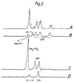
- FIG. 2 shows the volume-selection possible with the pulse sequence of the present invention applied to Sample B. In this instance only one dimensional slicing was carried out and the clean editing obtained is clearly seen.
- FIGS. 2(B) to (D) are the volume-selected subspectra obtained showing only the resonances from vials (3), (2) and (1), respectively. These were each obtained using only a z-direction slice and are the result of 128 scans averaged into 2048 data points with a sweep width of 5kHz.
- FIG. 2(A) is the parent spectrum obtained with no volume-selection applied.
- sample (1) which contains glucose-l-phosphate appearing only as a weakly discernible shoulder in the parent spectrum.
- Sample (3) actually contains a small amount of inorganic phosphate impurity, a result of chemical breakdown.
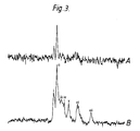
- FIG. 3 shows the complete spectrum of the leg with signals from both the tumour (sugar phosphate and inorganic phosphate) and muscle (ATP and PCr).
- FIG. 3(A) shows a two dimensional z and x sliced version of the present method where the tumour signal only is shown. It has been shown chemically that no PCr or ATP are present in tumours of this type.
- the present invention describes a new method for volume-selection in NMR spectroscopy. It is based on selective rf excitation of spins in the presence of a field gradient. In contrast to other approaches, the effect of chemical shift precession during the slice selection procedure is refocussed by a hard ⁇ pulse and refocussing delay. This yields a coherent signal along a known axis and consequently maximum signal strength is obtained. The coherence of this magnetization of interest is safe-guarded by placing it along the field axis during slice gradient fall times and then using a hard read pulse to acquire the high resolution signal. The technique can be extended to slice in all three dimensions and is based on the following principles.
- the Larmour frequency of nuclear spins varies with position. If a rf pulse with a narrow bandwidth is used to excite these spins, only those with Larmour frequency corresponding to the pulse will be excited. In this way, a slice of magnetization can be selected at the expense of all other magnetization. The width in space of this slice is dependent upon the frequency width of the pulse and the steepness of the gradient while the position of the slice is dependent on the central frequency of the pulse and the strength and direction of the applied gradient. It is limited by the requirement of hard homogenous pulses which cannot be applied by a surface coil. However, the good signal strength obtained compensates for this imposition particularly when one considers that the way is now clear for the present method to be applied to multipulse experiments with volume-selection in vivo .
- the present invention has certain important advantages. In contrast to ISIS, it is less dependent on an add/subtract routine wherein the weak signals in the presence of very much stronger background tend not to be detected efficiently because of the limited dynamic range of spectrometer pre-amps and analogue to digital converters. Further, it is well known that good signal cancellation is heavily dependent upon magnet stability - certainly a serious consideration in unlocked clinical NMR magnets. The present invention eliminates signals from outside of the slice of interest before detection and so it does not suffer from these concerns.
- Aue's VSE technique appears to lose signal because the signal is pulsed into the transverse plane but is in no way refocussed. This leads to serious signal loss.
- the present invention provides a new technique which gives excellent volume-selection along with suppression from outside of the volume of interest. It is particularly suited to extension to other multipulse experiments.
- a 1H- > 13C polarization transfer method is immediately obvious.
- the volume-selection could be performed in the proton regime where the spectral width demands are less demanding, and signal detection could then be carried out at the carbon frequency without the problems of a strong water signal and where signal overlap and homonuclear coupling is rarely a problem.
Landscapes
- Physics & Mathematics (AREA)
- High Energy & Nuclear Physics (AREA)
- General Physics & Mathematics (AREA)
- Optics & Photonics (AREA)
- Spectroscopy & Molecular Physics (AREA)
- Condensed Matter Physics & Semiconductors (AREA)
- Chemical & Material Sciences (AREA)
- Life Sciences & Earth Sciences (AREA)
- Health & Medical Sciences (AREA)
- Analytical Chemistry (AREA)
- Biochemistry (AREA)
- General Health & Medical Sciences (AREA)
- Immunology (AREA)
- Pathology (AREA)
- Magnetic Resonance Imaging Apparatus (AREA)
- Glass Compositions (AREA)
- Investigating Or Analysing Materials By Optical Means (AREA)
- Semiconductor Lasers (AREA)
Abstract
Description
- THIS INVENTION relates to improvements in volume-selected NMR spectroscopy.
- With the recent development of wholebody NMR spectroscopy, there is a need to find a technique to obtain high quality spectra from a particular volume in space. In this way, biochemical data may be obtained from one organ, or preferably from selected regions within such an organ, in a non-invasive manner. There have been a number of methods proposed to do this and these fall into three broad groups. They are:
- (1) The use of a pulse train which exploits the pulse inhomogeneity to select a volume within a sample by selecting only the region with a particular pulse angle;
- (2) The use of some form of field profile technique to produce a region of high magnetic field homogeneity within a larger region where the field changes sharply with position. (The result is that high resolution spectra are obtained from the region of interest whilst signals arising from the inhomogeneous region are so broad that they may be eliminated from spectra.)
In principle, methods (1) and (2) generally rely upon physical movement of the sample within the system to obtain signals from various regions. - (3) The third method is to use a combination of frequency selective radio-frequency pulses in the presence of magnetic field gradients to effect volume selection. It appears that this third approach may well be the most useful as it combines the versatility of complete software control of gradients and selective excitation pulses to produce volume-selection in all three directions. As well, the technique may be image encoded, the consequence being that the position of the selected volume is accurately known.
- For effective use in practice, strong signal-to-noise must be obtained from any particular technique. To do this, we must keep three objects in mind:
- (1) Maximum signal should be excited within the confines of the slice of choice;
- (2) Magnetization of interest should remain in the transverse plane for as little time as possible particularly while applied field gradients are present. The resultant loss of coherence leads to a reduction in signal strength; and
- (3) The spectra should be obtained after applied gradients have reached zero amplitude so that line widths are narrow and the chemical shifts displayed are those present independent of volume selection.
- These last two points combined mean it is preferable to have the magnetization aligned with the field axis while gradients are being changed.
- Some of the methods proposed previously for volume-selection include, DRESS, [Depth-resolved surface-coil in spectroscopy (DRESS) in vivo H, ³¹P, and ¹³C NMR, P. A. Bottomley, T. H. Foster and R. D. Darrow, J. Magn. Reson. 59, 338, (1984)]; VSE (H. Post, D. Ratzel and P. Brunner, West German Patent No. 3209263, 13th March, 1982, and also Volume-selective Excitation. A novel approach to topical NMR. W. P. Aue, S. Miller, T. A Cross and J. Seelig. J. Magn. Reson. 56, 350, (1984)]: and ISIS, [Image-selected in vivo Spectroscopy (ISIS). A new technique for spatially selective NMR Spectroscopy. R. J. Ordidge, A. Connelly and J. A. B. Lohman. J. Magn. Reson. 66, 283, (1986)]; These methods involve the application of narrow bandwidth radio-frequency excitation in the presence of a field gradient applied across the sample. The gradient is then switched off and the signal acquired. In principle, these methods can be extended to include slicing in all directions.
- The known methods can be summarised as follows:-
- (1) DRESS uses a selective rf pulse in the presence of an applied field gradient to excite a slice of magnetization. Gradient reversal is then used to form a spin-echo which refocusses the off-resonance effects of the pulse. The major drawback of this method is that the magnetization of interest lies in the transverse plane during refocussing and so is T₂ contrasted. Furthermore the acquired signals are either gradient broadened or reduced in intensity or both because of the need to allow gradient fall to occur before switching the receiver on. Volume selection is only one dimensional and the finite dimensions of the receiver coil are used to limit the volume in the plane of the slice.
- (2) ISIS is based on a phase inversion between signals in the slice of interest and those arising from outside. This is carried out by applying an inversion pulse tailored to the appropriate bandwidth in the presence of a gradient. The volume-selected spectrum is obtained by adding and subtracting appropriate combinations of phase inverted signals in the computer memory. The primary disadvantage of ISIS is that it relies on the ability of the computer to distinguish between the small signals within the volume of interest and the residual signal which will often be several orders of magnitude stronger. This, of course, is reliant upon the dynamic range of the computer memory as well as the spectrometer preamplifier and receiver systems.
- (3) SPARS (see, e.g., J. Magn. Reson. 67,148,1988) uses a refocussing pulse to form a spin-echo in the presence of an applied field gradient. A selective pulse of appropriate bandwidth is then applied to rotate the magnetization in the slice of interest back to the applied field direction following which the gradient is collapsed. This is carried out in all directions to yield the desired volume of interest which can then be read out with a single pulse. In this method the magnetization of interest suffers the effect of three gradient rises or falls while it is in the transverse plane for each direction of slicing which means any irrecoverable losses associated with gradient rises or falls are extreme. The time for refocussing is extended because gradient changes require finite time whereupon T₂ relaxation becomes important. The sensitivity of this technique is also dependent on efficient refocussing and on applying the selective pulse at precisely the correct moment. It appears that the phase evolution of the signal during this pulse also contributes to the weakening of signal strength.
- (4) VSE uses a composite pulse cluster in the presence of a field gradient for slice selection. It is of the form

- It is an object of the present invention to provide a method of volume-selected NMR spectroscopy which exhibits good volume-selection characteristics.
- It is a preferred object of the present invention that it can obtain NMR spectra from a small well defined region of space, without the need to change or move either the apparatus used to carry out the technique or the sample body.
- Other preferred objects of the present invention are to obtain this data with (1) high signal to noise, (2) little or no T₂ distortion, (3) low radio-frequency pulse power, (4) high spectral resolution, (5) complete volume selection within a single pass through the pulse sequence and, (6) the ability to locate the volume element of interest from information obtained from a NMR image.
- Just as in previously known methods for volume-selection, the present invention uses the relation between nuclear magnetic Larmour frequency and magnetic field strength. By applying a suitable rf pulse to the sample body in the presence of a field gradient superimposed upon the initial homogeneous magnetic field, certain spins can be excited. If the frequency of this excitation pulse and the strength of the gradient are known, the position and size of this volume element can be accurately known.
- In broad aspect, the present invention is directed to a method of volume-selected NMR spectroscopy where slice selection is facilitated by a pulse of selective band width applied in the presence of a field gradient. Before this gradient is switched off, a hard refocussing pulse and delay are used to refocus the signal phase roll which accompanies the soft pulse. This refocussed coherence is pulsed back to the z-axis before the gradient is collapsed.
- While this effect is of little consequence in high power broad band excitation, it becomes of paramount importance as pulse lengths become extended and is a key requirement when selective pulses are employed. In many cases, the phase shift noted above is linearly dependent on resonance off-set within the band width of the pulse. Thus it can be shown both theoretically and in practice, that a constant refocussing time τ can be used to refocus coherence across the bandwidth of the pulse. This time is dependent on the duration of the pulse. Thus the initial slice selection pulse cluster is preferably of the form

where, by convention,
- It now remains to record the signal in the absence of the gradient. To do this, the magnetization is preferably pulsed back to the z-axis so that the dephasing of transverse coherence which takes place in the presence of gradients has no effect on the excited slice. After a suitable time determined by the rate at which the gradients fall to zero, the magnetization of interest is again pulsed to the transverse plane and the signal may be acquired with the high resolution chemical shift information required for useful biochemical analysis.
- The pulse sequence then becomes

- To enable the invention to be fully understood, a preferred embodiment will now show how this technique can be extended to slice in further dimensions, the preferred embodiment being described with reference to the accompanying drawings in which:
- FIG. 1 shows the calculated variation in phase and intensity of the transverse magnetization generated following a 2.048ms sinc pulse as the amount off-resonance is varied. (The nutation angle on-resonance was

- FIG. 2 shows the volume-selective spectra recorded from the three vials discussed in the text. FIG. 2(A) shows the spectrum arising from all three vials using 128

- (i) Na₂HPO₄;
- (ii) G-l-P;
- (iii) PCr;
- (iv) α-ATP and β-ATP;
- (v) PPi;
- (vi) α-ATP and β-ADP;
- (vii) β-ATP.
- FIG. 3 shows the in vivo ³¹P spectrum determined from the leg of a Dark Agouti rat as per text. Spectrum (A) is the two-dimensional volume-selected spectrum following 1024 scans showing signals from the internal space of the tumour. Spectrum (B) is the normal spectrum from the whole leg following 128 scans using a

- (i) Sugar phosphate;
- (ii) Pi;
- (iii) Phosphodiesters;
- (iv) PCr;
- (v) α-ATP;
- (vi) α-ATP and NAD; and
- (vii) -ATP.
- All experiments were performed upon a Bruker MSL-100 spectroscopic system with a 2.35T 40cm bore magnet. A probe was built to accommodate small samples (≈40mm). It consisted of two coils both tuned to 40MHz. One coil, (diameter = 250mm) was built to accept high power (1.5kW) transmitted pulses. The second, a receiver coil, was constructed orthogonal to the original and had a smaller diameter (50mm) and so improved the filling factor for the smaller phantom and biological samples used in this work. Coupling between these coils was eliminated by careful positioning and so pulse power loss and signal reduction was minimized during pulse and receive experiments.
- Samples used in these experiments were as follows:
- (A) An aqueous sample of sodium phosphate (50mM);
- (B) Three sample vials (5mls) containing:
- (1) sodium pyrophosphate (Na₄P₂O₇) (120mM) and glucose-l-phosphate (G-l-P);
- (2) sodium phosphate (Na₂HPO₄) 50mM) and adenosine diphosphate (ADP) (160mM) (pH = 7.4); and
- (3) phosphocreatine (PCr) (180mM) and adenosine triphosphate (ATP) (200mM) (pH = 7.4).
- These were arranged so that the bottles were placed at the apices of an equilateral triangle; the long axes of the vials parallel with the y-axis of the magnet. In vivo volume-selection was carried out on a Dark Agouti female rat (200mg) with a tumour (5g) growing on one hind leg. Anaesthesia was by Ketalar (30mg) and Diazepam (0.1ml). The temperature within the probe head during experiments was 30°C.
- In all experiments discussed here, selective excitation was by a (sin x)/x (SINC) shaped pulse of duration 2ms. The pulse power for this pulse was set by observing the signal obtained from sample A after the pulse sequence ϑ(P,x)


- FIG. 1 shows the bandwidth and pulse distorttion associated with an unrefocussed sinc pulse applied to Sample A. Clearly, major signal loss will occur if further pulses are applied immediately to rotate this magnetization resulting from such a pulse to the z-axis because only those magnetization components which are orthogonal to the phase of the rf pulse are affected.
- FIG. 2 shows the volume-selection possible with the pulse sequence of the present invention applied to Sample B. In this instance only one dimensional slicing was carried out and the clean editing obtained is clearly seen. FIGS. 2(B) to (D) are the volume-selected subspectra obtained showing only the resonances from vials (3), (2) and (1), respectively. These were each obtained using only a z-direction slice and are the result of 128 scans averaged into 2048 data points with a sweep width of 5kHz. FIG. 2(A) is the parent spectrum obtained with no volume-selection applied. The good suppression of signals outside of any particular slice is demonstrated by the elimination of the sodium phosphate signal from the spectra of the other two samples, particularly sample (1) which contains glucose-l-phosphate appearing only as a weakly discernible shoulder in the parent spectrum. Sample (3) actually contains a small amount of inorganic phosphate impurity, a result of chemical breakdown.
- The effective volume-selection capability of the present method is again demonstrated in FIG. 3 where spectra of a rat carrying a tumour on its hind leg are shown. FIG. 3(B) shows the complete spectrum of the leg with signals from both the tumour (sugar phosphate and inorganic phosphate) and muscle (ATP and PCr). FIG. 3(A) shows a two dimensional z and x sliced version of the present method where the tumour signal only is shown. It has been shown chemically that no PCr or ATP are present in tumours of this type.
- The present invention describes a new method for volume-selection in NMR spectroscopy. It is based on selective rf excitation of spins in the presence of a field gradient. In contrast to other approaches, the effect of chemical shift precession during the slice selection procedure is refocussed by a hard π pulse and refocussing delay. This yields a coherent signal along a known axis and consequently maximum signal strength is obtained. The coherence of this magnetization of interest is safe-guarded by placing it along the field axis during slice gradient fall times and then using a hard read pulse to acquire the high resolution signal. The technique can be extended to slice in all three dimensions and is based on the following principles.
- In the presence of a field gradient, the Larmour frequency of nuclear spins varies with position. If a rf pulse with a narrow bandwidth is used to excite these spins, only those with Larmour frequency corresponding to the pulse will be excited. In this way, a slice of magnetization can be selected at the expense of all other magnetization. The width in space of this slice is dependent upon the frequency width of the pulse and the steepness of the gradient while the position of the slice is dependent on the central frequency of the pulse and the strength and direction of the applied gradient. It is limited by the requirement of hard homogenous pulses which cannot be applied by a surface coil. However, the good signal strength obtained compensates for this imposition particularly when one considers that the way is now clear for the present method to be applied to multipulse experiments with volume-selection in vivo.
- In comparison with other approaches, the present invention has certain important advantages. In contrast to ISIS, it is less dependent on an add/subtract routine wherein the weak signals in the presence of very much stronger background tend not to be detected efficiently because of the limited dynamic range of spectrometer pre-amps and analogue to digital converters. Further, it is well known that good signal cancellation is heavily dependent upon magnet stability - certainly a serious consideration in unlocked clinical NMR magnets. The present invention eliminates signals from outside of the slice of interest before detection and so it does not suffer from these concerns.
- Aue's VSE technique appears to lose signal because the signal is pulsed into the transverse plane but is in no way refocussed. This leads to serious signal loss.
- In summary, the present invention provides a new technique which gives excellent volume-selection along with suppression from outside of the volume of interest. It is particularly suited to extension to other multipulse experiments. In particular, it is noted that a ¹H- > ¹³C polarization transfer method is immediately obvious. Here the volume-selection could be performed in the proton regime where the spectral width demands are less demanding, and signal detection could then be carried out at the carbon frequency without the problems of a strong water signal and where signal overlap and homonuclear coupling is rarely a problem.
- The embodiments described are by way of illustrative examples only and various changes and modifications may be made thereto without departing from the scope of the present invention defined in the appended claims.
Claims (6)
- A method of volume-selective NMR spectroscopy comprising the steps of:(a) placing a sample body under investigation into a homogeneous magnetic field directed along the z-axis of a Cartesian coordinate system;(b) superimposing a magnetic field gradient (GS1) on the magnetic field;(c) applying a first rf pulse (π/2 (x)) of selected bandwidth for exciting nuclear spins in a selected volume element within the sample body to tip the nuclear spins from a first orientation of alignment with the z-axis into a second, different orientation with respect to the z-axis, the first rf pulse (π/2 (x)) being accompanied by a dephasing of the nuclear spins;(d) applying a hard second rf pulse (π [± y, ± x]) to the sample body for refocussing the nuclear spins;(e) allowing the nuclear spins to rephase during a first predetermined time interval (τ), the time interval (τ) being dependent on the duration of the first rf pulse (π/2 (x));(f) applying a hard third rf pulse (π/2 [± x]) to the sample body for tipping the refocussed nuclear spins back to the z-axis;(g) switching off the magnetic field gradient (GS1);(h) allowing the magnetic field gradient (GS1) to collapse during a second predetermined time interval (t₁);(i) applying a fourth rf pulse (π/2 [± x]) to the sample body for tipping the nuclear spins into an orientation of 90° to the z-axis; and(k) recording a nuclear magnetic resonance signal from the selected volume element.
- The method of claim 1, characterized in that:- the position of the volume element is determined by the frequency of the first rf pulse (π/2 (x)) as well as the strength and direction of the magnetic field gradient (GS1); and- the size of the volume element is determined by the bandwidth of the first pulse (π/2 (x)) as well as the strength and direction of the magnetic field gradient (GS1).
- The method of claim 1 or 2, characterized in that:- the first rf pulse (π/2 (x)) is a 90° pulse; and- the second rf pulse (π [± y, ± x]) is a non-selective 180° pulse.
- The method of claim 3, characterized in that the second rf pulse (π [± y, ± x]) follows an exorcycle during four cycles through the pulse sequence.
- The method of any of claims 1 through 4, characterized in that:- the first rf pulse (π/2 (x)) is a sinc pulse; and- the magnetic field gradient (GS1) is set such that after step (e) the nuclear spin magnetization is maximum in a plane transverse to the z-axis.
- The method of any of claims 1 through 5, characterized in that steps (b) through (g) are repeated for all three orthogonal directions of the magnetic field gradient (GS1).
Applications Claiming Priority (3)
| Application Number | Priority Date | Filing Date | Title |
|---|---|---|---|
| AU5608/86 | 1986-04-24 | ||
| AUPH560886 | 1986-04-24 | ||
| PCT/AU1987/000113 WO1987006700A1 (en) | 1986-04-24 | 1987-04-24 | Volume selected nmr spectroscopy |
Publications (3)
| Publication Number | Publication Date |
|---|---|
| EP0303604A1 EP0303604A1 (en) | 1989-02-22 |
| EP0303604A4 EP0303604A4 (en) | 1990-11-28 |
| EP0303604B1 true EP0303604B1 (en) | 1994-07-20 |
Family
ID=3771577
Family Applications (1)
| Application Number | Title | Priority Date | Filing Date |
|---|---|---|---|
| EP87902345A Expired - Lifetime EP0303604B1 (en) | 1986-04-24 | 1987-04-24 | Volume selected nmr spectroscopy |
Country Status (9)
| Country | Link |
|---|---|
| US (1) | US4945308A (en) |
| EP (1) | EP0303604B1 (en) |
| JP (1) | JPH01500057A (en) |
| KR (1) | KR880701374A (en) |
| AU (1) | AU590420B2 (en) |
| DE (1) | DE3750258T2 (en) |
| DK (1) | DK685087A (en) |
| WO (1) | WO1987006700A1 (en) |
| ZA (1) | ZA872922B (en) |
Families Citing this family (4)
| Publication number | Priority date | Publication date | Assignee | Title |
|---|---|---|---|---|
| GB8827833D0 (en) * | 1988-11-29 | 1988-12-29 | Briand J | Magnetic resonance signal acquisition methods |
| US5201311A (en) * | 1989-08-11 | 1993-04-13 | General Electric Company | Spatially-localized chemical-reaction-rate NMR spectroscopic imaging |
| US8970217B1 (en) | 2010-04-14 | 2015-03-03 | Hypres, Inc. | System and method for noise reduction in magnetic resonance imaging |
| US12497789B2 (en) | 2021-03-02 | 2025-12-16 | Werner Co. | Interlocking work platform system |
Family Cites Families (14)
| Publication number | Priority date | Publication date | Assignee | Title |
|---|---|---|---|---|
| US4480228A (en) * | 1982-10-15 | 1984-10-30 | General Electric Company | Selective volume method for performing localized NMR spectroscopy |
| US4486709A (en) * | 1982-11-22 | 1984-12-04 | Bendall Max R | Depth and refocusing pulses for use with inhomogeneous radiofrequency coils in nuclear magnetic resonance spectroscopy |
| US4549139A (en) * | 1983-06-03 | 1985-10-22 | General Electric Company | Method of accurate and rapid NMR imaging of computed T1 and spin density |
| EP0153303B1 (en) * | 1983-08-05 | 1989-04-12 | Oxford Research Systems Limited | Method and apparatus for obtaining n.m.r. spectra |
| GB8331501D0 (en) * | 1983-11-25 | 1984-01-04 | Picker Int Ltd | Nuclear magnetic resonance |
| US4629988A (en) * | 1984-07-02 | 1986-12-16 | General Electric Company | Method of imaging by depth-resolved surface coil spectroscopy |
| GB8419476D0 (en) * | 1984-07-31 | 1984-09-05 | Bendall M R | Obtaining nuclear magnetic resonance spectra |
| IL74942A (en) * | 1984-10-22 | 1988-11-30 | Univ Leland Stanford Junior | Flow measurement using nuclear magnetic resonance |
| JPS628747A (en) * | 1985-07-04 | 1987-01-16 | 株式会社東芝 | Magnetic resonance imaging apparatus |
| US4723030A (en) * | 1985-08-05 | 1988-02-02 | General Electric Company | Moderated reduction reactions for producing arylhydroxylamines |
| US4698592A (en) * | 1985-08-16 | 1987-10-06 | The Regents Of The University Of California | MRI of chemical shift spectra within limited inner volume |
| IL78240A (en) * | 1986-03-24 | 1989-09-10 | Elscint Ltd | Spatially localized spectroscopy |
| DE3769560D1 (en) * | 1986-08-18 | 1991-05-29 | Siemens Ag | METHOD FOR DETERMINING NUCLEAR MAGNETIC SPECTRES FROM SPACELY SELECTABLE AREAS OF AN EXAMINATION OBJECT. |
| US4733185A (en) * | 1987-06-01 | 1988-03-22 | General Electric Company | Methods for localization in NMR spectroscopy |
-
1987
- 1987-04-24 DE DE3750258T patent/DE3750258T2/en not_active Expired - Fee Related
- 1987-04-24 US US07/283,482 patent/US4945308A/en not_active Expired - Lifetime
- 1987-04-24 AU AU73502/87A patent/AU590420B2/en not_active Ceased
- 1987-04-24 KR KR1019870701208A patent/KR880701374A/en not_active Withdrawn
- 1987-04-24 WO PCT/AU1987/000113 patent/WO1987006700A1/en not_active Ceased
- 1987-04-24 ZA ZA872922A patent/ZA872922B/en unknown
- 1987-04-24 EP EP87902345A patent/EP0303604B1/en not_active Expired - Lifetime
- 1987-04-24 JP JP62502803A patent/JPH01500057A/en active Pending
- 1987-12-23 DK DK685087A patent/DK685087A/en not_active Application Discontinuation
Also Published As
| Publication number | Publication date |
|---|---|
| KR880701374A (en) | 1988-07-26 |
| US4945308A (en) | 1990-07-31 |
| JPH01500057A (en) | 1989-01-12 |
| EP0303604A1 (en) | 1989-02-22 |
| WO1987006700A1 (en) | 1987-11-05 |
| AU7350287A (en) | 1987-11-24 |
| DE3750258T2 (en) | 1994-10-27 |
| DK685087D0 (en) | 1987-12-23 |
| DK685087A (en) | 1987-12-23 |
| EP0303604A4 (en) | 1990-11-28 |
| ZA872922B (en) | 1987-10-15 |
| DE3750258D1 (en) | 1994-08-25 |
| AU590420B2 (en) | 1989-11-02 |
Similar Documents
| Publication | Publication Date | Title |
|---|---|---|
| EP0091008B1 (en) | Method of three-dimensional nmr imaging using selective excitation | |
| Bottomley et al. | Two‐dimensional spatially selective spin inversion and spin‐echo refocusing with a single nuclear magnetic resonance pulse | |
| US5652516A (en) | Spectroscopic magnetic resonance imaging using spiral trajectories | |
| EP0515197A1 (en) | Acquisition of multiple images in fast spin echo nmr scans | |
| US4068161A (en) | Gyromagnetic resonance spectroscopy employing spin echo spin-spin decoupling and two-dimensional spreading | |
| US4947119A (en) | Magnetic resonance imaging and spectroscopy methods | |
| Talagala et al. | Introduction to magnetic resonance imaging | |
| EP0347990B1 (en) | Method of and device for the volume-selective determination of an MR spectrum by means of selective polarization transfer pulse sequence | |
| EP0152879A2 (en) | Composite pulses for time reversal in NMR imaging | |
| US4678995A (en) | Apparatus and method for determining the presence of substances in a sample by NMR and producing an NMR image thereof | |
| WO1991002260A1 (en) | Time symmetric pulse to rotate magnetization vectors | |
| US8717022B2 (en) | Magnetic field gradient monitor apparatus and method | |
| EP0209374B1 (en) | Nmr phase encoding using phase-varying rf pulses | |
| US5064638A (en) | Simultaneous multinuclear magnetic resonance imaging and spectroscopy | |
| US5578921A (en) | Magnetic resonance imaging using three-dimensional spectral-spatial excitation | |
| US5077524A (en) | Gradient enhanced NMR correlation spectroscopy | |
| US4878021A (en) | Magnetic resonance spectroscopy studies of restricted volumes | |
| US4777439A (en) | Spatially localized spectroscopy | |
| EP0303604B1 (en) | Volume selected nmr spectroscopy | |
| US4855679A (en) | Magnetic resonance studies of restricted volumes | |
| US5317262A (en) | Single shot magnetic resonance method to measure diffusion, flow and/or motion | |
| US5168229A (en) | Multidimensional nmr spectroscopy using switched acquisition time gradients for multiple coherence transfer pathway detection | |
| EP0411710B1 (en) | 2-Quantum selective MR sequence for selectively determining a nuclear magnetisation of a metabolite | |
| US4743850A (en) | Method of mapping the nuclear magnetic properties of an object to be examined | |
| US5262723A (en) | Method and apparatus for obtaining pure-absorption two-dimensional lineshape data for multidimensional NMR spectroscopy using switched acquisition time gradients |
Legal Events
| Date | Code | Title | Description |
|---|---|---|---|
| PUAI | Public reference made under article 153(3) epc to a published international application that has entered the european phase |
Free format text: ORIGINAL CODE: 0009012 |
|
| 17P | Request for examination filed |
Effective date: 19881024 |
|
| AK | Designated contracting states |
Kind code of ref document: A1 Designated state(s): BE CH DE FR GB LI NL |
|
| A4 | Supplementary search report drawn up and despatched |
Effective date: 19901011 |
|
| AK | Designated contracting states |
Kind code of ref document: A4 Designated state(s): BE CH DE FR GB LI NL |
|
| 17Q | First examination report despatched |
Effective date: 19920925 |
|
| GRAA | (expected) grant |
Free format text: ORIGINAL CODE: 0009210 |
|
| AK | Designated contracting states |
Kind code of ref document: B1 Designated state(s): BE CH DE FR GB LI NL |
|
| PG25 | Lapsed in a contracting state [announced via postgrant information from national office to epo] |
Ref country code: BE Effective date: 19940720 |
|
| REF | Corresponds to: |
Ref document number: 3750258 Country of ref document: DE Date of ref document: 19940825 |
|
| ET | Fr: translation filed | ||
| PLBE | No opposition filed within time limit |
Free format text: ORIGINAL CODE: 0009261 |
|
| STAA | Information on the status of an ep patent application or granted ep patent |
Free format text: STATUS: NO OPPOSITION FILED WITHIN TIME LIMIT |
|
| 26N | No opposition filed | ||
| PGFP | Annual fee paid to national office [announced via postgrant information from national office to epo] |
Ref country code: GB Payment date: 20000313 Year of fee payment: 14 |
|
| PGFP | Annual fee paid to national office [announced via postgrant information from national office to epo] |
Ref country code: NL Payment date: 20000320 Year of fee payment: 14 |
|
| PGFP | Annual fee paid to national office [announced via postgrant information from national office to epo] |
Ref country code: CH Payment date: 20000323 Year of fee payment: 14 |
|
| PGFP | Annual fee paid to national office [announced via postgrant information from national office to epo] |
Ref country code: FR Payment date: 20000428 Year of fee payment: 14 |
|
| PGFP | Annual fee paid to national office [announced via postgrant information from national office to epo] |
Ref country code: DE Payment date: 20000527 Year of fee payment: 14 |
|
| PG25 | Lapsed in a contracting state [announced via postgrant information from national office to epo] |
Ref country code: GB Free format text: LAPSE BECAUSE OF NON-PAYMENT OF DUE FEES Effective date: 20010424 |
|
| PG25 | Lapsed in a contracting state [announced via postgrant information from national office to epo] |
Ref country code: FR Free format text: THE PATENT HAS BEEN ANNULLED BY A DECISION OF A NATIONAL AUTHORITY Effective date: 20010430 |
|
| PG25 | Lapsed in a contracting state [announced via postgrant information from national office to epo] |
Ref country code: LI Free format text: LAPSE BECAUSE OF NON-PAYMENT OF DUE FEES Effective date: 20010523 Ref country code: CH Free format text: LAPSE BECAUSE OF NON-PAYMENT OF DUE FEES Effective date: 20010523 |
|
| PG25 | Lapsed in a contracting state [announced via postgrant information from national office to epo] |
Ref country code: NL Free format text: LAPSE BECAUSE OF NON-PAYMENT OF DUE FEES Effective date: 20011101 |
|
| GBPC | Gb: european patent ceased through non-payment of renewal fee |
Effective date: 20010424 |
|
| REG | Reference to a national code |
Ref country code: CH Ref legal event code: PL |
|
| NLV4 | Nl: lapsed or anulled due to non-payment of the annual fee |
Effective date: 20011101 |
|
| PG25 | Lapsed in a contracting state [announced via postgrant information from national office to epo] |
Ref country code: DE Free format text: LAPSE BECAUSE OF NON-PAYMENT OF DUE FEES Effective date: 20020201 |
|
| REG | Reference to a national code |
Ref country code: FR Ref legal event code: ST |