EP0301600A2 - Method for detection of the presence of undesired microorganisms - Google Patents
Method for detection of the presence of undesired microorganisms Download PDFInfo
- Publication number
- EP0301600A2 EP0301600A2 EP88112452A EP88112452A EP0301600A2 EP 0301600 A2 EP0301600 A2 EP 0301600A2 EP 88112452 A EP88112452 A EP 88112452A EP 88112452 A EP88112452 A EP 88112452A EP 0301600 A2 EP0301600 A2 EP 0301600A2
- Authority
- EP
- European Patent Office
- Prior art keywords
- light
- change
- light absorption
- cultured
- living cells
- Prior art date
- Legal status (The legal status is an assumption and is not a legal conclusion. Google has not performed a legal analysis and makes no representation as to the accuracy of the status listed.)
- Granted
Links
Images


Classifications
-
- C—CHEMISTRY; METALLURGY
- C12—BIOCHEMISTRY; BEER; SPIRITS; WINE; VINEGAR; MICROBIOLOGY; ENZYMOLOGY; MUTATION OR GENETIC ENGINEERING
- C12Q—MEASURING OR TESTING PROCESSES INVOLVING ENZYMES, NUCLEIC ACIDS OR MICROORGANISMS; COMPOSITIONS OR TEST PAPERS THEREFOR; PROCESSES OF PREPARING SUCH COMPOSITIONS; CONDITION-RESPONSIVE CONTROL IN MICROBIOLOGICAL OR ENZYMOLOGICAL PROCESSES
- C12Q1/00—Measuring or testing processes involving enzymes, nucleic acids or microorganisms; Compositions therefor; Processes of preparing such compositions
- C12Q1/02—Measuring or testing processes involving enzymes, nucleic acids or microorganisms; Compositions therefor; Processes of preparing such compositions involving viable microorganisms
- C12Q1/04—Determining presence or kind of microorganism; Use of selective media for testing antibiotics or bacteriocides; Compositions containing a chemical indicator therefor
-
- C—CHEMISTRY; METALLURGY
- C12—BIOCHEMISTRY; BEER; SPIRITS; WINE; VINEGAR; MICROBIOLOGY; ENZYMOLOGY; MUTATION OR GENETIC ENGINEERING
- C12M—APPARATUS FOR ENZYMOLOGY OR MICROBIOLOGY; APPARATUS FOR CULTURING MICROORGANISMS FOR PRODUCING BIOMASS, FOR GROWING CELLS OR FOR OBTAINING FERMENTATION OR METABOLIC PRODUCTS, i.e. BIOREACTORS OR FERMENTERS
- C12M41/00—Means for regulation, monitoring, measurement or control, e.g. flow regulation
- C12M41/26—Means for regulation, monitoring, measurement or control, e.g. flow regulation of pH
-
- C—CHEMISTRY; METALLURGY
- C12—BIOCHEMISTRY; BEER; SPIRITS; WINE; VINEGAR; MICROBIOLOGY; ENZYMOLOGY; MUTATION OR GENETIC ENGINEERING
- C12M—APPARATUS FOR ENZYMOLOGY OR MICROBIOLOGY; APPARATUS FOR CULTURING MICROORGANISMS FOR PRODUCING BIOMASS, FOR GROWING CELLS OR FOR OBTAINING FERMENTATION OR METABOLIC PRODUCTS, i.e. BIOREACTORS OR FERMENTERS
- C12M41/00—Means for regulation, monitoring, measurement or control, e.g. flow regulation
- C12M41/30—Means for regulation, monitoring, measurement or control, e.g. flow regulation of concentration
- C12M41/36—Means for regulation, monitoring, measurement or control, e.g. flow regulation of concentration of biomass, e.g. colony counters or by turbidity measurements
-
- G—PHYSICS
- G01—MEASURING; TESTING
- G01N—INVESTIGATING OR ANALYSING MATERIALS BY DETERMINING THEIR CHEMICAL OR PHYSICAL PROPERTIES
- G01N15/00—Investigating characteristics of particles; Investigating permeability, pore-volume or surface-area of porous materials
- G01N15/10—Investigating individual particles
- G01N15/14—Optical investigation techniques, e.g. flow cytometry
- G01N15/1429—Signal processing
- G01N15/1433—Signal processing using image recognition
Definitions
- the present invention relates to a method for detection of the presence of undesired microorganisms contaminating a cell culture, which method permits early discovery of such microorganisms invaded the culture and prevention of the growth of microorganisms in the culture.
- a cell culture which contains a lot of nutritive substances is susceptible to contamination by undesired microorganisms floating in the air, such as incomplete bacteria and gram negative aerobic bacilli, especially when an exchange of a culture medium or an examination of the growth rate of the desired cells is conducted in the course of cultivation.
- the microorganisms invated the cell culture usually grow faster than the desired cells. Accordingly, it is often observed that the microorganisms which invated one well on a multiwell microplate spread to other wells, and it is not rare that all plates in an incubator are contaminated with undesired microorganisms.
- the contamination by microorganisms has long been visually examined using a microscope.
- such measure necessitates worker's manipulation and also gives additional chance of contamination by undesired microorganisms.
- the visual examination is not satisfactory in terms of accuracy or reliability on the detection of the microorganisms and also takes considerable time.
- the visual examination has an additional drawback, especially when a lot of microplates have to be examined, that the activity of the cultured cell tends to decline during the long-term exposure to outer-culturing atmosphere which is necessary for the examination.
- An essential object of the present invention is to provide a method of detection of undesired microorganisms in the cell culturing device which is able to detect the undesired microorganisms automatically and rapidly with a high accurate measurement result.
- a method of detecting undesired microorganisms when cells are cultured in a light permeable container characterrized by counting the number of cultured living cells appearing in a digital cultured cell picture which is obtained by a picture processing device for processing picture signals fed from a television camera coupled to a observation means provided for observing the container and change of the absorption of the visible light by the cultured solution.
- presence of the undesired microorganisms can be detected when the number of the living cells is small but the absorption of the visible light is increased compared to the absorption measured in a previous measurement. It is desired that the visible light is light of a wave length which is not absorbed by indicator contained in the culture solution.
- presence of the undesired microorganisms can be detected when the number of cultured cells are small and pH value of the culture solution is greatly decreased compared to the previous pH measurement value. It is desired to calculate the pH value by change of the absorption of the visible light.
- the change of light absorption includes more than two kinds of light including a first light having a wave length which is absorbed by the indicator contained in the culture solution and the a second light of wave length which is not absorbed by the indicator. It is further desired that at least one of the change of light absorption for detecting the change of pH value is due to the absorption change by phenol red contained in the culture solution.
- the picture data taken in a picture processing device is processed and the number of cultured living cells is measured by a computer.
- the change of the visible light absorption by the culture solution is measured or pH value of the culture solution is indirectly measured by the visible light absorption using indicator.
- the presence of the undesired microorganism can be detected based on the measured values in a semiquantitaive determination.
- the presence of the undesired microorganisms can be detected when the measured value of the living cells is small and the absorption of the visible light by the culture solution is greater than the absorption measured in the previous measurement or when the measured value of the living cells is small and pH value of the culture solution is greatly decreased compared to the pH value measured in the previous measurement.
- the number of the measured value of the living cells is small.
- the way of measuring of the number of the living cells there may be employed such a method that uses the difference of the brightness which is a property of the living cells or another method of performing a process using a spatial filter or matched filter as shown in the Japanese patent publication 60-256154.
- the visible light of a wave length which is not absorbed by the indicator contained in the culture solution is decreased in a light permeability only by the absorption and scattering by the dead cells and the micro plates, the light absorption is small. If the undesired organisms are mixed and grow under the condition mentioned above, the absorption and scattering of light by the undesired organisms increase despite that the measured number of the cultured living cells is small. As the result, when the measured number of the living cells is small and the change of the absorption of the visible light is largely greater than the light absorption measured in the last measurement period in the past, the presence of the undesired organisms can be detected.
- the presence of the undesired microorganisms can be detected by measuring the light absorption using not only such light of wave length which is not absorbed by the indicator contained in the culture solution but also such light of wave length which is absorbable in the indicator.
- the light absorption by the indicator contained in the culture solution may change by the change of pH value of the culture solution.
- the latter method of detecting the presence of the undesired microorganisms using the light of the wave length which is absorbable by the indicator possesses a problem. Therefore, according to the present invention, pH is measured without contacting the culture solution.
- the method of detecting the pH value without contacting the culture solution it is desired to detect the change of the light absorption by the indicator such as phenol red contained in the culture solution for calculations the pH value.
- phenol red with such a low concentration that does not harm the cells is contained in the culture solution.
- the phenol red with such low concentration it is possible to detect pH value of the culture solution by using any one or both of light absorption peaks at 430 to 440 nm (nano meter) wave lengths and at 560 nm.
- the pH value In order to calculate the pH value accurately, it is necessary to calculate the pH value excluding either the light absorption and scattering due to the stain of the container such as a microplate, a dish and a culture bottle and the light absorption and scattering due to the living cells and dead cells existing in the container. For this purpose, it is desired to calculate the pH value by subtracting the light absorption at the wave length of 650 nm from the light absorptions at the wave length of 430 to 440 nm and the wave length of 560 nm taking a ratio thereof. According to this method, it is possible to calculate the accurate pH value eliminating the undesired effects of the light absorption and scattering other than the change of pH value.
- the method of the present invention it is possible to detect automatically the invasion of the undesired microorganisms in a very short time with a high accuracy compared to the conventional manually visual measurement using a microscope.
- a microscope 2 to observe a well 1 of a microplate having 96 wells (referred to as 96 well plate hereinafter) from below.
- Each of the well is light permeable so that the microscope can view entire area of the bottom of the well 1 to view the contents contained in the well 1.
- the microscope 2 is coupled to a CCD camera for generating electric signals representing the image viewed by the microscope for every pixels of CCD camera in a known manner.
- the CCD camera 3 is coupled to a image processing device 5 the output of which are coupled to a computer 6 and a monitor television 4.
- the image signal processor 5 digitize the image signals of the CCD camera 2 and processes the image signals to produce the data showing the living cells in the well 1 by using matched filter.
- Fig. 2 An example of the matched filter is shown in Fig. 2.
- the process of generating the data representing the living cells is shown in a flow chart shown in Fig. 4.
- the image signals from the CCD camera 2 are taken in the image processing device 3.
- the image signals are digitized and stored in a memory (not shown) in the step S2.
- the digital image signals contained in 7 x 7 picture cells are subjected to a matching process using the matched filter or spatial filter for a convolution processing in a known manner.
- This process is generally expressed by the following equation. wherein Ax,y : digital brightness at x and y position of the input image A′x,y : digital brightness at x and y position after filtering f(i,j): coefficient corresponding to each pixel of the filter Cl : coefficient
- the number of the living cells is calculated in the step S6 using the following equation.
- C2 the number of the pixels which one living cell occupies.
- the computer 6 performs the processing as shown in Fig. 5 based on the light value signal obtained by the photo transistor 7 and converted in a digital form by the A/D converter 8.
- the computer takes the incident light value I0 when the no sample is present in the well 1.
- the computer 6 takes, from the output of the A/D converter 8, the light value I1 which is the amount of the light passed the sample in the well 1.
- the light absorption is calculated in the step S23 based on the equation" - log I1/I0". The measurement and calculation are repeated every predetermined period.
- the calculated light absorption obtained in the present period is calculated with the light absorption calculated in the last period in the past in the step S24 so as to detect whether or not the change of the light absorption occurs.
- the computer 6 judges there are undesired organisms in the well.
- the matching process for taking out the living cells may be made by known manner but there may be used a matching filter arrangement in the form of a hard wear.
- Myeloma cells Sp2/O contained in DULBECCO MEM culture solution with 103/ml were poured in every well of a 96 well microplate and were cultured in a CO2 incubator for a day. Subsequently the microplate was taken out of the CO2 incubator and the number of the culture living cells and the light absorption change were measured.
- the number of the cultured living cells were counted in such a manner that image signals obtained by a CCD camera 3 coupled to a microscope 2 for observing the cells in a well 1 were input into an image signal processor 5 (PIAS-1 manufactured by Nippon PC Systems) and were digitized. The image obtained by the CCD camera 3 were also input in a monitor television receiver 4.
- image signal processor 5 PIAS-1 manufactured by Nippon PC Systems
- Magnification was such that as shown in Fig. 2, the contours or out line of one cell inscribed with the peripheral edges of a spatial filter made of 7 x 7 matrix of pixels.
- the size of the single pixel was 1,8 ⁇ m X 1.8 ⁇ m.
- the image signals obtained by the CCD camera were digitized with 64 gradation gray scales.
- the pattern of the spatial filter for the matching was set as shown in Fig. 2, In Fig. 2, 0 shows the pixel area in which the image of the cell is not present and 1 shows the pixel area in which the brightness of the living cell is high and -1 shows the pixel area in which the brightness of the living cell is low.
- N N p /Nx (1) wherein N is the number of the living cells N p is the number of pixels in which the living cells are present and Nx is the number of the picture elements occupied by single cell.
- phenol red phenolsulhophtalein
- pH value was calculated using the equation 2. wherein Abs558, Abs650 and Abs430 are the light absorption of 558 nm, 650 nm and 430 nm wave lights.
- the micro plate containing the living cells was put in air for one day. Then the micro plate was taken in the incubator to culture for one day. Subsequently, the number of the living cells, light absorption of the light of 650 nm wave length and pH were again measured.
- the living cells were observed only in the NO. 1 well.
- the number of the cultured living cells was small and the visible light absorption was largely increased and it was recognized that undesired microorganisms grew.
- the NO. 3 well in which the number of the cultured living cells was small and pH calculated from the change of the visible light absorption was greatly decreased, it was recognized that the gram negative aerobic bacilli grew.
- the present invention it is possible to detect the presence of the undesired microorganisms in the culture solution by the change of the visible light absorption and the number of the cultured living cells which is obtained by the digital cultured cell image taken in the image processor through the television camera coupled to the observation means such as microscope when the cells grow in a container which allows to pass the visible light.
- the observation means such as microscope when the cells grow in a container which allows to pass the visible light.
Landscapes
- Chemical & Material Sciences (AREA)
- Health & Medical Sciences (AREA)
- Life Sciences & Earth Sciences (AREA)
- Engineering & Computer Science (AREA)
- Organic Chemistry (AREA)
- Zoology (AREA)
- Wood Science & Technology (AREA)
- Bioinformatics & Cheminformatics (AREA)
- Analytical Chemistry (AREA)
- General Health & Medical Sciences (AREA)
- Biochemistry (AREA)
- Biotechnology (AREA)
- Genetics & Genomics (AREA)
- General Engineering & Computer Science (AREA)
- Microbiology (AREA)
- Proteomics, Peptides & Aminoacids (AREA)
- Sustainable Development (AREA)
- Biomedical Technology (AREA)
- Physics & Mathematics (AREA)
- Immunology (AREA)
- Molecular Biology (AREA)
- Biophysics (AREA)
- Toxicology (AREA)
- Signal Processing (AREA)
- Dispersion Chemistry (AREA)
- General Physics & Mathematics (AREA)
- Pathology (AREA)
- Measuring Or Testing Involving Enzymes Or Micro-Organisms (AREA)
- Apparatus Associated With Microorganisms And Enzymes (AREA)
- Investigating Or Analysing Materials By Optical Means (AREA)
Abstract
Description
- The present invention relates to a method for detection of the presence of undesired microorganisms contaminating a cell culture, which method permits early discovery of such microorganisms invaded the culture and prevention of the growth of microorganisms in the culture.
- A cell culture which contains a lot of nutritive substances is susceptible to contamination by undesired microorganisms floating in the air, such as incomplete bacteria and gram negative aerobic bacilli, especially when an exchange of a culture medium or an examination of the growth rate of the desired cells is conducted in the course of cultivation. The microorganisms invated the cell culture usually grow faster than the desired cells. Accordingly, it is often observed that the microorganisms which invated one well on a multiwell microplate spread to other wells, and it is not rare that all plates in an incubator are contaminated with undesired microorganisms.
- It is necessary, therefore, to examine the presence or absence of contamination of the cell culture by microorganisms as early as possible so that the contaminated well or plate may be sterilized or discarded to prevent the microorganisms from further spreading.
- The contamination by microorganisms has long been visually examined using a microscope. However, such measure necessitates worker's manipulation and also gives additional chance of contamination by undesired microorganisms. In addition, the visual examination is not satisfactory in terms of accuracy or reliability on the detection of the microorganisms and also takes considerable time. Furthermore, the visual examination has an additional drawback, especially when a lot of microplates have to be examined, that the activity of the cultured cell tends to decline during the long-term exposure to outer-culturing atmosphere which is necessary for the examination.
- An essential object of the present invention is to provide a method of detection of undesired microorganisms in the cell culturing device which is able to detect the undesired microorganisms automatically and rapidly with a high accurate measurement result.
- According to the present invention, there is provided a method of detecting undesired microorganisms when cells are cultured in a light permeable container, characterrized by counting the number of cultured living cells appearing in a digital cultured cell picture which is obtained by a picture processing device for processing picture signals fed from a television camera coupled to a observation means provided for observing the container and change of the absorption of the visible light by the cultured solution.
- According to the present invention, presence of the undesired microorganisms can be detected when the number of the living cells is small but the absorption of the visible light is increased compared to the absorption measured in a previous measurement. It is desired that the visible light is light of a wave length which is not absorbed by indicator contained in the culture solution.
- According to another feature of the present invention, presence of the undesired microorganisms can be detected when the number of cultured cells are small and pH value of the culture solution is greatly decreased compared to the previous pH measurement value. It is desired to calculate the pH value by change of the absorption of the visible light.
- It is desired that the change of light absorption includes more than two kinds of light including a first light having a wave length which is absorbed by the indicator contained in the culture solution and the a second light of wave length which is not absorbed by the indicator. It is further desired that at least one of the change of light absorption for detecting the change of pH value is due to the absorption change by phenol red contained in the culture solution.
- According to one feature of the present invention, the picture data taken in a picture processing device is processed and the number of cultured living cells is measured by a computer. In addition, the change of the visible light absorption by the culture solution is measured or pH value of the culture solution is indirectly measured by the visible light absorption using indicator. The presence of the undesired microorganism can be detected based on the measured values in a semiquantitaive determination.
- According to the present invention, the presence of the undesired microorganisms can be detected when the measured value of the living cells is small and the absorption of the visible light by the culture solution is greater than the absorption measured in the previous measurement or when the measured value of the living cells is small and pH value of the culture solution is greatly decreased compared to the pH value measured in the previous measurement.
- In case where the cells have not yet cultured, the number of the measured value of the living cells is small. In this condition, as the way of measuring of the number of the living cells, there may be employed such a method that uses the difference of the brightness which is a property of the living cells or another method of performing a process using a spatial filter or matched filter as shown in the Japanese patent publication 60-256154.
- Under such a condition that the living cells are scarcely present, since the visible light of a wave length which is not absorbed by the indicator contained in the culture solution is decreased in a light permeability only by the absorption and scattering by the dead cells and the micro plates, the light absorption is small. If the undesired organisms are mixed and grow under the condition mentioned above, the absorption and scattering of light by the undesired organisms increase despite that the measured number of the cultured living cells is small. As the result, when the measured number of the living cells is small and the change of the absorption of the visible light is largely greater than the light absorption measured in the last measurement period in the past, the presence of the undesired organisms can be detected.
- According to the method of the present invention, the presence of the undesired microorganisms can be detected by measuring the light absorption using not only such light of wave length which is not absorbed by the indicator contained in the culture solution but also such light of wave length which is absorbable in the indicator. Besides, the light absorption by the indicator contained in the culture solution may change by the change of pH value of the culture solution. Accordingly, the latter method of detecting the presence of the undesired microorganisms using the light of the wave length which is absorbable by the indicator possesses a problem. Therefore, according to the present invention, pH is measured without contacting the culture solution. As the method of detecting the pH value without contacting the culture solution, it is desired to detect the change of the light absorption by the indicator such as phenol red contained in the culture solution for calculations the pH value.
- In order to detect the change of pH value, phenol red with such a low concentration that does not harm the cells is contained in the culture solution. With the phenol red with such low concentration, it is possible to detect pH value of the culture solution by using any one or both of light absorption peaks at 430 to 440 nm (nano meter) wave lengths and at 560 nm.
- In order to calculate the pH value accurately, it is necessary to calculate the pH value excluding either the light absorption and scattering due to the stain of the container such as a microplate, a dish and a culture bottle and the light absorption and scattering due to the living cells and dead cells existing in the container. For this purpose, it is desired to calculate the pH value by subtracting the light absorption at the wave length of 650 nm from the light absorptions at the wave length of 430 to 440 nm and the wave length of 560 nm taking a ratio thereof. According to this method, it is possible to calculate the accurate pH value eliminating the undesired effects of the light absorption and scattering other than the change of pH value.
- As mentioned above, according to the method of the present invention, it is possible to detect automatically the invasion of the undesired microorganisms in a very short time with a high accuracy compared to the conventional manually visual measurement using a microscope.
-
- Fig. 1 is a schematic diagram showing an embodiment of a system for detecting undesired microorganisms according to the present invention,
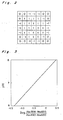
- Fig. 2 is a schematic diagram showing an example of a spatial filter used in the system shown in Fig. 1,
- Fig. 3 is a graph showing relations between the change of light absorption in the sample and pH value, and
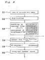
- Figs. 4 and 5 are flow charts showing essential parts of the operation of the system shown in Fig. 1.
- Referring to Fig. 1, there is provided a
microscope 2 to observe awell 1 of a microplate having 96 wells (referred to as 96 well plate hereinafter) from below. Each of the well is light permeable so that the microscope can view entire area of the bottom of thewell 1 to view the contents contained in thewell 1. Themicroscope 2 is coupled to a CCD camera for generating electric signals representing the image viewed by the microscope for every pixels of CCD camera in a known manner. TheCCD camera 3 is coupled to aimage processing device 5 the output of which are coupled to acomputer 6 and amonitor television 4. Theimage signal processor 5 digitize the image signals of theCCD camera 2 and processes the image signals to produce the data showing the living cells in thewell 1 by using matched filter. An example of the matched filter is shown in Fig. 2. The process of generating the data representing the living cells is shown in a flow chart shown in Fig. 4. In the step S1 the image signals from theCCD camera 2 are taken in theimage processing device 3. The image signals are digitized and stored in a memory (not shown) in the step S2. The digital image signals contained in 7 x 7 picture cells are subjected to a matching process using the matched filter or spatial filter for a convolution processing in a known manner. This process is generally expressed by the following equation.
Ax,y : digital brightness at x and y position of the input image
A′x,y : digital brightness at x and y position after filtering
f(i,j): coefficient corresponding to each pixel of the filter
Cl : coefficient - The image data passed or matched with the matched filter in the step S3 which representing the culture cell is compared with a threshold value T1 in terms of the brightness of the cell for selecting the living cell so that Ax,y is converted into Bx, y of a binary digitized form (Bx, y = 1 or 0). Then *8-neighbor expansion process is performed for each picture cell in the step S5.
- Then the number of the living cells is calculated in the step S6 using the following equation.

- The above mentioned processing is performed in the
image processor 5 andcomputer 6. - In order to calculate the change of the light absorption, the
computer 6 performs the processing as shown in Fig. 5 based on the light value signal obtained by thephoto transistor 7 and converted in a digital form by the A/D converter 8. - Referring to Fig. 5, In the step S20, the computer takes the incident light value I₀ when the no sample is present in the
well 1. After the sample is inserted in thewell 1 in the step S21, thecomputer 6 takes, from the output of the A/D converter 8, the light value I₁ which is the amount of the light passed the sample in thewell 1. Then the light absorption is calculated in the step S23 based on the equation" - log I₁/I₀". The measurement and calculation are repeated every predetermined period. The calculated light absorption obtained in the present period is calculated with the light absorption calculated in the last period in the past in the step S24 so as to detect whether or not the change of the light absorption occurs. - In case the number of the living cells calculated in the step S6 is smaller than a predetermined value and the change of the light absorption is larger than a further predetermined value, the
computer 6 judges there are undesired organisms in the well. - The details of the process of the counting of the living cells are disclosed in the PCT /JP 87/ 00750 and the disclosure thereof is cited as the reference.
- The matching process for taking out the living cells may be made by known manner but there may be used a matching filter arrangement in the form of a hard wear.
-
- Myeloma cells Sp2/O contained in DULBECCO MEM culture solution with 10³/ml were poured in every well of a 96 well microplate and were cultured in a CO₂ incubator for a day. Subsequently the microplate was taken out of the CO₂ incubator and the number of the culture living cells and the light absorption change were measured.
- The number of the cultured living cells were counted in such a manner that image signals obtained by a
CCD camera 3 coupled to amicroscope 2 for observing the cells in awell 1 were input into an image signal processor 5 (PIAS-1 manufactured by Nippon PC Systems) and were digitized. The image obtained by theCCD camera 3 were also input in amonitor television receiver 4. - Magnification was such that as shown in Fig. 2, the contours or out line of one cell inscribed with the peripheral edges of a spatial filter made of 7 x 7 matrix of pixels. The size of the single pixel was 1,8 µm X 1.8 µm. The image signals obtained by the CCD camera were digitized with 64 gradation gray scales. The pattern of the spatial filter for the matching was set as shown in Fig. 2, In Fig. 2, 0 shows the pixel area in which the image of the cell is not present and 1 shows the pixel area in which the brightness of the living cell is high and -1 shows the pixel area in which the brightness of the living cell is low.
- After the image signals were converted into a binary digitized form, the number of the living cells were measured by a
computer 6 according to an equation (1).
N = Np/Nx (1)
wherein N is the number of the living cells Np is the number of pixels in which the living cells are present and Nx is the number of the picture elements occupied by single cell. - In order to measure the change of the light absorption, as the test agent, phenol red (phenolsulhophtalein) was used with such a low concentration that did not harm the living cells. The light absorption was measured with respect to either the lights of 430 nm and 558 nm wave lengths at which light absorption peaks occurred and the light of 650 nm wave length at which the absorption by the phenol red did not occur. Then pH value was calculated using the
equation 2.
- pH was calculated according to the linear relation shown in Fig. 3.
- After the measurements of the number of the living cells, the light absorption of the 650 nm wave length and pH value, the micro plate containing the living cells was put in air for one day. Then the micro plate was taken in the incubator to culture for one day. Subsequently, the number of the living cells, light absorption of the light of 650 nm wave length and pH were again measured.
- The result of the measurement are shown in the table 1.

- By the observation of the wells by a microscope, the living cells were observed only in the NO. 1 well. In the NO. 2 well, the number of the cultured living cells was small and the visible light absorption was largely increased and it was recognized that undesired microorganisms grew. In the NO. 3 well, in which the number of the cultured living cells was small and pH calculated from the change of the visible light absorption was greatly decreased, it was recognized that the gram negative aerobic bacilli grew. By these results, it is apparent that contaminant by the undesired microorganisms can be detected by the method according to the present invention.
- As apparent from the foregoing, according to the present invention, it is possible to detect the presence of the undesired microorganisms in the culture solution by the change of the visible light absorption and the number of the cultured living cells which is obtained by the digital cultured cell image taken in the image processor through the television camera coupled to the observation means such as microscope when the cells grow in a container which allows to pass the visible light. According to the method of the present invention, since the invasion by the undesired microorganisms can be automatically detected in a short period without contacting, invasion of the undesired microorganisms can be decreased. It is possible to decrease the bad effect of the undesired microorganisms to the desired organisms by shortening the period of time during which the cells are put out of the culturing atmosphere. In particular, it is advantageous that when a large amount of the cell containers must be controlled, the invasion of the undesired microorganisms can be easily detected with a high accuracy in a short period.
Claims (8)
a step of radiating at least one kind light to a light permeable container which contains organisms;
a step of monitoring image of the microorganisms in the container by receiving the light passed through said container by a television camera attached to a microscope;
a step of converting the image information obtained by said camera into digital information representing the picture of cultured living cell contained in said container;
a step of counting the number of the living cells;
a fifth step of detecting change of light absorption of the light passed through said container; and
a step of judging whether the count value of the living cells is smaller than a predetermined value and the change of the light absorption is greater than a further predetermined value for detecting presence of undesired microorganisms in said container.
Applications Claiming Priority (2)
| Application Number | Priority Date | Filing Date | Title |
|---|---|---|---|
| JP62192023A JPS6435347A (en) | 1987-07-31 | 1987-07-31 | Detection of intrusion of various bacteria |
| JP192023/87 | 1987-07-31 |
Publications (3)
| Publication Number | Publication Date |
|---|---|
| EP0301600A2 true EP0301600A2 (en) | 1989-02-01 |
| EP0301600A3 EP0301600A3 (en) | 1990-07-18 |
| EP0301600B1 EP0301600B1 (en) | 1993-04-28 |
Family
ID=16284312
Family Applications (1)
| Application Number | Title | Priority Date | Filing Date |
|---|---|---|---|
| EP88112452A Expired - Lifetime EP0301600B1 (en) | 1987-07-31 | 1988-08-01 | Method for detection of the presence of undesired microorganisms |
Country Status (5)
| Country | Link |
|---|---|
| US (1) | US5003611A (en) |
| EP (1) | EP0301600B1 (en) |
| JP (1) | JPS6435347A (en) |
| CA (1) | CA1296081C (en) |
| DE (1) | DE3880573T2 (en) |
Cited By (14)
| Publication number | Priority date | Publication date | Assignee | Title |
|---|---|---|---|---|
| EP0590775A1 (en) * | 1992-09-01 | 1994-04-06 | Becton, Dickinson and Company | Method and apparatus for detecting microorganisms |
| DE4308486A1 (en) * | 1993-03-17 | 1994-09-22 | Grundig Emv | Arrangement for measuring turbidity |
| WO1994026870A1 (en) * | 1993-05-14 | 1994-11-24 | Minnesota Mining And Manufacturing Company | Method for rapid quantification of microorganism growth |
| US5403722A (en) * | 1992-07-13 | 1995-04-04 | Minnesota Mining And Manufacturing Company | Technique to count objects in a scanned image |
| EP0731951A4 (en) * | 1993-02-26 | 1997-09-17 | E Y Lab Inc | Optical specimen analysis system and method |
| US5694478A (en) * | 1994-12-15 | 1997-12-02 | Minnesota Mining And Manufacturing Company | Method and apparatus for detecting and identifying microbial colonies |
| US5744322A (en) * | 1993-12-17 | 1998-04-28 | Minnesota Mining And Manufacturing Company | Automated incubating and imaging system for a disposable microorganism culturing device and method of use |
| ES2137879A1 (en) * | 1997-12-02 | 1999-12-16 | Francisco Soria Melguizo S A | ANALYZING SYSTEM OF IMAGES PRODUCED BY BACTERIAL REACTIONS. |
| WO2009111298A3 (en) * | 2008-03-04 | 2010-01-07 | 3M Innovative Properties Company | Processing of biological growth media based on measured manufacturing characteristics |
| US7738689B2 (en) | 2003-09-05 | 2010-06-15 | 3M Innovative Properties Company | Counting biological agents on biological growth plates |
| US7865008B2 (en) | 2003-09-04 | 2011-01-04 | 3M Innovative Properties Company | Biological growth plate scanner with automated intake |
| US7901933B2 (en) | 2002-11-27 | 2011-03-08 | 3M Innovative Properties Company | Methods of processing a biological growth plate in a biological growth plate scanner |
| EP2597147A1 (en) | 2007-07-09 | 2013-05-29 | 3M Innovative Properties Company of 3M Center | Method for detecting microorganisms |
| CN105671123A (en) * | 2016-01-19 | 2016-06-15 | 西南交通大学 | Method for counting bacteria in liquid |
Families Citing this family (11)
| Publication number | Priority date | Publication date | Assignee | Title |
|---|---|---|---|---|
| US5229849A (en) * | 1984-09-17 | 1993-07-20 | University Of Delaware | Laser doppler spectrometer for the statistical study of the behavior of microscopic organisms |
| US5290701A (en) * | 1991-08-28 | 1994-03-01 | Wilkins Judd R | Microbial detection system and process |
| BR9809154B1 (en) * | 1997-05-23 | 2012-09-04 | diagnostic microbiological testing apparatus and system. | |
| AU3212400A (en) * | 1999-01-22 | 2000-08-07 | Human Genome Sciences, Inc. | Metalloproteinase adam 22 |
| US6498862B1 (en) | 1999-05-18 | 2002-12-24 | The United States Of America As Represented By The Administrator Of The National Aeronautics And Space Administration | Evaluation of biofilms and the effects of biocides thereon |
| DE10158964A1 (en) * | 2001-11-30 | 2003-08-07 | Digi Plan Gmbh Planungsgesells | Detecting the presence of living cells and the like, in a solid, liquid or gas medium, comprises isolated cells from a sample, cultivating them in a chamber under light, and taking optical images from a sensor for evaluation |
| US20040101954A1 (en) | 2002-11-27 | 2004-05-27 | Graessle Josef A. | Back side plate illumination for biological growth plate scanner |
| US20040102903A1 (en) | 2002-11-27 | 2004-05-27 | Graessle Josef A. | Biological growth plate scanner |
| CN101061232A (en) | 2004-06-23 | 2007-10-24 | 罗斯玛丽·凯瑟琳·卡梅伦·莎平 | Improvement of Microorganism Detecting Instrument and Its Application Method |
| WO2009111301A1 (en) | 2008-03-04 | 2009-09-11 | 3M Innovative Properties Company | Information management in automated processing of biological growth media |
| US20220404286A1 (en) * | 2019-10-04 | 2022-12-22 | Kyocera Corporation | Ph measurement method and ph measurement device |
Family Cites Families (9)
| Publication number | Priority date | Publication date | Assignee | Title |
|---|---|---|---|---|
| US3773426A (en) * | 1972-02-22 | 1973-11-20 | Department Of Health Educ Welf | Bacterial growth detector |
| US4519087A (en) * | 1983-06-20 | 1985-05-21 | International Remote Imaging Systems, Inc. | Method of determining the diagnostic significance of the content of a volume of biological sample containing particles |
| JPH07114685B2 (en) * | 1983-07-28 | 1995-12-13 | 株式会社日立製作所 | Sewage treatment equipment |
| JPS60210997A (en) * | 1984-04-04 | 1985-10-23 | Mitsubishi Chem Ind Ltd | Method of counting number of particle of microorganism |
| JPH07114691B2 (en) * | 1984-12-27 | 1995-12-13 | 住友電気工業株式会社 | Cell culture method |
| EP0189599B1 (en) * | 1984-12-27 | 1989-03-22 | Sumitomo Electric Industries Limited | Method and apparatus for incubating cells |
| US4724215A (en) * | 1985-02-27 | 1988-02-09 | Sherwood Medical Company | Automated microbiological testing apparatus and method |
| JPH0634754B2 (en) * | 1985-11-14 | 1994-05-11 | 住友電気工業株式会社 | Method for measuring pH of cell culture medium |
| EP0257660B1 (en) * | 1986-08-29 | 1993-01-27 | FUJIREBIO KABUSHIKI KAISHA also trading as FUJIREBIO INC. | Method and apparatus for judging agglutination |
-
1987
- 1987-07-31 JP JP62192023A patent/JPS6435347A/en active Pending
-
1988
- 1988-08-01 US US07/226,775 patent/US5003611A/en not_active Expired - Fee Related
- 1988-08-01 DE DE88112452T patent/DE3880573T2/en not_active Expired - Fee Related
- 1988-08-01 EP EP88112452A patent/EP0301600B1/en not_active Expired - Lifetime
- 1988-08-02 CA CA000573629A patent/CA1296081C/en not_active Expired - Lifetime
Cited By (23)
| Publication number | Priority date | Publication date | Assignee | Title |
|---|---|---|---|---|
| US5403722A (en) * | 1992-07-13 | 1995-04-04 | Minnesota Mining And Manufacturing Company | Technique to count objects in a scanned image |
| EP0590775A1 (en) * | 1992-09-01 | 1994-04-06 | Becton, Dickinson and Company | Method and apparatus for detecting microorganisms |
| US5432061A (en) * | 1992-09-01 | 1995-07-11 | Becton Dickinson And Company | Method and apparatus for detecting microorganisms |
| EP0731951A4 (en) * | 1993-02-26 | 1997-09-17 | E Y Lab Inc | Optical specimen analysis system and method |
| DE4308486A1 (en) * | 1993-03-17 | 1994-09-22 | Grundig Emv | Arrangement for measuring turbidity |
| WO1994026870A1 (en) * | 1993-05-14 | 1994-11-24 | Minnesota Mining And Manufacturing Company | Method for rapid quantification of microorganism growth |
| US5510246A (en) * | 1993-05-14 | 1996-04-23 | Minnesota Mining And Manufacturing Company | Method for rapid quantification of microorganism growth |
| US5744322A (en) * | 1993-12-17 | 1998-04-28 | Minnesota Mining And Manufacturing Company | Automated incubating and imaging system for a disposable microorganism culturing device and method of use |
| US5694478A (en) * | 1994-12-15 | 1997-12-02 | Minnesota Mining And Manufacturing Company | Method and apparatus for detecting and identifying microbial colonies |
| ES2137879A1 (en) * | 1997-12-02 | 1999-12-16 | Francisco Soria Melguizo S A | ANALYZING SYSTEM OF IMAGES PRODUCED BY BACTERIAL REACTIONS. |
| US7901933B2 (en) | 2002-11-27 | 2011-03-08 | 3M Innovative Properties Company | Methods of processing a biological growth plate in a biological growth plate scanner |
| US7865008B2 (en) | 2003-09-04 | 2011-01-04 | 3M Innovative Properties Company | Biological growth plate scanner with automated intake |
| US7957575B2 (en) | 2003-09-05 | 2011-06-07 | 3M Innovative Properties Company | Counting biological agents on biological growth plates |
| US7738689B2 (en) | 2003-09-05 | 2010-06-15 | 3M Innovative Properties Company | Counting biological agents on biological growth plates |
| EP2597147A1 (en) | 2007-07-09 | 2013-05-29 | 3M Innovative Properties Company of 3M Center | Method for detecting microorganisms |
| EP2597146A1 (en) | 2007-07-09 | 2013-05-29 | 3M Innovative Properties Company of 3M Center | Modular system for detecting microorganisms |
| EP2597145A1 (en) | 2007-07-09 | 2013-05-29 | 3M Innovative Properties Company | Modular system for detecting microorganisms |
| US9834748B2 (en) | 2007-07-09 | 2017-12-05 | 3M Innovative Properties Company | Modular system and method for detecting microorganisms |
| US10190089B2 (en) | 2007-07-09 | 2019-01-29 | 3M Innovative Properties Company | Modular system and method for detecting microorganisms |
| WO2009111298A3 (en) * | 2008-03-04 | 2010-01-07 | 3M Innovative Properties Company | Processing of biological growth media based on measured manufacturing characteristics |
| US9933446B2 (en) | 2008-03-04 | 2018-04-03 | 3M Innovative Properties Company | Processing of biological growth media based on measured manufacturing characteristics |
| CN105671123A (en) * | 2016-01-19 | 2016-06-15 | 西南交通大学 | Method for counting bacteria in liquid |
| CN105671123B (en) * | 2016-01-19 | 2018-11-20 | 西南交通大学 | The method of counting of bacterium in a kind of liquid |
Also Published As
| Publication number | Publication date |
|---|---|
| DE3880573T2 (en) | 1993-10-28 |
| EP0301600B1 (en) | 1993-04-28 |
| JPS6435347A (en) | 1989-02-06 |
| EP0301600A3 (en) | 1990-07-18 |
| US5003611A (en) | 1991-03-26 |
| CA1296081C (en) | 1992-02-18 |
| DE3880573D1 (en) | 1993-06-03 |
Similar Documents
| Publication | Publication Date | Title |
|---|---|---|
| EP0301600B1 (en) | Method for detection of the presence of undesired microorganisms | |
| EP0731951B1 (en) | Optical specimen analysis system and method | |
| JP4842473B2 (en) | Apparatus and method for detecting, quantifying and characterizing microorganisms | |
| JP2803810B2 (en) | Method and apparatus for detecting microorganisms in culture vessel | |
| CA2312086C (en) | System for analyzing images produced by bacterial reactions | |
| US6107054A (en) | Microbiological testing apparatus and method | |
| KR100466305B1 (en) | Water monitoring method using algae | |
| WO1993018182A1 (en) | Diagnostic microbiological testing apparatus and method | |
| WO1999040176A1 (en) | Inspection method for microorganisms and the like, and unit therefor | |
| EP0301699A2 (en) | Method and apparatus for determining the bioactivity of biological samples | |
| EP0750045A1 (en) | Method for rapid diagnostic of urinary tract infections | |
| EP0340783B1 (en) | Apparatus for culturing animal cells, method of culturing thereof and diagnostics of the culture | |
| Hallas et al. | Evaluation of heterotrophic plate and chromogenic agar colony counting in water quality laboratories | |
| van Hoof et al. | Evaluation of a biomonitor based on the phototactic behaviour of Daphnia magna using infrared detection and digital image processing | |
| JPH0430798A (en) | Counting of viable microorganism and device therefor | |
| Semeniuk et al. | Evaluation of the Coral UTI Screen™ system for rapid automated screening of significant bacteriuria in a regional centralized laboratory | |
| JP3385078B2 (en) | How to detect microorganisms | |
| JP2001149091A (en) | Method for assaying microorganism and device therefor | |
| CN208752089U (en) | A kind of POCT detection device having auto-scaling | |
| EP0745683B1 (en) | Process for measuring microbial activity state | |
| EP4151744A1 (en) | Automatic analyzer and automatic analysis method | |
| JPS6253792A (en) | Apparatus for detecting microorganism phase | |
| Church et al. | Screening urine samples for significant bacteriuria in the clinical microbiology laboratory | |
| JPH09159612A (en) | Method for measuring number of microorganisms, image analyzer for measuring microorganism and incubator | |
| MX2007004871A (en) | Infrared measurement for the rapid enumeration of microbial concentrations. |
Legal Events
| Date | Code | Title | Description |
|---|---|---|---|
| PUAI | Public reference made under article 153(3) epc to a published international application that has entered the european phase |
Free format text: ORIGINAL CODE: 0009012 |
|
| AK | Designated contracting states |
Kind code of ref document: A2 Designated state(s): DE FR GB SE |
|
| PUAL | Search report despatched |
Free format text: ORIGINAL CODE: 0009013 |
|
| AK | Designated contracting states |
Kind code of ref document: A3 Designated state(s): DE FR GB SE |
|
| 17P | Request for examination filed |
Effective date: 19900928 |
|
| 17Q | First examination report despatched |
Effective date: 19911223 |
|
| GRAA | (expected) grant |
Free format text: ORIGINAL CODE: 0009210 |
|
| AK | Designated contracting states |
Kind code of ref document: B1 Designated state(s): DE FR GB SE |
|
| REF | Corresponds to: |
Ref document number: 3880573 Country of ref document: DE Date of ref document: 19930603 |
|
| ET | Fr: translation filed | ||
| PLBE | No opposition filed within time limit |
Free format text: ORIGINAL CODE: 0009261 |
|
| STAA | Information on the status of an ep patent application or granted ep patent |
Free format text: STATUS: NO OPPOSITION FILED WITHIN TIME LIMIT |
|
| 26N | No opposition filed | ||
| EAL | Se: european patent in force in sweden |
Ref document number: 88112452.3 |
|
| PGFP | Annual fee paid to national office [announced via postgrant information from national office to epo] |
Ref country code: GB Payment date: 19950721 Year of fee payment: 8 |
|
| PGFP | Annual fee paid to national office [announced via postgrant information from national office to epo] |
Ref country code: FR Payment date: 19950809 Year of fee payment: 8 Ref country code: DE Payment date: 19950809 Year of fee payment: 8 |
|
| PGFP | Annual fee paid to national office [announced via postgrant information from national office to epo] |
Ref country code: SE Payment date: 19950816 Year of fee payment: 8 |
|
| PG25 | Lapsed in a contracting state [announced via postgrant information from national office to epo] |
Ref country code: GB Effective date: 19960801 |
|
| PG25 | Lapsed in a contracting state [announced via postgrant information from national office to epo] |
Ref country code: SE Effective date: 19960802 |
|
| GBPC | Gb: european patent ceased through non-payment of renewal fee |
Effective date: 19960801 |
|
| PG25 | Lapsed in a contracting state [announced via postgrant information from national office to epo] |
Ref country code: FR Effective date: 19970430 |
|
| PG25 | Lapsed in a contracting state [announced via postgrant information from national office to epo] |
Ref country code: DE Effective date: 19970501 |
|
| EUG | Se: european patent has lapsed |
Ref document number: 88112452.3 |
|
| REG | Reference to a national code |
Ref country code: FR Ref legal event code: ST |