EP0286700A1 - Biologically active recombinant human soluble Fc R-fragment, plasmids encoding this Fc R-fragment and the preparation thereof - Google Patents
Biologically active recombinant human soluble Fc R-fragment, plasmids encoding this Fc R-fragment and the preparation thereof Download PDFInfo
- Publication number
- EP0286700A1 EP0286700A1 EP87105425A EP87105425A EP0286700A1 EP 0286700 A1 EP0286700 A1 EP 0286700A1 EP 87105425 A EP87105425 A EP 87105425A EP 87105425 A EP87105425 A EP 87105425A EP 0286700 A1 EP0286700 A1 EP 0286700A1
- Authority
- EP
- European Patent Office
- Prior art keywords
- fragment
- vector
- water
- soluble
- glycosylated
- Prior art date
- Legal status (The legal status is an assumption and is not a legal conclusion. Google has not performed a legal analysis and makes no representation as to the accuracy of the status listed.)
- Withdrawn
Links
Images


Classifications
-
- C—CHEMISTRY; METALLURGY
- C07—ORGANIC CHEMISTRY
- C07K—PEPTIDES
- C07K14/00—Peptides having more than 20 amino acids; Gastrins; Somatostatins; Melanotropins; Derivatives thereof
- C07K14/435—Peptides having more than 20 amino acids; Gastrins; Somatostatins; Melanotropins; Derivatives thereof from animals; from humans
- C07K14/705—Receptors; Cell surface antigens; Cell surface determinants
- C07K14/7056—Lectin superfamily, e.g. CD23, CD72
-
- C—CHEMISTRY; METALLURGY
- C07—ORGANIC CHEMISTRY
- C07K—PEPTIDES
- C07K14/00—Peptides having more than 20 amino acids; Gastrins; Somatostatins; Melanotropins; Derivatives thereof
- C07K14/435—Peptides having more than 20 amino acids; Gastrins; Somatostatins; Melanotropins; Derivatives thereof from animals; from humans
- C07K14/52—Cytokines; Lymphokines; Interferons
- C07K14/54—Interleukins [IL]
- C07K14/5412—IL-6
-
- C—CHEMISTRY; METALLURGY
- C07—ORGANIC CHEMISTRY
- C07K—PEPTIDES
- C07K2319/00—Fusion polypeptide
- C07K2319/01—Fusion polypeptide containing a localisation/targetting motif
- C07K2319/02—Fusion polypeptide containing a localisation/targetting motif containing a signal sequence
Definitions
- the membrane spanning region of the Fc ⁇ R does not function as a signal sequence in the usual recombinant processes and therefore secretion of the water-soluble receptor protein from a suitable host, it is necessary to use an appropriate eucaryotic signal sequence.
- signal sequence may be provided in addition and in a position in front of the cDNA coding for the water-soluble part.
- a further aspect of the present invention comprises the novel plasmids encoding for said soluble Fc ⁇ R-fragment, the DNA-sequences containing these novel plasmids, and the preparation thereof (see e.g. Figure 2: schemes of pFc ⁇ R-l and psFc ⁇ R-l).
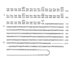
- a preferred O-glycosylated water-soluble Fc ⁇ R-fragment contains the amino acids l50 to 32l as shown in Figure l, which are identical to the amino acids 55 to 226 as shown in Figure 3.
- the cDNA for at least part of the amino acids l to l48 of the Fc ⁇ R, in particular the coding sequence for the N-terminal cytoplasmic region is absent so that e.g. only the coding sequence for the insoluble membrane protein and the portion encoding the water-soluble part of the cDNA of the whole receptor is present.
- a plasmid containing a cDNA insert encoding the Fc ⁇ R is modified by replacing at least a part of the coding sequence for the amino acids l to l48 e.g. the amino acids l to l34 by an eucaryotic cDNA signal sequence e.g. an interleukin cDNA signal sequence e.g. by the BSF-2 signal sequence.
- an eucaryotic cDNA signal sequence e.g. an interleukin cDNA signal sequence e.g. by the BSF-2 signal sequence.
- the recessed 3 ⁇ -ends of this fragment were then filled in with the Klenow fragment of DNA polymerase and the DNA subsequently digested with PstI.
- the obtained fragment was then cloned in a suitable vector, preferably with a BamHI-PstI digested pBSF2-L8.
- the vector is conveniently prepared as follows:
- the EcoRI-BamHI l,2 kbp BSF-2 cDNA insert was prepared by digestion of pBSF-2.38 (see Nature 324 , 73-76 (l986)) with HindIII and BamHI.
- the obtained fragment containing a full length BSF-2 cDNA was then digested with HinfI and the recessed 3 ⁇ -end filled in with Klenow fragment of DNA polymerase.
- KpnI digestion the obtained KpnI-HinfI ll0 bp fragment containing the BSF-2 leader sequence was cloned into the multiple cloning site of pGEM4 digested previously with KpnI and SmaI.
- One of the selected clones was propagated and named as pBSF2-L8 (see Figure 4).
- pBSF2-L8 was digested with BamHI and the recessed 3 ⁇ -ends filled in with Klenow fragment of DNA polymerase. After the filling in of the BamHI site, the above mentioned HindIIIPstI Fc ⁇ R cDNA was cloned into BamHI-PstI digested pBSF2-L8 as mentioned hereinbefore. One of the selected clones was propagated and named as psFc ⁇ R-l (see Figure 3).
- Fc ⁇ R activities in the culture supernatants and the lysates of oocytes are determined by an enzyme linked immunosorbent assay (ELISA) utilizing anti-Fc ⁇ R antibodies 3-5 and 8-30 (see European Patent Application 86 lll 488.2, filed on August l9, l986), which recognize two different epitopes on Fc ⁇ R.
- ELISA enzyme linked immunosorbent assay
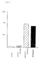
- Figure 5 Fc ⁇ R activity was determined for the NP-40 lysate of oocytes, in PBS-lysate of oocytes and in oocyte culture supernatant.
- the Fc ⁇ R water-soluble fragment secretion product from oocytes have the properties of binding IgE by means of a modified ELISA using anti-Fc ⁇ Rantibody 3-5, IgE and AP-anti-human IgE:
- the culture supernatant from the oocytes injected with psFc ⁇ R-l mRNA was incubated on 3-5 antibody-coated plates which were then incubated with human IgE and finally with AP-anti-IgE.
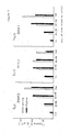
- the results established clearly that Fc ⁇ R secreted from the oocytes formed a complex with IgE (see Figure 6). Binding with non-transformed oocyte supernatant, buffer, and RPMI 8866 supernatant were carried out as a control.
- the corresponding genes containing an eucaryotic signal sequence and at least the coding sequence for the amino acids l50 to 32l of the full-length Fc ⁇ R cDNA can be introduced into organisms under conditions which lead to high yields thereof, as mentioned hereinbefore.
- Useful hosts and vectors are well known to those of skill in the art.
- eukaryotic microbes such as yeast cultures can be used for expression.
- Saccharomyces cerevisiae is most commonly used among eukaryotic microorganisms, although a number of other species are commonly available.
- the plasmid YRp7 for example (Stinchcomb, et al., Nature 282 , 39, (l979); Kingsman et al., Gene 7 , l4l (l979); Tschumper, et al., Gene l0 , l57 (l980)) and plasmid YEpl3 (Bwach et al., Gene 8 , l2l-l33 (l979)) are commonly used.
- the plasmid YRp7 contains the TRPl gene which provides a selection marker for a mutant strain of yeast lacking the ability to grow in tryptophan, for example ATCC No. 44076.
- the presence of the TRPl lesion as a characteristic of the yeast host cell genome then provides an effective environment for detecting transformation by growth in the absence of tryptophan.
- the plasmid YEpl3 contains the yeast LEU2 gene which can be used for complementation of a LEU2 minus mutant strain.
- Suitable promoting sequence in yeast vectors include the 5 ⁇ -flanking region of the genes for ADH I (Ammerer, G., Methods of Enzymology l0l , l92-20l (l983)), 3-phosphoglycerate kinase (Hitzemann, et al., J. Biol. Chem.
- glycolytic enzymes such as enolase, glyceraldehyde-3-phosphate dehydrogenase, hexokinase, pyruvate decarboxylase, phosphofructokinase, glucose-6-phosphate isomerase, phosphoglucose isomerase, and glucokinase.
- enolase glyceraldehyde-3-phosphate dehydrogenase
- hexokinase hexokinase
- pyruvate decarboxylase phosphofructokinase
- glucose-6-phosphate isomerase phosphoglucose isomerase
- glucokinase glucokinase.
- the termination sequences associated with these genes are also ligated into the expression vector 3 ⁇ to the sequence to be expressed, to provide polyadenylation of the mRNA and termination.
- promotors which have the additional advantage of transcription controlled by growth conditions are the promotor regions of the genes for alcohol dehydrogenase-2, isocytochrome C, acid phosphatase, degradative enzymes associated with nitrogen metabolism, the aforementioned glyceraldehyde-3-phosphate dehydrogenase, and enzymes responsible for maltose and galactose utilization.
- Promotors which are regulated by the yeast mating type locus such as the promotors of the genes BARI, MR-alpha-l, STE2, STE3, STE5 can be used for temperature regulated system by using temperature dependent siv mutations (Rhine, Ph. D.
- any plasmid vector containing a yeast compatible promotor, origin of replication and termination sequences is suitable.
- cultures of cells derived from multicellular organisms may also be used as hosts.
- any such cell culture is applicable, whether from vertebrate or invertebrate cell cultures.
- interest has been greatest in vertebrate cells, and propagation of vertebrate cells in culture (tissue culture) has become a routine procedure in recent years (Tissue Culture, Academic Press, Kruse and Patterson, Editors (l973)).
- useful host cell lines are VERO and HeLa cells, Chinese hamster ovary (CHO) cell lines, and Wl38, BHK, COS-7 and MDCK cell lines.
- Expression vectors for such cells usually include (if necessary) an origin of replication, a host specific or viral promotor located in front of the gene to be expressed, along with any necessary ribosome binding sites, RNA splice sites, polyadenylation site, and transcriptional terminator sequences.
- the control functions on the expression vectors are often provided by viral material.
- promotors are derived from polyoma, Adenovirus 2, and most frequently Simian Virus 40 (SV40).
- the early and late promotors of SV40 are particularly useful because both are obtained easily from the virus as a fragment which also contains the SV40 viral origin of replication (Fiers et al., Nature 273 , ll3 (l978)).
- Smaller or larger SV40 fragments may also be used, provided they include the approximately 250 bp sequence extending from the Hind III site toward the Bgl I site at the viral origin of replication. It is also possible, and often desirable, to utilize promotor or control sequences normally associated with the desired gene sequence, provided such control sequences are compatible with the host cell systems.
- An origin of replication may be provided either by onstruction of the vector to include an exogenous origin, derived from SV40 or other viral (e.g., Polyoma, Adeno, VSV, BPV, etc.) sources, or may be provided by the host cell chromosomal replication mechanism. If the vector is integrated into the host cell chromosome, the latter is often sufficient.
- the prepared biologically active recombinant water-soluble human Fc ⁇ -receptor prepared according to the invention is suitable for the treatment of local and allergic reactions induced by expression of IgE and may be incorporated in the suitable pharmaceutical compositions such as solutions or sprays.
- the plasmids p ⁇ NFc ⁇ R-l and p ⁇ NFc ⁇ R-2 used as comparison plasmids were prepared as follows:
- the isolated cDNA-fragment starting with the nucleotide 584 as shown in Figure l was cloned into a HindIII and EcoRI digested pGEM4.
- One of the selected clones is propagated and named as p ⁇ NFc ⁇ R-l.
- the above mentioned plasmid LE 392 or pGEM4 (pFc ⁇ R-l) was digested with EcoRI, and a fragment containing the fulllength ⁇ l.7 Kbp Fc ⁇ R cDNA obtained.
- the obtained EcoRI-fragment was then partially digested with Sau3A to remove the cDNA sequence encoding for the putative cytoplasmic domain, e.g.
- the monoclonal anti-Fc ⁇ R antibodies 3-5 ( ⁇ 1) and 8-30 ( ⁇ ) were produced by hybridization of P3Ul myeloma with spleen cells from Balb/c mice immunized with RPMI-8866 cells (see European Patent Application No. 86 ll0 420.6 of the same applicant, filed on July 29, l986).
- the 8-30 antibody recognizes the epitope close to the IgE binding site of Fc ⁇ R and could block binding of IgE to 8866 lymphoblastoid cells.
- the 3-5 antibody recognizes a different epitope on Fc ⁇ R and can not block efficiently IgE binding to its receptors.
- the monoclonal antibodies were purified from ascites by 50 % saturated ammonium sulfate precipitation followed by gel filtration using Sepharose 6B (Pharmacia Fine Chemical, Uppsala, Sweden) for IgM class or ion exchange chromatography using QAE-Sephadex (Pharmacia Fine Chemical) for IgGl.
- the polyclonal mouse IgG was isolated in the same fashion.
- the anti-mouse IgM-alkaline phosphatase conjugate was purchased from Tago (Burlingame, CA).
- l0 ⁇ g of pFc ⁇ R-l DNA were digested with 20 units of HindIII in 50 ⁇ l of a medium salt buffer (l0 mM Tris-HCl, pH 7.5, l0 mM MgCl2, l mM DTT) for l hr, followed with 20 units of EcoRI in l00 ⁇ l of a high salt buffer (l00 mM NaCl, 50 mM Tris-HCI, pH 7.5, l0 mM MgCl2, l mM DTT) for l hr to obtain the HindIII-EcoRI fragment containing a l kbp region of soluble Fc ⁇ R-cDNA (see Figure l: starting nucleotide 584).
- a medium salt buffer l0 mM Tris-HCl, pH 7.5, l0 mM MgCl2, l mM DTT
- the digested DNA was applied on a l % preparative agarose electrophoresis and the approximately l kbp HindIII-EcoRI fragments were electroeluted from minced gels and precipitated in 70 % ethanol at -80°C for l hr.
- l ⁇ g of pGEM4 Promega Biotec was digested with 2 units HindIII and 2 units EcoRI as described above, phenol-extracted and ethanol-precipitated at -80°C for l hr.
- HindIII-EcoRI fragment and HindIII-EcoRI digested pGEM4 were incubated with 200 units/T4 ligase in l0 ⁇ l of a ligation buffer (50 mM Tris-HCl, pH 7.4, l0 mM MgCl2, l0 mM DTT, l mM spermidine, l mM ATP, 0,l mg/ml BSA) at 4°C for l6 hrs and transfected into E.coli (MCl065).
- a ligation buffer 50 mM Tris-HCl, pH 7.4, l0 mM MgCl2, l0 mM DTT, l mM spermidine, l mM ATP, 0,l mg/ml BSA
- 200 ⁇ g of pFc ⁇ R-l were digested with 400 units EcoRI in 200 ⁇ l of a high salt buffer for 2 hrs at 37°C and were subjected to electrophoresis on a l % preparative agarose gel. Approximately l.7 kb EcoRI fragment was electroeluted and ethanol-precipitated at -80°C for l hr.
- the DNA fragment was ligated to a l.7 ⁇ g synthetic linker (5 ⁇ -GATCTGAGTCATGGTACCATGACTCAGATCGTGCTG-3 ⁇ ) with 200 units of T4 ligase in l0 ⁇ l of a ligation buffer at 4°C for l6 hrs, then phenol-extracted and ethanol-precipitated.
- the fragments were dissolved, digested with 24 units KpnI in 40 ⁇ l of a low salt buffer at 37°C for 3 hrs, phenol-extracted and ethanol-precipitated.
- the excess linkers were removed by gel chromatography with a Biogel A50m column. The fragments were ethanol-precipitated.
- l ⁇ g of pGEM4 was digested with 8 units KpnI in 20 ⁇ l of a low salt buffer at 37°C for 2 hrs, followed by incubation with 8 units EcoRI in 40 ⁇ l of a high salt buffer at 37°C for 2 hrs.
- the fragments which had been previously ligated to synthesis linkers were then ligated to the KpnI-EcoRI-digested pGEM4 by incubating with 200 units of T4 ligase in l0 ⁇ l of a ligation buffer at 4°C for l6 hrs and transfected into E.coli (MCl065).
- T4 ligase in l0 ⁇ l of a ligation buffer at 4°C for l6 hrs and transfected into E.coli (MCl065).
- colony hybridization technique the clone, p ⁇ NFc ⁇ R-2, which lacks only a cytoplasmic domain, was isolated and propagated.
- the plasmid psFc ⁇ R-l containing the modified Fc ⁇ R cDNA was linearized by digestion with the restriction enzyme BamHI.
- mRNA was synthesized with SP6 RNA polymerase using the linearized plasmid DNA as the template according to Melton et al.
- About ten nanogram of mRNA was injected into each Xenopus leavis oocyte, and the oocytes were incubated at 20°C in Barth's modified medium supplemented with l00 ⁇ g/ml penicillin and l ⁇ g/ml streptomycin.
- the culture supernatant was collected and the oocytes were homogenized in Dulbecco's PBS containing l mM PMSF.
- the PBS-lysate was separated by high speed centrifugation at l5,000 rpm for l0 min.
- the pellet was again extracted with NP-40 to solubilize membrane bound receptor (0,5 % NP-40, 0,l M NaCl, 0,05 M Tris-HCl, pH 7,5).
- the beads were washed 2 times with lysis buffer and 2 times with high salt buffer (0,5 % NP-40, 0,5 M NaCl, 0,05 M Tris-HCl, pH 8.0) followed by a final wash with lysis buffer.
- the immunoprecipitates were eluted with SDS sample buffer by boiling for 2 min. Samples were analysed on 9 % SDS Page (see Figure 8).
- Fc ⁇ R activity was measured with a double antibody enzyme-linked immunosorbent assay (ELISA) by using two different monoclonal anti-Fc ⁇ R antibodies, 3-5(IgGl) and 8-248(IgM) which react with two different epitopes on Fc ⁇ R.
- ELISA enzyme-linked immunosorbent assay
- the plates were then washed 4 times with rinse buffer (Dulbecco's phosphate buffer, pH 7.4, containing 0,05 % (v/v) Tween 20) followed by the addition of l00 ⁇ l samples diluted with diluent buffer (0,05 M Tris-HCl, pH 8.l, with l mM MgCl2, 0,l5 M NaCl, 0,05 % Tween 20, l % BSA and 0,02 % NaN3). The plates were incubated for 2 hours at room temperature, and washed 4 times with rinse buffer, followed by the addition of l00 ml of 8-248 antibody-alkaline phosphatase conjugate (0,75 mg/ml).
- rinse buffer Dulbecco's phosphate buffer, pH 7.4, containing 0,05 % (v/v) Tween 20
- l00 ⁇ l samples diluted with diluent buffer (0,05 M Tris-HCl, pH 8.l, with l mM MgC
- the 3-5 antibody-coated plates were incubated with l00 ⁇ l samples for 2 hours, washed 4 times with rinse buffer, followed by the addition of l0 ⁇ g/ml human monoclonal IgE (PS myeloma). After 2 hour's incubation at room temperature, the plates were washed 4 times and incubated further with alkaline phosphataseconjugated monoclonal anti-human IgE and then developed with the addition of substrated, p-nitrophenyl phosphate as described earlier.
- Fc ⁇ R on lymphocytes are detected by an assay with the use of fixed ox RBC (ORBC) coated with human IgE (Gonzalez-Molina, A. and Spiegelberg, H.L. J. Clin. Invest 59, 6l6 (l977)).
- the number of IgE rosette-forming cells is estimated after subtracting the number of non-specific binding with fixed ORBC coated with bovine serum albumin.
- 25 ⁇ l of Fc ⁇ R bearing cells (5 ⁇ l06/ml) are mixed with a specific volume (e.g. l00 ⁇ l) of test sample or control medium and incubated for l hour at 4°C.
- the number of rosettes with 3 or more ORBC are counted.
- the Fc ⁇ R bearing cells are RPMI8866 cells and in experiment II SKW6-CL4 cells.
- the control medium is the supernatant of the control oocytes, i.e. non-transformed oocytes.
Abstract
The object of the present invention is a novel recombinant water-soluble human FcεR-fragment having at least one O-glycosylation site, preferably a fragment accompanied by native O-glycosylation, which is thereby biologically active, novel plasmids encoding for said soluble FcεR, the DNA-sequences containing these novel plasmids, and the preparation thereof.
Description
- In
Cell 47, 657-665 (l986) is described the molecular structure of human lymphocyte receptor for Immunoglobulin E and a cDNA clone encoding said FcεR, which consist of 32l amino acids without any signal sequence. This molecule is unusual in that it is oriented with its N-terminus in the cytoplasm side and its C-terminus located outside the cell. Moreover, from the publication in Cell it is known that the native water-soluble part of FcεR is a product of proteolytic cleavage between amino acids l49 and l50 (Arg and Met) of membrane-bound receptor (see Figure l: scheme of pFcεR-l). - It is generally accepted, that although the function of glycosylation in human proteins is not known, it has been observed that, generally speaking, a recombinant, non-glycosylated protein will have the same biological activity as the natural glycosylated molecule, for instance the glycosylated interferons are undistinguishable in biological activity with the non-glycosylated.
- We have found that, unexpectedly, while glycosylated FcεR water-soluble fragment has IgE binding activity, the nonglycosylated FcεR water-soluble fragment does not demonstrate such activity. Thus for pharmacological activity e.g. in treating allergic reactions, it is necessary to ensure that recombinant product is O-glycosylated.
- In addition, we have found that the membrane spanning region of the FcεR does not function as a signal sequence in the usual recombinant processes and therefore secretion of the water-soluble receptor protein from a suitable host, it is necessary to use an appropriate eucaryotic signal sequence. Such signal sequence may be provided in addition and in a position in front of the cDNA coding for the water-soluble part.
- According to one aspect of the present invention therefore, we provide a novel recombinant water-soluble human FcεRfragment having at least one O-glycosylation site, preferably a fragment accompanied by native O-glycosylation, which is thereby biologically active, and the production thereof.
- A further aspect of the present invention comprises the novel plasmids encoding for said soluble FcεR-fragment, the DNA-sequences containing these novel plasmids, and the preparation thereof (see e.g. Figure 2: schemes of pFcεR-l and psFcεR-l).
- A preferred O-glycosylated water-soluble FcεR-fragment contains the amino acids l50 to 32l as shown in Figure l, which are identical to the
amino acids 55 to 226 as shown in Figure 3. - In a novel cDNA-sequence according to the present invention, the cDNA for at least part of the amino acids l to l48 of the FcεR, in particular the coding sequence for the N-terminal cytoplasmic region is absent so that e.g. only the coding sequence for the insoluble membrane protein and the portion encoding the water-soluble part of the cDNA of the whole receptor is present.
- In this novel cDNA-sequence at least a part of the cDNA sequence coding for the amino acids l to l48 is replaced by a suitable cDNA fragment coding for an eucaryotic signal sequence using suitable restriction endonucleases and ligases.
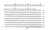
- For example, a plasmid containing a cDNA insert encoding the FcεR is modified by replacing at least a part of the coding sequence for the amino acids l to l48 e.g. the amino acids l to l34 by an eucaryotic cDNA signal sequence e.g. an interleukin cDNA signal sequence e.g. by the BSF-2 signal sequence. Thus, in the example described below a corresponding plasmid e.g. plasmid LE 392 deposited on August 0l, l986 under number FERM BP-lll6 (Fermentation Research Institute, Agency of Industrial Science and Technology, Japan - see also
European Patent Application 86 lll 58l.4, filed on August 2l, l986) or pGEM4 (pFcεR-l) (see Figure l) described inCell 47, 659 (l986) was digested with HindIII, whereby a l.0 kbp HindIII-fragment was obtained containing the coding sequence for the amino acids l34 to 32l of the full-length FcεR cDNA. The recessed 3ʹ-ends of this fragment were then filled in with the Klenow fragment of DNA polymerase and the DNA subsequently digested with PstI. The obtained fragment was then cloned in a suitable vector, preferably with a BamHI-PstI digested pBSF2-L8. The vector is conveniently prepared as follows: - The EcoRI-BamHI l,2 kbp BSF-2 cDNA insert was prepared by digestion of pBSF-2.38 (see Nature 324, 73-76 (l986)) with HindIII and BamHI. The obtained fragment containing a full length BSF-2 cDNA was then digested with HinfI and the recessed 3ʹ-end filled in with Klenow fragment of DNA polymerase. After KpnI digestion, the obtained KpnI-HinfI ll0 bp fragment containing the BSF-2 leader sequence was cloned into the multiple cloning site of pGEM4 digested previously with KpnI and SmaI. One of the selected clones was propagated and named as pBSF2-L8 (see Figure 4).
- pBSF2-L8 was digested with BamHI and the recessed 3ʹ-ends filled in with Klenow fragment of DNA polymerase. After the filling in of the BamHI site, the above mentioned HindIIIPstI FcεR cDNA was cloned into BamHI-PstI digested pBSF2-L8 as mentioned hereinbefore. One of the selected clones was propagated and named as psFcεR-l (see Figure 3).
- To compare the biological activity of the proteins produced by the clones pFcεR-l, psFcεR-l, pΔNFcεR-l and pΔNFcεR-2 (see Figure 2) these plasmids were linearized by digestion with the appropriate enzymes, e.g. pFcεR-l and psFcεR-l with BamHI and pΔNFcεR-l and pΔNFcεR-2 with EcoRI, and the obtained fragments are used as a template to synthesize mRNA with SP6 RNA polymerase. The resulting mRNA's were injected into Xenopus leavis oocytes. After 2 days of incubation, the FcεR activities in the culture supernatants and the lysates of oocytes are determined by an enzyme linked immunosorbent assay (ELISA) utilizing anti-FcεR antibodies 3-5 and 8-30 (see
European Patent Application 86 lll 488.2, filed on August l9, l986), which recognize two different epitopes on FcεR. As shown in Figure 5 FcεR activity was determined for the NP-40 lysate of oocytes, in PBS-lysate of oocytes and in oocyte culture supernatant. It will be seen that whereas no activity could be detected in PBS-lysate and culture supernatant of oocytes injected with transcripts of pFcεR-l, pΔNFcεR-l and pΔNFcεR-2 FcεR activity was detected in supernatants and PBS-lysates after injection with fragments of psFcεR-l. - Furthermore, we determined that the FcεR water-soluble fragment secretion product from oocytes have the properties of binding IgE by means of a modified ELISA using anti-FcεRantibody 3-5, IgE and AP-anti-human IgE: The culture supernatant from the oocytes injected with psFcεR-l mRNA was incubated on 3-5 antibody-coated plates which were then incubated with human IgE and finally with AP-anti-IgE. The results established clearly that FcεR secreted from the oocytes formed a complex with IgE (see Figure 6). Binding with non-transformed oocyte supernatant, buffer, and RPMI 8866 supernatant were carried out as a control.
- In order to evaluate the IgE-binding property of the soluble FcεR fragment derived from oocytes injected with psFcεR-l mRNA, the oocyte supernatant was further tested for its abi lity to inhibit the rosette formation between ORBC coated with human IgE and SKW6-CL4 cells bearing on their surface FcεR: The presence of soluble FcεR reduced the number of rosette forming cells to which more than 20 ORBC were bound indicating that the soluble receptor is competing with the cell membrane FcεR for the IgE bound on the surface of ORBC (see Figure 7).
- The corresponding genes containing an eucaryotic signal sequence and at least the coding sequence for the amino acids l50 to 32l of the full-length FcεR cDNA can be introduced into organisms under conditions which lead to high yields thereof, as mentioned hereinbefore. Useful hosts and vectors are well known to those of skill in the art.
- In general, eukaryotic microbes, such as yeast cultures can be used for expression. For example, Saccharomyces cerevisiae is most commonly used among eukaryotic microorganisms, although a number of other species are commonly available. For expression in Saccharomyces, the plasmid YRp7, for example (Stinchcomb, et al., Nature 282, 39, (l979); Kingsman et al., Gene 7, l4l (l979); Tschumper, et al., Gene l0, l57 (l980)) and plasmid YEpl3 (Bwach et al., Gene 8, l2l-l33 (l979)) are commonly used. The plasmid YRp7 contains the TRPl gene which provides a selection marker for a mutant strain of yeast lacking the ability to grow in tryptophan, for example ATCC No. 44076. The presence of the TRPl lesion as a characteristic of the yeast host cell genome then provides an effective environment for detecting transformation by growth in the absence of tryptophan. Similarly, the plasmid YEpl3 contains the yeast LEU2 gene which can be used for complementation of a LEU2 minus mutant strain.
- Suitable promoting sequence in yeast vectors include the 5ʹ-flanking region of the genes for ADH I (Ammerer, G., Methods of Enzymology l0l, l92-20l (l983)), 3-phosphoglycerate kinase (Hitzemann, et al., J. Biol. Chem. 255, 2073 (l980)) or other glycolytic enzymes (Kawasaki and Fraenkel, BBRC l08, ll07-lll2 (l982)), such as enolase, glyceraldehyde-3-phosphate dehydrogenase, hexokinase, pyruvate decarboxylase, phosphofructokinase, glucose-6-phosphate isomerase, phosphoglucose isomerase, and glucokinase. In constructing suitable expression plasmids, the termination sequences associated with these genes are also ligated into the expression vector 3ʹ to the sequence to be expressed, to provide polyadenylation of the mRNA and termination.
- Other promotors, which have the additional advantage of transcription controlled by growth conditions are the promotor regions of the genes for alcohol dehydrogenase-2, isocytochrome C, acid phosphatase, degradative enzymes associated with nitrogen metabolism, the aforementioned glyceraldehyde-3-phosphate dehydrogenase, and enzymes responsible for maltose and galactose utilization. Promotors which are regulated by the yeast mating type locus, such as the promotors of the genes BARI, MR-alpha-l, STE2, STE3, STE5 can be used for temperature regulated system by using temperature dependent siv mutations (Rhine, Ph. D. Thesis, University of Oregon, Eugene, Oregon (l979), Herskowitz and Oshima in The Molecular Biology of the Yeast Sacharomyces, Part I, l8l-209 (l98l), Cold Spring Harbor Laboratory). These mutations directly influence the expressions of the silent mating type cassettes of yeast, and therefore indirectly the mating type dependent promotors.
- Generally, however, any plasmid vector containing a yeast compatible promotor, origin of replication and termination sequences is suitable.
- In addition to microorganisms, cultures of cells derived from multicellular organisms may also be used as hosts. In principle, any such cell culture is applicable, whether from vertebrate or invertebrate cell cultures. However, interest has been greatest in vertebrate cells, and propagation of vertebrate cells in culture (tissue culture) has become a routine procedure in recent years (Tissue Culture, Academic Press, Kruse and Patterson, Editors (l973)). Examples of such useful host cell lines are VERO and HeLa cells, Chinese hamster ovary (CHO) cell lines, and Wl38, BHK, COS-7 and MDCK cell lines. Expression vectors for such cells usually include (if necessary) an origin of replication, a host specific or viral promotor located in front of the gene to be expressed, along with any necessary ribosome binding sites, RNA splice sites, polyadenylation site, and transcriptional terminator sequences.
- For use in mammalian cells, the control functions on the expression vectors are often provided by viral material. For example, commonly used promotors are derived from polyoma,
Adenovirus 2, and most frequently Simian Virus 40 (SV40). The early and late promotors of SV40 are particularly useful because both are obtained easily from the virus as a fragment which also contains the SV40 viral origin of replication (Fiers et al., Nature 273, ll3 (l978)). Smaller or larger SV40 fragments may also be used, provided they include the approximately 250 bp sequence extending from the Hind III site toward the Bgl I site at the viral origin of replication. It is also possible, and often desirable, to utilize promotor or control sequences normally associated with the desired gene sequence, provided such control sequences are compatible with the host cell systems. - An origin of replication may be provided either by onstruction of the vector to include an exogenous origin, derived from SV40 or other viral (e.g., Polyoma, Adeno, VSV, BPV, etc.) sources, or may be provided by the host cell chromosomal replication mechanism. If the vector is integrated into the host cell chromosome, the latter is often sufficient.
- The prepared biologically active recombinant water-soluble human Fcε-receptor prepared according to the invention is suitable for the treatment of local and allergic reactions induced by expression of IgE and may be incorporated in the suitable pharmaceutical compositions such as solutions or sprays.
- The plasmids pΔNFcεR-l and pΔNFcεR-2 used as comparison plasmids (see Figure 5) were prepared as follows:
- The plasmid LE 392 deposited on August 0l, l986 under number FERM BP-lll6 (Fermentation Research Institute, Agency of Industrial Science and Technology, Japan - see also
European Patent Application 86 lll 58l.4, filed on August 2l, l986) or pGEM4 (pFcεR-l) (see Figure l) described inCell 47, 659 (l986) was digested with HindIII and EcoRI. The isolated cDNA-fragment starting with the nucleotide 584 as shown in Figure l was cloned into a HindIII and EcoRI digested pGEM4. One of the selected clones is propagated and named as pΔNFcεR-l. - The above mentioned plasmid LE 392 or pGEM4 (pFcεR-l) was digested with EcoRI, and a fragment containing the fulllength ∼l.7 Kbp FcεR cDNA obtained. The obtained EcoRI-fragment was then partially digested with Sau3A to remove the cDNA sequence encoding for the putative cytoplasmic domain, e.g. for the amino acids l to 23 and a cDNA-fragment starting with the nucleotide 254 as shown in Figure l obtained, which was ligated with a palyndoromic 26mer linker of formula
5ʹ-GATCTGAGTCATGGTACCATGACTCA-3ʹ
to restore an ATG start codon at the 5ʹ-end and a KpnI restriction site. The thus obtained ligated fragment was digested with KpnI and cloned into KpnI and EcoRI digested pGEM4. Only those clones were selected by colony hybridiza tions wherein the region for the putative cytoplasmic domain was missing. One clone was selected and propagated and named as pΔNFcεR-2. - The following examples, which are not exhaustive, will illustrate the invention in greater detail:
- The monoclonal anti-Fc ε R antibodies 3-5 (γ₁) and 8-30 (µ) were produced by hybridization of P3Ul myeloma with spleen cells from Balb/c mice immunized with RPMI-8866 cells (see European Patent Application No. 86 ll0 420.6 of the same applicant, filed on July 29, l986). The 8-30 antibody recognizes the epitope close to the IgE binding site of FcεR and could block binding of IgE to 8866 lymphoblastoid cells. The 3-5 antibody, recognizes a different epitope on FcεR and can not block efficiently IgE binding to its receptors. These antibodies precipitate 46 kd and 25 kd polypeptides under reducing and non reducing conditions. The monoclonal antibodies were purified from ascites by 50 % saturated ammonium sulfate precipitation followed by gel filtration using Sepharose 6B (Pharmacia Fine Chemical, Uppsala, Sweden) for IgM class or ion exchange chromatography using QAE-Sephadex (Pharmacia Fine Chemical) for IgGl. The polyclonal mouse IgG was isolated in the same fashion. The anti-mouse IgM-alkaline phosphatase conjugate was purchased from Tago (Burlingame, CA).
- The plasmid LE392 deposited in E.coli on August 0l, l986 under number FERM BP-lll6 (Fermentation Research Institute, Agency of Industrial Science and Technology, Japan) according to the convention of Budapest which is a pGEM4 vector was digested with HindIII to obtain the HindIII fragment containing a region of soluble FcεR-cDNA or with HindIII-EcoRI to obtain the HindIII-EcoRI fragment containing approximately l kbp (see Figure l: starting nucleotide 584).
- l0 µg of pFcεR-l DNA were digested with 20 units of HindIII in 50 µl of a medium salt buffer (l0 mM Tris-HCl, pH 7.5, l0 mM MgCl₂, l mM DTT) for l hr, followed with 20 units of EcoRI in l00 µl of a high salt buffer (l00 mM NaCl, 50 mM Tris-HCI, pH 7.5, l0 mM MgCl₂, l mM DTT) for l hr to obtain the HindIII-EcoRI fragment containing a l kbp region of soluble FcεR-cDNA (see Figure l: starting nucleotide 584). The digested DNA was applied on a l % preparative agarose electrophoresis and the approximately l kbp HindIII-EcoRI fragments were electroeluted from minced gels and precipitated in 70 % ethanol at -80°C for l hr. l µg of pGEM4 (Promega Biotec) was digested with 2 units HindIII and 2 units EcoRI as described above, phenol-extracted and ethanol-precipitated at -80°C for l hr. The HindIII-EcoRI fragment and HindIII-EcoRI digested pGEM4 were incubated with 200 units/T4 ligase in l0 µl of a ligation buffer (50 mM Tris-HCl, pH 7.4, l0 mM MgCl₂, l0 mM DTT, l mM spermidine, l mM ATP, 0,l mg/ml BSA) at 4°C for l6 hrs and transfected into E.coli (MCl065). One clone was selected, propagated and after confirmation of its construction named as pΔNFcεR-l.
- 200 µg of pFcεR-l were digested with 400 units EcoRI in 200 µl of a high salt buffer for 2 hrs at 37°C and were subjected to electrophoresis on a l % preparative agarose gel. Approximately l.7 kb EcoRI fragment was electroeluted and ethanol-precipitated at -80°C for l hr. 4 µg of the fragment were digested with l0 units of AccI at 37°C for 3 hrs in 40 µl of a low salt buffer and partially digested with 0.4 unit Sau3A at 37°C for 20 min, phenol-extracted and ethanol-precipitated to obtain the Sau3A-EcoRI fragment (see Figure l: starting nucleotide 254). The DNA fragment was ligated to a l.7 µg synthetic linker (5ʹ-GATCTGAGTCATGGTACCATGACTCAGATCGTGCTG-3ʹ) with 200 units of T4 ligase in l0 µl of a ligation buffer at 4°C for l6 hrs, then phenol-extracted and ethanol-precipitated. The fragments were dissolved, digested with 24 units KpnI in 40 µl of a low salt buffer at 37°C for 3 hrs, phenol-extracted and ethanol-precipitated. The excess linkers were removed by gel chromatography with a Biogel A50m column. The fragments were ethanol-precipitated.
- Separately, l µg of pGEM4 was digested with 8 units KpnI in 20 µl of a low salt buffer at 37°C for 2 hrs, followed by incubation with 8 units EcoRI in 40 µl of a high salt buffer at 37°C for 2 hrs.
- The fragments which had been previously ligated to synthesis linkers were then ligated to the KpnI-EcoRI-digested pGEM4 by incubating with 200 units of T4 ligase in l0 µl of a ligation buffer at 4°C for l6 hrs and transfected into E.coli (MCl065). Using colony hybridization technique, the clone, pΔNFcεR-2, which lacks only a cytoplasmic domain, was isolated and propagated.
-
- a) 350 µg of pBSF2-38 (see Nature 324 73-76 (l986)), which contains the BSF-2 cDNA in the SmaI site of pGEM4, were digested with 700 units of EcoRI and BamHI in 500 µl of a high salt buffer (l00 mM NaCl, 50 mM Tris-HCl, pH 7.5, l0 mM MgCl₂, l mM DTT) for 2 hrs at 37°C. The digested DNA was applied on a preparative l % agarose gel electrophoresis and the EcoRI-BamHI fragment containing the full-length l.2 kbp BSF-2 cDNA was electroeluted from the gel, precipitated with 70 % ethanol and dissolved at a concentration of l µg/µl in TE buffer. 20 µg of this fragment were digested with 40 units of HinfI in 50 µl of a high salt buffer for l hr, phenol-extracted and ethanol-precipitated. The digested DNAs were dissolved in 25 µl of l × nick translation buffer (50 mM Tris-HCl, pH 7.2, l0 mM MgSO₄, 0.l mM DTT, 50 µg/ml BSA) and incubated with l ml of 8.2 units/µl Klenow fragment and l mM dNTP at 20°C for 30 minutes. The filling in reaction was terminated by incubation at 70°C for 5 min. The resulting l27 bp blunt ended fragment was phenol-extracted, digested with 40 units KpnI in 50 µl of a low salt buffer (l0 mM Tris-HCl, pH 7.5, l0 mM MgCl₂, l mM DTT) for l hr at 37°C, incubated with 2.5 units bacterial alkaline phosphatase at 65°C for 30 min and then applied on l % preparative agarose gel and electrophoresed. The ll0 bp fragment containing the BSF-2 leader sequence was electroeluted and ethanol-precipitated. - The ll0 bp fragment was dissolved in l0 µl of ligation buffer (50 mM Tris-HCl, pH 7.4, l0 mM MgCl₂, l0 mM DTT, l mM spermidine, l mM ATP, 0,l mg/ml BSA) and ligated with l µg of KpnI and SmaI digested pGEM4 by incubating with 200 units of T4 ligase at 4°C for l6 hrs and transfected into E.coli (MCl065). From the obtained colonies four colonies were picked up, one clone was selected, propagated and after confirmation that the plasmid of this selected clone contained only one leader sequence, it was named as pBSF2-L8 (see Figure 4).
- b) 80 µg of plasmid LE-392 or pGEM4(pFcεR-l) were digested with l50 units of HindIII in 200 µl of a low salt buffer (l0 mM Tris-HCl, pH 7.5, l0 mM MgCl₂, l mM DTT) at 37°C for l hr and applied on l % preparative agarose gel electrophoresis. The HindIII fragment containing the soluble FcεR region was electroeluted from the gel, ethanol-precipitated and dissolved at a concentration of l µg/µl in TE buffer. l µg of the HindIII fragment was incubated with 8.2 units Klenow fragment and l mM dNTP in l0 µl of l × nick translation buffer at 20°C for 30 min to fill in the recessive 3ʹ-ends, phenol-extracted and ethanol-precipitated. The HindIII fragments, the 3ʹ-ends of which had been filled in, were digested with 2 units PstI in l0 µl of the medium salt buffer (50 mM NaCl, l0 mM Tris-HCl, pH 7.5, l0 mM MgCl₂, l mM DTT) at 37°C for l hr, incubated with 0.25 unit bacterial alkaline phosphatase at 65°C for 30 min and ethanol-precipitated. Separately, l µg of pBSF2-L8 was digested with 2 units BamHI in 20 µl of a high salt buffer at 37°C for l hr, phenol-extracted and ethanol-precipitated. The BamHI-digested pBSF2-L8 was dissolved in l0 µl of l × nick translation buffer and incubated with 8.2 units Klenow fragment and l mM dNTP at 20°C for 30 min to fill in the recessive 3ʹ-ends, phenol-extracted and ethanol-precipitated. The precipitate was dissolved in 20 µl of a high salt buffer, digested with 2 units Pstl at 37°C for l hr, phenol-extracted and ethanol-precipitated. - The Pstl-digested fragment containing the soluble FcεR coding region and PstI digested pBSF2-L8 were ligated by incubating with 200 units T4 ligase in l0 µl of ligation buffer at 4°C for l6 hrs and transfected into E.coli (MCl065). Eight colonies were picked up from the obtained colonies. One clone was selected, propagated and after confirmation of the plasmid construction named psFcεR-l. The propagated psFcεR-l contains seven bases from the multiple cloning into pGEM4 between BSF-2 leader and FcεR sequences in frame (see Figure l: nucleotides l37 to l43).
- The plasmid psFcεR-l containing the modified FcεR cDNA was linearized by digestion with the restriction enzyme BamHI. mRNA was synthesized with SP6 RNA polymerase using the linearized plasmid DNA as the template according to Melton et al. About ten nanogram of mRNA was injected into each Xenopus leavis oocyte, and the oocytes were incubated at 20°C in Barth's modified medium supplemented with l00 µg/ml penicillin and l µg/ml streptomycin. After incubation for 2 days, the culture supernatant was collected and the oocytes were homogenized in Dulbecco's PBS containing l mM PMSF. The PBS-lysate was separated by high speed centrifugation at l5,000 rpm for l0 min. The pellet was again extracted with NP-40 to solubilize membrane bound receptor (0,5 % NP-40, 0,l M NaCl, 0,05 M Tris-HCl,
pH 7,5). - Ten oocytes which had been injected with mRNA were incubated with l00 µl Barth's modified medium containing l50 µCi ³⁵S-methionine for 24 hours at 20°C. Labeled oocytes were lysed in l ml lysis buffer (0,5 % NP-40, 0,l M NaCl, 0,05 M TrisHCl, pH 7.5) and centrifuged at l5,000 rpm for l0 min. The clarified lysate and culture supernatant were precleared with normal mouse Ig coupled Sepharose 4B beads and subsequently incubated with 3-5 (IgGI)antibody coupled Sepharose 4B beads for 60 min. with frequent shaking on ice. The beads were washed 2 times with lysis buffer and 2 times with high salt buffer (0,5 % NP-40, 0,5 M NaCl, 0,05 M Tris-HCl, pH 8.0) followed by a final wash with lysis buffer. The immunoprecipitates were eluted with SDS sample buffer by boiling for 2 min. Samples were analysed on 9 % SDS Page (see Figure 8).
- FcεR activity was measured with a double antibody enzyme-linked immunosorbent assay (ELISA) by using two different monoclonal anti-FcεR antibodies, 3-5(IgGl) and 8-248(IgM) which react with two different epitopes on FcεR. Ninty six well microtiter plates (Nunc, Roskilde, Denmark) were precoated with l00 µl/well of 3-5 antibody (l0 µg/ml) in coating buffer (0,l M carbonate buffer,
pH % Tween 20, l % BSA and 0,02 % NaN3). The plates were incubated for 2 hours at room temperature, and washed 4 times with rinse buffer, followed by the addition of l00 ml of 8-248 antibody-alkaline phosphatase conjugate (0,75 mg/ml). After 4 hours of incubation, the plates were washed 4 times and the enzyme reaction was initiated by the addition of l00 µl/well of l mg/ml p-nitrophenyl phosphate (Sigma Chemical Co., St. Louis, MO) in substrate buffer (0,05 M carbonate buffer, pH 9.8, l0 mM MgCl₂). After an appropriate incubation (60-90 min.) absorbances were read with an automatic micro-ELISA reader (Nippon Inter. Med. Tokyo, Japan) at 405 and 620 nm wavelengths (see Figures 5 and 6). - The 3-5 antibody-coated plates were incubated with l00 µl samples for 2 hours, washed 4 times with rinse buffer, followed by the addition of l0 µg/ml human monoclonal IgE (PS myeloma). After 2 hour's incubation at room temperature, the plates were washed 4 times and incubated further with alkaline phosphataseconjugated monoclonal anti-human IgE and then developed with the addition of substrated, p-nitrophenyl phosphate as described earlier.
- FcεR on lymphocytes are detected by an assay with the use of fixed ox RBC (ORBC) coated with human IgE (Gonzalez-Molina, A. and Spiegelberg, H.L. J. Clin. Invest 59, 6l6 (l977)). The number of IgE rosette-forming cells is estimated after subtracting the number of non-specific binding with fixed ORBC coated with bovine serum albumin. For IgE rosette inhibition, 25 µl of FcεR bearing cells (5 × l0⁶/ml) are mixed with a specific volume (e.g. l00 µl) of test sample or control medium and incubated for l hour at 4°C. The number of rosettes with 3 or more ORBC are counted. In experiments I and III the FcεR bearing cells are RPMI8866 cells and in experiment II SKW6-CL4 cells. The control medium is the supernatant of the control oocytes, i.e. non-transformed oocytes.
- In the foregoing test results it will be seen, a) that SDS-PAGE analysis shows the product of psFcεR-l from oocytes, which is recognized by both antibodies 3-5 and 8-30 and has IgE-binding activity, yielded broad protein bands (see Figure 8) and, b) the product of pΔNFcεR-l from oocytes, which by comparison lacks the N-terminal transmembrane region, can be recognized by the 3-5 antibody, but not by the 8-30 antibody and does not have IgE binding activity. These results indicate that the product of pΔNFcεR-l which does not have a signal sequence is not processed properly and thus that a proper processing as in clone psFcεR-l create the epitope which is recognized by the 8-30 antibody and has IgE binding activity.
Claims (23)
1. Recombinant biologically active water-soluble fragment of human FcεR having at least one O-glycosylation site, preferably a fragment accompanied by native O-glycosylation.
2. O-glycosylated water-soluble fragment of FcεR as claimed in claim l characterized by the DNA-sequence of the fulllength FcεR cDNA, wherein at least a part of the coding sequence for the amino acids l to l48 as shown in Figure l is replaced by an eucaryotic signal sequence, or a degenerative derivate thereof.
3. O-glycosylated water-soluble fragment of FcεR as claimed in claim l or in claim 2 containing the amino acids l50 to 32l as shown in Figure l.
4. O-glycosylated water-soluble fragment of FcεR as claimed in claim 2 wherein the signal sequence of pBSF-2.38 is used as eucaryotic signal sequence, or a degenerative derivate thereof.
5. O-glycosylated water-soluble fragment of FcεR as claimed in claim 4 containing the amino acids 55 to 226 as shown in Figure 3 accompanied by native O-glycosylation, or a degenerative derivate thereof.
6. A recombinant DNA molecule which contains the genetic information for producing a water-soluble fragment of human FcεR accompanied by native O-glycosylation as claimed in any of the claims l to 5, or a degenerative derivate thereof.
7. A recombinant DNA molecule as claimed in claim 6 containing in addition the replicon and control sequences necessary for expression.
8. A recombinant DNA molecule as claimed in claim 7 containing the replicon and control sequences for expression in eukaryotes, especially in mammalian cells.
9. A recombinant DNA molecule as claimed in claim 7 or 8 containing the replicon and control sequences suitable for expression in yeast.
l0. A recombinant DNA molecule as claimed in claim 9 wherein as replicon and control sequences those of plasmid pGEM4 are used.
11. A vector containing a recombinant DNA molecule as claimed in any of the claim 6 to l0.
12. A vector as claimed in claim ll wherein the vector is a plasmid vector.
13. A plasmid as claimed in claim l2 containing the coding sequence as claimed in any of the claims 6 to l0.
14. psFcεR-l as shown in Figure 3 or a degenerative derivate thereof.
15. A host organism transformed by a vector as claimed in any of the claims ll to l4.
16. Pharmaceutical compositions containing a polypeptide as claimed in any of the claims l to 5.
17. Pharmaceutical compositions as claimed in claim l6 suitable for treatment of local or systemic IgE-allergic reactions.
18. Preparation of a pharmaceutical composition as claimed in claims l6 or l7 wherein an effective amount of a polypeptide as claimed in any of the claims l to 5 is incorporated in one or more excipients.
19. Use of a polypeptide as claimed in any of the claims l to 5 for the preparation of a pharmaceutical composition.
20. A process for preparing an O-glycosylated active water-soluble fragment of human FcεR as claimed in any of the claims l to 5 which comprises transforming a suitable host organism by a vector containing a DNA as claimed in any of the claim 6 to l0 for the desired fragment of FcεR at an appropriate site for expression, and isolating the secreted fragment from the resulting transformants.
2l. Process for preparing a host organism as claimed in claim l5, wherein a suitable host is transformed with a vector as claimed in any of the claims ll to l4.
22. Process for preparing a vector as claimed in any of the claims ll to l4, wherein a DNA as claimed in any of the claims 6 to l0 is inserted in a suitable vector.
23. Process for preparing a DNA as claimed in any of the claims 6 to l0, wherein a suitable vector is digested with one or more suitable restriction endonucleases and isolating the desired DNA.
Priority Applications (12)
| Application Number | Priority Date | Filing Date | Title |
|---|---|---|---|
| EP87105425A EP0286700A1 (en) | 1987-04-11 | 1987-04-11 | Biologically active recombinant human soluble Fc R-fragment, plasmids encoding this Fc R-fragment and the preparation thereof |
| EP87111392A EP0259615A1 (en) | 1986-08-21 | 1987-08-06 | Human low affinity Fc epsilon-receptor, the isolation, the recombinant preparation and purification thereof |
| AU77165/87A AU7716587A (en) | 1986-08-21 | 1987-08-18 | Human low affinity fc(epsilon)-receptor |
| HU873722A HUT44620A (en) | 1986-08-21 | 1987-08-19 | Human, low-affinity fc under epsilon receptor and process for the isolation, production by recombinant technique and cleaning thereof |
| NZ221501A NZ221501A (en) | 1986-08-21 | 1987-08-19 | Human low-affinity fc receptor, peptides, dna and host cells |
| PT85560A PT85560B (en) | 1986-08-21 | 1987-08-20 | A method for the insulation, for the preparation by recombinant techniques and for the purification, of a low-affinity Fc-human receptor |
| JP62207337A JPS6427496A (en) | 1986-08-21 | 1987-08-20 | Human low-affinity fce-receptor, its separation, preparation of its recombinant and purification thereof |
| IL83594A IL83594A0 (en) | 1986-08-21 | 1987-08-20 | Human low affinity fce-receptor,its isolation,its recombinant preparation and pharmaceutical compositions containing it |
| DK434287A DK434287A (en) | 1986-08-21 | 1987-08-20 | HUMAN LOW-AFFINITY FEPSILON RECEPTOR ITS MANUFACTURING, INSULATION AND CLEANING |
| NO873512A NO873512L (en) | 1986-08-21 | 1987-08-20 | HUMAN LOW-AFFINITY FCEPSILON RECEPTOR, INSULATION, RECOMBINANT PREPARATION AND PURIFICATION OF THIS. |
| FI873614A FI873614A (en) | 1986-08-21 | 1987-08-21 | HUMAN-FC -RECEPTOR MED LAOG AFFINITET, DESS ISOLERING, REKOMBINATFRAMSTAELLNING OCH RENING. |
| KR870009152A KR880003005A (en) | 1986-08-21 | 1987-08-21 | Human low affinity Fcε-receptors and their isolation, recombinant preparation and purification |
Applications Claiming Priority (1)
| Application Number | Priority Date | Filing Date | Title |
|---|---|---|---|
| EP87105425A EP0286700A1 (en) | 1987-04-11 | 1987-04-11 | Biologically active recombinant human soluble Fc R-fragment, plasmids encoding this Fc R-fragment and the preparation thereof |
Publications (1)
| Publication Number | Publication Date |
|---|---|
| EP0286700A1 true EP0286700A1 (en) | 1988-10-19 |
Family
ID=8196918
Family Applications (1)
| Application Number | Title | Priority Date | Filing Date |
|---|---|---|---|
| EP87105425A Withdrawn EP0286700A1 (en) | 1986-08-21 | 1987-04-11 | Biologically active recombinant human soluble Fc R-fragment, plasmids encoding this Fc R-fragment and the preparation thereof |
Country Status (1)
| Country | Link |
|---|---|
| EP (1) | EP0286700A1 (en) |
Cited By (3)
| Publication number | Priority date | Publication date | Assignee | Title |
|---|---|---|---|---|
| EP0321842A1 (en) * | 1987-12-22 | 1989-06-28 | Kishimoto, Tadamitsu, Prof. | Soluble recombinant Fc-Epsilon-receptor, the preparation and the use thereof |
| US5766943A (en) * | 1992-08-17 | 1998-06-16 | University Of Iowa Research Foundation | DNA sequences for soluble form of CD23 |
| US5770396A (en) * | 1992-04-16 | 1998-06-23 | The United States Of America As Represented By The Department Of Health And Human Services | Isolation characterization, and use of the human beta subunit of the high affinity receptor for immunoglobulin E |
Citations (1)
| Publication number | Priority date | Publication date | Assignee | Title |
|---|---|---|---|---|
| EP0248211A2 (en) * | 1986-04-30 | 1987-12-09 | Junji Yodoi | Novel protein and a method for production thereof |
-
1987
- 1987-04-11 EP EP87105425A patent/EP0286700A1/en not_active Withdrawn
Patent Citations (1)
| Publication number | Priority date | Publication date | Assignee | Title |
|---|---|---|---|---|
| EP0248211A2 (en) * | 1986-04-30 | 1987-12-09 | Junji Yodoi | Novel protein and a method for production thereof |
Non-Patent Citations (3)
| Title |
|---|
| CELL, vol. 47, December 1986, pages 657-665; H. KIKUTANI et al.: "Molecular structure of human lymphocyte receptor for immunoglobulin E" * |
| NATURE, vol. 324, 6th November 1986, pages 73-77; T. HIRANO et al.: "Complementary DNA for a novel human interleukin (BSF-2) that induces B lymphocytes to produce immunoglobulin" * |
| THE EMBO JOURNAL, vol. 6, no. 1, January 1987, pages 109-114, IRL Press Ltd, Eynsham, Oxford, GB; C. L]DIN et al.: "Cloning and expression of the cDNA coding for a human lymphocyte IgE receptor" * |
Cited By (4)
| Publication number | Priority date | Publication date | Assignee | Title |
|---|---|---|---|---|
| EP0321842A1 (en) * | 1987-12-22 | 1989-06-28 | Kishimoto, Tadamitsu, Prof. | Soluble recombinant Fc-Epsilon-receptor, the preparation and the use thereof |
| US5770396A (en) * | 1992-04-16 | 1998-06-23 | The United States Of America As Represented By The Department Of Health And Human Services | Isolation characterization, and use of the human beta subunit of the high affinity receptor for immunoglobulin E |
| US6171803B1 (en) | 1992-04-16 | 2001-01-09 | The United States Government As Represented By The Department Of Health And Human Services | Isolation, characterization, and use of the human β subunit of the high affinity receptor for immunoglobulin E |
| US5766943A (en) * | 1992-08-17 | 1998-06-16 | University Of Iowa Research Foundation | DNA sequences for soluble form of CD23 |
Similar Documents
| Publication | Publication Date | Title |
|---|---|---|
| CA1341581C (en) | Leukaemia inhibitory factor | |
| Yoshimura et al. | A novel cytokine‐inducible gene CIS encodes an SH2‐containing protein that binds to tyrosine‐phosphorylated interleukin 3 and erythropoietin receptors. | |
| EP0411946B1 (en) | DNA encoding human GP130 protein | |
| Young et al. | Yeast RNA polymerase II genes: isolation with antibody probes | |
| US5807991A (en) | Human interleukin-5 receptor | |
| Altmann et al. | A mammalian translation initiation factor can substitute for its yeast homologue in vivo | |
| JP2728968B2 (en) | TNF binding protein | |
| EP0136489A1 (en) | Analogs of human interleukin II and their preparation | |
| IE903467A1 (en) | Granulocyte-colony stimulating factor receptors | |
| EP0804578B1 (en) | Il-2r-associated polypeptide and dna molecules coding therefor | |
| JPH1080286A (en) | Method for producing human il-3 and protein having human il-3 activity | |
| Ichihara et al. | Impaired interleukin‐3 (IL‐3) response of the A/J mouse is caused by a branch point deletion in the IL‐3 receptor alpha subunit gene. | |
| US5643749A (en) | Soluble interferon α-receptor, its preparation and use | |
| KR19980066046A (en) | High-CTLA4-Ig fusion protein | |
| Schweighoffer et al. | Identification of a human guanine nucleotide-releasing factor (H-GRF55) specific for Ras proteins | |
| JPH03504439A (en) | cDNA encoding the gamma subunit of the high affinity receptor for immunoglobulin E | |
| EP0286700A1 (en) | Biologically active recombinant human soluble Fc R-fragment, plasmids encoding this Fc R-fragment and the preparation thereof | |
| AU609128B2 (en) | Leukaemia-inhibitory factor | |
| JPH09510344A (en) | Galanin receptors, nucleic acids, transformed cells, and uses thereof | |
| Clark-Lewis et al. | Molecular structure and biological activities of P cell-stimulating factor (interleukin 3) | |
| JPH10179180A (en) | Mouse genom clone of cc-ckr5 receptor | |
| EP0259615A1 (en) | Human low affinity Fc epsilon-receptor, the isolation, the recombinant preparation and purification thereof | |
| EP1207202A1 (en) | Nucleic acid molecules encoding a protein interacting with the chemokine receptor CCR5 or other chemokine receptor family members | |
| EP0257114A1 (en) | Human low affinity Fc epsilon-receptor, the isolation and purification thereof | |
| US5304637A (en) | Expression and purification of human interleukin-3 and muteins thereof |
Legal Events
| Date | Code | Title | Description |
|---|---|---|---|
| PUAI | Public reference made under article 153(3) epc to a published international application that has entered the european phase |
Free format text: ORIGINAL CODE: 0009012 |
|
| AK | Designated contracting states |
Kind code of ref document: A1 Designated state(s): GB |
|
| STAA | Information on the status of an ep patent application or granted ep patent |
Free format text: STATUS: THE APPLICATION IS DEEMED TO BE WITHDRAWN |
|
| 18D | Application deemed to be withdrawn |
Effective date: 19890420 |
|
| RIN1 | Information on inventor provided before grant (corrected) |
Inventor name: BARSUMIAN, EDWARD L., DR. Inventor name: KIKUTANI, HITOSHI, DR. Inventor name: SUEMURA, MASAKI, DR. Inventor name: KISHIMOTO, TADAMITSU, PROF. DR. |