EP0269526A2 - Method of quantitative determination of antigens and antibodies - Google Patents
Method of quantitative determination of antigens and antibodies Download PDFInfo
- Publication number
- EP0269526A2 EP0269526A2 EP87402674A EP87402674A EP0269526A2 EP 0269526 A2 EP0269526 A2 EP 0269526A2 EP 87402674 A EP87402674 A EP 87402674A EP 87402674 A EP87402674 A EP 87402674A EP 0269526 A2 EP0269526 A2 EP 0269526A2
- Authority
- EP
- European Patent Office
- Prior art keywords
- antigen
- antibody
- absorbance
- concentration
- latex
- Prior art date
- Legal status (The legal status is an assumption and is not a legal conclusion. Google has not performed a legal analysis and makes no representation as to the accuracy of the status listed.)
- Granted
Links
Images


Classifications
-
- G—PHYSICS
- G01—MEASURING; TESTING
- G01N—INVESTIGATING OR ANALYSING MATERIALS BY DETERMINING THEIR CHEMICAL OR PHYSICAL PROPERTIES
- G01N33/00—Investigating or analysing materials by specific methods not covered by groups G01N1/00 - G01N31/00
- G01N33/48—Biological material, e.g. blood, urine; Haemocytometers
- G01N33/50—Chemical analysis of biological material, e.g. blood, urine; Testing involving biospecific ligand binding methods; Immunological testing
- G01N33/53—Immunoassay; Biospecific binding assay; Materials therefor
- G01N33/543—Immunoassay; Biospecific binding assay; Materials therefor with an insoluble carrier for immobilising immunochemicals
- G01N33/54313—Immunoassay; Biospecific binding assay; Materials therefor with an insoluble carrier for immobilising immunochemicals the carrier being characterised by its particulate form
-
- G—PHYSICS
- G01—MEASURING; TESTING
- G01N—INVESTIGATING OR ANALYSING MATERIALS BY DETERMINING THEIR CHEMICAL OR PHYSICAL PROPERTIES
- G01N33/00—Investigating or analysing materials by specific methods not covered by groups G01N1/00 - G01N31/00
- G01N33/48—Biological material, e.g. blood, urine; Haemocytometers
- G01N33/50—Chemical analysis of biological material, e.g. blood, urine; Testing involving biospecific ligand binding methods; Immunological testing
- G01N33/68—Chemical analysis of biological material, e.g. blood, urine; Testing involving biospecific ligand binding methods; Immunological testing involving proteins, peptides or amino acids
-
- Y—GENERAL TAGGING OF NEW TECHNOLOGICAL DEVELOPMENTS; GENERAL TAGGING OF CROSS-SECTIONAL TECHNOLOGIES SPANNING OVER SEVERAL SECTIONS OF THE IPC; TECHNICAL SUBJECTS COVERED BY FORMER USPC CROSS-REFERENCE ART COLLECTIONS [XRACs] AND DIGESTS
- Y10—TECHNICAL SUBJECTS COVERED BY FORMER USPC
- Y10S—TECHNICAL SUBJECTS COVERED BY FORMER USPC CROSS-REFERENCE ART COLLECTIONS [XRACs] AND DIGESTS
- Y10S435/00—Chemistry: molecular biology and microbiology
- Y10S435/808—Optical sensing apparatus
Definitions
- This invention relates to a method of the quantitative determination of antigens or antibodies. More specifically, it relates to a method for quantitative analysis for an antigen (or an antibody) which comprises allowing said antigen (or antibody) to react with an antibody (or antigen) supported on an insoluble carrier of fine particle size, irradiating light onto the resultant antigen-antibody complex, and measuring its absorbance at a specific wavelength - particularly a method that can measure the amount of an antigen or antibody in samples taken from living bodies simply and at a high sensitivity.
- the method conventionally used for the quantitative analysis of antigens and antibodies comprises dispersing latex particles of carrier supporting an antibody ( or antigen ) in a solvent, allowing an antigen ( or antibody ) to react with said particles, and measuring the increase in turbidity ( or absorbance ) of the dispersion caused by the antigen-antibody reaction at a wavelength in the range from 600 to 2400 nm, thereby determining the amount of said antigen ( or antibody ) ( Japanese Unexamined Patent Publication No.11575/1983 ).
- Another method has been developed recently, which com severelyprises supplying a dispersion containing agglutinated latex particles yielded from an antigen-antibody reaction to a sheath flow so as to make a flow of individual pieces of agglomerates, and analyzing the degree of agglutination by the light scattering method using laser beam as ligh source, thereby determining the antigen (or antibody).
- the change in absorbance due to latex agglutination is very small compared with the absorb ance of the latex dispersion itself. If it is attempted to increase the change in absorbance by properly selecting the wavelength for measurement, the absorbance of the latex dispersion itself also tends to increase.
- the end-point assay (subtracting the absorbance of latex dispersion itself from the absorbance measured a sufficient period of time after the start of reaction) is difficult to adopt, and the two-point assay (measuring the change in absorbance at two-points prescribed periods of time after the start of antigen-antibody reaction) or rate assay (measuring the velocity of the change in absorbance), has to be employed.
- an automated analyzer must be used which automatically controls the operations from sample and reagent pipetting to absorbance measurement.
- the above-mentioned two-point assay requires a large quantity of costly latex reagent because a lower latex concentration results in lower agglutination speed and lower sensitivity.
- This invention was accomplished under such circumstances to provide a simple method for measuring the concentration of an antigen (or an antibody) by a kind of end-point assay using a versatile spectrophotometer without having to employ any exclusive apparatus.
- This invention relates to a method of the quantitative determination of an antigen (or an antibody) which comprises adding a sample containing said antigen (or an antibody) being tested to a dispersion of an insoluble carrier of fine particle size with an antibody (or an antigen) fixed thereto to effect an antigen-antibody reaction, measuring absorbance of the reaction mixture, A ⁇ 1 and A ⁇ 2 at two different wavelengths, ⁇ 1 and ⁇ 2, and calculating the concentration of said antigen (or antibody) in the sample from the absorbance ratio A ⁇ 1/A ⁇ 2.
- This invention is based on the newly found fact that the absorbance ratio between two different wavelengths, A ⁇ 1/A ⁇ 2, is a function of the median diameter of particles suspended in the dispersion.
- the degree of increase in the median particle diameter due to agglutination of the insoluble carrier corresponds to the concentration of antigen ( or antibody ) in the sample; hence, the amount of antigen ( or antibody ) can be simply determined by the value of absorbance ratio A ⁇ 1/A ⁇ 2.
- Another merit of this method is that the end-point assay can be adopted, because this ratio scarcely affected by the small change of the particle concentration in sample solution, and depends on the change of the relative particle size to the two wavelengths.
- the antigens and antibodies that can be measured by the process of this invention include those which can exist in samples taken from living bodies. Illustrative examples include albumin, ferritin, AFP, ⁇ 2-microglobulin, myoglobin, CRP, ASO, RF, FDP, hCG, hPL, CEA, fibrinogen and gonadotropin (antigens), and immunoglobulins A, E, D, G and M, and ⁇ -globulin (antibodies).
- the insoluble carriers of fine particle size used in the process of this invention are those materials which are insoluble in the dispersing medium employed and capable of fixing the antigens or antibodies. These include polystyrene, carobylated polystyrene, polymethylstyrenes, styrene-butadiene copolymers, carobylated styrene-butadiene copolymers, and poly(meth)acrylates.
- Preferred dispersing media are aqueous media, such as water, salt solutions and buffer solutions.
- the antibody or antigen to be fixed to the insoluble carrier is selected so that an effective antigen-antibody reaction will take place with the antigen or antibody being measured.
- Fixation of this antibody or antigen to the insoluble carrier may be effected by any known methods: for example, adding the antibody or antigen to a dispersion of insoluble carrier, and stirring the mixture for a definite time to effect adsorption ( so-called adsorption method ); or adding the antibody ( or antigen ) and a coupling agent to a dispersion of an insoluble carrier bearing carboxyl groups, and stirring the mixture for a definite time to complete the fixation reaction ( so-called covalent bonding method ).
- the particle diameter of insoluble carrier should preferably be in the range from 0.05 to 1.0 ⁇ m in terms of absorbance measurement range (usually 2 ABS or lower), and be in the range from 0.1 to 0.2 ⁇ m when high-sensitivity measurement is intended.
- Suitable concentration of the insoluble carrier in the dispersion is such that absorbance at 500 nm will be below 2 ABS, and should preferably be in the range from 1 to 100 mg/l for high-sensitivity measurement.
- the antigen-antibody reaction may be carried out by adding a test sample (for example, serum, plasma, lymphocytes and urine ) to a dispersion of insoluble carrier as described above, and by stirring the mixture or allowing it to stand for a definite time.
- a test sample for example, serum, plasma, lymphocytes and urine
- the suitable volume of test sample to be added is about 1/5 to 1/20 that of the dispersion.
- a reaction time of about 30 minutes usually suffices, but it should preferably be longer for high-sensitivity measurement; 0.5 to 2 hours when the concentration of insoluble carrier is in the range from 1 to 100 mg/l.
- Temperature has no appreciable effect upon the reaction, but is preferably in the range from 25 to 37°C when considering that samples taken from living bodies are handled.
- the optimum wavelengths should be selected depending upon the size and concentration of the particles and the concentration range of antigen ( or antibody ) to be measures.
- a wavelength difference in the range from 300 to 600 nm is the most preferred.
- the concentration of antigen ( or antibody ) in a test sample can be determined from absorbance ratio A ⁇ 1/A ⁇ 2 ( calculated from the values of absorbance measured at ⁇ 1 and ⁇ 2 ) using a calibration curve, which has previously been prepared using serial dilutions of an antigen ( or antibody ) solution of known concentration.
- the ratio A ⁇ /A1000 for the latex of 0.804 ⁇ m median particle diameter was 3.40 - 3.61, 2.88 - 3.08, 2.44 - 2.35, 1.88 - 1.93, 1.51 - 1.54 and 1.23 - 1.24, respectively.
- Figure 2 shows the relationship between A340/A1000, A500/A1000 and A600/A1000 versus particle diameter based on the data shown in Figure 1. It is apparent that the value of absorbance ratio decreases with increasing particle diameter in each case.
- a polystyrene latex ( particle diameter: about 0.2 ⁇ m ) with CRP antiserum fixed thereto ( Cellatestam M, CRP latex reagent; product of Hitachi Chemical Co., Ltd. ) was diluted 1: 15 with a buffer solution ( Cellatestam M, CRP diluent; product of Hitachi Chemical Co., Ltd. ), 1500 ⁇ l of this diluted dispersion was allowed to react with 12 ⁇ l of standard serum containing a known amount of CRP at 37°C for 90 minutes, and absorbance was measured at different wavelengths (cell-length : 1 cm). Table 1 shows the data at 500, 600 700 and 1000 nm, and absorbance ratios, A500/A1000 and A600/A1000.
- Figure 3 shows the relationship between absorbance at different wavelengths and CRP concentration ( calibration curves in conventional method ).
- CRP concentration calibration curves in conventional method .
- scatters in measured values due to scatters in the amounts of latices added are observed.
- the change in absorbance with CRP concentration is large for A500, indicating a high sensitivity, but the value of absorbance reaches a maximum and begins to decline at a certain level of CRP concentration. No such reversion of curve is observed for A1000, but the change in absorbance is small ( low sensitivity ) and the S/N ratio is also low at low concentrations ( particularly 1 mg/dl or less ).
- Figure 4 shows calibration curves illustrating the relationship between absorbance ratios, A500/A1000 and A600/A1000, versus CRP concentration (typical cases of this invention).
- the change in absorbance ratio is large in low-concentration regions (high sensitivity) and any reversion of curve, as observed in Figure 3, does not occur however high the CRP concentration may be.
- no scatter in measured value due to scatter in the amounts of latices added is observed inthis case, because measurement of absorbance ratio at two different wavelengths offsets the scatter in each absorbance.
- Figure 5 shows the relationship between median diameter of antigen-antibody complex (agglutinated particles) obtained from the curve of Figure 2 (A600/A1000) versus CRP concentration. This clearly indicates that the process of this invention provides a simple and useful method for quantitative determination of low-concentration antigens and antibodies based on latex agglutination.
- Example 2 The polystyrene latex with CRP antiserum fixed thereto used in Example 1 was diluted to different concentrations, each of the dilutions thus obtained was allowed to react with standard serum containing a known amount of CRP at 37°C for 30 minutes and 90 minutes, and absorbance was measured at different wavelengths (cell-length : 1 cm).
- Table 2 shows the data at 340, 500, 600 and 1000 nm, and absorbance ratios, A340/A1000, A500/A1000 and A600/A1000.
- Figure 6 shows the relationship between CRP concentration and A ⁇ at a dilution ratio of 1/2 based on the data shown in Table 2 ( curves for conventional method in which absorbance is measured only at one wavelength ). It is clear from the figure that, at lower wavelengths, absorbance is excessively high and can hardly be measured in extreme cases, and reversion of curves as observed in Figure 13 takes place.
- Figure 7 shows the relationship between CRP concentration versus absorbance at 600 nm ( A600 ) at different dilution ratios based on the data shown in Table 2. It is apparent from this figure that, when measuring the concentration of antigen ( or antibody ) from absorbance at one wavelength ( conventional method ), sensitivity becomes lower as latex concentration is decreased.
- Figure 8 shows the effect of latex dilution ratio upon the relationship between CRP concentration and A500/A1000.
- sensitivity is low at high CRP concentrations because of the saturation of agglutination, while it remains rather high at low CRP concentrations.
- the median particle diameter in Figure 9 was calculated from the values of A600/A1000 in Table 2 and the relationship shown in Figure 2.
- Figure 10 shows the relationship between absorbance ratios, A340/A1000, A500/A1000 and A600/A1000, versus CRP concentration at a latex dilution ratio of 1/4 based on the data shown in Table 2. It is apparent from this figure that higher sensivity can be achieved when the two wavelengths used, ⁇ 1 and ⁇ 2, are more apart from each other.
- a polystyrene latex with anti-AFP antibody fixed thereto was diluted 1:7 with its stabilizer solution (Product of IATRON), 1400 ⁇ l of this diluted dispersion was allowed to react with 50 ⁇ l of AFP solutions of different concentrations at 37°C for 40 and 80 minutes, and absorbance was measure at 400, 500, 600 and 1000 nm (A400, A500, A600 and A1000).
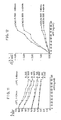
- Figure 11 shows the relationship between A ⁇ /A1000 and AFP concentration
- Figure 12 the relationship between the change in A ⁇ /A1000 versus AFP concentration.
- AFP can also be determined simply and at a high sensitivity based on absorbance ratio between two different wavelengths.
Abstract
Description
- This invention relates to a method of the quantitative determination of antigens or antibodies. More specifically, it relates to a method for quantitative analysis for an antigen (or an antibody) which comprises allowing said antigen (or antibody) to react with an antibody (or antigen) supported on an insoluble carrier of fine particle size, irradiating light onto the resultant antigen-antibody complex, and measuring its absorbance at a specific wavelength - particularly a method that can measure the amount of an antigen or antibody in samples taken from living bodies simply and at a high sensitivity.
- In the medical field, measuring the concentration of antigens or antibodies in samples taken from living bodies has recently been an important item for the diagnosis of diseases. Particularly, there has been a great demand for the development of a high-sensitivity method for quantitative analysis of those components which are present in samples ( for example, the blood ) in minute amounts, such as CRP ( C-reactive protein ) which is an acute reactive substance and AFP ( α-fetoprotein ) which is a tumor marker.
- The method conventionally used for the quantitative analysis of antigens and antibodies comprises dispersing latex particles of carrier supporting an antibody ( or antigen ) in a solvent, allowing an antigen ( or antibody ) to react with said particles, and measuring the increase in turbidity ( or absorbance ) of the dispersion caused by the antigen-antibody reaction at a wavelength in the range from 600 to 2400 nm, thereby determining the amount of said antigen ( or antibody ) ( Japanese Unexamined Patent Publication No.11575/1983 ).
- Another method has been developed recently, which comprises supplying a dispersion containing agglutinated latex particles yielded from an antigen-antibody reaction to a sheath flow so as to make a flow of individual pieces of agglomerates, and analyzing the degree of agglutination by the light scattering method using laser beam as ligh source, thereby determining the antigen (or antibody).
- However, the above-mentioned methods have the following disadvantages.
- In the former method, the change in absorbance due to latex agglutination is very small compared with the absorb ance of the latex dispersion itself. If it is attempted to increase the change in absorbance by properly selecting the wavelength for measurement, the absorbance of the latex dispersion itself also tends to increase. Hence, the end-point assay (subtracting the absorbance of latex dispersion itself from the absorbance measured a sufficient period of time after the start of reaction) is difficult to adopt, and the two-point assay (measuring the change in absorbance at two-points prescribed periods of time after the start of antigen-antibody reaction) or rate assay (measuring the velocity of the change in absorbance), has to be employed. As a result, an automated analyzer must be used which automatically controls the operations from sample and reagent pipetting to absorbance measurement. In addition, the above-mentioned two-point assay requires a large quantity of costly latex reagent because a lower latex concentration results in lower agglutination speed and lower sensitivity.
- Another problem associated with this method is that the change in absorbance is not determined by the degree of latex agglutination alone, because absorbance depends on both the number and size of the particles contained. As shown in Figure 13, cases are known in which, although latex agglutination proceeds with increasing antigen concentration, absorbance begins to decline when the concentration reaches a certain level. Large errors are unavoidable in these cases.
- These disadvantages have been eliminated in the latter method ( the light scattering method ); the result of measurement is dependent only upon the degree of latex agglutination, the end-point assay can be adopted, latex agglutination proceeds and sensitivty becomes higher with increasing reaction time, and sensitivty remains high even when the latex concentration is decreased. However, the problem is that an exclusive apparatus has to be used, because the flow channel must be of a sheath flow structure and a laser beam must be used as light source to detect scattered light from individual particles.
- This invention was accomplished under such circumstances to provide a simple method for measuring the concentration of an antigen (or an antibody) by a kind of end-point assay using a versatile spectrophotometer without having to employ any exclusive apparatus.
- This invention relates to a method of the quantitative determination of an antigen (or an antibody) which comprises adding a sample containing said antigen (or an antibody) being tested to a dispersion of an insoluble carrier of fine particle size with an antibody (or an antigen) fixed thereto to effect an antigen-antibody reaction, measuring absorbance of the reaction mixture, Aλ₁ and Aλ₂ at two different wavelengths, λ₁ and λ₂, and calculating the concentration of said antigen (or antibody) in the sample from the absorbance ratio Aλ₁/Aλ₂.
- This invention is based on the newly found fact that the absorbance ratio between two different wavelengths, Aλ₁/Aλ₂, is a function of the median diameter of particles suspended in the dispersion. The degree of increase in the median particle diameter due to agglutination of the insoluble carrier corresponds to the concentration of antigen ( or antibody ) in the sample; hence, the amount of antigen ( or antibody ) can be simply determined by the value of absorbance ratio Aλ₁/Aλ₂.
- Another merit of this method is that the end-point assay can be adopted, because this ratio scarcely affected by the small change of the particle concentration in sample solution, and depends on the change of the relative particle size to the two wavelengths.
- Thus, quantitative determination of an antigen (or an antibody) by utilizing the agglutination of insoluble carrier caused by an antigen-antibody reaction can be simply effected by the use of a versatile spectrophotometer, eliminating the need for an exclusive apparatus for automatic measurement of the velocity of absorbance changes or a special apparatus using a sheath flow and the laser-beam scattering technique.
- In addition, high-sensitivity and low-cost quantitative analysis not to be expected with conventional methods can be achieved by properly selecting the combination of measuring wavelengths, λ₁ and λ₂, the particle size of insoluble carrier, particle concentration in the dispersion, reaction time and other factors (cell length, etc.).
- Figure 1 is a graph illustrating the absorbance ratio at different wavelengths to that at 1000 nm measured with various latex dispersions being different in median diameter of the latex particle.
- Figure 2 shows the relation ship between A₃₄₀/A₁₀₀₀, A₅₀₀/A₁₀₀₀ and A₆₀₀/A₁₀₀₀ versus latex particle diameter.
- Figures 3, 4 and 5 show graphs illustrating the relationship between absorbance at various wavelengths, absorbance ratios (A₅₀₀/A₁₀₀₀ and A₆₀₀/A₁₀₀₀) and median particle diameter after latex agglutination reaction, versus CRP concentration, respectively.
- Figure 6 shows a graph illustrating the effect of reaction time for the relationship between absorbance at various wavelengths versus CRP concentration.
- Figures 7, 8 and 9 show graphs illustrating the effect of the latex concentration at various reaction times for the relationship between absorbance, absorbance ratio and median particle diameter, respectively, versus CRP concentration.
- Figure 10 show graphs illustrating the effect of the difference between two wavelengths for the relationship between absorbance ratio at various reaction times versus CRP concentration.
- Figure 11 shows graphs illustrating the relationship between concentration of AFP versus absorbance ratio between two different wavelengths at various reaction times.
- Figure 12 illustrates the relationship between the changes in the absorbance ratio, as shown in Figure 11, versus AFP concentration.
- Figure 13 shows graphs illustrating the relationship between the change in absorbance versus the change in median particle diameter caused by agglutination reaction in a model case.
- The antigens and antibodies that can be measured by the process of this invention include those which can exist in samples taken from living bodies. Illustrative examples include albumin, ferritin, AFP, β₂-microglobulin, myoglobin, CRP, ASO, RF, FDP, hCG, hPL, CEA, fibrinogen and gonadotropin (antigens), and immunoglobulins A, E, D, G and M, and γ-globulin (antibodies).
- The insoluble carriers of fine particle size used in the process of this invention are those materials which are insoluble in the dispersing medium employed and capable of fixing the antigens or antibodies. These include polystyrene, carobylated polystyrene, polymethylstyrenes, styrene-butadiene copolymers, carobylated styrene-butadiene copolymers, and poly(meth)acrylates. Preferred dispersing media are aqueous media, such as water, salt solutions and buffer solutions.
- The antibody or antigen to be fixed to the insoluble carrier is selected so that an effective antigen-antibody reaction will take place with the antigen or antibody being measured. Fixation of this antibody or antigen to the insoluble carrier may be effected by any known methods: for example, adding the antibody or antigen to a dispersion of insoluble carrier, and stirring the mixture for a definite time to effect adsorption ( so-called adsorption method ); or adding the antibody ( or antigen ) and a coupling agent to a dispersion of an insoluble carrier bearing carboxyl groups, and stirring the mixture for a definite time to complete the fixation reaction ( so-called covalent bonding method ).
- The particle diameter of insoluble carrier should preferably be in the range from 0.05 to 1.0 µm in terms of absorbance measurement range (usually 2 ABS or lower), and be in the range from 0.1 to 0.2 µm when high-sensitivity measurement is intended. Suitable concentration of the insoluble carrier in the dispersion is such that absorbance at 500 nm will be below 2 ABS, and should preferably be in the range from 1 to 100 mg/ℓ for high-sensitivity measurement.
- Such dispersions of carrier containing an antibody ( or an antigen ) fixed thereto as described above are commercially available, which may be advantageously used for the purpose of this invention.
- Usually, the antigen-antibody reaction may be carried out by adding a test sample ( for example, serum, plasma, lymphocytes and urine ) to a dispersion of insoluble carrier as described above, and by stirring the mixture or allowing it to stand for a definite time. The suitable volume of test sample to be added is about 1/5 to 1/20 that of the dispersion. A reaction time of about 30 minutes usually suffices, but it should preferably be longer for high-sensitivity measurement; 0.5 to 2 hours when the concentration of insoluble carrier is in the range from 1 to 100 mg/l. Temperature has no appreciable effect upon the reaction, but is preferably in the range from 25 to 37°C when considering that samples taken from living bodies are handled.
- Higher sensitivity can be achieved when the two wavelengths used in the process of this invention are more apart from each other. However, since absorbance increases with decreasing wavelength and cannot be measured at low wavelengths, the optimum wavelengths should be selected depending upon the size and concentration of the particles and the concentration range of antigen ( or antibody ) to be measures. In actual practice, it is preferred that two wavelengths, λ₁ and λ₂, be selected within the visible and near infrared regions, usually within the range from 330 to 1000 nm. Within the range as defined above, the larger the difference between λ₁ and λ₂, the higher sensitivity will be achieved. For high-sensitivity measurement, in particular, a wavelength difference in the range from 300 to 600 nm is the most preferred.
- Higher sensitivity is obtainable for a shorter cell length used for absorbance measurement, because a larger difference between two wavelengths can be adopted, but the length should preferably be in the range from 0.5 to 1 cm in terms of S/N ratio.
- The concentration of antigen ( or antibody ) in a test sample can be determined from absorbance ratio Aλ₁/Aλ₂ ( calculated from the values of absorbance measured at λ₁ and λ₂ ) using a calibration curve, which has previously been prepared using serial dilutions of an antigen ( or antibody ) solution of known concentration.
- In this test were used six latices of different particle diameters as listed below ( MISCELLANEOUS POLYSTYRENE SPHERES; products of Duke Scientific Corporation ).

- For each of the above six latices, five grades of dilutions with water were prepared ( for Cat No. 110, preliminary dilution 1:21 followed by further dilution 1:1, 1:2, 1:3, 1:4 and 1:5; and for the other five latices, preliminary dilution 1:101 followed by further dilution 1:1, 1:2, 1:3, 1:4 and 1:5 ). Absorbance was measured for each of the dilutions thus prepared at different wavelengths in the range from 340 nm to 1000 nm, and the ratio of absorbance at varying wavelengths to that at 1000 nm ( Aλ/A₁₀₀₀ ) was calculated and plotted against wavelength. The result is shown in Figure 1. Each of the values of Aλ/A₁₀₀₀ shown in this figure is the average of values for five dilutions of different concnetrations ( n = 5 ). Absorbance at each wavelength increased with increasing latex concentration, but the ratio Aλ/A₁₀₀₀ remained practically constant. For example, the ratio Aλ/A₁₀₀₀ for the latex of 0.260 µm median particle diameter was 14.78 - 15.29, 7.22 - 7.63, 4.22 - 4.51, 2.78 - 2.97, 1.78 - 2.03 and 1.33 - 1.41 for λ = 500, 600, 700, 800 and 900 nm, respectively. Further, the ratio Aλ/A₁₀₀₀ for the latex of 0.804 µm median particle diameter was 3.40 - 3.61, 2.88 - 3.08, 2.44 - 2.35, 1.88 - 1.93, 1.51 - 1.54 and 1.23 - 1.24, respectively.
- Figure 2 shows the relationship between A₃₄₀/A₁₀₀₀, A₅₀₀/A₁₀₀₀ and A₆₀₀/A₁₀₀₀ versus particle diameter based on the data shown in Figure 1. It is apparent that the value of absorbance ratio decreases with increasing particle diameter in each case.
- The result obtained in this test suggests that, in the agglutination reaction of carrier supporting an antibody (or an antigen), the degree of agglutination would increase with increasing concentration of antigen (or antibody) added, resulting in an increase in the median particle diameter and in a decline in the absorbance ratio Aλ/A₁₀₀₀, and that the concentration of the antigen (or antibody) could be determined by this method.
- It was also found that the smaller the particle diameter of latex supporting an antibody (or an antigen), the greater will be the change in the value of Aλ/A₁₀₀₀ after agglutination. This means that the use of a latex of smaller particle diameter allows high-sensitivity measurement for samples of lower antigen (or antibody) con centration, and also broadens the concentration range of antigen (or antibody) measured because of the wider range of particle diameter in which its changes can be measured.
- In addition, it was also demonstrated that the larger the difference between the two wavelengths used for measurement, the greater the changes in absorbance ratio; that is, adoption of two wavelenghts more apart from each other achieves high-sensitivity measurement for samples of lower antigen ( or antibody ) concentration.
- A polystyrene latex ( particle diameter: about 0.2 µm ) with CRP antiserum fixed thereto ( Cellatestam M, CRP latex reagent; product of Hitachi Chemical Co., Ltd. ) was diluted 1: 15 with a buffer solution ( Cellatestam M, CRP diluent; product of Hitachi Chemical Co., Ltd. ), 1500 µℓ of this diluted dispersion was allowed to react with 12 µℓ of standard serum containing a known amount of CRP at 37°C for 90 minutes, and absorbance was measured at different wavelengths (cell-length : 1 cm). Table 1 shows the data at 500, 600 700 and 1000 nm, and absorbance ratios, A₅₀₀/A₁₀₀₀ and A₆₀₀/A₁₀₀₀.

- Figure 3 shows the relationship between absorbance at different wavelengths and CRP concentration ( calibration curves in conventional method ). In this figure, scatters in measured values due to scatters in the amounts of latices added are observed. In addition, the change in absorbance with CRP concentration is large for A₅₀₀, indicating a high sensitivity, but the value of absorbance reaches a maximum and begins to decline at a certain level of CRP concentration. No such reversion of curve is observed for A₁₀₀₀, but the change in absorbance is small ( low sensitivity ) and the S/N ratio is also low at low concentrations ( particularly 1 mg/dl or less ).
- Figure 4 shows calibration curves illustrating the relationship between absorbance ratios, A₅₀₀/A₁₀₀₀ and A₆₀₀/A₁₀₀₀, versus CRP concentration (typical cases of this invention). The change in absorbance ratio is large in low-concentration regions (high sensitivity) and any reversion of curve, as observed in Figure 3, does not occur however high the CRP concentration may be. In addition, no scatter in measured value due to scatter in the amounts of latices added is observed inthis case, because measurement of absorbance ratio at two different wavelengths offsets the scatter in each absorbance.
- Figure 5 shows the relationship between median diameter of antigen-antibody complex (agglutinated particles) obtained from the curve of Figure 2 (A₆₀₀/A₁₀₀₀) versus CRP concentration. This clearly indicates that the process of this invention provides a simple and useful method for quantitative determination of low-concentration antigens and antibodies based on latex agglutination.
- The polystyrene latex with CRP antiserum fixed thereto used in Example 1 was diluted to different concentrations, each of the dilutions thus obtained was allowed to react with standard serum containing a known amount of CRP at 37°C for 30 minutes and 90 minutes, and absorbance was measured at different wavelengths (cell-length : 1 cm). Table 2 shows the data at 340, 500, 600 and 1000 nm, and absorbance ratios, A₃₄₀/A₁₀₀₀, A₅₀₀/A₁₀₀₀ and A₆₀₀/A₁₀₀₀.


- Figure 6 shows the relationship between CRP concentration and Aλ at a dilution ratio of 1/2 based on the data shown in Table 2 ( curves for conventional method in which absorbance is measured only at one wavelength ). It is clear from the figure that, at lower wavelengths, absorbance is excessively high and can hardly be measured in extreme cases, and reversion of curves as observed in Figure 13 takes place.
- Figure 7 shows the relationship between CRP concentration versus absorbance at 600 nm ( A₆₀₀ ) at different dilution ratios based on the data shown in Table 2. It is apparent from this figure that, when measuring the concentration of antigen ( or antibody ) from absorbance at one wavelength ( conventional method ), sensitivity becomes lower as latex concentration is decreased.
- Figure 8 shows the effect of latex dilution ratio upon the relationship between CRP concentration and A₅₀₀/A₁₀₀₀. As can be seen from the figure, sensitivity is low at high CRP concentrations because of the saturation of agglutination, while it remains rather high at low CRP concentrations. This is because the change in median particle diameter increases with decreasing latex concentration as shown in Figure 9. The median particle diameter in Figure 9 was calculated from the values of A₆₀₀/A₁₀₀₀ in Table 2 and the relationship shown in Figure 2.
- These indicate that adoption of a low latex concentration does not lead to any reduction in sensitivity, but achieves high sensitivity if a sufficient long time is taken for the reaction. This also makes for reduction in measurement cost because of the smaller amount of expensive latex reagent to be used.
- Figure 10 shows the relationship between absorbance ratios, A₃₄₀/A₁₀₀₀, A₅₀₀/A₁₀₀₀ and A₆₀₀/A₁₀₀₀, versus CRP concentration at a latex dilution ratio of 1/4 based on the data shown in Table 2. It is apparent from this figure that higher sensivity can be achieved when the two wavelengths used, λ₁ and λ₂, are more apart from each other.
- A polystyrene latex with anti-AFP antibody fixed thereto (anti-AFP antibody sensitized latex; product of IATRON LABORATORIES, INC.) was diluted 1:7 with its stabilizer solution (Product of IATRON), 1400 µℓ of this diluted dispersion was allowed to react with 50 µℓ of AFP solutions of different concentrations at 37°C for 40 and 80 minutes, and absorbance was measure at 400, 500, 600 and 1000 nm (A₄₀₀, A₅₀₀, A₆₀₀ and A₁₀₀₀). Figure 11 shows the relationship between Aλ/A₁₀₀₀ and AFP concentration, and Figure 12 the relationship between the change in Aλ/A₁₀₀₀ versus AFP concentration.
- It was demonstrated that AFP can also be determined simply and at a high sensitivity based on absorbance ratio between two different wavelengths.
- Reproducibility of measured values was studied for values of the decrease in A₆₀₀/A₁₀₀₀. The result obtained was -0.26 in mean value (corresponding to 26.9 ng/mℓ of AFP) and 0.03 in range (corresponding to 4.0 ng/mℓ of AFP), which indicates high reproducibility.
Claims (8)
Applications Claiming Priority (6)
| Application Number | Priority Date | Filing Date | Title |
|---|---|---|---|
| JP61284878A JPH076985B2 (en) | 1986-11-28 | 1986-11-28 | Method for measuring antigen-antibody reaction |
| JP284878/86 | 1986-11-28 | ||
| JP1366/87 | 1987-01-07 | ||
| JP62001366A JPH0635981B2 (en) | 1987-01-07 | 1987-01-07 | Sensitive assay for antigen-antibody reaction |
| JP62001367A JPH0635982B2 (en) | 1987-01-07 | 1987-01-07 | Sensitive assay for antigen-antibody reaction |
| JP1367/87 | 1987-01-07 |
Publications (3)
| Publication Number | Publication Date |
|---|---|
| EP0269526A2 true EP0269526A2 (en) | 1988-06-01 |
| EP0269526A3 EP0269526A3 (en) | 1990-07-04 |
| EP0269526B1 EP0269526B1 (en) | 1993-10-06 |
Family
ID=27274899
Family Applications (1)
| Application Number | Title | Priority Date | Filing Date |
|---|---|---|---|
| EP87402674A Expired - Lifetime EP0269526B1 (en) | 1986-11-28 | 1987-11-26 | Method of quantitative determination of antigens and antibodies |
Country Status (3)
| Country | Link |
|---|---|
| US (1) | US5093271A (en) |
| EP (1) | EP0269526B1 (en) |
| DE (1) | DE3787706T2 (en) |
Cited By (2)
| Publication number | Priority date | Publication date | Assignee | Title |
|---|---|---|---|---|
| EP2265947A2 (en) * | 2008-03-20 | 2010-12-29 | Abaxis, Inc. | Multi-wavelength analyses of sol-particle specific binding assays |
| CN104370761A (en) * | 2014-09-29 | 2015-02-25 | 安徽师范大学 | Benzyl butyl phthalate hapten derivative, preparation method of benzyl butyl phthalate hapten derivative and detection method of benzyl butyl phthalate |
Families Citing this family (9)
| Publication number | Priority date | Publication date | Assignee | Title |
|---|---|---|---|---|
| US5354498A (en) * | 1990-03-16 | 1994-10-11 | Fuji Xerox Co., Ltd. | Phase separation liquid crystal polymer |
| EP0597951B1 (en) * | 1991-07-26 | 1999-03-31 | Dade Chemistry Systems Inc. | An assay with signal detection in the presence of a suspended solid support |
| US5550630A (en) * | 1993-03-19 | 1996-08-27 | The United States Of America As Represented By The Secretary Of Agriculture | Spectrophotometric method for structural analysis of organic compounds, polymers, nucleotides and peptides |
| US6514770B1 (en) * | 1999-07-30 | 2003-02-04 | Mitsubishi Chemical Corporation | Immunoassay |
| AU2003291483A1 (en) * | 2002-11-12 | 2004-06-03 | Becton, Dickinson And Company | Diagnosis of sepsis or sirs using biomarker profiles |
| WO2004095009A1 (en) * | 2003-04-24 | 2004-11-04 | Moritex Corporation | Optical inspection device |
| KR20080006617A (en) | 2005-04-15 | 2008-01-16 | 백톤 디킨슨 앤드 컴퍼니 | Diagnosis of sepsis |
| WO2009123737A2 (en) | 2008-04-03 | 2009-10-08 | Becton, Dickinson And Company | Advanced detection of sepsis |
| EP2635895A1 (en) | 2010-11-03 | 2013-09-11 | Reametrix Inc. | Measurement system for fluorescent detection, and method therefor |
Citations (6)
| Publication number | Priority date | Publication date | Assignee | Title |
|---|---|---|---|---|
| US4174952A (en) * | 1978-01-23 | 1979-11-20 | Massachusetts Institute Of Technology | Immunoassay by light scattering intensity anisotropy measurements |
| US4197088A (en) * | 1977-09-23 | 1980-04-08 | Akro-Medic Engineering, Inc. | Method for qualitative and quantitative determination of immunological reactions |
| EP0070527A1 (en) * | 1981-07-17 | 1983-01-26 | Toray Industries, Inc. | Method of assaying biologically active substances and labelling agents therefor |
| EP0132556A1 (en) * | 1983-06-14 | 1985-02-13 | Kabushiki Kaisha Toshiba | Immunoassay |
| SU1251908A1 (en) * | 1983-12-27 | 1986-08-23 | Институт биохимии и физиологии растений и микроорганизмов АН СССР | Method of determining the rate of reaction of agglutination |
| JPS63149565A (en) * | 1986-12-12 | 1988-06-22 | Sekisui Chem Co Ltd | Immunoassay |
Family Cites Families (5)
| Publication number | Priority date | Publication date | Assignee | Title |
|---|---|---|---|---|
| US3813168A (en) * | 1971-03-19 | 1974-05-28 | Hitachi Ltd | Two-wavelength spectrophotometer |
| JPS5291483A (en) * | 1976-01-28 | 1977-08-01 | Hitachi Ltd | Multi-component analyzer |
| JPS5352180A (en) * | 1976-10-22 | 1978-05-12 | Hitachi Ltd | Two light beams spectrophotometer |
| US4225233A (en) * | 1978-02-02 | 1980-09-30 | The Trustees Of Boston University | Rapid scan spectrophotometer |
| US4954435A (en) * | 1987-01-12 | 1990-09-04 | Becton, Dickinson And Company | Indirect colorimetric detection of an analyte in a sample using ratio of light signals |
-
1987
- 1987-11-24 US US07/124,997 patent/US5093271A/en not_active Expired - Fee Related
- 1987-11-26 DE DE87402674T patent/DE3787706T2/en not_active Expired - Fee Related
- 1987-11-26 EP EP87402674A patent/EP0269526B1/en not_active Expired - Lifetime
Patent Citations (6)
| Publication number | Priority date | Publication date | Assignee | Title |
|---|---|---|---|---|
| US4197088A (en) * | 1977-09-23 | 1980-04-08 | Akro-Medic Engineering, Inc. | Method for qualitative and quantitative determination of immunological reactions |
| US4174952A (en) * | 1978-01-23 | 1979-11-20 | Massachusetts Institute Of Technology | Immunoassay by light scattering intensity anisotropy measurements |
| EP0070527A1 (en) * | 1981-07-17 | 1983-01-26 | Toray Industries, Inc. | Method of assaying biologically active substances and labelling agents therefor |
| EP0132556A1 (en) * | 1983-06-14 | 1985-02-13 | Kabushiki Kaisha Toshiba | Immunoassay |
| SU1251908A1 (en) * | 1983-12-27 | 1986-08-23 | Институт биохимии и физиологии растений и микроорганизмов АН СССР | Method of determining the rate of reaction of agglutination |
| JPS63149565A (en) * | 1986-12-12 | 1988-06-22 | Sekisui Chem Co Ltd | Immunoassay |
Non-Patent Citations (3)
| Title |
|---|
| PATENT ABSTRACTS OF JAPAN vol. 12, no. 413 (P-780) 02 November 1988, & JP-A-63 149565 (SEKISUI CHEM. CO. LTD.) 22 June 1988, * |
| SHIMADZU REVIEW, vol. 43, no. 4, 1986; H. YAMAMOTO et al., pp. 81-90# * |
| WPIL; S.Y.U. SHCHEGOLEV et al., acc. no. 87-106924/15# * |
Cited By (5)
| Publication number | Priority date | Publication date | Assignee | Title |
|---|---|---|---|---|
| EP2265947A2 (en) * | 2008-03-20 | 2010-12-29 | Abaxis, Inc. | Multi-wavelength analyses of sol-particle specific binding assays |
| EP2265947A4 (en) * | 2008-03-20 | 2011-03-30 | Abay Sa | Multi-wavelength analyses of sol-particle specific binding assays |
| US8137920B2 (en) | 2008-03-20 | 2012-03-20 | Abaxis, Inc. | Multi-wavelength analyses of sol-particle specific binding assays |
| US8673576B2 (en) | 2008-03-20 | 2014-03-18 | Abaxis, Inc. | Multi-wavelength analyses of sol-particle specific binding assays |
| CN104370761A (en) * | 2014-09-29 | 2015-02-25 | 安徽师范大学 | Benzyl butyl phthalate hapten derivative, preparation method of benzyl butyl phthalate hapten derivative and detection method of benzyl butyl phthalate |
Also Published As
| Publication number | Publication date |
|---|---|
| EP0269526A3 (en) | 1990-07-04 |
| DE3787706T2 (en) | 1994-02-03 |
| DE3787706D1 (en) | 1993-11-18 |
| EP0269526B1 (en) | 1993-10-06 |
| US5093271A (en) | 1992-03-03 |
Similar Documents
| Publication | Publication Date | Title |
|---|---|---|
| AU724443B2 (en) | Assays using reference microparticles | |
| US9310286B2 (en) | Patient sample classification based upon low angle light scattering | |
| US4080264A (en) | Immunoassay by light scattering spectroscopy | |
| US4118192A (en) | Method and apparatus for the measurement of antigens and antibodies | |
| EP0195623B1 (en) | Stabilized fluorescent rare earth labels and labeled physiologically reactive species | |
| EP0736178B1 (en) | Method of assay | |
| JPH03502246A (en) | Coagulation methods for the analysis of substances | |
| EP0118894A2 (en) | Particle reagent size distribution measurements for immunoassay | |
| EP0269526B1 (en) | Method of quantitative determination of antigens and antibodies | |
| EP1801590B1 (en) | Method of assaying antigen and reagent therefor | |
| EP0263731A1 (en) | Method for measuring antigen-antibody reactions | |
| Gella et al. | Latex agglutination procedures in immunodiagnosis | |
| Buffone et al. | Use of a laser-equipped centrifugal analyzer for kinetic measurement of serum IgG | |
| EP0201755A1 (en) | Immunoassay in centrigufal field with complementary particles of differing specific gravities | |
| Rocks et al. | Automatic analysers in clinical biochemistry | |
| US4379850A (en) | Hemolytic method for the kinetic determination of antistreptolysin O antibodies in blood or serum samples, using oxidized streptolysin O | |
| JP3618797B2 (en) | Immunoassay | |
| EP0433629B1 (en) | A method for the qualitative and quantitative determination of antibodies against bacterial antigens by means of the photometric measurement of agglutination | |
| GB1600139A (en) | Method and apparatus for the measurement of antigens and antibodies | |
| GB1600069A (en) | Method for the measurement of antigens and antibodies | |
| JPS6262291B2 (en) | ||
| CA2179826C (en) | Method of assay | |
| JPH076985B2 (en) | Method for measuring antigen-antibody reaction | |
| JPS63187157A (en) | Measurement of antigen-antibody reaction | |
| JPH09101307A (en) | Method for detecting or measuring immunologically reactive substance |
Legal Events
| Date | Code | Title | Description |
|---|---|---|---|
| PUAI | Public reference made under article 153(3) epc to a published international application that has entered the european phase |
Free format text: ORIGINAL CODE: 0009012 |
|
| AK | Designated contracting states |
Kind code of ref document: A2 Designated state(s): DE FR GB |
|
| PUAL | Search report despatched |
Free format text: ORIGINAL CODE: 0009013 |
|
| AK | Designated contracting states |
Kind code of ref document: A3 Designated state(s): DE FR GB |
|
| 17P | Request for examination filed |
Effective date: 19900830 |
|
| 17Q | First examination report despatched |
Effective date: 19911009 |
|
| GRAA | (expected) grant |
Free format text: ORIGINAL CODE: 0009210 |
|
| AK | Designated contracting states |
Kind code of ref document: B1 Designated state(s): DE FR GB |
|
| REF | Corresponds to: |
Ref document number: 3787706 Country of ref document: DE Date of ref document: 19931118 |
|
| ET | Fr: translation filed | ||
| PLBE | No opposition filed within time limit |
Free format text: ORIGINAL CODE: 0009261 |
|
| STAA | Information on the status of an ep patent application or granted ep patent |
Free format text: STATUS: NO OPPOSITION FILED WITHIN TIME LIMIT |
|
| 26N | No opposition filed | ||
| REG | Reference to a national code |
Ref country code: GB Ref legal event code: 746 Effective date: 19950927 |
|
| REG | Reference to a national code |
Ref country code: FR Ref legal event code: D6 |
|
| PGFP | Annual fee paid to national office [announced via postgrant information from national office to epo] |
Ref country code: FR Payment date: 20011113 Year of fee payment: 15 |
|
| PGFP | Annual fee paid to national office [announced via postgrant information from national office to epo] |
Ref country code: GB Payment date: 20011128 Year of fee payment: 15 |
|
| PGFP | Annual fee paid to national office [announced via postgrant information from national office to epo] |
Ref country code: DE Payment date: 20011210 Year of fee payment: 15 |
|
| REG | Reference to a national code |
Ref country code: GB Ref legal event code: IF02 |
|
| PG25 | Lapsed in a contracting state [announced via postgrant information from national office to epo] |
Ref country code: GB Free format text: LAPSE BECAUSE OF NON-PAYMENT OF DUE FEES Effective date: 20021126 |
|
| PG25 | Lapsed in a contracting state [announced via postgrant information from national office to epo] |
Ref country code: DE Free format text: LAPSE BECAUSE OF NON-PAYMENT OF DUE FEES Effective date: 20030603 |
|
| GBPC | Gb: european patent ceased through non-payment of renewal fee | ||
| PG25 | Lapsed in a contracting state [announced via postgrant information from national office to epo] |
Ref country code: FR Free format text: LAPSE BECAUSE OF NON-PAYMENT OF DUE FEES Effective date: 20030731 |
|
| REG | Reference to a national code |
Ref country code: FR Ref legal event code: ST |