EP0265778A1 - Factor VIII-C analogs - Google Patents
Factor VIII-C analogs Download PDFInfo
- Publication number
- EP0265778A1 EP0265778A1 EP87115043A EP87115043A EP0265778A1 EP 0265778 A1 EP0265778 A1 EP 0265778A1 EP 87115043 A EP87115043 A EP 87115043A EP 87115043 A EP87115043 A EP 87115043A EP 0265778 A1 EP0265778 A1 EP 0265778A1
- Authority
- EP
- European Patent Office
- Prior art keywords
- factor viii
- analog
- sequence
- dna
- cdna
- Prior art date
- Legal status (The legal status is an assumption and is not a legal conclusion. Google has not performed a legal analysis and makes no representation as to the accuracy of the status listed.)
- Withdrawn
Links
Images


Classifications
-
- C—CHEMISTRY; METALLURGY
- C07—ORGANIC CHEMISTRY
- C07K—PEPTIDES
- C07K14/00—Peptides having more than 20 amino acids; Gastrins; Somatostatins; Melanotropins; Derivatives thereof
- C07K14/435—Peptides having more than 20 amino acids; Gastrins; Somatostatins; Melanotropins; Derivatives thereof from animals; from humans
- C07K14/745—Blood coagulation or fibrinolysis factors
- C07K14/755—Factors VIII, e.g. factor VIII C (AHF), factor VIII Ag (VWF)
-
- A—HUMAN NECESSITIES
- A61—MEDICAL OR VETERINARY SCIENCE; HYGIENE
- A61K—PREPARATIONS FOR MEDICAL, DENTAL OR TOILETRY PURPOSES
- A61K38/00—Medicinal preparations containing peptides
Definitions
- This invention relates to the field of structural gene cloning and the use of such genes in the recombinant DNA directed synthesis of desired protein products. More particularly, it relates to new analogs of Factor VIII procoagulant activity protein (Factor VIII-C), its recombinant DNA directed synthesis and use in the treatment of coagulation disorders, such as hemophilia.
- Factor VIII-C Factor VIII procoagulant activity protein
- Genes coding for various polypeptides may be cloned by incorporating a DNA fragment coding for the polypeptide in a recombinant DNA vehicle, e.g., bacterial or viral vectors, and transforming a suitable host, typically an Escherichia coli (E. coli) cell line, and isolating clones incorporating the recombinant vectors. Such clones may be grown and used to produce the desired polypeptides on a large scale.
- a recombinant DNA vehicle e.g., bacterial or viral vectors
- E. coli Escherichia coli
- RNA-directed DNA polymerase also called reverse transcriptase.
- Reverse transcriptase synthesizes DNA in the 5 ⁇ -3 ⁇ direction, utilizes deoxyribonucleoside 5 ⁇ -triphosphates as precursors, and requires both a template and a primer stand, the latter of which must have a free 3 ⁇ -hydroxyl terminus.
- Reverse transcriptase products whether partial or complete copies of the mRNA template, often possess short, partially double-stranded hairpins ("loops") at their 3 ⁇ termini.
- these "hairpin loops” can be exploited as primers for DNA polymerases.
- Preformed DNA is required both as a template and as a primer in the action of DNA polymerase.
- the DNA polymerase requires the presence of a DNA strand having a free 3 ⁇ -hydroxyl group, to which new nucleotide residues are added to extend the chain in the 5 ⁇ -3 ⁇ direction.
- the products of such sequential reverse transcriptase and DNA polymerase reactions still possess a loop at one end.
- this single-strand segment is cleaved with the single-strand specific nuclease S1 to generate a "blunt- end" duplex DNA segment.
- This general method is applicable to any mRNA mixture, and is described by Buell, et al., J. Biol. Chem. 253 :2483 (1978),
- ds-cDNA double-stranded cDNA
- cloning vehicles any one of many known techniques, depending at least in part on the particular vehicle being used.
- Various insertion methods are discussed in considerable detail in Methods In Enzymology , 68 :16-18, and the references cited therein.
- the cloning vehicle is used to transform a suitable host.
- These cloning vehicles usually impart an antibiotic resistance trait on the host.
- Such hosts are generally prokaryotic or eukaryotic cells.
- Only a few of the transformed or transfected hosts contain the desired cDNA.
- the sum of all transformed or transfected hosts constitutes a gene "library".
- the overall ds-cDNA library created by this method provides a representative sample of the coding information present in the mRNA mixture used as the starting material.
- an appropriate oligonucleotide sequence it can be used to identify clones of interest as follows. Individual transformed or transfected cells are grown as colonies on nitrocellulose filter paper. These colonies are lysed, the DNA so-released is covalently attached to the filter paper by heating, and the sheet is incubated with a labeled oligonucleotide probe.
- the probe sequence is complementary to the structural gene of interest. The probe hybridizes with the ds-cDNA for which it is complementary and is identified by autoradiography.
- the clones are characterized to identify a clone containing all the structural information for the desired protein.
- the nucleic acid sequence coding for the protein of interest is isolated and reinserted into an expression vector.
- the expression vector brings the clones gene under the regulatory control of a specific prokaryotic or eukaryotic control element which allows the efficient expression (transcription and translation) of the cloned full-length ds-cDNA.
- this general technique is only applicable to those proteins for which at least a portion of their amino acid or DNA sequence is known and for which an oligonucleotide probe is available. See, generally, Maniatis, et al., supra.
- Factor VIII complex Normal human blood plasma contains a complex of two proteins which is referred to as the Factor VIII complex.
- One component of the Factor VIII complex has antihemophilic factor procoagulant activity and is designated Factor VIII-C.
- a deficiency in Factor VIII-C is characteristic of hemophilia, a disease transmitted by X-chromosomal inheritance.
- Factor VIII-C has therapeutic value because of its ability to correct the clotting deficiency in hemophilic patients.
- the available methods of obtaining Factor VIII-C are limited to fractionation of human plasma, as described above.
- Producing Factor VIII-C by plasma fractionation provides only limited quantities which vary from preparation to preparation, are expensive, and subject the recipient hemophiliac to the risk of acquiring blood transmitted diseases such as hepatitis and acquired immune deficiency syndrome (AIDS).
- AIDS acquired immune deficiency syndrome
- Factor VIII-C analogs could provide an alternative agent to be used in the treatment of hemophilia. Analogs may prove more efficient than native Factor VIII-C in restoring a normal clotting cascade. Secondly, production of Factor VIII-C analogs via recombinant DNA technology would reduce the disease-transmission risks associated with plasma-fractionated products. Whereas the administration of a genetically e ngineered analog, whose secondary or tertiary structure is sufficiently different from native Factor VIII-C, may result in an immune response, native analogs are not expected to trigger this response. Genetically engineered analogs to Factor VIII-C, therefore, could provide a dependable and readily available therapeutic agent to be used in the treatment of hemophilia.
- the present invention has made it possible to provide readily available large quantities of Factor VIII-C analogs. This has been achieved with nucleic acids and antibodies which react specifically with the Factor VIII-C analog coding sequence of the Factor VIII-C analog protein molecule or the Factor VIII-C protein molecule (or fragments thereof); with the application of recombinant DNA technology to preparing cloning vehicles encoding for the Factor VIII-C analogs and screening/isolating procedures for recovering human Factor VIII-C analogs essentially free of other proteins of human origin.
- the present invention provides human Factor VIII-C analogs or those fragments essentially free of other proteins of human origin.
- the Factor VIII-C analogs are produced by recombinant DNA techniques in host cells or other self-replicating systems and are provided in essentially pure form. Also provided are methods and compositions for preparing the above-described Factor VIII-C analogs as well as therapeutic compositions and uses for the Factor VIII-C analogs or subunit, in the treatment of coagulation disorders in humans and animals.
- the invention further provides replicable expression vectors incorporated with a DNA sequence encoding Factor VIII-C analogs and host cell or cell-free self-replicating systems transformed or transfected thereby.
- the host system is usually of prokaryotic origin, e.g. E. coli or B. subtilis , or eukaryotic cells.
- the Factor VIII-C analogs are produced by a process which comprises (a) preparing a replicable expression vector capable of expressing the DNA sequence encoding a Factor VIII-C analog in a suitable host cell or cell-free replicating system; (b) transforming said host system to obtain a recombinant host system; (c) maintaining said recombinant host system under conditions permitting expression of said Factor VIII-C analog encoding the DNA sequence to produce Factor VIII-C analog.
- the Factor VIII-C analog encoding replicable expression vector is made from a double-stranded complementary DNA (ds-cDNA) preparation representative of a messenger RNA pool containing messenger RNA for Factor VIII-C and incorporating DNA from the ds-cDNA pool into replicable expression vectors.
- the preferred mode of recovering Factor VIII-C analogs comprises reacting the proteins expressed by the recombinant host system with a reagent composition comprising at least one binding protein specific for Factor VIII-C analog.
- Figure 1 illustrates the preparation of cDNA from mRNA.
- Factor VIII-C analog denotes human Factor VIII-C analogs, or subunits thereof, produced in in vitro cell culture systems, in bioactive forms having the capacity to initiate blood coagulation as does Factor VIII-C native to human plasma.
- Analog of Factor VIII-C analogs may exist in nature. These variations may be characterized by difference(s) in the nucleotide sequence of the structural gene coding for proteins of identical biological function. These variations may also rise from alternate mRNA splicing patterns. It is possible to engineer analogs having single or multiple amino acid substitutions, deletions, additions, or replacements. All such variations, modifications, and analogs (naturally derived, synthetic or genetically engineered) resulting in derivatives of Factor VIII-C which retain the biologically active properties of native Factor VIII-C are included within the scope of this invention.
- the analog may be a recombinant-derived protein identical to analogs identified in cDNA libraries or newly engineered analogs with no counterparts in vivo .
- “Expression vectors” refer to vectors which are capable of transcribing and translating DNA sequences contained therein, where such sequences are linked to other regulatory sequences capable of effecting their expression. These expression vectors must be replicable in the host organisms or systems either as episomes, bacteriophage, or as an integral part of the chromosomal DNA.
- One expression vector which is particularly suitable for producing human Factor VIII-C analogs is the bacteriophage, viruses which normally inhabit and replicate in bacteria. Particularly desirable phages for this purpose are the lambda gt10 and gt11 phage, described by Young and Davis, supra . Lambda gt11 is a general recombinant DNA expression vector capable of producing polypeptides specified by the inserted DNA.
- Another form of expression vector useful in recombinant DNA techniques is the plasmid - a circular, unintegrated (extra-chromosomal), double-stranded DNA loop. Any other form of expression vector which serves an equivalent function is suitable for use in the process of this invention.
- Recombinant vectors and methodology disclosed herein are suitable for use in host cells covering a wide range of prokaryotic and eukaryotic organisms.
- Prokaryotics are preferred for the cloning of DNA sequences and in the construction of vectors.
- E . coli K12 strain HB101 ATCC No. 33694
- other microbial strains may be used, vectors containing replication and control sequence which are derived from species compatible with the host cell or system are used in connection with these hosts.
- the vector ordinarily carries an origin of replication, as well as characteristics capable of providing phenotypic selection in transformed cells.
- E . coli can be transformed using the vector pBR322, which contains genes for ampicillin and tetracycline resistance [Bolivar, et al., Gene , 2 :95 (1977)].
- the expression vector may also contain control elements which can be used by the vector for expression of its own proteins.
- Common prokaryotic control elements used for expression of foreign DNA sequences in E . coli include the promoters and regulatory sequences derived from the B-galactosidase and tryptophan (trp) operons of E . coli , as well as the pR and pL promoters of bacteriophage lambda. Combinations of these elements have also been used (e.g., TAC, which is a fusion of the trp promoter with the lactose operator).
- Other promoters have also been discovered and utilized, and details concerning their nucleotide sequences have been published enabling a skilled worker to combine and exploit them functionally.
- eukaryotic microbes such as yeast cultures
- Saccharomyces cerevisiae or common baker's yeast, is the most commonly used among eukaryotic microorganisms, although a number of other strains are commonly available.
- Suitable promoting sequences in yeast vectors include the promoters of 3-phosphoglycerate kinase or other glycolytic enzymes.
- Suitable expression vectors may contain termination signals which provide for the polyadenylation and termination of the cloned gene's mRNA. Any vector containing a yeast-compatible promoter, origin of replication, and appropriate termination sequence is suitable for expression of Factor VIII-C analogs.
- cultures of cells derived from multicellular organisms may also be used as hosts.
- any such cell culture is workable, whether from a vertebrate or invertebrate source.
- interest has been greatest in vertebrate cells and propation of vertebrate cells in culture (tissue culture) has become a routine procedure in recent years.
- useful hosts are the VERO, HeLa, mouse C127, Chinese hamster ovary (CHO), W138, BHK, COS-7, and MDCK cell lines.
- Expression vectors for such cells ordinarily include an origin of replication, a promoter located in front of the gene to be expressed, along with any necessary ribosome binding sites, RNA splice sites, polyadenylation site, and transcriptional terminatory sequence.
- control functions on the expression vectors are often provided by viral material.
- promoters are derived from polyoma, Adenovirus 2, and most frequently, Simian Virus 40 (SV40).
- SV40 Simian Virus 40
- eukaryotic enhancer sequences can also be added to the construction. These sequences can be obtained from a variety of animal cells, animal viruses or oncogenic retroviruses such as the mouse sarcoma virus.
- An origin of replication may be provided either by construction of the vector to include an exogenous origin, such as that provided by SV40 or other viral sources, or may be provided by the host cell chromosomal replication mechanism. If the vector is integrated into the host cell chromosome, the latter is sufficient.
- BPV bovine papillomavirus
- Use of BPV DNA as a viral vector is well documented and was originally based on the observation that the viral genome persists as an extrachromosomal plasmid in transformed cells (Law. M.-F., D.R. Lowy, I. Dvoretzky, and P.M. Howley, Proc . Natl . Acad . Sci . USA, 78 :2727-2731, 1981).
- the cloned gene is maintained in a uniform sequence environment of the BpV minichromosome, eliminating potential problems associated with the integration of the cloned DNA into inactive regions of the host chromosome.
- Host cells can prepare human Factor VIII-C analogs which can be of a variety of chemical compositions.
- the protein is produced having methionine as its first amino acid (present by virtue of the ATG start signal codon naturally existing at the origin of the structural gene or inserted before a segment of the structural gene).
- the protein may also be intra- or extracellularly cleaved, giving rise to the amino acid which is found naturally at the amino terminus of the protein.
- the protein may be produced together with either its signal polypeptide or a conjugated protein other than the conventional signal polypeptide; the signal polypeptide of the conjugate being specifically cleavable in an intra- or extracellular environment.
- Factor VIII-C analogs may be produced by direct expression in mature form without the necessity of cleaving away any extraneous polypeptide.
- Recombinant host cells are cells which have been transformed with vectors constructed using recombinant DNA techniques. As defined herein, Factor VIII-C analogs are produced as a consequence of this transformation. Factor VIII-C analogs or its subunits produced by such cells are referred to as "recombinant Factor VIII-C analogs”.
- the procedures below are but some of a wide variety of well-established procedures to produce specific reagents useful in this invention.
- the general procedures for obtaining a messenger RNA (mRNA) mixture is to prepare an extract from a tissue sample or to culture cells producing the desired protein, and to extract the mRNA by a process such as that disclosed by Cirgwin, et al., Biochemistry , 18 :5294 (1979).
- the mRNA is enriched by poly(A) mRNA-containing material by chromatography on oligo (dT) cellulose of poly(U) Sepharose, followed by elution of the poly(A) containing mRNA-enriched fraction.
- the above poly(A) containing mRNA-enriched fraction is used to synthesize a single-strand complementary cDNA (ss-cDNA) using reverse transcriptase.
- ss-cDNA single-strand complementary cDNA
- reverse transcriptase reverse transcriptase
- the resultant double-strand cDNA (ds-cDNA) is inserted into the expression vector by any one of many known techniques. In general, methods, etc., can be found in Maniatis, supra , and Methods In Enzymology , Vols. 65 and 68 (1980); and Vols. 100 and 101 (1983). In general, the vector is linearized by at least one restriction endonuclease, which will produce at least two blunt or cohesive ends. The ds-cDNA is ligated with or joined to the vector insertion site.
- prokaryotic cells or other cells which contain substantial cell wall material are employed, the most common method of transformation with the expression vector is calcium chloride pretreatment as described by Cohen, R.N., et al., Proc . Nat'l . Sci . USA , 69 :2110 (1972). If cells without c ell wall barriers are used as host cells, transfection is carried out by the calcium phosphate precipitation method described by Graham and Van der Eb, Virology , 62 :456 (1973). Other methods for introducing DNA into cells such as nuclear injection or protoplast fusion, have also been successfully used. The organisms are then cultured on selective media and proteins for which the expression vector encodes are produced.
- Clones containing part or the entire cDNA for Factor VIII-C analogs are identified with specific antibodies directed against part or all of the Factor VIII-C analog protein. This method of identification requires that the ds-cDNA be inserted into a vector containing appropriate regulatory nucleic acid sequences adjacent to the insertion site. These regulatory sequences initiate transcription and translation of those ds-cDNA molecules inserted in the vector. Those clones containing Factor VIII-C analog cDNA sequences correctly positioned relative to the regulatory sequences, synthesize part or all of the Factor VIII-C analog protein. Such clones are detected using appropriately specific antibodies. Such a cloning system is the lambda gt11 system first described by Young and Davis, supra .
- Clones containing the entire sequence of Factor VIII-C analogs are identified using the cDNA insert of the Factor VIII-C analogs recombinants isolated during the initial screening of the recombinant lambda gt11 cDNA library as a probe. Nucleotide sequencing techniques are used to determine the sequence of amino acids encoded by the cDNA fragments. This information may be used to determine the identity of cDNA clones as specific for human Factor VIII-C analogs by comparison to the known amino acid sequence of the amino-terminus of Factor VIII-C (Gitshier et al., and Wood et al., supra ). Alternatively, identification may be confirmed by employing techniques such as Northern blot analysis and hybrid-selected translation or by comparison to Factor VIII-C clones isolated from other species, e.g., mouse, rat, etc.
- the top figure depicts the complete FVIII protein molecule with the various A, B and C domains.
- the lower figure depicts the engineered truncated delta FVIII protein molecule.
- the B domain is devoid of amino acid residues 747-1560.
- the thrombin cleavage sites delineating the B domain (740/741 and 1689/1690) remain intact.
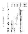
- the top line illustrates the complete FVIII cDNA starting at the methionine initiation codon (ATG) and ending with the translation termination codon (TGA).
- the restriction sites EcoR1, Sph1 and BamH1 indicate the positions used to engineer the deleted (delta FVIII) cDNA.
- the lower figure illustrates the resulting variant.
- the deleted variant (delta FVIII) contains an internal, contiguous deletion, spanning nt 2296-4737 (see Figure 15-18).
- a cDNA library was constructed in lambda gt11 from adult liver polyA-selected RNA (Ricca, unpublished) and a series of overlapping clones encompassing all but - 500 bp of EX14 region were identified and sequenced.
- a genomic clone of - 3000 bp spanning the missing region was isolated in EMBL3. This genomic clone and the appropriate cDNA clones were assembled into full length FVIII containing all 26 exons and cloned in pGEM2 (Promega). The resulting plasmid designated as p181-7.
- the following plasmids describe the construction schema used to generate a deletion in the exon 14 region of FVIII DNA. the numbering system used throughout is based on bp #1 being the A in the ATG translation initiation codon.
- p159-1 This plasmid contains the entire 5 ⁇ region (4744 bp) of FVIII plus additional 20 bp upstream of the ATG start codon. The 5 ⁇ terminus has been modified to an Xhol recognition site with synthetic linkers (collaborative research) for subsequent cloning. The 3 ⁇ end is deliminted by FVIII BamH1 site (nt 4744).
- p167-2 p159-1 was cleaved to completion with Sph1 (nt 3936) followed by a partial digest with EcoR1 (nt 2290). The ensuing gap was bridged with the following synthetic oligonucleotide containing cohesive EcoR1 and Sph1 ends (5-3) and an internal BamH1 site:
- the sequence between the EcoR1 to the BamH1 sites is identical to nucleotides 2290-2295 followed by nucleotides 4738-4744 in FVIII map.
- p182-4 A plasmid containing the fully assembled FVIII cDNA (p181-7) was cleaved with BamH1 and the resulting large fragment, devoid of the internal BamH1 fragment (nt 1870-4744), was doubly purified electrophoretically in agarose gel and recovered by electroelution. Plasmid p167-x was cleaved with BamH1, the small internal fragment containing the deletion in exon 14 similarly purified, and then ligated to the large BamH1 fragment of p181-7. DNA was used to transform competent E.
- coli HB101 and plasmid DNA isolated from several ampicillin resistant transformants analyzed by restriction mapping to identify plasmids harboring the mutated DNA with the BamH1 fragment in the proper orientation. Plasmid DNA was purified and the sequence across the mutated region verified by Maxam Gilbert sequencing. The salient features of p182-x include a 2442 bp internal deletion in exon 14 (nt 2296-4737); 20 bp upstream and 214 bp downstream of FVIIIC coding region, and an Xhol site at the 5 ⁇ and 3 ⁇ termini.
- p182-4-ATG-C To increase translation efficiency, the region surrounding the ATC codon was replaced with synthetic oligonucleotide conforming to a consensus 'AUG' sequence (Kozak 1984) but otherwise identical to FVIII sequence. This was accomplished by cleaving p182-4 at the AatII site located in the pGEM2 sequence upstream of FVIII insert, and Sac1 (nt 15, Figure 2). The large fragment lacking the AstII/Sac1 fragment was purified and ligated to the synthetic oligonucleotide indicated below containing an internal consensus ATG sequence, cohesive AatII and Sac1 termini (5-3) and internal Xhol site 20 bp upstream of the ATG codon
- delta FVIII insert was recovered as an XHO1 fragment from p182-4-ATG-C and cloned in BPV shuttle vector p143-7.
- the resulting recombinant vector, p261-26, contains the following genetic elements described in clockwise direction: BPV genomic lacking 1485 bp frm its nontransforming region (nt 5473-6958 in BPV map, Chen, 1982); PML2d sequence, Moloney murine sarcoma virus (MSV) enhancer; followed by mouse metallothionein promotor (both described in Sarver, et al ., 1985); delta FVIII DNA insert from p182-ATG-C; and RNA processing signals from SV40 genome (described in Sarver, et al ., 1985).
- p188-3 This expression vector is identical to p261-26 except for the presence of a full-length FVIII cDNA insert (from p182-4) instead of the deleted version.
- the recombinant delta FVIII protein was also known to be neutralizable by monoclonal antibody directed against the 72 kd carboxy subunit of the protein, to be inhibited by EDTA (SmM) due to chelation of CA+2 ions which are necessary for the association of the various peptides in the active molecules, and to be activated by thrombin (ten-fold increase in activity within one minute of incubation of 37°C).
- Factor VIIIC deficient plasma (George King Biomedical, Inc., Overland Park, KS) was removed from -70°C freezer, thawed at 37°C and transferred to 12 x 75 polystyrene tube. Actin (activated cephaloplastin, stored at 4°C) was transferred to 12 x 75 tube and placed on ice until use. A solution of 35 mM calcium chloride was prewarmed at 37°C. Reference normal plasma (George King Biomedicals) was thawed or reconstituted as per supplier and placed on ice until use.
- citrate albumin buffer 14.3 mM Na citrate/12.8 mM NaCl/6.1 mM Na Azide/0.5% albumin, pH 6.7
- Additional serial dilution at 1:20, 1:40, 1:80 and 1:160 were made in CAB solution.
- test sample containing greater than 0.4 U/ml For analysis of test sample containing greater than 0.4 U/ml, the same dilution as for the reference was used. Samples containing less than 0.4 U/ml a 2-fold dilution of series beginning with 1:5 was used. All samples were handled as described above.
- a standard curve is obtained by relating clotting times to Factor VIIIC activity in reference samples. Clotting times at each dilution are plotted on the ordinate of a log-log paper and the reciprocol of the dilution is plotted on the abscissa. A test fit line is drawn. Results obtained with the samples are drawn on same graph and a line of best fit which also parallels the reference line is constructed. The ratio between dilutions for the reference and the samples which give the same clotting time is determined. This ratio is then multiplied by the assigned activity of the reference to obtain activity/ml in the sample.
- RNA messenger, ribosomal, and transfer was extracted from fresh human umbilical vein endothelial cells essentially as described by Chirgwin, supra , (1979).
- Cell pellets were homogenized in 15 volumes of a solution containing 4 M guanidine thiocyanate, 25 mM sodium citrate at pH 7.0, 0.5% N-laurylsarcosine, 0.1 M 2-mercaptoethanol, and 0.2% Antifoam A (Sigma Chemical Co., St. Louis, MO). The homogenate was centrigued at 6,000 rpm in a Sorvall GSA rotor for 15 minutes at 10°C.
- RNA precipitate prepared as described above, was dissolved at a concentration of 40 A260 units in 20 mM HEPES buffer (pH 7.2) containing 10 mM EDTA and 1% SDS, heated at 65°C for 10 minutes, then quickly cooled to 25°C. The RNA solution was then diluted with an equal volume of water and NaCl was added to bring the final concentration to 300 mM NaCl. Samples containing up to 24000 A260 units of RNA were chromatographed on poly(U)-Sepharose using standard procedures. Poly(A)-containing RNA was eluted with 90% formamide containing 1 mM HEPES buffer (pH 7.2), and 2 mM EDTA. The eluate was adjusted to 0.24 M NaCL, and the RNA was precipitated by 2.5 volumes of ethanol at 02-°C.
- the reaction mixture consisted of 0.2 M sodium acetate (pH 4.5), 0.4 (M) sodium chloride, 2.5 mM zinc acetate and 0.1 unit of S1 nuclease per ng of ds-cDNA made to a final reaction volume of 100 ul.
- the ds-cDNA was incubated at 37°C for one hour, extracted with phenol:chloroform, and then desalted on a Sephadex G-50 column as described above .
- the ds-cDNA was then treated with Eco RI methylase and DNA polymerase I (Klenow) using reaction conditions described in Maniatis (supra) .
- the cDNA was again desalted on a Sephadex G-50 as decribed above and then ligated to 0.5 g of phosphorylated Eco RI linkers using T4 DNA ligase (Manitis, supra ).
- DNA with a size greater than 1 kilobase was eluted from the gel, recovered by binding to an Elutip-D column, eluted with 1 M Nacl and then collected by ethanol precipitation.
- the DNA fragments were then inserted into Eco RI-cleaved and phosphatase-treated lambda gt11 using T4 DNA ligase to produce a library of approximately twelve (12) million phage of which 50% contain inserts (i.e., six million clear plaques on x-gal plates).
- the library was amplified by producing plate stocks at 42°C on E . coli Y1088 [supE supF metB trpR hsdR ⁇ hsdm+ tonA21 strA lacU169 proc::Tn5 (pMC9)]. Amplification procedures are described in Maniatis, (supra) .
- supF reactive suppression of the phage amber mutation in the S gene
- hsdR ⁇ hsdm+ non-recombinants
- lacU169 a deletion of the lac operon which reduces host-phage recombination and which is necessary to distinguish between lambda gt11 recombinants and non-recombinants
- pMC9 a lacI-bearing pBR322 derivative which represses, in the absence of an inducer, the expression of foreign genes that may be detrimental to phage and/or cell growth.
- the tier of the amplified library was determined be 8 x 10¹⁰ pfu/ml.
- Albumin cDNA clones represent 3-5% of the clear plagues in the library as judged by hybridization with a nick-translated albumin cDNA clone.
- the filters were then incubated at room temperature overnight in a 1:100 dilution of an emu polyclonal antibody directed against Factor VIII-C in a buffer consisting of 1% gelatin in TBS.
- the antibody was prepared as described by Fulcher and Zimmerman, Proc . Nat . Acad . Sci . USA , 79 :1648 (1984).
- the filters were then incubated with a 1:200 dilution of rabbit anti-emu IgG. After 2 additional 30 minute washes with TBS, the filters were incubated with a 1:2,000 dilution of horseradish peroxidase (HRP) conjugated goat anti-rabbit antisera (Bio-Rad, Richmond, CA). The filters were then incubated for 2 hours at room temperature, and washed twice for 30 minutes in TBS. The filters were incubated at room temperature in HRP color development solution as described in the Bio-Rad accompanying literature. Twenty-six putative positive plaques were identified.
- HRP horseradish peroxidase
- a 4-mm diameter plug at the position of the color development signal was removed from the plates and incubated in 10 mM Tris HCl, pH 7.5, and 10 mM MgSO4 overnight. Approximately 103 plaque-forming units (PFU) were replated on 90 mm plates and rescreened as described above. Five clones remained positive through the second cycle of screening. This replating and rescreening process was repeated until all plaques on the plate produced a signal.
- PFU plaque-forming units
- the cDNA insert from one of the clones was excised using a combination of the restriction enzymes EcoRI and PvuI.
- the Eco RI site to the right of the cloned cDNA was unexpectedly missing in all 5 clones; thus PvuI, which cuts at position approximately 20066 Kb in lambda gt11, was used in combination with Eco RI.
- the cDNA insert (approximately 1.2 k:lobases in length) was subcloned between the PvuI and EcoRI sites of pSP64 and subsequently used as a probe to rescreen the lambda gt11 library and a human placental genomic library constructed in bacteriophage EMBL3.
- Factor VIII-C analogs have been genetically engineered. The sequence of two such analogs are shown in Figures 15-18 and Figures 12-22. As shown in Figures 4 and 15-18, a Factor VIII-C analog DNA has been engineered in which nucleotides 2296-4737 (as numbered in Figures 5-9) have been removed. This region contains a great portion of, but not all, Exon 14 deleted. The deletion is contained within the Exon 14 region and encodes a portion of the Factor VIII-C molecule which is removed during thrombin activation of Factor VIII-C during the clotting cascade. Our designation for this clone is pd delta BPV/delta F8C-ATG-C (26126).
- a second Factor VIII-C analog has been engineered in which the Exon 4 deletion and the engineered deletion of most of Exon 14 are combined, resulting in the cDNA whose sequence is shown in Figures 19-22.
- BPV DNA The complete BPV genome (early and late regions) from which the sequence between nucleotides 5463 and 6958 had been deleted is contained in this construct. This sequence was shown to be dispensible for cellular transformation and extrachromosomal replication (Sarver, unpublished results). The HindIII recognition sequence at the site of deletion has been abolished.
- pML2D1 This pBR322 derivate lacks the sequences (1095-2485) which inhibit replication of the prokaryotic DNA in mammalian cells [Lusky and Botchan, Nature 293:79 (1981)].
- the original pML2 was further modified by deleting the sequences between the HindIII and SalI (29 and 651, respectively, in the pBR322 map).
- the original HindIII s ite was converted to a BamHI site, whereas the original SalI site was retained.
- M-MSV Enhancer The Moloney murine sarcoma virus enhancer fragment extends from the HinfI (Base 141) to the XbaI (base 525) site in the proviral long terminal repeat ([Dhar, et al., Proc . Natl . Acad . Sci . USA , 77 :3937 (1980)]. Both sites were converted to BamHI sites with synthetic linkers to facilitate cloning.
- the mouse metallothionein promoter is the KpnI [approximately (-)300] to BglII [(+)365] fragment with the CAP site (mRNA start site) at (+)301.
- the BglII site was converted to an XhoI site with synthetic linkers to facilitate cloning. Since the coding region of the M-MT gene beings downstream (at +374) from the 3 ⁇ end (position +365) of the promoter described herein, no fusion products result.
- Factor VIII-C Analog Insert Derivation of the complete Factor VIII-C cDNA (sans most of the Exon 14 coding sequences) is detailed in Section D above.
- the insert contains 20 base pairs 5 ⁇ upstream of the first ATG codon in the Factor VIII-C sequence, the sequence encoding for the signal peptide, the coding region, a translational termination codon, and 214 base pairs of Factor VIII-C 3 ⁇ flanking sequences.
- the fragment terminates with a PvuII site.
- the 5 ⁇ and 3 ⁇ termini were modified to XhoI sites (with synthetic oligonucleotides and synthetic linkers, respectively) to facilitate subsequent cloning.
- This fragment also contains a consensus ATG sequence (M. Kozak nucleic acid Research 12:857-872 1984).
- the SV40-derived RNA processing elements consist of a fused fragment comprised of two noncontiguous regions of the SV40 genome.
- a 610 base pair region (4710 - 4100 on the SV40 map) contains an intron (66 bp) from the early region of the viral genome. This is fused to a 237 base pair fragment (2770 - 2533 on the SV40 map) containing the polyadenylation signal at position 2586.
- the BglII site at the 5; end was converted to an XhoI site with synthetic linkers to permit subsequent ligation.
- the BamHI site at the 3 ⁇ end is ligated to the BamHI at the 5 ⁇ end of the BPV genome to complete the circular structure.
- pd delta BPV/delta FVIII.ATG.c (p261-26) is 15433 base pairs in length.
- Mouse C127 cells [Lowy, D.R., et al., J . Virol ., 26 :72 (1978)] were maintained in Dulbecco modified Eagle medium (Meloy Laboratories, Springfield, VA ) supplemented with penicillin (10 U/ml), streptomycin (100 ug/ml), and 10% heat-inactivated fetal bovine serum (GIBCO).
- Dulbecco modified Eagle medium Meloy Laboratories, Springfield, VA
- penicillin 10 U/ml
- streptomycin 100 ug/ml
- GIBCO heat-inactivated fetal bovine serum
- DNA transformation was performed by using the calcium precipitation method (Graham and van der Eb, supra ) followed by a dimethyl sulfoxide [Stowe and Wolkie, J . Gen . Virol ., 33 :447 (1976)] or glycerol enhancement [Frost and Williams, Virology , 91 :39-50 (1978)].
- Portions representing 1 ug of the recombinant DNA were added to cell cultures in 60-mm petri dishes containing 4 ml of fresh medium, and incubation was contained at 37°C for 4 hours. After the medium was changed, the cells were treated with either 1 ml of 25% dimethylsulfoxide in HBS for 4 minutes at room temperature or with 15% glycerol in HBS for 2 minutes at room temperature. The monolayers were washed again and refed with fresh medium.
- Factor VIII-C analog cDNA was confirmed by Southern blot DNA analysis and the sequence verified by the dideoxy chain termination DNA sequencing technique [Sanger, et al., Proc . Natl . Acad . Sci . USA , 74 :5463-5467 (1977)].
- Expression of Factor VIII-C analog protein was verified by Northern blot RNA analysis and by in vitro translation of RNAs. The RNAs were either extracted from Factor VIII-C analog engineered cells or were obtained from an in vitro transcription system based on the SP6 method [Melton, et al., Nuc . Acids Res ., 12 :7035-7056 (1984)]. This was followed by immunoprecipitations, competitive immunoprecipitation of extracellular and intracellular proteins produced by recombinant cells, clotting assays, and biological activity assays.
- a deposit of biologically pure cultures of the following strains was made with the American Type Culture Collection, 12301 Parklawn Drive, Rockville, Maryland, the accession number indicated was assigned after successful viability testing, and the requisite fees were paid. Access to said culture will be available during pendency of the patent application to one determined by the above-mentioned Culture Collection Agency. Said culture will remain permanently available for a term of at least five years after the most recent request for the furnishing of a sample and in any case for a period of at least 30 years after the date of the deposit. Should the culture become nonviable or be inadventently destroyed, it will be replace with a viable culture(s) of the same taxonomic description.
Abstract
Analogs of Factor VIII-C and their recombinant DNA directed synthesis are disclosed. These analogs have use in the treatment of coagulation disorders.
Description
- This invention relates to the field of structural gene cloning and the use of such genes in the recombinant DNA directed synthesis of desired protein products. More particularly, it relates to new analogs of Factor VIII procoagulant activity protein (Factor VIII-C), its recombinant DNA directed synthesis and use in the treatment of coagulation disorders, such as hemophilia.
- In general, recombinant DNA techniques are known. See Methods in Enzymology, (Academic Press) Volumes 65 and 68 (1979); 100 and 101 (1983) and the references cited therein, all of which are incorporated herein by reference. An extensive technical discussion embodying most commonly used recombinant DNA methodologies can be found in Maniatis, et al., Molecular Cloning, Cold Spring Harbor Laboratory (1982). Genes coding for various polypeptides may be cloned by incorporating a DNA fragment coding for the polypeptide in a recombinant DNA vehicle, e.g., bacterial or viral vectors, and transforming a suitable host, typically an Escherichia coli (E. coli) cell line, and isolating clones incorporating the recombinant vectors. Such clones may be grown and used to produce the desired polypeptides on a large scale.
- Mixtures of mRNA from eukaryotic cells employing a series of three enzymatic reactions to synthesize double-stranded DNA copies of entire genes which are complementary to this mRNA mixture have been isolated. In the first reaction, mRNA is transcribed to form a single-strand complementary DNA (ss-cDNA) by an RNA-directed DNA polymerase, also called reverse transcriptase. Reverse transcriptase synthesizes DNA in the 5ʹ-3ʹdirection, utilizes deoxyribonucleoside 5ʹ-triphosphates as precursors, and requires both a template and a primer stand, the latter of which must have a free 3ʹ-hydroxyl terminus. Reverse transcriptase products, whether partial or complete copies of the mRNA template, often possess short, partially double-stranded hairpins ("loops") at their 3ʹ termini. In the second reaction, these "hairpin loops" can be exploited as primers for DNA polymerases. Preformed DNA is required both as a template and as a primer in the action of DNA polymerase. The DNA polymerase requires the presence of a DNA strand having a free 3ʹ-hydroxyl group, to which new nucleotide residues are added to extend the chain in the 5ʹ-3ʹ direction. The products of such sequential reverse transcriptase and DNA polymerase reactions still possess a loop at one end. The apex of the loop or "fold-point" of the double-stranded DNA, which has thus been created, is substantially a single-strand segment. In the third reaction, this single-strand segment is cleaved with the single-strand specific nuclease S1 to generate a "blunt- end" duplex DNA segment. This general method is applicable to any mRNA mixture, and is described by Buell, et al., J. Biol. Chem. 253:2483 (1978),
- The resulting double-stranded cDNA (ds-cDNA) mixture is inserted into cloning vehicles by any one of many known techniques, depending at least in part on the particular vehicle being used. Various insertion methods are discussed in considerable detail in Methods In Enzymology, 68:16-18, and the references cited therein.
- Once the DNA segment is inserted, the cloning vehicle is used to transform a suitable host. These cloning vehicles usually impart an antibiotic resistance trait on the host. Such hosts are generally prokaryotic or eukaryotic cells. At this point, only a few of the transformed or transfected hosts contain the desired cDNA. The sum of all transformed or transfected hosts constitutes a gene "library". The overall ds-cDNA library created by this method provides a representative sample of the coding information present in the mRNA mixture used as the starting material.
- If an appropriate oligonucleotide sequence is available, it can be used to identify clones of interest as follows. Individual transformed or transfected cells are grown as colonies on nitrocellulose filter paper. These colonies are lysed, the DNA so-released is covalently attached to the filter paper by heating, and the sheet is incubated with a labeled oligonucleotide probe. The probe sequence is complementary to the structural gene of interest. The probe hybridizes with the ds-cDNA for which it is complementary and is identified by autoradiography. The clones are characterized to identify a clone containing all the structural information for the desired protein. The nucleic acid sequence coding for the protein of interest is isolated and reinserted into an expression vector. The expression vector brings the clones gene under the regulatory control of a specific prokaryotic or eukaryotic control element which allows the efficient expression (transcription and translation) of the cloned full-length ds-cDNA. Thus, this general technique is only applicable to those proteins for which at least a portion of their amino acid or DNA sequence is known and for which an oligonucleotide probe is available. See, generally, Maniatis, et al., supra.
- More recently, methods have been developed to identify specific clones by probing bacterial colonies with antibodies specific for the encoded protein of interest. This method can only be used with "expression vector" cloning vehicles since elaboration of the product protein is required. The structural gene is inserted into the vector adjacent to the regulatory gene lysed either by the vector or by chemical methods, and the protein detected by the specific antibody and a labeling system, e.g., enzyme immunoassay. An example of this is the lambda gt₁₁ system described by Young and Davis, Proc. Nat'l. Acad. Sci. USA, 80:1194-1198 (1983) and Young and Davis, Science, 22:778 (1983).
- Normal human blood plasma contains a complex of two proteins which is referred to as the Factor VIII complex. One component of the Factor VIII complex has antihemophilic factor procoagulant activity and is designated Factor VIII-C. A deficiency in Factor VIII-C is characteristic of hemophilia, a disease transmitted by X-chromosomal inheritance.
- There is little information in the literature about Factor VIII-C analogs. Analogs to Factor VIII-C can be characterized by differences in the nucleic acid or amino acid sequence of Factor VIII-C as published in Gitschier, et al., [Nature, 312:326-330 (1984)] and by Wood, et al., [ Nature, 312:330-337 (1984)]. Such analogs may arise from a mutation in the genetic code, from alternative mRNA splicing, or from post-translational modifications. No such Factor VIII-C analogs have been heretofore isolated or genetically engineered.
- Factor VIII-C has therapeutic value because of its ability to correct the clotting deficiency in hemophilic patients. Unfortunately, the available methods of obtaining Factor VIII-C are limited to fractionation of human plasma, as described above. Producing Factor VIII-C by plasma fractionation provides only limited quantities which vary from preparation to preparation, are expensive, and subject the recipient hemophiliac to the risk of acquiring blood transmitted diseases such as hepatitis and acquired immune deficiency syndrome (AIDS).
- Factor VIII-C analogs could provide an alternative agent to be used in the treatment of hemophilia. Analogs may prove more efficient than native Factor VIII-C in restoring a normal clotting cascade. Secondly, production of Factor VIII-C analogs via recombinant DNA technology would reduce the disease-transmission risks associated with plasma-fractionated products. Whereas the administration of a genetically e ngineered analog, whose secondary or tertiary structure is sufficiently different from native Factor VIII-C, may result in an immune response, native analogs are not expected to trigger this response. Genetically engineered analogs to Factor VIII-C, therefore, could provide a dependable and readily available therapeutic agent to be used in the treatment of hemophilia.
- In recognizing and overcoming the above-described problems, the present invention has made it possible to provide readily available large quantities of Factor VIII-C analogs. This has been achieved with nucleic acids and antibodies which react specifically with the Factor VIII-C analog coding sequence of the Factor VIII-C analog protein molecule or the Factor VIII-C protein molecule (or fragments thereof); with the application of recombinant DNA technology to preparing cloning vehicles encoding for the Factor VIII-C analogs and screening/isolating procedures for recovering human Factor VIII-C analogs essentially free of other proteins of human origin.
- Accordingly, the present invention provides human Factor VIII-C analogs or those fragments essentially free of other proteins of human origin. The Factor VIII-C analogs are produced by recombinant DNA techniques in host cells or other self-replicating systems and are provided in essentially pure form. Also provided are methods and compositions for preparing the above-described Factor VIII-C analogs as well as therapeutic compositions and uses for the Factor VIII-C analogs or subunit, in the treatment of coagulation disorders in humans and animals.
- The invention further provides replicable expression vectors incorporated with a DNA sequence encoding Factor VIII-C analogs and host cell or cell-free self-replicating systems transformed or transfected thereby. The host system is usually of prokaryotic origin, e.g. E. coli or B. subtilis, or eukaryotic cells.
- The Factor VIII-C analogs are produced by a process which comprises (a) preparing a replicable expression vector capable of expressing the DNA sequence encoding a Factor VIII-C analog in a suitable host cell or cell-free replicating system; (b) transforming said host system to obtain a recombinant host system; (c) maintaining said recombinant host system under conditions permitting expression of said Factor VIII-C analog encoding the DNA sequence to produce Factor VIII-C analog. The Factor VIII-C analog encoding replicable expression vector is made from a double-stranded complementary DNA (ds-cDNA) preparation representative of a messenger RNA pool containing messenger RNA for Factor VIII-C and incorporating DNA from the ds-cDNA pool into replicable expression vectors. The preferred mode of recovering Factor VIII-C analogs comprises reacting the proteins expressed by the recombinant host system with a reagent composition comprising at least one binding protein specific for Factor VIII-C analog.
- In the accompanying drawings, Figure 1 illustrates the preparation of cDNA from mRNA.
- Figure 2 illustrates the preparation of a cDNA library and subsequent screening for FVIIIC specific clones.
- Figure 3 diagrams the assembly of full length Factor VIII-C.
- Figure 4A illustrates the design of a Factor VIII protein.
- Figure 4B illustrates the complete Factor VIII cDNA and the resulting variant.
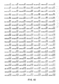
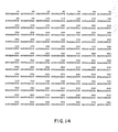
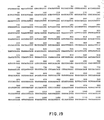
- Figures 5-9 illustrates the DNA sequence of assembled full-length Factor VIII-C.
- Figures 10-14 illustrate the DNA sequence of naturally occurring Factor VIII-C analog with
Exon 4 deletion. - Figures 15-18 illustrate the DNA sequence of genetically engineered Factor VIII-C analog with a major portion of Exon 14 deleted.
- Figures 19-22 illustrate the DNA sequence of genetically engineered Factor VIII-C analog with
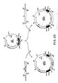
Exon 4 and 14 deletions. - Figure 23 illustrates plasmids used in the invention.
- Figure 24 illustr ates plasmid p182-4-ATG-C.
- Figure 25 illustrates plasmids p-261-26 and p188-3.
- As used herein, "Factor VIII-C analog" denotes human Factor VIII-C analogs, or subunits thereof, produced in in vitro cell culture systems, in bioactive forms having the capacity to initiate blood coagulation as does Factor VIII-C native to human plasma.
- Analog of Factor VIII-C analogs may exist in nature. These variations may be characterized by difference(s) in the nucleotide sequence of the structural gene coding for proteins of identical biological function. These variations may also rise from alternate mRNA splicing patterns. It is possible to engineer analogs having single or multiple amino acid substitutions, deletions, additions, or replacements. All such variations, modifications, and analogs (naturally derived, synthetic or genetically engineered) resulting in derivatives of Factor VIII-C which retain the biologically active properties of native Factor VIII-C are included within the scope of this invention. The analog may be a recombinant-derived protein identical to analogs identified in cDNA libraries or newly engineered analogs with no counterparts in vivo.
- "Expression vectors" refer to vectors which are capable of transcribing and translating DNA sequences contained therein, where such sequences are linked to other regulatory sequences capable of effecting their expression. These expression vectors must be replicable in the host organisms or systems either as episomes, bacteriophage, or as an integral part of the chromosomal DNA. One expression vector which is particularly suitable for producing human Factor VIII-C analogs is the bacteriophage, viruses which normally inhabit and replicate in bacteria. Particularly desirable phages for this purpose are the lambda gt₁₀ and gt₁₁ phage, described by Young and Davis, supra. Lambda gt₁₁ is a general recombinant DNA expression vector capable of producing polypeptides specified by the inserted DNA.
- To minimize degradation, upon induction with a synthetic analog of lactose (IPTG), foreign proteins or portions thereof are synthesized fused to the prokaryotic protein B-galactosidase. The use of host cells defective in protein degradation pathways may also increase the lifetime of novel proteins produced from the induced lambda gt₁₁ clones. Proper expression of foreign DNA in lambda gt₁₁ clones depends upon the proper orientation and reading frame of the inserted DNA with respect to the B-galactosidase promoter and ribosome binding site.
- Another form of expression vector useful in recombinant DNA techniques is the plasmid - a circular, unintegrated (extra-chromosomal), double-stranded DNA loop. Any other form of expression vector which serves an equivalent function is suitable for use in the process of this invention.
- Recombinant vectors and methodology disclosed herein are suitable for use in host cells covering a wide range of prokaryotic and eukaryotic organisms. Prokaryotics are preferred for the cloning of DNA sequences and in the construction of vectors. For example, E. coli K12 strain HB101 (ATCC No. 33694) is particularly useful. Of course, other microbial strains may be used, vectors containing replication and control sequence which are derived from species compatible with the host cell or system are used in connection with these hosts. The vector ordinarily carries an origin of replication, as well as characteristics capable of providing phenotypic selection in transformed cells. For example, E. coli can be transformed using the vector pBR322, which contains genes for ampicillin and tetracycline resistance [Bolivar, et al., Gene, 2:95 (1977)].
- These antibiotic resistance genes provide a means of identifying transformed cells. The expression vector may also contain control elements which can be used by the vector for expression of its own proteins. Common prokaryotic control elements used for expression of foreign DNA sequences in E. coli include the promoters and regulatory sequences derived from the B-galactosidase and tryptophan (trp) operons of E. coli, as well as the pR and pL promoters of bacteriophage lambda. Combinations of these elements have also been used (e.g., TAC, which is a fusion of the trp promoter with the lactose operator). Other promoters have also been discovered and utilized, and details concerning their nucleotide sequences have been published enabling a skilled worker to combine and exploit them functionally.
- In addition to prokaryotes, eukaryotic microbes, such as yeast cultures, may also be used. Saccharomyces cerevisiae, or common baker's yeast, is the most commonly used among eukaryotic microorganisms, although a number of other strains are commonly available. Suitable promoting sequences in yeast vectors include the promoters of 3-phosphoglycerate kinase or other glycolytic enzymes. Suitable expression vectors may contain termination signals which provide for the polyadenylation and termination of the cloned gene's mRNA. Any vector containing a yeast-compatible promoter, origin of replication, and appropriate termination sequence is suitable for expression of Factor VIII-C analogs.
- In addition to microorganisms, cultures of cells derived from multicellular organisms may also be used as hosts. In principle, any such cell culture is workable, whether from a vertebrate or invertebrate source. However, interest has been greatest in vertebrate cells and propation of vertebrate cells in culture (tissue culture) has become a routine procedure in recent years. Examples of such useful hosts are the VERO, HeLa, mouse C127, Chinese hamster ovary (CHO), W138, BHK, COS-7, and MDCK cell lines. Expression vectors for such cells ordinarily include an origin of replication, a promoter located in front of the gene to be expressed, along with any necessary ribosome binding sites, RNA splice sites, polyadenylation site, and transcriptional terminatory sequence.
- For use in mammalian cells, the control functions on the expression vectors are often provided by viral material. For example, commonly used promoters are derived from polyoma,
Adenovirus 2, and most frequently, Simian Virus 40 (SV40). Further, it is also possible, and often desirable, to utilize promoter or control sequences naturally associated with the desired gene sequence, provided such control sequences are compatible with the host system. To increase the rate of transcription, eukaryotic enhancer sequences can also be added to the construction. These sequences can be obtained from a variety of animal cells, animal viruses or oncogenic retroviruses such as the mouse sarcoma virus. - An origin of replication may be provided either by construction of the vector to include an exogenous origin, such as that provided by SV40 or other viral sources, or may be provided by the host cell chromosomal replication mechanism. If the vector is integrated into the host cell chromosome, the latter is sufficient.
- Specific to the example contained herein is the bovine papillomavirus (BPV). Use of BPV DNA as a viral vector is well documented and was originally based on the observation that the viral genome persists as an extrachromosomal plasmid in transformed cells (Law. M.-F., D.R. Lowy, I. Dvoretzky, and P.M. Howley, Proc. Natl. Acad. Sci. USA, 78:2727-2731, 1981). In this form, the cloned gene is maintained in a uniform sequence environment of the BpV minichromosome, eliminating potential problems associated with the integration of the cloned DNA into inactive regions of the host chromosome. This property was fully exploited in the establishment of BPV shuttle vectors capable of replicating in prokaryotic and eukaryotic cells [Sarver, N., J.C. Byrne, and P.M. Howley, Proc. Natl. Acad. Sci. USA, 79(13):4030-4034, 1982; and Kushner, P.J., B.B. Levinson, and H.M. Goodman, J. Mol. Appl. Genet., 1(6):527-528, 1982]. Such vectors are efficient in directing the regulated expression from inducible promoters and in expressing gene products destined for intra- and extracellular location. Vectors which may be employed as starting materials useful for the construction of the vectors of the subject invention are described in U.S. Patent 4,419, 446, the contents of which are incorporated herein by reference.
- Host cells can prepare human Factor VIII-C analogs which can be of a variety of chemical compositions. The protein is produced having methionine as its first amino acid (present by virtue of the ATG start signal codon naturally existing at the origin of the structural gene or inserted before a segment of the structural gene). The protein may also be intra- or extracellularly cleaved, giving rise to the amino acid which is found naturally at the amino terminus of the protein. The protein may be produced together with either its signal polypeptide or a conjugated protein other than the conventional signal polypeptide; the signal polypeptide of the conjugate being specifically cleavable in an intra- or extracellular environment. Finally, Factor VIII-C analogs may be produced by direct expression in mature form without the necessity of cleaving away any extraneous polypeptide.
- Recombinant host cells are cells which have been transformed with vectors constructed using recombinant DNA techniques. As defined herein, Factor VIII-C analogs are produced as a consequence of this transformation. Factor VIII-C analogs or its subunits produced by such cells are referred to as "recombinant Factor VIII-C analogs".
- The procedures below are but some of a wide variety of well-established procedures to produce specific reagents useful in this invention. The general procedures for obtaining a messenger RNA (mRNA) mixture is to prepare an extract from a tissue sample or to culture cells producing the desired protein, and to extract the mRNA by a process such as that disclosed by Cirgwin, et al., Biochemistry, 18:5294 (1979). The mRNA is enriched by poly(A) mRNA-containing material by chromatography on oligo (dT) cellulose of poly(U) Sepharose, followed by elution of the poly(A) containing mRNA-enriched fraction.
- The above poly(A) containing mRNA-enriched fraction is used to synthesize a single-strand complementary cDNA (ss-cDNA) using reverse transcriptase. As a consequence of DNA synthesis, a hairpin loop is formed at the 3ʹ end of the DNA which will initiate second-strand DNA synthesis. Under appropriate conditions, this hairpin loop is used to effect synthesis of the second strand in the presence of DNA polymerase and nucleotide triphosphates.
- The resultant double-strand cDNA (ds-cDNA) is inserted into the expression vector by any one of many known techniques. In general, methods, etc., can be found in Maniatis, supra, and Methods In Enzymology, Vols. 65 and 68 (1980); and Vols. 100 and 101 (1983). In general, the vector is linearized by at least one restriction endonuclease, which will produce at least two blunt or cohesive ends. The ds-cDNA is ligated with or joined to the vector insertion site.
- If prokaryotic cells or other cells which contain substantial cell wall material are employed, the most common method of transformation with the expression vector is calcium chloride pretreatment as described by Cohen, R.N., et al., Proc. Nat'l. Sci. USA, 69:2110 (1972). If cells without c ell wall barriers are used as host cells, transfection is carried out by the calcium phosphate precipitation method described by Graham and Van der Eb, Virology, 62:456 (1973). Other methods for introducing DNA into cells such as nuclear injection or protoplast fusion, have also been successfully used. The organisms are then cultured on selective media and proteins for which the expression vector encodes are produced.
- Clones containing part or the entire cDNA for Factor VIII-C analogs are identified with specific antibodies directed against part or all of the Factor VIII-C analog protein. This method of identification requires that the ds-cDNA be inserted into a vector containing appropriate regulatory nucleic acid sequences adjacent to the insertion site. These regulatory sequences initiate transcription and translation of those ds-cDNA molecules inserted in the vector. Those clones containing Factor VIII-C analog cDNA sequences correctly positioned relative to the regulatory sequences, synthesize part or all of the Factor VIII-C analog protein. Such clones are detected using appropriately specific antibodies. Such a cloning system is the lambda gt₁₁ system first described by Young and Davis, supra.
- Clones containing the entire sequence of Factor VIII-C analogs are identified using the cDNA insert of the Factor VIII-C analogs recombinants isolated during the initial screening of the recombinant lambda gt₁₁ cDNA library as a probe. Nucleotide sequencing techniques are used to determine the sequence of amino acids encoded by the cDNA fragments. This information may be used to determine the identity of cDNA clones as specific for human Factor VIII-C analogs by comparison to the known amino acid sequence of the amino-terminus of Factor VIII-C (Gitshier et al., and Wood et al., supra). Alternatively, identification may be confirmed by employing techniques such as Northern blot analysis and hybrid-selected translation or by comparison to Factor VIII-C clones isolated from other species, e.g., mouse, rat, etc.
- The top figure depicts the complete FVIII protein molecule with the various A, B and C domains. The lower figure depicts the engineered truncated delta FVIII protein molecule. The B domain is devoid of amino acid residues 747-1560. The thrombin cleavage sites delineating the B domain (740/741 and 1689/1690) remain intact.
- The top line illustrates the complete FVIII cDNA starting at the methionine initiation codon (ATG) and ending with the translation termination codon (TGA). The restriction sites EcoR1, Sph1 and BamH1 indicate the positions used to engineer the deleted (delta FVIII) cDNA.
- The lower figure illustrates the resulting variant. The deleted variant (delta FVIII) contains an internal, contiguous deletion, spanning nt 2296-4737 (see Figure 15-18).
- A cDNA library was constructed in lambda gt11 from adult liver polyA-selected RNA (Ricca, unpublished) and a series of overlapping clones encompassing all but - 500 bp of EX14 region were identified and sequenced. A genomic clone of - 3000 bp spanning the missing region was isolated in EMBL3. This genomic clone and the appropriate cDNA clones were assembled into full length FVIII containing all 26 exons and cloned in pGEM2 (Promega). The resulting plasmid designated as p181-7. The following plasmids describe the construction schema used to generate a deletion in the exon 14 region of FVIII DNA. the numbering system used throughout is based on
bp # 1 being the A in the ATG translation initiation codon. - p159-1: This plasmid contains the entire 5ʹ region (4744 bp) of FVIII plus additional 20 bp upstream of the ATG start codon. The 5ʹ terminus has been modified to an Xhol recognition site with synthetic linkers (collaborative research) for subsequent cloning. The 3ʹ end is deliminted by FVIII BamH1 site (nt 4744).
- p167-2: p159-1 was cleaved to completion with Sph1 (nt 3936) followed by a partial digest with EcoR1 (nt 2290). The ensuing gap was bridged with the following synthetic oligonucleotide containing cohesive EcoR1 and Sph1 ends (5-3) and an internal BamH1 site:

- The sequence between the EcoR1 to the BamH1 sites is identical to nucleotides 2290-2295 followed by nucleotides 4738-4744 in FVIII map.
- p182-4: A plasmid containing the fully assembled FVIII cDNA (p181-7) was cleaved with BamH1 and the resulting large fragment, devoid of the internal BamH1 fragment (nt 1870-4744), was doubly purified electrophoretically in agarose gel and recovered by electroelution. Plasmid p167-x was cleaved with BamH1, the small internal fragment containing the deletion in exon 14 similarly purified, and then ligated to the large BamH1 fragment of p181-7. DNA was used to transform competent E. coli HB101 and plasmid DNA isolated from several ampicillin resistant transformants analyzed by restriction mapping to identify plasmids harboring the mutated DNA with the BamH1 fragment in the proper orientation. Plasmid DNA was purified and the sequence across the mutated region verified by Maxam Gilbert sequencing. The salient features of p182-x include a 2442 bp internal deletion in exon 14 (nt 2296-4737); 20 bp upstream and 214 bp downstream of FVIIIC coding region, and an Xhol site at the 5ʹ and 3ʹ termini.
- p182-4-ATG-C: To increase translation efficiency, the region surrounding the ATC codon was replaced with synthetic oligonucleotide conforming to a consensus 'AUG' sequence (Kozak 1984) but otherwise identical to FVIII sequence. This was accomplished by cleaving p182-4 at the AatII site located in the pGEM2 sequence upstream of FVIII insert, and Sac1 (nt 15, Figure 2). The large fragment lacking the AstII/Sac1 fragment was purified and ligated to the synthetic oligonucleotide indicated below containing an internal consensus ATG sequence, cohesive AatII and Sac1 termini (5-3) and
internal Xhol site 20 bp upstream of the ATG codon
- p-261-26: delta FVIII insert was recovered as an XHO1 fragment from p182-4-ATG-C and cloned in BPV shuttle vector p143-7. The resulting recombinant vector, p261-26, contains the following genetic elements described in clockwise direction: BPV genomic lacking 1485 bp frm its nontransforming region (nt 5473-6958 in BPV map, Chen, 1982); PML2d sequence, Moloney murine sarcoma virus (MSV) enhancer; followed by mouse metallothionein promotor (both described in Sarver, et al., 1985); delta FVIII DNA insert from p182-ATG-C; and RNA processing signals from SV40 genome (described in Sarver, et al., 1985).
- p188-3: This expression vector is identical to p261-26 except for the presence of a full-length FVIII cDNA insert (from p182-4) instead of the deleted version.

- Supernatant media from arbitrarily selected transformed lines were assayed for delta FVIII biological activity defined as activity which will correct the coagulation defect of FVIII deficient plasma. The activity is expressed as the clotting time required for the formation of a visible fibrin clot in Factor VIII deficient plasma upon the addition of samples containing Factor VIII activity. The shorter the clotting time, the greater the Factor VIII activity in a given sample. When used in conjunction with reference samples with known activity levels (U/ml), the clotting time is then proportioned to Factor VIIIC activity level. As shown above, coagulation activities in these isolates the range from 0.93-1.325 u/ml of media. Normal levels of circulating FVIII are defined as 1 u/ml.
- The recombinant delta FVIII protein was also known to be neutralizable by monoclonal antibody directed against the 72 kd carboxy subunit of the protein, to be inhibited by EDTA (SmM) due to chelation of CA⁺² ions which are necessary for the association of the various peptides in the active molecules, and to be activated by thrombin (ten-fold increase in activity within one minute of incubation of 37°C).
- All assays were performed in 12 x 75 polystyrene tubes (American Scientific Products). Factor VIIIC deficient plasma (George King Biomedical, Inc., Overland Park, KS) was removed from -70°C freezer, thawed at 37°C and transferred to 12 x 75 polystyrene tube. Actin (activated cephaloplastin, stored at 4°C) was transferred to 12 x 75 tube and placed on ice until use. A solution of 35 mM calcium chloride was prewarmed at 37°C. Reference normal plasma (George King Biomedicals) was thawed or reconstituted as per supplier and placed on ice until use.
- Immediately before use, an appropriate dilution of the reference was made in citrate albumin buffer (CAB, 14.3 mM Na citrate/12.8 mM NaCl/6.1 mM Na Azide/0.5% albumin, pH 6.7) resulting in a 1 U/ml Factor VIIIC concentration. Additional serial dilution at 1:20, 1:40, 1:80 and 1:160 were made in CAB solution. To a cuvette which was been preheated at 37°C and in rapid succession, were added 100 mu l of Factor VIIIC deficient plasma, 100 mu l of actin, and 100 mu l of 1:20 dilution of the reference. The procedure was repeated for the remaining dilutions at 90-second intervals.
- Each mixture was incubated for precisely 5 minutes. The coagulation cascade was initiated with the addition of 100 mu l of prewarmed calcium chloride (35mM), and the time required for the formation of a fibrin clot was carefully monitored.
- For analysis of test sample containing greater than 0.4 U/ml, the same dilution as for the reference was used. Samples containing less than 0.4 U/ml a 2-fold dilution of series beginning with 1:5 was used. All samples were handled as described above.
- A standard curve is obtained by relating clotting times to Factor VIIIC activity in reference samples. Clotting times at each dilution are plotted on the ordinate of a log-log paper and the reciprocol of the dilution is plotted on the abscissa. A test fit line is drawn. Results obtained with the samples are drawn on same graph and a line of best fit which also parallels the reference line is constructed. The ratio between dilutions for the reference and the samples which give the same clotting time is determined. This ratio is then multiplied by the assigned activity of the reference to obtain activity/ml in the sample.
- Total RNA (messenger, ribosomal, and transfer) was extracted from fresh human umbilical vein endothelial cells essentially as described by Chirgwin, supra, (1979). Cell pellets were homogenized in 15 volumes of a solution containing 4 M guanidine thiocyanate, 25 mM sodium citrate at pH 7.0, 0.5% N-laurylsarcosine, 0.1 M 2-mercaptoethanol, and 0.2% Antifoam A (Sigma Chemical Co., St. Louis, MO). The homogenate was centrigued at 6,000 rpm in a Sorvall GSA rotor for 15 minutes at 10°C. The supernatant fluid was adjusted to pH 5.0 by addition of acetic acid and the RNA precipitated by 0.75 volumes of ethanol at -20°C for two hours. RNA was collected by centrifugation and dissolved in 7.5 M guanidine hydrochloride containing 2 mM sodium citrate and 5 mM dithiotreitol. Following two additional precipitations using 0.5 volumes of eth anol, the residual guanidine hydrochloride was extracted from the precipitate with absolute ethanol. RNA was dissolved in sterile water, insolvable material was removed by centrifugation, and the pellet was re-extracted with water. The RNA was adjusted to 0.2 M potassium acetate and precipitated by addition of 2.5 volumes of ethanol at -20°C overnight.
- The total RNA precipitate, prepared as described above, was dissolved at a concentration of 40 A₂₆₀ units in 20 mM HEPES buffer (pH 7.2) containing 10 mM EDTA and 1% SDS, heated at 65°C for 10 minutes, then quickly cooled to 25°C. The RNA solution was then diluted with an equal volume of water and NaCl was added to bring the final concentration to 300 mM NaCl. Samples containing up to 24000 A₂₆₀ units of RNA were chromatographed on poly(U)-Sepharose using standard procedures. Poly(A)-containing RNA was eluted with 90% formamide containing 1 mM HEPES buffer (pH 7.2), and 2 mM EDTA. The eluate was adjusted to 0.24 M NaCL, and the RNA was precipitated by 2.5 volumes of ethanol at 02-°C.
- The procedures followed for the enzymatic reaction is shown in Figure 1. The mRNA (20 ug) was copied into ds-cDNA with reverse transcriptase and DNA polymerase I exactly as described by Buell, et al., supra, and Wildensen, et al., J. Biol. Chem., 253:2483 (1978). The ds-cDNA was desalted on Sephadex G-50 and the void volume fractions further purified on an Elutip-D column (Schleicher & Schuell, Kenne, NH) following the manufacturer's directions. The ds-cDNA was made blunt-ended by incubation with S1 nuclease, [Ricca, et al., J. Biol. Chem. 256:10362 (1981)]. The reaction mixture consisted of 0.2 M sodium acetate (pH 4.5), 0.4 (M) sodium chloride, 2.5 mM zinc acetate and 0.1 unit of S1 nuclease per ng of ds-cDNA made to a final reaction volume of 100 ul. The ds-cDNA was incubated at 37°C for one hour, extracted with phenol:chloroform, and then desalted on a Sephadex G-50 column as described above .
- The ds-cDNA was then treated with EcoRI methylase and DNA polymerase I (Klenow) using reaction conditions described in Maniatis (supra). The cDNA was again desalted on a Sephadex G-50 as decribed above and then ligated to 0.5 g of phosphorylated EcoRI linkers using T₄ DNA ligase (Manitis, supra). DNA with a size greater than 1 kilobase was eluted from the gel, recovered by binding to an Elutip-D column, eluted with 1 M Nacl and then collected by ethanol precipitation.
- As shown in Figure 2, the DNA fragments were then inserted into EcoRI-cleaved and phosphatase-treated lambda gt₁₁ using T₄ DNA ligase to produce a library of approximately twelve (12) million phage of which 50% contain inserts (i.e., six million clear plaques on x-gal plates). The library was amplified by producing plate stocks at 42°C on E. coli Y1088 [supE supF metB trpR hsdR⁻ hsdm⁺ tonA21 strA lacU169 proc::Tn5 (pMC9)]. Amplification procedures are described in Maniatis, (supra). Important features of this strain described by Young and Davis, include (1) supF (required suppression of the phage amber mutation in the S gene), (2) hsdR⁻ hsdm⁺ (necessary to prevent restriction of foreign DNA prior to host modification), (3) lacU169 (a deletion of the lac operon which reduces host-phage recombination and which is necessary to distinguish between lambda gt₁₁ recombinants and non-recombinants), and (4) pMC9 (a lacI-bearing pBR322 derivative which represses, in the absence of an inducer, the expression of foreign genes that may be detrimental to phage and/or cell growth). The tier of the amplified library was determined be 8 x 10¹⁰ pfu/ml. Albumin cDNA clones represent 3-5% of the clear plagues in the library as judged by hybridization with a nick-translated albumin cDNA clone.
- To screen the library for Factor VIII-C antigenic determinants producing clones, 1.1 x 10⁷ lambda gt₁₁ recombinant phage were plated (70,000 pfu/plage, 160 plates) on a lawn of E. coli Yo1090 [lacU169 proA⁺ lon araD139 strA supF [trpC22::Tn10] (pMC9)] and incubated at 42°C for 3 hours. This host is deficient in the lon protease, thereby reducing the degradation of expressed foreign protein. Nitrocellulose filters, previously saturated with 10 mM isopropyl thio-B-d-galactopyranoside (1PTG) and dried, were overlaid on the plates. The plates were then removed to 37°C incubator overnight. Since IPTG is an inducer of lacZ transcription, the expression of foreign DNA inserts in lambda gt₁₁ is under common control with lacZ transcription and, as such, is also induced. The position of the filters was marked with a needle, the filters removed, washed in TBS buffer (20 mM Tris, pH 7.5 and 500 mM NaCl), and incubated in TBS plus 3% gelatin for 30 minutes at room temperature.
- The filters were then incubated at room temperature overnight in a 1:100 dilution of an emu polyclonal antibody directed against Factor VIII-C in a buffer consisting of 1% gelatin in TBS. The antibody was prepared as described by Fulcher and Zimmerman, Proc. Nat. Acad. Sci. USA, 79:1648 (1984).
- After 2 thirty-minute washes with TBS at room temperature, the filters were then incubated with a 1:200 dilution of rabbit anti-emu IgG. After 2 additional 30 minute washes with TBS, the filters were incubated with a 1:2,000 dilution of horseradish peroxidase (HRP) conjugated goat anti-rabbit antisera (Bio-Rad, Richmond, CA). The filters were then incubated for 2 hours at room temperature, and washed twice for 30 minutes in TBS. The filters were incubated at room temperature in HRP color development solution as described in the Bio-Rad accompanying literature. Twenty-six putative positive plaques were identified.
- A 4-mm diameter plug at the position of the color development signal was removed from the plates and incubated in 10 mM Tris HCl, pH 7.5, and 10 mM MgSO₄ overnight. Approximately 10³ plaque-forming units (PFU) were replated on 90 mm plates and rescreened as described above. Five clones remained positive through the second cycle of screening. This replating and rescreening process was repeated until all plaques on the plate produced a signal.
- Phage DNA was isolated from each of the 5 clones. Restriction endonuclease cleavage analysis indicated that the 5 clones were identical and probably resulted as an artifact of the amplification procedure.
- The cDNA insert from one of the clones was excised using a combination of the restriction enzymes EcoRI and PvuI. The EcoRI site to the right of the cloned cDNA was unexpectedly missing in all 5 clones; thus PvuI, which cuts at position approximately 20066 Kb in lambda gt₁₁, was used in combination with EcoRI. The cDNA insert (approximately 1.2 k:lobases in length) was subcloned between the PvuI and EcoRI sites of pSP64 and subsequently used as a probe to rescreen the lambda gt₁₁ library and a human placental genomic library constructed in bacteriophage EMBL3.
- A series of overlapping clones extending to the 5ʹ and 3ʹ direction of the original antibody positive clones and encompassing full-length Factor VIII-C were thus identified. The complete set of overlapping clones was sequenced and then assembled into a full-length cDNA clone as indicated diagrammatically in Figure 3. The DNA sequence of the full-length Factor VIII-C coding region as assembled is given in Figure s 5-9. Our designation for this clone is pd delta BPV/F8C-20 (p188-3).
- During the assembly of the full-length Factor VIII-C sequence, one cDNA clone was identified in which the nucleotides comprising the entire Exon 4 (nucleotides 389-601 in Gitschier, et al., supra, were missing. The deletion mapped in the Factor VIII-C analog is exact and complete in that it consists of 213 base pairs, all of which comprise
Exon 4. The deletion does not overlap into any other Exon and is not a subset ofExon 4. Additionally, the 213 base pair deletion results in the removal of exactly 71 amino acids from the final Factor VIII-C protein. Thus, this deletion conserves the open reading frame of the cDNA in that the amino acid content and sequence of the remaining portion of the Factor VIII-C peptide is identical to native Factor VIII-C. The complete DNA sequence of this Factor VIII-C analog (pd BPV/F8C-20-Ex4) is shown in Figures 10-14. - Two sources of the
Exon 4 deletion discovered in this cDNA are possible: (1) an in vitro artifactual cloning event, or (2) a naturally occurring mRNA from which this cDNA has been transcribed. Artifactual deletions are random, variable in size, and non-conservative. At best, they result in a mutant protein of similar biological function as the native protein. Most often, however, they result in non-functional proteins or in premature termination of the translation process. The precise, complete, and conservative nature of this deletion argues that this cDNA is the reverse transcript of a naturally occurring mRNA which resulted from an alternate in vivo splicing of the Factor VIII-C RNA. Figures 10-14, therefore, depict what is believed to be a naturally occurring Factor VIII-C analog. - Other Factor VIII-C analogs have been genetically engineered. The sequence of two such analogs are shown in Figures 15-18 and Figures 12-22. As shown in Figures 4 and 15-18, a Factor VIII-C analog DNA has been engineered in which nucleotides 2296-4737 (as numbered in Figures 5-9) have been removed. This region contains a great portion of, but not all, Exon 14 deleted. The deletion is contained within the Exon 14 region and encodes a portion of the Factor VIII-C molecule which is removed during thrombin activation of Factor VIII-C during the clotting cascade. Our designation for this clone is pd delta BPV/delta F8C-ATG-C (26126).
- A second Factor VIII-C analog has been engineered in which the
Exon 4 deletion and the engineered deletion of most of Exon 14 are combined, resulting in the cDNA whose sequence is shown in Figures 19-22. - The assembled Factor VIII-C analog coding sequences shown in Figures 10-14, Figures 15-18 and Figures 19-22 were inserted into a bovine papillomavirus (BPV)-based shuttle vector constructed at Meloy Laboratories, Inc. A diagrammatic restriction map of the resulting pdBPV/Factor VIII-C analog recombinant DNA is illustrated in Figure 25. A detailed description of the generic elements in pdBPV/Factor VIII-C analog follows. The elements are described in a clockwise direction beginning with the 5ʹ end of BPV DNA.
- BPV DNA: The complete BPV genome (early and late regions) from which the sequence between nucleotides 5463 and 6958 had been deleted is contained in this construct. This sequence was shown to be dispensible for cellular transformation and extrachromosomal replication (Sarver, unpublished results). The HindIII recognition sequence at the site of deletion has been abolished.
- pML2D1: This pBR322 derivate lacks the sequences (1095-2485) which inhibit replication of the prokaryotic DNA in mammalian cells [Lusky and Botchan, Nature 293:79 (1981)]. The original pML2 was further modified by deleting the sequences between the HindIII and SalI (29 and 651, respectively, in the pBR322 map). The original HindIII s ite was converted to a BamHI site, whereas the original SalI site was retained.
- M-MSV Enhancer: The Moloney murine sarcoma virus enhancer fragment extends from the HinfI (Base 141) to the XbaI (base 525) site in the proviral long terminal repeat ([Dhar, et al., Proc. Natl. Acad. Sci. USA, 77:3937 (1980)]. Both sites were converted to BamHI sites with synthetic linkers to facilitate cloning.
- M-MT Promoter: The mouse metallothionein promoter is the KpnI [approximately (-)300] to BglII [(+)365] fragment with the CAP site (mRNA start site) at (+)301. The BglII site was converted to an XhoI site with synthetic linkers to facilitate cloning. Since the coding region of the M-MT gene beings downstream (at +374) from the 3ʹ end (position +365) of the promoter described herein, no fusion products result.
- Factor VIII-C Analog Insert: Derivation of the complete Factor VIII-C cDNA (sans most of the Exon 14 coding sequences) is detailed in Section D above. The insert contains 20 base pairs 5ʹ upstream of the first ATG codon in the Factor VIII-C sequence, the sequence encoding for the signal peptide, the coding region, a translational termination codon, and 214 base pairs of Factor VIII-C 3ʹ flanking sequences. The fragment terminates with a PvuII site. The 5ʹ and 3ʹ termini were modified to XhoI sites (with synthetic oligonucleotides and synthetic linkers, respectively) to facilitate subsequent cloning. This fragment also contains a consensus ATG sequence (M. Kozak nucleic acid Research 12:857-872 1984).
- SV40 Poly-Adenylation site: The SV40-derived RNA processing elements consist of a fused fragment comprised of two noncontiguous regions of the SV40 genome. A 610 base pair region (4710 - 4100 on the SV40 map) contains an intron (66 bp) from the early region of the viral genome. This is fused to a 237 base pair fragment (2770 - 2533 on the SV40 map) containing the polyadenylation signal at position 2586. The BglII site at the 5; end was converted to an XhoI site with synthetic linkers to permit subsequent ligation. The BamHI site at the 3ʹ end is ligated to the BamHI at the 5ʹ end of the BPV genome to complete the circular structure. pd delta BPV/delta FVIII.ATG.c (p261-26) is 15433 base pairs in length.
- Mouse C127 cells [Lowy, D.R., et al., J. Virol., 26:72 (1978)] were maintained in Dulbecco modified Eagle medium (Meloy Laboratories, Springfield, VA ) supplemented with penicillin (10 U/ml), streptomycin (100 ug/ml), and 10% heat-inactivated fetal bovine serum (GIBCO).
- DNA transformation was performed by using the calcium precipitation method (Graham and van der Eb, supra) followed by a dimethyl sulfoxide [Stowe and Wolkie, J. Gen. Virol., 33:447 (1976)] or glycerol enhancement [Frost and Williams, Virology, 91:39-50 (1978)]. Briefly, a 2X DNA-CaCl₂ solution [1 mM Tris-(mydroxymethyl) aminomethan (Tris) (pH 7.9), 0.1 mM ethylenediaminetetraacetate (EDTA), 250 mM CaCl₂, 25 ug of calf thymus DNA per ml, 2.5 ug of recombinant DNA per ml] was added to an equal volume of 2X HBS solution {1X HBS = 140 mM Nacl, 25 mM HEPES [N-2-hydroxyethylpiperazine-Nʹ-2-ethanesulfonic acid] 0.75 mM sodium phosphate, pH 7.1), and the DNA precipitate was allowed to form at room temperature over a 45 minute period. Portions representing 1 ug of the recombinant DNA were added to cell cultures in 60-mm petri dishes containing 4 ml of fresh medium, and incubation was contained at 37°C for 4 hours. After the medium was changed, the cells were treated with either 1 ml of 25% dimethylsulfoxide in HBS for 4 minutes at room temperature or with 15% glycerol in HBS for 2 minutes at room temperature. The monolayers were washed again and refed with fresh medium.
- The presence of Factor VIII-C analog cDNA in engineered cells was confirmed by Southern blot DNA analysis and the sequence verified by the dideoxy chain termination DNA sequencing technique [Sanger, et al., Proc. Natl. Acad. Sci. USA, 74:5463-5467 (1977)]. Expression of Factor VIII-C analog protein was verified by Northern blot RNA analysis and by in vitro translation of RNAs. The RNAs were either extracted from Factor VIII-C analog engineered cells or were obtained from an in vitro transcription system based on the SP6 method [Melton, et al., Nuc. Acids Res., 12:7035-7056 (1984)]. This was followed by immunoprecipitations, competitive immunoprecipitation of extracellular and intracellular proteins produced by recombinant cells, clotting assays, and biological activity assays.
- A deposit of biologically pure cultures of the following strains was made with the American Type Culture Collection, 12301 Parklawn Drive, Rockville, Maryland, the accession number indicated was assigned after successful viability testing, and the requisite fees were paid. Access to said culture will be available during pendency of the patent application to one determined by the above-mentioned Culture Collection Agency. Said culture will remain permanently available for a term of at least five years after the most recent request for the furnishing of a sample and in any case for a period of at least 30 years after the date of the deposit. Should the culture become nonviable or be inadventently destroyed, it will be replace with a viable culture(s) of the same taxonomic description.

- Submitted concurrently with filing of this application under the above conditions. pd delta BPV/delta F8C-ATG-C (p261-26).
Claims (15)
1. Human Factor VIII-C analog essentially free of other proteins of human origin.
2. Human Factor VIII-C analog of Claim 1 wherein the sequence of the gene encoding analogue is set forth in Figures 10-14.
3. Human Factor VIII-C analog of Claim 1 wherein the sequence of the gene encoding analogue is set forth in Figure 15-18.
4. Human factor VIII-C analog of Claim 1 wherein the sequence of the gene encoding analogue is set forth in Figures 19-22.
5. Human Factor VIII-C analog according to Claim 1 wherein the sequence of the gene encoding analogue is set forth in Figures 5-9 minus one or more exons or portions thereof.
6. A cDNA clone comprising the complete sequence for the analog of Claim 1.
7. A cDNA clone comprising the complete sequence for the analog of Claim 2, 3 or 4.
8. A cDNA clone comprising the complete sequence for the analog of Claim 5.
9. A self-replicating expression vector capable of expressing in an in vitro culture system the analog of claim 1. (p. 261-118)
10. A cell or cell-free, self-replicating system transformed with the vector of Claim 9.
11. A composition comprising a therapeutically effective amount of the analog of any of Claims 1 to 5 in a mixture with a pharmaceutically acceptable carrier.
12. The composition of Claim 11 suitable for parenteral administration.
13. A process for preparing Factor VIII-C analogs which comprises (a) preparing a replicable expression vector capable of expressing the DNA sequence encoding a Factor VIII-C analog in a suitable host cell or cell-free replicating system; (b) transforming said host system to obtain a recombinant host system; (c) maintaining said recombinant host system under conditions permitting expression of said Factor VIII-C analog encoding the DNA sequence to produce Factor VIII-C analog; and (d) recovering said Factor VIII-C analog.
14. A process according to Claim 1 wherein the DNA sequence encoding Factor VIII-C analog is as set forth in Figures 5-9 minus one or more exons or portions thereof.
15. A process according to Claim 1 or 2 wherein the DNA sequence encoding Factor VIII-C analog is as set forth in Figures 10-14, 15-18, or 19-22.
Applications Claiming Priority (2)
| Application Number | Priority Date | Filing Date | Title |
|---|---|---|---|
| US91915386A | 1986-10-15 | 1986-10-15 | |
| US919153 | 1986-10-15 |
Publications (1)
| Publication Number | Publication Date |
|---|---|
| EP0265778A1 true EP0265778A1 (en) | 1988-05-04 |
Family
ID=25441601
Family Applications (1)
| Application Number | Title | Priority Date | Filing Date |
|---|---|---|---|
| EP87115043A Withdrawn EP0265778A1 (en) | 1986-10-15 | 1987-10-14 | Factor VIII-C analogs |
Country Status (7)
| Country | Link |
|---|---|
| EP (1) | EP0265778A1 (en) |
| JP (1) | JPS6456696A (en) |
| AU (1) | AU614497B2 (en) |
| DK (1) | DK539587A (en) |
| FI (1) | FI874546A (en) |
| IL (1) | IL84168A0 (en) |
| NO (1) | NO874292L (en) |
Cited By (14)
| Publication number | Priority date | Publication date | Assignee | Title |
|---|---|---|---|---|
| EP0275305A1 (en) * | 1986-08-01 | 1988-07-27 | Inc. Biogen | Dna sequences coding for modified factor viii:c and modified factor viii:c-like polypeptides and processes for producing these polypeptides in high yields |
| EP0294910A1 (en) * | 1987-06-12 | 1988-12-14 | Immuno Ag | Novel proteins with factor VIII activity, process for their preparation using genetically engineered cells and pharmaceutical compositions containing them |
| WO1988009813A1 (en) * | 1987-06-12 | 1988-12-15 | Gist-Brocades N.V. | Novel proteins with factor viii activity: process for their preparation using genetically-engineered cells and pharmaceutical compositions containing them |
| EP0303540A1 (en) * | 1987-08-11 | 1989-02-15 | Transgene S.A. | Factor VIII analog, process for its preparation and composition containing it |
| WO1991000347A1 (en) * | 1989-07-04 | 1991-01-10 | Novo Nordisk A/S | Method of producing proteins with fviii activity and/or fviii derivatives |
| US5198349A (en) * | 1986-01-03 | 1993-03-30 | Genetics Institute, Inc. | Method for producing factor VIII:C and analogs |
| US5356798A (en) * | 1992-04-24 | 1994-10-18 | Novo Nordisk A/S | Recombinant protein production in serum-free medium |
| US5595886A (en) * | 1986-01-27 | 1997-01-21 | Chiron Corporation | Protein complexes having Factor VIII:C activity and production thereof |
| US6060447A (en) * | 1987-05-19 | 2000-05-09 | Chiron Corporation | Protein complexes having Factor VIII:C activity and production thereof |
| US6346513B1 (en) | 1987-06-12 | 2002-02-12 | Baxter Trading Gmbh | Proteins with factor VIII activity: process for their preparation using genetically-engineered cells and pharmaceutical compositions containing them |
| EP2258860A1 (en) | 2005-03-29 | 2010-12-08 | Octapharma AG | Method for isolation of recombinantly produced proteins |
| WO2011095604A1 (en) | 2010-02-04 | 2011-08-11 | Octapharma Biopharmaceuticals Gmbh | Half-life prolongation of proteins |
| US8871439B2 (en) | 2005-06-30 | 2014-10-28 | Octapharma Ag | Serum-free stable transfection and production of recombinant human proteins in human cell lines |
| US9394353B2 (en) | 2011-10-18 | 2016-07-19 | Csl Limited | Method for improving the stability of purified factor VIII after reconstitution |
Citations (3)
| Publication number | Priority date | Publication date | Assignee | Title |
|---|---|---|---|---|
| EP0150735A2 (en) * | 1984-01-12 | 1985-08-07 | Chiron Corporation | Protein composition exhibiting coagulation activity and method for the preparation thereof |
| WO1986006101A1 (en) * | 1985-04-12 | 1986-10-23 | Genetics Institute, Inc. | Novel procoagulant proteins |
| EP0253455A1 (en) * | 1986-07-18 | 1988-01-20 | Gist-Brocades N.V. | Method for the preparation of proteins with factor VIII activity by microbial host cells; expression vectors, host cells, antibodies |
-
1987
- 1987-10-13 IL IL84168A patent/IL84168A0/en unknown
- 1987-10-14 NO NO874292A patent/NO874292L/en unknown
- 1987-10-14 EP EP87115043A patent/EP0265778A1/en not_active Withdrawn
- 1987-10-14 AU AU79816/87A patent/AU614497B2/en not_active Expired - Fee Related
- 1987-10-15 DK DK539587A patent/DK539587A/en not_active Application Discontinuation
- 1987-10-15 JP JP62258484A patent/JPS6456696A/en active Pending
- 1987-10-15 FI FI874546A patent/FI874546A/en not_active IP Right Cessation
Patent Citations (3)
| Publication number | Priority date | Publication date | Assignee | Title |
|---|---|---|---|---|
| EP0150735A2 (en) * | 1984-01-12 | 1985-08-07 | Chiron Corporation | Protein composition exhibiting coagulation activity and method for the preparation thereof |
| WO1986006101A1 (en) * | 1985-04-12 | 1986-10-23 | Genetics Institute, Inc. | Novel procoagulant proteins |
| EP0253455A1 (en) * | 1986-07-18 | 1988-01-20 | Gist-Brocades N.V. | Method for the preparation of proteins with factor VIII activity by microbial host cells; expression vectors, host cells, antibodies |
Non-Patent Citations (3)
| Title |
|---|
| BIOCHEMISTRY, vol. 25, no. 26, 30th December 1986, pages 8343-8347, American Chemical Society, Washington, DC, US; D.L. EATON et al.: "Construction and characterization of an active factor VIII variant lacking the central one-third of the molecule" * |
| PROCEEDINGS OF THE NATIONAL ACADEMY OF SCIENCE OF THE USA, vol. 83, August 1986, pages 5939-5942; J.J. TOOLE et al.: "A large region ( 95kDa) of human factor VIII is dispensable for in vitro procoagulant activity" * |
| THE JOURNAL OF BIOLOGICAL CHEMISTRY, vol. 261, no. 27, 25th September 1986, pages 12574-12578, American Society of Biological Chemists, Inc., US; R.L. BURKE et al.: "The functional domains of coagulation factor VIII:C" * |
Cited By (30)
| Publication number | Priority date | Publication date | Assignee | Title |
|---|---|---|---|---|
| US5198349A (en) * | 1986-01-03 | 1993-03-30 | Genetics Institute, Inc. | Method for producing factor VIII:C and analogs |
| US6228620B1 (en) | 1986-01-27 | 2001-05-08 | Chiron Corporation | Protein complexes having factor VIII:C activity and production thereof |
| US5789203A (en) * | 1986-01-27 | 1998-08-04 | Chiron Corporation | Protein complexes having factor VIII:C activity and production thereof |
| US5595886A (en) * | 1986-01-27 | 1997-01-21 | Chiron Corporation | Protein complexes having Factor VIII:C activity and production thereof |
| EP0275305A4 (en) * | 1986-08-01 | 1988-11-07 | Biogen Nv | Dna sequences coding for modified factor viii:c and modified factor viii:c-like polypeptides and processes for producing these polypeptides in high yields. |
| EP0275305A1 (en) * | 1986-08-01 | 1988-07-27 | Inc. Biogen | Dna sequences coding for modified factor viii:c and modified factor viii:c-like polypeptides and processes for producing these polypeptides in high yields |
| US6060447A (en) * | 1987-05-19 | 2000-05-09 | Chiron Corporation | Protein complexes having Factor VIII:C activity and production thereof |
| US6316226B1 (en) | 1987-06-12 | 2001-11-13 | Baxter Trading Gmbh | Proteins with Factor VIII activity: process for their preparation using genetically-engineered cells and pharmaceutical compositions containing them |
| EP0690126A1 (en) * | 1987-06-12 | 1996-01-03 | Immuno Ag | Novel proteins with factor VIII activitiy: process for their preparation using genetically-engineered cells and pharmaceutical compositions containing them |
| US5171844A (en) * | 1987-06-12 | 1992-12-15 | Gist-Brocades N.W. | Proteins with factor viii activity: process for their preparation using genetically-engineered cells and pharmaceutical compositions containing them |
| WO1988009813A1 (en) * | 1987-06-12 | 1988-12-15 | Gist-Brocades N.V. | Novel proteins with factor viii activity: process for their preparation using genetically-engineered cells and pharmaceutical compositions containing them |
| EP0294910A1 (en) * | 1987-06-12 | 1988-12-14 | Immuno Ag | Novel proteins with factor VIII activity, process for their preparation using genetically engineered cells and pharmaceutical compositions containing them |
| US6346513B1 (en) | 1987-06-12 | 2002-02-12 | Baxter Trading Gmbh | Proteins with factor VIII activity: process for their preparation using genetically-engineered cells and pharmaceutical compositions containing them |
| EP0303540A1 (en) * | 1987-08-11 | 1989-02-15 | Transgene S.A. | Factor VIII analog, process for its preparation and composition containing it |
| WO1991000347A1 (en) * | 1989-07-04 | 1991-01-10 | Novo Nordisk A/S | Method of producing proteins with fviii activity and/or fviii derivatives |
| US5356798A (en) * | 1992-04-24 | 1994-10-18 | Novo Nordisk A/S | Recombinant protein production in serum-free medium |
| US9388402B2 (en) | 2005-03-29 | 2016-07-12 | Octapharma Ag | Method for improved isolation of recombinantly produced proteins |
| EP2258860A1 (en) | 2005-03-29 | 2010-12-08 | Octapharma AG | Method for isolation of recombinantly produced proteins |
| US10626431B2 (en) | 2005-03-29 | 2020-04-21 | Octapharma Ag | Method for improved isolation of recombinantly produced proteins |
| EP3467116A1 (en) | 2005-03-29 | 2019-04-10 | Octapharma AG | Method for isolation of recombinantly produced proteins |
| US8871439B2 (en) | 2005-06-30 | 2014-10-28 | Octapharma Ag | Serum-free stable transfection and production of recombinant human proteins in human cell lines |
| US9512457B2 (en) | 2005-06-30 | 2016-12-06 | Octapharma Ag | Serum-free stable transfection and production of recombinant human proteins in human cell lines |
| US9796986B2 (en) | 2005-06-30 | 2017-10-24 | Octapharma Ag | Serum-free stable transfection and production of recombinant human proteins in human cell lines |
| US9273325B2 (en) | 2005-06-30 | 2016-03-01 | Carola Schroeder | Serum-free stable transfection and production of recombinant human proteins in human cell lines |
| WO2011095604A1 (en) | 2010-02-04 | 2011-08-11 | Octapharma Biopharmaceuticals Gmbh | Half-life prolongation of proteins |
| US9394353B2 (en) | 2011-10-18 | 2016-07-19 | Csl Limited | Method for improving the stability of purified factor VIII after reconstitution |
| US9956269B2 (en) | 2011-10-18 | 2018-05-01 | Csl Limited | Method for improving the stability of purified factor VIII after reconstitution |
| US10537616B2 (en) | 2011-10-18 | 2020-01-21 | Csl Limited | Method for improving the stability of purified factor VIII after reconstitution |
| US10881717B2 (en) | 2011-10-18 | 2021-01-05 | Csl Limited | Method for improving the stability of purified Factor VIII after reconstitution |
| US11510968B2 (en) | 2011-10-18 | 2022-11-29 | Csl Limited | Method for improving the stability of purified Factor VIII after reconstitution |
Also Published As
| Publication number | Publication date |
|---|---|
| FI874546A (en) | 1988-04-16 |
| DK539587A (en) | 1988-04-16 |
| NO874292L (en) | 1988-04-18 |
| AU614497B2 (en) | 1991-09-05 |
| FI874546A0 (en) | 1987-10-15 |
| DK539587D0 (en) | 1987-10-15 |
| AU7981687A (en) | 1988-04-21 |
| NO874292D0 (en) | 1987-10-14 |
| JPS6456696A (en) | 1989-03-03 |
| IL84168A0 (en) | 1988-03-31 |
Similar Documents
| Publication | Publication Date | Title |
|---|---|---|
| KR970002915B1 (en) | Novel proteins with factor vñ activity, process for their preparation using genetically-engineered cells | |
| US5149637A (en) | Recombinant Factor VIIIC fragments | |
| EP0160457B1 (en) | Human factor viii, compositions containing it, methods and materials for use in it production | |
| US5112950A (en) | Factor viii analog, preparation process, and pharmaceutical composition containing it | |
| CA1341640C (en) | Recombinant fibroblast growth factors | |
| US4868113A (en) | Recombinant DNA vector encoding human endothelial cell growth factor | |
| US5767251A (en) | Recombinant heterodimeric human fertility hormones, and methods, cells, and vectors and DNA for the production thereof | |
| US5856137A (en) | Nucleic acids encoding and recombinant production of the β subunit of lutenizing hormone | |
| JP2957974B2 (en) | Method for producing protein having erythropoietin activity | |
| EP0265778A1 (en) | Factor VIII-C analogs | |
| EP0255771A1 (en) | Recombinant dna encoding mature human protein s. | |
| US4980456A (en) | Recombinant factor VIIIC derived fragments | |
| AU591671B2 (en) | Von willebrand factor | |
| JPH0655144B2 (en) | DNA sequence encoding erythropoietin, recombinant DNA vector and transformant | |
| EP0275305A4 (en) | Dna sequences coding for modified factor viii:c and modified factor viii:c-like polypeptides and processes for producing these polypeptides in high yields. | |
| EP0294910B1 (en) | Novel proteins with factor VIII activity, process for their preparation using genetically engineered cells and pharmaceutical compositions containing them | |
| EP0157556A2 (en) | Recombinant factor VIII-C | |
| JP2872255B2 (en) | High yield production method of factor VIII | |
| US5840832A (en) | Transcription factor regulating MHC expression, CDNA and genomic clones encoding same and retroviral expression constructs thereof | |
| EP0204302A2 (en) | Laminin and the production thereof | |
| US5571790A (en) | Recombinant human endothelial cell growth factor | |
| EP0169562A1 (en) | Recominant factor VIII-R | |
| EP0648268A1 (en) | Therapeutic fragments of von willebrand factor | |
| AU1809788A (en) | Novel proteins with factor VIII activity: process for their preparation using genetically-engineered cells and pharmaceutical compositions containing them |
Legal Events
| Date | Code | Title | Description |
|---|---|---|---|
| PUAI | Public reference made under article 153(3) epc to a published international application that has entered the european phase |
Free format text: ORIGINAL CODE: 0009012 |
|
| AK | Designated contracting states |
Kind code of ref document: A1 Designated state(s): AT BE CH DE ES FR GB IT LI LU NL SE |
|
| 17P | Request for examination filed |
Effective date: 19881004 |
|
| 17Q | First examination report despatched |
Effective date: 19910411 |
|
| STAA | Information on the status of an ep patent application or granted ep patent |
Free format text: STATUS: THE APPLICATION IS DEEMED TO BE WITHDRAWN |
|
| 18D | Application deemed to be withdrawn |
Effective date: 19920227 |