EP0259833B1 - Reagent and method for classifying leukocytes by flow cytometry - Google Patents
Reagent and method for classifying leukocytes by flow cytometry Download PDFInfo
- Publication number
- EP0259833B1 EP0259833B1 EP19870113112 EP87113112A EP0259833B1 EP 0259833 B1 EP0259833 B1 EP 0259833B1 EP 19870113112 EP19870113112 EP 19870113112 EP 87113112 A EP87113112 A EP 87113112A EP 0259833 B1 EP0259833 B1 EP 0259833B1
- Authority
- EP
- European Patent Office
- Prior art keywords
- leukocytes
- fluorescence
- staining
- fluorochrome
- basophils
- Prior art date
- Legal status (The legal status is an assumption and is not a legal conclusion. Google has not performed a legal analysis and makes no representation as to the accuracy of the status listed.)
- Expired - Lifetime
Links
- 210000000265 leukocyte Anatomy 0.000 title claims description 147
- 238000000034 method Methods 0.000 title claims description 67
- 239000003153 chemical reaction reagent Substances 0.000 title claims description 25
- 238000000684 flow cytometry Methods 0.000 title description 8
- 238000010186 staining Methods 0.000 claims description 52
- 210000003743 erythrocyte Anatomy 0.000 claims description 48
- 210000003651 basophil Anatomy 0.000 claims description 47
- 210000003979 eosinophil Anatomy 0.000 claims description 47
- 210000000440 neutrophil Anatomy 0.000 claims description 45
- 239000012530 fluid Substances 0.000 claims description 43
- ZOMLUNRKXJYKPD-UHFFFAOYSA-N 1,3,3-trimethyl-2-[2-(2-methylindol-3-ylidene)ethylidene]indole;hydrochloride Chemical compound [Cl-].C1=CC=C2C(C)(C)C(/C=C/C=3C4=CC=CC=C4NC=3C)=[N+](C)C2=C1 ZOMLUNRKXJYKPD-UHFFFAOYSA-N 0.000 claims description 42
- PGSADBUBUOPOJS-UHFFFAOYSA-N neutral red Chemical compound Cl.C1=C(C)C(N)=CC2=NC3=CC(N(C)C)=CC=C3N=C21 PGSADBUBUOPOJS-UHFFFAOYSA-N 0.000 claims description 38
- 210000004369 blood Anatomy 0.000 claims description 33
- 239000008280 blood Substances 0.000 claims description 33
- 239000000872 buffer Substances 0.000 claims description 29
- 238000005259 measurement Methods 0.000 claims description 26
- 210000001616 monocyte Anatomy 0.000 claims description 25
- 210000004698 lymphocyte Anatomy 0.000 claims description 24
- BFMYDTVEBKDAKJ-UHFFFAOYSA-L disodium;(2',7'-dibromo-3',6'-dioxido-3-oxospiro[2-benzofuran-1,9'-xanthene]-4'-yl)mercury;hydrate Chemical compound O.[Na+].[Na+].O1C(=O)C2=CC=CC=C2C21C1=CC(Br)=C([O-])C([Hg])=C1OC1=C2C=C(Br)C([O-])=C1 BFMYDTVEBKDAKJ-UHFFFAOYSA-L 0.000 claims description 19
- 230000002934 lysing effect Effects 0.000 claims description 12
- 210000004027 cell Anatomy 0.000 claims description 9
- 239000003795 chemical substances by application Substances 0.000 claims description 6
- 239000002253 acid Substances 0.000 claims description 4
- 125000004368 propenyl group Chemical group C(=CC)* 0.000 claims description 3
- 230000003472 neutralizing effect Effects 0.000 claims description 2
- JPIYZTWMUGTEHX-UHFFFAOYSA-N auramine O free base Chemical compound C1=CC(N(C)C)=CC=C1C(=N)C1=CC=C(N(C)C)C=C1 JPIYZTWMUGTEHX-UHFFFAOYSA-N 0.000 claims 1
- 239000000975 dye Substances 0.000 description 78
- 239000000243 solution Substances 0.000 description 32
- 238000000926 separation method Methods 0.000 description 19
- 238000011282 treatment Methods 0.000 description 17
- FAPWRFPIFSIZLT-UHFFFAOYSA-M Sodium chloride Chemical compound [Na+].[Cl-] FAPWRFPIFSIZLT-UHFFFAOYSA-M 0.000 description 16
- 239000008187 granular material Substances 0.000 description 11
- 230000007423 decrease Effects 0.000 description 9
- 230000002378 acidificating effect Effects 0.000 description 8
- 210000000601 blood cell Anatomy 0.000 description 8
- 239000012634 fragment Substances 0.000 description 8
- 239000011780 sodium chloride Substances 0.000 description 8
- -1 argon ion Chemical class 0.000 description 7
- 230000004069 differentiation Effects 0.000 description 7
- BTBUEUYNUDRHOZ-UHFFFAOYSA-N Borate Chemical compound [O-]B([O-])[O-] BTBUEUYNUDRHOZ-UHFFFAOYSA-N 0.000 description 6
- HEMHJVSKTPXQMS-UHFFFAOYSA-M Sodium hydroxide Chemical compound [OH-].[Na+] HEMHJVSKTPXQMS-UHFFFAOYSA-M 0.000 description 6
- XKRFYHLGVUSROY-UHFFFAOYSA-N argon Substances [Ar] XKRFYHLGVUSROY-UHFFFAOYSA-N 0.000 description 6
- 229910052786 argon Inorganic materials 0.000 description 6
- KRKNYBCHXYNGOX-UHFFFAOYSA-N citric acid Chemical compound OC(=O)CC(O)(C(O)=O)CC(O)=O KRKNYBCHXYNGOX-UHFFFAOYSA-N 0.000 description 6
- 239000000203 mixture Substances 0.000 description 6
- XOAAWQZATWQOTB-UHFFFAOYSA-N taurine Chemical compound NCCS(O)(=O)=O XOAAWQZATWQOTB-UHFFFAOYSA-N 0.000 description 6
- KSCQDDRPFHTIRL-UHFFFAOYSA-N auramine O Chemical compound [H+].[Cl-].C1=CC(N(C)C)=CC=C1C(=N)C1=CC=C(N(C)C)C=C1 KSCQDDRPFHTIRL-UHFFFAOYSA-N 0.000 description 5
- 230000009089 cytolysis Effects 0.000 description 5
- 238000013459 approach Methods 0.000 description 4
- 239000007853 buffer solution Substances 0.000 description 4
- 230000008859 change Effects 0.000 description 4
- 201000010099 disease Diseases 0.000 description 4
- 208000037265 diseases, disorders, signs and symptoms Diseases 0.000 description 4
- 238000000295 emission spectrum Methods 0.000 description 4
- 230000005284 excitation Effects 0.000 description 4
- 238000000695 excitation spectrum Methods 0.000 description 4
- 102000004169 proteins and genes Human genes 0.000 description 4
- 108090000623 proteins and genes Proteins 0.000 description 4
- WSFSSNUMVMOOMR-UHFFFAOYSA-N Formaldehyde Chemical compound O=C WSFSSNUMVMOOMR-UHFFFAOYSA-N 0.000 description 3
- OFOBLEOULBTSOW-UHFFFAOYSA-N Propanedioic acid Natural products OC(=O)CC(O)=O OFOBLEOULBTSOW-UHFFFAOYSA-N 0.000 description 3
- 239000000999 acridine dye Substances 0.000 description 3
- DPKHZNPWBDQZCN-UHFFFAOYSA-N acridine orange free base Chemical compound C1=CC(N(C)C)=CC2=NC3=CC(N(C)C)=CC=C3C=C21 DPKHZNPWBDQZCN-UHFFFAOYSA-N 0.000 description 3
- 150000003863 ammonium salts Chemical class 0.000 description 3
- 238000004458 analytical method Methods 0.000 description 3
- 238000006243 chemical reaction Methods 0.000 description 3
- 238000002156 mixing Methods 0.000 description 3
- 230000004660 morphological change Effects 0.000 description 3
- 238000001179 sorption measurement Methods 0.000 description 3
- 239000000126 substance Substances 0.000 description 3
- 239000004094 surface-active agent Substances 0.000 description 3
- 229960003080 taurine Drugs 0.000 description 3
- KCXVZYZYPLLWCC-UHFFFAOYSA-N EDTA Chemical compound OC(=O)CN(CC(O)=O)CCN(CC(O)=O)CC(O)=O KCXVZYZYPLLWCC-UHFFFAOYSA-N 0.000 description 2
- VZCYOOQTPOCHFL-OWOJBTEDSA-N Fumaric acid Chemical compound OC(=O)\C=C\C(O)=O VZCYOOQTPOCHFL-OWOJBTEDSA-N 0.000 description 2
- DHMQDGOQFOQNFH-UHFFFAOYSA-N Glycine Chemical compound NCC(O)=O DHMQDGOQFOQNFH-UHFFFAOYSA-N 0.000 description 2
- NTYJJOPFIAHURM-UHFFFAOYSA-N Histamine Chemical compound NCCC1=CN=CN1 NTYJJOPFIAHURM-UHFFFAOYSA-N 0.000 description 2
- 239000007983 Tris buffer Substances 0.000 description 2
- 238000013019 agitation Methods 0.000 description 2
- 230000008901 benefit Effects 0.000 description 2
- DZBUGLKDJFMEHC-UHFFFAOYSA-N benzoquinolinylidene Natural products C1=CC=CC2=CC3=CC=CC=C3N=C21 DZBUGLKDJFMEHC-UHFFFAOYSA-N 0.000 description 2
- 238000005119 centrifugation Methods 0.000 description 2
- 230000003247 decreasing effect Effects 0.000 description 2
- 238000004043 dyeing Methods 0.000 description 2
- 230000000694 effects Effects 0.000 description 2
- 239000011521 glass Substances 0.000 description 2
- 230000003993 interaction Effects 0.000 description 2
- QSHDDOUJBYECFT-UHFFFAOYSA-N mercury Chemical compound [Hg] QSHDDOUJBYECFT-UHFFFAOYSA-N 0.000 description 2
- 229910052753 mercury Inorganic materials 0.000 description 2
- 230000000877 morphologic effect Effects 0.000 description 2
- 230000003287 optical effect Effects 0.000 description 2
- XNGIFLGASWRNHJ-UHFFFAOYSA-N phthalic acid Chemical compound OC(=O)C1=CC=CC=C1C(O)=O XNGIFLGASWRNHJ-UHFFFAOYSA-N 0.000 description 2
- 238000012545 processing Methods 0.000 description 2
- 238000004062 sedimentation Methods 0.000 description 2
- 230000008961 swelling Effects 0.000 description 2
- 230000036962 time dependent Effects 0.000 description 2
- VZCYOOQTPOCHFL-UHFFFAOYSA-N trans-butenedioic acid Natural products OC(=O)C=CC(O)=O VZCYOOQTPOCHFL-UHFFFAOYSA-N 0.000 description 2
- LENZDBCJOHFCAS-UHFFFAOYSA-N tris Chemical compound OCC(N)(CO)CO LENZDBCJOHFCAS-UHFFFAOYSA-N 0.000 description 2
- 230000000007 visual effect Effects 0.000 description 2
- MTCFGRXMJLQNBG-REOHCLBHSA-N (2S)-2-Amino-3-hydroxypropansäure Chemical compound OC[C@H](N)C(O)=O MTCFGRXMJLQNBG-REOHCLBHSA-N 0.000 description 1
- BJEPYKJPYRNKOW-REOHCLBHSA-N (S)-malic acid Chemical compound OC(=O)[C@@H](O)CC(O)=O BJEPYKJPYRNKOW-REOHCLBHSA-N 0.000 description 1
- JKMHFZQWWAIEOD-UHFFFAOYSA-N 2-[4-(2-hydroxyethyl)piperazin-1-yl]ethanesulfonic acid Chemical compound OCC[NH+]1CCN(CCS([O-])(=O)=O)CC1 JKMHFZQWWAIEOD-UHFFFAOYSA-N 0.000 description 1
- UXFQFBNBSPQBJW-UHFFFAOYSA-N 2-amino-2-methylpropane-1,3-diol Chemical compound OCC(N)(C)CO UXFQFBNBSPQBJW-UHFFFAOYSA-N 0.000 description 1
- HRGUSFBJBOKSML-UHFFFAOYSA-N 3',5'-di-O-methyltricetin Chemical compound COC1=C(O)C(OC)=CC(C=2OC3=CC(O)=CC(O)=C3C(=O)C=2)=C1 HRGUSFBJBOKSML-UHFFFAOYSA-N 0.000 description 1
- CDLMLYASVIQVPH-UHFFFAOYSA-M 3-pentyl-2-[3-(3-pentyl-1,3-benzoxazol-3-ium-2-yl)prop-2-enylidene]-1,3-benzoxazole;iodide Chemical compound [I-].O1C2=CC=CC=C2[N+](CCCCC)=C1\C=C\C=C1/N(CCCCC)C2=CC=CC=C2O1 CDLMLYASVIQVPH-UHFFFAOYSA-M 0.000 description 1
- LWWWYHFXRJVZBL-UHFFFAOYSA-M 3-propyl-2-[3-(3-propyl-1,3-benzoxazol-3-ium-2-yl)prop-2-enylidene]-1,3-benzoxazole;iodide Chemical compound [I-].O1C2=CC=CC=C2[N+](CCC)=C1/C=C/C=C1/N(CCC)C2=CC=CC=C2O1 LWWWYHFXRJVZBL-UHFFFAOYSA-M 0.000 description 1
- 201000010000 Agranulocytosis Diseases 0.000 description 1
- NLXLAEXVIDQMFP-UHFFFAOYSA-N Ammonia chloride Chemical compound [NH4+].[Cl-] NLXLAEXVIDQMFP-UHFFFAOYSA-N 0.000 description 1
- 208000032467 Aplastic anaemia Diseases 0.000 description 1
- FIZZUEJIOKEFFZ-UHFFFAOYSA-M C3-oxacyanine Chemical compound [I-].O1C2=CC=CC=C2[N+](CC)=C1C=CC=C1N(CC)C2=CC=CC=C2O1 FIZZUEJIOKEFFZ-UHFFFAOYSA-M 0.000 description 1
- BVKZGUZCCUSVTD-UHFFFAOYSA-L Carbonate Chemical compound [O-]C([O-])=O BVKZGUZCCUSVTD-UHFFFAOYSA-L 0.000 description 1
- KRKNYBCHXYNGOX-UHFFFAOYSA-K Citrate Chemical compound [O-]C(=O)CC(O)(CC([O-])=O)C([O-])=O KRKNYBCHXYNGOX-UHFFFAOYSA-K 0.000 description 1
- 208000035473 Communicable disease Diseases 0.000 description 1
- 208000034657 Convalescence Diseases 0.000 description 1
- FEWJPZIEWOKRBE-JCYAYHJZSA-N Dextrotartaric acid Chemical compound OC(=O)[C@H](O)[C@@H](O)C(O)=O FEWJPZIEWOKRBE-JCYAYHJZSA-N 0.000 description 1
- QEVGZEDELICMKH-UHFFFAOYSA-N Diglycolic acid Chemical compound OC(=O)COCC(O)=O QEVGZEDELICMKH-UHFFFAOYSA-N 0.000 description 1
- 239000004471 Glycine Substances 0.000 description 1
- 102000001554 Hemoglobins Human genes 0.000 description 1
- 108010054147 Hemoglobins Proteins 0.000 description 1
- HTTJABKRGRZYRN-UHFFFAOYSA-N Heparin Chemical compound OC1C(NC(=O)C)C(O)OC(COS(O)(=O)=O)C1OC1C(OS(O)(=O)=O)C(O)C(OC2C(C(OS(O)(=O)=O)C(OC3C(C(O)C(O)C(O3)C(O)=O)OS(O)(=O)=O)C(CO)O2)NS(O)(=O)=O)C(C(O)=O)O1 HTTJABKRGRZYRN-UHFFFAOYSA-N 0.000 description 1
- 208000017604 Hodgkin disease Diseases 0.000 description 1
- 208000010747 Hodgkins lymphoma Diseases 0.000 description 1
- 206010021074 Hypoplastic anaemia Diseases 0.000 description 1
- 206010061218 Inflammation Diseases 0.000 description 1
- 206010024305 Leukaemia monocytic Diseases 0.000 description 1
- FSVCELGFZIQNCK-UHFFFAOYSA-N N,N-bis(2-hydroxyethyl)glycine Chemical compound OCCN(CCO)CC(O)=O FSVCELGFZIQNCK-UHFFFAOYSA-N 0.000 description 1
- QZLYAZMKIKMAGP-UHFFFAOYSA-N O.O.[K].[K] Chemical compound O.O.[K].[K] QZLYAZMKIKMAGP-UHFFFAOYSA-N 0.000 description 1
- 229910019142 PO4 Inorganic materials 0.000 description 1
- 208000030852 Parasitic disease Diseases 0.000 description 1
- IDDMFNIRSJVBHE-UHFFFAOYSA-N Piscigenin Natural products COC1=C(O)C(OC)=CC(C=2C(C3=C(O)C=C(O)C=C3OC=2)=O)=C1 IDDMFNIRSJVBHE-UHFFFAOYSA-N 0.000 description 1
- MTCFGRXMJLQNBG-UHFFFAOYSA-N Serine Natural products OCC(N)C(O)=O MTCFGRXMJLQNBG-UHFFFAOYSA-N 0.000 description 1
- FEWJPZIEWOKRBE-UHFFFAOYSA-N Tartaric acid Natural products [H+].[H+].[O-]C(=O)C(O)C(O)C([O-])=O FEWJPZIEWOKRBE-UHFFFAOYSA-N 0.000 description 1
- 238000010521 absorption reaction Methods 0.000 description 1
- 230000009471 action Effects 0.000 description 1
- BJEPYKJPYRNKOW-UHFFFAOYSA-N alpha-hydroxysuccinic acid Natural products OC(=O)C(O)CC(O)=O BJEPYKJPYRNKOW-UHFFFAOYSA-N 0.000 description 1
- 235000019270 ammonium chloride Nutrition 0.000 description 1
- HOPRXXXSABQWAV-UHFFFAOYSA-N anhydrous collidine Natural products CC1=CC=NC(C)=C1C HOPRXXXSABQWAV-UHFFFAOYSA-N 0.000 description 1
- 238000010420 art technique Methods 0.000 description 1
- 239000007998 bicine buffer Substances 0.000 description 1
- 230000008033 biological extinction Effects 0.000 description 1
- KGBXLFKZBHKPEV-UHFFFAOYSA-N boric acid Chemical compound OB(O)O KGBXLFKZBHKPEV-UHFFFAOYSA-N 0.000 description 1
- 239000004327 boric acid Substances 0.000 description 1
- 230000003139 buffering effect Effects 0.000 description 1
- 230000001413 cellular effect Effects 0.000 description 1
- 238000012512 characterization method Methods 0.000 description 1
- 235000015165 citric acid Nutrition 0.000 description 1
- UTBIMNXEDGNJFE-UHFFFAOYSA-N collidine Natural products CC1=CC=C(C)C(C)=N1 UTBIMNXEDGNJFE-UHFFFAOYSA-N 0.000 description 1
- 230000002301 combined effect Effects 0.000 description 1
- 239000000470 constituent Substances 0.000 description 1
- 238000011109 contamination Methods 0.000 description 1
- 230000000093 cytochemical effect Effects 0.000 description 1
- 210000000805 cytoplasm Anatomy 0.000 description 1
- 230000001086 cytosolic effect Effects 0.000 description 1
- 230000007547 defect Effects 0.000 description 1
- 230000036425 denaturation Effects 0.000 description 1
- 238000004925 denaturation Methods 0.000 description 1
- 238000010586 diagram Methods 0.000 description 1
- KCFYHBSOLOXZIF-UHFFFAOYSA-N dihydrochrysin Natural products COC1=C(O)C(OC)=CC(C2OC3=CC(O)=CC(O)=C3C(=O)C2)=C1 KCFYHBSOLOXZIF-UHFFFAOYSA-N 0.000 description 1
- 238000007865 diluting Methods 0.000 description 1
- 239000003085 diluting agent Substances 0.000 description 1
- 238000010790 dilution Methods 0.000 description 1
- 239000012895 dilution Substances 0.000 description 1
- XVLXYDXJEKLXHN-UHFFFAOYSA-M dioc6 Chemical compound [I-].O1C2=CC=CC=C2[N+](CCCCCC)=C1C=CC=C1N(CCCCCC)C2=CC=CC=C2O1 XVLXYDXJEKLXHN-UHFFFAOYSA-M 0.000 description 1
- 239000003792 electrolyte Substances 0.000 description 1
- 230000002327 eosinophilic effect Effects 0.000 description 1
- 210000003617 erythrocyte membrane Anatomy 0.000 description 1
- 238000001914 filtration Methods 0.000 description 1
- 239000001530 fumaric acid Substances 0.000 description 1
- 235000011087 fumaric acid Nutrition 0.000 description 1
- 230000014509 gene expression Effects 0.000 description 1
- 229960002897 heparin Drugs 0.000 description 1
- 229920000669 heparin Polymers 0.000 description 1
- 229960001340 histamine Drugs 0.000 description 1
- 239000000815 hypotonic solution Substances 0.000 description 1
- 238000003384 imaging method Methods 0.000 description 1
- 230000001771 impaired effect Effects 0.000 description 1
- 230000004054 inflammatory process Effects 0.000 description 1
- 229910052743 krypton Inorganic materials 0.000 description 1
- 208000032839 leukemia Diseases 0.000 description 1
- VZCYOOQTPOCHFL-UPHRSURJSA-N maleic acid Chemical compound OC(=O)\C=C/C(O)=O VZCYOOQTPOCHFL-UPHRSURJSA-N 0.000 description 1
- 239000011976 maleic acid Substances 0.000 description 1
- 239000001630 malic acid Substances 0.000 description 1
- 235000011090 malic acid Nutrition 0.000 description 1
- 230000007246 mechanism Effects 0.000 description 1
- 239000012528 membrane Substances 0.000 description 1
- 229910052751 metal Inorganic materials 0.000 description 1
- 239000002184 metal Substances 0.000 description 1
- 238000000386 microscopy Methods 0.000 description 1
- 201000006894 monocytic leukemia Diseases 0.000 description 1
- 208000010125 myocardial infarction Diseases 0.000 description 1
- 125000004123 n-propyl group Chemical group [H]C([H])([H])C([H])([H])C([H])([H])* 0.000 description 1
- 210000005259 peripheral blood Anatomy 0.000 description 1
- 239000011886 peripheral blood Substances 0.000 description 1
- NBIIXXVUZAFLBC-UHFFFAOYSA-K phosphate Chemical compound [O-]P([O-])([O-])=O NBIIXXVUZAFLBC-UHFFFAOYSA-K 0.000 description 1
- 239000010452 phosphate Substances 0.000 description 1
- 230000008569 process Effects 0.000 description 1
- 230000002035 prolonged effect Effects 0.000 description 1
- 238000006862 quantum yield reaction Methods 0.000 description 1
- 230000005855 radiation Effects 0.000 description 1
- 150000003839 salts Chemical class 0.000 description 1
- 125000005504 styryl group Chemical group 0.000 description 1
- GFYHSKONPJXCDE-UHFFFAOYSA-N sym-collidine Natural products CC1=CN=C(C)C(C)=C1 GFYHSKONPJXCDE-UHFFFAOYSA-N 0.000 description 1
- 239000011975 tartaric acid Substances 0.000 description 1
- 235000002906 tartaric acid Nutrition 0.000 description 1
- 238000012360 testing method Methods 0.000 description 1
- ANRHNWWPFJCPAZ-UHFFFAOYSA-M thionine Chemical compound [Cl-].C1=CC(N)=CC2=[S+]C3=CC(N)=CC=C3N=C21 ANRHNWWPFJCPAZ-UHFFFAOYSA-M 0.000 description 1
- BMCJATLPEJCACU-UHFFFAOYSA-N tricin Natural products COc1cc(OC)c(O)c(c1)C2=CC(=O)c3c(O)cc(O)cc3O2 BMCJATLPEJCACU-UHFFFAOYSA-N 0.000 description 1
- 238000012108 two-stage analysis Methods 0.000 description 1
- 230000003612 virological effect Effects 0.000 description 1
- 239000001018 xanthene dye Substances 0.000 description 1
- 229910052724 xenon Inorganic materials 0.000 description 1
- FHNFHKCVQCLJFQ-UHFFFAOYSA-N xenon atom Chemical compound [Xe] FHNFHKCVQCLJFQ-UHFFFAOYSA-N 0.000 description 1
Images


Classifications
-
- G—PHYSICS
- G01—MEASURING; TESTING
- G01N—INVESTIGATING OR ANALYSING MATERIALS BY DETERMINING THEIR CHEMICAL OR PHYSICAL PROPERTIES
- G01N15/00—Investigating characteristics of particles; Investigating permeability, pore-volume or surface-area of porous materials
- G01N15/10—Investigating individual particles
- G01N15/14—Optical investigation techniques, e.g. flow cytometry
- G01N15/1456—Optical investigation techniques, e.g. flow cytometry without spatial resolution of the texture or inner structure of the particle, e.g. processing of pulse signals
-
- G—PHYSICS
- G01—MEASURING; TESTING
- G01N—INVESTIGATING OR ANALYSING MATERIALS BY DETERMINING THEIR CHEMICAL OR PHYSICAL PROPERTIES
- G01N33/00—Investigating or analysing materials by specific methods not covered by groups G01N1/00 - G01N31/00
- G01N33/48—Biological material, e.g. blood, urine; Haemocytometers
- G01N33/50—Chemical analysis of biological material, e.g. blood, urine; Testing involving biospecific ligand binding methods; Immunological testing
- G01N33/5005—Chemical analysis of biological material, e.g. blood, urine; Testing involving biospecific ligand binding methods; Immunological testing involving human or animal cells
- G01N33/5094—Chemical analysis of biological material, e.g. blood, urine; Testing involving biospecific ligand binding methods; Immunological testing involving human or animal cells for blood cell populations
Definitions
- the present invention relates to a reagent and a method for classifying leukocytes in the practice of clinical testing. More particularly, the present invention relates to a reagent and a method for classifying leukocytes with a flow cytometer by means of optical measurements on fluorochrome-stained blood cells.
- Leukocytes in the peripheral blood of normal subjects can be classified as being of five types consisting of lymphocytes, monocytes, neutrophils, eosinophils, and basophils.
- Different leukocyte types have different functions and counting of leukocytes in the blood according to their type will provide valuable information for diagnostic purposes. For instance, an increase in the number of neutrophils is associated with such diseases as inflammations, myocardial infarction and leukemia, and a decrease in their number is associated with viral diseases, hypoplastic anemia, agranulocytosis, etc.
- an increase in the number of eosinophils is found in such diseases as parasitosis, Hodgkin's disease and allergosis. An increased number of monocytes occurs either during the convalescence period of patients suffering from infectious diseases or in such diseases as monocytic leukemia.
- Classification and counting of leukocytes have been made most commonly by the differential counting method which is also referred to as the visual counting method or simply as the manual method.
- a blood sample is spread on a glass slide and the blood corpuscles in the smear are fixed and stained for examination by microscopy
- the technician identifies the type of individual leukocytes according to their morphological features (e.g., their size, the morphology of their nucleus and cytoplasm, and the presence or absence of granules) or the degree of dye uptake and performs classification and counting of them.
- 100 - 200 leukocytes are usually counted for each sample and the percentage of the total leukocyte count occupied by each type of corpuscle is recorded as a measured value.
- the differential counting method has several disadvantages.
- microscopic observation must be preceded by cumbersome procedures for preparing a specimen that involve such steps as smearing a blood sample on a glass slide, fixing the corpuscles and staining them.
- the first method consists of recording the images of corpuscles with a video camera or some other suitable imaging device and classifying the leukocytes by means of image processing on a computer.
- the operating principle of this method is similar to that of the conventional visual counting method but primarily due to the existence of many corpuscles that defy classification by processing with a computer, this method has not yet become an ideal alternative to the manual method. Furthermore, this method is not economically feasible since it requires sophisticated equipment which is large and costly.
- the other approach toward automatic classification and counting of leukocytes is based on a flow system.
- a blood sample having corpuscles suspended in a diluent is permitted to flow in such a way that the corpuscles will individually (one by one) pass through a narrowed detecting area and leukocyte classification is made by analyzing the signal generated by the detector.
- This second method of leukocyte counting which makes use of a flow system is further subdivided into two categories.
- an electrolyte in which all red cells that were present have been destroyed with a lysing agent so that only leukocytes will be suspended is permitted to flow through an orifice and the change in electrical impedance that occurs at the orifice when each corpuscle passes through it is detected, with the magnitude of the detected signal being used as a basis for classification of leukocytes.
- a method of the second category is characterized by the use of a flow cytometer that comprises a light source, a flow cell that permits the blood cells in a sample to flow one by one through a constricted channel, a photometric unit that detects light issuing from each blood cell, and an analyzer for analyzing the detected signals.
- the corpuscles in the sample which are stained are illuminated under light and the fluorescence emitted from the irradiated corpuscles is detected, optionally together with scattered light, with leukocyte classification being made in accordance with the intensity of the detected signals.
- the flow cytometer employed in these methods used either three mercury lamps each of which produces a separate wavelength of light, or three lasers, so as to excite the three fluorescent stains in the dye solutions.
- the parameters measured are three kinds of fluorescence, forward scattered light, 90 o scattered light and absorbed light. Based on these six parameters, two-dimensional plots are constructed in four stages and analyzed to make leukocyte classification and counting.
- the disruption of erythrocytes is a perequisite but depending on a blood sample, it is impossible to effect complete lysis of erythrocytes and the accuracy of measurements may be impaired in such a case.
- Erythrocytes in the blood sample emit only fluorescence of very low intensity, so if all that is needed is to measure the intensity of fluorescence, erythrocytes will not interfere with the counting of leukocytes even if coincidence of erythrocytes and leukocytes occurs (i.e., erythrocytes and leukocytes pass through the detecting portion simultaneously). However, if one wants to measure the scattered light, erythrocytes which produce scattered light having an intensity comparable to that of the scattered light emitted from leukocytes will interfere with the counting of leukocytes. In this case, one may measure fluorescence and scattered light simultaneously and regard as leukocytes only the corpuscles that emit fluorescence having an intensity greater than a certain level. However, if coincidence of leukocytes and erythrocytes occurs, the scattered light from erythrocytes is superposed on the scattered light from leukocytes, thereby making it difficult to accomplish accurate measurement of scattered light from the leukocytes.
- Blood samples for leukocyte counting that are free from erythrocytes are commonly prepared by the following methods.
- This method inhibits subsequent staining and in addition to lysing of erythrocytes, it causes morphological changes in leukocytes such as loss of nuclei, swelling and shrinking, thereby making it difficult to achieve 3-part differentiation of leukocytes by signals of scattered light.
- leukocytes in the sample treated with a surfactant will experience morphological changes with time.
- ammonium salt does not have a great ability to lyse erythrocytes and a thick sample that is a 20-fold dilution of the whole blood is difficult to prepare by this method. Furthermore, it takes as many as 3 - 5 minutes to achieve complete lysis of erythrocytes by method (b).
- This method leaves leukocytes intact while lysing erythrocytes by making use of the fact that leukocytes are more resistant than erythrocytes in hypotonic solutions. However, at a physiological pH and under conditions that cause complete lysis of erythrocytes, part of the leukocytes will be destroyed.
- the present invention has been accomplished in order to solve the aforementioned problems of the prior art techniques for leukocyte classification and counting and it provides a reagent and a method that enable accurate classification and counting of leukocytes by simple procedures.
- the present invention provides a reagent system for use in classifying leukocytes contained in a blood sample into lymphocytes, monocytes, neutrophils, eosinophils and basophils with a flow cytometer by means of measurement on fluorochrome-stained cells of the emitted fluorescence and of the scattered light which includes:
- the fluorochrome for selectively staining eosinophils is Neutral Red.
- the preferred fluorochrome for selectively staining basophils is either Astrazon Orange G or Auramine O.
- the reagent system includes in the first fluid (a) a fluorochrome for selectively staining monocytes.
- This fluorochrome may be selected from the group consisting of 1,1'-dimethyloxacarbocyanine, 1,1'-diethyloxacarbocyanine, 1,1'-di(n-propyl)-oxacarbocyanine, 1,1'-di-(n-pentyl)-oxacarbocyanine, 1,1'-di-(n-hexyl)oxacarbocyanine, TA-2, and 2-[ ⁇ -(1'-ethyl-4',5'-benzothiazolylidene)propenyl]-1-ethyl-4,5-benzoxazolium iodide.
- the dyes used have the following chemical structural formulae:
- Neutral Red (C.I.No. 50,040 or C.I. Basic Red 5)
- Astrazon Orange G (C.I. No. 48,035 or C.I.Basic Orange 21)
- Auramine O (C.I.No. 41,000 or C.I.Basic Yellow 2)
- the reagent system of the present invention is used, no complicated preliminary treatments are necessary and selective classification and counting of leukocytes can be accomplished with a flow cytometer by simply performing a one-step staining operation on the blood sample.
- the present inventors found that there were 17 dyes with which leukocytes could be stained for classification into at least 4 different types based on two-dimensional plots of two of the parameters for measurement that consist of right-angle scattered light and several fluorescence emissions, with an argon ion laser that operates at 488 nm being employed as the sole light source.
- an argon ion laser that operates at 488 nm being employed as the sole light source.
- Leukocytes can be classified into five or more types if acridine dyes such as Acridine Orange and Rhoduline Orange are used.
- Figs. 3 to 5 are two-dimensional plots of the intensities of right-angle scattered light and fluorescence as measured with a flow cytometer from leukocytes that were stained with one of the 17 dyes listed in Table A such that they were clearly distinguishable from erythrocytes and platelets.
- the numerals and symbols used in these figures have the following definitions: 1, lymphocytes; 2, monocytes; 3, neutrophils; 4, eosinophils; 5, basophils; Side Sc., the relative intensity of right-angle scattered light; and FL., the relative intensity of fluorescence.
- the separation pattern sown in Fig. 3 is typical of staining with Xanthene dyes, oxacarbocyanine dyes or acridine dyes. Simiar patterns are obtained by constructing two-dimensional plots of the intensities of green and red fluorescence from leukocytes stained with acridine dyes.
- the separation pattern shown in Fig. 4 is typical of staining with Neutral Red.
- the separation pattern shown in Fig. 5 is typical of staining with Astrazon orange G or Auramine O.
- the present inventors also found that there were about 20 dyes with which leukocytes could be stained for classification into three types and the separation pattern that is typical of staining with these dyes is shown in Fig. 6.
- a pattern of the type shown in Fig. 7 is produced by measurement of the intensities of fluorescence and right-angle scattered light. If two fluorescence emissions (i.e., green fluorescence and red fluorescence) that have wavelength appropriate for specific dyes are employed, eosinophils and basophils can be separated from other leukocyte types as shown in Fig. 8 (eosinophils 4 and basophils 5). The remaining components of leukocytes (i.e., lymphocytes, monocytes and neutrophils) can be separated from one another by the intensities of fluorescence and right-angle scattered light as shown in Fig. 9.
- leukocytes i.e., lymphocytes, monocytes and neutrophils
- a dye that produces a pattern of the type shown in Fig. 6 is added to dyes that produce the pattern of Fig. 7, a better resolution of lymphocytes, monocytes and neutrophils is achieved to produce a pattern of the type shown in Fig 10 (in which the respective leukocyte populations are designated by 1, 2 and 3).
- a two-stage analysis can be effected by first emplying green and red fluorescence (Fig. 8), then employing fluorescence and right-angle scattered light (Fig. 11).
- a two-dimensional plot of the intensities of right-angle scattered light and red fluorescence from leukocytes stained with Neutral Red is shown in Fig. 4.
- Fig. 12 shows the excitation and emission spectra of fluorescence of Neutral Red.
- Neutral Red produces a specific fluorescence of eosinophils in the band of 580 - 640 nm (orange to red).
- a two-dimensional plot of the pattern shown in Fig. 4 is produced by using a dye solution having a pH of 5 - 11 and a dye concentration of 3 - 300 ⁇ g/ml. Even if the dye concentration is less than 3 ⁇ g/ml, a specific pattern of the distribution of eosinophils is produced but the other leukocytes are too noisy to be accurately measured. If one needs to obtain only the signal of eosinophils, the dye concentration may be at least about 0.1 ⁇ g/ml.
- a two-dimensional plot of the intensities of right-angle scattered light and green fluorescence from leukocytes stained with Astrazon Orange G is shown in Fig. 5.
- Fig. 13 shows the excitation and emission spectra of fluorescence of Astrazon Orange G.
- Astrazon Orange G produces a specific fluorescence of basophils in the yellow-green band having a central wavelength of about 540 nm.
- a two-dimensional plot of the pattern shown in Fig. 5 is produced by using a dye solution having a pH of 5 - 11 and a dye concentration of 1 - 300 ⁇ g/ml.
- a similar separation pattern is obtained with Auramine O.
- fluorochromes that stain leukocytes can also be used. They stain monocytes to a greater extent than other leukocytes. They are capable of differentiating leukocytes into at least three types in terms of right-angle scattered light and fluorescence as shown in Fig. 6.
- Neutral Red produces a specific fluorescence of eosinophils while Astrazon orange G specifically stains basophils, thereby producing a two-dimensional plot of the intensities of right-angle scattered light and yellow to red fluorescence as shown in Fig. 7.
- This plot is obtained by using a dye solution having a pH of 5 - 11, a Neutral Red concentration of 0.1 - 30 ⁇ g/ml, and an Astrazon Orange G concentration of 1 - 300 ⁇ g/ml.
- leukocytes can be stained in such a way that a better resolution of monocytes (less contamination by lymphocytes and neutrophils) can be attained as compared with the case of using dyes of group d. alone.
- a two-dimensional plot of the intensities of right-angle scattered light and yellow to red fluorescence from leukocytes stained with combinations of neutral Red, Astrazon Orange and other appropriate dyes is shown in Fig. 10.
- Illustrative dyes that fall under category c. and which can be used to produce a separation pattern of the type shown in Fig. 10 include oxacarbocyanine dyes such as DiOC1(3), DiOC2(3), DiOC3(3), DiOC5(3) and DiOC6(3), TA-2 (a styryl dye produced by Nippon Kankoh-Shikiso Kenkyusho Co., Ltd., Okayama, Japan), and cyanine dyes such as 2-[ ⁇ -(1'-ethyl-4'-5'-benzothiazolylidene)-propenyl]-1-ethyl-4,5-benzoxazolium iodide.
- oxacarbocyanine dyes such as DiOC1(3), DiOC2(3), DiOC3(3), DiOC5(3) and DiOC6(3)
- TA-2 a styryl dye produced by Nippon Kankoh-Shikiso Kenkyusho Co., Ltd., Okayama,
- oxacarbocyanine dyes used alone will allow leukocytes to be classified into 4 types, with eosinophils (4) stained to a smaller extent than neutrophils (3). If such dyes are mixed with Neutral Red which has a strong specificity for staining of eosinophils, a plot of the pattern shown in Fig. 10 is obtained, in which eosinophils (4) are distributed above neutrophils (3).
- the buffer is used to maintain the pH of the dye solution at an optimum level. It is important that the pH of the dye solution be maintained at an optimum level since a dye's adsorption mass and this specificity to cytoplasmic proteins vary with pH. Blood itself has a buffering action to maintain a pH near 7.4, so the buffer must be added in an amount sufficient to cancel this action and provide a desired pH.
- buffers such as phosphate, citrate, borate, Tris, 4-(2-hydroxyethyl)-1-piperazineethanesulfonic acid, glycine, carbonate, collidine and taurine are used in amounts ranging from 5 to 200 ⁇ g/ml (ppm).
- the osmolarity compensating agent is used to prevent leukocytes from experiencing such defects as extreme deformation and lysis.
- alkaline metal salts are used in amounts of 60 - 380 mM so as to provide an osmolarity that is within the range of 40 - 250% of the physiological osmolarity of human blood (280 mOsm/kg).
- Natural Red and Astrazon Orange G which may be used in the method of the present invention have the following chemical formulae:
- Neutral Red (C.I.No.50,040 or C.I. Basic Red 5)
- Astrazon Orange G (C.I.No.48,035 or C.I.Basic Orange 21)
- the right-angle scattered light signal reflects the structural information of an individual white cell.
- a lymphocyte contains very few or no granules, so the scattered light produced from the lymphocyte is the weakest of all leukocytes.
- a neutrophil contains many granules and has a large nucleus, so that it produces the intense scattered light.
- Monocytes, eosinophils and basophils produce scattered light the intensity of which is intermediate between the intensities of scattered light from lymphocytes and neutrophils. For these reasons, the relative intensites of right-angle scattered light from individual leukocyte types are plotted as shown in Fig. 2a.
- the fluorescence signal reflects the cytochemical characters of leukocytes and depending on the interaction between stains and individual leukocyte types, signals of different intensities are produced from the leukocytes.
- leukocytes can be classified into five types by first performing selective staining of eosinophils and basophils so that the clusters of these two types of leukocytes can be separated from each other by the intensities of two fluorescences, and subsequently differentiating the remaining leukocytes (i.e., lymphocytes, monocytes and neutrophils) by means of the intensity of right-angle scattered light.
- leukocytes i.e., lymphocytes, monocytes and neutrophils
- the method of the present invention has the advantage that no cumbersome operations involving a complicated preliminary treatment are required and that the leukocytes in blood alone can be classified and counted with a flow cytometer after a simple two-stage staining operation has been completed.
- FIG. 1 A specific example of the optics of a flow cytometer employed in the present invention is hereunder described with reference to Fig. 1.
- the optics shown in Fig. 1 is used in a flow cytometer designed for measuring right-angle scattered light, red fluorescence and green fluorescence.
- the optics generally indicated by 10 uses an argon ion laser 12 as a light source and it operates at a wavelength of 488 nm, producing an output of 10 mW.
- Light emitted from the laser 12 is converged by a cylindrical lens 16 and illuminates a blood sample flowing through a flow cell 14.
- the stained leukocytes in the sample When the stained leukocytes in the sample are irradiated by the laser light, they produce scattered light and fluorescence.
- the right-angle scattered light and the fluorescence are converged with a condenser lens 18 and pass through an aperture 20 to fall upon a dichroic mirror 22.
- the dichroic mirror 22 reflects the right-angle scattered light 24 and transmits the flurescence 26.
- the right angle scattered light 24 reflected from the dichroic mirror 22 is detected in a photomultiplier tube 28.
- red fluorescence 32 is reflected by a dichroic mirror 30 and green fluorescence 38 is transmitted through that mirror.
- the reflected red fluorescence 32 passes through a color filter 34 and is detected in a photomultiplier tube 36.
- the transmitted green fluorescence 38 passes through a color filter 40 and is detected in a photomultiplier tube 42.
- erythrocytes in a blood sample are disrupted by an acidic and hypotonic treatment such as to reduce the disturbance that occurs in the intensity distribution of right-angle scattered light on account of coincidence of red and white blood cells.
- a hypotonic treatment is performed in the physiological pH range, not only the erythrocytes but also some leukocytes will be destroyed.
- a hypotonic treatment is performed in an acidic pH range, for example, at a pH between 2.0 and 5.0, leukocytes will remain intact and only erythrocytes will be disrupted. In this case, no morphological changes such as loss of nuclei, swelling and shrinkage will occur in leukocytes.
- Astrazon Orange G and Neutral Red used as fluorochromes in the present invention are described below.
- a sample of anti-coagulated blood is first mixed with the first fluid so that the erythrocytes in the blood are reduced to ghosts and fragments. Subsequently, the second fluid is added so as to stain the leukocytes and platelets in the blood.
- Astrazon Orange G would bind strongly to acidic substances such as heparin and histamine in basophilic granules and, as a consequence, the wavelength of fluorescence emitted from Astrazon Orange G shifts from 520 - 540 nm to 560 - 580 nm (this phenomenon is generally referred to as metachromasia).
- Astrazon Orange G also binds to the granules in the other leukocytes (i.e., eosinophils, lymphocytes, monocytes and neutrophils) but unlike in the case of its binding to basophils, no detectable metachromasia occurs.
- Astrazon Orange G binds weakly to the surfaces of nuclei and cells and emits fluorescence in the wavelength range of 520 - 540 nm.
- Neutral Red also principally stains granules and emits fluorescence of 620 nm. This dye binds to eosinophilic granules to a greater extent than the granules in other leukocytes, thereby emitting a stronger fluorescence radiation than that emitted from any other leukocytes.
- FIG. 16 A two-dimensional plot constructed from the measurement with a flow cytometer of a blood sample to which both the first and second fluids have been added is shown in Fig. 16, in which Red FL. signifies the relative intensity of red fluorescence and Green FL. denotes the relative intensity of green fluorescence.
- the numerals used in Fig. 16 have the following meanings: 1, lymphocytes, 2, monocytes; 3, neutrophils; 4, eosinophils; 5, basophils; and 6, non-leukocytes, namely, platelets and erythrocytic ghosts and fragments (the same symbols and numerals used hereinafter have the same definitions).
- the leuykocytes are clearly distinguished from platelets and erythrocytic ghosts and fragments denoted by 6 since the latter emit a lower intensity of green fluorescence.
- Eosinophils 4 and basophils 5 are completely separated from others in the two-dimensional plot of Fig. 16.
- the other leukocytes i.e., lymphocytes 1, monocytes 2 and neutrophils 3 which do not emit any specific fluorescence cannot be separated from one another on the two-dimensional plot of the intensities of green and red fluorescences and can be classified as shown in Fig. 2C based on the intensities of right-angle scattered light.
- compositions, pHs and osmolarities of the first and second fluids used in the method of the present invention are described below in detail.
- Astrazon Orange G produces the best separation of basophils and neutrophils when its final concentration is 15 ⁇ g/ml (ppm) with the staining pH being at 9.0. If the final concentration of Astrazon Orange G is less than 15 ⁇ g/ml (ppm), a lower resolution results because of the decrease in the intensity of green fluorescence from basophils. The same result also occurs if the final concentration of Astrazon Orange G is more than 15 ⁇ g/ml (ppm) and this is because of the combined effect of the decrease in the intensity of green fluorescence from basophils and the increase in the intensity of green fluorescence from neutrophils. The concentration of Astrazon Orange G that provides an optimum resolution varies with pH. The adsorption mass of Astrazon Orange G decreases with decreasing pH.
- a good resolution between eosinophils and neutrophils can be attained at the higher end of the concentration range of neutral Red from 1 to 10 ⁇ g/ml (ppm). Eosinophils have better staining characteristics at lower pHs.
- Neutral Red also stains the granules in basophils (i.e., the intensity of fluorescence it emits has no specificity to basophils), so it inhibits selective staining of basophils by Astrazon orange G. It is therefore necessary to determine a concentration of neutral Red that provides for good resolution between neutrophils and each of basophils and eosinophils.
- Fig. 15 shows the profiles of resolution between eosinophils and neutrophils and between basophils and neutrophils as a function of the concentration of neutral Red with the concentration of Astrazon Orange G and pH fixed at 15 ⁇ g/ml (ppm) and 9.0, respectively.
- the term "green fluorescence ratio of basophils/neutrophils” means the ratio of the intensity of green fluorescence from basophils to that from neutrophils
- the term “red fluorescence ratio of eosinophils/neutrophils” means the ratio of the intensity of red fluorescence from eosinophils to that from neutrophils (the same expressions used hereinafter have the same meanings). The higher the points in the figure, the better separation that can be achieved between neutrophils and basophils or eosinophils.
- the separation between basophils and neutrophils coincides with that between eosinophils and neutrophils but in practice, there usually are fewer basophils in leukocytes than eosinophils, so in order to improve the resolution of basophils from neutrophils, it is desirable to set the concentration of neutral Red at a comparatively low level, say 2 ⁇ g/ml (ppm).
- the concentrations of Astrazon Orange G and neutral red in the first fluid may be adjusted to 16.5 ⁇ g/ml (ppm) and 2.2 ⁇ g/ml (ppm), respectively, in order that their final concentrations will be at 15 ⁇ g/ml (ppm) and 2 ⁇ g/ml (ppm), respectively.
- Fig. 17 shows the profile of resolution between neutrophils and basophils or eosinophils as a function of pH, with the concentrations of Astrazon Orange G and Neutral Red being fixed at 15.0 ⁇ g/ml (ppm) and 3.0 ⁇ g/ml (ppm), respectively.
- ppm concentrations of Astrazon Orange G and Neutral Red
- ppm concentrations of Astrazon Orange G and Neutral Red
- the rate at which basophils are stained i.e., the time required for the intensity of fluorescence to reach a maximum
- the rate at which basophils are stained increases, but once a maximum fluorescence intensity has been reached, the subsequent decrease in fluorescence intensity is rapid.
- the staining rate of eosinophils does not vary greatly with pH.
- the final pH is referred to as the "staining pH”.
- the pH of the first fluid influences the lysing efficiency of erythrocytes. Erythrocytes lyse rapidly at pHs of 5.0 and below, and the lower the pH, the faster the rate of lysis. However, at pHs below 2.0, proteins such as hemoglobin begin to denature as the lysing of erythrocytes progresses, and the rate of protein denaturation increases as pH decreases. A denatured protein will clog at the time when the final "staining" pH has been attained. In consideration of these facts, it is desirable to adjust the pH of the first fluid to be at a value between 2.0 and 5.0. (3) Buffer
- the buffer in the first fluid is used to maintain the pH of the first fluid at a level suitable for lysing erythrocytes, and any buffer that has a pKa value of 3.5 ⁇ 1.5 may be employed for this purpose.
- Illustrative examples include maleic acid, malonic acid, phthalic acid, diglycolic acid, saliyclic acid, fumaric acid, tartaric acid, citric acid and malic acid.
- the concentration of the buffer is desirably held as low as possible.
- the concentration of the buffer in the first fluid is preferably at 50 mM and below, more preferably at 5 - 30 mM.
- the buffer in the second fluid is used to neutralize the acid in the buffer in the first fluid and to maintain the pH of the resulting dye solution at the staining pH.
- Any buffer that has pKa value of 8.0 - 9.5 may be employed for this purpose.
- Illustrative examples include Tris, tricin, bicine, 2-amino-2-methyl-1,3-propanediol, taurine, boric acid and serine.
- These buffers are preferably used at concentrations of at least 10 mM in terms of the final concentration which is attained as a result of mixing of the first and second fluids.
- the buffer in the second fluid advantageously has a final concentration of 30 - 100 mM.
- the osmolarity of the first fluid is preferably adjusted to a value in the range of 0 - 100 mOsm/kg, more preferably in the range of 0 - 50 mOsm/kg.
- the osmolarity of the second fluid determines the final osmolarity which is to be attained as a result of mixing the first and second fluids.
- the final osmolarity influences the ability of leukocytes to retain their own shape and is preferably within the range of 150 - 600 mOsm/kg, more preferably in the range of 150 - 300 mOsm/kg.
- a dye solution having a pH of 8.0 was prepared by adding 10 ⁇ g/ml of Astrazon Orange G and 1 ⁇ g/ml of neutral Red to a 10 mM borate buffer solution containing 75 mM of NaCl.
- Two additional dye solutions were prepared in the same manner as described above except that their pHs were adjusted to 9.0 and 10.0, respectively.
- flow cytometry was conducted as in Example 1. With the dye solution having a pH of 10.0, 5-part differentiation of leukocytes could not be successfully achieved by measurement of the intensities of red fluorescence and right-angle scattered light.
- the intended results could be attained by first differentiating basophils(5) and eosinophils (4) from others in terms of green fluorescence and red fluorescence and then distinguishing between the remaining three types of leukocytes based on green fluorescence and right-angle scattered light.
- basophils(5) and eosinophils (4) from others in terms of green fluorescence and red fluorescence and then distinguishing between the remaining three types of leukocytes based on green fluorescence and right-angle scattered light.
- the dye solution having a pH of 9.0 5-part differentiation of leukocytes could be accomplished based on red fluorescence and right-angle scattered light.
- the dye solution having a pH of 8.0 4-part differentiation was possible on the basis of the red fluorescence and right-angle scattered light.
- a dye solution was prepared by adding 75 mM NaCl, 10 ⁇ g/ml of Astrazon Orange G and 1 ⁇ g/ml of Neutral Red to a borate buffer soloution (pH, 9.0) wherein the buffer was incorporated in an amount of 3 mM.
- Two additional dye solutions were prepared in the same manner as described above except that the buffer concentration was adjusted to 10 mM and 30 mM, respectively. Using these dye solutions, flow cytometry was conducted as in Example 1. No significant changes in separation pattern were observed within the tested range of buffer concentrations and 5-part differentiation of leukocytes could successfully be achieved with each of the dye solutions.
- Example 2 Flow cytometry was conducted as in Example 1 using a dye solution that was composed of a 10 mM borate buffer solution (pH,9.0) containing 75 mM NaCl, 10 ⁇ g/ml of Astrazon orange G and 1 ⁇ g/ml of Neutral Red.
- the analysis was based on the measurement of the intensities of right-angle scattered light and six fluorescence emissions not shorter in wavelength than 520 nm, 540 nm, 560 nm, 580 nm, 600 nm and 620 nm, respectively, that were collected with a photomultiplier tube 36 in the optics shown in Fig. 1.
- a total reflection mirror was used instead of a dichroic mirror 30, and a long-pass filter as a color filter 34.
- the resolution between basophils and lymphocytes decreased whereas the resolution between eosinophils and neutrophils increased.
- the efficiency of 5-part differentiation of leukocytes was particularly high when fluorescence emissions having wavelengths not shorter than 560 nm and 580 mn were collected.
- Astrazon Orange G (selective dye for basophils) 16.5 ⁇ g/ml (ppm) Neutral Red (selective dye for eosinophils) 2.2 ⁇ g/ml (ppm) Citric acid/sodium hydroxide (buffer) 10 mM pH, 3.0; osmolarity, 10 mOsm/kg
- the first fluid was added to one part by volume of dipotassium salt dihydrate of EDTA anti-coagulated blood. After agitation, the mixture was incubated at 25°C for 20 seconds. Thereafter, 2 parts by volume of the second fluid was added and, after agitation, the mixture was incuvated at 25 o C for 40 seconds.
- the finally attained staining conditions were a pH of 8.7 and an osmolarity of 260 mOsm/kg.
- Dichroic mirror 22 530 nm (reflect blue light)
- Dichroic mirror 30 600 nm (reflect red light)
- Color filter 34 600 nm (long-pass filter transmitting wavelengths not shorter than 600 nm)
- Color filter 40 540 nm long-pass filter transmitting wavelengths not shorter than 540 nm
- FIG. 16 A two-dimensional plot of the intensities of red and green fluorescences as measured with a flow cytometer under the conditions described above is shown in Fig. 16.
- Population 6 (consisting of platelets, red cell ghosts and fragments) was successfully separated from leukocytes, and it was possible for both an eosinophil cluster 4 and a basophil cluster 5 to be separated from all other leukocytes with high resolution.
- the remaining leukocytes will also successfully separated from one another with good resolution, as indicated in Fig. 2c which is a frequency distribution curve for lymphocytes 1, monocytes 2 and neutrophils 3.
- Side Sc. signifies the relative intensity of right-angle scatterd light and Freq. stands for frequency.
- the light source in the flow cytometer used in the present invention is not limited to the aforementioned argon ion laser of low output and any of the other light sources can be employed, such as a mercury arc lamp, xenon arc lamp, a He-Cd laser, a He-Ne laser and a krypton ion laer, as well as an argon ion laser of high output. If these light sources are used, the conditions of staining, reaction and measurement may be selected as appropriate.
- the reagent system and the method of the present invention as applied to classify and count leukocytes in blood by flow cytometry have the following advantages.
Landscapes
- Life Sciences & Earth Sciences (AREA)
- Health & Medical Sciences (AREA)
- Chemical & Material Sciences (AREA)
- Immunology (AREA)
- Engineering & Computer Science (AREA)
- Urology & Nephrology (AREA)
- Physics & Mathematics (AREA)
- Biomedical Technology (AREA)
- Pathology (AREA)
- Hematology (AREA)
- General Physics & Mathematics (AREA)
- Molecular Biology (AREA)
- General Health & Medical Sciences (AREA)
- Cell Biology (AREA)
- Biochemistry (AREA)
- Analytical Chemistry (AREA)
- Microbiology (AREA)
- Medicinal Chemistry (AREA)
- Food Science & Technology (AREA)
- Ecology (AREA)
- Tropical Medicine & Parasitology (AREA)
- Biotechnology (AREA)
- Dispersion Chemistry (AREA)
- Investigating Or Analysing Biological Materials (AREA)
Description
- The present invention relates to a reagent and a method for classifying leukocytes in the practice of clinical testing. More particularly, the present invention relates to a reagent and a method for classifying leukocytes with a flow cytometer by means of optical measurements on fluorochrome-stained blood cells.
- Leukocytes in the peripheral blood of normal subjects can be classified as being of five types consisting of lymphocytes, monocytes, neutrophils, eosinophils, and basophils. Different leukocyte types have different functions and counting of leukocytes in the blood according to their type will provide valuable information for diagnostic purposes. For instance, an increase in the number of neutrophils is associated with such diseases as inflammations, myocardial infarction and leukemia, and a decrease in their number is associated with viral diseases, hypoplastic anemia, agranulocytosis, etc. On the other hand, an increase in the number of eosinophils is found in such diseases as parasitosis, Hodgkin's disease and allergosis. An increased number of monocytes occurs either during the convalescence period of patients suffering from infectious diseases or in such diseases as monocytic leukemia.
- Classification and counting of leukocytes have been made most commonly by the differential counting method which is also referred to as the visual counting method or simply as the manual method. In this method, a blood sample is spread on a glass slide and the blood corpuscles in the smear are fixed and stained for examination by microscopy The technician identifies the type of individual leukocytes according to their morphological features (e.g., their size, the morphology of their nucleus and cytoplasm, and the presence or absence of granules) or the degree of dye uptake and performs classification and counting of them. At ordinary laboratories, 100 - 200 leukocytes are usually counted for each sample and the percentage of the total leukocyte count occupied by each type of corpuscle is recorded as a measured value.
- The differential counting method has several disadvantages. First, microscopic observation must be preceded by cumbersome procedures for preparing a specimen that involve such steps as smearing a blood sample on a glass slide, fixing the corpuscles and staining them. Secondly, it is a great burden for the technician to identify subtle differences between corpuscles by microscopic classification and counting. Thirdly, it is difficult even for a skilled technician to yield consistent counts by the manual method since aside from the small number of leukocytes computed, the smeared sample often has an uneven distribution of blood corpuscles.
- Various methods have been proposed for eliminating these disadvantages of the manual method of leukocyte classification by achieving automation and such automated techniques may be roughly divided into two types. The first method consists of recording the images of corpuscles with a video camera or some other suitable imaging device and classifying the leukocytes by means of image processing on a computer. The operating principle of this method is similar to that of the conventional visual counting method but primarily due to the existence of many corpuscles that defy classification by processing with a computer, this method has not yet become an ideal alternative to the manual method. Furthermore, this method is not economically feasible since it requires sophisticated equipment which is large and costly.
- The other approach toward automatic classification and counting of leukocytes is based on a flow system. In this method, a blood sample having corpuscles suspended in a diluent is permitted to flow in such a way that the corpuscles will individually (one by one) pass through a narrowed detecting area and leukocyte classification is made by analyzing the signal generated by the detector. This second method of leukocyte counting which makes use of a flow system is further subdivided into two categories.
- In a method of the first category, an electrolyte in which all red cells that were present have been destroyed with a lysing agent so that only leukocytes will be suspended is permitted to flow through an orifice and the change in electrical impedance that occurs at the orifice when each corpuscle passes through it is detected, with the magnitude of the detected signal being used as a basis for classification of leukocytes.
- A method of the second category is characterized by the use of a flow cytometer that comprises a light source, a flow cell that permits the blood cells in a sample to flow one by one through a constricted channel, a photometric unit that detects light issuing from each blood cell, and an analyzer for analyzing the detected signals. In this method, the corpuscles in the sample which are stained are illuminated under light and the fluorescence emitted from the irradiated corpuscles is detected, optionally together with scattered light, with leukocyte classification being made in accordance with the intensity of the detected signals.
- Techniques that fall within the category of this flow cytometric method are described, for example, in Japanese Patent Publication No. 853/1984 and L. A. Kamentsky, Blood Cells, 6, 121 - 140 (1980). According to these techniques, a blood sample is stained with 10 volumes of an acridine orange solution, incubated for 1 minute, and irradiated under a light source such as an argon ion laser. The green fluorescence and red fluorescence that are emitted from the individual corpuscles are measured and classification and counting of leukocytes are subsequently made based on a two-dimensional plot of the florescence measurements.
- Other examples of techniques that are classified as being within the flow cytometric approach are shown in Unexamined Published Japanese Patent Application No. 20820/1975; H. M. Shapiro et al., J. Histochem. Cytochem., 24, (1), 396-411, (1976); and idem, ibid, 25, (8), 976-989, (1977). According to these methods, a blood sample is stained with 4 volumes of a Dye Solution I, incubated for 3 minutes, further mixed with 20% formaldehyde in a volume equal to the blood, fixed for 5 minutes, and diluted with a diluting Dye Solution II to obtain a concentration 15 - 20 times as low as the initial value. The so prepared specimen is subjected to measurement with a flow cytometer.
- The flow cytometer employed in these methods used either three mercury lamps each of which produces a separate wavelength of light, or three lasers, so as to excite the three fluorescent stains in the dye solutions. The parameters measured are three kinds of fluorescence, forward scattered light, 90o scattered light and absorbed light. Based on these six parameters, two-dimensional plots are constructed in four stages and analyzed to make leukocyte classification and counting.
- In the first version of the method that uses a flow system for leukocyte classification and counting, the disruption of erythrocytes is a perequisite but depending on a blood sample, it is impossible to effect complete lysis of erythrocytes and the accuracy of measurements may be impaired in such a case.
- The examples of the flow cytometric approach that are described in Japanese Patent Publication No. 853/1984 and Blood Cells, 6, 121 - 140 (1980) are characterized by performing measurements before dye absorption by the cells reaches an equilibrium, or at the time when the difference between the intensities of fluorescence from individual leukocytes attains a maximum during the staining process. However, the time required for attaining an appropriate level of fluorescence intensity in a sample whose leukocyte count is at either one of two extremes will be different from the time for a normal sample and an appropriate staining time must be selected for each samples. As a further problem, this method relies solely on the differential intensities of fluorescences for leukocyte classification and does not necessarily ensure precise separation between different leukocyte types such as lymphocytes and monocytes.
- The other examples of the cytometric approach that are described in Unexamined Published Japanese Patent Application No. 20820/1975, J. Histochem. Cytochem., 24 (1) 396 - 411 (1976) and supra, 25 (8) 976 - 989 (1977) have the disadvantage that they involve many steps of operation, take a prolonged staining time and require the use of reagents in a complex system. Furthermore, practice of these methods requires a very sophisticated and costly apparatus that includes three light source and which is capable of measuring six parameters. In addition, analysis of such a large number of parameters is inevitably complicated and requires an analyzer of large capacity.
- Erythrocytes in the blood sample emit only fluorescence of very low intensity, so if all that is needed is to measure the intensity of fluorescence, erythrocytes will not interfere with the counting of leukocytes even if coincidence of erythrocytes and leukocytes occurs (i.e., erythrocytes and leukocytes pass through the detecting portion simultaneously). However, if one wants to measure the scattered light, erythrocytes which produce scattered light having an intensity comparable to that of the scattered light emitted from leukocytes will interfere with the counting of leukocytes. In this case, one may measure fluorescence and scattered light simultaneously and regard as leukocytes only the corpuscles that emit fluorescence having an intensity greater than a certain level. However, if coincidence of leukocytes and erythrocytes occurs, the scattered light from erythrocytes is superposed on the scattered light from leukocytes, thereby making it difficult to accomplish accurate measurement of scattered light from the leukocytes.
- The aforementioned problems associated with the interference by erythrocytes can be solved if they are eliminated from the blood sample by a suitable technique such as lysing but this idea has not been put to practice in the present art because of the absence of any erythrocyte eliminating method such as lysing that matches the conditions of staining. In the prior art of classification of leukocytes into five types by staining with fluorochromes there is no method known wherein erythrocytes are lysed. Also, there is no technique available that successfully lyses only erythrocytes within one minute and which yet does not deteriorate the right-angle scattered light (morphological information from leukocytes.
- Blood samples for leukocyte counting that are free from erythrocytes are commonly prepared by the following methods.
- i) lysing of erythrocytes
- a) treatment with a surfactant;
- b) treatment with an ammonium salt (e.g. NH₄Cl);
- c) hypotonic treatment (at physiological pH)
- ii) separation
- c) centrifugation;
- e) sedimentation;
- f) others.
- The methods (a) to (e) are briefly described below.
- This method inhibits subsequent staining and in addition to lysing of erythrocytes, it causes morphological changes in leukocytes such as loss of nuclei, swelling and shrinking, thereby making it difficult to achieve 3-part differentiation of leukocytes by signals of scattered light.
- Furthermore, leukocytes in the sample treated with a surfactant will experience morphological changes with time.
- This method inhibits subsequent staining. In addition, the ammonium salt does not have a great ability to lyse erythrocytes and a thick sample that is a 20-fold dilution of the whole blood is difficult to prepare by this method. Furthermore, it takes as many as 3 - 5 minutes to achieve complete lysis of erythrocytes by method (b).
- This method leaves leukocytes intact while lysing erythrocytes by making use of the fact that leukocytes are more resistant than erythrocytes in hypotonic solutions. However, at a physiological pH and under conditions that cause complete lysis of erythrocytes, part of the leukocytes will be destroyed.
- Both methods have such disadvantages as cumbersome and lengthy procedures, and high incidence of leukocyte loss and fluctuations in each leukocyte's count and ratio.
- The present invention has been accomplished in order to solve the aforementioned problems of the prior art techniques for leukocyte classification and counting and it provides a reagent and a method that enable accurate classification and counting of leukocytes by simple procedures.
- In one aspect, the present invention provides a reagent system for use in classifying leukocytes contained in a blood sample into lymphocytes, monocytes, neutrophils, eosinophils and basophils with a flow cytometer by means of measurement on fluorochrome-stained cells of the emitted fluorescence and of the scattered light which includes:
- (a) a first fluid comprising a fluorochrome for selectively staining eosinophils, a fluorochrome for selectively staining basophils and a buffer and having a pH value between 2.0 and 5.0 and an osmolarity in the range of 0 to 100 mOsm/kg, and
- (b) a second fluid comprising a buffer for neutralizing the acid in the buffer of the first fluid and maintaining the pH of the resulting dye solution at a staining pH and an osmolarity compensating agent for adjusting the osmolarity of the dye solution to a value at which the leukocytes remain unchanged in their shape.
- In a preferred embodiment, the fluorochrome for selectively staining eosinophils is Neutral Red. Furthermore, the preferred fluorochrome for selectively staining basophils is either Astrazon Orange G or Auramine O.
- In a further preferred embodiment, the reagent system includes in the first fluid (a) a fluorochrome for selectively staining monocytes. This fluorochrome may be selected from the group consisting of 1,1'-dimethyloxacarbocyanine, 1,1'-diethyloxacarbocyanine, 1,1'-di(n-propyl)-oxacarbocyanine, 1,1'-di-(n-pentyl)-oxacarbocyanine, 1,1'-di-(n-hexyl)oxacarbocyanine, TA-2, and 2-[γ-(1'-ethyl-4',5'-benzothiazolylidene)propenyl]-1-ethyl-4,5-benzoxazolium iodide.
- The dyes used have the following chemical structural formulae:
-

-

-

-

-

- If the reagent system of the present invention is used, no complicated preliminary treatments are necessary and selective classification and counting of leukocytes can be accomplished with a flow cytometer by simply performing a one-step staining operation on the blood sample.
- During the course of experimentation conducted on a trial-and-error basis that finally led to the accomplishment of the present invention, the present inventors found that there were 17 dyes with which leukocytes could be stained for classification into at least 4 different types based on two-dimensional plots of two of the parameters for measurement that consist of right-angle scattered light and several fluorescence emissions, with an argon ion laser that operates at 488 nm being employed as the sole light source. For the names, color index numbers and fluorescence characteristics of the individual dyes, see Table A below.

- Leukocytes can be classified into five or more types if acridine dyes such as Acridine Orange and Rhoduline Orange are used.
- Fig. 1 is a schematic diagram of the optics of a flow cytometer that may be employed in implementing the method of the present invention;
- Fig. 2a is a graph showing the relative intensities of right-angle scattered light from five different types of leukocytes;
- Fig. 2b is a frequency distribution curve for the intensities of right-angle scattered light from lymphocytes, monocytes and neutrophils as influenced by the coincidence of erythrocytes and leukocytes;
- Fig. 2c is a frequency distribution curve for the intensities or right-angle scattered light from lymphocytes, monocytes and neutrophils in the absence of any coincidence of erythrocytes and leukocytes;
- Figs. 3 to 11 and 14 are two-dimensional plots of two signals as used to classify leukocytes;
- Fig. 12 is a graph showing the excitation and emission spectra of fluorescence of neutral Red;
- Fig. 13 is a graph showing the excitation and emission spectra of fluorescence of Astrazon Orange G;
- Fig. 15 is a graph showing the resolution between eosinophils and neutrophils and that between basophils and neutrophils as a function of the concentration of Neutral Red;
- Fig. 16 is a two-dimensional plot of the intensities of red and green fluorescence as used to classify leukocytes; and
- Fig. 17 is a graph showing the resolution between eosinophils and neutrophils and that between basophils and neutrophils as a function of the pH of dye solution.
- Fig. 18 is a graph showing the relation between the intensities of fluorescence of classified leukocytes and wave lengths.
- Figs. 3 to 5 are two-dimensional plots of the intensities of right-angle scattered light and fluorescence as measured with a flow cytometer from leukocytes that were stained with one of the 17 dyes listed in Table A such that they were clearly distinguishable from erythrocytes and platelets. The numerals and symbols used in these figures have the following definitions: 1, lymphocytes; 2, monocytes; 3, neutrophils; 4, eosinophils; 5, basophils; Side Sc., the relative intensity of right-angle scattered light; and FL., the relative intensity of fluorescence.
- The separation pattern sown in Fig. 3 is typical of staining with Xanthene dyes, oxacarbocyanine dyes or acridine dyes. Simiar patterns are obtained by constructing two-dimensional plots of the intensities of green and red fluorescence from leukocytes stained with acridine dyes.
- The separation pattern shown in Fig. 4 is typical of staining with Neutral Red.
- The separation pattern shown in Fig. 5 is typical of staining with Astrazon orange G or Auramine O.
- The present inventors also found that there were about 20 dyes with which leukocytes could be stained for classification into three types and the separation pattern that is typical of staining with these dyes is shown in Fig. 6.
- If one of the dyes that produce a separation pattern of the type shown in Fig. 4 is mixed with an appropriate amount of one of the dyes that produce a separation pattern of the type shown in Fig. 5, and if the fluorescence of each dye is received, a pattern of the type shown in Fig. 7 is produced by measurement of the intensities of fluorescence and right-angle scattered light. If two fluorescence emissions (i.e., green fluorescence and red fluorescence) that have wavelength appropriate for specific dyes are employed, eosinophils and basophils can be separated from other leukocyte types as shown in Fig. 8 (
eosinophils 4 and basophils 5). The remaining components of leukocytes (i.e., lymphocytes, monocytes and neutrophils) can be separated from one another by the intensities of fluorescence and right-angle scattered light as shown in Fig. 9. - If a dye that produces a pattern of the type shown in Fig. 6 is added to dyes that produce the pattern of Fig. 7, a better resolution of lymphocytes, monocytes and neutrophils is achieved to produce a pattern of the type shown in Fig 10 (in which the respective leukocyte populations are designated by 1, 2 and 3). In this case, too, a two-stage analysis can be effected by first emplying green and red fluorescence (Fig. 8), then employing fluorescence and right-angle scattered light (Fig. 11).
- This is a fluorochrome dye that selectively stains leukocytes. It stains eosinophils to a greater extent than other leukocytes. A two-dimensional plot of the intensities of right-angle scattered light and red fluorescence from leukocytes stained with Neutral Red is shown in Fig. 4.
- Fig. 12 shows the excitation and emission spectra of fluorescence of Neutral Red. Neutral Red produces a specific fluorescence of eosinophils in the band of 580 - 640 nm (orange to red).
- A two-dimensional plot of the pattern shown in Fig. 4 is produced by using a dye solution having a pH of 5 - 11 and a dye concentration of 3 - 300 µg/ml. Even if the dye concentration is less than 3 µg/ml, a specific pattern of the distribution of eosinophils is produced but the other leukocytes are too noisy to be accurately measured. If one needs to obtain only the signal of eosinophils, the dye concentration may be at least about 0.1 µg/ml.
- This is also a fluorochrome dye that selectively stains leukocytes. It stains basophils to a greater extent than other leukocytes. A two-dimensional plot of the intensities of right-angle scattered light and green fluorescence from leukocytes stained with Astrazon Orange G is shown in Fig. 5.
- Fig. 13 shows the excitation and emission spectra of fluorescence of Astrazon Orange G. Astrazon Orange G produces a specific fluorescence of basophils in the yellow-green band having a central wavelength of about 540 nm.
- A two-dimensional plot of the pattern shown in Fig. 5 is produced by using a dye solution having a pH of 5 - 11 and a dye concentration of 1 - 300 µg/ml. A similar separation pattern is obtained with Auramine O.
- Other fluorochromes that stain leukocytes can also be used. They stain monocytes to a greater extent than other leukocytes. They are capable of differentiating leukocytes into at least three types in terms of right-angle scattered light and fluorescence as shown in Fig. 6.
- Neutral Red produces a specific fluorescence of eosinophils while Astrazon orange G specifically stains basophils, thereby producing a two-dimensional plot of the intensities of right-angle scattered light and yellow to red fluorescence as shown in Fig. 7. This plot is obtained by using a dye solution having a pH of 5 - 11, a Neutral Red concentration of 0.1 - 30 µg/ml, and an Astrazon Orange G concentration of 1 - 300 µg/ml.
- By employing appropriate combinations of dyes of groups d. and c., leukocytes can be stained in such a way that a better resolution of monocytes (less contamination by lymphocytes and neutrophils) can be attained as compared with the case of using dyes of group d. alone. A two-dimensional plot of the intensities of right-angle scattered light and yellow to red fluorescence from leukocytes stained with combinations of neutral Red, Astrazon Orange and other appropriate dyes is shown in Fig. 10.
- Illustrative dyes that fall under category c. and which can be used to produce a separation pattern of the type shown in Fig. 10 include oxacarbocyanine dyes such as DiOC₁(3), DiOC₂(3), DiOC₃(3), DiOC₅(3) and DiOC₆(3), TA-2 (a styryl dye produced by Nippon Kankoh-Shikiso Kenkyusho Co., Ltd., Okayama, Japan), and cyanine dyes such as 2-[γ-(1'-ethyl-4'-5'-benzothiazolylidene)-propenyl]-1-ethyl-4,5-benzoxazolium iodide.
- As shown in Fig. 14, oxacarbocyanine dyes used alone will allow leukocytes to be classified into 4 types, with eosinophils (4) stained to a smaller extent than neutrophils (3). If such dyes are mixed with Neutral Red which has a strong specificity for staining of eosinophils, a plot of the pattern shown in Fig. 10 is obtained, in which eosinophils (4) are distributed above neutrophils (3).
- There are many other dyes that belong to group c. but because of several limiting factors such as dyeing conditions, degree of dye uptake and the wavelength of fluorescence emissions, those which are specifically mentioned above and analogs thereof are the sole examples that can be advantageously used in the present invention.
- The buffer is used to maintain the pH of the dye solution at an optimum level. It is important that the pH of the dye solution be maintained at an optimum level since a dye's adsorption mass and this specificity to cytoplasmic proteins vary with pH. Blood itself has a buffering action to maintain a pH near 7.4, so the buffer must be added in an amount sufficient to cancel this action and provide a desired pH.
- For this purpose, buffers such as phosphate, citrate, borate, Tris, 4-(2-hydroxyethyl)-1-piperazineethanesulfonic acid, glycine, carbonate, collidine and taurine are used in amounts ranging from 5 to 200 µg/mℓ (ppm).
- The osmolarity compensating agent is used to prevent leukocytes from experiencing such defects as extreme deformation and lysis. For this purpose, alkaline metal salts are used in amounts of 60 - 380 mM so as to provide an osmolarity that is within the range of 40 - 250% of the physiological osmolarity of human blood (280 mOsm/kg).
- In using the reagent sytem of the present invention, the following precautions must be taken:
- (a) If two or more dyes are mixed together, dyeing conditions that permit the individual dyes to exhibit intended specificities must be located since optimum concentrations and pHs for achieving specific staining usually vary from dye to dye.
- (b) The amount of each of the dyes to be added must be adjusted in such a way that a desired separation pattern is produced since different dyes have different intensities of fluorescence (fluorescence intensity is generally determined by multiplying the quantity of illuminating light, Io, by the molecular extinction coefficient, ε, quantum yield, φ, dye concentration, c, and the compensation factor, α, which is determined by the specific optics used), and
- (c) The wavelength of light to be received must be selected in such a way that a two-dimensional plot having a desired specificity can be obtained.
- According to a second aspect of the present invention, it provides a method for classifying leukocytes by the following steps:
- (a) lysing the erythrocytes in a fresh sample of anticoagulated blood by adding it to the first fluid of the reagent system specified above;
- (b) staining the leukocytes in the so-treated blood sample by adding to it the second fluid of the reagent system specified above;
- (c) permitting the stained sample to flow through a flow cytometer, differentiating leukocytes from all other corpuscles and ghosts by intensity of fluorescence, and measuring the signals of one or two fluorescence and right-angle (rectangular) scattered light from leukocytes; and
- (d) identifying the type of each of the leukocytes based on said two or three signals emitted therefrom, counting the number of detected leukocytes according to their type, and calculating the proportions of individual leukocyte types.
- Natural Red and Astrazon Orange G which may be used in the method of the present invention have the following chemical formulae:

-

- Of the signals emitted from leukocytes in the method described above, the right-angle scattered light signal reflects the structural information of an individual white cell. The larger the nucleus of a white blood cell and the more granules that are present in it, the greater light reflection will occur in the cell to produce more intense right-angle scattered light. A lymphocyte contains very few or no granules, so the scattered light produced from the lymphocyte is the weakest of all leukocytes. On the other hand, a neutrophil contains many granules and has a large nucleus, so that it produces the intense scattered light. Monocytes, eosinophils and basophils produce scattered light the intensity of which is intermediate between the intensities of scattered light from lymphocytes and neutrophils. For these reasons, the relative intensites of right-angle scattered light from individual leukocyte types are plotted as shown in Fig. 2a.
- The fluorescence signal reflects the cytochemical characters of leukocytes and depending on the interaction between stains and individual leukocyte types, signals of different intensities are produced from the leukocytes.
- Therefore, leukocytes can be classified into five types by first performing selective staining of eosinophils and basophils so that the clusters of these two types of leukocytes can be separated from each other by the intensities of two fluorescences, and subsequently differentiating the remaining leukocytes (i.e., lymphocytes, monocytes and neutrophils) by means of the intensity of right-angle scattered light.
- As will be understood from the foregoing explanation, the method of the present invention has the advantage that no cumbersome operations involving a complicated preliminary treatment are required and that the leukocytes in blood alone can be classified and counted with a flow cytometer after a simple two-stage staining operation has been completed.
- A specific example of the optics of a flow cytometer employed in the present invention is hereunder described with reference to Fig. 1. The optics shown in Fig. 1 is used in a flow cytometer designed for measuring right-angle scattered light, red fluorescence and green fluorescence. The optics generally indicated by 10 uses an
argon ion laser 12 as a light source and it operates at a wavelength of 488 nm, producing an output of 10 mW. Light emitted from thelaser 12 is converged by acylindrical lens 16 and illuminates a blood sample flowing through aflow cell 14. - When the stained leukocytes in the sample are irradiated by the laser light, they produce scattered light and fluorescence. The right-angle scattered light and the fluorescence are converged with a
condenser lens 18 and pass through anaperture 20 to fall upon adichroic mirror 22. Thedichroic mirror 22 reflects the right-angle scatteredlight 24 and transmits theflurescence 26. The right angle scattered light 24 reflected from thedichroic mirror 22 is detected in aphotomultiplier tube 28. Of thefluorescence 26 that passes through thedichroic mirror 22,red fluorescence 32 is reflected by adichroic mirror 30 andgreen fluorescence 38 is transmitted through that mirror. The reflectedred fluorescence 32 passes through acolor filter 34 and is detected in aphotomultiplier tube 36. The transmittedgreen fluorescence 38 passes through acolor filter 40 and is detected in aphotomultiplier tube 42. - In the method of the present invention, erythrocytes in a blood sample are disrupted by an acidic and hypotonic treatment such as to reduce the disturbance that occurs in the intensity distribution of right-angle scattered light on account of coincidence of red and white blood cells.
- As already mentioned, if a hypotonic treatment is performed in the physiological pH range, not only the erythrocytes but also some leukocytes will be destroyed. On the other hand, if a hypotonic treatment is performed in an acidic pH range, for example, at a pH between 2.0 and 5.0, leukocytes will remain intact and only erythrocytes will be disrupted. In this case, no morphological changes such as loss of nuclei, swelling and shrinkage will occur in leukocytes.
- The mechanism by which erythrocytes are selectively lysed is not clear but as erythrocytes are progressively lysed by hypotonic treatment, embrittlement of their membranes and acidic fixation of leukocytes will probably proceed under acidic pH conditions, with the result that only leukocytes which are more resistant than erythrocytes remain intact.
- As a result of this hypotonic treatment under acidic conditions, most of the erythrocytes become "ghosty" and only part of them is reduced to "fragments". As a consequence, the intensity of right-angle scattered light signals from erythrocytes is reduced to no more than a half to a third of the intensity of right-angle scattered light signals from lymphocytes, and the coincidence of red and white blood cells can be disregarded for practical purposes.
- Since not all of the erythrocytes are reduced to "fragments" by the hypotonic treatment under acidic conditions, it is difficult to discriminate erythrocytes from leukocytes solely on the basis of the intensity of scattered light signals. Therefore, as already mentioned, it is desirable to discriminate erythrocytes from leukocytes by the intensity of a fluorescence signal.
- The functions of Astrazon Orange G and Neutral Red used as fluorochromes in the present invention are described below.
- A sample of anti-coagulated blood is first mixed with the first fluid so that the erythrocytes in the blood are reduced to ghosts and fragments. Subsequently, the second fluid is added so as to stain the leukocytes and platelets in the blood.
- It is speculated that the stains in the dye solution (i.e., first fluid) combine with the cellular constituents (granules, in particular) in the leukocytes by ionic adsorption. Astrazon Orange G would bind strongly to acidic substances such as heparin and histamine in basophilic granules and, as a consequence, the wavelength of fluorescence emitted from Astrazon Orange G shifts from 520 - 540 nm to 560 - 580 nm (this phenomenon is generally referred to as metachromasia). Astrazon Orange G also binds to the granules in the other leukocytes (i.e., eosinophils, lymphocytes, monocytes and neutrophils) but unlike in the case of its binding to basophils, no detectable metachromasia occurs. Astrazon Orange G binds weakly to the surfaces of nuclei and cells and emits fluorescence in the wavelength range of 520 - 540 nm.
- Neutral Red also principally stains granules and emits fluorescence of 620 nm. This dye binds to eosinophilic granules to a greater extent than the granules in other leukocytes, thereby emitting a stronger fluorescence radiation than that emitted from any other leukocytes.
- A two-dimensional plot constructed from the measurement with a flow cytometer of a blood sample to which both the first and second fluids have been added is shown in Fig. 16, in which Red FL. signifies the relative intensity of red fluorescence and Green FL. denotes the relative intensity of green fluorescence. The numerals used in Fig. 16 have the following meanings: 1, lymphocytes, 2, monocytes; 3, neutrophils; 4, eosinophils; 5, basophils; and 6, non-leukocytes, namely, platelets and erythrocytic ghosts and fragments (the same symbols and numerals used hereinafter have the same definitions).
- In Fig. 16, the leuykocytes are clearly distinguished from platelets and erythrocytic ghosts and fragments denoted by 6 since the latter emit a lower intensity of green fluorescence.
Eosinophils 4 andbasophils 5 are completely separated from others in the two-dimensional plot of Fig. 16. However, the other leukocytes (i.e.,lymphocytes 1,monocytes 2 and neutrophils 3) which do not emit any specific fluorescence cannot be separated from one another on the two-dimensional plot of the intensities of green and red fluorescences and can be classified as shown in Fig. 2C based on the intensities of right-angle scattered light. - The compositions, pHs and osmolarities of the first and second fluids used in the method of the present invention are described below in detail.
- Astrazon Orange G produces the best separation of basophils and neutrophils when its final concentration is 15 µg/mℓ (ppm) with the staining pH being at 9.0. If the final concentration of Astrazon Orange G is less than 15 µg/mℓ (ppm), a lower resolution results because of the decrease in the intensity of green fluorescence from basophils. The same result also occurs if the final concentration of Astrazon Orange G is more than 15 µg/mℓ (ppm) and this is because of the combined effect of the decrease in the intensity of green fluorescence from basophils and the increase in the intensity of green fluorescence from neutrophils. The concentration of Astrazon Orange G that provides an optimum resolution varies with pH. The adsorption mass of Astrazon Orange G decreases with decreasing pH.
- A good resolution between eosinophils and neutrophils can be attained at the higher end of the concentration range of neutral Red from 1 to 10 µg/mℓ (ppm). Eosinophils have better staining characteristics at lower pHs.
- Neutral Red also stains the granules in basophils (i.e., the intensity of fluorescence it emits has no specificity to basophils), so it inhibits selective staining of basophils by Astrazon orange G. It is therefore necessary to determine a concentration of neutral Red that provides for good resolution between neutrophils and each of basophils and eosinophils.
- Fig. 15 shows the profiles of resolution between eosinophils and neutrophils and between basophils and neutrophils as a function of the concentration of neutral Red with the concentration of Astrazon Orange G and pH fixed at 15 µg/mℓ (ppm) and 9.0, respectively. In Fig. 15, the term "green fluorescence ratio of basophils/neutrophils" means the ratio of the intensity of green fluorescence from basophils to that from neutrophils, and the term "red fluorescence ratio of eosinophils/neutrophils" means the ratio of the intensity of red fluorescence from eosinophils to that from neutrophils (the same expressions used hereinafter have the same meanings). The higher the points in the figure, the better separation that can be achieved between neutrophils and basophils or eosinophils.
- In Fig. 15, the separation between basophils and neutrophils coincides with that between eosinophils and neutrophils but in practice, there usually are fewer basophils in leukocytes than eosinophils, so in order to improve the resolution of basophils from neutrophils, it is desirable to set the concentration of neutral Red at a comparatively low level, say 2 µg/mℓ (ppm).
- If the volume ratio of the first to second fluid is set at 9:1 as in Example 7 to be described later in this specification, the concentrations of Astrazon Orange G and neutral red in the first fluid may be adjusted to 16.5 µg/mℓ (ppm) and 2.2 µg/mℓ (ppm), respectively, in order that their final concentrations will be at 15 µg/mℓ (ppm) and 2 µg/mℓ (ppm), respectively. (2) pH
- Fig. 17 shows the profile of resolution between neutrophils and basophils or eosinophils as a function of pH, with the concentrations of Astrazon Orange G and Neutral Red being fixed at 15.0 µg/mℓ (ppm) and 3.0 µg/mℓ (ppm), respectively. Obviously, the resolution of eosinophils from neutrophils decreases with increasing pH. On the other hand, the resolution of basophils from neutrophils increases with the increase in pH up to about 9.0 - 9.5 and decreases thereafter.
- As pH increases, the rate at which basophils are stained (i.e., the time required for the intensity of fluorescence to reach a maximum) also increases, but once a maximum fluorescence intensity has been reached, the subsequent decrease in fluorescence intensity is rapid. The staining rate of eosinophils does not vary greatly with pH.
- Therefore, with the resolution of neutrophils from each of eosinophils and basophils and the decrease in the intensity of fluorescence from basophils being taken into consideration, it is desirable to adjust the final pH to a value in the neighborhood of 8.6 - 8.7. In the present invention, the value of the final pH attained is referred to as the "staining pH".
-
- The pH of the first fluid influences the lysing efficiency of erythrocytes. Erythrocytes lyse rapidly at pHs of 5.0 and below, and the lower the pH, the faster the rate of lysis. However, at pHs below 2.0, proteins such as hemoglobin begin to denature as the lysing of erythrocytes progresses, and the rate of protein denaturation increases as pH decreases. A denatured protein will clog at the time when the final "staining" pH has been attained. In consideration of these facts, it is desirable to adjust the pH of the first fluid to be at a value between 2.0 and 5.0. (3) Buffer
- The buffer in the first fluid is used to maintain the pH of the first fluid at a level suitable for lysing erythrocytes, and any buffer that has a pKa value of 3.5 ± 1.5 may be employed for this purpose. Illustrative examples include maleic acid, malonic acid, phthalic acid, diglycolic acid, saliyclic acid, fumaric acid, tartaric acid, citric acid and malic acid. In order to reduce the osmolarity of the first fluid, the concentration of the buffer is desirably held as low as possible. For the purposes of the present invention, the concentration of the buffer in the first fluid is preferably at 50 mM and below, more preferably at 5 - 30 mM.
- The buffer in the second fluid is used to neutralize the acid in the buffer in the first fluid and to maintain the pH of the resulting dye solution at the staining pH. Any buffer that has pKa value of 8.0 - 9.5 may be employed for this purpose. Illustrative examples include Tris, tricin, bicine, 2-amino-2-methyl-1,3-propanediol, taurine, boric acid and serine. These buffers are preferably used at concentrations of at least 10 mM in terms of the final concentration which is attained as a result of mixing of the first and second fluids. For the purposes of the present invention, the buffer in the second fluid advantageously has a final concentration of 30 - 100 mM.
- The lower the osmolarity of the first fluid, the more rapid the lysing of erythrocytes. For the purposes of the present invention, the osmolarity of the first fluid is preferably adjusted to a value in the range of 0 - 100 mOsm/kg, more preferably in the range of 0 - 50 mOsm/kg.
- The osmolarity of the second fluid determines the final osmolarity which is to be attained as a result of mixing the first and second fluids. The final osmolarity influences the ability of leukocytes to retain their own shape and is preferably within the range of 150 - 600 mOsm/kg, more preferably in the range of 150 - 300 mOsm/kg.
- The present invention is hereinafter described in greater detail with reference to the following Examples 1 to 7, which are given here for illustrative purposes only and are by no means intended to limit the present invention.
- To a 10 mM borate buffer solution (pH, 9.0) containing 75 mM of NaCl, Astrazon Orange G and Neutral Red were added in the amounts shown in Table 1, so as to prepare dye solutions. Two milliliters each of these dye solutions was mixed with 80µl of a fresh sample of EDTA anticoagulated blood and the mixture were incubated for 1 minute. The so prepared specimens were permitted to flow through a flow cytometer having the optical arrangement of the composition shown in Fig. 1. The results of leukocyte classification based on the measurement of the intensities of green fluorescence, red fluorescence and right-angle scattered light are shown in Table 1.

- A dye solution having a pH of 8.0 was prepared by adding 10 µg/ml of Astrazon Orange G and 1 µg/ml of neutral Red to a 10 mM borate buffer solution containing 75 mM of NaCl. Two additional dye solutions were prepared in the same manner as described above except that their pHs were adjusted to 9.0 and 10.0, respectively. Using these dye solutions, flow cytometry was conducted as in Example 1. With the dye solution having a pH of 10.0, 5-part differentiation of leukocytes could not be successfully achieved by measurement of the intensities of red fluorescence and right-angle scattered light. But the intended results could be attained by first differentiating basophils(5) and eosinophils (4) from others in terms of green fluorescence and red fluorescence and then distinguishing between the remaining three types of leukocytes based on green fluorescence and right-angle scattered light. With the dye solution having a pH of 9.0, 5-part differentiation of leukocytes could be accomplished based on red fluorescence and right-angle scattered light. With the dye solution having a pH of 8.0, 4-part differentiation was possible on the basis of the red fluorescence and right-angle scattered light.
- Four dye solutions were prepared by adding 50, 75, 150 and 300 mM of NaCl to a 10 mM borate buffer solution (pH, 9.0) containing 10 µg/ml of Astrazon Orange G and 1 µg/ml of Neutral Red. Using these dye solutions, flow cytometry was conducted as in Example 1. No significant changes in separation pattern were observed within the tested range of NaCl concentrations and 5-part differentiation of leukocytes could successfully be achieved with each of the dye solutions.
- A dye solution was prepared by adding 75 mM NaCl, 10 µg/ml of Astrazon Orange G and 1 µg/ml of Neutral Red to a borate buffer soloution (pH, 9.0) wherein the buffer was incorporated in an amount of 3 mM. Two additional dye solutions were prepared in the same manner as described above except that the buffer concentration was adjusted to 10 mM and 30 mM, respectively. Using these dye solutions, flow cytometry was conducted as in Example 1. No significant changes in separation pattern were observed within the tested range of buffer concentrations and 5-part differentiation of leukocytes could successfully be achieved with each of the dye solutions.
-
- Flow cytometry was conducted as in Example 1 using a dye solution that was composed of a 10 mM borate buffer solution (pH,9.0) containing 75 mM NaCl, 10 µg/ml of Astrazon orange G and 1 µg/ml of Neutral Red. The analysis was based on the measurement of the intensities of right-angle scattered light and six fluorescence emissions not shorter in wavelength than 520 nm, 540 nm, 560 nm, 580 nm, 600 nm and 620 nm, respectively, that were collected with a
photomultiplier tube 36 in the optics shown in Fig. 1. A total reflection mirror was used instead of adichroic mirror 30, and a long-pass filter as acolor filter 34. - As the wavelength of fluorescence collected was increased, the resolution between basophils and lymphocytes decreased whereas the resolution between eosinophils and neutrophils increased. The efficiency of 5-part differentiation of leukocytes was particularly high when fluorescence emissions having wavelengths not shorter than 560 nm and 580 mn were collected.
- Flow cytometry was conducted as in Example 5, with the wavelengths of red and green fluorescence collected being varied as shown in Table 2 below.
Table 2 Green fluorescence (nm) Red fluorescence (nm) a. 540 - 600 ≧560 b. 540 - 600 ≧580 c. 540 - 580 ≧560 d. 540 - 580 ≧580 e. 500 - 540 ≧560 - When fluorescence emissions having the wavelengths c. or e. were collected, basophils and eosinophils were selectively stained to permit good resolution from the other leukocytes.
- The foregoing examples show that the reagent system of the present invention will produce good results when it is used under the following conditions.
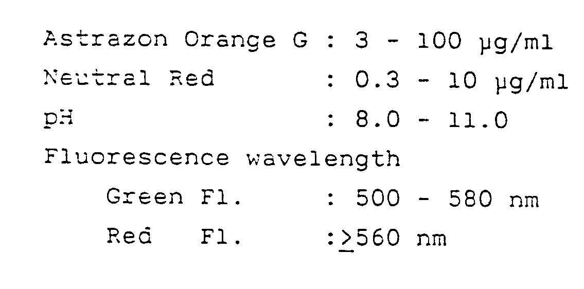
- This is an example of the method of the present invention as it was carried out with the composition of the reagent system described above being adjusted to an optimum range.
-
Astrazon Orange G (selective dye for basophils) 16.5 µg/mℓ (ppm) Neutral Red (selective dye for eosinophils) 2.2 µg/mℓ (ppm) Citric acid/sodium hydroxide (buffer) 10 mM pH, 3.0; osmolarity, 10 mOsm/kg -
Taurine/sodium hydroxide (buffer) 500 mM Sodium chloride (osmolarity compensating agent) 300 mM pH, 9.7 - 9.8; osmolarity, 2,600 mOsm/kg - Eighteen parts by volume of the first fluid was added to one part by volume of dipotassium salt dihydrate of EDTA anti-coagulated blood. After agitation, the mixture was incubated at 25°C for 20 seconds. Thereafter, 2 parts by volume of the second fluid was added and, after agitation, the mixture was incuvated at 25oC for 40 seconds. The finally attained staining conditions were a pH of 8.7 and an osmolarity of 260 mOsm/kg.
- The fluorescence emission intensity vs. wavelength characteristics of the individual leukocyte types as stained with the reagent system described above are shown in Fig. 18.
- Based on the emission characteristics shown in Fig. 18, the following filters and dichroic mirrors were selected as optimum devices:
Dichroic mirror 22530 nm (reflect blue light) Dichroic mirror 30600 nm (reflect red light) Color filter 34600 nm (long-pass filter transmitting wavelengths not shorter than 600 nm) Color filter 40540 nm (long-pass filter transmitting wavelengths not shorter than 540 nm) - A two-dimensional plot of the intensities of red and green fluorescences as measured with a flow cytometer under the conditions described above is shown in Fig. 16. Population 6 (consisting of platelets, red cell ghosts and fragments) was successfully separated from leukocytes, and it was possible for both an
eosinophil cluster 4 and abasophil cluster 5 to be separated from all other leukocytes with high resolution. The remaining leukocytes will also successfully separated from one another with good resolution, as indicated in Fig. 2c which is a frequency distribution curve forlymphocytes 1,monocytes 2 andneutrophils 3. In Fig. 2c, Side Sc. signifies the relative intensity of right-angle scatterd light and Freq. stands for frequency. - In Examples 1 to 7, all measurements are initiated after the necessary procedurs of staining have been completed (namely, after staining has reached an equilibrium). Therefore, the sample will not experience any time-dependent change during measurements, and an appropriate level of the intensity of staining or reaction can be attained within a certain period of time no matter how large or small the number of leukocytes in the sample is. This allows for consistent results in measurement and a fluorescence signal of an adequate intensity can be attained even if a light source of a comparatively low output is used. In Examples 1 - 7 described above, an argon ion laser of 10 mW was employed as a light source in the flow cytometer.
- However, the light source in the flow cytometer used in the present invention is not limited to the aforementioned argon ion laser of low output and any of the other light sources can be employed, such as a mercury arc lamp, xenon arc lamp, a He-Cd laser, a He-Ne laser and a krypton ion laer, as well as an argon ion laser of high output. If these light sources are used, the conditions of staining, reaction and measurement may be selected as appropriate.
- The reagent system and the method of the present invention as applied to classify and count leukocytes in blood by flow cytometry have the following advantages.
- (1) A sample of measurement can be prepared by simple preliminary treatments that consist of merely adding anticoagulated blood to a dye solution.
- (2) The sample can be prepared in approximately one minute and this provides a rapid access time for measurement.
- (3) Since measurements are conducted after the neceesary procedures of staining have been completed, the sample will not experiene any time-dependent change during measurements and an appropriate intensity of staining or reaction can always be attained within a certain period of time irrespective of the nature of the sample (whether it is normal or contains an extremely large or small number of leukocytes). This eliminates the need to change the staining time from sample to sample.
- (4) Since measurements are conducted after staining has been completed to provide a high staining intensity, a light source of low output may be employed. In addition, only one light source need to be used and two parameters appropriately selected from among two channels of fluorescence and one cahnnel of right-angle scattered light may be measured. Because the number of parameters to be measured and analyzed in this few, the reagent system of the present invention can be used to accomplish flow cytometry of blood with a simple and inexpensive apparatus.
- (5) The reagent system of the present invention has a very good ability to stain blood cells in a differential manner and therefore enable leukocytes to be classified with good resolution.
- (6) The method of the present invention effects measurement not only of fluorescence but also of right-angle scattered light and this contributes to better classification of leukocytes including separation between lymphocytes and monocytes.
- (7) In accordance with the method of the present invention, erythrocytes are selectively lysed by an isotonic treatment under acidic conditions. Since the coincidence of erythrocytes and leukocytes is eliminated by this treatment, a very efficient separation between lymphocytes, monocytes and neutrophils can be achieved by means of a right-angle scattered light signal.
- (8) Leukocytes can be classified into five types with a very high resolution by first separating eosinophils from basophils on the basis of a fluorescence signal, and then separating the remaining leukocytes (i.e., lymphocytes, monocytes and neutrophils) based on right-angle scattered light.
- (9) In the method of the present invention, separation of leukocytes from other corpuscles including their ghosts and fragments is achieved on the basis of fluorescence intensity, so correct measurements are ensured even if not all erythrocytes have been reduced to fragments.
- According to the method of the present invention, accurate and reproducible measurements are ensured by counting no less than 10,000 leukocytes for each sample.
Claims (8)
- A reagent system for use in classifying leukocytes contained in a blood sample into lymphocytes, monocytes, neutrophils, eosinophils and basophils with a flow cytometer by means of measurement on fluorochrome-stained cells of the emitted fluorescence and of the scattered light which includes:(a) a first fluid comprising a fluorochrome for selectively staining eosinophils, a fluorochrome for selectively staining basophils and a buffer and having a pH value between 2.0 and 5.0 and an osmolarity in the range of 0 to 100 mOsm/kg, and(b) a second fluid comprising a buffer for neutralizing the acid in the buffer of the first fluid and maintaining the pH of the resulting dye solution at a staining pH and an osmolarity compensating agent for adjusting the osmolarity of the dye solution to a value at which the leukocytes remain unchanged in their shape.
- A reagent system according to claim 1 wherein the fluorochrome for selectively staining eosinophils is Neutral Red (color index No. 50.040).
- A reagent system according to claim 1 wherein the fluorochrome for selectively staining basophils is either Astrazon Orange G (color index No. 48.035) or Auramine C (color index No. 41.000).
- A reagent system according to claim 1 wherein in the first fluid (a) a fluorochrome for selectively staining monocytes is furthermore included.
- A reagent system according to Claim 4 wherein the fluorochrome for selectively staining monocytes is selected from the group consisting of 1,1'-dimethyloxacarbocyanine, 1,1'-diethyloxacarbocyanine, 1,1'-di(n-propyl)oxacarbocyanine, 1,1'-di-(n-pentyl)oxacarbocyanine, 1,1'-di-(n-hexyl)oxacarbocyanine, TA-2, and 2-[γ-(1'-ethyl-4',5'-benzothiazolylidene)propenyl]-1-ethyl-4,5-benzoxazolium iodide.
- A method for classifying leukocytes into lymphocytes, monocytes, neutrophils, eosinophils and basophils with a flow cytometer that comprises the following steps (a) to (d):(a) lysing the erythrocytes in a fresh sample of anticoagulated blood by adding it to the first fluid of the reagent system according to any of the preceding claims 1-5;(b) staining the leukocytes in the so-treated blood sample by adding it to the second fluid of the reagent system according to any of the preceding claims 1-5;(c) permitting the stained sample to flow through a flow cytometer, differentiating leukocytes from all other corpuscles and ghosts by intensity of fluorescence, and measuring the signals of one or two fluorescence and right-angle (rectangular) scattered light from leukocytes; and(d) identifying the type of each of the leukocytes based on said two or three signals emitted therefrom, counting the number of detected leukocytes according to their type, and calculating the proportions of the individual leukocyte types.
- A method according to claim 6 wherein the fluorochrome for selectively staining eosinophils is Neutral Red.
- A method according to claim 6 wherein the fluorochrome for selectively staining basophils is Astrazon Orange G.
Applications Claiming Priority (4)
| Application Number | Priority Date | Filing Date | Title |
|---|---|---|---|
| JP213716/86 | 1986-09-10 | ||
| JP61213716A JPH076979B2 (en) | 1986-09-10 | 1986-09-10 | Reagents used for leukocyte classification by flow cytometry |
| JP282697/86 | 1986-11-27 | ||
| JP61282697A JPH0635972B2 (en) | 1986-11-27 | 1986-11-27 | Method for classifying leukocytes by flow cytometry |
Publications (3)
| Publication Number | Publication Date |
|---|---|
| EP0259833A2 EP0259833A2 (en) | 1988-03-16 |
| EP0259833A3 EP0259833A3 (en) | 1988-12-07 |
| EP0259833B1 true EP0259833B1 (en) | 1993-01-27 |
Family
ID=26519957
Family Applications (1)
| Application Number | Title | Priority Date | Filing Date |
|---|---|---|---|
| EP19870113112 Expired - Lifetime EP0259833B1 (en) | 1986-09-10 | 1987-09-08 | Reagent and method for classifying leukocytes by flow cytometry |
Country Status (3)
| Country | Link |
|---|---|
| EP (1) | EP0259833B1 (en) |
| CA (1) | CA1309327C (en) |
| DE (1) | DE3783838T2 (en) |
Families Citing this family (9)
| Publication number | Priority date | Publication date | Assignee | Title |
|---|---|---|---|---|
| US5389549A (en) * | 1987-05-29 | 1995-02-14 | Toa Medical Electronics Co., Ltd. | Method for classifying leukocytes and a reagent used therefor |
| JP3070968B2 (en) * | 1991-05-14 | 2000-07-31 | シスメックス株式会社 | Urine cell analysis reagents and methods |
| JP3067849B2 (en) * | 1991-07-29 | 2000-07-24 | シスメックス株式会社 | Sample preparation method for leukocyte classification and counting |
| JP3048260B2 (en) * | 1991-07-29 | 2000-06-05 | シスメックス株式会社 | Sample preparation method for leukocyte classification and counting |
| EP0626067A4 (en) * | 1992-02-07 | 1997-06-25 | Abbott Lab | Method for accurately enumerating and sensitively qualifying heterogeneous cell populations in cytolytic processing conditions. |
| DE4309328C2 (en) * | 1993-03-18 | 1998-03-12 | Volker Ost | Methods for differentiation, concentration determination and sorting of erythrocytes, platelets and leukocytes |
| EP3260842B1 (en) * | 2007-02-01 | 2022-04-13 | Sysmex Corporation | Hematological analyzer and method for analyzing a sample |
| EP3388833B1 (en) | 2010-08-05 | 2021-12-29 | Abbott Point Of Care, Inc. | Method and apparatus for automated whole blood sample analyses from microscopy images |
| WO2017100869A1 (en) * | 2015-12-17 | 2017-06-22 | Quintino De Oliveira Henrique Jesus | Water-based cell staining composition, method for preparing same and use thereof, cell sample preparation and cell counting methods |
Family Cites Families (4)
| Publication number | Priority date | Publication date | Assignee | Title |
|---|---|---|---|---|
| US3684377A (en) * | 1970-07-13 | 1972-08-15 | Bio Physics Systems Inc | Method for analysis of blood by optical analysis of living cells |
| CH613523A5 (en) * | 1975-06-27 | 1979-09-28 | Inst Nat Sante Rech Med | Method for displaying basophils |
| US4581223A (en) * | 1980-03-12 | 1986-04-08 | Lawrence Kass | Individual leukocyte determination by means of differential metachromatic dye sorption |
| DE3238353A1 (en) * | 1982-10-15 | 1984-04-19 | Max Planck Gesellschaft zur Förderung der Wissenschaften e.V., 3400 Göttingen | METHOD FOR SIMULTANEOUSLY QUANTITATIVE DETERMINATION OF BLOOD CELLS AND REAGENT THEREFOR |
-
1987
- 1987-09-04 CA CA000546200A patent/CA1309327C/en not_active Expired - Fee Related
- 1987-09-08 EP EP19870113112 patent/EP0259833B1/en not_active Expired - Lifetime
- 1987-09-08 DE DE19873783838 patent/DE3783838T2/en not_active Expired - Fee Related
Also Published As
| Publication number | Publication date |
|---|---|
| EP0259833A2 (en) | 1988-03-16 |
| CA1309327C (en) | 1992-10-27 |
| DE3783838D1 (en) | 1993-03-11 |
| EP0259833A3 (en) | 1988-12-07 |
| DE3783838T2 (en) | 1993-07-22 |
Similar Documents
| Publication | Publication Date | Title |
|---|---|---|
| US5175109A (en) | Reagent for classifying leukocytes by flow cytometry | |
| US5434081A (en) | Method of classifying leukocytes by flow cytometry | |
| EP0268766B1 (en) | Method of classifying leukocytes by flow cytometry and reagents used in the method | |
| EP0513762B1 (en) | Reagent and method for analyzing cells in urine | |
| JP3048260B2 (en) | Sample preparation method for leukocyte classification and counting | |
| CA2207396C (en) | Method for rapid and simultaneous analysis of nucleated red blood cells | |
| US7625730B2 (en) | Method for classifying and counting leukocytes | |
| US4933293A (en) | Method of classifying leukocytes by flow cytometry and reagents used in the method | |
| US20030219850A1 (en) | Method for automatically analyzing nucleated bone marrow cells | |
| US5179026A (en) | Method of classifying leukocytes by flow cytometry and reagents used in the method | |
| JP4087560B2 (en) | How to identify erythroblasts | |
| JP2009080122A (en) | Method for discriminating and counting erythroblasts | |
| CA1309327C (en) | Reagent and method for classifying leukocytes by flow cytometry | |
| Ronot et al. | Assessment of cell viability in mammalian cell lines | |
| JPS63134957A (en) | Method for classifying white blood cell by flow cytometry | |
| JPH079423B2 (en) | Leukocyte classification reagent by flow cytometry | |
| JPH0650310B2 (en) | Method for classifying white blood cells by flow cytometry | |
| JPS6370167A (en) | Reagent used for classification of white corpuscle by flow sight metry | |
| JPH0664053B2 (en) | Method for classifying leukocytes by flow cytometry |
Legal Events
| Date | Code | Title | Description |
|---|---|---|---|
| PUAI | Public reference made under article 153(3) epc to a published international application that has entered the european phase |
Free format text: ORIGINAL CODE: 0009012 |
|
| AK | Designated contracting states |
Kind code of ref document: A2 Designated state(s): DE FR GB IT NL |
|
| PUAL | Search report despatched |
Free format text: ORIGINAL CODE: 0009013 |
|
| AK | Designated contracting states |
Kind code of ref document: A3 Designated state(s): DE FR GB IT NL |
|
| 17P | Request for examination filed |
Effective date: 19881229 |
|
| 17Q | First examination report despatched |
Effective date: 19910607 |
|
| RAP1 | Party data changed (applicant data changed or rights of an application transferred) |
Owner name: TOA MEDICAL ELECTRONICS CO., LTD. |
|
| ITF | It: translation for a ep patent filed | ||
| GRAA | (expected) grant |
Free format text: ORIGINAL CODE: 0009210 |
|
| AK | Designated contracting states |
Kind code of ref document: B1 Designated state(s): DE FR GB IT NL |
|
| REF | Corresponds to: |
Ref document number: 3783838 Country of ref document: DE Date of ref document: 19930311 |
|
| ET | Fr: translation filed | ||
| PLBE | No opposition filed within time limit |
Free format text: ORIGINAL CODE: 0009261 |
|
| STAA | Information on the status of an ep patent application or granted ep patent |
Free format text: STATUS: NO OPPOSITION FILED WITHIN TIME LIMIT |
|
| 26N | No opposition filed | ||
| PGFP | Annual fee paid to national office [announced via postgrant information from national office to epo] |
Ref country code: GB Payment date: 19970901 Year of fee payment: 11 |
|
| PGFP | Annual fee paid to national office [announced via postgrant information from national office to epo] |
Ref country code: NL Payment date: 19970929 Year of fee payment: 11 |
|
| PG25 | Lapsed in a contracting state [announced via postgrant information from national office to epo] |
Ref country code: GB Free format text: LAPSE BECAUSE OF NON-PAYMENT OF DUE FEES Effective date: 19980908 |
|
| PG25 | Lapsed in a contracting state [announced via postgrant information from national office to epo] |
Ref country code: NL Free format text: LAPSE BECAUSE OF NON-PAYMENT OF DUE FEES Effective date: 19990401 |
|
| GBPC | Gb: european patent ceased through non-payment of renewal fee |
Effective date: 19980908 |
|
| NLV4 | Nl: lapsed or anulled due to non-payment of the annual fee |
Effective date: 19990401 |
|
| PGFP | Annual fee paid to national office [announced via postgrant information from national office to epo] |
Ref country code: FR Payment date: 20020910 Year of fee payment: 16 |
|
| PGFP | Annual fee paid to national office [announced via postgrant information from national office to epo] |
Ref country code: DE Payment date: 20020911 Year of fee payment: 16 |
|
| PG25 | Lapsed in a contracting state [announced via postgrant information from national office to epo] |
Ref country code: DE Free format text: LAPSE BECAUSE OF NON-PAYMENT OF DUE FEES Effective date: 20040401 |
|
| PG25 | Lapsed in a contracting state [announced via postgrant information from national office to epo] |
Ref country code: FR Free format text: LAPSE BECAUSE OF NON-PAYMENT OF DUE FEES Effective date: 20040528 |
|
| REG | Reference to a national code |
Ref country code: FR Ref legal event code: ST |
|
| PG25 | Lapsed in a contracting state [announced via postgrant information from national office to epo] |
Ref country code: IT Free format text: LAPSE BECAUSE OF NON-PAYMENT OF DUE FEES;WARNING: LAPSES OF ITALIAN PATENTS WITH EFFECTIVE DATE BEFORE 2007 MAY HAVE OCCURRED AT ANY TIME BEFORE 2007. THE CORRECT EFFECTIVE DATE MAY BE DIFFERENT FROM THE ONE RECORDED. Effective date: 20050908 |