EP0245979A2 - Novel phosphotyrosyl protein phosphatase - Google Patents
Novel phosphotyrosyl protein phosphatase Download PDFInfo
- Publication number
- EP0245979A2 EP0245979A2 EP87303612A EP87303612A EP0245979A2 EP 0245979 A2 EP0245979 A2 EP 0245979A2 EP 87303612 A EP87303612 A EP 87303612A EP 87303612 A EP87303612 A EP 87303612A EP 0245979 A2 EP0245979 A2 EP 0245979A2
- Authority
- EP
- European Patent Office
- Prior art keywords
- enzyme
- phosphatase
- human
- isolated
- protein
- Prior art date
- Legal status (The legal status is an assumption and is not a legal conclusion. Google has not performed a legal analysis and makes no representation as to the accuracy of the status listed.)
- Withdrawn
Links
Images

Classifications
-
- C—CHEMISTRY; METALLURGY
- C12—BIOCHEMISTRY; BEER; SPIRITS; WINE; VINEGAR; MICROBIOLOGY; ENZYMOLOGY; MUTATION OR GENETIC ENGINEERING
- C12N—MICROORGANISMS OR ENZYMES; COMPOSITIONS THEREOF; PROPAGATING, PRESERVING, OR MAINTAINING MICROORGANISMS; MUTATION OR GENETIC ENGINEERING; CULTURE MEDIA
- C12N9/00—Enzymes; Proenzymes; Compositions thereof; Processes for preparing, activating, inhibiting, separating or purifying enzymes
- C12N9/14—Hydrolases (3)
- C12N9/16—Hydrolases (3) acting on ester bonds (3.1)
Definitions
- the instant invention relates to a novel enzyme isolated from human membranes with a large number of insulin receptors and to the isolation and purification of such enzyme.
- Certain enzymes present in various tissues control biological processes by promoting or inhibiting biochemical events of metabolism, growth and transformation.
- examples of such agents are tyrosine protein kinases which are necessary for cell growth.
- Receptors for insulin, epidermal growth factor, platelet derived growth factor and insulin-like growth factor have tyrosine protein kinases capable of both autophosphorylation and transphosphorylation.
- certain retroviral oncogene products are tyrosine protein kinases.
- phosphatases, and in particular phosphotyrosyl protein phosphatases are likely to play an important role in the control of hormone action and viral transformation.
- Alkaline phosphatase purified from several membrane sources, including placenta, and acid phosphatase purified from human prostatic cytosol have also been shown to prefer phosphotyrosyl proteins over phosphoserine proteins as substrate. See Brautigan et al ., J. Biol. Chem. 256 pp. 6519-6522 (1981); Shriver et al ., J. Biol. Chem. 259 pp. 11383-11399 (1984); Swarup et al ., J. Biol. Chem. 256 pp. 8187-8201 (1981); and Li et al ., Eur. J. Biochem. 138 pp. 45-51 (1984).
- the instant invention is concerned with a new enzyme, distinct from the previously described phosphatases, which is isolated, from human tissues high in insulin receptors.
- Atherosclerotic plaques and in tumor formation certain receptor sites involve a phosphate group or tyrosine protein kinase which through phosphorylation activates the enzyme or binds the enzyme to appropriate receptor sites.
- Certain of these processes are natural processes such as insulin binding, and certain processes are unnatural processes such as unwanted vascular growth in diabetics, atherosclerotic plaque formation or tumor formation.
- the enzyme of the instant invention is capable of dephosphorylation of tyrosine protein kinases which can lead to the blocking of detrimental vascular growth as is seen in diabetic retinopathy; in the blocking of the formation of atherosclerotic plaque formation thus avoiding the complications of atherosclerosis such as myocardial infarction, thrombosis formation, and stroke; and a mechanism for blocking the formation of certain tumors, such as those which require retroviral oncogene products for example pp 60 src.
- the dephosphorylation enzyme of this invention can block insulin receptor action thus preventing some or many aspects of insulin action, a natural process, which can provide for a screening mechanism for drugs effective at improving insulin resistance.
- the phosphotyrosyl protein phosphatase enzyme of this invention is isolated from those human tissues which are relatively high in insulin receptors. Examples of such tissues are placental membrane, liver, adipose, brain muscle, and the like.
- the extraction of the enzyme is carried out in the steps of (1) removing unnecessary supporting membranes from the tissue, (2) homogenizing the remaining tissue, (3) centrifuging the homogenate in an aqueous buffer to remove solid tissue debris, (4) ultracentrifuging the supernatant to concentrate the enzyme containing membrane material, (5) solubilizing the washed membrane material and removing the unsolubilized residue and (6) extraction and purification of the enzyme from the solubilized membrane protein with chromatographic techniques.
- the tissues which are to provide the source of the new enzyme are initially treated to remove supporting membranes and connective tissues which do not contain the enzyme.
- the remaining tissue is homogenized in order to physically break down the cell to release the cell material, including the enzyme.
- the homogenization is carried out using standard techniques and usually in the presence of an aqueous buffer solution. Using fractional centrifugation the heavier tissue debris is removed leaving the enzyme-containing tissue material suspended in the buffer solution. Further ultracentrifugation produces a precipitate, commonly referred to as a "pellet" of enzyme-containing tissue material.
- the centrifugal pellet of tissue material is treated with a detergent and emulsifying agent such as octoxynol, sold under the trade name of "Triton X-100".
- the detergent usually provided as an aqueous solution, solubilizes the tissue material and releases the enzyme from such tissue material.
- the solubilized tissue and enzyme is purified using normal purification techniques known to those skilled in the art such as chromatographic techniques, for example column, thin layer, preparative layer and high pressure liquid chromatography.
- the human phosphotyrosyl protein phosphatase enzyme of this invention has not yet been precisely characterized as to its structure. However, during its isolation, purification and subsequent analytical and biological studies, sufficient characterizing information has been obtained which clearly demon strates that the enzyme is pure and significantly different from other enzymes isolated from similar tissues.
- the enzyme has a molecular weight of approximately 200,000 as shown by gel filtration on a TSK HPLC column in the presence of 0.2% octoxynol and a subunit molecular weight of approximately 73,000 as shown by SDS reducing gel electrophosesis.
- the instant phosphotyrosyl protein phosphatase is also characterized by its biological activities.
- the enzyme has been observed to be a dephosphorylating agent and the effects on this dephosphorylation by various phosphatase inhibitors can be readily employed to characterize the enzyme and also differentiate it from other enzymes.
- Table I shows the effect of phosphatase inhibitors on the instant enzyme and also on a placental alkaline phosphatase and a prostatic acid phosphatase.
- the percentage change of the effects of the enzyme in response to the specified amounts of phosphatase inhibitors are reproducible characteristics of the enzyme and can differentiate it from other enzymes.
- the other enzymes are shown for comparison purposes and to show the significant differences between the instant enzyme and the others.
- Human placenta was obtained from a nearby hospital within 1 hour after delivery and was chilled on ice. The supporting membranes were removed and the tissue was washed with 10 mM Tris, pH 7.4, 0.15M NaCl and homogenized in a buffer containing 0.25M sucrose, 25 mM benzamidine, 5 mM EDTA and 0.2 mM PMSF in a Waring blender. The homogenate was centrifuged at 10,000 ⁇ g for 30 minutes in a Beckman centrifuge. The supernatant was centrifuged at 100,000 ⁇ g for one hour in two Ti 45 rotors in Beckman ultracentrifuges.
- the membrane pellets were resuspended and washed with 40 mM imidazole, pH 7.2, and 0.2M NaCl and repelleted by centrifugation at 100,000 ⁇ g for one hour.
- the final membrane preparation was resuspended at approximately 20-50 mg/ml of protein in the above buffer.
- the washed membranes were solubilized with 2% Triton X-100 at a protein concentration of 5 mg/ml at 4° for 30 minutes.
- the unsolubilized residues were removed by centrifugation at 140,000 ⁇ g for 30 minutes.
- Wheat germ lectin agarose was prepared by coupling 200 mg of wheat germ lectin to approximately 40 ml of cyanogen bromide activated Sepharose 4B from Pharmacia according to the manufacturer's instructions. Approximately 400 mg of solubilized membrane protein was loaded on such a lectin affinity column equilibrated with a buffer containing 40 mM imidazole, pH 7.2, 10% glycerol, 0.2% Triton X-100 and 0.5M NaCl collecting 5 ml fractions. The column was washed extensively with the same buffer and eluted with 0.3M N-acetyl glucosamine in the same buffer. Aliquots of each fraction were taken for determination of protein concentration by the method of Bradford. Phosphatase activity using both PNPP and 32P-Tyr angiotensin II as substrates were also determined.
- a second DEAE-HPLC step was performed after dialysis of the pooled fractions against IGT buffer. Similar conditions as in the first DEAE-HPLC column were employed except that a 500 ml gradient of 0-300 mM NaCl in IGT buffer was used.
- 3-5 mg protein of purified human prostatic acid phosphatase, human placental alkaline phosphatase, and the purified phosphatase were iodinated each with 250 mCi of Na-125I in the presence of chloramine T (0-25 mg/ml) in 50 mM Tris, pH 6.5, for 30 seconds at room temperature. Free iodide was removed by desalting the samples on Sephadex G50 columns. Immunoprecipitation was carried out at 4°C overnight using approximately 10,000 cpm of iodinated proteins in 25 ml and 10 ml of undiluted normal rabbit serum, rabbit anti-human prostatic acid phosphatase, or rabbit anti-human alkaline phosphatase antisera.
- the phosphatase peak fractions eluted from the lectin column were pooled and dialyzed to lower salt concentrations. This preparation was loaded onto a DEAE-HPLC column and the retained proteins were eluted with a linear NaCl gradient.
- Figure 3 shows the elution profiles. When 32P-Tyr-angiotensin II phosphatase activity was assayed at pH 5 in the absence of divalent cations, two peaks of activity were found. A sharp peak was eluted at 0.1M NaCl and was found to correspond to the major peak of alkaline PNPP phosphatase activity assayed at pH 10 in the presence of 10 mM MgCl2.
- a second peak was eluted at 0.15M NaCl and corresponded to the acid PNPP phosphatase activity assayed at pH 5 in the absence of divalent cations. Since alkaline phosphatase has been well characterized, we focused our attention on the second peak.
- the peak of phosphatase activity was further purified by a second chromatography on preparative DEAE-HPLC.
- the PNPP phosphatase activity was eluted at 0.15M NaCl as in the first DEAE-HPLC column ( Figure 5).
- Figure 5 When aliquots of the peak fractions were subjected to SDS polyacrylamide gel electrophoresis and then the gel stained with silver nitrate, the intensity of staining of a band corresponding to a Mr of 73,000 corresponded to the peak of activity.
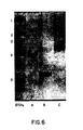
- Figure 6 shows that this Mr 73,000 band migrated differently from either human placental alkaline phosphatase or human prostatic acid phosphatase (available commercially) under reducing conditions.
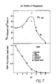
- Figure 7 shows the pH profiles of the highly purified phosphatase using 32P-Tyr-angiotensin II and PNPP as substrates.
- the PNPP phosphatase activity decreased as the pH increased from 3.5 to 10.
- the PNPP phosphatase activity was assayed at pH 4 and 6 at various concentrations of PNPP and the Km values were determined by Lineweaver-Burk plots to be 0.77 mM at pH 4 and 0.42 mM at pH 6.
- the K cat values were 1.02 ⁇ mol/min/mg protein and 0.39 ⁇ mol/min/mg protein at pH 4 and pH 6, respectively.
- the pH optimum was 7.
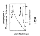
- the purified enzyme catalyzed the dephosphorylation of 32P-Tyr-casein (0.25 nmoles/hour/mg and 0.07 nmoles/hour/mg) (0.01 ⁇ M Pi) at initial rates that were 3.5 times higher than that for 32P-Ser-casein ( Figure 8).
- the rate of dephosphorylation of the P-Tyr substrate was 3.3X that for the P-Ser substrate (0.05 nmoles/hour/mg and 0.015 nmoles/hour/mg, respectively).
- the purified phosphatase dephosphorylated 32P-Tyr-casein at rates 10 fold higher than that for 32P-Tyr-histone f2b. It has been reported that phosphorylated basic proteins, such as histones and peptides such as angiotensin II, were poor substrates for the phosphotyrosyl protein phosphatases purified from rabbit kidney cytosol (19). It appears that this substrate specificity also holds for this enzyme.
- Table 1 shows the effects of various alkaline or acid phosphatase inhibitors on the PNPP phosphatase activities of the purified enzyme and on the pure alkaline phosphatase of human placental membranes and the acid phosphatase of human prostate.
- the enzyme described in this report was nearly completely inhibited by 10 ⁇ M molybdate, 5 mM L-tartrate, 5 mM NaF and 100 ⁇ M vanadate. It is slightly inhibited by phosphotyrosine and rather insensitive to ZnCl. These properties are similar to those of the prostatic acid phosphatase.
- this enzyme was stimulated by EDTA (79%) and phosphoserine (73%), whereas the prostatic enzyme was not affected by EDTA and was slightly inhibited by phosphoserine.
- 32P-Tyr-angiotensin II was used as substrate, the purified enzyme was similarly inhibited by 10 ⁇ M molybdate, 5 mM tartrate, 5 mM NaF, 10 ⁇ M vanadate and 10 mM phosphotyrosine (20.8, 27.6, 28.7, 46.4, and 11.5%, respectively).
- 20 mM EDTA and 10 mM phosphoserine stimulated the enzymatic activity by 29.8% and 4.3%, respectively.
- PNPP p-Nitrophenyl phosphate
- wheat germ lectin the catalytic subunit of cAMP-dependent protein kinase
- histone f2b Sigma type VII
- casein human semen acid phosphatase
- Protein A-Sepharose purchased from Sigma.
- Angiotensin II was purchased from Boerhinger Mannheim.
- [ ⁇ -32P]ATP 3000 Ci/mmol
- I125-sodium iodide and Aquasol II were purchased from New England Nuclear.
- Human placental alkaline phosphatase was purchased from Worthington Biochem.
- Epidermal growth factor (electrophoretically pure) was purchased from Biochemical Technologies Inc. (Cambridge, MA).
- Rabbit anti-human placental alkaline phosphatase antiserum was purchased from Arnel Products, Inc. (New York, NY). Rabbit anti-human prostatic acid Phosphatase antiserum was purchased from Miles Scientific (Naperville, IL).
- Angiotensin II was phosphorylated by EGF receptor kinase from human placental membranes.
- Human placental membranes (2.5 mg/ml) were preincubated with EGF (0.3 ⁇ M dissolved in 1 mg/ml BSA) in a final volume of 2 ml containing 50 mM HEPES, pH 7, 0.1M NaCl, and 1 mM sodium vanadate for 15 minutes at 4°C.
- the kinase reaction was started by the addition of a reaction mixture such that the final reaction contained 12 mM MgCl2, 2 mM MnCl2, 50 mM PNPP, 100 ⁇ M ATP and 800 ⁇ Ci [ ⁇ -32P]-ATP.
- the reaction was incubated at 4°C for 30 minutes and terminated by centrifugation at 100,000 ⁇ g for 30 minutes to remove membranes.
- the supernatant was mixed with an equal volume of 0.2% trifluoroacetic acid in water and loaded on a hydrated Sep Pak (Waters Associates).
- the free [ ⁇ -32P]-ATP was removed by washing the Sep Pak with 10 ⁇ 10 ml of 0.2% TFA in water.
- the phosphorylated angiotensin II was eluted with 80% acetonitrile and 0.2% trifluoroacetic acid in water. The fractions containing radioactivity were pooled and evaporated to dryness under nitrogen.
- the phosphorylated angiotensin II was taken up in 0.25 ml of water containing 1 mg/ml BSA.
- Histone f2b and casein were phosphorylated on serine/threonine residues in a final volume of 1 ml containing 50 mM Tris, PH 7.5, 25 mM MgCl2, 1 mM dithiothreotol, 5 mg/ml histone f2b or 10 mg/ml casein, 50 ⁇ M ATP, 250 ⁇ Ci [ ⁇ -32P]-ATP and approximately 60 units of the catalytic subunit of cAMP-dependent protein kinase.
- the reaction was carried out overnight at room temperature with rotation and terminated by precipitation with 25% TFA.
- the pellets were taken up in 1 ml of 20 mM HEPES, pH 7, and dialyzed extensively against the same buffer.
- Histone f2b and casein were phosphorylated on tyrosine residues using insulin-stimulated insulin receptor kinase from the N-acetyl-glucosamine eluted peak from the lectin agarose affinity column loaded with solubilized human placental membranes.
- Approximately 80 ⁇ g of protein from the lectin column was preincubated with 1 ⁇ M insulin in a final volume of 500 ⁇ l containing 20 mM HEPES, pH 7.4, 30 mM NaCl, 1 mM MnCl2, 100 mM sodium vanadate, 12 mM PNPP at room temperature for 15 minutes.
- the kinase activity was started by the addition of a reaction mixture such that the final concentration of ATP was 50 ⁇ M with 125 ⁇ Ci of [ ⁇ -32P]-ATP.
- Histone f2b was added to a final concentration of 1 mg/ml and casein was added to a final concentration of 3 mg/ml.
- the reaction was carried out at room temperature for 15 minutes and terminated by precipitation with 10% TCA.
- the precipitated proteins were treated with 0.5N NaOH at 55°C for 60 minutes to hydrolyze any phosphorylation on Ser/Thr residues.
- the solution was neutralized with 0.5M HCl and reprecipitated with 10% TCA.
- the pellet was taken up in 100 ⁇ l of 20 mM HEPES, pH 7, and dialyzed extensively against the same buffer.
- the phosphorylated proteins were partially hydrolyzed with 6N HCl at 100° under vacuum for 2 hours.
- the phosphoamino acids were analyzed on a Synchropak Q300 HPLC column (SynChrom, Inc., Linden, Indiana) by elution with 40 mM potassium phosphate, pH 4.
- Figure 1 shows that the 32P-Ser/Thr-casein was mainly phosphorylated on serine residues and that the 32P-Tyr-casein was mainly phosphorylated on tyrosine residues. Similar results were obtained with phosphorylations using histone f2b.
- Phosphotyrosine phosphatase activity was measured by the release of 32Pi from 32P-Tyr-angiotensin II in a 100 ml reaction mixture containing 0.1M acetate, pH 5, 5 mM DTT, 2.5 mg/ml BSA and 0.05 mM 32Pi of 32P-Tyr-angiotensin II. After incubation at 30° for 30 minutes, the reaction was terminated by the addition of 100 ml of 10% TCA. 0.2 ml of 1.25 mM KH2PO4 in lN H2SO4 was added as carrier. 0.1 ml of 5% ammonium molybdate was then added. After extraction with 0.5 ml of 1:1 isobutanol:benzene, 0.25 ml of the upper phase was taken and mixed with 10 ml Aquasol II to be counted in a Packard scintillation counter.
- p-Nitrophenylphosphatase activity was determined in a final volume of 500 ml containing 100 mM acetate, pH 5, 5 mM DTT, 2.5 mg/ml BSA and 1 mM PNPP. The reaction was carried out at 30°C and terminated with 0.5 ml of 0.2N NaOH. Alkaline PNPP phosphatase activity was similarly determined except that 50 mM glycine NaOH, pH 10, was used as buffer and 10 mM MgCl2 was included in the reaction mixture. Absorption at 410 nm wavelength was recorded on a Gilson spectrophotometer. Units of phosphatase activity were calculated with a millimolar extinction coefficient of 17.5 for p-nitrophenol at 410 nm and pH 11.
- Phosphatase activity against phosphorylated proteins was measured by incubation of the substrates at 0.01 mM Pi with the purified enzyme at pH 7. At the end of the incubation at 30°C the protein substrates were precipitated by 10% TCA with added BSA as carrier and the radioactivity remaining was determined by filtration on Whatman GF/F filters after washing with 4 ⁇ 2.5 ml of 10% TCA with 10 mM sodium pyrophosphate. Control incubations of the substrates with buffer alone showed no decrease in precipitable radioactivity with time.
Landscapes
- Life Sciences & Earth Sciences (AREA)
- Chemical & Material Sciences (AREA)
- Health & Medical Sciences (AREA)
- Genetics & Genomics (AREA)
- Organic Chemistry (AREA)
- Engineering & Computer Science (AREA)
- Bioinformatics & Cheminformatics (AREA)
- Zoology (AREA)
- Wood Science & Technology (AREA)
- Molecular Biology (AREA)
- Microbiology (AREA)
- Biotechnology (AREA)
- Biomedical Technology (AREA)
- Biochemistry (AREA)
- General Engineering & Computer Science (AREA)
- General Health & Medical Sciences (AREA)
- Medicinal Chemistry (AREA)
- Enzymes And Modification Thereof (AREA)
- Medicines That Contain Protein Lipid Enzymes And Other Medicines (AREA)
Abstract
Description
- Human placental membrane alkaline phosphatases and human prostatic cytosol acid phosphatases have been isolated and purified. However, such enzymes differ from the instant enzyme in the analytical properties of each and also in biological properties since the previously isolated enzymes have vastly different inhibition sensitivities when compared with the instant enzyme.
- The instant invention relates to a novel enzyme isolated from human membranes with a large number of insulin receptors and to the isolation and purification of such enzyme. Thus, it is an object to define the novel enzyme in terms of its analytical and biological properties. It is a further object to define the novel techniques used to isolate and purify the enzyme from the tissue with which it originates. A still further object is to define the utility of such an enzyme. Further objects will become apparent from a reading of the following description.
- Certain enzymes present in various tissues control biological processes by promoting or inhibiting biochemical events of metabolism, growth and transformation. Examples of such agents are tyrosine protein kinases which are necessary for cell growth. Receptors for insulin, epidermal growth factor, platelet derived growth factor and insulin-like growth factor have tyrosine protein kinases capable of both autophosphorylation and transphosphorylation. Also certain retroviral oncogene products are tyrosine protein kinases. Thus, phosphatases, and in particular phosphotyrosyl protein phosphatases, are likely to play an important role in the control of hormone action and viral transformation.
- Several reports have described phosphotyrosyl protein phosphatases which are distinct from phosphoserine or phosphothreonine protein phosphatase. See Foulkeset al., J. Biol. Chem. 258, pp. 431-438 (1983) and Chernoff et al., Arch. Biochem. Biophys. 226 pp. 517-550 (1983). The differences in their inhibitor sensitivities indicate that at least several different classes of phosphotyrosyl protein phosphatases exist, with only a few of these enzymes having been purified. One class of phosphotyrosyl phosphatases is inhibited by zinc ions; two such enzymes have been purified from rabbit kidney cytosol. Alkaline phosphatase purified from several membrane sources, including placenta, and acid phosphatase purified from human prostatic cytosol have also been shown to prefer phosphotyrosyl proteins over phosphoserine proteins as substrate. See Brautigan et al., J. Biol. Chem. 256 pp. 6519-6522 (1981); Shriver et al., J. Biol. Chem. 259 pp. 11383-11399 (1984); Swarup et al., J. Biol. Chem. 256 pp. 8187-8201 (1981); and Li et al., Eur. J. Biochem. 138 pp. 45-51 (1984). The instant invention is concerned with a new enzyme, distinct from the previously described phosphatases, which is isolated, from human tissues high in insulin receptors.
- In diabetes, in the formation of atherosclerotic plaques and in tumor formation certain receptor sites involve a phosphate group or tyrosine protein kinase which through phosphorylation activates the enzyme or binds the enzyme to appropriate receptor sites. Certain of these processes are natural processes such as insulin binding, and certain processes are unnatural processes such as unwanted vascular growth in diabetics, atherosclerotic plaque formation or tumor formation. The enzyme of the instant invention is capable of dephosphorylation of tyrosine protein kinases which can lead to the blocking of detrimental vascular growth as is seen in diabetic retinopathy; in the blocking of the formation of atherosclerotic plaque formation thus avoiding the complications of atherosclerosis such as myocardial infarction, thrombosis formation, and stroke; and a mechanism for blocking the formation of certain tumors, such as those which require retroviral oncogene products for
example pp 60 src. In other processes, the dephosphorylation enzyme of this invention can block insulin receptor action thus preventing some or many aspects of insulin action, a natural process, which can provide for a screening mechanism for drugs effective at improving insulin resistance. - The phosphotyrosyl protein phosphatase enzyme of this invention is isolated from those human tissues which are relatively high in insulin receptors. Examples of such tissues are placental membrane, liver, adipose, brain muscle, and the like. The extraction of the enzyme is carried out in the steps of (1) removing unnecessary supporting membranes from the tissue, (2) homogenizing the remaining tissue, (3) centrifuging the homogenate in an aqueous buffer to remove solid tissue debris, (4) ultracentrifuging the supernatant to concentrate the enzyme containing membrane material, (5) solubilizing the washed membrane material and removing the unsolubilized residue and (6) extraction and purification of the enzyme from the solubilized membrane protein with chromatographic techniques.
- In the instant application certain abbreviations are employed which may be generally recognized by those skilled in the art but are fully defined here in order to ensure the avoidance of all ambiguity.

- The tissues which are to provide the source of the new enzyme are initially treated to remove supporting membranes and connective tissues which do not contain the enzyme. The remaining tissue is homogenized in order to physically break down the cell to release the cell material, including the enzyme. The homogenization is carried out using standard techniques and usually in the presence of an aqueous buffer solution. Using fractional centrifugation the heavier tissue debris is removed leaving the enzyme-containing tissue material suspended in the buffer solution. Further ultracentrifugation produces a precipitate, commonly referred to as a "pellet" of enzyme-containing tissue material.
- To release the enzyme from the homogenized cell material, the centrifugal pellet of tissue material is treated with a detergent and emulsifying agent such as octoxynol, sold under the trade name of "Triton X-100". The detergent, usually provided as an aqueous solution, solubilizes the tissue material and releases the enzyme from such tissue material. After removal of any unsolubilized tissue by ultracentrifugation, the solubilized tissue and enzyme is purified using normal purification techniques known to those skilled in the art such as chromatographic techniques, for example column, thin layer, preparative layer and high pressure liquid chromatography.
- The human phosphotyrosyl protein phosphatase enzyme of this invention has not yet been precisely characterized as to its structure. However, during its isolation, purification and subsequent analytical and biological studies, sufficient characterizing information has been obtained which clearly demon strates that the enzyme is pure and significantly different from other enzymes isolated from similar tissues.
- The enzyme has a molecular weight of approximately 200,000 as shown by gel filtration on a TSK HPLC column in the presence of 0.2% octoxynol and a subunit molecular weight of approximately 73,000 as shown by SDS reducing gel electrophosesis.
- Other analytical data which characterize the instant enzyme are responses to a variety of protein phosphatase inhibitors, pH-activity profile, unique immunologic profile and subunit specificity.
- The instant phosphotyrosyl protein phosphatase is also characterized by its biological activities. The enzyme has been observed to be a dephosphorylating agent and the effects on this dephosphorylation by various phosphatase inhibitors can be readily employed to characterize the enzyme and also differentiate it from other enzymes. Table I below shows the effect of phosphatase inhibitors on the instant enzyme and also on a placental alkaline phosphatase and a prostatic acid phosphatase. The percentage change of the effects of the enzyme in response to the specified amounts of phosphatase inhibitors are reproducible characteristics of the enzyme and can differentiate it from other enzymes. The other enzymes are shown for comparison purposes and to show the significant differences between the instant enzyme and the others.

- The instant enzyme is still further characterized by the various analytical and biological procedures which are employed in its isolation and purification. In conjunction with these procedures, Figures 1 to 9 are appended hereto and are described as follows:
- Figure 1.
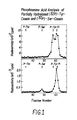
Phosphoamino Acid Analysis of Partially Hydrolyzed ³²P-Ser-casein and ³²P-Tyr-casein.
Samples of ³²P-Ser-casein and ³²P-Tyr-casein were hydrolyzed with 6N HCl under vacuum at 100°C for 2 hours. Samples containing approximately 12,000 cpm of radioactivity were loaded on a Synchropak Q300 column equilibrated with 40 mM potassium phosphate,pH 4. The column was eluted isocratically with the same buffer at a flow rate of 1.5 ml/minute and 0.2 minute fractions were collected and counted in 10 ml Aquasol II. Phosphothreonine, phosphoserine and phosphotyrosine standards were detected by monitoring absorbance at 214 nm wavelength. A, ³²P-Ser-casein. B, ³²P-Tyr-casein. - Figure 2.
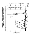
Fractionation of Solubilized Human Placental Membranes by Wheat Germ Lectin Agarose Affinity Chromatography.
Solubilized human placental membrane protein (400 mg) was loaded onto 40 ml of wheat germ lectin agarose equilibrated with a buffer containing 40 mM imidazole, pH 7.2, 10% glycerol, 0.2% Triton X-100, and 0.5M NaCl. After washing the column with the same buffer, elution was done with 0.3M N-acetyl glucosamine in the same buffer. 5 ml fractions were collected. Aliquots were taken for the determination of protein, and phosphatase activity against PNPP (1 mM at pH 5) and ³²P-Tyr-angiotensin II (pH 5). - Figure 3.
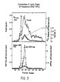
Fractionation of the Lectin Eluate by Preparative DEAE-HPLC.
Fractions eluted by N-acetylglucosamine from the lectin column were pooled and dialyzed against IGT buffer and loaded onto a preparative DEAE-HPLC column. The column was eluted with a linear NaCl gradient (0-0.4M) collecting 5 ml fractions. This chromatography resolved two peaks of ³²P-angiotensin II phosphatase activities. One of these corresponded to the peak of alkaline PNPPase activity eluted at 0.1M NaCl. Further purification was done by pooling the three fractions containing the ³²P-angiotensin II phosphatase peak of activity eluted at 0.15M NaCl. This peak corresponded to a peak of acidic PNPPase activity. - Figure 4.
Hydroxyapatite Column Chromatography
Pooled fractions from the preparative DEAE HPLC column were loaded onto 20 ml of hydroxyapatite equilibrated with IGT buffer and eluted initially with a 0-50 mM phosphate followed by a 50-400 mM phosphate gradient. A minor peak of acidic PNPPase activity was unretained while the major PNPPase activity was eluted at around 5 mM phosphate. The ³²P-Tyr angiotensin II phosphatase activity showed a similar profile. - Figure 5.
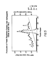
Second DEAE HPLC Column Pooled fractions from the hydroxyapatite column were dialyzed and rechromatographed on a second DEAE HPLC column. Similar conditions were employed as in Figure 3 except that a 0-0.3M NaCl gradient was used. - Figure 6.
SDS Gel Electrophoresis of the Purified Enzyme Preparation.
2-5 µg protein of (A) commercially available human placental alkaline phosphatase were subjected to gel electrophoresis on a 7.5% polyacrylamide gel. Proteins were stained with silver nitrate. Standards were: (1) myosin, Mr 200,000, (2) B galactosidase, Mr 116,250, (3) phosphorylase B, Mr 92,500, (4) bovine serum albumin, Mr 66,200, and (5) ovalbumin, Mr 45,000, (B) human prostatic acid phosphatase, and (C) the purified enzyme preparation. - Figure 7.
pH Profile of the Purified Phosphatase Using ³²P-Tyr Angiotensin II and PNPP as substrates.
Assay conditions were as described in the Example. - Figure 8.
Dephosphorylation of ³²P-Tyr-casein and ³²P-Ser-casein.
³²P-Tyr-casein and ³²P-Ser-casein were prepared as described under Materials and Methods. 0.01 µM Pi of each substrate was incubated at 30° with the purified enzyme in a buffer containing 4 mM imidazole, pH 7.2, 10% glycerol, 0.1M NaCl, 0.2% Triton X-100. At the end of the incubation, phosphoproteins were precipitated by 10% TCA with BSA added as carrier. The radioactivity remaining was determined by filtration on Whatman GF/F filters after washing with 4×2.5 ml of 10% TCA with 10 mM sodium pyrophosphate. Pi released was calculated by difference of remaining from added radioactivity. Control incubation of either substrate with buffer alone showed no decrease in precipitable radioactivity with time. - Figure 9.
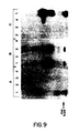
Immunological Characterization of the Purified Phosphotyrosyl Protein Phosphatase.
3-5 µg of the purified enzyme, commercially available human placental alkaline phosphatase and human prostatic acid phosphatase were iodinated as described in the Example. Approximately 10,000 cpm of each of the iodinated protein was reacted with (1) buffer alone, (2) normal rabbit serum, (3) rabbit anti-human prostatic acid phosphatase, and (4) rabbit anti-human alkaline phosphatase. After an overnight incubation at 4°C, 50 ml of a 1:1 mixture of Protein A Sepharose was added and incubated for one hour at room temperature. Non-specifically bound protein was washed off by sequential washes as described in the Example. Specifically bound proteins were extracted by the SDS gel electrophoresis sample buffer with 10% mercaptoethanol, and electrophoresis was done on 8.5% polyacrylamide gels. After drying the gels, iodinated proteins were visualized by autoradiography: A. Phosphotyrosyl protein phosphatase purified from human placental membranes. B. Human prostatic acid phosphatase. C. Human placental alkaline phosphatase. - The following example is provided in order that the instant invention might be more fully understood. It should not be construed as being limitative of this invention.
- Human placenta was obtained from a nearby hospital within 1 hour after delivery and was chilled on ice. The supporting membranes were removed and the tissue was washed with 10 mM Tris, pH 7.4, 0.15M NaCl and homogenized in a buffer containing 0.25M sucrose, 25 mM benzamidine, 5 mM EDTA and 0.2 mM PMSF in a Waring blender. The homogenate was centrifuged at 10,000 × g for 30 minutes in a Beckman centrifuge. The supernatant was centrifuged at 100,000 × g for one hour in two Ti 45 rotors in Beckman ultracentrifuges. The membrane pellets were resuspended and washed with 40 mM imidazole, pH 7.2, and 0.2M NaCl and repelleted by centrifugation at 100,000 × g for one hour. The final membrane preparation was resuspended at approximately 20-50 mg/ml of protein in the above buffer.
- The washed membranes were solubilized with 2% Triton X-100 at a protein concentration of 5 mg/ml at 4° for 30 minutes. The unsolubilized residues were removed by centrifugation at 140,000 × g for 30 minutes.
- All of the following procedures were carried out at 4°C except where noted.
- Wheat germ lectin agarose was prepared by coupling 200 mg of wheat germ lectin to approximately 40 ml of cyanogen bromide activated Sepharose 4B from Pharmacia according to the manufacturer's instructions. Approximately 400 mg of solubilized membrane protein was loaded on such a lectin affinity column equilibrated with a buffer containing 40 mM imidazole, pH 7.2, 10% glycerol, 0.2% Triton X-100 and 0.5M NaCl collecting 5 ml fractions. The column was washed extensively with the same buffer and eluted with 0.3M N-acetyl glucosamine in the same buffer. Aliquots of each fraction were taken for determination of protein concentration by the method of Bradford. Phosphatase activity using both PNPP and ³²P-Tyr angiotensin II as substrates were also determined.
- The fractions eluted by N-acetylglucosamine from two similar lectin columns were pooled and dialyzed against 40 mM imidazole, pH 7.2, 10% glycerol, 0.2% Triton X-100 (IGT buffer) overnight at 4°C and loaded onto a DEAE 5SW HPLC column (Bio Rad Laboratories) equilibrated with the same buffer. Elution was done on a 500 ml 0-0.4M NaCl gradient in IGT buffer using a flow rate of 1.5 ml/minute.
- Pooled fractions from the DEAE column were loaded onto 20 ml of BioGel HT (Bio Rad Laboratories) equilibrated in IGT buffer. Elution was done initially with a 200 ml 0-50 mM potassium phosphate gradient,
pH 7, in 10% glycerol and 0.2% Triton X-100. Subsequent elution was done with a 100 ml gradient of 50-400 mM potassium phosphate,pH 7, in 10% glycerol and 0.2% Triton X-100 to remove absorbed proteins. - A second DEAE-HPLC step was performed after dialysis of the pooled fractions against IGT buffer. Similar conditions as in the first DEAE-HPLC column were employed except that a 500 ml gradient of 0-300 mM NaCl in IGT buffer was used.
- 3-5 mg protein of purified human prostatic acid phosphatase, human placental alkaline phosphatase, and the purified phosphatase were iodinated each with 250 mCi of Na-¹²⁵I in the presence of chloramine T (0-25 mg/ml) in 50 mM Tris, pH 6.5, for 30 seconds at room temperature. Free iodide was removed by desalting the samples on Sephadex G50 columns. Immunoprecipitation was carried out at 4°C overnight using approximately 10,000 cpm of iodinated proteins in 25 ml and 10 ml of undiluted normal rabbit serum, rabbit anti-human prostatic acid phosphatase, or rabbit anti-human alkaline phosphatase antisera. 50 ml of a 1:1 suspension of protein A-Sepharose was added to each incubation to precipitate the immunoglobulins. After sequential washings of the Protein A Sepharose beads with 10 mM Tris HCl, pH 7.5, 0.5% w/v NP-40 (NT buffer), NT buffer with 1 mM EDTA, NT buffer with 0.5M NaCl, and NT buffer with 0.1% w/v SDS, the specifically bound proteins were eluted in the SDS gel electrophoresis sample buffer with 10% mercaptoethanol and subjected to gel electrophoresis on 8.5% polyacrylamide gels. Autoradiography of the dried gels was done using Kodak XAR-5 film with intensifying screens.
- Gel electrophoresis in the presence of sodium dodecyl sulfate was performed as described by Laemmli (Nature (London) 227 pp. 680-685 (1970)) and stained with silver nitrate (Bio Rad Laboratories). Protein concentrations were determined by Coomassie G250 dye binding using gamma globulin as standard (Bio Rad Laboratories).
- Human placental membrane fraction was solubilized with Triton X-100 and subjected to wheat germ lectin agarose affinity chromatography. The proteins retained by the lectin gel were eluted with N-acetylglucosamine. Phosphatase activities were assayed using p-nitrophenyl phosphate and ³²P-angiotensin II as substrates in acetate buffer,
pH 5, giving the chromatographic profiles shown in Figure 2. The N-acetylglucosamine eluted peak contained 20% of protein originally loaded on the column, 70% of the p-nitrophenyl phosphatase activity and 26% of the ³²P-Tyr-angiotensin II phosphatase activity. The specific activity of p-nitrophenyl phosphatase was 10 fold higher for the eluted fractions than for the unretained fractions, while the specific activity of the ³²P-Tyr-angiotensin phosphatase was only 1.4 fold higher in the eluted fractions than in the unretained fractions. However, under these chromatographic conditions, 95% of the epidermal growth factor receptor kinase activity was retained and eluted from the column by N-acetylglucosamine, indicating that the column was not overloaded. Our present studies focus on the glycoprotein phosphatases retained by this lectin column. - The phosphatase peak fractions eluted from the lectin column were pooled and dialyzed to lower salt concentrations. This preparation was loaded onto a DEAE-HPLC column and the retained proteins were eluted with a linear NaCl gradient. Figure 3 shows the elution profiles. When ³²P-Tyr-angiotensin II phosphatase activity was assayed at
pH 5 in the absence of divalent cations, two peaks of activity were found. A sharp peak was eluted at 0.1M NaCl and was found to correspond to the major peak of alkaline PNPP phosphatase activity assayed atpH 10 in the presence of 10 mM MgCl₂. A second peak was eluted at 0.15M NaCl and corresponded to the acid PNPP phosphatase activity assayed atpH 5 in the absence of divalent cations. Since alkaline phosphatase has been well characterized, we focused our attention on the second peak. - Only the peak fractions of acid phosphatase were pooled since the shoulder fractions could represent different enzymes. The pooled sample was loaded onto a hydroxyapatite column and the column was initially eluted with a shallow phosphate gradient (0-50 mM) followed by a steep gradient (50-400 mM). A minor peak of acidic PNPPase activity was unretained, while the major PNPP phosphatase activity was eluted around 5 mM phosphate (Figure 4). The ³²P-Tyr-angiotensin II phosphatase activity showed a similar profile. However, the Phosphate carried over from the samples interfered with the extraction of free-³²P from the reaction solution and could not be accurately corrected, interfering with the quantitation of the angiotensin II assay in the higher salt sample. Several representative fractions, among fraction numbers 30-80, with higher phosphate concentrations were dialyzed before assaying and showed no PNPP phosphatase or ³²P-Tyr-angiotensin phosphatase activity.
- The peak of phosphatase activity was further purified by a second chromatography on preparative DEAE-HPLC. The PNPP phosphatase activity was eluted at 0.15M NaCl as in the first DEAE-HPLC column (Figure 5). When aliquots of the peak fractions were subjected to SDS polyacrylamide gel electrophoresis and then the gel stained with silver nitrate, the intensity of staining of a band corresponding to a Mr of 73,000 corresponded to the peak of activity. Figure 6 shows that this Mr 73,000 band migrated differently from either human placental alkaline phosphatase or human prostatic acid phosphatase (available commercially) under reducing conditions. When electrophoresis was done under non-reducing conditions, the same Mr 73,000 band was seen. However, when a sample of the pure enzyme was subjected to gel filtration on a TSK HPLC column in the presence of 0.2% Triton X-100, the activity eluted in a single symmetrical peak corresponding to an estimated MW of 200,000. It is not clear whether this reflects the non-globular nature of the protein or whether the enzyme consists of several non-covalently linked subunits. The purity of the enzyme was also confirmed when the preparation was iodinated and subjected to SDS gel electrophoresis. Autoradiography of the dried gel showed a major band corresponding to the Mr 73,000 band (Figure 9). A minor band with low molecular weight corresponded to a faint band on the silver stained gel, the intensity of which did not correspond to the peak of phosphatase activity.
- Figure 7 shows the pH profiles of the highly purified phosphatase using ³²P-Tyr-angiotensin II and PNPP as substrates. The PNPP phosphatase activity decreased as the pH increased from 3.5 to 10. The PNPP phosphatase activity was assayed at
pH pH 4 and 0.42 mM atpH 6. The Kcat values were 1.02 µmol/min/mg protein and 0.39 µmol/min/mg protein atpH 4 andpH 6, respectively. In contrast, when ³²P-Tyr-angiotensin II was used as substrate, the pH optimum was 7. Different pH optima for PNPP and ³²P-histone have also been observed with purified alkaline phosphatase from calf intestine and E. coli. The neutral pH optimum of this placental phosphatase for a phosphorylated peptide suggests that this enzyme may play a role in the dephosphorylation of phosphotyrosyl proteins in cells. - The purified enzyme catalyzed the dephosphorylation of ³²P-Tyr-casein (0.25 nmoles/hour/mg and 0.07 nmoles/hour/mg) (0.01 µM Pi) at initial rates that were 3.5 times higher than that for ³²P-Ser-casein (Figure 8). Similarly, when ³²P-Tyr-histone f2b and ³²P-Ser-histone f2b were used at substrates (0.01 µM Pi), the rate of dephosphorylation of the P-Tyr substrate was 3.3X that for the P-Ser substrate (0.05 nmoles/hour/mg and 0.015 nmoles/hour/mg, respectively). Thus, the purified phosphatase dephosphorylated ³²P-Tyr-casein at
rates 10 fold higher than that for ³²P-Tyr-histone f2b. It has been reported that phosphorylated basic proteins, such as histones and peptides such as angiotensin II, were poor substrates for the phosphotyrosyl protein phosphatases purified from rabbit kidney cytosol (19). It appears that this substrate specificity also holds for this enzyme. - Table 1 shows the effects of various alkaline or acid phosphatase inhibitors on the PNPP phosphatase activities of the purified enzyme and on the pure alkaline phosphatase of human placental membranes and the acid phosphatase of human prostate. The enzyme described in this report was nearly completely inhibited by 10 µM molybdate, 5 mM L-tartrate, 5 mM NaF and 100 µM vanadate. It is slightly inhibited by phosphotyrosine and rather insensitive to ZnCl. These properties are similar to those of the prostatic acid phosphatase. However, this enzyme was stimulated by EDTA (79%) and phosphoserine (73%), whereas the prostatic enzyme was not affected by EDTA and was slightly inhibited by phosphoserine. When ³²P-Tyr-angiotensin II was used as substrate, the purified enzyme was similarly inhibited by 10 µM molybdate, 5 mM tartrate, 5 mM NaF, 10 µM vanadate and 10 mM phosphotyrosine (20.8, 27.6, 28.7, 46.4, and 11.5%, respectively). Similarly, 20 mM EDTA and 10 mM phosphoserine stimulated the enzymatic activity by 29.8% and 4.3%, respectively. 100 µM ZnCl₂, which was only slightly inhibitory on the PNPP phosphatase activity, inhibited ³²P-Tyr-angiotensin II activity by only 4.2%. These results suggest that the phosphatase that we have purified is a unique enzyme, with biochemical properties different from those of previously described phosphatases.
- Commercially available antiserum against human alkaline phosphatase and human prostatic acid phosphatase were reacted with an iodinated preparation of the purified enzyme. Figure 9 shows that while the respective antisera reacted with the phosphatases to which the sera were made, there was no cross reactivity of either of the antisera with the purified enzyme.
- p-Nitrophenyl phosphate (PNPP), wheat germ lectin, the catalytic subunit of cAMP-dependent protein kinase, histone f2b (Sigma type VII), casein, human semen acid phosphatase, and Protein A-Sepharose were purchased from Sigma. Angiotensin II was purchased from Boerhinger Mannheim. [γ-³²P]ATP (3000 Ci/mmol), I¹²⁵-sodium iodide and Aquasol II were purchased from New England Nuclear. Human placental alkaline phosphatase was purchased from Worthington Biochem. Epidermal growth factor (electrophoretically pure) was purchased from Biochemical Technologies Inc. (Cambridge, MA). Rabbit anti-human placental alkaline phosphatase antiserum was purchased from Arnel Products, Inc. (New York, NY). Rabbit anti-human prostatic acid Phosphatase antiserum was purchased from Miles Scientific (Naperville, IL).
- Angiotensin II was phosphorylated by EGF receptor kinase from human placental membranes. Human placental membranes (2.5 mg/ml) were preincubated with EGF (0.3 µM dissolved in 1 mg/ml BSA) in a final volume of 2 ml containing 50 mM HEPES,
pH 7, 0.1M NaCl, and 1 mM sodium vanadate for 15 minutes at 4°C. After addition of angiotensin II to a final concentration of 1 mM, the kinase reaction was started by the addition of a reaction mixture such that the final reaction contained 12 mM MgCl₂, 2 mM MnCl₂, 50 mM PNPP, 100 µM ATP and 800 µCi [γ-³²P]-ATP. The reaction was incubated at 4°C for 30 minutes and terminated by centrifugation at 100,000 × g for 30 minutes to remove membranes. The supernatant was mixed with an equal volume of 0.2% trifluoroacetic acid in water and loaded on a hydrated Sep Pak (Waters Associates). The free [γ-³²P]-ATP was removed by washing the Sep Pak with 10×10 ml of 0.2% TFA in water. The phosphorylated angiotensin II was eluted with 80% acetonitrile and 0.2% trifluoroacetic acid in water. The fractions containing radioactivity were pooled and evaporated to dryness under nitrogen. The phosphorylated angiotensin II was taken up in 0.25 ml of water containing 1 mg/ml BSA. - Histone f2b and casein were phosphorylated on serine/threonine residues in a final volume of 1 ml containing 50 mM Tris, PH 7.5, 25 mM MgCl₂, 1 mM dithiothreotol, 5 mg/ml histone f2b or 10 mg/ml casein, 50 µM ATP, 250 µCi [γ-³²P]-ATP and approximately 60 units of the catalytic subunit of cAMP-dependent protein kinase. The reaction was carried out overnight at room temperature with rotation and terminated by precipitation with 25% TFA. The pellets were taken up in 1 ml of 20 mM HEPES,
pH 7, and dialyzed extensively against the same buffer. - Histone f2b and casein were phosphorylated on tyrosine residues using insulin-stimulated insulin receptor kinase from the N-acetyl-glucosamine eluted peak from the lectin agarose affinity column loaded with solubilized human placental membranes. Approximately 80 µg of protein from the lectin column was preincubated with 1 µM insulin in a final volume of 500 µl containing 20 mM HEPES, pH 7.4, 30 mM NaCl, 1 mM MnCl₂, 100 mM sodium vanadate, 12 mM PNPP at room temperature for 15 minutes. The kinase activity was started by the addition of a reaction mixture such that the final concentration of ATP was 50 µM with 125 µCi of [γ-³²P]-ATP. Histone f2b was added to a final concentration of 1 mg/ml and casein was added to a final concentration of 3 mg/ml. The reaction was carried out at room temperature for 15 minutes and terminated by precipitation with 10% TCA. The precipitated proteins were treated with 0.5N NaOH at 55°C for 60 minutes to hydrolyze any phosphorylation on Ser/Thr residues. The solution was neutralized with 0.5M HCl and reprecipitated with 10% TCA. The pellet was taken up in 100 µl of 20 mM HEPES,
pH 7, and dialyzed extensively against the same buffer. - The phosphorylated proteins were partially hydrolyzed with 6N HCl at 100° under vacuum for 2 hours. The phosphoamino acids were analyzed on a Synchropak Q300 HPLC column (SynChrom, Inc., Linden, Indiana) by elution with 40 mM potassium phosphate,
pH 4. Figure 1 shows that the ³²P-Ser/Thr-casein was mainly phosphorylated on serine residues and that the ³²P-Tyr-casein was mainly phosphorylated on tyrosine residues. Similar results were obtained with phosphorylations using histone f2b. - Phosphotyrosine phosphatase activity was measured by the release of ³²Pi from ³²P-Tyr-angiotensin II in a 100 ml reaction mixture containing 0.1M acetate,
pH - p-Nitrophenylphosphatase activity was determined in a final volume of 500 ml containing 100 mM acetate,
pH pH 10, was used as buffer and 10 mM MgCl₂ was included in the reaction mixture. Absorption at 410 nm wavelength was recorded on a Gilson spectrophotometer. Units of phosphatase activity were calculated with a millimolar extinction coefficient of 17.5 for p-nitrophenol at 410 nm and pH 11. - Phosphatase activity against phosphorylated proteins was measured by incubation of the substrates at 0.01 mM Pi with the purified enzyme at
pH 7. At the end of the incubation at 30°C the protein substrates were precipitated by 10% TCA with added BSA as carrier and the radioactivity remaining was determined by filtration on Whatman GF/F filters after washing with 4×2.5 ml of 10% TCA with 10 mM sodium pyrophosphate. Control incubations of the substrates with buffer alone showed no decrease in precipitable radioactivity with time.
Claims (12)

Applications Claiming Priority (2)
| Application Number | Priority Date | Filing Date | Title |
|---|---|---|---|
| US06/858,622 US4710469A (en) | 1986-05-02 | 1986-05-02 | Novel phosphotyrosyl protein phosphatase |
| US858622 | 1986-05-02 |
Publications (2)
| Publication Number | Publication Date |
|---|---|
| EP0245979A2 true EP0245979A2 (en) | 1987-11-19 |
| EP0245979A3 EP0245979A3 (en) | 1988-12-14 |
Family
ID=25328749
Family Applications (1)
| Application Number | Title | Priority Date | Filing Date |
|---|---|---|---|
| EP87303612A Withdrawn EP0245979A3 (en) | 1986-05-02 | 1987-04-24 | Novel phosphotyrosyl protein phosphatase |
Country Status (3)
| Country | Link |
|---|---|
| US (1) | US4710469A (en) |
| EP (1) | EP0245979A3 (en) |
| JP (1) | JPS6322186A (en) |
Cited By (5)
| Publication number | Priority date | Publication date | Assignee | Title |
|---|---|---|---|---|
| WO1994001119A1 (en) * | 1992-07-10 | 1994-01-20 | National University Of Singapore | INHIBITORS OF PROTEIN TYROSINE-PHOSPHATASE α FOR TREATMENT OF TUMORS |
| EP0627489A1 (en) * | 1990-03-14 | 1994-12-07 | Washington Research Foundation | Methods and compositions for the treatment of malignancies in which a protein kinase is associated |
| WO1995023231A1 (en) * | 1994-02-28 | 1995-08-31 | MAX-PLANCK-Gesellschaft zur Förderung der Wissenschaften e.V. | Cell lines for the identification of substances affecting insulin receptor mediated signal transduction |
| US6054285A (en) * | 1995-12-15 | 2000-04-25 | Novartis Ag | Screening method |
| US7786103B2 (en) | 2002-08-06 | 2010-08-31 | Sanofi-Aventis Deutschland Gmbh | Method for isolating an intestinal cholesterol binding protein |
Families Citing this family (8)
| Publication number | Priority date | Publication date | Assignee | Title |
|---|---|---|---|---|
| US5242397A (en) * | 1989-06-20 | 1993-09-07 | Cedars-Sinai Medical Center | Catheter device and method of use for intramural delivery of protein kinase C and tyrosine protein kinase inhibitors to prevent restenosis after balloon angioplasty |
| US5155031A (en) * | 1990-06-07 | 1992-10-13 | Posner Barry I | Use of pervanadate as an inhibitor of phosphotyrosine phosphatase |
| US5180665A (en) * | 1990-11-21 | 1993-01-19 | Her Majesty The Queen In Right Of Canada, As Represented By The National Research Council Of Canada | Method for quantitatively assaying the presence of diarrhetic shellfish poisoning toxins in marine samples |
| US5401830A (en) * | 1992-10-05 | 1995-03-28 | The State University Of New Jersey | Insulin receptor-like protein |
| US5512434A (en) * | 1992-12-14 | 1996-04-30 | The United States Of America As Represented By The Department Of Health And Human Services | Expression cloning of a human phosphatase |
| JPH09504689A (en) * | 1993-10-01 | 1997-05-13 | ニューヨーク ユニバーシティー | Novel receptor-type phosphotyrosine phosphatase σ |
| US5763490A (en) * | 1994-09-20 | 1998-06-09 | University Of South Carolina | Treating prostate cancer with tartrate ions |
| WO2012166715A2 (en) * | 2011-05-27 | 2012-12-06 | The Children's Hospital Of Philadelphia | Compositions and methods for the detection of dna cleavage complexes |
-
1986
- 1986-05-02 US US06/858,622 patent/US4710469A/en not_active Expired - Fee Related
-
1987
- 1987-04-24 EP EP87303612A patent/EP0245979A3/en not_active Withdrawn
- 1987-05-01 JP JP62108778A patent/JPS6322186A/en active Pending
Non-Patent Citations (6)
| Title |
|---|
| BIOCHEMISTRY, vol. 27, no. 12, 14th June 1988, pages 4207-4590, American Chemical Society, US; A. WAHEED et al.: "Purification and physicochemical characterization of a human placental acid phosphatase possessing phosphotyrosyl protein phosphatase activity" * |
| CHEMICAL ABSTRACTS, vol. 101, no. 5, 30th July 1984, page 92, abstract no. 33626r, Columbus, Ohio, US; H.U. HARING et al.: "Phosphorylation and dephosphorylation of the insulin receptor: evidence against an intrinsic pho sphatase activity", & BIOCHEMISTRY 1984, 23(14) 3298-306 * |
| CHEMICAL ABSTRACTS, vol. 103, no. 5, 5th August 1985, page 198, abstract no. 33916b, Columbus, Ohio, US; F. SAKO et al.: "Phosphotyrosine phosphatase activity in human placenta", & JPN. J. EXP. MED. 1985, 55(1), 21-7 * |
| CHEMICAL ABSTRACTS, vol. 67, no. 9, 28th August 1967, page 3782, abstract no. 40415d, Columbus, Ohio, US; D.L. DIPIETRO et al.: "Separation and properties of three acid phosphatases from human placenta", & J. BIOL. CHEM. 242(14), 3391-6(1967) * |
| CHEMICAL ABSTRACTS, vol. 97, no. 7, 16th August 1982, page 267, abstract no. 51640u, Columbus, Ohio, US; G. SWARUP et al.: "Phosphotyrosyl-protein phosphatase of TCRC-2cells", & J. BIOL. CHEM. 1982, 257(13), 7298-301 * |
| CHEMICAL ABSTRACTS, vol. 98, no. 3, 17th January 1983, page 369, abstract no. 14962a, Columbus, Ohio, US; J.F. LEIS et al.: "An acid phosphatase in the plasma membranes of human astrocytoma showing marked specificity toward phosphotyrosine protein", & PROC. NATL. ACAD. SCI. U.S.A. 1982, 79(21), 6507-11 * |
Cited By (5)
| Publication number | Priority date | Publication date | Assignee | Title |
|---|---|---|---|---|
| EP0627489A1 (en) * | 1990-03-14 | 1994-12-07 | Washington Research Foundation | Methods and compositions for the treatment of malignancies in which a protein kinase is associated |
| WO1994001119A1 (en) * | 1992-07-10 | 1994-01-20 | National University Of Singapore | INHIBITORS OF PROTEIN TYROSINE-PHOSPHATASE α FOR TREATMENT OF TUMORS |
| WO1995023231A1 (en) * | 1994-02-28 | 1995-08-31 | MAX-PLANCK-Gesellschaft zur Förderung der Wissenschaften e.V. | Cell lines for the identification of substances affecting insulin receptor mediated signal transduction |
| US6054285A (en) * | 1995-12-15 | 2000-04-25 | Novartis Ag | Screening method |
| US7786103B2 (en) | 2002-08-06 | 2010-08-31 | Sanofi-Aventis Deutschland Gmbh | Method for isolating an intestinal cholesterol binding protein |
Also Published As
| Publication number | Publication date |
|---|---|
| JPS6322186A (en) | 1988-01-29 |
| EP0245979A3 (en) | 1988-12-14 |
| US4710469A (en) | 1987-12-01 |
Similar Documents
| Publication | Publication Date | Title |
|---|---|---|
| Shenolikar et al. | Protein (serine and threonine) phosphate phosphatases | |
| Gronich et al. | Purification of a high-molecular-mass form of phospholipase A2 from rat kidney activated at physiological calcium concentrations | |
| Naito et al. | 5'-Nucleotidase from rat heart | |
| Lanzillo et al. | Angiotensin-converting enzyme from human tissues. Physicochemical, catalytic, and immunological properties. | |
| Gorelick et al. | Calmodulin-stimulated protein kinase activity from rat pancreas. | |
| Hashimoto et al. | Regulation of calcineurin by phosphorylation: Identification of the regulatory site phosphorylated by Ca2+/calmodulin-dependent protein kinase II and protein kinase C | |
| Ashby et al. | [48] Purification and characterization of an inhibitor protein of cyclic AMP-dependent protein kinases | |
| Allen et al. | Purification and characterization of human bone tartrate‐resistant acid phosphatase | |
| US4710469A (en) | Novel phosphotyrosyl protein phosphatase | |
| Huang et al. | Phosphorylation of glycogen synthase by cyclic AMP-independent casein kinase-2 from rabbit skeletal muscle. | |
| Ahmad et al. | Glycogen synthase kinases. Classification of a rabbit liver casein and glycogen synthase kinase (casein kinase-1) as a distinct enzyme. | |
| Kahn | [23] Pyruvate kinases from human erythrocytes and liver | |
| Butterworth et al. | Presence in human cells and tissues of two prolidases and their alteration in prolidase deficiency | |
| Leustek et al. | Calcium-dependent autophosphorylation of the glucose-regulated protein, Grp78 | |
| Chernoff et al. | Multiple forms of phosphotyrosyl-and phosphoseryl-protein phosphatase from cardiac muscle: partial purification and characterization of an EDTA-stimulated phosphotyrosyl-protein phosphatase | |
| Swarup et al. | Purification and characterization of a protein-phosphotyrosine phosphatase from rat spleen which dephosphorylates and inactivates a tyrosine-specific protein kinase | |
| Brunati et al. | Isolation and partial characterization of distinct species of phosphotyrosyl protein phosphatases from rat spleen | |
| Bennun et al. | Properties of the induced acid phosphatase and of the constitutive acid phosphatase of Euglena | |
| Cheng et al. | Purification and characterization of a phosphotyrosyl-protein phosphatase from wheat seedlings | |
| Gopalakrishna et al. | Quantitation of tissue calpain activity after isolation by hydrophobic chromatography | |
| Hui et al. | Interaction of plasma lipoproteins with erythrocytes. II. Modulation of membrane-associated enzymes | |
| Banno et al. | Purification and characterization of a cytosolic phosphoinositide-phospholipase C (γ2-type) from human platelets | |
| Koskelo | Isoelectric focusing of glutathione S-transferases: comparison of the acidic transferases from human liver, kidney, lung, spleen and placenta | |
| Mieskes et al. | Purification and characterization of a protein phosphatase from rat liver acting on key enzymes of glucose metabolism | |
| Garcia et al. | A protein kinase copurified with chick oviduct progesterone receptor |
Legal Events
| Date | Code | Title | Description |
|---|---|---|---|
| PUAI | Public reference made under article 153(3) epc to a published international application that has entered the european phase |
Free format text: ORIGINAL CODE: 0009012 |
|
| AK | Designated contracting states |
Kind code of ref document: A2 Designated state(s): CH DE FR GB IT LI NL |
|
| ITCL | It: translation for ep claims filed |
Representative=s name: SOCIETA' ITALIANA BREVETTI S.P.A. |
|
| PUAL | Search report despatched |
Free format text: ORIGINAL CODE: 0009013 |
|
| AK | Designated contracting states |
Kind code of ref document: A3 Designated state(s): CH DE FR GB IT LI NL |
|
| 18W | Application withdrawn |
Withdrawal date: 19890321 |
|
| STAA | Information on the status of an ep patent application or granted ep patent |
Free format text: STATUS: THE APPLICATION HAS BEEN WITHDRAWN |
|
| R18W | Application withdrawn (corrected) |
Effective date: 19890321 |
|
| RIN1 | Information on inventor provided before grant (corrected) |
Inventor name: SLATER, EVE E. Inventor name: LIANG, THEMING |