EP0245051B1 - Killer cell cytotoxic composition - Google Patents
Killer cell cytotoxic composition Download PDFInfo
- Publication number
- EP0245051B1 EP0245051B1 EP19870303945 EP87303945A EP0245051B1 EP 0245051 B1 EP0245051 B1 EP 0245051B1 EP 19870303945 EP19870303945 EP 19870303945 EP 87303945 A EP87303945 A EP 87303945A EP 0245051 B1 EP0245051 B1 EP 0245051B1
- Authority
- EP
- European Patent Office
- Prior art keywords
- serine protease
- cells
- protease
- functional fragment
- cell
- Prior art date
- Legal status (The legal status is an assumption and is not a legal conclusion. Google has not performed a legal analysis and makes no representation as to the accuracy of the status listed.)
- Expired - Lifetime
Links
Images


Classifications
-
- C—CHEMISTRY; METALLURGY
- C12—BIOCHEMISTRY; BEER; SPIRITS; WINE; VINEGAR; MICROBIOLOGY; ENZYMOLOGY; MUTATION OR GENETIC ENGINEERING
- C12N—MICROORGANISMS OR ENZYMES; COMPOSITIONS THEREOF; PROPAGATING, PRESERVING, OR MAINTAINING MICROORGANISMS; MUTATION OR GENETIC ENGINEERING; CULTURE MEDIA
- C12N9/00—Enzymes; Proenzymes; Compositions thereof; Processes for preparing, activating, inhibiting, separating or purifying enzymes
- C12N9/14—Hydrolases (3)
- C12N9/48—Hydrolases (3) acting on peptide bonds (3.4)
- C12N9/50—Proteinases, e.g. Endopeptidases (3.4.21-3.4.25)
- C12N9/64—Proteinases, e.g. Endopeptidases (3.4.21-3.4.25) derived from animal tissue
- C12N9/6421—Proteinases, e.g. Endopeptidases (3.4.21-3.4.25) derived from animal tissue from mammals
- C12N9/6424—Serine endopeptidases (3.4.21)
- C12N9/6467—Granzymes, e.g. granzyme A (3.4.21.78); granzyme B (3.4.21.79)
-
- C—CHEMISTRY; METALLURGY
- C07—ORGANIC CHEMISTRY
- C07K—PEPTIDES
- C07K16/00—Immunoglobulins [IGs], e.g. monoclonal or polyclonal antibodies
- C07K16/40—Immunoglobulins [IGs], e.g. monoclonal or polyclonal antibodies against enzymes
-
- A—HUMAN NECESSITIES
- A61—MEDICAL OR VETERINARY SCIENCE; HYGIENE
- A61K—PREPARATIONS FOR MEDICAL, DENTAL OR TOILETRY PURPOSES
- A61K38/00—Medicinal preparations containing peptides
Description
- The continuing expansion of new tools, protocols, techniques, and reagents has allowed molecular biologists and immumologists to ask novel questions concerning obscure physiological processes and, in many situations, obtain some insight into the components of the process and the manner in which the components operate. Important to the existence of all vertebrates is their ability to defend themselves against pathogens. In mammals, the immune system is divided into a number of different pathways, each pathway having different defense mechanisms, different components, and different modes of regulation.
- The killer cells, of which there are many subsets, are able in a restricted or unrestricted manner to kill cells which can be distinguished from normal cells of the host. These cells may arise from viral transfection or transduction, neoplastic transformation, or transplantation from an allogeneic host, where the transplanted tissue or organ has one or more different major histocompatibility (MHC) Class I or minor histocompatibility surface antigens from the host.
- There is a substantial interest in being able to understand and influence the natural physiological processes. In the case of transplantation, the ability to inhibit graft rejection would greatly increase the success of the transplantation and possibly allow for broader disparity between the MHC antigens of the donor and the recipient. Understanding of the processes by which killer cells select and destroy other cells will aid in an understanding of autoimmune diseases, as well as allow for aiding individuals who are deficient in their immune response.
- It is therefore of substantial interest to be able to identify the structural genes, the regulatory regions associated with the structural genes, and the expression products of the structural genes associated with the various immune mechanisms, particularly in humans. One avenue which would have significant beneficial effect in diagnosis and therapy would be the availability of the genes and components of the killer cell lytic process.
- Polypeptides released from killer cells and their cytoplasic granules have been implicated in the lytic event of killer cell lysis mechanisms, such polypeptides including serine proteases, toxic lymphokines and pore forming poly-perforins. (Henkart, et al., J. Exp. Med. (1984) 160:75; Podack and Konigsberg, ibid (1984) 160:695; Podack, Immunology Today (1985) 6:21; Henkart, Ann. Rev. Immunol. (1985) 3:31; Martz, Immunology Today (1984) 5(9):254.) The inhibition of CTL or NK mediated target cell lysis by low and high molecular weight serine protease inhibitors has been demonstrated. (Wright and Bonavida in Natural Killer Activity and Its Regulation (Ed. T. Hoshinu, et al.) Excerpta Medica, Amsterdam, p. 145 (1984) and references cited therein). Hatcher, J. Immunol. (1978) 120:665 isolated a cytotoxic serine protease from unstimulated human peripheral blood lymphocytes with an approximate molecular weight of 30kB. Pasternak and Eisen, Nature (Lond.) (1985) 314:743, reported a trypsin-like serine protease of 28kD specific for CTL cells. Marks, Science (1986) 231:1367 describes general theories concerning cell mediated cytoxicity. See also U.S. Patent Application Serial No. 860,085, filed May 6, 1986, which reports a murine killer cell protease.
- Novel DNA sequences are provided which code for human serine proteases characterized by being produced by activated killer cells, having a molecular weight in the range of about 20-30kD, and having active site "charge relay" residues analogous to other serine proteases. The subject human serine protease acts in conjunction with other components of a killer cell to provide cytolytic capability.
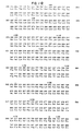
- The Figure is a DNA sequence showing a comparison of the amino acid sequence of the subject human protease and a mouse killer-cell protease.
- Novel compositions and methods are provided related to novel serine protease produced by human killer cells, where the compositions comprise nucleic acid sequences coding for biologically active fragments of the serine proteases, the serine proteases, and precursors to the serine proteases. Other compositions include nucleic acid sequences joined to other nucleic acid sequences for cloning and expression of such sequences. Also included are poly(amino acid) compositions, which include biologically active fragment of the serine proteases, the serine proteases, precursors to the serine proteases, and conjugates of the various poly(amino acids) to other moieties for a variety of purposes.
- The human serine proteases of the subject invention are characterized by being found in various subsets of human killer cells but in substantially lower amounts of being absent in other kinds of cells. The subject serine protease is further characterized by having a polypeptide molecular weight in the range of about 20-30kD, more usually in the range of about 23-28kD, and particularly in the range of about 25-26kD. The serine protease is further characterized by having an active site "charge-relay" with similar spacing and conformation to that of chymotrypsin, namely having histidine, aspartate, and serine spaced approximately as observed with chymotrypsin, as well as having the trypsin specific aspartate in about the same position as trypsin. Spacing here refers to the number of intervening amino acids. Particularly, the His-Asp spacing is about 41-47 amino acids, particularly 44 amino acids, and the Asp-Ser spacing is about 94-100 amino acids, particularly 97 amino acids. The subject serine protease has an Asp residue from about 3-8 amino acids, particularly 6 amino acids, toward the N-terminus from the Ser residue, similar to trypsin. The serin protease is further characterized by being part of the lytic process of killer cells.
- The naturally occurring subject serine proteases are found in a number of subsets of killer cells, such as killer T cells, cytotoxic, T lymphocytes (CTL), some T helper cells, NK/NC cells, K cells (which use antibodies to target on a foreign cell), and lymphokine activated killer cell (LAK cells). The expression of the serine protease suggests an "activation" gene related to a lysis mechanism.
- The subject serine proteases are involved in a system which requires divalent cations, energy sources and which is responsive to inhibition by low and high molecular weight serine protease inhibitors, such as α-2-macroglobulin and soybean trypsin inhibitor.
- The subject serine proteases are not found in significant amounts in such cells as normal muscle cells, liver cells, unstimulated peripheral blood lymphocyte cells, and thymus cells, as well as a number of B cell tumor cell lines.
- In the Figure the amino acid sequence is set forth in comparison with the amino acid sequences of a mouse killer-cell protease. The amino acid homology within the active-enzyme portion of the protein is 71% with 77% homology at the DNA level within the corresponding region. The overall DNA homology is 72% when the complete coding region and the 3ʹ untranslated region are included. An arrow indicates the site of cleavage which generates the active enzyme. The amino acids of the charge-relay system, His⁴¹, Asp⁸⁶, and Ser¹⁸⁴, are each marked with a star. The acidic residue Asp¹⁷⁸, marked with a $, determines substrate specificity for Lys or Arg. The AATAAA polyadenylation consensus sequence is underlined in the 3ʹ noncoding region. The Asn-linked carbohydrate site which occurs at Asn¹⁴² is marked by a plus.
- The amino acids may be substituted by conservative changes, with non-conservative changes generally being restricted to positions removed from the active site. Groups of amino acids which may be substituted one for the other include G, A; V,I,L; S,T,M; D,E; K,R; N,Q; and F,W,T,H.
- Of particular interest is the amino acid region from
amino acid 30 toamino acid 70, more particularly fromamino acid 40 toamino acid 60. Also of interest is the region fromamino acid 90 toamino acid 120, more particularly fromamino acid 100 toamino acid 110. Of further interest is the amino acid sequence of from about 190 to 250, more particularly from about 200 to 240, more particularly from about 220 to about 240. Of further interest is a conserved amino acid sequence of at least about 10 amino acids, usually at least about 12 amino acids, and not more than about 30 amino acids, usually not more than about 20 amino acids, included in the fragments indicated above. Peptides consisting of amino acids from these regions of interest will be useful in preparing antibodies that bind and interfere with the active site of the enzyme. - The nucleotide sequence, either the DNA or RNA, more particularly the DNA sequence, encoding the subject serine proteases or active fragments thereof may be used in a variety of ways. Fragments of the serine proteases may be used as probes for detecting the presence of non-mutated or mutated serine proteases present in mammalian cells. Alternatively, the sequences may be used for expression of amino acid fragments having biological activity or extended fragments having enzymatic activity coming within the sequence indicated in the Figure. Thus, the various sequences may be used in conjunction with other DNA sequences to provide constructs for cloning or expression of the indicated DNA sequences. Thus the coding sequence will be joined to flanking regions other than the natural flanking regions. The sequence encoding the serine protease will be less than 5knt (kilonucleotides), usually less than about 2knt. For expression, the DNA sequences will be joined to regulatory regions and other functional regions other than the natural regions to provide for the production of the desired poly(amino acids), including oligopeptides of from about 8 to 30 amino acids, more usually from about 10 to 20 amino acids, or polypeptides, of at least about 30 amino acids to about 235 amino acids, usually not more than about 233 amino acids, particularly not more than about 232 amino acids, which may code for the entire naturally occurring serine protease.
- The DNA constructs in the direction of transcription will usually include a transcriptional initiation region, the open reading frame beginning with the initiation codon (Met) and the desired peptide, followed by the transcriptional termination region. The transcriptional initiation and termination regions will be chosen so as to be functional in the expression host, which may be prokaryotic or eukaryotic, including such hosts as bacteria, e.g., E. coli, fungi, e.g, yeast, such as Saccharomyces, Kluveromyces, filamentous fungi, such as Neurospora, Aspergillus, etc., silkworm cells, mammalian cells, e.g., Chinese hamster ovary cells, hamster kidney cells, etc. For cloning and expression, unicellular organisms are of particular interest.
- In addition to the expression construct, there may be one or more markers which allow for selection of hosts containing the expression construct. Markers may include structural genes capable of expression in the host which provide for antibiotic resistance, complementation, plaque formation, or the like.
- Where extrachromosomal maintenance is desired, an origin of replication system will be provided, which allows for extrachromosomal maintenance of the expression construct in the host. The extrachromosomal replication system may be derived from plasmids, viruses, chromosomes (centromeres and autonomous replication systems) and the like. In some instances, the expression construct may be introduced into transposons for integration into the host genome. The cells containing the expression construct are grown in an appropriate nutrient medium and depending upon whether the product is secreted, the cells may be lysed and the product isolated by conventional ways or the supernatant isolated and the product extracted.
- The subject peptides may be used for a wide variety of purposes. The subject peptides may be used for preparation of polyclonal or monoclonal antibodies. Where only a fragment of the subject serine proteases is employed, the fragment may be joined to an immunogen to provide for an immunogenic product for injection into a vertebrate for the production of antibodies. The immunogenic protein will be foreign to the intended host and one where polyclonal antibodies may or may not be encountered. The immunogens will usually be greater than 30kD.
- Joining of haptenic or antigenic peptides to a larger polypeptide is well known in the art and a variety of linking groups are available, such as formaldehyde, glutaraldehyde, maleimidobenzoic acid, methyldithioacetic acid, Ellman's reagent, or the like. The particular manner in which the polypeptide fragment of the subject serine proteases is joined to the immunogenic protein is not critical to this invention. Convenient immunogenic proteins include bovine serum albumin, tetanus toxoid, keyhole limpet hemocyanin, bovine betaglobulin, and the like.
- Various hosts which may be injected with the immunogen include mice, rats, birds, hamsters, or other mammals, e.g., primates such as humans. The manner of injection and obtaining of polyclonal or monoclonal antibodies has been amply described in the literature and need not be described in detail here. Usually, the immunogen will be injected in one or more sites of the host in volumes of about 0.5 to 5ml with an immunizing effective amount, sufficient to produce a hemagglutinating titer in the range of about 1:32 to 1:256, where one or more injections may be employed at intervals of from about 2 to 4 weeks. Shortly after the last injection, blood may be harvested from the host and the immunoglobulins isolated.
- For polyclonal antibodies, the immunoglobulins may be purified by a wide variety of ways, particularly affinity chromatography. For monoclonal antibodies, the spleens may be removed and fused with syngeneic myeloma cells for production of hybridomas, which may be screened for the production of antibodies specific for the desired epitopic site.
- The antibodies may be neutralizing or non-neutralizing, depending upon their effect on the activity of the enzyme, the purpose or result of complex formation, and the like.
- The antibodies to the subject serine proteases may find use both in vivo and in vitro. For in vivo use, the antibodies may be used for therapeutic purposes for passive immunization to inhibit immune disorders, inhibit graft rejection, and modulating the immune system. In vitro, the antibodies may be used for diagnostic purposes, in detecting the nature of the cell population, for determining pathological lesions, for determining rejection of organ grafts, and for determining the differentiation state of various cells.
- The subject human serine proteases and fragments thereof may be used by themselves or in conjunction with other materials as labels in diagnostic assays. In addition, the serine proteases may be used for removing particular cell types from a heterogeneous population of cells. For example, serine protease-containing cells could be removed from bone marrow or other mixture of cells, where cells are susceptible to the lytic cascade or other inhibitory products of NK or CTL cells.
- Depending upon the manner in which the subject compositions are to be used, they may be formulated in a variety of ways, being formulated in aqueous media, for example, aqueous buffered media, e.g., phosphate-buffered saline, Tris-buffered solutions, or the like, where the concentrations may vary from about 0.05mM, to about 5mM. Other additives may be present, such as protein stabilizers, inert proteins, bacteriostats and bacteriocides, and the like. The particular formulation will be chosen in relation to the particular application.
- Formulations may involve additional members of the lytic mechanism for cytotoxicity, such as the precursors of the polyperforins, activators for the subject protease, substrates for the subject protease, and the like. Thus, some or all of the components of the secretory granules of killer cells may be isolated in crude form and used in conjunction with the subject serine protease in substantially pure form. Usually, the subject serine protease can be provided with at least 90% of its native activity, preferably at least about 95% of its native activity.
- The subject compositions may be used in a variety of ways. Antibodies may be prepared from fragments of the serine protease or the entire protease which may act to neutralize the enzymatic function of the serine protease. In addition, the serine protease may serve to identify suicide substrates, natural protease inhibitors, substrate transitional state analogs, or other inhibitors, which may serve to neutralize the active site of HF gene products in mammals, so as to block cytotoxic cell functions.
- The ability to inhibit the serine protease may serve in the treatment of graft rejection, in the treatment of immune disorders, where the function of killer cells leads to a pathological state, and in the diagnosis of pathological lesions, where the number, type of activity of killer cells may serve as an important pathognomonic sign.
- The serine proteases may be used in the development of labeled substrates, e.g., fluoresceinated or umbelliferyl labeled substrates, to serve in the purification of killer cells and natural killer cells, as may be used in therapy, prior to expansion for subsequent reinfusion or in autoimmune disorders for removal of cells by plasmaphoresis. In addition, by preparing antibodies to the zymogen peptide or the junction of the zymogen peptide and the active serine protease, the antibodies may serve as a diagnostic tool for determining the frequency of blood cells or tissue cells which are in the killer cell set. In addition, the serine protease by itself or in combination with the other members of the cytolytic process of T-cells, including components of the secreted granules, may be used for in vitro and in vivo u lysis of cells, permitting a powerful biological purification method. The human serine protease can also serve to identify transition state analogs and other small molecular weight protease inhibitors that are preferentially specific to this enzyme's active site, thereby identifying molecules capable of inhibiting T-cell and/or NK cytotoxicity.
- The following examples are offered by way of illustration and not by way of limitation.
- A cDNA phage library was prepared from human peripheral blood lymphocytes (PBL) after 4 days of stimulation with phytohemagglutinin (PHA). This cDNA library was made in λgt10 by modifications of a cDNA procedure described by Huynh DNA Cloning Techniques: A Practical Approach (Ed. D. Glover) IRL PRESS, Oxford (1984). The two modifications were (1) the replacement of all phenol-chloroform extractions with spermine precipitation as described by Hoops et al., Nucl. Acid Res. (1981) 9:5493, and (2) the replacement of the Biogel A-50m column with 1% to 2% agarose horizontal gel electrophoresis for the purpose of removing the excess EcoRI linkers and size fractionating ds cDNA. The ds cDNAs were size selected initially for lengths greater than 0.5kb and subsequently for lengths greater than 0.95kb. The selected agarose slices were electroeluted in dialysis bags (Smith, Methods in Enzymology (1980) 65:371) and spermine precipitated. All RNAs for the cDNA libraries, Northerns and S1 analysis were prepared by guanidinium thiocyanate extraction (Chirgwin et al., Biochem. (1979) 18:5294) and polyA selected with oligo-dT cellulose.
- 2x10⁵ recombinant phage plaques of the PHA stimulated PBL cDNA library were screened with the mouse serine protease cDNA. The probe was prepared by nick translation as described by Meinkoth and Wahl, supra, and the cDAN libraries were plated at a density of approximately 50,000pfu/150cm plate as described by Hunyh et al., supra. One phage was picked and rescreened through two additional rounds of hybridization, yielding a plaque-purified clone. The purified lambda phage contained a 1.3 kilobase (kb) EcoRI cDNA insert encoding the human equivalent of the mouse serine protease HF gene (designated HuHF)> By Northern analysis, this cDNA hybridized to a 1.3 kb polyA-RNA species present in human CTL cells generated in a four-day alloreactive mixed lymphocyte culture and in Jurkat tumor cells. By Northern analysis, the RNA was not detected in normal human muscle, liver, tonsil, or lymphoid tissue. Futhermore, no RNA could be detected in the following tumors: KB cell (a nasopharyngael carcinoma), RPMI 4265 and NA (B cell tumors), and SS II (T cell). From RNA dot blot experiments, the RNA was detectable in three human CTL alloreactive cloned lines (AI5.1, AMSB.3, AMW.6), in non-stimulated, cell sorted Leu 11+ NK and Leu 11- Leu 4+ T cell large granular lymphocytes (LGL) from PBL.
- The nucleotide sequence was completely determined on both strands, except for the 5 prime most 400 nucleotides, yielding a single open reading frame (see Fig. 1). In Fig. 1, the nucleotide sequence and amino acid translation of the human cDNA is aligned with the mouse sequence. The amino acid sequence is numbered sequentially from the predicted amino terminus of the putative active enzyme. An arrow indicates a putative site of cleavage, generating the active enzyme predicted based on homology alignments. The amino acids of the charge relay system, His⁴¹, Asp⁸⁶ and Ser¹⁸⁴, are each marked with a star. The acidic residue Asp¹⁷⁸, marked with a $, determines the substrate's specificity by analogy with other serine proteases. The AATAAA polyadenylation consensus sequence is underlined in the 3ʹ noncoding region. A potential Asn-linked carbohydrate site occurs at Asn¹⁴² marked by +.
- By protein sequence homology, the DNA sequence encodes an active serine protease of 234 amino acids, with a non-glycosylated, polypeptide molecular weight of approximately 25.8kD. The active enzyme is probably preceded by a zymogen peptide by analogy with other serine proteases, cleaving C-terminal to Lys (-1). The amino acids of the serine protease charge-relay catalytic mechanism are conserved, with the His and Asp being separated by 44 amino acids and the Asp and Ser being separated by 97 amino acids as compared to a separation in chymotrypsin of 44 and 92, respectively. The HF serine protease contains an Asp¹⁷⁸ residue equivalent to the Asp¹⁸⁹ of trypsin, suggesting trypsin-like substrate specificity.
- The amino acid composition is shown in Table 1 for the uncleaved protease and the cleaved, active protein.

- All publications and patent applications cited in this specification are indicative of the level of skill of those skilled in the art to which this invention pertains. All publications and patent applications are herein incorporated by reference to the same extent as if each individual publication or patent application was specifically and individually indicated to be incorporated by reference.
- Although the foregoing invention has been described in some detail by way of illustration and example for purposes of clarity of understanding, it will be obvious that certain changes and modifications may be practiced.
Claims (9)
- A human serine protease or biologically functional fragment thereof substantially free of killer cell granule components, said serine protease being characterized by having an amino acid sequence which is at least 90% conserved in relation to the amino acid sequence of human serine protease set forth in the Figure, having a molecular weight in the range of about 20-30kD, and being capable of being a member of the cytolytic activity of cytolytic T cells.
- A human serine protease according to claim 1, found in cytotoxic T cells.
- A DNA sequence encoding a serine protease or biologically functional fragment thereof according to claim 1, joined to other than a natural flanking region.
- A DNA sequence according to claim 3, joined at the 5'-terminus to a transcriptional initiation region other than the natural transcriptional initiation region.
- A DNA sequence according to claim 4, joined at its 3'-terminus to a transcriptional termination region wherein said transcriptional initiation and termination regions are functional in a host cell.
- An antibody composition specific for a serine protease or biologically functional fragment thereof according to any one of the preceding claims.
- An antibody composition according to claim 6, wherein said antibody composition is monoclonal.
- A method for producing a serine protease or biologically functional fragment thereof which comprises:
growing cells containing a DNA sequence according to claim 5, whereby said DNA is expressed; and
isolating said serine protease or biologically functional fragment thereof free of cellular debris. - A method for identifying molecules capable of inhibiting T-cell or NK cytotoxicity, which comprises:
contacting said molecule with the human serine protease of any of claims 1 to 7; and
measuring protease activity relative to activity in the absence of said molecule.
Applications Claiming Priority (6)
| Application Number | Priority Date | Filing Date | Title |
|---|---|---|---|
| US86008586A | 1986-05-06 | 1986-05-06 | |
| US860085 | 1986-05-06 | ||
| US86122186A | 1986-05-08 | 1986-05-08 | |
| US861221 | 1986-05-08 | ||
| US06/948,248 US4973555A (en) | 1986-05-06 | 1986-12-31 | Human serine protease gene |
| US948248 | 1986-12-31 |
Publications (2)
| Publication Number | Publication Date |
|---|---|
| EP0245051A1 EP0245051A1 (en) | 1987-11-11 |
| EP0245051B1 true EP0245051B1 (en) | 1992-08-12 |
Family
ID=27420406
Family Applications (1)
| Application Number | Title | Priority Date | Filing Date |
|---|---|---|---|
| EP19870303945 Expired - Lifetime EP0245051B1 (en) | 1986-05-06 | 1987-05-01 | Killer cell cytotoxic composition |
Country Status (2)
| Country | Link |
|---|---|
| EP (1) | EP0245051B1 (en) |
| DE (1) | DE3781017T2 (en) |
Families Citing this family (4)
| Publication number | Priority date | Publication date | Assignee | Title |
|---|---|---|---|---|
| FR2673952A1 (en) * | 1991-03-14 | 1992-09-18 | Inst Nat Sante Rech Med | MEANS FOR THE IN VITRO DIAGNOSIS OF COMPONENTS OF CYTOPLASMIC GRANULES AND BIOLOGICAL APPLICATIONS. |
| US20020064856A1 (en) * | 2000-06-26 | 2002-05-30 | Gregory Plowman | Novel proteases |
| CA2416691A1 (en) * | 2000-07-21 | 2002-01-31 | Incyte Genomics, Inc. | Proteases |
| WO2003074669A2 (en) * | 2002-03-01 | 2003-09-12 | Applera Corporation | Isolated human enzyme proteins, nucleic acid molecules encoding human enzyme proteins, and uses thereof |
Family Cites Families (1)
| Publication number | Priority date | Publication date | Assignee | Title |
|---|---|---|---|---|
| NZ208612A (en) * | 1983-06-24 | 1991-09-25 | Genentech Inc | Method of producing "procaryotic carbonyl hydrolases" containing predetermined, site specific mutations |
-
1987
- 1987-05-01 EP EP19870303945 patent/EP0245051B1/en not_active Expired - Lifetime
- 1987-05-01 DE DE19873781017 patent/DE3781017T2/en not_active Expired - Fee Related
Also Published As
| Publication number | Publication date |
|---|---|
| DE3781017D1 (en) | 1992-09-17 |
| DE3781017T2 (en) | 1993-03-18 |
| EP0245051A1 (en) | 1987-11-11 |
Similar Documents
| Publication | Publication Date | Title |
|---|---|---|
| US5602102A (en) | Dipeptidyl peptidase-I inhibitors and uses thereof | |
| EP0585257A1 (en) | Dna and amino acid sequence specific for natural killer cells | |
| US6673904B2 (en) | Stem cell growth factor-like polypeptides | |
| CN1352686A (en) | Cytokine receptor chain | |
| EP0948536B1 (en) | A g protein-coupled receptor with an enlarged extracellular domain | |
| US4973555A (en) | Human serine protease gene | |
| CA2139127A1 (en) | Compositions for the inhibition of protein hormone formation and uses thereof | |
| US7067301B2 (en) | Methods and materials relating to novel prothrombinase-like polypeptides and polynucleotides | |
| EP0245051B1 (en) | Killer cell cytotoxic composition | |
| Kelly et al. | Cloning and expression of the recombinant mouse natural killer cell granzyme Met-ase-1 | |
| US5213977A (en) | Serine protease from cytotoxic killer cells | |
| US7205143B2 (en) | Feline IL-12 single chain nucleic acid molecules | |
| JPH05184387A (en) | Intracellular antigen discovered in subpopulation of cd8 plus t lymphocyte and monoclonal antibody thereagainst | |
| WO1998020034A2 (en) | Novel serine protease inhibitor nucleic acid molecules, proteins and uses thereof | |
| KR100802140B1 (en) | Novel gene encoding adam3 isolated from porcine and method for discriminating fertility of porcine sperm using thereof | |
| JPS62285783A (en) | Killer composition of killer cell | |
| CA2174089A1 (en) | Plasmodium falciparum ribonucleotide reductase, encoding dna, and inhibitors | |
| US20020098546A1 (en) | Canine TAg1 proteins, nucleic acid molecules, and uses thereof | |
| US6204010B1 (en) | Flea protease proteins, nucleic acid molecules, and uses thereof | |
| JPH01502198A (en) | Cytotoxic lymphocyte-specific protease-related molecules and methods | |
| AU2005201964B2 (en) | Novel Galectin Sequences and Compositions and Methods Utilizing Same for Treating or Diagnosing Arthritis and Other Chronic Inflammatory Diseases | |
| EP0973876A1 (en) | DNA ENCODING $i(PNEUMOCYSTIS CARINII) PROTEASE | |
| Hershberger | A serine protease made by cytotoxic lymphocytes | |
| JPH09291100A (en) | Rat cd86, its geme and its antibody | |
| CN1271006A (en) | New human proteinase subunit protein and its code sequence |
Legal Events
| Date | Code | Title | Description |
|---|---|---|---|
| PUAI | Public reference made under article 153(3) epc to a published international application that has entered the european phase |
Free format text: ORIGINAL CODE: 0009012 |
|
| AK | Designated contracting states |
Kind code of ref document: A1 Designated state(s): CH DE FR GB IT LI NL |
|
| 17P | Request for examination filed |
Effective date: 19880328 |
|
| 17Q | First examination report despatched |
Effective date: 19901105 |
|
| GRAA | (expected) grant |
Free format text: ORIGINAL CODE: 0009210 |
|
| AK | Designated contracting states |
Kind code of ref document: B1 Designated state(s): CH DE FR GB IT LI NL |
|
| REF | Corresponds to: |
Ref document number: 3781017 Country of ref document: DE Date of ref document: 19920917 |
|
| ITF | It: translation for a ep patent filed |
Owner name: MODIANO & ASSOCIATI S.R |
|
| ET | Fr: translation filed | ||
| PGFP | Annual fee paid to national office [announced via postgrant information from national office to epo] |
Ref country code: GB Payment date: 19930420 Year of fee payment: 7 |
|
| PGFP | Annual fee paid to national office [announced via postgrant information from national office to epo] |
Ref country code: FR Payment date: 19930510 Year of fee payment: 7 |
|
| PGFP | Annual fee paid to national office [announced via postgrant information from national office to epo] |
Ref country code: CH Payment date: 19930517 Year of fee payment: 7 |
|
| PGFP | Annual fee paid to national office [announced via postgrant information from national office to epo] |
Ref country code: DE Payment date: 19930524 Year of fee payment: 7 |
|
| PGFP | Annual fee paid to national office [announced via postgrant information from national office to epo] |
Ref country code: NL Payment date: 19930531 Year of fee payment: 7 |
|
| PLBE | No opposition filed within time limit |
Free format text: ORIGINAL CODE: 0009261 |
|
| STAA | Information on the status of an ep patent application or granted ep patent |
Free format text: STATUS: NO OPPOSITION FILED WITHIN TIME LIMIT |
|
| 26N | No opposition filed | ||
| PG25 | Lapsed in a contracting state [announced via postgrant information from national office to epo] |
Ref country code: GB Effective date: 19940501 |
|
| PG25 | Lapsed in a contracting state [announced via postgrant information from national office to epo] |
Ref country code: LI Effective date: 19940531 Ref country code: CH Effective date: 19940531 |
|
| PG25 | Lapsed in a contracting state [announced via postgrant information from national office to epo] |
Ref country code: NL Effective date: 19941201 |
|
| GBPC | Gb: european patent ceased through non-payment of renewal fee |
Effective date: 19940501 |
|
| NLV4 | Nl: lapsed or anulled due to non-payment of the annual fee | ||
| PG25 | Lapsed in a contracting state [announced via postgrant information from national office to epo] |
Ref country code: FR Effective date: 19950131 |
|
| REG | Reference to a national code |
Ref country code: CH Ref legal event code: PL |
|
| PG25 | Lapsed in a contracting state [announced via postgrant information from national office to epo] |
Ref country code: DE Effective date: 19950201 |
|
| REG | Reference to a national code |
Ref country code: FR Ref legal event code: ST |
|
| PGFP | Annual fee paid to national office [announced via postgrant information from national office to epo] |
Ref country code: IT Payment date: 20060531 Year of fee payment: 20 |