EP0243797A2 - Method for amplifying enzyme activity, enzyme conjugates suitable therefor and their preparation - Google Patents
Method for amplifying enzyme activity, enzyme conjugates suitable therefor and their preparation Download PDFInfo
- Publication number
- EP0243797A2 EP0243797A2 EP87105569A EP87105569A EP0243797A2 EP 0243797 A2 EP0243797 A2 EP 0243797A2 EP 87105569 A EP87105569 A EP 87105569A EP 87105569 A EP87105569 A EP 87105569A EP 0243797 A2 EP0243797 A2 EP 0243797A2
- Authority
- EP
- European Patent Office
- Prior art keywords
- enzyme
- leash
- support
- fragment
- conjugate
- Prior art date
- Legal status (The legal status is an assumption and is not a legal conclusion. Google has not performed a legal analysis and makes no representation as to the accuracy of the status listed.)
- Ceased
Links
Images


Classifications
-
- C—CHEMISTRY; METALLURGY
- C12—BIOCHEMISTRY; BEER; SPIRITS; WINE; VINEGAR; MICROBIOLOGY; ENZYMOLOGY; MUTATION OR GENETIC ENGINEERING
- C12Q—MEASURING OR TESTING PROCESSES INVOLVING ENZYMES, NUCLEIC ACIDS OR MICROORGANISMS; COMPOSITIONS OR TEST PAPERS THEREFOR; PROCESSES OF PREPARING SUCH COMPOSITIONS; CONDITION-RESPONSIVE CONTROL IN MICROBIOLOGICAL OR ENZYMOLOGICAL PROCESSES
- C12Q1/00—Measuring or testing processes involving enzymes, nucleic acids or microorganisms; Compositions therefor; Processes of preparing such compositions
- C12Q1/68—Measuring or testing processes involving enzymes, nucleic acids or microorganisms; Compositions therefor; Processes of preparing such compositions involving nucleic acids
-
- C—CHEMISTRY; METALLURGY
- C12—BIOCHEMISTRY; BEER; SPIRITS; WINE; VINEGAR; MICROBIOLOGY; ENZYMOLOGY; MUTATION OR GENETIC ENGINEERING
- C12Q—MEASURING OR TESTING PROCESSES INVOLVING ENZYMES, NUCLEIC ACIDS OR MICROORGANISMS; COMPOSITIONS OR TEST PAPERS THEREFOR; PROCESSES OF PREPARING SUCH COMPOSITIONS; CONDITION-RESPONSIVE CONTROL IN MICROBIOLOGICAL OR ENZYMOLOGICAL PROCESSES
- C12Q1/00—Measuring or testing processes involving enzymes, nucleic acids or microorganisms; Compositions therefor; Processes of preparing such compositions
-
- Y—GENERAL TAGGING OF NEW TECHNOLOGICAL DEVELOPMENTS; GENERAL TAGGING OF CROSS-SECTIONAL TECHNOLOGIES SPANNING OVER SEVERAL SECTIONS OF THE IPC; TECHNICAL SUBJECTS COVERED BY FORMER USPC CROSS-REFERENCE ART COLLECTIONS [XRACs] AND DIGESTS
- Y10—TECHNICAL SUBJECTS COVERED BY FORMER USPC
- Y10S—TECHNICAL SUBJECTS COVERED BY FORMER USPC CROSS-REFERENCE ART COLLECTIONS [XRACs] AND DIGESTS
- Y10S435/00—Chemistry: molecular biology and microbiology
- Y10S435/962—Prevention or removal of interfering materials or reactants or other treatment to enhance results, e.g. determining or preventing nonspecific binding
-
- Y—GENERAL TAGGING OF NEW TECHNOLOGICAL DEVELOPMENTS; GENERAL TAGGING OF CROSS-SECTIONAL TECHNOLOGIES SPANNING OVER SEVERAL SECTIONS OF THE IPC; TECHNICAL SUBJECTS COVERED BY FORMER USPC CROSS-REFERENCE ART COLLECTIONS [XRACs] AND DIGESTS
- Y10—TECHNICAL SUBJECTS COVERED BY FORMER USPC
- Y10S—TECHNICAL SUBJECTS COVERED BY FORMER USPC CROSS-REFERENCE ART COLLECTIONS [XRACs] AND DIGESTS
- Y10S435/00—Chemistry: molecular biology and microbiology
- Y10S435/966—Chemistry: molecular biology and microbiology involving an enzyme system with high turnover rate or complement magnified assay, e.g. multi-enzyme systems
Definitions
- This invention relates to a method for enzyme amplification, to a system of immobilized enzyme material which releases a large amount of enzyme in response to the application of a small amount of the same enzyme, and a method for preparing these conjugate systems.
- Chemical amplification the formation of an enhanced chemical response, occurs in three ways.
- the first is catalysis, a common example of which is the action of an enzyme or coenzyme.
- Enzyme or substrate cycling (1) in which a substrate acts as a catalyst by first participating in one reaction, and then cycling back to its original state in a second reaction, also illustrates catalytic amplification.
- gate amplification the opening of a molecular gate such as a channel in a membrane amplifies the passage of molecules from one zone to another.
- multiplicative amplification in which the amount of a substance is multiplied by a constant factor repetitively, e.g., the unhindered successive generations of a virus.
- Amplification components such as enzyme, coenzymes, inorganic iodide, catalytic electrodes,bacteriophages andlipoeomes were all discussed. When such components inherently do not provide tie specificity required, or do not directly involve the analyte, then an antibody, secondary enzyme, or secondary chemical reaction is used to make the amplification system specific or analyte-responsive.. Aside from the simple use of enzymes as amplification catalysts, many of the techniques discussed by these authors for clinical analysis were not very practical at that time.
- a more recent example of gate amplification in chemical analysis is the report by Litchfield et aL (3) of an immunoassay for digoxin in which ouabain-mellitin triggers the release of alkaline phosphatase entrapped in a liposome.
- liposomes tend to be unstable; a threshold dose of a mellitin substance is required and probably acts only once to release alkaline phosphatase; mellitin is a hydrophobic substance that will tend to behave eratically be undergoing losses onto surfaces and forming complexes with interfering macromolecules; and the behavior of liposomes in an actual assay can be influenced by other substances in the sample that bind to the liposome aside from the ouabain-mellitin reagent.
- Harris U.S. Patent 4 463 090 has used cascade amplification in an enzyme immunoassay in which one or more proenzymes are present.
- an initial enzyme or activator begins a sequence of activating a proenzyme to an enzyme, which enzyme product may be detected or may activate another proenzyme to an enzyme that may be detected, or it may in turn activate another proenzyme etc.
- This invention uses naturally occurring -enzyme cascades that unfortunately are limited in number. It comprises components that tend to be unstable, susceptible to inhibitors, complex, not readily available, poorly characterized and expensive. Such reagents tend to vary in their properties batch-to-batch.
- substrate-leash amplification a new concept for chemical amplification, here called “substrate-leash amplification” (SLA) whereby both a catalytic and a multiplicative mechanism are involved.
- SLA substrate-leash amplification
- the key feature is that a chain-cutting enzyme or enzyme component is attached via its substrate leash to a surface. The details of the assembly restrict spontaneous release of the enzyme or its components. When free enzyme is introduced that cleaves the substrate leash, a cascade of released enzyme results.
- the method according to the invention foramplifying enzymatic activity involves connecting an enzyme to a support by means of molecular leash material which is cleavable by that enzyme, to produce a support-leash-enzyme conjugate having many -leash-enzyme units connected to the support; adding a small amount of that enzyme in the free state to the conjugate to produce a mixture of free enzyme and support-leash-enzyme conjugate; incubating this mixture for a predetermined period of time to cause enzyme in the free state to be released from the conjugate; and recovering the released free enzyme.
- the leash material between the support and the immobilized enzyme is constructed to prevent the bound enzyme of a given -leash-enzyme unit of the conjugate from cleaving the leash portion of that unit.
- This is done by controlling the lengths and mobility of the leashes including the use of a spacer substance within the leash, and cross-links within a given -leash-enzyme unit or between one such unit and another.
- the -leash-enzyme units are connected to the support at positions sufficiently distant from each other that the enzyme portion of a given -leash-enzyme unit cannot cleave the leash portion of another -leash-enzyme unit.
- two-stage enzyme reservoirs are prepared as follows: providing first and second enzyme fragments; herein designated fragment A and fragment B respectively, each of these fragments being complementary to the other and enzymatically inactive, and being capable of recombining with each other to produce active enzyme; bonding the first enzyme fragment to a support by means of molecular leash material which is cleavable by the active enzyme, to produce a support-leash-fragment A conjugate having a large number of -leash-fragment A units connected to the support material; bonding the second enzyme fragment to a support material similarly, to produce a support-leash-fragment B conjugate, also having a large number of -leash-fragment B units connected to the support material; and connecting the first and second conjugates in series, forming the two-stage enzyme reservoir.
- the two-stage enzyme reservoir is caused to release a large amount of enzyme in either of two ways: by application of active enzyme, or by application of inactive enzyme fragment to that portion of the enzyme reservoir containing the support-leash-(enzyme fragment) conjugate in which the enzyme fragment is complementary to the added enzyme fragment.
- the enzyme amplification is carried out by providing a mixed enzyme two-stage enzyme reservoir as follows: providing first and second enzymes, designated C and D respectively; bonding enzyme C to a support by means of molecular leash material which is cleavable by enzyme D, to produce a support-leash-enzyme C conjugate having a large number of -leash-enzyme C units connected to the support material; bonding enzyme D to a support material similarly, to produce a support-leash-enzyme D conjugate having a large number of -leash-enzyme D units connected to the support material; and connecting the first and second conjugates in series, forming the mixed enzyme two-stage enzyme reservoir.
- the mixed enzyme two-stage enzyme reservoir is caused to release a large amount of enzyme D, as well as some enzyme C, by application of free enzyme D to that portion of the mixed enzyme two stage enzyme reservoir containing support-leash-enzyme C conjugate. This releases enzyme C, which in turn catalyzes release of enzyme D in the second stage of the reservoir.
- the same principle again applies in that the large amount of enzyme D produced in the second stage of the enzyme reservoir may serve as the initial charge of enzyme D to be added to the first stage of a second mixed enzyme two-stage enzyme reservoir, and so on.
- the invention includes compositions having the generalized formula support-leash-enzyme, where the support is a solid support material, the leash is a material including a molecular chain which is cleavable by the enzyme in the free state, and the support-leash-enzyme conjugate has a large number of -leash-enzyme units connected to the support.
- the invention also includes compositions having the generalized formula suppoct-leash-(enzyme fragment) in which the enzyme fragment is one of two complementary enzymatically inactive fragments of an active enzyme, and is capable of recombining with the second of these fragments to regenerate active enzyme, the support is a solid support material, the leash is a material containing a molcecular chain which is cleavable by /the active enzyme, and the supoort-lersh-(enzyme fragment) conjugate has a large number of -leash-(enzyme fragment) units connected to the support.
- the invention further includes compositions having the generalized formula support-leash-enzyme in which the support is a solid support material, the leash is a molecular chain which is not cleavable by the enzyme in the free or immobilized form, and the support-leash-enzyme conjugate has a large number of -leash-enzyme units connected to the support
- the support may also be a soluble macromolecule, other chain-cutting substances besides enzymes may be used, and the leash-enzyme unit may be attached noncovalently to the surface.
- Fig. 1 is a schematic representation of the enzyme amplification method of the invention, in a general form.
- An enzyme is attached to a support by means of molecular chain covalently attached to both the support and the enzyme.
- This chain or leash is made up of or includes material which is cleavable by the enzyme.
- the enzyme is attached to the support by molecular material which is a substrate for the enzyme.
- cleavage of the leash chain occurs, releasing free enzyme which in turn cleaves more leashing chains and frees more enzyme.
- support-leash-enzyme conjugate having a large number of -leash-enzyme units on the support is prepared and treated with a small amount of enzyme, forming-initially a mixture of free enzyme and support-leash-enzyme conjugate, then this mixture is incubated for a predetermined period of time to permit the enzyme-catalyzed cleavage of leash material to occur, freeing formerly immobilized enzyme.
- the free enzyme is recovered from the support-leash-enzyme conjugate materiaL
- the nature of the system ensures that the amount of enzyme ultimately recovered is substantially greater than the amount of enzyme initially added to the system.
- the enzyme may be any enzyme which can be covalently attached to a material which is a substrate of that enzyme.
- the leashing material may be any substrate for the enzyme, provided only that it may be covalently attached to both the enzyme and the support materiaL
- the support is a solid support material such as agarose, silica, cellulose, paper, Sepharose, Trisacryl, glass, nylon, poly methacrylate, Immobilon Membranes, polyacrylamides, polyamides and gelatin.
- Table I lists examples of many enzymes in several classes, and some examples of molecular chains they cleave. These materials, many of which are illustrated in the Examples below, are useful in the enzyme amplification method and compositions.
- the leash material joining the support and the enzyme is constructed so that an enzyme portion of a given -leash-enzyme unit cannot cleave the leash portion of that unit.
- the leash material can be made to have a length within a predetermined range of lengths so that the attached enzyme is unable to have its active site contact the leash in a
- leash lengths can be established by synthesizing molecular chains of desired lengths for attachment of the conjugates to the solid supports. Since the leashes for chain cutting enzymes are available naturally in much longer lengths than are needed here, a suitable strategy is to conduct a limited hydrolysis, either with the chain-cutting enzyme itself or with aqueous acid or base, of the longer leashes, varying the time of incubation. This yields a mixture of shorter leashes. The latter can then be fractionated by gel filtration into several size ranges, and the appropriate size range can be used.
- the next mechanism is to restrict the movement of the leash so that the attached enzyme is unable to bring its active site into contact with the leash. This is most easily done by attaching the leash at two or more points along its length to the support, so that the leash has a restricted mobility. For this the leash needs to have two or more functional groups along its length that can bind to the complementary functional groups on the support surface. This strategy is commonly used in affinity chromatography to increase the stability of attaching a polymeric substance to a surface. The random motion of functional groups on the surface easily allows multiple attachment sites to be made with a multi-functional polymer.
- the third mechanism is to place a spacer substance between the portion of the leash that can be cut by the enzyme, and the attached enzyme.
- This spacer is not a substrate for the enzyme, and because of its size, prevents the active site of the enzyme from reaching the leash.
- a folded (e.g., a protein) or nonfolded (e.g., a polynucleotide) spacer can be used that is not a substrate for the enzyme.
- cross-linking reagents that are commercially available and commonly used to cross-link neighboring macromolecules. This constitutes a secondary mechanism for controlling spontaneous release of an enzyme, that can be superimposed on the primary mechanisms.
- the cross-linkages within a -leash-enzyme unit can be directed to stay within the leash, or to make additional ties from the leash to the enzyme. Similar variations can take place for cross-links between different -leash-enzyme units. The ultimate effect of this chemical fixation step is to further restrict the mobility of the -leash-enzyme units, further increasig their stability.
- Either a surface can be prepared with a reduced density of such groups, or some of the groups can be reacted, before or during coupling of the -leash-enzyme units, with nonleash molecules that merely occupy some of the space on the surface.
- These techniques are used commonly in the field of affinity chromatography to regulate ligand density on a solid surface.
- Porous particles with -leash-enzyme units attached to the surfaces within the particles, and within which pores unattached molecule move only by diffusion, are not appropriate for immobilizing -leash-enzyme units according to this invention.
- the problem is' that any spontaneous release of enzyme from a -leash-enzyme unit will tend to give an unstoppable cascade of released enzyme since it will leave the particle only slowly by diffusion.
- a second problem, although this is less fundamental, is that physical contact between such particles can allow an enzyme on the outside surface of one particle to cut the leash of a -leash-enzyme unit on the outside surface of another particle.
- a second reason for attaching -leash-enzyme units to solid supports such as membranes, filters, and tubes, or onto the outside surface only of particles that subsequently are immobilized in a filter, is that this allows direct exposure of the -leash-enzyme units on the surface to bulk flow of liquid. This is important because there will always be some -leash-enzyme units in a freshly constructed system that will undergo spontaneous release once the enzyme is activated. (As discussed below, the surface-leash-enzyme is initially assembled with the enzyme in an inactive form, followed by activation of the enzyme.) This spontaneously released enzyme must be removed from the system before it triggers the release of other enzymes.
- a good way to do this is to activate the -leash-enzyme units on the surface under rapid washing conditions in which these units are directly exposed to the bulk flow of buffer wash. Thus any released enzyme is quickly washed out of the system before it can cut additional leashes.
- a noncovalent, reversible inhibitor of the enzyme can be included in this buffer wash as needed, along with buffer conditions (e.g., adjust the pH) that are not optimum for the activity of the enzyme.
- a variant of this invention employs in lieu of immobilized active enzyme inactive enzyme fragments which are complementary to each other and which recombine to form active enzyme.
- Each of these complementary enzyme fragments are separately joined to support materials by means of a molecular chain or leash which is cleavable by the active enzyme. This leash material is covalently attached . to both the support and the enzyme fragment, forming a support-leash-(enzyme fragment) conjugate for each of the two enzyme fragments.
- a large number -leash-fragment units are connected to the support material. Connecting the separate conjugates containing the respective enzyme fragments in series provides a two-stage enzyme reservoir from which active enzyme is derived upon addition of either active enzyme or enzyme fragment complementary to the enzyme portion of the particular conjugate of the enzyme reservoir to which it is added.
- the two-stage enzyme reservoir will be considered as having in its first stage a first conjugate support-leash-fragment A, and having in its second stage a second conjugate support-leash-fragment B, where fragment A and fragment B are the complementary enzyme components. Addition of enzyme material whether active enzyme or enzyme fragment, is considered to be made into the first stage of the enzyme reservoir.
- a first conjugate section of the two-stage enzyme reservoir a first starting mixture is initially produced, containing the first conjugate and the added active enzyme; this is then incubated for a predetermined period of time, to form a first product mixture containing the added active enzyme and released first enzyme fragment.
- the first product mixture is then transferred from the first conjugate onto the second conjugate in the enzyme reservoir, to produce a second starting mixture containing the second conjugate, the added active enzyme, and the first enzyme fragment released from the first conjugate.
- This second starting mixture is then incubated for a predetermined period of time to form a second product mixture containing released second enzyme fragment and active enzyme in an amount greater than the first amount initially charged into the enzyme reservoir.
- the second product mixture is recovered from the second conjugate of the two-stage enzyme reservoir.
- the system operates to produce a large amount of free active enzyme because the small amount of active enzyme initially added to the support-leash-fragment A first conjugate causes the release of a large number of fragment A units, which are then transferred, along with the originally added active enzyme, to the support-leash-fragment B second conjugate in the second stage of the enzyme reservoir where they in the second stage of the enzyme reservoir complex to form active enzyme which is covalently bound to the support. Some of this covalently bound active enzyme can release itself spontaneously, and some is released by action of the initially added active enzyme, resulting in the production of a large amount of active enzyme in the free state.
- the product mixture from the second stage of the enzyme reservoir may be employed as the initial charge of enzyme material added to a second two-stage enzyme reservoir in series with the first.
- this material contains released second enzyme fragment in addition to a large amount of free enzyme
- its addition to the first stage of the second enzyme reservoir produces a third starting mixture containing first conjugate, active enzyme, and released second enzyme fragment.
- the released second enzyme fragment is now able to recombine with bound first enzyme fragment, forming active enzyme which can release itself spontaneously or be released by the action of other active enzyme, free or bound, present in the system.
- the third starting mixture is incubated a third predetermined period of time, producing a third product mixture containing active enzyme and released first enzyme fragment.
- This third product mixture is then transferred from the first conjugate in the third stage onto the second conjugate in the fourth stage of the enzyme reservoir, giving a fourth starting mixture containing second conjugate, active enzyme, and released first enzyme fragment.
- the fourth starting mixture is incubated for a predetermined period of time to form a fourth product mixture containing active enzyme and released second enzyme fragment, as before.
- the active enzyme recovered from the fourth stage of the enzyme reservoir is a very much larger amount than was initially applied to the enzyme reservoir at the first stage. This process can be continued throughout many stages in succession up to n stages where n can be a large number, e.g., 10 6 .
- a first starting mixture which contains the first conjugate and the added second enzyme fragment.
- This mixture is then incubated for a predetermined period of time, forming a first product mixture containing released active enzyme and released first enzyme fragment.
- This mixture results from the fact that the second enzyme fragment which was added recombines with the immobilized first enzyme fragment forming active enzyme, and this either cleaves the leash immobilizing it or cleaves the leash immobilizing a neighboring active enzyme.
- the product from the second stage of the first enzyme reservoir may be employed as the enzyme material added to the first stage of a second two-stage enzyme reservoir, to produce an even greater enzyme amplification, and this can be continued to third, fourth, and ultimately many stages in succession.
- support-leash-enzyme fragment variation of this invention it is important to avoid unintentional direct physical contact of support-leash-enzyme fragment A and support-leash-enzyme fragment B. This is because direct contact of these two materials will tend to allow the two different enzyme fragment portions to contact and form an active enzyme, initiating cleavage of the leash portions to produce a cascade of enzymatic activity at an unintentional time. Separation of the support-Ieash-enzyme fragment A and support-leash-enzyme fragment B materials is easily achieved by placing an inert membrane or filter between these materials.
- a variant of this invention employs a mixed system of two different active enzymes (C and D) and two leashes c and d, where leash c is cleaved by enzyme C but not D, and leash d is cleaved by enzyme D but not C; and where enzyme C is immobilized on leash d and enzyme D is immobilized on leash c.
- each active enzyme is immobilized on a leash which cannot be cleaved by this enzyme, but each leash can be cleaved by the other enzyme in the system.
- the overall system still provides a mechanism for a cascade of enzymatic activity to develop, triggered by a dose of free enzyme and involving cleavage of leashes to which enzymes are attached.
- inactive enzyme fragments A and B are immobilized solid supports such that physical contact between support-leash-enzyme fragment A and support-leash-enzyme fragment B are avoided
- active enzymes C and D with leashes c and d that physical contact of support-leash c-enzyme D and support-leash d-enzyme C are avoided.
- This is done as before with the enzyme fragment materials, by placing an inert membrane or filter between successive support-leash c-enzyme D and support-leash d-enzyme C materials.
- physical contact between -leash d-enzyme C and leash c-enzyme D units can be avoided by immobilizing them each onto separate, noncontacting regions of a solid surface such as a membrane, filter or tube.
- the enzyme amplification is carried out in the presence of a liquid carrier in which the support-leash- ⁇ enzyme or enzyme fragment) conjugate is stable. Most generally, this is an aqueous buffer at or near physiological pH.
- the enzyme material added to the conjugates to start the enzyme amplification is added as a solution and is carried onto the support-leash- ⁇ enzyme or enzyme fragment) conjugate by the flow of the carrier liquid.
- the carrier liquid may move through the enzyme amplification system continuously, carrying starting materials and reaction products with it. In this event, the predetermined incubation times for each of the reactions are determined by the carrier flow rate and by the physical length of the enzyme amplification system through which carrier flows.
- the carrier liquid may also be caused to move intermittently through the enzyme amplification system, in which case reaction incubation times are established by the times during which the flow of carrier liquid is stopped. Transferring materials onto the enzyme amplification system, from one stage of the system to a succeeding stage, or out of the system for recovery of released enzyme, are all accomplished by means of the flowing carrier liquid. Other transference means may, however, be envisaged, such as diffusion and electrophoretic migration.
- compositions according to the invention have the general formula support-leash-enzyme where the support is a solid support material, the leash is a molecular chain including material which is cleayable by the enzyme in the free state, and the support-leash-enzyme conjugate has many -leash-enzyme units connected to the support.
- Suitable solid support materials include agarose, silica, cellulose, paper, Sepharose, Trisacryl, glass, nylon, polymethacrylates,Immobilon Membranes,polyacrylamides,polyamides and gelatin.
- the leash has a length within a predetermined range of lengths chosen such that the lengths are sufficient to prevent enzyme of a given -leash-enzyme unit from cleaving the leash portion of that unit.
- the leash-enzyme units of the support-leash-enzyme conjugate are connected to the support at positions sufficiently distant from each other that the enzyme portion of a given -leash-enzyme unit cannot cleave the leash portion of another -leash-enzyme unit.
- compositions of the invention have the generalized formula support-leash-(enzyme fragment) where the enzyme fragment is one of two complementary enzymatically inactive fragments of an active enzyme and is capable of recombining with the second of these fragments to regenerate the active enzyme;
- the support as before, is a solid support material;
- the leash is a molecular chain containing material which is cleavable by the active enzyme, and the support-leash-(enzyme fragment) conjugate has a large number of -leash-(enzyme fragment) units connected to the support.
- compositions of the invention have the generalized formula support-leash c-enzyme D and support-leash d-enzyme C where D and C are active enzymes and leash c and leash d are molecular chains which can be cleaved by enzymes C and D respectively, but not by enzymes D and C respectively.
- the support-leash-enzyme conjugates have a large number of -leash-enzyme units connected to the support.
- the chemical reactions and conditions are selected to meet the following criteria: (1) that they make the enzyme inactive during the assembly of the support-leash-enzyme system; (2) that they allow the enzyme to become active after the support-leash-enzyme system is assembled; (3) that they avoid side reactions attaching some of the enzyme directly to the solid surface, forming surface-enzyme units with no intervening leash; and (4) that they avoid reactions among leash-enzyme units, forming leash-enzyme polymers.
- the importance of each of these criteria, and how these criteria are satisfied by appropriate chemical reactions and conditions, is explained below.
- Enzymes can be reversibly inactivated by several techniques, depending on the particular enzyme involved. Some of the smaller enzymes can be reversibly inactivated by the addition of denaturing agents such as high concentrations of guanidine hydrochloride, urea, or a low concentration of a detergent like sodium dodecylsulfate. Other enzymes require a specific metal ion such as Ca2+ for their activity. Such enzymes can often be inactivated by treatment with a metal chelating agent like EDTA.
- Some enzymes have a sulfhydryl side chain in their active site that is essential for their activity, and can be reversibly inactivated by reaction with a sulfhydryl compound such as cysteine or mercaptoethanol forming a disulfide linkage between the sulfhydryl compound and the active site sulfhydryl side chain.
- a sulfhydryl compound such as cysteine or mercaptoethanol forming a disulfide linkage between the sulfhydryl compound and the active site sulfhydryl side chain.
- an inhibitor is available that reversibly binds to and inhibits the enzyme activity.
- An antibody can be made against the enzyme that forms a complex with the enzyme devoid of enzymatic activity.
- the enzyme it is then necessary for the enzyme to be re-activated in the support-leash-enzyme system in order to amplify a triggering dose of free enzyme. Generally this will be done by simply washing the enzyme inhibitors out of the system with a buffer that allows the enzyme to be active. This buffer will contain metal ions as necessary to reactivate enzymes which have been inhibited by removal of their essential metal ions. To reactivate a sulfhydryl enzyme that has been inactivated by disulfide coupling to a sulfhydryl inhibitor, an excess of a reducing thiol compound such as mercaptoethanol or dithiothreitol can be included in the buffer.
- a reducing thiol compound such as mercaptoethanol or dithiothreitol
- the immobilized enzymes and the enzyme amplification method of the invention have many uses in analytical chemistry, one example being to increase the sensitivity of a standard immunoassay in which an analyte is reacted with excess tagged antibody in which the tagging material is an enzyme, and then passed through an affinity chromatographic column containing immobilized analyte, to remove excess tagged antibody and permit tagged antibody. analyte complex to elute.
- analyte is determined by measuring the enzymatic activity of the eluted enzyme tag. If the concentration of analyte in the sample solution is very low or the enzyme tag is not very active, analytical difficulties result.
- the enzyme-tagged antibody can be complexed to the immobilized analyte in an affinity column, and free analyte added to this column can complex and cause the elution of some enzyme-tagged antibody for amplification and easier detection by the system of this invention.
- Polycytidylic acid (5'), cytidine 2':3'-cyclic monophosphate (cCMP), uridine 5'-triphosphate agarose, bovine pancreatic ribonuclease type III-A, ribonuclease S-peptide grade X11-PE, ribonuclease S-protein grade Xll-PR, Thiol-Sepharose 4B, Thiopropyl-Sepharose 6B, bovine serum albumin Cohn Fraction V, egg white lysozyme Grade 1, 2-mercaptoethanol, dithiothreitol and 5, 5'-dithiobis-2-nitrobenzoic acid (Ellman'sreagent) were obtained from Sigma Chemical Co, St.
- cCMP cytidine 2':3'-cyclic monophosphate
- uridine 5'-triphosphate agarose bovine pancreatic ribonuclease type III-A
- N-succinimidyl-3-(2-pyridyldithio)propionate (SPDP), N-ethylmaleimide and dimethylsulfoxide were purchased from Pierce Chemical Co, Rockford, IL; N- ⁇ -maleimidobutyryloxysuccinimide (GMBS) was from Calbiochem, San Diego, CA; PD-10 columns, Sephadex G-10 and G-50, polyethylene capillary tubing and polyamide column netting were from Pharmacia Inc., Piscataway, NJ; one ml filtration columns were from Supelco, BelleFonte, PA; lanthanum acetate was purchased from Alfa Products, Danvers, MA; all buffer reagents, solvents and polyethylene centrifuge tubes were obtained from Fisher Scientific, Medford, MA.
- DAO-poly C Polycytidylic acid partly transaminated (34%) at its N 4- cytosine positions with 1,8-diaminooetane (DAO-poly C) was prepared and characterized as described in (5).
- This DAO-poly C (25 mg; 21 ⁇ mol of NH 2 ) was dissolved in 3 ml of 0,1 M H EPE S buffer, pH 7,6, containing 2 mM EDTA and 65 mg (232 ⁇ mol) of GMBS was added in 0,5 m of DMSO. After approximately 10 min a white precipitate began to appear. At 60 min the pH was adjusted to 4,5 with glacial acetic acid and the mixture was centrifuged.
- the soluble fraction was desalted over a PD-10 column (Pharmacia) in 0,01 M sodium acetate, pH 4,5, 2 mM EDTA, and lyophilized (Yield: 23 mg).
- the extent of reaction with GMBS was determined by reacting the MI-DAO Poly C with excess mercaptoethanol and back titrating the excess with Ellman's reagent as described in (6).
- the S-peptide concentration was determined by the method of Lowry et al.(7).
- the enzymatic activity of the PDP-S-peptide in the presence of S-protein relative to the unmodified NRase S was determined with RNA and cCMP as substrates.
- PDP-S-Peptide (5,5 mg; 2,5 ⁇ mol) was dissolved in 0,2 ml of 0,1 M potassium phosphate buffer, pH 6,5, containing 20 mM EDTA, and 3.0 ⁇ l of mercaptoethanol were added. After 1 h at room temperature, the mercaptoethanol and pyridine-2-thione were removed on a Sephadex G-10 column (1,5 x 6 cm) in the same buffer. The early eluting fractions containing the thiolated-S-peptide were pooled (total volume 3 ml), and 10 mg of MI-DAO-poly C (8.5 ⁇ moi maleimide groups) were added.
- Affinity purified S-protein (25 mg; 2,1 ⁇ mol) was dissolved in 1,0 ml of 0,5 M potassium phosphate buffer, pH 7 1 5.
- SPDP (2,7 mg; 8 1 4 ⁇ mou was added in 150 ⁇ l of DMSO,and the solution was stirred for 30 min at room temperature. A precipitate formed which was dissolved by the addition of a few drops of glacial acetic acid.
- the PDP-S-protein was desalted two times over a Pharmacia PD-10 column using 0,01 M sodium acetate, pH 4,5, 2 mM EDTA as eluent, and lyophilized.
- the degree of substitution with 3-(2-pyridyldithio)-propionate was determined spectrophotometrically as described for the PDP-S-peptide.
- the enzymatic activity of the PDP-S-protein 50 ⁇ g; 4,3 nmol was measured towards cytidine cyclic monophosphate (cCMP) in the presence of 30 ⁇ g (14 nmol) of S-peptide.
- cCMP cytidine cyclic monophosphate
- the activity was determined before and after the release of pyridine-2-thione with dithiothreitol (see below). In the latter case, the free thiol groups were reacted with a slight excess of N-ethylmaleimide at pH 6,5 prior to the enzyme assay.
- PDP-S-protein 22,4 mg; 1,9 ⁇ mol
- Dithiothreitol (0,8 mg; 5,4 ⁇ mol) was added in 100 ⁇ l of 0,01 M sodium acetate buffer, pH 4,5, 2 mM EDTA. After 1,5 h , the pyridine-2-thione and the excess dithiothreitol were removed by gel filtration (PD-10) using the same buffer.
- the early eluting fractions containing the thiolated-S-protein (6 m l ) were added to 15 mg of MI-DAO-poly C (12,7 umol maleimide groups) in 3,5 ml of 1,5 M potassium phosphate buffer, pH 6,5. After stirring for 10 min, the mixture was poured over 1,4 g of activated Thiol Sepharose 4B (see above) and placed on a rocking plate for another 10 min. The excess thiol groups on the gel were quenched with a 4-fold excess of N-ethylmaleimide. The gel was washed with 200 ml of 0,5 M sodium chloride, 200 ml of water and 20 ml of methanoL The gel was dried under high vacuum at room temperature overnight and stored at -20°C.
- RNase activity towards wheat germ or yeast ribonucleic acid was determined as follows: RNase standards (30 to 3000 pg) and sample solutions were prepared in 0,5 M tris-HCl buffer pH 7,5 containing 5 mM EDTA and 0.1% BSA. 50 ⁇ l of these solutions were added to 70 mg of RNA dissolved in 100 ⁇ l of water. The tubes were incubated at 37 °C for 20 min and then placed in an ice bath. 50 ⁇ l of an ice cold solution of 14 mM lanthanum acetate in 2 4 % perchloric acid were added. After 2 min incubation the tubes were centrifuged at a relative centrifugal acceleration of 1700 for 20 min at 4 °C.
- RNase activity towards cytidine cyclic monophosphate 9: 1 mg of cytidine cyclic monophosphate was dissolved in 10 ml of 0,5 M Tris-HCl buffer, pH 7,5, 5 mM EDTA,and 800 ⁇ l of this solution were pipetted into 1 ml cuvettes. Aliquots (100 ⁇ l) containing 5-80 ⁇ g of enzyme were then added,and the increase in absorbance at 284 nm with time was measured.
- Enzyme fragment content 1 mg of S-peptide or S-protein gel was hydrolyzed under nitrogen in 1 ml of 6 N HC1. for 20 h at 100°C. The content of enzyme fragment per mg of gel was then determined by amino acid analysis using reversed phase HPLC ( 1 0).
- RNA substrate 1- mg portions of S-peptide-poly C-Sepharose were suspended in 1 ml of 0,25 M Tris-HCl buffer, 0.05% BSA, 2,5 mM EDTA, pH 7,5. To each was added 100 ⁇ l of the same buffer containing 0, 1, 10 or 100 ng of RNase and the tubes were incubated at room temperature with gentle rocking. At various times the suspensions were centrifuged and 25 ⁇ l aliquots of the supernatant were added to 225 ⁇ l of the Tris BSA buffer containing 2.5 ⁇ g of S-protein. This solution was diluted 1:5 and 50 ⁇ l were removed for RNase assay (RNA substrate).
- RNA assay Portions of 1,0 mg of S-protein-poly C-Sepharose were suspended in 1,0 m.1 of 0 1 25 M Tris-HCl buffer, 0.05% BSA, 2,5 mM EDTA pH 7.5. To each was added 40 ⁇ l of the same buffer containing 0, 1,6, 16 or 160 ng of S-peptide. The tubes were incubated at room temperature with gentle rocking. At various times the suspensions were centrifuged and 25 ⁇ l aliquots of the supernatant were added to 225 ⁇ l of tris/BSA buffer containing 1,0 ⁇ g of S-Peptide and checked for RNase activity (RNA assay).
- Duplicate 2 mg portions of the Thiopropyl-Sepharose S-peptide gel were swollen in conical centrifuge tubes in 1,0 ml of 0,1 M sodium acetate buffer, p H 6,0, 0,2 M sodium chloride, 5 mM EDTA, containing 0,2 mg of performic acid-oxidized lysozyme per mL
- the gels were centrifuged (relative centrifugal acceleration 9000) and the buffer removed with a Pasteur pipet. 10 ⁇ l containing 10 to 200 pg of RNase were added to the slurry. An identical set of gels were run in parallel without the addition of RNase to determine the background activity.
- RNA substrate After 22 h at room temperature, 1 ml of buffer was added and the gels were centrifuged. The supernatants were diluted 1:100 with 05 M Tris buffer, 5 mM EDTA, 0,1% BSA (m/v), pH 7 1 5 containing 3,4 mg of S-protein per ml and assayed for RNase activity (RNA substrate).
- a chain-cutting enzyme or its inactive components A and B are attached to a surface via a leash that is also a substrate for the enzyme.
- a and B are selected so that they will combine, when permitted, to form an active version of their parent enzyme.
- the details of the assembly restrict spontaneous release of the enzyme or it components.
- cleavage of the substrate leash begins, and this cleavage progressively accelerates due to the formation of released enzyme that can also cleave the leash.
- a cascade of enzymatic activity develops, amplifying the initial enzyme dose.
- the overall reaction sequence for assembling the SLA system using RNase S-peptide and S-protein is shown in Fig. 2.
- Poly C was transaminated at some of its N 4- cytosine sites with diaminooctane (DAO) in the presence of sodium hydrogensulfite a catalyst for this type of reaction (5, 12).
- DAO-poly C was then fully modified on its amino-DAO-sites with y-maleimidobutyryloxysuccinimide (GMBS), forming GMB-DAO-poly C.
- DAO-poly C The preparation and characterization of DAO-poly C products having a controlled, variable DAO content and a superimposed variation in average strand length have been reported (5).
- a DAO-poly C was selected in which 34% of the cytosine residues were substituted with DAO based on alkaline digestion to individual nucleotides and quantitative analysis of the latter by HPLC.
- the 10-fold molar excess of GMBS used here over DAO-poly C modified all of the DAO groups based on a titration with mercaptoethanol and Ellman's reagent for maleimide groups (6).
- the S-protein was purified twice over a uridine-5'-triphosphate agarose gel as described elsewhere (13).
- the enzymatic activity (RNA substrate) of the purified S-protein was 0,02% that of native RNase A compared to 0 7 2% before the affinity chromatography.
- the coupling reaction between the PDP-S-protein and GMB-DAO-poly C, and the subsequent immobilization of the resulting product on Thiol-Sepharose-4B were run in a 0,5 M phosphate buffer. Phosphate competitively inhibits RNase (11) and should retard the degradation of the DAO-poly C by the residual RNase activity remaining in the affinity purified S-protein.
- the S-peptide contained less than 0,003% RNase activity and no further purification was attempted.
- the affinity purified S-protein was reacted with a 4-fold molar excess of SPDP, giving an average of 1,4 PDP groups per S-protein molecule (pyridine-2-thione measurement as above). After recombining the PDP S-protein with excess S-peptide, full enzymatic activity relative to unmodified RNase S was recovered (cCMP substrate).
- the poly C and RNase fragment content of the SLA gels are shown in Table II.
- the Thiol-Sepharose 6B gel for example, there was a total of 3,3 nmol (7,3 pg) of S-peptide and 52 nmol (19 ⁇ g) of cytidylic acid per mg of vacuum-dried S-peptide geL
- the corresponding S-protein gel there was 2,1 nmol (24 ⁇ g) of S-protein and 137 nmol (50 ⁇ g) of cytidylic acid per mg of geL This corresponds to 1 S-peptide molecule per 15 cyticylic acid residues and 1 S-protein per 65 residues.
- Table II Also shown in Table II are the total RNase S activities digested off the gels when an excess of the complementary enzyme fragment is added. A significant fraction of the immobilized S-peptide is seen to be reactivated by the addition of excess S-protein. Based on the total amount of S-peptide present in the S-peptide SLA gel (3,3 nmol), 25% (0,83 nmol) of the total possible activity following the addition of excess S-protein was found to be recovered when the resultant RNase S was assayed under moderate dilution conditions ( C CMP substrate). For the S-protein gel, the yield of RNase S was only examined in the more dilute assay (RNA substrate) where dissociation of this enzyme can give a lower apparent recovery.
- the rate of release was dependent on the dose of RNase. Comparable results were obtained when the S-peptide gel was incubated with S-protein instead of RNase (data not shown). Similarly, the dose dependent release of substrate-immobilized S-protein following the addition of S-peptide is shown in Fig. 4B. Similar release of the S-protein from the gel was observed when a comparable dose of RNase was used in place of the S-peptide (data not shown).
- Background activity i.e. the spontaneous release of RNase fragment from the gels in the absence of added enzyme or complementary enzyme fragment (Fig. 4a, B, zero dose), was present in both cases.
- One source of the background .activity is contamination of the BSA used in the buffer as a carrier protein with trace amounts of RNase. Consequently, in later experiments performic acid oxidized lysozyme was used in place of BSA. Performic acid oxidation fully inactivates ( 0,00001% activity) RNase.
- the spontaneous release of the immobilized RNase fragments was found to be reduced at weakly acidic pH. For example, at pH 5 the background activity of the S-protein and S-peptide gels were 2 and 5 times lower, respectively, than at pH 7 [ 5.
- the S-peptide-poly C conjugate was also immobilized onto Thiopropyl-Sepharose 6B.
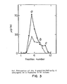
- the release of S-peptide from this gel and the prior gel (Thiol-Sepharose 4B) in buffer alone (pH 6,7, performic acid-oxidized lysozyme as carrier) and in the presence of 30 ng of RNase is shown in Figure 5.
- 2 mg of gel were suspended in 1,0 ml of a 0,1 M MES, 0,2 M NaCl, 5 mM EDTA, 0,02% performic acid oxidized lysozyme buffer, pH 6,7.
- RNase was added in 3 ⁇ l of buffer giving the final concentrations cited above.
- the tubes were incubated at room temperature with gentle rocking.
- RNase A (0-200 pg) was added in 10 ⁇ l to a slurry containing 2 mg of the gel in a pH 5 acetate buffer. After 22 h 1 1 ml of buffer was added and the tubes were centrifuged.
- a demonstration of the amplification of RNase activity using a multi-stage system consisting of an alternating sequence of the S-peptide and S-protein SLA gels was next conducted.
- One ng of RNase was passed through a 3 stage system in a pH 5 acetate buffer.
- Each stage shown schematically in Fig. 7, consisted of a 15-30 min incubation with 2 mg of S-peptide gel followed by a 15 min incubation with 2 mg of S-protein geL RNase was transferred from the upper to the lower gel by centrifugation through a filter. After 15 min, the lower tube was centrifuged and an aliquot of the supernatant was removed for RNase S determination.
- RNase S activity in the supernatant was measured after each stage, giving the results shown in Table III.
- the amount of amplification was calculated relative to the initial ng dose of RNase A after correcting for the background activity obtained from a 0 dose.
- the maximum total amplification was achieved (52 times the initial amount of RNase) after the second stage.
- the overall degree of amplification decreased in stage 3, due largely to the increase in background activity.
- the increase in RNase S activity was only 42 ng in the latter stage for the RNase treated gel, while it was 78 ng in stage 2.
- RNase activity using a continuous flow two-gel chromatographic bed was also done.
- the upper section consisted of S-peptide gel packed in polyethylene tubing (0,5 x 60 mm).
- the lower section (0,5 x 60 mm) contained the S-protein geL
- RNase A activities of 27,5, 37,8, 44,8 and 67,9 ng were obtained in the first 500 pl of eluent. After subtracting the background activity (0 dose; 27,5 ng), this corresponds to a 52, 34 and 40-fold amplification of 0,2, 0,5 and 1,0 ng of RNase activity, respectively.
- Chemical amplification can play a useful role in chemical analysis. Some of the general benefits it offers are improved sensitivity, convenience and miniaturization. Increased sensitivity helps us to better detect trace analytes. Examples of analytes which now stretch the sensitivity limits of current techniques are gene fragments related to inherited or infectious diseases (detected with DNA probes in hybridization assays), infectious disease antigens (detected with antibody probes), DNA adducts (14), and the lower level hormones and drug/drug metabolites encountered in physiological samples.
- a larger chemical signal is more convenient to detect than a small one mostly because the demands on electrical detection, amplification, and display of the signal are reduced. Indeed, when the chemical amplification yields a colored product and a qualitative or semiquantitative measurement is adequate, then an instrument is no taken longer required. Or advantage can be of smaller instruments such as those based on microchip technology, including potential detection of the amplified chemical signal by such devices as a CHEMFET (15). Miniaturization, then, comes from the use of latter techniques or direct visual detection. From this it is clear that chemical amplification will play a key role in biosensors and related techniques.
- substrate-leash amplification for chemical amplification has several attractive features. It combines catalytic and multiplicative mechanisms potentially giving a high speed and degree of amplification. As a generic mechanism, it offers flexibiilty in the choice of its components. Its compatibility with a solid surface makes washing steps convenient and allows it to be incorporated into a flow device such as chromatographic column, tube, plate or membrane.
- the key role played by a chain-cutting enzyme means that additional flexibility and amplification are available in the detection step: the released enzyme can encounter an analogous solid-phase substrate leash containing many signal molecules like dyes, fluorophores or electrophoric release tags (16).
- the S-peptide and S-protein components of RNase A in conjunction with a poly C leash, comprised a logical first choice to demonstrate SLA.
- These RNase fragments are well-characterized, commerically available, low in molecular mass for rapid diffusion, and tightly (17) form RNase S, a fully active form of the native enzyme.
- the combination reaction between S-peptide and S-protein is heterogeneous, including a dependence on S-protein-folding (18), the overall kinetics of recombinations are rapid. It was previously demonstrated that extrinsic chemical groups (two molecules of thyroxine) can be attached to S-peptide while maintaining a significant capacity (47%) to reactivate S-protein (19).
- Poly C also is commercially available, can be functionalized extensively by transamination at its N 4- cytosine sites with an alkyldiamine (5, 12) and is rapidly cleaved by RNase (11).
Landscapes
- Chemical & Material Sciences (AREA)
- Organic Chemistry (AREA)
- Life Sciences & Earth Sciences (AREA)
- Zoology (AREA)
- Wood Science & Technology (AREA)
- Proteomics, Peptides & Aminoacids (AREA)
- Health & Medical Sciences (AREA)
- Engineering & Computer Science (AREA)
- Microbiology (AREA)
- Biochemistry (AREA)
- Physics & Mathematics (AREA)
- Molecular Biology (AREA)
- Biotechnology (AREA)
- Biophysics (AREA)
- Analytical Chemistry (AREA)
- Immunology (AREA)
- Bioinformatics & Cheminformatics (AREA)
- General Engineering & Computer Science (AREA)
- General Health & Medical Sciences (AREA)
- Genetics & Genomics (AREA)
- Immobilizing And Processing Of Enzymes And Microorganisms (AREA)
- Enzymes And Modification Thereof (AREA)
Abstract
Description
- This invention relates to a method for enzyme amplification, to a system of immobilized enzyme material which releases a large amount of enzyme in response to the application of a small amount of the same enzyme, and a method for preparing these conjugate systems.
- Chemical amplification, the formation of an enhanced chemical response, occurs in three ways. The first is catalysis, a common example of which is the action of an enzyme or coenzyme. Enzyme or substrate cycling (1), in which a substrate acts as a catalyst by first participating in one reaction, and then cycling back to its original state in a second reaction, also illustrates catalytic amplification. In gate amplification, the opening of a molecular gate such as a channel in a membrane amplifies the passage of molecules from one zone to another. Ultimately complement, for example, has such an effect on a target cell Third, there is multiplicative amplification, in which the amount of a substance is multiplied by a constant factor repetitively, e.g., the unhindered successive generations of a virus. These three mechanisms of amplification also can occur combined. For example, in the overall action of immune complement, catalysis and gate mechanisms are both present.
- The role of chemical amplification in chemical analysis, including clinical chemistry, was carefully reviewed by Blaedel and Boguslaski in 1978 (2). Amplification components such as enzyme, coenzymes, inorganic iodide, catalytic electrodes,bacteriophages andlipoeomes were all discussed. When such components inherently do not provide tie specificity required, or do not directly involve the
analyte, then an antibody, secondary enzyme, or secondary chemical reaction is used to make the amplification system specific or analyte-responsive.. Aside from the simple use of enzymes as amplification catalysts, many of the techniques discussed by these authors for clinical analysis were not very practical at that time. The most general problem was the tendency of these techniques to require several complex, highly-purified reagents, leading to the secondary difficulties of short shelf-life, high cost, and assay irreproducibility. The techniques were also tedious to set up or use, and gave a limited or slow rate of amplification. - A more recent example of gate amplification in chemical analysis is the report by Litchfield et aL (3) of an immunoassay for digoxin in which ouabain-mellitin triggers the release of alkaline phosphatase entrapped in a liposome. This approach to chemical amplification has many disadvantages: liposomes tend to be unstable; a threshold dose of a mellitin substance is required and probably acts only once to release alkaline phosphatase; mellitin is a hydrophobic substance that will tend to behave eratically be undergoing losses onto surfaces and forming complexes with interfering macromolecules; and the behavior of liposomes in an actual assay can be influenced by other substances in the sample that bind to the liposome aside from the ouabain-mellitin reagent.
- Stanley et al,(4) have extended substrate cycling by incorporating a color reaction (formation of a formazan dye) directly into a NAD/NADH cycle. A cycling time of about 50 min-1 was reported, and the system was used in an enzyme immunoassy for thyroid stimulating hormone. However, this is not a very high rate of chemical amplification. Other disadvantages of this system are as follows: the reagents involved are expensive, especially because they must be provided in a highly purified form and some of them are not commonly used; the system is not very flexible in terms of its components; physiological samples tend to contain enzymes that utilize NAD/NADh and therefore can interfere with the assay; the system is unable to yield other signals beside a color rejection tied to NAD/NADH; three different enzymes are used, making it impossible to find conditions that are simultaneously optimum for each, and increasing the likelihood that a given sample will contain an inhibitor for one of these enzymes, leading to faulty results; and NADH is not a stable substance.
- Harris (U.S.
Patent 4 463 090) has used cascade amplification in an enzyme immunoassay in which one or more proenzymes are present. In this invention, an initial enzyme or activator begins a sequence of activating a proenzyme to an enzyme, which enzyme product may be detected or may activate another proenzyme to an enzyme that may be detected, or it may in turn activate another proenzyme etc. Thus there is a cascade of reactions in which different enzymatic activities sequentially are activated. This invention uses naturally occurring -enzyme cascades that unfortunately are limited in number. It comprises components that tend to be unstable, susceptible to inhibitors, complex, not readily available, poorly characterized and expensive. Such reagents tend to vary in their properties batch-to-batch. This is especially true for the enzyme components, and these problems are multiplied because two or more such enzymes are used in each system. Thus this approach to chemical amplification is not very practical. Consistent with this, no experimental work is cited in this invention. A model Ugand assay using two proenzymes from the blood-coagulation cascade has been reported (D.A. Blake, M.T. Skarstedt, J.L. Shultz and D.P. Wilson, Clin. Chem. 30 [ 1984] 1452-1456), but the degree of amplification was low, the assay was not very sensitive, and the reproducibility of the system was not evaluated. - Thus, for chemical amplification in chemical analysis, not much progress has been made since 1978 when Blaedel and Boguslaski, as cited above (reference 2), concluded that the systems available then weren't very practicaL Beyond the simple use of enzymes as inherent amplification catalysts, no systems have been adopted for general use because of these problems. The main limitation of a simple enzyme as an inherent catalyst is the limited amplification this provides, since an enzyme by itself only produces a constant amount of product molecules from substrate per unit time.
- It is the object of this invention to provide a method for amplifying enzymatic activity by releasing a large amount of enzyme molecules from immobilized enzyme material, corresponding enzyme and enzyme fragment conjugates and a method for their preparation.
- The above object is achieved according to the claims. The dependent claims relate to preferred embodiments.
- In this invention a new concept for chemical amplification, here called "substrate-leash amplification" (SLA), is disclosed whereby both a catalytic and a multiplicative mechanism are involved.The key feature is that a chain-cutting enzyme or enzyme component is attached via its substrate leash to a surface. The details of the assembly restrict spontaneous release of the enzyme or its components. When free enzyme is introduced that cleaves the substrate leash, a cascade of released enzyme results.
- The method according to the invention foramplifying enzymatic activity involves connecting an enzyme to a support by means of molecular leash material which is cleavable by that enzyme, to produce a support-leash-enzyme conjugate having many -leash-enzyme units connected to the support; adding a small amount of that enzyme in the free state to the conjugate to produce a mixture of free enzyme and support-leash-enzyme conjugate; incubating this mixture for a predetermined period of time to cause enzyme in the free state to be released from the conjugate; and recovering the released free enzyme. Because the introduction of a single free enzyme molecule to the support-leash-enzyme conjugate system can cleave the substrate leashes immobilizing many enzyme units, each of which can in turn cleave many further substrate leashes, it is clear that a large amount of enzyme is released by application of a small amount of enzyme to the system.
- In this method, the leash material between the support and the immobilized enzyme is constructed to prevent the bound enzyme of a given -leash-enzyme unit of the conjugate from cleaving the leash portion of that unit. This is done by controlling the lengths and mobility of the leashes including the use of a spacer substance within the leash, and cross-links within a given -leash-enzyme unit or between one such unit and another. Furthermore, the -leash-enzyme units are connected to the support at positions sufficiently distant from each other that the enzyme portion of a given -leash-enzyme unit cannot cleave the leash portion of another -leash-enzyme unit.
- In a variant of the enzyme amplification method, two-stage enzyme reservoirs are prepared as follows: providing first and second enzyme fragments; herein designated fragment A and fragment B respectively, each of these fragments being complementary to the other and enzymatically inactive, and being capable of recombining with each other to produce active enzyme; bonding the first enzyme fragment to a support by means of molecular leash material which is cleavable by the active enzyme, to produce a support-leash-fragment A conjugate having a large number of -leash-fragment A units connected to the support material; bonding the second enzyme fragment to a support material similarly, to produce a support-leash-fragment B conjugate, also having a large number of -leash-fragment B units connected to the support material; and connecting the first and second conjugates in series, forming the two-stage enzyme reservoir.
- The two-stage enzyme reservoir is caused to release a large amount of enzyme in either of two ways: by application of active enzyme, or by application of inactive enzyme fragment to that portion of the enzyme reservoir containing the support-leash-(enzyme fragment) conjugate in which the enzyme fragment is complementary to the added enzyme fragment.
- In a second variant, the enzyme amplification is
carried out by providing a mixed enzyme two-stage enzyme reservoir as follows: providing first and second enzymes, designated C and D respectively; bonding enzyme C to a support by means of molecular leash material which is cleavable by enzyme D, to produce a support-leash-enzyme C conjugate having a large number of -leash-enzyme C units connected to the support material; bonding enzyme D to a support material similarly, to produce a support-leash-enzyme D conjugate having a large number of -leash-enzyme D units connected to the support material; and connecting the first and second conjugates in series, forming the mixed enzyme two-stage enzyme reservoir. - The mixed enzyme two-stage enzyme reservoir is caused to release a large amount of enzyme D, as well as some enzyme C, by application of free enzyme D to that portion of the mixed enzyme two stage enzyme reservoir containing support-leash-enzyme C conjugate. This releases enzyme C, which in turn catalyzes release of enzyme D in the second stage of the reservoir.
- Many stages of enzyme amplification may be employed in series, in each variant of this method. For the version in which active enzyme is immobilized on a support, the larger amount of active enzyme produced upon application of a small amount of active enzyme to a batch of this immobilized enzyme material may be in turn applied to a second batch of support-immobilized enzyme to produce a still greater yield of active enzyme, and the second larger amount of active enzyme may in turn be applied to a third batch, and so on. For the case in which enzyme fragments are immobilized and a two-stage enzyme reservoir is constructed, the same principle applies in that the relatively large amount of active enzyme produced after the second stage of enzyme amplification may serve as the initial charge of active enzyme and enzyme precursor to be added to the first stage of a second enzyme reservoir, and so on. For the case in which a mixed enzyme two-stage enzyme reservoir is constructed, the same principle again applies in that the large amount of enzyme D produced in the second stage of the enzyme reservoir may serve as the initial charge of enzyme D to be added to the first stage of a second mixed enzyme two-stage enzyme reservoir, and so on.
- The invention includes compositions having the generalized formula support-leash-enzyme, where the support is a solid support material, the leash is a material including a molecular chain which is cleavable by the enzyme in the free state, and the support-leash-enzyme conjugate has a large number of -leash-enzyme units connected to the support. The invention also includes compositions having the generalized formula suppoct-leash-(enzyme fragment) in which the enzyme fragment is one of two complementary enzymatically inactive fragments of an active enzyme, and is capable of recombining with the second of these fragments to regenerate active enzyme, the support is a solid support material, the leash is a material containing a molcecular chain which is cleavable by /the active enzyme, and the supoort-lersh-(enzyme fragment) conjugate has a large number of -leash-(enzyme fragment) units connected to the support. The invention further includes compositions having the generalized formula support-leash-enzyme in which the support is a solid support material, the leash is a molecular chain which is not cleavable by the enzyme in the free or immobilized form, and the support-leash-enzyme conjugate has a large number of -leash-enzyme units connected to the support The support may also be a soluble macromolecule, other chain-cutting substances besides enzymes may be used, and the leash-enzyme unit may be attached noncovalently to the surface.
- The invention will now be further explained with reference to the drawing in which:
- Fig. 1 is a schematic representation showing the concept for substrate-leash amplification;
- Fig. 2 is a schematic representation showing the overall reaction sequence for preparing the support-leash-(ehzyme fragment) conjugates, where the enzyme fragments are fragments of RNase and the leash is polycytidylic acid;
- Fig. 3 is a diagram showing the results of gel filtration of the S-peptide-DAO-poly C conjugate on a Sephadex G-50 column;
- Fig. 4A is a diagram showing the release of immobilized S-peptide from poly C Thiol-
Sepharose 4B upon treatment with RNase in dependence of the digestion time; - Fig. 4B is a diagram showing the release of immobilized S-protein from poly C Thiol-
Sepharose 4B upon treatment with S-peptide solution in dependence of the digestion time; - Fig. 5 is a diagram showing the S-peptide release from poly C Thiol-
Sepharose 4B and poly C thiolpropyl-Sepharose 6B, in buffer and in RNase; - Fig. 6 is a diagram showing the amplification of RNase activity on S-peptide-DAO-poly C Thiopropyl-Sepharose as a function of the RNase dose;
- Fig. 7 is a schematic representation of one stage of a basic multi-stage amplification system and
- Fig. 8 is a schematic representation of the concept for mixed substrate-leash amplification.
- Fig. 1 is a schematic representation of the enzyme amplification method of the invention, in a general form. An enzyme is attached to a support by means of molecular chain covalently attached to both the support and the enzyme. This chain or leash is made up of or includes material which is cleavable by the enzyme. In other words, the enzyme is attached to the support by molecular material which is a substrate for the enzyme. Upon application of a small amount of free enzyme to the support-leash-enzyme conjugate, cleavage of the leash chain occurs, releasing free enzyme which in turn cleaves more leashing chains and frees more enzyme. In principle, addition of a single free enzyme molecule will ultimately result in the release of all the enzyme which is immobilized on the support initially, given sufficient time and appropriate reaction conditions. In practice, support-leash-enzyme conjugate having a large number of -leash-enzyme units on the support is prepared and treated with a small amount of enzyme, forming-initially a mixture of free enzyme and support-leash-enzyme conjugate, then this mixture is incubated for a predetermined period of time to permit the enzyme-catalyzed cleavage of leash material to occur, freeing formerly immobilized enzyme. At the end of this reaction time period, the free enzyme is recovered from the support-leash-enzyme conjugate materiaL The nature of the system ensures that the amount of enzyme ultimately recovered is substantially greater than the amount of enzyme initially added to the system.
- In this system, the enzyme may be any enzyme which can be covalently attached to a material which is a substrate of that enzyme. By the same token, the leashing material may be any substrate for the enzyme, provided only that it may be covalently attached to both the enzyme and the support materiaL The support is a solid support material such as agarose, silica, cellulose, paper, Sepharose, Trisacryl, glass, nylon, poly methacrylate, Immobilon Membranes, polyacrylamides, polyamides and gelatin.
- Table I below lists examples of many enzymes in several classes, and some examples of molecular chains they cleave. These materials, many of which are illustrated in the Examples below, are useful in the enzyme amplification method and compositions.


- For the case where the support-leash-enzyme conjugate is employed, the leash material joining the support and the enzyme is constructed so that an enzyme portion of a given -leash-enzyme unit cannot cleave the leash portion of that unit. Three types of primary mechanisms are available to achieve this. First, the leash material can be made to have a length within a predetermined range of lengths so that the attached enzyme is unable to have its active site contact the leash in a
- be cut. manner that the leash can / Such leash lengths can be established by synthesizing molecular chains of desired lengths for attachment of the conjugates to the solid supports. Since the leashes for chain cutting enzymes are available naturally in much longer lengths than are needed here, a suitable strategy is to conduct a limited hydrolysis, either with the chain-cutting enzyme itself or with aqueous acid or base, of the longer leashes, varying the time of incubation. This yields a mixture of shorter leashes. The latter can then be fractionated by gel filtration into several size ranges, and the appropriate size range can be used.
- The next mechanism is to restrict the movement of the leash so that the attached enzyme is unable to bring its active site into contact with the leash. This is most easily done by attaching the leash at two or more points along its length to the support, so that the leash has a restricted mobility. For this the leash needs to have two or more functional groups along its length that can bind to the complementary functional groups on the support surface. This strategy is commonly used in affinity chromatography to increase the stability of attaching a polymeric substance to a surface. The random motion of functional groups on the surface easily allows multiple attachment sites to be made with a multi-functional polymer.
- The third mechanism is to place a spacer substance between the portion of the leash that can be cut by the enzyme, and the attached enzyme. Thus one would have the sequence support-leash-spacer-enzyme. This spacer is not a substrate for the enzyme, and because of its size, prevents the active site of the enzyme from reaching the leash. Either a folded (e.g., a protein) or nonfolded (e.g., a polynucleotide) spacer can be used that is not a substrate for the enzyme.
- These three mechanisms (chain length, multi-site attachment, and spacer substance) for preventing cleavage of the -leash-enzyme unit by its attached enzyme can also be combined. Further, residual functional sites on the -leash-enzyme, both within a given leash or from one-leash-enzyme to another, can be tied together by reagents called "cross-linking reagents" that are commercially available and commonly used to cross-link neighboring macromolecules. This constitutes a secondary mechanism for controlling spontaneous release of an enzyme, that can be superimposed on the primary mechanisms. The cross-linkages within a -leash-enzyme unit can be directed to stay within the leash, or to make additional ties from the leash to the enzyme. Similar variations can take place for cross-links between different -leash-enzyme units. The ultimate effect of this chemical fixation step is to further restrict the mobility of the -leash-enzyme units, further increasig their stability.
- For each of the three primary and secondary mechanisms to control spontaneous release of enzyme, it will be useful to optimize the spacing of the -leash-enzyme units on the support material. Neither widely spaced nor excessively crowded -leash-enzyme units are desirable. Wide spacing is not appropriate because it means that the capacity of the amplification device will be low: the device will be empty before much amplification develops. Excessive crowding of -leash-enzyme units will increase the problem of the enzyme portion of one cleaving the leash portion of another. While the above three primary and one secondary mechanisms will help to control this problem as well, the simplest remedy is to space the -leash-enzyme units so that they do not touch each other This can be done by controlling the density of functional groups on the support surface. Either a surface can be prepared with a reduced density of such groups, or some of the groups can be reacted, before or during coupling of the -leash-enzyme units, with nonleash molecules that merely occupy some of the space on the surface. These techniques are used commonly in the field of affinity chromatography to regulate ligand density on a solid surface.
- Porous particles with -leash-enzyme units attached to the surfaces within the particles, and within which pores unattached molecule move only by diffusion, are not appropriate for immobilizing -leash-enzyme units according to this invention. The problem is' that any spontaneous release of enzyme from a -leash-enzyme unit will tend to give an unstoppable cascade of released enzyme since it will leave the particle only slowly by diffusion. 'A second problem, although this is less fundamental, is that physical contact between such particles can allow an enzyme on the outside surface of one particle to cut the leash of a -leash-enzyme unit on the outside surface of another particle.
- Nevertheless, it is still possible to attach -leash-enzyme units to porous or solid particles, as long as any pores are too small to permit entry of -leash-enzyme units or enzymes molecules into the pores of the particles. Such particles would then restrict the -leash-enzyme units and released enzyme to the outside surfaces of these particles. Such particles would then be fixed by attachment or trapping in a filter, so that any physical contact between the particles does not change or is avoided. In this manner, the problem of an enzyme in a -leash-enzyme unit on one particle cutting the -leash-enzyme unit on another particle can be avoided or controlled by a washing step as explained below.
- A second reason for attaching -leash-enzyme units to solid supports such as membranes, filters, and tubes, or onto the outside surface only of particles that subsequently are immobilized in a filter, is that this allows direct exposure of the -leash-enzyme units on the surface to bulk flow of liquid. This is important because there will always be some -leash-enzyme units in a freshly constructed system that will undergo spontaneous release once the enzyme is activated. (As discussed below, the surface-leash-enzyme is initially assembled with the enzyme in an inactive form, followed by activation of the enzyme.) This spontaneously released enzyme must be removed from the system before it triggers the release of other enzymes.
- A good way to do this is to activate the -leash-enzyme units on the surface under rapid washing conditions in which these units are directly exposed to the bulk flow of buffer wash. Thus any released enzyme is quickly washed out of the system before it can cut additional leashes. A noncovalent, reversible inhibitor of the enzyme can be included in this buffer wash as needed, along with buffer conditions (e.g., adjust the pH) that are not optimum for the activity of the enzyme.
- A variant of this invention employs in lieu of immobilized active enzyme inactive enzyme fragments which are complementary to each other and which recombine to form active enzyme. Each of these complementary enzyme fragments are separately joined to support materials by means of a molecular chain or leash which is cleavable by the active enzyme. This leash material is covalently attached . to both the support and the enzyme fragment, forming a support-leash-(enzyme fragment) conjugate for each of the two enzyme fragments. In each of these conjugates, a large number -leash-fragment units are connected to the support material. Connecting the separate conjugates containing the respective enzyme fragments in series provides a two-stage enzyme reservoir from which active enzyme is derived upon addition of either active enzyme or enzyme fragment complementary to the enzyme portion of the particular conjugate of the enzyme reservoir to which it is added.
- For purposes of the following discussion, the two-stage enzyme reservoir will be considered as having in its first stage a first conjugate support-leash-fragment A, and having in its second stage a second conjugate support-leash-fragment B, where fragment A and fragment B are the complementary enzyme components. Addition of enzyme material whether active enzyme or enzyme fragment, is considered to be made into the first stage of the enzyme reservoir.
- Where active enzyme is added to the support-leash-fragment A first conjugate section of the two-stage enzyme reservoir, a first starting mixture is initially produced, containing the first conjugate and the added active enzyme; this is then incubated for a predetermined period of time, to form a first product mixture containing the added active enzyme and released first enzyme fragment. The first product mixture is then transferred from the first conjugate onto the second conjugate in the enzyme reservoir, to produce a second starting mixture containing the second conjugate, the added active enzyme, and the first enzyme fragment released from the first conjugate. This second starting mixture is then incubated for a predetermined period of time to form a second product mixture containing released second enzyme fragment and active enzyme in an amount greater than the first amount initially charged into the enzyme reservoir. Finally, the second product mixture is recovered from the second conjugate of the two-stage enzyme reservoir.
- The system operates to produce a large amount of free active enzyme because the small amount of active enzyme initially added to the support-leash-fragment A first conjugate causes the release of a large number of fragment A units, which are then transferred, along with the originally added active enzyme, to the support-leash-fragment B second conjugate in the second stage of the enzyme reservoir where they in the second stage of the enzyme reservoir complex to form active enzyme which is covalently bound to the support. Some of this covalently bound active enzyme can release itself spontaneously, and some is released by action of the initially added active enzyme, resulting in the production of a large amount of active enzyme in the free state.
- The product mixture from the second stage of the enzyme reservoir may be employed as the initial charge of enzyme material added to a second two-stage enzyme reservoir in series with the first. As this material contains released second enzyme fragment in addition to a large amount of free enzyme, its addition to the first stage of the second enzyme reservoir produces a third starting mixture containing first conjugate, active enzyme, and released second enzyme fragment. The released second enzyme fragment is now able to recombine with bound first enzyme fragment, forming active enzyme which can release itself spontaneously or be released by the action of other active enzyme, free or bound, present in the system. The third starting mixture is incubated a third predetermined period of time, producing a third product mixture containing active enzyme and released first enzyme fragment. This third product mixture is then transferred from the first conjugate in the third stage onto the second conjugate in the fourth stage of the enzyme reservoir, giving a fourth starting mixture containing second conjugate, active enzyme, and released first enzyme fragment. The fourth starting mixture is incubated for a predetermined period of time to form a fourth product mixture containing active enzyme and released second enzyme fragment, as before. The active enzyme recovered from the fourth stage of the enzyme reservoir is a very much larger amount than was initially applied to the enzyme reservoir at the first stage. This process can be continued throughout many stages in succession up to n stages where n can be a large number, e.g., 106.
- In the event that free second enzyme fragment is added to the first stage of the two-stage enzyme reservoir, a first starting mixture is formed which contains the first conjugate and the added second enzyme fragment. This mixture is then incubated for a predetermined period of time, forming a first product mixture containing released active enzyme and released first enzyme fragment. This mixture results from the fact that the second enzyme fragment which was added recombines with the immobilized first enzyme fragment forming active enzyme, and this either cleaves the leash immobilizing it or cleaves the leash immobilizing a neighboring active enzyme. Once active enzyme is present, leashes immobilizing both active enzyme and first enzyme fragment are cleaved, liberating these units The product mixture from the first stage reaction is transferred onto the second conjugate in the enzyme reservoir, producing a second starting mixture containing second conjugate, released active enzyme, and released first enzyme fragment. This mixture is then incubated for a predetermined period of time to form a second product mixture containing active enzyme and released second enzyme fragment. The amount of released active enzyme in the second product mixture is greater than that corresponding to the amount of second enzyme fragment initially applied to the enzyme reservoir.
- As before, the product from the second stage of the first enzyme reservoir may be employed as the enzyme material added to the first stage of a second two-stage enzyme reservoir, to produce an even greater enzyme amplification, and this can be continued to third, fourth, and ultimately many stages in succession.
- For the support-leash-enzyme fragment variation of this invention, it is important to avoid unintentional direct physical contact of support-leash-enzyme fragment A and support-leash-enzyme fragment B. This is because direct contact of these two materials will tend to allow the two different enzyme fragment portions to contact and form an active enzyme, initiating cleavage of the leash portions to produce a cascade of enzymatic activity at an unintentional time. Separation of the support-Ieash-enzyme fragment A and support-leash-enzyme fragment B materials is easily achieved by placing an inert membrane or filter between these materials. Alternatively, direct physical contact between these two different -leash-enzyme units can be avoided by immobilizing the -leash-enzyme fragment A and -leash-enzyme fragment B units onto separate, noncontacting regions of a solid surface such as a membrane, filter or tube.
- A variant of this invention employs a mixed system of two different active enzymes (C and D) and two leashes c and d, where leash c is cleaved by enzyme C but not D, and leash d is cleaved by enzyme D but not C; and where enzyme C is immobilized on leash d and enzyme D is immobilized on leash c. Thus each active enzyme is immobilized on a leash which cannot be cleaved by this enzyme, but each leash can be cleaved by the other enzyme in the system. Thus the overall system still provides a mechanism for a cascade of enzymatic activity to develop, triggered by a dose of free enzyme and involving cleavage of leashes to which enzymes are attached. This is done by first exposing free enzyme D to a support-leash d-enzyme C material, which results in cleavage of many d leashes, releasing many molecules of active enzyme C. These many released enzyme C molecules then contact a support-leash c-enzyme D material, and cleave an even greater number of c leashes, releasing a much longer amount of active enzyme D than was originally applied to the system. This process can be continued through several or even many successive, alternating support-leash d-enzyme C followed by support-leash c-enzyme D materials to multiply the amount of released enzyme D activity to larger and larger levels.
- As in the variation of this invention in which inactive enzyme fragments A and B are immobilized solid supports such that physical contact between support-leash-enzyme fragment A and support-leash-enzyme fragment B are avoided, it is necessary in this variation involving active enzymes C and D with leashes c and d that physical contact of support-leash c-enzyme D and support-leash d-enzyme C are avoided. This is done as before with the enzyme fragment materials, by placing an inert membrane or filter between successive support-leash c-enzyme D and support-leash d-enzyme C materials. Also, physical contact between -leash d-enzyme C and leash c-enzyme D units can be avoided by immobilizing them each onto separate, noncontacting regions of a solid surface such as a membrane, filter or tube.
- The same criteria and chemical reactions useful for preparing support-leash-enzyme fragment materials are useful here for preparing support-leash (c, d)-enzyme (D, C) materials.
- In practice, the enzyme amplification is carried out in the presence of a liquid carrier in which the support-leash-{enzyme or enzyme fragment) conjugate is stable. Most generally, this is an aqueous buffer at or near physiological pH. The enzyme material added to the conjugates to start the enzyme amplification is added as a solution and is carried onto the support-leash-{enzyme or enzyme fragment) conjugate by the flow of the carrier liquid. The carrier liquid may move through the enzyme amplification system continuously, carrying starting materials and reaction products with it. In this event, the predetermined incubation times for each of the reactions are determined by the carrier flow rate and by the physical length of the enzyme amplification system through which carrier flows. The carrier liquid may also be caused to move intermittently through the enzyme amplification system, in which case reaction incubation times are established by the times during which the flow of carrier liquid is stopped. Transferring materials onto the enzyme amplification system, from one stage of the system to a succeeding stage, or out of the system for recovery of released enzyme, are all accomplished by means of the flowing carrier liquid. Other transference means may, however, be envisaged, such as diffusion and electrophoretic migration.
- The compositions according to the invention have the general formula support-leash-enzyme where the support is a solid support material, the leash is a molecular chain including material which is cleayable by the enzyme in the free state, and the support-leash-enzyme conjugate has many -leash-enzyme units connected to the support. Suitable solid support materials include agarose, silica, cellulose, paper, Sepharose, Trisacryl, glass, nylon, polymethacrylates,Immobilon Membranes,polyacrylamides,polyamides and gelatin. In these conjugates, the leash has a length within a predetermined range of lengths chosen such that the lengths are sufficient to prevent enzyme of a given -leash-enzyme unit from cleaving the leash portion of that unit. Similarly, the leash-enzyme units of the support-leash-enzyme conjugate are connected to the support at positions sufficiently distant from each other that the enzyme portion of a given -leash-enzyme unit cannot cleave the leash portion of another -leash-enzyme unit.
- Other compositions of the invention have the generalized formula support-leash-(enzyme fragment) where the enzyme fragment is one of two complementary enzymatically inactive fragments of an active enzyme and is capable of recombining with the second of these fragments to regenerate the active enzyme; the support, as before, is a solid support material; the leash is a molecular chain containing material which is cleavable by the active enzyme, and the support-leash-(enzyme fragment) conjugate has a large number of -leash-(enzyme fragment) units connected to the support.
- Further compositions of the invention have the generalized formula support-leash c-enzyme D and support-leash d-enzyme C where D and C are active enzymes and leash c and leash d are molecular chains which can be cleaved by enzymes C and D respectively, but not by enzymes D and C respectively. As before, the support-leash-enzyme conjugates have a large number of -leash-enzyme units connected to the support.
- The chemical reactions to be employed in joining the support material to the leash material, and the leash material to the enzyme to be immobilized, are known and will vary depending on the support material leash material, and enzyme employed. Details for some of this chemistry are given in the examples below.
- The chemical reactions and conditions are selected to meet the following criteria: (1) that they make the enzyme inactive during the assembly of the support-leash-enzyme system; (2) that they allow the enzyme to become active after the support-leash-enzyme system is assembled; (3) that they avoid side reactions attaching some of the enzyme directly to the solid surface, forming surface-enzyme units with no intervening leash; and (4) that they avoid reactions among leash-enzyme units, forming leash-enzyme polymers. The importance of each of these criteria, and how these criteria are satisfied by appropriate chemical reactions and conditions, is explained below.
- The importance of inactivating the enzyme during the assembly of the support-leash-enzyme system is to avoid premature cleavage of the leashes, that would prevent the system from being assembled. Enzymes can be reversibly inactivated by several techniques, depending on the particular enzyme involved. Some of the smaller enzymes can be reversibly inactivated by the addition of denaturing agents such as high concentrations of guanidine hydrochloride, urea, or a low concentration of a detergent like sodium dodecylsulfate. Other enzymes require a specific metal ion such as Ca2+ for their activity. Such enzymes can often be inactivated by treatment with a metal chelating agent like EDTA. Sometimes a denaturant must be used in combination with a metal chelating agent, or low pH conditions may be required. Some enzymes have a sulfhydryl side chain in their active site that is essential for their activity, and can be reversibly inactivated by reaction with a sulfhydryl compound such as cysteine or mercaptoethanol forming a disulfide linkage between the sulfhydryl compound and the active site sulfhydryl side chain. For many enzymes an inhibitor is available that reversibly binds to and inhibits the enzyme activity. An antibody can be made against the enzyme that forms a complex with the enzyme devoid of enzymatic activity.
- It is then necessary for the enzyme to be re-activated in the support-leash-enzyme system in order to amplify a triggering dose of free enzyme. Generally this will be done by simply washing the enzyme inhibitors out of the system with a buffer that allows the enzyme to be active. This buffer will contain metal ions as necessary to reactivate enzymes which have been inhibited by removal of their essential metal ions. To reactivate a sulfhydryl enzyme that has been inactivated by disulfide coupling to a sulfhydryl inhibitor, an excess of a reducing thiol compound such as mercaptoethanol or dithiothreitol can be included in the buffer.
- It is important for successful re-activation of the enzyme that the chemical reactions used to attach the enzyme to the leash, and the leash-enzyme conjugate to the surface (or to attach the enzyme to a leash that was previously attached to a surface) do not interfere with the ability of the enzyme to be reactivated. This is done by choosing chemical reactions and conditions that attach the leash to the enzyme at a site that does not interfere with the active site of the enzyme. Usually there are some reactive sites on the enzyme that can be modified without interfering with the enzymatic activity. When the active site is highly reactive, it can be reacted first with a reversible protecting agent. For example, citraconyl and trifluoroacetyl are reversible protecting groups for primary amino groups on enzymes. Generally one can allow random attachment of a leash to the enzyme since the active site is a small region of the enzyme surface and only a small fraction of the enzyme will be attached at this site and thereby inactivated. This will therefore not have any significant effect on the operation of the surface-leash-enzyme system.
- Side reactions of the enzyme directly onto the surface, or of leash-enzyme units to form polymers, should be avoided simply because this wastes some of these reagents and also tends to make the construction of the surface-leash-enzyme system less reproducible. Thus it is useful to employ specific coupling reagents and conditions to assure that only the intended linkages are formed. A wide variety of such specific reagents are commercially available, or can be .readuy synthesized. Related to this, an extensive literature has developed on the chemical modification of macromolecules, including the attachment of macromolecules to each other and to surfaces. Prominent among these chemical reagents and techniques are those that involve coupling of thiol substances to maleimide substances. This reaction proceeds rapidly under mild, aqueous conditions needed to work with most enzymes. Also useful are N-hydroxysuceinimide ester groups that have specificity for primary amino groups an aqueous macromolecules. Water-soluble carbodiimide reagents are widely used to form amide bonds between carboxylic acid groups and primary amino groups. Glycols can be specifically oxidized with periodate for reaction with hydrazide groups or primary amino groups in the presence of cyanoborohydride. Thus, many familiar chemical reagents and conditions are available to form the specific linkages for many types of support-leash-enzyme systems,
- The chemical reactions to be employed in joining the support material to the leash material, and the leash material to the enzyme fragment to be immobilized, follow similar guidelines and considerations. Even though the enzyme fragments are inherently inactive, they still require the use of specific chemical reactions and conditions so that, once the enzyments fragments are released from the support-leash-enzyme fragment units, these fragments can still recombine to form active enzyme. The same types of chemical reactions and conditions useful for preparing support-leash-enzyme units can be used here.
- The immobilized enzymes and the enzyme amplification method of the invention have many uses in analytical chemistry, one example being to increase the sensitivity of a standard immunoassay in which an analyte is reacted with excess tagged antibody in which the tagging material is an enzyme, and then passed through an affinity chromatographic column containing immobilized analyte, to remove excess tagged antibody and permit tagged antibody. analyte complex to elute. In such an immunoassay, analyte is determined by measuring the enzymatic activity of the eluted enzyme tag. If the concentration of analyte in the sample solution is very low or the enzyme tag is not very active, analytical difficulties result. If the eluting enzyme tag were employed as the free enzyme to be added to the system of the invention, however, a large amount of enzyme would ultimately result, making detection and measurement of the desired analyte easier at low levels. Similarly, the enzyme-tagged antibody can be complexed to the immobilized analyte in an affinity column, and free analyte added to this column can complex and cause the elution of some enzyme-tagged antibody for amplification and easier detection by the system of this invention.
- Polycytidylic acid (5'), cytidine 2':3'-cyclic monophosphate (cCMP), uridine 5'-triphosphate agarose, bovine pancreatic ribonuclease type III-A, ribonuclease S-peptide grade X11-PE, ribonuclease S-protein grade Xll-PR, Thiol-
Sepharose 4B, Thiopropyl-Sepharose 6B, bovine serum albumin Cohn Fraction V, eggwhite lysozyme Grade 1, 2-mercaptoethanol, dithiothreitol and 5, 5'-dithiobis-2-nitrobenzoic acid (Ellman'sreagent) were obtained from Sigma Chemical Co, St. Louis, MO; N-succinimidyl-3-(2-pyridyldithio)propionate (SPDP), N-ethylmaleimide and dimethylsulfoxide were purchased from Pierce Chemical Co, Rockford, IL; N-Υ-maleimidobutyryloxysuccinimide (GMBS) was from Calbiochem, San Diego, CA; PD-10 columns, Sephadex G-10 and G-50, polyethylene capillary tubing and polyamide column netting were from Pharmacia Inc., Piscataway, NJ; one ml filtration columns were from Supelco, BelleFonte, PA; lanthanum acetate was purchased from Alfa Products, Danvers, MA; all buffer reagents, solvents and polyethylene centrifuge tubes were obtained from Fisher Scientific, Medford, MA. - Polycytidylic acid partly transaminated (34%) at its N4-cytosine positions with 1,8-diaminooetane (DAO-poly C) was prepared and characterized as described in (5). This DAO-poly C (25 mg; 21 µmol of NH2) was dissolved in 3 ml of 0,1 M HEPES buffer,
pH 7,6, containing 2 mM EDTA and 65 mg (232 µmol) of GMBS was added in 0,5 m of DMSO. After approximately 10 min a white precipitate began to appear. At 60 min the pH was adjusted to 4,5 with glacial acetic acid and the mixture was centrifuged. The soluble fraction was desalted over a PD-10 column (Pharmacia) in 0,01 M sodium acetate,pH - S-peptide (10 mg; 4.58 µmol) was dissolved in 1,36 ml of 0,1 M phosphate buffer,
pH 7,2. SPDP (2,37 mg; 7,6 µmol) was added in 8,5,µl of DMSO,and the reaction mixture was stirred for 2 hours at room temperature. The (PDP)-S-peptide was desalted on a 28 mL Sephadex G-10 column (1 x 36 cm) using 0,01 M sodium acetate,pH 4,5, as the mobile phase. The material was lyophilized and stored at -20"C. - To determine the degree of substitution with 3-(2-pyridyldithio)-propionate the (PDP)-S-peptide was reduced with mercaptoethanol, and the release of pyridine-2-thione was followed spectrophotometrically at 343 nm using an extinction coefficient of ε = 8,08 103. The S-peptide concentration was determined by the method of Lowry et al.(7). The enzymatic activity of the PDP-S-peptide in the presence of S-protein relative to the unmodified NRase S was determined with RNA and cCMP as substrates.
- 1g of dry Activated Thiol-
Sepharose 4B (4 µmol of thiol groups) was swollen in 10 m of water for 30 min. The supernatant was removed and 8 m of 0,317 M sodium carbonate solution, 20 mM EDTA, 1% / (mass/volume-m/v) dithiothreitol, adjusted topH pH 6,5, and immediately used for coupling to the peptide or protein (see below). 1 g of Thiopropyl-Sepharose 6B (60µmol of disulfide groups) was treated in a similar manner except that the reduction step was carried out for 2 h in 2% (m /v) dithiothreitoL - (PDP)-S-Peptide (5,5 mg; 2,5µmol) was dissolved in 0,2 ml of 0,1 M potassium phosphate buffer,
pH 6,5, containing 20 mM EDTA, and 3.0 µl of mercaptoethanol were added. After 1 h at room temperature, the mercaptoethanol and pyridine-2-thione were removed on a Sephadex G-10 column (1,5 x 6 cm) in the same buffer. The early eluting fractions containing the thiolated-S-peptide were pooled (total volume 3 ml), and 10 mg of MI-DAO-poly C (8.5 µmoi maleimide groups) were added. After 12 min no free thiol groups could be detected by Ellman's reagent (6). The solution was added to 1,5 g of Thiol-Sepharose 4B or Thiopropyl-Sepharose 6B,and the mixture was shaken for 1 h at room temperature. To quench the remaining thiol groups, 20 mg of N-ethylmaleimide were added. The absence of residual thiol groups on the gel was confirmed by the Ellman's test. The gel was washed with 200 ml .each of 0,5 M NaCl and water, and with 20 ml of methanoL The S-peptide-poly C Sepharose gel, after brief filtration-drying, was dried under vacuum and stored at -20"C. - Affinity purified S-protein (25 mg; 2,1 µmol) was dissolved in 1,0 ml of 0,5 M potassium phosphate buffer, pH 715. SPDP (2,7 mg; 814 µmou was added in 150µl of DMSO,and the solution was stirred for 30 min at room temperature. A precipitate formed which was dissolved by the addition of a few drops of glacial acetic acid. The PDP-S-protein was desalted two times over a Pharmacia PD-10 column using 0,01 M sodium acetate,
pH - The degree of substitution with 3-(2-pyridyldithio)-propionate was determined spectrophotometrically as described for the PDP-S-peptide. The enzymatic activity of the PDP-S-protein (50µ g; 4,3 nmol) was measured towards cytidine cyclic monophosphate (cCMP) in the presence of 30 µg (14 nmol) of S-peptide. The activity was determined before and after the release of pyridine-2-thione with dithiothreitol (see below). In the latter case, the free thiol groups were reacted with a slight excess of N-ethylmaleimide at
pH 6,5 prior to the enzyme assay. - PDP-S-protein (22,4 mg; 1,9 µmol) was dissolved in a solution of 4 ml of water and 200 µl of glacial acetic acid. Dithiothreitol (0,8 mg; 5,4 µmol) was added in 100 µl of 0,01 M sodium acetate buffer,
pH pH 6,5. After stirring for 10 min, the mixture was poured over 1,4 g of activatedThiol Sepharose 4B (see above) and placed on a rocking plate for another 10 min. The excess thiol groups on the gel were quenched with a 4-fold excess of N-ethylmaleimide. The gel was washed with 200 ml of 0,5 M sodium chloride, 200 ml of water and 20 ml of methanoL The gel was dried under high vacuum at room temperature overnight and stored at -20°C. - RNase activity towards wheat germ or yeast ribonucleic acid was determined as follows: RNase standards (30 to 3000 pg) and sample solutions were prepared in 0,5 M tris-HCl buffer pH 7,5 containing 5 mM EDTA and 0.1% BSA. 50 µl of these solutions were added to 70 mg of RNA dissolved in 100 µl of water. The tubes were incubated at 37 °C for 20 min and then placed in an ice bath. 50 µl of an ice cold solution of 14 mM lanthanum acetate in 24 % perchloric acid were added. After 2 min incubation the tubes were centrifuged at a relative centrifugal acceleration of 1700 for 20 min at 4 °C.
- A 100 µl aliquot was withdrawn from the supernatant, diluted 10-fold with water, and the absorbance was measured at 260 nm.
- RNase activity towards cytidine cyclic monophosphate (9): 1 mg of cytidine cyclic monophosphate was dissolved in 10 ml of 0,5 M Tris-HCl buffer, pH 7,5, 5 mM EDTA,and 800µl of this solution were pipetted into 1 ml cuvettes. Aliquots (100µl) containing 5-80 µg of enzyme were then added,and the increase in absorbance at 284 nm with time was measured.
- Poly C content: 2 mg of the vacuum-dried S-peptide or S-protein gel were added to 1ml of 0,5 M Tris, 5mM EDTA, 0,196 BSA, pH 7,5 buffer and incubated for 24 h; with 100 ng of RNase. The suspensions were centrifuged, and the amount of poly C per mg of gel was determined from the absorbance in the supernatant at 272 nm ( e= 8900). Enzyme fragment content: 1 mg of S-peptide or S-protein gel was hydrolyzed under nitrogen in 1 ml of 6 N HC1. for 20 h at 100°C. The content of enzyme fragment per mg of gel was then determined by amino acid analysis using reversed phase HPLC (10)..
- 1- mg portions of S-peptide-poly C-Sepharose were suspended in 1 ml of 0,25 M Tris-HCl buffer, 0.05% BSA, 2,5 mM EDTA, pH 7,5. To each was added 100 µl of the same buffer containing 0, 1, 10 or 100 ng of RNase and the tubes were incubated at room temperature with gentle rocking. At various times the suspensions were centrifuged and 25 µl aliquots of the supernatant were added to 225 µl of the Tris BSA buffer containing 2.5µg of S-protein. This solution was diluted 1:5 and 50 µl were removed for RNase assay (RNA substrate).
- Portions of 1,0 mg of S-protein-poly C-Sepharose were suspended in 1,0 m.1 of 0125 M Tris-HCl buffer, 0.05% BSA, 2,5 mM EDTA pH 7.5. To each was added 40 µl of the same buffer containing 0, 1,6, 16 or 160 ng of S-peptide. The tubes were incubated at room temperature with gentle rocking. At various times the suspensions were centrifuged and 25 µl aliquots of the supernatant were added to 225 µl of tris/BSA buffer containing 1,0 µg of S-Peptide and checked for RNase activity (RNA assay).
- Duplicate 2 mg portions of the Thiopropyl-Sepharose S-peptide gel were swollen in conical centrifuge tubes in 1,0 ml of 0,1 M sodium acetate buffer,
p - 30 mg of Thiopropyl-Sepharose S-peptide gel were swollen in 4 ml of 0,1 M sodium acetate, 0,2 M sodium chloride, 5 mM EDTA, pH 5, containing 0,02% performic acid-oxidized lysozyme with gentle stirring. Aliquots (0,3 ml) containing 2 2 mg of gel were pipeted into 400 µl polyethylene tubes. After centrifugation (relative rentrifugal acceleration 9000) the excess buffer was removed with a Pasteur pipet. 10 µl of the same buffer containing 1 ng of RNase were added to the slurry. After 30 min at room temperature, 100 µl of the buffer were added and the suspension was vortexed. In order to transfer the solution to the next gel, a pinhole was punctured in the bottom of the tube which was then placed in a 1 ml-filtration column. The outlet of the column was fitted into a second tube containing 2 mg of the Thiol-Sepharose S-protein gel preswollen and washed in the same buffer. The solution was then transferred to the lower gel by 1 min centrifugation (relative centrifugal acceleration 800) and the lower gel was vortexed. After 15 min the latter gel was centrifuged as before and a 10 µl aliquot was removed for RNase determination (siage 1). The solution was centrifuged onto a third gel (S-peptide gel) and the above steps were repeated 2 more times (15 min incubation on each gel) for the second and third stages. An identical set of gels were run in parallel without the initial addition of RNase A (instead buffer was added) to determine the background activity. All samples were done in duplicate.
- 6mg of S-peptide-poly C-Sepharose and 6 mg of S-protein-poly C Sepharose were each swollen in 1 m of 0,5 M Tris-HCl buffer, 0.1% BSA, 5 mM EDTA pH 7,5 for 30 min. The resulting slurries were drawn using a peristalic pump into polyethylene capillary tubing plugged at one end with silanized glass wool, giving a gel bed dimension of 1 x 60 mm for each. The glass wool was held in place by capping -the end of the tubing with a polyamide column net. The S-peptide and S-protein columns were washed individually with the Tris-BSA then buffer for 1 h at a flow rate of 10 ml/h. The flow was/stopped, and the columns were connected such that the dead volume between the two gels was less than 10 pl and the S-peptide column was first. 10 µl of buffer containing 0, 0,2, 0,5, or 1,0 ng of RNase were applied to the upper column. A flow rate of 40 µl/h was then begun with the same buffer. Fractions of 100 µl were collected and assayed for RNase activity (RNA substrate).
- The general concept for the substrate-leash amplification (SLA) according to the invention is shown in Fig. 1. A chain-cutting enzyme or its inactive components A and B are attached to a surface via a leash that is also a substrate for the enzyme. A and B are selected so that they will combine, when permitted, to form an active version of their parent enzyme. The details of the assembly restrict spontaneous release of the enzyme or it components. When free enzyme is introduced, cleavage of the substrate leash begins, and this cleavage progressively accelerates due to the formation of released enzyme that can also cleave the leash. Thus a cascade of enzymatic activity develops, amplifying the initial enzyme dose.
- The fastest SLA results when a "live" enzyme is immobilized. The more conservative version of this scheme involves inactive enzyme components, however. In particular, the concept of the amplification is demonstrated with the S-peptide and S-protein fragments of RNase A (11). Cleavage of a single bond adjacent to residue 21 in this enzyme by subtilisin forms RNase S that is fully active. RNase S can be dissociated into inactive S-peptide and S-protein fragments and these spontaneously recombine to from RNase S. For the substrate leash, polycytidylic acid (poly C) was selected.
- The overall reaction sequence for assembling the SLA system using RNase S-peptide and S-protein is shown in Fig. 2. Poly C was transaminated at some of its N4-cytosine sites with diaminooctane (DAO) in the presence of sodium hydrogensulfite a catalyst for this type of reaction (5, 12). The product, DAO-poly C, was then fully modified on its amino-DAO-sites with y-maleimidobutyryloxysuccinimide (GMBS), forming GMB-DAO-poly C. In parallel work, the S-peptide and S-protein fractions of RNase A were each lightly modified with succinylpyridyldithio- propionate (SPDP), forming PDP-S-peptide and PDP-S-protein. Each of these PDP RNase fragments was reduced to a corresponding thiopropyl (TP) derivative, releasing a pyridine-2-thione moiety. This derivative was coupled in turn to the above GMB-DAO-poly C, forming GMB-DAO-poly C-DAO-GMB-TP-RNase fragment. The residual unreacted GMB groups on the latter allowed its coupling to an agarose sulfhydryl gel (either Thio-
Sepharose 4B or Thiopropyl-Sepharose GB). Finally, residual unreacted thiol groups on the gel were capped with N-ethylmaleimide. - The preparation and characterization of DAO-poly C products having a controlled, variable DAO content and a superimposed variation in average strand length have been reported (5). For the present work, a DAO-poly C was selected in which 34% of the cytosine residues were substituted with DAO based on alkaline digestion to individual nucleotides and quantitative analysis of the latter by HPLC. The 10-fold molar excess of GMBS used here over DAO-poly C modified all of the DAO groups based on a titration with mercaptoethanol and Ellman's reagent for maleimide groups (6).
- The reaction of S-peptide with a 2 fold molar excess of SPDP resulted in the incorporation of an average of 0,6 PDP groups per S-peptide molecule based on absorbance measurement of the pyridine-2-thione released when PDP S-peptide is reduced with mercaptoethanol. When recombined with S-protein, the enzymatic activity of PDP S-peptide relative to an equivalent amount of unmodified RNase S was 87% using RNA as a substrate and 100% usingcCMP.
- In order to reduce residual RNase activity, the S-protein was purified twice over a uridine-5'-triphosphate agarose gel as described elsewhere (13). The enzymatic activity (RNA substrate) of the purified S-protein was 0,02% that of native RNase A compared to 072% before the affinity chromatography. Moreover, the coupling reaction between the PDP-S-protein and GMB-DAO-poly C, and the subsequent immobilization of the resulting product on Thiol-Sepharose-4B, were run in a 0,5 M phosphate buffer. Phosphate competitively inhibits RNase (11) and should retard the degradation of the DAO-poly C by the residual RNase activity remaining in the affinity purified S-protein. The S-peptide contained less than 0,003% RNase activity and no further purification was attempted.
- The affinity purified S-protein was reacted with a 4-fold molar excess of SPDP, giving an average of 1,4 PDP groups per S-protein molecule (pyridine-2-thione measurement as above). After recombining the PDP S-protein with excess S-peptide, full enzymatic activity relative to unmodified RNase S was recovered (cCMP substrate).
- Release of pyridine-2-thione with excess mercaptoethanol gave the thiopropyl-S-peptide. Treatment of the PDP-S-protein under similar conditions resulted in a significant loss of enzymatic activity. Consequently, the reduction step for the S-protein was carried out with only a 2,8 fold molar excess of dithiothreitoL After capping the thiopropyl-S-protein with N-ethylmaleimide, of 46% enzymatic activity was retained in the presence of excess S-peptide.
- The coupling of the thiopropyl-RNase fragments with the GMB-DAO-poly C was rapid. After 10 min no free thiol groups were left based on Ellmans test. A portion of the S-peptide-poly C conjugate was applied to a Sephadex G-50 gel filtration column (1,6 x 37 cm; 0,01 M potassium phosphate pH 7,5; flow rate 25 ml/
h 8,5 ml fractions). Results are shown in Fig. 3. The DAO-poly C and S-peptide content in each fraction were determined by the absorbance at 272 nm and the Lowry method, respectively. The majority of the DAO-poly C eluted in the void volume (peak a). Approximately 60% of the S-peptide co-migrated with this material at an estimated ratio of 1 µ g (0,45 nmol) of S-peptide per 2,2 µg (6,00 nmol) of 66% cytidylic acid: 34% DAO-cytidylic acid, assuming an average molecular mass (MM) / of 366 per residue) of DAO-poly C. Peak b corresponds to unreacted S-peptide, and c is the salt peak consisting primarily of low molecular weight oligonucleotides. - The poly C and RNase fragment content of the SLA gels are shown in Table II.

- Also shown in Table II are the total RNase S activities digested off the gels when an excess of the complementary enzyme fragment is added. A significant fraction of the immobilized S-peptide is seen to be reactivated by the addition of excess S-protein. Based on the total amount of S-peptide present in the S-peptide SLA gel (3,3 nmol), 25% (0,83 nmol) of the total possible activity following the addition of excess S-protein was found to be recovered when the resultant RNase S was assayed under moderate dilution conditions (CCMP substrate). For the S-protein gel, the yield of RNase S was only examined in the more dilute assay (RNA substrate) where dissociation of this enzyme can give a lower apparent recovery. At least under these conditions, its recovery was 37% (0,07/0,19) of that observed for RNase S obtained from the S-peptide gel and assayed under the same dilute conditions. The amount of recoverable enzymatic activity was sufficient in both cases to allow subsequent amplification experiments to be conducted with only 2 mg of each gel per stage.
- When the S-peptide gel was treated with RNase, release of S-peptide into the supernatant was observed (Fig. 4A). 1 mg portions of gel were suspended in 1 ml of 0,5 M Tris, 5 mM EDTA, 0,1% BSA buffer, pH 715. RNase or S-peptide were added in 40 µl of buffer giving the final concentrations cited in the figure caption. The tubes were incubated et room temperature with gentle rocking. Intermittently the suspensions were centrifuged and 25 µl aliquots of the supernatants were added to an excess of the complementary RNase S fragment and assayed for RNase S activity. For the S-peptide gel, the contribution of added RNase A activity was subtracted out. The rate of release was dependent on the dose of RNase. Comparable results were obtained when the S-peptide gel was incubated with S-protein instead of RNase (data not shown). Similarly, the dose dependent release of substrate-immobilized S-protein following the addition of S-peptide is shown in Fig. 4B. Similar release of the S-protein from the gel was observed when a comparable dose of RNase was used in place of the S-peptide (data not shown).
- Background activity, i.e. the spontaneous release of RNase fragment from the gels in the absence of added enzyme or complementary enzyme fragment (Fig. 4a, B, zero dose), was present in both cases. One source of the background .activity is contamination of the BSA used in the buffer as a carrier protein with trace amounts of RNase. Consequently, in later experiments performic acid oxidized lysozyme was used in place of BSA. Performic acid oxidation fully inactivates ( 0,00001% activity) RNase. Also, the spontaneous release of the immobilized RNase fragments was found to be reduced at weakly acidic pH. For example, at pH 5 the background activity of the S-protein and S-peptide gels were 2 and 5 times lower, respectively, than at pH 7[5.
- The S-peptide-poly C conjugate was also immobilized onto Thiopropyl-Sepharose 6B. The release of S-peptide from this gel and the prior gel (Thiol-
Sepharose 4B) in buffer alone (pH 6,7, performic acid-oxidized lysozyme as carrier) and in the presence of 30 ng of RNase is shown in Figure 5. 2 mg of gel were suspended in 1,0 ml of a 0,1 M MES, 0,2 M NaCl, 5 mM EDTA, 0,02% performic acid oxidized lysozyme buffer,pH 6,7. RNase was added in 3 µl of buffer giving the final concentrations cited above. The tubes were incubated at room temperature with gentle rocking. Intermittently the suspensions were centrifuged and 25 µl ,aliquots of the supernatants were added to an excess of S-protein and assayed for RNase S activity. The contribution of added RNase A activity was subtracted out. The background activity (buffer alone) for the two were comparable. However, the Thiopropyl-Sepharose 6B gave a better response to RNase and was used in the amplification experiments that follow. - Initially, amplification of RNase was demonstrated using the Thiopropyl-poly C-S-peptide gel alone. Increasing amounts of RNase (0-200 pg) were incubated with 2 mg of the gel for 22 h at
pH 6. Each supernatant was combined with S-protein and the RNase activity was measured. After correcting for the background activity (0-dose), the degree of amplification was calculated from the RNase S activity divided by the initial dose of RNase A. The dose response curve and the amplication values are shown in Fig. 6. RNase A (0-200 pg) was added in 10 µl to a slurry containing 2 mg of the gel in a pH 5 acetate buffer. After 22h 1 1 ml of buffer was added and the tubes were centrifuged. An aliquot of the supernatant was combined with excess S-peptide and RNase S activity was determined. After correcting for the background activity (0-dose), the degree of amplification was equal to the RNase S activity divided by the initial dose of RNase A. The maximum amplification (1,0 104) was achieved with the 10 pg dose. Although less than half of the total available 5-peptide was digested from the gel even at the highest dose of RNase A, a leveling of the curve and a progressive decrease in degree of amplification (1,0 104 down to 4,2 103) was observed with increasing dose. - were In a similar experiment, 100 pg of RNase A incubated with 2 mg of gel at pH 6.7. After a 20 h digestion, the gels that were treated with RNase and yielded an average of 2,77 µg of RNase S activity per mg of geL After subtracting the background activity (0,87 pg) this corresponds to a 1,9. 104 amplification of the initial 100 pg dose.
- A demonstration of the amplification of RNase activity using a multi-stage system consisting of an alternating sequence of the S-peptide and S-protein SLA gels was next conducted. One ng of RNase was passed through a 3 stage system in a pH 5 acetate buffer. Each stage, shown schematically in Fig. 7, consisted of a 15-30 min incubation with 2 mg of S-peptide gel followed by a 15 min incubation with 2 mg of S-protein geL RNase was transferred from the upper to the lower gel by centrifugation through a filter. After 15 min, the lower tube was centrifuged and an aliquot of the supernatant was removed for RNase S determination. RNase S activity in the supernatant was measured after each stage, giving the results shown in Table III.
-

- The amount of amplification was calculated relative to the initial ng dose of RNase A after correcting for the background activity obtained from a 0 dose. The maximum total amplification was achieved (52 times the initial amount of RNase) after the second stage. The overall degree of amplification decreased in stage 3, due largely to the increase in background activity. Also, the increase in RNase S activity was only 42 ng in the latter stage for the RNase treated gel, while it was 78 ng in
stage 2. - Amplification of RNase activity using a continuous flow two-gel chromatographic bed was also done. The upper section consisted of S-peptide gel packed in polyethylene tubing (0,5 x 60 mm). The lower section (0,5 x 60 mm) contained the S-protein geL When 0, 0,2, 0,5 or 1 ng of RNase A were eluted in a continuous stream through these two gels (a series of identical two-gel columns was each used once), RNase A activities of 27,5, 37,8, 44,8 and 67,9 ng were obtained in the first 500 pl of eluent. After subtracting the background activity (0 dose; 27,5 ng), this corresponds to a 52, 34 and 40-fold amplification of 0,2, 0,5 and 1,0 ng of RNase activity, respectively.
- Chemical amplification can play a useful role in chemical analysis. Some of the general benefits it offers are improved sensitivity, convenience and miniaturization. Increased sensitivity helps us to better detect trace analytes. Examples of analytes which now stretch the sensitivity limits of current techniques are gene fragments related to inherited or infectious diseases (detected with DNA probes in hybridization assays), infectious disease antigens (detected with antibody probes), DNA adducts (14), and the lower level hormones and drug/drug metabolites encountered in physiological samples.
- The benefits of convenience and miniaturization go together. A larger chemical signal is more convenient to detect than a small one mostly because the demands on electrical detection, amplification, and display of the signal are reduced. Indeed, when the chemical amplification yields a colored product and a qualitative or semiquantitative measurement is adequate, then an instrument is no taken longer required. Or advantage can be of smaller instruments such as those based on microchip technology, including potential detection of the amplified chemical signal by such devices as a CHEMFET (15). Miniaturization, then, comes from the use of latter techniques or direct visual detection. From this it is clear that chemical amplification will play a key role in biosensors and related techniques.
- The present invention, substrate-leash amplification (SLA), for chemical amplification has several attractive features. It combines catalytic and multiplicative mechanisms potentially giving a high speed and degree of amplification. As a generic mechanism, it offers flexibiilty in the choice of its components. Its compatibility with a solid surface makes washing steps convenient and allows it to be incorporated into a flow device such as chromatographic column, tube, plate or membrane. The key role played by a chain-cutting enzyme (or related chain-cutting substance) means that additional flexibility and amplification are available in the detection step: the released enzyme can encounter an analogous solid-phase substrate leash containing many signal molecules like dyes, fluorophores or electrophoric release tags (16). Some of these general aspects will be discussed in more detail below.
- The S-peptide and S-protein components of RNase A, in conjunction with a poly C leash, comprised a logical first choice to demonstrate SLA. These RNase fragments are well-characterized, commerically available, low in molecular mass for rapid diffusion, and tightly (17) form RNase S, a fully active form of the native enzyme. Although the combination reaction between S-peptide and S-protein is heterogeneous, including a dependence on S-protein-folding (18), the overall kinetics of recombinations are rapid. It was previously demonstrated that extrinsic chemical groups (two molecules of thyroxine) can be attached to S-peptide while maintaining a significant capacity (47%) to reactivate S-protein (19). Poly C also is commercially available, can be functionalized extensively by transamination at its N4-cytosine sites with an alkyldiamine (5, 12) and is rapidly cleaved by RNase (11).
- The strategy for assembling this SLA relied on two general and related guidelines. First, it was desirable to fully assemble the SLA system in the solution phase prior to attaching it to a solid surface. This facilitates the characterization of this system, since it is harder to study solid-phase as opposed to dissolved organic substances. The flexibility of varying the surface to which the SLA system is attached, while keeping the SLA system constant was also important. In this way a systematic progression could be made to understanding and optimizing the performance of SLA. Second, it was desirable to employ well-defined and highly specific coupling reactions os that the SLA system was not a mixture of poorly defined and irreproducible products. For this latter requirement, N-hydroxysuccinimide esters having specificity for primary amino groups, and maleimide having specificity for thiol groups, were attractive choices.
- What has been achieved here is a first demonstration of an SLA. The system is well-characterized, gives significant amplification in both single stage and multi-stage modes, and has a good reproducibility and precision. The stoichiometry of SLA gels shows a significant incorporation of all the components, and the amount of recoverable enzymatic activity was sufficient to allow the amplification experiments to be conducted with only 2 mg of each gel per stage.
- The highest degree of amplification was achieved by treatment of the S-peptide-poly C-Thiopropyl gel with 100 pg of RNase for 20 h. When released S-peptide was combined with excess soluble S-protein, 1,9 104 times more activity was obtained than was applied after correcting for background activity.
- Next, a three-stage SLA system for amplifying RNase activity was investigated, where each stage was comprised of a S-peptide followed by S-protein geL Potentially a multi-stage system can give a more rapid amplification of RNase activity than a single gel or single stage (two gel) system. The most promising finding was the successive 4,9 and then 52 fold amplifications found in
stages stage 2 can be rationalized as a dilution effect. The increasing concentration of the RNase fragments with time would favor their recombination to form active RNase S, causing a more rapid digestion of the geL - A theoretical estimation of the potential speed and magnitude of chemical amplification available from a RNase fragment-poly C-SLA system suggests that more amplification may be available than achieved here. The turnover number of RNase A towards cytidyl (3',5')-cytidine at 25°C is 160 s-1 (11). It would seem reasonable, taking all the differences into account between such an assay and the according to the invention SLA system that a fully-optimized SLA conservatively might form RNase S at a 1000-fold lower rate that this, i.e., with an overall Vmax value of 0.16 s-1 for the appearance of RNase S. This would amplify an initial dose of RNase by 6,0 105 after 2 min and 4,7 108 after 3 min. Such amplification would quickly exceed the capacity of a realistic system. For example, after 10 min the amplification would reach 7,9 - 1029.. If one started with a hypothetical dose of one molecule of ribonuclease, the yield of RNase S after this brief period of time would be 16 106 kg. While the actual preparation of such a system is inconceivable, it is clear that the potential for high sensitivity from SLA is enormous.
-
- 1. Bovine ribonuclease A (RNase) is lightly modified with SPDP in DMSO, forming SPDP-RNase that retains considerable enzymatic activity towards poly C.
- 2. Agarose adipic acid dihydrazide (PL Biochemicals) is reacted with GMBS, forming maleimido-agarose.
- 3. SPDP-RNase is reduced with one equivalent of dithiothreitol, forming sulfhydryl-RNase that is reacted with maleimido-agarose, forming RNase-agarose.
- 4. A monoclonal antibody is obtained by immunizing mice with bovine RNase following a standard procedure (Goding, J.W., Monoclonal Antibodies: Principles and Practice, Academic Press, N.Y., 1983), screening for and cloning hybridoma cells that secrete an antibody that both inhibits RNase enzymatic activity and binds to RNase-agarose, giving anti-RNase antibody.
- 5. Immobilon Membrane is reacted with adipic acid dihydrazide, followed by reaction with N-acetyl-homocysteine thiolactone, forming sulfhydryl-Immobilon Membrane, that is reacted with maleimido-poly C and quenched with N-ethylmaleimide, forming maleimido-poly C-Immobilon Membrane.
- 6. SPDP RNase is reduced with one equivalent of dithiothreitol, forming sulfhydryl RNase that is complexed with excess anti-RNase antibody, reacted with maleimido-poly C-Immobilon Membrane, and washed extensively at low pH in the presence of guanidine-HCl to remove antibody, followed by washing with phosphate buffer at pH 7 and then HEPES buffer at pH 7, forming RNase-poly C-Immobilon Membrane.
- 7. Addition of extrinsic RNase (free RNase or RNase conjugated to an antibody, DNA probe, hormone, drug, etc.) to RNase-poly C-Immobilon Membrane initiates a cascade of released RNase activity.
-
- 1. Immobilon Membrane is reacted with 1, 6-hexanediamine, followed by quenching with ethanolamine, forming aminohexyl-Immobilon Membrane.
- 2. DNA is immobilized onto aminohexyl-Immobilon Membrane by the Schiff base/cyanoborohydride or UV irradiation methods as described (Welsh, J. and Cantor, D.R., Trends in Biochem. Sci. 9 (1984) 505-508), followed by capping residual amino sites with acetyl-N-hydroxysuccinimide, giving DNA-Immobilon Membrane.
- 3. S. Nase is made metal-free with EDTA, and is coupled to DNA-Immob1lon by the Schiff base method, forming apo-S. Nase-DNA-Immobilon Membrane.
- 4. Calcium ions are restored to S. Nase-DNA-Immobilon Membrane under washing conditions, that activate S. Nase and remove spontaneous-release enzyme forming S. Nase-DNA-Immobilon Membrane.
- 5. Addition of extrinsic S. Nase (free S. Nase or S. Nase conjugated to an antibody, hormone, drug, etc.; or S. Nase conjugated to a DNA probe in the presence of EDTA that is treated before measurement with excess calcium ion) to S. Nuclease-DNA-Immobilon Membrane initiates a cascade of released S. Nase activity.
- 6. Alternatively, DNA-Immobilon Membrane is capped with acetic anhydride or acetyl-N-hydroxysuccinimide (to quench the residual aminohexyl groups), and hydrogensulfite reacted with diaminooctane (DAO) in the presence of followed by reaction with GMBS forming maleimido-DNA-Immobilon Membrane that is reacted in turn with sulfhydryl-calcium free-S. Nase (prepared as described in Example 3). This product is then activated under washing conditions with calcium iors, forming S. Nase-DNA-Immobilon Membrane.
- 7. Alternatively, DNA is reacted with diaminooctane (DAO) in the presence of hydrogensulfite to form DAO-DNA that is reacted onto Immobilon Membrane, followed by quenching with ethanolamine and reaction with GMBS to form maleimido-DNA-Immobilon Membrane that is reacted further as in
step 6. -
- 1. Glass filter paper or glass/silica capillary tubing or glass wool is converted with γ-aminopropyltriethoxysilane to amino-glass as described (Robinson, P.J., Dunnill P., and Lilly, M.D., Biochem. Biophys. Acta, 242 (1971) 659-661).
- 2. Amino-glass is activated with glutaraldehyde as described (Robinson, P.J., ibid.), giving glutaraldehyde-glass. Glutaraldehyde-porous silica particles are also commercially available from Boehringer-Mannheim.
- 3. S. Nase is reacted with SPDP in the presence of calcium ions,the purified product is made enzymatically inactive by treatment with EDTA, and then it is reduced with one equivalent of dithiothreitol, giving sulfhydryl-calcium free-S. Nase that is kept in aqueous EDTA.
- 4. Calf thymus DNA (Sigma) used as received or partly fragmented with aqueous piperdine, is partly transaminated with 1,8-diaminooctane (DAO) in the hydrogensulfite presence of as described (Ehrat, M., ibid.) forming DAO-DNA.
- 5. DAO-DNA is reacted with glutaraldehyde-glass, followed by quenching with ethanolamine and cyanoborohydride, forming DAO-DNA-glass. This product is reacted with GMBS, forming maleimido-DNA-glass.
- 6. Sulfhydryl-calcium free-S. Nase in EDTA is reacted with maleimido-DNA-glass followed by extensive washing in EDTA-free buffer, and then with calcium buffer to activate S. Nase and remove spontaneous-release enzyme, forming S. Nase-DNA-glass.
- 7. The addition of extrinsic S. Nase (free or conjugated to an antibody, drug, hormone, etc.,; or to a DNA probe in the presence of EDTA and then activated before measurement with calcium ion) to S. Nase-DNA-glass initiates a cascade of released S. Nase activity.
- 8. Alternatively, S. Nase is partly reacted with citraconic anhydride in the presence of EDTA, purified, and then the residual amino groups are reacted with GMBS, forming maleimido-citraconyl-S. Nase; DAO-DNA is partly reacted with SPDP, is reacted with glutaraldehyde-glass, and the product is quenched with ethanolamine and/or N-ethylmaleimide-treated bovine serum albumin and then treated with one equivalent (relative to SPDP content) of dithiothreitol or cysteine, followed by washing, giving sulfhydryl-DNA-glass; maleimido-citraconyl-S. Nase is reacted with sulfhydryl-DNA-glass followed by washing at
pH 4 to remove the citraconyl groups and washing with calcium buffer to obtain S. Nase-DNA-glass that can be activated by extrinsic S. Nase. -
- 1. By site-specific mutagenesis (reviewed by Craik, C.S., BioTechniques Jan/Feb (1985), 12-19), a cysteine-S. Nase is prepared and kept in aqueous mercaptoethanoL
- 2. 3'5'-Thymidine diphosphate (pdTp, Sigma) is activated with carbonyldiimidazole and reacted with 1,8-diaminooctane (DAO) as described (Chu, B.C.F. and Orgel, L.E., Proc. NatL Acad. Sci., 82 (1985) 963-967), giving 3'5'-bis (DAOphosphory)-thymidine.
- 3. 3'5'-Bis (DAOphosphoryl)-thymidine is reacted with GMBS, forming 3'5'-bis' (maleimidoDAOphosphoryl)-thymidine, abbreviated MB-DAO-PT.
- 4. Cysteine-S. Nase is purified by gel-filtration, reduced with dithiothreitol in aqueous EDTA, re-purified by gel filtration in EDTA, combined with MB-DAO-PT in EDTA, and gel filtered to remove EDTA giving S. Nase-pdTp-S. Nase, which is stable because of inherent structural restrictions making the pdTp linkage inaccessible to S. Nase on the same or other S. Nase-pdTp-S. Nuclease molecules.
- 5. The pdTp linkage in S. Nase-pdTp-S. Nase is however susceptible to cleavage by extrinsic S. Nase, 'or by S. Nase derived from cleavage of S. Nase-pdTp-S. Nase at the pdTp linkage, so that addition of extrinsic S. Nase (free S. Nase or a conjugate of S. Nase with an antibody, hormone drug, etc.) initiates a cascade of released S. Nase activity.
- 6. S. Nase-pdTp-S. Nase may also be prepared with alternate lengths of diaminoalkane linkers.
- 7. S. Nase-pdTp-S. Nase may also be prepared by reacting 3',5'-thymidine diphosphate in pyridine with 5-trifluoroacetamido-pentanol (prepared by reacting 5-amino-l-pentanol with trifluoroacetic anhydride) in the presence of triispropylbenzenesulfonyl chloride as described (Agarwal, K.L., et al., J. Am. Chem. Soc. 98 (1976) 1065-1072), followed by deprotection in aqueous piperidine giving 3',5'-bis (5-aminopentylphosphoryl)-thymidine, that can be reacted with GMBS as in step 3 and taken through
steps 4 and 5 the same as MB-DAO-PT. Other lengths of the aminoalkanol spacer may also be used. - 8. 3',S'-Bis (5-aminopentylphosphoryl)-thymidine of step 7 may also be prepared by reacting 5-trifluoroacetamido-pentanol with phosphoryl chloride, forming 5-trifluoroacetamidopentylphosphorodichloridite, that can be reacted in pyridine with thymidine followed by hydrolysis in aqueous piperidine.
- 9. Alternatively, a sulfhydryl-S. Nase, prepared as described in Example 3, is used in place of cysteine-S. Nase.
- 10. Alternatively, other alkyl chains may be used to give other lengths at the pdTp leash.
-
- 1. Nylon tubing or membrane is activated by hydrolysis in 3 M HCl, washing with water and treatment with glutaraldehyde as described (Sundaram, P.V., Igloi, M.P., Wasserman, R., Hinsch, W., and Knoke, K.J., Clin. Chem., 24 (1978) 234-239).
- 2. Bacteriophage DNA (substrate leash) is prepared as described (Sata, S., Hutchison III, C.A., and Ieuan Harris, J., Proc. NatL Acad. Sci., 74 (1977) 542-546), partly transaminated with 1,8-diaminooctane in the presence of bisulfite as described (Ehrat, M., Cecchini, D.J., and Giese, R.W., J. Chromatogr., 326 (1985) 311-320), and reacted onto the activated nylon surface followed by quenching unreacted glutaraldehyde sites with aqueous ethanolamine, forming nylon-immobilized DNA.
- 3. The residual aminooctyl sites on the nylon-immobilized DNA are activated with GMBS, leading to maleimido-DNA-nylon.
- 4. The sequence-specific, thermostable endonuclease Taq I, obtained as described (Sata, S., ibid) or from United States Biochem. Corp., is activated with SPDP followed by dithiothreitol, giving sulfhydryl-Taq L
- 5. Sulfhydryl-Taq I is reacted onto maleimido-DNA-nylon under rapid flow conditions, to wash out any immobilized Taq I that can spontaneously release itself followed by extensive washing to complete the removal of labile immobilized Taq I, giving Taq I-DNA-nylon.
- 6. The addition of extrinsic Taq I (free Taq I or Taq I conjugated to an antibody, hormone, drug, etc.) to Taq I-DNA-nylon initiates a cascade of released Taq I enzymatic activity.
- 7. Alternatively, Taq I-DNA-nylon may be formed by activating nylon-immobilized DNA of
step 2 with disuccinimidylsuberate forming NHS-DNA-nylon, followed by direct reaction with Taq L - 8. Alternatively, Taq I-DNA-nylon may be formed by reacting Taq I with trifluoroacetic anhydride to cover up some of its reactive amino groups, forming trifluoroacetyl-Taq L This latter substance is then reacted onto NHS-DNA-nylon followed by washing with 1 M piperidine at 0°C to remove trifluoroacetyl groups and then extensive washing with phosphate buffer, pH 7,5.
- 9. Alternatively, Taq I-DNA may be formed by reacting Taq I with citraconic anhydride to cover up some of its reactive amino groups, forming citraconyl-Taq L This latter substance is then reacted onto NHS-DNA-nylon followed by washing at
pH 4.0 to remove citraconyl groups and then extensive washing with phosphate buffer, pH 7,5. -
- 1. 5-(3-amino)-allyldeoxyuridine-5'-triphosphate (AADUT) is prepared as described (Langer, P.R., Waldrop, A.A., and Ward, D.C., Proc. NatL Acad. Sci., 78 (1981) 6633-6637).
- 2. e -Aminocaproic acid is reacted with trifluoroacetic anhydride or trifluoroacetyl-N-hydroxysuccinimide and then dicyclohexylcarbodiimide and N-hydroxysuccinimide, giving ε-(trifluoracetamido) caproic acid N-hydroxysuccinimide ester (TFA-CA-NHS).
- 3. AADUT is coupled with TFA-CA-NHS using conditions described for analogous compounds (Langer, P.R., ibid), and the product is hydrolyzed at alkaline pH, giving ε-aminocaproylamino-allyldeoxyuridine 5'-triphosphate (AC-AADUT).
- 4. Plasmid pBr322, obtained as described (Frankel, A.D., Ackers, G.K. and Smith, H.O., Biochem. 24 (1985) 3049-3054) is nick translated with AC-AADUT as described for nick translation with biotin-aminoallyldeoxyuridine-5'-triphosphate (Langer, P.R., ibid.), giving amino-pBr322.
- 5. Amino-pBr322 is reacted onto Immobilon Membrane (Amicon), followed by quenching with ethanolamine, washing and reaction with GMBS, giving maleimido-pBr322-Immobilon Membrane.
- 6. HinfI (New England Biolabs) is reacted with SPDP, followed by activation with one equivalent of dithiothreitol, giving sulfhydryl-HinfL
- 7. Sulfhydryl-HinfI in the presence of a competitive inhibitor for the enzyme, e.g., a pentanucleotide analog of its DNA substrate sequence, 5'-G-A-N-T-C (Frankel, A.D., ibid.) is reacted with maleimido-pBr322-Immobilon, followed by extensive washing to remove spontaneous-release HinfI, giving Hinfl-plasmid pBr322-Immobilon Membrane.
- 8. The addition of extrinsic HinfI (free Hinfl or Hinfl conjugated to an antibody, nonsusceptable DNA probe, hormone, drug, etc.) to HinfI-plasmid pBr322-Immobilon Membrane initiates a cascade of released HinfI activity.
- 9. Alternatively, amino-pBr322, native or partly denatured, can be reacted with GMBS, electrostatically immobilized onto Zeta-Probe Membrane or DEAE filter paper (Bio-Rad), or covalently onto ABM or APT paper (Bio-Rad), followed by quenching with N-ethylmaleimide-treated bovine serum albumin, reaction with and competitively-inhibited sulfhydryl-HinfI (see
above steps 6 7), and washing to give additional HinfI-plasmid-Membrane or paper products that respond to extrinsic Hinfl or an extrinsic endonuclease. - 10. Alternatively, (a) Immobilon Membrane is reacted with adipic acid dihydrazide or carbohydrazide, forming hydrazide-Immobilon Membrane that is reacted with HSP (Schwartz, D.C., Saffran, W., Welsh, J., Haas, R., Goldenberg, M. and Cantor, C.R., "New Techniques for Purifying Large DNAs and Studying Their Properties and Packaging" Cold Spring Harbor Symposia on Quantitative Biology, VoL 47, Structures of DNA, Cold Spring Harbor Laboratory, 1983, pp. 189-195; HSP is a protein-nucleic acid cross-linker having both an N-hydroxysuccinimide and psoralen reactivity group). This forms psoralen-Immobilon that is reacted with plasmid pBr322 using UV radiation to photochemically couple the psoralen moiety to the DNA as described (Welsh, J. and Cantor, C.R., Trends in Biochem. Sci., 9 (1984) 505-508), forming plasmid-Immobilon Membrane; (b) HinfI is reacted with HSP forming psoralen-Hinfl; (c) psoralen-HinfI is photochemically coupled to plasmid Immobilon Membrane in the presence of a competitive inhibitor for HinfI, followed by extensive washing, forming Hinfl-plasmid-Immobilon Membrane. A cascade of released Hinf I activity arises not only from the addition of extrinsic Hinf I or another endonuclease, but also by the exposure of this psoralen-coupled product to short-wave UV irradiation that reverses the psoralen-DNA adduct (Welsh, J., ibid.).
- 11. Alternatively, (a) Immobilon Membrane is reacted with 1,6-hexanediamine forming amino-Immobilon Membrane; (b) pBr322 is covalently attached to amino-Immobilon Membrane by UV irradiation at 260 mm (Saito, L and Matsura, T., Accts. Chem. Res., 18 (1985) 134-141) giving pBr322-Immohilon Membrane; Hinf I in the presence of competitive inhibitor is similarly UV-immobilized onto pBr322-Immobilon Membrane, followed by extensive washing to remove spontaneous-release enzyme, forming Hinf I-pBr322-Immobilon Membrane.
-
- 1. Immobilon Membrane is reacted with adipic acid dihydrazide or carbohydrazide, forming hydrazide-Immobilon Membrane.
- 2. Galactan (Aldrich Chem. Co.) or another polysaccharide substrate for β-galactosidase (Wallenfels, K. and Weil, R., " β-Galactosidase" in Boyer, P.D., ed., The Enzymes, VoL 7, 3rd edn., Academic Press, N.Y., (1972) pp. 617-663) is oxidized with aqueous periodate and reacted with hydrazide-Immobilon Membrane, followed by quenching hydrazide groups with acetyl-N-hydroxysuccinimide, and quenching aldehyde groups with ethanolamine/cyanoborohydride, forming galactan-Immobilon Membrane.
- 3. a-D-Galactosidase (Boehringer-Mannheim) is inactivated by removal (dialysis) of sodium and potassium ions, and/or by treatment with EDTA (Wallenfels; K., ibid.) forming inactive B-galactosidase.
- 4. Galactan-Immobilon Membrane is reactivated with aqueous periodate and reacted with inactive β -galactosidase, followed by addition of cyanoborohydride. This product is washed with buffer containing metal ions including sodium and potassium to activate the enzyme and remove spontaneous-release enzyme, forming β-galactosidase-galactan-Immobilon Membrane.
- 5. Addition of extrinsic 6 -galactosidase (free β-galactosidose or β -galactosidase conjugated to an antibody, DNA probe, hormone, drug, etc.) to β-galactosidase-gaIactose-Immobilon Membrane initiates a cascade of released β -galactosidase activity.
- 6. Alternatively, galactan-Immobilon Membrane is activated with aqueous periodate and reacted with carbohydrazide or adipic acid dihydrazide in the presence or absence of cyanoborohydride, followed after washing by reaction with GMBS, forming maleimido-galactan-Immobilon Membrane that is reacted with inactive β-galactosidose, followed by extensive washing with buffer containing metal ions to activate and wash out spontaneous-release enzyme, forming β-galactosidase-galactan-Immobilon Membrane.
- 7. Alternatively, galactan is activated with tresyl chloride and reacted with hydrozide-Immobilon Membrane. This product is reacted with inactive B -galactosidase and washed with buffer including sodium and potassium ions, forming β-galactosidase-galactan-Immobilon Membrane.
-
- 1. An amylase, obtained as described (Takagi, T., Toda, H., and Isemura, T., "Bacterial and Mold Amylases" in Boyer, P.D., ed., The Enzymes. VoL 5, 3rd edn, Academic Press, (1971) pp. 235-271), or from Sigma Chem. Co, is reacted with SPDP, forming SPDP-amylase that is inactivated by electrodialysis or treatment with EDTA to remove one or more essential calcium ions, forming apo-SP DP-amylase.
- 2. Polyacrylamide particles (Bio Rad) are reacted with hydrazine as described (Inman, J.K. and Dintzis, H.M., Biochem., 8 (1969) 4074-4082), forming polyacrylamide hydrazide.
- 3. Dextrin (dextrin, starch or amylose) is activated with aqueous periodate and coupled to polyacrylamide hydrazide, forming dextrin-polyacrylamide-hydrazide having residual hydrazide groups.
- 4. Dextrin-polyacrylamide-hydrazide is reacted with Immobilon Membrane (Amicon), followed by quenching with ethanolamine, forming dextrin-polyacrylamide-Immobilon Membrane (DPIM).
- 5. The residual hydrazide sites on DPIM are reacted with acetyl-N-hydroxysuccinimide.
- 6. DPIM is reacted with aqueous periodate, forming aldehyde-DPIM, that is reacted with adipic acid dihydrazide or 1,6-hexanediamine in the presence or absence of cyanoborohydride, forming amino-DPIM, that is reacted with GMBS forming maleimido-DPIM.
- 7. Apo-SPDP-amylase in EDTA is converted to sulfhydryl-apo-amylase by treatment with one equivalent of dithiothreitol, and sulfhydryl-apo-amylase is reacted with maleimido-DPIM, and the product is washed with buffer containing calcium ion to activate and remove spontaneous-release amylase, forming amylase-dextrin-polyacrylamide-Immobilon Membrane.
- 8. Addition of extrinsic amylase (free amylase or amylase conjugated to an antibody, DNA probe, hormone, drug, etc.) to amylase-dextrin-polyacrylamide-Immobilon Membrane initiates a cascade of released amylase activity.
- 9. Alternatively amylase or apo-amylase (amylase inactivated by partial or complete removal of its intrinsic calcium ions) may be reacted with aldehyde-DPIM and then cyanoborohydride, followed by washing with calcium buffer, forming amylase-dextrin-polyacrylamide Immobilon Membrane.
-
- 1. Dried cells, cell walls, cell wall fragments, or derived oligosaccharides (Sigma) or cell walls, cell wall fragments, or oligosaccharides obtained as described (Imoto, T., Johnson, L.N., North, A.C.T., Phillips, D.C., and Rupley, J.A., "Vertebrate Lysozymes" in Boyer, D.d., ed., The Enzvmes. VoL 7, 3rd edn; Academic Press, N.Y. (1912) 665-868) from Micrococcus Lysodeikticus are reacted with 1,6-diaminohexane in the presence of 1-ethyl-3-(3-dimethylaminopropyl)-carbodiimide (EDAC), giving aminohexyl-Micrococcus Lysodeikticus (AHML).
- 2. AHML is reacted with Immobilon Membrane (Amicon), follwed by quenching with ethanolamine and extensive washing, giving AHML-Immobilon.
- 3. AHML-Immobilon is reacted with GMBS, giving maleimido-AHML-Immobilon.
- 4. Egg white lysozyme (Imoto, T., ibid.) is reacted with SPDP, followed by reduction with mercaptoethanol or one equivalent (relative to SPDP content) of dithiothreitol, giving sulfhydryl-lysozyme.
- 5. Sulfhydryl-lysozyme solution containing the reversible inhibitor tri-N-acetylglucosamine (tri-NAG) or the lactone analog of tetra-NAG (Stryer, L., Biochemistry, 2nd edn., W. H. Freeman & Co., San Francisco (1981) p. 146) is washed through maleimido-AHML-Immobilon, giving lysozyme-Micrococcus Lysodeikticus-Immobilon Membrane that is washed extensively with buffer to remove inhibitor and spontaneous-release lysozyme.
- 6. The addition of extrinsic lysozyme (free lysozyme or lysozyme conjugated to an antibody, DNA or RNA probe, hormone, drug, etc.) to lysozyme-Micrococcus Lysodeikticus-Immobilon Membrane initiates a cascade of released lysozyme activity.
- 7. Alternatively, phage lysozyme (Sugita, A., "Phage Lysozyme and Other Lytic Enzymes" in Boyer, P.D., ed. The Enzvmes, VoL V, 3rd edn. Academic Press, N.Y. (1971) 343-411) that has been pretreated with N-ethylmaleimide can be substituted for egg white lysozyme starting in
step 4. -
- 1. Cellulosic filter paper or fibers or cotton (90% cellulose) is activated with tresyl chloride as described (Nilsson, K. and Mosbach, K., Meth. Enz. 104 (1984) 56-69), forming tresyl-cellulose.
- 2. Tresyl-cellulose is washed rapidly with cellulase (Sigma Chem. Co.) in the presence of cellobiose (Aldrich Chem. Co.) followed by extensive washing with buffer containing cellobiose and then buffer.
- 3. Addition of extrinsic cellulase (free cellulase or a conjugate of cellulase with an antibody, Fab antibody, DNA probe, hormone, drug, etc.) initiates a cascade of released cellulase activity.
- 4. Exposure of cellulose-azure (Sigma) to released cellulase provides an amplified, colorimetric detection of the original extrinsic cellulase-5. Alternatively, carboxymethyl cellulose paper is reacted with EDAC in the presence of adipic acid dihydrazide, forming hydrazide-cellulose, that is coupled to cellulase in the presence of cellobiose with glutaraldehyde/cyanoborohydride, p-benzoquinone, disuccinimidyl suberate, or 2,4-dinitro-1,5-difluorobenzene; or cellulose paper is activated with carbonyldiimidazole as described (Bethell, G.S., Ayers, J.S., Hearn, M.T.W. and Hancock, W.S., J. Chromatogr. 219 (1981) 361-372) and coupled with cellulase in the presence of cellobiose. Washing with cellobiose- free buffer then gives cellulase-cellulose.
-
- 1. A sulfhydryl protease (e.g., papain, streptococcal proteinase) is inactivated by exposure to air in the presence of cysteine as described (Sluytermann, L.A. AE., BiochPm. Bioohvs. Acta. 139 (1967) 430), and residual activity is removed by treatment with iodoacetate, forming cysteinyl-protease.
- 2. A polypeptide (e.g., gelatin, bovine serum albumin, insulin, lysozyme, immunoglobulin, etc., either in a native or denatured form, e.g., denatured by oxidation with performic acid) is reacted with Immobilon Membrane, followed by quenching with ethanolamine, forming polypeptide-Immobilon Membrane.
- 3. Cysteinyl-protease is coupled to polypeptide-Immobilon Membrane using glutaraldehyde, EDAC, disuccinimidylsuberate or p-benzoquinone, forming cysteinyl-protease-peptide-Immobilon Membrane that is activated under washing conditions with dithiothreitol followed by washing with buffer to remove spontaneous-release enzyme, forming sulfhydryl protease-polypeptide-Immmobilon Membrane.
- 4. Addition of extrinsic protease (free protease or protease conjugated to a DNA probe or to a non-peptide hormone, drug, etc.; or a cysteinyl-protease (inactive) conjugated to an antibody that is activated before measurement by exposure to a reducing agent such as thiol-Sepharose or dithioththreitol) to sulfhydryl protease-polypeptide-Immobilon Membrane initiates a cascade of released protease activity.
- 5. Alternatively, other protecting groups besides cysteine may be used to reversibly inactivate the sulfhydryl protease, e.g., 2-thiopyridyl as described (Carlsson, J., Drevin, H., and Axen, R., Biochem. J. 173 (1978) 723-737).
-
- 1. A microbial, metal-chelator sensitive neutral protease (Matsubara, H. and Feder, J., "Other Bacterial, Mold and Yeast Proteases" in Buyer, P.O., ed. The Enzymes, VoL 3, 3rd edn., Academic Press, N.Y. (1971) pp. 721-795), e.g., from B. subtilis or B. thermoproteolyticus (Sigma Chem. Co.), is reacted with SPDP, forming SPDP-protease.
- 2. Glutaraldehyde-glass is prepared as described above, reacted with a polypeptide (e.g., gelatin, bovine serum albumin, insulin, lysozyme, immunoglobulin, etc., either in a native form or denatured) and quenched with ethanolamine and then cyanoborohydride, forming polypeptide-glass. Alternatively, tresyl-activated glyceropropyl glass (silica) available from Pierce Chem. Co., is used as described (Nilsson, K. and Mosbach, K., Meth. Enz., 104 (1984) 56-69) to form polypeptide glass.
- 3. Polypeptide glass is reacted with GMBS to form .maleimido-polypeptide glass.
- 4. The SPDP protease is inactivated by treatment with EDTA to remove essential metal ions, giving apo-SPDP-protease, and this product is reduced with one equivalent (relative to SPDP content) of dithiothreitol to form sulfhydryl-apo-protease.
- 5. Sulfhydryl-apo-protease in aqueous EDTA is reacted with maleimido-polypeptide glass, followed by washing with buffer, forming apo-protease-polypeptide-glass. This product is washed extensively with buffer containing the metal ions (e.g. Ca2+, Zn2+) required by the protease, to activate the protease and remove spontaneous-release protease, forming protease-polypeptide-glass.
- 6. Addition of extrinsic protease (free protease or protease conjugated to a DNA probe or to a non-peptide hormone or drug; or an apo-protease conjugated to an antibody or a peptide-containing hormone or drug that is utilized in the presence of EDTA and activated before measurement by addition of metal ion) to the protease-polypeptide-glass initiates a cascade of released protease activity.
- 7. Alternatively, apo-protease is obtained by treating protease with EDTA; polypeptide-glass is activated by treatment with disuccinimidylsuberate (Pierce Chem. Co.) and reacted with apo-protease, and the immobilized protease is activated and spontaneous-release protease is removed by washing with buffer containing metal ion to form protease-polypeptide-glass.
-
- 1. Metal-free bleomycin or bleomycin complexed with ionic iron or copper is activated with tresyl chloride and reacted with adipic acid dihydrazide, forming hydrazido-bleomycin, which is reacted with SPDP and made metal-free with deferoxamine or bathocuproine as described (Suzuki, T., Kuwahara, J., and Sugiara, Y., Biochem. 24 (1985) 4719-4721), forming SPDP-bleomycin.
- 2. SPDP-Bleomycin is activated with dithiothreitol, forming sulfhydryl-bleomycin, that is reacted onto maleimido-pBr322-Immobilon Membrane (prepared as in Example 6), followed by washing with buffer, washing with buffer containing ferous ion, washing with buffer under aerobic conditions, and washing with buffer containing dithiothreitol, forming bleomycin Fe (III)-plasmid-Immobilon Membrane.
- 3. Addition of extrinsic bleomycin Fe (III) in dithiothreitol buffer [ free bleomycin Fe (IIp or bleomycin Fe (III) conjugated to an antibody, hormone or drug; or metal-free bleomycin conjugated to a DNA probe that is then converted to bleomycin Fe (III)] to bleomycin Fe (III)-plasmid-Immobilon Membrane initiates a cascade of released bleomycin Fe (III).
- 4. Alternatively, amino-pBr322 (prepared as in Example 6) is reacted onto Immobilon Membrane (Amicon), followed by quenching with ethanolamine and washing, forming amino-pBr322-Immobilon Membrane, that is coupled with hydrazido-bleomycin Cu (ID in the presence of deferoxamine using glutaraldehyde, p-benzoquinone, or disuccinimidy suberate, followed by replacing the Cu (II) with Fe (III) under washing conditions, forms bleomycin-plasmid-Immobilon Membrane.
- 5. Alternatively, bleomycin Cu (II) in the presence of deferoxamine is reacted with HSP (N-hydroxysuceinimide/psoralen reagent; see Example 6), and then photolytically coupled to pBr322-Immobilon Membrane (prepared as described in Example 6), followed by replacing the Cu (II) with Fe (III) under washing conditions, forming bleomycin-plasmid-Immobilon Membrane.
-
- 1. Staphylococcal Nuclease (S. Nase) fragments 1-126 and 99-149 are prepared as described (Anfinsen, D.B., Cuatrecassas, P., Taniuchi, H., "Staphylococcal Nuclease, Chemical Properties and Catalysis" in P.D. Boyer, ed., The Enzvmes. VoL IV, 3rd Edn., Academic Press, N.Y. (1971) pp. 177-204.
- 2. S. Nase (1-126) is reacted with SPDP and activated with dithiothreitol, forming sulfhydryl-S. Nase (1-126).
- 3. S. Nase (99-149) is similarly converted to sulfhydryiS. Nase (99-149).
- 4. Deoxyribonucleic acid (calf thymus or bacterial or phage or viral or sperm or testes DNA from Sigma; this DNA may be fragmented by prior treatment with an endonuclease, or with piperidine as described by Ambrose, B.J.B. and Pless, R.C., Biochem., 24 (1985) 6194-6200) is transaminated at its cytosine residues with 1,8-diaminooctane as described (Ehrat, M., ibid.), and then reacted with GMBS, forming maleimido-DNA (MI-DNA).
- 5. Sulfhydryl S. Nase (1-126) is reacted onto MI-DNA, and the resulting conjugate is reacted with
sulfhydryl Sepharose 4B, giving S. Nase (1-126)-DNA-Sepharose. - 6. Similarly sulfhydryl-S. Nase (99-149) is converted to S. Nase (99-149)-DNA-Sepharose.
- 7. A packed column is prepared from S. Nase (1-126)-DNA-Sepharose and washed with buffer, giving column A.
- 8. A packed column is prepared from S. Nase (99-149)-DNA-Sepharose and washed with buffer, giving column B.
- 9. Columns A and B are connected, and extrinsic S. Nase (free S. Nase or a conjugate of S. Nase with an antibody, hormone, drug, etc.) is eluted through column A and then column B allowing released S. Nase (1-126) in column A to combine with and activate S. Nase (99-149) in column B, yielding an increased amount of S. Nase activity eluting from column B.
-
- 1. DNA-Immobilon Membrane is prepared as described in Example 2, and RNase is coupled to DNA-Immobilon Membrane by the Schiff base method, giving RNase-DNA-Immobilon Membrane (Membrane I).
- 2. Maleimido-poly C-Immobilon Membrane is prepared as described in Example 1.
- 3. S. Nase is reacted with SPDP, followed by reaction with dithiothreitol, forming sulfhydryl-S. Nase.
- 4. Sulfhydryl-S. Nase is reacted with maleimido-poly C-Immobilon Membrane, giving S. Nase-poly C-Immobilon Membrane (Membrane II).
- 5. Addition of extrinsic RNase (free RNase or RNase conjugated to an antibody, DNA probe, hormone, etc.) to a system comprising alternating layers of Membrane I and II, with Membrane II first, and with no direct physical contact between the membranes (e.g., place an inert membrane between each -leash-enzyme membrane) such that the extrinsic RNase sample flows through I, then II, then I, then II etc., gives a cascade of released RNase activity.
- 6. Alternatively, RNase-DNA-Immobilon Membrane can be prepared by reacting RNase with citraconic anhydride, then SPDP, and then dithiothreitol, forming sulfhydryl-citraconyl-RNase that is reacted with maleimido-DNA-Immobilon Membrane prepared as described in Example 2, and treated with
pH 2,5 buffer at 37°C for 2 h forming RNase-DNA-Immobilon Membrane. - 7. Alternatively, extrinsic S. Nase .may be used to activate a similar sequence of membranes I and II, only with membrane I first.
-
- 1. Maleimido-DNA-Immobilon Membrane is prepared as described in .. Example 2.
- 2. A neutral protease is reacted with SPDP, followed by dithiothreitol, forming sulfhydryl-protease.
- 3. Sulfhydryl protease is reacted with maleimido-DNA-Immobilon Membrane, forming protease-DNA-Immobilon Membrane (Membrane III).
- 4. Sulfhydryl-S. Nase is prepared as in Example 15.
- 5. Polypeptide (see Example 12) is reacted with Immobilon Membrane, followed by reaction with GMBS, forming maleimido-polypeptide-Immobilon Membrane.
- 6. Sulfhydryl-S. Nase is reacted with maleimido-polypeptide-Immobilon Membrane, forming S. Nase-Polypeptide-Immobilon Membrane (Membrane IV).
- 7. As in.Example 15, a system of alternating layers of membranes III and IV can be constructed with inert membrane layers in between, and activated by extrinsic protease when Membrane IV is first, producing a cascade of protease activity; or activated by S. Nuclease when Membrane III is first, producing a cascade of S. Nuclease activity.
-
- 1. Lowry QH,) An Unlimited Hicroanalytical System, Accts. Chem. Res., 289-294 (1973).
- 2. Blaedel W.J. Boguslaski R.C, Chemical amplification in analysis: A review. Anal. Chem. 50, 1026-1032 (1978).
- 3. Litchfield W.J. Freytag J,W,, Adamich H. Highly sensitive immunoassays based on use of liposomes without complement, Clin. Chem. 30, 1441-1445 (1984).
- 4. Stanley CJ" Paris F., Plumb A., Webb A., Johannsson A., Enzyme amplification, Amer. Biotechnol. Lab. 48-54 (1985).
- 5. Ehrat H., Cecchini D,J, Giese R.W., Neonucleoproteins, preparation and high-performance liquid chromatographic characterization of succinyl-lysozyme-diaminooctyl-polycytidylic acid and related polycytidylic acid conjugates, J. Chromatogr. 326, 311-320 (1985).
- 6. Sedlak J., Lindsay R,H., Estimation of total, protein bound, and nonprotein bound sulfhydryl groups in tissue with Ellmans reagent, Anal. Biochem. 25, 192-205 (1968).
- 7. Stuchbury T., Shipton M., Norris R., Malthouse P., Brocklehurst K., A reported group delivery system with both absolute and selective specificity for thiol groups and an improved fluorescent probe containing the 7-nitrobenzo-2-oxa-1,3-diazole moiety, Biochem. J. 151, 417-432 (1975).
- 8. Garvey, J.S., Cremer N.E., Sussdorf, D.H., Methods in immunology: a laboratory text for instruction and research W.A. Benjamin, Inc., Reading, Mass. 3rd ed., 87-89 (1977).
- 9. Crook EM, Mathias AP, Rabin BR., Spectrophotometric assay of bovine pancreatic ribonuclease by the use of cytidine 2':3'-phosphate, Biochem. J. 74,234-238 (1960).
- 10. Jones BN, Gilligan JP, o-Phthaldialdehyde precolumn derivatization and reversed-phase high-pressure liquid chromatography of polypeptide hydrolysates and physiological fluids. J. Chromatogr. 266, 471-482 (1983).
- 11. Richards FH, Wyckoff HW. Bovine Pancreatic Ribonuclease, in The Enzymes, 24, Boyer PD, ed ., Academic Press, New York, 1971, 647-806.
- 12. Schulman LH, Pelka H,, Reines SA., Attachment of protein affinity-labeling reagents of variable length and amino acid specificity to E. coli tRNA fMet, Nucleic Acids Research. 9, 1203-1217 (1981).
- 3. Smith GK, Schray KJ. Sehaffer SW., Use of 5'-UTP-agarose for ribonuclease affinity chromatography, Anal. Biochem. 84, 407-414 (1978).
- 4. Adams J. Mordechai D" Giese R.W.,Pentafluorobenzylation of 04-ethylthymidine and analogues by phase-transfer catalysis for determination by gas chromatography with electron capture detection, Anal. Chem 58, 345-348 (1986).
- 15. Love CR., Biosensors, Trends in Biotechnol. 2, 59-65 (1984).
- 6. Giese RW., Electrophoric release tags: ultrasensitive molecular labels providing multiplicity, Trends in Anal. chem. 2, 166-168 (1983).
- 17. Blackburn P., Hoore S., Pancreatic Ribonuclease, in The Enzymes, Boyer PD, ed., Academic Press, New York, 1982, 319-434.
- 18. Labhardt A,M., Kinetic circular dichroism shows that the S-peptide α-helix of ribonuclease S unfolds fast and refolds slowly, Proc. Natl. Acad. Sci. 81, 7674-7678 (1984).
- 19. Gonnelli M, Gabellieri E, Hontagnoli G, Felicioli R., Complementing S-Peptide as Hodulator in Enzyme Immunoassay Biochem. Biophys. Res. Comm. 102, 917-923 (1981).
- 20. Smyth D,C.,
Blumeafeld - 21. Nambiar KP, Stackhouse J., Stauffer DM, Kennedy WP, Eldredge JK, Benner SA, Total synthesis and cloning of a gene coding for the ribonuclease S protein, Science 223, 1299-1301 (1983).
- 22. Mendelsohn SL, Young D,A., Inhibition of ribonuclease, Efficacy of sodium dodecyl sulfate, diethyl pyrocarbonate, proteinase K and heparin using a sensitive ribonuclease assay, Biochim Biophys. Acta 519, 461-473 (1978).
Claims (71)
Applications Claiming Priority (2)
| Application Number | Priority Date | Filing Date | Title |
|---|---|---|---|
| US06/852,237 US4937188A (en) | 1986-04-15 | 1986-04-15 | Enzyme activity amplification method for increasing assay sensitivity |
| US852237 | 1986-04-15 |
Publications (2)
| Publication Number | Publication Date |
|---|---|
| EP0243797A2 true EP0243797A2 (en) | 1987-11-04 |
| EP0243797A3 EP0243797A3 (en) | 1990-09-05 |
Family
ID=25312817
Family Applications (1)
| Application Number | Title | Priority Date | Filing Date |
|---|---|---|---|
| EP19870105569 Ceased EP0243797A3 (en) | 1986-04-15 | 1987-04-15 | Method for amplifying enzyme activity, enzyme conjugates suitable therefor and their preparation |
Country Status (3)
| Country | Link |
|---|---|
| US (1) | US4937188A (en) |
| EP (1) | EP0243797A3 (en) |
| CA (1) | CA1299123C (en) |
Cited By (2)
| Publication number | Priority date | Publication date | Assignee | Title |
|---|---|---|---|---|
| EP0502108A1 (en) * | 1989-11-17 | 1992-09-09 | STAPLETON, Marilyn J. | Device for processing biological specimens for analysis of nucleic acids |
| US5858665A (en) * | 1996-07-25 | 1999-01-12 | Navix, Inc. | Homogeneous diagnostic assay method utilizing simultaneous target and signal amplification |
Families Citing this family (29)
| Publication number | Priority date | Publication date | Assignee | Title |
|---|---|---|---|---|
| US5190864A (en) * | 1986-04-15 | 1993-03-02 | Northeastern University | Enzyme amplification by using free enzyme to release enzyme from an immobilized enzyme material |
| US5037735A (en) * | 1988-06-24 | 1991-08-06 | Microgenics Corporation | Visual discrimination qualitative enzyme complementation assay |
| US5188938A (en) * | 1988-12-29 | 1993-02-23 | Microgenics Corporation | Enzyme quantitation wicking assay |
| US6943034B1 (en) * | 1991-11-22 | 2005-09-13 | Affymetrix, Inc. | Combinatorial strategies for polymer synthesis |
| US6849462B1 (en) | 1991-11-22 | 2005-02-01 | Affymetrix, Inc. | Combinatorial strategies for polymer synthesis |
| US5412087A (en) * | 1992-04-24 | 1995-05-02 | Affymax Technologies N.V. | Spatially-addressable immobilization of oligonucleotides and other biological polymers on surfaces |
| DE69233331T3 (en) * | 1991-11-22 | 2007-08-30 | Affymetrix, Inc., Santa Clara | Combinatorial Polymersynthesis Strategies |
| US5565324A (en) * | 1992-10-01 | 1996-10-15 | The Trustees Of Columbia University In The City Of New York | Complex combinatorial chemical libraries encoded with tags |
| US5721099A (en) * | 1992-10-01 | 1998-02-24 | Trustees Of Columbia University In The City Of New York | Complex combinatorial chemical libraries encoded with tags |
| US6503759B1 (en) | 1992-10-01 | 2003-01-07 | The Trustees Of Columbia University In The City Of New York | Complex combinatorial chemical libraries encoded with tags |
| CN1525171A (en) * | 1993-10-01 | 2004-09-01 | ŦԼ�и��ױ��Ǵ�ѧ���� | Complex combinatorial chemical libraries encoded with tags |
| US6936477B2 (en) * | 1994-04-13 | 2005-08-30 | The Trustees Of Columbia University In The City Of New York | Complex combinatorial chemical libraries encoded with tags |
| US6066446A (en) * | 1997-12-19 | 2000-05-23 | Nen Life Science Products, Inc. | Assay member and method for its manufacture |
| US5972595A (en) * | 1997-12-19 | 1999-10-26 | Nen Life Science Products, Inc. | Enzyme assay using a solid phase substrate |
| US6979728B2 (en) * | 1998-05-04 | 2005-12-27 | Baylor College Of Medicine | Articles of manufacture and methods for array based analysis of biological molecules |
| US6048695A (en) * | 1998-05-04 | 2000-04-11 | Baylor College Of Medicine | Chemically modified nucleic acids and methods for coupling nucleic acids to solid support |
| US6824987B1 (en) | 1999-05-11 | 2004-11-30 | President And Fellows Of Harvard College | Small molecule printing |
| US7932213B2 (en) * | 1999-05-11 | 2011-04-26 | President And Fellows Of Harvard College | Small molecule printing |
| US20020031788A1 (en) * | 2000-02-18 | 2002-03-14 | Rosenberg E. William | Characterization of microbial deposition and immune response at the basement membrane and methods relating thereto |
| WO2001094630A2 (en) * | 2000-06-07 | 2001-12-13 | Baylor College Of Medicine | Compositions and methods for array-based nucleic acid hybridization |
| CA2463725A1 (en) * | 2001-10-12 | 2003-11-06 | Spectral Genomics, Inc. | Compilations of nucleic acids and arrays and methods of using them |
| US7439346B2 (en) | 2001-10-12 | 2008-10-21 | Perkinelmer Las Inc. | Nucleic acids arrays and methods of use therefor |
| US20030124599A1 (en) * | 2001-11-14 | 2003-07-03 | Shiping Chen | Biochemical analysis system with combinatorial chemistry applications |
| US20030124542A1 (en) * | 2001-12-28 | 2003-07-03 | Spectral Genomics, Inc. | Methods for mapping the chromosomal loci of genes expressed by a cell |
| AU2007277445A1 (en) * | 2006-01-03 | 2008-01-31 | Dana-Farber Cancer Institute | Small molecule printing |
| US10093967B2 (en) | 2014-08-12 | 2018-10-09 | The Regents Of The University Of Michigan | Detection of nucleic acids |
| KR102369014B1 (en) | 2016-08-16 | 2022-03-02 | 리제너론 파아마슈티컬스, 인크. | Methods for quantifying individual antibodies from mixtures |
| PL3532838T3 (en) | 2016-10-25 | 2022-10-03 | Regeneron Pharmaceuticals, Inc. | Methods and systems for chromatography data analysis |
| TW202005694A (en) | 2018-07-02 | 2020-02-01 | 美商里珍納龍藥品有限公司 | Systems and methods for preparing a polypeptide from a mixture |
Citations (9)
| Publication number | Priority date | Publication date | Assignee | Title |
|---|---|---|---|---|
| DE2823573A1 (en) * | 1977-05-31 | 1978-12-14 | Bethesda Res Lab | IMMOBILIZED RESTRICTIONS-ENDONUCLEASES AND PROCEDURES FOR IMMOBILIZED RESTRICTIONS-ENDONUCLEASES |
| EP0014530A1 (en) * | 1979-01-18 | 1980-08-20 | Unilever Plc | Method for detecting and determining proteinaceous specific binding agents and materials bindable thereto, test composition and testkit therefor |
| EP0027036A1 (en) * | 1979-10-03 | 1981-04-15 | Colin Henry Self | Assay method using enzymes as labeling substances |
| US4279998A (en) * | 1976-06-17 | 1981-07-21 | The Board Of Regents Of The University Of Nebraska | Regenerable insoluble support for protein immobilization |
| EP0085554A1 (en) * | 1982-02-01 | 1983-08-10 | Northeastern University | Molecular analytical release tags and their use in chemical analysis |
| EP0088695A2 (en) * | 1982-03-09 | 1983-09-14 | Cytogen Corporation | Antibody conjugates |
| EP0123300A2 (en) * | 1983-04-20 | 1984-10-31 | Enzo Biochem, Inc. | Complexing of biologically active or functional compounds and methods of preparing and utilizing same |
| WO1984004970A1 (en) * | 1983-06-10 | 1984-12-20 | Univ Yale | Detecting agent carrying polymer having multiple units of visualization monomer |
| EP0156641A2 (en) * | 1984-03-26 | 1985-10-02 | London Biotechnology Limited | Enzymic method of detecting analytes and novel substrates therefor |
Family Cites Families (20)
| Publication number | Priority date | Publication date | Assignee | Title |
|---|---|---|---|---|
| US3654090A (en) * | 1968-09-24 | 1972-04-04 | Organon | Method for the determination of antigens and antibodies |
| US3975237A (en) * | 1972-11-06 | 1976-08-17 | Syva Company | Compounds for enzyme amplification assay - - ecgonine analogs |
| US3966556A (en) * | 1972-11-06 | 1976-06-29 | Syva Company | Compounds for enzyme amplification assay methadone analogs |
| US4629688A (en) * | 1975-04-28 | 1986-12-16 | Miles Laboratories, Inc. | Homogeneous specific binding assay method |
| US4056519A (en) * | 1976-05-10 | 1977-11-01 | Eli Lilly And Company | Substrate for assay of plasmin |
| DE2757992A1 (en) * | 1977-12-24 | 1979-06-28 | Boehringer Mannheim Gmbh | METHOD FOR PROTHROMBIN DETERMINATION |
| US4233402A (en) * | 1978-04-05 | 1980-11-11 | Syva Company | Reagents and method employing channeling |
| US4275149A (en) * | 1978-11-24 | 1981-06-23 | Syva Company | Macromolecular environment control in specific receptor assays |
| US4193983A (en) * | 1978-05-16 | 1980-03-18 | Syva Company | Labeled liposome particle compositions and immunoassays therewith |
| US4376165A (en) * | 1978-06-22 | 1983-03-08 | Miles Laboratories, Inc. | Method of preparing an enzyme-labeled ligand for use in specific binding assays and the labeled conjugate produced thereby |
| JPS5570742A (en) * | 1978-11-22 | 1980-05-28 | Kyorin Pharmaceut Co Ltd | Measuring method of engyme immunity of hito placenta lactogen with sandwich method and kit used for it |
| US4289748A (en) * | 1979-05-31 | 1981-09-15 | United States Of America | Ultrasensitive enzymatic radioimmunoassay method |
| US4327710A (en) * | 1980-06-18 | 1982-05-04 | The United States Of America As Represented By The Secretary Of Agriculture | Process for encapsulating additives in resealed erythrocytes for disseminating chemicals via the circulatory system |
| US4366241A (en) * | 1980-08-07 | 1982-12-28 | Syva Company | Concentrating zone method in heterogeneous immunoassays |
| EP0072902B1 (en) * | 1981-08-21 | 1985-04-24 | F. HOFFMANN-LA ROCHE & CO. Aktiengesellschaft | Method for the determination of carcinoembryonic antigen (cea) and suitable antibody solution for the determination |
| DE3134361A1 (en) * | 1981-08-31 | 1983-03-10 | Boehringer Mannheim Gmbh, 6800 Mannheim | METHOD FOR ENZYME IMMUNO DETERMINATION IN HOMOGENEOUS PHASE |
| US4463090A (en) * | 1981-09-30 | 1984-07-31 | Harris Curtis C | Cascade amplification enzyme immunoassay |
| US4446232A (en) * | 1981-10-13 | 1984-05-01 | Liotta Lance A | Enzyme immunoassay with two-zoned device having bound antigens |
| JPS58172550A (en) * | 1982-04-02 | 1983-10-11 | Amano Pharmaceut Co Ltd | Estimation of calcitonin |
| US4657853A (en) * | 1984-09-14 | 1987-04-14 | E. I. Du Pont De Nemours And Company | Immunoassays utilizing covalent conjugates of polymerized enzyme and antibody |
-
1986
- 1986-04-15 US US06/852,237 patent/US4937188A/en not_active Expired - Lifetime
-
1987
- 1987-04-14 CA CA000534718A patent/CA1299123C/en not_active Expired - Lifetime
- 1987-04-15 EP EP19870105569 patent/EP0243797A3/en not_active Ceased
Patent Citations (9)
| Publication number | Priority date | Publication date | Assignee | Title |
|---|---|---|---|---|
| US4279998A (en) * | 1976-06-17 | 1981-07-21 | The Board Of Regents Of The University Of Nebraska | Regenerable insoluble support for protein immobilization |
| DE2823573A1 (en) * | 1977-05-31 | 1978-12-14 | Bethesda Res Lab | IMMOBILIZED RESTRICTIONS-ENDONUCLEASES AND PROCEDURES FOR IMMOBILIZED RESTRICTIONS-ENDONUCLEASES |
| EP0014530A1 (en) * | 1979-01-18 | 1980-08-20 | Unilever Plc | Method for detecting and determining proteinaceous specific binding agents and materials bindable thereto, test composition and testkit therefor |
| EP0027036A1 (en) * | 1979-10-03 | 1981-04-15 | Colin Henry Self | Assay method using enzymes as labeling substances |
| EP0085554A1 (en) * | 1982-02-01 | 1983-08-10 | Northeastern University | Molecular analytical release tags and their use in chemical analysis |
| EP0088695A2 (en) * | 1982-03-09 | 1983-09-14 | Cytogen Corporation | Antibody conjugates |
| EP0123300A2 (en) * | 1983-04-20 | 1984-10-31 | Enzo Biochem, Inc. | Complexing of biologically active or functional compounds and methods of preparing and utilizing same |
| WO1984004970A1 (en) * | 1983-06-10 | 1984-12-20 | Univ Yale | Detecting agent carrying polymer having multiple units of visualization monomer |
| EP0156641A2 (en) * | 1984-03-26 | 1985-10-02 | London Biotechnology Limited | Enzymic method of detecting analytes and novel substrates therefor |
Non-Patent Citations (2)
| Title |
|---|
| CLINICAL CHEMISTRY. vol. 32, no. 9, 1986, WINSTON US pages 1622 - 1630; M.Ehrat et al.: "Substrate-leash amplification with RNAse S-peptide and S-protein" * |
| JOURNAL OF THE AMERICAN CHEMICAL SOCIETY. vol. 108, no. 24, 1986, GASTON, PA US pages 7841 - 7843; D.J.Cecchini et al.: "Substrate-leash amplification of ribonuclease activity" * |
Cited By (4)
| Publication number | Priority date | Publication date | Assignee | Title |
|---|---|---|---|---|
| EP0502108A1 (en) * | 1989-11-17 | 1992-09-09 | STAPLETON, Marilyn J. | Device for processing biological specimens for analysis of nucleic acids |
| EP0502108A4 (en) * | 1989-11-17 | 1994-11-23 | Marilyn J Stapleton | Device for processing biological specimens for analysis of nucleic acids |
| US5858665A (en) * | 1996-07-25 | 1999-01-12 | Navix, Inc. | Homogeneous diagnostic assay method utilizing simultaneous target and signal amplification |
| US6114117A (en) * | 1996-07-25 | 2000-09-05 | Medical Analysis Systems, Inc. | Homogeneous diagnostic assay method utilizing simultaneous target and signal amplification |
Also Published As
| Publication number | Publication date |
|---|---|
| CA1299123C (en) | 1992-04-21 |
| US4937188A (en) | 1990-06-26 |
| EP0243797A3 (en) | 1990-09-05 |
Similar Documents
| Publication | Publication Date | Title |
|---|---|---|
| US4937188A (en) | Enzyme activity amplification method for increasing assay sensitivity | |
| US5190864A (en) | Enzyme amplification by using free enzyme to release enzyme from an immobilized enzyme material | |
| EP0779934B1 (en) | Compositions and methods for use in detection of analytes | |
| Turkova | Oriented immobilization of biologically active proteins as a tool for revealing protein interactions and function | |
| US5011770A (en) | DNA detection method | |
| US5223393A (en) | Detection of analytes having binding sites for at least two binding moieties | |
| JPS6147381B2 (en) | ||
| JP3524401B2 (en) | Enzyme-antibody complex and method for producing the same | |
| JPH10253632A (en) | Method, kit and device for analysis | |
| JPH09510289A (en) | Interference-eliminating agents for use in immunoassays | |
| US20100068696A1 (en) | Universal biosensor and methods of use | |
| JPS60262055A (en) | Hybrid forming alalysis method of nucleic acid and reagent system used for said method | |
| EP0394398B1 (en) | Nanoparticle with bound enzyme and bound ligand useful in analysis | |
| US4963658A (en) | DNA detection method | |
| EP1448991A2 (en) | Activated enzyme-linked detection systems for detecting and quantifying nucleid acids, antigens, antibodies and other analytes | |
| US20020110846A1 (en) | Amplified array analysis method and system | |
| US5891648A (en) | Reaction-based selection for expression of and concentration of catalytic moieties | |
| Pei et al. | Enhanced surface plasmon resonance immunoassay for human complement factor 4 | |
| US5136027A (en) | Method for renaturing proteins in the presence of alkyl sulfate detergents | |
| Ehrat et al. | Substrate-leash amplification with ribonuclease S-peptide and S-protein. | |
| US8153367B2 (en) | Amplified array analysis system | |
| EP0383860A1 (en) | Method for specific binding assays | |
| JPH01314966A (en) | Detection of biological substance | |
| HUT67677A (en) | Label trapping assay - nonseparation binding assay method |
Legal Events
| Date | Code | Title | Description |
|---|---|---|---|
| PUAI | Public reference made under article 153(3) epc to a published international application that has entered the european phase |
Free format text: ORIGINAL CODE: 0009012 |
|
| AK | Designated contracting states |
Kind code of ref document: A2 Designated state(s): AT BE CH DE ES FR GB GR IT LI LU NL SE |
|
| PUAL | Search report despatched |
Free format text: ORIGINAL CODE: 0009013 |
|
| AK | Designated contracting states |
Kind code of ref document: A3 Designated state(s): AT BE CH DE ES FR GB GR IT LI LU NL SE |
|
| 17P | Request for examination filed |
Effective date: 19901227 |
|
| 17Q | First examination report despatched |
Effective date: 19911217 |
|
| PGFP | Annual fee paid to national office [announced via postgrant information from national office to epo] |
Ref country code: LU Payment date: 19920131 Year of fee payment: 6 |
|
| EPTA | Lu: last paid annual fee | ||
| STAA | Information on the status of an ep patent application or granted ep patent |
Free format text: STATUS: THE APPLICATION HAS BEEN REFUSED |
|
| 18R | Application refused |
Effective date: 19950129 |
|
| RIN1 | Information on inventor provided before grant (corrected) |
Inventor name: CECCHINI, DOUGLAS J. Inventor name: EHRAT, MARKUS Inventor name: GIESE, ROGER W. |