EP0229126B1 - Plasminogen activator inhibitor monoclonal antibodies, hybridomas, monoclonal antibody production and use of the antibodies for purification, removal, inhibition or assay of the inhibitors - Google Patents
Plasminogen activator inhibitor monoclonal antibodies, hybridomas, monoclonal antibody production and use of the antibodies for purification, removal, inhibition or assay of the inhibitors Download PDFInfo
- Publication number
- EP0229126B1 EP0229126B1 EP86904118A EP86904118A EP0229126B1 EP 0229126 B1 EP0229126 B1 EP 0229126B1 EP 86904118 A EP86904118 A EP 86904118A EP 86904118 A EP86904118 A EP 86904118A EP 0229126 B1 EP0229126 B1 EP 0229126B1
- Authority
- EP
- European Patent Office
- Prior art keywords
- plasminogen activator
- activator inhibitor
- cells
- human
- inhibitor
- Prior art date
- Legal status (The legal status is an assumption and is not a legal conclusion. Google has not performed a legal analysis and makes no representation as to the accuracy of the status listed.)
- Expired - Lifetime
Links
Images

Classifications
-
- C—CHEMISTRY; METALLURGY
- C07—ORGANIC CHEMISTRY
- C07K—PEPTIDES
- C07K14/00—Peptides having more than 20 amino acids; Gastrins; Somatostatins; Melanotropins; Derivatives thereof
- C07K14/81—Protease inhibitors
- C07K14/8107—Endopeptidase (E.C. 3.4.21-99) inhibitors
- C07K14/811—Serine protease (E.C. 3.4.21) inhibitors
- C07K14/8121—Serpins
- C07K14/8132—Plasminogen activator inhibitors
-
- C—CHEMISTRY; METALLURGY
- C07—ORGANIC CHEMISTRY
- C07K—PEPTIDES
- C07K16/00—Immunoglobulins [IG], e.g. monoclonal or polyclonal antibodies
- C07K16/38—Immunoglobulins [IG], e.g. monoclonal or polyclonal antibodies against protease inhibitors of peptide structure
-
- G—PHYSICS
- G01—MEASURING; TESTING
- G01N—INVESTIGATING OR ANALYSING MATERIALS BY DETERMINING THEIR CHEMICAL OR PHYSICAL PROPERTIES
- G01N33/00—Investigating or analysing materials by specific methods not covered by groups G01N1/00 - G01N31/00
- G01N33/48—Biological material, e.g. blood, urine; Haemocytometers
- G01N33/50—Chemical analysis of biological material, e.g. blood, urine; Testing involving biospecific ligand binding methods; Immunological testing
- G01N33/53—Immunoassay; Biospecific binding assay; Materials therefor
- G01N33/577—Immunoassay; Biospecific binding assay; Materials therefor involving monoclonal antibodies binding reaction mechanisms characterised by the use of monoclonal antibodies
-
- G—PHYSICS
- G01—MEASURING; TESTING
- G01N—INVESTIGATING OR ANALYSING MATERIALS BY DETERMINING THEIR CHEMICAL OR PHYSICAL PROPERTIES
- G01N33/00—Investigating or analysing materials by specific methods not covered by groups G01N1/00 - G01N31/00
- G01N33/48—Biological material, e.g. blood, urine; Haemocytometers
- G01N33/50—Chemical analysis of biological material, e.g. blood, urine; Testing involving biospecific ligand binding methods; Immunological testing
- G01N33/86—Chemical analysis of biological material, e.g. blood, urine; Testing involving biospecific ligand binding methods; Immunological testing involving blood coagulating time or factors, or their receptors
-
- G—PHYSICS
- G01—MEASURING; TESTING
- G01N—INVESTIGATING OR ANALYSING MATERIALS BY DETERMINING THEIR CHEMICAL OR PHYSICAL PROPERTIES
- G01N2333/00—Assays involving biological materials from specific organisms or of a specific nature
- G01N2333/81—Protease inhibitors
- G01N2333/8107—Endopeptidase (E.C. 3.4.21-99) inhibitors
- G01N2333/811—Serine protease (E.C. 3.4.21) inhibitors
- G01N2333/8121—Serpins
- G01N2333/8132—Plasminogen activator inhibitors
Definitions
- This invention relates to monoclonal antibodies, a method of producing such antibodies, hybridoma cells capable of producing the antibodies and uses of the antibodies.
- mice The fusion of mouse myeloma cells with spleen cells from immunized mice (Köhler and Milstein, Nature (1975), 256 , 496-497) was the first indication that it is possible to obtain continuous cell lines which produce homogenous (so-called "monoclonal") antibodies. Since then, a large number of attempts have been made to produce various hybrid cells (so-called “hybridomas”) and to employ the antibodies formed by these cells for various scientific investigations (cf.
- Monoclonal antibodies against human plasminogen activators (urokinase-type (u-PA) and tissue-type (t-PA)) and produced by hybridomas have been prepared and have been used for purification, identification, and immunochemical localization of the activators and their proenzymes (Kaltoft, K., Nielsen, L.S., Zeuthen, J., and Dan ⁇ , K. (1982) Proc.Natl.Acad.Sci. USA, 79 , 3720-3723; Nielsen, L.S., Hansen, J.G., Andreasen, P.A., Skriver, L., Dan ⁇ , K., and Zeuthen, J.
- u-PA urokinase-type
- t-PA tissue-type
- plasminogen activitor inhibitors include (1) protease nexin, (2) plasminogen activator inhibitor purified from placenta ( ⁇ stedt, B., Lecander, I., Brodin, T., Lundblad, A., and Löw, K., (1985) Thromb. Haemost.
- plasminogen activator inhibitors that inhibit u-PA and t-PA and which typically have been obtained from human endothelial cells, human blood platelets, and rat hepatoma cells (HTC), in the following referred to as endothelial type plasminogen activator inhibitor (e-PAI).
- Monoclonal antibodies against placental plasminogen activator inhibitor have been prepared and such antibodies have been used for the purification of said inhibitor ( ⁇ stedt, B., Lecander, I., Brodin, T., Lundblad, A., and Löw, K., (1985) Thromb. Haemost. 53 , 122-125).
- the present invention provides monoclonal antibodies against human endothelial type plasminogen activator inhibitor and immunologically similar human inhibitors.
- immunologically similar inhibitors denotes plasminogen activator inhibitors which cross-react with polyclonal or monoclonal antibodies raised against inhibitors derived from any of the sources mentioned in connection with the above definition of endothelial type plasminogen activator inhibitor.
- the invention relates to monoclonal antibodies against plasminogen activator inhibitor type 1 (PAI-1) [in the nomenclature recommended by the Subcommittee on Fibrinolysis of the international Committee on Thrombosis and Hemostasis, June 8, 1986, endothelial type plasminogen activator inhibitor (e-PAI) should he denominated plasminogen activator inhibitor type 1 (PAI-1).
- PAI-1 plasminogen activator inhibitor type 1
- plasminogen activator inhibitor type 1 (PAI-1) is therefore used instead of endothelial type plasminogen activator inhibitor (e-PAI) in most contexts in the present description], such as plasminogen activator inhibitor derived from human endothelial cells, plasminogen activator inhibitor derived from human platelets and plasminogen activator inhibitor released into the culture fluid from dexamethasone-treated human HT-1080 fibrosarcoma cells.
- these antibodies make it possible to study the role of plasminogen activator inhibitors in plasmin mediated proteolysis including fibrinolysis and the mutual relationship of the above mentioned plasminogen activator inhibitors.
- monoclonal antibodies are useful for the purification of plasminogen activator inhibitor by means of immunoadsorption chromatography, for removal of the inhibitor from body fluids and other biological materials by means of immunoadsorption, for neutralization of the inhibitory activity of the plasminogen activator inhibitor and for the detection, identification and quantification, e.g. by the ELISA technique, of plasminogen activator inhibitor in body fluids, normal or malignant cells and tissues, and other biological materials.
- the invention also provides a method of producing the above mentioned antibodies.
- This method comprises fusing myeloma cells with antibody-producing cells obtained from mammals which have been immunized with endothelial type plasminogen activator inhibitor or immunologically similar inhibitors or with antibody-producing cells which in vitro has been immunized with said plasminogen activator inhibitor, and selecting the hybridomas producing antibodies against the above mentioned inhibitors.
- the hybridomas are produced by a derivation of the method of Köhler and Milstein mentioned above.
- the antibody-producing cells used are preferably spleen cells or lymph node cells.
- the particular species of mammals from which the myeloma and antibody producing cells are derived is not critical insofar as it is possible to fuse the cells of the one species with another, e.g. mouse to rat, rat to human, or mouse to human.
- One preferred cell line for the practice of this invention is a fused cell hybrid between a plasminogen activator inhibitor primed mouse spleen cell and a mouse myeloma cell.
- the hybridomas resulting from the fusion are systematically examined for production of antibodies which selectively react with plasminogen activator inhibitor.
- monoclonal antibodies raised against a single antigen may be distinct from each other depending on the determinant that induced their formation; but for any given hybridoma (clone), all of the antibodies it produces are monospecific for a particular antigenic determinant in the plasminogen activator inhibitor molecule.
- the invention also relates to hybridoma cells capable of producing monoclonal antibodies against the human endothelial type plasminogen activator inhibitor and immunologically similar human inhibitors.
- the production of the hybridomas comprises the following steps:
- the selected hybridoma can be injected into a, preferably syngeneic or semisyngeneic, mouse. After a certain incubation time, this leads to the formation of a tumour in the mouse which releases high concentrations of antibody (5-20 mg/ml) in the blood and in the peritoneal exudate (ascites) of the host animal. Even though these mice have normal antibodies in the blood and ascites, nevertheless these only arise at a concentration of about 5% of the monoclonal antibodies.
- Examples 1 to 5 illustrate the production and use of monoclonal antibodies against an endothelial type plasminogen activator inhibitor (e-PAI) released into the culture fluid from dexamethasone-treated human fibrosarcoma cells.
- the inhibitor inhibits human urokinase-type plasminogen activator (u-PA) and tissue-type plasminogen activator (t-PA).
- Inhibitor was purified from serum-free conditioned culture fluid of dexamethasone-treated human fibrosarcoma cells of the line HT-1080 (ATCC CCL121) by a procedure adapted from that described by van Mourik, J.A., Lawrence, D.A., and Loskutoff, D.J. (1984) J.Biol. Chem. 259 , 14914-14921 for the plasminogen activator inhibitor from bovine endothelial cells.
- the HT-1080 cell line was maintained as a monolayer culture, using Dulbecco-modified Eagle's medium supplemented with 10% fetal bovine serum. Serum-free culture fluid was prepared from confluent monolayer cultures.
- Dexamethasone a synthetic glucocorticoid
- HT-1080 cells produce relatively high amounts of u-PA, which under the culture conditions used is in the proenzyme form.
- the culture fluid was depleted of u-PA by passing it through a column of monoclonal anti-u-PA IgG immobilized on Sepharose (Nielsen, L.S., Hansen, J.G., Skriver, L., Wilson, E.L., Kaltoft, K., Zeuthen, J., and Dan ⁇ , K. (1982) Biochemistry, 24 , 6410-6415).
- the culture fluid was then applied to a column of concanavalin A-Sepharose equilibrated with 0.01 M sodium phosphate, pH 7.4, 0.15 M NaCl (PBS), at a flow rate of 30 ml per h, using 5 ml Concanavalin A-Sepharose per liter culture fluid.
- the column was washed with 5 column volumes of PBS with 0.3 M NaCl.
- Bound protein was eluted with PBS with 0.5 M NaCl and 0.2 M ⁇ -methyl-D-mannoside. Fractions containing the peak of protein, as determined by measuring the absorbancy at 280 nm, were pooled and used for further analysis.
- the partly purified preparation was estimated to contain approximately 75% of a M r ⁇ 54,000 protein, the electrophoretic mobility of which coincided with inhibitory activity as determined by reverse zymography (see below). Before immunization this preparation was dialysed against PBS.
- BALB/c-mice were immunized intradermally with approximately 20 ⁇ g of the M r ⁇ 54,000 protein obtained above in Freund's incomplete adjuvant on day 0, 7, 14, and 21.
- the plasma of each mouse was analyzed by ELISA (Enzyme Linked Immunosorbent Assay) and the mouse showing the highest titer against the immunization preparation was chosen for intraveneous injection and fusion with myeloma cells.
- the intraveneous injection of a similar dose as above dissolved in PBS was given on day 28 and the spleen was removed 3 days later.
- Spleen cells were mixed with NSI-Ag 4/1 myeloma cells (resistant to 0.1 mM 6-thioguanine; synthesize but do not secrete kappa light chains) (Köhler and Milstein (1976) Eur.J.Immunol. 6 , 511-519) in a ratio of 10:1 (108 spleen cells to 107 NSI-Ag 4/1 cells) and incubated with 1 ml of 50% (wt/vol) polyethylene glycol in a phosphate-buffered saline solution for 90 sec. at 37°C. Dulbecco's modified Eagle's medium (20 ml) was added to the suspension, and the cells were centrifuged at 1000 x g.
- the cell pellet was resuspended in 96 ml of hypoxanthine/aminopterin/thymidine medium (Littlefield, J.W. (1964) Science 145 , 709-710) supplemented with 10% fetal bovine serum and was distributed in 48 wells of Costar trays (Costar, Cambridge, M.A.). The medium was changed twice weekly.
- proteins in 10 ml serum-free medium from HT-1080 cells were concentrated by precipitation with trichloroacetic acid and separated by SDS-PAGE in a 10 cm wide lane.
- the proteins were transferred electrophoretically (10 V, 250 mA for 16 h at room temperature) from the polyacrylamide gel to nitrocellulose paper.
- the transfer buffer used was 0.125 M Tris HCl, 0.19 M glycine, 20% (v/v) methanol, 0.1% (w/v) SDS, pH 8.6.
- the nitrocellulose paper was washed in 0.05 M Tris HCl, pH 7.4, 0.15 M NaCl, 1% Triton X-100 (TBS-Triton) for 15 min.
- TBS-Triton containing human serum albumin (10 mg/ml).
- the paper was then washed 2 x 15 min. in TBS-Triton.
- Vertical lanes were cut out and incubated overnight at 4°C with culture supernatants from the hybridomas.
- the lanes were washed in TBS-Triton 3 x 15 min., incubated for 1 h at room temperature with peroxidase-conjugated rabbit IgG anti-mouse immunoglobulins (diluted 1:50 in TBS-Triton), and washed 3 x 10 min. in 0.05 M Tris-HCl, pH 7.6.
- the peroxidase reaction was then performed with 0.5 mg/ml of di-aminobenzidine in 0.01% H2O2 for 5 min. at room temperature.
- hybridomas After 10 days cultivation of the hybridomas supernatant from 16 primary wells showed a strongly positive ELISA-reaction.
- the hybridomas from said wells were cloned and recloned by limiting dilution (Kennett, R.H., McKearn, J.T., and Bechtol, K.B. (1980) Monoclonal Antibodies. Hybridomas: A New Dimension in Biological Analysis (Plenum, New York)). After cloning and recloning 4 stable ELISA-positive clones remained. Immunoblotting analysis showed that all four clones produced antibody which reacted with an M r ⁇ 54,000 band in crude conditioned culture fluid from HT-1080 cells.
- Monoclonal antibodies produced by the 4 clones obtained were purified from hybridoma culture fluid on a protein A-Sepharose column as follows: 200 ml of conditioned culture fluid from hybridomas was applied to a 5 ml protein A-Sepharose column (12x43 mm). The column was washed with 30 ml 0.1M Tris HCl, pH 8.1. Elution was performed with 0.1M sodium acetate, pH 4.0, 0.15 M NaCl. Fractions of 2 ml were collected in tubes containing 200 ⁇ l 1 M Tris-HCl, pH 9.0. The IgG concentration in the purified preparation was determined by spectrophotometry at 280 nm Concentrations of IgG in impure solutions were determined by single radial immunodiffusion using purified mouse IgG as a standard.
- the classes and subclasses of the antibodies produced by the hybrid clones were analysed by immunodiffusion against class- and subclass-specific goat antibodies (Meloy, V.A., USA). All 4 antibodies produced by the 4 clones were of the IgG1 subclass.
- Conditioned culture fluid from human umbilical cord endothelial cells also contains a plasminogen activator inhibitor detectable by reverse fibrin-agarose zymography (Sprengers, E.O., Verheijen, J.H., van Hindsberg, V.W.M., and Emeis, J.J. (1984) Biochim. Biophys. Acta. 801 , 163-170).
- This inhibitor has an electrophoretic mobility indistinguishable from that of the HT-1080-inhibitor.
- Figure 1 is a zymogram showing reverse zymography for plasminogen activator inhibitor in culture fluid conditioned by HT-1080 cells (a-c) or umbilical cord endothelial cells (d-f) before (a, d) and after passage of Sepharose columns coupled with monoclonal antibodies against TNP (b, e) and against HT-1080 plasminogen activator inhibitor (c, e).
- a-c HT-1080 cells
- d-f umbilical cord endothelial cells
- c plasminogen activator inhibitor
- Reverse zymography was carried out as described by Eriksson, L.A., Lawrence, D.A., and Loskutoff, D.J. (1984) Anal.Biochem. 137 , 454-463.
- Plasminogen activator inhibitors in SDS polyacrylamide gels are detected by layering the gels over agarose gels containing fibrin, plasminogen, and plasminogen activator. Inhibitors diffuse into the fibrin/plasminogen/plasminogen activator gel from the polyacrylamide gel, and their presence is revealed by zones of fibrin resistant to plasminogen activator-catalyzed lysis. The position of M r -markers are indicated.
- rabbit antibodies against the HT-1080-inhibitor cross-react with a plasminogen activator inhibitor extracted from human blood platelets prepared by the method described by Erikson, L.A., Ginsberg, M.H., and Loskutoff, D.J. (1984), J.Clin.Invest., 74 , 1465-1472.
- antibody produced by anti-plasminogen activator inhibitor clone 1 was used for purification of inhibitor from HT-1080 cell culture fluid by a column procedure. 8 mg of monoclonal antibodies from anti-inhibitor IgG clone 1 was coupled to 2 ml of cyanogen bromide-activated Sepharose 4B. The material was packed in a column (20 x 16 mm), which was equilibrated with 0.1 M Tris HCl, pH 8.1. Conditioned cell culture fluid from HT-1080 cells was applied at a flow rate of 50 ml/hr. The column was washed with equilibration buffer (flow rate 50 ml/hr) followed by 0.1 M Tris HCl.
- urokinase standard in 25 ⁇ l of assay buffer was mixed with 25 ⁇ l of inhibitor diluted in the same buffer; this mixture was diluted to 500 ⁇ l with assay buffer and added to [125I]-fibrin plate wells (see example 3). From assays with fixed concentrations of urokinase standard and serial dilutions of inhibitor preparations, the dilution of inhibitor causing 50% inhibition of the urokinase standard was calculated. The amount of inhibitor in wells with 50% inhibition was defined as 0.0025 inhibitor unit (Inh.U.). Before assay, inhibitor preparations were treated with SDS to a final concentration of 0.1%, and after a one hour incubation at 25°C, Triton X-100 was added to a final concentration of 1%.
- SDS-polyacrylamide gel electrophoresis were: 1.5 ml crude culture fluid from HT-1080 cells (a), eluate corresponding to 10 ⁇ g of protein (b), 10 ⁇ l crude culture fluid from HT-1080 cells (a'), and eluate corresponding to 50 ng protein (b').
- the gels were either stained with Coomassie Blue (a, b), or inhibitor in the gels was visualized by reverse fibrin agarose zymography for 2 hours (a', b').
- Rabbit phosphorylase b (97 K), Bovine serum albumin (67 K), ovalbumin (43 K), carbonic anhydrase (30 K), soybean trypsin inhibitor (20.1 K), and ⁇ -lactalbumin (14.4 K).
- the [125I]-fibrin plate assay was carried out as follows: 10 ng of inhibitor was added to [125I]-fibrin plate assay wells together with 0.2 ng active u-PA, 1 ⁇ g Glu-plasminogen and IgG as indicated, in a total volume of 500 ⁇ l 0.1 M Tris HCl, pH 8.1, 0.1% Triton X-100, 0.25% gelatine (assay buffer). Radioactivity released in parallel control assays without u-PA (approximately 500 cpm) was subtracted and the radioactivity released in the presence of inhibitor calculated as a percentage of that released in the absence of inhibitor (approximately 3000 cpm).
- u-PA forms an equimolar complex with the HT-1080 inhibitor.
- This complex has an electrophoretic mobility corresponding to M r ⁇ 110,000 in SDS-PAGE and is detectable because it regains its plasminogen activator activity as measured by fibrin-agarose zymography: Plasminogen activator activity in polyacrylamide gels is detected by layering the gels over agarose gels containing fibrin and plasminogen - the plasminogen activators diffuse into the agarose gels and activate plasminogen to produce visible lysis zones (Granelli-Piperno, A. and Reich, E. (1978) J. Exp. Med., 148 , 223-234).
- Figure 4 is a zymogram showing the binding of complexes of u-PA and HT-1080-inhibitor to Sepharose columns with monoclonal antibodies against the HT-1080 inhibitor.
- One ml columns containing approximately 1 mg of monoclonal anti-TNP antibody (c), antibodies from anti-inhibitor IgG clone 1 (a), or monoclonal anti-plasminogen activator inhibitor IgG from clone 2 (b) were equilibrated with a buffer containing 0.1 M Tris HCl, pH 8.1, 0.1% Triton X-100, 0.25% gelatin.
- the monoclonal anti-plasminogen activator inhibitor antibodies can be used for immunocytochemical localization of the inhibitor in normal or malignant cells and tissues.
- HT-1080 cells cultured in serum free medium in the presence of dexamethasone were seeded on microscope slides and fixed for 30 min. with 4% paraformaldehyde in 0.1 M sodium phosphate buffer, pH 7.3. After washing with 0.05 M Tris-HCl, pH 7.4, 0.15 M NaCl (TBS) containing 1% Triton X-100 (TBS-Triton) for 30 min., the cells were exposed to 10% rabbit serum in TBS for 30min. and incubated overnight at 4°C with purified monoclonal antibody (10 ⁇ g/ml) diluted in TBS with 10% rabbit serum.
- the invention has been illustrated with reference to the production and use of monoclonal antibodies against a M r ⁇ 54,000 plasminogen activator inhibitor released into the culture fluid from dexamethasone-treated human fibrosarcoma cells, but since said inhibitor is immunologically similar to the human plasminogen activator inhibitors derived from human endothelial cells and human platelets, it should be understood that monoclonal antibodies against plaminogen activator inhibitors from these sources and plasminogen activator inhibitors which are immunologically similar to the plasminogen activator inhibitors from any of these sources also fall within the scope of the invention.
Landscapes
- Health & Medical Sciences (AREA)
- Chemical & Material Sciences (AREA)
- Life Sciences & Earth Sciences (AREA)
- Immunology (AREA)
- Molecular Biology (AREA)
- Engineering & Computer Science (AREA)
- Hematology (AREA)
- General Health & Medical Sciences (AREA)
- Biochemistry (AREA)
- Medicinal Chemistry (AREA)
- Urology & Nephrology (AREA)
- Biomedical Technology (AREA)
- Organic Chemistry (AREA)
- Genetics & Genomics (AREA)
- Physics & Mathematics (AREA)
- Proteomics, Peptides & Aminoacids (AREA)
- Biotechnology (AREA)
- Cell Biology (AREA)
- Biophysics (AREA)
- Microbiology (AREA)
- Pathology (AREA)
- Food Science & Technology (AREA)
- General Physics & Mathematics (AREA)
- Analytical Chemistry (AREA)
- Chemical Kinetics & Catalysis (AREA)
- Gastroenterology & Hepatology (AREA)
- Preparation Of Compounds By Using Micro-Organisms (AREA)
- Micro-Organisms Or Cultivation Processes Thereof (AREA)
- Medicines Containing Antibodies Or Antigens For Use As Internal Diagnostic Agents (AREA)
- Peptides Or Proteins (AREA)
Abstract
Description
- This invention relates to monoclonal antibodies, a method of producing such antibodies, hybridoma cells capable of producing the antibodies and uses of the antibodies.
- The fusion of mouse myeloma cells with spleen cells from immunized mice (Köhler and Milstein, Nature (1975), 256, 496-497) was the first indication that it is possible to obtain continuous cell lines which produce homogenous (so-called "monoclonal") antibodies. Since then, a large number of attempts have been made to produce various hybrid cells (so-called "hybridomas") and to employ the antibodies formed by these cells for various scientific investigations (cf. Current Topics in Microbiology and Immunology, volume 81 - "Lymphocyte Hybridomas", F.Melchers et al., Springer-Verlag (1978) and references therein; C.J.Barnstable et al., Cell, (1978), 14, 9-20; P.Parham, W.F.Bodmer, Nature (1978), 276, 397-399; Handbook of Experimental Immunology ,3rd edition, vol. 2, D.M.Wier, editor, Blackwell, 1978, Chapter 25, Chem.Eng.News, 15-17 (1979); Kennett, R.H., McKearn, J.T., and Bechtol, K.B. (1980) Monoclonal Antibodies. Hybridomas: A New Dimension in Biological Analysis (Plenum, New York)). These reports describe the principal techniques for the production of monoclonal antibodies by hybridomas.
- Monoclonal antibodies against human plasminogen activators (urokinase-type (u-PA) and tissue-type (t-PA)) and produced by hybridomas have been prepared and have been used for purification, identification, and immunochemical localization of the activators and their proenzymes (Kaltoft, K., Nielsen, L.S., Zeuthen, J., and Danø, K. (1982) Proc.Natl.Acad.Sci. USA, 79, 3720-3723; Nielsen, L.S., Hansen, J.G., Andreasen, P.A., Skriver, L., Danø, K., and Zeuthen, J. (1983) The EMBO Journal, 2, 115-119; Nielsen, L.S., Hansen, J.G., Skriver, L., Wilson, E.L., Kaltoft, K., Zeuthen, J., and Danø, K. (1982), Biochemistry, 21, 6410-6415; Danø, K., Dabelsteen, E., Nielsen, L.S., Kaltoft, K., Wilson, E.L., and Zeuthen, J. (1982), J. Histochem. Cytochem., 30, 1165-1170). Andreasen, P.A., Nielsen, L.S., Grøndahl-Hansen, J., Skriver, L., Zeuthen, J., Stephens, R.W., and Danø, K. (1984), The EMBO Journal, 3, 51-56). It has recently been shown that inhibitors of plasminogen activators play an important role in the regulation of the plasmin mediated proteolysis. Such inhibitors have been identified in a variety of tissues, body fluids and cultured cell lines (Holmberg, L, Lecander, I., Persson, B., and Åstedt, B. (1978), Biochim. Biophys. Acta, 544 128-137; Seifert, S.C. and Gelehrter, T.D. (1978) Proc.Natl.Acad.Sci. USA, 75, 6130-6133; Chmielewska, J., Rånby, M., and Wiman, B. (1983) Thromb.Res., 31, 427-431; Emeis, J.J., Van Hindsbergh, V.W.M., Verheijen, J.H. and Wijngaards, G. (1983) Biochem. Biophys. Res. Commun., 110, 391-398; Golder, J.P. and Stephens, R.W. (1983) Eur.J.Biochem., 136, 517-522; Loskutoff, D.J., van Mourik, J.A., Erickson, L.A., and Lawrence, D. (1983), Proc. Natl. Acad. Sci. USA, 80, 2956-2960; Philips, M., Juul, A.-G., and Thorsen, S. (1984) Biochim. Biophys. Acta, 802, 99-110; Vassalli, J.-D., Dayer, J.-M., Wohlwend, A. and Belin, D. (1984) J.Exp. Med., 159, 1653-1668; Erickson, L.A., Ginsberg, M.H., and Loskutoff, D.J. (1984), J. Clin.Invest., 74, 1465-1472; Cwikel, B.J., Barouski-Miller, P.A., Coleman, P.L., and Gelehrter, T.D. (1984), J.Biol.Chem., 259, 6847-6851; Åstedt, B., Lecanders, I., Brodin, T., Lundblad, A., and Löw, K. (1985), Thrombos. Haemost., 53, 122-125; J.Biol.Chem. (1985), 260, 7029-7034).
- The mutual relationship of these inhibitors is at present not fully clarified, although recent evidence indicates that at least three immunologically dissimilar types of plasminogen activitor inhibitors exist. These include (1) protease nexin, (2) plasminogen activator inhibitor purified from placenta (Åstedt, B., Lecander, I., Brodin, T., Lundblad, A., and Löw, K., (1985) Thromb. Haemost. 53, 122-125), and (3) plasminogen activator inhibitors that inhibit u-PA and t-PA and which typically have been obtained from human endothelial cells, human blood platelets, and rat hepatoma cells (HTC), in the following referred to as endothelial type plasminogen activator inhibitor (e-PAI).
- An inhibitor with remarkable similarities to e-PAI has been found in human plasma (Thorsen, S. and.Philips, M. (1984) Biochim. Biophys. Acta 802, 111-118).
- Monoclonal antibodies against placental plasminogen activator inhibitor have been prepared and such antibodies have been used for the purification of said inhibitor (Åstedt, B., Lecander, I., Brodin, T., Lundblad, A., and Löw, K., (1985) Thromb. Haemost. 53, 122-125).
- The present invention provides monoclonal antibodies against human endothelial type plasminogen activator inhibitor and immunologically similar human inhibitors.
- The term "immunologically similar inhibitors" denotes plasminogen activator inhibitors which cross-react with polyclonal or monoclonal antibodies raised against inhibitors derived from any of the sources mentioned in connection with the above definition of endothelial type plasminogen activator inhibitor.
- Thus, the invention relates to monoclonal antibodies against plasminogen activator inhibitor type 1 (PAI-1) [in the nomenclature recommended by the Subcommittee on Fibrinolysis of the international Committee on Thrombosis and Hemostasis, June 8, 1986, endothelial type plasminogen activator inhibitor (e-PAI) should he denominated plasminogen activator inhibitor type 1 (PAI-1). The term plasminogen activator inhibitor type 1 (PAI-1) is therefore used instead of endothelial type plasminogen activator inhibitor (e-PAI) in most contexts in the present description], such as plasminogen activator inhibitor derived from human endothelial cells, plasminogen activator inhibitor derived from human platelets and plasminogen activator inhibitor released into the culture fluid from dexamethasone-treated human HT-1080 fibrosarcoma cells.
- The provision of these antibodies makes it possible to study the role of plasminogen activator inhibitors in plasmin mediated proteolysis including fibrinolysis and the mutual relationship of the above mentioned plasminogen activator inhibitors. Moreover, such monoclonal antibodies are useful for the purification of plasminogen activator inhibitor by means of immunoadsorption chromatography, for removal of the inhibitor from body fluids and other biological materials by means of immunoadsorption, for neutralization of the inhibitory activity of the plasminogen activator inhibitor and for the detection, identification and quantification, e.g. by the ELISA technique, of plasminogen activator inhibitor in body fluids, normal or malignant cells and tissues, and other biological materials.
- The invention also provides a method of producing the above mentioned antibodies. This method comprises fusing myeloma cells with antibody-producing cells obtained from mammals which have been immunized with endothelial type plasminogen activator inhibitor or immunologically similar inhibitors or with antibody-producing cells which in vitro has been immunized with said plasminogen activator inhibitor, and selecting the hybridomas producing antibodies against the above mentioned inhibitors. Thus the hybridomas are produced by a derivation of the method of Köhler and Milstein mentioned above. The antibody-producing cells used are preferably spleen cells or lymph node cells. The particular species of mammals from which the myeloma and antibody producing cells are derived is not critical insofar as it is possible to fuse the cells of the one species with another, e.g. mouse to rat, rat to human, or mouse to human.
- It is preferred, however, to use the same species of animal as a source of both myeloma and anti plasminogen activator inhibitor antibody-producing cells. One preferred cell line for the practice of this invention is a fused cell hybrid between a plasminogen activator inhibitor primed mouse spleen cell and a mouse myeloma cell.
- The hybridomas resulting from the fusion are systematically examined for production of antibodies which selectively react with plasminogen activator inhibitor.
- It should be noted that monoclonal antibodies raised against a single antigen may be distinct from each other depending on the determinant that induced their formation; but for any given hybridoma (clone), all of the antibodies it produces are monospecific for a particular antigenic determinant in the plasminogen activator inhibitor molecule.
- The invention also relates to hybridoma cells capable of producing monoclonal antibodies against the human endothelial type plasminogen activator inhibitor and immunologically similar human inhibitors.
- In general, the production of the hybridomas comprises the following steps:
- A. Immunization of mammals with partially purified plasminogen activator inhibitor. Balb/c-mice have been found useful for this purpose, but other mammals can also be used. The immunization scheme and the concentration of plasminogen activator inhibitor should be selected such that adequate amounts of antigen-stimulated lymphocytes are formed.
- B. Obtaining the spleens or lymph nodes of the immunized mammals and preparation of a spleen cell suspension or a lymph node cell suspension in a suitable medium.
- C. Fusion of the suspended spleen cells or lymph node cells with myeloma cells of a suitable cell line (for example NS1-Ag 4/1 myeloma cells), using a suitable fusion promotor (for example polyethylene glycol). A ratio of about 10 spleen cells or lymph cells per myeloma cell is preferred. A total volume of about 1 ml of fusion medium is adequate for 10⁸ spleen cells or lymph node cells. The myeloma cell line used should preferably be of the so-called "drug resistant" type, so that, in a selective medium, unfused myeloma cells die whilst hybrids survive. Cell lines resistant to 8-azaguanine, which cells lack the enzyme hypoxanthine-guanine phosphoribosyltransferase and which therefore cannot grow in a HAT medium (hypoxanthine, aminopterin, thymidine), are most frequently used.
The myeloma cell line used should also preferably be of the "non-secreting" type so that it does not itself form antibodies or H or L chains of immunoglobulins. - D. Dilution and cultivation in individual vessels of the unfused spleen cells or lymph node cells, the unfused myeloma cells and the fused cells in a selective medium, in which the unfused myeloma cells do not divide so that the unfused cells die (about 1-2 weeks). The individual fused cells are isolated by adjusting the volume of the diluent so that a given number of cells (about 1-4) is placed in each individual vessel (for example each well of a microtitre plate). The medium (for example HAT medium) prevents the growth of the resistant (for example against 8-azaguanine) unfused myeloma cell line, and thus it dies. The unfused spleen cells or lymph node cells have only a limited number of division cycles and hence these cells also die after a certain period (about 1-2 weeks). In contrast, the fused cells continue to divide since they have inherited permanent growth from the parent myeloma cells and the ability to synthesize the enzyme hypoxanthine-guanine phosphoribosyltransferase from the parent spleen cells or lymph node cells, and thus they are able to survive in the selective medium.
- E. Checking for the presence of antibodies against plasminogen activator inhibitor in each vessel.
- F. Selecting (for example by limiting dilution) and cloning the hybridomas which produce the desired antibody.
- When the desired hybridoma has been selected and cloned monoclonal antibodies of very high purity are obtained when the hybridomas are cultured in a suitable medium for a certain time and the antibody is obtained and purified from the supernatent. A suitable medium and the optimum culture time can easily be determined. This in vitro technique provides monoclonal antibodies which are contaminated with only small amounts of proteins from the heterologous serum (for example fetal calf serum).
- In order to produce a significantly higher concentration of monoclonal antibodies of only very slightly reduced purity, the selected hybridoma can be injected into a, preferably syngeneic or semisyngeneic, mouse. After a certain incubation time, this leads to the formation of a tumour in the mouse which releases high concentrations of antibody (5-20 mg/ml) in the blood and in the peritoneal exudate (ascites) of the host animal. Even though these mice have normal antibodies in the blood and ascites, nevertheless these only arise at a concentration of about 5% of the monoclonal antibodies.
- The invention will now be described in more detail with reference to the drawings in which
- Figure 1 is a zymogram showing reverse zymography for plasminogen activator inhibitor in culture fluid conditioned by dexamethasone-treated human fibrosarcoma cells of the line HT-1080 or umbilical cord endothelial cells before and after passage through Sepharose columns coupled with monoclonal antibodies against trinitrophenyl (control) and e-PAI,
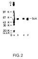
- Figure 2 is a photography showing SDS-PAGE and reverse fibrin agarose zymography of HT-1080 cell medium and e-PAI purified by immunosorbent chromatography with a monoclonal antibody against e-PAI,
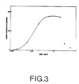
- Figure 3 is a graph showing neutralization of inhibitory action of e-PAI by monoclonal antibody against e-PAI,
- Figure 4 is a zymogram showing binding of complexes of u-PA with e-PAI to Sepharose columns with monoclonal antibodies against e-PAI, and
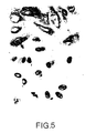
- Figure 5 is a photography showing immunoperoxidase staining of HT-1080 cells with a monoclonal antibody against e-PAI.
- The invention will be described in further detail with reference to the examples. Examples 1 to 5 illustrate the production and use of monoclonal antibodies against an endothelial type plasminogen activator inhibitor (e-PAI) released into the culture fluid from dexamethasone-treated human fibrosarcoma cells. The inhibitor inhibits human urokinase-type plasminogen activator (u-PA) and tissue-type plasminogen activator (t-PA).
- Inhibitor was purified from serum-free conditioned culture fluid of dexamethasone-treated human fibrosarcoma cells of the line HT-1080 (ATCC CCL121) by a procedure adapted from that described by van Mourik, J.A., Lawrence, D.A., and Loskutoff, D.J. (1984) J.Biol. Chem. 259, 14914-14921 for the plasminogen activator inhibitor from bovine endothelial cells. The HT-1080 cell line was maintained as a monolayer culture, using Dulbecco-modified Eagle's medium supplemented with 10% fetal bovine serum. Serum-free culture fluid was prepared from confluent monolayer cultures. Dexamethasone, a synthetic glucocorticoid, was added to the serum-free cultures in a concentration of 10⁻⁶M. HT-1080 cells produce relatively high amounts of u-PA, which under the culture conditions used is in the proenzyme form. Before purification of inhibitor, the culture fluid was depleted of u-PA by passing it through a column of monoclonal anti-u-PA IgG immobilized on Sepharose (Nielsen, L.S., Hansen, J.G., Skriver, L., Wilson, E.L., Kaltoft, K., Zeuthen, J., and Danø, K. (1982) Biochemistry, 24, 6410-6415). The culture fluid was then applied to a column of concanavalin A-Sepharose equilibrated with 0.01 M sodium phosphate, pH 7.4, 0.15 M NaCl (PBS), at a flow rate of 30 ml per h, using 5 ml Concanavalin A-Sepharose per liter culture fluid. The column was washed with 5 column volumes of PBS with 0.3 M NaCl. Bound protein was eluted with PBS with 0.5 M NaCl and 0.2 M α-methyl-D-mannoside. Fractions containing the peak of protein, as determined by measuring the absorbancy at 280 nm, were pooled and used for further analysis.
- From photometric scanning at 600 nm of Coomassie Blue stained polyacrylamide gels, the partly purified preparation was estimated to contain approximately 75% of a Mr ∼ 54,000 protein, the electrophoretic mobility of which coincided with inhibitory activity as determined by reverse zymography (see below). Before immunization this preparation was dialysed against PBS.
- 4 BALB/c-mice were immunized intradermally with approximately 20 µg of the Mr ∼ 54,000 protein obtained above in Freund's incomplete adjuvant on day 0, 7, 14, and 21. The plasma of each mouse was analyzed by ELISA (Enzyme Linked Immunosorbent Assay) and the mouse showing the highest titer against the immunization preparation was chosen for intraveneous injection and fusion with myeloma cells. The intraveneous injection of a similar dose as above dissolved in PBS was given on day 28 and the spleen was removed 3 days later.
- Spleen cells were mixed with NSI-Ag 4/1 myeloma cells (resistant to 0.1 mM 6-thioguanine; synthesize but do not secrete kappa light chains) (Köhler and Milstein (1976) Eur.J.Immunol. 6, 511-519) in a ratio of 10:1 (10⁸ spleen cells to 10⁷ NSI-Ag 4/1 cells) and incubated with 1 ml of 50% (wt/vol) polyethylene glycol in a phosphate-buffered saline solution for 90 sec. at 37°C. Dulbecco's modified Eagle's medium (20 ml) was added to the suspension, and the cells were centrifuged at 1000 x g. The cell pellet was resuspended in 96 ml of hypoxanthine/aminopterin/thymidine medium (Littlefield, J.W. (1964) Science 145, 709-710) supplemented with 10% fetal bovine serum and was distributed in 48 wells of Costar trays (Costar, Cambridge, M.A.). The medium was changed twice weekly.
- For screening of hybridoma supernatants using ELISA (Enzyme Linked Immunosorbent Assay), wells of Immuno Plates were coated with 100 µl per well of concanavalin A-Sepharose-purified plasminogen activator inhibitor containing ∼ 4 µg of protein per ml in 0.1 M Na₂CO₃, pH 9.8 over night at 37°C. In order to block residual binding sites, the wells were incubated with 0.25% BSA in PBS for more than 15 minutes. Then the wells were incubated with hybridoma supernatants for 1 hour and finally with peroxidase-conjugated rabbit antibodies against mouse Ig (Dakopatts, Copenhagen, Denmark), diluted 1:800 in PBS with 0.1% Tween 20 for 1 hour. Peroxidase reaction was performed for 5 min. with 100 µl of 0.1% o-phenylenediamine, 0.01% H₂O₂ in 0.1 M citrate-phosphate, pH 5.0. The reaction was stopped by the addition of 100 µl 1 M H₂SO₄, and absorbancy was read at 492 nm.
- For screening by immunoblotting, proteins in 10 ml serum-free medium from HT-1080 cells were concentrated by precipitation with trichloroacetic acid and separated by SDS-PAGE in a 10 cm wide lane. The proteins were transferred electrophoretically (10 V, 250 mA for 16 h at room temperature) from the polyacrylamide gel to nitrocellulose paper. The transfer buffer used was 0.125 M Tris HCl, 0.19 M glycine, 20% (v/v) methanol, 0.1% (w/v) SDS, pH 8.6. The nitrocellulose paper was washed in 0.05 M Tris HCl, pH 7.4, 0.15 M NaCl, 1% Triton X-100 (TBS-Triton) for 15 min. at room temperature and incubated for 30 min. with TBS-Triton containing human serum albumin (10 mg/ml). The paper was then washed 2 x 15 min. in TBS-Triton. Vertical lanes were cut out and incubated overnight at 4°C with culture supernatants from the hybridomas. The lanes were washed in TBS-Triton 3 x 15 min., incubated for 1 h at room temperature with peroxidase-conjugated rabbit IgG anti-mouse immunoglobulins (diluted 1:50 in TBS-Triton), and washed 3 x 10 min. in 0.05 M Tris-HCl, pH 7.6. The peroxidase reaction was then performed with 0.5 mg/ml of di-aminobenzidine in 0.01% H₂O₂ for 5 min. at room temperature.
- As a control nitrocellulose lanes were incubated with supernatant from hybridomas (Hy 2.15) producing antibody of irrelevant specificity (anti-trinitrophenyl) (Shulman, M., Wilde, C.D., and Köhler, G. (1978) Nature, 276, 269).
- After 10 days cultivation of the hybridomas supernatant from 16 primary wells showed a strongly positive ELISA-reaction. The hybridomas from said wells were cloned and recloned by limiting dilution (Kennett, R.H., McKearn, J.T., and Bechtol, K.B. (1980) Monoclonal Antibodies. Hybridomas: A New Dimension in Biological Analysis (Plenum, New York)). After cloning and recloning 4 stable ELISA-positive clones remained. Immunoblotting analysis showed that all four clones produced antibody which reacted with an Mr ∼ 54,000 band in crude conditioned culture fluid from HT-1080 cells.
- Monoclonal antibodies produced by the 4 clones obtained were purified from hybridoma culture fluid on a protein A-Sepharose column as follows: 200 ml of conditioned culture fluid from hybridomas was applied to a 5 ml protein A-Sepharose column (12x43 mm). The column was washed with 30 ml 0.1M Tris HCl, pH 8.1. Elution was performed with 0.1M sodium acetate, pH 4.0, 0.15 M NaCl. Fractions of 2 ml were collected in tubes containing 200 µl 1 M Tris-HCl, pH 9.0. The IgG concentration in the purified preparation was determined by spectrophotometry at 280 nm

Concentrations of IgG in impure solutions were determined by single radial immunodiffusion using purified mouse IgG as a standard. - The classes and subclasses of the antibodies produced by the hybrid clones were analysed by immunodiffusion against class- and subclass-specific goat antibodies (Meloy, V.A., USA). All 4 antibodies produced by the 4 clones were of the IgG₁ subclass.
- Isoelectric focusing of the 4 purified monoclonal antibodies in slab gels containing 6% polyacrylamide and 6% carrier ampholyte solution (Pharmalyte) showed that their isoelectric points were different (ranging between 5 and 7.5). The binding characteristics of the antibodies to solid-phase inhibitor as measured by ELISA differed greatly. They were therefore considered to originate from different hybridization events. The four clones were designated anti-plasminogen activator inhibitor clone 1, 2, 3, and 4, respectively.
- Conditioned culture fluid from human umbilical cord endothelial cells also contains a plasminogen activator inhibitor detectable by reverse fibrin-agarose zymography (Sprengers, E.O., Verheijen, J.H., van Hindsberg, V.W.M., and Emeis, J.J. (1984) Biochim. Biophys. Acta. 801, 163-170). This inhibitor has an electrophoretic mobility indistinguishable from that of the HT-1080-inhibitor. Figure 1 is a zymogram showing reverse zymography for plasminogen activator inhibitor in culture fluid conditioned by HT-1080 cells (a-c) or umbilical cord endothelial cells (d-f) before (a, d) and after passage of Sepharose columns coupled with monoclonal antibodies against TNP (b, e) and against HT-1080 plasminogen activator inhibitor (c, e). For coupling procedure see example 2. Two one ml columns containing approximately 1 mg of monoclonal anti-TNP IgG and monoclonal anti-plasminogen activator inhibitor IgG from clone 1, respectively, were equilibrated with a buffer containing 0.1 M Tris HCl, pH 8.1, 0.1% Triton X-100. To both columns was added 1 ml of serum-free cell culture fluid from HT-1080 cells and the run-through was collected. Serum-free cell culture fluid from human umbilical cord endothelial cells was treated identically. After electrophoresis, the gel was processed for reverse zymography for plasminogen activator inhibitors with an incubation period of 1.5 hours. Reverse zymography was carried out as described by Eriksson, L.A., Lawrence, D.A., and Loskutoff, D.J. (1984) Anal.Biochem. 137, 454-463. Plasminogen activator inhibitors in SDS polyacrylamide gels are detected by layering the gels over agarose gels containing fibrin, plasminogen, and plasminogen activator. Inhibitors diffuse into the fibrin/plasminogen/plasminogen activator gel from the polyacrylamide gel, and their presence is revealed by zones of fibrin resistant to plasminogen activator-catalyzed lysis. The position of Mr-markers are indicated.
- Passage of HT-1080 medium through the Sepharose column with antibodies from anti-inhibitor IgG clone 1 removed the inhibitory activity as revealed by reverse fibrin-agarose zymography; there was no effect of passage through a control column with a monoclonal control antibody (anti-TNP IgG). When plasminogen activator inhibitor from said human endothelial cells are applied to the column with monoclonal antibodies from clone 1 the inhibitor is bound to the column (Figure 1). When columns with the antibodies from clone 2, 3, or 4 were used, the results were identical (results not shown). This demonstrates immunological similarities between this inhibitor and the HT-1080 plasminogen activator inhibitor.
- Using the same technique we have also shown that rabbit antibodies against the HT-1080-inhibitor cross-react with a plasminogen activator inhibitor extracted from human blood platelets prepared by the method described by Erikson, L.A., Ginsberg, M.H., and Loskutoff, D.J. (1984), J.Clin.Invest., 74, 1465-1472.
- It has been reported that the endothelial cell plasminogen activator inhibitor, the platelet inhibitor (Erikson, L.A., Ginsberg, M.H., and Loskutoff, D.J. (1984), J.Clin.Invest., 74, 1465-1472) and the inhibitor from rat hepatoma cells of the HTC line show immunological similarities (D.J.Loskutoff and T.D.Gelehrter, personal communication). Thus it appears that the HT-1080-inhibitor is similar to a number of plasminogen activator inhibitors isolated from different cells and tissues.
- After coupling to Sepharose, antibody produced by anti-plasminogen activator inhibitor clone 1 was used for purification of inhibitor from HT-1080 cell culture fluid by a column procedure. 8 mg of monoclonal antibodies from anti-inhibitor IgG clone 1 was coupled to 2 ml of cyanogen bromide-activated Sepharose 4B. The material was packed in a column (20 x 16 mm), which was equilibrated with 0.1 M Tris HCl, pH 8.1. Conditioned cell culture fluid from HT-1080 cells was applied at a flow rate of 50 ml/hr. The column was washed with equilibration buffer (flow rate 50 ml/hr) followed by 0.1 M Tris HCl. pH 8.1, 1 M NaCl (flow rate 50 ml/hr). Elution was performed at a flow rate of 25 ml/h with 0.1 M CH₃COOH, pH 2.7, collecting fractions of 2 ml into tubes containing 200 µl of 1 M Tris HCl, pH 9,0 in order to neutralize the eluate. Fractions containing protein as determined by absorbance measurements at 280 nm were pooled.
- For quantification of inhibitory activity 0.005 Ploug Units urokinase standard in 25 µl of assay buffer was mixed with 25 µl of inhibitor diluted in the same buffer; this mixture was diluted to 500 µl with assay buffer and added to [¹²⁵I]-fibrin plate wells (see example 3). From assays with fixed concentrations of urokinase standard and serial dilutions of inhibitor preparations, the dilution of inhibitor causing 50% inhibition of the urokinase standard was calculated. The amount of inhibitor in wells with 50% inhibition was defined as 0.0025 inhibitor unit (Inh.U.). Before assay, inhibitor preparations were treated with SDS to a final concentration of 0.1%, and after a one hour incubation at 25°C, Triton X-100 was added to a final concentration of 1%.
- Results from the purification of inhibitor from human fibrosarcoma cells are shown in table 1.

- As shown in table I 86% of the inhibitory activity was bound by the column. After washing 99% of the inhibitory activity bound to the column could be eluted at low pH. A 45-fold purification of inhibitory activity was obtained and the eluate contained only an Mr ∼ 54,000 protein band as evaluated by Coomassie Blue staining of a SDS-polyacrylamide gel. The electrophoretic mobility of this protein coincided with the mobility of inhibitory activity as determined by reverse fibrin-agarose zymography (Figure 2). The samples subjected to SDS-polyacrylamide gel electrophoresis (SDS-PAGE) were: 1.5 ml crude culture fluid from HT-1080 cells (a), eluate corresponding to 10 µg of protein (b), 10 µl crude culture fluid from HT-1080 cells (a'), and eluate corresponding to 50 ng protein (b'). After electrophoresis, the gels were either stained with Coomassie Blue (a, b), or inhibitor in the gels was visualized by reverse fibrin agarose zymography for 2 hours (a', b'). The position of the following markers are indicated: Rabbit phosphorylase b (97 K), Bovine serum albumin (67 K), ovalbumin (43 K), carbonic anhydrase (30 K), soybean trypsin inhibitor (20.1 K), and α-lactalbumin (14.4 K).
- As judged from spectrophotometric scannings of stained polyacrylamide gels with crude culture fluid and the purified preparation, the strong Mr ∼ 54,000 band was purified, to the same extent as the inhibitory activity. SDS-PAGE in slab gels with a 6-16% linear concentration gradient of polyacrylamide of crude culture fluid and run-through from the immunosorbent column showed that the Mr ∼ 54,000 band was greatly diminished, while no other bands were affected (results not shown).
- SDS-PAGE under reducing conditions showed one band with Mr ∼ 54,000, indicating that the purified inhibitor consisted of one polypeptide chain (results not shown).
- The effect of the monoclonal antibodies on the inhibitory activity of the inhibitor was tested by a [¹²⁵I]-fibrin plate assay, which involved the activation of plasminogen by u-PA and the subsequent degradation of [¹²⁵I]-fibrin by the plasmin formed (cf. Nielsen, L.S., Hansen, J.G., Andreasen, P.A., Skriver, L., Danø, K., and Zeuthen, J. (1983) EMBO J., 2, 115-119). The [¹²⁵I]-fibrin plate assay was carried out as follows: 10 ng of inhibitor was added to [¹²⁵I]-fibrin plate assay wells together with 0.2 ng active u-PA, 1 µg Glu-plasminogen and IgG as indicated, in a total volume of 500 µl 0.1 M Tris HCl, pH 8.1, 0.1% Triton X-100, 0.25% gelatine (assay buffer). Radioactivity released in parallel control assays without u-PA (approximately 500 cpm) was subtracted and the radioactivity released in the presence of inhibitor calculated as a percentage of that released in the absence of inhibitor (approximately 3000 cpm). The total radioactivity in the [¹²⁵I]-fibrin plate assay wells was approximately 60,000 cpm. Each point represents the mean of two determinations. A neutralization of inhibitory activity that increased with increasing concentrations of anti-inhibitor IgG from clone 2 was observed (Figure 3, (●)) while there was no significant effect on inhibition of antibodies from clone 1 (Figure 3 (o)) and the monoclonal control antibody of irrelevant specificity (anti-TNP IgG) (Figure 3 (∇)). No neutralizing effect was observed with antiinhibitor IgG from clone 3 and 4 (results not shown).
- It has been shown that u-PA forms an equimolar complex with the HT-1080 inhibitor. This complex has an electrophoretic mobility corresponding to Mr ∼ 110,000 in SDS-PAGE and is detectable because it regains its plasminogen activator activity as measured by fibrin-agarose zymography: Plasminogen activator activity in polyacrylamide gels is detected by layering the gels over agarose gels containing fibrin and plasminogen - the plasminogen activators diffuse into the agarose gels and activate plasminogen to produce visible lysis zones (Granelli-Piperno, A. and Reich, E. (1978) J. Exp. Med., 148, 223-234). Figure 4 is a zymogram showing the binding of complexes of u-PA and HT-1080-inhibitor to Sepharose columns with monoclonal antibodies against the HT-1080 inhibitor. One ml columns containing approximately 1 mg of monoclonal anti-TNP antibody (c), antibodies from anti-inhibitor IgG clone 1 (a), or monoclonal anti-plasminogen activator inhibitor IgG from clone 2 (b) were equilibrated with a buffer containing 0.1 M Tris HCl, pH 8.1, 0.1% Triton X-100, 0.25% gelatin. One ml u-PA-inhibitor complex obtained by incubating the activator (25 ng/ml) with the inhibitor (500 ng/ml) for 1h at 25°C in a buffer of 0.1M Tris-HCl, pH 8.1, 0.1% Triton X-100 was added to each column, and 75 µl of the run-through from each column was subjected to SDS-PAGE followed by zymography for plasminogen activators. The positions of Mr-markers are indicated. Antibodies from anti-inhibitor clone 2 bound these complexes, while no binding was observed with antibodies from anti-inhibitor clone 1 or anti-TNP. Likewise, monoclonal anti-plasminogen activator inhibitor IgG from clone 4 bound complexes, while antibodies from 3 did not (results not shown). Said differential reactivities can be used in the quantitation of free versus complex-bound inhibitor.
- The monoclonal anti-plasminogen activator inhibitor antibodies can be used for immunocytochemical localization of the inhibitor in normal or malignant cells and tissues.
- HT-1080 cells cultured in serum free medium in the presence of dexamethasone were seeded on microscope slides and fixed for 30 min. with 4% paraformaldehyde in 0.1 M sodium phosphate buffer, pH 7.3. After washing with 0.05 M Tris-HCl, pH 7.4, 0.15 M NaCl (TBS) containing 1% Triton X-100 (TBS-Triton) for 30 min., the cells were exposed to 10% rabbit serum in TBS for 30min. and incubated overnight at 4°C with purified monoclonal antibody (10 µg/ml) diluted in TBS with 10% rabbit serum. Following a 1h temperature reequilibration, the cells were washed with TBS-Triton and bound monoclonal antibody was demonstrated by incubation with peroxidase conjugated rabbit anti-mouse IgG (1:60) diluted in TBS with 10% rabbit-serum followed by development with diaminobenzidine-hydrogen peroxide. The cells were lightly counterstained with haematoxylin, dehydrated, mounted and photographed. A strong granular staining was observed often with a perinuclear localization together with a weak diffuse staining apparently distributed in the entire cytoplasm (Figure 5, top). A distinct granular staining was observed using antibody from all 4 clones (results shown for clone 1 only). When the monoclonal anti-inhibitor antibody was substituted by monoclonal IgG of irrelevant specificity (anti-TNP antibody) (Figure 5, bottom) or by buffer alone, no staining was seen.
- The invention has been illustrated with reference to the production and use of monoclonal antibodies against a Mr ∼ 54,000 plasminogen activator inhibitor released into the culture fluid from dexamethasone-treated human fibrosarcoma cells, but since said inhibitor is immunologically similar to the human plasminogen activator inhibitors derived from human endothelial cells and human platelets, it should be understood that monoclonal antibodies against plaminogen activator inhibitors from these sources and plasminogen activator inhibitors which are immunologically similar to the plasminogen activator inhibitors from any of these sources also fall within the scope of the invention.
Claims (14)
- Monoclonal antibodies against human plasminogen activator inhibitor type 1 (e-PAI), such as plasminogen activator inhibitor derived from human endothelial cells, plasminogen activator inhibitor derived from human platelets and plasminogen activator inhibitor released into the culture fluid from dexamethasone-treated human HT-1080 fibrosarcoma cells.
- A method of producing monoclonal antibodies against human plasminogen activator inhibitor type 1 (e-PAI), such as plasminogen activator inhibitor derived from human endothelial cells, plasminogen activator inhibitor derived from human platelets and plasminogen activator inhibitor released into the culture fluid from dexamethasone-treated human HT-1080 fibrosarcoma cells, characterized in fusing myeloma cells with antibody-producing cells obtained from mammals which have been immunized with said type of plasminogen activator inhibitor or with antibody-producing cells which in vitro has been immunized with said plasminogen activator inhibitor, and selecting the hybridomas producing antibodies against the above mentioned inhibitors.
- A method according to claim 2, characterized in that the antibody-producing cells are selected from the group consisting of spleen cells and lymph node cells.
- A method according to claim 3, characterized in that the antibody-producing cell is a spleen cell.
- A method according to any of claims 2 to 4, characterized in that mouse cells are used.
- A method according to any of claims 2 to 5, characterized in that the plasminogen activator inhibitor is isolated from the culture fluid of dexamethasone-treated human HT-1080 fibrosarcoma cells.
- Hybridoma cells capable of producing monoclonal antibodies against human plasminogen activator inhibitor type 1 (e-PAI), such as plasminogen activator inhibitor derived from human endothelial cells, plasminogen activator inhibitor derived from human platelets and plasminogen activator inhibitor released into the culture fluid from dexamethasone-treated human HT-1080 fibrosarcoma cells.
- Hybridoma cells according to claim 7, characterized in that they are produced by fusion of spleen cells from immunized mice with mouse myeloma cells.
- Hybridoma cells according to claim 8, characterized in that they are produced by fusion of spleen cells from mice immunized with Mr ≈ 54,000 plasminogen activator inhibitor isolated from the culture fluid of dexamethasone-treated human fibrosarcoma cells of the line HT-1080 and mouse myeloma cells.
- Use of monoclonal antibodies against human plasminogen activator inhibitor type 1 (e-PAI), such as plasminogen activator inhibitor derived from human endothelial cells, plasminogen activator inhibitor derived from human platelets and plasminogen activator inhibitor released into the culture fluid from dexamethasone-treated human HT-1080 fibrosarcoma cells, for the purification of said inhibitor by means of immunoadsorption chromatography.
- Use of monoclonal antibodies against human plasminogen activator inhibitor type 1 (e-PAI) such as plasminogen activator inhibitor derived from human endothelial cells, plasminogen activator inhibitor derived from human platelets and plasminogen activator inhibitor released into the culture fluid from dexamethasone-treated human fibrosarcoma cells, for removal of said inhibitor from body fluids and other biological materials.
- Use of monoclonal antibodies against human plasminogen activator inhibitor type 1 (e-PAI) such as plasminogen activator inhibitor derived from human endothelial cells, plasminogen activator inhibitor derived from human platelets and plasminogen activator inhibitor released into the culture fluid from dexamethasone-treated human HT-1080 fibrosarcoma cells, for neutralization of the inhibitory activity of said inhibitor.
- Use of monoclonal antibodies against human plasminogen activator inhibitor type 1 (e-PAI), such as plasminogen activator inhibitor derived from human endothelial cells, plasminogen activator inhibitor derived from human platelets and plasminogen activator inhibitor released into the culture fluid from dexamethasone-treated human fibrosarcoma cells, for the quantification of free versus complex-bound forms of said inhibitor.
- Use of monoclonal antibodies against human plasminogen activator inhibitor type 1 (e-PAI), such as plasminogen activator inhibitor derived from human endothelial cells, plasminogen activator inhibitor derived from human platelets and plasminogen activator inhibitor released into the culture fluid from dexamethasone-treated human HT-1080 fibrosarcoma cells, for the detection, identification, immuno-staining and quantification of said inhibitor in body fluids, normal or malignant cells and tissues and other biological materials.
Priority Applications (1)
| Application Number | Priority Date | Filing Date | Title |
|---|---|---|---|
| AT86904118T ATE103818T1 (en) | 1985-07-12 | 1986-07-11 | MONOCLONAL ANTIBODIES INHIBITOR OF PLASMINOGEN ACTIVATOR, HYBRIDOMAS, PRODUCTION OF MONOCLONAL ANTIBODIES AND THEIR USE FOR PURIFICATION, REMOVAL, INHIBITION OR TESTING OF INHIBITORS. |
Applications Claiming Priority (3)
| Application Number | Priority Date | Filing Date | Title |
|---|---|---|---|
| DK3196/85 | 1985-07-12 | ||
| DK319685A DK319685A (en) | 1985-07-12 | 1985-07-12 | MONOCLONAL ANTIBODIES, PROCEDURES FOR PRODUCING ANTIBODIES, HYBRIDOMA CELLS PRODUCING ANTIBODIES, AND USING ANTIBODIES |
| PCT/DK1986/000080 WO1987000549A1 (en) | 1985-07-12 | 1986-07-11 | Plasminogen activator inhibitor monoclonal antibodies, hybridomas, monoclonal antibody production and use of the antibodies for purification, removal, inhibition or assay of the inhibitors |
Related Child Applications (1)
| Application Number | Title | Priority Date | Filing Date |
|---|---|---|---|
| EP93202290.8 Division-Into | 1993-08-03 |
Publications (2)
| Publication Number | Publication Date |
|---|---|
| EP0229126A1 EP0229126A1 (en) | 1987-07-22 |
| EP0229126B1 true EP0229126B1 (en) | 1994-04-06 |
Family
ID=8120155
Family Applications (1)
| Application Number | Title | Priority Date | Filing Date |
|---|---|---|---|
| EP86904118A Expired - Lifetime EP0229126B1 (en) | 1985-07-12 | 1986-07-11 | Plasminogen activator inhibitor monoclonal antibodies, hybridomas, monoclonal antibody production and use of the antibodies for purification, removal, inhibition or assay of the inhibitors |
Country Status (6)
| Country | Link |
|---|---|
| EP (1) | EP0229126B1 (en) |
| JP (1) | JPS63500564A (en) |
| AU (1) | AU6144686A (en) |
| DE (1) | DE3689766T2 (en) |
| DK (1) | DK319685A (en) |
| WO (1) | WO1987000549A1 (en) |
Families Citing this family (10)
| Publication number | Priority date | Publication date | Assignee | Title |
|---|---|---|---|---|
| US4952512A (en) * | 1984-06-22 | 1990-08-28 | Loskutoff David J | Gene encoding an inhibitor of tissue-type and urokinase-type plasminogen activators |
| FI89634C (en) * | 1987-07-10 | 1993-10-25 | Yamanouchi Pharma Co Ltd | FOERFARANDE, ANORDNING OCH KITT FOER MAETNING AV VAEVNADSPLASMINOGENAKTIVATOR-KTIVITET |
| DE3742997A1 (en) * | 1987-12-18 | 1989-06-29 | Behringwerke Ag | PEPTIDES, METHOD FOR THE PRODUCTION THEREOF, THEIR USE FOR THE OBTAINING OF ANTIBODIES, AND THEIR USE FOR BLOCKING THE PAI-1 ACTIVITY OF HUMAN BLOOD |
| JP2750732B2 (en) * | 1988-04-04 | 1998-05-13 | 帝人株式会社 | Immunoassay method for human plasminogen activator inhibitor 1-human tissue plasminogen activator complex |
| NO891395L (en) * | 1988-04-04 | 1989-10-05 | Teijin Ltd | Reagent systems. |
| JP2868530B2 (en) * | 1989-04-03 | 1999-03-10 | 帝人株式会社 | Monoclonal antibodies to plasminogen activator inhibitors |
| EP1013285A3 (en) * | 1989-04-07 | 2004-05-19 | Cancerforskningsfonden af 1989 (fonden til fremme af eksperimentel cancerforskning) | Urokinase-type plasminogen activator receptor |
| EP0450086A4 (en) * | 1989-10-02 | 1993-03-10 | Teijin Limited | Kit for immunoassay of human tissue plasminogen activator/human plasminogen activator inhibitor complex and method of immunoassay |
| DK85193D0 (en) * | 1993-07-16 | 1993-07-16 | Cancerforskningsfondet Af 1989 | SUPPRESSION OF INHIBITORS |
| EA033403B1 (en) * | 2013-08-13 | 2019-10-31 | Sanofi Sa | ANTIBODIES TO PLASMINOGEN 1 (PAI-1) ACTIVATOR INHIBITOR AND WAYS OF THEIR APPLICATION |
Citations (2)
| Publication number | Priority date | Publication date | Assignee | Title |
|---|---|---|---|---|
| WO1986000413A1 (en) * | 1984-06-22 | 1986-01-16 | Scripps Clinic And Research Foundation | Diagnostic assay for inhibitor or tissue-type and urokinase-type plasminogen activators |
| EP2169549A1 (en) * | 2007-07-05 | 2010-03-31 | Panasonic Corporation | Data processing device, data processing method, data processing program, recording medium, and integrated circuit |
Family Cites Families (2)
| Publication number | Priority date | Publication date | Assignee | Title |
|---|---|---|---|---|
| BE896543A (en) * | 1983-04-22 | 1983-08-16 | Wallone Region | Mono:clonal antibody systems against blood clotting proteins - comprising antibodies against antithrombin, anti:plasmin and antitrypsin |
| DE3586577T2 (en) * | 1984-07-26 | 1993-03-25 | Teijin Ltd | IMMUNOLOGICAL DETERMINATION OF HUMAN PLASMIN / ALPHA-2-PLASMIN INHIBITOR. |
-
1985
- 1985-07-12 DK DK319685A patent/DK319685A/en not_active Application Discontinuation
-
1986
- 1986-07-11 EP EP86904118A patent/EP0229126B1/en not_active Expired - Lifetime
- 1986-07-11 DE DE3689766T patent/DE3689766T2/en not_active Expired - Lifetime
- 1986-07-11 JP JP61504151A patent/JPS63500564A/en active Pending
- 1986-07-11 AU AU61446/86A patent/AU6144686A/en not_active Abandoned
- 1986-07-11 WO PCT/DK1986/000080 patent/WO1987000549A1/en not_active Ceased
Patent Citations (2)
| Publication number | Priority date | Publication date | Assignee | Title |
|---|---|---|---|---|
| WO1986000413A1 (en) * | 1984-06-22 | 1986-01-16 | Scripps Clinic And Research Foundation | Diagnostic assay for inhibitor or tissue-type and urokinase-type plasminogen activators |
| EP2169549A1 (en) * | 2007-07-05 | 2010-03-31 | Panasonic Corporation | Data processing device, data processing method, data processing program, recording medium, and integrated circuit |
Non-Patent Citations (1)
| Title |
|---|
| The Journal of biological chemistry, vol.259, n° 23, pages 14914-14921, 1984 * |
Also Published As
| Publication number | Publication date |
|---|---|
| DK319685D0 (en) | 1985-07-12 |
| WO1987000549A1 (en) | 1987-01-29 |
| DK319685A (en) | 1987-01-13 |
| DE3689766D1 (en) | 1994-05-11 |
| DE3689766T2 (en) | 1994-12-01 |
| EP0229126A1 (en) | 1987-07-22 |
| AU6144686A (en) | 1987-02-10 |
| JPS63500564A (en) | 1988-03-03 |
Similar Documents
| Publication | Publication Date | Title |
|---|---|---|
| Nielsen et al. | Monoclonal antibodies to human 54,000 molecular weight plasminogen activator inhibitor from fibrosarcoma cells-inhibitor neutralization and one-step affinity purification | |
| Birkedal-Hansen et al. | Monoclonal antibodies to human fibroblast procollagenase. Inhibition of enzymatic activity, affinity purification of the enzyme, and evidence for clustering of epitopes in the amino-terminal end of the activated enzyme | |
| Marynen et al. | A monoclonal antibody to a neo-antigen on alpha2-macroglobulin complexes inhibits receptor-mediated endocytosis. | |
| Holvoet et al. | A monoclonal antibody specific for Lys-plasminogen. Application to the study of the activation pathways of plasminogen in vivo. | |
| Åstedt et al. | Purification of a specific placental plasminogen activator inhibitor by monoclonal antibody and its complex formation with plasminogen activator | |
| Nielsen et al. | Monoclonal antibody to human 66,000 molecular weight plasminogen activator from melanoma cells. Specific enzyme inhibition and one‐step affinity purification. | |
| Angles-Cano et al. | Production of monoclonal antibodies to the high fibrin-affinity, tissue-type plasminogen activator of human plasma. Demonstration of its endothelial origin by immunolocalization | |
| Philips et al. | Immunological relationship between the fast-acting plasminogen activator inhibitors from plasma, blood platelets and endothelial cells demonstrated with a monoclonal antibody against an inhibitor from placenta | |
| US4833085A (en) | Monoclonal antibody specific for human colon fibroblast-derived t-PA | |
| US5422245A (en) | Plasminogen activator inhibitor monoclonal antibodies, hybridomas, monoclonal antibody production and use of the antibodies for assay of the inhibitors | |
| WO1987000549A1 (en) | Plasminogen activator inhibitor monoclonal antibodies, hybridomas, monoclonal antibody production and use of the antibodies for purification, removal, inhibition or assay of the inhibitors | |
| US5830681A (en) | Immunoassays for and monoclonal antibodies to prothrombin activation peptide F1.2 | |
| Holvoet et al. | Measurement of free, one-chain tissue-type plasminogen activator in human plasma with an enzyme-linked immunosorbent assay based on an active site-specific murine monoclonal antibody | |
| EP0998555A1 (en) | Monospecific antibody reactive with matrix metalloproteinase cleavage products of fibrin(ogen) | |
| Schleef et al. | Characterization of two monoclonal antibodies against human tissue-type plasminogen activator | |
| US6271352B1 (en) | PAI-1 determination and use thereof | |
| Veloso et al. | A monoclonal anti-human plasma prekallikrein antibody that inhibits activation of prekallikrein by factor XIIa on a surface | |
| EP0585995A1 (en) | Pure human endothelial type plasminogen activator inhibitor (e-PAI) and production thereof | |
| EP0192675B1 (en) | Anti-urokinase monoclonal antibody; its production and use | |
| US4891312A (en) | Monoclonal antibody specific for human colon fibroblast-derived t-PA | |
| US4889922A (en) | Monoclonal antibody specific for human colon fibroblast-derived T-PA | |
| Reagan et al. | Immunoaffinity purification of tissue plasminogen activator from serum-supplemented conditioned media using monoclonal antibody | |
| EP0169549B1 (en) | Immunological determination of human plasmin/alpha2-plasmin inhibitor | |
| US5362857A (en) | Purification of plasminogen activator inhibitor 2 by immunoaffinity chromatography | |
| Kaizu et al. | One-step sandwich enzyme-linked immunosorbent assay of human tissue plasminogen activator using monoclonal antibodies |
Legal Events
| Date | Code | Title | Description |
|---|---|---|---|
| PUAI | Public reference made under article 153(3) epc to a published international application that has entered the european phase |
Free format text: ORIGINAL CODE: 0009012 |
|
| AK | Designated contracting states |
Kind code of ref document: A1 Designated state(s): AT BE CH DE FR GB IT LI LU NL SE |
|
| 17P | Request for examination filed |
Effective date: 19870709 |
|
| 17Q | First examination report despatched |
Effective date: 19891004 |
|
| RAP3 | Party data changed (applicant data changed or rights of an application transferred) |
Owner name: FONDEN TIL FREMME AF EKSPERIMENTAL CANCERFORSKNING |
|
| GRAA | (expected) grant |
Free format text: ORIGINAL CODE: 0009210 |
|
| AK | Designated contracting states |
Kind code of ref document: B1 Designated state(s): AT BE CH DE FR GB IT LI LU NL SE |
|
| RAP3 | Party data changed (applicant data changed or rights of an application transferred) |
Owner name: FONDEN TIL FREMME AF EKSPERIMENTAL CANCERFORSKNING |
|
| REF | Corresponds to: |
Ref document number: 103818 Country of ref document: AT Date of ref document: 19940415 Kind code of ref document: T |
|
| XX | Miscellaneous (additional remarks) |
Free format text: TEILANMELDUNG 93202290.8 EINGEREICHT AM 11/07/86. |
|
| REF | Corresponds to: |
Ref document number: 3689766 Country of ref document: DE Date of ref document: 19940511 |
|
| ITF | It: translation for a ep patent filed | ||
| ET | Fr: translation filed | ||
| EPTA | Lu: last paid annual fee | ||
| PLBI | Opposition filed |
Free format text: ORIGINAL CODE: 0009260 |
|
| EAL | Se: european patent in force in sweden |
Ref document number: 86904118.6 |
|
| 26 | Opposition filed |
Opponent name: DR. RICHARD HART Effective date: 19950105 Opponent name: THE SCRIPPS RESEARCH INSTITUTE Effective date: 19950109 |
|
| NLR1 | Nl: opposition has been filed with the epo |
Opponent name: THE SCRIPPS RESEARCH INSTITUTE. Opponent name: DR. RICHARD HART. |
|
| PLBO | Opposition rejected |
Free format text: ORIGINAL CODE: EPIDOS REJO |
|
| PLBN | Opposition rejected |
Free format text: ORIGINAL CODE: 0009273 |
|
| STAA | Information on the status of an ep patent application or granted ep patent |
Free format text: STATUS: OPPOSITION REJECTED |
|
| 27O | Opposition rejected |
Effective date: 19980222 |
|
| NLR2 | Nl: decision of opposition | ||
| REG | Reference to a national code |
Ref country code: GB Ref legal event code: IF02 |
|
| PGFP | Annual fee paid to national office [announced via postgrant information from national office to epo] |
Ref country code: NL Payment date: 20050616 Year of fee payment: 20 |
|
| PGFP | Annual fee paid to national office [announced via postgrant information from national office to epo] |
Ref country code: AT Payment date: 20050621 Year of fee payment: 20 |
|
| PGFP | Annual fee paid to national office [announced via postgrant information from national office to epo] |
Ref country code: GB Payment date: 20050704 Year of fee payment: 20 |
|
| PGFP | Annual fee paid to national office [announced via postgrant information from national office to epo] |
Ref country code: FR Payment date: 20050712 Year of fee payment: 20 |
|
| PGFP | Annual fee paid to national office [announced via postgrant information from national office to epo] |
Ref country code: SE Payment date: 20050714 Year of fee payment: 20 Ref country code: DE Payment date: 20050714 Year of fee payment: 20 Ref country code: CH Payment date: 20050714 Year of fee payment: 20 |
|
| PGFP | Annual fee paid to national office [announced via postgrant information from national office to epo] |
Ref country code: LU Payment date: 20050718 Year of fee payment: 20 Ref country code: IT Payment date: 20050718 Year of fee payment: 20 |
|
| PGFP | Annual fee paid to national office [announced via postgrant information from national office to epo] |
Ref country code: BE Payment date: 20050801 Year of fee payment: 20 |
|
| REG | Reference to a national code |
Ref country code: GB Ref legal event code: PE20 |
|
| PG25 | Lapsed in a contracting state [announced via postgrant information from national office to epo] |
Ref country code: GB Free format text: LAPSE BECAUSE OF EXPIRATION OF PROTECTION Effective date: 20060710 |
|
| PG25 | Lapsed in a contracting state [announced via postgrant information from national office to epo] |
Ref country code: NL Free format text: LAPSE BECAUSE OF EXPIRATION OF PROTECTION Effective date: 20060711 |
|
| REG | Reference to a national code |
Ref country code: CH Ref legal event code: PL |
|
| NLV7 | Nl: ceased due to reaching the maximum lifetime of a patent |
Effective date: 20060711 |
|
| EUG | Se: european patent has lapsed | ||
| BE20 | Be: patent expired |
Owner name: *FONDEN TIL FREMME AF EKSPERIMENTAL CANCERFORSKNIN Effective date: 20060711 |