EP0219988A2 - Instrument manuel autonome à ultrason pour usage ophtalmologique - Google Patents
Instrument manuel autonome à ultrason pour usage ophtalmologique Download PDFInfo
- Publication number
- EP0219988A2 EP0219988A2 EP19860307392 EP86307392A EP0219988A2 EP 0219988 A2 EP0219988 A2 EP 0219988A2 EP 19860307392 EP19860307392 EP 19860307392 EP 86307392 A EP86307392 A EP 86307392A EP 0219988 A2 EP0219988 A2 EP 0219988A2
- Authority
- EP
- European Patent Office
- Prior art keywords
- transducer
- instrument
- housing
- connector
- mhz
- Prior art date
- Legal status (The legal status is an assumption and is not a legal conclusion. Google has not performed a legal analysis and makes no representation as to the accuracy of the status listed.)
- Granted
Links
Images

Classifications
-
- A—HUMAN NECESSITIES
- A61—MEDICAL OR VETERINARY SCIENCE; HYGIENE
- A61B—DIAGNOSIS; SURGERY; IDENTIFICATION
- A61B8/00—Diagnosis using ultrasonic, sonic or infrasonic waves
- A61B8/44—Constructional features of the ultrasonic, sonic or infrasonic diagnostic device
- A61B8/4444—Constructional features of the ultrasonic, sonic or infrasonic diagnostic device related to the probe
- A61B8/4455—Features of the external shape of the probe, e.g. ergonomic aspects
-
- A—HUMAN NECESSITIES
- A61—MEDICAL OR VETERINARY SCIENCE; HYGIENE
- A61B—DIAGNOSIS; SURGERY; IDENTIFICATION
- A61B3/00—Apparatus for testing the eyes; Instruments for examining the eyes
- A61B3/10—Objective types, i.e. instruments for examining the eyes independent of the patients' perceptions or reactions
- A61B3/1005—Objective types, i.e. instruments for examining the eyes independent of the patients' perceptions or reactions for measuring distances inside the eye, e.g. thickness of the cornea
-
- A—HUMAN NECESSITIES
- A61—MEDICAL OR VETERINARY SCIENCE; HYGIENE
- A61B—DIAGNOSIS; SURGERY; IDENTIFICATION
- A61B8/00—Diagnosis using ultrasonic, sonic or infrasonic waves
- A61B8/08—Detecting organic movements or changes, e.g. tumours, cysts, swellings
- A61B8/0858—Detecting organic movements or changes, e.g. tumours, cysts, swellings involving measuring tissue layers, e.g. skin, interfaces
-
- A—HUMAN NECESSITIES
- A61—MEDICAL OR VETERINARY SCIENCE; HYGIENE
- A61B—DIAGNOSIS; SURGERY; IDENTIFICATION
- A61B8/00—Diagnosis using ultrasonic, sonic or infrasonic waves
- A61B8/10—Eye inspection
-
- A—HUMAN NECESSITIES
- A61—MEDICAL OR VETERINARY SCIENCE; HYGIENE
- A61B—DIAGNOSIS; SURGERY; IDENTIFICATION
- A61B8/00—Diagnosis using ultrasonic, sonic or infrasonic waves
- A61B8/46—Ultrasonic, sonic or infrasonic diagnostic devices with special arrangements for interfacing with the operator or the patient
- A61B8/461—Displaying means of special interest
- A61B8/462—Displaying means of special interest characterised by constructional features of the display
-
- A—HUMAN NECESSITIES
- A61—MEDICAL OR VETERINARY SCIENCE; HYGIENE
- A61F—FILTERS IMPLANTABLE INTO BLOOD VESSELS; PROSTHESES; DEVICES PROVIDING PATENCY TO, OR PREVENTING COLLAPSING OF, TUBULAR STRUCTURES OF THE BODY, e.g. STENTS; ORTHOPAEDIC, NURSING OR CONTRACEPTIVE DEVICES; FOMENTATION; TREATMENT OR PROTECTION OF EYES OR EARS; BANDAGES, DRESSINGS OR ABSORBENT PADS; FIRST-AID KITS
- A61F9/00—Methods or devices for treatment of the eyes; Devices for putting-in contact lenses; Devices to correct squinting; Apparatus to guide the blind; Protective devices for the eyes, carried on the body or in the hand
- A61F9/007—Methods or devices for eye surgery
- A61F9/013—Instruments for compensation of ocular refraction ; Instruments for use in cornea removal, for reshaping or performing incisions in the cornea
Definitions
- Myopia is the most common optical refractive error. Over 133 million individuals in the United States wear some form of refractive correction, either in the form of spectacles or contact lenses.
- RK radial keratotomy
- the length and number of incisions required in an RK operation varies according to the degree of optical correction desired, and the depth of the incision-'is critical to the satisfactory outcome of the surgery. Inadequate depth will result in undercorrection, whereas excessive depth will result in overcorrection or perforation of the cornea and potentially serious harm to the eye. Accurate measurement of the corneal thickness is therefore essential to safe and successful RK surgery.
- pachymeters Instruments that measure corneal thickness are referred to as pachymeters. Early pachymeters were purely optical devices and had been shown to be imprecise. Ultrasonic technology is now routinely used to measure corneal thickness. All current instruments employ an ultrasonic probe which contacts the eye and which is connected via cable to a desk unit which is about the size of an oscilloscope. Some units have a smaller oscilloscope screen that displays the transducer tracings while other more recent units simply display a digital readout of the corneal thickness.
- cataract surgery The most commonly performed operation in ophthalmology is cataract surgery.
- a cataract is an opacification of the biological lens inside the eye.
- cataract extraction one of the several techniques is used to remove the opacified lens . material. Once removed, it is possible to implant an artificial lens in order to restore optical integrity to the eye, eliminating the need for thick cataract glasses or contact lenses.
- a biometric ruler employed a piezoelectric transducer, in a hand-held probe attached via a cable to an oscilloscope.
- Second generation instruments still require the examiner to detect the pattern consistent with an axial scan. Then, electronics are used to calculate and display the desired measurement in millimeters. It would be desirable to have a self-contained, hand-held, digital, ultrasonic biometric ruler instrument for displaying the axial length of the eye using microprocessor technology to assess the echo waveforms and displaying digitally a readout representing as accurately as possible the true axial length of the eye.
- the present invention is related to the system and circuits disclosed in U.S. application 781,257 entitled “Digital Ultrasonic Instrument for Ophthalmic Use” filed 27 September 1985 in the names of David A. Wallace M.D., Steven E. Feldon M.D., Gary P. Mezack, Douglas L. Whiting Ph.D., William F. Dally and Scott A. Karns and the disclosure of which is incorporated herein by reference.
- the present invention is also related to the system disclosed in U.S. application 781,240 entitled “Hand-Held Self-Contained Electronic Tonometer” filed 27 September 1985 in the names of Steven E. Feldon M.D., David A. Wallace M.D., Robert A. Monsour and Gary P. Mezack and the disclosure of which is incorporated herein by reference.
- the present invention relates to a completely portable, hand held digital ultrasonic instrument about the size and shape of an ordinary pen.
- the housing is contoured such that it can be easily grasped in the manner of a writing pen.
- An activation button is located on the interior dorsal surface in close approximation to the index fingertip of the user.
- a liquid crystal display provides a digital readout of the corneal thickness on the barrel of the instrument housing.
- the instrument incorporates a 10 MHz or 20 MHz solid phase piezoelectric transducer, a microprocessor, a gate array, a hybrid transceiver, a liquid crystal display, batteries and a removable battery cover.
- the pachymeter uses a 20 MHz transducer that is attached to a swan neck connector which is attached to the insrument housing.
- the biometric ruler uses a 10 MHz transducer that is attached to a truncated conical connector which is also attached to the instrument housing.
- a pinjack connector is available to link the unit to other electronic media such as a microcomputer, personal computer, or printer.
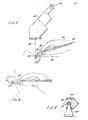
- the instrument comprises a housing 20 which is contoured such that it can be easily grasped, in the manner of a writing pen.
- the tip of the instrument is the piezoelectric ultrasonic transducer 22 mounted on a plastic standoff 34.
- the other functioning components of the instrument include an activation button 26, located on the anterior dorsal surface in close approximation to the index fingertip of the user, a liquid crystal display 28 (LCD), a reset button (not shown), a removable battery cover 30, and a pinjack connector 32 ( Figure 4).
- the 20 MHz transducer 22 comprises a plastic, ultrasonically transparent, contact head 34, and a recessed piezoelectric ultrasonic transducer 22.
- This cone shaped contact head 34 is attached to the transducer 22 which is attached to the swan neck connector 24 which connects to the main housing 20 of the pachymeter 36.
- the backing of the transducer is a .005 inch piece of tungsten-loaded epoxy which is an acoustically inert material.
- the piezoelectric transducer element has a frequency of 20 MHz. It is activated by a hybrid transceiver which connects to a microprocessor via a gate array. One to three pulses of current are delivered to the transducer from the hybrid transceiver.
- the first echo corresponds to the front corneal surface.
- the second echo corresponds to the back corneal surface.
- the time between the two corneal echoes is proportional to the corneal thickness. Because sound travels at a rate of approximately 1640 meters/sec through corneal tissue, the time between the emitted signal and the reflected echoes can be converted to millimeters, using standard formulae.
- the active transducer element is opaque and is centered at the posterior edge of the cone shaped contact head 34.
- the contact head is made of a clear acrylic material that allows the instrument tip to be better visualized under the operating microscope 60 ( Figure 5).
- the small size of the transducer 22 and the contact head 34 minimizes the area of the corneal surface which is obscured by the transducer 22.
- the swan neck connector 24 is designed such that it is rather easy for the user to place the contact head 34 into contact with the corneal surface perpendicularly to the visual axis shown by dotted line 62 in Figure 5.
- the combined clear acrylic contact head 34 and the swan neck connector 24 both facilitate perpen- dicular alignment of the transducer 22 to the corneal surface.
- the transducer must be aligned perpendicular to the corneal surface in order to obtain detectable echoes from the inner corneal surface adjacent to the aqueous humor.
- the shortest echographic distance corresponds to the actual corneal thickness. If the transducer 22 is slightly off perpendicular to the visual axis, then spurious readings may result.
- an acrylic multistepped calibration block which simulates the corneal interfaces, is used which is placed on a flat surface.
- the pachymeter 36 is then held perpendicularly to the surface of the block and the activation switch 26 is pressed once and then released.
- a beeper and miniature speaker provide a series of clicks followed by a beep.
- the pachymeter reading from the surface will be displayed on the display 28. This output should be within .01 mm of the calibration block measurement printed on its surface.
- the tip of the instrument is a piezoelectric ultrasonic transducer element (not shown) mounted within a standoff 38 as shown in Figure 1B.
- a special acoustically mismatched damping material is utilized to prevent "ringing" of the ultrasonic transducer which might preclude interpretation or detection of early echoes.
- Other functional components of the instrument include an activation button 26 located on the anterior dorsal surface in the close approximation to the index fingertip of the user, liquid crystal display 28, a reset button (not shown), and a removable battery cover 30.
- the transducer head 42 is a focusing element with a focal length of 24 millimeters, corresponding to an area near the retina of the eye.
- the diameter of the focusing element is 0.3 inch.
- a light-emitting diode 40 In the center of the focusing element is a light-emitting diode 40. The- patient is asked to look at the light-emitting diode 40, which has been activated, as the probe is placed in contact with the corneal surface. This allows the patient to center the eye on the transducer, facilitating axial measurements.
- the average speed of sound through the eye is approximately 1560 meters/second.
- the time between the emitted signal, corresponding to the corneal surface, and reflected echoes are then converted into millimeters using standard formulae.
- One to three pulses of current from the hybrid transceiver are delivered to activate the transducer.
- the transducer After sending out the ultrasonic signal, the transducer is used in a detection mode.
- the first echo corresponds to the transducer-corneal interface. All subsequent echoes are measured relative to this.
- the second echo found within a "window" of 1.5 to 5 millimeters from the corneal surface, corresponds to the anterior lenticular surface.
- the third echo falling into a window of 1.5 to 6.5 millimeters behind the anterior lenticular echo, corresponds to the posterior lenticular surface.
- the fourth echo located 18.5 to 29.0 millimeters behind the corneal surface echo, corresponds to the retinal surface.
- the fifth echo 0.29 to 2.5 millimeters behind the retina, corresponds to the scleral surface. Echoes which occur outside the given windows will be ignored.
- the four annunciator bars 56, Figure 2B separately indicate the presence of these four echos.
- the 10 MHz Transducer is calibrated by use of an acrylic block with multiple interfaces which simulates a standardized eye. This is placed on a flat surface and the transducer head 42 is then held perpendicular to the surface of the block and the activation switch 26 is pressed once and then released. A series of clicks will sound, following which there will be a beep. The biometric ruler reading from this source will be demonstrated on the display 28. This output should be within 0.1 millimeters of the calibration block measurement printed on its surface.
- Figure 2A shows typical information that is displayed on the digital readout 28 when the 20 MHz transducer is used and Figure 2B shows typical information displayed when the 10 MHz transducer is substituted for the 20 MHz transducer of Figure lA.
- Figure 3 shows a top view of the instrument with the top cover 42 ( Figure lA) removed.
- the batteries 40 are located to the rear of the instrument.
- the liquid crystal display 28 is connected to a circuit board 44 ( Figure 4) and is located adjacent to the microprocessor 46.
- a gate array 48 is located on the same circuit board 44 between the microprocessor 46 and the hybrid transceiver 54.
- the activation switch 26 is located between the hybrid receiver 54 and the connector 24.
- the total length of the unit is approximately 7.25 inches and it weighs approximately 2 ounces.
- Figure 4 shows a sectional view of the housing 20 of the hand-held pachymeter 36 with the various components installed.
- the same housing 20 and component parts shown in Figure 4 are used when the instrument is used as a biometric ruler except that the connector 38 and its associated parts are substituted for the connector 24 and its associated parts.
- a reset switch 50 At the rear end of the casing is located a reset switch 50 and a Murata beeper 52 is located directly beneath the microprocessor 46 on the opposite side of circuit board 44.
- a pinjack 32 located directly below the printed circuit board 44. This pinjack 32 is used to send data such as corneal thickness or axial length from the instrument, whether used as a pachymeter or biometric ruler, to outside instruments such as a microcomputer, a personal computer or printer.
- All elements of the instrument are connected to a multilayered circuit board 44. Mounted off the circuit board are four silver oxide batteries 41. Mounted on the circuit board is the discrete circuitry related to transducer signal processing. Also on the circuit board are connectors to the display 28, the reset button 50, and the RS232 pinjack 32.
- Figure 5 shows the pachymeter 36 with a 20 MHz transducer 22 being used for measuring the thickness of the cornea.
- the user is able to look through a microscope 60 and at the same time grasp the pachymeter 36 in the manner shown in Figure 5.
- the shape of the swan neck connector 24 allows the user 58 to position the transducer element 22 on the central axis of the cornea, shown by dotted line 62, and still be able to view the patient's eye through the microscope 60.
- Dotted line 82 shows the perpendicular tangent of the cornea to the visual axis shown by line 62.
- the central axis of the pachymeter 36, shown by dotted line 80 is approximately 27° from the tangent of the cornea shown by line 82.
- the swan neck connecter allows the pachymeter 36 to be comfortably held in a position in which the transducer 22 is aligned with the visual axis of the eye and the pachymeter clears the patient's eyebrow 84 and allows the user to- comfortably rest his hand on the patient's forehead.
- a clearer view of what the user sees when viewing the transducer 22 in contact with the eye 65 is shown in Figure 7.
- the lid retractor 64 holds the patient's eyelids in an open position to facilitate the thickness measurement.
- the size of the transducer 22 along with the swan neck connector 24 allows the user to partially view the optical zone as shown by dotted lines 66 in Figure 7.
- the optical zone marks the central limit of the radial corneal cuts performed during radial keratotomy surgery. This zone, usually 3 to 4.5 millimeters in diameter, is demarcated by the surgeon by the superficial application of a calibrated trephine.
- FIG 6 shows a user holding the biometric ruler as it would be held when making an axial length measurement.
- the biometric ruler unit 68 is held perpendicular to the patient's eye 70 along the visual axis of the patient's eye shown by dotted line 72.
- the activation button 26, Figure 1B, is depressed and the measurement sequence is initiated. The results of the successful measurement are displayed on the LCD 28 as shown in , Figure 2B.
- the anterior and posterior lenticular spikes are not available. Axial length calculation is required for the placement of secondary lens implants with appropriate power. Because it is impossible to align multiple interfaces to ensure axial scan, the reliability of such measurements is less than could be expected in aphakic patients. Therefore, in order to initiate an aphakia mode measurement, the activation button 26 is pressed twice in rapid succession. The patient is then asked to look at LED emitter 40 ( Figure 1B) in the middle of the focusing element 42 ( Figure 1B). Once the retinal and scleral spikes are identified using thresholding identification algorithms, the maximum length is sought in a manner analogous to the evaluation performed in the phakic examination.
- the results are displayed on the display 28, sent to the pinjack 32, and the annunciators 56, corresponding to the retinal and scleral spikes, are illuminated on the display 28.
- the biometric ruler 68 as shown in Figure 6 weighs approximately 2 ounces and is approximately 7.25 inches in length.
- the microprocessor 46 and transducer elements are automatically turned off in order to reduce power consumption and preserve the battery life.
- a small discrete circuit performs this function and also responds to depression of the activation button by activating the electronic elements and the transducer.
Priority Applications (1)
| Application Number | Priority Date | Filing Date | Title |
|---|---|---|---|
| AT86307392T ATE95998T1 (de) | 1985-09-27 | 1986-09-25 | Unabhaengiges, tragbares ultraschall-instrument zur anwendung in der augenheilkunde. |
Applications Claiming Priority (2)
| Application Number | Priority Date | Filing Date | Title |
|---|---|---|---|
| US78114885A | 1985-09-27 | 1985-09-27 | |
| US781148 | 1985-09-27 |
Publications (3)
| Publication Number | Publication Date |
|---|---|
| EP0219988A2 true EP0219988A2 (fr) | 1987-04-29 |
| EP0219988A3 EP0219988A3 (en) | 1988-07-20 |
| EP0219988B1 EP0219988B1 (fr) | 1993-10-20 |
Family
ID=25121842
Family Applications (1)
| Application Number | Title | Priority Date | Filing Date |
|---|---|---|---|
| EP19860307392 Expired - Lifetime EP0219988B1 (fr) | 1985-09-27 | 1986-09-25 | Instrument manuel autonome à ultrason pour usage ophtalmologique |
Country Status (5)
| Country | Link |
|---|---|
| US (1) | US5165415A (fr) |
| EP (1) | EP0219988B1 (fr) |
| JP (1) | JPS62122640A (fr) |
| AT (1) | ATE95998T1 (fr) |
| DE (1) | DE3689190T2 (fr) |
Cited By (5)
| Publication number | Priority date | Publication date | Assignee | Title |
|---|---|---|---|---|
| US5100318A (en) * | 1990-04-13 | 1992-03-31 | Periosonics, Inc. | Ultrasonic method and apparatus for measuring the periodontal pocket |
| EP0763344A3 (fr) * | 1995-09-13 | 1999-02-24 | Medison Co., Ltd. | Appareil diagnostique portatif à ultrasons |
| EP1006879A4 (fr) * | 1996-10-21 | 2000-06-14 | Philipp Lang | Mesure d'epaisseur de couche d'un objet par utilisation de procedes et de dispositifs a ultrasons |
| WO2008070580A3 (fr) * | 2006-12-04 | 2008-07-31 | Therative Inc | Dispositifs et procédés de traitement de problèmes cutanés |
| ITUA20163905A1 (it) * | 2016-05-30 | 2017-11-30 | Gualtiero Regini | Metodo e dispositivo per la misura della pressione intraoculare e dello spessore corneale |
Families Citing this family (42)
| Publication number | Priority date | Publication date | Assignee | Title |
|---|---|---|---|---|
| JP2736652B2 (ja) * | 1988-04-22 | 1998-04-02 | キヤノン株式会社 | 眼科装置 |
| US5308355A (en) * | 1992-11-06 | 1994-05-03 | Alexander Dybbs | Ophthalmic surgical instrument and method |
| US5930744A (en) * | 1995-09-15 | 1999-07-27 | Defelsko Corporation | Coating thickness gauge |
| FR2773459B1 (fr) * | 1998-01-12 | 2000-04-14 | Centre Nat Rech Scient | Procede d'exploration et de visualisation de tissus d'origine humaine ou animale a partir d'une sonde ultrasonore a haute frequence |
| US6113542A (en) * | 1998-12-15 | 2000-09-05 | Hyman; George F. | Diagnostic apparatus and method to provide effective intraocular pressure based on measured thickness of the cornea |
| US6139502A (en) * | 1998-12-30 | 2000-10-31 | G.E. Vingmed Ultrasound A/S | Ultrasonic transducer probe and handle housing and stand-off pad |
| US6356442B1 (en) | 1999-02-04 | 2002-03-12 | Palm, Inc | Electronically-enabled encasement for a handheld computer |
| US6535199B1 (en) | 1999-02-04 | 2003-03-18 | Palm, Inc. | Smart cover for a handheld computer |
| US6388877B1 (en) | 1999-02-04 | 2002-05-14 | Palm, Inc. | Handheld computer with open accessory slot |
| US6865076B2 (en) | 1999-02-04 | 2005-03-08 | Palmone, Inc. | Electronically-enabled housing apparatus for a computing device |
| US6388870B1 (en) | 1999-02-04 | 2002-05-14 | Palm, Inc. | Housing for a handheld computer |
| US6266240B1 (en) | 1999-02-04 | 2001-07-24 | Palm, Inc. | Encasement for a handheld computer |
| US6356443B2 (en) | 1999-11-30 | 2002-03-12 | Palm, Inc. | Handheld computer configured for attachment with an external device |
| US6532148B2 (en) | 1999-11-30 | 2003-03-11 | Palm, Inc. | Mechanism for attaching accessory devices to handheld computers |
| US6703570B1 (en) * | 2000-05-10 | 2004-03-09 | International Business Machines Corporation | Digital pen using ultrasonic tracking |
| US6443881B1 (en) * | 2000-06-06 | 2002-09-03 | Paul T. Finger | Ophthalmic brachytherapy device |
| US6788285B2 (en) | 2001-04-10 | 2004-09-07 | Palmone, Inc. | Portable computer with removable input mechanism |
| US6634751B2 (en) * | 2001-09-10 | 2003-10-21 | Bausch & Lomb Incorporated | Intraocular lens derivation system |
| EP1374758B1 (fr) * | 2002-06-27 | 2004-03-10 | SIS AG Surgical Instrument Systems | Dispositif pour la détection des valeurs de mesure de l'oeil |
| US7931596B2 (en) * | 2002-07-12 | 2011-04-26 | Iscience Interventional Corporation | Ultrasound interfacing device for tissue imaging |
| US7070554B2 (en) | 2003-01-15 | 2006-07-04 | Theragenics Corporation | Brachytherapy devices and methods of using them |
| US20040193054A1 (en) * | 2003-02-19 | 2004-09-30 | Leblanc Paul D. | Hand-held ophthalmic device |
| US20050030473A1 (en) * | 2003-06-12 | 2005-02-10 | Welch Allyn, Inc. | Apparatus and method for determining intraocular pressure and corneal thickness |
| US20070123769A1 (en) * | 2003-07-24 | 2007-05-31 | Fuller Terry A | Tonometer-pachymeter apparatus for measurement of intraocular pressure |
| US20060084856A1 (en) * | 2004-10-20 | 2006-04-20 | David Biggins | Combination ophthalmic instrument |
| US9142369B2 (en) | 2005-03-14 | 2015-09-22 | Qualcomm Incorporated | Stack assembly for implementing keypads on mobile computing devices |
| US7525534B2 (en) | 2005-03-14 | 2009-04-28 | Palm, Inc. | Small form-factor keypad for mobile computing devices |
| US7623118B2 (en) | 2005-03-14 | 2009-11-24 | Palm, Inc. | Actuation mechanism for use with keyboards on mobile computing devices |
| US7511700B2 (en) | 2005-03-14 | 2009-03-31 | Palm, Inc. | Device and technique for assigning different inputs to keys on a keypad |
| US20070055199A1 (en) | 2005-08-10 | 2007-03-08 | Gilbert Scott J | Drug delivery device for buccal and aural applications and other areas of the body difficult to access |
| US7275836B2 (en) | 2005-08-13 | 2007-10-02 | Palm, Inc. | Lighting and usability features for key structures and keypads on computing devices |
| US7294802B2 (en) | 2005-08-13 | 2007-11-13 | Palm, Inc. | Lighting and usability features for key structures and keypads on computing devices |
| US20070123768A1 (en) * | 2005-11-30 | 2007-05-31 | Duke University | Ophthalmic instruments, systems and methods especially adapted for conducting simultaneous tonometry and pachymetry measurements |
| US8989822B2 (en) | 2006-09-08 | 2015-03-24 | Qualcomm Incorporated | Keypad assembly for use on a contoured surface of a mobile computing device |
| US7259339B1 (en) | 2006-09-08 | 2007-08-21 | Palm, Inc. | Enhanced key structure with combined keycap for a mobile computing device |
| US9706976B2 (en) * | 2007-02-08 | 2017-07-18 | Siemens Medical Solutions Usa, Inc. | Ultrasound imaging systems and methods of performing ultrasound procedures |
| US8270158B2 (en) * | 2007-08-30 | 2012-09-18 | Hewlett-Packard Development Company, L.P. | Housing construction for mobile computing device |
| USD613743S1 (en) | 2007-08-30 | 2010-04-13 | Palm, Inc. | Mobile computing device |
| US20090141436A1 (en) * | 2007-11-30 | 2009-06-04 | Yoshimichi Matsuoka | Trim element for housing of computing device |
| US20110208060A1 (en) * | 2010-02-24 | 2011-08-25 | Haase Wayne C | Non-contact Biometric Monitor |
| US8350728B2 (en) | 2010-04-23 | 2013-01-08 | Hewlett-Packard Development Company, L.P. | Keyboard with integrated and numeric keypad |
| JP5215372B2 (ja) * | 2010-12-08 | 2013-06-19 | 富士フイルム株式会社 | 超音波探触子 |
Citations (2)
| Publication number | Priority date | Publication date | Assignee | Title |
|---|---|---|---|---|
| US4413629A (en) | 1982-04-22 | 1983-11-08 | Cryomedics, Inc. | Portable ultrasonic Doppler System |
| DE3338745A1 (de) | 1982-10-28 | 1984-05-03 | Storz Instrument Co., St. Louis, Mo. | Ultraschalleinrichtung fuer augenmessungen |
Family Cites Families (14)
| Publication number | Priority date | Publication date | Assignee | Title |
|---|---|---|---|---|
| US3049001A (en) * | 1959-06-18 | 1962-08-14 | Univ California | Tonometer |
| US3656481A (en) * | 1969-08-01 | 1972-04-18 | Richard A Ness | Magnetic ophthalmic instrument for eye therapy |
| US3677074A (en) * | 1971-02-01 | 1972-07-18 | Berkeley Bio Eng Inc | Tonometer probe for digital read-out |
| US4127114A (en) * | 1976-08-30 | 1978-11-28 | Carba S.A. | Apparatus for the automatic measurement of the arterial pressure of a patient |
| US4213464A (en) * | 1978-01-09 | 1980-07-22 | Sonometrics Systems, Inc. | Transducer assembly |
| US4192317A (en) * | 1978-06-30 | 1980-03-11 | Ical, Incorporated | Indentation tonometer |
| US4261367A (en) * | 1979-10-25 | 1981-04-14 | Radionics Limited | Apparatus for measuring the axial length of an eye |
| DE3207255C2 (de) * | 1982-03-01 | 1984-04-12 | Siemens AG, 1000 Berlin und 8000 München | Handgeführter Ultraschall-Applikator |
| DE3226916A1 (de) * | 1982-07-19 | 1984-01-19 | Siemens AG, 1000 Berlin und 8000 München | Ultraschall-geraet fuer sektorabtastung |
| US4582066A (en) * | 1983-02-02 | 1986-04-15 | Lawrence Medical Systems, Inc. | Ultrasonic transducer probe |
| US4508121A (en) * | 1983-08-08 | 1985-04-02 | Medsys, Inc. | Apparatus for measurement of corneal thickness |
| US4583553A (en) * | 1983-11-15 | 1986-04-22 | Medicomp, Inc. | Ambulatory ECG analyzer and recorder |
| US4627445A (en) * | 1985-04-08 | 1986-12-09 | Garid, Inc. | Glucose medical monitoring system |
| US4817432A (en) * | 1985-09-27 | 1989-04-04 | Design Team Partners | Digital ultrasonic instrument for ophthalmic use |
-
1986
- 1986-09-25 AT AT86307392T patent/ATE95998T1/de active
- 1986-09-25 DE DE19863689190 patent/DE3689190T2/de not_active Expired - Fee Related
- 1986-09-25 EP EP19860307392 patent/EP0219988B1/fr not_active Expired - Lifetime
- 1986-09-26 JP JP61227908A patent/JPS62122640A/ja active Pending
-
1992
- 1992-03-30 US US07/861,334 patent/US5165415A/en not_active Expired - Lifetime
Patent Citations (2)
| Publication number | Priority date | Publication date | Assignee | Title |
|---|---|---|---|---|
| US4413629A (en) | 1982-04-22 | 1983-11-08 | Cryomedics, Inc. | Portable ultrasonic Doppler System |
| DE3338745A1 (de) | 1982-10-28 | 1984-05-03 | Storz Instrument Co., St. Louis, Mo. | Ultraschalleinrichtung fuer augenmessungen |
Cited By (9)
| Publication number | Priority date | Publication date | Assignee | Title |
|---|---|---|---|---|
| US5100318A (en) * | 1990-04-13 | 1992-03-31 | Periosonics, Inc. | Ultrasonic method and apparatus for measuring the periodontal pocket |
| EP0763344A3 (fr) * | 1995-09-13 | 1999-02-24 | Medison Co., Ltd. | Appareil diagnostique portatif à ultrasons |
| EP1006879A4 (fr) * | 1996-10-21 | 2000-06-14 | Philipp Lang | Mesure d'epaisseur de couche d'un objet par utilisation de procedes et de dispositifs a ultrasons |
| EP1006879A1 (fr) * | 1996-10-21 | 2000-06-14 | Philipp Lang | Mesure d'epaisseur de couche d'un objet par utilisation de procedes et de dispositifs a ultrasons |
| WO2008070580A3 (fr) * | 2006-12-04 | 2008-07-31 | Therative Inc | Dispositifs et procédés de traitement de problèmes cutanés |
| CN101622031B (zh) * | 2006-12-04 | 2013-01-23 | 皇家飞利浦电子股份有限公司 | 利用超声治疗皮肤病症的装置 |
| US9492686B2 (en) | 2006-12-04 | 2016-11-15 | Koninklijke Philips N.V. | Devices and methods for treatment of skin conditions |
| ITUA20163905A1 (it) * | 2016-05-30 | 2017-11-30 | Gualtiero Regini | Metodo e dispositivo per la misura della pressione intraoculare e dello spessore corneale |
| WO2017208131A1 (fr) * | 2016-05-30 | 2017-12-07 | Gualtiero Regini | Dispositif et procédé de mesure de l'épaisseur cornéenne et de la pression intraoculaire |
Also Published As
| Publication number | Publication date |
|---|---|
| DE3689190D1 (de) | 1993-11-25 |
| EP0219988A3 (en) | 1988-07-20 |
| US5165415A (en) | 1992-11-24 |
| DE3689190T2 (de) | 1994-02-10 |
| JPS62122640A (ja) | 1987-06-03 |
| ATE95998T1 (de) | 1993-11-15 |
| EP0219988B1 (fr) | 1993-10-20 |
Similar Documents
| Publication | Publication Date | Title |
|---|---|---|
| EP0219988B1 (fr) | Instrument manuel autonome à ultrason pour usage ophtalmologique | |
| US4817432A (en) | Digital ultrasonic instrument for ophthalmic use | |
| KR100411363B1 (ko) | 편평화 및/또는 인덴테이션에 의해 눈 내부의 압력을 측정하는 | |
| Shammas | A comparison of immersion and contact techniques for axial length measurement | |
| EP0217642B1 (fr) | Ionomètre électronique portatif et autonome | |
| EP1423043A2 (fr) | Tonometre a aplanation | |
| Fisher et al. | A new handheld air impulse tonometer | |
| COLEMAN et al. | A new system for visual axis measurements in the human eye using ultrasound | |
| EP1299028B1 (fr) | Appareil de mesure de l'oeil | |
| CN104000624A (zh) | 一种贴于眼表用于眼轴测量的超声探头 | |
| AU2001249330A1 (en) | Eye measurement system | |
| US6394968B1 (en) | Trans-scleral method and apparatus for measuring intraocular pressure | |
| Barrett et al. | Clinical assessment of anterior chamber | |
| Freudiger et al. | Influence of intraocular lenses on ultrasound axial length measurement: in vitro and in vivo studies | |
| EP1701652B1 (fr) | Dispositif de mesure de la pression intra-oculaire | |
| CA2147149A1 (fr) | Dispositif d'examen de l'oeil, en particulier de l'oeil humain | |
| JP2001500419A (ja) | 軸外れ流体パルスシステムを有する非接触式眼圧計 | |
| Thijssen | Echo-ophthalmology: physical principles and diagnostic value | |
| Belkin et al. | Ultrasonography in the refraction of aphakic infants. | |
| Fisher et al. | 10 Diagnostic Ophthalmic Ultrasound | |
| US20050143637A1 (en) | Disposable tip for tonometer | |
| Rabie et al. | Pupil size in relation to ultrasonic anterior chamber depth measurements in pseudophakic eyes | |
| Kristensen et al. | A clinical evaluation of the Biopen | |
| Chiu | An exploration of through-the-eye intraocular pressure measurement device | |
| Tanev et al. | Biometry with the echo-memory of the ophthason A 11 equipment |
Legal Events
| Date | Code | Title | Description |
|---|---|---|---|
| PUAI | Public reference made under article 153(3) epc to a published international application that has entered the european phase |
Free format text: ORIGINAL CODE: 0009012 |
|
| AK | Designated contracting states |
Kind code of ref document: A2 Designated state(s): AT BE CH DE FR GB IT LI LU NL SE |
|
| RHK1 | Main classification (correction) |
Ipc: A61B 8/10 |
|
| PUAL | Search report despatched |
Free format text: ORIGINAL CODE: 0009013 |
|
| AK | Designated contracting states |
Kind code of ref document: A3 Designated state(s): AT BE CH DE FR GB IT LI LU NL SE |
|
| 17P | Request for examination filed |
Effective date: 19890118 |
|
| 17Q | First examination report despatched |
Effective date: 19910712 |
|
| RAP1 | Party data changed (applicant data changed or rights of an application transferred) |
Owner name: BIO-RAD LABORATORIES, INC. |
|
| GRAA | (expected) grant |
Free format text: ORIGINAL CODE: 0009210 |
|
| RAP1 | Party data changed (applicant data changed or rights of an application transferred) |
Owner name: MENTOR O & O, INC |
|
| AK | Designated contracting states |
Kind code of ref document: B1 Designated state(s): AT BE CH DE FR GB IT LI LU NL SE |
|
| PG25 | Lapsed in a contracting state [announced via postgrant information from national office to epo] |
Ref country code: NL Effective date: 19931020 Ref country code: AT Effective date: 19931020 |
|
| REF | Corresponds to: |
Ref document number: 95998 Country of ref document: AT Date of ref document: 19931115 Kind code of ref document: T |
|
| REF | Corresponds to: |
Ref document number: 3689190 Country of ref document: DE Date of ref document: 19931125 |
|
| ET | Fr: translation filed | ||
| ITF | It: translation for a ep patent filed |
Owner name: PROPRIA PROT. PROPRIETA' IND. |
|
| NLV1 | Nl: lapsed or annulled due to failure to fulfill the requirements of art. 29p and 29m of the patents act | ||
| PLBE | No opposition filed within time limit |
Free format text: ORIGINAL CODE: 0009261 |
|
| STAA | Information on the status of an ep patent application or granted ep patent |
Free format text: STATUS: NO OPPOSITION FILED WITHIN TIME LIMIT |
|
| PG25 | Lapsed in a contracting state [announced via postgrant information from national office to epo] |
Ref country code: LU Free format text: LAPSE BECAUSE OF NON-PAYMENT OF DUE FEES Effective date: 19940930 |
|
| 26N | No opposition filed | ||
| EAL | Se: european patent in force in sweden |
Ref document number: 86307392.0 |
|
| PGFP | Annual fee paid to national office [announced via postgrant information from national office to epo] |
Ref country code: GB Payment date: 19960808 Year of fee payment: 11 |
|
| PGFP | Annual fee paid to national office [announced via postgrant information from national office to epo] |
Ref country code: SE Payment date: 19960819 Year of fee payment: 11 |
|
| PGFP | Annual fee paid to national office [announced via postgrant information from national office to epo] |
Ref country code: FR Payment date: 19960910 Year of fee payment: 11 |
|
| PGFP | Annual fee paid to national office [announced via postgrant information from national office to epo] |
Ref country code: BE Payment date: 19961003 Year of fee payment: 11 |
|
| PGFP | Annual fee paid to national office [announced via postgrant information from national office to epo] |
Ref country code: CH Payment date: 19961008 Year of fee payment: 11 |
|
| REG | Reference to a national code |
Ref country code: CH Ref legal event code: PFA Free format text: MENTOR O & O INC. TRANSFER- MENTOR OPHTHALMICS, INC. |
|
| REG | Reference to a national code |
Ref country code: FR Ref legal event code: CD |
|
| PG25 | Lapsed in a contracting state [announced via postgrant information from national office to epo] |
Ref country code: GB Free format text: LAPSE BECAUSE OF NON-PAYMENT OF DUE FEES Effective date: 19970925 |
|
| PG25 | Lapsed in a contracting state [announced via postgrant information from national office to epo] |
Ref country code: SE Free format text: LAPSE BECAUSE OF NON-PAYMENT OF DUE FEES Effective date: 19970926 |
|
| PG25 | Lapsed in a contracting state [announced via postgrant information from national office to epo] |
Ref country code: LI Free format text: LAPSE BECAUSE OF NON-PAYMENT OF DUE FEES Effective date: 19970930 Ref country code: FR Free format text: THE PATENT HAS BEEN ANNULLED BY A DECISION OF A NATIONAL AUTHORITY Effective date: 19970930 Ref country code: CH Free format text: LAPSE BECAUSE OF NON-PAYMENT OF DUE FEES Effective date: 19970930 Ref country code: BE Free format text: LAPSE BECAUSE OF NON-PAYMENT OF DUE FEES Effective date: 19970930 |
|
| BERE | Be: lapsed |
Owner name: MENTOR OPHTHALMICS INC. Effective date: 19970930 |
|
| GBPC | Gb: european patent ceased through non-payment of renewal fee |
Effective date: 19970925 |
|
| REG | Reference to a national code |
Ref country code: CH Ref legal event code: PL |
|
| EUG | Se: european patent has lapsed |
Ref document number: 86307392.0 |
|
| REG | Reference to a national code |
Ref country code: FR Ref legal event code: ST |
|
| PGFP | Annual fee paid to national office [announced via postgrant information from national office to epo] |
Ref country code: DE Payment date: 19990927 Year of fee payment: 14 |
|
| PG25 | Lapsed in a contracting state [announced via postgrant information from national office to epo] |
Ref country code: DE Free format text: LAPSE BECAUSE OF NON-PAYMENT OF DUE FEES Effective date: 20010601 |
|
| PG25 | Lapsed in a contracting state [announced via postgrant information from national office to epo] |
Ref country code: IT Free format text: LAPSE BECAUSE OF NON-PAYMENT OF DUE FEES Effective date: 20050925 |