EP0180171B1 - Process for making a targeted cell susceptible to lysis by cytotoxic t lymphocytes - Google Patents
Process for making a targeted cell susceptible to lysis by cytotoxic t lymphocytes Download PDFInfo
- Publication number
- EP0180171B1 EP0180171B1 EP85113657A EP85113657A EP0180171B1 EP 0180171 B1 EP0180171 B1 EP 0180171B1 EP 85113657 A EP85113657 A EP 85113657A EP 85113657 A EP85113657 A EP 85113657A EP 0180171 B1 EP0180171 B1 EP 0180171B1
- Authority
- EP
- European Patent Office
- Prior art keywords
- receptor
- molecule
- antibody
- targeted cell
- process according
- Prior art date
- Legal status (The legal status is an assumption and is not a legal conclusion. Google has not performed a legal analysis and makes no representation as to the accuracy of the status listed.)
- Expired - Lifetime
Links
- 210000004027 cell Anatomy 0.000 title claims abstract description 109
- 210000001151 cytotoxic T lymphocyte Anatomy 0.000 title claims abstract description 79
- 238000000034 method Methods 0.000 title claims abstract description 43
- 230000008569 process Effects 0.000 title claims abstract description 33
- 230000009089 cytolysis Effects 0.000 title claims abstract description 24
- 210000001744 T-lymphocyte Anatomy 0.000 claims abstract description 17
- 230000027455 binding Effects 0.000 claims abstract description 10
- 150000001720 carbohydrates Chemical class 0.000 claims abstract description 8
- 239000005556 hormone Substances 0.000 claims abstract description 7
- 229940088597 hormone Drugs 0.000 claims abstract description 7
- 239000003431 cross linking reagent Substances 0.000 claims abstract description 6
- 108020003175 receptors Proteins 0.000 claims description 55
- 239000000427 antigen Substances 0.000 claims description 26
- 108091007433 antigens Proteins 0.000 claims description 26
- 102000036639 antigens Human genes 0.000 claims description 26
- 235000014633 carbohydrates Nutrition 0.000 claims description 9
- 239000003153 chemical reaction reagent Substances 0.000 claims description 8
- 230000000295 complement effect Effects 0.000 claims description 8
- 206010028980 Neoplasm Diseases 0.000 claims description 6
- 230000001588 bifunctional effect Effects 0.000 claims description 4
- 230000003612 virological effect Effects 0.000 claims description 4
- 108091008039 hormone receptors Proteins 0.000 claims 2
- 230000002934 lysing effect Effects 0.000 claims 1
- 230000009149 molecular binding Effects 0.000 claims 1
- 230000003993 interaction Effects 0.000 abstract description 6
- 102000005962 receptors Human genes 0.000 description 39
- 238000001727 in vivo Methods 0.000 description 7
- 239000000047 product Substances 0.000 description 7
- 108090000623 proteins and genes Proteins 0.000 description 7
- 210000004881 tumor cell Anatomy 0.000 description 7
- 230000000890 antigenic effect Effects 0.000 description 6
- 238000003556 assay Methods 0.000 description 6
- NHBKXEKEPDILRR-UHFFFAOYSA-N 2,3-bis(butanoylsulfanyl)propyl butanoate Chemical compound CCCC(=O)OCC(SC(=O)CCC)CSC(=O)CCC NHBKXEKEPDILRR-UHFFFAOYSA-N 0.000 description 5
- 241000699670 Mus sp. Species 0.000 description 5
- 241000700159 Rattus Species 0.000 description 5
- 239000000499 gel Substances 0.000 description 5
- 230000001404 mediated effect Effects 0.000 description 5
- 239000002953 phosphate buffered saline Substances 0.000 description 5
- 108091003079 Bovine Serum Albumin Proteins 0.000 description 4
- SXRSQZLOMIGNAQ-UHFFFAOYSA-N Glutaraldehyde Chemical compound O=CCCCC=O SXRSQZLOMIGNAQ-UHFFFAOYSA-N 0.000 description 4
- 241001529936 Murinae Species 0.000 description 4
- 239000012980 RPMI-1640 medium Substances 0.000 description 4
- 230000001413 cellular effect Effects 0.000 description 4
- 102000054766 genetic haplotypes Human genes 0.000 description 4
- 102000004169 proteins and genes Human genes 0.000 description 4
- 210000004989 spleen cell Anatomy 0.000 description 4
- DGVVWUTYPXICAM-UHFFFAOYSA-N β‐Mercaptoethanol Chemical compound OCCS DGVVWUTYPXICAM-UHFFFAOYSA-N 0.000 description 4
- 102000008949 Histocompatibility Antigens Class I Human genes 0.000 description 3
- 241001465754 Metazoa Species 0.000 description 3
- 101150076359 Mhc gene Proteins 0.000 description 3
- 206010035226 Plasma cell myeloma Diseases 0.000 description 3
- 150000001413 amino acids Chemical group 0.000 description 3
- 239000012894 fetal calf serum Substances 0.000 description 3
- 238000000338 in vitro Methods 0.000 description 3
- 201000000050 myeloid neoplasm Diseases 0.000 description 3
- 241000894007 species Species 0.000 description 3
- 210000001519 tissue Anatomy 0.000 description 3
- GUBGYTABKSRVRQ-WFVLMXAXSA-N DEAE-cellulose Chemical compound OC1C(O)C(O)C(CO)O[C@H]1O[C@@H]1C(CO)OC(O)C(O)C1O GUBGYTABKSRVRQ-WFVLMXAXSA-N 0.000 description 2
- 102000003886 Glycoproteins Human genes 0.000 description 2
- 108090000288 Glycoproteins Proteins 0.000 description 2
- 108091054437 MHC class I family Proteins 0.000 description 2
- 108700018351 Major Histocompatibility Complex Proteins 0.000 description 2
- 239000000637 Melanocyte-Stimulating Hormone Substances 0.000 description 2
- 108010007013 Melanocyte-Stimulating Hormones Proteins 0.000 description 2
- PXIPVTKHYLBLMZ-UHFFFAOYSA-N Sodium azide Chemical compound [Na+].[N-]=[N+]=[N-] PXIPVTKHYLBLMZ-UHFFFAOYSA-N 0.000 description 2
- FAPWRFPIFSIZLT-UHFFFAOYSA-M Sodium chloride Chemical compound [Na+].[Cl-] FAPWRFPIFSIZLT-UHFFFAOYSA-M 0.000 description 2
- 206010042971 T-cell lymphoma Diseases 0.000 description 2
- 208000027585 T-cell non-Hodgkin lymphoma Diseases 0.000 description 2
- IQFYYKKMVGJFEH-XLPZGREQSA-N Thymidine Chemical compound O=C1NC(=O)C(C)=CN1[C@@H]1O[C@H](CO)[C@@H](O)C1 IQFYYKKMVGJFEH-XLPZGREQSA-N 0.000 description 2
- 208000036142 Viral infection Diseases 0.000 description 2
- 241000700605 Viruses Species 0.000 description 2
- 210000004369 blood Anatomy 0.000 description 2
- 239000008280 blood Substances 0.000 description 2
- 210000001185 bone marrow Anatomy 0.000 description 2
- 201000011510 cancer Diseases 0.000 description 2
- 230000006037 cell lysis Effects 0.000 description 2
- 238000005119 centrifugation Methods 0.000 description 2
- 239000012531 culture fluid Substances 0.000 description 2
- 230000001461 cytolytic effect Effects 0.000 description 2
- 231100000135 cytotoxicity Toxicity 0.000 description 2
- 238000002474 experimental method Methods 0.000 description 2
- 239000011536 extraction buffer Substances 0.000 description 2
- 229930182830 galactose Natural products 0.000 description 2
- 210000002443 helper t lymphocyte Anatomy 0.000 description 2
- 210000004408 hybridoma Anatomy 0.000 description 2
- FDGQSTZJBFJUBT-UHFFFAOYSA-N hypoxanthine Chemical compound O=C1NC=NC2=C1NC=N2 FDGQSTZJBFJUBT-UHFFFAOYSA-N 0.000 description 2
- 210000000987 immune system Anatomy 0.000 description 2
- 238000001114 immunoprecipitation Methods 0.000 description 2
- 239000007928 intraperitoneal injection Substances 0.000 description 2
- 210000004698 lymphocyte Anatomy 0.000 description 2
- 210000002540 macrophage Anatomy 0.000 description 2
- 230000004048 modification Effects 0.000 description 2
- 238000012986 modification Methods 0.000 description 2
- 230000037361 pathway Effects 0.000 description 2
- YBYRMVIVWMBXKQ-UHFFFAOYSA-N phenylmethanesulfonyl fluoride Chemical compound FS(=O)(=O)CC1=CC=CC=C1 YBYRMVIVWMBXKQ-UHFFFAOYSA-N 0.000 description 2
- 238000001556 precipitation Methods 0.000 description 2
- 108090000765 processed proteins & peptides Proteins 0.000 description 2
- 102000004196 processed proteins & peptides Human genes 0.000 description 2
- 239000002516 radical scavenger Substances 0.000 description 2
- 239000011734 sodium Substances 0.000 description 2
- UCSJYZPVAKXKNQ-HZYVHMACSA-N streptomycin Chemical compound CN[C@H]1[C@H](O)[C@@H](O)[C@H](CO)O[C@H]1O[C@@H]1[C@](C=O)(O)[C@H](C)O[C@H]1O[C@@H]1[C@@H](NC(N)=N)[C@H](O)[C@@H](NC(N)=N)[C@H](O)[C@H]1O UCSJYZPVAKXKNQ-HZYVHMACSA-N 0.000 description 2
- 239000006228 supernatant Substances 0.000 description 2
- 230000020382 suppression by virus of host antigen processing and presentation of peptide antigen via MHC class I Effects 0.000 description 2
- LWIHDJKSTIGBAC-UHFFFAOYSA-K tripotassium phosphate Chemical compound [K+].[K+].[K+].[O-]P([O-])([O-])=O LWIHDJKSTIGBAC-UHFFFAOYSA-K 0.000 description 2
- 230000009385 viral infection Effects 0.000 description 2
- BHNQPLPANNDEGL-UHFFFAOYSA-N 2-(4-octylphenoxy)ethanol Chemical compound CCCCCCCCC1=CC=C(OCCO)C=C1 BHNQPLPANNDEGL-UHFFFAOYSA-N 0.000 description 1
- JKMHFZQWWAIEOD-UHFFFAOYSA-N 2-[4-(2-hydroxyethyl)piperazin-1-yl]ethanesulfonic acid Chemical compound OCC[NH+]1CCN(CCS([O-])(=O)=O)CC1 JKMHFZQWWAIEOD-UHFFFAOYSA-N 0.000 description 1
- QKNYBSVHEMOAJP-UHFFFAOYSA-N 2-amino-2-(hydroxymethyl)propane-1,3-diol;hydron;chloride Chemical compound Cl.OCC(N)(CO)CO QKNYBSVHEMOAJP-UHFFFAOYSA-N 0.000 description 1
- QQHITEBEBQNARV-UHFFFAOYSA-N 3-[[2-carboxy-2-(2,5-dioxopyrrolidin-1-yl)-2-sulfoethyl]disulfanyl]-2-(2,5-dioxopyrrolidin-1-yl)-2-sulfopropanoic acid Chemical compound O=C1CCC(=O)N1C(S(O)(=O)=O)(C(=O)O)CSSCC(S(O)(=O)=O)(C(O)=O)N1C(=O)CCC1=O QQHITEBEBQNARV-UHFFFAOYSA-N 0.000 description 1
- TVZGACDUOSZQKY-LBPRGKRZSA-N 4-aminofolic acid Chemical compound C1=NC2=NC(N)=NC(N)=C2N=C1CNC1=CC=C(C(=O)N[C@@H](CCC(O)=O)C(O)=O)C=C1 TVZGACDUOSZQKY-LBPRGKRZSA-N 0.000 description 1
- 206010003445 Ascites Diseases 0.000 description 1
- 238000011725 BALB/c mouse Methods 0.000 description 1
- DWRXFEITVBNRMK-UHFFFAOYSA-N Beta-D-1-Arabinofuranosylthymine Natural products O=C1NC(=O)C(C)=CN1C1C(O)C(O)C(CO)O1 DWRXFEITVBNRMK-UHFFFAOYSA-N 0.000 description 1
- UXVMQQNJUSDDNG-UHFFFAOYSA-L Calcium chloride Chemical compound [Cl-].[Cl-].[Ca+2] UXVMQQNJUSDDNG-UHFFFAOYSA-L 0.000 description 1
- 108010062580 Concanavalin A Proteins 0.000 description 1
- 239000007995 HEPES buffer Substances 0.000 description 1
- 108010088652 Histocompatibility Antigens Class I Proteins 0.000 description 1
- UGQMRVRMYYASKQ-UHFFFAOYSA-N Hypoxanthine nucleoside Natural products OC1C(O)C(CO)OC1N1C(NC=NC2=O)=C2N=C1 UGQMRVRMYYASKQ-UHFFFAOYSA-N 0.000 description 1
- DGAQECJNVWCQMB-PUAWFVPOSA-M Ilexoside XXIX Chemical compound C[C@@H]1CC[C@@]2(CC[C@@]3(C(=CC[C@H]4[C@]3(CC[C@@H]5[C@@]4(CC[C@@H](C5(C)C)OS(=O)(=O)[O-])C)C)[C@@H]2[C@]1(C)O)C)C(=O)O[C@H]6[C@@H]([C@H]([C@@H]([C@H](O6)CO)O)O)O.[Na+] DGAQECJNVWCQMB-PUAWFVPOSA-M 0.000 description 1
- 108060003951 Immunoglobulin Proteins 0.000 description 1
- 206010061218 Inflammation Diseases 0.000 description 1
- 102100022339 Integrin alpha-L Human genes 0.000 description 1
- ZDXPYRJPNDTMRX-VKHMYHEASA-N L-glutamine Chemical compound OC(=O)[C@@H](N)CCC(N)=O ZDXPYRJPNDTMRX-VKHMYHEASA-N 0.000 description 1
- 229930182816 L-glutamine Natural products 0.000 description 1
- 102100038609 Lactoperoxidase Human genes 0.000 description 1
- 108010023244 Lactoperoxidase Proteins 0.000 description 1
- 108090001090 Lectins Proteins 0.000 description 1
- 102000004856 Lectins Human genes 0.000 description 1
- 108090001030 Lipoproteins Proteins 0.000 description 1
- 102000004895 Lipoproteins Human genes 0.000 description 1
- 108010064548 Lymphocyte Function-Associated Antigen-1 Proteins 0.000 description 1
- 206010025323 Lymphomas Diseases 0.000 description 1
- 108010000410 MSH receptor Proteins 0.000 description 1
- 201000005505 Measles Diseases 0.000 description 1
- 102000018697 Membrane Proteins Human genes 0.000 description 1
- 108010052285 Membrane Proteins Proteins 0.000 description 1
- 241000699666 Mus <mouse, genus> Species 0.000 description 1
- 101100438957 Mus musculus Cd8a gene Proteins 0.000 description 1
- 102000016979 Other receptors Human genes 0.000 description 1
- 101710160107 Outer membrane protein A Proteins 0.000 description 1
- 229930182555 Penicillin Natural products 0.000 description 1
- JGSARLDLIJGVTE-MBNYWOFBSA-N Penicillin G Chemical compound N([C@H]1[C@H]2SC([C@@H](N2C1=O)C(O)=O)(C)C)C(=O)CC1=CC=CC=C1 JGSARLDLIJGVTE-MBNYWOFBSA-N 0.000 description 1
- 239000002202 Polyethylene glycol Substances 0.000 description 1
- 102000016611 Proteoglycans Human genes 0.000 description 1
- 108010067787 Proteoglycans Proteins 0.000 description 1
- 108020004511 Recombinant DNA Proteins 0.000 description 1
- 108091008874 T cell receptors Proteins 0.000 description 1
- 102000016266 T-Cell Antigen Receptors Human genes 0.000 description 1
- 101150052863 THY1 gene Proteins 0.000 description 1
- 230000002159 abnormal effect Effects 0.000 description 1
- 230000004913 activation Effects 0.000 description 1
- 230000001919 adrenal effect Effects 0.000 description 1
- 210000001132 alveolar macrophage Anatomy 0.000 description 1
- 229960003896 aminopterin Drugs 0.000 description 1
- BFNBIHQBYMNNAN-UHFFFAOYSA-N ammonium sulfate Chemical class N.N.OS(O)(=O)=O BFNBIHQBYMNNAN-UHFFFAOYSA-N 0.000 description 1
- 238000004458 analytical method Methods 0.000 description 1
- 238000005571 anion exchange chromatography Methods 0.000 description 1
- 230000002238 attenuated effect Effects 0.000 description 1
- 102000015736 beta 2-Microglobulin Human genes 0.000 description 1
- 108010081355 beta 2-Microglobulin Proteins 0.000 description 1
- IQFYYKKMVGJFEH-UHFFFAOYSA-N beta-L-thymidine Natural products O=C1NC(=O)C(C)=CN1C1OC(CO)C(O)C1 IQFYYKKMVGJFEH-UHFFFAOYSA-N 0.000 description 1
- VYLDEYYOISNGST-UHFFFAOYSA-N bissulfosuccinimidyl suberate Chemical compound O=C1C(S(=O)(=O)O)CC(=O)N1OC(=O)CCCCCCC(=O)ON1C(=O)C(S(O)(=O)=O)CC1=O VYLDEYYOISNGST-UHFFFAOYSA-N 0.000 description 1
- 229940098773 bovine serum albumin Drugs 0.000 description 1
- 239000001110 calcium chloride Substances 0.000 description 1
- 235000011148 calcium chloride Nutrition 0.000 description 1
- 229910001628 calcium chloride Inorganic materials 0.000 description 1
- 238000010382 chemical cross-linking Methods 0.000 description 1
- 230000004154 complement system Effects 0.000 description 1
- 230000008878 coupling Effects 0.000 description 1
- 238000010168 coupling process Methods 0.000 description 1
- 238000005859 coupling reaction Methods 0.000 description 1
- 239000012228 culture supernatant Substances 0.000 description 1
- 230000001086 cytosolic effect Effects 0.000 description 1
- 230000001419 dependent effect Effects 0.000 description 1
- 229960000633 dextran sulfate Drugs 0.000 description 1
- LOKCTEFSRHRXRJ-UHFFFAOYSA-I dipotassium trisodium dihydrogen phosphate hydrogen phosphate dichloride Chemical compound P(=O)(O)(O)[O-].[K+].P(=O)(O)([O-])[O-].[Na+].[Na+].[Cl-].[K+].[Cl-].[Na+] LOKCTEFSRHRXRJ-UHFFFAOYSA-I 0.000 description 1
- 229940079593 drug Drugs 0.000 description 1
- 239000003814 drug Substances 0.000 description 1
- 239000012636 effector Substances 0.000 description 1
- 230000000694 effects Effects 0.000 description 1
- 238000001962 electrophoresis Methods 0.000 description 1
- 230000002708 enhancing effect Effects 0.000 description 1
- 230000002255 enzymatic effect Effects 0.000 description 1
- 239000000284 extract Substances 0.000 description 1
- 239000012847 fine chemical Substances 0.000 description 1
- 239000012530 fluid Substances 0.000 description 1
- 108010074605 gamma-Globulins Proteins 0.000 description 1
- 210000004907 gland Anatomy 0.000 description 1
- 210000003547 hepatic macrophage Anatomy 0.000 description 1
- 210000003494 hepatocyte Anatomy 0.000 description 1
- 210000005260 human cell Anatomy 0.000 description 1
- 102000018358 immunoglobulin Human genes 0.000 description 1
- 229940072221 immunoglobulins Drugs 0.000 description 1
- 239000012133 immunoprecipitate Substances 0.000 description 1
- 208000015181 infectious disease Diseases 0.000 description 1
- 230000004054 inflammatory process Effects 0.000 description 1
- 239000007924 injection Substances 0.000 description 1
- 238000002347 injection Methods 0.000 description 1
- 230000002452 interceptive effect Effects 0.000 description 1
- 230000016507 interphase Effects 0.000 description 1
- 210000002510 keratinocyte Anatomy 0.000 description 1
- 210000001865 kupffer cell Anatomy 0.000 description 1
- 238000002372 labelling Methods 0.000 description 1
- 229940057428 lactoperoxidase Drugs 0.000 description 1
- 239000002523 lectin Substances 0.000 description 1
- 210000005229 liver cell Anatomy 0.000 description 1
- 210000004072 lung Anatomy 0.000 description 1
- 210000004324 lymphatic system Anatomy 0.000 description 1
- 239000000463 material Substances 0.000 description 1
- 201000001441 melanoma Diseases 0.000 description 1
- 239000000203 mixture Substances 0.000 description 1
- 230000037125 natural defense Effects 0.000 description 1
- 239000002773 nucleotide Substances 0.000 description 1
- 125000003729 nucleotide group Chemical group 0.000 description 1
- 230000000242 pagocytic effect Effects 0.000 description 1
- 230000001717 pathogenic effect Effects 0.000 description 1
- 239000008188 pellet Substances 0.000 description 1
- 229940049954 penicillin Drugs 0.000 description 1
- 230000008884 pinocytosis Effects 0.000 description 1
- 229920002401 polyacrylamide Polymers 0.000 description 1
- 229920001223 polyethylene glycol Polymers 0.000 description 1
- 229920001184 polypeptide Polymers 0.000 description 1
- 229910000160 potassium phosphate Inorganic materials 0.000 description 1
- 235000011009 potassium phosphates Nutrition 0.000 description 1
- 238000000159 protein binding assay Methods 0.000 description 1
- 238000000746 purification Methods 0.000 description 1
- 230000005855 radiation Effects 0.000 description 1
- 239000012723 sample buffer Substances 0.000 description 1
- 230000035939 shock Effects 0.000 description 1
- 229910052708 sodium Inorganic materials 0.000 description 1
- 239000011780 sodium chloride Substances 0.000 description 1
- 238000001179 sorption measurement Methods 0.000 description 1
- 230000002269 spontaneous effect Effects 0.000 description 1
- 239000007858 starting material Substances 0.000 description 1
- 230000000638 stimulation Effects 0.000 description 1
- 229960005322 streptomycin Drugs 0.000 description 1
- 229940104230 thymidine Drugs 0.000 description 1
- 210000001541 thymus gland Anatomy 0.000 description 1
- 239000005495 thyroid hormone Substances 0.000 description 1
- 229940036555 thyroid hormone Drugs 0.000 description 1
- 229960005486 vaccine Drugs 0.000 description 1
- 230000003442 weekly effect Effects 0.000 description 1
- 239000002023 wood Substances 0.000 description 1
Images
Classifications
-
- C—CHEMISTRY; METALLURGY
- C07—ORGANIC CHEMISTRY
- C07K—PEPTIDES
- C07K19/00—Hybrid peptides, i.e. peptides covalently bound to nucleic acids, or non-covalently bound protein-protein complexes
-
- C—CHEMISTRY; METALLURGY
- C07—ORGANIC CHEMISTRY
- C07K—PEPTIDES
- C07K16/00—Immunoglobulins [IGs], e.g. monoclonal or polyclonal antibodies
- C07K16/18—Immunoglobulins [IGs], e.g. monoclonal or polyclonal antibodies against material from animals or humans
- C07K16/28—Immunoglobulins [IGs], e.g. monoclonal or polyclonal antibodies against material from animals or humans against receptors, cell surface antigens or cell surface determinants
- C07K16/2803—Immunoglobulins [IGs], e.g. monoclonal or polyclonal antibodies against material from animals or humans against receptors, cell surface antigens or cell surface determinants against the immunoglobulin superfamily
- C07K16/2809—Immunoglobulins [IGs], e.g. monoclonal or polyclonal antibodies against material from animals or humans against receptors, cell surface antigens or cell surface determinants against the immunoglobulin superfamily against the T-cell receptor (TcR)-CD3 complex
-
- G—PHYSICS
- G01—MEASURING; TESTING
- G01N—INVESTIGATING OR ANALYSING MATERIALS BY DETERMINING THEIR CHEMICAL OR PHYSICAL PROPERTIES
- G01N33/00—Investigating or analysing materials by specific methods not covered by groups G01N1/00 - G01N31/00
- G01N33/48—Biological material, e.g. blood, urine; Haemocytometers
- G01N33/50—Chemical analysis of biological material, e.g. blood, urine; Testing involving biospecific ligand binding methods; Immunological testing
- G01N33/53—Immunoassay; Biospecific binding assay; Materials therefor
- G01N33/531—Production of immunochemical test materials
-
- G—PHYSICS
- G01—MEASURING; TESTING
- G01N—INVESTIGATING OR ANALYSING MATERIALS BY DETERMINING THEIR CHEMICAL OR PHYSICAL PROPERTIES
- G01N33/00—Investigating or analysing materials by specific methods not covered by groups G01N1/00 - G01N31/00
- G01N33/48—Biological material, e.g. blood, urine; Haemocytometers
- G01N33/50—Chemical analysis of biological material, e.g. blood, urine; Testing involving biospecific ligand binding methods; Immunological testing
- G01N33/53—Immunoassay; Biospecific binding assay; Materials therefor
- G01N33/566—Immunoassay; Biospecific binding assay; Materials therefor using specific carrier or receptor proteins as ligand binding reagents where possible specific carrier or receptor proteins are classified with their target compounds
-
- A—HUMAN NECESSITIES
- A61—MEDICAL OR VETERINARY SCIENCE; HYGIENE
- A61K—PREPARATIONS FOR MEDICAL, DENTAL OR TOILETRY PURPOSES
- A61K38/00—Medicinal preparations containing peptides
Definitions
- the present invention is a process for making any selected or targeted cell susceptible to lysis by cytotoxic T lymphocytes (CTLs).
- CTLs cytotoxic T lymphocytes
- the process consists of binding an antibody, directed against a region of the T lymphocyte receptor that is shared by most cytotoxic T lymphocytes or a receptor-associated molecule, to the surface of the cell to be lysed.
- the antibody called anti-receptor antibody, may be bound directly by means of a chemical crosslinking agent such as glutaraldehyde, or indirectly, by means of a "linker" molecule bound to the CTL receptor.
- Monoclonal anti-receptor antibodies of the desired specificity may be produced from hybridomas made from spleen cells isolated from immunized rats and fused with myeloma cells, wherein the rats are immunized using either
- lysis of a target cell 14 is produced by bridging the CTL to the target cell 14 by means of a "hybrid" molecule 20 made by joining an anti-receptor antibody 12 with another molecule 22 which selectively binds to a surface molecule 24 on the targeted cell 14.
- a second antibody directed against a target cell surface antigen such as a viral antigen or a tumor antigen, may be used as the molecule 22.
- a hormone such as melanocyte stimulating hormone (MSH), which is selectively bound by a receptor on the targeted cell may also be used.
- Rat mAbs against lymphocyte function-associated antigen type 1 (4-16-1) and class I-H-2 b surface antigens (3-18-8) were produced as previously described by Kranz et al, Proc. Nat. Acad. Sci. USA , 81:573-577 (1984).
- Antibody was coupled to Affi-GelTM 10 (Bio-Rad Laboratories) at about 5 mg per ml of packed gel for use in immunoprecipitation assays.
Abstract
Description
- The U.S. government has rights in this invention by virtue of Grant Number P01 28900 and RO1 CA 15472 from the National Cancer Institute.
- The body's natural defenses against many infections, particularly viral infections, and probably against many kinds of tumor cells are largely due to a set of thymus-derived lymphocytes called cytolytic or cytotoxic T lymphocytes, CTLs. CTLs exist in the body as clones. Each clone is specific for a particular antigenic structure on a target cell. Because of this specificity, the CTLs of a particular clone are able to lyse only cells with the corresponding antigenic structure on their surface, target cells. These antigenic structures, characteristically involving products of the genes of the major histocompatibility complex (MHC), may be categorized as either (1) foreign antigens that are recognized only in the context of class I self-MHC molecules or (2) allogenic MHC class I molecules.
- The T lymphocyte receptor is a glycoprotein formed from two subunits which is located on and in the surface of a T lymphocyte. Each subunit is composed of a variable region which is specific for a particular antigenic structure, a joining region, a constant region which is common to all T lymphocyte receptors except for perhaps some differences between helper and cytotoxic T lymphocytes, a transmembrane region, and a cytoplasmic region. There are some differences in the amino acid sequences between species but substantial homology has been shown for at least the beta subunit of mouse and human origin. Other receptor-associated molecules, such as T3, are located in close proximity to the T lymphocyte receptor molecule and may act in conjunction with the T lymphocyte receptor in recognizing antigenic structures. The T lymphocyte receptor and associated molecules are reviewed by Stephen C. Meuer, Oreste Acuto, Thierry Hercend, Stuart F. Schlossman, and Ellis L. Reinherz in Ann. Rev. Immunol. 2:23-50 (1984).
- In most individuals with viral infections or with cancer cells, the immune system is not capable of eradicating all the infected or abnormal cells. The reasons for the immune system's limited capacity are not presently known. With the discovery of the nucleotide and amino acid structure of the T lymphocyte receptor, new avenues for enhancing this capacity are likely to become available. One of these avenues is the present invention, which is the result of a totally unexpected discovery made during a series of experiments designed to elicit more knowledge about the recognition of the antigenic structures on cells for which the T lymphocyte is specific. The essence of the present invention is a process far making any selected cell a target for attack and lysis by the host's own cytotoxic T lymphocytes.
- It is therefore an object of the present invention to provide a process for making any cell susceptible to lysis by cytotoxic T lymphocytes.
- A further object of the present invention is to provide reagents for use in a process to make any cell susceptible to lysis by cytotoxic T lymphocytes.
- A still further object of the present invention is to provide a process for in vivo or in vitro lysis of selected cells by cytotoxic T lymphocytes.
- The present invention is a process for making any selected or targeted cell susceptible to lysis by cytotoxic T lymphocytes (CTLs). The process consists of binding an antibody, directed against a region of the T lymphocyte receptor that is shared by most cytotoxic T lymphocytes or a receptor-associated molecule, to the surface of the cell to be lysed. The antibody, called anti-receptor antibody, may be bound directly by means of a chemical crosslinking agent such as glutaraldehyde, or indirectly, by means of a "linker" molecule bound to the CTL receptor. The "linker" may be another antibody against an antigen on the targeted cell surface, a hormone that binds to a receptor on the targeted cell surface, or a carbohydrate such as galactose which binds to receptors on liver cells. The process is applicable to both in vitro and in vivo situations. Possible uses for the process include selective lysis of tumor cells with specific tumor antigens on their surface or of viral infected cells with virus-specified antigens on their surface.
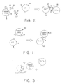
- Fig. 1 is a schematic of the interaction between a CTL with specific surface receptors and a target cell with antibody directed against the CTL receptors bound to its surface.
- Fig. 2 is a schematic of the interaction between a CTL with specific surface receptors, a target cell with specific surface molecules, and a bifunctional molecule consisting of an antibody specific for the receptor on the CTL surface and a molecule specific for the surface molecule on the target cell.
- Fig. 3 is a schematic showing lysis of the target cell following the interaction described in either Fig. 1 or Fig. 2 with phagocytic scavenger cells (e.g. macrophage) ingesting the cellular debris.
- The present invention is a process for making a selected or targeted cell susceptible to lysis by a cytotoxic T lymphocyte (CTL).
- In this process, the receptor on the CTL surface is recognized by and interacts with another molecule which is bound to the surface of the targeted cell. It is preferable to use the host's own CTLs, particularly if the interaction is to occur in vivo, but, in special circumstances, CTLs from other sources may also be used. Other sources would include tissue culture, another animal or the same species, or even an animal of a different species.
- It is also preferrable to increase the number of "activated" or stimulated CTLs prior to interaction with the targeted cells. In vivo, the increase in number and the activation may be brought about by exposing the host to convenient, suitable antigens or combinations of antigens, e.g. allogenic cells such as cultured normal keratinocytes or buffy coat cells from human blood, attenuated virus vaccines (measles) and certain non-pathogenic virus. Combinations of more than one antigen are preferable for they are more likely than a single antigen to activate many CTL clones.
- Lysis of the targeted cell requires binding of the target cell to the CTL, normally via the CTL's antigen specific receptor. The present invention is based on both the discovery of a practical and novel means for achieving this binding and on the unexpected discovery that the selected target cell can be lysed even when binding does not occur within the molecular context of a MHC gene product. Under normal conditions, the presence of the MHC gene product appears to be indispensible for recognition of a target cell by T lymphocytes. It is theorized that T lymphocytes with effector functions (helper and cytotoxic T lymphocytes) use the MHC gene products as their means for deciding whether or not a cell is foreign. Indeed, the conclusion drawn from one recent analysis by G. Berke, E. McVey, V. Hu, and W. R. Clark in the Journal of Immunology, 127:782-787 (1981) of lectin-dependent lysis of target cells by CTLs is that even in this putatively antigen-independent process the presence of class I MHC products is still necessary for target cell lysis.
- In the present invention, the CTL may be "bound" or brought into an interactive context with the target cell by means of a molecule that binds to the surfaces of both the CTL and the target cell. In one embodiment, shown in Fig. 1, an
antibody 12 to the CTL receptor, an anti-receptor antibody, is bound to thetarget cell 14 by means of a crosslinking agent such as glutaraldehyde. Theanti-receptor antibody 12 then binds to theCTL receptor 16 on theCTL 18, which lyses thetarget cell 14. - The preferred anti-receptor antibody is a monoclonal antibody directed against a region of the CTL's antigen-specific receptor which is shared by the corresponding receptors of most CTLs. Such a constant region is described by Saito et al. in Nature 309, 757-762 (1984) and Nature (November 1, 1984). A second anti-receptor antibody useful in the present invention is one directed against a cytotoxic T lymphocyte receptor-associated molecule, such as T3. The associated molecule must be located in close enough proximity to the CTL receptor to provide bridging between the CTL and the cell to be lysed so that the CTL receptor is located between the two linked cells.
- Monoclonal anti-receptor antibodies of the desired specificity may be produced from hybridomas made from spleen cells isolated from immunized rats and fused with myeloma cells, wherein the rats are immunized using either
- (1) CTL receptor protein isolated using antibody directed against the variable region of a particular CTL receptor,
- (2) chemically synthesized peptides with a ten to twelve amino acid sequence corresponding to a part of the constant region of the CTL receptor, or
- (3) polypeptides corresponding to most or all of a subunit of the receptor, made by recombinant DNA techniques.
- In a second embodiment shown in Fig. 2, more suitable to an in vivo situation, lysis of a
target cell 14 is produced by bridging the CTL to thetarget cell 14 by means of a "hybrid"molecule 20 made by joining ananti-receptor antibody 12 with anothermolecule 22 which selectively binds to asurface molecule 24 on the targetedcell 14. For example, a second antibody directed against a target cell surface antigen, such as a viral antigen or a tumor antigen, may be used as themolecule 22. A hormone, such as melanocyte stimulating hormone (MSH), which is selectively bound by a receptor on the targeted cell may also be used. The latter may be efficacious in the treatment of a melanoma type cancer, which is known to have specific MSH receptors on its surface. Other hormones, such as adrenal or thyroid hormones, might be useful in cases where removal of these glands is indicated. In most cases, thepreferred molecule 22 to be attached to theanti-receptor antibody 12 is a protein. However, other molecules, such as carbohydrates, may also be used. An example of such a carbohydrate is galactose which binds to receptors on mammalian hepatocytes, described in "Carbohydrate Recognition Systems for Receptor-Mediated Pinocytosis" by E.F. Neufeld and G. Ashwell in The Biochemistry of Glycoproteins and Proteoglycans, ed. by William J. Lennarz (Plenum Press, N.Y., 1980) at pages 241-266. - The target cell specific protein may be linked to the anti-receptor antibody using any of the bifunctional crosslinking reagents known to those those skilled in the art. Examples of such cross-linking agents are glutaraldehyde and bifunctional sulfo-NHS-Esters such as 3,3'-Dithiobis(sulfosuccinimidyl propionate) described by J.V. Staros, in Biochemistry, 21: 3950-3955; Bis (sulfosuccinimidyl) suberate, described by D.P. Giedroc et al. in J. Biol. Chem., 258: 16-19 (1983); and N-succinimidyl-3-(2-pyridyldithio) proprionate described by A.R. Neurath, and N. Strick in J. Virol. Methods, 3: 155-165 (1981), which may be obtained from Pierce Chemical Co. The active or binding sites of the crosslinked molecules should be at the exposed ends so that they are able to interact with the molecules on the target cell and CTL surfaces.
- For an in vivo situation wherein the crosslinked molecule is injected intravascularly, intraperitoneally, subcutaneously, or by other routes, the antibody molecules should either be selected from the classes or isotypes of antibodies which do not fix complement by the classical pathway, or be treated to remove those portions of the antibody constant region that are required to activate complement. The complement system consists of a group of protein molecules which can recognize certain sites on complexed or cell bound antibodies, attach to the complex or cell in a sequential fashion, and ultimately cause cellular lysis and inflammation. This is undesireable when it occurs in a non-specific or uncontrolled fashion. IgG₄, IgA and IgE do not fix complement by the classical pathway. IgM, IgG₁, IgG₂ and IgG₃ do fix complement. Enzymatic treatment may be used to eliminate the complement-fixing portions of IgG₁, IgG2, IgG₃, and IgM while leaving the activated, antigen-binding sites intact.
- An in vitro application of this method for promotion of CTL-mediated lysis would be in the selective treatment of cell populations removed from a patient or grown in tissue culture. An example would be to use a molecule consisting of an antibody against the CTL receptor and an antibody to an antigen found on the surface of tumor cells to eliminate tumor cells from bone marrow removed from a patient undergoing radiation treatment prior to re-introducing the bone marrow.
- CTLs are found throughout the body: in tissues, the lymphatic system, and in the blood. Accordingly, it is possible to inject the appropriate crosslinked molecule into an animal or human and cause CTL mediated lysis of the targeted cells anywhere in the body. The protein molecules themselves have a relatively short half life. For example, human IgG has a half life of 23 days. The other human immunoglobulins have half lives of less than 6 days. See Davis, Dulbecco, Eisen, Ginsberg, Wood, and McCarty, Microbiology, p. 483 (2nd ed. 1973). If not bound and destroyed, the antibodies will circulate until they degrade and are removed, for example, by scavenger cells such as circulating macrophages, Kupffer cells in the liver, and alveolar macrophages in the lungs. The cellular debris resulting from CTL-mediated lysis may be disposed of in the same fashion, as shown in Fig. 3. In an in vivo situation, as with any drug, care must be exercised in the administration of the crosslinked molecule to avoid anaphylatic shock.
- The present invention will be further understood from the following non-limiting example. All of the starting materials for this procedure are readily available to those skilled in the art from commercial or other sources.
- In the following detailed example, a monoclonal antibody (mAb) was produced against a CTL clone, clone 2C. This mAb was shown to block the CTL clone's cytolytic activity and to immunoprecipitate its CTL receptor, but not to block the activity or to precipitate the corresponding receptors of other CTL clones of similar specificity. The finding may be interpreted to mean the mAb was directed specifically against a portion of the variable region of the receptor unique to that CTL clone. The anti-CTL receptor mAb was purified and covalently attached to ⁵¹Cr-labeled cells. The labeled cells were then tested for susceptibility to lysis by CTL clones 2C and 2.1.1. Clone 2.1.1 is also specific for the product of a gene at the D end of the H-2d haplotype and, like clone 2C, it has its own unique cell surface molecule with the characteristics of the T lymphocyte receptor. The cells to which the mAb was attached included a human T cell lymphoma cell line, HPB-ALL , and murine cell lines that lack the H-2d haplotype of the natural targets for clone 2C; tumor cell lines EL4(H-2b), BW5147 (H-2k), Rl.l (H-2k) and Rl.E (a variant of Rl.l which is negative for Class I surface antigens). A tumor cell line, P815 (H-2d), which is normally lysed by clone 2C and clone 2.1.1 was used as a control.
- Both clones 2C and 2.1.1 lysed the unmodified and modified (mAb coupled) P815 control cells to an equal extent. Cells lacking the H-2d haplotype (murine EL4 and human HPB-ALL cells) are lysed poorly, if at all by these clones. However, after attaching the mAb directed against clone 2C's receptor, murine EL 4 and human HPB-ALL were lysed by clone 2C but not by clone 2.1.1. Attaching mAbs directed against other CTL surface molecules such as LFA-1 and Thy-1 to EL-4 and BW5417 (H-2k) cells did not render the cells susceptible to lysis by clone 2C.
- Not only a human cell line but also cells without any detectable cell surface MHC class I molecules were shown to be convertible into target cells for a clone of murine CTLs using mAb to the CTL clone receptor. In this example, two cell lines which are not normally lysed by clone 2C were used. Rl.l is a cultured C58/J lymphoma (H-2k) whose variant, Rl.E, does not produce beta₂-microglobulin and is devoid of cell surface class I molecules. After attachment of the mAb specific for the CTL clone 2C receptor, both Rl.l and Rl.E cells became susceptible to lysis by clone 2C. Neither modified nor unmodified Rl.l or Rl.E cells were lysed by the control clone 2.1.1.
- The following materials and methods were used in the above described example.
- Mice: BALB/c AnN (H-2d) and BALB.B (H-2b) mice were produced in the Center for Cancer Research, Massachusetts Institute of Technology.
- Tumor Cell Lines: P815 (H-2d), EL4(H-2b), BW5147(H-2k), Rl.l(H-2k), Rl.E (a variant of Rl.l that is negative for class I surface antigens), HPB-ALL (a human T cell lymphoma), and X63.653 (a BALB/c derived myeloma) were all maintained in culture with RPMI 1640 containing 10% fetal calf serum, 10 mM HEPES, 2 mM L-glutamine, 100 Units penicillin/ml of culture fluid, 100 micrograms streptomycin/ml culture fluid, and 5 X 10⁻⁵ M 2-mercaptoethanol.
- Cloned CTL: Alloreactive CTL clones 2C, G4, and 2.1.1 were produced from spleen cells of BALB.B mice as described by M.V. Sitkovsky, M.S. Pasternack, and H.N. Eisen in J. Immunology, 124:1372-1376 (1982). The clones were maintained by weekly stimulation with irradiated (4000 rads) P815 cells plus supernatants from rat spleen cells maintained in culture for 48 hours in the presence of concanavalin A (Vector Laboratories, Burlingame, CA). All three clones lysed target cells that expressed a product of the D-end gene of the H-2d haplotype.
- Monoclonal Antibodies: The mAb (1B2) that recognizes the T cell receptor of clone 2C was produced as described by D.M. Kranz, D.H. Sherman, M.V. Sitkovsky, M.S. Pasternack and H.N. Eisen in Proc. Nat. Acad. Sci. USA, 81:573-577, (1984). BALB/c AnN mice were immunized by intraperitoneal injection 6, times at 2-3 week intervals, each time with 10-20 x 10⁶ cells from clone 2C. Four days after the final injection, spleen cells were harvested from the mice and fused with X63.653 myeloma cells using 50% polyethylene glycol. Cells were distributed into ten 24-well plates (Costar, Inc.) and grown in HAT (hypoxanthine, aminopterin, thymidine) selection media. After 2 to 3 weeks, culture supernatants were screened for their ability to block lysis of P815 target cells by clone 2C in a ⁵¹Cr-release assay, as described below.
- Rat mAbs against lymphocyte function-associated antigen type 1 (4-16-1) and class I-H-2b surface antigens (3-18-8) were produced as previously described by Kranz et al, Proc. Nat. Acad. Sci. USA, 81:573-577 (1984). Rat mAb against Lyt-2 (3.155.2) described by M. Sarmiento, A. L. Glasebrook, and F. W. Fitch in J. Immunol., 125: 2665-1672 (1980) was a generous gift from Frank Fitch and Marian Sarmiento (University of Chicago).
- Cytoxicity Assays: CTL-mediated target cell lysis was measured by a standard ⁵¹Cr-release assay as described by K. Grabstein in Selected Methods in Cellular Immunology, B.B. Mishell and S.M. Shiigi, eds. p. 124-137 (San Francisco: Freeman 1980). Various numbers of CTLs (in 100 microliters of media) were added to 2 X 10⁴ ⁵¹Cr-labled target cells (in 100 microliters) and incubated at 37°C for 4 hours. Cells were pelleted by centrifugation, supernatants were assayed for radioactivity, and the percent specific ⁵¹Cr release was calculated from

where a is ⁵¹Cr release in the presence of CTL, b is the spontaneous release of ⁵¹Cr from labeled target cells in the absence of the CTL (less than 15%), and t is the total ⁵¹Cr content of the target cells. In some experiments, CTLs were preincubated with mAbs for 30 minutes prior to addition of ⁵¹Cr labeled target cells. All assays were performed in triplicate. - Purification and coupling of mAb 1B2: Antibodies were purified from ascites fluid that had been elicited by intraperitoneal injection of BALB/c mice with 5 X 10⁵ viable 1B2 hybridoma cells. Lipoproteins were removed by adsorption to 0.25% sodium dextran sulfate and precipitation with 1.5% CaCl₂. The enriched gamma globulin fraction, obtained by precipitation with 50% saturated ammonium sulfate, was dialysed against 50 mM potassium phosphate, PH 8.0. Since mAb 1B2 contains gamma₁ heavy chains (determined by indirect radioimmune binding assays), the antibody was further purified by anion exchange chromatography on DEAE-cellulose. The pass-through from the DEAE-cellulose contained the antibody and was dialyzed against phosphate buffered saline, pH 7.4 (PBS).
- Several methods of attaching mAb 1B2 to target cells were examined. In the mildest effective method, the mAb was crosslinked to the cell surface with 0.1% glutaraldehyde. In this procedure, ⁵¹Cr-labled cells were washed two times with RPMI-1640, resuspended in 0.25 ml of RPMI-1640 containing 0.2% gutaraldehyde, and 0.25 ml of mAb 1B2 (2 mg per ml in PBS) was added. The mixture was incubated on ice for 20 minutes and layered over a Ficol™ (Pharmacia Fine Chemicals) gradient. After centrifugation (3000 rpm, 15 min), the interphase (viable) cells were collected, washed three times with RPMI 1640 containing 10% fetal calf serum (FCS) and used in the cytoxicity assay.
- Antibody was coupled to Affi-Gel™ 10 (Bio-Rad Laboratories) at about 5 mg per ml of packed gel for use in immunoprecipitation assays.
- Labelling of Cell Surface Proteins with 125 I.: Cloned CTLs (2-3 x 10⁷) were washed three times with PBS and the following were added to the cell pellet: 100 microgram of lactoperoxidase, 1 mCi of Na ¹²⁵I (New England Nuclear), and 50 microliters of 0.03% H₂O₂. Labeled cells were washed three times with PBS and extracted with 1 ml of 0.5% Nonidet P40 in 0.15 M NaCl, 0.02% NaN₃, 25 micromolar phenylmethylsulfonyl fluoride, 10 mM Tris-HCl, pH 7.2 (extraction buffer).
- Immunoprecipitation and SDS/PAGE: ¹²⁵I-labeled cell extracts (approximately 10⁶ cpm), containing 0.5% bovine serum albumin, were incubated with 50 microliters of 1B2-Affi-Gel 10™ for 3 hours on ice. The gel was then washed three times with the previously described extraction buffer and heated at 100°C in SDS/PAGE sample buffer, with or without 0.1 M 2-mercaptoethanol. Electrophoresis was performed in 10% polyacrylamide gels containing SDS according to the method of U.K. Laemmli, Nature (London) New Biol, 227:680-685 (1975). Gels were fixed, dried, and exposed to Kodak X-Omat XAR film™ using a Dupont Cronex Lighting Plus intensifying screen.
- Although this invention has been described with reference to specific embodiments, it is understood that modifications and variations may occur to those skilled in the art. Such other embodiments and modifications are intended to fall within the scope of the appended claims.
Claims (26)
- A process for making a targeted cell susceptible to lysis by a cytotoxic T lymphocyte comprising binding an antibody specific for cytotoxic T lymphocyte receptor determinants to said targeted cell.
- The process of claim 1 wherein said receptor determinants are on the constant domain of a cytotoxic T lymphocyte receptor.
- The process of claim 1 wherein said determinants are located on a cytotoxic T lymphocyte receptor-associated molecule.
- The process of claim 3 wherein said receptor-associated molecule is T3.
- The process according to claim 1 wherein said anti-receptor antibody is bound to said targeted cell by the non-antigen binding region of said antibody.
- The process according to claim 5 wherein said anti-receptor antibody is bound to said targeted cell by means of a second molecule that specifically binds to the surface of said targeted cell.
- The process according to claim 6 wherein said second molecule binding the antireceptor antibody to the targeted cell is a second antibody specific for an antigen on the surface of said targeted cell.
- The process according to claim 6 wherein said second molecule is a hormone that specifically binds to a receptor on the surface of said targeted cell.
- The process according to claim 6 wherein said second molecule is a carbohydrate that specifically binds to a receptor on the surface of said targeted cell.
- The process according to claim 1 further comprising eliminating the complement fixing portion of said antibodies.
- The process according to claim 7 further comprising eliminating the complement fixing portion of said antibodies.
- The process according to claim 1 wherein said antibody is selected from the group of non-complement fixing antibodies consisting of IgG₄, IgA, and IgE.
- The process according to claim 7 wherein said antibodies are selected from the group of non-complement fixing antibodies consisting of IgG₄, IgA, and IgE.
- The process according to claim 6 wherein said anti-receptor antibody is bound to said second molecule using a bifunctional crosslinking reagent.
- The process according to claim 6 wherein said second molecule specifically binds to a third molecule on the surface of said targeted cell selected from the group consisting of tumor antigens, viral antigens, major histocompatibility antigens, specific protein receptors, hormone receptors, and carbohydrate receptors.
- A reagent for use in making a targeted cell susceptible to lysis by cytotoxic T lymphocytes comprising an antibody specific for T lymphocyte receptor determinants.
- The reagent of claim 16 wherein said anti-receptor antibody is directed against a portion of a T lymphocyte receptor shared by cytotoxic T lymphocyte receptors.
- The reagent of claim 16 wherein said anti-receptor antibody is directed against a portion of a cytotoxic T lymphocyte receptor-associated molecule.
- The reagent of claim 16 further comprising a second molecule which specifically binds to the surface of said targeted cell.
- The reagent of claim 19 wherein said second molecule is covalently hound to said anti-receptor antibody.
- The reagent of claim 19 wherein said second molecule is selected from the group consisting of antibodies, hormones, and carbohydrates.
- The reagent of claim 19 wherein said second molecule specifically binds to a third molecule on the surface of said targeted cell selected from the group consisting of tumor antigens, viral antigens, specific protein receptors, hormone receptors, and carbohydrate receptors.
- A process for lysing targeted cells comprising binding an antibody directed against T lymphocyte receptor determinants shared by most cytotoxic T lymphocytes to said targeted cell and exposing said anti-receptor antibody-bound cells to cytotoxic T lymphocytes.
- The process according to claim 23 wherein said anti-receptor antibody is bound to said targeted cell by means of a second molecule that specifically binds to the surface of said targeted cell.
- The process according to claim 24 wherein said second molecule is selected from the group consisting of antibodies, hormones, and carbohydrates.
- The process according to claim 23 wherein said anti-receptor antibody is directed against T3.
Priority Applications (1)
| Application Number | Priority Date | Filing Date | Title |
|---|---|---|---|
| AT85113657T ATE74622T1 (en) | 1984-10-31 | 1985-10-26 | METHOD OF SENSITIZING A TARGET CELL TO LYSIS BY CYTOTOXIC T-LYMPH BODIES. |
Applications Claiming Priority (2)
| Application Number | Priority Date | Filing Date | Title |
|---|---|---|---|
| US66688084A | 1984-10-31 | 1984-10-31 | |
| US666880 | 1996-06-25 |
Publications (3)
| Publication Number | Publication Date |
|---|---|
| EP0180171A2 EP0180171A2 (en) | 1986-05-07 |
| EP0180171A3 EP0180171A3 (en) | 1987-09-09 |
| EP0180171B1 true EP0180171B1 (en) | 1992-04-08 |
Family
ID=24675887
Family Applications (1)
| Application Number | Title | Priority Date | Filing Date |
|---|---|---|---|
| EP85113657A Expired - Lifetime EP0180171B1 (en) | 1984-10-31 | 1985-10-26 | Process for making a targeted cell susceptible to lysis by cytotoxic t lymphocytes |
Country Status (4)
| Country | Link |
|---|---|
| EP (1) | EP0180171B1 (en) |
| JP (1) | JPS61234779A (en) |
| AT (1) | ATE74622T1 (en) |
| DE (1) | DE3585818D1 (en) |
Families Citing this family (7)
| Publication number | Priority date | Publication date | Assignee | Title |
|---|---|---|---|---|
| EP0308936B1 (en) * | 1987-09-23 | 1994-07-06 | Bristol-Myers Squibb Company | Antibody heteroconjugates for the killing of HIV-infected cells |
| US5766947A (en) * | 1988-12-14 | 1998-06-16 | Astra Ab | Monoclonal antibodies reactive with an epitope of a Vβ3.1 variable region of a T cell receptor |
| US5223426A (en) | 1988-12-15 | 1993-06-29 | T Cell Sciences, Inc. | Monoclonal antibodies reactive with defined regions of the t-cell antigen receptor |
| AU6290090A (en) * | 1989-08-29 | 1991-04-08 | University Of Southampton | Bi-or trispecific (fab)3 or (fab)4 conjugates |
| SE9002484L (en) | 1990-07-20 | 1992-01-21 | Kabi Pharmacia Ab | NEW SUBSTITUTED POLYETERS |
| JP3105629B2 (en) † | 1991-04-23 | 2000-11-06 | サングスタット メディカル コーポレイション | Cell activity regulating conjugates of members of specific binding pairs |
| AU5697001A (en) | 2000-03-31 | 2001-10-15 | Purdue Research Foundation | Method of treatment using ligand-immunogen conjugates |
Family Cites Families (1)
| Publication number | Priority date | Publication date | Assignee | Title |
|---|---|---|---|---|
| EP0103139B1 (en) * | 1982-08-11 | 1990-01-03 | Kabushiki Kaisha Toshiba | Reagent composition for immunoassay and immunoassay using the same |
-
1985
- 1985-10-26 DE DE8585113657T patent/DE3585818D1/en not_active Expired - Fee Related
- 1985-10-26 AT AT85113657T patent/ATE74622T1/en not_active IP Right Cessation
- 1985-10-26 EP EP85113657A patent/EP0180171B1/en not_active Expired - Lifetime
- 1985-10-31 JP JP60243026A patent/JPS61234779A/en active Pending
Non-Patent Citations (3)
| Title |
|---|
| CHEMICAL ABSTRACTS, vol. 102, no. 11, 18 March 1985, Columbus, OH (US); D.M.KRANZ et al., p. 440, no. 94055w * |
| CHEMICAL ABSTRACTS, vol. 81, no. 15, 14 October 1974, Columbus, OH (US); F.WISLOEFF et al., p. 317, no. 89587q * |
| CHEMICAL ABSTRACTS, vol. 97, no. 9, 30 August 1982, Columbus, OH (US); D.DIALYNAS et al., p. 481, no. 70590u * |
Also Published As
| Publication number | Publication date |
|---|---|
| ATE74622T1 (en) | 1992-04-15 |
| JPS61234779A (en) | 1986-10-20 |
| EP0180171A3 (en) | 1987-09-09 |
| EP0180171A2 (en) | 1986-05-07 |
| DE3585818D1 (en) | 1992-05-14 |
Similar Documents
| Publication | Publication Date | Title |
|---|---|---|
| Acuto et al. | The human T cell receptor: appearance in ontogeny and biochemical relationship of α and β subunits on IL-2 dependent clones and T cell tumors | |
| Carrel et al. | Subsets of human Ia-like molecules defined by monoclonal antibodies | |
| Weissman et al. | A new subunit of the human T-cell antigen receptor complex | |
| Tony et al. | Major histocompatibility complex-restricted, polyclonal B cell responses resulting from helper T cell recognition of antiimmunoglobulin presented by small B lymphocytes. | |
| Waldmann | The structure, function, and expression of interleukin-2 receptors on normal and malignant lymphocytes | |
| Smith et al. | Antibodies to CD3/T-cell receptor complex induce death by apoptosis in immature T cells in thymic cultures | |
| Terhorst et al. | Biochemical studies of the human thymocyte cell-surface antigens T6, T9 and T10 | |
| US5078998A (en) | Hybrid ligand directed to activation of cytotoxic effector T lymphocytes and target associated antigen | |
| Staerz et al. | Monoclonal antibodies specific for a murine cytotoxic T-lymphocyte clone. | |
| White et al. | Definition of the antigenic polypeptides in the Sm and RNP ribonucleoprotein complexes | |
| Van de Rijn et al. | The thymic differentiation markers T6 and M241 are two unusual MHC class I antigens. | |
| Bartlett et al. | Cognate interactions between helper T cells and B cells. IV. Requirements for the expression of effector phase activity by helper T cells. | |
| EP0180171B1 (en) | Process for making a targeted cell susceptible to lysis by cytotoxic t lymphocytes | |
| Tsujisaki et al. | Fine specificity and idiotype diversity of the murine anti-HLA-A2, A28 monoclonal antibodies CR11–351 and KS1 | |
| Kingsley et al. | Abnormal Helper‐Inducer/Suppressor–Inducer T‐Cell Subset Distribution and T‐Cell Activation Status are Common to All Types of Chronic Synovitis | |
| Binz et al. | T cell receptors with allo-major histocompatibility complex specificity from rat and mouse. Similarity of size, plasmin susceptibility, and localization of antigen-binding region. | |
| Turkewitz et al. | Large-scale purification of murine I-Ak and I-Ek antigens and characterization of the purified proteins | |
| Revilla et al. | Targeting to porcine sialoadhesin receptor improves antigen presentation to T cells | |
| BISHARA et al. | Human leukocyte antigens (HLA) class I and class II on sperm cells studied at the serological, cellular, and genomic levels | |
| Mellstedt et al. | Patients treated with a monoclonal antibody (ab1) to the colorectal carcinoma antigen 17–1A develop a cellular response (DTH) to the “internal image of the antigen”(ab2) | |
| JPH04505401A (en) | Monoclonal antibody that recognizes peptides associated with major histocompatibility antigen | |
| Hua et al. | Lysis of hybridoma cells bearing anti‐clonotypic surface immunoglobulin by clonotype‐expressing alloreactive cytotoxic T cells | |
| Hiramatsu et al. | Unique T cell Ia antigen expressed on a hybrid cell line producing antigen-specific augmenting T cell factor. | |
| Emmrich et al. | Selective stimulation of human T lymphocyte subsets by heteroconjugates of antibodies to the T cell receptor and to subset‐specific differentiation antigens | |
| Sakato et al. | A small hypervariable segment in the variable domain of an immunoglobulin light chain stimulates formation of anti-idiotypic suppressor T cells. |
Legal Events
| Date | Code | Title | Description |
|---|---|---|---|
| PUAI | Public reference made under article 153(3) epc to a published international application that has entered the european phase |
Free format text: ORIGINAL CODE: 0009012 |
|
| AK | Designated contracting states |
Kind code of ref document: A2 Designated state(s): AT BE CH DE FR GB IT LI LU NL SE |
|
| PUAL | Search report despatched |
Free format text: ORIGINAL CODE: 0009013 |
|
| AK | Designated contracting states |
Kind code of ref document: A3 Designated state(s): AT BE CH DE FR GB IT LI LU NL SE |
|
| 17P | Request for examination filed |
Effective date: 19880304 |
|
| 17Q | First examination report despatched |
Effective date: 19900301 |
|
| GRAA | (expected) grant |
Free format text: ORIGINAL CODE: 0009210 |
|
| AK | Designated contracting states |
Kind code of ref document: B1 Designated state(s): AT BE CH DE FR GB IT LI LU NL SE |
|
| REF | Corresponds to: |
Ref document number: 74622 Country of ref document: AT Date of ref document: 19920415 Kind code of ref document: T |
|
| ITF | It: translation for a ep patent filed |
Owner name: JACOBACCI & PERANI S.P.A. |
|
| REF | Corresponds to: |
Ref document number: 3585818 Country of ref document: DE Date of ref document: 19920514 |
|
| ET | Fr: translation filed | ||
| PLBE | No opposition filed within time limit |
Free format text: ORIGINAL CODE: 0009261 |
|
| STAA | Information on the status of an ep patent application or granted ep patent |
Free format text: STATUS: NO OPPOSITION FILED WITHIN TIME LIMIT |
|
| 26N | No opposition filed | ||
| PGFP | Annual fee paid to national office [announced via postgrant information from national office to epo] |
Ref country code: FR Payment date: 19931011 Year of fee payment: 9 |
|
| PGFP | Annual fee paid to national office [announced via postgrant information from national office to epo] |
Ref country code: SE Payment date: 19931015 Year of fee payment: 9 |
|
| PGFP | Annual fee paid to national office [announced via postgrant information from national office to epo] |
Ref country code: GB Payment date: 19931018 Year of fee payment: 9 |
|
| PGFP | Annual fee paid to national office [announced via postgrant information from national office to epo] |
Ref country code: DE Payment date: 19931021 Year of fee payment: 9 |
|
| PGFP | Annual fee paid to national office [announced via postgrant information from national office to epo] |
Ref country code: CH Payment date: 19931025 Year of fee payment: 9 |
|
| PGFP | Annual fee paid to national office [announced via postgrant information from national office to epo] |
Ref country code: AT Payment date: 19931029 Year of fee payment: 9 |
|
| PGFP | Annual fee paid to national office [announced via postgrant information from national office to epo] |
Ref country code: NL Payment date: 19931031 Year of fee payment: 9 |
|
| PGFP | Annual fee paid to national office [announced via postgrant information from national office to epo] |
Ref country code: LU Payment date: 19931101 Year of fee payment: 9 |
|
| PGFP | Annual fee paid to national office [announced via postgrant information from national office to epo] |
Ref country code: BE Payment date: 19931130 Year of fee payment: 9 |
|
| EPTA | Lu: last paid annual fee | ||
| PG25 | Lapsed in a contracting state [announced via postgrant information from national office to epo] |
Ref country code: LU Free format text: LAPSE BECAUSE OF NON-PAYMENT OF DUE FEES Effective date: 19941026 Ref country code: GB Effective date: 19941026 Ref country code: AT Effective date: 19941026 |
|
| PG25 | Lapsed in a contracting state [announced via postgrant information from national office to epo] |
Ref country code: SE Effective date: 19941027 |
|
| PG25 | Lapsed in a contracting state [announced via postgrant information from national office to epo] |
Ref country code: LI Effective date: 19941031 Ref country code: CH Effective date: 19941031 Ref country code: BE Effective date: 19941031 |
|
| EAL | Se: european patent in force in sweden |
Ref document number: 85113657.2 |
|
| BERE | Be: lapsed |
Owner name: MASSACHUSETTS INSTITUTE OF TECHNOLOGY Effective date: 19941031 |
|
| PG25 | Lapsed in a contracting state [announced via postgrant information from national office to epo] |
Ref country code: NL Effective date: 19950501 |
|
| NLV4 | Nl: lapsed or anulled due to non-payment of the annual fee | ||
| GBPC | Gb: european patent ceased through non-payment of renewal fee |
Effective date: 19941026 |
|
| PG25 | Lapsed in a contracting state [announced via postgrant information from national office to epo] |
Ref country code: FR Effective date: 19950630 |
|
| REG | Reference to a national code |
Ref country code: CH Ref legal event code: PL |
|
| PG25 | Lapsed in a contracting state [announced via postgrant information from national office to epo] |
Ref country code: DE Effective date: 19950701 |
|
| EUG | Se: european patent has lapsed |
Ref document number: 85113657.2 |
|
| REG | Reference to a national code |
Ref country code: FR Ref legal event code: ST |